/


Автор: Kent R.D.
Теги: psychology encyclopedia bradford books publisher psychiatry communication disorders
ISBN: 0-262-11278-7
Год: 2004


Текст
The MIT Encyclopedia of
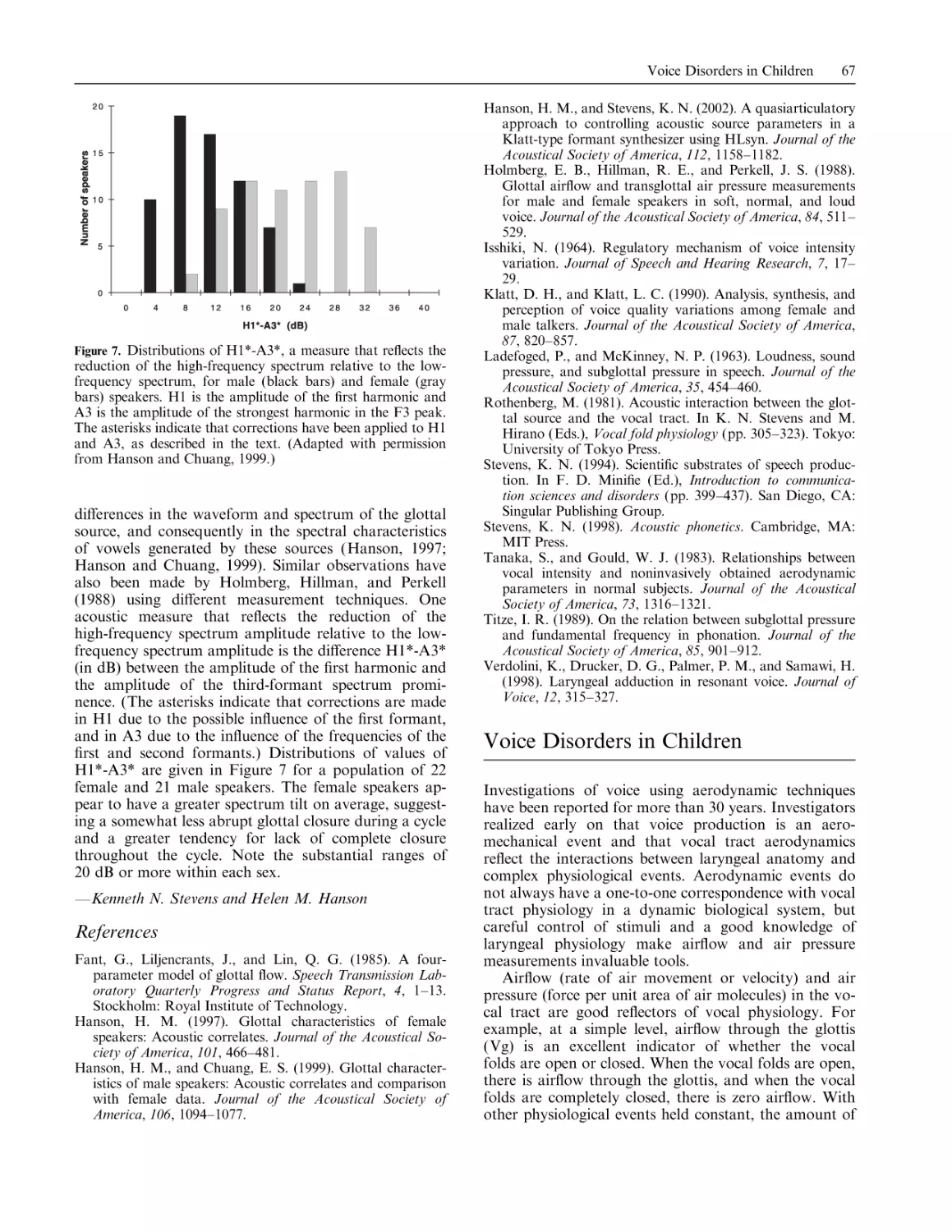
Communication Disorders
The MIT Encyclopedia of
Communication Disorders
Edited by Raymond D. Kent
A Bradford Book
The MIT Press
Cambridge, Massachusetts
London, England
( 2004 Massachusetts Institute of Technology
All rights reserved. No part of this book may be reproduced in any form by any electronic or mechanical
means (including photocopying, recording, or information storage and retrieval) without permission in
writing from the publisher.
This book was set in Times New Roman on 3B2 by Asco Typesetters, Hong Kong, and was printed and
bound in the United States of America.
Library of Congress Cataloging-in-Publication Data
The MIT encyclopedia of communication disorders / edited by Raymond D. Kent.
p. cm.
Includes bibliographical references and index.
ISBN 0-262-11278-7 (cloth)
1. Communicative disorders—Encyclopedias. I. Kent, Raymond D. II. Massachusetts Institute of
Technology.
RC423.M56 2004
616.850 50 003—dc21
2003059941
Contents
Introduction ix
Acknowledgments
Part I: Voice
xi
1
Acoustic Assessment of Voice 3
Aerodynamic Assessment of Vocal Function 7
Alaryngeal Voice and Speech Rehabilitation 10
Anatomy of the Human Larynx 13
Assessment of Functional Impact of Voice
Disorders 20
Electroglottographic Assessment of Voice 23
Functional Voice Disorders 27
Hypokinetic Laryngeal Movement Disorders 30
Infectious Diseases and Inflammatory Conditions of
the Larynx 32
Instrumental Assessment of Children’s Voice 35
Laryngeal Movement Disorders: Treatment with
Botulinum Toxin 38
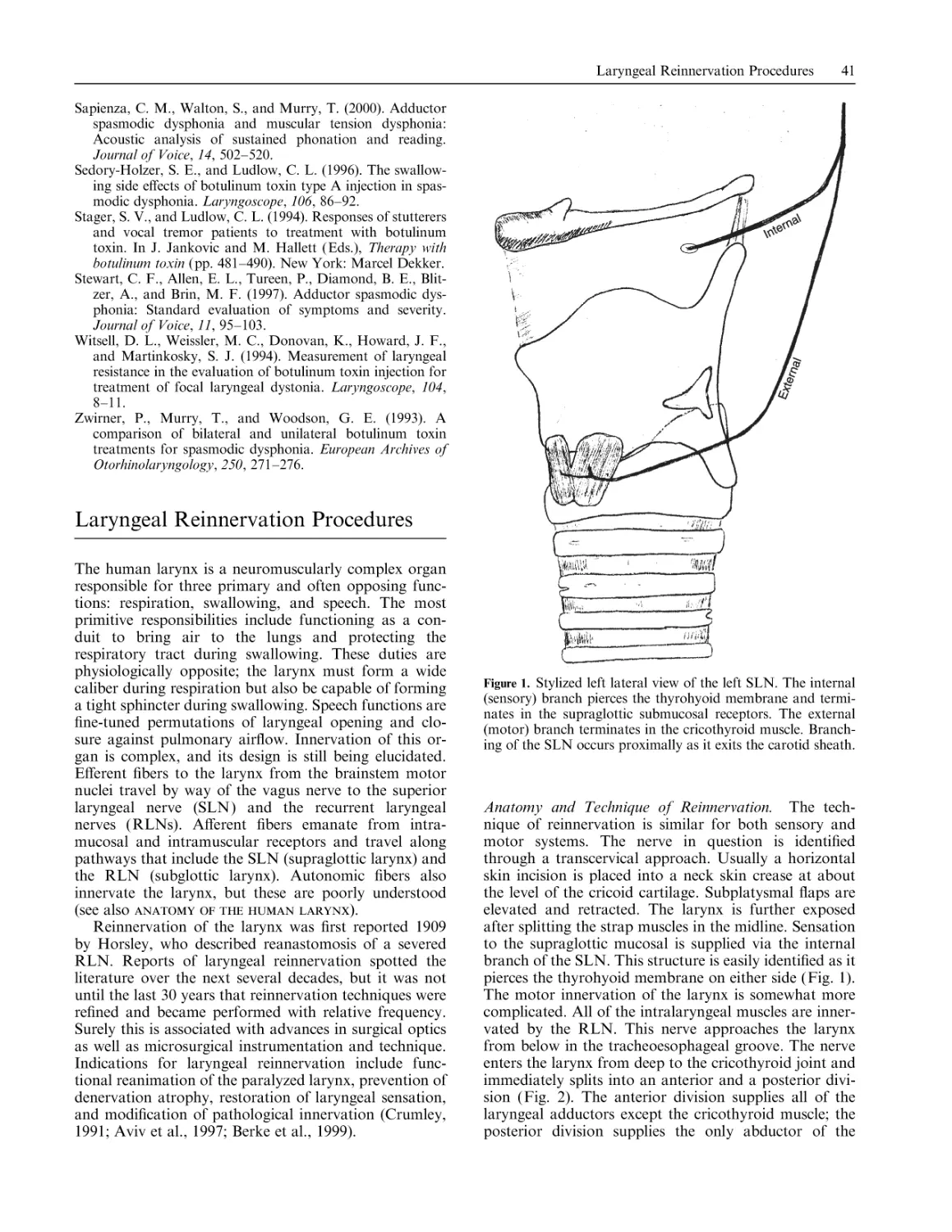
Laryngeal Reinnervation Procedures 41
Laryngeal Trauma and Peripheral Structural
Ablations 45
Psychogenic Voice Disorders: Direct Therapy 49
The Singing Voice 51
Vocal Hygiene 54
Vocal Production System: Evolution 56
Vocalization, Neural Mechanisms of 59
Voice Acoustics 63
Voice Disorders in Children 67
Voice Disorders of Aging 72
Voice Production: Physics and Physiology 75
Voice Quality, Perceptual Evaluation of 78
Voice Rehabilitation After Conservation
Laryngectomy 80
Voice Therapy: Breathing Exercises 82
Voice Therapy: Holistic Techniques 85
Voice Therapy for Adults 88
Voice Therapy for Neurological Aging-Related Voice
Disorders 91
Voice Therapy for Professional Voice Users 95
Part II: Speech
99
Apraxia of Speech: Nature and Phenomenology
Apraxia of Speech: Treatment 104
Aprosodia 107
Augmentative and Alternative Communication
Approaches in Adults 110
Augmentative and Alternative Communication
Approaches in Children 112
Autism 115
Bilingualism, Speech Issues in 119
Developmental Apraxia of Speech 121
Dialect, Regional 124
101
Dysarthrias: Characteristics and Classification 126
Dysarthrias: Management 129
Dysphagia, Oral and Pharyngeal 132
Early Recurrent Otitis Media and Speech
Development 135
Laryngectomy 137
Mental Retardation and Speech in Children 140
Motor Speech Involvement in Children 142
Mutism, Neurogenic 145
Orofacial Myofunctional Disorders in Children 147
Phonetic Transcription of Children’s Speech 150
Phonological Awareness Intervention for Children with
Expressive Phonological Impairments 153
Phonological Errors, Residual 156
Phonology: Clinical Issues in Serving Speakers of
African-American Vernacular English 158
Psychosocial Problems Associated with Communicative
Disorders 161
Speech and Language Disorders in Children: ComputerBased Approaches 164
Speech and Language Issues in Children from AsianPacific Backgrounds 167
Speech Assessment, Instrumental 169
Speech Assessment in Children: Descriptive Linguistic
Methods 174
Speech Development in Infants and Young Children
with a Tracheostomy 176
Speech Disfluency and Stuttering in Children 180
Speech Disorders: Genetic Transmission 183
Speech Disorders in Adults, Psychogenic 186
Speech Disorders in Children: A Psycholinguistic
Perspective 189
Speech Disorders in Children: Behavioral Approaches to
Remediation 192
Speech Disorders in Children: Birth-Related Risk
Factors 194
Speech Disorders in Children: Cross-Linguistic
Data 196
Speech Disorders in Children: Descriptive Linguistic
Approaches 198
Speech Disorders in Children: Motor Speech Disorders
of Known Origin 200
Speech Disorders in Children: Speech-Language
Approaches 204
Speech Disorders Secondary to Hearing Impairment
Acquired in Adulthood 207
Speech Issues in Children from Latino
Backgrounds 210
Speech Sampling, Articulation Tests, and Intelligibility
in Children with Phonological Errors 213
Speech Sampling, Articulation Tests, and Intelligibility
in Children with Residual Errors 215
Speech Sound Disorders in Children: Description and
Classification 218
vi
Contents
Stuttering 220
Transsexualism and Sex Reassignment: Speech
Di¤erences 223
Ventilator-Supported Speech Production 226
Part III: Language 229
Agrammatism 231
Agraphia 233
Alexia 236
Alzheimer’s Disease 240
Aphasia, Global 243
Aphasia, Primary Progressive 245
Aphasia: The Classical Syndromes 249
Aphasia, Wernicke’s 252
Aphasia Treatment: Computer-Aided
Rehabilitation 254
Aphasia Treatment: Pharmacological Approaches 257
Aphasia Treatment: Psychosocial Issues 260
Aphasic Syndromes: Connectionist Models 262
Aphasiology, Comparative 265
Argument Structure: Representation and
Processing 269
Attention and Language 272
Auditory-Motor Interaction in Speech and
Language 275
Augmentative and Alternative Communication: General
Issues 277
Bilingualism and Language Impairment 279
Communication Disorders in Adults: Functional
Approaches to Aphasia 283
Communication Disorders in Infants and Toddlers
285
Communication Skills of People with Down
Syndrome 288
Dementia 291
Dialect Speakers 294
Dialect Versus Disorder 297
Discourse 300
Discourse Impairments 302
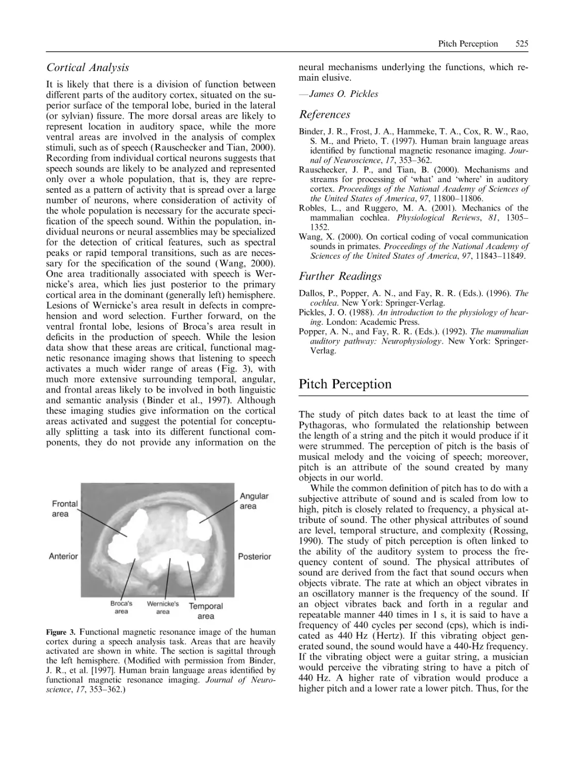
Functional Brain Imaging 305
Inclusion Models for Children with Developmental
Disabilities 307
Language Development in Children with Focal
Lesions 311
Language Disorders in Adults: Subcortical
Involvement 314
Language Disorders in African-American
Children 318
Language Disorders in Latino Children 321
Language Disorders in School-Age Children: Aspects of
Assessment 324
Language Disorders in School-Age Children:
Overview 326
Language Impairment and Reading Disability 329
Language Impairment in Children: Cross-Linguistic
Studies 331
Language in Children Who Stutter 333
Language of the Deaf: Acquisition of English 336
Language of the Deaf: Sign Language 339
Lingustic Aspects of Child Language Impairment—
Prosody 344
Melodic Intonation Therapy 347
Memory and Processing Capacity 349
Mental Retardation 352
Morphosyntax and Syntax 354
Otitis Media: E¤ects on Children’s Language 358
Perseveration 361
Phonological Analysis of Language Disorders in
Aphasia 363
Phonology and Adult Aphasia 366
Poverty: E¤ects on Language
369
Pragmatics 372
Prelinguistic Communication Intervention for Children
with Developmental Disabilities 375
Preschool Language Intervention 378
Prosodic Deficits 381
Reversibility/Mapping Disorders 383
Right Hemisphere Language and Communication
Functions in Adults 386
Right Hemisphere Language Disorders 388
Segmentation of Spoken Language by Normal Adult
Listeners 392
Semantics 395
Social Development and Language Impairment 398
Specific Language Impairment in Children 402
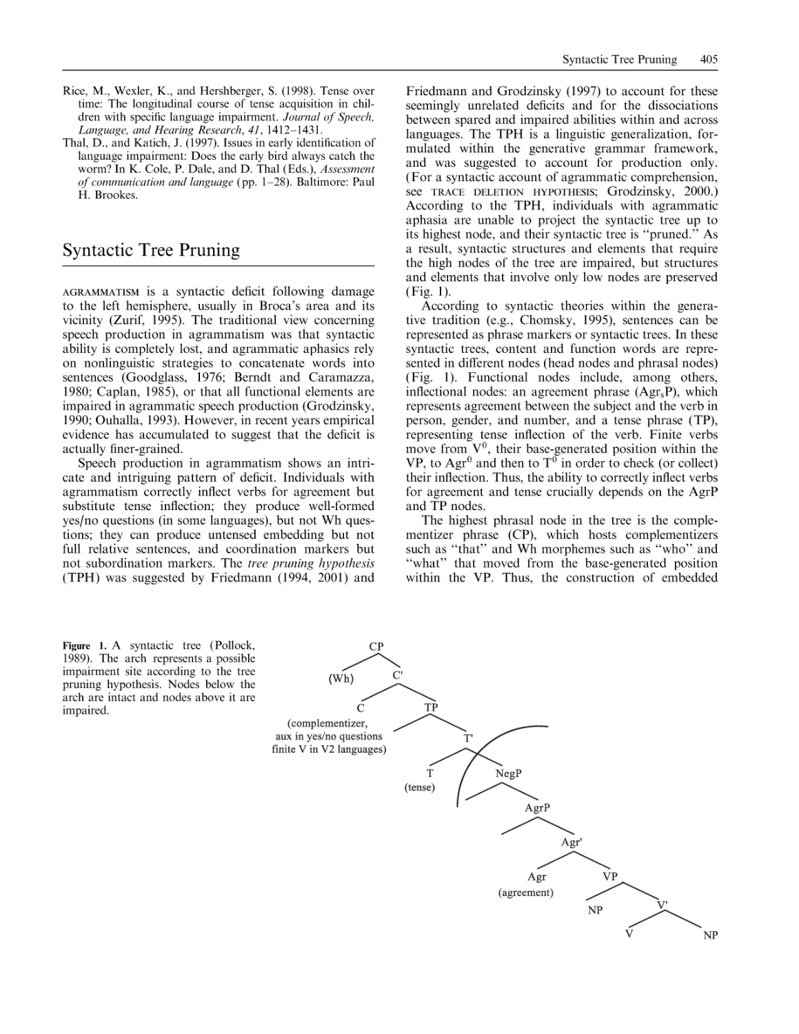
Syntactic Tree Pruning 405
Trace Deletion Hypothesis 407
Part IV: Hearing
411
Amplitude Compression in Hearing Aids 413
Assessment of and Intervention with Children Who Are
Deaf or Hard of Hearing 421
Audition in Children, Development of 424
Auditory Brainstem Implant 427
Auditory Brainstem Response in Adults 429
Auditory Neuropathy in Children 433
Auditory Scene Analysis 437
Auditory Training 439
Classroom Acoustics 442
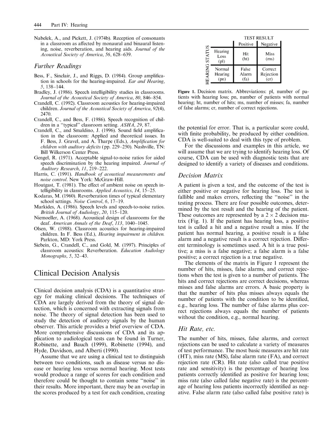
Clinical Decision Analysis 444
Cochlear Implants 447
Cochlear Implants in Adults: Candidacy 450
Cochlear Implants in Children 454
Dichotic Listening 458
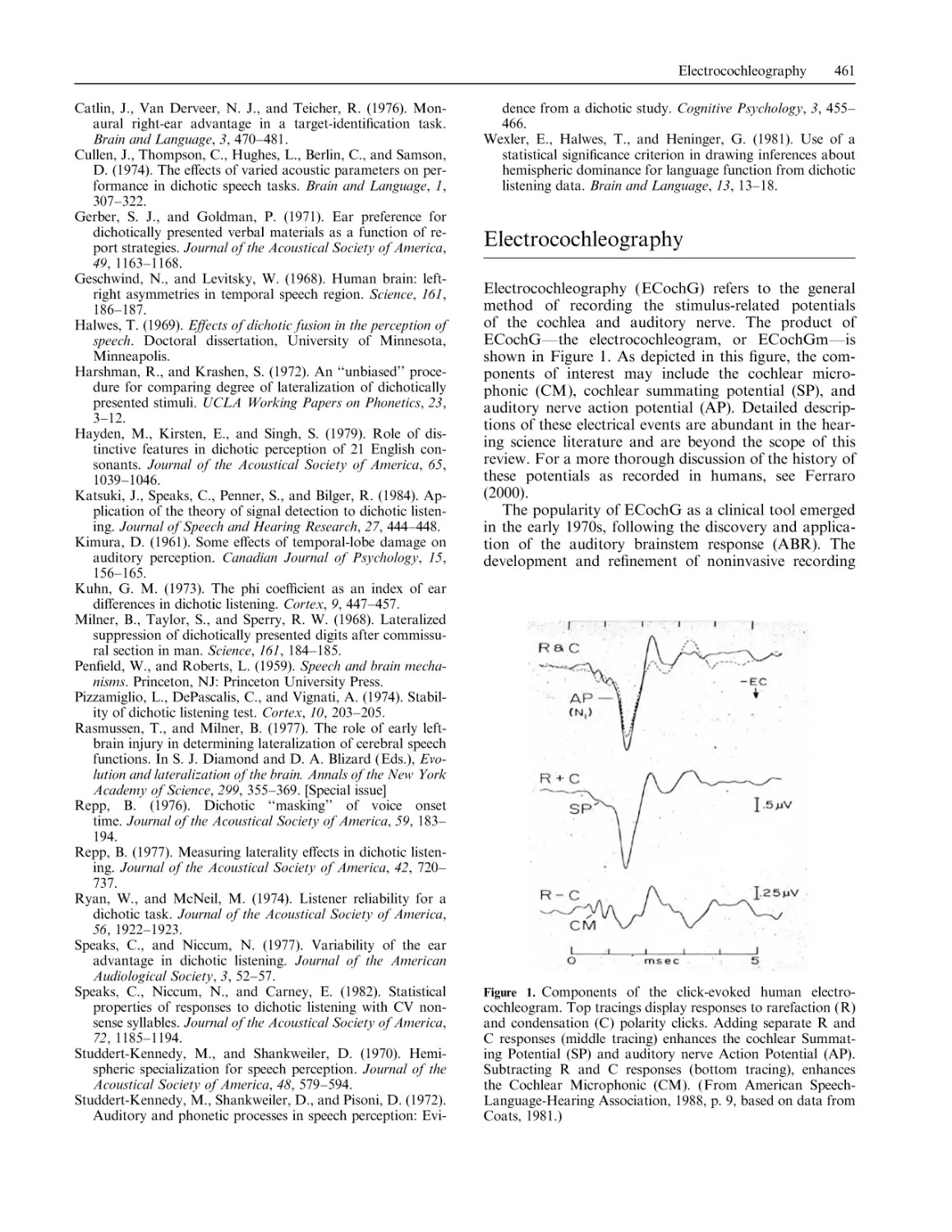
Electrocochleography 461
Electronystagmography 467
Frequency Compression 471
Functional Hearing Loss in Children 475
Genetics and Craniofacial Anomalies 477
Hearing Aid Fitting: Evaluation of Outcomes 480
Hearing Aids: Prescriptive Fitting 482
Hearing Aids: Sound Quality 487
Hearing Loss and the Masking-Level Di¤erence 489
Hearing Loss and Teratogenic Drugs or Chemicals 493
Hearing Loss Screening: The School-Age Child 495
Hearing Protection Devices 497
Masking 500
Middle Ear Assessment in the Child 504
Contents
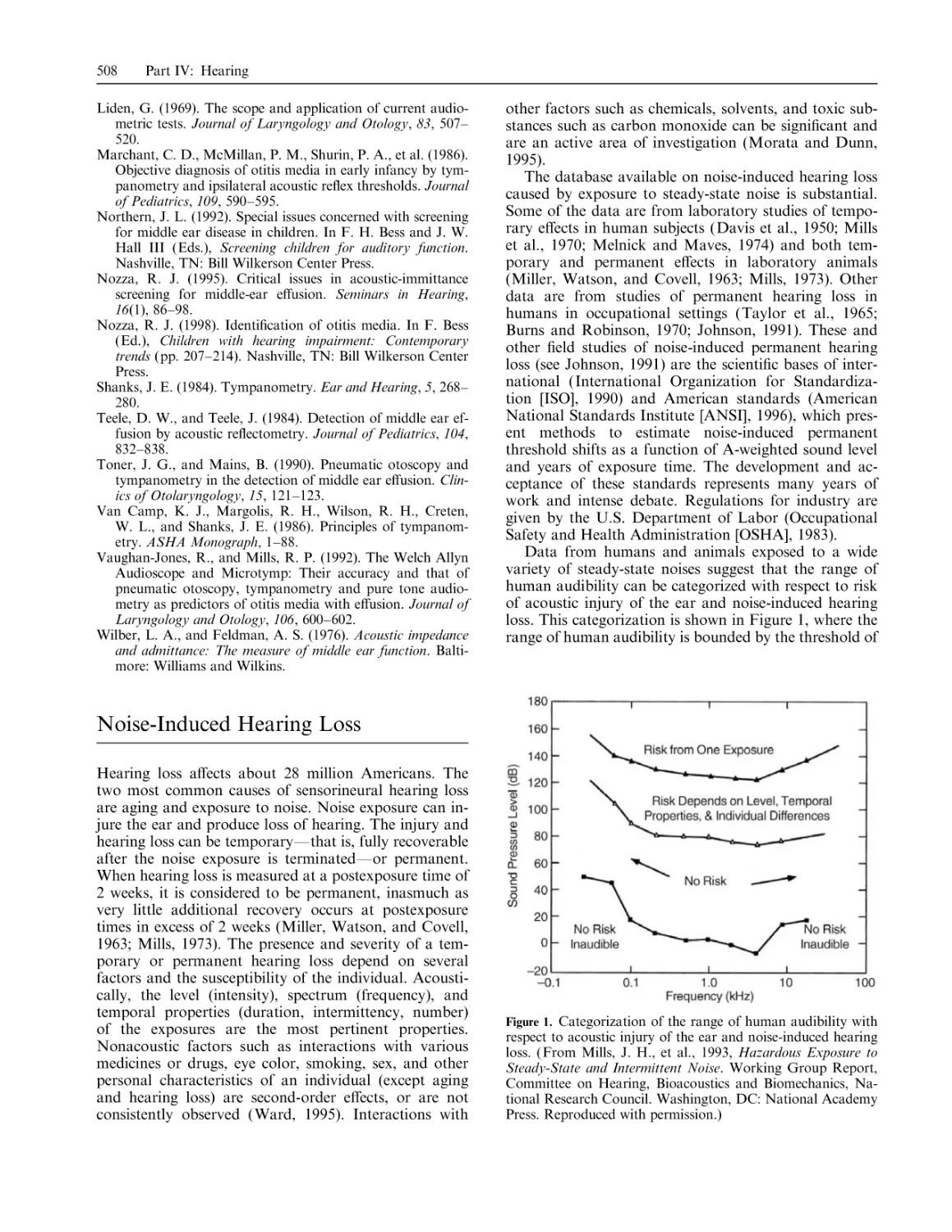
Noise-Induced Hearing Loss 508
Otoacoustic Emissions 511
Otoacoustic Emissions in Children 515
Ototoxic Medications 518
Pediatric Audiology: The Test Battery Approach
Physiological Bases of Hearing 522
Pitch Perception 525
Presbyacusis 527
Pseudohypacusis 531
Pure-Tone Threshold Assessment 534
Speech Perception Indices 538
Speech Tracking 541
520
Speechreading Training and Visual Tracking
543
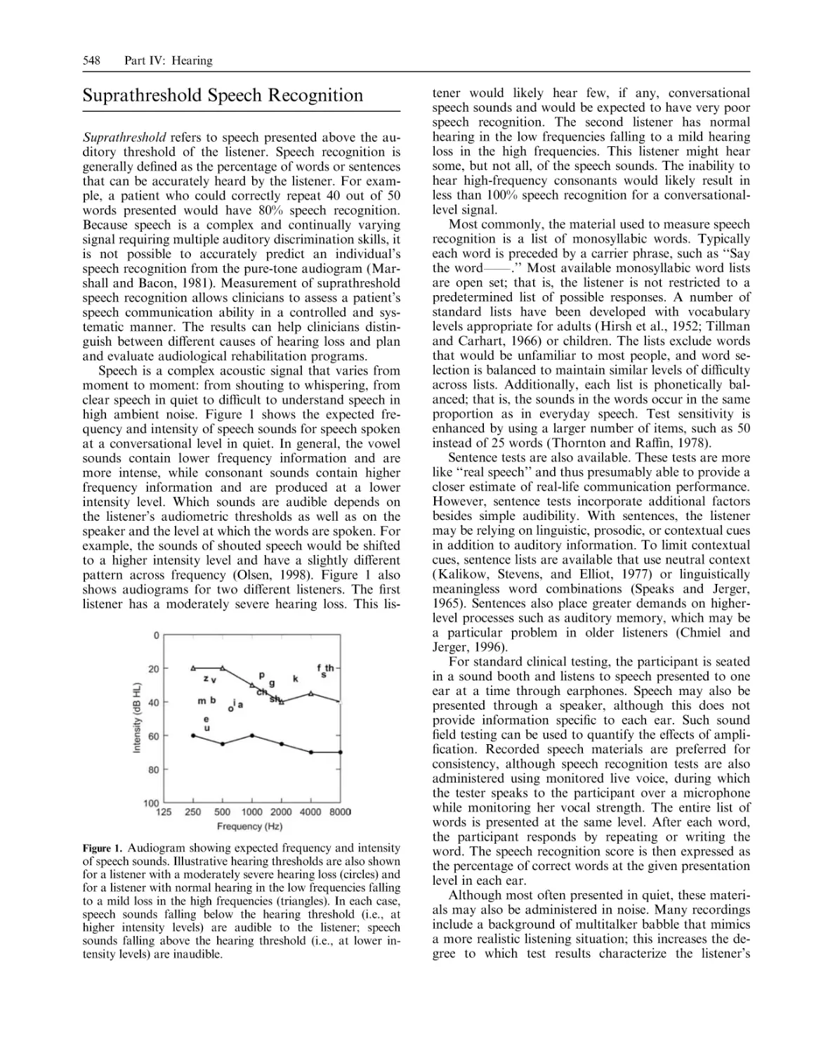
Suprathreshold Speech Recognition 548
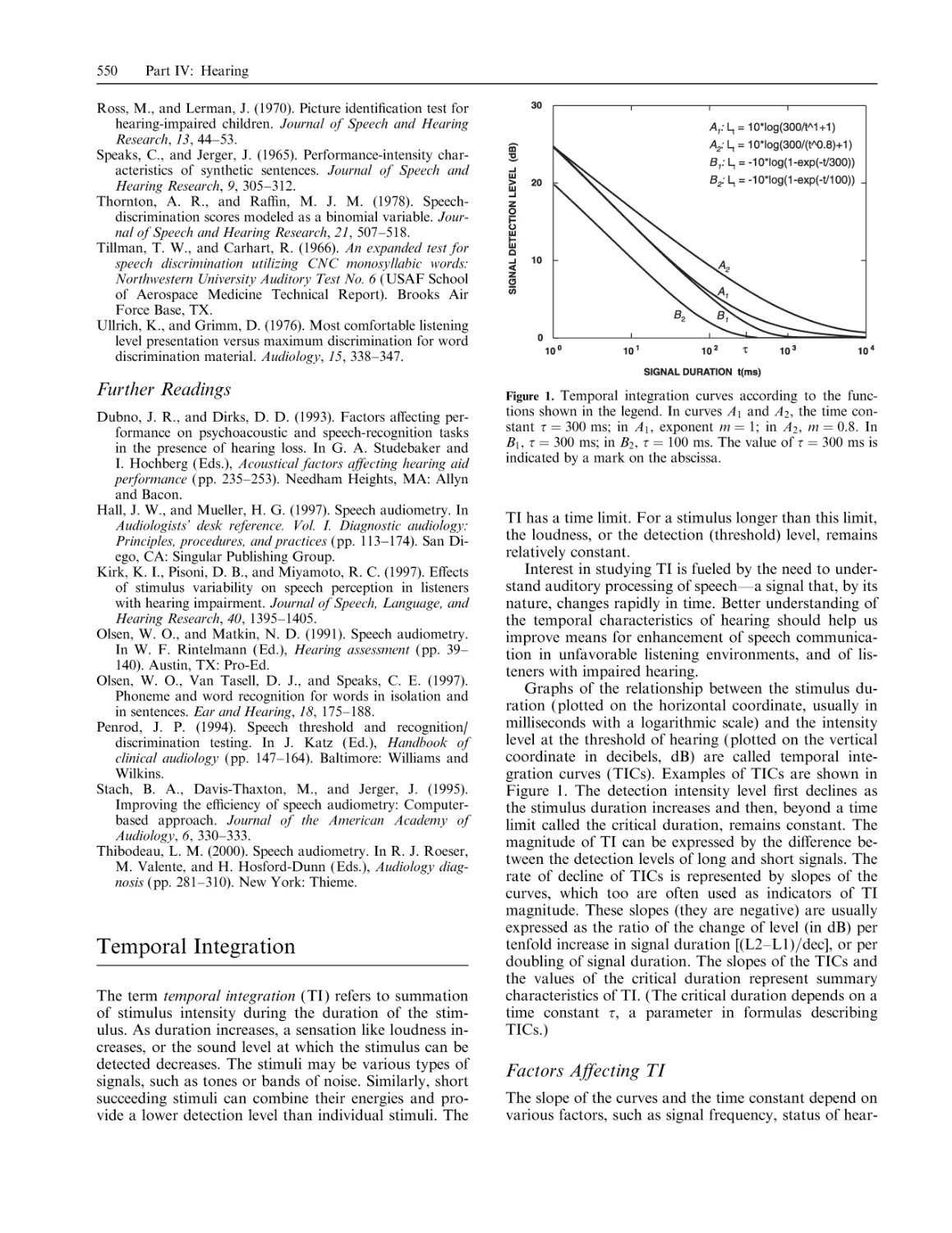
Temporal Integration 550
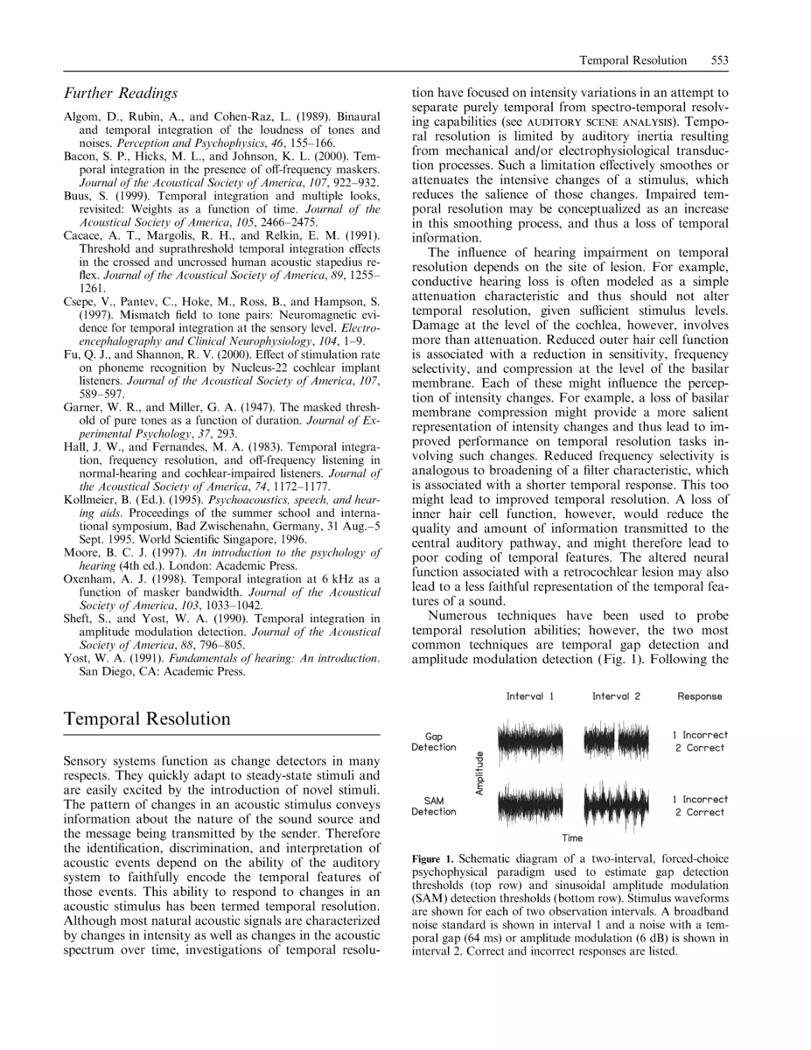
Temporal Resolution 553
Tinnitus 556
Tympanometry 558
Vestibular Rehabilitation 563
Contributors 569
Name Index 577
Subject Index 603
vii
Introduction
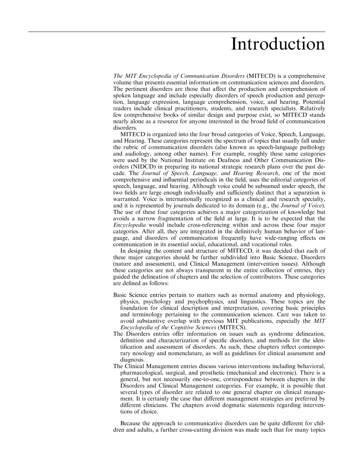
The MIT Encyclopedia of Communication Disorders (MITECD) is a comprehensive
volume that presents essential information on communication sciences and disorders.
The pertinent disorders are those that a¤ect the production and comprehension of
spoken language and include especially disorders of speech production and perception, language expression, language comprehension, voice, and hearing. Potential
readers include clinical practitioners, students, and research specialists. Relatively
few comprehensive books of similar design and purpose exist, so MITECD stands
nearly alone as a resource for anyone interested in the broad field of communication
disorders.
MITECD is organized into the four broad categories of Voice, Speech, Language,
and Hearing. These categories represent the spectrum of topics that usually fall under
the rubric of communication disorders (also known as speech-language pathology
and audiology, among other names). For example, roughly these same categories
were used by the National Institute on Deafness and Other Communication Disorders (NIDCD) in preparing its national strategic research plans over the past decade. The Journal of Speech, Language, and Hearing Research, one of the most
comprehensive and influential periodicals in the field, uses the editorial categories of
speech, language, and hearing. Although voice could be subsumed under speech, the
two fields are large enough individually and su‰ciently distinct that a separation is
warranted. Voice is internationally recognized as a clinical and research specialty,
and it is represented by journals dedicated to its domain (e.g., the Journal of Voice).
The use of these four categories achieves a major categorization of knowledge but
avoids a narrow fragmentation of the field at large. It is to be expected that the
Encyclopedia would include cross-referencing within and across these four major
categories. After all, they are integrated in the definitively human behavior of language, and disorders of communication frequently have wide-ranging e¤ects on
communication in its essential social, educational, and vocational roles.
In designing the content and structure of MITECD, it was decided that each of
these major categories should be further subdivided into Basic Science, Disorders
(nature and assessment), and Clinical Management (intervention issues). Although
these categories are not always transparent in the entire collection of entries, they
guided the delineation of chapters and the selection of contributors. These categories
are defined as follows:
Basic Science entries pertain to matters such as normal anatomy and physiology,
physics, psychology and psychophysics, and linguistics. These topics are the
foundation for clinical description and interpretation, covering basic principles
and terminology pertaining to the communication sciences. Care was taken to
avoid substantive overlap with previous MIT publications, especially the MIT
Encyclopedia of the Cognitive Sciences (MITECS).
The Disorders entries o¤er information on issues such as syndrome delineation,
definition and characterization of specific disorders, and methods for the identification and assessment of disorders. As such, these chapters reflect contemporary nosology and nomenclature, as well as guidelines for clinical assessment and
diagnosis.
The Clinical Management entries discuss various interventions including behavioral,
pharmacological, surgical, and prosthetic (mechanical and electronic). There is a
general, but not necessarily one-to-one, correspondence between chapters in the
Disorders and Clinical Management categories. For example, it is possible that
several types of disorder are related to one general chapter on clinical management. It is certainly the case that di¤erent management strategies are preferred by
di¤erent clinicians. The chapters avoid dogmatic statements regarding interventions of choice.
Because the approach to communicative disorders can be quite di¤erent for children and adults, a further cross-cutting division was made such that for many topics
x
Introduction
separate chapters for children and adults are included. Although some disorders that
are first diagnosed in childhood may persist in some form throughout adulthood (e.g,
stuttering, specific language impairment, and hearing loss may be lifelong conditions
for some individuals), many disorders can have an onset either in childhood or in
adulthood and the timing of onset can have implications for both assessment and
intervention. For instance, when a child experiences a significant loss of hearing, the
sensory deficit may greatly impair the learning of speech and language. But when a
loss of the same degree has an onset in adulthood, the problem is not in acquiring
speech and language, but rather in maintaining communication skills. Certainly, it is
often true that an understanding of a given disorder has common features in both the
developmental and acquired forms, but commonality cannot be assumed as a general
condition.
Many decisions were made during the preparation of this volume. Some were
easy, but others were not. In the main, entries are uniform in length and number of
references. However, in a few instances, two or more entries were combined into a
single longer entry. Perhaps inevitably in a project with so many contributors, a small
number of entries were dropped because of personal issues, such as illness, that
interfered with timely preparation of an entry. Happily, contributors showed great
enthusiasm for this project, and their entries reflect an assembled expertise that is
high tribute to the science and clinical practice in communication disorders.
Raymond D. Kent
Acknowledgments
MITECD began as a promising idea in a conversation with Amy Brand, a previous
editor with MIT Press. The idea was further developed, refined, elaborated, and refined again in many ensuing e-mail communications, and I thank Amy for her constant support and assistance through the early phases of the project. When she left
MIT Press, Tom Stone, Senior Editor of Cognitive Sciences, Linguistics, and Bradford Books, stepped in to provide timely advice and attention. I also thank Mary
Avery, Acquisitions Assistant, for her help in keeping this project on track. I am
indebted to all of them.
Speech, voice, language, and hearing are vast domains individually, and several
associated editors helped to select topics for inclusion in MITECD and to identify
contributors with the necessary expertise. The associate editors and their fields of responsibility are as follows:
Fred H. Bess, Ph.D., Hearing Disorders in Children
Joseph R. Du¤y, Ph.D., Speech Disorders in Adults
Steven D. Gray, M.D. (deceased), Voice Disorders in Children
Robert E. Hillman, Ph.D., Voice Disorders in Adults
Sandra Gordon-Salant, Ph.D., Hearing Disorders in Adults
Mabel L. Rice, Ph.D., Language Disorders in Children
Lawrence D. Shriberg, Ph.D., Speech Disorders in Children
David A. Swinney, Ph.D., and Lewis P. Shapiro, Ph.D., Language Disorders in
Adults
The advice and cooperation of these individuals is gratefully acknowledged. Sadly,
Dr. Steven D. Gray died within the past year. He was an extraordinary man, and
although I knew him only briefly, I was deeply impressed by his passion for knowledge and life. He will be remembered as an excellent physician, creative scientist, and
valued friend and colleague to many.
Dr. Houri Vorperian greatly facilitated this project through her inspired planning
of a computer-based system for contributor communications and record management. Sara Stuntebeck and Sara Brost worked skillfully and accurately on a variety
of tasks that went into di¤erent phases of MITECD. They o¤ered vital help with
communications, file management, proofreading, and the various and sundry tasks
that stood between the initial conception of MITECD and the submission of a full
manuscript.
P. M. Gordon and Associates took on the formidable task of assembling 200
entries into a volume that looks and reads like an encyclopedia. I thank Denise
Bracken for exacting attention to the editing craft, creative solutions to unexpected
problems, and forbearance through it all.
MITECD came to reality through the e¤orts of a large number of contributors—
too many for me to acknowledge personally here. However, I draw the reader’s attention to the list of contributors included in this volume. I feel a sense of community
with all of them, because they believed in the project and worked toward its completion by preparing entries of high quality. I salute them not only for their contributions to MITECD but also for their many career contributions that define them
as experts in the field. I am honored by their participation and their patient cooperation with the editorial process.
Raymond D. Kent
Part I: Voice
Acoustic Assessment of Voice
Acoustic assessment of voice in clinical applications is
dominated by measures of fundamental frequency ( f0 ),
cycle-to-cycle perturbations of period ( jitter) and intensity (shimmer), and other measures of irregularity, such
as noise-to-harmonics ratio (NHR). These measures are
widely used, in part because of the availability of electronic and microcomputer-based instruments (e.g., Kay
Elemetrics Computerized Speech Laboratory [CSL] or
Multispeech, Real-Time Pitch, Multi-Dimensional Voice
Program [MDVP], and other software/hardware systems), and in part because of long-term precedent for
perturbation (Lieberman, 1961) and spectral noise
measurements (Yanagihara, 1967). Absolute measures
of vocal intensity are equally basic but require calibrations and associated instrumentation (Winholtz and
Titze, 1997).
Independently, these basic acoustic descriptors—f0 ,
intensity, jitter, shimmer, and NHR—can provide
some very basic characterizations of vocal health.
The first two, f0 and intensity, have very clear perceptual correlates—pitch and loudness, respectively—and
should be assessed for both stability and variability and
compared to age and sex norms (Kent, 1994; Baken
and Orliko¤, 2000). Ideally, these tasks are recorded
over headset microphones with direct digital acquisition
at very high sampling rates (at least 48 kHz). The materials to be assessed should be obtained following standardized elicitation protocols that include sustained
vowel phonations at habitual levels, levels spanning a
client’s vocal range in both f0 and intensity, running
speech, and speech tasks designed to elicit variation
(Titze, 1995; Awan, 2001). Note, however, that not all
measures will be appropriate for all tasks; perturbation
statistics, for example, are usually valid only when
extracted from sustained vowel phonations.
These basic descriptors are not in any way comprehensive of the range of available measures or the
available signal properties and dimensions. Table 1 categorizes measures (Buder, 2000) based on primary basic
signal representations from which measures are derived.
Although these categories are intended to be exhaustive
and mutually exclusive, some more modern algorithms
process components through several types. (For more
detail on the measurement types, see Buder, 2000, and
Baken and Orliko¤, 2000.) Modern algorithmic approaches should be selected for (1) interpretability with
respect to aerodynamic and physiological models of
phonation and (2) the incorporation of multivariate
measures to characterize vocal function.
Interdependence of Basic Measures. The interdependence between f0 and intensity is mapped in a voice
range profile, or phonetogram, which is an especially
valuable assessment for the professional voice user
(Coleman, 1993). Furthermore, the dependence of perturbations and signal-to-noise ratios on both f0 and intensity is well known (Klingholz, 1990; Pabon, 1991).
Table 1. Outline of Traditional Acoustic Algorithm Types
f0 statistics
Short-term perturbations
Long-term perturbations
Amplitude statistics
Short-term perturbations
Long-term perturbations
f0 /amplitude covariations
Waveform perturbations
Spectral measures
Spectrographic measures
Fourier and LPC spectra
Long-term average spectra
Cepstra
Inverse filter measures
Radiated signal
Flow-mask signals
Dynamic measures
This dependence is not often assessed rigorously, perhaps because of the time-consuming and strenuous nature of a full voice profile. However, an abbreviated or
focused profiling in which samples related to habitual f0
by a set number of semitones, or related to habitual
intensity by a set number of decibels, could be standardized to control for this dependence e‰ciently. Finally, it should be understood that perturbations and
NHR-type measures will usually covary for many reasons, the simplest ones being methodological (Hillenbrand, 1987): an increase in any one of the underlying
phenomena detected by a single measure will also a¤ect
the other measures.
Periodicity as a Reference. The chief problem with
nearly all acoustic assessments of voice is the determination of f0 . Most voice quality algorithms are based on
the prior identification of the periodic component in the
signal (based on glottal pulses in the time domain or
harmonic structure in the frequency domain). Because
phonation is ideally a nearly periodic process, it is
logical to conceive of voice measures in terms of the degree to which a given sample deviates from pure periodicity. There are many conceptual problems with this
simplification, however. At the physiological level, glottal morphology is multidimensional—superior-inferior
asymmetry is a basic feature of the two-mass model
(Ishizaka and Flanagan, 1972), and some anteriorposterior asymmetry is also inevitable—rendering it unlikely that a glottal pulse will be marked by a discrete or
even a single instant of glottal closure. At the level of the
signal, the deviations from periodicity may be either
random or correlated, and in many cases they are so extreme as to preclude identification of a regular period.
Finally, at the perceptual level, many factors related to
deviations from a pure f0 can contribute to pitch perception (Zwicker and Fastl, 1990).
At any or all of these levels, it becomes questionable
to characterize deviations with pure periodicity as a reference. In acoustic assessment, the primary level of concern is the signal. The National Center for Voice and
4
Part I: Voice
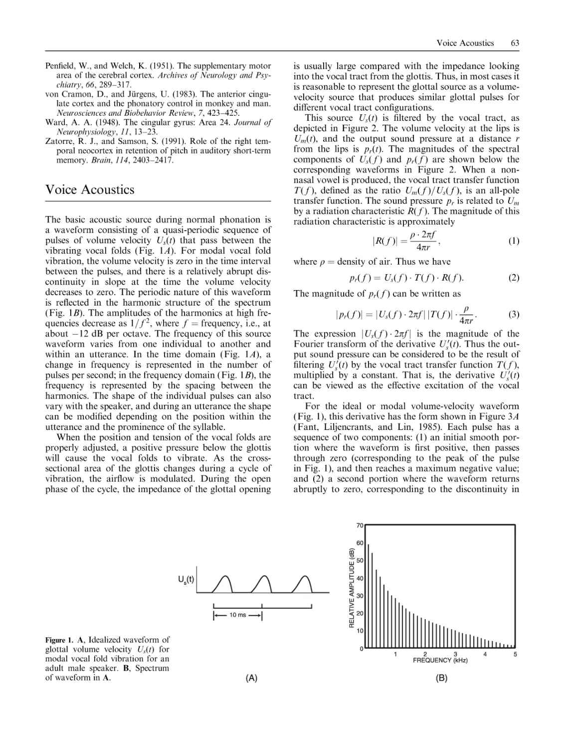
Figure 1. Approximately 900 ms of a sustained vowel phonation waveform (top panel) with two fundamental frequency
analyses (bottom panel). Average f0 , %jitter, %shimmer, and
SNR results for selected segments were from the ‘‘newjit’’ routine of TF32 program (Milenkovic, 2001).
Speech issued a summary statement (Titze, 1995) recommending a typology for categorizing deviations from
periodicity in voices (see also Baken and Orliko¤, 2000,
for further subtypes). This typology capitalizes on the
categorical nature of dynamic states in nonlinear systems; all the major categories, including stable points,
limit cycles, period-doubling/tripling/. . . , and chaos can
be observed in voice signals (Herzel et al., 1994; Satalo¤
and Hawkshaw, 2001). As in most highly nonlinear
dynamic systems, deviations from periodicity can be
categorized on the basis of bifurcations, or sudden qualitative changes in vibratory pattern from one of these
states to another.
Figure 1 displays a common form for one such bifurcation and illustrates the importance of accounting for
its presence in the application of perturbation measures.
In this sustained vowel phonation by a middle-aged
woman with spasmodic dysphonia, a transition to subharmonics is clearly visible in segment b (similar patterns occur in individuals without dysphonias). Two f0
extractions are presented for this segment, one at the
targeted level of approximately 250 Hz and another
which the tracker finds one octave below this; inspection of the waveform and a perceived biphonia both
justify this 125-Hz analysis as a new fundamental frequency, although it can also be understood in this
context as a subharmonic to the original fundamental. There is therefore some ambiguity as to which
fundamental is valid during this episode, and an automatic analysis could plausibly identify either frequency.
(Here the waveform-matching algorithm implemented in
CSpeechSP [Milenkovic, 1997] does identify either frequency, depending on where in the waveform the algorithm is applied; initiating the algorithm within the
subharmonic segment predisposes it to identify the lower
fundamental.)
The acoustic measures of the segments displayed in
Figure 1 reveal the nontrivial di¤erences that result,
depending on the basic glottal pulse form under consideration. When the pulses of segment a are considered,
Acoustic Assessment of Voice
the perturbations around the base period associated with
the high f0 are low and normative; in segment b, perturbations around the longer periods of the lower f0 are
still low ( jitter is improved, while shimmer and the
signal-to-noise ratio show some degradation). However,
when all segments are considered together to include the
perturbations around the high f0 tracked through segment b and into c, the perturbation statistics are all
increased by an order of magnitude. Many important
methodological and theoretical questions should be
raised by such common scenarios in which we must
consider not just voice typing, but the segment-bysegment validity of applying perturbation measures with
a particular f0 as reference. If, as is often assumed, jitter
and shimmer are ascribed to ‘‘random’’ variations, then
the correlated modulations of a strong subharmonic episode should be excluded. Alternatively, the perturbations might be analyzed with respect to the subharmonic
f0 . In any case, assessment by means of perturbation
statistics with no consideration of their underlying
sources is unwise.
Perceptual, Aerodynamic, and Physiological Correlates
of Acoustic Measures. Regarding perceptual voice rat-
Figure 2. Spectral features associated with models of phonation,
including the Liljencrants-Fant (LF) model of glottal flow and
aperiodicity source models developed by Stevens. The LF
model of glottal flow is shown at top left. At bottom left is the
LF model of glottal flow derivative, showing the rate of change
in flow. At right is a spectrum schematic showing four e¤ects.
These e¤ects include three derived parameters of the LF model:
(a) excitation strength (the maximum negative amplitude of the
flow derivative, which is positively correlated with overall harmonic energy), (b) dynamic leakage or non-zero return phase
following the point of maximum excitation (which is negatively
5
ings, Gerratt and Kreiman (2000) have critiqued traditional assessments on several important methodological
and theoretical points. However, these points may not
apply to acoustic analysis if (1) acoustic analysis is validated on its own success and not exclusively in relation
to the problematic perceptual classifications, and (2)
acoustic analysis is thoroughly grounded for interpretation in some clear aerodynamic or physiological model
of phonation. Gerratt and Kreiman also argue that
clinical classification may not be derived along a continuum that is defined with reference to normal qualities,
but again, this argument may need to be reversed for the
acoustic domain. It is only by reference to a specific
model that any assessment on acoustic grounds can be
interpreted (though this does not preclude development
of an independent model for a pathological phonatory
mechanism). In clinical settings, acoustic voice assessment often serves to corroborate perceptual assessment.
However, as guided by auditory experience and in conjunction with the ear and other instrumental assessments, careful acoustic analysis can be oriented to the
identification of physiological status.
In attempting to draw safe and reasonably direct
inferences from acoustic signal, aerodynamic models
correlated with high-frequency harmonic energy), and (c) pulse
skewing (which is negatively correlated with low-frequency
harmonic energy; this low-frequency region is also positively
correlated with open quotient and peak volume velocity measures of the glottal flow waveform). The e¤ect of turbulence
due to high airflow through the glottis is schematized by (d),
indicating the associated appearance of high-frequency aperiodic energy in the spectrum. See voice acoustics for other
graphical and quantitative associations between glottal status
and spectral characteristics.
6
Part I: Voice
of glottal behavior present important links to the
physiological domain. Attempts to recover the glottal
flow waveform, either from a face mask-transduced
flow recording (Rothenberg, 1973) or a microphonetransduced acoustic recording (Davis, 1975), have
proved to be labor-intensive and prone to error (Nı́
Chasaide and Gobl, 1997). Rather than attempting to
eliminate the e¤ects of the vocal tract, it may be more
fruitful to understand its in situ relationship with phonation, and infer, via the types of features displayed in
Figure 2, the status of the glottis as a sound source. Interpretation of spectral features, such as the amplitudes
of the first harmonics and at the formant frequencies,
may be an e¤ective alternative when guided by knowledge of glottal aerodynamics and acoustics (Hanson,
1997; Nı́ Chasaide and Gobl, 1997; Hanson and
Chuang, 1999). Deep familiarity with acoustic mechanisms is essential for such interpretations (Titze, 1994;
Stevens, 1998), as is a model with clear and meaningful
parameters, such as the Liljencrants-Fant (LF) model
(Fant, Liljencrants, and Lin, 1985). The parameters of
the LF model have proved to be meaningful in acoustic
studies (Gau‰n & Sundberg, 1989) and useful in refined
e¤orts at inverse filtering (Fröhlich, Michaelis, and
Strube, 2001). Figure 2 summarizes selected parameters
of the LF source model following Nı́ Chasaide and Gobl
(1997) and the glottal turbulence source following
Stevens (1998); see also voice acoustics for other approaches relating glottal status to spectral measures.
Other spectral-based measures implement similar
model-based strategies by selecting spectral component
ratios (e.g., the VTI and SPI parameters of MDVP).
Sophisticated spectral noise characterizations control for
perturbations and modulations (Murphy, 1999; Qi,
Hillman, and Milstein, 1999), or employ curve-fitting
and statistical models to produce more robust measures
(Alku, Strik, and Vilkman, 1997; Michaelis, Fröhlich,
and Strube, 1998; Schoentgen, Bensaid, and Bucella,
2000). A particularly valuable modern technique for
detecting turbulence at the glottis, the glottal-to-noiseexcitation ratio (Michaelis, Gramss, and Strube, 1997),
has been especially successful in combination with other
measures (Fröhlich et al., 2000). The use of acoustic
techniques for voice will only improve with the inclusion
of more knowledge-based measures in multivariate representations (Wolfe, Cornell, and Palmer, 1991; Callen
et al., 2000; Wuyts et al., 2000).
—Eugene H. Buder
References
Alku, P., Strik, H., and Vilkman, E. (1997). Parabolic spectral
parameter: A new method for quantification of the glottal
flow. Speech Communication, 22, 67–79.
Awan, S. N. (2001). The voice diagnostic profile: A practical
guide to the diagnosis of voice disorders. Gaithersburg, MD:
Aspen.
Baken, R. J., and Orliko¤, R. F. (2000). Clinical measurement
of speech and voice. San Diego, CA: Singular Publishing
Group.
Buder, E. H. (2000). Acoustic analysis of voice quality: A tabulation of algorithms 1902–1990. In M. J. Ball (Ed.), Voice
quality measurement (pp. 119–244). San Diego, CA: Singular Publishing Group.
Callen, D. E., Kent, R. D., Roy, N., and Tasko, S. M. (2000).
The use of self-organizing maps for the classification of voice
disorders. In M. J. Ball (Ed.), Voice quality measurement
(pp. 103–116). San Diego, CA: Singular Publishing Group.
Coleman, R. F. (1993). Sources of variation in phonetograms.
Journal of Voice, 7, 1–14.
Davis, S. B. (1975). Preliminary results using inverse filtering of
speech for automatic evaluation of laryngeal pathology.
Journal of the Acoustical Society of America, 58, SIII.
Fant, G., Liljencrants, J., and Lin, Q. (1985). A four-parameter
model of glottal flow. Speech Transmission Laboratory
Quarterly Progress and Status Report, 4, 1–13.
Fröhlich, M., Michaelis, D., and Strube, H. (2001). SIMsimultaneous inverse filtering and matching of a glottal flow
model for acoustic speech signals. Journal of the Acoustical
Society of America, 110, 479–488.
Fröhlich, M., Michaelis, D., Strube, H., and Kruse, E. (2000).
Acoustic voice analysis by means of the hoarseness diagram. Journal of Speech, Language, and Hearing Research,
43, 706–720.
Gau‰n, J., and Sundberg, J. (1989). Spectral correlates of
glottal voice source waveform characteristics. Journal of
Speech and Hearing Research, 32, 556–565.
Gerratt, B., and Kreiman, J. (2000). Theoretical and methodological development in the study of pathological voice
quality. Journal of Phonetics, 28, 335–342.
Hanson, H. M. (1997). Glottal characteristics of female
speakers: Acoustic correlates. Journal of the Acoustical Society of America, 101, 466–481.
Hanson, H. M., and Chuang, E. S. (1999). Glottal characteristics of male speakers: Acoustic correlates and comparison
with female data. Journal of the Acoustical Society of
America, 106, 1064–1077.
Herzel, H., Berry, D., Titze, I. R., and Saleh, M. (1994).
Analysis of vocal disorders with methods from nonlinear
dynamics. Journal of Speech and Hearing Research, 37,
1008–1019.
Hillenbrand, J. (1987). A methodological study of perturbation
and additive noise in synthetically generated voice signals.
Journal of Speech and Hearing Research, 30, 448–461.
Ishizaka, K., and Flanagan, J. L. (1972). Synthesis of voiced
sounds from a two-mass model of the vocal cords. Bell
System Technical Journal, 51, 1233–1268.
Kent, R. D. (1994). Reference manual for communicative
sciences and disorders: Speech and language. Austin, TX:
Pro-Ed.
Klingholz, F. (1990). Acoustic representation of speaking-voice
quality. Journal of Voice, 4, 213–219.
Lieberman, P. (1961). Perturbations in vocal pitch. Journal of
the Acoustical Society of America, 33, 597–603.
Michaelis, D., Fröhlich, M., and Strube, H. W. (1998). Selection and combination of acoustic features for the description of pathologic voices. Journal of the Acoustical Society
of America, 103, 1628–1638.
Michaelis, D., Gramss, T., and Strube, H. W. (1997). Glottal
to noise excitation ratio: A new measure for describing
patholocial voices. Acustica, 83, 700–706.
Milenkovic, P. (1997). CSpeechSP [Computer software]. Madison, WI: University of Wisconsin–Madison.
Milenkovic, P. (2001). TF32 [Computer software]. Madison,
WI: University of Wisconsin–Madison.
Murphy, P. J. (1999). Perturbation-free measurement of the
harmonics-to-noise ratio in voice signals using pitch synchronous harmonic analysis. Journal of the Acoustical Society of America, 105, 2866–2881.
Aerodynamic Assessment of Vocal Function
7
A number of methods have been used to quantitatively
assess the air volumes, airflows, and air pressures involved in voice production. The methods have been
mostly used in research to investigate mechanisms that
underlie normal and disordered voice and speech production. The clinical use of aerodynamic measures to
assess patients with voice disorders has been increasing
(Colton and Casper, 1996; Hillman, Montgomery, and
Zeitels, 1997; Hillman and Kobler, 2000).
Goldman, and Mead, 1973; Watson and Hixon, 1985;
Hoit and Hixon, 1987; Hoit et al., 1990). Air volumes
are measured in standard metric units (liters, cubic centimeters, milliliters) and lung inflation levels are usually
specified in terms of a percentage of the vital capacity or
total lung volume.
Both direct and indirect methods have been used to
measure air volumes expended during phonation. Direct
measurement of orally displaced air volumes during
phonatory tasks can be accomplished, to a limited extent, by means of a mouthpiece or face mask connected
to a measurement device such as a spirometer (Beckett,
1971) or pneumotachograph (Isshiki, 1964). The use of a
mouthpiece essentially limits speech production to sustained vowels, which are su‰cient for assessing selected
volumetric-based phonatory parameters. There are also
concerns that face masks interfere with normal jaw
movements and that the oral acoustic signal is degraded,
so that auditory feedback is reduced or distorted and
simultaneous acoustic analysis is limited. These limitations, which are inherent to the use of devices placed
in or around the mouth to directly collect oral airflow,
plus additional measurement-related restrictions (Hillman and Kobler, 2000) have helped motivate the development and application of indirect measurement
approaches.
Most speech breathing research has been carried out
using indirect approaches for estimating lung volumes
by means of monitoring changes in body dimensions.
The basic assumption underlying the indirect approaches
is that changes in lung volume are reflected in proportional changes in body torso size. One relatively cumbersome but time-honored approach has been to place
subjects in a sealed chamber called a body plethysmograph to allow estimation of the air volume displaced by
the body during respiration (Draper, Ladefoged, and
Whitteridge, 1959). More often used for speech breathing research are transducers (magnetometers: Hixon,
Goldman, and Mead, 1973; inductance plethysmographs: Sperry, Hillman, and Perkell, 1994) that unobtrusively monitor changes in the dimensions of the rib
cage and abdomen (referred to collectively as the chest
wall) that account for the majority of respiratory-related
changes in torso dimension (Mead et al., 1967). These
approaches have been primarily employed to study respiratory function during continuous speech and singing
tasks that include both voiced and voiceless sound production, as opposed to assessing air volume usage during
phonatory tasks that involve only laryngeal production
of voice (e.g., sustained vowels). There are also ongoing
e¤orts to develop more accurate methods for noninvasively monitoring chest wall activity to capture finer
details of how the three-dimensional geometry of the
body is altered during respiration (see Cala et al., 1996).
Measurement of Air Volumes. Respiratory research in
human communication has focused primarily on the
measurement of the air volumes that are typically
expended during selected speech and singing tasks, and
on specifying the ranges of lung inflation levels across
which such tasks are normally performed (cf. Hixon,
Measurement of Airflow. Airflow associated with phonation is usually specified in terms of volume velocity
(i.e., volume of air displaced per unit of time). Volume
velocity airflow rates for voice production are typically
reported in metric units of volume displaced (liters or
cubic centimeters) per second.
Nı́ Chasaide, A., and Gobl, C. (1997). Voice source variation.
In J. Laver (Ed.), The handbook of phonetic sciences (pp.
427–461). Oxford, UK: Blackwell.
Pabon, J. P. H. (1991). Objective acoustic voice-quality
parameters in the computer phonetogram. Journal of Voice,
5, 203–216.
Qi, Y., Hillman, R., and Milstein, C. (1999). The estimation of
signal-to-noise ratio in continuous speech for disordered
voices. Journal of the Acoustical Society of America, 105,
2532–2535.
Rothenberg, M. (1973). A new inverse-filtering technique for
deriving the glottal air flow waveform during voicing. Journal of the Acoustical Society of America, 53, 1632–1645.
Satalo¤, R. T., and Hawkshaw, M. (Eds.). (2001). Chaos in
medicine: Source readings. San Diego, CA: Singular Publishing Group.
Schoentgen, J., Bensaid, M., and Bucella, F. (2000). Multivariate statistical analysis of flat vowel spectra with a view
to characterizing dysphonic voices. Journal of Speech, Language, and Hearing Research, 43, 1493–1508.
Stevens, K. N. (1998). Acoustic phonetics. Cambridge, MA:
MIT Press.
Titze, I. R. (1994). Principles of voice production. Englewood
Cli¤s, NJ: Prentice Hall.
Titze, I. R. (1995). Workshop on acoustic voice analysis: Summary statement. Iowa City, IA: National Center for Voice
and Speech.
Winholtz, W. S., and Titze, I. R. (1997). Conversion of a headmounted microphone signal into calibrated SPL units.
Journal of Voice, 11, 417–421.
Wolfe, V., Cornell, R., and Palmer, C. (1991). Acoustic correlates of pathologic voice types. Journal of Speech and
Hearing Research, 34, 509–516.
Wuyts, F. L., De Bodt, M. S., Molenberghs, G., Remacle, M.,
Heylen, L., Millet, B., et al. (2000). The Dysphonia Severity
Index: An objective measure of vocal quality based on a
multiparameter approach. Journal of Speech, Language, and
Hearing Research, 43, 796–809.
Yanagihara, N. (1967). Significance of harmonic changes and
noise components in hoarseness. Journal of Speech and
Hearing Research, 10, 531–541.
Zwicker, E., and Fastl, H. (1990). Psychoacoustics: Facts and
models. Heidelberg, Germany: Springer-Verlag.
Aerodynamic Assessment of Vocal
Function
8
Part I: Voice
Estimates of average airflow rates can be obtained by
simply dividing air volume estimates by the duration of
the phonatory task. Average glottal airflow rates have
usually been estimated during vowel phonation by using
a mouthpiece or face mask to channel the oral air stream
through a pneumotachograph (Isshiki, 1964). There has
also been somewhat limited use of hot wire anemometer
devices (mounted in a mouthpiece) to estimate average
glottal airflow during sustained vowel phonation (Woo,
Colton, and Shangold, 1987). Estimates of average glottal airflow rates can be obtained from the oral airflow
during vowel production because the vocal tract is relatively nonconstricted, with no major sources of turbulent
airflow between the glottis and the lips.
There have also been e¤orts to obtain estimates of the
actual airflow waveform that is generated as the glottis
rapidly opens and closes during flow-induced vibration
of the vocal folds (the glottal volume velocity waveform). The glottal volume velocity waveform cannot be
directly observed by measuring the oral airflow signal
because the waveform is highly convoluted by the resonance activity (formants) of the vocal tract. Thus, recovery of the glottal volume velocity waveform requires
methods that eliminate or correct for the influences of
the vocal tract. This has typically been accomplished
aerodynamically by processing the output of a fastresponding pneumotachograph (high-frequency response) using a technique called inverse filtering, in
which the major resonances of the vocal tract are estimated and the oral airflow signal is processed (inverse
filtered) to eliminate them (Rothenberg, 1977; Holmberg, Hillman, and Perkell, 1988).
Figure 1. Instrumentation and resulting signals for
simultaneous collection of oral airflow, intraoral air
pressure, the acoustic signal, and chest wall (rib
cage and abdomen) dimensions during production of
the syllable string /pi-pi-pi/. Signals shown in the
bottom panel are processed and measured to provide
estimates of average glottal airflow rate, average
subglottal air pressure, lung volume, and glottal
waveform parameters.
Aerodynamic Assessment of Vocal Function
Measurement of Air Pressure. Measurements of air
pressures below (subglottal) and above (supraglottal) the
vocal folds are of primary interest for characterizing the
pressure di¤erential that must be achieved to initiate and
maintain vocal fold vibration during normal exhalatory phonation. In practice, air pressure measurements
related specifically to voice production are typically
acquired during vowel phonation when there are no
vocal tract constrictions of su‰cient magnitude to build
up positive supraglottal pressures. Under these conditions, it is usually assumed that supraglottal pressure is
essentially equal to atmospheric pressure and only subglottal pressure measurements are obtained. Air pressures associated with voice and speech production are
usually specified in centimeters of water (cm H2 O).
Both direct and indirect methods have been used to
measure subglottal air pressures during phonation. Direct measures of subglottal air pressure can be obtained
by inserting a hypodermic needle into the subglottal airway through a puncture in the anterior neck at the cricothyroid space (Isshiki, 1964). The needle is connected
to a pressure transducer by tubing. This method is very
accurate but also very invasive. It is also possible to insert a very thin catheter through the posterior cartilaginous glottis (between the arytenoids) to sense subglottal
air pressure during phonation, or to use an array of
miniature transducers positioned directly above and below the glottis (Cranen and Boves, 1985). These methods
cannot be tolerated by all subjects, and the heavy topical
anesthetization of the larynx that is required can a¤ect
normal function.
Indirect estimates of tracheal (subglottal) air pressure
can be obtained via the placement of an elongated
balloon-like device into the esophagus (Liberman, 1968).
The deflated esophageal balloon is attached to a catheter
that is typically inserted transnasally and then swallowed
into the esophagus to be positioned at the midthoracic
level. The catheter is connected to a pressure transducer
and the balloon is slightly inflated. Accurate use of this
invasive method also requires simultaneous monitoring
of lung volume.
Noninvasive, indirect estimates of subglottal air pressure can be obtained by measuring intraoral air pressure during specially constrained utterances (Smitheran
and Hixon, 1981). This is usually done by sensing air
pressure just behind the lips with a translabially placed
catheter connected to a pressure transducer. These
intraoral pressure measures are obtained as subjects
produce strings of bilabial /p/ þ vowel syllables (e.g.,
/pi-pi-pi-pi-pi/) at constant pitch and loudness. This
method works because the vocal folds are abducted
during /p/ production, thus allowing pressure to equilibrate throughout the airway, making intraoral pressure
equal to subglottal pressure (Fig. 1).
Additional Derived Measures. There have been numerous attempts to extend the utility of aerodynamic measures by using them in the derivation of additional
parameters aimed at better elucidating underlying
mechanisms of vocal function. Such derived measures
9
usually take the form of ratios that relate aerodynamic
parameters to each other, or that relate aerodynamic
parameters to simultaneously obtained acoustic measures. Common examples include (1) airway (glottal)
resistance (see Smitheran and Hixon, 1981), (2) vocal
e‰ciency (Schutte, 1980; Holmberg, Hillman, and Perkell, 1988), and (3) measures that interrelate glottal
volume velocity waveform parameters (Holmberg, Hillman, and Perkell, 1988).
Normative Data. As is the case for most measures of
vocal function, there is not currently a set of normative
data for aerodynamic measures that is universally
accepted and applied in research and clinical work.
Methods for collecting such data have not been standardized, and study samples have generally not been of
su‰cient size or appropriately stratified in terms of age
and sex to ensure unbiased estimates of underlying aerodynamic phonatory parameters in the normal population. However, there are several sources in the literature
that provide estimates of normative values for selected
aerodynamic measures (Kent, 1994; Baken, 1996; Colton and Casper, 1996).
See also voice production: physics and physiology.
—Robert E. Hillman
References
Baken, R. J. (1996). Clinical measurement of voice and speech.
San Diego, CA: Singular Publishing Group.
Beckett, R. L. (1971). The respirometer as a diagnostic and
clinical tool in the speech clinic. Journal of Speech and
Hearing Disorders, 36, 235–241.
Cala, S. J., Kenyon, C. M., Ferrigno, G., Carnevali, P.,
Aliverti, A., Pedotti, A., et al. (1996). Chest wall and lung
volume estimation by optical reflectance motion analysis.
Journal of Applied Physiology, 81, 2680–2689.
Colton, R. H., and Casper, J. K. (1996). Understanding voice
problems: A physiological perspective for diagnosis and
treatment. Baltimore: Williams and Wilkins.
Cranen, B., and Boves, L. (1985). Pressure measurements during speech production using semiconductor miniature pressure transducers: Impact on models for speech production.
Journal of the Acoustical Society of America, 77, 1543–
1551.
Draper, M., Ladefoged, P., and Whitteridge, P. (1959). Respiratory muscles in speech. Journal of Speech and Hearing
Research, 2, 16–27.
Hillman, R. E., and Kobler, J. B. (2000). Aerodynamic measures of voice production. In R. Kent and M. Ball (Eds.),
The handbook of voice quality measurement, San Diego, CA:
Singular Publishing Group.
Hillman, R. E., Montgomery, W. M., and Zeitels, S. M.
(1997). Current diagnostics and o‰ce practice: Use of objective measures of vocal function in the multidisciplinary management of voice disorders. Current Opinion in
Otolaryngology–Head and Neck Surgery, 5, 172–175.
Hixon, T. J., Goldman, M. D., and Mead, J. (1973). Kinematics of the chest wall during speech production: Volume
displacements of the rib cage, abdomen, and lung. Journal
of Speech and Hearing Research, 16, 78–115.
Hoit, J. D., and Hixon, T. J. (1987). Age and speech breathing.
Journal of Speech and Hearing Research, 30, 351–366.
10
Part I: Voice
Hoit, J. D., Hixon, T. J., Watson, P. J., and Morgan, W. J.
(1990). Speech breathing in children and adolescents. Journal of Speech and Hearing Research, 33, 51–69.
Holmberg, E. B., Hillman, R. E., and Perkell, J. S. (1988).
Glottal airflow and transglottal air pressure measurements
for male and female speakers in soft, normal, and loud
voice [published erratum appears in Journal of the Acoustical Society of America, 1989, 85(4), 1787]. Journal of the
Acoustical Society of America, 84, 511–529.
Isshiki, N. (1964). Regulatory mechanisms of vocal intensity
variation. Journal of Speech and Hearing Research, 7,
17–29.
Kent, R. D. (1994). Reference manual for communicative
sciences and disorders. San Diego, CA: Singular Publishing
Group.
Lieberman, P. (1968). Direct comparison of subglottal and
esophageal pressure during speech. Journal of the Acoustical
Society of America, 43, 1157–1164.
Mead, J., Peterson, N., Grimgy, N., and Mead, J. (1967). Pulmonary ventilation measured from body surface movements. Science, 156, 1383–1384.
Rothenberg, M. (1977). Measurement of airflow in speech.
Journal of Speech and Hearing Research, 20, 155–176.
Schutte, H. (1980). The e‰ciency of voice production. Groningen, The Netherlands: Kemper.
Smitheran, J. R., and Hixon, T. J. (1981). A clinical method
for estimating laryngeal airway resistance during vowel
production. Journal of Speech and Hearing Disorders, 46,
138–146.
Sperry, E., Hillman, R. E., and Perkell, J. S. (1994). The use of
an inductance plethysmograph to assess respiratory function in a patient with nodules. Journal of Medical SpeechLanguage Pathology, 2, 137–145.
Watson, P. J., and Hixon, T. J. (1985). Respiratory kinematics
in classical (opera) singers. Journal of Speech and Hearing
Research, 28, 104–122.
Woo, P., Colton, R. H., and Shangold, L. (1987). Phonatory
airflow analysis in patients with laryngeal disease. Annals of
Otology, Rhinology, and Laryngology, 96, 549–555.
Alaryngeal Voice and Speech
Rehabilitation
Loss of the larynx due to disease or injury will result in
numerous and significant changes that cross anatomical,
physiological, psychological, social, psychosocial, and
communication domains. Surgical removal of the larynx, or total laryngectomy, involves resectioning the
entire framework of the larynx. Although total laryngectomy may occur in some instances due to traumatic
injury, the majority of cases worldwide are the result of
cancer. Approximately 75% of all laryngeal tumors arise
from squamous epithelial tissue of the true vocal fold
(Bailey, 1985). In some instances, and because of the
location of many of these lesions, less aggressive approaches to medical intervention may be pursued. This
may include radiation therapy or partial surgical resection, which seeks to conserve portions of the larynx, or
the use of combined chemoradiation protocols (Hillman
et al., 1998; Orliko¤ et al., 1999). However, when malignant lesions are su‰ciently large or when the location
of the tumor threatens the lymphatic compartment of
the larynx, total laryngectomy is often indicated for reasons of oncological safety (Doyle, 1994).
E¤ects of Total Laryngectomy
The two most prominent e¤ects of total laryngectomy as
a surgical procedure are change of the normal airway
and loss of the normal voicing mechanism for verbal
communication. Once the larynx is surgically removed
from the top of the trachea, the trachea is brought forward to the anterior midline neck and sutured into place
near the sternal notch. Thus, total laryngectomy necessitates that the airway be permanently separated from
the upper aerodynamic (oral and pharyngeal) pathway.
When the laryngectomy is completed, the tracheal airway will remain separate from the oral cavity, pharynx,
and esophagus. Under these circumstances, not only is
the primary structure for voice generation lost, but the
intimate relationship between the pulmonary system and
that of the structures of the upper airway, and consequently the vocal tract, is disrupted. Therefore, if
verbal communication is to be acquired and used postlaryngectomy, an alternative method of creating an
alaryngeal voice source must be achieved.
Methods of Postlaryngectomy Communication
Following laryngectomy, the most significant communicative component to be addressed via voice and speech
rehabilitation is the lost voice source. Once the larynx is
removed, some alternative method of providing a new,
‘‘alaryngeal’’ sound source is required. There are two
general categories in which an alternative, alaryngeal
voice source may be achieved. These categories are best
described as intrinsic and extrinsic methods. The distinction between these two methods is contingent on the
manner in which the alaryngeal voice source is achieved.
Intrinsic alaryngeal methods imply that the alaryngeal
voice source is found within the system; that is, alternative physical-anatomical structures are used to generate
sound. In contrast, extrinsic methods of alaryngeal
speech rely on the use of an external sound source, typically an electronic source, or what is termed the artificial
larynx, or the electrolarynx. The fundamental di¤erences
between intrinsic and extrinsic methods of alaryngeal
speech are discussed below.
Intrinsic Methods of Alaryngeal Speech
The two most prominent methods of intrinsic alaryngeal
speech are esophageal speech (Diedrich, 1966; Doyle,
1994) and tracheoesophageal (TE) speech (Singer and
Blom, 1980). While these two intrinsic methods of
alaryngeal speech are dissimilar in some respects, both
rely on generation of an alaryngeal voice source by creating oscillation of tissues in the area of the lower pharynx and upper esophagus. This vibratory structure is
somewhat variable in regard to width, height, and location (Diedrich and Youngstrom, 1966; Damste, 1986);
hence, the preferred term for this alaryngeal voicing
source is the pharyngoesophageal (PE) segment. One
Alaryngeal Voice and Speech Rehabilitation
muscle that comprises the PE segment is the cricopharyngeal muscle. Beyond the commonality in the use of the
PE segment as a vicarious voicing source for both
esophageal and TE methods of alaryngeal speech, the
manner in which these methods are achieved does di¤er.
Esophageal Speech. For esophageal speech, the
speaker must move air from the oral cavity across the
tonically closed PE segment in order to insu¿ate
the esophageal reservoir (located inferior to the PE segment). Two methods of insu¿ation may be utilized.
These methods might be best described as being either
direct or indirect approaches to insu¿ation. Direct
methods require the individual speaker to actively manipulate air in the oral cavity to e¤ect a change in pressure. When pressure build-up is achieved in the oral
cavity via compression maneuvers, and when the pressure becomes of su‰cient magnitude to overcome the
muscular resistance of the PE segment, air will move
across the segment (inferiorly) into the esophagus. This
may be accomplished with nonspeech tasks (tongue
maneuvers) or as a result of producing specific sounds
(e.g., stop consonants).
In contrast, for the indirect (inhalation) method of air
insu¿ation, the speaker indirectly creates a negative
pressure in the esophageal reservoir via rapid inhalation
through the tracheostoma. This results in a negative
pressure in the esophagus relative to the normal atmospheric pressure within the oral cavity/vocal tract (Diedrich and Youngstrom, 1966; Diedrich, 1968; Doyle,
1994). Air then moves passively across the PE segment
in order to equalize pressures between the pharynx and
esophagus. Once insu¿ation occurs, this air can be used
to generate PE segment vibration in the same manner
following other methods of air insu¿ation. While a distinction between direct and indirect methods permits
increased understanding of the physical requirements
for esophageal voice production, many esophageal
speakers who exhibit high levels of proficiency will often
utilize both methods for insu¿ation. Regardless of
which method of air insu¿ation is used, this air can then
be forced back up across the PE segment, and as a result,
the tissue of this sphincter will oscillate. This esophageal
sound source can then be manipulated in the upper
regions of the vocal tract into the sounds of speech.
The acquisition of esophageal speech is a complex
process of skill building that must be achieved under the
direction of an experienced instructor. Clinical emphasis
typically involves tasks that address four skills believed
to be fundamental to functional esophageal speech
(Berlin, 1963): (1) the ability to phonate reliably on demand, (2) the ability to maintain a short latency between
air insu¿ation and esophageal phonation, (3) the ability
to maintain adequate duration of voicing, and (4) the
ability to sustain voicing while articulating. These foundation skills have been shown to reflect those progressive
abilities that have historically defined speech skills of
‘‘superior’’ esophageal speakers (Wepman et al., 1953;
Snidecor, 1968). However, the successful acquisition of
esophageal speech may be limited, for many reasons.
11
Regardless of which method of insu¿ation is used,
esophageal speakers will exhibit limitations in the physical dimensions of speech. Specifically, fundamental
frequency is reduced by about one octave (Curry and
Snidecor, 1961), intensity is reduced by about 10 dB SPL
from that of the normal speaker (Weinberg, Horii, and
Smith, 1980), and the durational characteristics of
speech are also reduced. Speech intelligibility is also
decreased due to limits in the aerodynamic and voicing
characteristics of esophageal speech. As it is not an
abductory-adductory system, voiced-for-voiceless perceptual errors (e.g., perceptual identification of b for p)
are common. This is a direct consequence of the esophageal speaker’s inability to insu¿ate large or continuous
volumes of air into the reservoir. Esophageal speakers
must frequently reinsu¿ate the esophageal reservoir to
maintain voicing. Because of this, it is not uncommon to
see esophageal speakers exhibit pauses at unusual points
in an utterance, which ultimately alters the normal
rhythm of speech. Similarly, the prosodic contour of
esophageal speech and associated features is often perceived to be abnormal. In contrast to esophageal speech,
the TE method capitalizes on the individual’s access to
pulmonary air for esophageal insu¿ation, which o¤ers
several distinct advantages relative to esophageal speech.
Tracheoesophageal Speech. TE speech uses the same
voicing source as traditional esophageal speech, the PE
segment. However, in TE speech the speaker is able to
access and use pulmonary air as a driving source. This is
achieved by the surgical creation of a controlled midline
puncture in the trachea, followed by insertion of a oneway TE puncture voice prosthesis (Singer and Blom,
1980), either at the time of laryngectomy or as a second
procedure at some point following laryngectomy. Thus,
TE speech is best described as a surgical-prosthetic
method of voice restoration. Though widely used, TE
voice restoration is not problem-free. Limitations in
application must be considered, and complications may
occur.
The design of the TE puncture voice prosthesis is such
that when the tracheostoma is occluded, either by hand
or via use of a complementary tracheostoma breathing
valve, air is directed from the trachea through the prosthesis and into the esophageal reservoir. This access
permits a variety of frequency, intensity, and durational
variables to be altered in a fashion di¤erent from that of
the traditional esophageal speaker (Robbins et al., 1984;
Pauloski, 1998). Because the TE speaker has direct access to a pulmonary air source, his or her ability to
modify the physical (frequency, intensity, and durational) characteristics of the signal in response to
changes in the aerodynamic driving source, along with
associated changes in prosodic elements of the speech
signal (i.e., stress, intonation, juncture), is enhanced
considerably. Such changes have a positive impact on
auditory-perceptual judgments of this method of alaryngeal speech.
While the frequency of TE speech is still reduced from
that of normal speech, the intensity is greater, and the
12
Part I: Voice
durational capabilities meet or exceed those of normal
speakers (Robbins et al., 1984). Finally, research into
the influence of increased aerodynamic support in TE
speakers relative to traditional esophageal speech on
speech intelligibility has suggested that positive e¤ects
may be observed (Doyle, Danhauer, and Reed, 1988)
despite continued voiced-for-voiceless perceptual errors.
Clearly, the rapidity of speech reacquisition in addition
to the relative increases in speech intelligibility and the
changes in the overall physical character of TE speech
o¤ers considerable advantages from the perspective of
communication rehabilitation.
Artificial Laryngeal Speech. Extrinsic methods of
alaryngeal voice production are common. Although
some pneumatic devices have been introduced, they are
not widely used today. The most frequently used extrinsic method of producing alaryngeal speech uses an electronic artificial larynx, or electrolarynx. These devices
provide an external energy (voice) source that is introduced either directly into the oral cavity (intraoral) or by
placing a device directly on the tissues of the neck
(transcervical). Whether the electrolaryngeal tone is
introduced into the oral cavity directly or through
transmission via tissues of the neck, the speaker is able to
modulate the electrolaryngeal source into speech.
The electrolayrnx is generally easy to use. Speech
can be acquired relatively quickly, and the device o¤ers
a reasonable method of functional communication to
those who have undergone total laryngectomy (Doyle,
1994). Its major limitations have traditionally related to
negative judgments of electrolaryngeal speech relative to
the mechanical nature of many devices. Current research
is seeking to modify the nature of the electronic sound
source produced. The intelligibility of electrolaryngeal
speech is relatively good, given the external nature of the
alaryngeal voice source and the electronic character of
sound production. A reduction in speech intelligibility is
primarily observed for voiceless consonants (i.e., voicedfor-voiceless errors) due to the fact that the electrolarynx
is a continuous sound source (Weiss and Basili, 1985).
Rehabilitative Considerations
All methods of alaryngeal speech, whether esophageal,
TE, or electrolaryngeal, have distinct advantages and
disadvantages. Advantages for esophageal speech include
a nonmechanical and hands-free method of communication. For TE speech, pitch is near normal, loudness
exceeds normal, and speech rate and prosody is near
normal; for artificial larynx speech, it may be acquired
quickly by most people and may be used in conditions of
background noise. In contrast, disadvantages for esophageal speech include lowered pitch, loudness, and speech
rate. For TE speech, it involves use and maintenance of
a prosthetic device with associated costs; for artificial
larynx speech, a mechanical quality is common and it
requires the use of one hand. While ‘‘normal’’ speech
cannot be restored with these methods, no matter how
proficient the speaker’s skills, all methods are viable
postlaryngectomy communication options, and at least
one method can be used with a functional communicative outcome in most instances. Professionals who work
with individuals who have undergone total laryngectomy
must focus on identifying a method that meets each
speaker’s particular needs. Although clinical intervention must focus on making any given alaryngeal method
as proficient as possible, the individual speaker’s needs,
as well as the relative strengths and weaknesses of each
method, must be considered. In this way, use of a given
method may be enhanced so that the individual may
achieve the best level of social reentry following laryngectomy. Further, nothing prevents an individual
from using multiple methods of alaryngeal speech, although one or another may be preferred in a given
communication context or environment. But an important caveat is necessary: Just because a method of
alaryngeal speech has been acquired and it has been
deemed ‘‘proficient’’ at the clinical level (e.g., results in
good speech intelligibility) and is ‘‘functional’’ for basic
communication purposes, this does not imply that ‘‘rehabilitation’’ has been successfully achieved.
The reacquisition of verbal communication is without
question a critical component of recovery and rehabilitation postlaryngectomy; however, it is only one dimension of the complex picture of a successful return to as
normal a life as possible. All individuals who have undergone a laryngectomy will confront myriad restrictions
in multiple domains, including anatomical, physiological, psychological, communicative, and social. As a
result, postlaryngectomy rehabilitation e¤orts that address these areas may increase the likelihood of a successful postlaryngectomy outcome.
See also laryngectomy.
—Philip C. Doyle and Tanya L. Eadie
References
Bailey, B. J. (1985). Glottic carcinoma. In B. J. Bailey and
H. F. Biller (Eds.), Surgery of the larynx (pp. 257–278).
Philadelphia: Saunders.
Berlin, C. I. (1963). Clinical measurement of esophageal
speech: I. Methodology and curves of skill acquisition.
Journal of Speech and Hearing Disorders, 28, 42–51.
Curry, E. T., and Snidecor, J. C. (1961). Physical measurement
and pitch perception in esophageal speech. Laryngoscope,
71, 415–424.
Damste, P. H. (1986). Some obstacles to learning esophageal
speech. In R. L. Keith and F. L. Darley (Eds.), Laryngectomee rehabilitation (2nd ed., pp. 85–92). San Diego:
College-Hill Press.
Diedrich, W. M. (1968). The mechanism of esophageal speech.
Annals of the New York Academy of the Sciences, 155,
303–317.
Diedrich, W. M., and Youngstrom, K. A. (1966). Alaryngeal
speech. Springfield, IL: Charles C. Thomas.
Doyle, P. C. (1994). Foundations of voice and speech rehabilitation following laryngeal cancer. San Diego, CA: Singular
Publishing Group.
Doyle, P. C., Danhauer, J. L., and Reed, C. G. (1988). Listeners’ perceptions of consonants produced by esophageal
and tracheoesophageal talkers. Journal of Speech and
Hearing Disorders, 53, 400–407.
Anatomy of the Human Larynx
Hillman, R. E., Walsh, M. J., Wolf, G. T., Fisher, S. G., and
Hong, W. K. (1998). Functional outcomes following treatment for advanced laryngeal cancer. Annals of Otology,
Rhinology and Laryngology, 107, 2–27.
Orliko¤, R. F., Kraus, D. S., Budnick, A. S., Pfister, D. G.,
and Zelefsky, M. J. (1999). Vocal function following successful chemoradiation treatment for advanced laryngeal
cancer: Preliminary results. Phonoscope, 2, 67–77.
Pauloski, B. R. (1998). Acoustic and aerodynamic characteristics of tracheoesophageal voice. In E. D. Blom, M. I.
Singer, and R. C. Hamaker (Eds.), Tracheoesophageal voice
restoration following total laryngectomy (pp. 123–141). San
Diego, CA: Singular Publishing Group.
Robbins, J., Fisher, H. B., Blom, E. D., and Singer, M. I.
(1984). A comparative acoustic study of normal, esophageal, and tracheoesophageal speech production. Journal of
Speech and Hearing Disorders, 49, 202–210.
Singer, M. I., and Blom, E. D. (1980). An endoscopic technique for restoration of voice after laryngectomy. Annals of
Otology, Rhinology, and Laryngology, 89, 529–533.
Snidecor, J. C. (1968). Speech rehabilitation of the laryngectomized. Springfield, IL: Charles C. Thomas.
Weiss, M. S., and Basili, A. M. (1985). Electrolaryngeal speech
produced by laryngectomized subjects: Perceptual characteristics. Journal of Speech and Hearing Research, 28,
294–300.
Wepman, J. M., MacGahan, J. A., Rickard, J. C., and Shelton, N. W. (1953). The objective measurement of progressive esophageal speech development. Journal of Speech and
Hearing Disorders, 18, 247–251.
Further Readings
Andrews, J. C., Mickel, R. D., Monahan, G. P., Hanson,
D. G., and Ward, P. H. (1987). Major complications following tracheoesophageal puncture for voice restoration.
Laryngoscope, 97, 562–567.
Batsakis, J. G. (1979). Tumors of the head and neck: Clinical
and pathological considerations (2nd ed.). Baltimore: Williams and Wilkins.
Blom, E. D., Singer, M. I., and Hamaker, R. C. (1982). Tracheostoma valve for postlaryngectomy voice rehabilitation. Annals of Otology, Rhinology, and Laryngology, 91,
576–578.
Doyle, P. C. (1997). Speech and voice rehabilitation of patients
treated for head and neck cancer. Current Opinion in Otolaryngology and Head and Neck Surgery, 5, 161–168.
Doyle, P. C., and Keith, R. L. (Eds.). (2003). Contemporary
considerations in the treatment and rehabilitation of head and
neck cancer: Voice, speech, and swallowing. Austin, TX:
Pro-Ed.
Gandour, J., and Weinberg, B. (1984). Production of intonation and contrastive contrasts in electrolaryngeal speech.
Journal of Speech and Hearing Research, 27, 605–612.
Gates, G., Ryan, W. J., Cantu, E., and Hearne, E.
(1982). Current status of laryngectomy rehabilitation: II.
Causes of failure. American Journal of Otolaryngology, 3,
8–14.
Gates, G., Ryan, W. J., Cooper, J. C., Lawlis, G. F., Cantu,
E., Hayashi, T., et al. (1982). Current status of laryngectomee rehabilitation: I. Results of therapy. American
Journal of Otolaryngology, 3, 1–7.
Gates, G., Ryan, W. J., and Lauder, E. (1982). Current status
of laryngectomee rehabilitation: IV. Attitudes about laryngectomee rehabilitation should change. American Journal of
Otolaryngology, 3, 97–103.
13
Hamaker, R. C., Singer, M. I., and Blom, E. D. (1985). Primary voice restoration at laryngectomy. Archives of Otolaryngology, 111, 182–186.
Iverson-Thoburn, S. K., and Hayden, P. A. (2000). Alaryngeal
speech utilization: A survey. Journal of Medical SpeechLanguage Pathology, 8, 85–99.
Pfister, D. G., Strong, E. W., Harrison, L. B., Haines, I. E.,
Pfister, D. A., Sessions, R., et al. (1991). Larynx preservation with combined chemotherapy and radiation therapy in
advanced but respectable head and neck cancer. Journal of
Clinical Oncology, 9, 850–859.
Reed, C. G. (1983). Surgical-prosthetic techniques for alaryngeal speech. Communicative Disorders, 8, 109–124.
Salmon, S. J. (1996/1997). Using an artificial larynx. In E.
Lauder (Ed.), Self-help for the laryngectomee (pp. 31–33).
San Antonio, TX: Lauder Enterprises.
Scarpino, J., and Weinberg, B. (1981). Junctural contrasts in
esophageal and normal speech. Journal of Speech and
Hearing Research, 46, 120–126.
Shanks, J. C. (1986). Essentials for alaryngeal speech: Psychology and physiology. In R. L. Keith and F. L. Darley
(Eds.), Laryngectomee rehabiliation (pp. 337–349). San
Diego, CA: College-Hill Press.
Shipp, T. (1967). Frequency, duration, and perceptual measures in relation to judgment of alaryngeal speech acceptability. Journal of Speech and Hearing Research, 10, 417–
427.
Singer, M. I. (1988). The upper esophageal sphincter: Role in
alaryngeal speech acquisition. Head and Neck Surgery,
Supplement II, S118–S123.
Singer, M. I., Hamaker, R. C., Blom, E. D., and Yoshida,
G. Y. (1989). Applications of the voice prosthesis during
laryngectomy. Annals of Otology, Rhinology, and Laryngology, 98, 921–925.
Weinberg, B. (1982). Speech after laryngectomy: An overview
and review of acoustic and temporal characteristics of
esophageal speech. In A. Sekey (Ed.), Electroacoustic analysis and enhancement of alaryngeal speech (pp. 5–48).
Springfield, IL: Charles C. Thomas.
Weinberg, B., Horii, Y., and Smith, B. E. (1980). Long-time
spectral and intensity characteristics of esophageal speech.
Journal of the Acoustical Society of America, 67, 1781–
1784.
Williams, S., and Watson, J. B. (1985). Di¤erences in speaking
proficiencies in three laryngectomy groups. Archives of
Otolaryngology, 111, 216–219.
Woodson, G. E., Rosen, C. A., Murry, T., Madasu, R., Wong,
F., Hengested, A., et al. (1996). Assessing vocal function
after chemoradiation for advanced laryngeal carcinoma.
Archives of Otolaryngology–Head and Neck Surgery, 122,
858–864.
Anatomy of the Human Larynx
The larynx is an organ that sits in the hypopharynx,
at the crossroads of the upper respiratory and upper digestive tracts. The larynx is intimately involved in respiration, deglution, and phonation. Although it is the
primary sound generator of the peripheral speech mechanism, it must be viewed primarily as a respiratory
organ. In this capacity it controls the flow of air into and
out of the lower respiratory tract, prevents food from
becoming lodged in the trachea or bronchi (which would
threaten life and interfere with breathing), and, through
14
Part I: Voice
the cough reflex, assists in dislodging material from the
lower airway. The larynx also plays a central role in the
development of the intrathoracic and intra-abdominal
pressures needed for lifting, elimination of bodily wastes,
and sound production.
Throughout life, the larynx undergoes maturational
and involutional (aging) changes (Kahane, 1996), which
influence its capacity as a sound source. Despite these
naturally and slowly occurring structural changes, the
larynx continues to function relatively flawlessly. This is
a tribute to the elegance of its structure.
Regional Anatomical Relationships. The larynx is
located in the midline of the neck. It lies in front of the
vertebral column and between the hyoid bone above and
the trachea below. In adults, it lies between the third and
sixth cervical vertebrae. The root, or pharyngeal portion,
of the tongue is interconnected with the epiglottis of the
larynx by three fibroelastic bands, the glossoepiglottic
folds. The lowermost portion of the pharynx, the hypopharynx, surrounds the posterior aspect of the larynx.
Muscle fibers of the inferior pharyngeal constrictor attach to the posterolateral aspect of the thyroid and cricoid cartilages. The esophagus lies inferior and posterior
to the larynx. It is a muscular tube that interconnects the
pharynx and the stomach. Muscle fibers originating
from the cricoid cartilage form part of the muscular
valve, which opens to allow food to pass from the pharynx into the esophagus.
Cartilaginous Skeleton. The larynx is composed of five
major cartilages: thyroid, cricoid, one pair of arytenoids,
and the epiglottis (Fig. 1). The hyoid bone, though intimately associated with the larynx, is not part of it. The
cartilaginous components of the larynx are joined by
ligaments and membranes. The thyroid and cricoid cartilages are composed of hyaline cartilage, which provides
them with form and rigidity. They are interconnected by
the cricothyroid joints and surround the laryngeal cavity.
These cartilages support the soft tissues of the laryngeal
cavity, thereby protecting this vital passageway for
unencumbered movement of air into and out of the
lower airway. The thyroid cartilage is composed of two
quadrangular plates that are united at midline in an
angle called the thyroid angle or laryngeal prominence.
In the male, the junction of the laminae forms an acute
angle, while in the female it is obtuse. This sexual
dimorphism emerges after puberty. The cricoid cartilage
is signet ring shaped and sits on top of the first ring of
the trachea, ensuring continuity of the airway from the
larynx into the trachea (the origin of the lower respiratory tract). The epiglottis is a flexible leaf-shaped cartilage whose deformability results from its elastic cartilage
composition. During swallowing, the epiglottis closes
over the entrance into the laryngeal cavity, thus preventing food and liquids from passing into the laryngeal
cavity, which could obstruct the airway and interfere
with breathing. The arytenoid cartilages are interconnected to the cricoid cartilage via the cricoarytenoid
joint. These pyramid-shaped cartilages serve as points of
Figure 1. Laryngeal cartilages shown separately (top) and
articulated (bottom) at the laryngeal joints. The hyoid bone is
not part of the larynx but is attached to it by the thyrohyoid
membrane. (From Orliko¤, R. F., and Kahane, J. C. [1996].
Structure and function of the larynx. In N. J. Lass [Ed.], Principles of experimental phonetics. St. Louis: Mosby. Reproduced
with permission.)
attachment for the vocal folds, all but one pair of the
intrinsic laryngeal muscles, and the vestibular folds.
The thyroid, cricoid and arytenoid cartilages are
interconnected to each other by two movable joints, the
cricothyroid and cricoarytenoid joints. The cricothyroid
joint joins the thyroid and cricoid cartilages and allows
the cricoid cartilage to rotate upward toward the cricoid
(Stone and Nuttal, 1974). Since the vocal folds are
attached anteriorly to the inside face of the thyroid cartilage and posteriorly to the arytenoid cartilages, which
in turn are attached to the upper rim of the cricoid,
this rotation e¤ects lengthening and shortening of the
vocal folds, with concomitant changes in tension. Such
changes in tension are the principal method of changing
the rate of vibration of the vocal folds. The cricoarytenoid joint joins the arytenoid cartilages to the superolateral rim of the cricoid. Rocking motions of the
arytenoids on the upper rim of the cricoid cartilage allow
Anatomy of the Human Larynx
Figure 2. The laryngeal cavity, as viewed posteriorly. (From
Kahane, J. C. [1988]. Anatomy and physiology of the organs
of the peripheral speech mechanism. In N. J. Lass, L. L.
McReynolds, J. L. Northern, and D. E. Yoder [Eds.], Handbook of speech-language pathology and audiology. Toronto:
B. C. Decker. Reproduced with permission.)
the arytenoids and the attached vocal folds to be drawn
away (abducted) from midline and brought toward
(adducted) midline. The importance of these actions has
been emphasized by von Leden and Moore (1961), as
they are necessary for developing the transglottal impedances to airflow that are needed to initiate vocal fold
vibration. The e¤ect of such movements is to change the
size and shape of the glottis, the space between the vocal
folds, which is of importance in laryngeal articulation,
producing devoicing and pauses, and facilitating modes
of vocal atttack.
Laryngeal Cavity. The laryngeal cartilages surround an
irregularly shaped tube called the laryngeal cavity, which
forms the interior of the larynx (Fig. 2). It extends from
the laryngeal inlet (laryngeal aditus), through which it
communicates with the hypopharynx, to the level of the
inferior border of the cricoid cartilage. Here the laryngeal cavity is continuous with the lumen of the trachea.
The walls of the laryngeal cavity are formed by fibroelastic tissues lined with epithelium. These fibroelastic
tissues (quadrangular membrane and conus elasticus)
restore the dimensions of the laryngeal cavity, which
become altered through muscle activity, passive stretch
from adjacent structures, and aeromechanical forces.
15
The laryngeal cavity is conventionally divided into
three regions. The upper portion is a somewhat expanded supraglottal cavity or vestibule whose walls
are reinforced by the quadrangular membrane. The
middle region, called the glottal region, is bounded by
the vocal folds; it is the narrowest portion. The lowest
region, the infraglottal or subglottal region, is bounded
by the conus elasticus. The area of primary laryngeal
valving is the glottal region, where the shape and size of
the rima glottidis or glottis (space between the vocal
folds) is modified during respiration, vocalization, and
sphincteric closure. The rima glottidis consists of an
intramembranous portion, which is bordered by the soft
tissues of the vocal folds, and an intracartilaginous portion, the posterior two-fifths of the rima glottidis, which
is located between the vocal processes and the bases of
the arytenoid cartilages. The anterior two-thirds of the
glottis is an area of dynamic change occasioned by the
positioning and aerodynamic displacement of the vocal
folds. The overall dimensions of the intracartilaginous
glottis remain relatively stable except during strenuous
sphincteric valving.
The epithelium that lines the laryngeal cavity exhibits
regional specializations. Stratified squamous epithelium
covers surfaces subjected to contact, compressive, and
vibratory forces. Typical respiratory epithelium (pseudostratified ciliated columnar epithelium with goblet cells)
is plentiful in the laryngeal cavity and lines the supraglottis, ventricles, and nonvibrating portions of the vocal
folds; it also provides filtration and moisturization
of flowing air. The epithelium and immediately underlying connective tissue form the muscosa, which is supplied by an array of sensory receptors sensitive to
pressure, chemical, and tactile stimuli, pain, and direction and velocity of airflow (Wyke and Kirchner, 1976).
These receptors are innervated by sensory branches
from the superior and recurrent laryngeal nerves. They
are essential components of the exquisitely sensitive
protective reflex mechanism within the larynx that includes initiating coughing, throat clearing, and sphincteric closure.
Laryngeal Muscles. The larynx is acted upon by extrinsic and intrinsic laryngeal muscles (Tables 1 and 2).
The extrinsic laryngeal muscles are attached at one end
to the larynx and have one or more sites of attachment
to a distant site (e.g., the sternum or hyoid bone). The
suprahyoid and infrahyoid muscles attach to the hyoid
bone and are generally considered extrinsic laryngeal
muscles (Fig. 3). Although these muscles do not attach
to the larynx, they influence laryngeal position in the
neck through their action on the hyoid bone. The
thyroid cartilage is connected to the hyoid bone by the
hyothyroid membrane and ligaments. The larynx is
moved through displacement of the hyoid bone. The
suprahyoid and infrahyoid muscles also stabilize the
hyoid bone, allowing other muscles in the neck to act
directly on the laryngeal cartilages. The suprahyoid and
infrahyoid muscles are innervated by a combination
of cranial and spinal nerves. Cranial nerves V and VII
16
Part I: Voice
Table 1. Morphological Characteristics of the Suprahyoid and Infrahyoid Muscles
Muscles
Suprahyoid Muscles
Anterior digastric
Posterior digastric
Stylohyoid
Mylohyoid
Geniohyoid
Infrahyoid Muscles
Sternohyoid
Omohyoid
Sternothyroid
Thyrohyoid
Origin
Insertion
Digastric fossa of mandible
Mastoid notch of temporal
bone
Posterior border of styloid
process
Mylohyoid line of mandible
Inferior pair of genial
tubercles of mandible
Deep surface of manubrium;
medial end of clavical
From upper border of
scapula (inferior belly)
into tendon issuing
superior belly
Posterior surface of
manubrium; edge of first
costal cartilage
Oblique line of thyroid
cartilage
Function
Innervation
Body of hyoid bone
To hyoid bone via an
intermediate tendon
Body of hyoid
Raises hyoid bone
Raises and retracts
hyoid bone
Raises hyoid bone
Cranial nerve V
Cranial nerve VII
Median raphe,
extending from deep
surface of mandible at
midline to hyoid bone
Anterior surface of body
of hyoid bone
Raises hyoid bone
Cranial nerve V
Raises hyoid bone
and draws it
forward
Cervical nerve I carried
via descendens
hypoglossi
Depresses hyoid
bone
Ansa cervicalis
Depresses hyoid
bone
Cervical nerves I–III
carried by the ansa
cervicalis
Lowers hyoid bone;
stabilizes hyoid
bone
When larynx is
stabilized, lowers
hyoid bone; when
hyoid is fixed,
larynx is raised
Ansa cervicalis
Medial portion of
inferior surface of
body of hyoid bone
Inferior aspect of body
of hyoid bone
Oblique line of thyroid
cartilage
Lower border of body
and greater wing of
hyoid bone
Cranial nerve VII
Cervical nerve I, through
descendens hypoglossi
Table 2. Morphological Characteristics of the Intrinsic Laryngeal Muscles
Muscle
Origin
Insertion
Function
Innervation
Cricothyroid
Lateral surface of cricoid
cartilage arch; fibers
divide into upper
portion (pars recta)
and lower portion
(pars obliqua)
Rotational approximation
of the cricoid and
thyroid cartilages;
lengthens and tenses
vocal folds
External branch
of superior
laryngeal nerve
(cranial nerve X)
Lateral
cricoarytenoid
Upper border of arch of
cricoid cartilage
Adducts vocal folds;
closes rima glottidis
Posterior
cricoarytenoid
Cricoid lamina
Pars recta fibers attach to
anterior lateral half of
inferior border of thyroid
cartilage; pars obliqua
fibers attach to anterior
margin of inferior corner of
thyroid cartilage
Anterior aspect of muscular
process of arytenoid
cartilage
Muscular process of arytenoid
cartilage
Recurrent laryngeal
nerve (cranial
nerve X)
Recurrent laryngeal
nerve (cranial
nerve X)
Interarytenoid
Transverse
fibers
Oblique fibers
Thyroarytenoid
Horizontally coursing
fibers extending
between the dorsolateral ridges of each
arytenoid cartilage
Obliquely coursing fibers
from base of one
arytenoid cartilage
Deep surface of thyroid
cartilage at midline
Abducts vocal folds;
opens rima glottidis
Dorsolateral ridge of opposite
arytenoid cartilage
Approximates bases of
arytenoid cartilages,
assists vocal fold
adduction
Recurrent laryngeal
nerve (cranial
nerve X)
Inserts onto apex of opposite
arytenoid cartilage
Same as transverse fibers
Fovea oblonga of arytenoid
cartilage; vocalis fibers
attach close to vocal
process; muscularis fibers
attach more laterally
Adduction, tensor,
relaxer of vocal folds
(depending on what
parts of muscles are
active)
Recurrent laryngeal
nerve (cranial
nerve X)
Recurrent laryngeal
nerve (cranial
nerve X)
Anatomy of the Human Larynx
Figure 3. The extrinsic laryngeal muscles. (From Bateman,
H. E., and Mason, R. M. [1984]. Applied anatomy and physiology of the speech and hearing mechanism. Springfield, IL:
Charles C Thomas. Reproduced with permission.)
supply all of the suprahyoid muscles except the geniohyoid. All of the infrahyoid muscles are innervated by
spinal nerves from the upper (cervical) portion of the
spinal cord.
The suprahyoid and infrahyoid muscles have been
implicated in fundamental frequency control under a
construct proposed by Sonninen (1956), called the external frame function. Sonninen suggested that the
extrinsic laryngeal muscles are involved in producing
fundamental frequency changes by exerting forces on the
laryngeal skeleton that e¤ect length and tension changes
in the vocal folds.
The designation of extrinsic laryngeal muscles
adopted here is based on strict anatomical definition as
well as on research data on the action of the extrinsic
laryngeal muscles during speech and singing. One of the
most convincing studies in this area was done by Shipp
(1975), who showed that the sternothyroid and thyrohyoid muscles systematically change the vertical position
of the larynx in the neck, particularly with changes in
fundamental frequency. Shipp demonstrated that the
sternothyroid lowers the larynx with decreasing pitch,
while the thyrohyoid raises it.
The intrinsic muscles of the larynx (Fig. 4) are a collection of small muscles whose points of attachment are
all in the larynx (to the laryngeal cartilages). The anatomical properties of the intrinsic laryngeal muscles are
summarized in Table 2. The muscles can be categorized
according to their e¤ects on the shape of the rima glottidis, the positioning of the folds relative to midline,
and the vibratory behavior of the vocal folds. Hirano
and Kakita (1985) nicely summarized these behaviors
(Table 3). Among the most important functional or
17
biomechanical outcomes of the actions of the intrinsic
laryngeal muscles are (1) abduction and adduction of
the vocal folds, (2) changing the position of the laryngeal
cartilages relative to each other, (3) transiently changing
the dimensions and physical properties of the vocal folds
(i.e., length, tension, mass per unit area, compliance, and
elasticity), and (4) modifying laryngeal airway resistance
by changing the size or shape of the glottis.
The intrinsic laryngeal muscles are innervated by
nerve fibers carried in the trunk of the vagus nerve.
These branches are usually referred to as the superior
and inferior laryngeal nerves. The cricothyroid muscle
is innervated by the superior laryngeal nerve, while all
other intrinsic laryngeal muscles are innervated by the
inferior (recurrent) laryngeal nerve. Sensory fibers from
these nerves supply the entire laryngeal cavity.
Histochemical studies of intrinsic laryngeal muscles
(Matzelt and Vosteen, 1963; Rosenfield et al., 1982)
have enabled us to appreciate the unique properties of
the intrinsic muscles. The intrinsic laryngeal muscles
contain, in varying proportions, fibers that control fine
movements for prolonged periods (type 1 fibers) and
fibers that develop tension rapidly within a muscle (type
2 fibers). In particular, laryngeal muscles di¤er from the
standard morphological reference for striated muscles,
the limb muscles, in several ways: (1) they typically have
a smaller mean diameter of muscle fibers; (2) they are
less regular in shape; (3) the muscle fibers are generally
uniform in diameter across the various intrinsic muscles;
(4) individual muscle fibers tend not to be uniform in
their directionality within a fascicle but exhibit greater
variability in the course of muscle fibers, owing to the
tendency for fibers to intermingle in their longitudinal
and transverse planes; and (5) laryngeal muscles have a
greater investment of connective tissues.
Vocal Folds. The vocal folds are multilayered vibrators, not a single homogeneous band. Hirano (1974)
showed that the vocal folds are composed of several
layers of tissues, each with di¤erent physical properties
and only 1.2 mm thick. The vocal fold consists of one
layer of epithelium, three layers of connective tissue
(lamina propria), and the vocalis fibers of the thyroarytenoid muscle (Fig. 5). Based on examination of ultrahigh-speed films and biomechanical testing of the vocal
folds, Hirano (1974) found that functionally, the epithelium and superficial layer of the lamina propria form the
cover, which is the most mobile portion of the vocal fold.
Wavelike mucosal disturbances travel along the surface
during sound production. These movements are essential
for developing the agitation and patterning of air molecules in transglottal airflow during voice production.
The superficial layer of the lamina propria is composed of sparse amounts of loosely interwoven collagenous and elastic fibers. This area, also known as
Reinke’s space, is important clinically because it is the
principal site of swelling or edema formation in the
vocal folds following vocal abuse or in laryngitis. The
intermediate and deep layers of the lamina propria are
called the transition. The vocal ligament is formed from
elastic and collagenous fibers in these layers. It provides
18
Part I: Voice
Figure 4. The intrinsic laryngeal muscles as shown in lateral (A),
posterior (B), and superior (C) views. (From Kahane, J. C.
[1988]. Anatomy and physiology of the organs of the peripheral speech mechanism. In N. J. Lass, L. L. McReynolds, J. L.
Northern, and D. E. Yoder [Eds.], Handbook of speechlanguage pathology and audiology. Toronto: B. C. Decker.
Reproduced with permission.)
Anatomy of the Human Larynx
19
Table 3. Actions of Intrinsic Laryngeal Muscles on Vocal Fold Position and Shape
Vocal Fold
Parameter
Position
Level
Length
Thickness
Edge
Muscle (body)
Mucosa (cover
and transition)
CT
VOC
LCA
IA
PCA
Paramedian
Lower
Elongate
Thin
Sharpen
Sti¤en
Sti¤en
Adduct
Lower
Shorten
Thicken
Round
Sti¤en
Slacken
Adduct
Lower
Elongate
Thin
Sharpen
Sti¤en
Sti¤en
Adduct
0
(Shorten)
(Thicken)
0
(Slacken)
(Slacken)
Adduct
Elevate
Elongate
Thin
Round
Sti¤en
Sti¤en
Note: 0 indicates no e¤ect; parentheses indicate slight e¤ect; italics indicate marked e¤ect;
normal type indicates consistent, strong e¤ect.
Abbreviations: CT, cricothyroid muscle; VOC, vocalis muscle; LCA, lateral cricoarytenoid
muscle; IA, interarytenoid muscle; PCA, posterior cricoarytenoid muscle.
From Hirano, M., and Kakita, Y. (1985). Cover-body theory of vocal fold vibration. In
R. G. Danilo¤ (Ed.), Speech science: Recent advances. San Diego, CA: College-Hill Press.
Reproduced with permission.
Figure 5. Schematic of the layered
structure of the vocal folds. The leading edge of the vocal fold with its epithelium is at left. Co, collaginous
fibers; Elf, elastic fibers; M, vocalis
muscle fibers. (From Hirano, M.
[1975]. O‰cial report: Phonosurgery.
Basic and clinical investigations. Otologia [Fukuoka], 21, 239–440. Reproduced with permission.)
resiliency and longitudinal stability to the vocal folds
during voice production. The transition is sti¤er than the
cover but more pliant than the vocalis muscle fibers,
which form the body of the vocal folds. These muscle
fibers are active in regulating fundamental frequency by
influencing the tension in the vocal fold and the compliance and elasticity of the vibrating surface (cover).
See also voice production: physics and physiology.
—Joel C. Kahane
References
Faaborg-Andersen, K., and Sonninen, A. (1960). The function
of the extrinsic laryngeal muscles at di¤erent pitch. Acta
Otolaryngologica, 51, 89–93.
Hirano, M. (1974). Morphological structure of the vocal
cord as a vibrator and its vartions. Folia Phoniatrica, 26,
89–94.
Hirano, M., and Kakita, Y. (1985). Cover-body theory of vocal fold vibration. In R. G. Danolo¤ (Ed.), Speech science:
Recent advances (pp. 1–46). San Diego, CA: College-Hill
Press.
Kahane, J. C. (1996). Life span changes in the larynx: An
anatomical perspective. In W. S. Brown, B. P. Vinson, and
M. A. Crary (Eds.), Organic voice disorders (pp. 89–111).
San Diego, CA: Singular Publishing Group.
Matzelt, D., and Vosteen, K. H. (1963). Electroenoptische und
enzymatische Untersuchungen an menschlicher Kehlkopfmuskulatur. Archiv für Ohren-Nasen- und Kehlkopfheilkunde, 181, 447–457.
Rosenfield, D. B., Miller, R. H., Sessions, R. B., and Patten,
B. M. (1982). Morphologic and histochemical characteristics
of laryngeal muscle. Archives of Otolaryngology, 108, 662–
666.
Shipp, T. (1975). Vertical laryngeal positioning during continuous and discrete vocal frequency change. Journal of Speech
and Hearing Research, 18, 707–718.
Sonninen, A. (1956). The role of the external laryngeal muscles
in length-adjustment of the vocal cords in singing. Archives
of Otolaryngology (Stockholm), Supplement, 118, 218–231.
Stone, R. E., and Nuttal, A. L. (1974). Relative movements of
the thyroid and cricoid cartilages assisted by neural stimulation in dogs. Acta Otolaryngologica, 78, 135–140.
von Leden, H., and Moore, P. (1961). The mechanics of the cricoarytenoid joint. Archives of Otolaryngology, 73, 541–550.
Wyke, B. D., and Kirchner, J. A. (1976). Neurology of the
larynx. In R. Hinchcli¤e and D. Harrison (Eds.), Scientific
foundations of otolaryngology (pp. 546–574). London:
Heinemann.
20
Part I: Voice
Further Readings
Fink, B. (1975). The human larynx. New York: Raven Press.
Hast, M. H. (1970). The developmental anatomy of the larynx.
Otolaryngology Clinics of North America, 3, 413–438.
Hirano, M. (1981). Structure of the vocal fold in normal and
disease states anatomical and physical studies. In C. L.
Ludlow and M. O. Hart (Eds.), Proceedings of the Conference on the Assessment of Vocal Pathology (ASHA Reports
11). Rockville, MD: American Speech-Language-Hearing
Assoc.
Hirano, M., and Sato, K. (1993). Histological color atlas of the
human larynx. San Diego, CA: Singular Publishing Group.
Kahane, J. C. (1998). Functional histology of the larynx
and vocal folds. In C. W. Cummings, J. M. Frederickson,
L. A. Harker, C. J. Krause, and D. E. Schuller (Eds.),
Otolaryngology Head and Neck Surgery (pp. 1853–1868).
St. Louis: Mosby.
Konig, W. F., and von Leden, H. (1961). The peripheral nervous system of the human larynx: I. The mucous membrane. Archives of Otolaryngology, 74, 1–14.
Negus, V. (1928). The mechanism of the larynx. St. Louis:
Mosby.
Orliko¤, R. F., and Kahane, J. C. (1996). Structure and function of the larynx. In N. J. Lass (Ed.), Principles of experimental phonetics. St. Louis: Mosby.
Rossi, G., and Cortesina, G. (1965). Morphological study of
the laryngeal muscles in man: Insertions and courses of
muscle fibers, motor end-plates and proprioceptors. Acta
Otolaryngologica, 59, 575–592.
Assessment of Functional Impact of
Voice Disorders
Introduction
Voice disorders occur in approximately 6% of all adults
and in as many as 12% of children. Within the adult
group, specific professions report the presence of a voice
problem that interferes with their employment. As many
as 50% of teachers and 33% of secretaries complain of
voice problems that restrict their ability to work or to
function in a normal social environment (Smith et al.,
1998). The restriction of work, or lifestyle, due to a voice
disorder has gone virtually undocumented until recently.
While voice scientists and clinicians have focused most
of their energy, talent, and time on diagnosing and
measuring the severity of voice disorders with various
perceptual, acoustic, or physiological instruments, little
attention has been given to the e¤ects of a voice disorder
on the daily needs of the patient. Over the past few
years, interest has increased in determining the functional impact of the voice disorder due to the Internet in
using patient-based outcome measures to establish e‰cacy of treatments and the desire to match treatment
needs with patient’s needs. This article reviews the evolution of the assessment of functional impact of voice
disorders and selected applications of those assessments.
Assessment of the physiological consequences of
voice disorders has evolved from a strong interest in the
relationship of communication ability to global qualityof-life measurement. Hassan and Weymuller (1993), List
et al. (1998), Picarillo (1994), and Murry et al. (1998)
have all demonstrated that voice communication is an
essential element in patients’ perception of their quality
of life following treatment for head and neck cancer.
Patient-based assessment of voice handicap has been
lacking in the area of noncancerous voice disorders. The
developments and improvements of software for assessing acoustic objective measures of voice and relating
measures of abnormal voices to normal voices have gone
on for a number of years. However, objective measures
primarily assess specific treatments and do not encompass functional outcomes from the patient’s perspective.
These measures do not necessarily discriminate the severity of handicap as it relates to specific professions.
Objective test batteries are useful to quantify disease severity (Rosen, Lombard, and Murry, 2000), categorize
acoustic/physiological profiles of the disease (Hartl et al.,
2001), and measure changes that occur as a result of
treatment (Dejonckere, 2000). A few objective and subjective measures are correlated with the diagnosis of
the voice disorder (Wolfe, Fitch, and Martin, 1997), but
until recently, none have been related to the patient’s
perception of the severity of his or her problem. This
latter issue is important in all diseases and disorders
when life is not threatened since it is ultimately the
patient’s perception of disease severity and his or her
motivation to seek treatment that dictates the degree of
treatment success.
Functional impact relates to the degree of handicap
or disability. Accordingly, there are three levels of a
disorder: impairment, disability, and handicap (World
Health Organization, 1980). Handicap is the impact of
the impairment of the disability on the social, environmental, or economic functioning of the individual.
Treatment usually relates to the physical well-being of a
patient, and it is this physical well-being that generally
takes priority when attempting to assess the severity of
the handicap. A more comprehensive approach might
seek to address the patient’s own impression of the severity of the disorder and how the disorder interferes
with the individual’s professional and personal lifestyle.
Measurement of functional impact is somewhat
di¤erent from assessment of disease status in that it
does not directly address treatment e‰cacy, but rather
addresses the value of a particular treatment for a particular individual. This may be considered treatment effectiveness. E‰cacy, on the other hand, looks at whether
or not a treatment can produce an expected result based
on previous studies. Functional impact relates to the degree of impact a disorder has on an individual patient,
not necessarily to the severity of the disease.
Voice Disorders and Outcomes Research
Assessment of functional impact on the voice is barely
beyond the infancy stage. Interest in the issues relating to
functional use of the voice stems from the development
of instruments to measure all aspects of vocal function
related to the patient, the disease, and the treatment.
Assessment of Functional Impact of Voice Disorders
21
Moreover, there are certain parameters of voice disorders that cannot be easily measured in the voice laboratory, such as endurance, acceptance of a new voice,
and vocal e¤ectiveness.
The measurement of voice handicap must take into
account issues such as ‘‘can the person teach in the
classroom all day?’’ or ‘‘can a shop foreman talk loud
enough to be heard over the noise of factory machines?’’
An outcome measure that takes into account the
patient’s ability to speak in the classroom or a factory
will undoubtedly provide a more accurate assessment
of voice handicap (although not necessarily an accurate
assessment of the disease, recovery from disease, or
quality of voice) than the acoustic measures obtained in
the voice laboratory. Thus, patient-based measures of
voice handicap provide significant information that cannot be obtained from biological and physiologic variables traditionally used in voice assessment models.
Voice handicap measures may measure an individual’s perceived level of general health, an individual’s
quality of life, her ability to continue with her current
employment versus opting for a change in employment,
her satisfaction with treatment regardless of the disease
state, or the cost of the treatment. Outcome of treatment
for laryngeal cancer is typically measured using KaplanMyer curves (Adelstein et al., 1990). While this tool
measures the disease-related status of the patient, it does
not presume to assess overall patient satisfaction with
treatment. Rather, the degree to which swallowing status
improves and voice communication returns to normal
are measured by instruments that generally focus on
quality of life (McHorney et al., 1993).
Voice disorders are somewhat di¤erent than the
treatment of a life threatening disease such as laryngeal
cancer. Treatment that involves surgery, pharmacology,
or voice therapy requires the patient’s full cooperation
throughout the course of treatment. The quality and accuracy of surgery or the level of voice therapy may not
necessarily reflect the long-term outcome if the patient
does not cooperate with the treatment procedure. Assessment of voice handicap involves the patient’s ability
to use his or her voice under normal circumstances of
social and work-related speaking situations. The voice
handicap will be reflected to the extent that the voice is
usable in those situations.
functioning, bodily pain, general health, vitality, social
functioning, mental health, and health transition. The
SF-36 has been used for a wide range of disease-specific
topics once it was shown to be a valid measure of
the degree of general health. The SF-36 is a pencil-andpaper test that has been used in numerous studies for
assessing outcomes of treatment. In addition, because
each scale has been determined to be a reliable and valid
measure of health in and of itself, this assessment has
been used to validate other assessments of quality of life
and handicap that are disease specific. However, one of
the di‰culties with using such a test for a specific disease
is that one or more of the subscales may not be important or appropriate. For example, when considering certain voice disorders, the subscale of the SF-36 known as
bodily pain may not be quite appropriate. Thus, the SF36 is not a direct assessment of voice handicap but rather
a general measure of well-being.
The challenge to develop a specific scale related to a
specific organ function such as a scale for voice disorders
presents problems unlike the development of the SF-36
or other general quality-of-life scales.
Outcome Measures: General Health Versus
Specific Disease
Development of the Voice Handicap Index
There are two primary ways to assess the handicap of
a voice disorder. One is to look at the patient’s overall
well-being. The other is to compare his or her voice to
normal voice measures. The first usually encompasses
social factors as well as physical factors that are related
to the specific disorder. One measure that has been used
to look at the e¤ect of disease on life is the Medical
Outcomes Study (MOS), a 36-item short-form general
health survey (McHorney et al., 1993). The 36-item
short form, otherwise known as SF-36, measures eight
areas of health that are commonly a¤ected or changed
by diseases and treatments: physical functioning, role
Assessing Voice Handicap
Currently there are no federal regulations defining voice
handicap, unlike the handicap measures associated with
hearing loss, which is regulated by the Department of
Labor. The task of measuring the severity of a voice
disorder may be somewhat di‰cult because of the areas
that are a¤ected, namely emotional, physical, functional,
economic, etc. Moreover, as already indicated, while
measures such a perceptual judgments of voice characteristics, videostroboscopic visual perceptual findings,
acoustic perceptual judgments, as well as physiological
measures objectively obtained provide some input as
to the severity of the voice compared to normal, these
measures do not provide insight as to the degree of
handicap and disability that a specific patient is experiencing. It should be noted, however, that there are
handicap/disability measures developed for other aspects
of communication, namely hearing loss and dizziness
(Newman et al., 1990; Jacobson et al., 1994). These
measures have been used to quantify functional outcome
following various interventions in auditory function.
In 1997, Jacobson and her colleagues proposed a measure of voice handicap known as the Voice Handicap
Index (VHI) (Jacobson et al., 1998). This patient selfassessment tool consists of ten items in each of three
domains: emotional, physical, and functional aspects of
voice disorders. The functional subscale includes statements that describe the impact of a person’s voice on
his daily activities. The emotional subscale indicates the
patient’s a¤ective responses to the voice disorder. The
items in the physical subscale are statements that relate
to either the patient’s perception of laryngeal discomfort
or the voice output characteristics such as too low or too
high a pitch. From an original 85-item list, a 30-item
22
Part I: Voice
questionnaire using a five-point response scale from 0,
indicating he ‘‘never’’ felt this about his voice problem to
4, where he ‘‘always’’ felt this to be the case, was finally
obtained. This 30-item questionnaire was then assessed
for test-retest stability in total as well as the three subscales, and was validated against the SF-36. A shift in
the total score of 18 points or greater is required in order
to be certain that a change is due to intervention and not
to unexplained variability. The Voice Handicap Index
was designed to assess all types of voice disorders, even
those encountered by tracheoesophageal speakers. A
detailed analysis of patient data using this test has
recently been published (Benninger et al., 1998).
Since the VHI has been published, others have proposed similar tests of handicap. Hogikian (1999) and
Glicklich (1999) have both demonstrated their assessment tools to have validity and reliability in assessing a
patient’s perception of the severity of a voice problem.
One of the additional uses of the VHI as suggested by
Benninger and others is to assess measures after treatment (1998). Murry and Rosen (2001) evaluated the
VHI in three groups of speakers to determine the relative severity of voice disorders in patients with muscular
tension dysphonia (MTD), benign vocal fold lesions
(polyps/cysts), and vocal fold paralysis prior to and following treatments. Figure 1 shows that subjects with
vocal fold paralysis displayed the highest self-perception
of handicap both before and after treatment. Subjects
with benign vocal fold lesions demonstrated the lowest
perception of handicap severity before and after treatment. It can be seen that in general, there was a 50%
or greater improvement in the mean VHI for the combined groups. However, the patients with vocal fold
paralysis initially began with the highest pretreatment
VHI and remained with the highest VHI after treatment.
Although the VHI scores following treatment were significantly lower, there still remained a measure of handicap in all subjects. Overall, in 81% of the patients, there
was a perception of significantly reduced voice handicap,
Figure 1. Pre- and post-treatment voice handicap scores for
selected populations.
either because of surgery, voice therapy, or a combination of both.
The same investigators examined the application of
the VHI to a specific group of patients with voice disorders, singers (Murry and Rosen, 2000). Singers are
unique in that they often complain of problems related
only to their singing voice. Murry and Rosen examined
73 professional and 33 nonprofessional singers and
compared them with a control group of 369 nonsingers.
The mean VHI score for the 106 singers was 34.7,
compared with a mean of 53.2 for the 336 nonsingers.
The VHI significantly separated singers from nonsingers
in terms of severity. Moreover, the mean VHI score for
the professional singers was significantly lower (31.0 vs.
43.2) than for the recreational singers. Although lower
VHI scores were found in singers than in nonsingers, this
does not imply that the VHI is not a useful instrument
for assessing voice problems in singers. On the contrary,
several questions were singled out as specifically sensitive to singers. The findings of this study should alert
clinicians that the use of the VHI points to the specific
needs as well as the seriousness of a singer’s handicap.
Although the quality of voice may be mildly disordered,
the voice handicap may be significant.
Recently, Rosen and Murry (in press) presented reliability data on a revised 10-question VHI. The results
suggest that a 10-question VHI produces is highly correlated with the original VHI. The 10-item questionnaire
provides a quick, reliable assessment of the patient’s
perception of voice handicap.
Other measures of voice outcome have been proposed
and studied. Recently, Gliklich, Glovsky, and Montgomery examined outcomes in patients with vocal fold
paralysis (Hogikyan and Sethuraman, 1999). The instrument, which contains five questions, is known as the
Voice Outcome Survey (VOS). Overall reliability of the
VOS was related to the subscales of the SF-36 for a
group of patients with unilateral vocal fold paralysis.
Additional work has been done by Hogikyan (1999).
These authors presented a measure of voice-related
quality of life (VR-QOL). They also found that this
self-administered 10-question patient assessment of severity was related to changes in treatment. Their subjects consisted primarily of unilateral vocal fold paralysis
patients and showed a significant change from pre- to
post-treatment.
A recent addition to functional assessment is the
Voice Activity and Participation Profile (VAPP). This
tool assesses the e¤ects voice disorders have on limiting
and participating in activities which require use of the
voice (Ma and Yiu, 2001). Activity limitation refers to
constraints imposed on voice activities and participation
restriction refers to a reduction or avoidance of voice
activities. This 28-item tool examines five areas: selfperceived severity of the voice problem; e¤ect on the job;
e¤ect on daily communication; e¤ect on social comminication; and e¤ect on emotion. The VAPP has been
found to be a reliable and valid assessment tool for
assessing self-perceived voice severity as it relates to
activity and participation in vocal activities.
Electroglottographic Assessment of Voice
Summary
The study of functional voice assessment to identify the
degree of handicap is novel for benign voice disorders.
For many years, investigators have focused on acoustic
and aerodynamic measures of voice production to assess
change in voice following treatment. These measures,
although extremely useful in understanding treatment
e‰cacy, have not shed significant light on patients’ perception of their disorder. Measures such as the VHI,
VOS, and VR-QOL have demonstrated that regardless
of age, sex, or disease type, the degree of handicap can
be identified. Furthermore, treatment for these handicaps can also be assessed in terms of e¤ectiveness for the
patient. Patients’ self-assessment of perceived severity
also allows investigators to make valid comparisons of
the impact of an intervention for patients who use their
voices in di¤erent environments and the patients’ perception of the treatment from a functional perspective.
Assessment of voice based on a patient’s perceived severity and the need to recover vocal function may be
the most appropriate manner to assess severity of voice
handicap.
—Thomas Murry and Clark A. Rosen
References
Adelstein, D. J., Sharon, V. M., Earle, A. S., et al. (1990).
Long-term results after chemoradiotherapy of locally confined squamous cell head and neck cancer. American Journal of Clinical Oncology, 13, 440–447.
Benninger, M. S., Atiuja, A. S., Gardner, G., and Grywalski,
C. (1998). Assessing outcomes for dysphonic patients.
Journal of Voice, 12, 540–550.
Dejonckere, P. H. (2000). Perceptual and laboratory assessment of dysphonia. Otolaryngology Clinics of North America, 33, 33–34.
Glicklich, R. E., Glovsky, R. M., Montgomery, W. W. (1999).
Validation of a voice outcome survey for unilateral vocal
fold paralysis. Otolaryngology–Head and Neck Surgery,
120, 152–158.
Hartl, D. M., Hans, S., Vaissiere, J., Riquet, M., et al. (2001).
Objective voice analysis after autologous fat injection for
unilateral vocal fold paralysis. Annals of Otology, Rhinology, and Laryngology, 110, 229–235.
Hassan, S. J., and Weymuller, E. A. (1993). Assessment of
quality of life in head and neck cancer patients. Head and
Neck Surgery, 15, 485–494.
Hogikyan, N. D., and Sethuraman, G. (1999). Validation of
an instrument to measure voice-related quality of life (VRQOL). Journal of Voice, 13, 557–559.
Jacobson, B. H., Johnson, A., Grywalski, C., et al. (1998). The
Voice Handicap Index (VHI): Development and validation.
Journal of Voice, 12, 540–550.
Jacobson, G. P., Ramadan, N. M., Aggarwal, S., and Newman, C. W. (1994). The development of the Henry Ford
Hospital Headache Disability Inventory (HDI). Neurology,
44, 837–842.
List, M. A., Ritter-Sterr, C., and Lansky, S. B. (1998). A performance status scale for head and neck patients. Cancer,
66, 564–569.
Ma, E. P., and Yiu, E. M. (2001). Voice activity and participation profile: Assessing the impact of voice disorders on
23
daily activities. Journal of Speech, Language, and Hearing
Research, 44, 511–524.
McHorney, C. A., Ware, J. E., Jr., Lu, J. F., and Sherbourne,
C. D. (1993). The MOS 36-item short form health survey
(SF-36): II. Psychometric and clinical tests of validity in
measuring physical and medical health constructs. Medical
Care, 31, 247–263.
Murry, T., Madassu, R., Martin, A., and Robbins, K. T.
(1998). Acute and chronic changes in swallowing and quality of life following intraarterial chemoradiation for organ
preservation in patients with advanced head and neck cancer. Head and Neck Surgery, 20, 31–37.
Murry, T., and Rosen, C. A. (2001). Occupational voice disorders and the voice handicap index. In P. Dejonckere
(Ed.), Occupational voice disorders: Care and cure (pp. 113–
128). The Hague, the Netherlands: Kugler Publications.
Murry, T., and Rosen, C. A. (2000). Voice Handicap Index
results in singers. Journal of Voice, 14, 370–377.
Newman, C., Weinstein, B., Jacobson, G., and Hug, G. (1990).
The hearing handicap inventory for adults: Psychometric
adequacy and audiometric correlates. Ear and Hearing, 11,
430–433.
Picarillo, J. F. (1994). Outcome research and otolaryngology.
Otolaryngology–Head and Neck Surgery, 111, 764–769.
Rosen, C. A., and Murry, T. (in press). The VHI 10: An outcome measure following voice disorder treatment. Journal
of Voice.
Rosen, C., Lombard, L. E., and Murry, T. (2000). Acoustic,
aerodynamic and videostroboscopic features of bilateral
vocal fold lesions. Annals of Otology Rhinology and Larynology, 109, 823–828.
Smith, E., Lemke, J., Taylor, M., Kirchner, L., and Ho¤man,
H. (1998). Frequency of voice problems among teachers
and other occupations. Journal of Voice, 12, 480–488.
Wolfe, V., Fitch, J., and Martin, D. (1997). Acoustic measures
of dysphonic severity across and within voice types. Folia
Phoniatrica, 49, 292–299.
World Health Organization. (1980). International Classification
of Impairments, Disabilities and Handicaps: A manual of
classification relating to the consequences of disease (pp. 25–
43). Geneva: World Health Organization.
Electroglottographic Assessment of
Voice
A number of instruments can be used to help characterize the behavior of the glottis and vocal folds during
phonation. The signals derived from these instruments
are called glottographic waveforms or glottograms (Titze
and Talkin, 1981). Among the more common glottograms are those that track change in glottal flow, via
inverse filtering; glottal width, via kymography; glottal
area, via photoglottography; and vocal fold movement,
via ultrasonography (Baken and Orliko¤, 2000). Such
signals can be used to obtain several di¤erent physiological measures, including the glottal open quotient
and the maximum flow declination rate, both of which
are highly valuable in the assessment of vocal function.
Unfortunately, the routine application of these techniques has been hampered by the cumbersome and timeconsuming way in which these signals must be acquired,
conditioned, and analyzed. One glottographic method,
24
Part I: Voice
electroglottography (EGG), has emerged as the most
commonly used technique, for several reasons: (1) it is
noninvasive, requiring no probe placement within the
vocal tract; (2) it is easy to acquire, alone or in conjunction with other speech signals; and (3) it o¤ers unique
information about the mucoundulatory behavior of the
vocal folds, which contemporary theory suggests is a
critical element in the assessment of voice production.
Electroglottography (known as electrolaryngography
in the United Kingdom) is a plethysmographic technique
that entails fixing a pair of surface electrodes to each side
of the neck at the thyroid lamina, approximating the
level of the vocal folds. An imperceptible low-amplitude,
high-frequency current is then passed between these
electrodes. Because of their electrolyte content, tissue
and body fluids are relatively good conductors of electricity, whereas air is a particularly poor conductor.
When the vocal folds separate, the current path is forced
to circumvent the glottal air space, decreasing e¤ective
voltage. Contact between the vocal folds a¤ords a conduit through which current can take a more direct route
across the neck. Electrical impedance is thus highest
when the current path must completely bypass an open
glottis and progressively decreases as greater contact between the vocal folds is achieved. In this way, the voltage
across the neck is modulated by the contact of the vocal
folds, forming the basis of the EGG signal. The glottal
region, however, is quite small compared with the total
region through which the current is flowing. In fact,
most of the changes in transcervical impedance are due to
strap muscle activity, laryngeal height variation induced
by respiration and articulation, and pulsatile blood volume changes. Because increasing and decreasing vocal
fold contact has a relatively small e¤ect on the overall
impedance, the electroglottogram is both high-pass filtered to remove the far slower nonphonatory impedance
changes and amplified to boost the laryngeal contribution to the signal. The result is a waveform—sometimes
designated Lx—that varies chiefly as a function of vocal
fold contact area (Gilbert, Potter, and Hoodin, 1984).
First proposed by Fabre in 1957 as a means to assess
laryngeal physiology, the clinical potential of EGG was
recognized by the mid-1960s. Interest in EGG increased
in the 1970s as the importance of mucosal wave dynamics for vocal fold vibration was confirmed, and accelerated greatly in the 1980s with the advent of personal
computers and commercially available EGGs that were
technologically superior to previous instruments. Today,
EGG has a worldwide reputation as a useful tool to
supplement the evaluation and treatment of vocal pathology. The clinical challenge, however, is that a valid
and reliable EGG assessment demands a firm understanding of normal vocal fold vibratory behavior along
with recognition of the specific capabilities and limitations of the technique.
Instead of a simple mediolateral oscillation, the vocal
folds engage in a quite complex undulatory movement
during phonation, such that their inferior margins approximate before the more superior margins make contact. Because EGG tracks e¤ective medial contact area,
Figure 1. At the top is shown a schematic representation of a
single cycle of vocal fold vibration viewed coronally (left) and
superiorly (right) (after Hirano, 1981). Below it is a normal
electroglottogram depicting relative vocal fold contact area.
The numbered points on the trace correspond approximately to
the points of the cycle depicted above. The contact phases of
the vibratory cycle are shown beneath the electroglottogram.
the pattern of vocal fold vibration can be characterized
quite well (Fig. 1). The contact pattern will vary as a
consequence of several factors, including bilateral vocal
fold mass and tension, medial compression, and the
anatomy and orientation of the medial surfaces. Considerable research has been devoted to establishing the
important features of the EGG and how they relate
to specific aspects of vocal fold status and behavior.
Despite these e¤orts, however, the contact area function
is far from perfectly understood, especially in the face of
pathology. Given the complexity of the ‘‘rolling and
peeling’’ motion of the glottal margins and the myriad
possibilities for abnormality of tissue structure or biomechanics, it is not surprising that e¤orts to formulate
simple rules relating abnormal details to specific pathologies have not met with notable success. In short, the
clinical value of EGG rests in documenting the vibratory
consequence of pathology rather than in diagnosing the
pathology itself.
Electroglottographic Assessment of Voice
Using multiple glottographic techniques, Baer,
Löfqvist, and McGarr (1983) demonstrated that, for
normal modal-register phonation, the ‘‘depth of closure’’
was very shallow just before glottal opening and quite
deep soon after closure was initiated. Most important,
they showed that the instant at which the glottis first
appears occurs sometime before all contact is lost, and
that the instant of glottal closure occurs sometime after
the vocal folds first make contact. Thus, although the
EGG is sensitive to the depth of contact, it cannot be
used to determine the width, area, or shape of the glottis.
For this reason, EGG is not a valid technique for the
measurement of glottal open time or, therefore, the open
quotient. Likewise, since EGG does not specify which
parts of the vocal folds are in contact, it cannot be used
to measure glottal closed time, nor can it, without additional evidence, be used to determine whether maximal
vocal fold contact indeed represents complete obliteration of the glottal space. Identifying the exact moment
when (and if ) all medial contact is lost has also proved
particularly problematic. Once the vocal folds do lose
contact, however, it can no longer be assumed that the
EGG signal conveys any information whatsoever about
laryngeal behavior. During such intervals, the signal
may vary solely as a function of the instrument’s automatic gain control and filtering (Rothenberg, 1981).
Although the EGG provides useful information only
about those parts of the vibratory cycle during which
there is some vocal fold contact, these characteristics
may provide important clinical insight, especially when
paired with videostroboscopy and other data traces.
EGG, with its ability to demonstrate contact change in
both the horizontal and vertical planes, can quite e¤ectively document the normal voice registers (Fig. 2) as
well as abnormal and unstable modes of vibration (Fig.
3). However, to qualitatively assess EGG wave characteristics and to derive useful indices of vocal fold contact
behavior, it may be best to view the EGG in terms of
a vibratory cycle composed of a contact phase and a
minimal-contact phase (see Fig. 1). The contact phase
includes intervals of increasing and decreasing contact,
whereas the peak represents maximal vocal fold contact
and, presumably, maximal glottal closure. The minimalcontact phase is that portion of the EGG wave during
which the vocal folds are probably not in contact. Much
clinical misinterpretation can be avoided if no attempt
is made to equate the vibratory contact phase with the
glottal closed phase or the minimal-contact phase with
the glottal open phase.
For the typical modal-register EGG, the contact
phase is asymmetrical; that is, the increase in contact
takes less time than the interval of decreasing contact.
The degree of contact asymmetry is thought to vary not
only as a consequence of vocal fold tension but also as a
function of vertical mucosal convergence and dynamics
(i.e., phasing; Titze, 1990). A dimensionless ratio, the
contact index (CI), can be used to assess contact symmetry (Orliko¤, 1991). Defined as the di¤erence between
the increasing and decreasing contact durations divided
by the duration of the contact phase, CI will vary be-
25
Figure 2. Typical electroglottograms obtained from a normal
man prolonging phonation in the low-frequency pulse,
moderate-frequency modal, and high-frequency falsetto voice
registers.
tween 1 for a contact phase maximally skewed to the
left and þ1 for a contact phase maximally skewed to the
right. For normal modal-register phonation, CI varies
between 0.6 and 0.4 for both men and women, but,
as can be seen in Figure 2, it is markedly di¤erent for
other voice registers. Pulse-register EGGs typically have
CIs in the vicinity of 0.8, whereas in falsetto it would
not be uncommon to have a CI that approximates zero,
indicating a symmetrical or nearly symmetrical contact
phase.
Another EGG measure that is gaining some currency
in the clinical literature is the contact quotient (CQ).
Defined as the duration of the contact phase relative to
the period of the entire vibratory cycle, there is evidence
from both in vivo testing and mathematical modeling to
suggest that CQ varies with the degree of medial compression of the vocal folds (see Fig. 3) along a hypoadducted ‘‘loose’’ (or ‘‘breathy’’) to a hyperadducted
‘‘tight’’ (or ‘‘pressed’’) phonatory continuum (Rothenberg and Mahshie, 1988; Titze, 1990). Under typical
vocal circumstances, CQ is within the range of 40%–
60%, and despite the propensity for a posterior glottal
26
Part I: Voice
intonation, voicing, and fluency characteristics. In fact,
EGG has, for many, become the preferred means by
which to measure vocal fundamental frequency and jitter.
In summary, EGG provides an innocuous, straightforward, and convenient way to assess vocal fold vibration through its ability to track the relative area of
contact. Although it does not supply valid information
about the opening and closing of the glottis, the technique a¤ords a unique perspective on vocal fold behavior. When conservatively interpreted, and when
combined with other tools of laryngeal evaluation, EGG
can substantially further the clinician’s understanding of
the malfunctioning larynx and play an e¤ective role in
therapeutics as well.
See also acoustic assessment of voice.
—Robert F. Orliko¤
References
Figure 3. Electroglottograms representing di¤erent abnormal
modes of vocal fold vibration.
chink in women, there does not seem to be a significant
sex e¤ect. This is probably due to the fact that EGG
(and thus the CQ) is insensitive to glottal gaps that are
not time varying. Unlike men, however, women tend
to show an increase in CQ with vocal F0. It has been
conjectured that this may be the result of greater medial
compression employed by women at higher F0s that
serves to diminish the posterior glottal gap. Nonetheless,
a strong relationship between CQ and vocal intensity has
been documented in both men and women, consistent
with the known relationship between vocal power and
the adductory presetting of the vocal folds. Because
vocal intensity is also related to the rate of vocal fold
contact (Kakita, 1988), there have been some preliminary attempts to derive useful EGG measures of the
contact rise time.
Because EGG is relatively una¤ected by vocal tract
resonance and turbulence noise (Orliko¤, 1995), it allows evaluation of vocal fold behavior under conditions
not well-suited to other voice assessment techniques. For
this reason, and because the EGG waveshape is a relatively simple one, the EGG has found some success both
as a trigger signal for laryngeal videostroboscopy and as
a means to define and describe phonatory onset, o¤set,
Baer, T., Löfqvist, A., and McGarr, N. S. (1983). Laryngeal
vibrations: A comparison between high-speed filming and
glottographic techniques. Journal of the Acoustical Society
of America, 73, 1304–1308.
Baken, R. J., and Orliko¤, R. F. (2000). Laryngeal function. In
Clinical measurement of speech and voice (2nd ed., pp. 394–
451). San Diego, CA: Singular Publishing Group.
Fabre, P. (1957). Un procédé électrique percutané d’inscription
de l’accolement glottique au cours de la phonation: Glottographie de haute fréquence. Premiers resultats. Bulletin de
l’Académie Nationale de Médecine, 141, 66–69.
Gilbert, H. R., Potter, C. R., and Hoodin, R. (1984). Laryngograph as a measure of vocal fold contact area. Journal of
Speech and Hearing Research, 27, 178–182.
Hirano, M. (1981). Clinical examination of voice. New York:
Springer-Verlag.
Kakita, Y. (1988). Simultaneous observation of the vibratory
pattern, sound pressure, and airflow signals using a physical
model of the vocal folds. In O. Fujimura (Ed.), Vocal
physiology: Voice production, mechanisms, and functions
(pp. 207–218). New York: Raven Press.
Orliko¤, R. F. (1991). Assessment of the dynamics of vocal
fold contact from the electroglottogram: Data from normal
male subjects. Journal of Speech and Hearing Research, 34,
1066–1072.
Orliko¤, R. F. (1995). Vocal stability and vocal tract configuration: An acoustic and electroglottographic investigation.
Journal of Voice, 9, 173–181.
Rothenberg, M. (1981). Some relations between glottal air flow
and vocal fold contact area. ASHA Reports, 11, 88–96.
Rothenberg, M., and Mahshie, J. J. (1988). Monitoring vocal
fold abduction through vocal fold contact area. Journal of
Speech and Hearing Research, 31, 338–351.
Titze, I. R. (1990). Interpretation of the electroglottographic
signal. Journal of Voice, 4, 1–9.
Titze, I. R., and Talkin, D. (1981). Simulation and interpretation of glottographic waveforms. ASHA Reports, 11, 48–55.
Further Readings
Abberton, E., and Fourcin, A. J. (1972). Laryngographic
analysis and intonation. British Journal of Disorders of
Communication, 7, 24–29.
Baken, R. J. (1992). Electroglottography. Journal of Voice, 6,
98–110.
Functional Voice Disorders
Carlson, E. (1993). Accent method plus direct visual feedback
of electroglottographic signals. In J. C. Stemple (Ed.), Voice
therapy: Clinical studies (pp. 57–71). St. Louis: Mosby–
Year Book.
Carlson, E. (1995). Electrolaryngography in the assessment
and treatment of incomplete mutation (puberphonia) in
adults. European Journal of Disorders of Communication,
30, 140–148.
Childers, D. G., Hicks, D. M., Moore, G. P., and Alsaka,
Y. A. (1986). A model of vocal fold vibratory motion, contact area, and the electroglottogram. Journal of the Acoustical Society of America, 80, 1309–1320.
Childers, D. G., Hicks, D. M., Moore, G. P., Eskenazi, L., and
Lalwani, A. L. (1990). Electroglottography and vocal fold
physiology. Journal of Speech and Hearing Research, 33,
245–254.
Childers, D. G., and Krishnamurthy, A. K. (1985). A critical
review of electroglottography. CRC Critical Review of Biomedical Engineering, 12, 131–161.
Colton, R. H., and Conture, E. G. (1990). Problems and pitfalls of electroglottography. Journal of Voice, 4, 10–24.
Cranen, B. (1991). Simultaneous modelling of EGG, PGG,
and glottal flow. In J. Gau‰n and B. Hammarberg (Eds.),
Vocal fold physiology: Acoustic, perceptual, and physiological aspects of voice mechanisms (pp. 57–64). San Diego,
CA: Singular Publishing Group.
Croatto, L., and Ferrero, F. E. (1979). L’esame elettroglottografico appliato ad alcuni casi di disodia. Acta Phoniatrica Latina, 2, 213–224.
Fourcin, A. J. (1981). Laryngographic assessment of phonatory function. ASHA Reports, 11, 116–127.
Gleeson, M. J., and Fourcin, A. J. (1983). Clinical analysis of
laryngeal trauma secondary to intubation. Journal of the
Royal Society of Medicine, 76, 928–932.
Gómez Gonzáles, J. L., and del Cañizo Alvarez, C. (1988).
Nuevas tecnicas de exploración funcional ları́ngea: La
electroglotografı́a. Anales Oto-Rino-Otoları́ngologica IberoAmericana, 15, 239–362.
Hacki, T. (1989). Klassifizierung von Glottisdysfunktionen mit
Hilfe der Elecktroglottographie. Folia Phoniatrica, 41, 43–48.
Hertegård, S., and Gau‰n, J. (1995). Glottal area and vibratory patterns studied with simultaneous stroboscopy, flow
glottography, and electroglottography. Journal of Speech
and Hearing Research, 38, 85–100.
Kitzing, P. (1990). Clinical applications of electroglottography.
Journal of Voice, 4, 238–249.
Kitzing, P. (2000). Electroglottography. In A. Ferlito (Ed.),
Diseases of the larynx (pp. 127–138). New York: Oxford
University Press.
Motta, G., Cesari, U., Iengo, M., and Motta, G., Jr. (1990).
Clinical application of electroglottography. Folia Phoniatrica, 42, 111–117.
Neil, W. F., Wechsler, E., and Robinson, J. M. (1977). Electrolaryngography in laryngeal disorders. Clinical Otolaryngology, 2, 33–40.
Nieto Altazarra, A., and Echarri San Martin, R. (1996). Electroglotografı́a. In R. Garcı́a-Tapia Urrutia and I. Cobeta
Marco (Eds.), Diagnóstico y tratamiento de los transtornos
de la voz (pp. 163–169). Madrid, Spain: Editorial Garsi.
Orliko¤, R. F. (1998). Scrambled EGG: The uses and abuses
of electroglottography. Phonoscope, 1, 37–53.
Roubeau, C., Chevrie-Muller, C., and Arabia-Guidet, C.
(1987). Electroglottographic study of the changes of voice
registers. Folia Phoniatrica, 39, 280–289.
Wechsler, E. (1977). A laryngographic study of voice disorders.
British Journal of Disorders of Communication, 12, 9–22.
27
Functional Voice Disorders
The human voice is acutely responsive to changes in
emotional state, and the larynx plays a prominent role as
an instrument for the expression of intense emotions
such as fear, anger, grief, and joy. Consequently, many
regard the voice as a sensitive barometer of emotions
and the larynx as the control valve that regulates the release of these emotions (Aronson, 1990). Furthermore,
the voice is one of the most individual and characteristic
expressions of a person—a ‘‘mirror of personality.’’
Thus, when the voice becomes disordered, it is not uncommon for clinicians to suggest personality traits,
psychological factors, or emotional or inhibitory processes as primary causal mechanisms. This is especially
true in the case of functional dysphonia or aphonia,
in which no visible structural or neurological laryngeal
pathology exists to explain the partial or complete loss of
voice.
Functional dysphonia, which may account for more
than 10% of cases referred to multidisciplinary voice
clinics, occurs predominantly in women, commonly follows upper respiratory infection symptoms, and varies
in its response to treatment (Bridger and Epstein, 1983;
Schalen and Andersson, 1992). The term functional
implies a voice disturbance of physiological function
rather than anatomical structure. In clinical circles,
functional is usually contrasted with organic and often
carries the added meaning of psychogenic. Stress, emotion, and psychological conflict are frequently presumed
to cause or exacerbate functional symptoms.
Some confusion surrounds the diagnostic category
of functional dysphonia because it includes an array of
medically unexplained voice disorders: psychogenic,
conversion, hysterical, tension-fatigue syndrome, hyperkinetic, muscle misuse, and muscle tension dysphonia.
Although each diagnostic label implies some degree of
etiologic heterogeneity, whether these disorders are
qualitatively di¤erent and etiologically distinct remains
unclear. When applied clinically, these various labels
frequently reflect clinician supposition, bias, or preference. Voice disorder taxonomies have yet to be
adequately operationalized; consequently, diagnostic
categories often lack clear thresholds or discrete boundaries to determine patient inclusion or exclusion. To
improve precision, some clinicians prefer the term psychogenic voice disorder, to put the emphasis on the psychological origins of the disorder. According to Aronson
(1990), a psychogenic voice disorder is synonymous with
a functional one but o¤ers the clinician the advantage
of stating confidently, after an exploration of its causes,
that the voice disorder is a manifestation of one or more
forms of psychological disequilibrium. At the purely
phenomenological level there may be little di¤erence
between functional and psychogenic voice disorders.
Therefore, in this discussion, the terms functional and
psychogenic will be used synonymously, which reflects
current trends in the clinical literature (nosological imprecision notwithstanding).
28
Part I: Voice
In clinical practice, ‘‘psychogenic voice disorder’’
should not be a default diagnosis for a voice problem
of undetermined cause. Rather, at least three criteria
should be met before such a diagnosis is o¤ered: symptom psychogenicity, symptom incongruity, and symptom reversibility (Sapir, 1995). Symptom psychogenicity
refers to the finding that the voice disorder is logically
linked in time of onset, course, and severity to an identifiable psychological antecedent, such as a stressful life
event or interpersonal conflict. Such information is
acquired through a complete case history and psychosocial interview. Symptom incongruity refers to the observation that the vocal symptoms are physiologically
incompatible with existing or suspected disease, are
internally inconsistent, and are incongruent with other
speech and language characteristics. An often cited example of symptom incongruity is complete aphonia
(whispered speech) in a patient who has a normal throat
clear, cough, laugh, or hum, whereby the presence of
such normal nonspeech vocalization is at odds with
assumptions regarding neural integrity and function of
the laryngeal system. Finally, symptom reversibility
refers to complete, sustained amelioration of the voice
disorder with short-term voice therapy (usually one or
two sessions) or through psychological abreaction. Furthermore, maintaining the voice improvement requires
no compensatory e¤ort on the part of the patient. In
general, psychogenic dysphonia may be suspected when
strong evidence exists for symptom incongruity and
symptom psychogenicity, but it is confirmed only when
there is unmistakable evidence of symptom reversibility.
A wide array of psychopathological processes contributing to voice symptom formation in functional dysphonia have been proposed. These mechanisms include,
but are not limited to, conversion reaction, hysteria,
hypochondriasis, anxiety, depression and various personality dispositions or emotional stresses or conflicts
that induce laryngeal musculoskeletal tension. Roy and
Bless (2000) provide a more complete exploration of the
putative psychological and personality processes involved
in functional dysphonia, as well as related research.
The dominant psychological explanation for dysphonia unaccounted for by pathological findings is the concept of conversion disorder. According to the DSM-IV,
conversion disorder involves unexplained symptoms or
deficits a¤ecting voluntary motor or sensory function
that suggest a neurological or other general medical
condition (American Psychiatric Association, 1994). The
conversion symptom represents an unconscious simulation of illness that ostensibly prevents conscious awareness of emotional conflict or stress, thereby displacing
the mental conflict and reducing anxiety. When the laryngeal system is involved, the condition is referred to as
conversion dysphonia or aphonia. In aphonia, patients
lose their voice suddenly and completely and articulate
in a whisper. The whisper may be pure, harsh, or sharp,
with occasional high-pitched squeaklike traces of phonation. In dysphonia, phonation is preserved but disturbed in quality, pitch, or loudness. Myriad dysphonia
types are encountered, including hoarseness (with or
without strain), breathiness, and high-pitched falsetto, as
well as voice and pitch breaks that vary in consistency
and severity.
In conversion voice disorders, psychological factors
are judged to be associated with the voice symptoms
because conflicts or other stressors precede the onset or
exacerbation of the dysphonia. In short, patients convert
intrapsychic distress into a voice symptom. The voice
loss, whether partial or complete, is also often interpreted to have symbolic meaning. Primary or secondary
gains are thought to play an important role in maintaining and reinforcing the conversion disorder. Primary
gain refers to anxiety alleviation accomplished by preventing the psychological conflict from entering conscious awareness. Secondary gain refers to the avoidance
of an undesirable activity or responsibility and the extra
attention or support conferred on the patient.
Butcher and colleagues (Butcher et al., 1987; Butcher,
Elias, and Raven, 1993; Butcher, 1995) have argued that
there is little research evidence that conversion disorder
is the most common cause of functional voice loss.
Butcher advised that the conversion label should be reserved for cases of aphonia in which lack of concern and
motivation to improve the voice coexists with clear evidence of a temporally linked psychosocial stressor. In the
place of conversion, Butcher (1995) o¤ered two alternative models to account for psychogenic voice loss. Both
models minimized the role of primary and secondary
gain in maintaining the voice disorder. The first was
a slightly reformulated psychoanalytic model that stated,
‘‘if predisposed by social and cultural bias as well as
early learning experiences, and then exposed to interpersonal di‰culties that stimulate internal conflict,
particularly in situations involving conflict over selfexpression or voicing feelings, intrapsychic conflict or
stress becomes channeled into musculoskeletal tension,
which physically inhibits voice production’’ (p. 472). The
second model, based on cognitive-behavioral principles,
stated that ‘‘life stresses and interpersonal problems in
an individual predisposed to having di‰culties expressing feelings or views would produce involuntary anxiety
symptoms and musculoskeletal tension, which would
center on and inhibit voice production’’ (p. 473). Both
models clearly emphasized the inhibitory e¤ects of excess
laryngeal muscle tension on voice production, although
through slightly di¤erent causal mechanisms.
Recently, Roy and Bless (2000) proposed a theory
that links personality to the development of functional
dysphonia. The ‘‘trait theory of functional dysphonia’’
shares Butcher’s (1995) theme of inhibitory laryngeal
behavior but attributes this muscularly inhibited voice
production to specific personality types. In brief, the
authors speculate that the combination of personality
traits such as introversion and neuroticism (trait anxiety)
and constraint leads to predictable and conditioned laryngeal inhibitory responses to certain environmental
signals or cues. For instance, when undesirable punishing or frustrating outcomes have been paired with previous attempts to speak out, this can lead to muscularly
inhibited voice. The authors contend that this conflict
Functional Voice Disorders
between laryngeal inhibition and activation (with origins
in personality and nervous system functioning) results in
elevated laryngeal tension states and can give rise to incomplete or disordered vocalization in a structurally and
neurologically intact larynx.
As is apparent from the foregoing discussion, the exquisite sensitivity and prolonged hypercontraction of the
intrinsic and extrinsic laryngeal muscles in response to
stress, anxiety, depression, and inhibited emotional expression is frequently cited as the common denominator
underlying the majority of functional voice problems.
Nichol, Morrison, and Rammage (1993) proposed that
excess muscle tension arises from overactivity of autonomic and voluntary nervous systems in individuals
who are unduly aroused and anxious. They added that
such overactivity leads to hypertonicity of the intrinsic
and extrinsic laryngeal muscles, resulting in muscle tension dysphonias sometimes associated with adjustment
or anxiety disorders, or with certain personality trait
disturbances.
Finally, some researchers have noted that their ‘‘psychogenic dysphonia and aphonia’’ patients had an
abnormally high number of reported allergy, asthma,
or upper respiratory infection symptoms, suggesting a
link between psychological factors and respiratory and
phonatory disorders (Milutinovic, 1991; Schalen and
Andersson, 1992). They have speculated that organic
changes in the larynx, pharynx, and nose facilitate the
appearance of a functional voice problem; that is, these
changes direct the somatization of psychodynamic conflict. Likewise, Rammage, Nichol, and Morrison (1987)
proposed that a relatively minor organic change such as
edema, infection, or reflux laryngitis may trigger functional misuse, particularly if the individual is exceedingly
anxious about his or her voice or health. In a similar
vein, the same authors felt that anticipation of poor
voice production in hypochondriacal, dependent, or
obsessive-compulsive individuals leads to excessive vigilance over sensations arising from the throat (larynx)
and respiratory system that may lead to altered voice
production.
Research evidence to support the various psychological mechanisms o¤ered to explain functional voice
problems has seldom been provided. A complete review
of the relevant findings and interpretations is provided in
Roy et al. (1997). The empirical literature evaluating the
functional dysphonia–psychology relationship is characterized by divergent results regarding the frequency and
degree of specific personality traits (Aronson, Peterson,
and Litin, 1966; Kinzl, Biebl, and Rauchegger, 1988;
Gerritsma, 1991; Roy, Bless, and Heisey, 2000a, 2000b),
conversion reaction (House and Andrews, 1987; Roy
et al., 1997), and psychopathological symptoms such
as depression and anxiety (Aronson, Peterson, and Litin,
1966; Pfau, 1975; House and Andrews, 1987; Gerritsma,
1991; Roy et al., 1997; White, Deary, and Wilson, 1997;
Roy, Bless, and Heisey, 2000a, 2000b). Despite methodological di¤erences, these studies have identified a general trend toward elevated levels of (1) state and trait
anxiety, (2) depression, (3) somatic preoccupation or
29
complaints, and (4) introversion in the functional dysphonia population. Patients have been described as
inhibited, stress reactive, socially anxious, nonassertive,
and with a tendency toward restraint (Friedl, Friedrich,
and Egger, 1990; Gerritsma, 1991; Roy, Bless, and Heisey, 2000a, 2000b).
In conclusion, the larynx can be a site of neuromuscular tension arising from stress, emotional inhibition,
fear or threat, communication breakdown, and certain
personality types. This tension can produce severely disordered voice in the context of a structurally normal
larynx. Although the precise mechanisms underlying and
maintaining psychogenic voice problems remain unclear,
the voice disorder is a powerful reminder of the intimate
relationship between mind and body.
See also psychogenic voice disorders: direct
therapy.
—Nelson Roy
References
American Psychiatric Association. (1994). Diagnostic and statistical manual of mental disorders—Fourth edition. Washington, DC: American Psychiatric Press.
Aronson, A. E. (1990). Clinical voice disorders: An interdisciplinary approach (3rd ed.). New York: Thieme.
Aronson, A. E., Peterson, H. W., and Litin, E. M. (1966).
Psychiatric symptomatology in functional dysphonia and
aphonia. Journal of Speech and Hearing Disorders, 31, 115–
127.
Bridger, M. M., and Epstein, R. (1983). Functional voice disorders: A review of 109 patients. Journal of Laryngology
and Otology, 97, 1145–1148.
Butcher, P. (1995). Psychological processes in psychogenic
voice disorder. European Journal of Disorders of Communication, 30, 467–474.
Butcher, P., Elias, A., Raven, R. (1993). Psychogenic voice
disorders and cognitive behaviour therapy. San Diego, CA:
Singular Publishing Group.
Butcher, P., Elias, A., Raven, R., Yeatman, J., and Littlejohns,
D. (1987). Psychogenic voice disorder unresponsive to
speech therapy: Psychological characteristics and cognitivebehaviour therapy. British Journal of Disorders of Communication, 22, 81–92.
Friedl, W., Friedrich, G., and Egger, J. (1990). Personality and
coping with stress in patients su¤ering from functional dysphonia. Folia Phoniatrica, 42, 144–149.
Gerritsma, E. J. (1991). An investigation into some personality
characteristics of patients with psychogenic aphonia and
dysphonia. Folia Phoniatrica, 43, 13–20.
House, A. O., and Andrews, H. B. (1987). The psychiatric and
social characteristics of patients with functional dysphonia.
Journal of Psychosomatic Research, 3, 483–490.
Kinzl, J., Biebl, W., and Rauchegger, H. (1988). Functional
aphonia: Psychosomatic aspects of diagnosis and therapy.
Folia Phoniatrica, 40, 131–137.
Milutinovic, Z. (1991). Inflammatory changes as a risk factor
in the development of phononeurosis. Folia Phoniatrica, 43,
177–180.
Nichol, H., Morrison, M. D., and Rammage, L. A. (1993).
Interdisciplinary approach to functional voice disorders:
The psychiatrist’s role. Otolaryngology–Head and Neck
Surgury, 108, 643–647.
30
Part I: Voice
Pfau, E. M. (1975). Psychologische Untersuchungsergegnisse
für Ätiologie der psychogenen Dysphonien. Folia Phoniatrica, 25, 298–306.
Rammage, L. A., Nichol, H., and Morrison, M. D. (1987).
The psychopathology of voice disorders. Human Communications Canada, 11, 21–25.
Roy, N., and Bless, D. M. (2000). Toward a theory of the dispositional bases of functional dysphonia and vocal nodules:
Exploring the role of personality and emotional adjustment.
In R. D. Kent and M. J. Ball (Eds.), Voice quality measurement (pp. 461–480). San Diego, CA: Singular Publishing Group.
Roy, N., Bless, D. M., and Heisey, D. (2000a). Personality and
voice disorders: A superfactor trait analysis. Journal of
Speech, Language and Hearing Research, 43, 749–768.
Roy, N., Bless, D. M., and Heisey, D. (2000b). Personality and
voice disorders: A multitrait-multidisorder analysis. Journal
of Voice, 14, 521–548.
Roy, N., McGrory, J. J., Tasko, S. M., Bless, D. M., Heisey, D.,
and Ford, C. N. (1997). Psychological correlates of functional dysphonia: An evaluation using the Minnesota Multiphasic Personality Inventory. Journal of Voice, 11, 443–451.
Sapir, S. (1995). Psychogenic spasmodic dysphonia: A case
study with expert opinions. Journal of Voice, 9, 270–281.
Schalen, L., and Andersson, K. (1992). Di¤erential diagnosis
and treatment of psychogenic voice disorder. Clinical Otolaryngology, 17, 225–230.
White, A., Deary, I. J., and Wilson, J. A. (1997). Psychiatric
disturbance and personality traits in dysphonic patients. European Journal of Disorders of Communication, 32, 121–128.
Further Readings
Deary, I. J., Scott, S., Wilson, I. M., White, A., MacKenzie,
K., and Wilson, J. A. (1998). Personality and psychological
distress in dysphonia. British Journal of Health Psychology,
2, 333–341.
Friedl, W., Friedrich, G., Egger, J., and Fitzek, I. (1993). Psychogenic aspects of functional dysphonia. Folia Phoniatrica,
45, 10–13.
Green, G. (1988). The inter-relationship between vocal and
psychological characteristics: A literature review. Australian
Journal of Human Communication Disorders, 16, 31–43.
Gunther, V., Mayr-Graft, A., Miller, C., and Kinzl, H. (1996).
A comparative study of psychological aspects of recurring
and non-recurring functional aphonias. European Archives
of Otorhinolaryngology, 253, 240–244.
House, A. O., and Andrews, H. B. (1988). Life events and difficulties preceding the onset of functional dysphonia. Journal of Psychosomatic Research, 32, 311–319.
Koufman, J. A., and Blalock, P. D. (1982). Classification and
approach to patients with functional voice disorders. Annals
of Otology, Rhinology, and Laryngology, 91, 372–377.
Morrison, M. D., and Rammage, L. (1993). Muscle misuse
voice disorders: Description and classification. Acta Otolaryngologica (Stockholm), 113, 428–434.
Moses, P. J. (1954). The voice of neurosis. New York: Grune
and Stratton.
Pennebaker, J. W., and Watson, D. (1991). The psychology of
somatic symptoms. In L. J. Kirmayer and J. M. Robbins
(Eds.), Current concepts of somatization. Washington, DC:
American Psychiatric Press.
Roy, N., and Bless, D. M. (2000). Personality traits and psychological factors in voice pathology: A foundation for
future research. Journal of Speech, Language, and Hearing
Research, 43, 737–748.
Hypokinetic Laryngeal Movement
Disorders
Hypokinetic laryngeal movement disorders are observed
most often in individuals diagnosed with the neurological disorder, parkinsonism. Parkinsonism has the
following features: bradykinesia, postural instability,
rigidity, resting tremor, and freezing (motor blocks)
(Fahn, 1986). For the diagnosis to be made, at least two
of these five features should be present, and one of the
two features should be either tremor or rigidity. Parkinsonism as a syndrome can be classified as idiopathic
Parkinson’s disease (PD) (i.e., symptoms of unknown
cause); secondary (or symptomatic) PD, caused by a
known and identifiable cause; or parkinsonism-plus syndromes, in which symptoms of parkinsonism are caused
by a known gene defect or have a distinctive pathology.
The specific diagnosis depends on findings in the clinical
history, the neurological examination, and laboratory
tests. No single feature is completely reliable for di¤erentiating among the di¤erent causes of parkinsonism.
Idiopathic PD is the most common type of parkinsonism encountered by the neurologist. Pathologically,
idiopathic PD a¤ects many structures in the central
nervous system (CNS), with preferential involvement
of dopaminergic neurons in the substantia nigra pars
compacta (SNpc). Lewy bodies, eosinophilic intracytoplasmatic inclusions, can be found in these neurons
(Galvin, Lee, and Trojanowski, 2001). Alpha-synuclein
is the primary component of Lewy body fibrils (Galvin,
Lee, and Trojanowski, 2001). However, only about 75%
of patients with the clinical diagnosis of idiopathic PD
are found at autopsy to have the pathological CNS
changes characteristic of PD (Hughes et al., 1992).
Many patients and their families consider the reduced
ability to communicate one of the most di‰cult aspects
of PD. Hypokinetic dysarthria, characterized by a soft
voice, monotone, a breathy, hoarse voice quality, and
imprecise articulation (Darley, Aronson, and Brown,
1975; Logemann et al., 1978), and reduced facial expression (masked facies) contribute to limitations in
communication in the vast majority of individuals with
idiopathic PD (Pitcairn et al., 1990). During the course
of the disease, approximately 45%–89% of patients will
report speech problems (Logemann and Fisher, 1981;
Sapir et al., 2002). Repetitive speech phenomena (Benke
et al., 2000), voice tremor, and hyperkinetic dysarthria
may also be encountered in individuals with idiopathic
PD. When hyperkinetic dysarthria is reported in idiopathic PD, it is most frequently seen together with other
motor complications (e.g., dyskinesia) of prolonged
levodopa therapy (Critchley, 1981).
Logemann et al. (1978) suggested that the clusters of
speech symptoms they observed in 200 individuals with
PD represented a progression in dysfunction, beginning
with disordered phonation in recently diagnosed patients
and extending to include disordered articulation and
other aspects of speech in more advanced cases. Recent
findings by Sapir et al. (2002) are consistent with this
Hypokinetic Laryngeal Movement Disorders
suggestion. Sapir et al. (2002) observed voice disorders in
individuals with recent onset of PD and low Unified
Parkinson Disease Rating Scale (UPDRS) scores; in
individuals with longer duration of disease and higher
UPDRS scores, they observed a significantly higher incidence of abnormal articulation and fluency, in addition
to the disordered voice. Hypokinetic dysarthria of parkinsonism is considered to be a part of basal ganglia
damage (Darley, Aronson, and Brown, 1975). However,
there are no studies on pathological changes in the
hypokinetic dysarthria of idiopathic PD. A significant
correlation between neuronal loss and gliosis in SNpc
and substantia nigra pars reticulata (SNpr) and severity
of hypokinetic dysarthria was found in patients with
Parkinson-plus syndromes (Kluin et al., 2001). Speech
and voice characteristics may di¤er between idiopathic
PD and Parkinson-plus syndromes (e.g., Shy-Drager
syndrome, progressive supranuclear palsy, multisystem
atrophy). In addition to the classic hypokinetic symptoms, these patients may have more slurring, a strained,
strangled voice, pallilalia, and hypernasality (Countryman, Ramig, and Pawlas, 1994) and their symptoms
may progress more rapidly.
Certain aspects of hypokinetic dysarthria in idiopathic PD have been studied extensively. Hypophonia
(reduced loudness, monotone, a breathy, hoarse quality)
may be observed in as many as 89% of individuals with
idiopathic PD (Logemann et al., 1978). Fox and Ramig
(1997) reported that sound pressure levels in individuals
with idiopathic PD were significantly lower (2–4 dB
[30 cm]) across a variety of speech tasks than in an ageand sex-matched control group. Lack of vocal fold closure, including bowing of the vocal cords and anterior
and posterior chinks (Hanson, Gerratt, and Ward, 1984;
Smith et al., 1995), has been implicated as a cause of this
hypophonia. Perez et al. (1996) used videostroboscopic
observations to study vocal fold vibration in individuals
with idiopathic PD. They reported abnormal phase closure and symmetry and tremor (both at rest and during
phonation) in nearly 50% of patients. Whereas reduced
loudness and disordered voice quality in idiopathic PD
have been associated with glottal incompetence (lack of
vocal fold closure—e.g., bowing; Hanson, Gerratt, and
Ward, 1984; Smith et al., 1995; Perez et al., 1996), the
specific origin of this glottal incompetence has not been
clearly defined. Rigidity or fatigue secondary to rigidity,
paralysis, reduced thyroarytenoid longitudinal tension
secondary to cricothyroid rigidity (Aronson, 1990), and
misperception of voice loudness (Ho, Bradshaw, and
Iansek 2000; Sapir et al., 2002) are among the explanations. It has been suggested that glottal incompetence
(e.g., vocal fold bowing) might be due to loss of muscle
or connective tissue volume, either throughout the entire
vocal fold or localized near the free margin of the vocal
fold. Recent physiological studies of laryngeal function
in idiopathic PD have shown a reduced amplitude of
electromyographic activity in the thyroarytenoid muscle accompanying glottal incompetence when compared
with both aged-matched and younger controls (Baker
et al., 1998). These findings and the observation of
31
reduced and variable single motor unit activity in the
thyroarytenoid muscle of individuals with idiopathic PD
(Luschei et al., 1999) are consistent with a number of
hypotheses, the most plausible of which is reduced central drive to laryngeal motor neuron pools.
Although the origin of the hypophonia in PD is currently undefined, Ramig and colleagues (e.g., Fox et al.,
2002) have hypothesized that there are at least three
features underlying the voice disorder in individuals with
PD: (1) an overall neural amplitude scaledown (Penny
and Young, 1983) to the laryngeal mechanism (reduced
amplitude of neural drive to the muscles of the larynx);
(2) problems in sensory perception of e¤ort (Berardelli et
al., 1986), which prevents the individual with idiopathic
PD from accurately monitoring his or her vocal output;
which results in (3) the individual’s di‰culty in independently generating (through internal cueing or scaling)
adequate vocal e¤ort (Hallet and Khoshbin, 1980) to
produce normal loudness. Reduced neural drive, problems in sensory perception of e¤ort, and problems scaling adequate vocal output e¤ort may be significant
factors underlying the voice problems in individuals with
PD.
—Lorraine Olson Ramig, Mitchell F. Brin, Miodrag
Velickovic, and Cynthia Fox
References
Aronson, A. E. (1990). Clinical voice disorders. New York:
Thieme-Stratton.
Baker, K. K., Ramig, L. O., Luschei, E. S., and Smith, M. E.
(1998). Thyroarytenoid muscle activity associated with
hypophonia in Parkinson disease and aging. Neurology, 51,
1592–1598.
Benke, T., Hohenstein, C., Poewe, W., and Butterworth, B.
(2000). Repetitive speech phenomena in Parkinson’s disease. Journal of Neurology, Neurosurgery, and Psychiatry,
69, 319–324.
Berardelli, A., Dick, J. P., Rothwell, J. C., Day, B. L., and
Marsden, C. D. (1986). Scaling of the size of the first agonist EMG burst during rapid wrist movements in patients
with Parkinson’s disease. Journal of Neurology, Neurosurgery, and Psychiatry, 49, 1273–1279.
Countryman, S., Ramig, L. O., and Pawlas, A. A. (1994).
Speech and voice deficits in Parkinsonian Plus syndromes:
Can they be treated? Journal of Medical Speech-Language
Pathology, 2, 211–225.
Critchley, E. M. (1981). Speech disorders of Parkinsonism: A
review. Journal of Neurology, Neurosurgery, and Psychiatry,
44, 751–758.
Darley, F. L., Aronson, A. E., and Brown, J. B. (1975). Motor
speech disorders. Philadelphia: Saunders.
Fahn, S. (1986). Parkinson’s disease and other basal ganglion
disorders. In A. K. Asbury, G. M. McKhann, and W. I.
McDonald (Eds.), Diseases of the nervous system: Clinical
neurobiology. Philadelphia: Ardmore.
Fox, C., Morrison, C. E., Ramig, L. O., and Sapir, S. (2002).
Current perspectives on the Lee Silverman voice treatment
(LSVT) for individuals with idiopathic Parkinson’s disease.
American Journal of Speech-Language Pathology, 11, 111–
123.
Fox, C., and Ramig, L. (1997). Vocal sound pressure level and
self-perception of speech and voice in men and women with
32
Part I: Voice
idiopathic Parkinson disease. American Journal of SpeechLanguage Pathology, 6, 85–94.
Galvin, J. E., Lee, V. M., and Trojanowski, J. Q. (2001). Synucleinopathies: Clinical and pathological implications.
Archives of Neurology, 58, 186–190.
Hallet, M., and Khoshbin, S. (1980). A physiological mechanism of bradykinesia. Brain, 103, 301–314.
Hanson, D., Gerratt, B., and Ward, P. (1984). Cinegraphic
observations of laryngeal function in Parkinson’s disease.
Laryngoscope, 94, 348–353.
Ho, A. K., Bradshaw, J. L., and Iansek, T. (2000). Volume
perception in parkinsonian speech. Movement Disorders, 15,
1125–1131.
Hughes, A. J., Daniel, S. E., Kilford, L., and Lees, A. J.
(1992). Accuracy of clinical diagnosis of idiopathic Parkinson’s disease: A clinico-pathological study of 100 cases.
Journal of Neurology, Neurosurgery, and Psychiatry, 55,
181–184.
Kluin, K. J., Gilman, S., Foster, N. L., Sima, A., D’Amato,
C., Bruch, L., et al. (2001). Neuropathological correlates
of dysarthria in progressive supranuclear palsy. Archives of
Neurology, 58, 265–269.
Logemann, J. A., and Fisher, H. B. (1981). Vocal tract control
in Parkinson’s disease: Phonetic feature analysis of misarticulations. Journal of Speech and Hearing Disorders, 46,
348–352.
Logemann, J. A., Fisher, H. B., Boshes, B., and Blonsky, E.
(1978). Frequency and concurrence of vocal tract dysfunctions in the speech of a large sample of Parkinson’s patients.
Journal of Speech and Hearing Disorders, 43, 47–57.
Luschei, E. S., Ramig, L. O., Baker, K. L., and Smith, M. E.
(1999). Discharge characteristics of laryngeal single motor
units during phonation in young and older adults and in
persons with Parkinson disease. Journal of Neurophysiology,
81, 2131–2139.
Penny, J. B., and Young, A. B. (1983). Speculations on the
functional anatomy of basal ganglia disorders. Annual Review of Neurosciences, 6, 73–94.
Perez, K., Ramig, L. O., Smith, M., and Dromey, C. (1996).
The Parkinson larynx: Tremor and videostroboscopic findings. Journal of Voice, 10, 354–361.
Pitcairn, T., Clemie, S., Gray, J., and Pentland, B. (1990).
Non-verbal cues in the self-presentation of parkinsonian
patients. British Journal of Clinical Psychology, 29, 177–
184.
Sapir, S., Pawlas, A. A., Ramig, L. O., Countryman, S., et al.
(2002). Speech and voice abnormalities in Parkinson disease: Relation to severity of motor impairment, duration of
disease, medication, depression, gender, and age. Journal of
Medical Speech-Language Pathology, 9, 213–226.
Smith, M. E., Ramig, L. O., Dromey, C., et al. (1995). Intensive voice treatment in Parkinson disease: Laryngostroboscopic findings. Journal of Voice, 9, 453–459.
Further Readings
Ackerman, H., and Ziegler, W. (1991). Articulatory deficits
in Parkinsonian dysarthria. Journal of Neurology, Neurosurgery, and Psychiatry, 54, 1093–1098.
Brown, R. G., and Marsden, C. D. (1988). An investigation of
the phenomenon of ‘‘set’’ in Parkinson’s disease. Movement
Disorders, 3, 152–161.
Caliguri, M. P. (1989). Short-term fluctuations in orofacial
motor control in Parkinson’s disease. In K. M. Yorkson
and D. R. Beukelman (Eds.), Recent advances in clinical
dysarthria. Boston: College-Hill Press.
Conner, N. P., Abbs, J. H., Cole, K. J., and Gracco, V. L.
(1989). Parkinsonian deficits in serial multiarticulate movements for speech. Brain, 112, 997–1009.
Contreras-Vidal, J., and Stelmach, G. (1995). A neural model
of basal ganglia-thalamocortical relations in normal and
parkinsonian movement. Biological Cybernetics, 73, 467–
476.
DeLong, M. R. (1990). Primate models of movement disorders
of basal ganglia origin. Trends in Neuroscience, 13, 281–
285.
Forrest, K., Weismer, G., and Turner, G. (1989). Kinematic,
acoustic and perceptual analysis of connected speech produced by Parkinsonian and normal geriatric adults. Journal
of the Acoustical Society of America, 85, 2608–2622.
Jobst, E. E., Melnick, M. E., Byl, N. N., Dowling, G. A., and
Amino¤, M. J. (1997). Sensory perception in Parkinson
disease. Archives of Neurology, 54, 450–454.
Jurgens, U., and von Cramon, D. (1982). On the role of
the anterior cingulated cortex in phonation: A case report.
Brain and Language, 15, 234–248.
Larson, C. (1985). The midbrain periaqueductal fray: A brainstem structure involved in vocalization. Journal of Speech
and Hearing Research, 28, 241–249.
Ludlow, C. L., and Bassich, C. J. (1984). Relationships between perceptual ratings and acoustic measures of hypokinetic speech. In M. R. McNeil, J. C. Rosenbek, and A. E.
Aronson (Eds.), Dysarthria of speech: Physiology-acousticslinguistics-management. San Diego, CA: College-Hill Press.
Netsell, R., Daniel, B., and Celesia, G. G. (1975). Acceleration
and weakness in parkinsonian dysarthria. Journal of Speech
and Hearing Disorders, 40, 170–178.
Sarno, M. T. (1968). Speech impairment in Parkinson’s disease. Journal of Speech and Hearing Disorders, 49, 269–275.
Schneider, J. S., Diamond, S. G., and Markham, C. H. (1986).
Deficits in orofacial sensorimotor function in Parkinson’s
disease. Annals of Neurology, 19, 275–282.
Solomon, N. P., Hixon, T. J. (1993). Speech breathing in Parkinson’s disease. Journal of Speech and Hearing Research,
36, 294–310.
Stewart, C., Winfield, L., Hunt, A., et al. (1995). Speech dysfunction in early Parkinson’s disease. Movement Disorders,
10, 562–565.
Infectious Diseases and Inflammatory
Conditions of the Larynx
Infectious and inflammatory conditions of the larynx
can a¤ect the voice, swallowing, and breathing to
varying extents. Changes can be acute or chronic and
can occur in isolation or as part of systemic processes.
The conditions described in this article are grouped by
etiology.
Infectious Diseases
Viral Laryngotracheitis. Viral laryngotracheitis is the
most common infectious laryngeal disease. It is typically
associated with upper respiratory infection, for example,
by rhinoviruses and adenoviruses. Dysphonia is usually
self-limiting but may create major problems for a professional voice user. The larger diameter upper airway in
Infectious Diseases and Inflammatory Conditions of the Larynx
adults makes airway obstruction much less likely than in
children.
In a typical clinical scenario, a performer with mild
upper respiratory symptoms has to carry on performing
but complains of reduced vocal pitch and increased effort on singing high notes. Mild vocal fold edema and
erythema may occur but can be normal for this patient
group. Thickened, erythematous tracheal mucosa visible
between the vocal folds supports the diagnosis.
Hydration and rest may be su‰cient treatment.
However, if the performer decides to proceed with the
show, high-dose steroids can reduce inflammation, and
antibiotics may prevent opportunistic bacterial infection.
Cough suppressants, expectorants, and steam inhalations may also be useful. Careful vocal warmup should
be undertaken before performing, and ‘‘rescue’’ must be
balanced against the risk of vocal injury.
Other Viral Infections. Herpes simplex and herpes zoster infection have been reported in association with vocal
fold paralysis (Flowers and Kernodle, 1990; Nishizaki
et al., 1997). Laryngeal vesicles, ulceration, or plaques
may lead to suspicion of the diagnosis, and antiviral
therapy should be instituted early. New laryngeal muscle
weakness may also occur in post-polio syndrome (Robinson, Hillel, and Waugh, 1998). Viral infection has also
been implicated in the pathogenesis of certain laryngeal tumors. The most established association is between
human papillomavirus (HPV) and laryngeal papillomatosis (Levi et al., 1989). HPV, Epstein-Barr virus,
and even herpes simplex virus have been implicated in
the development of laryngeal malignancy (Ferlito et al.,
1997; Garcia-Milian et al., 1998; Pou et al., 2000).
Bacterial Laryngitis. Bacterial laryngitis is most commonly due to Hemophilus influenzae, Staphylococcus
aureus, Streptococcus pneumoniae, and beta-hemolytic
streptococcus. Pain and fever may be severe, with airway and swallowing di‰culties generally overshadowing
voice loss. Typically the supraglottis is involved, with the
aryepiglottic folds appearing boggy and edematous,
often more so than the epiglottis. Unlike in children,
laryngoscopy is usually safe in adults and is the best
means of diagnosis. Possible underlying causes such as a
laryngeal foreign body should be considered. Treatment
includes intravenous antibiotics, hydration, humidification, and corticosteroids. Close observation is essential
in case airway support is needed. Rarely, infected mucous retention cysts and epiglottic abscesses occur (Stack
and Ridley, 1995). Tracheostomy and drainage may be
required.
Mycobacterial Infections. Laryngeal tuberculosis is
rare in industrialized countries but must be considered in
the di¤erential diagnosis of laryngeal disease, especially
in patients with AIDS or other immune deficiencies
(Singh et al., 1996). Tuberculosis can infect the larynx
primarily, by direct spread from the lungs, or by hematogenous or lymphatic dissemination (Ramandan,
Tarayi, and Baroudy, 1993). Most patients have hoarse-
33
ness and odynophagia, typically out of proportion to the
size of the lesion. However, these symptoms are not
universally present. The vocal folds are most commonly
a¤ected, although all areas of the larynx can be
involved. Laryngeal tuberculosis is often di‰cult to distinguish from carcinoma on laryngoscopy. Chest radiography and the purified protein derivative (PPD) test
help establish the diagnosis, although biopsy and histological confirmation may be required. Patients are treated
with antituberculous chemotherapy. The laryngeal symptoms usually respond within 2 weeks.
Leprosy is rare in developing countries. Laryngeal
infection by Mycobacterium leprae can cause nodules,
ulceration, and fibrosis. Lesions are often painless but
may progress over the years to laryngeal stenosis.
Treatment is with antileprosy chemotherapy (Soni, 1992).
Other Bacterial Infections. Laryngeal actinomycosis
can occur in immunocompromised patients and following laryngeal radiotherapy (Nelson and Tybor, 1992).
Biopsy may be required to distinguish it from radionecrosis or tumor. Treatment requires prolonged antibiotic therapy.
Scleroma is a chronic granulomatous disease due to
Klebsiella scleromatis. Primary involvement is in the
nose, but the larynx can also be a¤ected. Subglottic
stenosis is the main concern (Amoils and Shindo, 1996).
Fungal Laryngitis. Fungal laryngitis is rare and typically occurs in immunocompromised individuals. Fungi
include yeasts and molds. Yeast infections are more frequent in the larynx, with Candida albicans most commonly identified (Vrabec, 1993). Predisposing factors in
nonimmunocompromised patients include antibiotic and
inhaled steroid use, and foreign bodies such as silicone
voice prostheses.
The degree of hoarseness in laryngeal candidiasis may
not reflect the extent of infection. Pain and associated
swallowing di‰culty may be present. Typically, thick
white exudates are seen, and oropharyngeal involvement
can coexist. Biopsy may show epithelial hyperplasia with
a pseudocarcinomatous appearance. Potential complications include scarring, airway obstruction, and systemic
dissemination.
In mild localized disease, topical nystatin or clotrimazole are usually e¤ective. Discontinuing antibiotics
or inhaled steroids should be considered. More severe
cases may require oral antifungal azoles such as ketoconazole, fluconazole, or itraconazole. Intravenous
amphotericin is e‰cacious but has potentially severe side
e¤ects. It is usually used for invasive or systemic disease.
Less common fungal diseases include blastomycosis,
histoplasmosis, and coccidiomycosis. Infection may be
confused with laryngeal carcinoma, and special histological stains are usually required for diagnosis. Longterm treatment with amphotericin B may be necessary.
Syphilis. Syphilis is caused by the spirochete Treponema pallidum. Laryngeal involvement is rare but may
occur in later stages of the disease. Secondary syphilis
34
Part I: Voice
may present with laryngeal papules, ulcers and edema
that mimic carcinoma, or tuberculous laryngitis. Tertiary syphilis may cause gummas, leading to scarring
and stenosis (Lacy, Alderson, and Parker, 1994). Serologic tests are diagnostic. Active disease is treated with
penicillin.
Inflammatory Processes
Chronic Laryngitis. Chronic laryngeal inflammation
can result from smoking, gastroesophageal reflux
(GER), voice abuse, or allergy. Patients often complain
of hoarseness, sore throat, a globus sensation, and throat
clearing. The vocal folds are usually thickened, dull, and
erythematous. Posterior laryngeal involvement usually
suggests GER. Besides direct chemical irritation, GER
can promote laryngeal muscle misuse, which contributes
to wear-and-tear injury (Gill and Morrison, 1998).
Although seasonal allergies may cause vocal fold
edema and hoarseness (Jackson-Menaldi, Dzul, and
Holland, 1999), it is surprising that allergy-induced
chronic laryngitis is not more common. Even patients
with significant nasal allergies or asthma have a low
incidence of voice problems. The severity of other allergic accompaniments helps the clinician identify patients
with dysphonia of allergic cause.
Treatment of chronic laryngitis includes voice rest
and elimination of irritants. Dietary modifications and
postural measures such as elevating the head of the bed
can reduce GER. Proton pump inhibitors can be e¤ective for persistent laryngeal symptoms (Hanson, Kamel,
and Kahrilas, 1995).
Traumatic and Iatrogenic Causes. Inflammatory polyps, polypoid degeneration, and contact granuloma can
arise from vocal trauma. Smoking contributes to polypoid degeneration, and intubation injury can cause contact granulomas. GER may promote inflammation in all
these conditions. Granulomas can also form many years
after Teflon injection for glottic insu‰ciency.
Rheumatoid Arthritis and Systemic Lupus Erythematosus. Laryngeal involvement occurs in almost a
third of patients with rheumatoid arthritis (Lofgren and
Montgomery, 1962). Patients present with a variety of
symptoms. In the acute phase the larynx may be tender
and inflamed. In the chronic phase the laryngeal mucosa
may appear normal, but cricoarytenoid joint ankylosis
may be present. Submucosal rheumatoid nodules or
‘‘bamboo nodes’’ can form in the membranous vocal
folds. If the mucosal wave is severely damped, microlaryngeal excision can improve the voice. Corticosteroids
can be injected intracordally following excision. Other
autoimmune diseases such as systemic lupus erythematosus can cause similar laryngeal pathology (Woo,
Mendelsohn, and Humphrey, 1995).
Relapsing Polychondritis. Relapsing polychondritis is
an autoimmune disease causing inflammation of cartilaginous structures. The pinna is most commonly affected, although laryngeal involvement occurs in around
50% of cases. Dapsone, corticosteroids, and immunosuppressive drugs have been used to control the disease.
Repeated attacks of laryngeal chondritis can cause subglottic scarring, necessitating permanent tracheostomy
(Spraggs, Tostevin, and Howard, 1997).
Cicatricial Pemphigoid. This chronic subepithelial
bullous disease predominantly involves the mucous
membranes. Acute laryngeal lesions are painful, and
examination shows mucosal erosion and ulceration.
Later, scarring and stenosis may occur, with supraglottic
involvement (Hanson, Olsen, and Rogers, 1988). Treatment includes dapsone, systemic or intralesional steroids, and cyclophosphamide. Scarring may require laser
excision and sometimes tracheostomy.
Amyloidosis. Amyloidosis is characterized by deposition of acellular proteinaceous material (amyloid) in
tissues (Lewis et al., 1992). It can occur primarily or
secondary to other diseases such as multiple myeloma or
tuberculosis. Deposits may be localized or generalized.
Laryngeal involvement is usually due to primary localized disease. Submucosal deposits may a¤ect any part of
the larynx but most commonly occur in the ventricular
folds. Treatment is by conservative laser excision.
Recurrences are frequent.
Sarcoidosis. Sarcoidosis is a multiorgan granulomatous
disease of unknown etiology. About 6% of cases involve
the larynx, producing dysphonia and airway obstruction.
Pale, di¤use swelling of the epiglottis and aryepiglottic
folds is characteristic (Benjamin, Dalton, and Croxson, 1995). Systemic or intralesional steroids, antilepromatous therapy, and laser debulking are all possible
treatments.
Wegener’s Granulomatosis. Wegener’s granulomatosis
is an idiopathic syndrome characterized by vasculitis
and necrotizing granulomas of the respiratory tract and
kidneys. The larynx is involved in 8% of cases (Waxman and Bose, 1986). Ulcerative lesions and subglottic
stenosis may occur, causing hoarseness and dyspnea.
Treatment includes corticosteroids and cyclophosphamide. Laser resection or open surgery is sometimes
necessary for airway maintenance.
—David P. Lau and Murray D. Morrison
References
Amoils, C., and Shindo, M. (1996). Laryngotracheal manifestations of rhinoscleroma. Annals of Otology, Rhinology, and
Laryngology, 105, 336–340.
Benjamin, B., Dalton, C., and Croxson, G. (1995). Laryngoscopic diagnosis of laryngeal sarcoid. Annals of Otology,
Rhinology, and Laryngology, 104, 529–531.
Ferlito, A., Weiss, L., Rinaldo, A., et al. (1997). Clinicopathologic consultation: Lymphoepithelial carcinoma of the
larynx, hypopharynx and trachea. Annals of Otology, Rhinology, and Laryngology, 106, 437–444.
Flowers, R., and Kernodle, D. (1990). Vagal mononeuritis
caused by herpes simplex virus: Association with unilateral
vocal cord paralysis. American Journal of Medicine, 88,
686–688.
Instrumental Assessment of Children’s Voice
Garcia-Milian, R., Hernandez, H., Panade, L., et al. (1998).
Detection and typing of human papillomavirus DNA in
benign and malignant tumours of laryngeal epithelium.
Acta Oto-Laryngologica, 118, 754–758.
Gill, C., and Morrison, M. (1998). Esophagolaryngeal reflux in
a porcine animal model. Journal of Otolaryngology, 27, 76–
80.
Hanson, D., Kamel, P., and Kahrilas, P. (1995). Outcomes of
antireflux therapy for the treatment of chronic laryngitis.
Annals of Otology, Rhinology, and Laryngology, 104, 550–
555.
Hanson, R., Olsen, K., and Rogers, R. (1988). Upper aerodigestive tract manifestations of cicatricial pemphigoid.
Annals of Otology, Rhinology, and Laryngology, 97, 493–499.
Jackson-Menaldi, C., Dzul, A., and Holland, R. (1999). Allergies and vocal fold edema: A preliminary report. Journal of
Voice, 13, 113–122.
Lacy, P., Alderson, D., and Parker, A. (1994). Late congenital
syphilis of the larynx and pharynx presenting at endotracheal intubation. Journal of Laryngology and Otology,
108, 688–689.
Levi, J., Delcelo, R., Alberti, V., Torloni, H., and Villa, L.
(1989). Human papillomavirus DNA in respiratory papillomatosis detected by in situ hybridization and polymerase
chain reaction. American Journal of Pathology, 135, 1179–
1184.
Lewis, J., Olsen, K., Kurtin, P., and Kyle, R. (1992). Laryngeal
amyloidosis: A clinicopathologic and immunohistochemical
review. Otolaryngology–Head and Neck Surgery, 106, 372–
377.
Lofgren, R., and Montgomery, W. (1962). Incidence of laryngeal involvement in rheumatoid arthritis. New England
Journal of Medicine, 267, 193.
Nelson, E., and Tybor, A. (1992). Actinomycosis of the larynx.
Ear, Nose, and Throat Journal, 71, 356–358.
Nishizaki, K., Onada, K., Akagi, H., Yuen, K., Ogawa, T.,
and Masuda, Y. (1997). Laryngeal zoster with unilateral
laryngeal paralysis. ORL: Journal for Oto-rhino-laryngology
and Its Related Specialties, 59, 235–237.
Pou, A., Vrabec, J., Jordan, J., Wilson, D., Wang, S., and
Payne, D. (2000). Prevalence of herpes simplex virus in
malignant laryngeal lesions. Laryngoscope, 110, 194–197.
Ramandan, H., Tarayi, A., and Baroudy, F. (1993). Laryngeal
tuberculosis: Presentation of 16 cases and review of the literature. Journal of Otolaryngology, 22, 39–41.
Robinson, L., Hillel, A., and Waugh, P. (1998). New laryngeal
muscle weakness in post-polio syndrome. Laryngoscope,
108, 732–734.
Singh, B., Balwally, A., Nash, M., Har-El, G., and Lucente, F.
(1996). Laryngeal tuberculosis in HIV-infected patients: A
di‰cult diagnosis. Laryngoscope, 106, 1238–1240.
Soni, N. (1992). Leprosy of the larynx. Journal of Laryngology
and Otology, 106, 518–520.
Spraggs, P., Tostevin, P., and Howard, D. (1997). Management of laryngotracheobronchial sequelae and complications of relapsing polychondritis. Laryngoscope, 107, 936–
941.
Stack, B., and Ridley, M. (1995). Epiglottic abscess. Head and
Neck, 17, 263–265.
Vrabec, D. (1993). Fungal infections of the larynx. Otolaryngologic Clinics of North America, 26, 1091–1114.
Waxman, J., and Bose, W. (1986). Laryngeal manifestations of
Wegener’s granulomatosis: Case reports and review of the
literature. Journal of Rheumatology, 13, 408–411.
Woo, P., Mendelsohn, J., and Humphrey, D. (1995). Rheumatoid nodules of the larynx. Otolaryngology–Head and
Neck Surgery, 113, 147–150.
35
Further Readings
Badaracco, G., Venuti, A., Morello, R., Muller, A., and Marcante, M. (2000). Human papillomavirus in head and neck
carcinomas: Prevalence, physical status and relationship
with clinical/pathological parameters. Anticancer Research,
20, 1305.
Cleary, K., and Batsakis, J. (1995). Mycobacterial disease of
the head and neck: Current perspective. Annals of Otology,
Rhinology, and Laryngology, 104, 830–833.
Herridge, M., Pearson, F., and Downey, G. (1996). Subglottic
stenosis complicating Wegener’s granulomatosis: Surgical
repair as a viable treatment option. Journal of Thoracic and
Cardiovascular Surgery, 111, 961–966.
Jones, K. (1998). Infections and manifestations of systemic
disease in the larynx. In C. W. Cummings, J. M. Fredrickson, L. A. Harker, C. J. Krause, D. E. Schuller, and
M. A. Richardson (Eds.), Otolaryngology—head and neck
surgery (3rd ed., pp. 1979–1988). St Louis: Mosby.
Langford, C., and Van Waes, C. (1997). Upper airway obstruction in the rheumatic diseases. Rheumatic Diseases
Clinics of North America, 23, 345–363.
Morrison, M., Rammage, L., Nichol, H., Pullan, B., May, P.,
and Salkeld, L. (2001). Management of the voice and its disorders (2nd ed.). San Diego, CA: Singular Publishing Group.
Raymond, A., Sneige, N., and Batsakis, J. (1992). Amyloidosis
in the upper aerodigestive tracts. Annals of Otology, Rhinology, and Laryngology, 101, 794–796.
Richter, B., Fradis, M., Kohler, G., and Ridder, G. (2001).
Epiglottic tuberculosis: Di¤erential diagnosis and treatment. Case report and review of the literature. Annals of
Otology, Rhinology, and Laryngology, 110, 197–201.
Ridder, G., Strohhacker, H., Lohle, E., Golz, A., and Fradis,
M. (2000). Laryngeal sarcoidosis: Treatment with the antileprosy drug clofazimine. Annals of Otology, Rhinology, and
Laryngology, 109, 1146–1149.
Satalo¤, R. (1997). Common infections and inflammations and
other conditions. In R. T. Satalo¤ (Ed.), Professional voice:
The science and art of clinical care (2nd ed., pp. 429–436).
San Diego, CA: Singular Publishing Group.
Tami, T., Ferlito, A., Rinaldo, A., Lee, K., and Singh, B.
(1999). Laryngeal pathology in the acquired immunodeficiency syndrome: Diagnostic and therapeutic dilemmas.
Annals of Otology, Rhinology, and Laryngology, 108, 214–
220.
Thompson, L. (2001). Pathology of the larynx, hypopharynx
and tarachea. In Y. Fu, B. M. Wenig, E. Abemayor, and
B. L. Wenig (Eds.), Head and neck pathology with clinical correlations (pp. 369–454). New York: Churchill
Livingstone.
Vrabec, J., Molina, C., and West, B. (2000). Herpes simplex
viral laryngitis. Annals of Otology, Rhinology, and Laryngology, 109, 611–614.
Instrumental Assessment of
Children’s Voice
Disorders of voice may a¤ect up to 5% of children, and
instrumental procedures such as acoustics, aerodynamics, or electroglottography (EGG) may complement
auditory-perceptual and imaging procedures by providing objective measures that help in determining the
nature and severity of laryngeal pathology. The use of
36
Part I: Voice
these procedures should take into account the developmental features of the larynx and special problems
associated with a pediatric population.
An important starting point is the developmental
anatomy and physiology of the larynx. This background
is essential in understanding children’s vocal function as
determined by instrumental assessments. The larynx of
the infant and young child di¤ers considerably in its
anatomy and physiology from the adult larynx (see
anatomy of the human larynx). The vocal folds in an
infant are about 3–5 mm long, and the composition of
the folds is uniform. That is, the infant’s vocal folds are
not only very short compared with those of the adult,
but they lack the lamination seen in the adult folds. The
lamination has been central to modern theories of phonation, and its absence in infants and marginal development in young children presents interesting challenges to
theories of phonation applied to a pediatric population.
An early stage of development of the lamina propria
begins between 1 and 4 years, with the appearance of the
vocal ligament (intermediate and deep layers of the
lamina propria). During this same interval, the length of
the vocal fold increases (reaching about 7.5 mm by age
5) and the entire laryngeal framework increases in size.
The di¤erentiation of the superficial layer of the lamina
propria apparently is not complete until at least the age
of 12 years.
Studies on the time of first appearance of sexual
dimorphism in laryngeal size are conflicting, ranging
from 3 years to no sex di¤erences in laryngeal size observable during early childhood. Sexual dimorphism of
vocal fold length has been reported to appear at about
age 6–7 years. These reported anatomical di¤erences
do not appear to contribute to significant di¤erences in
vocal fundamental frequency ( f0 ) between males and
females until puberty, at which time laryngeal growth
is remarkable, especially in boys. For example, in boys,
the anteroposterior dimension of the thyroid cartilage
increases threefold, along with increases in vocal fold
length.
Acoustic Studies of Children’s Voice. Mean f0 has been
one of the most thoroughly studied aspects of the pediatric voice. For infants’ nondistress utterances, such as
cooing and babbling, mean f0 falls in the range of 300–
600 Hz and appears to be stable until about 9 months,
when it begins to decline until adulthood (Kent and
Read, 2002). A relatively sharp decline occurs between
the ages of 12 months and 3 years, so that by the age of 3
years, the mean f0 in both males and females is about
250 Hz. Mean f0 is stable or gradually falling between 6
and 11 years, and the value of 250 Hz may be taken as
a reasonable estimate of f0 in both boys and girls. Some
studies report no significant change in f0 during this
developmental period, but Glaze et al. (1988) reported
that f0 decreased with increasing age, height, and weight
for boys and girls ages 5–11 years, and Ferrand and
Bloom (1996) observed a decrease in the mean, maximum, and range of f0 in boys, but not in girls, at about
7–8 years of age.
Sex di¤erences in f0 emerge especially strongly during
adolescence. The overall f0 decline from infancy to
adulthood is about one octave for girls and two octaves
for boys. There is some question as to when the sex
di¤erence emerges. Lee et al. (1999) observed that f0
di¤erences between male and female children were statistically significant beginning at about age 12 years, but
Glaze et al. (1988) observed di¤erences between boys
and girls for the age period 5–11 years. Further, Hacki
and Heitmuller (1999) reported a lowering of both the
habitual pitch and the entire speaking pitch range between the ages of 7 and 8 years for girls and between the
ages of 8 and 9 years for boys. Sex di¤erences emerge
strongly with the onset of mutation. Hacki and Heitmuller (1999) concluded that the beginning of the mutation occurs at age 10–11 years. Mean f0 change is
pronounced in males between the ages of about 12 and
15 years. For example, Lee et al. (1999) reported a 78%
decrease in f0 for males between these ages. No significant change was observed after the age of 15 years,
which indicates that the voice change is e¤ectively complete by that age (Hollien, Green, and Massey, 1994;
Kent and Vorperian, 1995).
Other acoustic aspects of children’s voices have not
been extensively studied. In apparently the only largescale study of its kind, Campisi et al. (2002) provided
normative data for children for the parameters of
the Multi-Dimensional Voice Program (MDVP). On the
majority of parameters (excluding, of course, f0 ), the
mean values for children were fairly consistent with
those for adults, which simplifies the clinical application
of MDVP. However, this conclusion does not apply to
the pubescent period, during which variability in amplitude and fundamental frequency increases in both girls
and boys, but markedly so in the latter (Boltezar, Burger, and Zargi, 1997). It should also be noted voice
training can a¤ect the degree of aperiodicity in children’s
voices (Dejonckere et al., 1996) (see acoustic assessment of voice).
Aerodynamic Studies of Children’s Voice. There are
only limited data describing developmental patterns
in voice aerodynamics. Table 1 shows normative data
for flow, pressure, and laryngeal airway resistance from
three sources (Netsell et al., 1994; Keilman and Bader,
1995; Zajac, 1995, 1998). All of the data were collected
during the production of /pi/ syllable trains, following
the procedure first described by Smitheran and Hixon
(1981). Flow appears to increase with age, ranging from
75–79 mL/s in children aged 3–5 years to 127–188 mL/s
in adults. Pressure decreases slightly with age, ranging
from 8.4 cm H2 O in children ages 3–5 years to 5.3–6.0
cm H2 O in adults. Laryngeal airway pressure decreases
with age, ranging from 111–119 cm H2 O/L/s in children
aged 3–6 years to 34–43 cm H2 O/L/s in adults. This
decrease in laryngeal airway pressure occurs as a function of the rate of flow increase exceeding the rate of
pressure decrease across the age range.
Netsell et al. (1994) explained the developmental
changes in flow, pressure, and laryngeal airway pressure
Instrumental Assessment of Children’s Voice
37
Table 1. Aerodynamic normative data from three sources: N (Netsell et al., 1994), K (Keilman & Bader, 1995), and Z (Zajac,
1995, 1998). All data were collected using the methodology described by Smitheran and Hixon (1981). Values shown are means,
with standard deviations in parentheses
Reference
N
N
K
N
N
Z
K
N
N
K
K
N
N
Age
(yr)
Sex
N
Flow
(mL/s)
Pressure
(cm H2 O)
3–5
3–5
4–7
6–9
6–9
7–11
8–12
9–12
9–12
13–15
4–15
Adult
Adult
F
M
F&M
F
M
F&M
F&M
F
M
F&M
F&M
F
M
10
10
79 (16)
75 (20)
10
9
10
86 (19)
101 (42)
123 (30)
10
10
121 (21)
115 (42)
8.4 (1.3)
8.4 (1.4)
7.46 (2.26)
7.4 (1.5)
8.3 (2.0)
11.4 (2.3)
6.81 (2.29)
7.1 (1.2)
7.9 (1.3)
5.97 (2.07)
100
10
10
50–150
127 (29)
188 (51)
5.3 (1.2)
6.0 (1.4)
LAR
(cm H2 O/L/s)
111 (26)
119 (20)
89 (25)
97 (39)
95.3 (24.4)
59 (7)
77 (23)
87.82 (62.95)
43 (10)
34 (9)
F ¼ female, M ¼ male, N ¼ number of participants, LAR ¼ laryngeal airway resistance.
as secondary to an increasing airway size and decreasing
dependence on expiratory muscle forces alone for speech
breathing with age. No consistent di¤erences in aerodynamic parameters were observed between female and
male children. High standard deviations reflect considerable variation between children of similar ages (see
aerodynamic assessment of voice).
that instrumental procedures can be used successfully
with children as young as 3 years of age. Therefore, these
procedures may play a valuable role in the objective assessment of voice in children.
See also voice disorders in children.
Electroglottographic Studies of Children’s Voice. Although EGG data on children’s voice are not abundant, one study provides normative data on a sample
of 164 children, 79 girls and 85 boys, ages 3–16 years
(Cheyne, Nuss, and Hillman, 1999). Cheyne et al.
reported no significant e¤ect of age on the EGG measures of jitter, open quotient, closing quotient, and
opening quotient. The means and standard deviations
(in parentheses) for these measures were as follows: jitter—0.76% (0.61), open quotient—54.8% (3.3), closing
quotient—14.1% (3.8), and opening quotient—31.1%
(4.1). These values are reasonably similar to values
reported for adults, although caution should be observed
because of di¤erences in procedures across studies
(Takahashi and Koike, 1975) (see electroglottographic assessment of voice).
One of the most striking features of the instrumental
studies of children’s voice is that, except for f0 and the
aerodynamic measures, the values obtained from instrumental procedures change relatively little from childhood to adulthood. This stability is remarkable in view
of the major changes that are observed in laryngeal
anatomy and physiology. Apparently, children are able
to maintain normal voice quality in the face of considerable alteration in the apparatus of voice production.
With the mutation, however, stability is challenged, and
the suitability of published normative data is open to
question. The maintenance of rather stable values across
a substantial period of childhood (from about 5 to 12
years) for many acoustic and EGG parameters holds a
distinct advantage for clinical application. It is also clear
References
—Ray D. Kent and Nathan V. Welham
Boltezar, I. H., Burger, Z. R., and Zargi, M. (1997). Instability
of voice in adolescence: Pathologic condition or normal
developmental variation? Journal of Pediatrics, 130, 185–
190.
Campisi, P., Tewfik, T. L., Manoukian, J. J., Schloss, M. D.,
Pelland-Blais, E., and Sadeghi, N. (2002). Computerassisted voice analysis: Establishing a pediatric database.
Archives of Otolaryngology–Head and Neck Surgery, 128,
156–160.
Cheyne, H. A., Nuss, R. C., and Hillman, R. E. (1999). Electroglottography in the pediatric population. Archives of
Otolaryngology–Head and Neck Surgery, 125, 1105–1108.
Dejonckere, P. H. (1999). Voice problems in children: Pathogenesis and diagnosis. International Journal of Pediatric
Otorhinolaryngology, 49(Suppl. 1), S311–S314.
Dejonckere, P. H., Wieneke, G. H., Bloemenkamp, D., and
Lebacq, J. (1996). F0 -perturbation and f0 -loudness dynamics in voices of normal children, with and without education
in singing. International Journal of Pediatric Otorhinolaryngology, 35, 107–115.
Ferrand, C. T., and Bloom, R. L. (1996). Gender di¤erences in
children’s intonational patterns. Journal of Voice, 10, 284–
291.
Glaze, L. E., Bless, D. M., Milenkovic, P., and Susser, R.
(1988). Acoustic characteristics of children’s voice. Journal
of Voice, 2, 312–319.
Hacki, T., and Heitmuller, S. (1999). Development of the
child’s voice: Premutation, mutation. International Journal
of Pediatric Otorhinolaryngology, 49(Suppl. 1), S141–S144.
Hollien, H., Green, R., and Massey, K. (1994). Longitudinal
research on adolescent voice change in males. Journal of the
Acoustical Society of America, 96, 2646–2654.
38
Part I: Voice
Keilman, A., and Bader, C. (1995). Development of aerodynamic aspects in children’s voice. International Journal of
Pediatric Otorhinolaryngology, 31, 183–190.
Kent, R. D., and Read, C. (2002). The acoustic analysis
of speech (2nd ed.). San Diego, CA: Singular/Thompson
Learning.
Kent, R. D., and Vorperian, H. K. (1995). Anatomic development of the craniofacial-oral-laryngeal systems: A review.
Journal of Medical Speech-Language Pathology, 3, 145–190.
Lee, S., Potamianos, A., and Narayanan, S. (1999). Acoustics
of children’s speech: Developmental changes of temporal
and spectral parameters. Journal of the Acoustical Society of
America, 105, 1455–1468.
Netsell, R., Lotz, W. K., Peters, J. E., and Schulte, L. (1994).
Developmental patterns of laryngeal and respiratory function for speech production. Journal of Voice, 8, 123–131.
Smitheran, J., and Hixon, T. (1981). A clinical method for estimating laryngeal airway resistance during vowel production. Journal of Speech and Hearing Disorders, 46, 138–146.
Takahashi, H., and Koike, Y. (1975). Some perceptual dimensions and acoustical correlates of pathological voices. Acta
Otolaryngolica Supplement (Stockholm), 338, 1–24.
Zajac, D. J. (1995). Laryngeal airway resistance in children
with cleft palate and adequate velopharyngeal function.
Cleft Palate–Craniofacial Journal, 32, 138–144.
Zajac, D. J. (1998). E¤ects of a pressure target on laryngeal
airway resistance in children. Journal of Communication
Disorders, 31, 212–213.
Laryngeal Movement Disorders:
Treatment with Botulinum Toxin
The laryngeal dystonias include spasmodic dysphonia,
tremor, and paradoxical breathing dystonia. All of these
conditions are idiopathic and all have distinctive symptoms, which form the basis for diagnosis. In adductor
spasmodic dysphonia (ADSD), voice breaks during
vowels are associated with involuntary spasmodic muscle bursts in the thyroarytenoid and other adductor laryngeal muscles, although bursts can also occur in the
cricothyroid muscle in some persons (Nash and Ludlow,
1996). When voice breaks are absent, however, muscle
activation is normal in both adductor and abductor laryngeal muscles (Van Pelt, Ludlow, and Smith, 1994). In
the abductor type of spasmodic dysphonia (ABSD),
breathy breaks are due to prolonged vocal fold opening during voiceless consonants. The posterior cricoarytenoid muscle is often involved in ABSD, although not
in all patients (Cyrus et al., 2001). In the 1980s, ‘‘spastic’’
dysphonia was renamed ‘‘spasmodic’’ dysphonia to denote the intermittent aspect of the voice breaks and was
classified as a task-specific focal laryngeal dystonia
(Blitzer and Brin, 1991). Abnormalities in laryngeal
adductor responses to sensory stimulation are found in
both ADSD and ABSD (Deleyiannis et al., 1999), indicating a reduction in the normal central suppression
of laryngeal sensorimotor responses in these disorders.
ADSD a¤ects 85% of patients with spasmodic dysphonia; the other 15% have ABSD.
Vocal tremor is present in at least one-third of
patients with ADSD or ABSD and can also occur in
isolation. A 5-Hz tremor can be heard on prolonged
vowels, owing to intensity and frequency modulation.
Tremor can a¤ect either or both the adductor or abductor muscles, producing voice breaks in vowels or breathy
intervals in the abductor type. Voice tremor occurs more
often in women, sometimes with an associated head
tremor. A variety of muscles may be involved in voice
tremor (Koda and Ludlow, 1992).
Intermittent voice breaks are specific to the spasmodic
dysphonias, either prolonged glottal stops and intermittent intervals of a strained or strangled voice quality
during vowels in ADSD or prolonged voiceless consonants (p, t, k, f, s, h), which are perceived as breathy
breaks, in ABSD. Other idiopathic voice disorders, such
as muscular tension dysphonia, do not involve intermittent spasmodic changes in the voice. Rather, consistent
abnormal hypertense laryngeal postures are maintained
during voice production. Such persons may respond to
manual laryngeal manipulation (Roy, Ford, and Bless,
1996). Muscular tension dysphonia may be confused
with spasmodic dysphonia when ADSD patients develop
increased muscle tension in an e¤ort to overcome vocal
instability, resulting in symptoms of both disorders.
Some patients with voice tremor may also develop muscular tension dysphonia in an e¤ort to overcome vocal
instability.
Paradoxical breathing dystonia is rare, with adductory movements of the vocal folds during inspiration
that remit during sleep (Marion et al., 1992). It di¤ers
from vocal fold dysfunction, which is usually intermittent and often coincides with irritants a¤ecting the upper
airway (Christopher et al., 1983; Morrison, Rammage,
and Emami, 1999).
Botulinum toxin type A (BTX-A) is e¤ective in
treating a myriad of hyperkinetic disorders by partially
denervating the muscle. The toxin is injected into muscle, di¤uses, and is endocytosed into nerve endings. The
toxin cleaves SNAP 25, a vesicle-docking protein essential for acetylcholine release into the neuromuscular
junction (Aoki, 2001). When acetylcholine release is
blocked, the muscle fibers become temporarily denervated. The e¤ect is reversible: within a few weeks
new nerve endings sprout, which may provide synaptic
transmission and some reduction in muscle weakness.
These nerve endings are later replaced by restitution of
the original end-plates (de Paiva et al., 1999). In ADSD,
BTX-A injection, either small bilateral injections or a
unilateral injection produces a partial chemodenervation of the thyroarytenoid muscle for up to 4 months.
Reductions in spasmodic muscle bursts relate to voice
improvement (Bielamowicz and Ludlow, 2000). This
therapy is e¤ective in least 90% of ADSD patients, as
has been demonstrated in a small randomized controlled trial (Truong et al., 1991) and in multiple case
series (Ludlow et al., 1988; Blitzer and Brin, 1991; Blitzer, Brin, and Stewart, 1998). BTX-A is less e¤ective in
ABSD. When only ABSD patients with cricothyroid
muscle spasms are injected in that muscle, significant
improvements occur in 60% of cases (Ludlow et al.,
1991). Similarly, two-thirds of ABSD patients obtain
some degree of benefit from posterior cricoarytenoid
Laryngeal Movement Disorders: Treatment with Botulinum Toxin
injections (Blitzer et al., 1992). When speech symptoms
were measured in blinded fashion before and after teatment, BTX-A was less e¤ective in ABSD (Bielamowicz
et al., 2001) than in ADSD (Ludlow et al., 1988).
Patients with adductor tremor confined to the vocal
folds often receive some benefit from thyroarytenoid
muscle BTX-A injections. When objective measures
were used (Warrick et al., 2000), BTX-A injection was
beneficial in 50% of patients with voice tremors. Either
unilateral or bilateral thyroarytenoid injections can be
used, although larger doses are sometimes more e¤ective. BTX-A is much less e¤ective, however, for treating
tremor than it is in ADSD (Warrick et al., 2000), and it
is rarely helpful in patients with abductor tremor.
BTX-A administered as either unilateral or bilateral
injections into the thyroarytenoid muscle has been
used successfully to treat paradoxical breathing dystonia
(Marion et al., 1992; Grillone et al., 1994).
Changes in laryngeal function following BTX-A injection in persons with ADSD are similar, whether the
injection was unilateral or bilateral. A few persons report a sense of reduction in laryngeal tension within 8
hours following injection, although voice loudness is
not yet reduced. Voice loudness and breaks gradually
diminish as BTX-A di¤uses through the muscle, causing
progressive denervation. Most people report that benefits become apparent the second day, while the side
e¤ects of progressive breathiness and swallowing di‰culties increase over the 3–5 days after injection. Di‰culty swallowing liquids may occur and occasionally
results in aspiration. Patients are advised to ingest liquids slowly and in small volumes, by sipping through
a straw. The di‰culties with swallowing gradually subside between the first and second weeks after an injection
(Ludlow, Rhew, and Nash, 1994), possibly as patients
learn to compensate. The breathiness resolves somewhat later, reaching normal loudness levels as late as 3–4
weeks after injection, during the period when axonal
sprouting may occur (de Paiva et al., 1999). Because
improvements in voice volume seem independent of recovery of swallowing, di¤erent mechanisms may underlie recovery of these functions.
The benefit is greatest between 1 and 3 months after
injection, when the patient’s voice is close to normal
volume, voice breaks are significantly reduced, and
speech is more fluent, with reduced hoarseness (Ludlow
et al., 1988). This benefit period di¤ers among the disorders, lasting from 3 to 5 months in ADSD but from
1 to 3 months in other disorders such as ABSD and
tremor.
The return of symptoms in ADSD is gradual, usually
occurring over a period of about 2 months during endplate reinnervation. To maintain symptom control, most
patients return for injection about 3 months before the
full return of symptoms. Some individuals, however,
maintain benefit for more than a year following injection, returning 2 or more years later for reinjection.
The mechanisms responsible for benefit from BTXA in laryngeal dystonia likely di¤er with the di¤erent
pathophysiologies: although BTX-A is beneficial in
many hyperkinetic disorders, it is more e¤ective in some
39
than in others. In all cases BTX-A causes partial denervation, which reduces the force that can be exerted by
a muscle following injection. In ADSD, then, the forcefulness of vocal fold hyperadduction is reduced and
patients are less able to produce voice breaks even
if muscle spasms continue to occur. Central control
changes also appear to occur, however, in persons with
ADSD following BTX-A injection. When thyroarytenoid muscle injections were unilateral, spasmodic
bursts were significantly reduced on both sides of the
larynx, and there were also reductions in overall levels of
muscle tone (measured in microvolts) and maximum
activity levels (Bielamowicz and Ludlow, 2000). Such
reductions in muscle activity and spasms may be the
result of reductions in muscle spindle and mechanoreceptor feedback, resulting in lower motor neuron
pool activity for all the laryngeal muscles. The physiological e¤ects of BTX-A may be greater on the fusimotor system than on muscle fiber innervation (On et al.,
1999). Although only one portion of the human thyroarytenoid muscle may contain muscle spindles (Sanders
et al., 1998), mucosal mechanoreceptor feedback will
also change with reductions in adductory force between
the vocal folds following BTX-A injection. Perhaps
changes in sensory feedback account for the longer period of benefit in ADSD than in other laryngeal movement disorders, although the duration of side e¤ects is
similar in all disorders. Future approaches to altering
sensory feedback may also have a role in the treatment
of laryngeal dystonia, in addition to e¤erent denervation
by BTX-A.
—Christy L. Ludlow
References
Aoki, K. R. (2001). Pharmacology and immunology of botulinum toxin serotypes. Journal of Neurology, 248(Suppl. 1),
3–10.
Bielamowicz, S., and Ludlow, C. L. (2000). E¤ects of botulinum toxin on pathophysiology in spasmodic dysphonia.
Annals of Otology, Rhinology, and Laryngology, 109, 194–
203.
Bielamowicz, S., Squire, S., Bidus, K., and Ludlow, C. L.
(2001). Assessment of posterior cricoarytenoid botulinum
toxin injections in patients with abductor spasmodic dysphonia. Annals of Otology, Rhinology, and Laryngology,
110, 406–412.
Blitzer, A., and Brin, M. F. (1991). Laryngeal dystonia: A
series with botulinum toxin therapy. Annals of Otology,
Rhinology, and Laryngology, 100, 85–89.
Blitzer, A., Brin, M. F., and Stewart, C. F. (1998). Botulinum
toxin management of spasmodic dysphonia (laryngeal dystonia): A 12-year experience in more than 900 patients.
Laryngoscope, 108, 1435–1441.
Blitzer, A., Brin, M., Stewart, C., Aviv, J. E., and Fahn, S.
(1992). Abductor laryngeal dystonia: A series treated with
botulinum toxin. Laryngoscope, 102, 163–167.
Christopher, K. L., Wood, R., Eckert, R. C., Blager, F. B.,
Raney, R. A., and Souhrada, J. F. (1983). Vocal-cord dysfunction presenting as asthma. New England Journal of
Medicine, 306, 1566–1570.
Cyrus, C. B., Bielamowicz, S., Evans, F. J., and Ludlow,
C. L. (2001). Adductor muscle activity abnormalities in
40
Part I: Voice
abductor spasmodic dysphonia. Otolaryngology–Head and
Neck Surgery, 124, 23–30.
de Paiva, A., Meunier, F. A., Molgo, J., Aoki, K. R., and
Dolly, J. O. (1999). Functional repair of motor endplates
after botulinum neurotoxin type A poisoning: Biphasic
switch of synaptic activity between nerve sprouts and their
parent terminals. Proceedings of the National Academy of
Sciences of the United States of America, 96, 3200–3205.
Deleyiannis, F. W., Gillespie, M., Bielamowicz, S., Yamashita,
T., and Ludlow, C. L. (1999). Laryngeal long latency response conditioning in abductor spasmodic dysphonia.
Annals of Otology, Rhinology, and Laryngology, 108, 612–
619.
Grillone, G. A., Blitzer, A., Brin, M. F., Annino, D. J., and
Saint-Hilaire, M. H. (1994). Treatment of adductor laryngeal breathing dystonia with botulinum toxin type A. Laryngoscope, 24, 30.
Koda, J., and Ludlow, C. L. (1992). An evaluation of laryngeal
muscle activation in patients with voice tremor. Otolaryngology–Head and Neck Surgery, 107, 684–696.
Ludlow, C. L., Naunton, R. F., Sedory, S. E., Schulz, G. M.,
and Hallett, M. (1988). E¤ects of botulinum toxin injections
on speech in adductor spasmodic dysphonia. Neurology, 38,
1220–1225.
Ludlow, C. L., Naunton, R. F., Terada, S., and Anderson,
B. J. (1991). Successful treatment of selected cases of abductor spasmodic dysphonia using botulinum toxin injection.
Otolaryngology–Head and Neck Surgery, 104, 849–855.
Ludlow, C. L., Rhew, K., and Nash, E. A. (1994). Botulinum
toxin injection for adductor spasmodic dysphonia. In J.
Jankovic and M. Hallett (Eds.), Therapy with botulinum
toxin (pp. 437–450). New York: Marcel Dekker.
Marion, M. H., Klap, P., Perrin, A., and Cohen, M. (1992).
Stridor and focal laryngeal dystonia. Lancet, 339, 457–
458.
Morrison, M., Rammage, L., and Emami, A. J. (1999). The
irritable larynx syndrome. Journal of Voice, 13, 447–455.
Nash, E. A., and Ludlow, C. L. (1996). Laryngeal muscle
activity during speech breaks in adductor spasmodic dysphonia. Laryngoscope, 106, 484–489.
On, A. Y., Kirazli, Y., Kismali, B., and Aksit, R. (1999).
Mechanisms of action of phenol block and botulinus toxin
type A in relieving spasticity: Electrophysiologic investigation and follow-up. American Journal of Physical Medicine
and Rehabilitation, 78, 344–349.
Roy, N., Ford, C. N., and Bless, D. M. (1996). Muscle tension
dysphonia and spasmodic dysphonia: The role of manual
laryngeal tension reduction in diagnosis and management.
Annals of Otology, Rhinology, and Laryngology, 105, 851–
856.
Sanders, I., Han, Y., Wang, J., and Biller, H. (1998). Muscle
spindles are concentrated in the superior vocalis subcompartment of the human thyroarytenoid muscle. Journal of
Voice, 12, 7–16.
Truong, D. D., Rontal, M., Rolnick, M., Aronson, A. E., and
Mistura, K. (1991). Double-blind controlled study of botulinum toxin in adductor spasmodic dysphonia. Laryngoscope, 101, 630–634.
Van Pelt, F., Ludlow, C. L., and Smith, P. J. (1994). Comparison of muscle activation patterns in adductor and abductor
spasmodic dysphonia. Annals of Otology, Rhinology, and
Laryngology, 103, 192–200.
Warrick, P., Dromey, C., Irish, J. C., Durkin, L., Pakiam, A.,
and Lang, A. (2000). Botulinum toxin for essential tremor
of the voice with multiple anatomical sites of tremor: A
crossover design study of unilateral versus bilateral injection. Laryngoscope, 110, 1366–1374.
Further Readings
Barkmeier, J. M., Case, J. L., and Ludlow, C. L. (2001). Identification of symptoms for spasmodic dysphonia and vocal
tremor: A comparison of expert and nonexpert judges.
Journal of Communication Disorders, 34, 21–37.
Blitzer, A., Lovelace, R. E., Brin, M. F., Fahn, S., and Fink,
M. E. (1985). Electromyographic findings in focal laryngeal
dystonia (spastic dysphonia). Annals of Otology, Rhinology,
and Laryngology, 94, 591–594.
Braun, N., Abd, A., Baer, J., Blitzer, A., Stewart, C., and Brin,
M. (1995). Dyspnea in dystonia: A functional evaluation.
Chest, 107, 1309–1316.
Brin, M. F., Blitzer, A., and Stewart, C. (1998). Laryngeal
dystonia (spasmodic dysphonia): Observations of 901
patients and treatment with botulinum toxin. Advances in
Neurology, 78, 237–252.
Brin, M. F., Stewart, C., Blitzer, A., and Diamond, B. (1994).
Laryngeal botulinum toxin injections for disabling stuttering in adults. Neurology, 44, 2262–2266.
Cohen, L. G., Ludlow, C. L., Warden, M., Estegui, M. D.,
Agostino, R., Sedory, S. E., et al. (1989). Blink reflex curves
in patients with spasmodic dysphonia. Neurology, 39, 572–
577.
Davidson, B., and Ludlow, C. L. (1996). Long term e¤ects of
botulinum toxin injections in spasmodic dysphonia. Otolaryngology–Head and Neck Surgery, 105, 33–42.
Jankovic, J. (1987). Botulinum A toxin for cranial-cervical
dystonia: A double-blind, placebo-controlled study. Neurology, 37, 616–623.
Jankovic, J. (1988). Cranial-cervical dyskinesias: An overview.
Advances in Neurology, 49, 1–13.
Jankovic, J., and Linden, C. V. (1988). Dystonia and tremor
induced by peripheral trauma: Predisposing factors. Journal
of Neurology, Neurosurgery, and Psychiatry, 51, 1512–
1519.
Lange, D. J., Rubin, M., Greene, P. E., Kang, U. J., Moskowitz, C. B., Brin, M. F., et al. (1991). Distant e¤ects of
locally injected botulinum toxin: A double-blind study of
single fiber EMG changes. Muscle and Nerve, 14, 672–675.
Ludlow, C. L. (1990). Treatment of speech and voice disorders
with botulinum toxin. Journal of the American Medical Association, 264, 2671–2675.
Ludlow, C. L. (1995). Pathophysiology of the spasmodic dysphonias. In F. Bell-Berti and L. J. Rapheal (Eds.), Producing speech: Contemporary issues for Katherine Sa¤ord
Harris (pp. 291–308). Woodbury, NY: American Institute
of Physics.
Ludlow, C. L., VanPelt, F., and Koda, J. (1992). Characteristics of late responses to superior laryngeal nerve stimulation in humans. Annals of Otology, Rhinology, and
Laryngology, 101, 127–134.
Miller, R. H., Woodson, G. E., and Jankovic, J. (1987). Botulinum toxin injection of the vocal fold for spasmodic dysphonia. Archives of Otolaryngology–Head and Neck
Surgery, 113, 603–605.
Murry, T., Cannito, M. P., and Woodson, G. E. (1994). Spasmodic dysphonia: Emotional status and botulinum toxin
treatment. Archives of Otolaryngology–Head and Neck
Surgery, 120, 310–316.
Rhew, K., Fiedler, D., and Ludlow, C. L. (1994). Endoscopic
technique for injection of botulinum toxin through the
flexible nasolaryngoscope. Otolaryngology–Head and Neck
Surgery, 111, 787–794.
Rosenbaum, F., and Jankovic, J. (1988). Task-specific focal
tremor and dystonia: Categorization of occupational
movement disorders. Neurology, 38, 522–527.
Laryngeal Reinnervation Procedures
41
Sapienza, C. M., Walton, S., and Murry, T. (2000). Adductor
spasmodic dysphonia and muscular tension dysphonia:
Acoustic analysis of sustained phonation and reading.
Journal of Voice, 14, 502–520.
Sedory-Holzer, S. E., and Ludlow, C. L. (1996). The swallowing side e¤ects of botulinum toxin type A injection in spasmodic dysphonia. Laryngoscope, 106, 86–92.
Stager, S. V., and Ludlow, C. L. (1994). Responses of stutterers
and vocal tremor patients to treatment with botulinum
toxin. In J. Jankovic and M. Hallett (Eds.), Therapy with
botulinum toxin (pp. 481–490). New York: Marcel Dekker.
Stewart, C. F., Allen, E. L., Tureen, P., Diamond, B. E., Blitzer, A., and Brin, M. F. (1997). Adductor spasmodic dysphonia: Standard evaluation of symptoms and severity.
Journal of Voice, 11, 95–103.
Witsell, D. L., Weissler, M. C., Donovan, K., Howard, J. F.,
and Martinkosky, S. J. (1994). Measurement of laryngeal
resistance in the evaluation of botulinum toxin injection for
treatment of focal laryngeal dystonia. Laryngoscope, 104,
8–11.
Zwirner, P., Murry, T., and Woodson, G. E. (1993). A
comparison of bilateral and unilateral botulinum toxin
treatments for spasmodic dysphonia. European Archives of
Otorhinolaryngology, 250, 271–276.
Laryngeal Reinnervation Procedures
The human larynx is a neuromuscularly complex organ
responsible for three primary and often opposing functions: respiration, swallowing, and speech. The most
primitive responsibilities include functioning as a conduit to bring air to the lungs and protecting the
respiratory tract during swallowing. These duties are
physiologically opposite; the larynx must form a wide
caliber during respiration but also be capable of forming
a tight sphincter during swallowing. Speech functions are
fine-tuned permutations of laryngeal opening and closure against pulmonary airflow. Innervation of this organ is complex, and its design is still being elucidated.
E¤erent fibers to the larynx from the brainstem motor
nuclei travel by way of the vagus nerve to the superior
laryngeal nerve (SLN) and the recurrent laryngeal
nerves (RLNs). A¤erent fibers emanate from intramucosal and intramuscular receptors and travel along
pathways that include the SLN (supraglottic larynx) and
the RLN (subglottic larynx). Autonomic fibers also
innervate the larynx, but these are poorly understood
(see also anatomy of the human larynx).
Reinnervation of the larynx was first reported 1909
by Horsley, who described reanastomosis of a severed
RLN. Reports of laryngeal reinnervation spotted the
literature over the next several decades, but it was not
until the last 30 years that reinnervation techniques were
refined and became performed with relative frequency.
Surely this is associated with advances in surgical optics
as well as microsurgical instrumentation and technique.
Indications for laryngeal reinnervation include functional reanimation of the paralyzed larynx, prevention of
denervation atrophy, restoration of laryngeal sensation,
and modification of pathological innervation (Crumley,
1991; Aviv et al., 1997; Berke et al., 1999).
Figure 1. Stylized left lateral view of the left SLN. The internal
(sensory) branch pierces the thyrohyoid membrane and terminates in the supraglottic submucosal receptors. The external
(motor) branch terminates in the cricothyroid muscle. Branching of the SLN occurs proximally as it exits the carotid sheath.
Anatomy and Technique of Reinnervation. The technique of reinnervation is similar for both sensory and
motor systems. The nerve in question is identified
through a transcervical approach. Usually a horizontal
skin incision is placed into a neck skin crease at about
the level of the cricoid cartilage. Subplatysmal flaps are
elevated and retracted. The larynx is further exposed
after splitting the strap muscles in the midline. Sensation
to the supraglottic mucosal is supplied via the internal
branch of the SLN. This structure is easily identified as it
pierces the thyrohyoid membrane on either side (Fig. 1).
The motor innervation of the larynx is somewhat more
complicated. All of the intralaryngeal muscles are innervated by the RLN. This nerve approaches the larynx
from below in the tracheoesophageal groove. The nerve
enters the larynx from deep to the cricothyroid joint and
immediately splits into an anterior and a posterior division (Fig. 2). The anterior division supplies all of the
laryngeal adductors except the cricothyroid muscle; the
posterior division supplies the only abductor of the
42
Part I: Voice
Figure 2. Stylized left lateral view of the left RLN. The
branching pattern is quite consistent from patient to patient.
The RLN divides into an anterior and a posterior division just
deep to the cricothyroid joint. The posterior division travels to
the posterior cricoarytenoid (PCA) muscle. The anterior division gives o¤ branches at the interarytenoid (IA) and lateral
cricoarytenoid (LCA) muscles and then terminates in the midportion of the thyroarytenoid (TA) muscle. The branches to
the TA and LCA are easily seen through a large inferiorly
based cartilage window reminiscent of those done for thyroplasty. If preserved during an anterior approach, the inferior
cornu of the thyroid cartilage protects the RLN’s posterior division and the IA branch during further dissection.
larynx, the posterior cricoarytenoid muscle. The anterior division further arborizes to innervate each of the
intrinsic adductors in a well-defined order: the interarytenoid followed by the lateral cricoarytenoid, and lastly
the thyroarytenoid muscles. The interarytenoid muscle is
thought to receive bilateral innervation, while the other
muscles all receive unilateral innervation. The external
branch of the SLN innervates the only external adductor
of the larynx—the cricothyroid muscle. Although the
SLN and the main trunk of the RLN can be approached
without opening the larynx, the adductor branches of the
RLN can only be successfully approached after opening
the thyroid cartilage. A large inferiorly based window is
made in the thyroid lamina and centered over the inferior tubercle. Once the cartilage is opened the anterior
branch can be seen coursing obliquely toward the terminus in the midportion of the thyroarytenoid muscle.
The posterior division is approached by rotation of the
larynx, similar to an external approach to the arytenoid.
Identification and dissection of the fine distal nerve
branches is usually carried out under louposcopic or microscopic magnification using precision instruments.
Once the damaged nerve is identified, it is severed
sharply with a single cut of a sharp instrument. This is
important to avoid crushing trauma to the nerve stump.
The new motor or sensory nerve is brought into the field
under zero tension and then anastomosed with several
epineural sutures of fine microsurgical material (9-0 or
10-0 nylon or silk). The anastomosis must be tensionfree. The donor nerve is still connected to its proximal
(motor) or distal (sensory) cell bodies. Selection of the
appropriate donor nerve is discussed subsequently.
Over the next 3–9 months healing occurs, with
neurontization of the motor end-plates or the sensory
receptors. Although physiological reinnervation is the
goal, relatively few reports have demonstrated true volitional movement. Typically, one can hope to prevent
muscle atrophy and help restore muscle bulk. Sensory
reinnervation is less clear.
Prior to the microsurgical age, most reinnervation
procedures of the larynx were carried out with nervemuscle pedicle implantation into the a¤ected muscle.
With modern techniques, end-to-end nerve-nerve anastomosis with epineural suture fixation is a superior and
far more reliable technique of reinnervation.
Reinnervation for Laryngeal Paralysis. Paralysis of the
larynx is described as unilateral or bilateral. The diagnosis is made on the basis of history and physical examination including laryngoscopic findings, and sometimes
on the basis of laryngeal electromyography or radiographic imaging. The unilaterally paralyzed larynx is
more common and is characterized by a lateralized vocal
cord that prevents complete glottic closure during laryngeal tasks. The patient seeks care for dysphonia and
aspiration or cough during swallowing. The etiology
is commonly idiopathic, but the condition may be due
to inflammatory neuropathy, iatrogenic trauma, or neoplastic invasion of the recurrent nerve. Although many
patients are successfully treated with various static procedures, one can argue that, theoretically, the best results
would restore the organ to its preexisting physiological
state. Over the past 20 years there has been increased
interest in physiological restoration, a concept championed by Crumley (Crumley, 1983, 1984, 1991; Crumley,
Izdebski, and McMicken, 1988). A series of patients
with unilateral vocal cord paralysis were treated with
anastomosis of the distal RLN trunk to the ansa cervicalis. The ansa cervicalis is a good example of an acceptable motor donor nerve (Crumley, 1991; Berke et
al., 1999). This nerve normally supplies motor neurons
to the strap muscles (extrinsic accessory muscles of
Laryngeal Reinnervation Procedures
the larynx), whose function is to elevate and lower the
larynx during swallowing. Fortunately, when the ansa
cervicalis is sacrificed, the patient does not have noticeable disability. The size match to the RLN is excellent
when using either the whole nerve bundle for main trunk
anastomosis or an easily identifiable fascicle for connection to the anterior branch.
Hemilaryngeal reinnervation with the ansa cervicalis
has been shown to improve voicing in those patients
undergoing the procedure (Crumley and Izdebski, 1986;
Chhetri et al., 1999). Evaluation of these patients, however, does not demonstrate restoration of normal muscular physiology. The a¤ected vocal cord has good bulk,
but volitional movement typically is not restored. Proponents of other techniques have argued that the ansa
cervicalis may not have enough axons to properly
regenerate the RLN, or that synkinesis has occurred
(Paniello, Lee, and Dahm, 1999). Synkinesis refers to
mass firing of a motor nerve that can occur after reinnervation. In the facial nerve, for example, one may see
mass movement of the face with volitional movement
because all the braches are essentially acting as one. The
RLN contains both abductor and adductor (as well as a
small amount of sensory and autonomic) fibers. With
reinnervation, one may hypothesize that mass firing of
all fibers cancels the firings of individual fibers out and
thus produces a static vocal cord. With this concept in
mind, some have recommended combining reinnervation with another static procedure to augment results
(combination with arytenoid adduction) or to avoid the
potential for synkinesis by performing the anastomosis
of the donor nerve to the anterior branch of the recurrent nerve (Green et al., 1992; Nasri et al., 1994; Chhetri
et al., 1999).
Paniello has proposed that the ansa cervicalis is not
the best donor nerve to the larynx (Paniello, Lee, and
Dahm, 1999). He suggests that the hypoglossal nerve
would be more appropriate because of increased axon
bulk and little donor morbidity. Animal experiments
with this technique have demonstrated volitional and
reflexive movement of the reinnervated vocal cord.
Neurological bilateral vocal cord paralysis is often
post-traumatic or iatrogenic. These patients are troubled
by a fixed small airway and often find themselves tracheotomy dependent. Therapy is directed at restoring
airway caliber while avoiding aspiration. Voicing issues
are usually considered secondary to the airway concerns.
Although most practitioners currently treat with static
techniques, physiological restoration would be preferred.
In the 1970s, Tucker advocated reinnervation of the
posterior cricoarytenoid with a nerve muscle pedicle of
the sternohyoid muscle and ansa cervicalis (Tucker,
1978). Although his results were supportive of his hypothesis, many have had trouble repeating them. More
recently, European groups have studied phrenic nerve
to posterior branch of the RLN transfers (van Lith-Bijl
et al., 1997, 1998). The phrenic nerve innervates the diaphragm and normally fires with inspiration; it has been
shown that one of its nerve roots can be sacrificed without paralysis of the diaphragm. Others have suggested
43
use of the SLN as a motor source for the posterior cricoarytenoid muscle (Maniglia et al., 1989). Most techniques of reinnervation for bilateral vocal cord paralysis
still have not enjoyed the success of unilateral reinnervation and are only performed by a few practitioners.
The majority of patients with bilateral vocal cord paralysis undergo a static procedure such as cordotomy, arytenoidectomy, or tracheotomy to improve their airway.
Sensory Palsy. Recent work has highlighted the importance of laryngeal sensation. After development of
an air-pulse quantification system to measure sensation,
it was shown that patients with stroke and dysphagia
have a high incidence of laryngosensory deficit. Studies
performed in the laboratory demonstrated that reanastomosis of the internal branch of the SLN restored
protective laryngeal reflexes (Blumin, Berke, and Blackwell, 1999). Clinically, anastomosis of the greater auricular nerve to the internal branch of the SLN has been
successfully used to restore sensation to the larynx (Aviv
et al., 1997).
Modification of Dystonia. Spasmodic dysphonia is an
idiopathic focal dystonia of the larynx. The majority
of patients have the adductor variety, characterized by
intermittent and paroxysmal spasms of the vocal cords
during connected speech. The mainstay of treatment for
this disorder is botulinum toxin (Botox) injections into
the a¤ected laryngeal adductor muscles. Unfortunately,
the e¤ect of Botox is temporary, and repeated injections
are needed indefinitely. A laryngeal denervation and
reinnervation procedure has been designed to provide a
permanent alternative to Botox treatment (Berke et al.,
1999). In this procedure, the distalmost branches of the
laryngeal adductors are severed from their muscle insertion. These ‘‘bad’’ nerve stumps are then sutured outside
the larynx to avoid spontaneous reinnervation. A fascicle of the ansa cervicalis is then suture-anastomosed
to the distal thyroarytenoid branch for reinnervation.
Reinnervation maintains tone of the thyroarytenoid,
thus preventing atrophy and theoretically protecting that
muscle by occupying the motor end-plates with neurons
una¤ected by the dystonia. This approach has had great
success, with about 95% of patients achieving freedom
from further therapy.
Laryngeal Transplantation. Transplantation of a physiologically functional larynx is the sought-after grail of
reinnervation. For a fully functional larynx, eight nerves
would be anastomosed—bilateral anterior and posterior
branches of the RLN and bilateral external and internal
branches of the SLN. To date, success has been reliably
achieved in the canine model (Berke et al., 1993) and
partially achieved in one human (Strome et al., 2001).
Current research has been aimed at preventing transplant rejection. The technique of microneural, microvascular, and mucosal anastomosis has been well
worked out in the animal model.
—Joel H. Blumin and Gerald S. Berke
44
Part I: Voice
References
Aviv, J. E., Mohr, J. P., Blitzer, A., Thomson, J. E., and Close,
L. G. (1997). Restoration of laryngopharyngeal sensation
by neural anastomosis. Archives of Otolaryngology–Head
and Neck Surgery, 123, 154–160.
Berke, G. S., Blackwell, K. E., Gerratt, B. R., Verneil, A.,
Jackson, K. S., and Sercarz, J. A. (1999). Selective laryngeal
adductor denervation-reinnervation: A new surgical treatment for adductor spasmodic dysphonia. Annals of Otology,
Rhinology, and Laryngology, 108, 227–231.
Berke, G. S., Ye, M., Block, R. M., Sloan, S., and Sercarz, J.
(1993). Orthotopic laryngeal transplantation: Is it time?
Laryngoscope, 103, 857–864.
Blumin, J. H., Ye, M., Berke, G. S., and Blackwell, K. E.
(1999). Recovery of laryngeal sensation after superior laryngeal nerve anastomosis. Laryngoscope, 109, 1637–1641.
Chhetri, D. K., Gerratt, B. R., Kreiman, J., and Berke, G. S.
(1999). Combined arytenoid adduction and laryngeal reinnervation in the treatment of vocal fold paralysis. Laryngoscope, 109, 1928–1936.
Crumley, R. L. (1983). Phrenic nerve graft for bilateral vocal
cord paralysis. Laryngoscope, 93, 425–428.
Crumley, R. L. (1984). Selective reinnervation of vocal cord
adductors in unilateral vocal cord paralysis. Annals of
Otology, Rhinology, and Laryngology, 93, 351–356.
Crumley, R. L. (1991). Update: Ansa cervicalis to recurrent
laryngeal nerve anastomosis for unilateral laryngeal paralysis. Laryngoscope, 101, 384–387.
Crumley, R. L., Izdebski, K. (1986). Voice quality following
laryngeal reinnervation by ansa hypoglossi transfer. Laryngoscope, 96, 611–616.
Crumley, R. L., Izdebski, K., and McMicken, B. (1988). Nerve
transfer versus Teflon injection for vocal cord paralysis:
A comparison. Laryngoscope, 98, 1200–1204.
Green, D. C., Berke, G. S., Graves, M. C., Natividad, M.
(1992). Physiologic motion after vocal cord reinnervation:
A preliminary study. Laryngoscope, 102, 14–22.
Horsley, J. S. (1909). Suture of the recurrent laryngeal nerve
with report of a case. Transactions of the Southern Surgical
Gynecology Association, 22, 161–167.
Maniglia, A. J., Dodds, B., Sorensen, K., Katirji, M. B., and
Rosenbaum, M. L. (1989). Newer technique of laryngeal
reinnervation: Superior laryngeal nerve (motor branch) as
a driver of the posterior cricoarytenoid muscle. Annals of
Otology, Rhinology, and Laryngology, 98, 907–909.
Nasri, S., Sercarz, J. A., Ye, M., Kreiman, J., Gerratt, B. R.,
and Berke, G. S. (1994). E¤ects of arytenoid adduction on
laryngeal function following ansa cervicalis nerve transfer
for vocal fold paralysis in an in vivo canine model. Laryngoscope, 104, 1187–1193.
Paniello, R. C., Lee, P., and Dahm, J. D. (1999). Hypoglossal
nerve transfer for laryngeal reinnervation: A preliminary
study. Annals of Otology, Rhinology, and Laryngology, 108,
239–244.
Strome, M., Stein, J., Esclamado, R., Hicks, D., Lorenz, R. R.,
Braun, W., et al. (2001). Laryngeal transplantation and 40month follow-up. New England Journal of Medicine, 3443,
1676–1679.
Tucker, H. M. (1978). Human laryngeal reinnervation: Long
term experience with the nerve muscle pedicle technique.
Laryngoscope, 88, 598–604.
van Lith-Bijl, J. T., Stolk, R. J., Tonnaer, J. A. D. M.,
Groenhout, C., Konings, P. N. M., and Mahieu, H. F.
(1997). Selective laryngeal reinnervation with separate
phrenic and ansa cervicalis nerve transfers. Archives of
Otolaryngology–Head and Neck Surgery, 123, 406–411.
van Lith-Bijl, J. T., Stolk, R. J., Tonnaer, J. A. D. M.,
Groenhout, C., Konings, P. N. M., and Mahieu, H. F.
(1998). Laryngeal abductor reinnervation with a phrenic
nerve transfer after a 9-month delay. Archives of Otolaryngology–Head and Neck Surgery, 124, 393–398.
Further Readings
Benninger, M. S., Crumley, R. L., Ford, C. N., Gould, W. J.,
Hanson, D. G., Osso¤, R. H., et al. (1994). Evaluation
and treatment of the unilateral paralyzed vocal fold.
Otolaryngology–Head and Neck Surgery, 111, 497–508.
Crumley, R. L. (1989). Laryngeal synkinesis: Its significance to
the laryngologist. Annals of Otology, Rhinology, and Laryngology, 98, 87–92.
Crumley, R. L. (1991). Muscle transfer for laryngeal paralysis:
Restoration of inspiratory vocal cord abduction by phrenicomohyoid transfer. Archives of Otolaryngology–Head and
Neck Surgery, 117, 1113–1117.
Crumley, R. L. (2000). Laryngeal synkinesis revisited. Annals
of Otology, Rhinology, and Laryngology, 109, 365–371.
Flint, P. W., Downs, D. H., and Coltrera, M. D. (1991).
Laryngeal synkinesis following reinnervation in the rat:
Neuroanatomic and physiologic study using retrograde fluorescent tracers and electromyography. Annals of Otology,
Rhinology, and Laryngology, 100, 797–806.
Jacobs, I. N., Sanders, I., Wu, B. L., and Biller, H. F. (1990).
Reinnervation of the canine posterior cricoarytenoid muscle
with sympathetic preganglionic neurons. Annals of Otology,
Rhinology, and Laryngology, 99, 167–174.
Kreyer, R., and Pomaroli, A. (2000). Anastomosis between
the external branch of the superior laryngeal nerve and
the recurrent laryngeal nerve. Clinical Anatomy, 13, 79–
82.
Marie, J. P., Lerosey, Y., Dehesdin, D., Tadie, M., and
Andrieu-Guitrancourt, J. (1999). Cervical anatomy of
phrenic nerve roots in the rabbit. Annals of Otology, Rhinology, and Laryngology, 108, 516–521.
Paniello, R. C., West, S. E., and Lee, P. (2001). Laryngeal
reinnervation with the hypoglossal nerve: I. Physiology,
histochemistry, electromyography, and retrograde labeling
in a canine model. Annals of Otology, Rhinology, and
Laryngology, 110, 532–542.
Peterson, K. L., Andrews, R., Manek, A., Ye, M., and Sercarz,
J. A. (1998). Objective measures of laryngeal function after
reinnervation of the anterior and posterior recurrent laryngeal nerve branches. Laryngoscope, 108, 889–898.
Sercarz, J. A., Nguyen, L., Nasri, S., Graves, M. C., Wenokur,
R., and Berke, G. S. (1997). Physiologic motion after laryngeal nerve reinnervation: A new method. Otolaryngology–Head and Neck Surgery, 116, 466–474.
Tucker, H. M. (1999). Long-term preservation of voice improvement following surgical medialization and reinnervation for unilateral vocal fold paralysis. Journal of Voice, 13,
251–256.
van Lith-Bijl, J. T., and Mahieu, H. F. (1998). Reinnervation
aspects of laryngeal transplantation. European Archives of
Otorhinolaryngology, 255, 515–520.
Weed, D. T., Chongkolwatana, C., Kawamura, Y., Burkey,
B. B., Netterville, J. L., Osso¤, R. H., et al. (1995). Reinnervation of the allograft larynx in the rat laryngeal transplant model. Otolaryngology–Head and Neck Surgery, 113,
517–529.
Zheng, H., Li, Z., Zhou, S., Cuan, Y., and Wen, W. (1996).
Update: laryngeal reinnervation for unilateral vocal cord
paralysis with the ansa cervicalis. Laryngoscope, 106, 1522–
1527.
Laryngeal Trauma and Peripheral Structural Ablations
Zheng, H., Zhou, S., Chen, S., Li, Z., and Cuan, Y. (1998). An
experimental comparison of di¤erent kinds of laryngeal
muscle reinnervation. Otolaryngology–Head and Neck Surgery, 119, 540–547.
Laryngeal Trauma and Peripheral
Structural Ablations
Alterations of the vibratory, articulatory, or resonance
system consequent to traumatic injury or the treatment
of disease can significantly alter the functions of both
voice and speech, and potentially deglutition and swallowing. In some instances, subsequent changes to the
larynx and oral peripheral system may be relatively
minor and without substantial consequence to the individual. In other instances these changes may result in
dramatic alteration of one or more anatomical structures
necessary for normal voice and speech production, in
addition to other oral functions. Traumatic injury and
surgical treatment for disease also may a¤ect isolated
structures of the peripheral speech mechanism, or may
have more widespread influences on entire speech
subsystems (e.g., articulatory, velopharyngeal) and the
related structures necessary for competent and e¤ective
verbal communication.
Laryngeal Trauma
Trauma to the larynx is a relatively rare clinical entity.
Estimates in the literature indicate that acute laryngeal
trauma accounts for 1 in 15,000 to 1 in 42,000 emergency room visits. A large number of such traumatic
injuries are the result of accidental blunt trauma to the
neck; causes include motor vehicle collisions, falls, athletic injuries, and the like. Another portion of these
injuries are the result of violence, such as shooting or
stabbing, which may result in penetrating injuries not
only of the larynx but also of other critical structures
in the neck. Blunt laryngeal trauma is most commonly
reported in persons less than 30 years old.
In cases of blunt trauma to the larynx, the primary
presenting symptoms are hoarseness, pain, dyspnea
(shortness of breath), dysphagia, and swelling of the
tissues of the neck (cervical emphysema). Injuries may
involve fractures of laryngeal cartilages, partial or complete dislocations, lacerations of soft tissues, or combined types of injury. Because laryngeal trauma, no
matter how minor, holds real potential to a¤ect breathing, medical intervention is first directed at determining
airway patency and, when necessary, maintaining an
adequate airway through emergency airway management (Schaefer, 1992). When airway compromise is
observed, emergency tracheotomy is common. Laryngeal trauma is truly an emergency medical condition.
When injuries are severe, additional surgical treatment
may be warranted. Thus, whereas vocal disturbances are
possible, the airway is of primary concern; changes to
the voice are of secondary importance.
45
Injuries to the intrinsic structures of the oral cavity
are also rare, although when they do occur, changes in
speech, deglutition, and swallowing may exist. Although
the clinical literature is meager in relation to injuries of
the lip, alveolus, floor of the mouth, tongue, hard palate,
and velum, such injuries or their medical treatment
may have a significant impact on speech. Trauma to the
mandible also can directly impact verbal communication. Unfortunately, the literature in this area is sparse,
and information on speech outcomes following injuries
of this type is frequently anecdotal. However, in
addressing any type of traumatic injury to the peripheral
structures of the speech mechanism, assessment methods
typically employed with the dysarthrias may be most
appropriate (e.g., Dworkin, 1991). In this regard, the
point-place system may provide essential information
on the extent and degree of impairment of speech subsystems (Netsell and Daniel, 1979).
Speech Considerations. Speech management initially
focuses on identifying which subsystems are impaired,
the severity of impairment, and the consequent reduction
in speech intelligibility and communicative proficiency.
A comprehensive evaluation that involves aerodynamic,
acoustic, and auditory-perceptual components is essential. Information from each of these areas is valuable in
identifying the problem, developing management strategies, and monitoring patient progress. Because of variability in the extent of traumatic injuries, the literature
on the dysarthrias often provides a useful framework
for establishing clinical goals and evaluating treatment
e¤ectiveness.
Peripheral Structural Changes Resulting from
Surgical Ablation
In contrast to peripheral structural changes due to traumatic injury, structural changes due to surgical ablation
for oral cancer also result in alterations in functions
necessary for speech or swallowing. The treatment itself
has clear potential to a¤ect speech production, but
changes in speech may be variable and ultimately depend on the structures treated.
Malignant Conditions A¤ecting Peripheral Structures of
the Speech System. Head and neck squamous cell carcinoma may occur in the epithelial tissue of the mouth,
nasal cavity, pharynx, or larynx. Tumors of the head
and neck account for approximately 5% of all malignancies in men and 2% of all malignancies in women
(Franceschi et al., 2000). The majority of head and neck
cancers involve squamous epithelial tissue. These tumors
have the potential to be invasive; therefore, the potential
for spread of disease is substantial, and radical dissection
of regional lymphatics is often required. Because of the
possibility of disease spread, radiation therapy is commonly used to eliminate occult disease, and this treatment may have negative consequences on vocal and
speech functions.
As tumors increase in size, they may invade adjacent tissue, which frequently requires more extensive
46
Part I: Voice
treatment because of the threat of distant spread of disease. Some individuals are asymptomatic at the time of
diagnosis; others report pain, di‰culty swallowing (dysphagia), or di‰culty breathing. The destructive nature of
a malignant tumor may cause adjacent muscular structures of the tongue or floor of mouth to become fixed.
In other situations, the tumor may encroach on the
nerve supply, which is likely to result in loss of sensation (paresthesia), paralysis, or pain. In both circumstances, the potential for metastatic spread of disease is
substantial.
Current treatment protocols for tumors involving
structures of the peripheral speech mechanism include
surgery, radiotherapy, or chemotherapy, alone or in
combination. The choice of modality usually depends
on tumor location, disease stage, cell type, and other
factors. Each treatment modality is associated with some
additional morbidity that can significantly a¤ect structure, function, cosmesis, and quality of life. Surgery carries a clear potential for anatomical and physiological
changes that may directly alter speech and swallowing,
while at the same time creating significant cosmetic
deformities. Similarly, radiotherapy is commonly associated with a range of side e¤ects. Radiation delivered to
the head and neck a¤ects both abnormal and normal
tissues. Salivary glands may be damaged, with a resulting decrease in salivary flow leading to a dry mouth
(xerostomia). This decrease in saliva may then challenge
normal oral hygiene and health, which may result in
dental caries following radiation treatment. Osteoradionecrosis may result in the exposure of bone, tissue
changes, or infection, which usually cause significant
discomfort to the individual. Osteoradionecrosis may be
decreased if the radiation exposure is limited to less
than 60 cGy (Thorn et al., 2000). In some cases, surgery
may be necessary to debride infected tissue or to remove
damaged bone, with a subsequent need for grafting.
Hyperbaric oxygen therapy is sometimes used to reduce
the degree of osteoradionecrosis (Marx, Johnson, and
Kline, 1985). This therapy facilitates oxygen uptake
in blood and related tissues, which in turn improves
vascularity, thus reducing tissue damage and related
osteoradionecrotic changes following extractions (Marx,
1983a, 1983b).
Once primary medical treatment (surgery, radiotherapy) has been completed, the goals of rehabilitation are
to maintain or restore anatomical structure and physiological function. However, if the lesion is extensive and
destructive, with distant spread, and subsequent treatment is contraindicated, palliative care may be initiated
instead. Palliative care focuses on pain control and
maintaining some level of nutrition, respiration, and human dignity.
Defects of the Maxilla and Velum. Surgery is the preferred method for treating cancer of the maxilla. Such
procedures vary in the extent of resection and may be
performed transorally or transfacially. Because surgical
extirpation of maxillary tumors may involve extensive
resection, a significant reduction in the essential struc-
tures for speech may exist post-treatment. Surgical reduction of the gum ridge (alveolus) as well as the hard
and soft palates may occur. Although defects of the
alveolus may be augmented quite well prosthetically,
more significant surgical resections of the hard and soft
palate frequently have a dramatic influence on speech
production. The primary deficit observed is velopharyngeal incompetence due to structural defects that eliminate the ability to e¤ectively seal the oral cavity from
the nasal cavity (Brown et al., 2000).
When considerable portions of both the hard and the
soft palate are removed, the integrity of the oral valve
for articulatory shaping and the subsequent demands of
the resonance system are substantially a¤ected, often
with profound influences on oral communication. The
goals of rehabilitation include reducing the surgical defect through prosthetic management so that the individual can eat, drink, and attain some level of functional
speech. Rehabilitation involves a multidisciplinary team
and occurs over several months, during which the postsurgical anatomy changes and the prosthesis is changed
in tandem. Healing and other e¤ects of treatment also
need to be addressed during this period.
Defects of the Tongue, Floor of Mouth, Alveolus, Lip, and
Mandible. Surgery for traumatic injury or tumors that
invade the mandible, tongue, and floor of the mouth
is frequently more radical than surgery for injuries or
tumors of the maxilla. The mandible and tongue are
movable structures that are intricately involved in swallowing and speech production. Focal excisions may
not result in any significant level of noticeable change
in deglutition, swallowing, or speech. However, when
larger resections are performed on the tongue (partial or
total glossectomy) or floor of the mouth, reconstruction
with one of a variety of flap procedures is necessary.
Large resections and reconstructions also increase the
potential for speech disruption because of limited mobility of the reconstructed areas (e.g., portions of the
tongue). Defects in the alveolar ridge or lip may have
variable e¤ects on speech and general oral function. The
literature in this area is scant to nonexistent; however,
changes to the lip and alveolus may certainly limit the
production of specific speech sounds. For example, anterior sounds in which the lip is an active articulator
(e.g., labiodental sounds), sounds that rely on pressure
build-up (e.g., stop-plosive sounds), or sounds that require a fixed position for continuous generation or oral
airway turbulence (e.g., lingual-alveolar sounds), among
others, may be altered by surgical ablation of these
structures. In many instances, these types of problems
can be addressed using methods commonly employed
for articulation therapy. Slowing the speech rate may
help to augment articulatory precision in the presence of
such defects following surgical treatment.
Isolated mandibular tumors are rare, but regional
spread of cancer to the mandible from other oral structures is not uncommon. Extended lesions involving
the mandible often require surgical resection. Because
cancer has invaded bone, resections are often substan-
Laryngeal Trauma and Peripheral Structural Ablations
tial, subsequently requiring reconstruction with donor
bone or plate reconstruction methods. However, changes
in the mobility of the mandible pose a risk for changes in
the overall acoustic structure of speech due to changes in
oral resonance. Prosthetic rehabilitation for jaw defects
resulting from injury or malignancy is essential (Adisman, 1990). However, mandibular reconstructions may
permit the individual to manipulate the mandible with
relative e‰ciency for speech and swallowing movements.
Again, this area is not well documented in the clinical
literature.
Oral Articulatory Evaluation. Total or partial removal
of the tongue (glossectomy) or resection of the anterior
maxilla or mandible results in significant speech impairment. The results of peripheral structural ablations of
the oral speech system are best addressed using the
methods recommended for treatment of the dysarthrias
(e.g., Kent et al., 1989). Acoustic analysis, including
evaluation of formant frequency, amplitude, and spectral moments, is of clinical value (Gillis and Leonard,
1982; Leonard and Gillis, 1982; Tobey and Lincks,
1989). Acoustic considerations relative to speech intelligibility should also be addressed in this population using
tools developed for the dysarthrias (Kent et al., 1989).
Prosthetic Management. To optimize the care of each
patient, the expertise of multiple professionals, including
a dentist, prosthodontist, head and neck surgeon, prosthetist, speech-language pathologist, and radiologist, is
necessary. Surgical treatment of maxillary tumors can
result in a variety of problems postsurgically. For example, leakage of food or drink into the nasal cavity
significantly disrupts eating and drinking. This may be
compounded by di‰culties in chewing food or by dysphagia, so that nutritional problems must always be
considered. Additionally, such surgical defects reduce
articulatory precision and change the acoustic structure of speech because of changes in the volume of the
vocal tract as well as resultant hypernasality and
nasal emission (abnormal and perhaps excessive flow of
air through the nasal cavity). Together, these types of
changes to the peripheral oral structures will almost certainly result in decreased speech intelligibility and communication. In order for speech improvement to occur,
the surgical defect must be augmented with a prosthesis. This is typically a multistage process, with the first
obturator being placed in situ at the time of surgery. The
initial obturator is maintained for 1–2 weeks, at which
time an interim device is fabricated. Most individuals use
the interim device until complete healing has occurred
and related treatments are completed, usually anywhere
from 3 to 6 months after surgery. At this time a final or
definitive obturator is fabricated. Although some minor
adjustments to the definitive prosthesis are not uncommon, this obturator will be maintained permanently
(Desjardins, 1977, 1978; Anderson et al., 1992).
Prosthetic Management of Surgical Defects. Early oral
rehabilitation is essential, for reasons that go beyond
47
speech concerns. The surgical obturator allows careful
packing of the wound site, keeps food and other debris
from entering the wound, permits eating, drinking, and
swallowing without a nasogastric tube, and allows immediate oral communication (Doyle, 1999; Leeper and
Gratton, 1999). However, certain factors may alter the
course of rehabilitation. Fabrication of a prosthesis is
more di‰cult when teeth are absent, as this creates
problems in fitting and retention of the prosthesis, which
may result in secondary problems. When the jaw is disrupted, changes in symmetry may emerge that may influence chewing and swallowing. For soft palate defects,
early prostheses can be modified throughout the course
of treatment to facilitate swallowing and speech. Because
respiration is paramount, the devices must be created so
that they do not impede normal breathing.
In general, intraoral prostheses for those with surgical
defects of peripheral oral structures seek to maintain
the oral and nasal cavities as separate entities and to
reduce velopharyngeal orifice area. Reduction of surgically induced velopharyngeal incompetence is essential
to enhancing residual speech capabilities, particularly
following larger resections of the maxilla. In addition to
the obvious defects in the structural integrity of the peripheral oral mechanism, treatment may disrupt neural
processes, including both a¤erent and e¤erent components of the system. As such, speech assessment must
evaluate the motor capacity as well as the capacity of the
sensory system.
When reductions in the ability of the tongue to approximate superior structures of the oral cavity exist
after treatment, attempts to facilitate this ability may be
achieved by constructing a ‘‘palatal drop’’ prosthesis,
which supplements lingual inability by bringing the new
hard palate inferior in the oral cavity. The individual
may then have greater ability to make the necessary oral
contacts to improve articulatory precision and speech
intelligibility (e.g., Aramany et al., 1982; Gillis and
Leonard, 1983). Such a prosthesis is useful in patients
who have healed after undergoing a maxillectomy or
maxillary-mandibular resection. Prosthetic adaptations
of this type may also benefit eating and swallowing.
These devices serve both speech and swallowing by
helping to reduce velopharyngeal defects.
Speech Assessment
Systematic assessment of the peripheral speech mechanism includes formal evaluation of all subsystems—
respiratory, laryngeal, velopharyngeal, and articulatory.
As each component of the system is assessed, its relative
contributions to the overall speech deficits observed can
be better defined for purposes of treatment monitoring.
Subsystem evaluation may entail instrumentally based
assessments (speech aerodynamics and acoustics), which
elicit information on the aeromechanical relationship
to oral port size and tongue–hard palate valving (Warren
and DuBois, 1964), auditory-perceptual evaluations of
speech or voice parameters, and measures of speech intelligibility (Leeper, Sills, and Charles, 1993; Leeper and
48
Part I: Voice
Gratton, 1999). Use of Netsell and Daniel’s (1979)
physiological model allows the clinician to identify the
relative contribution of each speech subsystem and to
target appropriate therapy techniques in an e¤ort to
optimize the system. The physiologic approach also
permits continuous evaluation of each component in a
comparative manner for prosthesis-in and prosthesis-out
conditions. Such evaluations aid in fine-tuning prosthetic
management.
Once obturation occurs, direct speech treatment goals
may include improving the control of the respiratory
support system for speech, determining and maintaining the optimal speech rate, increasing the accuracy of
specific sound production (or potentially the directed
compensation of sound substitutions), improving intelligibility through overarticulation, modulating vocal intensity to improve oral resonance, and related tasks that
seek to improve intelligibility. Treatment goals may best
be achieved by using the methods previously described
for the dysarthrias (Dworkin, 1991).
—Philip C. Doyle
References
Adisman, I. (1990). Prosthesis serviceability for acquired jaw
defects. Dental Clinics of North America, 34, 265–284.
Anderson, J. D., Awde, J. D., Leeper, H. A., and Sills, P. S.
(1992). Prosthetic management of the head and neck cancer
patient. Canadian Journal of Oncology, 2, 110–118.
Aramany, M. A., Downs, J. A., Beery, Q. C., and Ayslan, F.
(1982). Prosthodontic rehabilitation for glossectomy
patients. Journal of Prosthetic Dentistry, 48, 78–81.
Brown, J. S., Rogers, S. N., McNally, D. N., and Boyle, M.
(2000). A modified classification for the maxillectomy defect. Head and Neck, 22, 17–26.
Desjardins, R. P. (1977). Early rehabilitative management of
the maxillectomy patient. Journal of Prosthetic Dentistry,
38, 311–318.
Desjardins, R. P. (1978). Obturator design for acquired maxillary defects. Journal of Prosthetic Dentistry, 39, 424–435.
Doyle, P. C. (1999). Postlaryngectomy speech rehabilitation:
Contemporary concerns in clinical care. Journal of SpeechLanguage Pathology and Audiology, 23, 109–116.
Dworkin, J. P. (1991). Motor speech disorders: A treatment
guide. St. Louis: Mosby.
Franceschi, S., Bidoli, E., Herrero, R., and Munoz, N. (2000).
Comparison of cancers of the oral cavity and pharynx
worldwide: Etiological clues. Oral Oncology, 36, 106–115.
Gillis, R. E., and Leonard, R. J. (1983). Prosthetic treatment
for speech and swallowing in patients with total glossectomy. Journal of Prosthetic Dentistry, 50, 808–814.
Kent, R. D., Weismer, G., Kent, J. F., and Rosenbek, J. C.
(1989). Toward phonetic intelligibility testing in dysarthria.
Journal of Speech and Hearing Disorders, 54, 482–499.
Leeper, H. A., and Gratton, D. G. (1999). Oral prosthetic
rehabilitation of individuals with head and neck cancer: A
review of current practice. Journal of Speech-Language Pathology and Audiology, 23, 117–133.
Leeper, H. A., Sills, P. S., and Charles, D. (1993). Prosthodontic management of maxillofacial and palatal defects.
In K. T. Moller and C. D. Starr (Eds.), Cleft palate: Interdisciplinary issues and treatment (p. 145). Austin, TX:
Pro-Ed.
Leonard, R. J., and Gillis, R. E. (1982). E¤ects of a prosthetic
tongue on vowel intelligibility and food management in
a patient with total glossectomy. Journal of Speech and
Hearing Disorders, 47, 25–29.
Marx, R. E. (1983a). Osteoradionecrosis: A new concept of its
pathophysiology. Journal of Oral and Maxillofacial Surgery, 41, 283–288.
Marx, R. E. (1983b). A new concept in the treatment of
osteoradionecrosis. Journal of Oral and Maxillofacial Surgery, 41, 351–357.
Marx, R. E., Johnson, R. P., and Kline, S. N. (1985). Prevention of osteoradionecrosis: A randomized prospective clinical trial of hyperbaric oxygen vs. penicillin. Journal of the
American Dental Association, 111, 49–54.
Netsell, R., and Daniel, B. (1979). Dysarthria in adults: Physiologic approach to rehabilitation. Archives of Physical
Medicine and Rehabilitation, 60, 502–508.
Schaefer, S. D. (1992). The acute management of external
laryngeal trauma. Archives of Otolaryngology–Head and
Neck Surgery, 118, 598–604.
Thorn, J. J., Hansen, H. S., Specht, L., and Bastholt, L. (2000).
Osteoradionecrosis of the jaws: Clinical characteristics
and relation to the field of irradiation. Journal of Oral and
Maxillofacial Surgery, 58, 1088–1093.
Tobey, E. A., and Lincks, J. (1989). Acoustic analyses of
speech changes after maxillectomy and prosthodontic management. Journal of Prosthetic Dentistry, 62, 449–455.
Warren, D. W., and DuBois, A. B. (1964). A pressure-flow
technique for measuring velopharyngeal orifice area during
spontaneous speech. Cleft Palate Journal, 1, 52–71.
Further Readings
Balogh, J., and Sutherland, S. (1983). Osteoradionecrosis of the
mandible: A review. American Journal of Otolaryngology,
13, 82.
Batsakis, J. G. (1979). Squamous cell carcinomas of the oral
cavity and the oropharynx. In J. G. Batsakis (Ed.), Tumours
of the head and neck: Clinical and pathological considerations (2nd ed.). Baltimore: Williams and Wilkins.
Beumer, J., Curtis, T., and Marunick, M. (1996). Maxillofacial
rehabilitation: Prosthodontic and surgical considerations. St.
Louis: Ishiyaki EuroAmerica.
Cherian, T. A., and Raman, V. R. R. (1993). External laryngeal trauma: Analysis of 30 cases. Journal of Laryngology
and Otology, 107, 920–923.
Doyle, P. C. (1994). Foundations of voice and speech rehabilitation following laryngeal cancer (pp. 97–122). San Diego,
CA: Singular Publishing Group.
Gussack, G. S., Jurkovich, G. J., and Luterman, A. (1986).
Laryngotracheal trauma. Laryngoscope, 96, 660–665.
Goldenberg, D., Golz, A., Flax-Goldenberg, R., and Joachims, H. Z. (1997). Severe laryngeal injury cased by blunt
trauma to the neck: A case report. Journal of Laryngology
and Otology, 111, 1174–1176.
Haribhakti, V., Kavarana, N., and Tibrewala, A. (1993). Oral
cavity reconstruction: An objective assessment of function.
Head and Neck, 15, 119–124.
Hassan, S. J., and Weymuller, E. A. (1993). Assessment of
quality of life in head and neck cancer patients. Head and
Neck, 15, 485–496.
Jewett, B. S., Shockley, W. W., and Rutledge, R. (1999). External laryngeal trauma analysis of 392 patients. Archives of
Otolaryngology–Head and Neck Surgery, 125, 877–880.
Kornblith, A. B., Zlotolow, I. M., Gooen, J., Huryn, J. M.,
Lerner, T., Strong, E. W., Shah, J. P., Spiro, R. H., and
Psychogenic Voice Disorders: Direct Therapy
Holland, J. C. (1996). Quality of life of maxillectomy
patients using an obturator prosthesis. Head and Neck, 18,
323–334.
Lapointe, H. J., Lampe, H. B., and Taylor, S. M. (1996).
Comparison of maxillectomy patients with immediate versus delayed obturator prosthesis placement. Journal of Otolaryngology, 25, 308–312.
Light, J. (1997). Functional assessment testing for maxillofacial
prosthetics. Journal of Prosthetic Dentistry, 77, 388–393.
List, M. A., Ritter-Sterr, C. A., Baker, T. M., Colangelo, L. A.,
Matz, G., Pauloksi, B. R., and Logemann, J. A. (1996).
Longitudinal assessments of quality of life in laryngeal cancer patients. Head and Neck, 18, 1–10.
Logemann, J. A., Kahrilas, P. J., Hurst, P., Davis, J., and
Krugler, C. (1989). E¤ects of intraoral prosthetics on swallowing in patients with oral cancer. Dysphagia, 4, 118–120.
Mahanna, G. K., Beukelman, D. R., Marshall, J. A., Gaebler,
C. A., and Sullivan, M. (1998). Obturator prostheses after
cancer surgery: An approach to speech outcome assessment.
Journal of Prosthetic Dentistry, 79, 310–316.
Myers, E. M., and Iko, B. O. (1987). The management of acute
laryngeal trauma. Journal of Trauma, 4, 448–452.
Pauloski, B., Logemann, J., Rademaker, A., McConnel, F.,
Heiser, M., Cardinale, S., et al. (1993). Speech and swallowing function after anterior tongue and floor of mouth
resection with distal flap reconstruction. Journal of Speech
and Hearing Research, 36, 267–276.
Robbins, K., Bowman, J., and Jacob, R. (1987). Postglossectomy deglutitory and articulatory rehabilitation with
palatal augmentation prosthesis. Archives of Otolaryngology–Head and Neck Surgery, 113, 1214–1218.
Sandor, G. K. B., Leeper, H. A., and Carmichael, R. P. (1997).
Speech and velopharyngeal function. Oral and Maxillofacial
Surgery Clinics of North America, 9, 147–165.
Schaefer, S. D., and Close, L. C. (1989). Acute management of
laryngeal trauma. Annals of Otology, Rhinology, and Laryngology, 98, 98–104.
Warren, D. W. (1979). PERCI: A method for rating palatal
e‰ciency. Cleft Palate Journal, 16, 279–285.
Wedel, A., Yontchev, E., Carlsson, G., and Ow, R. (1994).
Masticatory function in patients with congenital and
acquired maxillofacial defects. Journal of Prosthetic Dentistry, 72, 303–308.
Psychogenic Voice Disorders: Direct
Therapy
Psychogenic voice disorder refers to a voice disorder
that is a manifestation of some confirmed psychological
disequilibrium (Aronson, 1990). In its purest form, the
psychogenic voice disorder is not associated with structural laryngeal changes or frank central or peripheral
nervous system pathology. It is asserted that the larynx,
by virtue of its neural connections to emotional centers
within the brain, is vulnerable to excess or poorly regulated musculoskeletal tension arising from stress, conflict, fear, and emotional inhibition (Case, 1996). Such
dysregulated laryngeal muscle tension can interfere with
normal voice and give rise to complete aphonia (i.e.,
whispered speech) or partial voice loss (dysphonia). Although numerous theories have been o¤ered to explain
49
psychogenic voice loss, the precise mechanisms underlying such psychologically based disorders have not been
fully elucidated (see functional voice disorders for a
review). Despite considerable controversy surrounding
causal mechanisms, the clinical voice literature is replete
with evidence that symptomatic voice therapy for
psychogenic disorders can often result in rapid and dramatic voice improvement (Koufman and Blalock, 1982;
Aronson, 1990; Milutinovic, 1990; Carding and Horsley,
1992; Roy and Leeper, 1993; Gunther et al., 1996; Roy
et al., 1997; Andersson and Schalen, 1998; Carding,
Horsley, and Docherty, 1999; Stemple, 2000). The following discussion considers voice therapy techniques
aimed at directly alleviating vocal symptoms without
specific attention to the putative psychological dysfunction underlying the disorder.
Before symptomatic therapy is begun, the laryngological findings are reviewed with the patient, and he or
she is reassured regarding the absence of any structural
laryngeal pathology. An explanation of the problem
is then provided by the clinician. While the specific
approach and emphasis vary among clinicians, the discussion typically includes some explanation of the untoward e¤ects of excess or dysregulated muscle tension
on voice production and its probable link to stress,
situational conflicts, or other psychological precursors
that were identified during the interview. The confident
clinician provides brief information regarding the therapy plan and the likelihood of a positive outcome.
Because excess or dysregulated laryngeal muscle
tension is frequently o¤ered as the proximal cause of
the psychogenic voice disorder, many voice therapies
including yawn-sigh resonant voice therapy, visual and
electromyographic biofeedback, chewing therapy, progressive relaxation, and circumlaryngeal massage aimed
at reducing or rebalancing such tension (Boone and
McFarlane, 2000). Prolonged hypercontraction of laryngeal muscles is often associated with elevation of the
larynx and hyoid bone, with associated pain and discomfort when the circumlaryngeal region is palpated.
Aronson (1990) and Roy and Bless (1998) have described manual techniques to determine the presence and
degree of laryngeal musculoskeletal tension, as well as
methods to relieve such tension during the diagnostic
assessment and management session. Circumlaryngeal
massage is one such treatment approach. Skillfully applied, systematic kneading of the extralaryngeal region
is believed to stretch muscle tissue and fascia, promote
local circulation with removal of metabolic wastes, relax
tense muscles, and relieve pain and discomfort associated with muscle spasms. The hypothesized physical
e¤ect of such massage is reduced laryngeal height and
sti¤ness and increased mobility. Once the larynx is
‘‘released’’ and range of motion is normalized, proportional improvement in voice is said to follow. Improvement in voice and reductions in pain and laryngeal
height suggest a relief of tension (Roy and Ferguson,
2001). In a similar vein, Roy and Bless (1998) also
recently described a number of manual laryngeal reposturing techniques that can stimulate improved voice
50
Part I: Voice
and briefly interrupt patterns of muscle misuse. These
brief moments of voice improvement associated with
laryngeal reposturing maneuvers are immediately identified for the patient and reinforced. Digital cues can
then be faded and the patient taught to rely on sensory
feedback (auditory, kinesthetic, and proprioceptive) to
maintain improved laryngeal positioning and muscle
balance. Any partial relapses or return of abnormal
voice during the therapy process can be dealt with by
reassurance, verbal reinstruction, or manual cueing.
Once the larynx is correctly positioned, recovery of normal voice can occur rapidly.
Certain patients with psychogenic dysphonia and
aphonia appear to have lost kinesthetic awareness
and volitional control over voice production for speech
and communication purposes, yet display normal voicing for vegetative or nonspeech acts. For instance, some
aphonic and severely dysphonic patients may be able to
clear the throat, grunt, cough, sigh, gargle, laugh, hum,
or produce a high-pitched squeak with normal or nearnormal voice quality. Such preserved voicing for nonspeech purposes represents a clue to the capacity for
normal phonation. In symptomatic therapy, then, the
patient is asked to produce such vocal behaviors. The
goal of these voice maneuvers is to elicit even a brief
trace of clearer voice so that it may be shaped toward
normal quality or extended to longer utterances. These
e¤orts follow a trial-and-error pattern and require the
seasoned clinician to be constantly vigilant, listening for
any brief moments of clearer voice. When improved
voice is elicited, it is instantly reinforced, and the clinician provides immediate feedback regarding the positive
change. During this process, the patient needs to be an
active participant and is encouraged to continually selfmonitor the type and manner of voice produced. Once
this brief but relatively normal voice is reliably achieved,
it is shaped and extended into sustained vowels, words,
simple phrases, and oral reading. When this phase of
intervention is successful, the patient is then engaged in
casual conversation that begins with basic biographical
information and proceeds to brief narratives, and then
conversation about any topic and with anyone in the
clinical setting. If established, the restored voice is usually maintained without compensatory e¤ort and may
improve further during conversation. Finally, the clinician should debrief the patient regarding the cause of the
voice improvement, discuss the patient’s feelings about
the improved voice, and review possible causes of the
problem and the prognosis for maintaining normal
voice.
Certainly, direct symptomatic therapy for psychogenic voice disorders can produce rapid changes; however, in some cases voice therapy can be a frustrating
and protracted experience for both clinician and patient
(Bridger and Epstein, 1983; Fex et al., 1994). The rate of
improvement during therapy for psychogenic voice disorders varies. Patients may progress gradually through
various stages of dysphonia on their way to normal voice
recovery. Other patients will appear to experience sudden return of voice without necessarily transitioning
through phases of decreasing severity (Aronson, 1990).
Because there are few studies directly comparing the
e¤ectiveness of specific therapy techniques, not much
is known about whether one therapy approach for psychogenic voice disorder is superior to another. Although
signs of improvement should typically be observed
within the first session, some patients may need an
extended, intensive treatment session or several sessions,
depending on several variables, including the therapy
techniques selected, clinician experience and confidence
in administering the approach, and patient motivation
and tolerance, to mention only a few.
The anecdotal clinical literature suggests that the
prognosis for sustained removal of abnormal symptoms
in psychogenic aphonia or dysphonia may depend on
several factors. First, the time between the onset of voice
problem and the initiation of therapy may be important.
The sooner voice therapy is initiated following the onset
of the voice problem, the better the prognosis. If months
or years have elapsed, it may be more di‰cult to eliminate the abnormal symptoms. Second, the more extreme
the voice symptoms, the better the prognosis for improvement. Aphonia and extreme tension, according to
some authorities, may be easier to modify than intermittent or mild dysphonia. Third, if significant secondary gain is present, this may interfere with progress and
contribute to a poorer treatment outcome. Finally, if
the underlying psychological triggers are no longer
active, then normal voice should be established quickly
and improvement should be sustained (Aronson, 1990;
Du¤y, 1995; Case, 1996; Colton and Casper, 1996). As a
caveat, however, the foregoing observations have rarely
been studied in any objective manner; therefore they are
best regarded as clinical impressions rather than factual
statements.
The long-term e¤ectiveness of direct voice therapy for
psychogenic voice disorders also has not been rigorously
evaluated (Pannbacker, 1998; Ramig and Verdolini,
1998). Most clinicians report that relapse is infrequent
and isolated, yet others report more frequent posttreatment recurrences. Of the few investigations that
exist, the results regarding the durability of voice improvement following direct therapy are mixed (Gunther
et al., 1996; Roy et al., 1997; Andersson and Schalen,
1998; Carding, Horsley, and Docherty, 1999). It should
be acknowledged that following direct voice therapy,
only the symptom of psychological disturbance has been
removed, not the disturbance itself (Brodnitz, 1962;
Kinzl, Biebl, and Rauchegger, 1988). Therefore, the
nature of psychological dysfunction needs to be better
understood. If the situational, emotional, or personality
features that contributed to the development of the psychogenic voice disorder remain unchanged following
behavioral treatment, it would be logical to expect that
such persistent factors would increase the probability of
future recurrences (Nichol, Morrison, and Rammage,
1993; Andersson and Schalen, 1998). Therefore, in some
cases, post-treatment referral to a psychiatrist or psychologist may be necessary to achieve more enduring
improvements in the patient’s emotional adjustment and
voice function (Butcher, Elias, and Raven, 1993; Roy
et al., 1997).
The Singing Voice
In summary, psychogenic voice disorders are powerful examples of the intimate connection between mind
and body. These voice disorders, which occur in the absence of structural laryngeal pathology, often represent
some of the most severely disturbed voices encountered
by voice pathologists. In an experienced clinician’s
hands, direct symptomatic therapy for psychogenic voice
disorders can produce rapid and remarkable restoration
of normal voice. Much remains to be learned regarding
the underlying bases of these disorders and the long-term
e¤ect of direct therapeutic interventions.
See also functional voice disorders.
—Nelson Roy
References
Andersson, K., and Schalen, L. (1998). Etiology and treatment
of psychogenic voice disorder: Results of a follow-up study
of thirty patients. Journal of Voice, 12, 96–106.
Aronson, A. E. (1990). Clinical voice disorders: An interdisciplinary approach (3rd ed.). New York: Thieme.
Boone, D. R., and McFarlane, S. C. (2000). The voice and
voice therapy (6th ed.). Boston: Allyn and Bacon.
Bridger, M. M., and Epstein, R. (1983). Functional voice disorders: A review of 109 patients. Journal of Laryngology
and Otology, 97, 1145–1148.
Brodnitz, F. S. (1962). Functional disorders of the voice. In
N. M. Levin (Ed.), Voice and speech disorders: Medical
aspects (pp. 453–481). Springfield, IL: Charles C Thomas.
Butcher, P., Elias, A., and Raven, R. (1993). Psychogenic voice
disorders and cognitive behaviour therapy. San Diego, CA:
Singular Publishing Group.
Butcher, P., Elias, A., Raven, R., Yeatman, J., and Littlejohns,
D. (1987). Psychogenic voice disorder unresponsive to
speech therapy: Psychological characteristics and cognitivebehaviour therapy. British Journal of Disorders of Communication, 22, 81–92.
Carding, P., and Horsley, I. (1992). An evaluation study of
voice therapy in non-organic dysphonia. European Journal
of Disorders of Communication, 27, 137–158.
Carding, P., Horsley, I., and Docherty, G. (1999). A study of
the e¤ectiveness of voice therapy in the treatment of 45
patients with nonorganic dysphonia. Journal of Voice, 13,
72–104.
Case, J. L. (1996). Clinical management of voice disorders (3rd
ed.). Austin, TX: Pro-Ed.
Colton, R., and Casper, J. K. (1996). Understanding voice
problems: A physiological perspective for diagnosis and
treatment. Baltimore: Williams and Wilkins.
Du¤y, J. R. (1995). Motor speech disorders: Substrates, di¤erential diagnosis, and management. St. Louis: Mosby.
Fex, F., Fex, S., Shiromoto, O., and Hirano, M. (1994).
Acoustic analysis of functional dysphonia: Before and after
voice therapy (Accent Method). Journal of Voice, 8, 163–
167.
Gunther, V., Mayr-Graft, A., Miller, C., and Kinzl, H. (1996).
A comparative study of psychological aspects of recurring
and non-recurring functional aphonias. European Archives
of Otorhinolaryngology, 253, 240–244.
Kinzl, J., Biebl, W., and Rauchegger, H. (1988). Functional
aphonia: Psychosomatic aspects of diagnosis and therapy.
Folia Phoniatrica, 40, 131–137.
Koufman, J. A., and Blalock, P. D. (1982). Classification and
approach to patients with functional voice disorders. Annals
of Otology, Rhinology, and Laryngology, 91, 372–377.
51
Milutinovic, Z. (1990). Results of vocal therapy for phononeurosis: Behavior approach. Folia Phoniatrica, 42, 173–177.
Nichol, H., Morrison, M. D., and Rammage, L. A. (1993).
Interdisciplinary approach to functional voice disorders:
The psychiatrist’s role. Otolaryngology–Head and Neck
Surgery, 108, 643–647.
Pannbacker, M. (1998). Voice treatment techniques: A review
and recommendations for outcome studies. American Journal of Speech-Language Pathology, 7, 49–64.
Ramig, L. O., and Verdolini, K. (1998). Treatment e‰cacy:
Voice disorders. Journal of Speech, Language, and Hearing
Research, 41(Suppl.), S101–S116.
Roy, N., and Bless, D. M. (1998). Manual circumlaryngeal
techniques in the assessment and treatment of voice disorders. Current Opinion in Otolaryngology and Head and
Neck Surgery, 6, 151–155.
Roy, N., Bless, D. M., Heisey, D., and Ford, C. F. (1997).
Manual circumlaryngeal therapy for functional dysphonia:
An evaluation of short- and long-term treatment outcomes.
Journal of Voice, 11, 321–331.
Roy, N., and Ferguson, N. A. (2001). Formant frequency
changes following manual circumlaryngeal therapy for functional dysphonia: Evidence of laryngeal lowering? Journal
of Medical Speech-Language Pathology, 9, 169–175.
Roy, N., and Leeper, H. A. (1993). E¤ects of the manual
laryngeal musculoskeletal tension reduction technique as
a treatment for functional voice disorders: Perceptual and
acoustic measures. Journal of Voice, 7, 242–249.
Roy, N., McGrory, J. J., Tasko, S. M., Bless, D. M., Heisey,
D., and Ford, C. N. (1997). Psychological correlates of
functional dysphonia: An evaluation using the Minnesota
Multiphasic Personality Inventory. Journal of Voice, 11,
443–451.
Schalen, L., and Andersson, K. (1992). Di¤erential diagnosis
and treatment of psychogenic voice disorder. Clinical Otolaryngology, 17, 225–230.
Stemple, J. (2000). Voice therapy: Clinical studies (2nd ed.).
San Diego, CA: Singular Publishing Group.
The Singing Voice
The functioning of the voice organ in singing is similar
to that in speech. Thus, the origin of the sound is the
voice source—the pulsating airflow through the glottis.
The voice source is mainly controlled by three physiological factors, subglottal pressure, length and sti¤ness
of the vocal folds, and the degree of glottal adduction.
These control parameters determine vocal loudness, F0,
and mode of phonation, respectively. The voice source is
a complex tone composed of a series of harmonic partials of amplitudes decreasing by about 12 dB per octave
as measured in flow units. It propagates through the vocal tract and is thereby filtered in a manner determined
by its resonance or formant frequencies. These frequencies are determined by the vocal tract shape. For most
vowel sounds, the two lowest formant frequencies determine vowel quality, while the higher formant frequencies
belong to the personal voice characteristics.
Breathing. Subglottal pressure determines vocal loudness and is therefore used for expressive purposes in
singing. It is also varied with F0, such that higher pitches
52
Part I: Voice
are sung with higher subglottal pressures than lower
pitches. As a consequence, singers need to vary subglottal pressure constantly, adapting it to both loudness
and pitch. This is in sharp contrast to speech, where
subglottal pressure is much more constant. Singers
therefore need to develop a virtuosic control of the
breathing apparatus. In addition, subglottal pressures in
singing are varied over a larger range than in speech.
Thus, while in loud speech subglottal pressure may be
raised to 1.5 or 2 kPa, singers may use pressures as high
as 4 or 6 kPa for loud tones sung at high pitches.
Subglottal pressure is determined by active forces
produced by the breathing muscles and passive forces
produced by gravity and the elasticity of the breathing
apparatus. Elasticity, generated by the lungs and the rib
cage, varies with lung volume. At high lung volumes,
elasticity produces an exhalatory force that may amount
to 3 kPa or more. At low lung volumes, elasticity contributes an inhalatory force. Whereas in conversational
speech, no more than about 15%–20% of the total lung
capacity is used, classically trained singers use an average lung volume range that is more than twice as large
and occasionally may vary from 100% to 0% of the total
vital capacity in long phrases.
As the elasticity forces change from exhalatory at
high lung volumes to inhalatory at low lung volumes,
they reach an equilibrium at a certain lung volume. This
lung volume is called the functional residual capacity
(FRC). In tidal breathing, inhalations are started from
FRC. In both speech and singing, lung volumes above
FRC are preferred. Because much higher lung volumes
are used in singing than in speech, singers need to deal
with much greater exhalatory elasticity forces.
Voice Source. The airflow waveform of the voice
source is characterized by quasi-triangular pulses, produced when the vocal folds open the glottis, and followed by horizontal portions near or at zero airflow,
produced when the folds close the glottis more or less
completely (Fig. 1).
Figure 1. Typical flow glottogram showing transglottal airflow
versus time.
The acoustic significance of the waveform is straightforward. The slope of the source spectrum is determined
mainly by the negative peak of the di¤erentiated flow
waveform, frequently referred to as the maximum flow
declination rate. It represents the main excitation of the
vocal tract. This steepness is linearly related to the subglottal pressure in such a way that a doubling of subglottal pressure causes an SPL increase of about 10 dB.
The amplitude gain of higher partials is greater than that
of lower partials. Thus, if the sound level of a vowel
sound is increased by 10 dB, the partials near 3 kHz
typically increase by about 17 dB.
The air volume contained in a flow pulse is decisive
to the amplitude of the source spectrum fundamental
and is strongly influenced by the overall glottal adduction force. Thus, for a given subglottal pressure, a firmer
adduction produces a smaller air volume in a pulse,
which reduces the amplitude of the fundamental. An
exaggerated glottal adduction thus attenuates the fundamental. This phonation mode is generally referred to
as hyperfunctional or pressed. The opposite extreme—
that is, the habitual use of too faint adduction—is called
hypofunctional and prevents the vocal folds from closing
the glottis also during the vibratory cycle. As a result,
airflow escapes the glottis during the quasi-closed phase.
This generates noise and produces a strong fundamental.
This phonation mode is often referred to as breathy.
In classical singing, pressed phonation is typically
avoided. Instead, singers seem to strive to reduce glottal
adduction to the minimum that will still result in glottal
closure during the closed phase. This generates a source
spectrum with strong high partials and a strong fundamental. This type of phonation has been called flow
phonation or resonant voice. In nonclassical singing, on
the other hand, pressed phonation is occasionally used
for high, loud tones, apparently for expressive purposes.
A main characteristic of classical singing is the vibrato. It corresponds to a quasi-periodic modulation of
F0 (Fig. 2). The pitch perceived from a vibrato tone
corresponds to its average F0. The modulation frequency, mostly between 5 and 7 Hz, is generally referred
to as the vibrato rate and is rather constant for a singer.
Curiously enough, however, it tends to increase somewhat toward the end of tones. The peak-to-peak modulation range is varied between nil and less than two
semitones, or F0 2 1=6 . With increasing age, singers’ vibrato rates tend to decrease by about one-half hertz per
decade of years, and vibrato extent tends to increase by
about 15 cent per decade.
The vibrato is generated by a rhythmical pulsation of
the cricothyroid muscles. When contracting, these muscles cause a stretching of the vocal folds, and so raise F0.
The neural origin of this pulsation is not understood.
One possibility is that it emanates from a cocontraction
of the cricothyroid and vocalis muscles.
In speech, pitch is perceived in a continuous fashion,
such that a continuous variation in F0 is heard as a
continuous variation of pitch. In music, on the other
hand, pitch is perceived categorically, where the categories are scale tones or the intervals between them. Thus,
The Singing Voice
53
Figure 3. Spectra of the vowel [u] as spoken and sung by a
classically trained baritone singer.
Figure 2. Example of vibrato.
the F0 continuum is divided logarithmically into a series
of bins, each of which corresponds to a scale tone. The
width of each scale tone is approximately 6% wide, and
the center frequency of a scale tone is 2 1=12 higher than
its lower neighbor.
The demands for pitch accuracy are quite high in
singing. Experts generally find that a tone is out of tune
when it deviates from the target F0 by more than about
7 cent, or 0.07 of a semitone interval. This corresponds
to less than one-tenth of a typical vibrato extent. The
target F0 generally agrees with equal-tempered tuning,
where the interval between adjacent scale tones corresponds to the F0 ratio of 1 : 2 1=12 . However, apparently
depending on the musical context, the target F0 for a
scale tone may deviate from its value in equal-tempered
tuning by about a tenth of a semitone interval.
Resonance. The formant frequencies in classical singing di¤er between voice classifications. Thus, basses have
lower formant frequencies than baritones, who have
lower formant frequencies than tenors. These di¤erences
probably reflect di¤erences in vocal tract length. The
formant frequencies also deviate from those typically
found in speech. For example, the second formant of
the vowel [i] is generally considerably lower in classical
singing than in speech, such that the vowel quality
approaches that of the vowel [y].
These formant frequency deviations are related to
the singer’s formant, a marked spectrum envelope peak
between approximately 2.5 and 3 kHz, that appears in
all voiced sounds produced by classically trained male
singers and altos (Fig. 3). It is produced by a clustering
of F3, F4, and F5. This clustering seems to be achieved
by combining a narrow opening of the larynx tube with
a wide pharynx. If the area ratio between the larynx tube
opening and the pharynx approximates 1 : 6 or less, the
larynx tube acts as a separate resonator in the sense that
its resonance frequency is rather insensitive to the crosssectional area in the remaining parts of the vocal tract.
Its resonance frequency can be somewhere between 2.5
and 3 kHz. If this resonance is appropriately tuned, it
will provide a formant cluster.
A common method to achieve a wide pharynx seems
to be to lower the larynx, which is typically observed in
classically trained singers. Lowering the larynx lengthens
the pharynx cavity. As F2 of the vowel [i] is mainly
dependent on the pharynx length, it will be lowered by
a lowering of the larynx. In nonclassical singing, more
speechlike formant frequencies are used, and no singer’s
formant is produced.
The center frequency of the singer’s formant varies
between voice classifications. On average, it tends to be
about 2.4, 2.6, and 2.8 kHz for basses, baritones, and
tenors, respectively. These di¤erences, which contribute
significantly to the characteristic voice qualities of these
classifications, probably reflect di¤erences in vocal tract
length.
The singer’s formant spectrum peak is particularly
prominent in bass and baritone singers. In tenors and
altos it is less prominent and in sopranos it is generally
not observable. Thus, sopranos do not seem to produce
a singer’s formant.
The singer’s formant seems to serve the purpose of
enhancing the voice when accompanied by a loud orchestra. The long-term-average spectrum of a symphonic
orchestra typically shows a peak near 0.5 kHz followed
by a descent of about 9 dB per octave toward higher
frequencies (Fig. 4). Therefore, the sound level of an orchestra is comparatively low in the frequency region of
the singer’s formant, so that the singer’s formant makes
the singer’s voice easier to perceive. As the singer’s formant is produced mainly by vocal tract resonance, it can
be regarded as a manifestation of vocal economy. It does
not appear in nonclassical singing, where the soloist is
provided with a sound amplification system that takes
care of audibility problems. Also, it is absent or much
less prominent among choral singers, whose voices are
supposed to blend, such that individual singers’ voices
are di‰cult to discern.
The approximate pitch range of a singer is about two
octaves. Typical ranges for basses, baritones, tenors,
54
Part I: Voice
References
Sundberg, J. (1987). The science of the singing voice. DeKalb,
IL: Northern Illinois University Press.
Titze, I. (1994). Principles of voice production. Englewood
Cli¤s, NJ: Prentice Hall.
Vocal Hygiene
Figure 4. Long-term-average spectrum of an orchestra with
and without a tenor soloist (heavy solid and dashed curves).
The thin solid curve shows a rough approximation of a corresponding analysis of neutral speech at conversational loudness.
altos, and sopranos are E2–E4 (82–330 Hz), G2–G4
(98–392 Hz), C3–C5 (131–523 Hz), F3–F5 (175–
698 Hz), and C4–C6 (262–1047 Hz), respectively. This
implies that F0 is often higher than the typical value of
F1 in some vowels. Singers, however, seem to avoid the
situation in which F0 is higher than F1. Instead, they
increase F1 so that it is always higher than F0. For the
vowel [a], this is achieved by widening the jaw opening;
the higher the pitch, the wider the jaw opening. For
other vowels, singers seem first to reduce the tongue
constriction of the vocal tract, and resort to a widening
of the jaw opening when the e¤ect of this neutralization
of the articulation fails to produce further increase of F1.
Because F1 and F2 are decisive to the perception of
vowels, the substantial departures from the typical formant frequency values in speech a¤ect vowel identification. Yet vowel identification is surprisingly successful
also at high F0. Most isolated vowel sounds can be
correctly identified up to an F0 of about 500 Hz. Above
this frequency, identification deteriorates quickly and
remains low for most vowels at F0 higher than 700 Hz.
In reality, however, text intelligibility can be greatly
improved by consonants.
Health Risks. Because singers are extremely dependent
on the perfect functioning of their voices, they often need
medical attention. A frequent origin of their voice disorders is a cold, which typically causes dryness of the
glottal mucosa. This disturbs the normal function of the
vocal folds. Also relevant would be their use of high
subglottal pressures. An inappropriate vocal technique,
sometimes associated with a habitually exaggerated
glottal adduction or with singing in a too high pitch
range, also tends to cause voice disorders, which in
some cases may lead to developing vocal nodules. Such
nodules generally disappear after voice rest, and surgical
treatment is mostly considered inappropriate.
See also voice acoustics.
—Johan Sundberg
Vocal hygiene has been part of the voice treatment literature continuously since the publication of Mackenzie’s
The Hygiene of Vocal Organs, in 1886. In it the author, a
noted otolaryngologist, described many magical prescriptions used by famous singers to care for their voices.
In 1911, a German work by Barth included a chapter
with detailed discussion of vocal hygiene. The ideas
about vocal hygiene expressed in this book were similar
to those expressed in the current literature. Concern was
raised about the e¤ects of tobacco, alcohol, loud and
excessive talking, hormones, faulty habits, and diet on
the voice. Another classic text was Diseases and Injuries
of the Larynx, published in 1942. The authors, Jackson
and Jackson, implicated various vocal abuses as the
primary causes of voice disorders, and cited rest and
refraint from the behavior as the appropriate treatment.
Luchsinger and Arnold (1965) stressed the need for attention to the physiological norm as the primary postulate of vocal hygiene and preventive laryngeal medicine.
Remarkably, these authors discussed the importance of
this type of attention not only for teachers and voice
professionals, but also for children in the classroom.
Subsequently, virtually all voice texts have addressed the
issue of vocal hygiene.
Both the general public and professionals in numerous disciplines commonly use the term hygiene. The 29th
edition of Dorland’s Medical Dictionary defines it as ‘‘the
science of health and its preservation.’’ Thus, we can
take vocal hygiene to mean the science of vocal health
and the proper care of the vocal mechanism. Despite
long-held beliefs about the value of certain activities
most frequently discussed as constituting vocal hygiene,
the science on which these ideas are based was, until
quite recently, more implied and deduced than specific.
Patient education and vocal hygiene are both integral
to voice therapy. Persons who are educated about the
structure and function of the phonatory mechanism are
better able to grasp the need for care to restore it to
health and to maintain its health. Thus, the goal of patient education is understanding. Vocal hygiene, on the
other hand, focuses on changing an individual’s vocal
behavior. In some instances, a therapy program may be
based completely on vocal hygiene. More frequently,
however, vocal hygiene is but one spoke in a total therapy program that also includes directed instruction in
voice production techniques.
Although there are commonalities among vocal hygiene programs regardless of the pathophysiology of
the voice disorder, that pathophysiology should dictate
some specific di¤erences in the vocal hygiene approach.
In addition to the nature of the voice disorder, factors
Vocal Hygiene
such as timing of the program relative to surgery (i.e.,
pre or post), and whether the vocal hygiene training
stands alone or is but one aspect of a more extensive
therapy process, must also inform specific aspects of the
vocal hygiene program.
Hydration and environmental humidification are
particularly important to the health of the voice, and, as
such, should be a focus of all vocal hygiene programs. A
number of authors have studied the e¤ects of hydration
and dehydration of vocal fold mucosa and viscosity of
the folds on phonation threshold pressure (PTP). (PTP
is the minimum subglottal pressure required to initiate
and maintain vocal fold vibration.) For example, Verdolini et al. (1990, 1994) studied PTP in normal speakers
subjected to hydrating and dehydrating conditions. Both
PTP and self-perceived vocal e¤ort were lower after
hydration. Jiang, Ng, and Hanson (1999) showed that
vocal fold oscillations cease in a matter of minutes in
fresh excised canine larynges deprived of humidified air.
Rehydration by dripping saline onto the folds restored
the oscillations, demonstrating the need for hydration
and surface moisture for lower PTP.
In one light, viscosity is a measure of the resistance
to deformation of the vocal fold tissue. Viscosity is
increased by hydration and decreased by drying—hence
the importance of vocal fold hydration to ease of phonation. Moreover, it appears that the body has robust
cellular and neurophysiological mechanims to conserve
the necessary hydration of airway tissues. In a study of
patients undergoing dialysis, rapid removal of significant
amounts of body water increased PTP and was associated with symptoms of mild vocal dysfunction in some
patients. Restoration of the body fluid reversed this
trend (Fisher et al., 2001). Jiang, Lin, and Hanson
(2000) noted the presence of mucous glands in the tissue
of the vestibular folds and observed that these glands
distribute a very important layer of lubricating mucus to
the surface of the vocal folds. Environmental hydration
facilitates the vocal fold vibratory behavior, mainly because of the increased water content in this mucous layer
and in the superficial epithelium. The viscosity of secretions is thickened with ingestion of foods or medications
with a drying or diuretic e¤ect, radiation therapy, inadequate fluid intake, and the reduction in mucus production in aging.
Thus, there appear to be a number of mechanisms,
not yet fully understood, by which the hydration of vocal
fold mucosa and the viscosity of the vocal folds are
directly involved in the e¤ort required to initiate and
maintain phonation. Both environmental humidity and
surface hydration are important physiological factors
in determining the energy needed to sustain phonation. External or superficial hydration may occur as a
by-product of drinking large amounts of water, which
increases the secretions in and around the larynx and
lowers the viscosity of those secretions. Steam inhalation and environmental humidification further hydrate
the surface of the vocal folds, and mucolytic agents may
decrease the viscosity of the vocal folds. Clearly, the
lower the phonation threshold pressure, the less air
pressure is required and the greater is the ease of pho-
55
nating. Many questions remain in this area, such as the
most e¤ective method of hydration.
Other major components of vocal hygiene programs
are reducing vocal intensity by eliminating shouting or
speaking above high ambient noise levels, avoiding frequent throat clearing and other phonotraumatic behaviors. The force of collision (or impact) of the vocal folds
has been described by Titze (1994) as proportional to
vibrational amplitude and vibrational frequency. This
was explored further in phonation by Jiang and Titze
(1994), who showed that intraglottal contact increases
with increased vocal fold adduction. Titze (1994) theorized that if a vibrational dose reaches and exceeds a
threshold level in a predisposed individual, tissue injury
will probably ensue. This lends support to the widespread belief that loud and excessive voice use, and indeed other forms of harsh vocal productions, can cause
vocal fold pathology. It also supports the view that
teachers and others in vocally demanding professions are
prone to vibration overdose, with inadequate recovery
time. Thus, the stage is set for cyclic tissue injury, repair,
and eventual voice or tissue change.
The complexity of vocal physiology suggests a direct
connection between viscosity and hydration, phonation threshold pressure, and the e¤ects of collision and
shearing forces. The greater the viscosity of vocal fold
tissue, the higher the PTP that is required and the greater
is the internal friction or shearing force in the vocal fold.
These e¤ects may explain vocal fold injuries, particularly
with long-term vocal use that involves increased impact stress on the tissues during collision and shearing
stresses (Jiang and Titze, 1994). Thus, issues of collision
and the impact forces associated with increased loudness and phonotraumatic vocalization are appropriately
addressed in vocal hygiene programs and in directed
therapy approaches.
Reflux, both gastroesophageal and laryngopharyngeal, a¤ects the health of the larynx and pharynx.
Gastric acid and gastric pepsin, the latter implicated
in the delayed healing of submucosal laryngeal injury
(Koufman, 1991), have been found in refluxed material.
Laryngopharyngeal reflux has been implicated in a long
list of laryngeal conditions, including chronic or intermittent dysphonia, vocal fatigue, chronic throat clearing,
reflux laryngitis, vocal nodules, and malignant tissue
changes. Treatment may include dietary changes, lifestyle modifications, and medication. Surgery is usually a
treatment of last resort. Ca¤eine, tobacco, alcohol, fried
foods, and excessive food intake have all been implicated
in exacerbating the symptoms of laryngopharyngeal
reflux. Thus, vocal hygiene programs that address
healthy diet and lifestyle and that include reflux precautions appear to be well-founded. It is now common
practice for patients scheduled for any laryngeal surgery
to be placed on a preoperative course of antireflux medication that will be continued through the postoperative
healing stage. Although this is clearly a medical treatment, the speech-language pathologist should provide
information and supportive guidance through vocal
hygiene instruction to ensure that patients follow the
prescribed protocol.
56
Part I: Voice
An unanswered question is whether a vocal hygiene
therapy program alone is adequate treatment for vocal
problems. Roy et al. (2001) found no significant improvement in a group of teachers with voice disorders
after a course of didactic training in vocal hygiene.
Teachers who received a directed voice therapy program
(Vocal Function Exercises), however, experienced significant improvement. It should be noted that the vocal
hygiene program used in this study, being purely didactic
and requiring no activity on the part of the participants,
might more appropriately be described as a patient education program. Chan (1994) reported that a group
of non-voice-disordered kindergarten teachers did show
positive behavioral changes following a program of
vocal education and hygiene. In another study, Roy
et al. (2002) examined the outcome of voice amplification versus vocal hygiene instruction in a group of voicedisordered teachers. Most pairwise contrasts directly
comparing the e¤ects of the two approaches failed to
reach significance. Although the vocal hygiene group
showed changes in the desired direction on all dependent
measures, the study results suggest that the benefits of
amplification may have exceeded those of vocal hygiene
instruction. Of note, the amplification group reported
higher levels of extraclinical compliance with the program than the vocal hygiene group. This bears out the
received wisdom that it is easier to take a pill—or wear
an amplification device—than to change habits.
Although study results are mixed, there is insu‰cient
evidence to suggest that vocal hygiene instruction be
abandoned. The underlying rationale for vocal hygiene
is su‰ciently compelling that a vocal hygiene program
should continue to be a component of a broad-based
voice therapy intervention.
—Janina K. Casper
References
Barth, E. (1911). Einfuhrung in die Physiologie, Pathologie und
Hygiene der Menschlichen Stimme und Sprache. Leipzig:
Thieme.
Chan, R. W. (1994). Does the voice improve with vocal hygiene education? A study of some instrumental voice measures in a group of kindergarten teachers. Journal of Voice,
8, 279–291.
Fisher, K. V., Ligon, J., Sobecks, J. L., and Rose, D. M.
(2001). Phonatory e¤ects of body fluid removal. Journal of
Speech, Language, and Hearing Research, 44, 354–367.
Jackson, C., and Jackson, C. L. (1942). Diseases and injuries of
the larynx. New York: Macmillan.
Jiang, J., Lin, E., and Hanson, D. (2000). Vocal fold physiology. Otolaryngologic Clinics of North America, 1, 699–718.
Jiang, J., Ng, J., and Hanson, D. (1999). The e¤ects of rehydration on phonation in excised canine larynges. Journal of
Voice, 13, 51–59.
Jiang, J., and Titze, I. R. (1994). Measurement of vocal fold
intraglottal pressure and impact stress. Journal of Voice, 8,
132–144.
Koufman, J. A. (1991). The otolaryngologic manifestations
of gastroesophageal reflux disease: A clinical investigation
of 225 patients using ambulatory 24 hr pH monitoring
and an experimental investigation of the role of acid and
pepsin in the development of laryngeal injury. Laryngoscope, 101(Suppl. 53), 1–78.
Luchsinger, R., and Arnold, G. E. (1965). Voice-speechlanguage. Belmont, CA: Wadsworth.
Mackenzie, M. (1886). The hygiene of vocal organs. London:
Macmillan.
Roy, N., Gray, S. D., Simon, M., Dove, H., Corbin-Lewis, K.,
and Stemple, J. C. (2001). An evaluation of the e¤ects of
two treatment approaches for teachers with voice disorders:
A prospective randomized clinical trial. Journal of Speech,
Language, and Hearing Research, 44, 286–296.
Roy, N., Weinrich, V., Gray, S. D., Tanner, K., Toledo, S. W.,
Dove, H., et al. (2001). Voice amplification versus vocal
hygiene instruction for teachers with voice disorders: A
treatment outcome study. Journal of Speech, Language, and
Hearing Research, 44, 286–296.
Titze, I. R. (1994). Principles of voice production. Englewood
Cli¤s, NJ: Prentice Hall.
Verdolini, K., Titze, I. R., and Fennell, A. (1994). Dependence
of phonatory e¤ort on hydration level. Journal of Speech,
Language, and Hearing Research, 37, 1001–1007.
Verdolini-Marsten, K., Titze, I., and Druker, D. (1990).
Changes in phonation threshold pressure with induced conditions of hydration. Journal of Voice, 4, 142–151.
Vocal Production System: Evolution
The human vocal production system is similar in broad
outline to that of other terrestrial vertebrates. All tetrapods (nonfish vertebrates: amphibians, reptiles, birds,
and mammals) inherit from a common ancestor three
key components: (1) a respiratory system with lungs;
(2) a larynx that acts primarily as a quick-closing gate
to protect the lungs, and often secondarily to produce
sound; and (3) a supralaryngeal vocal tract which filters
this sound before emitting it into the environment. Despite this shared plan, a wide variety of interesting modifications of the vocal production system are known. The
functioning of the basic tetrapod vocal production system can be understood within the theoretical framework
of the myoelastic-aerodynamic and source/filter theories
familiar to speech scientists.
The lungs and attendant respiratory musculature
provide the air stream powering phonation. In primitive air-breathing vertebrates, the lungs were inflated
by rhythmic compression of the oral cavity, or ‘‘buccal
pumping,’’ and this system is still used by lungfish and
amphibians (Brainerd and Ditelberg, 1993). Inspiration
by active expansion of the thorax evolved later, in the
ancestor of reptiles, birds, and mammals. This was
powered originally by the intercostal muscles (as in lizards or crocodilians) and later (in mammals only) by a
muscular diaphragm (Liem, 1985). Phonation is typically powered by passive deflation of the elastic lungs,
or in some cases by active compression of the hypaxial
musculature. In many frogs, air expired from the lungs
during phonation is captured in an elastic air sac, which
then deflates, returning the air to the lungs. This allows
frogs to produce multiple calls from the same volume
Vocal Production System: Evolution
of air. The inflated sac also increases the e‰ciency with
which sound is radiated into the environment (Gans,
1973).
The lungs are protected by a larynx in all tetrapods.
This structure primitively includes a pair of barlike cartilages that can be separated (for breathing) or pushed
together (to seal the airway) (Negus, 1949). Expiration
through the partially closed larynx creates a turbulent
hiss—perhaps the most primitive vocalization, which
virtually all tetrapods can produce. However, more sophisticated vocalizations became possible after the innovation of elastic membranes within the larynx, the vocal
cords, which are found in most frogs, vocal reptiles
(geckos, crocodilians), and mammals. Although the
larynx in these species can support a wide variety of
vocalizations, its primary function as a protective gateway appears to have constrained laryngeal anatomy. In
birds, a novel phonatory structure called the syrinx
evolved at the base of the trachea. Dedicated to vocal
production, and freed from the necessity of tracheal
protection, the avian syrinx is a remarkably diverse
structure underlying the great variety of bird sounds
(King, 1989).
Although our knowledge of animal phonation is still
limited, phonation in nonhumans appears to follow the
principles of the myoelastic-aerodynamic theory of human phonation. The airflow from the lungs sets the vocal folds (or syringeal membranes) into vibration, and
the rate of vibration is passively determined by the size
and tension of these tissues. Vibration at a particular
frequency does not typically require neural activity at
that frequency. Thus, relatively normal phonation can
be obtained by blowing moist air through an excised
larynx, and rodents and bats can produce ultrasonic
vocalizations at 40 kHz and higher (Suthers and Fattu,
1973). However, cat purring relies on an active tensing
of the vocal fold musculature at the 20–30 Hz fundamental frequency of the purr (Frazer Sissom, Rice, and
Peters, 1991). During phonation, the movements of the
vocal folds can be periodic and stable (leading to tonal
sounds) or highly aperiodic or even chaotic (e.g., in
screams); while such aperiodic vocalizations are rare
in nonpathological human voices, they can be important
in animal vocal repertoires (Fitch, Neubauer, and Herzel, 2002).
Because the length of the vocal folds determines the
lowest frequency at which they could vibrate (Titze,
1994), with long folds producing lower frequencies, one
might expect that a low fundamental would provide a
reliable indication of large body size. However, the size
of the larynx is not tightly constrained by body size.
Thus, a huge larynx has independently evolved in many
mammal species, probably in response to selection for
low-pitched voices (Fig. 1A, B). For example, in howler
monkeys (genus Alouatta) the larynx and hyoid have
grown to fill the space between mandible and sternum,
giving these small monkeys remarkably impressive and
low-pitched voices (Kelemen and Sade, 1960). The most
extreme example of laryngeal hypertrophy is seen in the
hammerhead bat Hypsignathus monstrosus, in which the
57
larynx of males expands to fill the entire thoracic cavity,
pushing the heart, lungs, and trachea down into the abdomen (Schneider, Kuhn, and Keleman, 1967). A similar though less impressive increase in larynx dimensions
is observed in human males and is partially responsible
for the voice change at puberty (Titze, 1989).
Sounds created by the larynx must pass through the
air contained in the pharyngeal, oral, and nasal cavities,
collectively termed the supralaryngeal vocal tract or
simply vocal tract. Like any column of air, this air has
mass and elasticity and vibrates preferentially at certain
resonant frequencies. Vocal tract resonances are termed
formants (from the Latin formare, to shape): they act as
filters to shape the spectrum of the vocal output. Because
all tetrapods have a vocal tract, all have formants. Formant frequencies are determined by the length and shape
of the vocal tract. Because the vocal tract in mammals
rests within the confines of the head, and skull size and
body size are tightly linked (Fitch, 2000b), formant frequencies provide a possible indicator of body size not
as easily ‘‘faked’’ as the laryngeal cue of fundamental
frequency. Large animals have long vocal tracts and
low formants. Together with demonstrations of formant
perception by nonhuman animals (Sommers et al., 1992;
Fitch and Kelley, 2000), this suggests that formants
may have provided a cue to size in primitive vertebrates
(Fitch, 1997). However, it is possible to break the anatomical link between vocal tract length and body size,
and some intriguing morphological adaptations have
arisen to elongate the vocal tract (presumably resulting
from selection to sound larger; Fig. 1C–E ). Elongations
of the nasal vocal tract are seen in the long nose of
male proboscis monkeys or the impressive nasal crests
of hadrosaur dinosaurs (Weishampel, 1981). Vocal tract
elongation can also be achieved by lowering the larynx;
this is seen in extreme form in the red deer Cervus elaphus, which retract the larynx to the sternum during territorial roaring (Fitch and Reby, 2001). Again, a similar
change occurs in human males at puberty: the larynx
descends slightly to give men a longer vocal tract and
lower formants than same-sized women (Fitch and
Giedd, 1999).
Human speech is thus produced by the same conservative vocal production system of lungs, larynx, and vocal tract shared by all tetrapods. However, the evolution
of the human speech apparatus involved several important changes. One was the loss of laryngeal air sacs. All
great apes posses large balloon-like sacs that open into
the larynx directly above the glottis (Negus, 1949; Schön
Ybarra, 1995). Parsimony suggests that the common
ancestor of apes and humans also had such air sacs,
which were subsequently lost in human evolution. However, air sacs are occasionally observed in humans in
pathological situations, a laryngocele is a congenital or
acquired air sac that is attached to the larynx through
the laryngeal ventricle at precisely the same location as
in the great apes (Stell and Maran, 1975). Because the
function of air sacs in ape vocalizations is not understood, the significance of their loss in human evolution is
unknown.
58
Part I: Voice
Figure 1. Examples of unusual vocal
adaptations among vertebrates (not
to scale). A, Hammerheaded bat,
Hypsignathus monstrosus, has a huge
larynx (gray) enlarged to fill the
thoracic cavity. B, Howler monkeys
Alouatta spp. have the largest relative larynx size among primates,
which together with the enlarged
hyoid fills the space beneath the
mandible (larynx and hyoid shown
in gray). C, Male red deer Cervus
elaphus have a permanently descended larynx, which they lower to
the sternum when roaring, resulting
in an extremely elongated vocal
tract (shown in gray). D, Humans—
Homo sapiens—have a descended
larynx, resulting in an elongated
‘‘two-tube’’ vocal tract (shown in
gray). E, The now extinct duckbilled dinosaur Parasaurolophus
had a hugely elongated nasal cavity
(shown in gray) that filled the bony
crest adorning the skull.
A second change in the vocal production system during human evolution was the descent of the larynx from
its normal mammalian position high in the throat to
a lower position in the neck (Negus, 1949). In the 1960s,
speech scientists realized that this ‘‘descended larynx’’
allows humans to produce a wider variety of formant
patterns than would be possible with a high larynx
(Lieberman, Klatt, and Wilson, 1969). In particular, the
‘‘point vowels’’ /i, a, u/ seem to be impossible to attain
unless the tongue body is bent and able to move freely
within the oropharyngeal cavity. Given the existence
of these vowels in virtually all languages (Maddieson,
1984), speech typical of modern humans appears to require a descended larynx. Of course, all mammals can
produce a diversity of sounds, which could have served
a simpler speech system. Also, most mammals appear
to lower the larynx during vocalization (Fitch, 2000a),
lessening the gap between humans and other animals.
Despite these caveats, the descended larynx is clearly an
important component of human spoken language (Lieberman, 1984). The existence of nonhuman mammals
with a descended larynx raises the possibility that this
trait initially arose to exaggerate size in early hominids
and was later coopted for use in speech (Fitch and Reby,
2001). Finally, recent fossils suggest that an expansion
of the thoracic intervertebral canal occurred during the
evolution of Homo some time after the earliest Homo
erectus (MacLarnon and Hewitt, 1999). This change
may be associated with an increase in breathing control
necessary for singing and speech in our own species.
See also vocalization, neural mechanisms of.
—W. Tecumseh Fitch
References
Brainerd, E. L., and Ditelberg, J. S. (1993). Lung ventilation in
salamanders and the evolution of vertebrate air-breathing
mechanisms. Biological Journal of the Linnean Society, 49,
163–183.
Fitch, W. T. (1997). Vocal tract length and formant frequency dispersion correlate with body size in rhesus macaques. Journal of the Acoustical Society of America, 102,
1213–1222.
Fitch, W. T. (2000a). The phonetic potential of nonhuman
vocal tracts: Comparative cineradiographic observations of
vocalizing animals. Phonetica, 57, 205–218.
Fitch, W. T. (2000b). Skull dimensions in relation to body size
in nonhuman mammals: The causal bases for acoustic allometry. Zoology, 103, 40–58.
Fitch, W. T., and Giedd, J. (1999). Morphology and development of the human vocal tract: A study using magnetic
resonance imaging. Journal of the Acoustical Society of
America, 106, 1511–1522.
Fitch, W. T., and Kelley, J. P. (2000). Perception of vocal tract
resonances by whooping cranes, Grus americana. Ethology,
106, 559–574.
Fitch, W. T., Neubauer, J., and Herzel, H. (2002). Calls out of
chaos: The adaptive significance of nonlinear phenomena in
Vocalization, Neural Mechanisms of
mammalian vocal production. Animal Behaviour, 63, 407–
418.
Fitch, W. T., and Reby, D. (2001). The descended larynx is not
uniquely human. Proceedings of the Royal Society, Biological Sciences, 268, 1669–1675.
Frazer Sissom, D. E., Rice, D. A., and Peters, G. (1991). How
cats purr. Journal of Zoology (London), 223, 67–78.
Gans, C. (1973). Sound production in the Salientia: Mechanism and evolution of the emitter. American Zoologist, 13,
1179–1194.
Kelemen, G., and Sade, J. (1960). The vocal organ of the
howling monkey (Alouatta palliata). Journal of Morphology,
107, 123–140.
King, A. S. (1989). Functional anatomy of the syrinx. In A. S.
King and J. McLelland (Eds.), Form and function in birds
(pp. 105–192). New York: Academic Press.
Lieberman, P. (1984). The biology and evolution of language.
Cambridge, MA: Harvard University Press.
Lieberman, P. H., Klatt, D. H., and Wilson, W. H. (1969). Vocal
tract limitations on the vowel repertoires of rhesus monkey
and other nonhuman primates. Science, 164, 1185–1187.
Liem, K. F. (1985). Ventilation. In M. Hildebrand (Ed.),
Functional vertebrate morphology (pp. 185–209). Cambridge, MA: Belknap Press/Harvard University Press.
MacLarnon, A., and Hewitt, G. (1999). The evolution of
human speech: The role of enhanced breathing control.
American Journal of Physical Anthropology, 109, 341–363.
Maddieson, I. (1984). Patterns of sounds. Cambridge, UK:
Cambridge University Press.
Negus, V. E. (1949). The Comparative anatomy and physiology
of the larynx. New York: Hafner.
Schneider, R., Kuhn, H.-J., and Kelemen, G. (1967). Der Larynx des männlichen Hypsignathus monstrosus Allen, 1861
(Pteropodidae, Megachiroptera, Mammalia). Zeitschrift für
wissenschaftliche Zoologie, 175, 1–53.
Schön Ybarra, M. (1995). A comparative approach to the
nonhuman primate vocal tract: Implications for sound production. In E. Zimmerman and J. D. Newman (Eds.), Current topics in primate vocal communication (pp. 185–198).
New York: Plenum Press.
Sommers, M. S., Moody, D. B., Prosen, C. A., and Stebbins,
W. C. (1992). Formant frequency discrimination by Japanese macaques (Macaca fuscata). Journal of the Acoustical
Society of America, 91, 3499–3510.
Stell, P. M., and Maran, A. G. D. (1975). Laryngocoele. Journal of Laryngology and Otology, 89, 915–924.
Suthers, R. A., and Fattu, J. M. (1973). Mechanisms of sound
production in echolocating bats. American Zoologist, 13,
1215–1226.
Titze, I. R. (1989). Physiologic and acoustic di¤erences between male and female voices. Journal of the Acoustical
Society of America, 85, 1699–1707.
Titze, I. R. (1994). Principles of voice production. Englewood
Cli¤s, NJ: Prentice Hall.
Weishampel, D. B. (1981). Acoustic analysis of potential
vocalization in lambeosaurine dinosaurs (Reptilia: Ornithischia). Paleobiology, 7, 252–261.
59
Vocalization, Neural Mechanisms of
The capacity for speech and language separates humans
from other animals and is the cornerstone of our intellectual and creative abilities. This capacity evolved from
rudimentary forms of communication in the ancestors
of humans. By studying these mechanisms in animals
that represent stages of phylogenetic development, we
can gain insight into the neural control of human speech
that is necessary for understanding many disorders of
human communication.
Vocalization is an integral part of speech and is
widespread in mammalian aural communication systems. The limbic system, a group of neural structures
controlling motivation and emotion, also controls
most mammalian vocalizations. Although there are little
supporting empirical data, many human emotional
vocalizations probably involve the limbic system. This
discussion considers the limbic system and those neural
mechanisms thought to be necessary for normal speech
and language to occur.
The anterior cingulate gyrus (ACG), which lies on the
mesial surface of the frontal cortex just above and anterior to the genu of the corpus callosum, is considered
part of the limbic system (Fig. 1). Electrical stimulation
of the ACG in monkeys elicits vocalization and autonomic responses (Jürgens, 1994). Monkeys become mute
when the ACG is lesioned, and single neurons in the
ACG become active with vocalization or in response to
vocalizations from conspecifics (Sutton, Larson, and
Lindeman, 1974; Müller-Preuss, 1988; West and Larson,
1995). Electrical stimulation of the ACG in humans may
also result in oral movements or postural distortions
representative of an ‘‘archaic’’ level of behavior (Brown,
1988). Damage to the ACG in humans results in akinetic
mutism that is accompanied by open eyes, a fixed gaze,
lack of limb movement, lack of apparent a¤ect, and
nonreactance to painful stimuli (Jürgens and von
Cramon, 1982). These symptoms reflect a lack of drive
to initiate vocalization and many other behaviors
(Brown, 1988). During recovery, a patient’s ability to
communicate gradually returns, first as a whisper, then
with vocalization. However, the vocalizations lack prosodic features and are characterized as expressionless
(Jürgens and von Cramon, 1982). These observations
support the view that the ACG controls motivation for
primitive forms of behavior, including prelinguistic
vocalization.
The ACG has reciprocal connections with several
cortical and subcortical sites, including a premotor area
homologous with Broca’s area, the superior temporal
gyrus, the posterior cingulate gyrus, and the supplementary motor area (SMA) (Müller-Preuss, Newman, and
Jürgens, 1980). Electrical stimulation of the SMA elicits
vocalization, speech arrests, hesitation, distortions, and
palilalic iterations (Brown, 1988). Damage to the SMA
may result in mutism, poor initiation of speech, or
repetitive utterances, and during recovery, patients
often go through a period in which the production of
60
Part I: Voice
Figure 1. The lateral and mesial surface of the human brain.
propositional speech remains severely impaired but
nonpropositional speech (e.g., counting) remains relatively una¤ected (Brown, 1988). Studies of other motor
systems in primates suggest that the SMA is involved in
selection and initiation of a remembered motor act or
the correct sequencing of motor acts (Picard and Strick,
1997). Speech and vocalization fall into these categories,
as both are remembered and require proper sequencing.
Output from the SMA to other vocalization motor areas
is a subsequent stage in the execution of vocalization.
The ACG is also connected with the perisylvian
cortex of the left hemisphere (Müller-Preuss, Newman,
and Jürgens, 1980), an area important for speech and
language. Damage to Broca’s area may cause total or
partial mutism, along with expressive aphasia or apraxia
(Du¤y, 1995). However, vocalization is less frequently
a¤ected than speech articulation or language (Du¤y,
1995), and the e¤ect is usually temporary. In some cases,
aphonia may arise from widespread damage, and recovery following therapy may suggest a di¤use, motivational, or psychogenic etiology (Sapir and Aronson,
1987). Mutism seems to occur more frequently when the
opercular region of the pre- and postcentral gyri is
damaged bilaterally or when the damage extends deep
into the cortex, a¤ecting the insula and possibly the
basal ganglia (Jürgens, Kirzinger, and von Cramon,
1982; Starkstein, Berthier, and Leiguarda, 1988; Du¤y,
1995). Additional evidence linking the insula to vocalization comes from recent studies of apraxia in humans
(Dronkers, 1996) and findings of increased blood flow in
the insula during singing (Perry et al., 1999). Further
research is necessary to determine whether the opercular
cortex alone or deeper structures (e.g., insula) are im-
portant for vocalization. In specific cases, it is necessary
to know whether mutism results from psychogenic or
physiological mechanisms.
The perisylvian cortex may control vocalization by
one or more pathways to the medulla. The perisylvian
cortex is reciprocally connected to the ACG, which
projects to midbrain mechanisms involved in vocalization. The perisylvian cortex also projects directly to the
medulla, where motor neurons controlling laryngeal
muscles are located (Kuypers, 1958). These neuroanatomical projections are supported by observations of
a short time delay (13 ms) between stimulation of the
cortex and excitation of laryngeal muscles (Ludlow and
Lou, 1996). The perisylvian cortex also includes the right
superior temporal gyrus and Heschl’s gyrus, which are
preferentially active for perception of complex tones,
singing, perception of one’s own voice, and perhaps
control of the voice by self-monitoring auditory feedback (Perry et al., 1999; Belin et al., 2000).
The other widely studied limbic system structure
known for its role in vocalization, is the midbrain periaqueductal gray (PAG) (Jürgens, 1994). Lesions of the
PAG in humans and animals lead to mutism (Jürgens,
1994). Electrical and chemical stimulation of the PAG in
many animal species elicits species-specific vocalizations
(Jürgens, 1994). A variety of techniques have shown that
PAG neurons, utilizing excitatory amino acid transmitters (glutamate), activate or suppress coordinated groups
of oral, facial, respiratory, and laryngeal muscles for
species-specific vocalization (Larson, 1991; Jürgens,
1994). The specific pattern of activation or suppression
is determined by descending inputs from the ACG
and limbic system, along with sensory feedback from the
Vocalization, Neural Mechanisms of
auditory, laryngeal and respiratory systems (Davis,
Zhang, and Bandler, 1993; Ambalavanar et al., 1999).
The resultant vocalizations convey the a¤ective state of
the organism. Although this system probably is responsible for emotional vocalizations in humans, it is unknown whether this pathway is involved in normal,
nonemotive speech and language.
Neurons of the PAG project to several sites in the
pons and medulla, one of which is the nucleus retroambiguus (NRA) (Holstege, 1989). The NRA in turn
projects to the nucleus ambiguus (NA) and spinal cord
motor neurons of the respiratory muscles. Lesions of the
NRA eliminate vocalizations evoked by PAG stimulation (Shiba et al., 1997), and stimulation of the NRA
elicits vocalization (Zhang, Bandler, and Davis, 1995).
Thus, the NRA lies functionally between the PAG and
motor neurons of laryngeal and respiratory muscles
controlling vocalization and may play a role in coordinating these neuronal groups (Shiba et al., 1997; Luthe,
Hausler, and Jürgens, 2000).
The PAG also projects to the parvocellular reticular
formation, where neurons modulate their activity with
temporal and acoustical variations in monkey calls, and
lesions alter the acoustical structure of vocalizations
(Luthe, Hausler, and Jürgens, 2000). These data suggest
that the parvocellular reticular formation is important
for the regulation of vocal quality and pitch.
Finally, the NA contains laryngeal motor neurons
and is crucial to vocalization. Motor neurons in the NA
control laryngeal muscles during vocalization, swallowing, and respiration (Yajima and Larson, 1993), and
lesions of the NA abolish vocalizations elicited by PAG
stimulation (Jürgens and Pratt, 1979). The NA receives
projections either indirectly from the PAG, by way of
Figure 2. Block diagram and arrows indicating known connec-
tions between principal structures involved in vocalization.
Structures inside the dashed box are involved in vocalization
in other mammals as well as in humans. Structures outside
the dashed box may be found only in humans and perhaps anthropoid apes. NA, nucleus ambiguous; NRA, nucleus retroambiguus; PAG, periaqueductal gray; RF, reticular formation.
61
the NRA (Holstege, 1989), or directly from the cerebral
cortex (Kuypers, 1958). Sensory feedback for the reflexive control of laryngeal muscles flows through the superior and recurrent laryngeal nerves to the nucleus of the
solitary tract and spinal nucleus of the trigeminal nerve
(Tan and Lim, 1992).
In summary, vocalization is controlled by two pathways, one that is primitive and found in most animals,
and one that is found only in humans and perhaps anthropoid apes (Fig. 2). The pathway found in all mammals extends from the ACG through the limbic system
and midbrain PAG to medullary and spinal motor neurons, and seems to control most emotional vocalizations.
Voluntary vocal control, found primarily in humans,
aided by sensory feedback, may be exerted through a
direct pathway from the motor cortex to the medulla.
The tendency for vocalization and human speech to
be strongly a¤ected by emotions may suggest that all
vocalizations rely at least in part on the ACG-PAG
pathway. Details of how these two parallel pathways are
integrated are unknown.
See also vocal production system: evolution.
—Charles R. Larson
References
Ambalavanar, R., Tanaka, Y., Damirjian, M., and Ludlow,
C. L. (1999). Laryngeal a¤erent stimulation enhances fos
immunoreactivity in the periaqueductal gray in the cat.
Journal of Comparative Neurology, 409, 411–423.
Belin, P., Zatorre, R. J., Ladaille, P., Ahad, P., and Pike, B.
(2000). Voice-selective areas in human auditory cortex. Nature, 403, 309–312.
Brown, J. (1988). Cingulate gyrus and supplementary motor
correlates of vocalization in man. In J. D. Newman (Ed.),
The physiological control of mammalian vocalization (pp.
227–243). New York: Plenum Press.
Davis, P. J., Zhang, S. P., and Bandler, R. (1993). Pulmonary
and upper airway a¤erent influences on the motor pattern
of vocalization evoked by excitation of the midbrain periaqueductal gray of the cat. Brain Research, 607, 61–80.
Dronkers, N. F. (1996). A new brain region for coordinating
speech articulation. Nature, 384, 159–161.
Du¤y, J. R. (1995). Motor speech disorders. St. Louis: Mosby.
Holstege, G. (1989). An anatomical study on the final common
pathway for vocalization in the cat. Journal of Comparative
Neurology, 284, 242–252.
Jürgens, U. (1994). The role of the periaqueductal grey in vocal
behaviour. Behavioural Brain Research, 62, 107–117.
Jürgens, U., Kirzinger, A., and von Cramon, D. (1982). The
e¤ects of deep-reaching lesions in the cortical face area on
phonation: A combined case report and experimental monkey study. Cortex, 18, 125–140.
Jürgens, U., and Pratt, R. (1979). Role of the periaqueductal
grey in vocal expression of emotion. Brain Research, 167,
367–378.
Jürgens, U., and von Cramon, D. (1982). On the role of the
anterior cingulate cortex in phonation: A case report. Brain
and Language, 15, 234–248.
Kuypers, H. G. H. M. (1958). Corticobulbar connexions to the
pons and lower brain-stem in man. Brain, 81, 364–388.
Larson, C. R. (1991). On the relation of PAG neurons to laryngeal and respiratory muscles during vocalization in the
monkey. Brain Research, 552, 77–86.
62
Part I: Voice
Ludlow, C. L., and Lou, G. (1996). Observations on human
laryngeal muscle control. In P. J. Davis and N. H. Fletcher
(Eds.), Vocal fold physiology: Controlling complexity and
chaos (pp. 201–218). San Diego: Singular Publishing Group.
Luthe, L., Hausler, U., and Jürgens, U. (2000). Neuronal
activity in the medulla oblongata during vocalization: A
single-unit recording study in the squirrel monkey. Behavioural Brain Research, 116, 197–210.
Müller-Preuss, P. (1988). Neural correlates of audio-vocal behavior: Properties of anterior limbic cortex and related
areas. In J. D. Newman (Ed.), The physiological control of
mammalian vocalization (pp. 245–262). New York: Plenum
Press.
Müller-Preuss, P., Newman, J. D., and Jürgens, U. (1980).
Anatomical and physiological evidence for a relationship
between the ‘‘cingular’’ vocalization area and the auditory
cortex in the squirrel monkey. Brain Research, 202, 307–315.
Perry, D. W., Zatorre, R. J., Petrides, M., Alivisatos, B.,
Meyer, E., and Evans, A. C. (1999). Localization of
cerebral activity during simple singing. NeuroReport, 10,
3453–3458.
Picard, N., and Strick, P. L. (1997). Activation on the
medial wall during remembered sequences of reaching
movements in monkeys. Journal of Neurophysiology, 77,
2197–2201.
Sapir, S., and Aronson, A. E. (1987). Coexisting psychogenic
and neurogenic dysphonia: A source of diagnostic confusion.
British Journal of Disorders of Communication, 22, 73–80.
Shiba, K., Umezaki, T., Zheng, Y., and Miller, A. D. (1997).
The nucleus retroambigualis controls laryngeal muscle
activity during vocalization in the cat. Experimental Brain
Research, 115, 513–519.
Starkstein, S. E., Berthier, M., and Leiguarda, R. (1988). Bilateral opercular syndrome and crossed aphemia due to a
right insular lesion: A clinicopathological study. Brain and
Language, 34, 253–261.
Sutton, D., Larson, C., and Lindeman, R. C. (1974). Neocortical and limbic lesion e¤ects on primate phonation.
Brain Research, 71, 61–75.
Tan, C. K., and Lim, H. H. (1992). Central projection of the
sensory fibres of the recurrent laryngeal nerve of the cat.
Acta Anatomica, 143, 306–308.
West, R. A., and Larson, C. R. (1995). Neurons of the anterior
mesial cortex related to faciovocal behavior in the awake
monkey. Journal of Neurophysiology, 74, 1156–1169.
Yajima, Y., and Larson, C. R. (1993). Multifunctional properties of ambiguous neurons identified electrophysiologically during vocalization in the awake monkey. Journal of
Neurophysiology, 70, 529–540.
Zhang, S. P., Bandler, R., and Davis, P. J. (1995). Brain stem
integration of vocalization: Role of the nucleus retroambigualis. Journal of Neurophysiology, 74, 2500–2512.
Further Readings
Adametz, J., and O’Leary, J. L. (1959). Experimental mutism
resulting from periaqueductal lesions in cats. Neurology, 9,
636–642.
Aronson, A. E. (1985). Clinical voice disorders. New York:
Thieme-Stratton.
Barris, R. W., and Schuman, H. R. (1953). Bilateral anterior
cingulate gyrus lesions. Neurology, 3, 44–52.
Botez, M. I., and Barbeau, A. (1971). Role of subcortical
structures, and particularly of the thalamus, in the mechanisms of speech and language. International Journal of
Neurology, 8, 300–320.
Brown, J. W., and Perecman, E. (1985). Neurological basis of
language processing. In J. K. Darby (Ed.), Speech and language evaluation in neurology: Adult disorders (pp. 45–81).
New York: Grune and Stratton.
Davis, P. J., and Nail, B. S. (1984). On the location and size of
laryngeal motoneurons in the cat and rabbit. Journal of
Comparative Neurology, 230, 13–32.
Gemba, H., Miki, N., and Sasaki, K. (1995). Cortical field
potentials preceding vocalization and influences of cerebellar hemispherectomy upon them in monkeys. Brain Research, 69, 143–151.
Jonas, S. (1987). The supplementary motor region and speech.
In E. Perecman (Ed.), The frontal lobes revisited. New
York: IRBN Press.
Jürgens, U. (1982). Amygdalar vocalization pathways in the
squirrel monkey. Brain Research, 241, 189–196.
Jürgens, U. (2000). Localization of a pontine vocalizationcontrolling area. Journal of the Acoustical Society of America, 108, 1393–1396.
Jürgens, U., and Zwirner, P. (2000). Individual hemispheric
asymmetry in vocal fold control of the squirrel monkey.
Behavioural Brain Research, 109, 213–217.
Kalia, M., and Mesulam, M. (1980). Brainstem projections of
sensory and motor components of the vagus complex in the
cat: II. Laryngeal, tracheobronchial, pulmonary, cardiac,
and gastrointestinal branches. Journal of Comparative Neurology, 193, 467–508.
Kirzinger, A., and Jürgens, U. (1982). Cortical lesion e¤ects
and vocalization in the squirrel monkey. Brain Research,
233, 299–315.
Kirzinger, A., and Jürgens, U. (1991). Vocalization-correlated
single-unit activity in the brain stem of the squirrel monkey.
Experimental Brain Research, 84, 545–560.
Lamandella, J. T. (1977). The limbic system in human communication. In J. Whitaker and H. A. Whitaker (Eds.),
Studies in neurolinguistics (vol. 3, pp. 157–222). New York:
Academic Press.
Larson, C. R. (1975). E¤ects of cerebellar lesions on conditioned
monkey phonation. Unpublished doctoral dissertation, University of Washington, Seattle.
Larson, C. R., Wilson, K. E., and Luschei, E. S. (1983). Preliminary observations on cortical and brainstem mechanisms of laryngeal control. In D. M. Bless and J. H. Abbs
(Eds.), Vocal fold physiology: Contemporary research and
clinical issues. San Diego, CA: College-Hill Press.
Ludlow, C. L., Schulz, G. M., Yamashita, T., and Deleyiannis,
F. W.-B. (1995). Abnormalities in long latency responses to
superior laryngeal nerve stimulation in adductor spasmodic
dysphonia. Annals of Otology, Rhinology, and Laryngology,
104, 928–935.
Marshall, R. C., Gandour, J., and Windsor, J. (1988). Selective
impairment of phonation: A case study. Brain and Language, 35, 313–339.
Müller-Preuss, P., and Jürgens, U. (1976). Projections from the
‘‘cingular’’ vocalization area in the squirrel monkey. Brain
Research, 103, 29–43.
Ortega, J. D., DeRosier, E., Park, S., and Larson, C. R.
(1988). Brainstem mechanisms of laryngeal control as
revealed by microstimulation studies. In O. Fujimura (Ed.),
Vocal physiology: Voice production, mechanisms and functions (vol. 2, pp. 19–28). New York: Raven Press.
Paus, T., Petrides, M., Evans, A. C., and Meyer, E. (1993).
Role of the human anterior cingulate cortex in the control
of oculomotor, manual and speech responses: A positron
emission tomography study. Journal of Neurophysiology,
70, 453–469.
Voice Acoustics
Penfield, W., and Welch, K. (1951). The supplementary motor
area of the cerebral cortex. Archives of Neurology and Psychiatry, 66, 289–317.
von Cramon, D., and Jürgens, U. (1983). The anterior cingulate cortex and the phonatory control in monkey and man.
Neurosciences and Biobehavior Review, 7, 423–425.
Ward, A. A. (1948). The cingular gyrus: Area 24. Journal of
Neurophysiology, 11, 13–23.
Zatorre, R. J., and Samson, S. (1991). Role of the right temporal neocortex in retention of pitch in auditory short-term
memory. Brain, 114, 2403–2417.
Voice Acoustics
The basic acoustic source during normal phonation is
a waveform consisting of a quasi-periodic sequence of
pulses of volume velocity Us (t) that pass between the
vibrating vocal folds (Fig. 1A). For modal vocal fold
vibration, the volume velocity is zero in the time interval
between the pulses, and there is a relatively abrupt discontinuity in slope at the time the volume velocity
decreases to zero. The periodic nature of this waveform
is reflected in the harmonic structure of the spectrum
(Fig. 1B). The amplitudes of the harmonics at high frequencies decrease as 1= f 2 , where f ¼ frequency, i.e., at
about 12 dB per octave. The frequency of this source
waveform varies from one individual to another and
within an utterance. In the time domain (Fig. 1A), a
change in frequency is represented in the number of
pulses per second; in the frequency domain (Fig. 1B), the
frequency is represented by the spacing between the
harmonics. The shape of the individual pulses can also
vary with the speaker, and during an utterance the shape
can be modified depending on the position within the
utterance and the prominence of the syllable.
When the position and tension of the vocal folds are
properly adjusted, a positive pressure below the glottis
will cause the vocal folds to vibrate. As the crosssectional area of the glottis changes during a cycle of
vibration, the airflow is modulated. During the open
phase of the cycle, the impedance of the glottal opening
Figure 1. A, Idealized waveform of
glottal volume velocity Us (t) for
modal vocal fold vibration for an
adult male speaker. B, Spectrum
of waveform in A.
63
is usually large compared with the impedance looking
into the vocal tract from the glottis. Thus, in most cases it
is reasonable to represent the glottal source as a volumevelocity source that produces similar glottal pulses for
di¤erent vocal tract configurations.
This source Us (t) is filtered by the vocal tract, as
depicted in Figure 2. The volume velocity at the lips is
Um (t), and the output sound pressure at a distance r
from the lips is pr (t). The magnitudes of the spectral
components of Us ( f ) and pr ( f ) are shown below the
corresponding waveforms in Figure 2. When a nonnasal vowel is produced, the vocal tract transfer function
T( f ), defined as the ratio Um ( f )=Us ( f ), is an all-pole
transfer function. The sound pressure pr is related to Um
by a radiation characteristic R( f ). The magnitude of this
radiation characteristic is approximately
r 2pf
;
(1)
jR( f )j ¼
4pr
where r ¼ density of air. Thus we have
pr ( f ) ¼ Us ( f ) T( f ) R( f ):
(2)
The magnitude of pr ( f ) can be written as
r
:
(3)
4pr
The expression jUs ( f ) 2pf j is the magnitude of the
Fourier transform of the derivative Us0 (t). Thus the output sound pressure can be considered to be the result of
filtering Us0 (t) by the vocal tract transfer function T( f ),
multiplied by a constant. That is, the derivative Us0 (t)
can be viewed as the e¤ective excitation of the vocal
tract.
For the ideal or modal volume-velocity waveform
(Fig. 1), this derivative has the form shown in Figure 3A
(Fant, Liljencrants, and Lin, 1985). Each pulse has a
sequence of two components: (1) an initial smooth portion where the waveform is first positive, then passes
through zero (corresponding to the peak of the pulse
in Fig. 1), and then reaches a maximum negative value;
and (2) a second portion where the waveform returns
abruptly to zero, corresponding to the discontinuity in
jpr ( f )j ¼ jUs ( f ) 2pf j jT( f )j
64
Part I: Voice
Figure 2. Schema showing how the
acoustic source at the glottis is filtered by the vocal tract to yield a
volume velocity Um (t) at the lips,
which is radiated to obtain the sound
pressure pr (t) at some distance from
the lips. At the left of the figure both
the source waveform Us (t) and its
spectrum Us ( f ) are shown. At the
right is the waveform pr (t) and
spectrum pr ( f ) of the sound pressure. (Adapted with permission from
Stevens, 1994.)
slope of the original waveform Us (t) at the time the vocal
folds come together. The principal acoustic excitation of
the vocal tract occurs at the time of this discontinuity.
For this ideal or modal derivative waveform, the spectrum (Fig. 3B) at high frequencies decreases as 1= f , i.e.,
at 6 dB/octave, reflecting the discontinuity at closure.
For normal speech production, there are several ways
in which the glottal waveform can di¤er from the modal
waveform (or its derivative). One obvious attribute is
the frequency f 0 of the glottal pulses, which is controlled
primarily by changing the tension of the vocal folds,
although the subglottal pressure also influences the frequency, particularly when the folds are relatively slack
(Titze, 1989). Increasing or decreasing the subglottal
pressure Ps causes increases or decreases in the amplitude of the glottal pulses, or, more specifically, in the
magnitude of the discontinuity in slope at the time of
glottal closure. The magnitude of the glottal excitation
increases roughly as Ps3/2 (Ladefoged and McKinney,
1963; Isshiki, 1964; Tanaka and Gould, 1983).
Changes in the configuration of the membranous and
cartilaginous portions of the vocal folds relative to the
modal configuration can lead to changes in the waveform and spectrum of the glottal source. For some
speakers and for some styles of speaking, the vocal folds
and arytenoid cartilages are configured such that the
glottis is never completely closed during a cycle of vibration, introducing several acoustic consequences.
First, the speed with which the vocal folds approach the
midline is reduced; the e¤ect on the derivative waveform
Us0 (t) is that the maximum negative value is reduced
(that is, it is less negative). Thus, the excitation of the
vocal tract and the overall amplitude of the output are
decreased. Second, there is continuing airflow throughout the cycle. The inertia of the air in the glottis and
supraglottal airways prevents the occurrence of the
Figure 3. A, Derivative Us0 (t) of the
modal volume-velocity waveform
in Figure 1. B, Spectrum of waveform in A.
Voice Acoustics
65
abrupt discontinuity in Us0 (t) that occurs at the time of
vocal fold closure in modal phonation (Rothenberg,
1981). Rather, there is a non-zero return phase following
the maximum negative peak, during which Us0 (t) gradually returns to zero (Fant, Liljencrants, and Lin, 1985).
The derivative waveform Us0 (t) then has a shape that is
schematized in Figure 4. The corresponding waveform
Us (t) is shown below the waveform Us0 (t). The spectral
consequence of this non-zero return phase is a reduction
in the high-frequency spectrum amplitude of Us0 (t) relative to the low-frequency spectrum amplitude. A third
consequence of a somewhat abducted glottal configuration is an increased loss of acoustic energy from the
vocal tract through the partially open glottis and into
the subglottal airways. This energy loss a¤ects the vocal
tract filter rather than the source waveform. It is most
apparent in the first formant range and results in an
increased bandwidth of F1, causing a reduction in A1,
the amplitude of the first-formant prominence in the
spectrum (Hanson, 1997). The three consequences just
described lead to a vowel for which the spectrum amplitude A1 in the F1 range is reduced and the amplitudes of
the spectral prominences due to higher formants are
reduced relative to A1.
Still another consequence of glottal vibration with a
partially open glottis is that there is increased average
airflow through the glottis, as shown in the Us (t) waveform in Figure 4. This increased flow causes an increased
amplitude of noise generated by turbulence in the vicinity of the glottis. Thus, in addition to the quasi-periodic
source, there is an aspiration noise source with a continuous spectrum (Klatt and Klatt, 1990). Since the flow is
modulated by the periodic fluctuation in glottal area, the
noise source is also modulated. This type of phonation
has been called ‘‘breathy-voiced.’’
The aspiration noise source can be represented as an
equivalent acoustic volume-velocity source that is added
to the periodic source. In contrast to the periodic source,
the noise source has a spectrum that tilts upward with
increasing frequency. It appears to have a broad peak at
high frequencies, around 2–4 kHz (Stevens, 1998). Figure 5A shows estimated spectra of the periodic and noise
components that would occur during modal phonation.
The noise component is relatively weak, and is generated
only during the open phase of glottal vibration. Phonation with a more abducted glottis of the type represented
in Figure 4 leads to greater noise energy and reduced
high-frequency amplitude of the periodic component,
and the noise component may dominate the periodic
component at high frequencies (Fig. 5B). With breathyvoiced phonation, the individual harmonics corresponding to the periodic component may be obscured by the
noise component at high frequencies. At low frequencies,
however, the harmonics are well defined, since the noise
component is weak in this frequency region.
Figure 6 shows spectra of a vowel produced by a
speaker with modal glottal vibration (A) and the same
vowel produced by a speaker with a somewhat abducted
Figure 5. Schematized representation of spectra of the e¤ective periodic and noise components of the glottal source for
modal vibration (A) and breathy voicing (B). The spectrum of
the periodic component is represented by the amplitudes of
the harmonics. The spectrum of the noise is calculated with a
bandwidth of about 300 Hz.
Figure 4. Schematized representation of volume velocity waveform Us (t) and its derivative Us0 (t) when the glottis is never
completely closed within a cycle of vibration.
66
Part I: Voice
Figure 6. A, Spectrum of the vowel /e/ produced by a male
speaker with approximately modal phonation. Below the spectrum are waveforms of this vowel before and after being filtered with a bandpass filter centered on F3, with a bandwidth
of 600 Hz. The individual glottal pulses as filtered by F3 of the
vowel are evident. B, Spectrum of the vowel /e/ produced by a
male speaker who apparently phonated with a glottal chink.
The waveforms below are as described in A. The noise in the
waveform in the F3 region (and above) obscures the individual
glottal pulses. The spectra are from Hanson and Chuang
(1999). See text.
glottis (B). Below the spectra are waveforms of the vowel
before and after being filtered by a broad bandpass filter
(bandwidth of 600 Hz) centered on the third-formant
frequency F3. Filtered waveforms of this type have been
used to highlight the presence of noise at high frequencies during phonation by a speaker with a breathy
voice (Klatt and Klatt, 1990). The noise is also evident
in the spectrum at high frequencies for the speaker of
Figure 6B. Comparison of the two spectra in Figure 6
also shows the greater spectrum tilt and the reduced
prominence of the first formant peak associated with
an abducted glottis, as already noted.
As the average glottal area increases, the transglottal
pressure required to maintain vibration (phonation
threshold pressure) increases. Therefore, for a given
subglottal pressure an increase in the glottal area can
lead to cessation of vocalfold vibration.
Adduction of the vocal folds relative to their modal
configuration can also lead to changes in the source
waveform. As the vocal folds are adducted, pressed
voicing occurs, in which the glottal pulses are narrower and of lower amplitude than in modal phonation,
and may occur aperiodically (glottalization). In addition, phonation-threshold pressure increases, eventually
reaching a point where the folds no longer vibrate.
The above description of the glottal vibration pattern
for various degrees of glottal abduction and adduction
suggests that there is an optimum glottal width that
gives rise to a maximum in sound energy (Hanson and
Stevens, 2002). This optimum configuration has been
examined experimentally by Verdolini et al. (1998).
There are substantial individual and sex di¤erences in
the degree to which the folds are abducted or adducted
during phonation. These di¤erences lead to significant
Voice Disorders in Children
Figure 7. Distributions of H1*-A3*, a measure that reflects the
reduction of the high-frequency spectrum relative to the lowfrequency spectrum, for male (black bars) and female (gray
bars) speakers. H1 is the amplitude of the first harmonic and
A3 is the amplitude of the strongest harmonic in the F3 peak.
The asterisks indicate that corrections have been applied to H1
and A3, as described in the text. (Adapted with permission
from Hanson and Chuang, 1999.)
di¤erences in the waveform and spectrum of the glottal
source, and consequently in the spectral characteristics
of vowels generated by these sources (Hanson, 1997;
Hanson and Chuang, 1999). Similar observations have
also been made by Holmberg, Hillman, and Perkell
(1988) using di¤erent measurement techniques. One
acoustic measure that reflects the reduction of the
high-frequency spectrum amplitude relative to the lowfrequency spectrum amplitude is the di¤erence H1*-A3*
(in dB) between the amplitude of the first harmonic and
the amplitude of the third-formant spectrum prominence. (The asterisks indicate that corrections are made
in H1 due to the possible influence of the first formant,
and in A3 due to the influence of the frequencies of the
first and second formants.) Distributions of values of
H1*-A3* are given in Figure 7 for a population of 22
female and 21 male speakers. The female speakers appear to have a greater spectrum tilt on average, suggesting a somewhat less abrupt glottal closure during a cycle
and a greater tendency for lack of complete closure
throughout the cycle. Note the substantial ranges of
20 dB or more within each sex.
—Kenneth N. Stevens and Helen M. Hanson
References
Fant, G., Liljencrants, J., and Lin, Q. G. (1985). A fourparameter model of glottal flow. Speech Transmission Laboratory Quarterly Progress and Status Report, 4, 1–13.
Stockholm: Royal Institute of Technology.
Hanson, H. M. (1997). Glottal characteristics of female
speakers: Acoustic correlates. Journal of the Acoustical Society of America, 101, 466–481.
Hanson, H. M., and Chuang, E. S. (1999). Glottal characteristics of male speakers: Acoustic correlates and comparison
with female data. Journal of the Acoustical Society of
America, 106, 1094–1077.
67
Hanson, H. M., and Stevens, K. N. (2002). A quasiarticulatory
approach to controlling acoustic source parameters in a
Klatt-type formant synthesizer using HLsyn. Journal of the
Acoustical Society of America, 112, 1158–1182.
Holmberg, E. B., Hillman, R. E., and Perkell, J. S. (1988).
Glottal airflow and transglottal air pressure measurements
for male and female speakers in soft, normal, and loud
voice. Journal of the Acoustical Society of America, 84, 511–
529.
Isshiki, N. (1964). Regulatory mechanism of voice intensity
variation. Journal of Speech and Hearing Research, 7, 17–
29.
Klatt, D. H., and Klatt, L. C. (1990). Analysis, synthesis, and
perception of voice quality variations among female and
male talkers. Journal of the Acoustical Society of America,
87, 820–857.
Ladefoged, P., and McKinney, N. P. (1963). Loudness, sound
pressure, and subglottal pressure in speech. Journal of the
Acoustical Society of America, 35, 454–460.
Rothenberg, M. (1981). Acoustic interaction between the glottal source and the vocal tract. In K. N. Stevens and M.
Hirano (Eds.), Vocal fold physiology (pp. 305–323). Tokyo:
University of Tokyo Press.
Stevens, K. N. (1994). Scientific substrates of speech production. In F. D. Minifie (Ed.), Introduction to communication sciences and disorders (pp. 399–437). San Diego, CA:
Singular Publishing Group.
Stevens, K. N. (1998). Acoustic phonetics. Cambridge, MA:
MIT Press.
Tanaka, S., and Gould, W. J. (1983). Relationships between
vocal intensity and noninvasively obtained aerodynamic
parameters in normal subjects. Journal of the Acoustical
Society of America, 73, 1316–1321.
Titze, I. R. (1989). On the relation between subglottal pressure
and fundamental frequency in phonation. Journal of the
Acoustical Society of America, 85, 901–912.
Verdolini, K., Drucker, D. G., Palmer, P. M., and Samawi, H.
(1998). Laryngeal adduction in resonant voice. Journal of
Voice, 12, 315–327.
Voice Disorders in Children
Investigations of voice using aerodynamic techniques
have been reported for more than 30 years. Investigators
realized early on that voice production is an aeromechanical event and that vocal tract aerodynamics
reflect the interactions between laryngeal anatomy and
complex physiological events. Aerodynamic events do
not always have a one-to-one correspondence with vocal
tract physiology in a dynamic biological system, but
careful control of stimuli and a good knowledge of
laryngeal physiology make airflow and air pressure
measurements invaluable tools.
Airflow (rate of air movement or velocity) and air
pressure (force per unit area of air molecules) in the vocal tract are good reflectors of vocal physiology. For
example, at a simple level, airflow through the glottis
(Vg) is an excellent indicator of whether the vocal
folds are open or closed. When the vocal folds are open,
there is airflow through the glottis, and when the vocal
folds are completely closed, there is zero airflow. With
other physiological events held constant, the amount of
68
Part I: Voice
airflow can be an excellent indicator of the degree of
opening between the vocal folds.
Subglottal air pressure directly reflects changes in the
size of the subglottal air cavity. A simplified version of
Boyle’s law predicts the relationship in that a particular
pressure (P) in a closed volume (V) of air must equal a
constant (K), that is, K ¼ PV. Subglottal air pressure
will increase when the size of the lungs is decreased;
conversely, subglottal pressure will decrease when the
size of the lungs is made larger. Changes in subglottal air
pressure are mainly regulated through muscular forces
controlling the size of the rib cage, with glottal resistance
or glottal flow used to help increase or decrease the
pressures (the glottis can be viewed as a valve that helps
regulate pulmonary flows and pressures).
A small number of classic studies used average airflow and intraoral air pressure to investigate voice production in children (Subtelny, Worth, and Sakuda, 1966;
Arkebauer, Hixon, and Hardy, 1967; Van Hattum and
Worth, 1967; Beckett, Theolke and Cowan, 1971; Diggs,
1972; Bernthal and Beukelman, 1978; Stathopoulos and
Weismer, 1986). Measures of flow and pressure were
used to reflect laryngeal and respiratory function. During voice production, children produce lower average
airflow than adults, and boys tend to produce higher
average airflow than girls of the same age. Supraglottal
and glottal airway opening most likely account for the
di¤erent average airflow values as a function of age
and sex. Assuming that pressure is the same across all
speakers, a smaller supraglottal or glottal opening yields
a higher resistance at the constriction and therefore a
restricted or lower flow of air. The findings related to
intraoral air pressure have indicated that children produce higher intraoral air pressures than adults, especially
because they tend to speak at higher sound pressure
levels (SPLs). The higher pressures produced by children
versus adults reflect two physiological events. First,
children tend to speak at a higher SPL than adults, and
second, children’s airways are smaller and less compliant
than adults’ (Stathopoulos and Weismer, 1986). Intuitively, it would appear that the greater peak intraoral air
pressure in children should lead to a greater magnitude
of oral airflow. It is likely that children’s smaller glottal
and supraglottal areas substantially counteract the potentially large flows resulting from their high intraoral
air pressures.
Children were found to be capable of maintaining the
same linguistic contrasts as adults through manipulation of physiological events such as lung cavity size and
driving pressure, and laryngeal and articulatory configuration. Other intraoral air pressure distinctions in children are similar to the overall trends described for adult
pressures. Like adults, children produce higher pressures
during (1) voiceless compared to voiced consonants,
(2) prevocalic compared to postvocalic consonants, (3)
stressed compared to unstressed syllables, and (4) stops
compared to fricatives.
In the 1970s and 1980s, two important aerodynamic
techniques relative to voice production were developed
that stimulated new ways of analyzing children’s aero-
dynamic vocal function. The first technique was inverse
filtering of the easily accessible oral airflow signal
(Rothenberg, 1977). Rothenberg’s procedure allowed
derivation of the glottal airflow waveform. The derived
volume velocity waveform provides airflow values, permitting detailed, quantifiable analysis of vocal fold
physiology. The measures made from the derived volume velocity waveform can be related to the speed of
opening and closing of the vocal folds, the closed time of
the vocal folds, the amplitude of vibration, the overall
shape of the vibratory waveform, and the degree of
glottal opening during the closed part of the cycle.
The second aerodynamic technique developed was for
the estimation of subglottal pressure and laryngeal airway resistance (Rlaw) through noninvasive procedures
(Lofqvist, Carlborg, and Kitzing, 1982; Smitheran and
Hixon, 1981). Subglottal air pressure is of primary importance, because it is responsible for generating the
pressure di¤erential causing vocal fold vibration (the
pressure that drives the vocal folds). Subglottal pressure
is also important for controlling sound pressure level and
for contributing to changes in fundamental frequency—
all factors essential for normal voice production. The
estimation of Rlaw o¤ers a more general interpretation
of laryngeal dynamics and can be used as a screening
measure to quantify values outside normal ranges of
vocal function.
Measures made using the Smitheran and Hixon
(1981) technique include the following:
1. Average oral air flow: Measured during the open
vowel /A/ at midpoint to obtain an estimate of laryngeal airflow.
2. Intraoral air pressure: Measured peak pressure during the voiceless [p] to obtain an estimate of subglottal pressure.
3. Estimated laryngeal airway resistance: Calculated
by dividing the estimated subglottal pressure by estimated laryngeal airflow. This calculation is based
on analogy with Olm’s law, R ¼ V=I, where R ¼
resistance, V ¼ voltage, and I ¼ current. In the
speech system, R ¼ laryngeal airway resistance, V ¼
subglottal pressure (P), and I ¼ laryngeal airflow (V).
Thus, R ¼ P=V.
Measures made using the derived glottal airflow
waveform important to vocal fold physiology include the
following (Holmberg, Hillman, and Perkell, 1988):
1. Airflow open quotient: This measure is comparable to
the original open quotient defined by Timcke, von
Leden, and Moore (1958). The open time of the vocal
folds (defined as the interval of time between the
instant of opening and the instant of closing of the
vocal cords) is divided by the period of the glottal
cycle. Opening and closing instants on the airflow
waveform are taken at a point equal to 20% of alternating airflow (OQ-20%).
2. Speed quotient: The speed quotient is determined as
the time it takes for the vocal folds to open divided by
the time it takes for the vocal folds to close. Opening
Voice Disorders in Children
and closing instants on the waveform are taken at a
point equal to 20% of alternating air flow. The measure reflects how fast the vocal folds are opening and
closing and the asymmetry of the opening and closing
phases.
3. Maximum flow declination rate: The measure is
obtained during the closing portion of the vocal fold
cycle and reflects the fastest rate of airflow shut-o¤.
Di¤erentiating the airflow waveform and then identifying the greatest negative peak on di¤erentiated
waveform locates the fastest declination. The flow
measure corresponds to how fast the vocal folds are
closing.
4. Alternating glottal airflow: This measure is calculated
by taking the glottal airflow maximum minus minimum. This measure reflects the amplitude of vibration and can reflect the glottal area during vibratory
cycle.
5. Minimum flow: This measure is calculated by subtracting minimum flow from zero. It is indicative of
airflow leak due to glottal opening during the closed
part of the cycle.
Additional measures important to vocal fold physiology include the following:
6. Fundamental frequency: This measure is obtained
from the inverse-filtered waveform by means of a
peak-picking program. It is the lowest vibrating frequency of the vocal folds and corresponds perceptually to pitch.
7. Sound pressure level: This measure is obtained at the
midpoint of the vowel from a microphone signal and
corresponds physically to vocal intensity and perceptually to loudness.
Voice production arises from a multidimensional
system of anatomical, physiological, and neurological
components and from the complex coordination of these
biological systems. Many of the measures listed above
have been used to derive vocal physiology. Stathopoulos
and Sapienza (1997) empirically explored applying objective voice measures to children’s productions and discussed the data relative to developmental anatomical
data (Stathopoulos, 2000). From these cross-sectional
data as a function of children’s ages, a clearer picture of
child vocal physiology has emerged. Because the anatomical structure in children is constantly growing and
changing, children continually alter their movements to
make their voices sound ‘‘normal.’’ Figures 1 through 7
show cross-sectional vocal aerodynamic data obtained in
children ages 4–14 years. One of the striking features
that emerge from the aerodynamic data is the change in
function at 14 years of age for boys. After that age, boys
and men functionally group together, while women and
children seem to have more in common aerodynamically
and physiologically. The data are discussed in relation to
their physiological implications.
Estimated subglottal pressure: Children produce
higher subglottal pressures than adults, and all speakers
produce higher pressures when they produce higher
69
Figure 1. Estimated subglottal pressure as a function of age and
sound pressure level.
SPLs (Fig. 1). Anatomical di¤erences in the upper and
lower airway will a¤ect the aerodynamic output of the
vocal tract. The increased airway resistance in children
could substantially increase tracheal pressures (Muller
and Brown, 1980).
Airflow open quotient (OQ-20%): Open quotient has
traditionally been very closely correlated with SPL. In
adults, it is widely believed that as SPL increases, the
open quotient decreases. That is, the vocal folds remain
closed for a longer proportion of the vibratory cycle as
vocal intensity increases. As seen in Figures 2A and 2B,
which show data from a wide age span and both sexes,
only adults and older teenagers produce lower open
quotients for higher SPLs. It is notable that the younger
children and women produce higher OQ-20%, indicating
that the vocal folds are open for a longer proportion of
the cycle than in men and older boys, regardless of vocal
intensity.
Maximum flow declination rate (MFDR): Children
and adults regulate their airflow shut-o¤ through a
combination of laryngeal and respiratory strategies.
Their MFDRs range from about 250 cc/s/s for comfortable levels of SPL to about 1200 cc/s/s for quite high
SPLs. In children and adults, MFDR increases as SPL
increases (Fig. 3). Increasing MFDR as SPL increases
a¤ects the acoustic waveform by emphasizing the highfrequency components of the acoustic source spectra
(Titze, 1988).
Alternating glottal airflow: Fourteen-year-old boys
and men produce higher alternating glottal airflows than
younger children and women during vowel production
for the high SPLs (Fig. 4). We can interpret the flow
data to indicate that older boys and men produce higher
alternating glottal airflows because of their larger laryngeal structures and greater glottal areas. Additionally,
men and boys increase their amplitude of vibration during the high SPLs more than women and children do.
Greater SPLs result in greater lateral excursion of the
vibrating vocal folds; hence the higher alternating glottal
airflows for adults. Younger children also increase their
70
Part I: Voice
Figure 2. A, Airflow open quotient as a function of age and sex.
B, Airflow open quotient as a function of age and sound pressure level.
Figure 3. Maximum flow declination rate (MFDR) as a func-
tion of age, sex, and sound pressure level.
Figure 4. A, Alternating glottal airflow as a function of age and
sex. B, Alternating glottal airflow as a function of age and
sound pressure level.
amplitude of vibration when they increase their SPL,
and we would assume an increase in the alternating flow
values. The interpretation is somewhat complicated by
the fact that younger children and women have a shorter
vocal fold length and smaller area (Flanagan, 1958),
thereby limiting airflow through the glottis.
Fundamental frequency: As expected, older boys and
men produce lower fundamental frequencies than
women and younger children. An interesting result predicted by Titze’s (1988) modeling data is that the 4- and
6-year-olds produce unusually high f0 values when they
increase their SPL to high levels (Fig. 5). Changes in
fundamental frequency are more easily e¤ected by
increasing tracheal pressure when the vocal fold is characterized by a smaller e¤ective vibrating mass, as in
young children ages 4–6 years.
Laryngeal airway resistance: Children produce voice
with higher Rlaw than 14-year-olds and adults, and all
speakers increase their Rlaw when increasing their SPL
(Fig. 6). Since Rlaw is calculated by dividing subglottal
Voice Disorders in Children
71
Figure 5. Fundamental frequency as a function of age, sex, and
sound pressure level.
Figure 7. Length of vocal fold as a function of age and sex.
vocal function at age 14 in boys. It is not merely coincidental that at 14 years, male larynges continue to increase in size to approximate the size of adult male
larynges, whereas larynges in teenage girls plateau and
approximate the size of adult female larynges (Fig. 7).
Regardless of whether it is size or other anatomical factors a¤ecting vocal function, it is clear that use of an
adult male model for depicting normal vocal function
is inappropriate for children. Age- and sex-appropriate
aerodynamic, acoustic, and physiological models of
normal voice need to be referred to for the diagnosis and
remediation of voice disorders.
See also instrumental assessment of children’s
voice.
Figure 6. Laryngeal airway resistance as a function of age and
sound pressure level.
pressure by laryngeal airflow, the high Rlaw for high
SPL is largely due to higher values of subglottal pressure, since the average glottal airflow is the same across
age groups. A basic assumption needs to be discussed
here, and that is, that glottal airflow will increase when
subglottal air pressure increases if laryngeal configuration/resistance is held constant. The fact that subglottal
pressure increases for high SPLs but flow does not increase clearly indicates that Rlaw must be increasing.
Physiologically, the shape and configuration of the
laryngeal airway must be decreasing in size to maintain
the constant airflow in the setting of increasing subglottal pressures. In sum, children and adults alike
continually modify their glottal airway to control the
important variables of subglottal pressure and SPL.
The cross-sectional aerodynamic data, and in particular the flow data, make a compelling argument that the
primary factor a¤ecting children’s vocal physiology is
the size of their laryngeal structure. A general scan of
the cross-sectional data discussed here shows a change in
—Elaine T. Stathopoulos
References
Arkebauer, H. J., Hixon, T. J., and Hardy, J. C. (1967). Peak
intraoral air pressure during speech. Journal of Speech and
Hearing Research, 10, 196–208.
Beckett, R. L., Theolke, W. M., and Cowan, L. A. (1971).
Normative study of airflow in children. British Journal of
Disorders in Communication, 6, 13–17.
Bernthal, J. E., and Beukelman, D. R. (1978). Intraoral air
pressure during the production of /p/ and /b/ by children,
youths, and adults. Journal of Speech and Hearing Research,
21, 361–371.
Diggs, C. C. (1972). Intraoral air pressure for selected English
consonants: A normative study of children. Unpublished
master’s thesis, Purdue University, Lafayette, Indiana.
Flanagan, J. L. (1958). Some properties of the glottal sound
source. Journal of Speech and Hearing Disorders, 1, 99–
116.
Holmberg, E. B., Hillman, R. B., and Perkell, J. (1988). Glottal airflow and transglottal air pressure measurements for
male and female speakers in soft, normal, and loud voice.
Journal of the Acoustical Society of America, 84, 511–
529.
Lofqvist, A., Carlborg, B., and Kitzing, P. (1982). Initial validation of an indirect measure of subglottal pressure
72
Part I: Voice
during vowels. Journal of Acoustical Society of America, 72,
633–635.
Muller, E. M., and Brown, W. S. (1980). Variations in the
supraglottal air pressure waveform and their articulatory
interpretation. Speech and Language Advance in Basic Research and Practice, 4, 318–389.
Rothenberg, M. (1977). Measurement of airflow in speech.
Journal of Speech and Hearing Research, 20, 155–176.
Smitheran, J. R., and Hixon, T. J. (1981). A clinical method
for estimating laryngeal airway resistance during vowel
production. Journal of Speech, Language, and Hearing Research, 46, 138–146.
Stathopoulos, E. T. (2000). A review of the development of the
child voice: An anatomical and functional perspective. In P.
White (Ed.), Child voice. International Symposium. Stockholm, Sweden: KTH Voice Research Centre.
Stathopoulos, E. T., and Sapienza, C. M. (1997). Developmental changes in laryngeal and respiratory function
with variations in sound pressure level. Journal of Speech,
Language, and Hearing Research, 40, 595–614.
Stathopoulos, E. T., and Weismer, G. (1986). Oral airflow
and air pressure during speech production: A comparative
study of children, youths, and adults. Folia Phoniatrica, 37,
152–159.
Subtelny, J. D., Worth, J. H., and Sakuda, M. (1966). Intraoral
air pressure and rate of flow during speech. Journal of
Speech and Hearing Research, 9, 498–515.
Timcke, R., von Leden, H., and Moore, P. (1958). Laryngeal vibrations: Measurements of the glottic wave. AMA
Archives of Otolaryngology, 68, 1–19.
Titze, I. R. (1988). Regulation of vocal power and e‰ciency by
subglottal pressure and glottal width. In O. Fujimura (Ed.),
Vocal fold physiology: Voice production, mechanisms, and
functions (pp. 227–238). New York: Raven Press.
Van Hattum, R. J., and Worth, J. H. (1967). Airflow rates in
normal speakers. Cleft Palate Journal, 4, 137–147.
Voice Disorders of Aging
Voice disorders a¿ict up to 12% of the elderly population (Shindo and Hanson, 1990). Voice disorders in
elderly persons can result from normal age-related
changes in the voice production mechanism or from
pathological conditions separate from normal aging
(Linville, 2001). However, distinguishing between pathology and normal age-related changes can be di‰cult.
Indeed, a number of investigators have concluded that
the vast majority of elderly patients with voice disorders
su¤er from a disease process associated with aging rather
than from a disorder involving physiological aging alone
(Morrison and Gore-Hickman, 1986; Woo, Casper,
Colton, and Brewer, 1992). Therefore, a thorough medical examination and history are required to rule out
pathological processes a¤ecting voice in elderly patients
(Hagen, Lyons, and Nuss, 1996). In addition, stroboscopic examination of the vocal folds is recommended to
detect abnormalities of mucosal wave and amplitude of
vocal fold vibration that a¤ect voice production (Woo
et al., 1992).
A number of pathological conditions that a¤ect voice
are prevalent in the elderly population simply because of
advanced age. Such conditions include neurological disorders, benign lesions, trauma, inflammatory processes,
and endocrine disorders (Morrison and Gore-Hickman,
1986; Woo et al., 1992). Carcinoma of the head and
neck occasionally occurs late in life, although more
commonly it is diagnosed between the ages of 50 and 70
(Leon et al., 1998). Interestingly, multiple etiologic factors related to a voice disorder are more common in
elderly patients than in younger adults. In addition,
elderly persons are at increased risk for laryngeal side
e¤ects from pharmacological agents, since prescription
and nonprescription drugs are used disproportionately
by the elderly (Linville, 2001).
Elderly patients often exhibit neurological voice disorders, particularly in later stages of old age. Estimates
of the incidence of peripheral laryngeal nerve damage in
elderly dysphonic patients range from 7% to 21%. Generally, peripheral paralysis in the elderly tends to be
associated with disease processes associated with aging
(such as lung neoplasm), as opposed to idiopathic
peripheral paralysis, which occurs infrequently (Morrison and Gore-Hickman, 1986; Woo et al., 1992). Symptoms of peripheral paralysis include glottic insu‰ciency,
reduced loudness, breathiness, and diplophonia. Voice
therapy for peripheral paralysis frequently involves
increasing vocal fold adductory force to facilitate closure
of the glottis and improving breath support to minimize
fatigue and improve speech phrasing. After age 60, central neurological disorders such as stroke, focal dystonia,
Parkinson’s disease, Alzheimer’s disease, and essential
tremor also occur frequently. Treatment for central disorders involves attention to specific deficits in vocal fold
function such as positioning deficits, instability of vibration, and incoordination of movements. Functioning of
the velopharynx, tongue, jaw, lips, diaphragm, abdomen, and rib cage may also be compromised and may
require treatment. Treatment may focus on vocal fold
movement patterns, postural changes, coordination of
respiratory and phonatory systems, respiratory support,
speech prosody, velopharyngeal closure, or speech intelligibility. In some cases, augmentative communication
strategies might be used, or medical treatment may be
combined with speech or voice therapy to improve outcomes (Ramig and Scherer, 1992).
A variety of benign vocal lesions are particularly
prevalent in the elderly, including Reineke’s edema,
polypoid degeneration, unilateral sessile polyp, and benign epithelial lesions with variable dysplastic changes
(Morrison and Gore-Hickman, 1986; Woo et al., 1992).
Reineke’s edema and polypoid degeneration occur more
commonly in women and are characterized by chronic,
di¤use edema extending along the entire length of the
vocal fold. The specific site of the edema is the superficial
layer of the lamina propria. Although the etiology of
Reineke’s edema and polypoid degeneration is uncertain, reflux, cigarette smoking, and vocal abuse/misuse
have been mentioned as possible causal factors (Koufman, 1995; Zeitels et al., 1997). Some degree of edema
and epithelial thickening is a normal accompaniment of
aging in some individuals. The reason why women are at
greater risk than men for developing pathological epi-
Voice Disorders of Aging
thelial changes as they age is unknown, although di¤erences in vocal use patterns could be a factor. That is, elderly women may be more likely to develop hypertensive
phonatory patterns in an e¤ort to compensate for the
age-related pitch lowering that accompanies vocal fold
thickening and edema (Linville, 2001).
The incidence of functional hypertensive dysphonia
among elderly speakers is disputed. Some investigators
report significant evidence of phonatory behaviors consistent with hypertension, such as hyperactivity of the
ventricular vocal folds in the elderly population (Hagen,
Lyons, and Nuss, 1996; Morrison and Gore-Hickman,
1986). Others report a low incidence of vocal fold lesions
commonly associated with hyperfunction (vocal nodules,
pedunculated polyps), as well as relatively few cases of
functional dysphonia without tissue changes (Woo et al.,
1992). Clinicians are in agreement, however, that elderly
patients need to be evaluated for evidence of hypertensive phonation and provided with therapy to promote
more relaxed phonatory adjustments when evidence of
hypertension is found, such as visible tension in the cervical muscles, a report of increased phonatory e¤ort, a
pattern of glottal attack, high laryngeal position, and/or
anteroposterior laryngeal compression.
Inflammatory conditions such as pachydermia, laryngitis sicca, and nonspecific laryngitis also are diagnosed
with some regularity in the elderly (see infectious diseases and inflammatory conditions of the larynx).
These conditions might arise as a consequence of smoking, reflux, medications, or poor hydration and often
coexist with vocal fold lesions that may be either benign or malignant. Age-related laryngeal changes such
as mucous gland degeneration might be a factor in
development of laryngitis sicca (Morrison and GoreHickman, 1986; Woo et al., 1992). Gastroesophageal
reflux disease (GERD) is another inflammatory condition that is reported to occur with greater frequency in
the elderly (Richter, 2000). Since GERD has been present for a longer time in the elderly in comparison with
younger adults, it is a more complicated disease in this
group. Often elderly patients report less severe heartburn
but have more severe erosive damage to the esophagus
(Katz, 1998; Richter, 2000).
Because of advanced age, elderly patients may be at
increased risk for traumatic injury to the vocal folds.
Trauma might manifest as granuloma or scar tissue from
previous surgical procedures requiring general anesthesia, or from other traumatic vocal fold injuries. Vocal
fold scarring may be present as a consequence of previous vocal fold surgery, burns, intubation, inflammatory
processes, or radiation therapy for glottic carcinoma
(Morrison and Gore-Hickman, 1986; Kahane and
Beckford, 1991).
Age-related changes in the endocrine system also affect the voice. Secretion disorders of the thyroid (both
hyperthyroidism and hypothyroidism) occur commonly
in the elderly and often produce voice symptoms, either
as a consequence of altered hormone levels or as a result of increased pressure on the recurrent laryngeal
nerve. In addition, voice changes are possible with thyroidectomy, even if the procedure is uncomplicated (e.g.,
73
Debruyne et al., 1997; Francis and Wartofsky, 1992;
Satalo¤, Emerich, and Hoover, 1997). Elderly persons
also may experience vocal symptoms as a consequence
of hypoparathyroidism or hyperparathyroidism, or
from neuropathic disturbances resulting from diabetes
(Maceri, 1986).
Lifestyle factors and variability among elderly
speakers often blur the distinction between normal and
disordered voice. Elderly persons di¤er in the rate and
extent to which they exhibit normal age-related anatomical, physiological, and neurological changes. They
also di¤er in lifestyle. These factors result in considerable variation in phonatory characteristics, articulatory
precision habits, and respiratory function capabilities
among elderly speakers (Linville, 2001).
Lifestyle factors can either postpone or exacerbate the
e¤ects of aging on the voice. Although a potentially
limitless combination of environmental factors combine
to a¤ect aging, perhaps the most controllable and potentially significant lifestyle factors are physical fitness
and cigarette smoking. The elderly population is extremely variable in fitness levels. The rate and extent
of decline in motor and sensory performance with aging
varies both within and across elderly individuals (Finch
and Schneider, 1985). Declines in motor performance
are directly related to muscle use and can be minimized
by a lifestyle that includes exercise. The benefits of daily
exercise include facilitated muscle contraction, enhanced
nerve conduction velocity, and increased blood flow
(Spirduso, 1982; Finch and Schneider, 1985; De Vito et
al., 1997). A healthy lifestyle that includes regular exercise may also positively influence laryngeal performance,
although a direct link has yet to be established (Ringel
and Chodzko-Zajko, 1987). However, there is evidence
that variability on measures of phonatory function in
elderly speakers is reduced by controlling for a speaker’s
physiological condition (Ramig and Ringel, 1983).
Physical conditioning programs that include aerobic exercise often are recommended for aging professional
singers to improve respiratory and abdominal conditioning and to avoid tremolo, as well as to improve
endurance, accuracy, and agility. Physical conditioning
is also important in nonsingers to prevent dysphonia in
later life (Satalo¤, Spiegel, and Rosen, 1997; Linville,
2001).
The e¤ects of smoking coexist with changes related to
normal aging in elderly smokers. Smoking amplifies the
impact of normal age-related changes in both the pulmonary and laryngeal systems. Elderly smokers demonstrate accelerated declines in pulmonary function, even
if no pulmonary disease is detected (Hill and Fisher,
1993; Lee et al., 1999). Smoking also has a definite e¤ect
on the larynx and alters laryngeal function. Clinicians
must consider smoking history in assessing an elderly
speaker’s voice (Linville, 2001).
Clinicians also must be mindful of the overall health
status of older patients presenting with voice disorders.
Elderly dysphonic patients often are in poor general
health and have a high incidence of systemic illness.
Pulmonary disease and hypertensive cardiac disease
have been cited as particularly prevalent in elderly voice
74
Part I: Voice
patients (Woo et al., 1992). If multiple health problems
are present, elderly dysphonic patients may be less compliant in following therapeutic regimens, or treatment
for voice problems may need to be postponed. In general, the diagnosis and treatment of voice disorders are
more complicated if multiple medical conditions are
present (Linville, 2001).
—Sue Ellen Linville
References
Debruyne, F., Ostyn, F., Delaere, P., Wellens, W., and
Decoster, W. (1997). Temporary voice changes after uncomplicated thyroidectomy. Acta Oto-Rhino-Laryngologica
(Belgium), 51, 137–140.
De Vito, G., Hernandez, R., Gonzalez, V., Felici, F., and Figura, F. (1997). Low intensity physical training in older subjects. Journal of Sports Medicine and Physical Fitness, 37,
72–77.
Finch, C., and Schneider, E. (1985). Handbook of the biology of
aging. New York: Van Nostrand Reinhold.
Francis, T., and Wartofsky, L. (1992). Common thyroid disorders in the elderly. Postgraduate Medicine, 92, 225.
Hagen, P., Lyons, F., and Nuss, D. (1996). Dysphonia in the
elderly: Diagnosis and management of age-related voice
changes. Southern Medical Journal, 89, 204–207.
Hill, R., and Fisher, E. (1993). Smoking cessation in the older
chronic smoker: A review. In D. Mahler (Ed.), Lung biology in health and disease (vol. 63, pp. 189–218). New York:
Marcel Dekker.
Kahane, J., and Beckford, N. (1991). The aging larynx and
voice. In D. Ripich (Ed.), Handbook of geriatric communication disorders. Austin, TX: Pro-Ed.
Katz, P. (1998). Gastroesophageal reflux disease. Journal of the
American Geriatrics Society, 46, 1558–1565.
Koufman, J. (1995). Gastroesophageal reflux and voice disorders. In C. Korovin and W. Gould (Eds.), Diagnosis
and treatment of voice disorders (pp. 161–175). New York:
Igaku-Shoin.
Lee, H., Lim, M., Lu, C., Liu, V., Fahn, H., Zhang, C., et al.
(1999). Concurrent increase of oxidative DNA damage and
lipid peroxidation together with mitochondrial DNA mutation in human lung tissues during aging: Smoking enhances
oxidative stress on the aged tissues. Archives of Biochemistry
and Biophysics, 362, 309–316.
Leon, X., Quer, M., Agudelo, D., Lopez-Pousa, A., De Juan,
M., Diez, S., and Burgues, J. (1998). Influence of age on
laryngeal carcinoma. Annals of Otology, Rhinology, and
Laryngology, 107, 164–169.
Linville, S. E. (2001). Vocal aging. San Diego, CA: Singular
Publishing Group.
Maceri, D. (1986). Head and neck manifestations of endocrine disease. Otolaryngologic Clinics of North America, 19,
171–180.
Morrison, M., and Gore-Hickman, P. (1986). Voice disorders
in the elderly. Journal of Otolaryngology, 15, 231–234.
Ramig, L., and Ringel, R. (1983). E¤ects of physiological
aging on selected acoustic characteristics of voice. Journal
of Speech and Hearing Research, 26, 22–30.
Ramig, L., and Scherer, R. (1992). Speech therapy for neurologic disorders of the larynx. In A. Blitzer, M. Brin, C.
Sasaki, S. Fahn, and K. Harris (Eds.), Neurologic disorders
of the larynx (pp. 163–181). New York: Thieme Medical
Publishers.
Richter, J. (2000). Gastroesophageal disease in the older patient: Presentation, treatment, and complications. American
Journal of Gastroenterology, 95, 368–373.
Ringel, R., and Chodzko-Zajko, W. (1987). Vocal indices of
biological age. Journal of Voice, 1, 31–37.
Satalo¤, R., Emerich, K., and Hoover, C. (1997). Endocrine
dysfunction. In R. Satalo¤ (Ed.), Professional voice: The
science and art of clinical care (pp. 291–297). San Diego,
CA: Singular Publishing Group.
Satalo¤, R., Spiegel, J., and Rosen, D. (1997). The e¤ects of
age on the voice. In R. Satalo¤ (Ed.), Professional voice:
The science and art of clinical care (pp. 259–267). San
Diego, CA: Singular Publishing Group.
Shindo, M., and Hanson, S. (1990). Geriatric voice and laryngeal dysfunction. Otolaryngologic Clinics of North America,
23, 1035–1044.
Spirduso, W. (1982). Physical fitness in relation to motor aging.
In J. Mortimer, F. Pirozzolo, and G. Maletta (Eds.), The
aging motor system (pp. 120–151). New York: Praeger.
Woo, P., Casper, J., Colton, R., and Brewer, D. (1992). Dysphonia in the aging: Physiology versus disease. Laryngoscope, 102, 139–144.
Zeitels, S., Hillman, R., Bunting, G., and Vaughn, T. (1997).
Reineke’s edema: Phonatory mechanisms and management
strategies. Annals of Otology, Rhinology, and Laryngology,
106, 533–543.
Further Readings
Abitbol, J., Abitbol, P., and Abitbol, B. (1999). Sex hormones
and the female voice. Journal of Voice, 13, 424–446.
Benjamin, B. (1997). Speech production of normally
aging adults. Seminars in Speech and Language, 18, 135–
141.
Benninger, M., Alessi, D., Archer, S., Bastian, R., Ford, C.,
Koufman, J., et al. (1996). Vocal fold scarring: Current
concepts and management. Otolaryngology–Head and Neck
Surgery, 115, 474–482.
Finucane, P., and Anderson, C. (1995). Thyroid disease in
older patients: Diagnosis and treatment. Drugs and Aging,
6, 268–277.
Hirano, M., Kurita, S., and Sakaguchi, S. (1989). Ageing of
the vibratory tissue of human vocal folds. Acta Otolaryngologica, 107, 428–433.
Hoit, J., and Hixon, T. (1987). Age and speech breathing.
Journal of Speech and Hearing Research, 30, 351–366.
Hoit, J., Hixon, T., Altman, M., and Morgan, W. (1989).
Speech breathing in women. Journal of Speech and Hearing
Research, 32, 353–365.
Kahane, J. (1990). Age-related changes in the peripheral speech
mechanism: Structural and physiological changes. In Proceedings of the Research Symposium on Communicative
Sciences and Disorders and Aging (ASHA Reports No. 19).
Rockville, MD: American Speech-Language-Hearing Association, pp. 75–87.
Koch, W., Patel, H., Brennan, J., Boyle, J., and Sidransky, D.
(1995). Squamous cell carcinoma of the head and neck in
the elderly. Archives of Otolaryngology–Head and Neck
Surgery, 121, 262–265.
Linville, S. E. (1992). Glottal gap configurations in two age
groups of women. Journal of Speech and Hearing Research,
35, 1209–1215.
Linville, S. E. (2000). The aging voice. In R. D. Kent and M. J.
Ball (Eds.), Voice quality measurement (pp. 359–376). San
Diego, CA: Singular Publishing Group.
Voice Production: Physics and Physiology
Liss, J., Weismer, G., and Rosenbek, J. (1990). Selected
acoustic characteristics of speech production in very old
males. Journal of Gerontology Psychological Sciences, 45,
P35–P45.
Lu, F., Casiano, R., Lundy, D., and Xue, J. (1998). Vocal
evaluation of thyroplasty type I in the treatment of nonparalytic glottic incompetence. Annals of Otology, Rhinology, and Laryngology, 103, 547–553.
Luborsky, M., and McMullen, D. (1999). Culture and aging.
In J. Cavanaugh and S. Whitbourne (Eds.), Gerontology:
An interdisciplinary perspective (pp. 65–90). New York:
Oxford University Press.
Lundy, D., Silva, C., Casiano, R., Lu, F. L., and Xue,
J. (1998). Cause of hoarseness in elderly patients.
Otolaryngology–Head and Neck Surgery, 118, 481–485.
Morrison, M., Rammage, L., and Nichol, H. (1989). Evaluation and management of voice disorders in the elderly. In J.
Goldstein, H. Kashima, and C. Koopermann (Eds.), Geriatric otorhinolaryngology. Philadelphia: B.C. Decker.
Murry, T., Xu, J., and Woodson, G. (1998). Glottal configuration associated with fundamental frequency and vocal
register. Journal of Voice, 12, 44–49.
Omori, K., Slavit, D., Kacker, A., and Blaugrund, S. (1998).
Influence of size and etiology of glottal gap in glottic incompetence dysphonia. Laryngoscope, 108, 514–518.
Omori, K., Slavit, D., Matos, C., Kojima, H., Kacker, A.,
and Blaugrund, S. (1997). Vocal fold atrophy: Quantitative
glottic measurement and vocal function. Annals of Otology,
Rhinology, and Laryngology, 106, 544–551.
Owens, N., Fretwell, M. D., Willey, C., and Murphy, S. S.
(1994). Distinguishing between the fit and frail elderly,
and optimizing pharmacotherapy. Drugs and Aging, 4, 47–
55.
Ramig, L., Gray, S., Baker, K., Corbin-Lewis, K., Buder, E.,
Luschei, E., et al. (2001). The aging voice: A review, treatment data and familial and genetic perspectives. Folia Phoniatrica et Logopedica, 53, 252–265.
Sinard, R., and Hall, D. (1998). The aging voice: How to
di¤erentiate disease from normal changes. Geriatrics, 53,
76–79.
Slavit, D. (1999). Phonosurgery in the elderly: A review. Ear,
Nose, and Throat Journal, 78, 505–512.
Tanaka, S., Hirano, M., and Chijiwa, K. (1994). Some aspects
of vocal fold bowing. Annals of Otology, Rhinology, and
Laryngology, 103, 357–362.
Tolep, K., Higgins, N., Muza, S., Criner, G., and Kelsen, S.
(1995). Comparison of diaphragm strength between healthy
adult elderly and young men. American Journal of Respiratory and Critical Care Medicine, 152, 677–682.
Weismer, G., and Liss, J. (1991). Speech motor control and
aging. In D. Ripich (Ed.), Handbook of geriatric communication disorders. Austin, TX: Pro-Ed.
Voice Production: Physics and
Physiology
When the vocal folds are near each other, a su‰cient
transglottal pressure will set them into oscillation. This
oscillation produces cycles of airflow that create the
acoustic signal known as phonation, the voicing sound
source, the voice signal, or more generally, voice. This
article discusses some of the mechanistic aspects of phonation.
75
The most general expression of forces in the larynx
dealing with motion of the vocal folds during phonation
is
F (x) ¼ mx 00 þ bx 0 þ kx;
(1)
where F (x) is the air pressure forces on the vocal fold
tissues, m is the mass of the tissue in motion, b is a viscous coe‰cient, k is a spring constant coe‰cient, x is the
position of the tissue from rest, x 0 is the velocity of the
tissue, and x 00 is the acceleration of the tissue. In multimass models of phonation (e.g., Ishizaka and Flanagan,
1972), this equation is used for each mass proposed.
Each term on the right-hand side characterizes forces in
the tissue, and the left-hand side represents the external
forces. This equation emphasizes the understanding that
mass, viscosity, and sti¤ness each play a role in the motion (normal or abnormal) of the vocal folds, that these
are associated with the acceleration, velocity, and displacement of the tissue, respectively, and they are balanced by the external air pressure forces acting on the
vocal folds.
Glottal adduction has three parts. (1) How close the
vocal processes are to each other determines the posterior prephonatory closeness of the membranous vocal
folds. (2) The space created by the intercartilaginous
glottis determines the ‘‘constant’’ opening there through
which some or all of the dc (baseline) air will flow. (3)
The closeness of the membranous vocal folds partly
determines whether vocal fold oscillation can take place.
To permit oscillation, the vocal folds must be within
the phonatory adductory range (not too far apart, and
yet not too overly compressed; Scherer, 1995), and the
transglottal pressure must be at or greater than the phonatory threshold pressure (Titze, 1992) for the prevailing
conditions of the vocal fold tissues and adduction.
The fundamental frequency F0, related to the pitch of
the voice, will tend to rise if the tension of the tissue in
motion increases, and will tend to fall if the length, mass,
or density increases. The most general expression to date
for pitch control has been o¤ered by Titze (1994), viz.,
pffiffiffiffiffiffiffiffiffiffiffiffi
F0 ¼ (0:5=L) (sp =r) (1 þ (da =d ) (sam =sp ) aTA ) 0:5 ;
(2)
where L is the vibrating length of the vocal folds, sp is
the passive tension of the tissue in motion, da =d is the
ratio of the depth of the thyroarytenoid (TA) muscle in
vibration to the total depth in vibration (the other tissue
in motion is the more medial mucosal tissue), sam is the
maximum active stress that the TA muscle can produce,
aTA is the activity level of the TA muscle, and r is the
density of the tissue in motion. When the vocal folds
are lengthened by rotation of the thyroid and cricoid
cartilages through the contraction of the cricothyroid
(CT) muscles, the passive stretching of the vocal folds
increases their passive tension, and thus L and sp tend to
counter each other, with sp being more dominant (F0
generally rises with vocal fold elongation). Increasing
subglottal pressure increases the lateral amplitude of
motion of the vocal folds, thus increasing sp (via greater
passive stretch; Titze, 1994) and da , thereby increasing
76
Part I: Voice
Figure 1. One cycle of glottal airflow. Uac is the varying portion
of the waveform, and Udc is the o¤set or bias flow. The flow
peak is the maximum flow in the cycle, MFDR is the maximum flow declination rate (derivative of the flow), typically
located on the right-hand side of the flow pulse, and the corner
curvature at the end of the flow pulse describes how sharp the
corner ‘‘shut-o¤ ’’ is. The flow peak, MFDR, and corner
sharpness are all important for the spectral aspects of the flow
pulse (see text).
F0. Increasing the contraction of the TA muscle (aTA )
would tend to sti¤en the muscle and shorten the vocal
fold length (L), both of which would raise F0 but at the
same time decrease the passive tension (sp ) and the depth
of vibration (da ), which would decrease F0. Typically,
large changes in F0 are associated with increased contraction of both the CT and TA muscles (Hirano, Ohala,
and Vennard, 1969; Titze, 1994). Thus, the primary
control for F0 is through the coordinative contraction of
the TA and CT muscles, and subglottal pressure. F0
control, including the di¤erentiated contraction of the
complex TA muscle, anterior pull by the hyoid bone
(Honda, 1983), cricoid tilt via tracheal pull (Sundberg,
Leanderson, and von Euler, 1989), and the associations
with adduction and vocal quality all need much study.
The intensity of voiced sounds, related to the loudness
of the voice, is a combination and coordination of respiratory, laryngeal, and vocal tract aspects. Intensity
increases with an increase in subglottal pressure, which
itself depends on both lung volume reduction (an increase in air pressure in the lungs) and adduction of the
vocal folds (which o¤ers resistance to the flow of air
from the lungs). An increase in the subglottal pressure
during phonation can a¤ect the cyclic glottal flow waveform (Fig. 1) by increasing its flow peak, increasing the
maximum flow declination rate (MFDR, the maximum
rate that the flow shuts o¤ as the glottis is closing), and
the sharpness of the baseline corner when the flow is near
zero (or near its minimum value in the cycle). Greater
peak flow, MFDR, and corner sharpness respectively
increase the intensity of F0, the intensity of the first formant region (at least), and the intensity of the higher
partials (Fant, Liljencrants, and Lin, 1985; Gau‰n and
Sundberg, 1989). Glottal adduction level greatly a¤ects
the source spectrum or quality of the voice, increasing
the negative slope of the spectrum as one changes voice
production from highly compressed voice (a relatively
flat spectrum) to normal adduction to highly breathy
voice (a relatively steep spectrum) (Scherer, 1995). The
vocal tract filter function will augment the spectral intensity values of the glottal flow source in the region of
the formants (resonances), and will decrease their inten-
sity values in the valleys of the resonant structure (Titze,
1994). The radiation away from the lips will increase the
spectrum slope (by about 6 dB per octave).
Maintenance of vocal fold oscillation during phonation depends on the tissue characteristics mentioned
above, as well as the changing shape of the glottis and
the changing intraglottal air pressures during each cycle.
During glottal opening, the shape of the glottis corresponding to the vibrating vocal folds is convergent
(wider in the lower glottis, narrower in the upper glottis),
and the pressures on the walls of the glottis are positive
due to this shape and to the (always) positive subglottal
pressure (for normal egressive phonation) (Fig. 2). This
positive pressure separates the folds during glottal opening. During glottal closing, the shape of the glottis is divergent (narrower in the lower glottis, wider in the upper
glottis), and the pressures on the walls of the lower glottis are negative because of this shape (Fig. 2), and negative throughout the glottis when there also is rarefaction
(negative pressure) of the supraglottal region. This
alternation in glottal shape and intraglottal pressures,
along with the alternation of the internal forces of the
vocal folds, maintains the oscillation of the vocal folds.
The exact glottal shape and intraglottal pressure
changes, however, need to be established in the human
larynx for the wide range of possible phonatory and vocal tract acoustic conditions.
Figure 2. Pressure profiles within the glottis. The upper trace
corresponds to the data for a glottis with a 10 convergence
and the lower trace to data for a glottis with a 10 divergence,
both having a minimal glottal diameter of 0.04 cm (using a
Plexiglas model of the larynx; Scherer and Shinwari, 2000).
The transglottal pressure was 10 cm H2 O in this illustration.
Glottal entrance is at the minimum diameter position for the
divergent glottis. The length of the glottal duct was 0.3 cm.
Supraglottal pressure was taken to be atmospheric (zero). The
convergent glottis shows positive pressures and the divergent
glottis shows negative pressures throughout most of the glottis.
The curvature at the glottal exit of the convergent glottis prevents the pressures from being positive throughout (Scherer,
DeWitt, and Kucinschi, 2001).
Voice Production: Physics and Physiology
Figure 3. Factors leading to pitch, quality, and loudness pro-
duction. See text.
When the two medial vocal fold surfaces are not mirror images of each other across the midline, the geometric asymmetry creates di¤erent pressures on the two sides
(i.e., pressure asymmetries; Scherer et al., 2001, 2002)
and therefore di¤erent driving forces on the two sides.
Also, if there is tissue asymmetry, that is, if the two vocal
folds themselves do not have equal values of tension
(sti¤ness) and mass, one vocal fold may not vibrate like
the other one, creating roughness, subharmonics, and
cyclic groupings (Isshiki and Ishizaka, 1976; Gerratt
et al., 1988; Wong et al., 1991; Titze, Baken, and Herzel,
1993; Steinecke and Herzel, 1995).
Figure 3 summarizes some basic aspects of phonation. The upper left suggests muscle contraction e¤ects
of vocal fold length (via CT and TA action), adduction
(via TA, lateral cricoarytenoid, posterior cricoarytenoid,
and interarytenoid muscle contraction), tension (via CT,
TA, and adduction), and glottal shape (via vocal fold
length, adduction, and TA rounding e¤ect). When lung
volume reduction is then employed, glottal airflow and
subglottal pressure are created, resulting in motion of the
vocal folds (if the adduction and pressure are su‰cient),
glottal flow resistance (transglottal pressure divided by
the airflow), and the fundamental frequency (and pitch)
of the voice. With the vocal tract included, the glottal
flow is a¤ected by the resonances of the vocal tract
(pressures acting at the glottis level) and the inertance of
the air of the vocal tract (to skew the glottal flow waveform to the right; Rothenberg, 1983), and the output
spectra (quality) and intensity (loudness) result from the
combination of the glottal flow, resonance, and radiation from the lips.
Many basic issues of glottal aerodynamics, aeroacoustics, and modeling remain unclear for both normal
and abnormal phonation. The glottal flow (the volume
velocity flow) is considered a primary sound source, and
the presence of the false vocal folds may interfere with
the glottal jet and create a secondary sound source
77
(Zhang et al., 2001). The turbulence and vorticities of
the glottal flow may also contribute sound sources
(Zhang et al., 2001). The false vocal folds themselves
may contribute significant control of the flow resistance
through the larynx, from more resistance (decreasing the
flow if the false folds are quite close) to less resistance
(increasing the glottal flow when the false folds are in an
intermediate position) (Agarwal and Scherer, in press).
Computer modeling needs to be practical, as in twomass modeling (Ishizaka and Flanagan, 1972), but also
closer to physiological reality, as in finite element modeling (Alipour, Berry, and Titze, 2000; Alipour and
Scherer, 2000). The most complete approach so far is
to combine finite element modeling of the tissue with
computational fluid dynamics of the flow (to solve the
Navier-Stokes equations; Alipour and Titze, 1996).
However, we still need models of phonation that are
helpful in describing and predicting subtle aspects of
laryngeal function necessary for di¤erentiating vocal
pathologies, phonation styles and types, and approaches
for phonosurgery, as well as for providing rehabilitation
and training feedback for clients.
See also voice acoustics.
—Ron Scherer
References
Agarwal, M., Scherer, R. C., and Hollien, H. (in press). The
false vocal folds: Shape and size in coronal view during
phonation. Journal of Voice.
Alipour, F., Berry, D. A., and Titze, I. R. (2000). A finiteelement model of vocal-fold vibration. Journal of the
Acoustical Society of America, 108, 3003–3012.
Alipour, F., and Scherer, R. C. (2000). Vocal fold bulging:
E¤ects on phonation using a biophysical computer model.
Journal of Voice, 14, 470–483.
Alipour, F., and Titze, I. R. (1996). Combined simulation of
airflow and vocal fold vibrations. In P. Davis and N.
Fletcher (Eds.), Vocal fold physiology: Controlling complexity and chaos (pp. 17–29). San Diego, CA: Singular
Publishing Group.
Fant, G., Liljencrants, J., and Lin, Q. (1985). A four-parameter
model of glottal flow. Speech Transmission Laboratory
Quarterly Progress and Status Report, 4, 1–13. Stockholm:
Royal Institute of Technology.
Gau‰n, J., and Sundberg, J. (1989). Spectral correlates of
glottal voice source waveform characteristics. Journal of
Speech and Hearing Research, 32, 556–565.
Gerratt, B. R., Precoda, K., Hanson, D., and Berke, G. S.
(1988). Source characteristics of diplophonia. Journal of the
Acoustical Society of America, 83, S66.
Hirano, M., Ohala, J., and Vennard, W. (1969). The function
of the laryngeal muscles in regulating fundamental frequency and intensity of phonation. Journal of Speech and
Hearing Research, 12, 616–628.
Honda, K. (1983). Relationship between pitch control and
vowel articulation. In D. M. Bless and J. H. Abbs (Eds.),
Vocal fold physiology: Contemporary research and clinical
issues. San Diego, CA: College-Hill Press.
Ishizaka, K., and Flanagan, J. L. (1972). Synthesis of voiced
sounds from a two-mass model of the vocal cords. Bell
System Technology Journal, 51, 1233–1268.
78
Part I: Voice
Isshiki, N., and Ishizaka, K. (1976). Computer simulation of
pathological vocal cord vibration. Journal of the Acoustical
Society of America, 60, 1193–1198.
Rothenberg, M. (1983). An interactive model for the voice
source. In D. M. Bless and J. H. Abbs (Eds.), Vocal fold
physiology: Contemporary research and clinical issues (pp.
155–165). San Diego, CA: College-Hill Press.
Scherer, R. C. (1995). Laryngeal function during phonation. In
J. S. Rubin, R. T. Satalo¤, G. S. Korovin, and W. J. Gould
(Eds.), Diagnosis and treatment of voice disorders (pp. 86–
104). New York: Igaku-Shoin.
Scherer, R. C., DeWitt, K., and Kucinschi, B. R. (2001). The
e¤ect of exit radii on intraglottal pressure distributions in
the convergent glottis. Journal of the Acoustical Society of
America, 110, 2267–2269.
Scherer, R. C., and Shinwari, D. (2000). Glottal pressure profiles for a diameter of 0.04 cm. Journal of the Acoustical
Society of America, 107, 2905 (A).
Scherer, R. C., Shinwari, D., DeWitt, K., Zhang, C., Kucinschi, B., and Afjeh, A. (2001). Intraglottal pressure profiles
for a symmetric and oblique glottis with a divergence angle
of 10 degrees. Journal of the Acoustical Society of America,
109, 1616–1630.
Scherer, R. C., Shinwari, D., De Witt, K. J., Zhang, C.,
Kucinschi, B. R., and Afjeh, A. A. (2002). Intraglottal
pressure profiles for a symmetric and oblique glottis with a
uniform duct. Journal of the Acoustical Society of America,
112(4), 1253–1256.
Steinecke, I., and Herzel, H. (1995). Bifurcations in an asymmetric vocal-fold model. Journal of the Acoustical Society of
America, 97, 1874–1884.
Sundberg, J., Leanderson, R., and von Euler, C. (1989).
Activity relationship between diaphragm and cricothyroid
muscles. Journal of Voice, 3, 225–232.
Titze, I. R. (1992). Phonation threshold pressure: A missing
link in glottal aerodynamics. Journal of the Acoustical Society of America, 91, 2926–2935.
Titze, I. R. (1994). Principles of voice production. Englewood
Cli¤s, NJ: Prentice Hall.
Titze, I. R., Baken, R. J., and Herzel, H. (1993). Evidence of
chaos in vocal fold vibration. In I. R. Titze (Ed.), Vocal fold
physiology: Frontiers in basic science (pp. 143–188). San
Diego, CA: Singular Publishing Group.
Wong, D., Ito, M. R., Cox, N. B., and Titze, I. R. (1991).
Observation of perturbations in a lumped-element model
of the vocal folds with application to some pathological
cases. Journal of the Acoustical Society of America, 89, 383–
394.
Zhang, C., Zhao, W., Frankel, S. H., and Mongeau, L. (2001).
Computational aeroacoustics of phonation: E¤ect of subglottal pressure, glottal oscillation frequency, and ventricular folds. Journal of the Acoustical Society, 109, 2412
(A).
Voice Quality, Perceptual Evaluation of
Voice quality is the auditory perception of acoustic elements of phonation that characterize an individual
speaker. Thus, it is an interaction between the acoustic
speech signal and a listener’s perception of that signal.
Voice quality has been of interest to scholars for as long
as people have studied speech. The ancient Greeks asso-
ciated certain kinds of voices with specific character
traits; for example, a nasal voice indicated a spiteful and
immoral character. Ancient writers on oratory emphasized voice quality as an essential component of polished
speech and described methods for conveying a range of
emotions appropriately, for cultivating power, brilliance,
and sweetness, and for avoiding undesirable characteristics like roughness, brassiness, or shrillness (see Laver,
1981, for review).
Evaluation of vocal quality is an important part of
the diagnosis and treatment of voice disorders. Patients
usually seek clinical care because of their own perception
of a voice quality deviation, and most often they judge
the success of treatment for the voice problem by improvement in their voice quality. A clinician may also
judge success by documenting changes in laryngeal
anatomy or physiology, but in general, patients are more
concerned with how their voices sound after treatment.
Researchers from other disciplines are also interested
in measuring vocal quality. For example, linguists are
interested in how changes in voice quality can signal
changes in meaning; psychologists are concerned with
the perception of emotion and other personal information encoded in voice; engineers seek to develop algorithms for signal compression and transmission that
preserve voice quality; and law enforcement o‰cials
need to assess the accuracy of speaker identifications.
Despite this long intellectual history and the substantial cross-disciplinary importance of voice quality, measurement of voice quality is problematic, both clinically
and experimentally. Most techniques for assessing voice
quality fall into one of two general categories: perceptual
assessment protocols, or protocols employing an acoustic or physiologic measurement as an index of quality. In
perceptual assessments, a listener (or listeners) rates a
voice on a numerical scale or a set of scales representing
the extent to which the voice is characterized by critical
aspects of voice quality. For example, Fairbanks (1960)
recommended that voices be assessed on 5-point scales
for the qualities harshness, hoarseness, and breathiness.
In the GRBAS protocol (Hirano, 1981), listeners evaluate voices on the scales Grade (or extent of pathology),
Roughness, Breathiness, Asthenicity (weakness or lack
of power in the voice), and Strain, with each scale ranging from 0 (normal) to 4 (severely disordered). A recent
revision to this protocol (Dejonckere et al., 1998) has
expanded it to GIRBAS by adding a scale for Instability. Many other similar protocols have been proposed.
For example, the Wilson Voice Profile System (Wilson,
1977) includes 7-point scales for laryngeal tone, laryngeal tension, vocal abuse, loudness, pitch, vocal inflections, pitch breaks, diplophonia (perception of two
pitches in the voice), resonance, nasal emission, rate, and
overall vocal e‰ciency. A 13-scale protocol proposed by
Hammarberg and Gau‰n (1995) includes scales for
assessing aphonia (lack of voice), breathiness, tension,
laxness, creakiness, roughness, gratings, pitch instability,
voice breaks, diplophonia, falsetto, pitch, and loudness.
Even more elaborate protocols have been proposed by
Gelfer (1988; 17 parameters) and Laver (approximately
Voice Quality, Perceptual Evaluation of
50 parameters; e.g., Greene and Mathieson, 1989).
Methods like visual-analog scaling (making a mark on
an undi¤erentiated line to indicate the amount of a
quality present) or direct magnitude estimation (assigning any number—as opposed to one of a finite number
of scale values—to indicate the amount of a quality
present) have also been applied in e¤orts to quantify
voice quality. Ratings may be made with reference to
‘‘anchor’’ stimuli that exemplify the di¤erent scale
values, or with reference to a listener’s own internal
standards for the di¤erent levels of a quality.
The usefulness of such protocols for perceptual assessment is limited by di‰culties in establishing the correct and adequate set of scales needed to document the
sound of a voice. Researchers have never agreed on a
standardized set of scales for assessing voice quality, and
some evidence suggests that di¤erences between listeners
in perceptual strategies are so large that standardization
e¤orts are doomed to failure (Kreiman and Gerratt,
1996). In addition, listeners are apparently unable to
agree in their ratings of voices. Evidence suggests that on
average, more than 60% of the variance in ratings of
voice quality is due to factors other than di¤erences
between voices in the quality being rated. For example,
scale ratings may vary depending on variable listener
attention, di‰culty isolating single perceptual dimensions within a complex acoustic stimulus, and di¤erences
in listeners’ previous experience with a class of voices
(Kreiman and Gerratt, 1998). Evidence suggests that
traditional perceptual scaling methods are e¤ectively
matching tasks, where external stimuli (the voices) are
compared to stored mental representations that serve as
internal standards for the various rating scales. These
idiosyncratic, internal standards appear to vary with listeners’ previous experience with voices (Verdonck de
Leeuw, 1998) and with the context in which a judgment
is made, and may vary substantially across listeners
as well as within a given listener. In addition, severity
of vocal deviation, di‰culty isolating individual dimensions in complex perceptual contexts, and factors like
lapses in attention can also influence perceptual measures of voice (de Krom, 1994). These factors (and possibly others) presumably all add uncontrolled variability
to scalar ratings of vocal quality, and contribute to listener disagreement (see Gerratt and Kreiman, 2001, for
review).
In response to these substantial di‰culties, some
researchers suggest substituting objective measures of
physiologic function, airflow, or the acoustic signal for
these flawed perceptual measures, for example, using a
measure of acoustic frequency perturbation as a de facto
measure of perceived roughness (see acoustic assessment of voice). This approach reflects the prevailing
view that listeners are inherently unable to agree in their
perception of such complex auditory stimuli. Theoretical
and practical di‰culties also beset this approach. Theoretically, we cannot know the perceptual importance
of particular aspects of the acoustic signal without valid
measures of that perceptual response, because voice
quality is by definition the perceptual response to a par-
79
ticular acoustic stimulus. Thus, acoustic measures that
purport to quantify vocal quality can only derive their
validity as measures of voice quality from their causal
association with auditory perception. Practically, consistent correlations have never been found between perceptual and instrumental measures of voice, suggesting
that such instrumental measures are not stable indices of
perceived quality. Finally, correlation does not imply
causality: simply knowing the relationship of an acoustic
variable to a perceptual one does not necessarily illuminate its contribution to perceived quality. Even if an
acoustic variable were important to a listener’s judgment of vocal quality, the nature of that contribution
would not be revealed by a correlation coe‰cient. Further, given the great variability in perceptual strategies
and habits that individual listeners demonstrate in their
use of traditional rating scales, the overall correlation
between acoustic and perceptual variables, averaged
across samples of listeners and voices, fails to provide
useful insight into the perceptual process. (See Kreiman
and Gerratt, 2000, for an extended review of these
issues.)
Gerratt and Kreiman (2001) proposed an alternative
solution to this dilemma. They measured vocal quality
by asking listeners to copy natural voice samples with a
speech synthesizer. In this method, listeners vary speech
synthesis parameters to create an acceptable auditory
match to a natural voice stimulus. When a listener
chooses the best match to a test stimulus, the synthesis
settings parametrically represent the listener’s perception
of voice quality. Because listeners directly compare each
synthetic token they create with the target natural voice,
they need not refer to internal standards for particular
voice qualities. Further, listeners can manipulate acoustic parameters and hear the result of their manipulations
immediately. This process helps listeners focus attention
on individual acoustic dimensions, reducing the perceptual complexity of the assessment task and the associated
response variability. Preliminary evaluation of this
method demonstrated near-perfect agreement among
listeners in their assessments of voice quality, presumably because this analysis-synthesis method controls the
major sources of variance in quality judgments while
avoiding the use of dubiously valid scales for quality.
These results indicate that listeners do in fact agree in
their perceptual assessments of pathological voice quality, and that tools can be devised to measure perception
reliably. However, how such protocols will function in
clinical (rather than research) applications remains to be
demonstrated. Much more research is certainly needed
to determine a meaningful, parsimonious set of acoustic
parameters that successfully characterizes all possible
normal and pathological voice qualities. Such a set could
obviate the need for voice quality labels, allowing
researchers and clinicians to replace quality labels with
acoustic parameters that are causally linked to auditory
perception, and whose levels objectively, completely, and
validly specify the voice quality of interest.
—Bruce Gerratt and Jody Kreiman
80
Part I: Voice
References
de Krom, G. (1994). Consistency and reliability of voice quality ratings for di¤erent types of speech fragments. Journal of
Speech and Hearing Research, 37, 985–1000.
Dejonckere, P. H., Remacle, M., Fresnel-Elbaz, E., Woisard,
V., Crevier, L., and Millet, B. (1998). Reliability and
clinical relevance of perceptual evaluation of pathological
voices. Revue de Laryngologie Otologie Rhinologie, 119,
247–248.
Fairbanks, G. (1960). Voice and articulation drillbook. New
York: Harper.
Gelfer, M. P. (1988). Perceptual attributes of voice: Development and use of rating scales. Journal of Voice, 2, 320–
326.
Gerratt, B. R., and Kreiman, J. (2001). Measuring vocal quality with speech synthesis. Journal of the Acoustical Society
of America, 110, 2560–2566.
Greene, M. C. L., and Mathieson, L. (1989). The voice and its
disorders. London: Whurr.
Hammarberg, B., and Gau‰n, J. (1995). Perceptual and
acoustic characteristics of quality di¤erences in pathological
voices as related to physiological aspects. In O. Fujimura
and M. Hirano (Eds.), Vocal fold physiology: Voice quality
control (pp. 283–303). San Diego, CA: Singular Publishing
Group.
Hirano, M. (1981). Clinical examination of voice. New York:
Springer-Verlag.
Kreiman, J., and Gerratt, B. R. (1996). The perceptual structure of pathologic voice quality. Journal of the Acoustical
Society of America, 100, 1787–1795.
Kreiman, J., and Gerratt, B. R. (1998). Validity of rating scale
measures of voice quality. Journal of the Acoustical Society
of America, 104, 1598–1608.
Kreiman, J., and Gerratt, B. R. (2000). Measuring vocal quality. In R. D. Kent and M. J. Ball (Eds.), Voice quality
measurement (pp. 73–102). San Diego, CA: Singular Publishing Group.
Laver, J. (1981). The analysis of vocal quality: From the classical period to the 20th century. In R. Asher and E. Henderson (Eds.), Toward a history of phonetics (pp. 79–99).
Edinburgh: Edinburgh University Press.
Verdonck de Leeuw, I. M. (1998). Perceptual analysis of voice
quality: Trained and naive raters, and self-ratings. In
G. de Krom (Ed.), Proceedings of Voicedata98 Symposium
on Databases in Voice Quality Research and Education
(pp. 12–15). Utrecht, the Netherlands: Utrecht Institute of
Linguistics.
Wilson, F. B. (1977). Voice disorders. Austin; TX: Learning
Concepts.
Further Readings
Gerratt, B. R., and Kreiman, J. (2001). Measuring vocal quality with speech synthesis. Journal of the Acoustical Society
of America, 110, 2560–2566.
Kent, R. D., and Ball, M. J. (2000). Voice quality measurement. San Diego, CA: Singular Publishing Group.
Kreiman, J., and Gerratt, B. R. (2000). Sources of listener
disagreement in voice quality assessment. Journal of the
Acoustical Society of America, 108, 1867–1879.
Kreiman, J., Gerratt, B. R., Kempster, G., Erman, A., and
Berke, G. S. (1993). Perceptual evaluation of voice quality:
Review, tutorial, and a framework for future research.
Journal of Speech and Hearing Research, 36, 21–40.
Voice Rehabilitation After
Conservation Laryngectomy
Partial or conservation laryngectomy procedures are
performed not only to surgically remove a malignant
lesion from the larynx, but also to preserve some functional valving capacity of the laryngeal mechanism.
Retention of adequate valvular function allows conservation of some degree of vocal function and safe
swallowing. As such, the primary goal of conservation
laryngectomy procedures is cancer control and oncologic
safety, with a secondary goal of maintaining upper airway sphincteric function and phonatory capacity postsurgery. However, conservation laryngectomy will
always necessitate tissue ablation, with disruption of the
vibratory integrity of at least one vocal fold (Bailey,
1981). From the standpoint of voice production, any
degree of laryngeal tissue ablation has direct and potentially highly negative implications for the functional
capacity of the postoperative larynx. Changes in laryngeal structure result in aerodynamic, vibratory, and ultimately acoustic changes in the voice signal (Berke,
Gerratt, and Hanson, 1983; Rizer, Schecter, and Coleman, 1984; Doyle, 1994).
Vocal characteristics following conservation laryngectomy are a consequence of anatomical influences
and the resultant physiological function of the postsurgical laryngeal sphincter, as well as secondary physiological compensation. In some instances, this level of
compensation may facilitate the communicative process,
but in other instances such compensations may be detrimental to the speaker’s communicative e¤ectiveness
(Doyle, 1997). Perceptual observations following a variety of conservation laryngectomy procedures have been
diverse, but data clearly indicate perceived changes
in voice quality, the degree of air leakage through the
reconstructed laryngeal sphincter, the appearance of
compensatory hypervalving, and other features (Blaugrund et al., 1984; Leeper, Heeneman, and Reynolds,
1990; Hoasjoe et al., 1992; Doyle et al., 1995; Keith,
Leeper, and Doyle, 1996). Two factors in particular,
glottic insu‰ciency and the relative degree of compliance and resistance to airflow o¤ered by the reconstructed valve, appear to play a significant role in
compensatory behaviors influencing auditory-perceptual
assessments of voice quality (Doyle, 1997). Excessive
closure of the laryngeal mechanism at either glottic or
supraglottic (or both) levels might decrease air escape,
but may also create abnormalities in voice quality due
to active (volitional) hyperclosure (Leeper, Heeneman,
and Reynolds, 1990; Doyle et al., 1995; Keith, Leeper,
and Doyle, 1996; Doyle, 1997). Similarly, volitional,
compensatory adjustments in respiratory volume in an
e¤ort to drive a noncompliant voicing source characterized by postsurgical increases in its resistance to
airflow may negatively influence auditory-perceptual
judgments of the voice by listeners. This may then call
attention to the voice, with varied degrees of social pen-
Voice Rehabilitation After Conservation Laryngectomy
alty. In this regard, the ultimate postsurgical e¤ects of
conservation laryngectomy on voice quality may result
in unique limitations for men and women, and as such
require clinical consideration.
The clinical evaluation of individuals who have
undergone conservation laryngectomy initially focuses
on identifying behaviors that hold the greatest potential to negatively alter voice quality. Excessive vocal
e¤ort and a harsh, strained voice quality are commonly
observed (Doyle et al., 1995). Standard evaluation
may include videoendoscopy (via both rigid and flexible
endoscopy) and acoustic, aerodynamic, and auditoryperceptual assessment. Until recently, only limited
comprehensive data on vocal characteristics of those
undergoing conservation laryngectomy have been available. Careful, systematic perceptual assessment has
direct clinical implications in that information from such
an assessment will lead to the definition of treatment
goals and methods of monitoring potential progress. A
comprehensive framework for the evaluation and treatment of voice alterations in those who have undergone
conservation laryngectomy is available (Doyle, 1977).
Depending on the auditory-perceptual character of
the voice, the clinician should be able to discern functional (physiological) changes to the sphincter that may
have a direct influence on voice quality. Those auditoryperceptual features that most negatively a¤ect overall
voice quality should form the initial targets for therapeutic intervention. For example, the speaker’s attempt
to increase vocal loudness may create a level of hyperclosure that is detrimental to judgments of voice quality.
Although the rationale for such ‘‘abnormal’’ behavior
is easily understood, the speaker must understand the
relative levels of penalty it creates in a communicative
context. Further treatment goals should focus on (1)
enhancing residual vocal functions and capacities, and
(2) e¤orts to reduce or eliminate compensatory behaviors that negatively alter the voice signal (Doyle, 1997).
Thus, primary treatment targets will frequently address
changes in voice quality and/or vocal e¤ort. Increased
e¤ort may be compensatory in an attempt to alter pitch
or loudness, or simply to initiate the generation of
voice. Clinical assessment should determine whether
voice change is due to under- or overcompensation for
the disrupted sphincter. Therefore, strategies for voice
therapy must address changes in anatomical and physiological function, the contributions of volitional compensation, and whether changes in voice quality may be
the result of multiple factors.
Voice therapy strategics for those who have undergone conservation laryngectomy have evolved from
strategics used in traditional voice therapy (e.g., Colton
and Casper, 1990; Boone and McFarland, 1994). Doyle
(1997) has suggested that therapy following conservation
laryngectomy should focus on ‘‘(1) smooth and easy
phonation; (2) a slow, productive transition to voice
generation at the initiation of voice and speech production, (3) increasing the length of utterance in conjunction with consistently easy phonation, and (4) control of
speech rate via phrasing.’’ The intent is to improve vocal
81
e‰ciency and generate the best voice quality without
excessive physical e¤ort. Clinical goals that focus on
‘‘easy’’ voice production without excessive speech rate
are appropriate targets. Common facilitation methods
may involve the use of visual or auditory feedback, ear
training, and respiration training (Boone, 1977; Boone
and McFarland, 1994; Doyle, 1997).
Maladaptive compensations following conservation
laryngectomy often tend to be hyperfunctional behaviors. However, a subgroup of individuals may present
with weak and inaudible voices because of pain or discomfort in the early postsurgical period. Such compensations may remain when the discomfort has resolved,
and may result in perceptible limitations in verbal communication. In such cases of hypofunctional behavior,
voice therapy is usually directed toward facilitating
increased approximation of the laryngeal valve by means
of traditional voice therapy methods (Boone, 1977; Colton and Casper, 1990). A weak voice requires the clinician to orient therapy tasks toward systematically
increasing glottal resistance. Although a ‘‘rough’’ or
‘‘e¤ortful’’ voice may be judged as abnormal, it may
be preferable for some speakers when compared to a
breathy voice quality. This is of particular importance
when evaluating goals and potential voice outcomes relative to the speaker’s sex.
The physical and psychological demands placed on
the patient during initial attempts at voicing might
increase levels of tension that ultimately may reduce
the individual’s phonatory capability. Those individuals
who exhibit increased fundamental frequency, excessively aperiodic voices, or intermittent voice stoppages
may be experiencing problems that result from postoperative physiological overcompensation because they
are struggling to produce voice. Many individuals who
have undergone conservative laryngectomy may demonstrate considerable e¤ort during attempts at postsurgical
voice production, particularly early during treatment.
Because active glottic hypofunction is infrequently
noted in those who have undergone conservative laryngectomy, clinical tasks that focus on reducing overcompensation (i.e., hyperfunctional closure) are more
commonly used.
See also laryngectomy.
—Philip C. Doyle
References
Bailey, B. J. (1981). Partial laryngectomy and laryngoplasty.
Laryngoscope, 81, 1742–1771.
Berke, G. S., Gerratt, B. R., and Hanson, D. G. (1983).
An acoustic analysis of the e¤ects of surgical therapy on
voice quality. Otolaryngology–Head and Neck Surgery, 91,
502–508.
Blaugrund, S. M., Gould, W. J., Haji, T., Metzler, J., Bloch,
C., and Baer, T. (1984). Voice analysis of the partially
ablated larynx: A preliminary report. Annals of Otology,
Rhinology, and Laryngology, 93, 311–317.
Boone, D. R. (1977). The voice and voice therapy (2nd ed.).
Englewood Cli¤s, NJ: Prentice Hall.
82
Part I: Voice
Boone, D. R., and McFarlane, S. C. (1994). The voice and
voice therapy (5th ed.). Englewood Cli¤s, NJ: Prentice Hall.
Colton, R. H., and Casper, J. K. (1990). Understanding voice
problems: A physiological perspective for diagnosis and
treatment. Baltimore: Williams and Wilkins.
Doyle, P. C. (1994). Foundations of voice and speech rehabilitation following laryngeal cancer. San Diego, CA: Singular
Publishing Group.
Doyle, P. C. (1997). Voice refinement following conservation surgery for cancer of the larynx: A conceptual model
for therapeutic intervention. American Journal of SpeechLanguage Pathology, 6, 27–35.
Doyle, P. C., Leeper, H. A., Houghton-Jones, C., Heeneman,
H., and Martin, G. F. (1995). Perceptual characteristics of
hemilaryngectomized and near-total laryngectomized male
speakers. Journal of Medical Speech-Language Pathology,
3, 131–143.
Hoasjoe, D. R., Martin, G. F., Doyle, P. C., and Wong, F. S.
(1992). A comparative acoustic analysis of voice production
by near-total laryngectomy and normal laryngeal speakers.
Journal of Otolaryngology, 21, 39–43.
Keith, R. L., Leeper, H. A., and Doyle, P. C. (1996). Microacoustical measures of voice following near-total laryngectomy. Otolaryngology–Head and Neck Surgery, 113,
698–694.
Leeper, H. A., Heeneman, H., and Reynolds, C. (1990). Vocal
function following vertical hemilaryngectomy: A preliminary investigation. Journal of Otolaryngology, 19, 62–67.
Rizer, F. M., Schecter, G. L., and Coleman, R. F. (1984).
Voice quality and intelligibility characteristics of the reconstructed larynx and pseudolarynx. Otolaryngology–Head
and Neck Surgery, 92, 635–638.
Further Readings
Biacabe, B., Crevier-Buchman, L., Hans, S., Laccourreye, O.,
and Brasnu, D. (1999). Vocal function after vertical partial
laryngectomy with glottic reconstruction by false vocal fold
flap: Durational and frequency measures. Laryngoscope,
109, 698–704.
Crevier-Buchman, L., Laccourreye, O., Wuyts, F. L., Monfrais-Pfauwadel, M., Pillot, C., and Brasnu, D. (1998).
Comparison and evolution of perceptual and acoustic
characteristics of voice after supracricoid partial laryngectomy with cricohyoidoepiglottopexy. Acta Otolaryngologica, 118, 594–599.
DeSanto, L. W., Pearson, B. W., and Olsen, K. D. (1989).
Utility of near-total laryngectomy for supraglottic, pharyngeal, base-of-tongue, and other cancers. Annals of Otology,
Rhinology, and Laryngology, 98, 2–7.
De Vincentiis, M., Minni, A., Gallo, A., and DiNardo, A.
(1998). Supracricoid partial laryngectomies: Oncologic and
functional results. Head and Neck, 20, 504–509.
Fung, K., Yoo, J., Leeper, H. A., Hawkins, S., Heeneman, H.,
Doyle, P. C., et al. (2001). Vocal function following radiation for non-laryngeal versus laryngeal tumors of the head
and neck. Laryngoscope, 111, 1920–1924.
Gavilan, J., Herranz, J., Prim, P., and Rabanal, I. (1996).
Speech results and complications of near-total laryngectomy. Annals of Otology, Rhinology, and Laryngology,
105, 729–733.
Hanamitsu, M., Kataoka, H., Takeuchi, E., and Kitajima, K.
(1999). Comparative study of vocal function after near-total
laryngectomy. Laryngoscope, 109, 1320–1323.
Kirchner, J. A. (1975). Growth and spread of laryngeal cancer as related to partial laryngectomy. Laryngoscope, 85,
1516–1521.
Kirchner, J. A. (1984). Pathways and pitfalls in partial laryngectomy. Annals of Otology, Rhinology, and Laryngology,
93, 301–305.
Kirchner, J. A. (1989). What have whole organ sections contributed to the treatment of laryngeal cancer? Annals of
Otology, Rhinology, and Laryngology, 98, 661–667.
Lefebvre, J. (1998). Larynx preservation: The discussion is
not closed. Otolaryngology–Head and Neck Surgery, 118,
389–393.
Pressman, J. J. (1956). Submucosal compartmentalization of
the larynx. Annals of Otology, Rhinology, and Laryngology,
65, 766–773.
Robbins, K. T., and Michaels, L. (1985). Feasibility of subtotal
laryngectomy based on whole-organ examination. Archives
of Otolaryngology, 111, 356–360.
Tucker, G. (1964). Some clinical inferences from the study of
serial laryngeal sections. Laryngoscope, 73, 728–748.
Tucker, G. F., and Smith, H. R. (1982). A histological demonstration of the development of laryngeal connective tissue
compartments. Transactions of the American Academy of
Ophthalmology and Otolaryngology, 66, 308–318.
Voice Therapy: Breathing Exercises
Breathing—the mechanical process of moving air in and
out of the lungs—plays an important role in both speech
and voice production; however, the emphasis placed on
breathing exercises relative to voice disorders in the
published literature is mixed. A review of voice therapy
techniques by Casper and Murray (2000) did not suggest
any breathing exercises for voice disorders. Some books
on voice and voice disorders have no discussion of
changing breathing behavior relative to voice disorders
(Case, 1984; Colton and Casper, 1996), others do (e.g.,
Aronson, 1980; Boone and McFarlane, 2000; Cooper,
1973; Stemple, Glaze, and Gerdeman, 1995). Although
breathing exercises are advocated by some, little is
known about the role played by breathing, either directly
or indirectly, in disorders of the voice. Reed (1980) noted
the lack of empirical evidence that breathing exercises
were useful in ameliorating voice disorders.
At present, then, there is a paucity of data on the relationship of breathing to voice disorders. The data that
do exist generally describe the breathing patterns that
accompany voice disorders, but there are no data on
what kind of breathing behavior might contribute to
voice disorders. For example, Sapienza, Stathopoulos,
and Brown (1997) studied breathing kinematics during
reading in ten women with vocal fold nodules. They
found that the women used more air per syllable, more
lung volume per phrase, and initiated breath groups at
higher lung volumes than women without vocal nodules.
However, as the authors point out, the breathing behavior observed in the women with nodules was most likely
in response to ine‰cient valving at the larynx and did
not cause the nodules.
Normal Breathing
When assessing and planning therapy for disorders of
the voice, it is important to know what normal function
Voice Therapy: Breathing Exercises
is. Hixon (1975) provides a useful parameterization of
breathing for speech that includes volume, pressure,
flow, and body configuration or shape. For conversational speech in the upright body position, the following
apply. Lung volume is the amount of air available for
speaking or vocalizing. The volumes used for speech are
usually within the midvolume range of vital capacity
(VC), beginning at 60% VC and ending at around 40%
VC (Hixon, 1973; Hixon, Mead, and Goldman, 1976;
Hoit et al., 1989). This volume range is e‰cient and
economical, in that extra e¤ort is not required to overcome recoil forces (Hixon, Mead, and Goldman, 1976).
Most lung volume exchange for speech is brought about
by rib cage displacement and not by displacement of the
abdomen (Hixon, 1973). The rib cage is e‰cient in displacing lung volume because it covers a greater surface
area of the lungs, it consists of muscle fiber types that are
able to generate fast and accurate pressure changes, and
it is well endowed with spindle organs for purposes of
sensory feedback. Pressure (translaryngeal pressure) is
related to the intensity of the voice (Bouhuys, Proctor,
and Mead, 1966). Pressure for conversational speech is
typically around 4–7 cm H2 O, or 0.4–0.7 kPa (Isshiki,
1964; Stathopoulos and Sapienza, 1993). Pressure is
generated by both muscular and inherent recoil forces,
and the interworking of these forces depends on the level
of lung volume (Hixon, Mead, and Goldman, 1976).
Reference to flow is in the macro sense and denotes
shorter inspiratory durations relative to longer expiratory durations. This di¤erence in timing reflects the
speaker’s desire to maintain the flow of speech in his or
her favor. In the upright body position, body configuration refers to the size of the abdomen relative to the size
of rib cage. For speech production in the upright body
position, the abdomen is smaller and the rib cage is
larger relative to relaxation (Hixon, Goldman, and
Mead, 1973).
The upright body configuration provides an economical and e‰cient mechanical advantage to the breathing
apparatus. The abdomen not only produces lung volume
change in the expiratory direction, it also optimizes the
function of the diaphragm and rib cage. It does so in two
ways. First, inward abdominal placement lifts the diaphragm and the rib cage (Goldman, 1974). This action
positions the expiratory muscle fibers of the rib cage and
muscle fibers of the diaphragm on a more favorable
portion of their length-tension curve. This allows quicker
and more forceful contractions for both inspiration and
expiration while using less neural energy. Second, this
inward abdominal position as it is maintained provides a
platform against which the diaphragm and rib cage can
push in order to produce the necessary pressures and
flows for speech. If the abdomen did not o¤er resistance
to the rib cage during the expiratory phase of speech
breathing, it would be forced outward and would move
in a paradoxical manner during expiration. Paradoxical
motion results in reduced economy of movement, or
wasted motion. Thus, the pressure generated by the rib
cage would alter the shape of the breathing apparatus
and would not assist in developing as rapid and as large
an alveolar pressure change (Hixon and Weismer, 1995).
83
E¤ects of Posture
Although much is known about speech breathing, little
of this information has found its way into the clinical
literature and been applied to voice therapy. As a result,
only one breathing technique to improve voice production is usually described: The client is placed supine
and increased outward movement of the abdomen is
observed as the client breathes at rest.
The changes that occur in speech breathing with a
switch from the upright position are numerous and reflect the di¤erent e¤ects of gravity (see Hoit, 1995, for a
comprehensive tutorial). In supine speech breathing
involves approximately 20% less of VC. The change of
body configuration from the upright position to the supine position means that rib cage volume decreases and
abdominal volume increases. This modification changes
the mechanics of the breathing muscles and requires a
di¤erent motor control strategy for speech production.
For example, in the supine position there is little or no
muscular activity of the abdomen during speaking,
whereas in the upright body position the abdominal
muscles are quite active (Hixon, Mead, and Goldman,
1976; Hoit et al., 1988). In light of the mechanical and
neural control issues discussed earlier, it seems unwarranted to position an individual supine to teach ‘‘natural’’ breathing for speech and voice. With regard to
breathing at rest, it should be noted that this task is not
specific to speech. Kelso, Saltzman, and Tuller (1986)
hypothesize that the control of speech is task-specific.
Hixon (1982) showed kinematic data from a patient with
Friedreich’s ataxia whose abdominal wall was assumed
to be paralyzed because it showed no inward displacement and no movement during speech. However, when
this patient laughed, his abdominal wall was displaced
inward and displayed a great amount of movement. As
Hoit (1995) points out, it seems unlikely that techniques
for changing breathing behavior learned in a resting,
supine position would generalize to an upright body position. It seems curious why this technique is advocated.
It may be that this technique does not change breathing
behavior but is e¤ective in relaxing individuals with
voice disorders. Relaxation techniques have been advocated to reduce systemic muscular tension in individuals with voice disorders (Boone and McFarlane, 2000;
Greene and Mathieson, 1989), and breathing exercises
are known to be beneficial in reducing heart rate and
blood pressure (Grossman et al., 2001; Han, Stegen,
Valck, Clement, and Woestijne, 1996).
Other Approaches
If learning breathing techniques in the supine position is
not useful, what else might be done with the breathing
apparatus to ameliorate voice disorders? Perhaps modifying lung volume would be useful. Hixon and Putnam
(1983) described breathing behavior in a 30-year-old
woman (a local television broadcaster) with a functional
voice problem. Although she had a normal voice and no
positive laryngeal signs, audible inspiratory turbulence
was evident during her broadcasts. Using breathing kinematic measurement techniques, Hixon and Putnam
84
Part I: Voice
found that this person spoke in the lower range of her
VC, between 45% and 10%. They believed the noisy
inspirations were due to the turbulence created by increased resistance in the lower airways that occurs at low
lung volume. Therefore, it is possible the telecaster’s
noisy inspirations could have been eliminated or reduced
if she were to produce speech at higher (more normal)
lung volumes. However, the authors did not report any
attempts to modify lung volume. Of note, the woman
said that when she spoke at lower lung volumes, her voice
sounded more authoritative, and that her voice seemed
to be much lighter when she spoke at higher lung volumes.
When a person inspires to higher lung volumes, the
downward movement of the diaphragm pulls on the
trachea, and this pulling is believed to generate passive
adductory forces on the vocal folds (Zenker and Zenker,
1960). Solomon, Garlitz, and Milbrath (2000) found that
in men, there was a tendency for laryngeal airway resistance to be reduced during syllable production at high
lung volumes compared with low lung volumes. Milstein (1999), using video-endoscopic and breathing kinematic analysis, found that at high lung volumes, the
laryngeal area appeared more dilated and the larynx
was in a lower vertical position in the neck than during phonation at lower lung volumes. Plassman and
Lansing (1990) showed that with training, individuals
can produce consistently higher lung volumes during
inspiration.
Even after the call by Reed (1980) more than 20 years
ago for more empirical data on breathing exercises to
treat voice disorders, little has been done. More research
in this area is decisively needed. Research e¤orts should
focus first on how and whether abnormal breathing behavior contributes to voice disorders. Then researchers
should examine what techniques are viable for changing
this abnormal breathing behavior—if it exists. E‰cacy
research is of great importance because of the reluctance
of third-party insurers to cover voice disorders.
—Peter Watson
References
Aronson, A. (1980). Clinical voice disorders: An interdisciplinary approach. New York: B.C. Decker.
Boone, D., and McFarlane, S. (2000). The voice and voice
therapy (6th ed.). Boston: Allyn and Bacon.
Bouhuys, A., Proctor, D. F., and Mead, J. (1966). Kinetic
aspects of singing. Journal of Applied Physiology, 21,
483–496.
Case, J. (1984). Clinical management of voice disorders. Rockville, MD: Aspen.
Casper, J., and Murray, T. (2000). Voice therapy methods in
dysphonia. Otolaryngologic Clinics of North America, 33,
983–1002.
Colton, R., and Casper, J. (1996). Understanding voice problems (2nd ed.). Baltimore: Williams and Wilkins.
Cooper, M. (1973). Modern techniques of vocal rehabilitation.
Springfield: Charles C. Thomas.
Goldman, M. (1974). Mechanical coupling of the diaphragm
and rib cage. In L. Pengally, A. Rebuk, and C. Reed (Eds.),
Loaded breathing (pp. 50–63). London: Churchill Livingstone.
Greene, M., and Mathieson, L. (1989). The voice and its disorders (5th ed.). London: Whurr.
Grossman, E., Grossman, A., Schein, M., Zimlichman, R., and
Gavish, B. (2001). Breathing-control lowers blood pressure.
Journal of Human Hypertension, 14, 263–269.
Han, J., Stegen, K., Valck, C. D., Clement, J., and van de
Woestijne, K. (1996). Influence of breathing therapy on
complaints, anxiety and breathing pattern in patients with
hyperventilation syndrome and anxiety disorders. Journal of
Psychosomatic Research, 41, 481–493.
Hixon, T. (1975). Respiratory-laryngeal evaluation. Paper presented at the Veterans Administration Workshop on Motor
Speech Disorders, Madison, WI.
Hixon, T., and Putnam, A. (1983). Voice abnormalities in relation to respiratory kinematics. Seminars in Speech and
Language, 5, 217–231.
Hixon, T., and Weismer, G. (1995). Perspectives on the Edinburgh study of speech breathing. Journal of Speech and
Hearing Research, 38, 42–60.
Hixon, T. J. (1973). Respiratory function in speech. In F. D.
Minifie, T. J. Hixon, and F. Williams (Eds.), Normal
aspects of speech, hearing, and language. Englewood Cli¤s,
NJ: Prentice-Hall.
Hixon, T. J. (1982). Speech breathing kinematics and mechanism inferences therefrom. In S. Grillner, B. Lindblom,
J. Lubker, and A. Persson (Eds.), Speech motor control
(pp. 75–93). Oxford, UK: Pergamon Press.
Hixon, T. J., Goldman, M. D., and Mead, J. (1973). Kinematics of the chest wall during speech production: Volume
displacements of the rib cage, abdomen, and lung. Journal
of Speech and Hearing Research, 16, 78–115.
Hixon, T. J., Mead, J., and Goldman, M. D. (1976). Dynamics
of the chest wall during speech production: Function of the
thorax, rib cage, diaphragm, and abdomen. Journal of
Speech and Hearing Research, 19, 297–356.
Hoit, J. (1995). Influence of body position on breathing and its
implications for the evaluation and treatment of voice disorders. Journal of Voice, 9, 341–347.
Hoit, J. D., Hixon, T. J., Altman, M. E., and Morgan, W. J.
(1989). Speech breathing in women. Journal of Speech and
Hearing Research, 32, 353–365.
Hoit, J. D., Plassman, B. L., Lansing, R. W., and Hixon,
T. J. (1988). Abdominal muscle activity during speech
production. Journal of Applied Physiology, 65, 2656–
2664.
Isshiki, N. (1964). Regulatory mechanisms of voice intensity
variation. Journal of Speech and Hearing Research, 7,
17–29.
Kelso, J. A. S., Saltzman, E. L., and Tuller, B. (1986). The
dynamic perspective on speech production: Data and
theory. Journal of Phonetics, 14, 29–59.
Milstein, C. (1999). Laryngeal function associated with changes
in lung volume during voice and speech production in normal
speaking women. Unpublished dissertation, University of
Arizona, Tucson.
Plassman, B., and Lansing, R. (1990). Perceptual cues used to
reproduce an inspired lung volume. Journal of Applied
Physiology, 69, 1123–1130.
Reed, C. (1980). Voice therapy: A need for research. Journal of
Speech and Hearing Disorders, 45, 157–169.
Sapienza, C. M., Stathopoulos, E. T., and W. S. Brown, J.
(1997). Speech breathing during reading in women with
vocal nodules. Journal of Voice, 11, 195–201.
Solomon, N., Garlitz, S., and Milbrath, R. (2000). Respiratory
and laryngeal contributions to maximum phonation duration. Journal of Voice, 14, 331–340.
Voice Therapy: Holistic Techniques
Stathopoulos, E. T., and Sapienza, C. (1993). Respiratory
and laryngeal function of women and men during vocal intensity variation. Journal of Speech and Hearing Research,
36, 64–75.
Stemple, J., Glaze, L., and Gerdeman, B. (1995). Clinical voice
pathology: Theory and management (2nd ed.). San Diego,
CA: Singular Publishing Group.
Zenker, W., and Zenker, A. (1960). Über die Regelung der
Stimmlippenspannung durch von aussen eingreifende
Mechansimen. Folia Phoniatricia, 12, 1–36.
Further Readings
Agostoni, E., and Mead, J. (1986). Statics of the respiratory
system. In P. T. Macklem and J. Mead (Eds.), The respiratory system (3rd ed., vol. 3, pp. 387–409). Bethesda, MD:
American Physiological Society.
Druz, W. S., and Sharp, J. T. (1981). Activity of respiratory
muscles in upright and recumbent humans. Journal of
Applied Physiolgy, Respiration, and Environment, 51,
1552–1561.
Goldman-Eisler. (1968). The significance of breathing in
speech. In Psycholinguistics: Experiments in spontaneous
speech (pp. 100–106). New York: Academic Press.
Konno, K., and Mead, J. (1967). Measurement of the separate
volume changes of rib cage and abdomen during breathing.
Journal of Applied Physiology, 22, 407–422.
Lansing, R. W., and Banzett, R. B. (1996). Psychophysical
methods in the study of respiratory sensation. In L. Adams
and A. Guz (Eds.), Respiratory sensation. New York: Marcel Dekker.
Loring, S. H., and Bruce, E. N. (1986). Methods for study of
the chest wall. In P. T. Macklem and J. Mead (Eds.),
Handbook of physiology (vol. 3, pp. 415–428). Betheseda,
MD: American Physiological Society.
Loring, S., Mead, J., and Griscom, N. T. (1985). Dependence
of diaphragmatic length on lung volume and thoracoabdominal configuration. Journal of Applied Physiology,
59, 1961–1970.
McFarland, D. H., and Smith, A. (1989). Surface recordings
of respiratory muscle activity during speech: Some preliminary findings. Journal of Speech and Hearing Research, 32,
657–667.
Mead, J., and Agostoni, E. (1986). Dynamics of breathing. In
P. T. Macklem and J. Mead (Eds.), The respiratory system
(3rd ed., vol. 3, pp. 411–427). Bethesda, MD: American
Physiological Society.
Mitchell, H., Hoit, J., and Watson, P. (1996). Cognitivelinguistic demands and speech breathing. Journal of Speech
and Hearing Research, 39, 93–104.
Porter, R. J., Hogue, D. M., and Tobey, E. A. (1994). Dynamic analysis of speech and non-speech respiration. In
F. Bell-Berti and L. J. Raphael (Eds.), Studies in speech
production: A Festschrift for Katherine Sa¤ord Harris
(pp. 1–25). Washington, DC: American Institute of Physics.
Smith, J. C., and Loring, S. H. (1986). Passive mechanical
properties of the chest wall. In P. T. Macklem and J. Mead
(Eds.), Handbook of physiology (vol. 3, pp. 429–442).
Betheseda, MD: American Physiological Society.
Watson, P. J., and Hixon, T. J. (1985). Respiratory kinematics
in classical (opera) singers. Journal of Speech and Hearing
Research, 28, 104–122.
Watson, P., Hixon, T., and Maher, M. (1987). To breathe or
not to breathe—that is the question: An investigation of
speech breathing kinematics in world-class Shakespearean
actors. Journal of Voice, 1, 269–272.
85
Watson, P. J., Hixon, T. J., Stathopoulos, E. T., and Sullivan,
D. R. (1990). Respiratory kinematics in female classical
singers. Journal of Voice, 4, 120–128.
Watson, P. J., Hoit, J. D., Lansing, R. W., and Hixon, T. J.
(1989). Abdominal muscle activity during classical singing.
Journal of Voice, 3, 24–31.
Webb, R., Williams, F., and Minifie, F. (1967). E¤ects of
verbal decision behavior upon respiration during speech
production. Journal of Speech and Hearing Research, 10,
49–56.
Winkworth, A. L., and Davis, P. J. (1997). Speech breathing
and the lombard e¤ect. Journal of Speech, Language, and
Hearing Research, 40, 159–169.
Winkworth, A. L., Davis, P. J., Ellis, E., and Adams, R. D.
(1994). Variability and consistency in speech breathing during reading: Lung volumes, speech intensity, and linguistic
factors. Journal of Speech and Hearing Research, 37,
535–556.
Voice Therapy: Holistic Techniques
Whenever a voice disorder is present, a change in the
normal functioning of the physiology responsible for
voice production may be assumed. These physiological
events are measurable and may be modified by voice
therapy. Normal voice production depends on a relative
balance among airflow, supplied by the respiratory system; laryngeal muscle strength, balance, coordination,
and stamina; and coordination among these and the
supraglottic resonators (pharynx, oral cavity, nasal
cavity). Any disturbance in the relative physiological
balance of these vocal subsystems may lead to or be
perceived as a voice disorder. Disturbances may occur in
respiratory volume, power, pressure, and flow, and may
manifest in vocal fold tone, mass, sti¤ness, flexibility,
and approximation. Finally, the coupling of the supraglottic resonators and the placement of the laryngeal
tone may cause or be implicated in a voice disorder
(Titze, 1994).
The overall causes of vocal disturbances may be mechanical, neurological, or psychological. Whatever the
cause, one management approach is direct modification
of the inappropriate physiological activity through direct
exercise and manipulation. When all three subsystems of
voice are addressed in one exercise, this is considered
holistic voice therapy. Examples of holistic voice therapy
include Vocal Function Exercises (Stemple, Glaze, and
Klaben, 2000), Resonant Voice Therapy (Verdolini,
2000), the Accent Method of voice therapy (Kotby,
1995; Harris, 2000), and the Lee Silverman Voice
Treatment (Ramig, 2000). The following discussion
considers the use of Vocal Function Exercises to
strengthen and balance the vocal mechanism.
The Vocal Function Exercise program is based on an
assumption that has not been proved empirically.
Nonetheless, this assumption and the clinical logic that
follows have been supported through many years of
clinical experience and observation, as well as several e‰cacy studies (Stemple, 1994; Sabol, Lee, and
Stemple, 1995; Roy et al., 2001). In a double-blind,
86
Part I: Voice
placebo-controlled study, Stemple et al. (1994) demonstrated that Vocal Function Exercises were e¤ective in
enhancing voice production in young women without
vocal pathology. The primary physiological e¤ects were
reflected in increased phonation volumes at all pitch
levels, decreased airflow rates, and a subsequent increase
in maximum phonation times. Frequency ranges were
extended significantly in the downward direction.
Sabol, Lee, and Stemple (1995), experimenting with
the value of Vocal Function Exercises in the practice
regimen of singers, used graduate students of opera as
subjects. Significant improvements in the physiologic
measurements of voice production were achieved, including increased airflow volume, decreased airflow
rates, and increased maximum phonation time, even in
this group of superior voice users.
Roy et al. (2001) studied the e‰cacy of Vocal Function Exercises in a population with voice pathology.
Teachers who reported experiencing voice disorders were
randomly assigned to three groups: Vocal Function
Exercises, vocal hygiene, and control groups. For 6
weeks the experimental groups followed their respective therapy programs and were monitored by speechlanguage pathologists trained by the experimenters in
the two approaches. Pre- and post-testing of all three
groups using the Voice Handicap Index (VHI; Jacobson
et al., 1997) revealed significant improvement in the
Vocal Function Exercise group, and no improvement in
the vocal hygiene group. Subjects in the control group
rated themselves worse.
The laryngeal mechanism is similar to other muscle
systems and may become strained and imbalanced for a
variety of reasons (Saxon and Schneider, 1995). Indeed,
the analogy that we often draw with patients is a comparison of knee rehabilitation with rehabilitation of the
voice. Both the knee and the larynx consist of muscle,
cartilage, and connective tissue. When the knee is
injured, rehabilitation includes a short period of immobilization to reduce the e¤ects of the acute injury.
The immobilization is followed by assisted ambulation,
and then the primary rehabilitation begins, in the form
of systematic exercise. This exercise is designed to
strengthen and balance all of the supportive knee muscles for the purpose of returning the knee as close to its
normal functioning as possible.
Rehabilitation of the voice may also involve a short
period of voice rest after acute injury or surgery to permit healing of the mucosa to occur. The patient may
then begin conservative voice use and follow through
with all of the management approaches that seem necessary. Full voice use is then resumed quickly, and the
therapy program often is successful in returning the
patient to normal voice production. Often, however,
patients are not fully rehabilitated because an important
step was neglected—the systematic exercise program to
regain the balance among airflow, laryngeal muscle
activity, and supraglottic placement of the tone.
A series of laryngeal muscle exercises was first described by Bertram Briess (1957, 1959). Briess suggested
that for the voice to be most e¤ective, the intrinsic
muscles of the larynx must be in equilibrium. Briess’s
exercises concentrated on restoring the balance in the
laryngeal musculature and decreasing tension of the
hyperfunctioning muscles. Unfortunately, many assumptions Briess made regarding laryngeal muscle function were incorrect, and his therapy methods were not
widely followed. The concept of direct exercise to
strengthen voice production persisted. Barnes (1977)
described a modification of Briess’s work that she
termed Briess Exercises. These exercises were modified and expanded by Stemple (1984) into Vocal Function Exercises. The exercise program strives to balance
and strengthen the subsystems of voice production,
whether the disorder is one of vocal hyperfunction or
hypofunction.
The exercises are simple to teach and, when presented
appropriately, seem reasonable to patients. Indeed,
many patients are enthusiastic to have a concrete program, similar in concept to physical therapy, during
which they may plot the progress of their return to vocal
e‰ciency. The program begins by describing the problem to the patient, using illustrations as needed or the
patient’s own stroboscopic evaluation video. The patient
is then taught a series of four exercises to be practiced at
home, two times each, twice a day, preferably morning
and evening. These exercises include the following:
1. Sustain the /i/ vowel for as long as possible on a musical note: F above middle C for females and boys, F
below middle C for males. (Pitches may be modified
up or down to fit the needs of the patient. Seldom are
they modified by more than two scale steps in either
direction.)
The goal of the exercise is based on airflow volume.
In our clinic, the goal is based on reaching 80–100 mL/s
of airflow. So, if the flow volume is 4000 mL, the goal is
40–45 s. When airflow measurements are not available,
the goal is equal to the longest /s/ that the patient is able
to sustain. Placement of the tone should be in an extreme
forward focus, almost but not quite nasal. All exercises
are produced as softly as possible, but not breathy. The
voice must be engaged. This is considered a warm-up
exercise.
2. Glide from your lowest note to your highest note on
the word knoll.
The goal is to achieve no voice breaks. The glide
requires the use of all laryngeal muscles. It stretches the
vocal folds and encourages a systematic, slow engagement of the cricothyroid muscles. The word knoll
encourages a forward placement of the tone as well as an
expanded open pharynx. The patient’s lips are to be
rounded, and a sympathetic vibration should be felt on
the lips. (A lip trill, tongue trill, or the word whoop may
also be used.) Voice breaks will typically occur in the
transitions between low and high registers. When breaks
occur, the patient is encouraged to continue the glide
without hesitation. When the voice breaks at the top of
the current range and the patient typically has more
range, the glide may be continued without voice as the
Voice Therapy: Holistic Techniques
folds will continue to stretch. Glides improve muscular
control and flexibility. This is considered a stretching
exercise.
3. Glide from your highest note to your lowest note on
the word knoll.
The goal is to achieve no voice breaks. The patient is
instructed to feel a half-yawn in the throat throughout
this exercise. By keeping the pharynx open and focusing the sympathetic vibration at the lips, the downward
glide encourages a slow, systematic engagement of the
thyroarytenoid muscles without the presence of a backfocused growl. In fact, no growl is permitted. (A lip trill,
tongue trill, or the word boom may also be used.) This is
considered a contracting exercise.
4. Sustain the musical notes C-D-E-F-G for as long as
possible on the word knoll minus the kn. The range
should be around middle C for females and boys, an
octave below middle C for men.
The goal is the same as for exercise 1. The -oll is produced with an open pharynx and constricted, sympathetically vibrating lips. The shape of the pharynx in
respect to the lips is like an inverted megaphone. This
exercise may be tailored to the patient’s present vocal
ability. Although the basic range of middle C (an octave
lower for men) is appropriate for most voices, the exercises may be customized up or down to fit the current
vocal condition or a particular voice type. Seldom,
however, is the exercise shifted more than two scale steps
in either direction. This is considered a low-impact
adductory power exercise.
The quality of the tone is also monitored for voice
breaks, wavering, and breathiness. Tone quality
improves as times increase and pathologic conditions
begin to resolve. All exercises are done as softly as possible. It is much more di‰cult to produce soft tones;
therefore, the vocal subsystems will receive a better
workout than if louder tones are produced. Extreme care
is taken to teach the production of a forward tone that
lacks tension. In addition, attention is paid to the glottal onset of the tone. The patient is asked to breathe in
deeply, with attention paid to training abdominal
breathing, posturing the vowel momentarily, and then
initiating the exercise gesture without a forceful glottal
attack or an aspirated breathy attack. It is explained to
the patient that maximum phonation times increase as
the e‰ciency of the vocal fold vibration improves. Times
do not increase with improved lung capacity. (Even
aerobic exercise does not improve lung capacity, but
rather the e‰ciency of oxygen exchange with the circulatory system, thus giving the sense of more air.)
The musical notes are matched to the notes produced
by an inexpensive pitch pipe that the patient purchases
for use at home, or a tape recording of live voice doing
the exercises may be given to the patient for home use.
Many patients find the tape-recorded voice easier to
match than the pitch pipe. We have found that patients
who think they are ‘‘tone deaf’’ can often be taught to
87
approximate the correct notes well with practice and
guidance from the voice pathologist.
Finally, patients are given a chart on which to mark
their sustained times, which is a means of plotting progress. Progress is monitored over time and, because of
normal daily variability, patients are encouraged not
to compare today with tomorrow, and so on. Rather,
weekly comparisons are encouraged. The estimated time
of completion for the program is 6–8 weeks. Some
patients experience minor laryngeal aching for the first
day or two of the program, similar to the muscle aching
that might occur with any new muscular exercise. As this
discomfort will soon subside, they are encouraged to
continue the program through the discomfort should it
occur.
When the patient has reached the predetermined
therapy goal, and the voice quality and other vocal
symptoms have improved, then a tapering maintenance
program is recommended. Although some professional
voice users choose to remain in peak vocal condition,
many of our patients desire to taper the exercise program. The following systematic taper is recommended:
Full program, 2 times each, 2 times per day
Full program, 2 times each, 1 time per day (morning)
Full program, 1 time each, 1 time per day (morning)
Exercise 4, 2 times each, 1 time per day (morning)
Exercise 4, 1 time each, 1 time per day (morning)
Exercise 4, 1 time each, 3 times per week (morning)
Exercise 4, 1 time each, 1 time per week (morning)
Each taper should last 1 week. Patients should maintain
85% of their peak time; otherwise they should move up
one step in the taper until the 85% criterion is met.
Vocal Function Exercises provide a holistic voice
treatment program that attends to the three major subsystems of voice production. The program appears to
benefit patients with a wide range of voice disorders because it is reasonable in regard to time and e¤ort. It is
similar to other recognizable exercise programs: the
concept of physical therapy for the vocal folds is understandable; progress may be easily plotted, which is
inherently motivating; and it appears to balance and
strengthen the relationships among airflow, laryngeal
muscle activity, and supraglottic placement.
—Joseph Stemple
References
Barnes, J. (1977, October). Briess exercises. Workshop presented at the Southwestern Ohio Speech and Hearing Association, Cincinnati, OH.
Behrman, A., and Orliko¤, R. (1997). Instrumentation in voice
assessment and treatment: What’s the use? American Journal of Speech-Language Pathology, 6(4), 9–16.
Bless, D. (1991). Assessment of laryngeal function. In C. Ford
and D. Bless (Eds.), Phonosurgery (pp. 91–122). New York:
Raven Press.
Briess, B. (1957). Voice therapy: Part 1. Identification of specific laryngeal muscle dysfunction by voice testing. Archives
of Otolaryngology, 66, 61–69.
88
Part I: Voice
Briess, B. (1959). Voice therapy: Part II. Essential treatment
phases of laryngeal muscle dysfunction. Archives of Otolaryngology, 69, 61–69.
Harris, S. (2000). The accent method of voice therapy. In
J. Stemple (Ed.), Voice therapy: Clinical studies (2nd ed.,
pp. 62–75). San Diego, CA: Singular Publishing Group.
Hicks, D. (1991). Functional voice assessment: What to measure and why. In Assessment of speech and voice production:
Research and clinical applications (pp. 204–209). Bethesda,
MD: National Institute of Deafness and Other Communicative Disorders.
Jacobson, B., Johnson, A., Grywalski, C., Silbergleit, A.,
Jacobson, G., Benninger, M., and Newman, C. (1997). The
Voice Handicap Index (VHI): Development and validation.
American Journal of Speech-Language Pathology, 6(3),
66–70.
Kotby, N. (1995). The accent method of voice therapy. San
Diego, CA: Singular Publishing Group.
Ramig, L. (2000). Lee Silverman voice treatment for individuals with neurological disorders: Parkinson disease. In
J. Stemple (Ed.), Voice therapy: Clinical studies (2nd ed.,
pp. 76–81). San Diego, CA: Singular Publishing Group.
Roy, N., Gray, S., Simon, M., Dove, H., Corbin-Lewis, K.,
and Stemple, J. (2001). An evaluation of the e¤ects of two
treatment approaches for teachers with voice disorders: A
prospective randomized clinical trial. Journal of Speech and
Hearing Research, 44, 286–296.
Sabol, J., Lee, L., and Stemple, J. (1995). The value of vocal
function exercises in the practice regimen of singers. Journal
of Voice, 9, 27–36.
Saxon, K., and Schneider, C. (1995). Vocal exercise physiology.
San Diego, CA: Singular Publishing Group.
Stemple, J. (1984). Clinical voice pathology: Theory and management (1st ed.). Columbus, OH: Merrill.
Stemple, J. (2000). Vocal function exercises. In J. Stemple
(Ed.), Voice therapy: Clinical studies (2nd ed., pp. 34–46).
San Diego, CA: Singular Publishing Group.
Stemple, J., Glaze, L., and Klaben, B. (2000). Clinical voice
pathology: Theory and management (3rd ed.). San Diego,
CA: Singular Publishing Group.
Stemple, J., Lee, L., D’Amico, B., and Pickup, B. (1994). E‰cacy of vocal function exercises as a method of improving
voice production. Journal of Voice, 8, 271–278.
Titze, I. (1991). Measurements for assessment of voice disorders. In Assessment of speech and voice production: Research and clinical applications (pp. 42–49). Bethesda, MD:
National Institute of Deafness and Other Communicative
Disorders.
Titze, I. (1994). Principles of voice production. Englewood
Cli¤s, NJ: Prentice-Hall.
Verdolini, K. (2000). Resonant voice therapy. In J. Stemple
(Ed.), Voice therapy: Clinical studies (2nd ed., pp. 46–62).
San Diego, CA: Singular Publishing Group.
Voice Therapy for Adults
Voice therapy for adults may be motivated by functional, health-related, or diagnostic considerations.
Functional issues are the usual indication. Adults with
voice problems often experience significant functional
disruptions in occupational, social, communicative,
physical, or emotional domains, and in selected popula-
tions, voice therapy is e¤ective in reducing such disruptions. Health-related concerns are less common
precipitants of voice therapy in adults. However, physical disease such as cancerous, precancerous, inflammatory, or neurogenic disease may exist and may be
exacerbated by behavioral factors such as smoking, diet,
hydration, or phonotrauma. Voice therapy may be a
useful adjunct to medical or surgical treatment in these
cases. Finally, voice therapy may be indicated in cases of
diagnostic uncertainty. A classic situation is the need
to distinguish between functional and neurogenic conditions. The restoration of a normal or near-normal
voice with therapy may suggest a functional origin of
the problem. Lack of voice restoration suggests the need
for further clinical studies to rule out neurological causes.
Voice therapy can be characterized with reference
to several di¤erent classification schemes, which results
in a certain amount of nosological confusion. Many of
the conditions listed in the various classifications map
to several di¤erent voice therapy options, and by the
same token, each therapy option maps to multiple
classifications. Here we review voice therapy in relation
to (1) vocal biomechanics and (2) a specific therapy
approach—roughly the ‘‘what’’ and ‘‘how’’ of voice
therapy.
Vocal Biomechanics. The preponderance of voice
problems that are amenable to voice therapy involve
some form of abnormality in vocal fold adduction. Phonotraumatic lesions such as nodules, polyps, and nonspecific inflammation consequent on voice use are
traceable to hyperadduction resulting from vocal fold
impact stress. Adduction causes monotonic increases in
impact stress (Jiang and Titze, 1994). In turn, impact
stress appears to be a primary cause of phonotrauma
(Titze, 1994). Thus, therapy targeting a reduction in
adduction is indicated in cases of hyperadduction. Another large group of diagnostic conditions involves
hypoadduction of the vocal folds. Examples include
vocal fold paralysis, paresis, atrophy, bowing, and nonadducted hyperfunction (muscle tension dysphonia; for a
discussion, see Hillman et al., 1989). Treatment that
increases vocal fold closure is indicated in such cases.
Voice therapy addresses adductory deviations using a
variety of biomechanical solutions. The traditional approach to hyperadduction and its sequelae has targeted
the use of widely separated vocal folds and smallamplitude oscillations during voice production; examples are use of a ‘‘quiet, breathy voice’’ (Casper et al.,
1989; Casper, 1993) or quiet ‘‘yawn-sigh’’ phonation
(Boone and McFarlane, 1993). This general approach is
sensible for the reduction of hyperadduction and thus
phonotraumatic changes, in that vocal fold impact
stress, and phonotrauma, should be reduced by it. There
is evidence that the quiet, breathy voice approach is
e¤ective in reducing signs and symptoms of phonotraumatic lesions for individuals who use it outside the
clinic (Verdolini-Marston et al., 1995). However, individuals may also restrict their use of a quiet, breathy
voice extraclinically because it is functionally limiting
(Verdolini-Marston et al., 1995).
Voice Therapy for Adults
The traditional approach to hypoadduction has involved ‘‘pushing’’ and ‘‘pulling’’ exercises, which should
reduce the glottal gap (e.g., Boone and McFarlane,
1994). Indeed, some data corroborate clinicians’ impressions that this approach can increase voice intensity in
individuals with glottal incompetence (Yamaguchi et al.,
1993).
A more recent approach to treating adductory
abnormalities has focused on the use of a single ‘‘ideal’’
vocal fold configuration as the target for both hyperadduction and hypoadduction. The configuration involves barely separated vocal folds, which is ‘‘ideal’’
because it optimizes the trade-o¤ between voice output
strength (relatively strong) and vocal fold impact stress,
and thus reduces the potential for phonotraumatic injury
(Berry et al., 2001). Voice produced with this intermediate laryngeal configuration has been called ‘‘resonant
voice,’’ perceptually corresponding to anterior oral
vibratory sensations during ‘‘easy’’ voicing (Verdolini
et al., 1998). Programmatic approaches to resonant
voice training have shown reductions in phonatory effort, voice quality, and laryngeal appearance (VerdoliniMarston et al., 1995), as well as reductions in functional
disruptions due to voice problems in individuals with
conditions known or presumed to be related to hyperadduction, such as nodules. Moreover, there is evidence
that individuals use this type of voicing outside the clinic
more than the traditional ‘‘quiet, breathy voice’’ because
it is functionally tractable (Verdolini-Marston et al.,
1995). Resonant voice training may also be useful in
improving vocal and functional status in individuals
with hypoadducted dysphonia. Recent theoretical modeling has indicated that nonlinear source (vocal fold)–
filter (vocal tract) interactions are critical in maximizing
voice output germane to resonant voice and other voice
types (Titze, 2002).
A relatively small number of clinical cases involve
vocal fold elongation abnormalities as the salient feature
of the vocal condition. Often, the medical condition involves cricothyroid paresis, although thyroarytenoid paresis may also be implicated. Voice therapy has been less
successful in treating such conditions. Other elongation
abnormalities are functional, as in mutational falsetto.
The clinical consensus is that voice therapy generally is
useful in treating mutational falsetto.
Finally, in addition to addressing laryngeal kinematics, voice therapy usually also addresses nonphonatory
aspects of biomechanics that influence the vocal fold
mucosa. Such issues are addressed in voice hygiene programs (see voice hygiene). Mucosal performance and
mucosal vulnerability to trauma are the key concerns.
The primary issues targeted are hydration and behavioral control of laryngopharyngeal reflux. Dehydration
increases the pulmonary e¤ort required for phonation,
whereas hydration decreases it and also decreases laryngeal phonotrauma (e.g., Titze, 1988; Verdolini, Titze,
and Fennell, 1994; Solomon and DiMattia, 2000). Thus,
hydration regimens are appropriate for individuals with
voice problems and dehydration (Verdolini-Marston,
Sandage, and Titze, 1994). There is increasing support
for the view that laryngopharyngeal reflux plays a role in
89
a wide range of laryngeal diseases, including inflammatory and even neurogenic and malignant disease. Voice
therapy can play a supportive role to the medical or
surgical treatment of laryngopharyngeal reflux by educating patients regarding behavioral issues such as diet
and sleeping position. Some data are consistent with the
view that control of laryngopharyngeal reflux can improve both laryngeal appearance and voice symptoms in
individuals with a diagnosis of laryngopharyngeal reflux
(Shaw et al., 1996; Hamdan et al., 2001). However, vocal hygiene programs alone in voice therapy apparently
produce little benefit if they are not coupled with voice
production work.
Specific Therapy Approach. Recently, interest has
emerged in cognitive mechanisms involved in skill acquisition and factors a¤ecting patient compliance as
related to voice training and therapy models. Speechlanguage pathologists may train individuals to acquire
the basic biomechanical changes described in preceding
paragraphs, and others. The traditional approach is
eclectic and entails implementing a series of facilitating
techniques such as the ‘‘yawn-sigh’’ and ‘‘push-pull’’
techniques, as well as other maneuvers, such as altering
the tongue position, changing the loudness of the voice,
using chant talk, and using digital manipulation. Facilitating techniques are used by many clinicians and are
generally considered e¤ective. However, formal e‰cacy
data are lacking for most of the techniques. An exception is digital manipulation, specifically manual
circumlaryngeal therapy (laryngeal massage), used for
idiopathic, presumably hyperfunctional dysphonia. Brief
courses of aggressive laryngeal massage by skilled practitioners have dramatically improved voice in individuals
with this condition (Roy et al., 1997). Also, variants of
‘‘yawn-sigh’’ phonation, such as falsetto and breathy
voicing, may temporarily improve symptoms of adductory spasmodic dysphonia and increase the duration of
the e¤ectiveness of botulinum toxin injections (Murry
and Woodson, 1995).
Several programmatic approaches to voice therapy
have been developed, some of which have been submitted to formal clinical studies. An example is the Lee
Silverman Voice Treatment (LSVT). This treatment uses
‘‘loud’’ voice to treat not only hypoadduction and
hypophonia, but also prosodic and articulatory deficiencies in individuals with Parkinson’s disease. LSVT utilizes a predetermined hierarchy of speech tasks in 16
therapy sessions delivered over 4 weeks. In comparison
with control and alternative treatment groups, LSVT
has increased vocal loudness and voice inflection for
as long as 2 years following therapy termination
(Ramig, Sapir, Fox et al., 2001; Ramig, Sapir, Countryman et al., 2001). Critical aspects of LSVT that may
contribute to its success include a large number of repetitions of the target ‘‘loud voice’’ in a variety of physical
contexts.
Another programmatic approach to voice therapy,
the Lessac-Madsen Resonant Voice Therapy (LMRVT),
was developed for individuals with either hyper- or
hypoadducted voice problems associated with nodules,
90
Part I: Voice
polyps, nonspecific phonotraumatic changes, paralysis,
paresis, atrophy, bowing, and sulcus vocalis. LMRVT
targets the use of barely touching or barely separated
vocal folds for phonation, a configuration considered to
be ideal because it maximizes the ratio of voice output
intensity to vocal fold impact intensity (Berry et al.,
2001). In LMRVT, eight structured therapy sessions
typically are delivered over 8 weeks. Training emphasizes sensory processing and the extension of ‘‘resonant
voice’’ to a variety of communicative and emotional
environments. Data on preliminary versions of LMRVT
indicate that it is as useful as quiet, breathy voice training for sorority women with phonotrauma or the use
of amplification for teachers with voice problems in
reducing various combinations of phonatory e¤ort,
voice quality, laryngeal appearance, and functional status (Verdolini-Marston et al., 1995).
Another programmatic approach to voice therapy for
both hyper- and hypoadducted conditions is called Vocal
Function Exercises (VFE; Stemple et al., 1994). This
approach targets similar vocal fold biomechanics as
LMRVT, that is, vocal folds that are barely touching or
barely separated, for phonation. Training consists of
repeating maximally sustained vowels and pitch glides
twice daily over a period of 4–6 weeks. Carryover exercises to conversational speech may also be used. A 6week program of VFE in teachers with voice problems
resulted in greater self-perceived voice improvement,
greater phonatory ease, and better voice clarity than that
achieved with vocal hygiene treatment alone (Roy et al.,
2001).
Another program, Accent Therapy, addresses the
ideal laryngeal configuration—barely touching or barely
separated vocal folds—in individuals with hyper- and
hypoadducted conditions (Smith and Thyme, 1976).
Training entails the use of specified rhythmic, prosodically stressed vocal repetitions, beginning with sustained
consonants and progressing to phrases and extended
speech. The Accent Method is more widely used in Europe and Asia than in the United States.
Electromyographic biofeedback has been reported to
be e¤ective in reducing laryngeal hyperfunction and laryngeal appearance in individuals with voice problems
linked to hyperadduction (nodules). Also, visual feedback using videoendoscopy may be useful in treating
numerous voice conditions; specific clinical observations have been reported relative to ventricular phonation (Bastian, 1987).
Finally, some clinicians have found that sensory
di¤erentiation exercises may help in the treatment of
repetitive strain injury—one of the fastest growing occupational injuries. Repetitive strain injury involves
decreased use of manual digits or voice and pain subsequent to overuse. Attention to sensory di¤erentiation in
the treatment of repetitive strain injury is motivated by
reports of fused representation for groups of movements
in sensory cortex following extensive digit use (e.g., Byl,
Merzenich, and Jenkins, 1996).
—Katherine Verdolini
References
Bastian, R. W. (1987). Laryngeal image biofeedback for voice
disorder patients. Journal of Voice, 1, 279–282.
Berry, D. A., Verdolini, K., Montequin, D. W., Hess, M. M.,
Chan, R. W., and Titze, I. R. (2001). A quantitative outputcost ratio in voice production. Journal of Speech-LanguageHearing Research, 44, 29–37.
Boone, D. R., and McFarlane, S. C. (1993). A critical view of
the yawn-sigh as a voice therapy technique. Journal of
Voice, 7, 75–80.
Boone, D. R., and McFarlane, S. C. (1994). The voice and
voice therapy (5th ed.). Englewood Cli¤s, NJ: Prentice Hall.
Byl, N. N., Merzenich, M. M., and Jenkins, W. M. (1996). A
primate genesis model of focal dystonia and repetitive strain
injury: I. Learning-induced dedi¤erentiation of the representation of the hand in the primary somatosensory cortex
in adult monkeys. Neurology, 47, 508–520.
Casper, J. K. (1993). Objective methods for the evaluation of
vocal function. In J. Stemple (Ed.), Voice therapy: Clinical
methods (pp. 39–45). St. Louis: Mosby–Year Book.
Casper, J. K., Colton, R. H., Brewer, D. W., and Woo, P.
(1989). Investigation of selected voice therapy techniques.
Paper presented at the 18th Symposium of the Voice Foundation, Care of the Professional Voice, New York.
Hamdan, A. L., Sharara, A. I., Younes, A., and Fuleihan, N.
(2001). E¤ect of aggressive therapy on laryngeal symptoms
and voice characteristics in patients with gastroesophageal
reflux. Acta Otolaryngologica, 121, 868–872.
Hillman, R. E., Holmberg, E. B., Perkell, J. S., Walsh, M.,
and Vaughan, C. (1989). Objective assessment of vocal
hyperfunction: An experimental framework and initial
results. Journal of Speech and Hearing Research, 32, 373–
392.
Jiang, J. J., and Titze, I. R. (1994). Measurement of vocal fold
intraglottal stress and impact stress. Journal of Voice, 8,
132–144.
Koufman, J. A., Amin, M. R., and Panetti, M. (2000). Relevance of reflux in 113 consecutive patients with laryngeal
and voice disorders. Otolaryngology–Head and Neck Surgery, 123, 385–388.
Murry, T., and Woodson, G. (1995). Combined-modality
treatment of adductor spasmodic dysphonia with botulinum
toxin and voice therapy. Journal of Voice, 6, 271–276.
Ramig, L. O., Sapir, S., Countryman, S., Pawlas, A. A.,
O’Brien, C., Hoehn, M., et al. (2001). Intensive voice treatment (LSVT) for patients with Parkinson’s disease: A 2
year follow up. Journal of Neurology, Neurosurgery, and
Psychiatry, 71, 493–498.
Ramig, L. O., Sapir, S., Fox, C., and Countryman, S. (2001).
Changes in vocal loudness following intensive voice treatment (LSVT) in individuals with Parkinson’s disease:
A comparison with untreated patients and normal agematched controls. Movement Disorders, 16, 79–83.
Roy, N., Gray, S. D., Simon, M., Dove, H., Corbin-Lewis, K.,
and Stemple, J. C. (2001). An evaluation of the e¤ects of
two treatment approaches for teachers with voice disorders:
A prospective randomized clinical trial. Journal of SpeechLanguage-Hearing Research, 44, 286–296.
Shaw, G. Y., Searl, J. P., Young, J. L., and Miner, P. B.
(1996). Subjective, laryngoscopic, and acoustic measurements of laryngeal reflux before and after treatment with
omeprazole. Journal of Voice, 10, 410–418.
Smith, S., and Thyme, K. (1976). Statistic research on changes
in speech due to pedagogic treatment (the accent method).
Folia Phoniatrica, 28, 98–103.
Voice Therapy for Neurological Aging-Related Voice Disorders
Solomon, N. P., and Di Mattia, M. S. (2000). E¤ects of a vocally fatiguing task and systemic hydration on phonation
threshold pressure. Journal of Voice, 14, 341–362.
Stemple, J. C., Lee, L., D’Amico, and Pickup, B. (1994). E‰cacy of vocal function exercises as a method of improving
voice production. Journal of Voice, 8, 271–278.
Titze, I. R. (1994). Mechanical stress in phonation. Journal of
Voice, 8, 99–105.
Titze, I. R. (2002). Regulating glottal airflow in phonation:
Application of the maximum power transfer theorem to a
low dimensional phonation model. Journal of the Acoustical
Society of America, 111, 367–376.
Verdolini, K. (2000). Case study: Resonant voice therapy. In
J. Stemple (Ed.), Voice therapy: Clinical studies (2nd ed.,
pp. 46–62). San Diego, CA: Singular Publishing Group.
Verdolini, K., Druker, D. G., Palmer, P. M., and Samawi, H.
(1998). Laryngeal adduction in resonant voice. Journal of
Voice, 12, 315–327.
Verdolini, K., Titze, I. R., and Fennell, A. (1994). Dependence
of phonatory e¤ort on hydration level. Journal of Speech
and Hearing Research, 37, 1001–1007.
Verdolini-Marston, K., Burke, M. K., Lessac, A., Glaze, L.,
and Caldwell, E. (1995). Preliminary study of two methods
of treatment for laryngeal nodules. Journal of Voice, 9,
74–85.
Verdolini-Marston, K., Sandage, M., and Titze, I. R. (1994).
E¤ect of hydration treatments on laryngeal nodules and
polyps and related voice measures. Journal of Voice, 8,
30–47.
Yamaguchi, H., Yotsukura, Y., Hirosaku, S., Watanabe, Y.,
Hirose, H., Kobayashi, N., and Bless, D. (1993). Pushing
exercise program to correct glottal incompetence. Journal of
Voice, 7, 250–256.
Further Readings
Boone, D. R., McFarlane, S. C. (1994). The voice and voice
therapy (5th ed.). Englewood Cli¤s, NJ: Prentice Hall.
Colton, R., and Casper, J. K. (1996). Understand voice problems: A physiological perspective for diagnosis and treatment
(2nd ed.). Baltimore: Williams and Wilkins.
Kotby, M. N. (1995). The Accent Method of voice therapy. San
Diego, CA: Singular Publishing Group.
Stemple, J. C. (2000). Voice therapy: Clinical studies (2nd ed.).
San Diego, CA: Singular/Thompson Learning.
Stemple, J. C., Glaze, L. E., and Gerdeman, R. K. (1995).
Clinical voice pathology: Theory and management (2nd ed.).
San Diego, CA: Singular Publishing Group.
Titze, I. R. (1994). Principles of voice production. Englewood
Cli¤s, NJ: Prentice Hall.
Verdolini, K., and Krebs, D. (1999). Some considerations
on the science of special challenges in voice training. In
G. Nair, Voice tradition and technology: A state-of-the-art
studio (pp. 227–239). San Diego, CA: Singular Publishing
Group.
Verdolini, K., Ostrem, J., DeVore, K., and McCoy, S. (1998).
National Center for Voice and Speech’s guide to vocology.
Iowa City, IA: National Center for Voice and Speech.
Verdolini, K., and Ramig, L. (2001). Review: Occupational
risks for voice problems. Journal of Logopedics, Phoniatrics,
and Vocology, 26, 37–46.
91
Voice Therapy for Neurological AgingRelated Voice Disorders
Introduction
The neurobiological changes that a person undergoes
with advancing age produce structural and functional
changes in all of the organs and organ systems in the
body. The upper respiratory system, the larynx, vocal
tract, and oral cavity all reflect both normal and abnormal changes that result from aging. In 1983, Ramig and
Ringel suggested that age-related changes of the voice
must be viewed as part of the normal process of physiological aging of the entire body (Ramig and Ringel,
1983). Neurological, musculoskeletal, and circulatory
remodeling account for changes in laryngeal function
and vocal output in older adults. These changes, however, do not necessarily result in abnormal voice quality.
A thorough laryngological examination coupled with a
complete voice assessment will likely reveal obvious
voice disorders associated with aging. It still remains
for the clinicians along with the help of the patient
to identify and distinguish normal age-related voice
changes from voice disorders. This entry describes
neurological aging-related voice disorders and their
treatment options. Traumatic or idiopathic vocal fold
paralysis is described in another entry, as is Parkinson’s
disease. This article focuses on neurologically based
voice disorders associated with general aging.
Voice production in the elderly is associated with
other bodily changes that occur with advancing age
(Chodzko-Zajko and Ringel, 1987), although changes in
specific organs may derive from various causes and
mechanisms. The e¤ects of normal aging are somewhat
similar across organ systems. Aging of the vocal organs,
like other organ systems, is associated with decreased
strength, accuracy, endurance, speed, coordination, organ system interaction (i.e., larynx and respiratory systems), nerve conduction velocity, circulatory function,
and chemical degradation at synaptic junctions.
Anatomical (Hirano, Kurita, and Yukizane, 1989;
Kahane, 1987) and histological studies (Luchsinger and
Arnold, 1965) clearly demonstrate that di¤erences in
structure and function do exist as a result of aging. The
vocal fold epithelium, the layers of the lamina propria,
and the muscles of the larynx change with aging. The
vocal folds lose collaginous fibers, leading to increased
sti¤ness.
The neurological impact to the aging larynx includes
central and peripheral motor nervous system changes.
Central nervous system changes include nerve cell losses
in the cortex of the frontal, parietal, and temporal lobes
of the brain. This results in the slowing of motor movements (Scheibel and Scheibel, 1975). Nerve conduction
velocity also contributes to speed of voluntary movements such as pitch changes, increased loudness, and
speed of articulation (Leonard et al., 1997). Nervous
system changes are also associated with tremor, a
92
Part I: Voice
Table 1. Diagnoses of Subjects Age 65 and Older Seen at the
University of Pittsburgh Voice Center
Diagnosis
N
%
Vocal fold atrophy
Vocal fold paralysis
Laryngopharyngeal reflux
Parkinson’s disease
Essential tremor
Other neurological disorders
Muscular tension dysphonia—primary
Muscular tension dysphonia—secondary
Edema
Spasmodic dysphonia
46
39
32
26
8
16
15
38
14
7
23
19
16
13
4
8
7
19
6
3
N ¼ 205. The total number of diagnoses is larger since some
patients had more than one diagnosis.
condition seen more in the elderly than in young individuals. Finally, dopaminergic changes which decline
with aging may also a¤ect the speed of motor processing
(Morgan and Finch, 1988).
The peripheral changes that occur in the elderly are
thought to be broadly related to environmental e¤ects of
trauma (Woo et al., 1992), selective denervation of type
II fast twitch muscle fibers (Lexell, 1997) and decrease
in distal and motor neurons, resulting in decreased
contractile strength and an increase in muscle fatigue
(Doherty and Brown, 1993).
Voice Changes Related to Neurological Aging
The central and peripheral degeneration and concomitant regenerative neural changes that occur with
neurological aging may result in a number of voice disorders. Excluding vocal fold paralysis, these neurological
changes account for disorders of voice quality and overall vocal output. Murry and Rosen reported on 205
patients 65 years of age and older. Table 1 shows the
diagnosis of this group (Murry and Rosen, 1999).
The most common symptoms reported by this group
of patients are shown in Table 2.
Neurological changes to the voice accompanying
aging are related to decreased neurological structure and
Table 2. The Most Common Voice Symptoms Reported by
Patients 65 Years of Age and Older
Symptom
Loss of volume
Raspy or hoarse voice
Vocal fatigue
Di‰culty breathing during speech
Talking in noisy environments
Loss of clarity
Tremor
Intermittent voice loss
Articulation-related problems
% of Patients
28
24
22
18
15
16
7
6
5
Total exceeds 100% as some individuals reported more than
one complaint.
function which result in patient perceived and listener
perceived vocal dysfunction. Indeed, if the neuromotor
systems are intact and the elderly patient is healthy, the
speaking and singing voice is not likely to be perceived
as ‘‘old’’ nor function as ‘‘old’’ (McGlone and Hollien,
1963). Conversely, the voice may be perceived as ‘‘old’’
not solely due to neurological changes in the larynx and
upper airway, but due to muscular weakness of the
upper body (Ramig and Ringel, 1983), cardiovascular
changes (Orliko¤, 1990), or decreased hearing acuity
resulting in excessive vocal force (Chodzko-Zajko and
Ringel, 1987).
There are, however, certain aspects of voice production that are characteristically associated with agerelated neuropathy. The clinical examination of elderly
individuals who complain of voice disorders should specifically address and test for loss of vocal range and
volume, vocal fatigue, increased breathy quality during
extended conversations, presence of tremor, and pitch
breaks (especially breaks into falsetto and hoarse voice
quality). Elderly singers should be evaluated for pitch
inaccuracies, increased breathiness, and changes in vibrato (Tanaka, Hirano, and Chijina, 1994).
A careful examination of the elderly patient with a
complaint about his or her voice consists of an extensive
history including medications, previous surgeries, and
current and previously diagnosed diseases. Acoustic,
perceptual, and physiological assessment of vocal function may reveal evidence of tremor, vocal volume deficiencies, and/or vocal fatigue. Examination of the larynx
and vocal folds via flexible endoscopy as well as strobovideolaryngoscopy is essential to reveal vocal use patterns, asymmetrical vibration, scarring, tremor (of the
larynx or other structures), atrophy, or lesions. In the
absence of suspected malignancies or frank aspiration
due to lack of glottic closure, voice therapy is the treatment of choice for most elderly patients with neurological aging-related dysphonias.
Treatments for Neurological Aging-Related
Voice Disorders
Treatments for elderly patients with mobile vocal folds
presenting with dysphonia include behavioral, pharmacological, and surgical approaches. A review of surgical
treatments can be found in Ford (1986), Koufman
(2000), Postma (1998), and Durson (1996). The use of
medications and their relationship to vocal production
and vocal aging can be found in the work of Satalo¤ and
colleagues (1997) and Vogel (1995).
Voice therapy for neurological aging-related voice
disorders varies, depending on the patient’s complaints,
diagnosis, and vocal use requirements. The most common needs of patients with neurological age-related
voice disorders are to increase loudness and endurance,
to reduce hoarseness or breathy voice qualities, and to
maintain a broad pitch range for singing. These needs
are met with vocal education including awareness of
vocal hygiene; direct vocal exercises; and management of
the vocal environment. Prior to voice therapy, and as
Voice Therapy for Neurological Aging-Related Voice Disorders
part of the diagnostic process, a thorough audiological
assessment of the patient should be done. If the patient
wears a hearing aid or aids, he or she should wear them
for the therapy sessions.
Vocal Education
Vocal education coupled with vocal hygiene provides the
patient with an understanding of the aging process as it
relates to voice use. An understanding of how all body
organ systems are a¤ected by normal aging helps to explain why the voice may not have the same quality, pitch
range, endurance, or loudness that was present in earlier
years. Since the voice is the product of respiratory and
vocal tract functions, all of the systems that contribute
to the aging of these organs are responsible for the
final vocal output. Recently, Murry and Rosen published a vocal education and hygiene program for
patients (Murry and Rosen, 2000). This program is
an excellent guide for all aging patients with neurological voice disorders. Nursing homes, senior citizen residences, and geriatric specialists should consider o¤ering
this outline as the first step in patient education when
patients complain of voice disorders.
Direct Voice Therapy
Voice therapy is one treatment modality for almost
all types of neurological aging-related voice disorders.
The recent explosion of knowledge about the larynx is
matched by an equal growth of interest in its physiology,
its disorders, and their treatment. Increased use of laryngeal imaging and knowledge of laryngeal physiology
have provided a base for behavioral therapy that is increasingly focused on the specific nature of the observed
pathophysiology. While treatment is designed to restore
maximum vocal function, the aging process of weakness,
muscle wasting, and system endurance may not restore
the voice to its youthful characteristics. Rather, the
desired goals should be e¤ective vocal communication
and forestalling continued vocal deterioration (Satalo¤
et al., 1997).
Specific techniques for the aging voice have evolved
from our understanding of the aging neuromuscular
process. Confidential voice is the voice that one might
typically use to describe or discuss confidential matters.
Theoretically, it is produced with minimal vocal fold
contact. The confidential voice technique is used to (1)
eliminate hyperfunctional and traumatic behaviors; (2)
allow lesions such as vocal nodules to heal in the absence
of continued pounding; (3) eliminate excessive muscular
tension and vocal fatigue; (4) reset the internal volume
meter; and (5) force a heightened awareness of voice
use and the vocal environment. The goal is to create
healthier vocal folds and a neutral state from which
healthy voice use can be taught and developed through a
variety of other techniques (Verdolini-Marston, Burke,
and Lessass, 1995; Leddy, Samlan, and Poburka, 1997).
The confidential voice technique is appropriately
used to treat benign lesions, muscle tension dysphonia,
hyperfunctional dysphonia, and vocal fatigue in the
93
early postoperative period. It is not appropriate for
treatment of vocal fold paralysis, conditions with incomplete glottal closure, or a scarred vocal fold.
Resonant voice, or voice with forward focus, usually
refers to an easy voice associated with vibratory sensations in facial bones (Verdolini-Marston, Burke, and
Lessass, 1995). Therapy focuses on the production of
this voice primarily through feeling and hearing. Exercises to place the vocal mechanism in a specific manner
coupled with humming help the patient identify to optimum pitch/placement for maximum voice quality. Resonant voice therapy is described as being useful in the
treatment of vocal fold lesions, functional voice problems, mild vocal atrophy, and paralysis.
Manual circumlaryngeal massage (manual laryngeal
musculoskeletal tension reduction) is a direct, hands-on
approach in which the clinician massages and manipulates the laryngeal area in a particular manner while
observing changes in voice quality as the patient phonates. The technique was first proposed by Aronson
(1990) and later elaborated by Morrison and Rammage
(1993). Roy and colleagues reported on their use of
the massage technique in controlled studies (Roy, Bless,
and Heisey, 1997). They reported almost normal voice
following a single session in 93% of 17 subjects with
hyperfunctional dysphonia.
General body massage in which muscles are kneaded
and manipulated is known to reduce muscle tensions.
This concept is adapted to massage the muscles in the
laryngeal area. One focus of the circumlaryngeal massage is to relieve the contraction of those muscles and
allow the larynx to lower. This technique is most often
used with patients who report neck or upper body tension or sti¤ness, tenderness in the neck muscles, odynophonia, or those who demonstrate rigid postures. Vocal
function exercises are designed to pinpoint and exercise
specific laryngeal muscles. The four steps address warmup of the muscles, stretching and contracting of muscles,
and building muscle power. The softness of the productions is said to increase muscular and respiratory
e¤ort and control. The exercises are hypothesized to
restrengthen and balance laryngeal musculature, to improve vocal fold flexibility and movement, and to rebalance airflow (Stemple, Glaze, and Gerdeman, 1995).
Ramig and colleagues developed a structured intensive therapy program, the Lee Silverman voice treatment program, of four sessions per week for 4 weeks
specifically for patients with idiopathic Parkinson’s disease (Ramig, Bonitati, and Lemke, 1994). Since then,
the e‰cacy of the treatment for this population has been
extended to include aging patients and patients with
other forms of progressive neurological disease. This
treatment method of voice therapy may be the most
promising of all for neurological aging-related voice
disorders.
The Lee Silverman voice treatment program is based
on the principle that, to counteract the physical e¤ects of
reduced amplitude of motor acts including voice and
speech production, rigidity, bradykinesia, and reduction
in respiratory e¤ort, it is necessary to push the entire
94
Part I: Voice
phonatory mechanism to exert greater e¤ort by focusing on loudness. To increase loudness, the respiratory
system must provide more driving power, and the
vocal folds must adduct more completely. Indeed, it
seems that the respiratory, the laryngeal, and the articulatory mechanisms all benefit from the e¤ort to increase
loudness.
The program is highly structured and involves five
essential concepts: (1) voice is the focus; (2) a high degree
of e¤ort is required; (3) treatment is intensive; (4) the
patient’s self-perception must be calibrated; and (5) productions and outcomes are quantified. The scope of this
article does not permit inclusion of the extensive literature available on this method or an extensive description
of the therapy protocol, which is available in published
form.
The accent method, originally developed by Smith,
has been used to treat all types of dysphonias (Smith and
Thyme, 1976). It has been adopted more widely abroad
than in the United States and focuses on breathing as the
underlying control mechanism of vocal output and uses
accentuated and rhythmic movements of the body and
then of voicing. Easy voice production with an openthroat feeling is stressed, and attention is paid primarily
to an abdominal/diaphragmatic breathing pattern. This
method is useful for treating those individuals with
vocal fatigue, endurance problems, or overall volume
weakness.
All voice therapy is a directed way of changing a
particular behavior or set of behaviors. Regardless of the
methods used, voice therapy demands the cooperation
of the patient in ways that may be novel and unusual.
Voice therapy di¤ers from the medical approach, which
requires only that a pill be taken or an injection received.
It di¤ers from the surgical approach, wherein the surgeon does the work. It di¤ers from the work of voice and
acting coaches, who work to enhance and strengthen a
normal voice. Voice clinicians work with individuals
who never thought about the voice until they acquired a
voice disorder. They are primarily interested in rapid
restoration of normal voice, a task that cannot always be
accomplished.
Vocal Tremor
One neurological aging-related disorder that often resists
change is vocal tremor. Tremor often accompanies many
voice disorders having a neurological component. Vocal
tremor has been treated in the past with medications,
and in some cases with laryngeal framework surgery,
when vocal fold atrophy is also diagnosed. The specific
therapeutic techniques presented in this article may also
be helpful in reducing the perception of tremor especially
those that focus on increasing vocal fold closure (i.e.,
Lee Silverman voice treatment and the Accent Method).
Finally, the treatment of aging-related dysphonias
should include techniques used in training singers
and actors (Satalo¤ et al., 1997). General physical conditioning, warmup, increased respiratory function exercises, and the ability to monitor voice change help to
maintain voice or retard vocal weakness, fatigue, and
loss of clear voice quality.
The vocal environment should not be ignored as a
factor in communication for patients with neurological
aging-related voice disorders. Voice use is maximized in
environments where background noise is minimal, sound
absorption materials such as rugs and cushions are used
in large meeting rooms, and proper lighting is available
to help with visual components of communication.
Summary
Progressive neurological aging-related disorders o¤er a
challenge to the speech-language pathologist. In the absence of surgery for vocal fold paralysis, the patient with
mobile vocal folds and a neurological disorder may
benefit from specific exercises to maintain vocal communication. Diagnosis, which identifies the vocal use
habits of the patient is critical to identify strategies and
the specific exercises needed to maintain and/or improve
voice production.
See also voice disorders of aging.
—Thomas Murry
References
Aronson, A. E. (1990). Clinical voice disorders. New York:
Thieme-Stratton.
Brody, H. (1992). The aging brain. Acta Neurologica Scandanavica (Supplement), 137, 40–44.
Chodzko-Zajko, W. J., and Ringel, R. L. (1987). Physiological
aspects of aging. Journal of Voice, 18–26.
Doherty, T., and Brown, W. (1993). The estimated numbers
and relative sizes of the nar motor units as selected by multiple point stimulation in young and older adults. Muscle
and Nerve, 16, 355–366.
Durson, G., Satalo¤, R. T., and Speigel, J. R. (1996). Superior
laryngeal nerve paresis and paralysis. Journal of Voice, 10,
206–211.
Ford, C. N. (1986). Histologic studies on the fate of soluble
collagen injected into canine vocal folds. Laryngoscope, 96,
1248–1257.
Hirano, M., Kurita, S., and Yukizane, K. (1989). Asymmetry
of the laryngeal framework: A morphologic study of cadaver larynges. Annals of Otology, Rhinology, and Laryngology, 98, 135–140.
Hollien, H., and Shipp, T. (1972). Speaking fundamental frequency and chronological age in males. Journal of Speech
and Hearing Research, 15, 155–159.
Kahane, J. C. (1987). Connective tissue changes in the larynx
and their e¤ects on voice. Journal of Voice, 1, 27–30.
Koufman, J. A., Postma, G. N., Cummins, M. M., and Blalock, P. D. (2000). Vocal fold paresis. Otolaryngology–Head
and Neck Surgery, 122, 537–541.
Leddy, M., Samlan, R., and Poburka, B. (1997). E¤ective
treatments for hyperfunctional voice disorders. Advances for
Speech-Language Pathologists and Audiologists, 14, 18.
Leonard, C., Matsumoto, T., Diedrich, P., and McMillan, J.
(1997). Changes in neural modulation and motor control
during voluntary movements of older individuals. Journal of
Gerontology, Medical Sciences, 52A, M320–M325.
Lexell, J. (1997). Evidence for nervous system degeneration
with advancing age. Journal of Nutrition, 127, 1011S–
1013S.
Voice Therapy for Professional Voice Users
Luchsinger, R., and Arnold, G. E. (1965). Voice, speech, language: Clinical communicology—Its physiology and pathology. Belmont, CA: Wadsworth.
McGlone, R., and Hollien, H. (1963). Vocal pitch characteristics of aged women. Journal of Speech and Hearing Research, 6, 164–170.
Morgan, D., and Finch, C. (1988). Dopaminergic changes in
the basal ganglia: A generalized phenomenon of aging in
mammals. Annuals of the New York Academy of Sciences,
Part III. Central Neuronal Alterations Related to Motor
Behavior Control in Normal Aging: Basil Ganglia, 145–159.
Morrison, M. D., and Ramage, L. A. (1993). Muscle misuse
voice disorders: Description and classification. Acta Otolaryngologica, 113, 428–434.
Murry, T., Rosen, C. A. (1999). Vocal aging: Treatment
options (abstr.). In Proceedings of 2nd World Voice Congress. São Paulo, Brazil.
Murry, T., and Rosen, C. A. (2000). Vocal education for the
professional voice user and singer. In C. A. Rosen and
T. Murry (Eds.), Otolaryngologic Clinics of North America,
33, 967–982.
Orliko¤, R. F. (1990). The relationship of age and cardiovascular health to certain acoustic characteristics of
male voices. Journal of Speech and Hearing Research, 33,
450–457.
Postma, G. N., Blalock, P. D., and Koufman, J. A. (1998).
Bilateral medialization laryngoplasty. Laryngoscope, 108,
1429–1434.
Ramig, L., and Ringel, R. L. (1983). E¤ects of physiological
aging on selected acoustic characteristics of voice. Journal
of Speech and Hearing Research, 26, 22–30.
Ramig, L. O., Bonitati, C. M., and Lemke, J. H. (1994). Voice
treatment for patients with Parkinson’s disease: Development of an approach and preliminary e‰cacy data. Journal
of Medical Speech-Language Pathology, 2, 191–209.
Rosenburg, S., Malmgren, L. T., and Woo, P. (1989). Agerelated change in the internal branch of the rat superior
laryngeal nerve. Archives of Otolaryngology–Head and
Neck Surgery, 115, 78–86.
Roy, N., Bless, D. M., and Heisey, D. (1997). Manual circularyngeal therapy for functional dysphonia: An evaluation
of short- and long-term treatment outcomes. Journal of
Voice, 11, 321–331.
Satalo¤, R. T., Emerich, K. A., and Hoover, C. A. (1997).
Endocrine dysfunction. In R. T. Satalo¤ (Ed.), Professional
voice (pp. 291–299). San Diego, CA: Singular Publishing
Group.
Satalo¤, R. T., Rosen, D. C., Hawkshaw, M., and Spiegel,
J. R. (1997). The aging adult voice. Journal of Voice, 11,
156–160.
Scheibel, M., and Scheibel, A. (1975). Structural changes in the
aging brain. In H. Brody (Ed.), Clinical morphological and
neuromechanical aspects of the aging nervous system. New
York: Raven Press.
Smith, S., and Thyme, K. (1976). Statistic research on changes
in speech due to pedagogic treatment (the accent method).
Folia Phoniatrica, 28, 98–103.
Stemple, J. C., Glaze, L. E., and Gerdeman, B. K. (1995).
Clinical voice pathology (2nd ed.). San Diego, CA: Singular
Publishing Group.
Tanaka, S., Hirano, M., and Chijina, K. (1994). Some aspects
of vocal fold bowing. Annals of Otology, Rhinology, and
Laryngology, 103, 357–362.
Verdolini-Marston, K., Burke, M. K., and Lessas, A. (1995). A
preliminary study on two methods of treatment for laryngeal nodules. Journal of Voice, 9, 74–85.
95
Vogel, D., and Carter, J. (1995). The e¤ects of drugs on communication disorders. San Diego, CA: Singular Publishing
Group.
Woo, P., Casper, J., Colton, R., and Brewer, D. (1992). Dsyphonia in the aging: Physiology versus disease. Laryngoscope, 102, 139–144.
Voice Therapy for Professional
Voice Users
A professional voice user is a person whose job function
critically depends on use of the voice. Not only singers
and actors but teachers, lawyers, clergy, counselors, air
tra‰c controllers, telemarketers, firefighters, police, and
auctioneers are among those who use their voices significantly in their line of work.
Probably the preponderance of professional voice
users who seek treatment for voice problems have voiceinduced conditions. Typically, such conditions involve
either phonotrauma or functional problems. The full
range of non-use-related vocal pathologies may occur in
professional voice users as well, at about the same rate
as in the population at large. However, special considerations may be required in therapy for professional
voice users because of their job demands.
The teaching profession is at highest risk for voice
problems. In 1999, teachers made up between 5% and
6% of the employed population in the United States.
At any given time, between one-fifth and one-half of
teachers in the United States and elsewhere are experiencing a voice problem (Sapir et al., 1993; Russell,
Oates, and Greenwood, 1998; E. Smith et al., 1998).
Voice problems appear to occur at about the same rates
among singers. Other occupations at risk for voice
problems are lawyers, clergy, telemarketers, and possibly
even counselors and social workers. Increasingly, phonotrauma is considered an occupational hazard in these
populations (Villkman, 2000).
A new occupational hazard for voice problems has
recently surfaced in the form of repetitive strain injury.
This condition, one of the fastest growing occupational
injuries in the United States in general, involves weakness and pain from somatic overuse. Symptoms of repetitive strain injury typically begin in the fingers after
keyboard use. However, laryngeal symptoms may develop if the individual replaces the keyboard with voice
recognition software.
The consequences of voice problems for professionals
are not trivial and may include temporary or permanent
loss of work. Conservative estimates of costs associated
with voice problems in teachers alone are on the order of
$2 billion annually in the United States (Verdolini and
Ramig, 2001). Thus, voice problems can be devastating
both occupationally and personally to many professional
voice users.
The goal of treatment for professional voice users is
to restore the best possible voice use—and, where relevant, anatomy and physical function—relative to the job
96
Part I: Voice
in question. Vocal hygiene, including hydration and
reflux control, plays a role in most treatment programs
for professional voice users (see vocal hygiene). Surgical management may be appropriate in selected cases.
However, for most professional voice users, the mainstay
of intervention for voice problems is behavioral work on
voice production, or voice therapy.
Traditional therapy for phonotrauma in professional
groups that use the voice quantitatively (e.g., teachers,
clergy, attorneys), that is, over an extended period of
time or at sustained loudness, has emphasized voice
conservation. In this approach, however, individuals are
limited at least as much by the treatment as by the disease. The current emphasis is on training individuals to
meet their voice needs while they recover from existing
problems, and to prevent new ones. An intermediate
vocal fold configuration, involving slight separation of
the vocal processes during phonation, appears relevant
to this goal (Berry et al., 2001). A variety of training
methods are available for this approach to vocalization,
including Lessac-Madsen resonant voice therapy (Verdolini, 2000), Vocal Function Exercises (Stemple et al.,
1994), the Accent Method (S. Smith and Thyme, 1976),
and flow mode therapy (see, e.g., Gau‰n and Sundberg,
1989). Training in this general laryngeal configuration
appears to be more e¤ective than vocal hygiene intervention alone and more e¤ective than intensive respiratory training in reducing self-reported functional
problems due to voice in at least one class of professional
voice users, teachers (Roy et al., 2001).
Therapy for individuals with qualitative both qualitative voice needs recognizes that a special sound of
the voice is required occupationally. Therapy for performers—singers and actors—with voice problems is
conceptually challenging, for many reasons. Vocal performers have exacting voice needs, which may be complicated by pathology; the voice training of singers and
actors is not standardized; few scientific studies on
training e‰cacy exist; and performers are subject to a
suite of special personality, career, and lifestyle issues.
All of these factors make many speech-language practitioners feel that a specialty focus on vocology is important in working with performing artists.
Voice therapy for performers often replicates voice
pedagogy methods. The primary di¤erences are an emphasis on injury reduction and a shorter-term intervention, with specific, measurable goals, in voice therapy.
The most comprehensive technical framework for professional voice training in general has been proposed by
Estill (2000). The system identifies 11 or 12 physical
‘‘degrees of freedom,’’ such as voice onset type, false
vocal fold position, laryngeal height, palatal position,
and aryepiglottic space, that are independently varied to
create ‘‘recipes’’ for a variety of sung and spoken voice
qualities. Research conducted thus far has corroborated
some aspects of the approach (e.g., Titze, 2002). The
system recently has gained currency in voice therapy as
well as vocal pedagogy. Voice training for acting tends
to be less technically oriented and more ‘‘meaning
driven’’ than singing training (e.g. Linklater, 1997).
However, exceptions exist. Also, theatre and increasingly singing training and voice therapy incorporate
general body work (alignment, movement) as a central
part of training.
In respect to training modalities, traditional speechlanguage pathology models tend to be more analytical
and less experiential than typical performing arts models
of training. The motor learning literature indicates that
the performers may be right. The literature describes a
critical dependence of motor learning on sensory processing and deemphasizes mechanical instruction (Verdolini, 1997; see also Wulf, Höß, and Prinz, 1998). The
motor learning literature also clearly indicates the need
for special attention to transfer in training. Skills acquired in a clinic or studio may transfer poorly to
untrained stimuli in untrained environments if less specific transfer exercises are used. Biofeedback may be a
useful adjunct to voice therapy and training; however,
cautions exist. Terminal biofeedback, provided after
the completion of performance, contributes to greater
learning than on-line feedback, which occurs during ongoing performance (Armstrong, 1970, cited in Schmidt
and Lee, 1999, pp. 316–317). Also, systematic fading of
biofeedback support appears critical for transfer.
The voice therapist may need to address special challenges in the physical and political environments of performers. Stage environments can be frankly toxic, and
compromising to vocal and overall physical health. Specific noxious substances that have been measured on
stages include aromatic diisocyanates, Penicillium frequentans and formaldehyde in cork granulate, cobalt
and aluminum (pigment components), and alveolar-size
quartz sand (Richter et al., 2002). Open-air performing
environments can present particular vocal challenges to
performers, especially if these are unmiked. Heavy costumes weighing 80–90 lb or more and unusual, contorted postures required during vocal performance may
add further challenges and may even contribute to
injury.
Politically, performers may find themselves contractually linked to heavy performance schedules without
the possibility of rest if they are ill or vocally indisposed.
Performers are threatened with loss of income, loss of
health care benefits, and loss of professional reputation if
they refuse to perform when they should not. Another
political issue has to do with directors’ drive toward
meeting commercial goals. Such goals may dictate vocal
practices that are at odds with performers’ best interest.
Directors and producers may sometimes show little concern for performers’ vocal health, because numerous
vocalists are available to replace injured ones who are
unwilling or unable to perform.
It is probably safe to say that individuals who are
drawn to vocal performance are more extroverted, and
more emotionally variable, on average, than many individuals in the population at large. The vocal practitioner
should be comfortable dealing with performers’ individual personal styles. Moreover, mental attitude toward
performance plays a central role in the performing domain. The principles of sports psychology fully apply to
Voice Therapy for Professional Voice Users
the performing arts. A robust finding is that intermediate
anxiety levels, as opposed to low or high anxiety, tend to
maximize physical performance. Performers need to find
ways to establish intermediate arousal states and stay
there even in high-stress situations. Also, the direction of
attention appears key for distinguishing ‘‘chokers’’
(people who tend to perform poorly under pressure)
from persons who perform well under high stress.
According to some reports, chokers tend to show a predominance of left hemisphere activation when under the
gun, implying verbal analytic thinking and evaluative
self-awareness. High-level performers tend to show more
distributed brain activation, including right-hemisphere
activity consistent with imagery and target awareness
(Crews, 2001). Many other findings from the sports psychology literature are applicable to attitude issues in
vocal performance.
Vocal performers may have erratic lifestyles that are
linked to their jobs. Touring groups literally may live on
buses. Exercise and fresh air may be restricted. Daily
routines may be nonexistent. Pay may be poor and sporadic. Benefits often are not provided unless the performers belong to a union. Vocal performers with voice
problems often cannot pay for treatment because their
voice problems lead to lack of employment and thus lack
of income and benefits. Clinics wishing to work with
professional voice users should be equipped to provide
some form of fiscal support for treatment.
Practitioners working with vocal performers agree
that no single individual can fully assist a vocalist with
voice problems. Rather, convergent e¤orts are required
across specialities, to minimally include an otolaryngologist, speech-language pathologist, voice teacher or
coach, and, patient. Di¤erent individuals take the lead,
depending on the issues at hand. The physician is responsible for medical issues. The speech-language pathologist and voice teacher generally work together on
technical issues. The voice teacher is the most appropriate person to address career issues with the performer,
particularly issues that bear on a potential mismatch between the individual’s aspirations and capabilities. The
importance of communication across individuals within
the team cannot be overemphasized.
—Katherine Verdolini
References
Berry, D. A., Verdolini, K., Montequin, D. W., Hess, M. M.,
Chan, R. W., and Titze, I. R. (2001). A quantitative outputcost ratio in voice production. Journal of Speech-LanguageHearing Research, 44, 29–37.
Crews, D. (2001). First prize: Putting under stress. Golf, 43,
94–96.
Estill, J. (2000). Level one primer of basic figures. Santa Rosa,
CA: Estill Voice Training Systems.
Gau‰n, J., and Sundberg, J. (1989). Spectral correlates of
glottal voice source waveform characteristics. Journal of
Speech and Hearing Research, 32, 556–565.
Lessac, A. (1997). The use and training of the human voice:
Bio-dynamic approach to vocal life. Mountain View, CA:
Mayfield.
97
Linklater, K. (1997). Thoughts on theatre, therapy and the art
of voice. In M. Hampton and B. Acker (Eds.), The vocal
vision (pp. 3–12). New York: Applause.
Richter, B., Löhle, E., Knapp, B., Weikert, M., SchlömicherTheir, J., and Verdolini, K. (2002). Harmful substances on
the opera stage: Possible negative e¤ects on singers’ respiratory tract. Journal of Voice, 16, 72–80.
Roy, N., Gray, S. D., Simon, M., Dove, H., Corbin-Lewis, K.,
and Stemple, J. C. (2001). An evaluation of the e¤ects of
two treatment approaches for teachers with voice disorders:
A prospective randomized clinical trial. Journal of SpeechLanguage-Hearing Research, 44, 286–296.
Russell, A., Oates, J., and Greenwood, K. M. (1998). Prevalence
of voice problems in teachers. Journal of Voice, 12, 467–479.
Sapir, S., Keidar, A., and Marthers-Schmidt, B. (1993). Vocal
attrition in teachers: Survey findings. European Journal of
Disorders of Communication, 27, 129–135.
Schmidt, R. A., and Lee, T. D. (1999). Motor control and
learning: A behavioral emphasis (3rd ed.). Champaign, IL:
Human Kinetics.
Smith, E., Lemke, J., Taylor, M., Kirchner, H. L., and Ho¤man,
H. (1998). Frequency of voice problems among teachers and
other occupations. Journal of Voice, 12, 480–499.
Smith, S., and Thyme, K. (1976). Statistic research on changes
in speech due to pedagogic treatment (the accent method).
Folia Phoniatrica, 28, 98–103.
Stemple, J. C., Lee, L., D’Amico, B., and Pickup, B. (1994).
E‰cacy of vocal function exercises as a method of improving voice production. Journal of Voice, 83, 271–278.
Titze, I. R. (2002). Regulating glottal airflow in phonation:
Application of the maximum power transfer theorem to a
low dimensional phonation model. Journal of the Acoustical
Society of America, 111(1 Pt 1), 367–376.
Verdolini, K. (1997). Principles of skill acquisition: Implications for voice training. In M. Hampton and B. Acker
(Eds.), The vocal vision: Views on voice (pp. 65–80). New
York: Applause.
Verdolini, K. (2000). Case study: Resonant voice therapy.
In J. Stemple (Ed.), Voice therapy: Clinical studies (2nd ed.,
pp. 46–62). San Diego, CA: Singular Publishing Group.
Verdolini, K., and Ramig, L. O. (2001). Review: Occupational
risks for voice problems. Logopedics, Phoniatrics, Vocology,
26, 37–46.
Villkman, E. (2000). Voice problems at work: A challenge for
occupational safety and health arrangement. Folia Phoniatrica et Logopaedica, 52, 120–125.
Wulf, G., Höß, M., and Prinz, W. (1998). Instructions for
motor learning: Di¤erential e¤ects of internal versus external focus of attention. Journal of Motor Behavior, 30,
169–179.
Further Readings
DeVore, K., Verdolini, K. (1998). Professional speaking voice
training and applications to speech-language pathology.
Current Opinion in Otolaryngology–Head and Neck Surgery, 6, 145–150.
Hampton, M., and Acker, B. (Eds.), The vocal vision: Views on
voice. New York: Applause.
Lessac, A. (1996). The use and training of the human voice: A
practical approach to speech and voice dynamics (3rd ed.).
Mountain View, CA: Mayfield.
Linklater, K. (1976). Freeing the natural voice. New York:
Drama Book Specialists.
Machlin, E. (1992). Speech for the stage (rev. ed.). New York:
Theatre Arts Books.
98
Part I: Voice
Maisel, E. (1974). The Alexander technique: The essential writings
of F. Matthias Alexander. London: Thames and Hudson.
Miller, R. (1977). English, French, German and Italian techniques of singing: A study in national tonal preferences and
how they relate to functional e‰ciency. Metuchen, NJ:
Scarecrow Press.
Nair, G. (1999). Voice tradition and technology: A state-of-theart studio. San Diego, CA: Singular Publishing Group.
Orlick, T. (2000). In pursuit of excellence. Champaign, IL:
Human Kinetics.
Raphael, B. N. (1997). Special considerations relating to
members of the acting profession. In R. T. Satalo¤ (Ed.),
Professional voice: The science and art of clinical care (2nd ed.,
pp. 203–205). San Diego, CA: Singular Publishing Group.
Rodenburg, P. (1997). The actor speaks. London: Methuen
Drama.
Schmidt, R. A., and Lee, T. D. (1999). Motor control and
learning: A behavioral emphasis (3rd ed.). Champaign, IL:
Human Kinetics.
Satalo¤, R. T. (1997). Professional voice: The science and art of
clinical care (2nd ed.). San Diego, CA: Singular Publishing
Group.
Skinner, E., Monich, T., and Mansell, L. (1990). Speak with
distinction (rev. ed.). New York: Applause.
Sundberg, J. (1977, March). The acoustics of the singing voice.
Scientific American, 236, 82–91.
Sundberg, J. (1987). The science of the singing voice. DeKalb,
IL: Northern Illinois University Press.
Thurman, L., and Welch, G. (1997). Body mind and voice:
Foundations of voice education. Collegeville, MN: VoiceCare Network.
Verdolini, K., Ostrem, J., DeVore, K., and McCoy, S.
(1998). National Center for Voice and Speech’s guide to
vocology. Iowa City, IA: National Center for Voice and
Speech.
Verdolini, K., and Ramig, L. O. (2001). Review: Occupational
risks for voice problems. Logopedics, Phoniatrics, Vocology,
26, 37–46.
Part II: Speech
Apraxia of Speech: Nature and
Phenomenology
Apraxia of speech is
a phonetic-motoric disorder of speech production caused by
ine‰ciencies in the translation of a well-formed and filled
phonologic frame to previously learned kinematic parameters
assembled for carrying out the intended movement, resulting in
intra- and inter-articulator temporal and spatial segmental and
prosodic distortions. It is characterized by distortions of segments, intersegment transitionalization resulting in extended
durations of consonants, vowels and time between sounds, syllables and words. These distortions are often perceived as
sound substitutions and the misassignment of stress and other
phrasal and sentence-level prosodic abnormalities. Errors are
relatively consistent in location within the utterance and invariable in type. It is not attributable to deficits of muscle tone
or reflexes, nor to deficits in the processing of auditory, tactile,
kinesthetic, proprioceptive, or language information. (McNeil,
Robin, and Schmidt, 1997, p. 329)
The kernel perceptual behaviors that di¤erentiate
apraxia of speech (AOS) from other motor speech disorders and from phonological paraphasia are (1) lengthened segment (slow movements) and intersegment
(segment segregation) durations (overall slowed speech),
resulting in (2) abnormal prosody across multisyllable
words and phrases, with a tendency to make errors on
more stressed than unstressed syllables; (3) relatively
consistent trial-to-trial location of errors and relatively
nonvariable error types; and (4) impaired measures of
coarticulation. Although apraxic speakers may produce
a preponderance of sound substitutions, these substitutions do not serve as evidence of either AOS or
phonemic paraphasia. Sound distortions serve as evidence of a motor-level mechanism or influence in the
absence of an anatomical explanation; however, they
are not localizable to one part of the motor control architecture and, taken alone, do not di¤erentiate AOS
from the dysarthrias. Acoustically well-produced (nondistorted) sound-level serial order (e.g., perseverative,
anticipatory, and exchange) errors that cross word
boundaries are not compatible with motor planning- or
programming-generated mechanisms and are attributable to the phonological encoding mechanism.
Although this motor speech disorder has a languorous and tortuous theoretical and clinical history and is
frequently confused with other motor speech disorders
and with phonemic paraphasia, a first-pass estimate of
some of its epidemiological characteristics has been presented by McNeil, Doyle, and Wambaugh (2000).
Based on retrospective analysis of the records of
3417 individuals evaluated at the Mayo Clinic for
acquired neurogenic communication disorders, including
dysarthria, AOS, aphasia, and other neurogenic speech,
language, and cognitive disorders, Du¤y (1995) reported
a 4.6% prevalence of AOS. Based on this same retrospective analysis of 107 patient records indicating a diagnosis of AOS, Du¤y reported that 58% had a vascular
etiology and 6% presented with a neoplasm. One per-
cent presented with a seizure disorder and 16% had a
diagnosis of degenerative disease, including CreutzfeldtJakob disease and leukoencephalopathy (of the
remaining, 9% were unspecified, 4% were associated
with dementia, and 3% were associated with primary
progressive aphasia). In 15% of cases the AOS was
traumatically induced (12% neurosurgically and 3%
concomitant with closed head injury), and in the
remaining cases the cause was undetermined or was of
mixed etiology. Without doubt, these proportions are
influenced by the type of patients typically seen at the
Mayo Clinic, and may not be representative of other
patient care sites.
Among all of the acquired speech and language
pathologies of neurological origin, AOS may be the
most infrequent. Its occurrence unaccompanied by
dysarthria, aphasia, limb apraxia, or oral-nonspeech
apraxia is extremely rare. Comorbidity estimates
averaged across studies and summarized by McNeil,
Doyle, and Wambaugh (2000) indicated an AOS/oralnonspeech apraxia comorbidity of 68%, an AOS/limb
apraxia comorbidity of 67%, an AOS/limb apraxia and
oral-nonspeech apraxia comorbidity of 83%, an AOS/
aphasia comorbidity of 81%, and an AOS/dysarthria
comorbidity of 31%. Its frequent co-occurrence with
other disorders and its frequent diagnostic confusion
with those disorders that share surface features with it
suggest that the occurrence of AOS in isolation (pure
AOS) is extremely rare.
The lesion responsible for AOS has been studied since
Darley (1968) and Darley, Aronson, and Brown (1975)
proposed it as a neurogenic speech pathology that is
theoretically and clinically di¤erent from aphasia and
the dysarthrias. Because Darley defined AOS as a disorder of motor programming, the responsible lesion has
been sought in the motor circuitry, especially in Broca’s
area. Luria (1966) proposed that the frontal lobe mechanisms for storing and accessing motor plans or programs for limb gestures or for speech segments were
represented in Broca’s area. He also proposed the facial
region of the postcentral gyrus in the parietal lobe as a
critical area governing coordinated movement between
gestures (speech or nonspeech). AOS-producing lesions
subtending Broca’s area (Mohr et al., 1978) as well as
those in the postcentral gyrus (Square, Darley, and
Sommers, 1982; Marquardt and Sussman, 1984; McNeil
et al., 1990) have received support. Retrospective studies
of admittedly poorly defined and poorly described persons purported to have AOS (e.g., Kertesz, 1984) do not
show a single site or common cluster of lesion sites responsible for the disorder. Prospective studies of the
AOS-producing lesion have been undertaken by a number of investigators. Deutsch (1984) was perhaps the first
to conduct a prospective search, with a result that set the
stage for most of the rest of the results to follow. He
found that 50% of his AOS subjects (N ¼ 18) had a
lesion in the frontal lobe and 50% had posterior lesions. Marquardt and Sussman’s (1984) prospective
study of 12 subjects with AOS also failed to reveal a
consistent relationship among lesion location (cortical
102
Part II: Speech
versus subcortical, anterior versus posterior), lesion volume, and the presence or absence of AOS. Dronkers
(1997) reported that 100% of 25 individuals with AOS
had a discrete left hemispheric cortical lesion in the precentral gyrus of the insula. One hundred percent of a
control group of 19 individuals with left hemispheric
lesions in the same arterial distribution as the AOS subjects but without the presence of AOS were reported to
have had a complete sparing of this specific region of the
insula. McNeil et al. (1990) reported computed tomographic lesion data from four individuals with AOS unaccompanied by other neurogenic speech or language
pathologies. The only common lesion site for these
‘‘pure’’ apraxic speakers was in the facial region of the
left postcentral gyrus. Two of the four AOS subjects had
involvement of the insula, while two of three subjects
with phonemic paraphasia (diagnosed with conduction
aphasia) had a lesion in the insula. Two of the four subjects with AOS and one of the three subjects with conduction aphasia evinced involvement of Broca’s area.
The unambiguous results of the Dronkers study notwithstanding, the lesions responsible for AOS remain
open to study. It is clear, however, that the major
anterior/posterior divisions common to aphasiology and
traditional neurology as sites responsible for nonfluent/
fluent (respectively) disorders of speech production are
challenged by the AOS/lesion data that are available
to date.
Theoretical Accounts
The study of and clinical approach to AOS operate
under a scientific paradigm generally consistent with the
mechanisms ascribed to apraxia. That is, the majority of
practitioners view AOS as a disorder of previously
learned movements that is di¤erent from other speech
movement disorders (i.e., the dysarthrias). The diagnosis
can be confidently applied when assurance can be
obtained that the person has the cognitive or linguistic
knowledge underlying the intended movement and the
fundamental structural and sensorimotor abilities to
carry out the movement. Additionally, most definitions
of apraxia suggest an impairment of movements carried
out volitionally but executed successfully when performed automatically. The diagnosis requires that
patients display the ability to process the language underlying the movement. These criteria are generally
consistent with those used for the identification of other
apraxias, including oral nonspeech (buccofacial), writing
(agraphic), and limb apraxia.
Although AOS is predominantly viewed as a disorder
of motor programming (Wertz, LaPointe, and Rosenbek, 1984), derived from its historical roots based in
other apraxias (particularly limb-kinetic apraxia;
McNeil, Doyle, and Wambaugh, 2000), there are competing theories. Whiteside and Varley (1998) proposed a
deficit of the direct phonetic encoding route to account
for AOS. In this theory, normal speech production involves the retrieval from storage of verbal motor patterns for frequently used syllables (the direct route), or
the patterns are calculated anew (presumably from
smaller verbal motor patterns) by an indirect route.
Speech produced by normal speakers for infrequently
occurring syllables, using the indirect route, are said to
share many of the core features of apraxic speakers, such
as (1) articulatory prolongation, (2) syllable segregation,
(3) inability to increase the speech rate and maintain
articulatory integrity, and (4) reduced coarticulation.
AOS is therefore proposed to be a deficit of the direct
encoding route, with a reliance on the indirect encoding
route.
Based on experimental evidence that the phonological similarity e¤ect should not be present in persons
with AOS, Rogers and Storkel (1998, 1999) hypothesized a reduced bu¤er capacity as the mechanism responsible for AOS. In this account, the apraxic speaker
with a reduced bu¤er capacity is required to reload or
reprogram the appropriate (unspecified) bu¤er in a
feature-by-feature, sound-by-sound, syllable-by-syllable,
or
motor-control-variable-by-motor-control-variable
fashion. This requirement would give rise to essentially
the same observable features of AOS as those commonly
used to define the entity and consistent with the observable features discussed earlier.
Van der Merwe (1997) proposed a model of sensorimotor speech disorders in which AOS is defined as a
disorder of motor planning. Critical to this view is the
separation of motor plans from motor programs. In this
model, motor plans carry information (e.g., lip rounding, jaw depression, glottal closure, raising or lowering
of the tongue tip, interarticulator phasing/coarticulation)
that is articulator-specific, not muscle-specific. Motor
plans are derived from specific speech sounds and specify
the spatial and temporal goals of the planned unit.
Motor programs, on the other hand, specify the movement parameters (e.g., muscle tone, direction, force,
range, and rate of movement) of specific muscles or
muscle groups. For Van der Merwe, disorders of motor
programs result in the dysarthrias and cannot account
for the di¤erent set of physiological and behavioral signs
of AOS. The attributes ascribed to motor plans in this
model are consistent with the array of cardinal behavioral features of AOS.
Though their view is expanded from the traditional
view of AOS as simply a disorder of motor programming, McNeil, Doyle, and Wambaugh (2000) argue that
a combined motor planning and motor programming
impairment as specified by Van der Merwe (1997) is
required to account for the array of well-established
perceptual, acoustic, and physiological features.
Other theoretical accounts of AOS include the overspecification of phonological representations theory of
Dogil, Mayer, and Vollmer (1994) and the coalitional/
dynamical systems breakdown theory of Kelso and
Tuller (1981). These accounts have received considerably less examination in the literature and will not be
described here.
AOS is an infrequently occurring pathology that is
clinically recognized by most professionals dedicated to
the management of speech production disorders. It is
classified as a motor speech disorder in the scientific
literature. When it occurs, it is frequently accompanied
Apraxia of Speech: Nature and Phenomenology
by other speech, language, and apraxic disorders. Its
defining features are not widely agreed upon; however,
evidence from perceptual, acoustic, kinematic, aerodynamic, and electromyographic studies, informed by recent models of speech motor control and phonological
encoding, have led to clearer criteria for subject/patient
selection and a resurgence of interest in its proposed
mechanisms. The lesions responsible for acquired AOS
remain a matter for future study.
See also apraxia of speech: treatment; developmental apraxia of speech.
—Malcolm R. McNeil and Patrick J. Doyle
References
Darley, F. L. (1968). Apraxia of speech: 107 years of terminological confusion. Paper presented at the American Speech
and Hearing Association Convention, Denver.
Darley, F. L., Aronson, A. E., and Brown, J. (1975). Motor
speech disorders. Philadelphia: Saunders.
Deutsch, S. (1984). Prediction of site of lesion from speech
apraxic error patterns. In J. C. Rosenbek, M. R. McNeil,
and A. E. Aronson (Eds.), Apraxia of speech: Physiology,
acoustics, linguistics, management (pp. 113–134). San Diego, CA: College-Hill Press.
Dogil, G., Mayer, J., and Vollmer, K. (1994). A representational account for apraxia of speech. Paper presented at the
Fourth Symposium of the International Clinical Phonetics
and Linguistics Association, New Orleans, LA.
Dronkers, N. F. (1997). A new brain region for coordinating
speech coordination. Nature, 384, 159–161.
Du¤y, J. R. (1995). Motor speech disorders: Substrates, di¤erential diagnosis and management. St. Louis: Mosby.
Kelso, J. A. S., and Tuller, B. (1981). Toward a theory of
apractic syndromes. Brain and Language, 12, 224–245.
Kertesz, A. (1984). Subcortical lesions and verbal apraxia.
In J. C. Rosenbek, M. R. McNeil, and A. E. Aronson
(Eds.), Apraxia of speech: Physiology, acoustics, linguistics,
management (pp. 73–90). San Diego, CA: College-Hill
Press.
Luria, A. R. (1966). Human brain and psychological processes.
New York: Harper.
Marquardt, T., and Sussman, H. (1984). The elusive lesionapraxia of speech link in Broca’s aphasia. In J. C. Rosenbek, M. R. McNeil, and A. E. Aronson (Eds.), Apraxia
of speech: Physiology, acoustics, linguistics, management
(pp. 91–112). San Diego, CA: College-Hill Press.
McNeil, M. R., Doyle, P. J., and Wambaugh, J. (2000).
Apraxia of speech: A treatable disorder of motor planning
and programming. In S. E. Nadeau, L. J. Gonzalez Rothi,
and B. Crosson (Eds.), Aphasia and language: Theory to
practice (pp. 221–266). New York: Guilford Press.
McNeil, M. R., Robin, D. A., and Schmidt, R. A. (1997).
Apraxia of speech: Definition, di¤erentiation, and treatment. In M. R. McNeil (Ed.), Clinical management of sensorimotor speech disorders (pp. 311–344). New York:
Thieme.
McNeil, M. R., Weismer, G., Adams, S., and Mulligan, M.
(1990). Oral structure nonspeech motor control in normal
dysarthric aphasic and apraxic speakers: Isometric force
and static position control. Journal of Speech and Hearing
Research, 33, 255–268.
Mohr, J. P., Pessin, M. S., Finkelstein, S., Funkenstein, H.,
Duncan, G. W., and Davis, K. R. (1978). Broca aphasia:
Pathological and clinical. Neurology, 28, 311–324.
103
Rogers, M. A., and Storkel, H. (1998). Reprogramming phonologically similar utterances: The role of phonetic features
in pre-motor encoding. Journal of Speech and Hearing Research, 41, 258–274.
Rogers, M. A., and Storkel, H. L. (1999). Planning speech one
syllable at a time: The reduced bu¤er capacity hypothesis in
apraxia of speech. Aphasiology, 13, 793–805.
Square, P. A., Darley, F. L., and Sommers, R. I. (1982). An
analysis of the productive errors made by pure apractic
speakers with di¤ering loci of lesions. Clinical Aphasiology,
10, 245–250.
Van der Merwe, A. (1997). A theoretical framework for the
characterization of pathological speech sensorimotor control. In M. R. McNeil (Ed.), Clinical management of sensorimotor speech disorders (pp. 1–25). New York: Thieme.
Wertz, R. T., LaPointe, L. L., and Rosenbek, J. C. (1984).
Apraxia of speech in adults: The disorder and its management. Orlando, FL: Grune and Stratton.
Whiteside, S. P., and Varley, R. A. (1998). A reconceptualisation of apraxia of speech: A synthesis of evidence. Cortex,
34, 221–231.
Further Readings
Ballard, K. J., Granier, J. P., and Robin, D. A. (2000). Understanding the nature of apraxia of speech: Theory, analysis and treatment. Aphasiology, 14, 969–995.
Code, C. (1998). Models, theories and heuristics in apraxia of
speech. Clinical Linguistics and Phonetics, 12, 47–65.
Croot, K. (2001). Integrating the investigation of apraxic,
aphasic and articulatory disorders in speech production:
A move towards sound theory. Aphasiology, 15, 58–62.
Dogil, G., and Mayer, J. (1998). Selective phonological impairment: A case of apraxia of speech. Phonology, 15,
143–188.
Geshwind, N. (1975). The apraxias: Neural mechanisms of
disorders of learned movement. American Scientist, 63,
188–195.
Goodglass, H. (1992). Diagnosis of conduction aphasia. In
S. E. Kohn (Ed.), Conduction aphasia (pp. 3–50). Hillsdale,
NJ: Erlbaum.
Keele, S. W., Cohen, A., and Ivry, R. (1990). Motor programs:
Concepts and issues. In M. Jeannerod (Ed.), Attention
and performance: XIII. Motor representation and control
(pp. 77–110). Hillsdale, NJ: Erlbaum.
Kent, R. D., and McNeil, M. R. (1987). Relative timing of
sentence repetition in apraxia of speech and conduction
aphasia. In J. H. Ryalls (Ed.), Phonetic approaches to
speech production in aphasia and related disorders (pp.
181–220). Boston: College-Hill Press.
Kent, R. D., and Rosenbek, J. C. (1983). Acoustic patterns of
apraxia of speech. Journal of Speech and Hearing Research,
26, 231–249.
Lebrun, Y. (1990). Apraxia of speech: A critical review. Neurolinguistics, 5, 379–406.
Martin, A. D. (1974). Some objections to the term apraxia of
speech. Journal of Speech and Hearing Disorders, 39, 53–
64.
McNeil, M. R., and Kent, R. D. (1990). Motoric characteristics of adult aphasic and apraxic speakers. In G. E. Hammond (Ed.), Cerebral control of speech and limb movements
(pp. 349–386). Amsterdam: North-Holland.
Mlcoch, A. G., and Noll, J. D. (1980). Speech production
models as related to the concept of apraxia of speech. In
N. J. Lass (Ed.), Speech and language: Advances in basic
research and practice (vol. 4, pp. 201–238). New York:
Academic Press.
104
Part II: Speech
Miller, N. (1986). Dyspraxia and its management. Rockville,
MD: Aspen.
Miller, N. (2001). Dual or duel route? Aphasiology, 15, 62–68.
Rogers, M. A., and Spencer, K. A. (2001). Spoken word production without assembly: Is it possible? Aphasiology, 15,
68–74.
Rosenbek, J. C. (2001). Darley and apraxia of speech in adults.
Aphasiology, 15, 261–273.
Rosenbek, J. C., Kent, R. D., and LaPointe, L. L. (1984).
Apraxia of speech: An overview and some perspectives. In
J. C. Rosenbek, M. R. McNeil, and A. E. Aronson (Eds.),
Apraxia of speech: Physiology, acoustics, linguistics, management (pp. 1–72). San Diego, CA: College-Hill Press.
Rothi, L. J. G., and Heilman, K. M. (Eds.). (1997). Apraxia:
The neuropsychology of action. East Sussex, UK: Psychology Press.
Roy, E. A. (Ed.). (1985). Neuropsychological studies of apraxia
and related disorders. Amsterdam: North-Holland.
Square-Storer, P. A. (Ed.). (1989). Acquired apraxia of speech
in aphasic adults. London: Taylor and Francis.
Varley, R., and Whiteside, S. P. (2001). What is the underlying
impairment in acquired apraxia of speech? Aphasiology, 15,
39–84.
Ziegler, W. (2001). Apraxia of speech is not a lexical disorder.
Aphasiology, 15, 74–77.
Ziegler, W., and von Cramon, D. (1985). Anticipatory coarticulation in a patient with apraxia of speech. Brain and
Language, 26, 117–130.
Ziegler, W., and von Cramon, D. (1986). Disturbed coarticulation in apraxia of speech: Acoustic evidence. Brain and
Language, 29, 3–47.
Apraxia of Speech: Treatment
In the years since Darley (1968) first described apraxia of
speech (AOS) as an articulatory programming disorder
that could not be accounted for by disrupted linguistic or
fundamental motor processes, considerable work has
been done to elucidate the perceptual, acoustic, kinematic, aerodynamic, and electromyographic features
that characterize AOS (cf. McNeil, Robin, and Schmidt,
1997; McNeil, Doyle, and Wambaugh, 2000). Explanatory models consistent with these observations have been
proposed (Van der Merwe, 1997).
Overwhelmingly, the evidence supports a conceptualization of AOS as a
neurogenic speech disorder caused by ine‰ciencies in the specification of intended articulatory movement parameters or
motor programs which result in intra- and interarticulator
temporal and spatial segmental and prosodic distortions. Such
movement distortions are realized as extended segmental, intersegmental transitionalization, syllable and word durations,
and are frequently perceived as sound substitutions, the misassignment of stress, and other phrasal and sentence-level
prosodic abnormalities. (McNeil, Robin, and Schmidt, 1997,
p. 329)
Traditional and contemporary conceptualizations of
the disorder have resulted in specific assumptions regarding appropriate tactics and targets of intervention,
and a number of treatment approaches have been proposed that seek to enhance (1) postural shaping and
phasing of the articulators at the segmental and syllable
levels, and (2) segmental sequencing of longer speech
units (Square-Storer and Hayden, 1989).
More recently, arguments supporting the application
of motor learning principles (Schmidt, 1988) for the
purposes of specifying the structure of AOS treatment
sessions have been proposed, based on evidence that
such principles facilitate learning and retention of motor
routines involved in skilled limb movements (Schmidt,
1991). The empirical support for each approach to
treatment is reviewed here.
Enhancing Articulatory Kinematics at the Segmental
Level. Several facilitative techniques have been recommended to enhance postural shaping and phasing of the
articulators at the segmental and syllable levels and have
been described in detail by Wertz, LaPointe, and Rosenbek (1984). These techniques include (1) phonetic derivation, which refers to the shaping of speech sounds
based on corresponding nonspeech postures, (2) progressive approximation, which involves the gradual
shaping of targeted speech segments from other speech
segments, (3) integral stimulation and phonetic placement,
which employ visual models, verbal descriptions and
physical manipulations to achieve the desired articulatory posture, and movement, and (4) minimal pairs
contrasts, which requires patients to produce syllable or
word pairs in which one member of the pair di¤ers minimally with respect to manner, place, or voicing features
from the other member of the pair.
Several early studies examined, in isolation or in
various combinations, the e¤ects of these facilitative
techniques on speech production, and reported positive
treatment responses (Rosenbek et al., 1973; Holtzapple
and Marshall, 1977; Deal and Florance, 1978; Thompson and Young, 1983; LaPointe, 1984; Wertz, 1984).
However, most of these studies su¤ered from methodological limitations, including inadequate subject selection criteria, nonreplicable treatment protocols, and
pre-experimental research designs, which precluded firm
conclusions regarding the validity and generalizability of
the reported treatment e¤ects. Contemporary investigations have addressed these methodological shortcomings
and support earlier findings regarding the positive e¤ects
of treatment techniques aimed at enhancing articulatory
kinematic aspects of speech at the sound, syllable, and
word levels.
Specifically, in a series of investigations using singlesubject experimental designs Wambaugh and colleagues
examined the e¤ects of a procedurally explicit treatment
protocol employing the facilitative techniques of integral
stimulation, phonetic placement, and minimal pair contrasts in 11 well-described subjects with AOS (Wambaugh et al., 1996, 1998, 1999; Wambaugh, West, and
Doyle, 1998; Wambaugh and Cort, 1998; Wambaugh,
2000). These studies revealed positive treatment e¤ects
on targeted phonemes in trained and untrained words
for all subjects across all studies, and positive maintenance e¤ects of targeted sounds at 6 weeks posttreatment. In addition, two subjects showed positive
Apraxia of Speech: Treatment
generalization of trained sounds to novel stimulus contexts (i.e., untrained phrases), and one subject showed
positive generalization to untrained sounds within the
same sound class (voiced stops). These results provide
initial experimental evidence that treatment strategies
designed to enhance postural shaping and phasing of
the articulators are e‰cacious in improving sound
production of treated and untreated words. Further,
there is limited evidence that for some patients and some
sounds, generalization to untrained contexts may be
expected.
Enhancing Segmental Sequencing of Longer Speech
Units. Several facilitative techniques have been recommended to improve speech production in persons with
AOS, based on the premise that the sequencing and coordination of movement parameters required for the
production of longer speech units (and other complex
motor behaviors) are governed by internal oscillatory
mechanisms (Gracco, 1990) and temporal constraints
(Kent and Adams, 1989). Treatment programs and tactics grounded in this framework employ techniques
designed to reduce or control speech rate while enhancing the natural rhythm and stress contours of the targeted speech unit. The e¤ects of several such specific
facilitative techniques have been studied. These include
metronomic pacing (Shane and Darley, 1978; Dworkin,
Abkarian, and Johns, 1988; Dworkin and Abkarian,
1996; Wambaugh and Martinez, 1999), prolonged
speech (Southwood, 1987), vibrotactile stimulation
(Rubow et al., 1982), and intersystemic facilitation (i.e.,
finger counting) (Simmons, 1978). In addition, the e¤ects
of similarly motivated treatment programs, melodic intonation therapy (Sparks, 2001) and surface prompts
(Square, Chumpelik, and Adams, 1985), have also been
reported.
As with studies examining the e¤ects of techniques
designed to enhance articulatory kinematic aspects of
speech at the segmental level, the empirical evidence
supporting the facilitative e¤ects of rhythmic pacing,
rate control, and stress manipulations on the production
of longer speech units in adults with AOS is limited.
That is, among the reports cited, only five subjects were
studied under conditions that permit valid conclusions
to be drawn regarding the relationship between application of the facilitative technique and the dependent
measures reported (Southwood, 1987; Dworkin et al.,
1988; Dworkin and Abkarian, 1996; Wambaugh et al.,
1999). Whereas each of these studies reported positive
results, it is di‰cult to compare them because of di¤erences in the severity of the disorder, in the frequency,
duration, and context in which the various facilitative
techniques were applied, in the behaviors targeted for
intervention, and in the extent to which important
aspects of treatment e¤ectiveness (i.e., generalized
e¤ects) were evaluated. As such, the limited available
evidence suggests that techniques that reduce the rate of
articulatory movements and highlight rhythmic and
prosodic aspects of speech production may be e‰cacious
in improving segmental coordination in longer speech
105
units. However, until these findings can be systematically
replicated, their generalizability remains unknown.
General Principles of Motor Learning. The contemporary explication of AOS as a disorder of motor planning
and programming has given rise to a call for the application of motor learning principles in the treatment of
AOS (McNeil et al., 1997, 2000; Ballard, 2001). The
habituation, transfer, and retention of skilled movements
(i.e., motor learning) and their controlling variables have
been studied extensively in limb systems from the perspective of schema theory (Schmidt, 1975). This research
has led to the specification of several principles regarding
the structure of practice and feedback that were found
to enhance retention of skilled limb movements posttreatment, and greater transfer of treatment e¤ects to
novel movements (Schmidt, 1991). Three such principles
are particularly relevant to the treatment of AOS: (1) the
need for intensive and repeated practice of the targeted
skilled movements, (2) the order in which targeted
movements are practiced, and (3) the nature and schedule of feedback.
With respect to the first of these principles, clinical
management of AOS has long espoused intensive drill of
targeted speech behaviors (Rosenbek, 1978; Wertz et al.,
1984). However, no studies have examined the e¤ects of
manipulating the number of treatment trials on the acquisition and retention of speech targets in AOS, and
little attention has been paid to the structure of drills
used in treatment. That is, research on motor learning
in limb systems has shown that practicing several di¤erent skilled actions in random order within training sessions facilitates greater retention and transfer of targeted
actions than does blocked practice of skilled movements
(Schmidt, 1991). This finding has been replicated by
Knock et al. (2000) in two adult subjects with AOS in
the only study to date to experimentally manipulate
random versus blocked practice to examine acquisition,
retention, and transfer of speech movements.
The final principle to be discussed concerns the nature
and schedule of feedback employed in the training of
skilled movements. Two types of feedback have been
studied, knowledge of results (KR) and knowledge of
performance (KP). KR provides information only with
respect to whether the intended movement was performed accurately or not. KP provides information regarding aspects of the movement that deviate from the
intended action and how the intended action is to be
performed. Schmidt and Lee (1999) argue that KP is
most beneficial during the early stages of training but
that KR administered at low response frequencies promotes greater retention of skilled movements. Both types
of feedback are frequently employed in the treatment
of AOS. Indeed, the facilitative techniques of integral
stimulation and phonetic placement provide the type
of information that is consistent with the concept of
KP. However, these facilitative techniques are most frequently used as antecedent conditions to enhance target
performance, and response-contingent feedback frequently takes the form of KR. The e¤ects of the nature,
106
Part II: Speech
schedule, and timing of performance feedback have not
been systematically investigated in AOS.
In summary, AOS is a treatable disorder of motor
planning and programming. Studies examining the effects of facilitative techniques aimed at improving postural shaping and phasing of the articulators at the
segmental level and sequencing and coordination of segments into long utterances have reported positive outcomes. These studies are in need of carefully controlled
systematic replications before generalizability can be inferred. Further, the e¤ects of motor learning principles
(Schmidt and Lee, 1999) on the habituation, maintenance, and transfer of speech behaviors require systematic evaluation in persons with AOS.
—Patrick J. Doyle and Malcolm R. McNeil
References
Ballard, K. J. (2001). Principles of motor learning and treatment
for AOS. Paper presented at the biennial Motor Speech
Conference, Williamsburg, VA.
Darley, F. L. (1968). Apraxia of speech: 107 years of terminological confusion. Paper presented at the annual convention
of the American Speech and Hearing Association, Denver,
CO.
Deal, J. L., and Florance, C. L. (1978). Modification of the
eight-step continuum for treatment of apraxia of speech in
adults. Journal of Speech and Hearing Disorders, 43, 89–95.
Dworkin, J. P., and Abkarian, G. G. (1996). Treatment of
phonation in a patient with apraxia and dysarthria secondary to severe closed head injury. Journal of Medical SpeechLanguage Pathology, 4, 105–115.
Dworkin, J. P., Abkarian, G. G., and Johns, D. F. (1988).
Apraxia of speech: The e¤ectiveness of a treatment regime.
Journal of Speech and Hearing Disorders, 53, 280–294.
Gracco, V. L. (1990). Characteristics of speech as a motor
control system. In G. E. Hammond (Ed.), Cerebral control
of speech and limb movements. Amsterdam: North-Holland.
Holtzapple, P., and Marshall, N. (1977). The application of
multiphonemic articulation therapy with apraxic patients.
In R. H. Brookshire (Ed.), Clinical Aphasiology Conference
proceedings (pp. 46–58). Minneapolis: BRK.
Kent, R. D., and Adams, S. G. (1989). The concept and
measurement of coordination in speech disorders. In
S. A. Wallace (Ed.), Advances in psychology: Perspectives
on the coordination of movement (pp. 415–450). New York:
Elsevier.
Knock, T. R., Ballard, K. J., Robin, D. A., and Schmidt, R.
(2000). Influence of order of stimulus presentation on
speech motor learning: A principled approach to treatment
for apraxia of speech. Aphasiology, 14, 653–668.
LaPointe, L. L. (1984). Sequential treatment of split lists: A
case report. In J. Rosenbek, M. McNeil, and A. Aronson
(Eds.), Apraxia of speech: Physiology, acoustics, linguistics,
management (pp. 277–286). San Diego, CA: College-Hill
Press.
McNeil, M. R., Doyle, P. J., and Wambaugh, J. (2000).
Apraxia of speech: A treatable disorder of motor planning
and programming. In S. E. Nadeau, L. J. Gonzalez-Rothi,
and B. Crosson (Eds.), Aphasia and language: From theory
to practice (pp. 221–265). New York: Guilford Press.
McNeil, M. R., Robin, D. A., and Schmidt, R. A. (1997).
Apraxia of speech: Definition, di¤erentiation, and treatment. In M. R. McNeil (Ed.), Clinical management of sen-
sorimotor speech disorders (pp. 311–344). New York:
Thieme.
Rosenbek, J. C. (1978). Treating apraxia of speech. In D. F.
Johns (Ed.), Clinical Management of neurogenic communicative disorders (pp. 191–241). Boston: Little, Brown.
Rosenbek, J. C., Lemme, M. L., Ahern, M. B., Harris, E. H.,
and Wertz, R. T. (1973). A treatment for apraxia of speech
in adults. Journal of Speech and Hearing Disorders, 38,
462–472.
Rubow, R. T., Rosenbek, J. C., Collins, M. J., and Longstreth,
D. (1982). Vibrotactile stimulation for intersystemic reorganization in the treatment of apraxia of speech. Archives
of Physical Medicine Rehabilitation, 63, 150–153.
Schmidt, R. A. (1975). A schema theory of discrete motor skill
learning. Psychological Review, 82, 225–260.
Schmidt, R. A. (1988). Motor control and learning: A behavioral emphasis (2nd ed.). Champaign, IL: Human
Kinetics.
Schmidt, R. A. (1991). Frequent augmented feedback can degrade learning: Evidence and interpretations. In R. Requin
and G. E. Stelmach (Eds.), Tutorials in neuroscience. Dordrecht: Kluwer Academic.
Schmidt, R. A., and Lee, T. D. (1999). Motor control and
learning: A behavioral emphasis (3rd ed.). Champaign, IL:
Human Kinetics.
Shane, H. C., and Darley, F. L. (1978). The e¤ect of auditory
rhythmic stimulation on articulatory accuracy in apraxia of
speech. Cortex, 14, 444–450.
Simmons, N. N. (1978). Finger counting as an intersystemic
reorganizer in apraxia of speech. In R. H. Brookshire (Ed.),
Clinical Aphasiology Conference proceedings (pp. 174–179).
Minneapolis: BRK.
Southwood, H. (1987). The use of prolonged speech in the
treatment of apraxia of speech. In R. H. Brookshire (Ed.),
Clinical Aphasiology Conference proceedings (pp. 277–287).
Minneapolis: BRK.
Sparks, R. W. (2001). Melodic intonation therapy. In R. Chapey (Ed.), Language intervention strategies in adult aphasia
(4th ed., pp. 703–717). Baltimore: Lippincott Williams and
Wilkins.
Square, P. A., Chumpelik, D., and Adams, S. (1985). E‰cacy
of the PROMPT system of therapy for the treatment of
acquired apraxia of speech. In R. H. Brookshire (Ed.),
Clinical Aphasiology Conference proceedings (pp. 319–320).
Minneapolis: BRK.
Square-Storer, P., and Hayden, D. C. (1989). PROMPT treatment. In P. Square-Storer (Ed.), Acquired apraxia of speech
in aphasic adults (pp. 190–219). London: Taylor and
Francis.
Thompson, C. K., and Young, E. C. (1983). A phonological
process approach to apraxia of speech: An experimental
analysis of cluster reduction. Paper presented at a meeting of
the American Speech Language and Hearing Association,
Cincinnati, OH.
Van der Merwe, A. (1997). A theoretical framework for the
characterization of pathological speech sensorimotor control. In McNeil (Ed.), Clinical management of sensorimotor
speech disorders (pp. 1–25). New York: Thieme.
Wambaugh, J. L. (2000). Stimulus generalization e¤ects of
treatment for articulatory errors in apraxia of speech. Poster
session presented at the biennial Motor Speech Conference,
San Antonio, TX.
Wambaugh, J. L., and Cort, R. (1998). Treatment for AOS:
Perceptual and VOT changes in sound production. Poster
session presented at the biennial Motor Speech Conference,
Tucson, AZ.
Aprosodia
Wambaugh, J. L., Doyle, P. J., Kalinyak, M., and West, J.
(1996). A minimal contrast treatment for apraxia of speech.
Clinical Aphasiology, 24, 97–108.
Wambaugh, J. L., Kalinyak-Flizar, M. M., West, J. E., and
Doyle, P. J. (1998). E¤ects of treatment for sound errors in
apraxia of speech and aphasia. Journal of Speech Language
and Hearing Research, 41, 725–743.
Wambaugh, J. L., and Martinez, A. L. (1999). E¤ects of rate
and rhythm control on sound production in apraxia of
speech. Aphasiology, 14, 603–617.
Wambaugh, J. L., Martinez, A. L., McNeil, M. R., and
Rogers, M. (1999). Sound production treatment for apraxia
of speech: Overgeneralization and maintenance e¤ects.
Aphasiology, 13, 821–837.
Wambaugh, J. L., West, J. E., and Doyle, P. J. (1998). Treatment for apraxia of speech: E¤ects of targeting sound
groups. Aphasiology, 12, 731–743.
Wertz, R. T. (1984). Response to treatment in patients with
apraxia of speech. In J. C. Rosenbek, M. R. McNeil, and
A. E. Aronson (Eds.), Apraxia of speech: Physiology, acoustics, linguistics, management (pp. 257–276). San Diego, CA:
College-Hill Press.
Wertz, R. T., LaPointe, L. L., and Rosenbek, J. C. (1984).
Apraxia of speech in adults: The disorder and its management. Orlando, FL: Grune and Stratton.
Further Readings
Dabul, B., and Bollier, B. (1976). Therapeutic approaches to
apraxia. Journal of Speech and Hearing Disorders, 41,
268–276.
Freed, D. B., Marshall, R. C., and Frazier, K. E. (1977). The
long-term e¤ectiveness of PROMPT treatment in a severely
apractic-aphasic speaker. Aphasiology, 11, 365–372.
Florance, C. L., Rabidoux, P. L., and McCauslin, L. S. (1980).
An environmental manipulation approach to treating
apraxia of speech. In R. H. Brookshire (Ed.), Clinical
Aphasiology Conference proceedings (pp. 285–293). Minneapolis: BRK.
Howard, S., and Varley, R. (1995). EPG in therapy: Using
electropalatography to treat severe acquired apraxia of
speech. European Journal of Disorders of Communication,
30, 246–255.
Keith, R. L., and Aronson, A. E. (1975). Singing as therapy for
apraxia of speech and aphasia: Report of a case. Brain and
Language, 2, 483–488.
Lane, V. W., and Samples, J. M. (1981). Facilitating communication skills in adult apraxics: Application of blisssymbols
in a group setting. Journal of Communication Disorders, 14,
157–167.
Lee, T. D., and Magill, R. A. (1983). The locus of contextual
interference in motor-skill acquisition. Journal of Experimental Psychology: Learning, Memory, and Cognition, 9,
730–746.
McNeil, M. R., Prescott, T. E., and Lemme, M. L. (1976). An
application of electro-myographic biofeedback to aphasia/
apraxia treatment. In R. H. Brookshire (Ed.), Clinical
Aphasiology Conference proceedings (pp. 151–171). Minneapolis: BRK.
Rabidoux, P., Florance, C., and McCauslin, L. (1980). The use
of a Handi Voice in the treatment of a severely apractic
nonverbal patient. In R. H. Brookshire (Ed.), Clinical
Aphasiology Conference proceedings (pp. 294–301). Minneapolis: BRK.
Raymer, A. M., and Thompson, C. K. (1991). E¤ects of verbal
plus gestural treatment in a patient with aphasia and severe
107
apraxia of speech. In T. E. Prescott (Ed.), Clinical Aphasiology, 20, 285–298. Austin, TX: Pro-Ed.
Robin, D. A. (1992). Developmental apraxia of speech:
Just another motor problem. American Journal of SpeechLanguage Pathology: A Journal of Clinical Practice, 1,
19–22.
Schmidt, R. A., and Bjork, R. A. (1992). New conceptualizations of practice: Common principles in three paradigms
suggest new concepts for training. Psychological Science, 3,
207–217.
Shea, C. H., and Morgan, R. L. (1979). Contextual interference e¤ects on the acquisition retention, and transfer of a
motor skill. Journal of Experimental Psychology: Human
Learning and Memory, 5, 179–187.
Shea, J. B., and Wright, D. L. (1991). When forgetting benefits
motor retention. Research Quarterly for Exercise and Sport,
62, 293–301.
Simmons, N. N. (1980). Choice of stimulus modes in treating
apraxia of speech: A case study. In R. H. Brookshire (Ed.),
Clinical Aphasiology Conference proceedings (pp. 302–307).
Minneapolis: BRK.
Square, P. A., Chumpelik, D., Morningstar, D., and Adams, S.
(1986). E‰cacy of the PROMPT system of therapy for
the treatment of acquired apraxia of speech: A follow-up
investigation. In R. H. Brookshire (Ed.), Clinical Aphasiology Conference proceedings (pp. 221–226). Minneapolis:
BRK.
Square-Storer, P. (1989). Traditional therapies for AOS:
Reviewed and rationalized. In P. Square-Storer (Ed.),
Acquired apraxia of speech in aphasic adults. London:
Taylor and Francis.
Square-Storer, P., and Martin, R. E. (1994). The nature and
treatment of neuromotor speech disorders in aphasia. In
R. Chapey (Ed.), Language intervention strategies in adult
aphasia (pp. 467–499). Baltimore: Williams and Wilkins.
Wambaugh, J. L., and Doyle, P. D. (1994). Treatment for
acquired apraxia of speech: A review of e‰cacy reports.
Clinical Aphasiology, 22, 231–243.
Wulf, G., and Schmidt, R. A. (1994). Feedback-induced variability and the learning of generalized motor programs.
Journal of Motor Behavior, 26, 348–361.
Aprosodia
Prosody consists of alterations in pitch, stress, and duration across words, phrases, and sentences. These same
parameters are defined acoustically as fundamental frequency, intensity, and timing. It is the variation in these
parameters that not only provides the melodic contour
of speech, but also invests spoken language with linguistic and emotional meaning. Prosody is thus crucial to
conveying and understanding communicative intent.
The term ‘‘aprosodia’’ was first used by MonradKrohn (1947) to describe loss of the prosodic features
of speech. It resurfaced in the 1980s in the work of
Ross and his colleagues to refer to the attenuated use
of and decreased sensitivity to prosodic cues by right
hemisphere damaged patients (Ross and Mesulam, 1979;
Ross, 1981; Gorelick and Ross, 1987).
Prosodic deficits in expression or comprehension can
accompany a variety of cognitive, linguistic, and psychiatric conditions, including dysarthria and other motor
108
Part II: Speech
speech disorders, aphasia, chronic alcoholism, schizophrenia, depression, and mania, as well as right hemisphere damage (RHD) (Du¤y, 1995; Myers, 1998;
Monnot, Nixon, Lovallo, and Ross, 2001). The term
aprosodia, however, typically refers to the prosodic
impairments that can accompany RHD from stroke,
head injury, or progressive neurologic disease with a
right hemisphere focus. Even the disturbed prosody of
other illnesses, such as schizophrenia, may be the result
of alterations in right frontal and extrapyramidal areas,
areas considered important to prosodic impairment subsequent to RHD (Sweet, Primeau, Fichtner et al., 1998;
Ross et al., 2001).
The clinical presentation of expressive aprosodia is a
flattened, monotonic, somewhat robotic, stilted prosodic
production characterized by reduced variation in prosodic features and somewhat uniform intersyllable pause
time. The condition often, but not always, accompanies
flat a¤ect, a more general form of reduced environmental responsivity, reduced sensitivity to the paralinguistic
features of communication (gesture, body language, facial expression), and attenuated animation in facial expression subsequent to RHD. Aprosodia can occur in
the absence of dysarthria and other motor speech disorders, in the absence of depression or other psychiatric
disturbances, and in the absence of motor programming
deficits typically associated with apraxia of speech. Because it is associated with damage to the right side of
the brain, it usually occurs in the absence of linguistic
impairments (Du¤y, 1995; Myers, 1998).
Expressive aprosodia is easily recognized in patients
with flat a¤ect. Deficits in prosodic perception and
comprehension are less apparent in clinical presentation. It is important to note that receptive and expressive prosodic processing can be di¤erentially a¤ected in
aprosodia.
First observed in the emotional domain, aprosodia
has also been found to occur in the linguistic domain.
Thus, patients may have problems both encoding and
decoding the tone of spoken messages and the intention
behind the message as conveyed through both linguistic
and emotional prosody.
In the acute stage, patients with aprosodia are usually
unaware of the problem until it is pointed out to them.
Even then, they may deny it, particularly if they su¤er
from other forms of denial of deficit. Severity of neglect,
for example, has been found to correlate with prosodic
deficits (Starkstein, Federo¤, Price et al., 1994). In rare
cases, aprosodia may last for months and even years
when other signs of RHD have abated. Patients with
persistent aprosodia may be aware of the problem but
feel incapable of correcting it.
Treatment of aprosodia is often limited to training
patients to adopt compensatory techniques. Patients
may be taught to attend more carefully to other forms of
emotional expression (e.g., gesture, facial expression)
and to signal mood by explicitly stating their mood to
the listener. There has, however, been at least one report
of successful symptomatic treatment using pitch biofeedback and modeling (Stringer, 1996). Treatment has
been somewhat limited by uncertainty about the underlying mechanisms of aprosodia. It is not clear the extent
to which expressive aprosodia is a motor problem, a
pragmatic problem, a resource allocation problem, or
some combination of conditions. Similarly, it is not clear
whether receptive aprosodia is due to perceptual interference in decoding prosodic features, to restricted attention (which may reduce sensitivity to prosodic cues),
or to some as yet unspecified mechanism.
Much of the research in prosodic processing has been
conducted to answer questions about the laterality of
brain function. Subjects with unilateral left or right brain
damage have been asked to produce linguistic and emotional prosody in spontaneous speech, in imitation, and
in reading tasks at the single word, phrase, and sentence
level. In receptive tasks they have been asked to determine the emotional valence of expressive speech and to
discriminate between various linguistic forms and emotional content in normal and in filtered-speech paradigms. Linguistic tasks include discriminating between
nouns and noun phrases based on contrastive stress patterns (e.g., greenhouse versus green house); using stress
patterns to identify sentence meaning (Joe gave Ella
flowers versus Joe gave Ella flowers); and identifying
sentence types based on prosodic contour (e.g., the rising
intonation pattern for interrogatives versus the flatter
pattern for declaratives).
The emphasis on laterality of function has helped to
establish that both hemispheres as well as some subcortical structures contribute to normal prosodic processing. The extensive literature supporting a particular
role for the right hemisphere in processing content generated the central hypotheses guiding prosodic laterality
research. The first hypothesis suggests that a¤ective or
emotional prosody is in the domain of the right hemisphere (Heilman, Scholes, and Watson, 1975; Borod,
Ko¤, Lorch et al., 1985; Blonder, Bowers, and Heilman,
1991). Another hypothesis holds that prosodic cues
themselves are lateralized, independent of their function
(emotional or linguistic) (Van Lancker and Sidtis, 1992).
Finally, lesion localization studies have found that certain subcortical structures, the basal ganglia in particular, play a role in prosodic processing (Cancelliere and
Kertesz, 1990; Bradvik, Dravins, Holtas et al., 1991).
Cancelliere and Kertesz (1990) speculated that the basal
ganglia may be important not only because of their role
in motor control, but also because of their limbic and
frontal connections which may influence the expression
of emotion in motor action.
Research findings have varied as a function of task
type, subject selection criteria, and methods of data
analysis. Subjects across and within studies may vary in
terms of time post-onset, the presence or absence of neglect and dysarthria, intrahemispheric site of lesion, and
severity of attentional and other cognitive deficits. With
the exception of site of lesion, these variables have rarely
been taken into account in research design. Data analysis has varied across studies. In some studies it has been
based on perceptual judgments by one or more listeners,
which adds a subjective component. In others, data are
Aprosodia
submitted to acoustic analysis, which a¤ords increased
objective control but in some cases may not match listener perception of severity of impairment (Ryalls, Joanette, and Feldman, 1987).
In general, acoustic analyses of prosodic productions
by RHD patients supports the theory that prosody is
lateralized according to individual prosodic cues rather
than according to the function prosody serves (emotional versus linguistic). In particular, pitch cues are
considered to be in the domain of the right hemisphere.
Duration and timing cues are considered to be in the
domain of the left hemisphere (Robin, Tranel, and
Damasio, 1990; Van Lancker and Sidtis, 1992; Baum
and Pell, 1997).
Research suggests that reduced pitch variation and a
somewhat restricted pitch range appear to be significant
factors in the impaired prosodic production of RHD
subjects (Colsher, Cooper, and Gra¤-Radford, 1987;
Behrens, 1989; Baum and Pell, 1997; Pell, 1999a). RHD
patients are minimally if at all impaired in the production of emphatic stress. However, they may have an
abnormally flat pitch pattern in declarative sentences,
less than normal variation in pitch for interrogative sentences, and may produce emotionally toned sentences
with less than normal acoustic variation (Behrens, 1988;
Emmory, 1987; Pell, 1999). Pitch variation is crucial
to signaling emotions, which may explain why impaired
production of emotional prosody appears particularly
prominent in aprosodia. Interestingly, in the case of
tonal languages (e.g., Chinese, Thai, and Norwegian) in
which pitch patterns in individual words serve a semantic role, pitch has been found to be a left hemisphere
function (Packard, 1986; Ryalls and Reinvang, 1986;
Gandour et al., 1992).
Prosodic perception or comprehension deficits associated with aprosodia tend to follow the pattern found in
production. Non-temporal properties such as pitch
appear to be more problematic than time-dependent
properties such as duration and timing (Divenyi and
Robinson, 1989; Robin et al., 1990; Van Lancker and
Sidtis, 1992). For example, Van Lancker and Sidtis
(1992) found that right- and left-hemisphere-damaged
patients used di¤erent cues to identify emotional stimuli.
Patients with RHD tended to base their decisions on
durational cues rather than on fundamental frequency
variability while left-hemisphere-damaged patients did
the opposite. These data suggest a perceptual, rather
than a functional (linguistic versus emotional), impairment. Although a study by Pell and Baum (1997) failed
to replicate these results, the data are supported by data
from dichotic listening and other studies that have
investigated temporal versus time-independent cues such
as pitch information (Chobor and Brown, 1987; Sidtis
and Volpe, 1988; Divenyi and Robinson, 1989; Robin
et al., 1990).
Almost all studies of prosodic deficits have focused on
whether unilateral brain damage produces prosodic deficits, rather than describing the characteristics of prosodic problems in patients known to have prosodic deficits.
The body of laterality research has established that pro-
109
sodic deficits can occur in both left as well as right
hemisphere damage, and has furthered our understanding of the mechanisms and di¤erences in prosodic processing across the hemispheres. However, the focus on
laterality has had some drawbacks for understanding
aprosodia per se. The main problem is that while subjects in laterality studies are selected for unilateral brain
damage, they are not screened for prosodic impairment.
Thus, the data pool on which we rely for conclusions
about the nature of RHD prosodic deficits consists
largely of subjects with and subjects without prosodic
impairment. The characteristics of aprosodia, its mechanisms, duration, frequency of occurrence in the general
RHD population, and the presence/absence of other
RHD deficits that may accompany it have yet to be
clearly delineated. These issues will remain unclear until
a working definition of aprosodia is established and descriptive studies using that definition as a means of
screening patients are undertaken.
See also prosodic deficits, right hemisphere language and communication functions in adults;
right hemisphere language disorder.
—Penelope S. Myers
References
Baum, S. R., and Pell, M. D. (1997). Production of a¤ective
and linguistic prosody by brain-damaged patients. Aphasiology, 11, 177–198.
Behrens, S. J. (1988). The role of the right hemisphere in the
production of linguistic stress. Brain and Language, 33,
104–127.
Behrens, S. J. (1989). Characterizing sentence intonation in a
right hemisphere-damaged population. Brain and Language,
37, 181–200.
Blonder, L. X., Bowers, D., and Heilman, K. M. (1991). The
role of the right hemisphere in emotional communication.
Brain, 114, 1115–1127.
Borod, J. C., Ko¤, E., Lorch, M. P., and Nicholas, M. (1985).
Channels of emotional expression in patients with unilateral
brain damage. Archives of Neurology, 42, 345–348.
Bradvik, B., Dravins, C., Holtas, S., Rosen, I., Ryding, E., and
Ingvar, D. H. (1991). Disturbances of speech prosody following right hemisphere infarcts. Acta Neurologica Scandinavica, 84, 114–126.
Cancelliere, A. E. B., and Kertesz, A. (1990). Lesion localization in acquired deficits of emotional expression and comprehension. Brain and Cognition, 13, 133–147.
Chobor, K. L., and Brown, J. W. (1987). Phoneme and timbre monitoring in left and right cerebrovascular accident
patients. Brain and Language, 30, 78–284.
Colsher, P. L., Cooper, W. E., and Gra¤-Radford, N. (1987).
Intonation variability in the speech of right-hemisphere
damaged patients. Brain and Language, 32, 379–383.
Divenyi, P. L., and Robinson, A. J. (1989). Nonlinguistic auditory compatabilities in aphasia. Brain and Language, 37,
290–396.
Du¤y, J. R. (1995). Motor speech disorders. St. Louis: Mosby.
Emmory, K. D. (1987). The neurological substrates for prosodic aspects of speech. Brain and Language, 30, 305–
320.
Gandour, J., Ponglorpisit, S., Khunadorn, F., Dechongkit, S.,
Boongird, P., Boonklam, R., and Potisuk, S. (1992). Lexical
110
Part II: Speech
tones in Thai after unilateral brain damage. Brain and Language, 43, 275–307.
Gorelick, P. B., and Ross, E. D. (1987). The aprosodias: Further functional-anatomical evidence for the organization of
a¤ective language in the right hemisphere. Journal of Neurology, Neurosurgery, and Psychiatry, 50, 553–560.
Heilman, K. M., Scholes, R., and Watson, R. T. (1975). Auditory a¤ective agnosia: Disturbed comprehension of affective speech. Journal of Neurology, Neurosurgery, and
Psychiatry, 38, 69–72.
Mohrad-Krohn, G. H. (1947). Dysprosody or altered ‘‘melody
of language.’’ Brain, 70, 405–415.
Monnot, M., Nixon, S., Lovallo, W., and Ross, E. (2001).
Altered emotional perception in alcoholics: Deficits in affective prosody comprehension. Alcoholism: Clinical and
Experimental Research, 25, 362–369.
Myers, P. S. (1998). Prosodic deficits. In Right hemisphere
damage: Disorders of communication and cognition (pp. 73–
90). San Diego, CA: Singular.
Packard, J. (1986). Tone production deficits in nonfluent
aphasic Chinese speech. Brain and Language, 29, 212–223.
Pell, M. D. (1999a). Fundamental frequency encoding of linguistic and emotional prosody by right hemisphere damaged speakers. Brain and Language, 62, 161–192.
Pell, M. D. (1999b). The temporal organization of a¤ective
and non-a¤ective speech in patients with right-hemisphere
infarcts. Cortex, 35, 163–182.
Pell, M. D., and Baum, S. R. (1997). Unilateral brain damage,
prosodic comprehension deficits, and the acoustic cues to
prosody. Brain and Language, 57, 195–214.
Robin, D. A., Tranel, D., and Damasio, H. (1990). Auditory
perception of temporal and spectral events in patients with
focal left and right cerebral lesions. Brain and Language, 39,
539–555.
Ross, E. D. (1981). The aprosodias: Functional-anatomic organization of the a¤ective components of language in the
right hemisphere. Archives of Neurology, 38, 561–569.
Ross, E. D., and Mesulam, M.-M. (1979). Dominant language
functions of the right hemisphere? Prosody and emotional
gesturing. Archives of Neurology, 36, 144–148.
Ross, E. D., Orbelo, D. M., Cartwright, J., Hansel, S., Burgard, M., Testa, J. A., and Buck, R. (2001). A¤ectiveprosodic deficits in schizophrenia: Profiles of patients with
brain damage and comparison with relation schizophrenic
symptoms. Journal of Neurology, Neurosurgery, and Psychiatry, 70, 597–604.
Ryalls, J., Joanette, Y., and Feldman, L. (1987). An acoustic
comparison of normal and right-hemisphere-damage speech
prosody. Cortex, 23, 685–694.
Ryalls, J., and Reinvang, I. (1986). Functional lateralization of
linguistic tones: Acoustic evidence from Norwegian. Language and Speech, 29, 389–398.
Sidtis, J. J., and Volpe, B. T. (1988). Selective loss of complex
pitch or speech discrimination after unilateral lesion. Brain
and Language, 34, 235–245.
Starkstein, S. E., Federo¤, J. P., Price, T. R., Leiguarda, R. C.,
and Robinsn, R. G. (1994). Neuropsychological and neuroradiologic correlates of emotional prosody comprehension. Neurology, 44, 516–522.
Stringer, A. Y. (1996). Treatment of motor aprosodia with
pitch biofeedback and expression modelling. Brain Injury,
10, 583–590.
Sweet, L. H., Primeau, M., Fichtner, C. G., Lutz, G. (1998).
Dissociation of a¤ect recognition and mood state from
blunting in patients with schizophrenia. Psychiatry Research, 81, 301–308.
Van Lancker, D., and Sidtis, J. J. (1992). The identification
of a¤ective-prosodic stimuli by left- and right-hemispheredamaged subjects: All errors are not created equal. Journal
of Speech and Hearing Research, 35, 963–970.
Further Reading
Baum, S. R., and Pell, M. D. (1999). The neural bases of
prosody: Insights from lesion studies and neuroimaging.
Aphasiology, 13, 581–608.
Augmentative and Alternative
Communication Approaches in Adults
An augmentative and alternative communication (AAC)
system is an integrated group of components used by
individuals with severe communication disorders to
enhance their competent communication. Competent
communication serves a variety of functions, of which
we can isolate four: (1) communication of wants and
needs, (2) information transfer, (3) social closeness, and
(4) social etiquette (Light, 1988). These four functions
broadly encompass all communicative interactions. An
appropriate AAC system addresses not only basic communication of wants and needs, but also the establishment, maintenance, and development of interpersonal
relationships using information transfer, social closeness,
and social etiquette.
AAC is considered multimodal, and as such it incorporates the full communication abilities of the adult. It
includes any existing natural speech or vocalizations,
gestures, formal sign language, and aided communication. ‘‘AAC allows individuals to use every mode possible to communicate’’ (Light and Binger, 1998, p. 1).
AAC systems are typically described as hightechnology, low- or light-technology, and no-technology
in respect to the aids used in implementation. Hightechnology AAC systems use electronic devices to support digitized or synthesized communication strategies.
Low- or light-technology systems include items such
as communication boards (symbols), communication
books, and light pointing devices. A no-technology system involves the use of strategies and techniques, such as
body movements, gestures, and sign language, without
the use of specific aids or devices.
AAC is used to assist adults with a wide range of
disabilities, including congenital disabilities (e.g., cerebral palsy, mental retardation), acquired disabilities
(e.g., traumatic head injury, stroke), and degenerative
conditions (e.g., multiple sclerosis, amyotrophic lateral
sclerosis) (American Speech-Language-Hearing Association [ASHA], 1989). Individuals at any point across the
life span and in any stage of communication ability may
use AAC (see the companion entry, augmentative and
alternative communication approaches in children).
Adults with severe communication disorders benefit
from AAC. ASHA (1991, p. 10) describes these people
as ‘‘those for whom natural gestural, speech, and/or
written communication is temporarily or permanently
Augmentative and Alternative Communication Approaches in Adults
inadequate to meet all of their communication needs.’’
An important consideration is that ‘‘although some
individuals may be able to produce a limited amount
of speech, it is inadequate to meet their varied communication needs’’ (ASHA, 1991, p. 10). AAC may also
be used to support comprehension and cognitive abilities
by capitalizing on residual skills and thus facilitating
communication.
Many adults with severe communication disorders
demonstrate some ability to communicate using natural
speech. Natural speech is more time-e‰cient and linguistically flexible than other modes (involving AAC).
Speech supplementation AAC techniques (alphabet
and topic supplementation) used in conjunction with
natural speech can provide extensive contextual knowledge to increase the listener’s ability to understand
a message. As the quality of the acoustic signal and
the quality of environmental information improve,
comprehensibility—intelligibility in context—of messages is enhanced (Lindblom, 1990). Similarly, poorquality acoustic signals and poor environmental
information result in a deterioration in message comprehensibility. When a speaker experiences reduced
acoustic speech quality, optimizing any available contextual information through AAC techniques will increase the comprehensibility of the message. ‘‘Given that
communication e¤ectiveness varies across social situations and listeners, it is important that individuals who
use natural speech, speech-supplementation, and AAC
strategies learn to switch communication modes depending on the situation and the listener’’ (Hustad and
Beukelman, 2000, p. 103).
The patterns of communication disorders in adults
vary from condition to condition. Persons with aphasia,
traumatic brain injury, Parkinson’s disease, GuillainBarré syndrome, multiple sclerosis, and numerous motor
speech impairments benefit from using AAC (Beukelman and Mirenda, 1998). ACC approaches for a few
adult severe communication disorders are described
here.
Amyotrophic lateral sclerosis (ALS) is a disease of
rapid degeneration involving the motor neurons of the
brain and spinal cord that leaves cognitive abilities generally intact. The cause is unknown, and there is no
known cure. For those whose initial impairments are in
the brainstem, speech symptoms typically occur early in
the disease progression. On average, speech intelligibility
in this (bulbar) group declines precipitously approximately 10 months after diagnosis. For those whose impairment begins in the lower spine, speech intelligibility
declines precipitously approximately 25 months after diagnosis. Some individuals maintain functional speech
much longer. Clinically, a drop in speaking rate predicts
the onset of the abrupt drop in speech intelligibility
(Ball, Beukelman, and Pattee, 2001). As a group, 80% of
individuals with ALS eventually require use of AAC.
Because the drop in intelligibility is so sudden, intelligibility is not a good measure to use in determining the
timing of an AAC evaluation. Rather, because the
speaking rate declines more gradually, an AAC evalua-
111
tion should be completed when an individual reaches
50% of his or her habitual speaking rate (approximately
100 words per minute) on a standard intelligibility assessment (such as the Sentence Intelligibility Test; Yorkston, Beukelman, and Tice, 1996). Frequent objective
measurement of speaking rate is important to provide
timely AAC intervention. Access to a communication
system is increasingly important as ALS advances
(Mathy, Yorkston, and Gutmann, 2000).
Traumatic brain injury (TBI) refers to injuries to the
brain that involve rapid acceleration and deceleration,
whereby the brain is whipped back and forth in a quick
motion, which results in compromised neurological
function (Levin, Benton, and Grossman, 1982). The goal
of AAC in TBI is to provide a series of communication
systems and strategies so that individuals can communicate at the level at which they are currently functioning
(Doyle et al., 2000). Generally, recovery of cognitive
functioning is categorized into phases (Blackstone,
1989). In the early phase, the person is minimally responsive to external stimuli. AAC goals include providing support to respond to one-step motor commands and
discriminate one of an array of choices (objects, people,
locations). AAC applications during this phase include
low-technology pictures and communication boards and
choices of real objects to support communication. In the
middle phase, the person exhibits improved consistency
of responses to external stimuli. It is in this phase that
persons who are unable to speak because of severe cognitive confusion become able to speak. If they do not
become speakers by the end of this phase, it is likely a
result of chronic motor control and language impairments. AAC goals during this phase address providing a
way to indicate basic needs and giving a response
modality that increases participation in the evaluation
and treatment process. AAC intervention strategies
usually involve nonelectronic, low-technology, or notechnology interventions to express needs. In the late
phase, if the person continues to be nonspeaking, it is
likely the result of specific motor or language impairment. AAC intervention may be complicated by coexisting cognitive deficits. Intervention goals address
provision of functional ways to interact with listeners in
a variety of settings and to assist the individual to participate in social, vocational, educational, and recreational settings. AAC intervention makes use of both
low- and high-technology strategies in this phase of
recovery.
Brainstem stroke (cerebrovascular accident, or CVA)
disrupts the circulation serving the lower brainstem. The
result is often severe dysarthria or anarthria, and reduced ability to control the muscles of the face, mouth,
and larynx voluntarily or reflexively. Communication
symptoms vary considerably with the extent of damage.
Some individuals are dysarthric but able to communicate partial or complete messages, while others may be
unable to speak. AAC intervention is typically described
in five stages (Beukelman and Mirenda, 1998). In stage
1, the person exhibits no functional speech. The goal of
intervention is to provide early communication so that
112
Part II: Speech
the person can respond to yes/no questions, initial choice
making, pointing, and introduction of a multipurpose
AAC device. In stage 2, the goal is to reestablish speech
by working directly to develop control over the respiratory, phonatory, velopharyngeal, and articulatory subsystems. Early in this stage, the AAC system will support
the majority of interactions; however, late in this stage
persons are able to convey an increasing percentage of
messages with natural speech. In stage 3, the person
exhibits independent use of natural speech. The AAC
intervention focuses on intelligibility, with alphabet supplementation used early, but later only to resolve communication breakdowns. In this stage, the use of AAC
may become necessary only to support writing. In stages
4 and 5, the person no longer needs to use an AAC
system.
In summary, adults with severe communication disorders are able to take advantage of increased communication through the use of AAC. The staging of AAC
interventions is influenced by the individual’s communication abilities and the natural course of the disorder,
whether advancing, remitting, or stable.
—Laura J. Ball
References
American Speech-Language-Hearing Association. (1989).
Competencies for speech-language pathologists providing services in augmentative communication. ASHA, 31,
107–110.
American Speech-Language-Hearing Association. (1991). Report: Augmentative and alternative communication. ASHA,
33(Suppl. 5), 9–12.
Ball, L., Beukelman, D., and Pattee, G. (2001). A protocol for
identification of early bulbar signs in ALS. Journal of Neurological Sciences, 191, 43–53.
Beukelman, D., and Mirenda, P. (1998). Augmentative and alternative communication: Management of severe communication disorders in children and adults. Baltimore: Paul H.
Brookes.
Blackstone, S. (1989). For consumer: Societal rehabilitation.
Augmentative Communication News, 2(3), 1–3.
Doyle, M., Kennedy, M., Jausalaitis, G., and Phillips, B.
(2000). AAC and traumatic brain injury: Influence of cognition on system design and use. In K. Yorkston, D. Beukelman, and J. Reichle (Eds.), Augmentative and alternative
communication for adults with acquired neurologic disorders
(pp. 271–304). Baltimore: Paul H. Brookes.
Hustad, K., and Beukelman, D. (2000). Integrating AAC
strategies with natural speech in adults. In K. Yorkston, D.
Beukelman, and J. Reichle (Eds.), Augmentative and alternative communication for adults with acquired neurologic
disorders (pp. 83–106). Baltimore: Paul H. Brookes.
Levin, H., Benton, A., and Grossman, R. (1982). Neurobehavioral consequences of closed head injury. New York:
Oxford University Press.
Light, J. (1988). Interaction involving individuals using augmentative and alternative communication systems: State of
the art and future directions. Augmentative and Alternative
Communication, 5(3), 66–82.
Light, J., and Binger, C. (1998). Building communicative competence with individuals who use augmentative and alternative
communication. Baltimore: Paul H. Brookes.
Lindblom, B. (1990). On the communication process: Speakerlistener interaction and the development of speech. Augmentative and Alternative Communication, 6, 220–230.
Mathy, P., Yorkston, K., and Gutmann, M. (2000). AAC for
individuals with amyotrophic lateral sclerosis. In K. Yorkston, D. Beukelman, and J. Reichle (Eds.), Augmentative
and alternative communication for adults with acquired
neurologic disorders (pp. 183–229). Baltimore: Paul H.
Brookes.
Yorkston, K., Beukelman, D., and Tice, R. (1996). Sentence
Intelligibility Test. Lincoln, NE: Tice Technologies.
Further Readings
Beukelman, D., Yorkston, K., and Reichle, J. (2000). Augmentative and alternative communication for adults with
acquired neurologic disorders. Baltimore: Paul H. Brookes.
Collier, B. (1997). See what we say: Vocabulary and tips for
adults who use augmentative and alternative communication.
North York, Ontario, Canada: William Bobek Productions.
DeRuyter, F., and Kennedy, M. (1991). Augmentative communication following traumatic brain injury. In D. Beukelman and K. Yorkston (Eds.), Communication disorders
following traumatic brain injury: Management of cognitive,
language, and motor impairments (pp. 317–365). Austin,
TX: Pro-Ed.
Fried-Oken, M., and Bersani, H. (2000). Speaking up and
spelling it out: Personal essays on augmentative and alternative communication. Baltimore: Paul H. Brookes.
Garrett, K., and Kimelman, M. (2000). AAC and aphasia:
Cognitive-linguistic considerations. In D. Beukelman, K.
Yorkston, and J. Reichle (Eds.), Augmentative and alternative communication for adults with acquired neurologic disorders (pp. 339–374). Baltimore: Paul H. Brookes.
Klasner, E., and Yorkston, K. (2000). AAC for Huntington
disease and Parkinson’s disease: Planning for change. In K.
Yorkston, D. Beukelman, and J. Reichle (Eds.), Augmentative and alternative communication for adults with acquired
neurologic disorders (pp. 233–270). Baltimore: Paul H.
Brookes.
Ladtkow, M., and Culp, D. (1992). Augmentative communication with the traumatically brain injured population. In
K. Yorkston (Ed.), Augmentative communication in the
medical setting (pp. 139–243). Tucson, AZ: Communication
Skill Builders.
Yorkston, K. (1992). Augmentative communication in the medical setting. San Antonio, TX: Psychological Corporation.
Yorkston, K., Miller, R., and Strand, E. (1995). Management
of speech and swallowing in degenerative diseases. Tucson,
AZ: Communication Skill Builders.
Augmentative and Alternative
Communication Approaches in
Children
The acquisition of communication skills is a dynamic,
bidirectional process of interactions between speaker and
listener. Children who are unable to meet their daily
needs using their own speech require alternative systems to support their communication interaction e¤orts
(Reichle, Beukelman, and Light, 2001). An augmentative and alternative communication (AAC) system is an
Augmentative and Alternative Communication Approaches in Children
integrated group of components used by a child to enhance or develop competent communication. It includes
any existing natural speech or vocalizations, gestures,
formal sign language, and aided communication. ‘‘AAC
allows individuals to use every mode possible to communicate’’ (Light and Drager, 1998, p. 1).
The goal of AAC support is to provide children with
access to the power of communication, language, and
literacy. This power allows them to express their needs
and wants, develop social closeness, exchange information, and participate in social, educational, and community activities (Beukelman and Mirenda, 1998). In
addition, it provides a foundation for language development and facilitates literacy development (Light and
Drager, 2001). Timeliness in implementing an AAC
system is paramount (Reichle, Beukelman, and Light,
2001). The earlier that graphic and gestural mode
supports can be put into place, the greater will be the
child’s ability to advance in communication development.
Children experience significant cognitive, linguistic,
and physical growth throughout their formative years,
from preschool through high school. AAC support for
children must address both their current communication
needs as well as predict future communication needs and
abilities, so that they will be prepared to communicate
e¤ectively as they mature. Because participation in the
general classroom requires many kinds of extensive
communication, e¤ective AAC systems that are age appropriate and context appropriate serve as critical tools
for academic success (Sturm, 1998, p. 391). Early interventions allow children to develop the linguistic, operational, and social competencies necessary to support
their participation in academic settings.
Many young children and those with severe multiple
disabilities cannot use traditional spelling and reading
skills to access their AAC systems. Very young children,
who are preliterate, have not yet developed reading and
writing skills, while older children with severe cognitive
impairments may remain nonliterate. For individuals
who are not literate, messages within their AAC systems must be represented by one or more symbols or
codes. With children, early communication development focuses on vocabulary that is needed to communicate essential messages and to develop language skills.
Careful analysis of environmental and communication
needs is used to develop vocabulary for the child’s AAC
system. This vocabulary selection assessment includes
examination of the ongoing process of vocabulary and
message maintenance.
The vocabulary needs of children comprise contextual
variations, including school talk, in which they speak
with relatively unfamiliar adults in order to acquire
knowledge. Home talk is used with familiar persons to
meet needs and develop social closeness, as well as to
assist parents in understanding their child. An example
of vocabulary needs is exhibited by preschool children,
who have been found to use generic small talk for nearly
half of their utterances, when in preschool and at home
(Ball et al., 1999). Generic small talk refers to messages
113
that can be used without change in interaction with a
variety of di¤erent listeners. Examples include ‘‘Hello’’;
‘‘What are you doing?’’; ‘‘What’s that?’’; ‘‘I like that!’’;
and ‘‘Leave me alone!’’
Extensive instructional resources are available to
school-age children. In the United States, the federal
government has mandated publicly funded education for
children with disabilities, in the form of the Individuals
with Disabilities Education Act, and provides a legal
basis for AAC interventions. Public policy changes have
been adopted in numerous other countries to address
resources available to children with disabilities.
AAC interventionists facilitate transitions from the
preschool setting to the school setting by ensuring comprehensive communication through systematic planning
and establishing a foundation for communication. A
framework for integrating children into general education programs may be implemented by following the
participation model (Beukelman and Mirenda, 1998),
which includes four variables that can be manipulated to
achieve appropriate participation for any child. Children
transitioning from preschool to elementary school, selfcontained to departmentalized programs, or school to
post-school (vocational) will attain optimal participation
when consideration is made for integration, social participation, academic participation, and independence.
An AAC system must be designed to support literacy
and other academic skill development as well as peer
interactions. It must be appealing to children so that
they find the system attractive and will continue using it
(Light and Drager, 2001).
AAC systems are used by children with a variety of
severe communication disorders. Cerebral palsy is a
developmental neuromuscular disorder resulting from a
nonprogressive abnormality of the brain. Children with
severe cerebral palsy primarily experience motor control
problems that impair their control of their speech mechanisms. The resulting motor speech disorder (dysarthria)
may be so severe that AAC technology is required to
support communication. Large numbers of persons with
cerebral palsy successfully use AAC technology (Beukelman, Yorkston, and Smith, 1985; Mirenda and
Mathy-Laikko, 1989). Typically, AAC support is provided to these children by a team of interventionists. In
addition to the communication/AAC specialist, the primary team often includes occupational and physical
therapists, technologists, teachers, and parents. A secondary support team might include orthotists, rehabilitation engineers, and pediatric ophthalmologists.
Intellectual disability, or mental retardation, is characterized by significantly subaverage intellectual functioning coexisting with limitations in adaptive skills
(communication, self-care, home living, social skills,
community use, self-direction, health and safety, academics, leisure, and work) that appear before the age of
18 (Luckasson et al., 1992). For children with communication impairments, it is important to engage in AAC
instruction and interactions in natural rather than segregated environments. Calculator and Bedrosian noted
that ‘‘communication is neither any more nor less than
114
Part II: Speech
a tool that facilitates individuals’ abilities to function
in the various activities of daily living’’ (1988, p. 104).
Children who are unable to speak because of cognitive
limitations, with and without accompanying physical
impairments, have demonstrated considerable success
using AAC strategies involving high-technology (electronic devices) and low-technology (communication
boards and books) options (Light, Collier, and Parnes,
1985a, 1985b, 1985c).
Autism and pervasive developmental disorders are
described with three main diagnostic features: (1) impaired social interaction, (2) impaired communication,
and (3) restricted, repetitive, and stereotypical patterns
of behaviors, interests, and activities (American Psychiatric Association, 1994). These disorders occur as
a spectrum of impairments of di¤erent causes (Wing,
1996). Children with a pervasive developmental disorder
may have cognitive, social/communicative, language,
and processing impairments. Early intervention with an
emphasis on speech, language, and communication is
extremely important (Dawson and Osterling, 1997). A
range of intervention approaches has been suggested,
and as a result, AAC interventionists may need to work
with professionals whose views di¤er from their own,
thus necessitating considerable collaboration (Simeonsson, Olley, and Rosenthal, 1987; Dawson and Osterling,
1997; Freeman, 1997).
Developmental apraxia of speech (DAS) results in
language delays, communication problems that influence
academic performance, communication problems that
limit e¤ective social interaction, and significant speech
production disorder. Children with suspected DAS have
di‰culty performing purposeful voluntary movements
for speech (Caruso and Strand, 1999). Their phonological systems are impaired because of their di‰culties in
managing the intense motor demands of connected
speech (Strand and McCauley, 1999). Children with
DAS have a guarded prognosis for the acquisition of
intelligible speech (Bernthal and Bankson, 1993). DASrelated speech disorders may result in prolonged periods
of unintelligibility, particularly during the early elementary grades.
There is ongoing debate over the best way to manage
suspected DAS. Some children with DAS have been
treated with phonologically based interventions and
others with motor learning tasks. Some interventionists
support very intense schedules of interventions. These
arguments have changed little in the last 20 years. However, the need to provide these children with some means
to communicate so that they can successfully participate
socially and in educational activities is becoming increasingly accepted. Cumley (1997) studied children with
DAS who were provided with AAC technology. He
reported that the group of children with lower speech
intelligibility scores used their AAC technology more
frequently than children with higher intelligibility. When
children with DAS with low intelligibility scores used
AAC technology, they did not reduce their speech efforts, but rather used the technology to resolve communication breakdowns. The negative e¤ect of reduced
speech intelligibility on social and educational participa-
tion has been documented extensively (Kent, 1993;
Camarata, 1996). The use of AAC strategies to support
the communicative interactions of children with such severe DAS that their speech is unintelligible is receiving
increased attention (Culp, 1989; Cumley and Swanson,
2000).
In summary, children with severe communication
disorders benefit from using AAC systems, from a variety of perspectives. Children with an assortment of clinical disorders are able to take advantage of increased
communication through the use of AAC. The provision of AAC intervention is influenced by the child’s
communication abilities and access to memberships.
Membership involves integration, social participation,
academic participation, and ultimately independence. A
web site (http://aac.unl.edu) provides current information about AAC resources for children and adults.
—Laura J. Ball
References
American Psychiatric Association. (1994). Diagnostic and statistical manual for mental disorders (4th ed.). Washington,
DC: Author.
Ball, L., Marvin, C., Beukelman, D., Lasker, J., and Rupp,
D. (1999). Generic small talk use by preschool children.
Augmentative and Alternative Communication, 15, 145–155.
Bernthal, J., and Bankson, N. (1993). Articulation and phonological disorders (3rd ed.). Englewood Cli¤s, NJ: Prentice
Hall.
Beukelman, D., and Mirenda, P. (1998). Augmentative and alternative communication: Management of severe communication disorders in children and adults (2nd ed.). Baltimore:
Paul H. Brookes.
Beukelman, D., Yorkston, K., and Smith, K. (1985). Thirdparty payer response to requests for purchase of communication augmentation systems: A study of Washington
state. Augmentative and Alternative Communication, 1, 5–
9.
Calculator, S., and Bedrosian, J. (1988). Communication assessment and intervention for adults with mental retardation.
San Diego, CA: College-Hill Press.
Camarata, S. (1996). On the importance of integrating
naturalistic language, social intervention, and speechintelligibility training. In L. Koegel and R. Koegel (Eds.),
Positive behavioral support: Including people with di‰cult
behavior in the community (pp. 333–351). Baltimore: Paul
H. Brookes.
Caruso, A., and Strand, E. (1999). Motor speech disorders in
children: Definitions, background, and a theoretical framework. In A. Caruso and E. Strand (Eds.), Clinical management of motor speech disorders in children (pp. 1–28). New
York: Thieme.
Culp, D. (1989). Developmental apraxia of speech and augmentative communication: A case example. Augmentative
and Alternative Communication, 5, 27–34.
Cumley, G. (1997). Introduction of augmentative and alternative
modality: E¤ects on the quality and quantity of communication interactions of children with severe phonological disorders. Unpublished doctoral dissertation, University of
Nebraska, Lincoln.
Cumley, G., and Swanson, S. (2000). Augmentative and
alternative communication options for children with developmental apraxia of speech: Three case studies. Augmentative and Alternative Communication, 15, 110–125.
Autism
Dawson, G., and Osterling, J. (1997). Early intervention
in autism. Journal of Applied Behavioral Analysis, 24,
369–378.
Freeman, B. (1997). Guidelines for evaluating intervention
programs for children with autism. Journal of Autism and
Developmental Disorders, 27(6), 641–651.
Kent, R. (1993). Speech intelligibility and communicative
competence in children. In A. Kaiser and D. Gray (Eds.),
Enhancing children’s communication: Research foundations
for intervention. Baltimore: Paul H. Brookes.
Light, J., Collier, B., and Parnes, P. (1985a). Communication
interaction between young nonspeaking physically disabled
children and their primary caregivers: Part I. Discourse
patterns. Augmentative and Alternative Communication,
1(2), 74–83.
Light, J., Collier, B., and Parnes, P. (1985b). Communication
interaction between young nonspeaking physically disabled
children and their primary caregivers: Part II. Communicative function. Augmentative and Alternative Communication,
1(3), 98–107.
Light, J., Collier, B., and Parnes, P. (1985c). Communication
interaction between young nonspeaking physically disabled
children and their primary caregivers: Part III. Modes of
communication. Augmentative and Alternative Communication, 1(4), 125–133.
Light, J., and Drager, K. (1998). Building communicative competence with individuals who use augmentative and alternative
communication. Baltimore: Paul H. Brookes.
Light, J., and Drager, K. (2001, Aug. 2). Improving the design
of AAC technologies for young children. Paper presented at a
meeting of the United States Society of Augmentative and
Alternative Communication, St. Paul, MN.
Luckasson, R., Coulter, D., Polloway, E., Reiss, S., Schalock,
R., Snell, M., et al. (1992). Mental retardation: Definition,
classification, and systems of support (9th ed.). Washington,
DC: American Association on Mental Retardation.
Mirenda, P., and Mathy-Laikko, P. (1989). Augmentative and
alternative communication applications for persons with
severe congenital communication disorders: An introduction.
Augmentative and Alternative Communication, 5(1), 3–14.
Reichle, J., Beukelman, D., and Light, J. (2001). Exemplary
practices for beginning communicators: Implications for
AAC (vol. 2). Baltimore: Paul H. Brookes.
Simeonsson, R., Olley, J., and Rosenthal, S. (1987). Early
intervention for children with autism. In M. Guralnick and
F. Bennett (Eds.), The e¤ectiveness of early intervention for
at-risk and handicapped children (pp. 275–293). New York:
Academic Press.
Strand, E., and McCauley, R. (1999). Assessment procedures
for treatment planning in children with phonologic and
motor speech disorders. In A. Caruso and E. Strand (Eds.),
Clinical management of motor speech disorders in children
(pp. 73–108). New York: Thieme.
Sturm, J. (1998). Educational inclusion of AAC users. In
D. Beukelman and P. Mirenda (Eds.), Augmentative and
alternative communication: Management of severe communication disorders in children and adults (2nd ed., pp.
391–424). Baltimore: Paul H. Brookes.
Wing, L. (1996). The autistic spectrum: A guide for parents and
professionals. London: Constable.
Further Readings
Bedrosian, J. (1997). Language acquisition in young AAC system users: Issues and directions for future research. Augmentative and Alternative Communication, 13, 179–185.
115
Bristow, D., and Fristoe, M. (1988). E¤ects of test adaptations
on test performance. Augmentative and Alternative Communication, 4, 171.
Culp, D. E. (1996). ChalkTalk: Augmentative communication in
the classroom. Anchorage: Assistive Technology Library of
Alaska.
Cumley, G., and Swanson, S. (1999). Augmentative and
alternative communication options for children with developmental apraxia of speech: Three Case Studies. Augmentative and Alternative Communication, 15(2), 110–125.
Dowden, P. (1997). Augmentative and alternative communication decision making for children with severely unintelligible speech. Augmentative and Alternative Communication,
13, 48–58.
Elder, P. G. (1996). Engineering training environments for
interactive augmentative communication: Strategies for
adolescents and adults who are moderately/severely developmentally delayed. Solana Beach, CA: Mayer-Johnson.
Goossens, C., Crain, S., and Elder, P. (1992). Engineering the
preschool environment for interactive, symbolic communication. Birmingham, AL: Southeast Augmentative Communication Conference Publications.
King-Debaun, P. (1993). Storytime just for fun: Stories, symbols, and emergent literacy activities for young, special needs
children. Park City, UT: Creative Communicating.
Light, J. B. (1998). Building communicative competence with
individuals who use augmentative and alternative communication. Baltimore: Paul H. Brookes.
Mirenda, P., Iacono, T., and Williams, R. (1990). Communication options for persons with severe and profound disabilities: State of the art and future directions. Journal of the
Association for Persons with Severe Handicaps, 15, 3–21.
Reichle, J. (1993). Communication alternatives to challenging
behavior: Integrating functional assessment and intervention
strategies (vol. 3). Baltimore: Paul H. Brookes.
Reichle, J., Beukelman, D., and Light, J. (2002). Exemplary
practices for beginning communicators: Implications for
AAC (vol. 2). Baltimore: Paul H. Brookes.
Autism
The term autism was first used in 1943 by Leo Kanner
to describe a syndrome of ‘‘disturbances in a¤ective
contact,’’ which he observed in 11 boys who lacked the
dysmorphology often seen in mental retardation, but
who were missing the social motivation toward communication and interaction that is typically present even in
children with severe intellectual deficits. Despite their
obvious impairments in social communication, the children Kanner observed did surprisingly well on some
parts of IQ tests, leading Kanner to believe they did not
have mental retardation.
Kanner’s observation about intelligence has been
modified by subsequent research. When developmentally
appropriate, individually administered IQ testing is
administered, approximately 80% of people with autism
score in the mentally retarded range, and scores remain
stable over time (Rutter et al., 1994). However, individuals with autism do show unusual scatter in their abilities, with nonverbal, visually based performance often
significantly exceeding verbal skills; unlike the performance seen in children with other kinds of retardation,
whose scores are comparable across all kinds of tasks.
116
Part II: Speech
Recent research on the genetics of autism suggests
that there are heritable factors that may convey susceptibility (Rutter et al., 1997). This vulnerability may be
expressed in a range of social, communicative, and cognitive di‰culties expressed in varying degrees in parents,
siblings, and other relatives of individuals with autism.
Although genetic factors appear to contribute to some
degree to the appearance of autism, the condition can
also be associated with other medical conditions. Dykens
and Volkmar (1997) reported the following:
Approximately 25% of individuals with autism develop
seizures.
Tuberous sclerosis (a disease characterized by abnormal tissue growth) is associated with autism with
higher than expected prevalence.
The co-occurrence of autism and fragile X syndrome
(the most common heritable form of mental retardation) is also higher than would be expected by chance.
Autism is considered one of a class of disabilities
referred to as pervasive developmental disorders, according to the Diagnostic and Statistical Manual of the
American Psychiatric Association (4th ed., 1994). The
diagnostic criteria for autism are more explicitly stated
in DSM-IV than the criteria for other pervasive developmental disorders. The criteria for autism are the result
of a large field study conducted by Volkmar et al. (1994).
The field trial showed that the criteria specified in
DSM-IV exhibit reliability and temporal stability. Similar research on diagnostic criteria for other pervasive
developmental disorders is not yet available.
The primary diagnostic criteria for autism include the
following:
Early onset. Many parents first become concerned at
the end of the first year of life, when a child does not
start talking. At this period of development, children
with autism also show reduced interest in other people;
less use of communicative gestures such as pointing,
showing, and waving; and noncommunicative sound
making, perhaps including echoing that is far in advance
of what can be produced in spontaneous or meaningful
contexts. There may also be unusual preoccupations
with objects (e.g., an intense interest in vacuum cleaners)
or actions (such as twanging rubber bands) that are not
like the preoccupations of other children at this age.
Impairment in social interaction. This is the hallmark
of the autistic syndrome. Children with autism do not
use facial expressions, eye contact, body posture, or gestures to engage in social interaction as other children do.
They are less interested in sharing attention to objects
and to other people, and they rarely attempt to direct
others’ attention to objects or events they want to point
out. They show only fleeting interest in peers, and often
appear content to be left on their own to pursue their
solitary preferred activities.
Impairment in communication. Language and communicative di‰culties are also core symptoms in
autism. Communicative di¤erences in autism include the
following:
Mutism. Approximately half of people with autism
never develop speech. Nonverbal communication, too,
is greatly restricted (Paul, 1987). The range of communicative intentions expressed is limited to requesting
and protesting. Showing o¤, labeling, acknowledging,
and establishing joint attention, seen in normal preverbal children, are absent in this population. Wants
and needs are expressed preverbally, but forms for expression are aberrant. Some examples are pulling a
person toward a desired object without making eye
contact, instead of pointing, and the use of maladaptive and self-injurious behaviors to express desires
(Donnellan et al., 1984). Pointing, showing, and turntaking are significantly reduced.
For people with autism who do develop speech, both
verbal and nonverbal forms of communication are
impaired. Forty percent of people with autism exhibit
echolalia, an imitation of speech they have heard—
either immediate echolalia, a direct parroting of speech
directed to them, or delayed echolalia, in which they
repeat snatches of language they have heard earlier,
from other people or on TV, radio, and so on. Both
kinds of echolalia are used to serve communicative
functions, such as responding to questions they do not
understand (Prizant and Duchan, 1981). Echolalia
decreases, as in normal development, with increases in
language comprehension.
A significant delay in comprehension is one of the
strongest distinctions between people with autism and
those with other developmental disabilities (Rutter,
Maywood, and Howlin, 1992). Formal aspects of
language production are on par with developmental
level. Children with autism are similar to mental age–
matched children in the acquisition of rule-governed
syntax, but language development lags behind nonverbal mental age (Lord and Paul, 1997). Articulation
is on par with mental age in children with autism who
speak; however, high-functioning adults with autism
show higher than expected rates of speech distortions
(Shriberg et al., 2001).
Word use is a major area of deficit in those who
speak (Tager-Flusberg, 1995). Words are assigned to
the same categories that others use (Minshew and
Goldsein, 1993), and scores on vocabulary tests are
often a strength. However, words may be used with
idiosyncratic meanings, and di‰culty is seen with
deictic terms (i.e., you/I, here/there), whose meaning
changes, depending on the point of view of the
speaker). This was first thought to reflect a lack of self,
as evidenced by di‰culty with saying I. More recent
research suggests that the flexibility required to shift
referents and di‰culty assessing others’ state of
knowledge are more likely to account for this observation (Lee, Hobson, and Chiat, 1994).
Pragmatic, interpersonal uses of language present
the greatest challenges to speakers with autism. The
rate of initiation of communication is low (Stone and
Caro-Martinez, 1990), and speech is often idiosyncratic and contextually inappropriate (Lord and Paul,
1997). Few references are made to mental states, and
Autism
people with autism have di‰culty inferring the mental
states of others (Tager-Flusberg, 1995). Deficits are
seen in providing relevant responses or adding new information to established topics; primitive strategies
such as imitation are used to continue conversations
(Tager-Flusberg and Anderson, 1991).
For individuals at the highest levels of functioning,
conversation is often restricted to obessive interests.
There is little awareness of listeners’ lack of interest in
extended talk about these topics. Di‰culty is seen in
adapting conversation to take into account all participants’ purposes. Very talkative people with autism are
impaired in their ability to use language in functional,
communicative ways (Lord and Paul, 1997), unlike
other kinds of children with language impairments,
whose language use improves with increased amount
of speech.
Paralinguistic features such as voice quality, intonation, and stress are frequently impaired in speakers
with autism. Monotonic intonation is one of the most
frequently recognized aspects of speech in autism. It is
a major contributor to listeners’ perception of oddness
(Mesibov, 1992). The use of pragmatic stress in spontaneous speech and speech fluency are also impaired
(Shriberg et al., 2001).
Stereotypic patterns of behavior. Abnormal preoccupations with objects or parts of objects are characteristic of autism, as is a need for routines and rituals
always to be carried out in precisely the same way.
Children with autism become exceedingly agitated over
small changes in routine. Stereotyped motor behaviors,
such as hand flapping, are also typical but are related to
developmental level and are likely to emerge in the preschool period.
Delays in imaginative play. Children with autism are
more impaired in symbolic play behaviors than in other
aspects of cognition, although strengths are seen in constructive play, such as stacking and nesting (Schuler,
Prizant, and Wetherby, 1997).
There is no medical or biological profile that can be
used to diagnose autism, nor is there one diagnostic test
that definitely identifies this syndrome. Current assessment methods make use primarily of multidimensional
scales, either interview or observational, that provide
separate documentation of aberrant behaviors in each of
the three areas that are known to be characteristic of
the syndrome: social reciprocity, communication, and
restricted, repetitive behaviors. The most widely used for
research purposes are the Autism Diagnostic Interview
(Lord, Rutter, and Le Conteur, 1994) and the Autism
Diagnostic Observation Scale (Lord et al., 2000).
Until recently, autism was thought to be a rare disorder, with prevalence estimates of 4–5 per 10,000 (Lotter,
1966). However, these prevalence figures were based on
identifying the disorder in children who, like the classic
patients described by Kanner, had IQs within normal
range. As it became recognized that the social and communicative deficits characteristic of autism could be
found in children along the full range of the IQ spec-
117
trum, prevalence estimates rose to 1 per 1000 (Bryson,
1997).
Currently, there is a great deal of debate about incidence and prevalence, particularly about whether incidence is rising significantly. Although clinicians see more
children today who receive a label of autism than they
did 10 years ago, this is likely to be due to a broadening
of the definition of the disorder to include children who
show some subset of symptoms without the full-blown
syndrome. Using this broad definition, current prevalence estimates range from 1 in 500 to as low as 1 in 300
(Fombonne, 1999). Although there is some debate about
the precise ratio, autism is more prevalent in males than
in females (Bryson, 1997).
In the vast majority of cases, children with autism
grow up to be adults with autism. Only 1%–2% of cases
have a fully normal outcome (Paul, 1987). The classic
image of the autistic child—mute or echolalic, with
stereotypic behaviors and a great need to preserve
sameness—is most characteristic of the preschool period. As children with autism grow older, they generally progress toward more, though still aberrant, social
involvement. In adolescence, 10%–35% of children with
autism show some degree of regression (Gillberg and
Schaumann, 1981). Still, with continued intervention,
growth in both language and cognitive skills can be seen
(Howlin and Goode, 1998).
Approximately 75% of adults with autism require
high degrees of support in living, with only about 20%
gainfully employed (Howlin and Goode, 1998). Outcome in adulthood is related to IQ, with good outcomes
almost always associated with IQs above 60 (Rutter,
Greenfield, and Lockyer, 1967). The development of
functional speech by age 5 is also a strong predictor of
good outcome (DeMyer et al., 1973).
Major changes have taken place in the treatments
used to address autistic behaviors. Although a variety of
pharmacological agents have been tried, and some are
e¤ective at treating certain symptoms (see McDougle,
1997, for a review), the primary forms of treatment for
autism are behavioral and educational. Early intervention, when provided with a high degree of intensity
(at least 20 hours per week), has proved particularly
e¤ective (Rogers, 1996). There is ongoing debate about
the best methods of treatment, particularly for lower
functioning children. There are proponents of operant
applied behavior treatments (Lovaas, 1987), of naturalistic child-centered approaches (Greenspan and Wieder,
1997), and of approaches that are some hybrid of the
two (Prizant and Wetherby, 1998). Recent innovations
focus on the use of alternative communication systems
(e.g., Bondi and Frost, 1998) and on the use of environmental compensatory supports, such as visual calendars,
to facilitate communication and learning (Quill, 1998).
Although all of these approaches have been shown to be
associated with growth in young children with autism,
no definitive study has yet compared approaches or
measured long-term change.
For higher functioning and older individuals with
autism, most interventions are derived from more
118
Part II: Speech
general strategies used in children with language impairments. These strategies focus on the development of
conversational skills, the use of scripts to support communication, strategies for communicative repair, and
the use of reading to support social interaction (Prizant
et al., 1997). ‘‘Social stories,’’ in which anecdotal narratives are encouraged to support social understanding and
participation, is a new method that is often used with
higher functioning individuals (Gray, 1995).
—Rhea Paul
References
American Psychiatric Association. (1994). Diagnostic and statistical manual of the American Psychiatric Association
(4th ed.). Washington, DC: Author.
Bondi, A., and Frost, L. (1998). The picture exchange communication system. Seminars in Speech and Language, 19,
373–389.
Bryson, S. (1997). Epidemiology of autism: Overview and
issues outstanding. In D. Cohen and F. Volkmar (Eds.),
Handbook of autism and pervasive developmental disorders
(pp. 41–46). New York: Wiley.
DeMyer, M., Barton, S., DeMyer, W., Norton, J., Allen, J., and
Steele, R. (1973). Prognosis in autism: A follow-up study.
Journal of Autism and Childhood Schizophrenia, 3, 199–246.
Donnellan, A., Mirenda, P., Mesaros, P., and Fassbender, L.
(1984). Analyzing the communicative function of aberrant
behavior. Journal of the Association for Persons with Severe
Handicaps, 9, 202–212.
Dykens, E., and Volkmar, F. (1997). Medical conditions associated with autism. In D. Cohen and F. Volkmar (Eds.),
Handbook of autism and pervasive developmental disorders
(pp. 388–410). New York: Wiley.
Fombonne, E. (1999). The epidemiology of autism: A review.
Psychological Medicine, 29, 769–786.
Gillberg, C., and Schaumann, H. (1981). Infantile autism and
puberty. Journal of Autism and Developmental Disorders,
11, 365–371.
Gray, C. (1995). The original social story book. Arlington, TX:
Future Horizons.
Greenspan, S., and Wieder, S. (1997). An integrated developmental approach to interventions for young children with
severe di‰culties relating and communicating. Zero to
Three, 18, 5–17.
Howlin, P., and Goode, S. (1998). Outcome in adult life for
people with autism and Asperger syndrome. In F. Volkmar
(Ed.), Autism and pervasive developmental disorders (pp.
209–241). Cambridge, UK: Cambridge University Press.
Kanner, L. (1943). Autistic disturbances of a¤ective contact.
Nervous Child, 2, 416–426.
Lee, A., Hobson, R., and Chiat, S. (1994). I, you, me, and
autism: An experimental study. Journal of Autism and
Developmental Disorders, 24, 155–176.
Lord, C., and Paul, R. (1997). Language and communication
in autism. In D. Cohen and F. Volkmar (Eds.), Handbook of autism and pervasive developmental disorders (pp.
195–225). New York: Wiley.
Lord, C., Risi, S., Lambrecht, L., Cook, E. H., Jr., Leventhal,
B. L., DiLavore, P. C., Pickles, A., and Rutter, M. (2000).
The Autism Diagnostic Observation Schedule—Generic: A
standard measure of social and communication deficits
associated with the spectrum of autism. Journal of Autism
and Developmental Disorders, 30(3), 205–223.
Lord, C., Rutter, M., and Le Conteur, A. (1994). Autism Diagnostic Interview—Revised: A revised version of a diagnostic interview for caregivers of individuals with possible
pervasive developmental disorders. Journal of Autism and
Developmental Disorders, 24, 659–685.
Lotter, V. (1966). Epidemiology of autistic conditions in young
children. Social Psychiatry, 1, 124–137.
Lovaas, O. (1987). Behavioral treatment and normal educational and intellectual functioning in young autistic children. Journal of Consulting and Clinical Psychology, 55,
3–9.
McDougle, C. (1997). Psychopharmocology. In D. Cohen and
F. Volkmar (Eds.), Handbook of autism and pervasive
developmental disorders (pp. 707–729). New York: Wiley.
Mesibov, G. (1992). Treatment issues with high functioning
adolescents and adults with autism. In E. Schopler and
G. Mesibov (Eds.), High functioning individuals with autism
(pp. 143–156). New York: Plenum Press.
Minshew, N., and Goldsein, G. (1993). Is autism an amnesic
disorder? Evidence from the California Verbal Learning
Test. Neuropsychology, 7, 261–170.
Paul, R. (1987). Communication. In D. J. Cohen and A. M.
Donnellan (Eds.), Handbook of autism and pervasive developmental disorders (pp. 61–84). New York: Wiley.
Prizant, B., and Duchan, J. (1981). The functions of immediate
echolalia in autistic children. Journal of Speech and Hearing
Disorders, 46, 241–249.
Prizant, B., and Wetherby, A. (1998). Understanding the continuum of discrete-trial traditional behavioral to socialpragmatic developmental approaches in communication
enhancement for young children with autism/PDD. Seminars in Speech and Language, 19, 329–354.
Prizant, B., Schuler, A., Wetherby, A., and Rydell, P. (1997).
Enhancing language and communication development:
Language approaches. In D. Cohen and F. Volkmar (Eds.),
Handbook of autism and pervasive developmental disorders
(pp. 572–605). New York: Wiley.
Quill, K. (1998). Environmental supports to enhance socialcommunication. Seminars in Speech and Language, 19,
407–423.
Rogers, S. (1996). Brief report: Early intervention in autism.
Journal of Autism and Developmental Disorders, 26, 243–
246.
Rutter, M., Bailey, A., Bolton, P., and Le Conteur, A. (1994).
Autism and known medical conditions: Myth and substance. Journal of Child Psychology and Psychiatry and
Allied Disciplines, 35, 311–322.
Rutter, M., Bailey, A., Simono¤, E., Pickles, A. (1997). Genetic influences of autism. In D. Cohen and F. Volkmar
(Eds.), Handbook of autism and pervasive developmental
disorders (pp. 370–387). New York: Wiley.
Rutter, M., Greenfield, D., and Lockyer, L. (1967). A five to
fifteen year follow-up study of infantile psychosis: II. Social
and behavioral outcome. British Journal of Psychiatry, 113,
1183–1199.
Rutter, M., Maywood, L., and Howlin, P. (1992). Language
delay and social development. In P. Fletcher and D. Hall
(Eds.), Specific speech and language disorders in children
(pp. 63–78). London: Whurr.
Schuler, A., Prizant, B., and Wetherby, A. (1997). Enhancing
language and communication development: Prelinguistic
approaches. In D. Cohen and F. Volkmar (Eds.), Handbook of autism and pervasive developmental disorders (pp.
539–571). New York: Wiley.
Shriberg, L., Paul, R., McSweeney, J., Klin, A., Cohen, D.,
and Volkmar, F. (2001). Speech and prosody in high func-
Bilingualism, Speech Issues in
tioning adolescents and adults with autism and Asperger
syndrome. Journal of Speech, Language, and Hearing Research, 44, 1097–1115.
Stone, W., and Caro-Martinez, L. (1990). Naturalistic observations of spontaneous communication in autistic children. Journal of Autism and Developmental Disorders, 20,
437–453.
Tager-Flusberg, H. (1995). Dissociations in form and function
in the acquisition of language by autistic children. In H.
Tager-Flusberg (Ed.), Constraints on language acquisition:
Studies of atypical children (pp. 175–194). Hillsdale, NJ:
Erlbaum.
Tager-Flusberg, H., and Anderson, M. (1991). The development of contingent discourse ability in autistic children.
Journal of Child Psychology and Psychiatry, 32, 1123–1143.
Volkmar, F. (1998). Autism and pervasive developmental disorders. Cambridge, UK: Cambridge University Press.
Volkmar, F., Klin, A., Siegel, B., Szatmari, P., Lord, C., et al.
(1994). Field trial for autistic disorder in DSM-IV. American Journal of Psychiatry, 151, 1361–1367.
Further Readings
Baron-Cohen, S. (1995). Mindblindness. Cambridge, MA: MIT
Press.
Baron-Cohen, S., and Bolton, P. (1993). Autism: The facts.
Oxford, UK: Oxford University Press.
Bauman, M., and Kempter, T. (1994). The neurobiology of
autism. Baltimore: Johns Hopkins University Press.
Catalano, R. (1998). When autism strikes. New York: Plenum
Press.
Cohen, D., and Volkmar, F. (Eds.). (1997). Handbook of
autism and pervasive developmental disorders. New York:
Wiley.
Dawson, G. (1989). Autism: Nature, diagnosis, and treatment.
New York: Guilford Press.
Donnellan, A. (1985). Classic readings in autism. New York:
Teachers’ College Press.
Frith, U. (1989). Autism: Explaining the enigma. Oxford, UK:
Blackwell.
Glidden, L. (2000). International review of research in mental
retardation: Autism. San Diego, CA: Academic Press.
Happe, F. (1995). Autism: An introduction to psychological
theory. Cambridge, MA: Harvard University Press.
Hogdon, L. (1999). Solving behavior problems in autism. Troy,
MI: QuirkRoberts Publishing.
Matson, J. (1994). Autism in children and adults. Pacific Grove,
CA: Brooks/Cole.
Morgan, H. (1996). Adults with autism. Cambridge, UK:
Cambridge University Press.
Powers, M. (1989). Children with autism: A parents’ guide.
Bethesda, MD: Woodbine House.
Quill, K. (1995). Teaching children with autism. Albany, NY:
Delmar.
Rimland, B. (1962). Infantile autism. New York: AppletonCentury-Crofts.
Rutter, M., and Schopler, M. (1978). Autism. New York: Plenum Press.
Schopler, E., and Mesibov, G. (1985–2000). Current issues in
autism (series). New York: Plenum Press.
Sigman, M., and Capps, L. (1997). Children with autism.
Cambridge, MA: Harvard University Press.
Sperry, V. (2000). Fragile success. Baltimore: Paul H. Brookes.
Wetherby, A., and Prizant, B. (2000). Autistic spectrum disorders. Baltimore: Paul H. Brookes.
Wing, L. (1972). Autistic children. New York: Brunner/Mazel.
119
Bilingualism, Speech Issues in
In evaluating the properties of bilingual speech, an anterior question that must be answered is who qualifies as
bilingual. Scholars have struggled with this question for
decades. Bilingualism defies delimitation and is open to a
variety of descriptions and interpretations. For example,
Bloomfield (1933) required native-like control of two
languages, while Weinreich (1968) and Mackey (1970)
considered as bilingual an individual who alternately
used two languages. Beatens-Beardsmore (1982), observing a wide range of variations in di¤erent contexts,
concluded that it is not possible to formulate a single
neat definition, and stated that bilingualism as a concept
has ‘‘open-ended semantics.’’
It has long been recognized that bilingual individuals form a heterogeneous population in that their
abilities in their two languages are not uniform. Although some bilingual speakers may have attained a
native-like production in each language, the great majority are not balanced between the two languages. The
result is interference from the dominant language.
Whether a child becomes bilingual simultaneously (two
languages are acquired simultaneously) or successively
(one language, generally the home language, is acquired
earlier, and the other language is acquired, for example,
when the child goes to school), it is impossible to rule
out interference. In the former case, this may happen
because of di¤erent degrees of exposure to the two languages; in the latter, the earlier acquired language may
put its imprint on the one acquired later. More children
become bilingual successively, and the influence of one
language on the other is more evident. Yet even here
there is no uniformity among speakers, and the range
of interference from the dominant language forms a
continuum.
The di¤erent patterns that a bilingual child reveals in
speech may not necessarily be the result of interference.
Bilingual children, like their monolingual counterparts,
may su¤er speech and/or language disorders. Thus,
when children who grow up with more than one language produce patterns that are erroneous with respect
to the speech of monolingual speakers, it is crucial to
determine whether these nonconforming patterns are due
to the influence of the child’s other language or are
indications of a speech-language disorder.
To be able to make accurate diagnoses, speechlanguage pathologists must use information from interference patterns, normal and disordered phonological
development in general as well as in the two languages,
and the specific dialect features. In assessing the phonological development of a bilingual child, both languages
should be the focus of attention, and each should be
examined in detail, even if the child seems to be a dominant speaker of one of the languages. To this end, all
phonemes of the languages should be assessed in different word positions, and phonotactic patterns should
be evaluated. Assessment tools that are designed for
English, no matter how perfect they are, will not be
120
Part II: Speech
appropriate for the other language and may be the cause
of over- or underdiagnosis.
Certain cases lend themselves to obvious identification of interference. For example, if we encounter in the
English language productions of a Portuguese-English
bilingual child forms like [tSiz] for ‘‘tease’’ and [tSIp] for
‘‘tip,’’ whereby target /t/ turns into [tS ], we can, with
confidence, say that these renditions were due to Portuguese interference, as such substitutions are not commonly observed in developmental phonologies, and the
change of /t/ to [tS ] before /i/ is a rule of Portuguese
phonology.
The decision is not always so straightforward, however. For example, substitutions may reflect certain
developmental simplification processes that are universally phonetically motivated and shared by many languages. If a bilingual child’s speech reveals any such
processes, and if the first language of the child does not
have the opportunities for such processes to surface, then
it would be very di‰cult to identify the dominant language as the culprit and label the situation as one of interference. For example, if a 6-year-old child bilingual in
Spanish and English reveals processes such as final
obstruent devoicing (e.g., [b0k] for ‘‘bag,’’ [bEt] for
‘‘bed’’) and/or deletion of clusters that do not follow sonority sequencing ([tap] for ‘‘stop,’’ [pIt] for ‘‘spit’’), we
cannot claim that these changes are due to Spanish interference. Rather, these processes are among the commonly occurring developmental processes that occur in
the speech of children in many languages. However, because these common simplification processes are usually
suppressed in normally developing children by age 6, this
particular situation suggests a delay or disorder. In this
case, these processes may not have surfaced until age 6
because none of these patterns are demanded by the
structure of Spanish. In other words, because Spanish
has no voiced obstruents in final position and no consonant clusters that do not follow sonority sequencing, it
is impossible to refer to the first language as the explanation. In such instances we must attribute these patterns to universally motivated developmental processes
that have not been eliminated according to the expected
timetable.
We may also encounter a third situation in which
the seemingly clear distinction between interference
and the developmental processes is blurred. This occurs
when one or more of the developmental processes are
also the patterns followed by the first (dominant) language. An example is final obstruent devoicing in the
English language productions of a child with German,
Russian, Polish, or Turkish as the first language. Although final obstruent devoicing is a natural process
that even occurs in the early speech of monolingual
English-speaking children, it is also a feature of the languages listed. Thus, the result is a natural tendency that
receives extra impetus from the rule of the primary system. Other examples that could be included in the same
category would be consonant cluster reduction in children whose primary language is Japanese, Turkish, or
Finnish, and single obstruent coda deletion in children
whose primary language is Japanese, Italian, Spanish, or
Portuguese.
Besides the interference patterns and common developmental processes, speech-language pathologists must
be watchful for some unusual (idiosyncratic) processes
that are observed in children (Grunwell, 1987; Dodd,
1993). Processes such as unusual cluster reduction, as in
[ren] for train (instead of the expected [ten]), fricative
gliding, as in [wIg] for fig, frication of stops, as in [v0n]
for ban, and backing, as in [p0k] for pat, may occur in
children with phonological disorders.
Studies that have examined the phonological patterns
in normally developing bilingual children (Gildersleeve,
Davis, and Stubbe, 1996) and bilingual children with a
suspected speech disorder (Dodd, Holm, and Wei, 1997)
indicate that children in both groups exhibit patterns
di¤erent from matched, monolingual peers. Compared
with their monolingual peers, normally developing bilingual children and bilingual children with phonological
disorders had a lower overall intelligibility rating, made
more errors overall, distorted more sounds, and produced more uncommon error patterns. As for the di¤erence between normally developing bilingual children and
bilingual children with phonological disorders, it appears
that children with phonological disorders manifest more
common simplification patterns, suppress such patterns
over time more slowly, and are likely to have uncommon
processes.
As speech-language pathologists become more adept
at di¤erentiating common and uncommon phonological
patterns and interference patterns in bilingual children,
they will also need to consider not only the languages of
the client, but also the specific dialects of those languages. Just as there are several varieties of English
spoken in di¤erent countries (e.g., British, American,
Australian, South African, Canadian, Indian) and even
within one country (New England variety, Southern
variety, General American, and African American Vernacular in the United States), other languages also show
dialectal variation. Because none of these varieties or
dialects of a given language is or can be considered a
disordered form of that language, the child’s dialectal
information is essential. Any assessment of the child’s
speech must be made with respect to the norm of the
particular variety she or he is learning. Not accounting
for dialect features may either result in the misdiagnosis
of a phonological disorder or escalate the child’s severity
rating.
Last but definitely not least is the desperate need for
information on phonological development in bilingual
children and assessment procedures unique to these individuals. Language skills in bilingual persons have almost always been appraised in reference to monolingual
standards (Grosjean, 1992). Accordingly, a bilingual
child is assessed with two procedures, one for each
language, that are designed to evaluate monolingual
speakers of these languages. This assumes that a bilingual individual is two monolingual individuals in one
person. However, because of the constant interaction of
the two languages, each phonological system of a bilin-
Developmental Apraxia of Speech
gual child may, and in most cases will, not necessarily be
acquired in a way identical to that of a monolingual
child (Watson, 1991).
In order to characterize bilingual phonology accurately, detailed information on both languages being
acquired by the children is indispensable. However, data
on the developmental patterns in two languages separately would not be adequate, as information on phonological development in bilingual children is the real key
to understanding bilingual phonology. Because bilingual
speakers’ abilities in the two languages vary immensely
from one individual to another, developing assessment
tools for phonological development is a huge task, perhaps the biggest challenge for the field.
See also bilingualism and language impairment.
—Mehmet Yavas
References
Beatens-Beardsmore, H. (1982). Bilingualism: Basic principles.
Clevedon, UK: Tieto.
Bloomfield, L. (1933). Language. New York: Wiley.
Dodd, B. (1993). Speech disordered children. In G. Blanken,
J. Dittman, H. Grimm, J. Marshall, and C. Wallesh (Eds.),
Linguistics: Disorders and pathologies (pp. 825–834). Berlin:
De Gruyter.
Dodd, B., Holm, A., and Wei, L. (1997). Speech disorder in
preschool children exposed to Cantonese and English.
Clinical Linguistics and Phonetics, 11, 229–243.
Gildersleeve, C., Davis, B., and Stubbe, E. (1996). When monolingual rules don’t apply: Speech development in a bilingual
environment. Paper presented at the annual convention of
the American Speech-Language-Hearing Association, Seattle, WA.
Grosjean, F. (1992). Another view of bilingualism. In R. J.
Harris (Ed.), Cognitive processing in bilinguals (pp. 51–62).
New York: Elsevier.
Grunwell, P. (1987). Clinical phonology (2nd ed.). London:
Croom Helm.
Mackey, W. (1970). Interference, integration and the synchronic fallacy. In J. Alatis (Ed.), Bilingualism and language
contact. Washington, DC: Georgetown University Press.
Watson, I. (1991). Phonological processing in two languages.
In E. Bialystok (Ed.), Language processing in bilingual children. Cambridge, UK: Cambridge University Press.
Weinreich, U. (1968). Languages in contact. The Hague:
Mouton.
Further Readings
Bernthal, J. E., and Bankson, N. W. (Eds.). (1998). Articulation and phonological disorders (4th ed.). Needham Heights,
MA: Allyn and Bacon.
Bortolini, U., and Leonard, L. (1991). The speech of phonologically disordered children acquiring Italian. Clinical Linguistics and Phonetics, 5, 1–12.
Cheng, L. R. (1987). Assessing Asian language performance:
Guidelines for evaluating limited-English-proficient students.
Rockville, MD: Aspen.
Deuchar, M., and Quay, S. (2000). Bilingual acquisition. New
York: Oxford University Press.
Ferguson, C. A., Menn, L., and Stoel-Gammon, C. (Eds.).
(1992). Phonological development: Models, research, implications. Timonium, MD: York Press.
121
Goldstein, B., and Iglesias, A. (1996a). Phonological patterns
in normally developing Spanish speaking 3- and 4-year-olds
of Puerto Rican descent. Language, Speech, and Hearing
Services in the Schools, 27, 82–90.
Goldstein, B., and Iglesias, A. (1996b). Phonological patterns
in Puerto Rican Spanish-speaking children with phonological disorders. Journal of Communication Disorders, 29, 367–
387.
Grunwell, P. (1985). Phonological assessment of child speech.
London: Nfer-Nelson.
Hodson, B. W. (1986). Assessment of phonological processes in
Spanish. San Diego, CA: Los Amigos.
Hodson, B. W., and Paden, E. (1991). Targeting intelligible
speech (2nd ed.). Austin, TX: Pro-Ed.
Keyser, H. (Ed.). (1995). Bilingual speech-language pathology:
An Hispanic focus. San Diego, CA: Singular Publishing
Group.
Magnusson, E. (1983). The phonology of language disordered
children: Production, perception and awareness. Lund, Sweden: Gleerup.
Mann, D., and Hodson, B. (1994). Spanish-speaking children’s
phonologies: Assessment and remediation of disorders.
Seminars in Speech and Language, 15, 137–147.
Mason, M., Smith, M., and Hinshaw, M. (1976). Medida
Espanola de Articulacion. San Ysidoro, CA: San Ysidoro
School District.
Nettelbladt, U. (1983). Developmental studies of dysphonology
in children. Lund, Sweden: Gleerup.
So, L. (1992). Cantonese Segmental Phonology Test. Hong
Kong University, Department of Speech and Hearing
Sciences.
So, L., and Dodd, B. (1995). The acquisition of phonology by
Cantonese speaking children. Journal of Child Language,
22, 473–495.
Stoel-Gammon, C., and Dunn, C. (1985). Normal and disordered phonology in children. Austin, TX: Pro-Ed.
Toronto, A. (1977). Southwest Spanish Articulation Test. Austin, TX: National Education Laboratory Publishers.
Yavas, M. (1998). Phonology: Development and disorders. San
Diego, CA: Singular Publishing Group.
Yavas, M. (2002). VOT patterns in bilingual phonological development. In F. Windsor, L. Kelly, and N. Hewlett (Eds.),
Investigations in clinical linguistics (pp. 341–350). Mahwah,
NJ: Erlbaum.
Yavas, M., and Goldstein, B. (1998). Speech sound di¤erences
and disorders in first and second language acquisition:
Theoretical issues and clinical applications. American Journal of Speech and Language Pathology, 7.
Yavas, M., Hernandorena, C., and Lamprecht, R. (1991).
Avaliacao fonologica da Crianca (Phonological assessment
tool for Brazilian Portuguese). Porto Alegre, Brazil: Artes
Medicas.
Developmental Apraxia of Speech
Developmental apraxia of speech (DAS) is a developmental speech disorder frequently defined as di‰culty
in programming of sequential speech movements based
on presumed underlying neurological di¤erences. Theoretical constructs motivating understanding of DAS
have been quite diverse. Motor-based or pre-motor
planning speech output deficits (e.g., Hall, Jordon, and
122
Part II: Speech
Robin, 1993), phonologically based deficits in representation (e.g., Velleman and Strand, 1993), or deficits in
neural tissue with organizational consequences (e.g.,
Crary, 1984; Sussman, 1988) have been posited. Reflecting these varied views of causality, a variety of terms
have been employed: developmental apraxia of speech,
developmental verbal dyspraxia, and developmental
articulatory dyspraxia. Clinically, DAS has most often
been defined by exclusion from functional speech disorder or delay using a complex of behavioral symptoms
(e.g., Stackhouse, 1992; Shriberg, Aram, and Kwiatkowski, 1997).
The characterization of DAS was originally derived
from apraxia of speech in adults, a disorder category
based on acquired brain damage resulting in di‰culty in
programming speech movements (Broca, 1861). Morley,
Court, and Miller (1954) first applied the term dyspraxia
to children based on a proposed similarity in behavioral
correlates with adult apraxic symptoms. A neurological
etiology was implied by the analogy but has not been
conclusively delineated, even with increasingly sophisticated instrumental techniques for understanding brainbehavior relations (see Bennett and Netsell, 1999;
LeNormand et al., 2000). Little coherence and consensus
is available in this literature at present. In addition, despite nearly 40 years of research, di¤erential diagnostic
correlates and range of severity levels characterizing
DAS remain imprecisely defined. Guyette and Deidrich
(1981) have suggested that DAS may not be a theoretically or clinically definable entity, as current empirical
evidence does not produce any behavioral symptom not
overlapping with other categories of developmental
speech disorder or delay. In addition, no currently
available theoretical constructs specifically disprove
other possible theories for the origins of DAS (see Davis,
Jakielski, and Marquardt, 1998). In contrast to developmental disorder categories such as hearing impairment or cleft palate, lack of a link of underlying cause or
theoretical base with behavioral correlates results in an
‘‘etiological’’ disorder label with no clearly established
basis. Evidence for a neurological etiology for DAS is
based on behavioral correlates that are ascribed to a
neurological basis, thus achieving a circular argument
structure for neural origins (Marquardt, Sussman, and
Davis, 2000).
Despite the lack of consensus on theoretical motivation, etiology, or empirical evidence precisely defining
behavioral correlates, there is some consensus among
practicing clinicians as well as researchers (e.g., Shriberg,
Aram, and Kwiatkowski, 1997) that DAS exists. It thus
represents an incompletely understood disorder that
poses important challenges both to practicing clinicians
and to the establishment of a consistent research base
for overall understanding. An ethical di¤erential diagnosis for clinical intervention and research investigations should, accordingly, be based on awareness of the
current state of empirically established data regarding
theories and behavioral correlates defining this disorder.
Cautious application of the diagnostic label should be
the norm, founded on a clear understanding of positive
benefits to the client in discerning long-term prognosis,
appropriate decisions regarding clinical intervention,
and valid theory building to understand the underlying
nature of the disorder. Use of DAS as an ‘‘umbrella term
for children with persisting and serious speech di‰culties
in the absence of obvious causation, regardless of the
precise nature of their unintelligibility’’ (Stackhouse,
1992, p. 30) is to be avoided. Such practice continues
to cloud the issue of precise definition of the presence and prevalence of the disorder in child clinical
populations.
Accordingly, a review of the range of behavioral correlates presently in use is of crucial importance to careful
definition and understanding of DAS. The relationship
of behavioral correlates to di¤erential diagnosis from
‘‘functional’’ speech disorder or delay is of primary importance to discriminating DAS as a subcategory of
functional speech disorder. If no single defining characteristic or complex of characteristics emerges to define
DAS, the utility of the label is seriously questionable for
either clinical or research purposes. In every instance,
observed behaviors need to be evaluated against developmental behaviors appropriate to the client’s chronological age. In the case of very young clients, the
di¤erential diagnosis of DAS is complicated (Davis
and Velleman, 2000). Some listed characteristics may
be normal aspects of earliest periods of speech and
language development (e.g., predominant use of simple
syllable shapes or variability in production patterns
at the onset of meaningful speech; see Vihman, 1997,
for a review of normal phonetic and phonological
development).
Before the clinical symptoms presently employed to
define DAS are outlined, specific issues with available
research will be reviewed briefly. It should be emphasized that behavioral inclusion criteria are not consistently reported and di¤ering criteria are included
across studies. Criteria for inclusion in studies then become recognized symptoms of involvement, achieving a
circularity that is not helpful for producing valid characterization of the disorder (Stackhouse, 1992). Subject
ages vary widely, from preschoolers (Bradford and
Dodd, 1996) to adults (Ferry, Hall, and Hicks, 1975).
Some studies include control populations of functional
speech disorders for di¤erential diagnosis (Stackhouse,
1992; Dodd, 1995); others do not (Horowitz, 1984).
Associated language and praxis behaviors are included
as di¤erential diagnostic correlates in some studies
(Crary and Towne, 1984), while others explicitly exclude
these deficits (e.g., Hall, Jordon, and Robin, 1993). Severity is not reported consistently. When it is reported,
the basis for assigning severity judgments is inconsistent
across studies. A consequence of this inconsistency is
lack of consensus on severity level appropriate to the
DAS label. In some reports, the defining characteristic is
severe and persistent disorder (e.g., Shriberg, Aram, and
Kwiatkowski, 1997). In other reports (e.g., Thoonen
et al., 1997), a continuum of severity is explored. In the
latter conceptualization, DAS can manifest as mild,
moderate, or severe speech disorder.
Despite the foregoing critique, the large available
literature on DAS suggests some consensus on behav-
Developmental Apraxia of Speech
ioral correlates that should be evaluated in establishing a
di¤erential diagnosis. The range of expression of these
characteristics, although frequently cited, has not been
specified quantitatively. Accordingly, these behaviors
should not be considered definitive but suggestive of
directions for future research as well as guidelines for the
practicing clinician based on emerging research.
Exclusionary criteria for a di¤erential diagnosis have
been suggested in the areas of peripheral motor and
sensory function, cognition, and receptive language. Exclusionary criteria frequently noted include (1) no peripheral organic disorder (e.g., cleft palate), (2) no sensory
deficit (i.e., in vision or hearing), (3) no peripheral muscle
weakness or dysfunction (e.g., dysarthria, cerebral palsy),
(4) normal IQ, and (5) normal receptive language.
Phonological and phonetic correlates have also been
listed. Descriptive terminology varies from phonetic
(e.g., Murdoch et al., 1995) to phonological (Forrest and
Morrisette, 1999; Velleman and Shriberg, 1999) according to the theoretical perspective of the researcher, complicating understanding of the nature of the disorder and
comparison across studies. In addition, behavioral correlates have been established across studies with highly
varied subject pools and di¤ering exclusionary criteria.
The range of expression of symptoms is not established
(i.e., what types and severity of suprasegmental errors
are necessary or su‰cient for the diagnosis?). Some
characteristics are in common with functional disorders
and thus do not constitute a di¤erential diagnostic characteristic (i.e., how limited does the consonant or vowel
repertoire have to be to express DAS?). In addition,
not all symptoms are consistently reported as being
necessary to a diagnosis of DAS (e.g., not all clients
show ‘‘groping postures of the articulators’’). Long-term
persistence of clinical symptoms in spite of intensive
therapy has also frequently been associated with DAS.
Phonological/phonetic correlates reported include (1)
limited consonant and vowel phonetic inventory, (2)
predominant use of simple syllable shapes, (3) frequent
omission of errors, (4) a high incidence of vowel errors,
(5) altered suprasegmental characteristics (including rate,
pitch, loudness, and nasality), (6) variability and lack of
consistent patterning in speech output, (7) increased
errors on longer sequences, (8) groping postures, and (9)
lack of willingness or ability to imitate a model.
Co-occurring characteristics of DAS in several related
areas have also been mentioned frequently. However,
co-occurrence may be optional for a di¤erential diagnosis, because these characteristics have not been consistently tracked across available studies. Co-occurring
characteristics frequently cited include (1) delays in gross
and fine motor skills, (2) poor volitional oral nonverbal
skills, (3) inconsistent diadokokinetic rates, (4) delay in
syntactic development, and (5) reading and spelling
delays.
Clearly, DAS is a problematic diagnostic category
for both research and clinical practice. Although it has
long been a focus of research and a subject of intense
interest to clinicians, little consensus exists on definition,
etiology, and characterization of behavioral or neural
correlates. Circularity in the way in which etiology and
123
behavioral correlates have been described and studied
does not lend to precision in understanding DAS. Research utilizing consistent subject selection criteria is
needed to begin to link understanding of DAS to ethical
clinical practices in assessment and intervention and to
elucidate the underlying causes of this disorder.
See also motor speech involvement in children.
—Barbara L. Davis
References
Bennett, S., and Netsell, R. W. (1999). Possible roles of the
insula in speech and language processing: Directions for research. Journal of Medical Speech-Language Pathology, 7,
255–272.
Bradford, A., and Dodd, B. (1996). Do all speech-disordered
children have motor deficits? Clinical Linguistics and Phonetics, 10, 77–101.
Broca, P. (1861). Remarques sur le siege do la faculte du language articule, suives d’une observation d’aphemie (peste de
la parole). Bulletin de la Societe d’Anatomique, 2nd série, 6,
330–337.
Crary, M. A. (1984). Phonological characteristics of developmental verbal dyspraxia. Seminars in Speech and Language, 6(2), 71–83.
Crary, M. A., and Towne, R. (1984). The asynergistic nature of
developmental verbal dyspraxia. Australian Journal of Human Communication Disorders, 12, 27–37.
Davis, B. L., Jakielski, K. J., and Marquardt, T. M. (1998).
Devlopmental apraxia of speech: Determiners of di¤erential
diagnosis. Clinical Linguistics and Phonetics, 12, 25–45.
Davis, B. L., and Velleman, S. L. (2000). Di¤erential diagnosis
of developmental apraxia of speech in infants and toddlers.
Infant Toddler Intervention, 10(3), 177–192.
Dodd, B. (1995). Procedures for classification of sub-groups of
speech disorder. In B. Dodd (Ed.), Di¤erential diagnosis and
treatment of children with speech disorders. London: Whurr.
Ferry, P. C., Hall, S. M., and Hicks, J. L. (1975). Dilapidated
speech: Developmental verbal dyspraxia. Developmental
Medicine and Child Neurology, 17, 432–455.
Forrest, K., and Morrisette, M. L. (1999). Feature analysis of
segmental errors in children with phonological disorders.
Journal of Hearing, Language, and Speech Research, 42,
187–194.
Guyette, T. W., and Deidrich, W. M. (1981). A critical review
of developmental apraxia of speech. In N. J. Lass (Ed.),
Speech and language advances in basic practice (No. 11).
London: Academic Press.
Hall, P. K., Jordan, L. S., and Robin, D. A. (1993). Developmental apraxia of speech, Austin, TX: Pro-Ed.
Horowitz, J. (1984). Neurological findings in developmental
verbal apraxia. Seminars in Speech and Language, 6(2),
11–18.
LeNormand, M.-T., Vaivre-Douret, L., Payan, C., and Cohen,
H. (2000). Neuromotor development and language processing in developmental dyspraxia: A follow-up case study.
Journal of Clinical and Experimental Neuropsychology, 22,
408–417.
Marquardt, T. M., Sussman, H. M., and Davis, B. L. (2000).
Developmental apraxia of speech: Advances in theory and
practice. In D. Vogel and M. Cannito (Eds.), Treating disorders of speech motor control. Austin, TX: Pro-Ed.
Morley, M. E., Court, D., and Miller, H. (1954). Developmental dysarthria. British Medical Journal, 1, 463–467.
Murdoch, B. E., Aqttard, M. D., Ozanne, A. E., and Stokes,
P. D. (1995). Impaired tongue strength and endurance in
124
Part II: Speech
developmental verbal dyspraxia: A physiological analysis.
European Journal of Disorders of Communication, 30, 51–64.
Shriberg, L. D., Aram, D. M., and Kwiatkowski, J. (1997).
Developmental apraxia of speech: I. Descriptive and theoretical perspectives. Journal of Speech, Language, and
Hearing Research, 40, 273–285.
Stackhouse, J. (1992). Developmental verbal dyspraxia: I. A
review and critique. European Journal of Disorders of Communication, 27, 19–34.
Sussman, H. M. (1988). The neurogenesis of phonology. In
H. Whitaker (Ed.), Phonological processes and brain mechanisms. New York: Springer-Verlag.
Thoonen, G., Maasen, B., Gabreels, F., Schreuder, R., and de
Swart, B. (1997). Towards a standardized assessment procedure for DAS of speech. European Journal of Disorders of
Communication, 32, 37–60.
Velleman, S. L., and Shriberg, L. D. (1999). Metrical analysis
of the speech of children with suspected developmental
apraxia of speech. Journal of Speech, Language, and Hearing Research, 42, 1444–1460.
Velleman, S. L., and Strand, K. (1993). Developmental verbal
dyspraxia. In J. E. Bernthal and N. W. Bankson (Eds.),
Child phonology: Characteristics, assessment, and intervention with special populations. New York: Thieme.
Vihman, M. M. (1997). Phonological development. Cambridge,
UK: Blackwell.
Further Readings
Aram, D. M., and Nelson, J. E. (1982). Child language disorders. St. Louis: Mosby.
Bradford, A., and Dodd, B. (1994). The motor planning abilities of phonologically disordered children. European Journal
of Disorders of Communication, 29, 349–369.
Bridgeman, E., and Snowling, M. (1988). The perception of
phoneme sequence: A comparison of dyspraxic and normal
children. British Journal of Communication, 23, 25–252.
Crary, M. (1993). Developmental motor speech disorders. San
Diego, CA: Singular Publishing Group.
Croce, R. (1993). A review of the neural basis of apractic disorders with implications for remediation. Adapted Physical
Review Quarterly, 10, 173–215.
Dewey, D. (1995). What is developmental dyspraxia? Brain and
Cognition, 29, 254–274.
Dewey, D., Roy, E. A., Square-Storer, P. A., and Hayden, D.
(1988). Limb and oral praxic abilities of children with verbal sequencing deficits. Developmental Medicine and Child
Neurology, 30, 743–751.
Hall, P. K. (1989). The occurrence of developmental apraxia of
speech in a mild articulation disorder: A case study. Journal
of Communication Disorders, 22, 265–276.
Hayden, D. A. (1994). Di¤erential diagnosis of motor speech
dysfunction in children. Clinics in Communication Disorders, 4, 119–141.
Hodge, M. (1994). Assessment of children with developmental
apraxia of speech: A rationale. Clinics in Communication
Disorders, 4, 91–101.
Kent, R. D. (2000). Research on speech motor control and its
disorders: A review and prospective. Journal of Communication Disorders, 33, 391–428.
Pollock, K. E., and Hall, P. K. (1991). An analysis of the
vowel misarticulations of five children with developmental
apraxia of speech. Clinical Linguistics and Phonetics, 5,
207–224.
Rosenbek, J. C., and Wertz, R. T. (1972). A review of fifty
cases of developmental apraxia of speech. Language,
Speech, and Hearing Services in the Schools, 5, 23–33.
Shriberg, L. D., Aram, D. M., and Kwiatkowski, J. (1997).
Developmental apraxia of speech: III. A subtype marked by
inappropriate stress. Journal of Speech, Language, and
Hearing Research, 40, 313–337.
Skinder, A., Connaghan, K., Strand, E. A., and Betz, S.
(2000). Acoustic correlates of perceived lexical stress errors
in children with developmental apraxia of speech. Journal of
Medical Speech-Language Pathology, 8, 279–284.
Skinder, M. S., Strand, E. A., and Mignerey, M. (1999). Perceptual and acoustic analysis of lexical and sentential stress
in children with developmental apraxia of speech. Journal of
Medical Speech-Language Pathology, 7, 133–144.
Square, P. (1994). Treatment approaches for developmental
apraxia of speech. Clinics in Communication Disorders, 4,
151–161.
Stackhouse, J., and Snowling, M. (1992). Barriers to literacy
development: I. Two cases of developmental verbal dyspraxia. Cognitive Neuropsychology, 9, 273–299.
Strand, E. A., and Debertine, P. (2000). The e‰cacy of integral
stimulation intervention with developmental apraxia of
speech. Journal of Medical Speech-Language Pathology, 8,
295–300.
Sussman, H. M., Marquardt, T. P., and Doyle, J. (2000). An
acoustic analysis of phonemic integrity and contrastiveness
in developmental apraxia of speech. Journal of Medical
Speech-Language Pathology, 8, 301–313.
Thoonen, G., Maasen, B., Wit, J., Gabreels, F., and
Schreuder, R. (1994). Feature analysis of singleton consonant errors in developmental verbal dyspraxia (DVD).
Journal of Speech, Language, and Hearing Research, 37,
1424–1440.
Thoonen, G., Maasen, B., Wit, J., Gabreels, F., and
Schreuder, R. (1996). The integrated use of maximum performance tasks in di¤erential diagnostic evaluations among
children with motor speech disorders. Clinical Linguistics
and Phonetics, 10, 311–336.
Thoonen, G., Maasen, B., Gabreels, F., and Schreuder, R.
(1999). Validity of maximum performance tasks to diagnose
motor speech disorders in children. Clinical Linguistics and
Phonetics, 13, 1–23.
Till, J. A., Yorkston, K. M., and Buekelman, D. R. (1994).
Motor speech disorders: Advances in assessment and treatment. Baltimore: Paul H. Brookes.
Walton, J. H., and Pollock, K. E. (1993). Acoustic validation
of vowel error patterns in developmental apraxia of speech.
Clinical Linguistics and Phonetics, 2, 95–101.
Williams, R., Packman, A., Ingham, R., and Rosenthal, J.
(1981). Clinician agreement of behaviors that identify
developmental articulatory dyspraxia. Australian Journal of
Human Communication Disorders, 8, 16–26.
Yorkston, K. M., Buekelman, D. R., Strand, E. A., and Bell,
K. R. (1999). Management of motor speech disorders.
Austin, TX: Pro-Ed.
Dialect, Regional
Dialects or language varieties are a result of systematic,
internal linguistic changes that occur within a language.
Unlike accents, in which linguistic changes occur mainly
at the phonological level, dialects reflect structural
changes in phonology, morphology, and syntax, as well
as lexical or semantic changes. The degree of mutual
intelligibility that a speaker’s language has with a des-
Dialect, Regional
ignated standard linguistic system is often used to distinguish dialect from language. Mutual intelligibility
means that speakers of one dialect can understand
speakers of another dialect.
Although the construct of mutual intelligibility is frequently employed to di¤erentiate dialect from language,
there are counterexamples. On one hand, speakers may
have the same language, but the dialects may not be
mutually intelligible. For example, Chinese has a number of dialects, such as Cantonese and Mandarin, each
spoken in di¤erent geographical regions. Although Cantonese and Mandarin speakers consider these dialects,
the two lack mutual intelligibility since those who speak
only Cantonese do not easily understand those who
speak only Mandarin, and vice versa. On the other hand,
speakers may produce di¤erent languages but have mutual intelligibility. For instance, Norwegian, Swedish,
and Danish are thought of as di¤erent languages, yet
speakers of these languages can easily understand one
another.
A dialect continuum or dialect continua may account
for lack of mutual intelligibility in a large territory. A
dialect continuum refers to a distribution of sequentially
arranged dialects that progressively change speech or
linguistic forms across a broad geographical area. Some
speech shifts may be subtle, others may be more dramatic. Assume widely dispersed territories are labeled
towns A, B, C, D, and E and are serially adjacent to one
another, thereby creating a continuum. B is adjacent to
A and C, C is adjacent to B and D, and so on. There will
be mutual intelligibility between dialects spoken in A
and B, between B and C, between C and D, and between
D and E. However, the dialects of the two towns at the
extremes, A and E, may not be mutually intelligible,
owing to the continuous speech and language shifts that
have occurred across the region. It is also possible that
some of the intermediate dialects, such as B and D, may
not be mutually intelligible.
Because di¤erent conditions influence dialects, it is
not easy to discriminate dialect precisely from language.
Using mutual intelligibility as a primary marker of distinction should be considered relative to the territories of
interest. For example, in the United States, the concept
of mutual intelligibility appears valid, whereas it is not
completely valid in many other countries.
Dialects exist in all languages and are often discussed
in terms of social or regional varieties. Social dialects
represent a speaker’s social stratification within a given
society or cultural group. Regional dialects are associated with geographical location or where speakers live.
Regional and social dialects may co-occur within language patterns of the same speaker. In other words, social and regional dialects are not mutually exclusive.
Regional dialects constitute a unique cluster of language characteristics that are distributed across a specified geographical area. Exploration of regional dialectal
systems is referred to as dialectology, dialect geography,
or linguistic geography. For many years, dialects spoken
in cities were thought of as prestigious. Therefore, in
traditional dialect studies, data were mainly collected in
rural areas. Surveys, questionnaires, and interview tech-
125
niques were used as primary mechanisms of data collection. A field worker would visit an area and talk to
residents using predetermined elicitation techniques that
would encourage the speaker to produce the distinctive
items of interest. The field worker would then manually
note whether the individual’s speech contained the distinctive linguistic features of interest. These methods
generated a number of linguistic atlases that contained
linguistic maps displaying geographical distributions of
language characteristics.
Data were used to determine where a selected set of
features was produced and where people stopped using
this same set of features. The selected features could include vocabulary, specific sounds, or grammatical forms.
Lines, or isoglosses, were drawn on a map to indicate the
existence of specific features. When multiple isoglosses,
or a bundle of isoglosses, surround a specific region, this
is used to designate dialect boundaries. The bundle of
isoglosses on a linguistic map would indicate that people
on one side produced a number of lexical items and
grammatical forms that were di¤erent from the speech of
those who lived outside the boundary. Theoretically, the
dialect was more distinctive, with a greater amount of
bundling.
After the 1950s, audio and, eventually, video recordings were made of speakers in designated regions.
Recordings allow a greater depth of analysis because
they can be repeatedly replayed. Concurrent with these
technological advances, there was increased interest in
urban dialects, and investigators began to explore diversity of dialects within large cities, such as Boston,
Detroit, New York, and London. Technological developments also led to more quantitative studies. Strong
statistical analysis (dialectometry) has evolved since the
1970s and allows the investigator to explore large data
sets with large numbers of contrasts.
Several factors contribute to the formation of regional
dialects. Among these factors are settlement and migration patterns. For instance, regional English varieties
began to appear in the United States as speakers immigrated from di¤erent parts of Britain. Speakers from the
eastern region of England settled in New England, and
those from Ulster settled in western New England and in
Appalachia. Each contributed di¤erent variations to the
region in which they settled.
Regional dialect formation may also result from
the presence of natural boundaries such as mountains,
rivers, and swamps. Because it was extremely di‰cult to
traverse the Appalachian mountain range, inhabitants of
the mountains were isolated and retained older English
forms that contributes some of the unique characteristics
of Appalachian English. For example, the morphological a-prefix in utterances such as ‘‘He come a-running’’
or ‘‘She was a-tellin’ the story’’ appears to be a retention
from older forms of English that were prevalent in the
seventeenth century.
Commerce and culture also play important roles in
influencing regional dialects, as can be observed in the
unique dialect of people in Baltimore, Maryland.
Speakers of ‘‘Bawlamerese’’ live in ‘‘Merlin’’ (Maryland), whose state capitol is ‘‘Napolis’’ (Annapolis),
126
Part II: Speech
located next to ‘‘Warshnin’’ (Washington, D.C.), and
refer to Bethlehem Steel as ‘‘Bethlum.’’ Because the
‘‘Bethlum’’ mill, located in Fells Point, has been a primary employer of many individuals, language has
evolved to discuss employment. Many will say they work
‘‘down a point’’ or ‘‘down a mill,’’ where the boss will
‘‘har and far’’ (hire and fire) people. While most people
working ‘‘down a point’’ live in ‘‘Dundock’’ (Dundalk),
some may live as far away as ‘‘Norf Abnew’’ (North
Avenue), ‘‘Habberdy Grace’’ (Harve de Grace), or even
‘‘Klumya’’ (Columbia).
Two other types of geolinguistic variables are often
associated with regional dialects. One variable is a set of
linguistic characteristics that are unique to a geographical area or that occur only in that area. For instance,
unique to western Pennsylvania, speakers say ‘‘youse’’
(you singular), ‘‘yens’’ (you plural) and ‘‘yens boomers’’
(a group of people). The second variable is the frequency
of occurrence of regional linguistic characteristics in a
specific geographic area. For example, the expression
‘‘take ’er easy’’ is known throughout the United States,
but mainly used in central and western Pennsylvania.
—Adele Proctor
Further Readings
Chambers, J. K. (1992). Dialect acquisition. Language, 68,
673–705.
Chambers, J. K., and Trudgill, P. (1998). Dialectology. New
York: Cambridge University Press.
Labov, W. (1994). Principles of linguistic change. Vol. 1. Internal factors. Cambridge, MA: Blackwell.
Linn, M. D. (1998). Handbook of dialects and language variation. New York: Academic Press.
Romaine, S. (2000). Language in society: An introduction to
sociolinguistics. New York: Oxford University Press.
Dysarthrias: Characteristics and
Classification
The dysarthrias are a group of neurological disorders
that reflect disturbances in the strength, speed, range,
tone, steadiness, timing, or accuracy of movements necessary for prosodically normal, e‰cient and intelligible
speech. They result from central or peripheral nervous
system conditions that adversely a¤ect respiratory, phonatory, resonatory, or articulatory speech movements.
They are often accompanied by nonspeech impairments
(e.g., dysphagia, hemiplegia), but sometimes they are the
only manifestation of neurological disease. Their course
can be transient, improving, exacerbating-remitting,
progressive, or stationary.
Endogenous or exogenous events as well as genetic
influences can cause dysarthrias. Their neurological
bases can be present congenitally or they can emerge
acutely, subacutely, or insidiously at any time of life.
They are associated with many neurological conditions,
but vascular, traumatic, and degenerative diseases are
their most common cause in most clinical settings; neoplastic, toxic-metabolic, infectious, and inflammatory
causes are also possible.
Although incidence and prevalence are not precisely
known, dysarthria often is present in a number of frequently occurring neurological diseases, and it probably
represents a significant proportion of all acquired neurological communication disorders. For example, approximately one-third of people with traumatic brain
injury may be dysarthric, with nearly double that prevalence during the acute phase (Sarno, Buonaguro, and
Levita, 1986; Yorkston et al., 1999). Dysarthria probably occurs in 50%–90% of people with Parkinson’s disease, with increased prevalence as the disease progresses
(Logemann et al., 1978; Mlcoch, 1992), and it can be
among the most disabling symptoms of the disease in
some cases (Dewey, 2000). Dysarthria emerges very frequently during the course of amyotrophic lateral sclerosis (ALS) and may be among the presenting symptoms
and signs in over 20% (Rose, 1977; Gubbay et al., 1985).
It occurs in 25% of patients with lacunar stroke (Arboix
and Marti-Vilata, 1990). In a large tertiary care center,
dysarthria was the primary communication disorder in
46% of individuals with any acquired neurological disease seen for speech-language pathology evaluation over
a 4-year period (Du¤y, 1995).
The clinical diagnosis is based primarily on auditory
perceptual judgments of speech during conversation,
sentence repetition, and reading, as well as performance
on tasks such as vowel prolongation and alternating
motion rates (AMRs; for example, repetition of ‘‘puh,’’
‘‘tuh,’’ and ‘‘kuh’’ as rapidly and steadily as possible).
Vowel prolongation permits judgments about respiratory support for speech as well as the quality, pitch, and
duration of voice. AMRs permit judgments about the
rate and rhythm of repetitive movements and are quite
useful in distinguishing among certain dysarthria types
(e.g., they are typically slow but regular in spastic dysarthria, but irregular in ataxic dysarthria). Visual and
physical examination of the speech mechanism at rest
and during nonspeech responses (e.g., observations of
asymmetry, weakness, atrophy, fasciculations, adventitious movements, pathological oral reflexes) and information from instrumental measures (e.g., acoustic,
endoscopic, videofluorographic) often provide confirmatory diagnostic evidence.
Dysarthria severity can be indexed in several ways,
but quantitative measures usually focus on intelligibility
and speaking rate. The most commonly used intelligibility measures are the Computerized Assessment of Intelligibility in Dysarthric Speakers (Yorkston, Beukelman,
and Traynor, 1984) and the Sentence Intelligibility Test
(Yorkston, Beukelman, and Tice, 1996), but other measures are available for clinical and research purposes
(Enderby, 1983; Kent et al., 1989).
A wide variety of acoustic, physiological, and anatomical imaging methods are available for assessment.
Some are easily used clinically, whereas others are primarily research tools. Studies using them have often
Dysarthrias: Characteristics and Classification
yielded results consistent with predictions about pathophysiology from auditory-perceptual classification, but
discrepancies that have been found make it clear that
correspondence between perceptual attributes and physiology cannot be assumed (Du¤y and Kent, 2001).
Methods that show promise or that already have refined
what we understand about the anatomical and physiological underpinnings of the dysarthrias include
acoustic, kinematic, and aerodynamic methods, electromyography, electroencephalography, radiography,
tomography, computed tomography, magnetic resonance imaging, functional magnetic resonance imaging,
positron emission tomography, single-photon emission
tomography, and magnetoencephalography (McNeil,
1997; Kent et al., 2001).
The dysarthrias can be classified by time of onset,
course, site of lesion, and etiology, but the most widely
used classification system in use today is based on the
auditory-perceptual method developed by Darley,
Aronson, and Brown (1969a, 1969b, 1975). Often referred to as the Mayo Clinic system, the method identifies dysarthria types, with each type representing a
perceptually distinguishable grouping of speech characteristics that presumably reflect underlying pathophysiology and locus of lesion. The following summarizes the
major types, their primary distinguishing perceptual
attributes, and their presumed underlying localization
and distinguishing neurophysiological deficit.
Flaccid dysarthria is due to weakness in muscles
supplied by cranial or spinal nerves that innervate respiratory, laryngeal, velopharyngeal, or articulatory
structures. Its specific characteristics depend on which
nerves are involved. Trigeminal, facial, or hypoglossal
nerve lesions are associated with imprecise articulation
of phonemes that rely on jaw, face, or lingual movement.
Vagus nerve lesions can lead to hypernasality or weak
pressure consonant production when the pharyngeal
branch is a¤ected or to breathiness, hoarseness, diplophonia, stridor, or short phrases when the laryngeal
branches are involved. When spinal respiratory nerves
are a¤ected, reduced loudness, short phrases, and alterations in breath patterning for speech may be evident. In
general, unilateral lesions and lesions of a single nerve
produce relatively mild deficits, whereas bilateral lesions
or multiple nerve involvement can have devastating
e¤ects on speech.
Spastic dysarthria is usually associated with bilateral
lesions of upper motor neuron pathways that innervate
relevant cranial and spinal nerves. Its distinguishing
characteristics are attributed to spasticity, and they often
include a strained-harsh voice quality, slow rate, slow
but regular speech AMRs, and restricted pitch and
loudness variability. All components of speech production are usually a¤ected.
Ataxic dysarthria is associated with lesions of the
cerebellum or cerebellar control circuits. Its distinguishing characteristics are attributed primarily to incoordination, and they are perceived most readily in
articulation and prosody. Characteristics often include
irregular articulatory breakdowns, irregular speech
127
AMRs, inappropriate variations in pitch, loudness, and
duration, and sometimes excess and equal stress across
syllables.
Hypokinetic dysarthria is associated with basal ganglia control circuit pathology, and its features seem
mostly related to rigidity and reduced range of motion.
Parkinson’s disease is the prototypic disorder associated
with hypokinetic dysarthria, but other conditions can
also cause it. Its distinguishing characteristics include
reduced loudness, breathy-tight dysphonia, monopitch
and monoloudness, and imprecise and sometimes rapid,
accelerating, or ‘‘blurred’’ articulation and AMRs. Dysfluency and palilalia also may be apparent.
Hyperkinetic dysarthria is also associated with basal
ganglia control circuit pathology. Unlike hypokinetic
dysarthria, its distinguishing characteristics are a product of involuntary movements that interfere with intended speech movements. Its manifestations vary across
several causal movement disorders, which can range
from relatively regular and slow (tremor, palatopharyolaryngeal myoclonus), to irregular but relatively sustained (dystonia), to relatively rapid and predictable or
unpredictable (chorea, action myoclonus, tics). These
movements may be a nearly constant presence, but
sometimes they are worse during speech or activated
only during speech. They may a¤ect any one or all levels
of speech production, and their e¤ects on speech can be
highly variable. Distinguishing characteristics usually
reflect regular or unpredictable variability in phrasing,
voice, articulation, or prosody.
Unilateral upper motor neuron dysarthria has an anatomical rather than pathophysiological label because it
has received little systematic study. It most commonly
results from stroke a¤ecting upper motor neuron pathways. Because the damage is unilateral, severity usually
is rarely worse than mild to moderate. Its characteristics
often overlap with varying combinations of those associated with flaccid, spastic, or ataxic dysarthria (Du¤y
and Folger, 1996; Hartman and Abbs, 1992).
Mixed dysarthrias reflect combinations of two or
more of the single dysarthria types. They occur more
frequently than any single dysarthria type in many clinical settings. Some diseases are associated only with a
specific mix; for example, flaccid-spastic dysarthria is the
only mix expected in ALS. Other diseases, because the
locus of lesions they cause is less predictable (e.g., multiple sclerosis, traumatic brain injury), may be associated
with virtually any mix. The presence of mixed dysarthria
is very uncommon or incompatible with some diseases
(e.g., myasthenia gravis is associated only with flaccid
dysarthria), so sometimes the presence of a mixed dysarthria can make a particular disease an unlikely cause
or raise the possibility that more than a single disease is
present.
Because of their potential to inform our understanding of the neural control of speech, and because their
prevalence in frequently occurring neurological diseases
is high and their functional e¤ects are significant, dysarthrias draw considerable attention from clinicians and
researchers. The directions of clinical and more basic
128
Part II: Speech
research are broad, but many current e¤orts are aimed
at the following: refining the di¤erential diagnosis and
indices of severity; delineating acoustic and physiological
correlates of dysarthria types and intelligibility; more
precisely establishing the relationships among perceptual
dysarthria types, neural structures and circuitry, and
acoustic and pathophysiological correlates; and developing more e¤ective treatments for the underlying
impairments and functional limitations imposed by
them. Advances are likely to come from several disciplines (e.g., speech-language pathology, speech science,
neurology) working in concert to integrate clinical,
anatomical, and physiological observations into a coherent understanding of the clinical disorders and their
underpinnings.
See also dysarthrias: management.
—Joseph R. Du¤y
References
Arboix, A., and Marti-Vilata, J. L. (1990). Lacunar infarctions
and dysarthria. Archives of Neurology, 47, 127.
Darley, F. L., Aronson, A. E., and Brown, J. R. (1969a). Differential diagnostic patterns of dysarthria. Journal of Speech
and Hearing Research, 12, 249–269.
Darley, F. L., Aronson, A. E., and Brown, J. R. (1969b).
Clusters of deviant dimensions in the dysarthrias. Journal of
Speech and Hearing Research, 12, 462–496.
Darley, F. L., Aronson, A. E., and Brown, J. R. (1975). Motor
speech disorders. Philadelphia: Saunders.
Dewey, R. B. (2000). Clinical features of Parkinson’s disease.
In C. H. Adler and J. E. Ahlskog (Eds.), Parkinson’s disease
and movement disorders (pp. 77–84). Totowa, NJ: Humana
Press.
Du¤y, J. R. (1995). Motor speech disorders: Substrates, di¤erential diagnosis, and management. St. Louis: Mosby.
Du¤y, J. R., and Folger, W. N. (1996). Dysarthria associated
with unilateral central nervous system lesions: A retrospective study. Journal of Medical Speech-Language Pathology,
4, 57–70.
Du¤y, J. R., and Kent, R. D. (2001). Darley’s contributions
to the understanding, di¤erential diagnosis, and scientific
study of the dysarthrias. Aphasiology, 15, 275–289.
Enderby, P. (1983). Frenchay dysarthria assessment. San Diego,
CA: College-Hill Press.
Gubbay, S. S., Kahana, E., Zilber, N., and Cooper, G. (1985).
Amyotrophic lateral sclerosis: A study of its presentation
and prognosis. Journal of Neurology, 232, 295–300.
Hartman, D. E., and Abbs, J. H. (1992). Dysarthria associated
with focal unilateral upper motor neuron lesion. European
Journal of Disorders of Communication, 27, 187–196.
Kent, R. D., Du¤y, J. R., Slama, A., Kent, J. F., and Clift, A.
(2001). Clinicoanatomic studies in dysarthria: Review, critique, and directions for research. Journal of Speech, Language, and Hearing Research, 44, 535–551.
Kent, R. D., Weismer, G., Kent, J. F., and Rosenbek, J. C.
(1989). Toward phonetic intelligibility testing in dysarthria.
Journal of Speech and Hearing Disorders, 54, 482–499.
Logemann, J. A., Fischer, H. B., Boshes, B., and Blonsky, E.
(1978). Frequency and cooccurence of vocal tract dysfunction in the speech of a large sample of Parkinson patients.
Journal of Speech and Hearing Disorders, 43, 47–57.
McNeil, M. R. (Ed.). (1997). Clinical management of sensorimotor speech disorders. New York: Thieme.
Mlcoch, A. G. (1992). Diagnosis and treatment of parkinsonian dysarthria. In W. C. Koller (Ed.), Handbook of Parkinson’s disease (pp. 227–254). New York: Marcel Dekker.
Rose, F. C. (1977). Motor neuron disease. New York: Grune
and Stratton.
Sarno, M. T., Buonaguro, A., and Levita, E. (1986). Characteristics of verbal impairment in closed head injured
patients. Archives of Physical Medicine and Rehabilitation,
67, 400–405.
Yorkston, K. M., Beukelman, D. R., Strand, E. A., and Bell,
K. R. (1999). Management of motor speech disorders in
children and adults. Austin, TX: Pro-Ed.
Yorkston, K. M., Beukelman, D. R., and Tice, R. (1996).
Sentence Intelligibility Test. Lincoln, NE: Tice Technology
Services.
Yorkston, K. M., Beukelman, D. R., and Traynor, C. (1984).
Computerized Assessment of Intelligibility of Dysarthric
Speech. Austin, TX: Pro-Ed.
Further Readings
Bunton, K., and Weismer, G. (2001). The relationship between
perception and acoustics for a high-low vowel contrast
produced by speakers with dysarthria. Journal of Speech,
Language, and Hearing Research, 44, 1215–1228.
Cannito, M. P., Yorkston, K. M., and Beukelman, D. R.
(Eds.). (1998). Neuromotor speech disorders: Nature, assessment, and management. Baltimore: Paul H. Brookes.
Du¤y, J. R. (1994). Emerging and future concerns in motor
speech disorders. American Journal of Speech-Language
Pathology, 3, 36–39.
Gentil, M., and Pollack, P. (1995). Some aspects of parkinsonian dysarthria. Journal of Medical Speech-Language Pathology, 3, 221–238.
Goozee, J. V., Murdoch, B. E., and Theodoros, D. G. (1999).
Electropalatographic assessment of articulatory timing
characteristics in dysarthria following traumatic brain injury. Journal of Medical Speech-Language Pathology, 7,
209–222.
Hammen, V. L., and Yorkston, K. M. (1994). Respiratory
patterning and variability in dysarthric speech. Journal of
Medical Speech-Language Pathology, 2, 253–262.
Kent, R. D., Du¤y, J. R., Kent, J. F., Vorperian, H. K., and
Thomas, J. E. (1999). Quantification of motor speech abilities in stroke: Time-energy analysis of syllable and word
repetition. Journal of Medical Speech-Language Pathology,
7, 83–90.
Kent, R. D., Kent, J. F., Du¤y, J. R., and Weismer, G. (1998).
The dysarthrias: Speech-voice profiles, related dysfunctions,
and neuropathology. Journal of Medical Speech-Language
Pathology, 6, 165–211.
Kent, R. D., Kent, J. F., Du¤y, J. R., Weismer, G., and Stuntebeck, S. (2000). Ataxic dysarthria. Journal of SpeechLanguage-Hearing Research, 43, 1275–1289.
Kent, R. D., Kent, J. F., Weismer, G., and Du¤y, J. R. (2000).
What dysarthrias can tell us about the neural control of
speech. Journal of Phonetics, 28, 273–302.
Kent, R. D., Kim, H., Weismer, H. G., Kent, J. F., Rosenbek,
J., Brooks, B. R., and Workinger, M. (1994). Laryngeal
dysfunction in neurological disease: Amyotrophic lateral
sclerosis, Parkinson’s disease, and stroke. Journal of Medical Speech-Language Pathology, 2, 157–176.
Kent, R. D., Weismer, G., Kent, J. F., Vorperian, H. K., and
Du¤y, J. R. (1999). Acoustic studies of dysarthric speech:
Methods, progress, and potential. Journal of Communication Disorders, 32, 141–186.
Dysarthrias: Management
King, J. B., Ramig, L. O., Lemke, J. H., and Horii, Y. (1994).
Parkinson’s disease: Longitudinal changes in acoustic
parameters of phonation. Journal of Medical SpeechLanguage Pathology, 2, 29–42.
Kleinow, J., Smith, A., and Ramig, L. O. (2001). Speech motor
stability in IPD: E¤ects of rate and loudness manipulations.
Journal of Speech, Language, and Hearing Research, 44,
1041–1051.
LaPointe, L. L. (1999). Journal of Medical Speech-Language
Pathology, 7(2) (entire issue).
LaPointe, L. L. (2000). Journal of Medical Speech-Language
Pathology, 8(4) (entire issue).
Lefkowitz, D., and Netsell, R. (1994). Correlation of clinical
deficits with anatomical lesions: Post-traumatic speech disorders and MRI. Journal of Medical Speech-Language Pathology, 2, 1–14.
Moore, C. A., Yorkston, K. M., and Beukelman, D. R. (Eds.).
(1991). Dysarthria and apraxia of speech: Perspectives on
management. Baltimore: Paul H. Brookes.
Murdoch, B. E., Thompson, E. C., and Stokes, P. D. (1994).
Phonatory and laryngeal dysfunction following upper motor neuron vascular lesions. Journal of Medical SpeechLanguage Pathology, 2, 177–190.
Robin, D. A., Yorkston, K. M., and Beukelman, D. R. (Eds.).
(1996). Disorders of motor speech: Assessment, treatment,
and clinical characterization. Baltimore: Paul H. Brookes.
Samlan, R., and Weismer, G. (1995). The relationship of
selected perceptual measures of diadochokinesis in speech
intelligibility in dysarthric speakers with amyotrophic lateral sclerosis. American Journal of Speech-Language Pathology, 4, 9–13.
Solomon, N. P., Lorell, D. M., Robin, D. R., Rodnitzky, R. L.,
and Luschei, E. S. (1994). Tongue strength and endurance
in mild to moderate Parkinson’s disease. Journal of Medical
Speech-Language Pathology, 3, 15–26.
Thompson, E. C., Murdoch, B. E., and Stokes, P. D. (1995).
Tongue function in subjects with upper motor neuron type
dysarthria following cerebrovascular accident. Journal of
Medical Speech-Language Pathology, 3, 27–40.
Till, J. A., Yorkston, K. M., and Beukelman, D. R. (Eds.).
(1994). Motor speech disorders: Advances in assessment and
treatment. Baltimore: Paul H. Brookes.
Ziegler, W., and Hartmann, E. (1996). Perceptual and acoustic
methods in the evaluation of dysarthric speech. In M. J.
Ball and M. Duckworth (Eds.), Advances in clinical phonetics (pp. 91–114). Amsterdam: John Benjamins.
Dysarthrias: Management
Dysarthria is a collective term for a group of neurological speech disorders caused by damage to mechanisms
of motor control in the central or peripheral nervous
system. The dysarthrias vary in nature, depending on
the particular neuromotor systems involved. Consequently, a number of issues are considered when devising
a management approach for a particular patient. These
issues include the type of dysarthria (reflecting the underlying neuromuscular status), the physiological processes
involved, severity, and the expected course.
Management of the dysarthrias is generally focused
on improving the intelligibility and naturalness of
speech, or on helping the speaker convey more commu-
129
nicative intent using speech plus the environment, context, and augmentative aids. Intelligibility refers to the
degree to which the listener is able to understand the
acoustic signal (Kent et al., 1989). Comprehensibility
refers to the dynamic process by which individuals convey communicative intent, using the acoustic signal plus
all information available from the environment (Yorkston, Strand, and Kennedy, 1996). In conversational
interaction, listeners take advantage of environmental
cues such as facial expression, gestures, the situation, the
topic, and so on. As the acoustic speech signal becomes
more degraded, contextual information becomes more
critical for maintaining comprehensibility.
Decisions regarding whether to focus treatment on
intelligibility or on comprehensibility depend largely
on the severity of the dysarthria. Management for
mildly dysarthric individuals focuses on improving intelligibility and naturalness. Individuals with moderate
levels of severity benefit from both intelligibility and
comprehensibility approaches. Finally, management of
very severe dysarthria often focuses on augmentative
communication.
Management focus also depends on whether the dysarthria is associated with a condition in which physiological recovery is likely to occur (e.g., cerebrovascular
accident) versus one in which the dysarthria is likely to
get progressively worse (e.g., amyotrophic lateral sclerosis [ALS]). For patients with degenerative diseases such
as ALS, early treatment may focus on maintaining intelligibility. Later in the disease progression, the focus of
treatment is less on the acoustic signal and more on
communicative interaction, maximizing listener support
and environmental cues, allowing the patient to continue
to use speech for a much longer period of time before
having to use augmentative and alternative communication. Yorkston (1996) provides a comprehensive
review of the treatment e‰cacy literature for the dysarthrias associated with a number of di¤erent neurological disorders.
Intelligibility
Deficits in intelligibility vary according to the type of
dysarthria as well as the relative contribution of the basic
physiological mechanisms involved in speech: respiration, phonation, resonance, and articulation. Medical
(e.g., surgical, pharmacological), prosthetic (e.g., palatal
lift), and behavioral interventions are used to improve
the function of those physiological systems.
Behavioral intervention for respiratory support focuses on achieving and maintaining a consistent subglottal air pressure level, allowing adequate loudness and
length of breath groups (Yorkston et al., 1999). Methods
to improve respiratory support (Netsell and Daniel,
1979; Hixon, Hawley, and Wilson, 1982) often involve
having the speaker blow and maintain target levels of
water pressure (i.e., 5 cm H2 O) for 5 seconds. Sustained
phonation tasks are also used, giving the speaker feedback on maintained loudness. Finally, individuals are
encouraged to produce sentences with appropriate
130
Part II: Speech
phrase lengths, focusing on maintaining adequate respiratory pressure. In each case, the clinician works to focus
the speaker’s attention and e¤ort toward taking in more
air and using more force with exhaled air. Occasionally speakers may release too much airflow during
speech. Netsell (1995) has suggested the use of inspiratory checking, in which patients are taught to use the
inspiratory muscles to counter the elastic recoil forces of
the respiratory system. For individuals who exhibit discoordination (e.g., ataxic dysarthria), respiratory treatment is focused on helping the speaker consistently
initiate phonation at appropriate inspiratory lung volume levels, taking the next breath at the appropriate
phrase boundary, given the expiratory lung volume
level.
Laryngeal system impairment frequently results in
either hypophonia, in which the vocal folds do not
achieve adequate closure for phonation (as in flaccid
dysarthria, or the hypokinetic dysarthria that accompanies Parkinson’s disease), or hyperphonia, in which
the vocal folds exhibit too much closure (as in spastic
dysarthria). Individuals with lower motor neuron deficits
involving the laryngeal muscles may benefit from surgical intervention either to medialize the vocal fold or to
augment the bulk of the fold. The most common procedure for medialization is a type I thyroplasty, often with
arytenoid adduction (Isshiki, Okamura, and Ishikawa,
1975; Nasseri and Maragos, 2000). Teflon and autogenous fat are also used to increase the bulk of a paralyzed
or atrophied fold (Heikki, 1998). Patients with myasthenia gravis are typically successfully treated with anticholinesterase drugs or with a thymectomy. This medical
management usually results in improvement in their
voice and vocal fatigue.
Behavioral treatment for hypophonia focuses on
increasing glottal closure, but it also requires that the
patient maximize respiratory pressures. For mild weakness, exercises to increase the patient’s awareness of e‰cient glottal adduction, without extraneous supraglottic
tension, are helpful. E¤ort closure techniques such as
pushing and grunting may maximize vocal fold adduction (Rosenbek and LaPoint, 1985). The Lee Silverman Voice Therapy Program (Ramig et al., 1995, 1996)
has been shown to be e‰cacious for individuals with
Parkinson’s disease and is a commonly used therapy
technique to reduce the hypophonic aspects of their
dysarthria.
Treatment of phonation due to laryngeal spasticity
is di‰cult, and behavioral intervention typically is
not successful for this group of patients. Techniques to
facilitate head and neck relaxation as well as laryngeal
relaxation, strategies to maximize e‰ciency of the respiratory system, and the use of postural control may be
helpful. Patients with phonatory deficits due to laryngeal
dystonia pose similar problems. Medical management,
such as botulinum toxin injection, is frequently used to
improve the vocal quality of individuals with spasmodic
dysphonia and laryngeal dystonias.
Behavioral approaches to the treatment of resonance
problems focus on increasing the strength and function
of the soft palate, but researchers and clinicians disagree
as to their e¤ectiveness. Kuehn and Wachtel (1994) suggest the use of continuous positive airway pressure in a
resistance exercise program to strengthen the velopharyngeal muscles. A common prosthetic approach is to
use a palatal lift, which is a rigid appliance that covers
the hard palate and extends along the surface of the soft
palate, raising it to the pharyngeal wall. Palatal lifts
should be considered for patients who are consistently
unable to achieve velopharyngeal closure and who have
relatively isolated velopharyngeal impairment.
Treatment focused on improving articulation often
uses the hierarchical practice of selected syllable, words
and phrases (Robertson, 2001). However, because articulatory imprecision may be due to reduced respiratory
support, velopharyngeal insu‰ciency, or rate control,
the treatment of articulation is not always focused on
improving the place and manner of articulatory contacts. When specific work on improving articulatory
function is warranted, behavioral approaches involve
focusing the speaker’s attention on increased e¤ort for
bigger and stronger movements. Compensatory strategies such as using a di¤erent place of articulation or
exaggerating selected articulatory movements may be
used (DeFao and Schaefer, 1983). The use of minimal
contrasts (tie/sigh) or intelligibility drills (having the
speaker produce a carefully selected set of stimulus
words) focus the speaker’s attention on making specific
sound contrasts salient and clear. Strength training is
sometimes advocated, but only when the speaker is habitually generating less force than is necessary for speech
and has the capacity to increase strength with e¤ort.
Strengthening is most appropriate for speakers with
flaccid dysarthria; it is contraindicated for patients with
disorders such as myasthenia gravis, in which muscular
activity causes increasing weakness, and for patients
with degenerative disorders such as ALS.
Surgical and medical management may also improve
articulation. Neural anastomosis is sometimes used to
improve function to a damaged nerve, usually the seventh cranial nerve (Daniel and Guitar, 1978). Botulinum
toxin has been used to improve speech in speakers with
orofacial and mandibular dystonias (Schulz and Ludlow,
1991). Although pharmacological treatment is frequently
used to decrease limb spasticity, its e¤ects on articulation
are less clear (Du¤y, 1995). Medications to decrease
tremor or chorea sometimes help improve speech by
reducing the extraneous movement.
Rate control is frequently the focus of treatment for
dysarthric individuals. Yorkston et al. (1999) point out
that this variable alone may result in the most dramatic
changes in speech intelligibility for some individuals.
Rate control is most e¤ective for individuals with hypokinetic or ataxic dysarthria, but it may be appropriate
for individuals with other types of dysarthria as well.
Rate reduction improves intelligibility by facilitating
increased precision of movement through the full range,
by facilitating more appropriate breath group units, and
by allowing listeners more time to process the degraded
acoustic signal.
Dysarthrias: Management
Behavioral approaches are geared toward slowing the
rate by increasing consonant and vowel duration, increasing interword interval durations, and increasing
pause time at phrasal boundaries, while working to
avoid any diminution in speech naturalness. Instrumentation can also be helpful in rate control. In delayed auditory feedback, the speaker’s own voice is fed back to
the speaker through earphones after an interval delay.
This technique typically slows the rate of speech and
improves intelligibility. Visual biofeedback is used to facilitate the speaker’s use of pause time and to slow the
rate. Oscilloscopes can provide real-time feedback regarding rate over time. Computer screens can be used
that cue the speaker to a target rate and mark the location of pauses (Beukelman, Yorkston, and Tice, 1997).
Comprehensibility
When there is evidence that the individual is able to improve respiratory, phonatory, articulatory, or resonating
aspects of speech through behavioral, prosthetic, or
medical management, intelligibility is the primary focus
of management. However, as the severity of dysarthria
increases, management focuses more on the communication interaction between the dysarthric speaker and his
or her communicative partners. Management strategies
are designed to help the listener maximize the use of
context to improve ability to understand even a very
degraded acoustic signal. Such strategies include being
sure to have the listener’s attention, making eye contact,
providing (or asking for) the topic, signaling topic
changes, reducing environmental noise, using simple but
complete grammatical constructs, using predictable
wording, adding gestures if possible, and using alphabet
board supplementation. Also important is to adopt a
consistent strategy for communication repair that is
agreed upon by both speaker and listener. By working
on communication interaction between speaker and listener, the dysarthric individual is often able to continue
to use speech as a primary mode of communication.
See also dysarthrias: characteristics and classification.
—Edythe A. Strand
References
Beukelman, D. R., Yorkston, K. M., and Tice, R. (1997).
Pacer/tally rate measurement software. Lincoln, NE: Tice
Technology Services.
Daniel, R., and Guitar, B. (1978). EMG feedback and recovery
of facial and speech gestures following neural anastomosis.
Journal of Speech and Hearing Disorders, 43, 9–20.
DeFao, A., and Schaefer, C. (1983). Bilateral facial paralysis in
a preschool child: Oral-facial and articulatory characteristics. A case study. In W. Berry (Ed.), Clinical dysarthria
(pp. 165–190). Autsin, TX: Pro-Ed.
Du¤y, J. (1995). Motor speech disorders: Substrates, di¤erential
diagnosis, and management. St. Louis: Mosby.
Heiki, R. (1998). Vocal fold augmentation by injection of
autologous fascia. Laryngoscope, 108, 51–54.
Hixon, T., Hawley, J., and Wilson, K. (1982). An around-thehouse device for the clinical determination of respiratory
131
driving pressure: A note on making the simple even simpler.
Journal of Speech and Hearing Disorders, 48, 413–415.
Isshiki, N., Okamura, H., and Ishikawa, T. (1975). Thyroplasty type I (lateral compression) for dysphonia due to
vocal cord paralysis or atrophy. Acta Oto-Laryngologica,
80, 465–473.
Kent, R., Weismer, G., Kent, J., and Rosenbek, J. (1989). Toward phonetic intelligibility testing in dysarthria. Journal of
Speech and Hearing Disorders, 54, 482–499.
Kuehn, D., and Wachtel, J. (1994). CPAP therapy for treating
hypernasality following closed head injury. In J. A. Till,
K. Yorkston, and D. Beukelman (Eds.), Motor speech disorders: Advances in assessment and treatment (pp. 207–212).
Baltimore: Paul H. Brookes.
Nasseri, S. S., and Maragos, N. E. (2000). Combination thyroplasty and the ‘‘twisted larynx’’: Combined type IV and
type I thyroplasty for superior laryngeal nerve weakness.
Journal of Voice, 14, 104–111.
Netsell, R. (1995). Speech rehabilitation for individuals with
unintelligible speech and dysarthria: The respiratory and
velopharyngeal systems. Neurophysiology and Neurogenic
Speech Language Disorders, 5, 6–9.
Netsell, R., and Daniel, B. (1979). Dysarthria in adults: Physiologic approach to rehabilitation. Archives of Physical
Medicine and Rehabilitation, 60, 502.
Ramig, L., Countryman, S., O’Brien, C., Hoehn, M., and
Thompson, L. (1996). Intensive speech treatment for patients with Parkinson’s disease: Short- and long-term comparison of two techniques. Neurology, 47, 1496–1504.
Ramig, L., Countryman, S., Thompson, L., and Horii, Y.
(1995). Comparison of two forms of intensive speech treatment for Parkinson disease. Journal of Speech and Hearing
Research, 38, 1232–1251.
Robertson, S. (2001). The e‰cacy of oro-facial and articulation
exercises in dysarthria following stroke. International Journal of Language and Communication Disorders, 36(Suppl.),
292–297.
Rosenbek, J., and LaPoint, L. (1985). The dysarthrias: Description, diagnosis, and treatment. In D. Johns (Ed.),
Clinical management of neurogenic communication disorders
(pp. 97–152). Boston: Little, Brown.
Schulz, G., and Ludlow, C. (1991). Botulinum treatment
for orolingual-mandibular dystonia: Speech e¤ects. In C.
Moore, K. Yorkston, and D. Beukelman (Eds.), Dysarthria
and apraxia of speech: Perspectives in management (pp.
227–242). Baltimore: Paul H. Brookes.
Yorkston, K. (1996). Treatment e‰cacy in dysarthria. Journal of Speech, Language, and Hearing Research, 39, S46–
S57.
Yorkston, K. M., Beukelman, D., Strand, E., and Bell, K.
(1999). Clinical management of motor speech disorders.
Boston: Little, Brown.
Yorkston, K., Strand, E., and Kennedy, M. (1996). Comprehensibility of dysarthric speech: Implications for assessment and treatment planning. American Journal of
Speech-Language Pathology, 5, 55–66.
Further Readings
Adams, S. (1994). Accelerating speech in a case of hypokinetic
dysarthria: Descriptions and treatment. In J. Till, K. Yorkston, and D. Beukelman (Eds.), Motor speech disorders:
Advances in assessment and treatment. Baltimore: Paul H.
Brookes.
Aronson, A. E. (1990). Clinical voice disorders. New York:
Thieme.
132
Part II: Speech
Bellaire, K., Yorkston, K., and Beukelman, D. (1986). Modification of breath patterning to increase naturalness of a
mildly dysarthric speaker. Journal of Communicative Disorders, 19, 271–280.
Cannito, M., Yorkston, K., and Beukelman, D. (1998). Neuromotor speech disorders. Baltimore: Paul H. Brookes.
Darley, F., Aronson, A., and Brown, J. (1969a). Di¤erential
diagnostic patterns of dysarthria. Journal of Speech and
Hearing Research, 12, 246–269.
Darley, F., Aronson, A., and Brown, J. (1969b). Clusters of
deviant speech dimensions in the dysarthrias. Journal of
Speech and Hearing Research, 12, 462–496.
Darley, F., Aronson, A., and Brown, J. (1975). Motor speech
disorders. Philadelphia: Saunders.
Dworkin, J. (1991). Motor speech disorders: A treatment guide.
St. Louis: Mosby.
Hanson, W., and Metter, E. (1983). DAF speech modification in Parkinson’s disease: A report of two cases. In W.
Berry (Ed.), Clinical dysarthria. Boston: College Hill Press.
Jankovic, J. (1989). Blepharospasm and oromandibularlaryngeal-cervical dystonia: A controlled trial of botulinum
A toxin therapy. In S. Fahn (Ed.), Advances in neurology,
Vol. 50. New York: Raven Press.
Liebson, E., Walsh, M., Jankowiak, J., and Albert, M. (1994).
Pharmacotherapy for posttraumatic dysarthria. Neuropsychiatry, Neuropsychology, and Behavioral Neurology, 7,
122–124.
McHenry, M., Wilson, R., and Minton, J. (1994). Management of multiple physiologic system deficits following traumatic brain injury. Journal of Medical Speech-Language
Pathology, 2, 58–74.
McNeil, M. (Ed.). (1997). Clinical management of sensorimotor
speech disorders. New York: Thieme.
McNeil, M., Rosenbek, J., and Aronson, A. (Eds.). (1984).
The dysarthrias: Physiology, acoustic perception, and management, San Diego: Singular Publishing Group.
Moore, C., Yorkston, K., and Beukelman, D. (1991). Dysarthria and apraxia of speech: Perspectives on management.
Baltimore: Paul H. Brookes.
Netsell, R., and Rosenbek, J. (1985). Treating the dysarthrias:
Speech and language evaluation in neurology. Adult Disorders. New York: Grune and Stratton.
Ramig, L. (1992). The role of phonation in speech intelligibility: A review and preliminary data from patients with Parkinson’s disease. In R. Kent (Ed.), Intelligibility in speech
disorders. Amsterdam: John Benjamin.
Ramig, L., and Dromey, C. (1996). Aerodynamic mechanisms
underlying treatment-related changes in vocal intensity in
patients with Parkinson’s disease. Journal of Speech and
Hearing Research, 39, 798–807.
Robin, D., Yorkston, K., and Beukelman, D. (1996). Disorder
of motor speech. Baltimore: Paul H. Brookes.
Rubow, R. (1984). The role of feedback, reinforcement, and
compliance on training and transfer in biofeedback-based
rehabilitation of motor speech disorders. In M. McNeil,
J. Rosenbek, and A. Aronson (Eds.), The dysarthrias:
Physiology, acoustic perception, and management. San
Diego, CA: Singular Publishing Group.
Rubow, R., and Swift, E. (1985). A microcomputer-based
wearable biofeedback device to improve transfer of treatment in parkinsonian dysarthria. Journal of Speech and
Hearing Disorders, 49, 26.
Till, J., Yorkston, K., and Beukelman, D. (1994). Motor speech
disorders: Advances in assessment and treatment. Baltimore:
Paul H. Brookes.
Vogel, D., and Miller, L. (1991). A top down approach to
treatment of dysarthric speech. In D. Vogel and M. Cannito
(Eds.), Treating disorders speech motor control: For clinician, by clinicians. Austin, TX: Pro-Ed.
Yorkston, K., and Beukelman, D. (Eds.). (1989). Recent
advances in clinical dysarthria. Boston: College-Hill Press.
Yorkston, K., Hammen, V., Beukelman, D., and Traynor, C.
(1990). The e¤ect of rate control on the intelligibility and
naturalness of dysarthric speech. Journal of Speech and
Hearing Disorders, 55, 550–561.
Yorkston, K., Honsinger, M., Beukelman, D., and Taylor, T.
(1989). The e¤ects of palatal lift fitting on the perceived
articulatory adequacy of dysarthric speakers. In K. Yorkston and D. Beukelman (Eds.), Recent advances in clinical
dysarthria. Boston: College-Hill Press.
Yorkston, K., Miller, R., and Strand, E. (1995). Management
of speech and swallowing in degenerative neurologic disease.
Tucson, AZ: Communication Skill Builders.
Dysphagia, Oral and Pharyngeal
‘‘Dysphagia’’ is an impaired ability to swallow. Dysphagia can result from anatomic variation or neuromuscular impairment anywhere from the lips to the
stomach. Although some investigators choose to consider the voluntary oral preparatory stage of deglutition
as a separate stage, swallowing is traditionally described
as a three-stage event (oral, pharyngeal, and esophageal). Historically, research as well as evaluation and
treatment of dysphagia were directed primarily toward
the esophageal stage, which is generally treated by a
gastroenterologist. However, over the past few decades,
speech-language pathologists have become increasingly
responsible for the research in, as well as the diagnosis
and treatment of, the oral and pharyngeal aspects of
deglutition.
The neuroanatomical substrate of dysphagia reflects
lower motor neuron innervation by cranial nerves V,
VII, IX, X, and XII. Dysphagia can result from unilateral or bilateral cortical insult. Within the cortex,
primary sites that contribute to deglution include the
premotor cortex, primary motor cortex, primary somatosensory cortex, insula, and the ventroposterior medial
nucleus of the thalamus (Alberts et al., 1992; Daniels,
Foundas, Iglesia, et al., 1996; Daniels and Foundas,
1997). Other portions of the cortical system have also
been found to be active during swallowing (Hamdy et
al., 1999, 2001; Martin et al., 2001).
Dysphagia is associated with an increased risk of
developing malnutrition and respiratory complications
such as aspiration pneumonia. In a study by Schmidt
et al. (1994), the odds ratio that pneumonia would develop was 7.6 times greater for stroke patients who were
identified as aspirators than for stroke patients who did
not aspirate. Furthermore, the odds ratio of dying was
9.2 times greater for patients who aspirated thickened
viscosities than for those who did not aspirate or who
aspirated only thin fluids. Davalos et al. (1996) studied
the e¤ects of dysphagia on nutritional status in stroke
patients who had similar nutritional status at the time of
hospital admission. One week after the stroke, 48.3%
of patients who developed dysphagia while in the hospi-
Dysphagia, Oral and Pharyngeal
Table 1. Clinical Signs Suggestive of Dysphagia in Adults
Di‰culty triggering the swallow
Di‰culty managing oral secretions, with or without drooling
Abnormal or absent laryngeal elevation during swallow
attempts
Choking or coughing during or after intake of food or liquid
Wet-sounding cough
Wet, gurgly voice quality
Decreased oral sensation
Weak sign of the cough
Prolonged oral preparation with food
Inability to clear the mouth of food after intake
Absent gag reflex
Food or liquid leaking from a tracheostomy site
Fullness or tightness in the throat (globus sensation)
Food or liquid leaking from the nose
Regurgitation of food
Sensation of food sticking in the throat or sternal region
Xerostomia (dry mouth)
Odynophagia (pain on swallowing)
Repeated incidents of upper respiratory infections with or
without a diagnosis of aspiration pneumonia
Tightness or pain in the chest, particularly after eating or
when lying down
Heartburn or indigestion
Unintended weight loss not related to disease
tal were malnourished, while only 13.6% of patients
without dysphagia were malnourished. In a study of the
nutritional status of patients admitted to a rehabilitation
service, 65% of patients admitted with stroke and dysphagia were malnourished (Finestone et al., 1995).
Inadequate nutrition negatively a¤ects the ability of
the immune system to fight disease and contributes to the
development of respiratory and cardiac insu‰ciency, the
formation of decubitus ulcers, and impaired gastrointestinal function. The already comprised patient can
become increasingly comprised, which prolongs the hospital length of stay and increases medical costs.
Certain clinical signs help to alert health care providers to the likely presence of dysphagia. Table 1 lists
commonly observed clinical signs that are suggestive of
dysphagia in the adult population. The absence of any or
all of these signs does not indicate that a patient has a
safe swallow or that the patient is able to ingest an adequate number of calories by mouth to remain properly
nourished. For example, a diminished or absent gag has
not been found to distinguish aspirators from nonaspirators (Horner and Massey, 1988). Many of these
signs can be indicative of a serious medical illness.
Therefore, patients who exhibit these signs and who have
not been seen by a physician should be referred for
medical examination.
Although clinical indicators have been found to have
a relationship to laryngeal penetration, a significant
number of patients who aspirate do so with no clinical
indication. The incidence of silent aspiration is very
high, and the di‰culty of detecting it is suggested by the
following: (1) Discriminant analysis of 11 clinical indicators resulted in identification of the presence of aspiration in only 66% of patients (Linden, Kuhlemeier, and
Patterson, 1993). (2) In a heterogeneous group of 1101
133
patients with dysphagia, 276 (59%) of the 469 patients
who aspirated were found to have silent aspiration
(Smith et al., 1999). (3) When 47 stroke patients with
mixed sites of lesions were examined, 24 (51%) of the
patients were found to aspirate; of those 24 patients, 11
(46%) were silent aspirators (Horner and Massey, 1988).
(4) In a study of 107 patients in a rehabilitation facility,
43 (40%) were found to aspirate on videofluoroscopic
examination; however, clinical evaluation identified only
18 (42%) of the aspirators (Splaingard et al., 1988). Because of the additional expense encountered in caring for
patients with respiratory or nutritional complications,
studies such as these support the argument that money,
as well as life, can be saved when patients are properly
evaluated.
Dysphagia can occur at any age across the life span.
Among young adults, traumatic brain injury is a not
uncommon cause of acquired dysphagia, whereas elderly
individuals are more likely to acquire dysphagia as a
result of illness. However, young adults are also susceptible to the same causes of dysphagia as the elderly.
Neurological disorders take a particular toll: it has been
estimated that 300,000–600,000 persons per year experience dysphagia secondary to neurological disorders, and
the greatest percentage of these experience dysphagia
secondary to stroke (Doggett et al., 2001). After stroke
and neurological disease, the most frequent causes of
dysphagia in adults include muscle disease, head and
neck surgery, radiation to the head and neck, dementia,
motor end-plate disease, traumatic brain injury, systemic
disease, cervical spine disease, medication e¤ects, and
senescent changes in the sensorimotor system.
Evaluation and Treatment
There are various methods for studying the swallow. The
choice of method for a particular patient depends on the
information that is sought. When a patient is first seen,
the assessment begins with a clinical examination (Perlman et al., 1991). The clinical examination provides important information that assists in the decision-making
process, but it is not intended to identify the underlying
variables that result in di‰culty with oral intake. Furthermore, this examination provides no information relative to the pharyngeal stage of the swallow and does
not elicit adequate information to determine proper
therapy. Therefore, the clinician will turn to one or more
imaging modalities or other specific techniques.
Videofluoroscopy is the most frequently used assessment technique because it provides the most complete
body of information. Interpretation of this examination
is performed after observation of no less than two dozen
events within the oral cavity, pharynx, and larynx. For
most patients, this is the only instrumental procedure
that will be performed.
Endoscopy permits the examiner to evaluate the
status of vocal fold function, the extent of vallecular or
pyriform sinus stasis, and the presence of spillover or of
delayed initiation of the pharyngeal stage of the swallow
(Langmore, Schatz, and Olsen, 1988; Aviv et al., 2000).
Additionally, the view of velar function is superior to
134
Part II: Speech
that obtained with videofluoroscopy. Information relating to the oral stage of deglutition, the extent of elevation of the hyoid bone, or information on the larynx or
pharynx during the moment of swallow is not observed
with endoscopy.
Ultrasound allows for observation of the motion of
the tongue (Sonies, 1991). Additionally, the shadow
reflected from the hyoid bone permits the examiner to
observe and to measure the displacement of the hyoid.
The advantages to using ultrasound for assessing the
oral stage of the swallow are the absence of exposure to
ionizing radiation and the fact that the parent can hold
an infant or small child and feed the child a familiar
food while the examination is being performed. The information obtainable with ultrasound is restricted to the
oral stage of deglutition. When a small child has a tracheotomy tube, it is often extremely di‰cult to obtain a
good ultrasound image, because the tracheostomy tube
prohibits good transducer placement.
Muscle paralysis is best determined with intramuscular electromyography (Cooper and Perlman, 1997; Perlman et al., 1999). In the examination of swallowing, it is
advisable to use bipolar hooked wire electrodes, because
needle electrodes can cause discomfort and the subject
may alter the swallowing pattern.
Respirodeglutometry (RDG) is a method for assessing the coordination of respiration and deglutition
(Perlman, Ettema, and Barkmeier, 2000). This technique
is presently being investigated to determine the physiological correlates of RDG output and to determine
changes in the respiratory-swallowing pattern as a function of age and various medical diagnoses.
Decisions regarding behavioral, medical, or surgical
intervention are made once the evaluation has been
completed. Therapeutic intervention is determined as a
function of the anatomical and physiological observations that were made during the evaluation process.
Specific treatments are beyond the scope of this discussion but can be found in textbooks listed in Further
Readings.
—Adrienne L. Perlman
References
Alberts, M. J., Horner, J., Gray, L., and Brazer, S. R. (1992).
Aspiration after stroke: Lesion analysis by brain MRI.
Dysphagia, 7, 170–173.
Aviv, J., Kaplan, S., Thomson, J., Spitzer, J., Diamond, B.,
and Close, L. (2000). The safety of flexible endoscopic
evaluation of swallowing with sensory testing (FEESST):
An analysis of 500 consecutive evaluations. Dysphagia, 15,
39–44.
Cooper, D., and Perlman, A. (1997). Electromyography in the
functional and diagnostic testing of deglutition. In A. Perlman and K. Schulze-Delrieu (Eds.), Deglutition and its disorders. San Diego, CA: Singular Publishing Group.
Daniels, S., and Foundas, A. (1997). The role of the insular
cortex in dysphagia. Dysphagia, 12, 146–156.
Daniels, S., Foundas, A., Iglesia, G., and Sullivan, M. (1996).
Lesion site in unilateral stroke patients with dysphagia.
Journal of Stroke and Cerebrovascular Disease, 6, 30–34.
Davalos, A., Ricart, W., Gonzalez-Huix, F., Soler, S., Marrugat, J., Molins, A., et al. (1996). E¤ect of malnutrition after
acute stroke on clinical outcome. Stroke, 27, 1028–1032.
Doggett, D., Tappe, K., Mitchell, M., Chapell, R., and Coates,
V. (2001). Prevention of pneumonia in elderly stroke
patients by systematic diagnosis and treatment of dysphagia: An evidence-based comprehensive analysis of the literature. Dysphagia, 16, 279–295.
Finestone, H., Greene-Finestone, L., Wilson, E., and Teasell,
R. (1995). Malnutrition in stroke patients on the rehabilitation service and at follow-up: Prevalence and predictors. Archives of Physical Medicine and Rehabilitation,
76, 310–316.
Hamdy, S., Aziz, Q., Thompson, D., and Rothwell, J. (2001).
Physiology and pathophysiology of the swallowing area of
the human motor cortex. Neural Plast., 8, 91–97.
Hamdy, S., Mikulis, D., Crawley, A., Xue, S., Lau, H., Henry,
S., et al. (1999). Cortical activation during human volitional
swallowing: An event-related fMRI study. American Journal of Physiology, 277, G219–G225.
Horner, J., and Massey, E. W. (1988). Silent aspiration following stroke. Neurology, 38, 317–319.
Langmore, S. E., Schatz, K., and Olsen, N. (1988). Fiberoptic
endoscopic examination of swallowing safety: A new procedure. Dysphagia, 2, 216–219.
Linden, P., Kuhlemeier, K., and Patterson, C. (1993). The
probability of correctly predicting subglottic penetration
from clinical observations. Dysphagia, 8, 170–179.
Martin, R. E., Goodyear, B. G., Gati, J. S., and Raiv, S. M.
(2001). Cerebral cortical representation of automatic and
volitional swallowing in humans. Journal of Neurophysiology, 85, 938–950.
Perlman, A., Ettema, S., and Barkmeier, J. (2000). Respiratory
and acoustic signals associated with bolus passage through
the pharynx. Dysphagia, 15, 89–94.
Perlman, A. L., Langmore, S., Milianti, F., Miller, R., Mills,
H., and Zenner, P. (1991). Comprehensive clinical examination of orophayngeal swallowing function: Veterans Administration procedure. Seminars in Speech and Language,
12, 246–254.
Perlman, A., Palmer, P., McCulloch, T., and VanDaele, D.
(1999). Electromyographic activity from human submental,
laryngeal, and pharyngeal muscles during swallowing.
Journal of Applied Physiology, 86, 1663–1669.
Schmidt, J., Holas, M., Halvorson, K., and Reding, M. (1994).
Videofluoroscopic evidence of aspiration predicts pneumonia and death but not dehydration following stroke. Dysphagia, 9, 7–11.
Smith, C. H., Logemann, J. A., Colangelo, L. A., Rademaker,
A. W., and Pauloski, B. R. (1999). Incidence and patient
characteristics associated with silent aspiration in the acute
care setting. Dysphagia, 14, 1–7.
Sonies, B. (1991). Normal and abnormal swallowing: Imaging in
diagnosis and therapy. New York: Springer-Verlag.
Splaingard, M. L., Hutchins, B., Sulton, L. D., and Chaundhuri, G. (1988). Aspiration in rehabilitation patients: Videofluoroscopy vs bedside clinical assessment. Archives of
Physical Medicine and Rehabilitation, 69, 637–640.
Further Readings
Huckabee, M., and Pelletier, C. (1999). Management of adult
neurogenic dysphagia. San Diego, CA: Singular Publishing
Group.
Logemann, J. (1998). Evaluation and treatment of swallowing
disorders (2nd ed.). Austin, TX: Pro-Ed.
Early Recurrent Otitis Media and Speech Development
Logemann, J. A. (1999). Behavioral management for oropharyngeal dysphagia. Folia Phoniatrica et Logopedica, 51,
199–212.
Lundy, D. S., Smith, C., Colangelo, L., Sullivan, P. A., Logemann, J. A., Lazarus, C. L., et al. (1999). Aspiration: Cause
and implications. Otolaryngology–Head and Neck Surgery,
120, 474–478.
McHorney, C. A., Bricker, D. E., Robbins, J., Kramer, A. E.,
Rosenbek, J. C., Chignell, K. A. (2000). The SWAL-QOL
outcomes tool for oropharyngeal dysphagia in adults: II.
Item reduction and preliminary scaling. Dysphagia, 15,
122–133.
Perlman, A., and Schulze-Delrieu, K. (Eds.). (1997). Deglutition and its disorders. San Diego, CA: Singular Publishing
Group.
Preiksaitis, H., Mayrand, S., Robins, K., and Diamant, N. E.
(1992). Coordination of respiration and swallowing: E¤ect
of bolus volume in normal adults. American Journal of
Physiology, 263, R624–R630.
Rosenbeck, J., Robbins, J., Roecker, J., Coyle, J., and Wood,
J. (1996). A penetration-aspiration scale. Dysphagia, 11,
93–98.
Sonies, B. (Ed.). (1997). Instrumental imaging technologies and
procedures. Gaithersburg, MD: Aspen.
Early Recurrent Otitis Media and
Speech Development
Otitis media can be defined as inflammation of the
middle ear mucosa, resulting from an infectious process
(Scheidt and Kavanagh, 1986). When the inflammation
results in the secretion of e¤usion, or liquid, into the
middle ear cavity, the terms otitis media with e¤usion
(OME) and middle ear e¤usion (MEE) are often used.
Middle ear e¤usion may be present during the period of
acute inflammation (when it is known as acute otitis
media), and it may persist for some time after the acute
inflammation has subsided (Bluestone and Klein,
1996a).
The prevalence of OME in young children is remarkably high. OME has been described as one of the most
common infectious diseases of childhood (Bluestone and
Klein, 1996a) and evidence from several large studies
supports this conclusion. For example, in a prospective
epidemiologic study of 2253 children enrolled by age 2
months, Paradise et al. (1997) reported that nearly 80%
of children had at least one episode of OME by 12
months of age; more than 90% had an episode by age 24
months. The mean cumulative percentage of days with
MEE was 20.4% between 2 and 12 months of age, and
16.6% between 12 and 24 months. Low socioeconomic
status, male sex, and amount of exposure to other children were associated with an increased prevalence of
OME during the first 2 years of life (Paradise et al.,
1997).
The literature addressing the hypothesis that early
recurrent OME poses a threat to children’s speech and
language development is large and contentious (for
reviews, see Stool et al., 1994; Shriberg, Flipsen, et al.,
2000). OME has been reported to result in adverse
135
e¤ects, no e¤ects, and small beneficial e¤ects (e.g., Shriberg, Friel-Patti, et al., 2000), sometimes within the same
study. Substantive methodological di¤erences may account for much of the disparity in findings, with the
method by which OME is diagnosed in di¤erent studies
being critically important. The gold standard for diagnosing OME is an examination of the tympanic membrane via pneumatic otoscopy, after any necessary
removal of cerumen, to determine whether indicators of
e¤usion such as bulging, retraction, bubbling, or abnormal mobility are present (Stool et al., 1994; Bluestone
and Klein, 1996b). Behavioral symptoms such as irritability are neither sensitive nor specific to the condition,
and the validity of parental judgments concerning the
frequency or duration of episodes of OME is poor even
when repeated feedback is provided (Anteunis et al.,
1999). In addition, it is important that OME be documented prospectively rather than via retrospective reports or chart reviews, because a substantial percentage
of apparently healthy and symptom-free children are
found to have OME on otoscopic assessment (Bluestone
and Klein, 1996a).
A second important di¤erence among studies of
OME and speech development is the extent to which
hearing levels are documented. The hypothesis that
OME poses a threat to speech or language development
is typically linked to the assumption that e¤usion causes
conductive hearing loss, which prevents children from
perceiving and processing speech input in the usual
fashion (e.g., K. Roberts, 1997). However, the presence
of e¤usion is a poor predictor of hearing loss. Although
hearing thresholds for the majority of children with
MEE fall between 21 and 30 dB (mild to moderate
degrees of impairment), thresholds from 0 to 50 dB are
not uncommon (Bess, 1986). Hearing thresholds must be
measured directly to determine whether OME has e¤ects
on development independent of its variable e¤ects on
hearing (e.g., Shriberg, Friel-Patti, et al., 2000).
Studies also vary with respect to their ability to separate the contribution of OME to poor developmental
outcome from the e¤ects of other variables with which
OME is known to be associated, such as sex and socioeconomic status. As noted earlier, OME is significantly
more prevalent in males than in females, and in children
from less privileged backgrounds than in their more
privileged counterparts (Paradise et al., 1997; Peters
et al., 1997). Statistical procedures are necessary to control for such confounding in order to distinguish the
e¤ects of OME from those of other variables. Several
recent studies have shown that after controlling for socioeconomic confounds, OME accounts for little if any
of the variance in developmental outcome measures
(e.g., J. E. Roberts et al., 1998; Paradise et al., 2000).
Finally, studies have also di¤ered substantially in the
measures used to document the outcome variable of
speech development and in the extent to which e¤ect
sizes for significant di¤erences on outcome measures are
reported (cf. Casby, 2001). No accepted standard metric
for speech delay or disorder currently exists, although
the Speech Disorders Classification System developed by
136
Part II: Speech
Shriberg et al. (1997) represents an important advance
toward meeting this need. Instead, the e¤ects of OME
on speech have been sought on a wide range of articulatory and phonological measures, not all of which are
known to be predictive of eventual speech outcome (e.g.,
Rvachew et al., 1999).
When these cautions are borne in mind, the literature
on early recurrent otitis media and speech development
suggests converging evidence that OME in and of itself
has a negligible relationship to early speech development. Several prospective investigations have shown
little or no relationship between cumulative duration of
otitis media (documented otoscopically) and measures of
speech production in otherwise healthy children. In a
longitudinal study of 55 low-SES children, J. E. Roberts
et al. (1988) found no correlation between OME and
number of consonant errors or phonological processes
on a single-word test of articulation at ages 3, 4, 5, 6, 7,
or 8 years. Shriberg, Friel-Patti, et al. (2000) examined
ten speech measures derived from spontaneous speech
samples obtained from 70 otherwise healthy, middle
to upper middle class 3-year-olds who were classified
according to the number of episodes of OME from 6 to
18 months of age; only one significant speech di¤erence
was found, in which the group with more OME paradoxically obtained higher intelligibility scores than the
group with fewer bouts of OME. Paradise et al. (2000)
likewise found no relationship between cumulative duration of MEE and scores on the Percentage of Consonants Correct–Revised measure (PCC-R; Shriberg,
1993) in 241 sociodemographically diverse children at
age 3 years. Paradise et al. (2001) reported that PCC-R
scores from children with even more persistent MEE
from 2 to 36 months of age did not di¤er significantly
from those of children with the less persistent levels of
e¤usion reported by Paradise et al. (2000). Further,
children with persistent and substantial MEE who were
randomly assigned to undergo prompt tympanostomy
tube placement had no better PCC-R scores at age 3
than children who underwent tube placement after a
delay of 6–9 months, during which their MEE persisted
(Paradise et al., 2001). These findings of little or no
relationship between OME and speech development
mirror those of several recent reports showing negligible associations between early OME and later oral
and written language performance (Peters et al., 1997;
Casby, 2001).
By contrast with these negative findings concerning
the impact of OME, several studies in which hearing was
documented showed poorer speech outcomes for children with elevated hearing thresholds. In a sample of 70
middle to upper middle class 3-year-olds who received
otoscopic evaluations every 6 weeks and hearing evaluations every 6 months between 6 and 18 months of age,
Shriberg, Friel-Patti, et al. (2000) reported that children
with hearing loss, defined as average thresholds b20 dB
(HL) during one evaluation between 6 and 18 months of
age, had a significantly increased risk of scoring more
than 1.3 standard deviations below the sample mean on
several percentage-consonants-correct metrics. Shriberg
et al. note the need for some caution in interpreting these
findings, given that increased risk was not found across
all speech metrics and that confidence intervals for risk
estimates were wide. In addition, the results of structural
equation modeling suggested that hearing loss did not
operate directly to lower speech performance, but rather
was mediated significantly by language performance,
providing another indication of the need for multifactorial approaches to identifying the factors and
pathways involved in normal and abnormal speech
development.
Although the best available current evidence suggests that OME itself does not represent a significant risk
to speech development in otherwise healthy children,
the question of whether OME may contribute independently to outcome when it occurs in conjunction with
other risk factors or health conditions (e.g., Wallace
et al., 1996; J. E. Roberts et al., 1998; Shriberg, Flipsen,
et al., 2000) remains open. Additional investigations that
include prospective otoscopic diagnosis of OME; frequent and independent assessments of hearing; valid,
reliable assessments of both medical and sociodemographic risk factors; and a multifactorial analytic strategy will be needed to answer this question.
See also otitis media: effects on children’s
language.
—Christine Dollaghan and Thomas Campbell
References
Anteunis, L. J. C., Engel, J. A. M., Hendriks, J. J. T., and
Manni, J. J. (1999). A longitudinal study of the validity of
parental reporting in the detection of otitis media and related hearing impairment in infancy. Audiology, 38, 75–82.
Bess, F. H. (1986). Audiometric approaches used in the identification of middle ear disease in children. In J. F. Kavanagh
(Ed.), Otitis media and child development (pp. 70–82).
Parkton, MD: York Press.
Bluestone, C. D., and Klein, J. O. (1996a). Otitis media, atelectasis, and eustachian tube dysfunction. In C. D. Bluestone, S. E. Stool, and M. A. Kenna (Eds.), Pediatric
otolaryngology (3rd ed., vol. 1, pp. 388–582). Philadelphia:
Saunders.
Bluestone, C. D., and Klein, J. O. (1996b). Methods of examination: Clinical examination. In C. D. Bluestone, S. E.
Stool, and M. A. Kenna (Eds.), Pediatric otolaryngology
(3rd ed., vol. 1, pp. 150–164). Philadelphia: Saunders.
Casby, M. W. (2001). Otitis media and language development:
A meta-analysis. American Journal of Speech-Language
Pathology, 10, 65–80.
Paradise, J. L., Dollaghan, C. A., Campbell, T. F., Feldman,
H. M., Bernard, B. S., Colborn, D. K., et al. (2000). Language, speech sound production, and cognition in threeyear-old children in relation to otitis media in their first
three years of life. Pediatrics, 105, 1119–1130.
Paradise, J. L., Feldman, H. M., Campbell, T. F., Dollaghan,
C. A., Colborn, D. K., Bernard, B. S., et al. (2001). E¤ect
of early or delayed insertion of tympanostomy tubes for
persistent otitis media on developmental outcomes at the
age of three years. New England Journal of Medicine, 344,
1179–1187.
Paradise, J. L., Rockette, H. E., Colborn, D. K., Bernard,
B. S., Smith, C. G., Kurs-Lasky, M., et al. (1997). Otitis
media in 2253 Pittsburgh-area infants: Prevalence and
Laryngectomy
risk factors during the first two years of life. Pediatrics, 9,
318–333.
Peters, S. A. F., Grievink, E. H., van Bon, W. H. J., van den
Bercken, J. H. L., and Schilder, A. G. M. (1997). The
contribution of risk factors to the e¤ect of early otitis
media with e¤usion on later language, reading and spelling.
Developmental Medicine and Child Neurology, 39, 31–39.
Roberts, J. E., Burchinal, M. R., Koch, M. A., Footo, M. M.,
and Henderson, F. W. (1988). Otitis media in early childhood and its relationship to later phonological development. Journal of Speech and Hearing Disorders, 53, 416–424.
Roberts, J. E., Burchinal, M. R., Zeisel, S. A., Neebe, E. C.,
Hooper, S. R., Roush, J., et al. (1998). Otitis media, the
caregiving environment, and language and cognitive outcomes at 2 years. Pediatrics, 102, 346–354.
Roberts, K. (1997). A preliminary account of the e¤ects of
otitis media on 15-month-olds’ categorization and some
implications for early language learning. Journal of Speech,
Language, and Hearing Research, 40, 508–518.
Rvachew, S., Slawinski, E. B., Williams, M., and Green, C. L.
(1999). The impact of early onset otitis media on babbling
and early language development. Journal of the Acoustical
Society of America, 105, 467–475.
Scheidt, P. C., and Kavanagh, J. F. (1986). Common terminology for conditions of the middle ear. In J. F. Kavanagh
(Ed.), Otitis media and child development (pp. xv–xvii).
Parkton, MD: York Press.
Shriberg, L. D. (1993). Four new speech and prosody-voice
measures for genetics research and other studies in developmental phonological disorders. Journal of Speech and
Hearing Research, 36, 105–140.
Shriberg, L. D., Austin, D., Lewis, B. A., McSweeny, J. L.,
and Wilson, D. L. (1997). The Speech Disorders Classification System (SDCS): Extensions and lifespan reference data.
Journal of Speech, Language, and Hearing Research, 40,
723–740.
Shriberg, L. D., Flipsen, P., Thielke, H., Kwiatkowski, J.,
Kertoy, M. K., Katcher, M. L., et al. (2000). Risk for
speech disorder associated with early recurrent otitis media
with e¤usion: Two retrospective studies. Journal of Speech,
Language, and Hearing Research, 43, 79–99.
Shriberg, L. D., Friel-Patti, S., Flipsen, P., and Brown, R. L.
(2000). Otitis media, fluctuant hearing loss, and speechlanguage outcomes: A preliminary structural equation
model. Journal of Speech, Language, and Hearing Research,
43, 100–120.
Stool, S. E., Berg, A. O., Berman, S., et al. (1994, July). Otitis
media with e¤usion in young children: Clinical practice
guideline No. 12 (AHCPR Publication No. 94-0622). Rockville, MD: Agency for Health Care Policy and Research.
Wallace, I. F., Gravel, J. S., Schwartz, R. G., and Ruben, R. J.
(1996). Otitis media, communication style of primary caregivers, and language skills of 2 year olds: A preliminary
report. Developmental and Behavioral Pediatrics, 17, 27–35.
Further Readings
Creps, C. L., and Vernon-Feagans, L. (2000). Infant daycare
and otitis media: Multiple influences on children’s later development. Journal of Applied Developmental Psychology,
21, 357–378.
Friel-Patti, S., and Finitzo, T. (1990). Language learning in a
prospective study of otitis media with e¤usion in the first
two years of life. Journal of Speech and Hearing Research,
33, 188–194.
Roberts, J. E., Wallace, I. F., and Henderson, F. W. (Eds.).
(1997). Otitis media in young children: Medical, develop-
137
mental, and educational considerations. Baltimore: Paul H.
Brookes.
Teele, D. W., Klein, J. O., Chase, C., Menyuk, P., and Rosner,
B. A. (1990). Otitis media in infancy and intellectual ability,
school achievement, speech, and language at age 7 years.
Journal of Infectious Diseases, 162, 685–694.
Teele, D. W., Klein, J. O., Rosner, B. A., and the Greater
Boston Otitis Media Study Group. (1984). Otitis media with
e¤usion during the first three years of life and development
of speech and language. Pediatrics, 74, 282–287.
Laryngectomy
Total laryngectomy is a surgical procedure to remove
the larynx. Located in the neck, where it is commonly
referred to as the Adam’s apple, the larynx contains the
vocal folds for production of voice for speech. Additionally, the larynx serves as a valve during swallowing
to prevent food and liquids from entering the airway and
lungs. When a total laryngectomy is performed, the patient loses his or her voice and must breathe through an
opening created in the neck called a tracheostoma.
Total laryngectomy is usually performed to remove
advanced cancers of the larynx, most of which arise from
prolonged smoking or a combination of tobacco use and
alcohol consumption. Laryngeal cancers account for less
than 1% of all cancers. About 10,000 new cases of laryngeal cancer are diagnosed each year in the United
States, with a male-female ratio approximately 4 to 1
(American Cancer Society, 2000). The Surveillance,
Epidemiology and End Results (SEER) program of the
National Cancer Institute (Ries et al., 2000) reports that
laryngeal cancer rates rise sharply in the fifth, sixth, and
first half of the seventh decades of life (Casper and Colton, 1998). The typical person diagnosed with cancer
of the larynx is a 60-year-old man who is a heavy smoker
with moderate to heavy alcohol intake (Casper and
Colton, 1998). Symptoms of laryngeal cancer vary,
depending on the exact site of the disease, but persistent
hoarseness is common. Other signs include lowered
pitch, sore throat, a lump in the throat, a lump in the
neck, earache, di‰culty swallowing, coughing, di‰culty
breathing, and audible breathing (National Cancer Institute, 1995). It is estimated that there are 50,000 laryngectomees (laryngectomized people) living in the
United States today.
As a treatment of laryngeal cancer, total laryngectomy is a proven technique to control disease. The primary disadvantages of total laryngectomy are the loss of
the vocal folds that produce voice for speech and the
need for a permanent tracheostomy for breathing. Before the introduction of the extended partial laryngectomy, patients with cancer of the larynx were treated
primarily with total laryngectomy (Weber, 1998). Today, early and intermediate laryngeal cancers can be
cured with conservation operations that preserve voice,
swallowing, and nasal breathing, and total laryngectomy
is performed only in cases of very advanced cancers that
are bilateral, extensive, and deeply invasive (Pearson,
1998). Radiation therapy is often administered before or
138
Part II: Speech
after total laryngectomy. In addition, radiation therapy
alone and sometimes in combination with chemotherapy
has proved to be curative treatment for laryngeal cancer,
depending on the site and stage of the disease (Chyle,
1998). Controversy and research continue over nonsurgical versus surgical intervention or a combination of
these for advanced laryngeal cancer, weighing the issues
of survival, preservation of function, and quality of life
(Weber, 1998).
A person with laryngeal cancer and the family members have many questions about survival, treatment
options, and the long-term consequences and outcomes
of various treatments. An otolaryngologist is the physician who usually diagnoses cancer of the larynx and
provides information about possible surgical interventions. A radiation oncologist is a physician consulted for
opinions about radiation and chemotherapy approaches
to management. If the patient decides to have a total
laryngectomy, a speech pathologist meets with the patient and family before the operation to provide information on basic anatomy and physiology of normal
breathing, swallowing, and speaking, and how these will
change after removal of the larynx (Keith, 1995). Also,
the patient is informed that, after a period of recovery
and rehabilitation, and with a few modifications, most
laryngectomized people return to the same vocational,
home, and recreational activities they participated in
prior to the laryngectomy.
Besides voicelessness, laryngectomized persons experience other changes. Since the nasal and oral tracts
filter the air as well as provide moisture and warmth in
normal breathing, laryngectomees often require an environment with increased humidity, and they may wear
heat- and moisture-exchanging filters over their tracheostoma to replicate the functions of nasal and oral
breathing (Grolman and Schouwenberg, 1998). There is
no concern for aspiration of food and liquids into the
lungs after total laryngectomy, because the respiratory
and digestive tracts are completely separated and no
longer share the pharynx as a common tract. Unless the
tongue is surgically altered or extensive pharyngeal or
esophageal reconstruction beyond total laryngectomy is
performed, most laryngectomees return to a normal diet
and have few complaints about swallowing other than
that it may require additional e¤ort (Logemann, Pauloski, and Rademaker, 1997).
There are nonspeech methods of communicating that
can be used immediately after total laryngectomy. These
include writing on paper or on a slate, pointing to letters
or words or pictures on a speech or communication
board, gesturing with pantomimes that are universally
recognizable, using e-mail, typing on portable keyboards
or speech-generating devices, and using life-line emergency telephone monitoring systems. None of these
methods of communicating is as e‰cient or as personal
as one’s own speech.
A common fear of the laryngectomee is that he or she
will never be able to speak without vocal folds. There
are several methods of alaryngeal (without a larynx)
speech. Immediately or soon after surgery, a laryngec-
tomized person can make speech movements with the
tongue and lips as before the surgery, but without voice.
This silent speech is commonly referred to as ‘‘mouthing
words’’ or ‘‘whispering’’; however, unlike a normal
whisper, air from the lungs does not move through
the mouth after a laryngectomy. The e¤ectiveness of
the technique is variable and depends largely on the
laryngectomee’s ability to precisely articulate speech
movements and the ability of others to recognize or
‘‘read’’ them.
Artificial larynges have been used since the first
recorded laryngectomy in 1873 (Billroth and Gussenbauer, 1874). Speech with an artificial larynx, also
known as an electrolarynx, can be an e¤ective method of
communicating after laryngectomy, and many people
can use one of these instruments as early as a day or two
after surgery. Most modern instruments are battery
powered and produce a mechanical tone. Usually the
device is pressed against the neck or under the chin at a
location where it produces the best sound, and the person articulates this ‘‘voice’’ into speech. If the neck is too
swollen after surgery or the skin is hard as a result of
radiation therapy, the tone of the artificial larynx may
not be conducted into the throat su‰ciently for production of speech. In this circumstance it may be possible to
use an oral artificial larynx with a plastic tube to place
the tone directly into the mouth, where it is articulated
into speech. Speech with an artificial larynx has a sound
quality that is mechanical, yet a person who uses an
artificial larynx well can produce intelligible speech in
practically all communication situations, including over
the telephone. Most laryngectomized people require
training by a speech pathologist to use an artificial
larynx optimally.
A laryngectomized person may be able to learn to use
esophageal voice, also known as esophageal speech. For
this method, commonly known as ‘‘burp speech,’’ the
person learns to use the esophagus (food tube) to produce voice. First the laryngectomee pumps or sucks air
into the esophagus. Sound or ‘‘voice’’ is generated as
the air trapped in the upper esophagus moves back up
through the narrow junction of the pharynx and esophagus known as the PE segment. Then the voice is
articulated into speech by the tongue and lips.
A key to producing successful esophageal speech is
getting air into the esophagus consistently and e‰ciently, followed by immediate sound production for
speech. Esophageal speech has distinct advantages over
other alaryngeal speech techniques. The esophageal
speaker requires no special equipment or devices, and
the speaker’s hands are not monopolized during conversation. A significant disadvantage of esophageal speech
is that it takes a relatively long time to learn to produce
voice that is adequate for everyday speech purposes.
Additionally, insu‰cient loudness and a speaking rate
that is usually slower than before laryngectomy are
common concerns of esophageal speakers. Although
some become excellent esophageal speakers, many do
not attain a level of fluent speech su‰cient for all communicative situations.
Laryngectomy
Tracheoesophageal puncture with a voice prosthesis is
another method of alaryngeal voice production (Singer
and Blom, 1980; Blom, 1998). During the surgery to remove the larynx or at a later time, the surgeon makes an
opening (puncture) just inside and inferior to the superior edge of the tracheostoma. The opening is a tract
through the posterior wall of the trachea and the anterior wall of the esophagus. Usually a catheter is placed
in the opening and a prosthesis is placed a few days
later. A speech pathologist specially trained in tracheoesophageal voice restoration measures the length of the
tract between the trachea and the esophagus, and a silicone tube with a one-way valve—a voice prosthesis—
is placed in the puncture site. The prosthesis is nonpermanent and must be replaced periodically. It does not
generate voice itself. When the person exhales and the
tracheostoma is covered with a thumb, finger, or special
valve, air from the lungs moves up the trachea, through
the prosthesis, into the upper esophagus, and through
the PE segment to produce voice. Because lung air is
used to produce voice with a tracheoesophageal puncture, the speech characteristics of pitch and loudness,
rate, phrasing, and timing more closely resemble the
laryngectomee’s presurgical speech qualities than can be
achieved with other forms of alaryngeal speech. For
many laryngectomees, fluent speech can be achieved
soon after placement of a voice prosthesis.
There are disadvantages associated with tracheoesophageal puncture. The laryngectomee may dislike
using a thumb or finger to cover the tracheostoma when
speaking, and use of a tracheostoma valve for handsfree speaking may not be possible. Expenses associated
with tracheoesophageal puncture include those for initial
training in the use and maintenance of the prosthesis
with a speech pathologist and subsequent clinical visits
for modification or replacement of the voice prosthesis,
and ongoing costs of prosthesis-related supplies. If the
PE segment is hypertonic and the tracheoesophageal
voice is not satisfactorily fluent for conversation, or if it
requires considerable e¤ort to produce, injection of botulinum neurotoxin, commonly known as Botox, may be
required (Ho¤man and McCulloch, 1998; Lewin et al.,
2001), or myotomy of the pharyngeal constrictor muscles may be considered (Hamaker and Chessman, 1998).
Historically, laryngectomees and speech pathologists
have felt strongly about one form of alaryngeal speech
being superior to others. In the 1960s, newly laryngectomized persons were discouraged from using artificial
larynges, which were thought to delay or interfere with
the learning of esophageal speech (Lauder, 1968). Today
some think tracheoesophageal speech is superior because
many laryngectomees are able to speak fluently and
fairly naturally with this method only a few weeks after
surgery. Others maintain that esophageal speech, with
no reliance on a prosthesis or other devices, is the gold
standard against which all other methods should be
compared (Stone, 1998). Most believe any form of
speech after laryngectomy is acceptable and should be
encouraged, since speaking is a fundamental and essential part of being human.
139
People who undergo total laryngectomy experience
the same emotions of shock, fear, stress, loss, depression, and grief as others with life-threatening illnesses.
Along with regular medical follow-up to monitor for
possible recurrence of cancer and to review all the body
systems, laryngectomized persons may benefit from referral to other professionals and resources for psychological, marital, nutritional, rehabilitation, and financial
concerns.
The International Association of Laryngectomees
and the American Cancer Society provide services to
laryngectomized persons. They sponsor peer support
groups, provide speech therapy, and distribute educational materials on topics of interest, such as cardiopulmonary resuscitation for neck breathers, smoking
cessation, and specialized products and equipment for
laryngectomized persons.
See also alaryngeal voice and speech rehabilitation.
—Jack E. Thomas
References
American Cancer Society. (2000). Cancer facts and figures
2000. Washington, DC: Author.
Billroth, C. A. T., and Gussenbauer, C. (1874). Über die erste
durch Th. Billroth am Menschen ausgeführte Kehlkopf Extirpation und die Anwendung eines kunstlichen Kehlkopes.
Archiv fur die Klinische Chirurgie, 17, 343–356. Cited by
R. L. Keith and J. C. Shanks in Historical highlights: Laryngectomee rehabilitation. In R. L. Keith and F. L. Darley
(Eds.), Laryngectomee rehabilitation (pp. 1–48). Austin,
TX: Pro-Ed.
Blom, E. D. (1998). Evolution of tracheoesophageal voice
prosthesis. In E. D. Blom, M. I. Singer, and R. C. Hamaker
(Eds.), Tracheoesophageal voice restoration following total
laryngectomy (pp. 1–8). San Diego, CA: Singular Publishing Group.
Casper, J. K., and Colton, R. H. (1998). Clinical manual for
laryngectomy and head/neck cancer rehabilitation (2nd ed.).
San Diego, CA: Singular/Thomson Learning.
Chyle, V. (1998). Toxicity of primary radiotherapy for early
stage cancers of the larynx. In T. Robbins and T. Murry
(Eds.), Head and neck cancer: Organ preservation, function,
and rehabilitation (pp. 19–21). San Diego, CA: Singular
Publishing Group.
Grolman, W., and Schouwenberg, P. F. (1998). Postlaryngectomy airway humidification and air filtration. In
E. D. Blom, M. I. Singer, and R. C. Hamaker (Eds.), Tracheoesophageal voice restoration following total laryngectomy
(pp. 109–121). San Diego, CA: Singular Publishing Group.
Hamaker, R. C., and Cheesman, A. D. (1998). Surgical management of pharyngeal constrictor muscle hypertonicity.
In E. D. Blom, M. I. Singer, and R. C. Hamaker (Eds.),
Tracheoesophageal voice restoration following total laryngectomy (pp. 33–39). San Diego, CA: Singular Publishing
Group.
Ho¤man, H. T., and McCulloch, T. M. (1998). Botulinum
neurotoxin for tracheoesophageal voice failure. In E. D.
Blom, M. I. Singer, and R. C. Hamaker (Eds.), Tracheoesophageal voice restoration following total laryngectomy
(pp. 83–87). San Diego, CA: Singular Publishing Group.
Keith, R. L. (1995). Looking forward: A guidebook for the laryngectomee (3rd ed.). New York: Thieme.
140
Part II: Speech
Lauder, E. (1968). The laryngectomee and the artificial larynx.
Journal of Speech and Hearing Disorders, 33, 147–157.
Lewin, J. S., Bishop-Leone, J. K., Forman, A. D., and Diaz,
E. M. (2001). Further experience with Botox injection
for tracheoesophageal speech failure. Head and Neck, 23,
456–460.
Logemann, J. A., Pauloski, B. R., and Rademaker, A. W.
(1997). Speech and swallowing rehabilitation for head and
neck cancer patients. Oncology, 11, 651–659.
National Cancer Institute. (1995). What you need to know
about cancer of the larynx (NIH Publication No. 95-1568).
Bethesda, MD: Author.
Pearson, B. W. (1998). Surgical options in laryngeal cancer. In
T. Robbins and T. Murry (Eds.), Head and neck cancer:
Organ preservation, function, and rehabilitation (pp. 37–44).
San Diego, CA: Singular Publishing Group.
Ries, L. A. G., Eisner, M. P., Kosary, C., Hankey, B. F.,
Miller, B. A., Clegg, L., et al. (Eds.). (2000). SEER cancer
statistics review, 1973–1997. Bethesda, MD: National Cancer Institute.
Singer, M. I., and Blom, E. D. (1980). An endoscopic technique for restoration of voice after laryngectomy. Annals of
Otology, Rhinology, and Laryngology, 89, 529–533.
Stone, R. E. (1998). Oral communication after laryngectomy.
In T. Robbins and T. Murry (Eds.), Head and neck cancer:
Organ preservation, function, and rehabilitation (pp. 59–71).
San Diego, CA: Singular Publishing Group.
Weber, R. S. (1998). Advanced laryngeal cancer: Defining
the issues. In T. Robbins and T. Murry (Eds.), Head and
neck cancer: Organ preservation, function, and rehabilitation (pp. 33–36). San Diego, CA: Singular Publishing
Group.
Further Readings
Doyle, P. C. (1994). Foundations of voice and speech rehabilitation following laryngeal cancer. San Diego, CA: Singular
Publishing Group.
Graham, M. S. (1997). The clinician’s guide to alaryngeal
speech therapy. Boston: Butterworth-Heinemann.
Keith, R. L., and Thomas, J. E. (1995). A handbook for the
laryngectomee (4th ed.). Austin, TX: Pro-Ed.
Rosenbaum, E. H., and Rosenbaum, I. (1998). Cancer supportive care: A comprehensive guide for patients and their
families. Toronto: Somerville House Books.
Salmon, S. J., and Mount, K. H. (Eds.). (1998). Alaryngeal
speech rehabilitation: For clinicians by clinicians (2nd ed.).
Austin, TX: Pro-Ed.
Shanks, J. C., and Stone, R. E. (Eds.). (1983). Help employ
laryngectomized persons. Stillwater, OK: National Clearinghouse of Rehabilitation Disorders, Oklahoma State
University.
Mental Retardation and Speech in
Children
Mental retardation is defined by the American Association on Mental Retardation as significantly subaverage
intellectual functions with related limitations in social
and behavioral skills. According to the most recent estimates (Larson et al., 2001), the prevalence of mental retardation in the noninstitutionalized population of the
United States is 7.8 people per thousand; if institutionalized individuals are included in the prevalence rates,
the number increases to 8.73 per thousand. Mental retardation is associated with limitations in learning and in
the ability to communicate, and has a profound e¤ect on
a child’s ability to learn to talk. At one time it was
believed that the language acquisition of all persons with
mental retardation represented a slow-motion version of
normal language development. This hypothesis has two
major flaws: first, patterns of language development vary
across types of mental retardation, and second, within a
single type of mental retardation, there is considerable
heterogeneity.
The majority of research on children with mental retardation has involved children with Down syndrome
(or trisomy 21). This syndrome is the most common
genetic cause of mental retardation, occurring in approximately one out of every 800 births. Because Down
syndrome is identifiable at birth, researchers have been
able to trace developmental patterns from the first
months of life. The development of speech and language
is severely a¤ected in children with Down syndrome,
with levels lower than would be expected, given mental
age (Miller, 1988). Speech intelligibility is compromised
throughout the life span because of problems with articulation, prosody, and voice.
Children with Down syndrome di¤er from the normal
population in respect to a variety of anatomical and
physiological features that may a¤ect speech production.
These features include di¤erences in the vocal cords, the
presence of a high palatal vault and a larger than normal
tongue in relation to the oral cavity, weak facial muscles,
and general hypotonicity. Although the precise e¤ect of
these di¤erences is di‰cult to determine, they undoubtedly influence speech-motor development and thus the
articulatory and phonatory abilities of children with
Down syndrome. An additional factor a¤ecting the
speech of children with Down syndrome is fluctuating
hearing loss associated with otitis media and middle ear
pathologies.
Fragile X syndrome, the most common known cause
of inherited mental retardation (Down syndrome is
more common but is not inherited), has an estimated
prevalence of approximately one per 1250 in males and
one per 2500 in females, with males exhibiting more severe e¤ects. Little research has been done on the speech
of young children with fragile X syndrome. Available
reports on older children (Abbeduto and Hagerman,
1997) indicate abnormalities in articulatory development, disfluncies, and the presence of atypical rate and
rhythm. These abnormalities may be attributed, in part,
to di¤erences in the structure and function of the oralmotor systems of boys with fragile X syndrome, including excessive drooling, hypotonia involving the
oral-facial muscles, and the presence of a narrow, higharched palate. Like their peers with Down syndrome,
children with fragile X syndrome have a high incidence
of otitis media and intermittent hearing loss.
Autism is a developmental disorder with prevalence
estimates ranging from two to five per 10,000 (3 : 1
males). This disorder is characterized by deficits in social
Mental Retardation and Speech in Children
interaction, communication, and play; two out of three
children with autism are mentally retarded (Pennington
and Bennetto, 1998). Although in phonetic form, the
prelinguistic vocalizations are like those of nonretarded
infants, social communication skills in the prelinguistic
period are atypical. About 50% of autistic children fail
to develop spoken language; the other 50% exhibit
delays in acquiring language, although not to the same
extent as children with Down syndrome do. Speech
production is characterized by echolalia and abnormal
prosody (see autism).
Williams syndrome, a genetic disorder that includes
mental retardation, is relatively rare, occurring in one in
25,000 live births. One of the most striking aspects of
Williams syndrome is that, in spite of marked impairments in cognition, linguistic skills appear to be relatively normal (Bellugi, Lai, and Wang, 1997; Mervis and
Bertrand, 1997). This dissociation of language and cognition underscores the importance of examining the relationship between mental retardation and speech in a
variety of mentally retarded populations.
The foundations for speech development are laid in
the first year of life, with the emergence of nonmeaningful vocal types that serve as precursors for the production of words and phrases. Of particular importance
is the production of consonant-vowel syllables, such as
[baba], which generally appear around age 6–7 months.
Phonetically, these ‘‘canonical’’ babbles are similar or
even identical to the forms used in first words; thus, the
production [mama] may be a nonmeaningful babble at 8
months and a word at 14 months. The di¤erence is recognition of the sound-meaning relationships that are the
basis for words. In general, prelinguistic vocal development of infants with mental retardation resembles that
of their nonretarded peers in terms of types of vocalizations and schedule for emergence. Infants with retardation begin to produce canonical babble within the
normal time frame or with minor delays.
Despite the nearly normal onset of canonical babble,
however, the emergence of words is often delayed among
infants with mental retardation, particularly those with
Down syndrome (Stoel-Gammon, 1997). Research suggests great variability among children in this domain,
with a few reports of word use in the second year of
life for a few children with Down syndrome but the majority showing first words appearing between 30 and 60
months. The magnitude of the delay cannot be easily
predicted from the degree of retardation. Moreover,
once words appear, vocabulary growth is relatively slow.
Whereas nonretarded children have a vocabulary of
250 words at 24 months, this milestone is not reached
until the age of 4–6 years for most children with Down
syndrome.
In terms of phonemic development, acquisition patterns for children with mental retardation are similar to
those documented for nonretarded children (Rondal and
Edwards, 1997). In the early stages, words are ‘‘simplified’’ in terms of their structure: consonant clusters are
reduced to single consonants, unstressed syllables are
deleted, and consonants at the ends of words may be
omitted. Phonemes that are later-acquired in normal
141
populations, primarily fricatives, a¤ricates, and liquids,
also pose di‰culties for children with mental retardation. Among nonretarded children acquiring English,
correct pronunciation of all phonemes is achieved by the
age of 8 years. Some reports suggest that the phonologies of children with Williams syndrome may be relatively adultlike by the (chronological) age of 8.
In contrast, individuals with Down syndrome, even
when they have a mental age of 8, exhibit many articulation errors. Moreover, comparisons of phonological
development in three populations matched for mental
age, Down syndrome, non-Down syndrome with mental
retardation, and typically developing, revealed a greater
number and variety of error types in the children with
Down syndrome (Dodd, 1976). A persistent problem in
children with Down syndrome, is that their speech is
hard to understand (Kumin, 1994). Parents report low
levels of intelligibility through adolescence as a result of
speech sound errors, rate of speech, disfluencies, abnormal voice quality, and unusual voice quality. There is
some indication that children with fragile X syndrome
also su¤er from low levels of intelligibility (Abbeduto
and Hagerman, 1997).
For many children with mental retardation, delays
in the acquisition of speech and language may serve
as the first indication of a cognitive delay (except for
Down syndrome, which is easily diagnosed at birth).
Parents may be the first to raise concerns about atypical
patterns of development, and pediatricians and social
workers should be aware of the link between linguistic
and cognitive development. Once mental retardation
has been confirmed, assessment typically adheres to
traditional practices in speech-language pathology. In
the prelinguistic period, which may be quite protracted
for some children, assessment is initially based on
unstructured observations and parental report. If language is slow to emerge, it is important to assess hearing and oral-motor function. More formal assessments
are done in two ways: by means of standardized tests
that focus on the individual sounds and structures of
a predetermined set of words (i.e., a normed articulation test) and by analyzing samples of conversational
speech to determine intelligibility and overall speech
characteristics.
Recommendations for the treatment of speech deficits
in children with mental retardation range from intervention directed toward underlying causes such as hearing loss and deficits in speech-motor skills (Yarter, 1980)
to programs aimed at modifying parent-to-child speech
in order to provide optimal input in the face of delayed
language acquisition. Most phonological interventions
focus on increasing the phonetic repertoire and reducing
the number of errors, using therapy techniques similar to
those for children with phonological delay or disorder.
In some cases, therapy may occur at home, with the
parents, as well as in the clinic (Dodd and Leahy, 1989;
Cholmain, 1994).
See also communication skills of people with
down syndrome; mental retardation.
—Carol Stoel-Gammon
142
Part II: Speech
References
Abbeduto, L., and Hagerman, R. J. (1997). Language and
communication in fragile X syndrome. Mental Retardation
and Developmental Disabilities Research Reviews, 3, 313–
322.
Bellugi, U., Lai, Z., and Wang, P. (1997). Language, communication, and neural systems in Williams syndrome.
Mental Retardation and Developmental Disabilities Research
Reviews, 3, 334–342.
Cholmain, C. N. (1994). Working on phonology with young
children with Down syndrome. Journal of Clinical Speech
and Language Studies, 1, 14–35.
Dodd, B. J. (1976). A comparison of the phonological systems
of mental age matched normal, severely abnormal and
Down’s syndrome children. British Journal of Disorders of
Communication, 11, 27–42.
Dodd, B. J., and Leahy, P. (1989). Phonological disorders and
mental handicap. In M. Beveridge, G. Conti-Ramsden, and
I. Leudar (Eds.), Language and communication in mentally
handicapped people (pp. 33–56). London: Chapman and
Hall.
Kumin, L. (1994). Intelligibility of speech in children with
Down syndrome in natural settings: Parents’ perspective.
Perceptual and Motor Skills, 78, 307–313.
Larson, S. A., Lakin, K. C., Anderson, L., Kwak, N., Lee,
J. H., and Anderson, D. (2001). Prevalence of mental retardation and developmental disabilities: Estimates from
the 1994/1995 National Health Interview Survey Disability
Supplements. American Journal on Mental Retardation, 106,
231–252.
Mervis, C. B., and Bertrand, J. (1997). Developmental relations between cognition and language: Evidence from Williams syndrome. In L. B. Adamson and M. A. Romski
(Eds.), Communication and language acquisition: Discoveries
from atypical development (pp. 75–106). Baltimore: Paul H.
Brookes.
Miller, J. F. (1998). The developmental asynchrony of language development in children with down syndrome. In
L. Nadel (Ed.), The psychobiology of Down syndrome (pp.
167–198). Cambridge, MA: MIT Press.
Pennington, B. F., and Bennetto, L. (1998). Toward a neuropsychology of mental retardation. In J. A. Burack, R. M.
Hodapp, and E. Zigler (Eds.), Handbook of mental retardation and development (pp. 80–114). Cambridge, UK:
Cambridge University Press.
Rondal, J., and Edwards, S. (1997). Language in mental retardation. London: Whurr.
Stoel-Gammon, C. (1997). Phonological development in Down
syndrome. Mental Retardation and Developmental Disabilities Research Reviews, 3, 300–306.
Yarter, B. H. (1980). Speech and language programs for the
Down’s population. Seminars in Speech, Language and
Hearing, 1, 49–61.
Motor Speech Involvement in Children
Motor speech involvement of unknown origin is a relatively new diagnostic category that is applied when
children’s speech production deficits are predominantly
linked to sensorimotor planning, programming, or execution (Caruso and Strand, 1999). The disorder occurs in
the absence of obvious neuromotor causes and often
includes concomitant language deficits. This category is
broader than and encompasses that of developmental
apraxia of speech (DAS), which refers specifically to
impaired planning, or praxis. Classically, developmental
speech production deficits have been categorized as either phonological or DAS. However, recent empirical
evidence suggests that a wider range of children (e.g.,
those with specific language impairment [SLI] or inconsistent speech errors) may exhibit deficits that are influenced by motor variables and, in these cases, may be
classified as motor speech involved.
Although the underlying causes of motor speech
involvement are unclear, there is general evidence that
motor and cognitive deficits often co-occur (Diamond,
2000). Neurophysiological findings support the interaction of cognitive and motor development, most notably in common brain mechanisms in the lateral
perisylvian cortex, the neocerebellum, and the dorsolateral prefrontal cortex (Diamond, 2000; Hill, 2001).
Apparently, speech motor and language domains codevelop and mutually influence one another across
development.
In late infancy, basic movement patterns observed in
babbling are linked to emerging intents and words
(de Boysson-Bardis and Vihman, 1991; Levelt, Roelofs,
and Meyer, 1999). At this level, it is apparent how language and motor levels constrain one another. However, the relations between language and motor levels in
later periods of development have not been specified.
Language models include categories such as concepts,
semantics, syntax, and phonology (Levelt, Roelofs, and
Meyer, 1999). Motor systems are discussed in the very
di¤erent terms of cortical inputs to pattern generators in
the brainstem, which in turn provide inputs to motor
neuron pools for the generation of muscle activity. Sensory feedback is also a necessary component of motor
systems (A. Smith, Go¤man, and Stark, 1995). Although
it is established that motor and language domains both
show a protracted developmental time course, speech
production models are not explicit about the nature of
the linkages. The general view is that increasingly complex linguistic structures are linked to increasingly complex movements in the course of development. Motor
speech deficits occur when movement variables interfere
with the acquisition of speech and language production.
A large range of speech and language characteristics
have been reported in children diagnosed with motor
speech disorders. In the following summary, emphasis
is placed on those that are at least partially motor in
origin.
Variability. Children with motor speech disorders
have been reported to produce highly variable errors,
even across multiple productions of the same word
(Davis, Jakielski, and Marquardt, 1998). When the deficit involves movement planning, imitation and repetition may not aid performance (Bradford and Dodd,
1996). Although variability is observed in speech motor
(A. Smith and Go¤man, 1998) and phonetic output of
young children who are normally developing, it is extreme and persistent in disordered children. Usually,
variability is discussed as a phonetic error type. How-
Motor Speech Involvement in Children
ever, kinematic analysis of lip and jaw movement reveals
that children with SLI show movement output that is
less stable than that of their normally developing peers,
even when producing an accurate phonetic segment
(Go¤man, 1999). Thus, both phonological and motor
factors may contribute. Deficits in planning and implementing spatially and temporally organized movements
may influence the acquisition of stable phonological
units (Hall, Jordan, and Robin, 1993).
Duration. Increased movement durations are a hallmark of immature motor systems (B. L. Smith, 1978;
Kent and Forner, 1980). In children with motor speech
involvement, the slow implementation of movement may
lead to decreased performance on a nonlinguistic diadochokinetic task (Crary, 1993) as well as increased
error rates on longer and more complex utterances. An
additional error type that may also be related to timing
is poor movement coordination across speech subsystems. Such timing deficits in articulatory and laryngeal
coordination may lead to voicing and nasality errors.
Hence, these errors may have origins in movement
planning and implementation. A decreased speech rate
provides the child with time to process, plan, and implement movement (Hall, Jordan, and Robin, 1993),
but it may also negatively influence speech motor
performance.
Phonetic Movement Organization and Sequencing. As
they develop, children produce increasingly di¤erentiated speech movements, both within and across articulatory, laryngeal, and respiratory subsystems (Gibbon,
1999; Moore, 2001). A lack of di¤erentiated and coordinated movement leads to a collapsing of phonetic
distinctions. It follows that segmental and syllabic inventories are reduced for children with motor speech
deficits (Davis, Jakielski, and Marquardt, 1998). Vowel
and consonant errors may be considered in reference
to articulatory complexity. Vowel production requires
highly specified movements of the tongue and jaw (Pollock and Hall, 1991). Consonant sounds that are earlydeveloping and that are most frequently seen in the
phonetic inventories of children with motor speech deficits make relatively few demands on the motor system
(Hall, Jordan, and Robin, 1993). Kent (1992) suggests
that early-developing stop consonants such as [b] and [d]
are produced with rapid, ballistic movements. Fricatives
require fine force control and are acquired later. Liquids,
which require highly controlled tongue movements, are
learned quite late in the developmental process. Using
electropalatography, Gibbon (1999) has provided direct
evidence that children with speech deficits contact the
entire palate with the tongue, not just the anterior region, in their production of alveolar consonants. Such
data indicate that motor control of di¤erentiated tongue
movements has not developed in these children. Overall,
as proposed by Kent (1992), motor variables account for
many aspects of the developmental sequence frequently
reported in speech- and language-impaired children.
Syllable shapes may also be influenced by motor
factors. The earliest consonant-vowel structure seen in
143
babbling is hypothesized to consist of jaw oscillation
without independent control of the lips and tongue
(MacNeilage and Davis, 2000). More complex syllable
structures probably require increased movement control,
such as the homing movement for final consonant production (Kent, 1992).
Prosodic Movement Organization and Sequencing. One
major aspect of motor development that has been
emphasized in motor speech disorders is rhythmicity.
Rhythmicity is thought to have origins in prelinguistic
babbling (and, perhaps, in early stereotypic movements,
such as kicking and banging objects) (e.g., Thelen and
Smith, 1994). Rhythmicity underlies the prosodic structure of speech, which is used to convey word and sentence meaning as well as a¤ect. Children with motor
speech disorders display particular deficits in prosodic
aspects of speech. Shriberg and his colleagues (Shriberg,
Aram, and Kwiatkowski, 1997) found that a significant
proportion of children diagnosed with DAS demonstrated errors characterized by even or misplaced stress
in their spontaneous speech. In a study using direct
measures of lip and jaw movement during the production of di¤erent stress patterns, Go¤man (1999) reported
that children with a diagnosis of SLI, who also demonstrated speech production and morphological errors,
were poor at producing large and small movements
sequentially across di¤erent stress contexts. For example, in the problematic weak-strong prosodic sequence,
these children had di‰culty producing small movements corresponding to unstressed syllables. Overall, the
control of movement for the production of stress is a
frequently cited deficit in children with motor speech
disorders.
General Motor Development. In the clinical literature,
general neuromotor status has long been implicated as
contributing to even relatively subtle speech and language deficits (Morris and Klein, 1987). Empirical
studies have provided evidence that aspects of gross and
fine motor (e.g., peg moving, gesture imitation) performance are below expected levels in children with variable speech errors, DAS (Bradford and Dodd, 1996),
and many diagnosed with SLI (Bishop and Edmundson,
1987; Hill, 2001). Such findings suggest that many
speech production disorders include a general motor
component.
As is apparent, an understanding of speech motor
contributions to the acquisition of speech and language
is in its infancy. However, it is clear that intervention
approaches for these children need to incorporate motor
as well as language components. Although e‰cacy
studies are scarce, several investigators have proposed
techniques for the treatment of motor speech disorders
in children. Although the emphasis has been on DAS,
these approaches could be tailored to more general
motor speech deficits. Major approaches to intervention
have focused on motor programming (Hall, Jordan,
and Robin, 1993) and tactile-kinesthetic and rhythmic
(Square, 1994) deficits. Hierarchical language organization has also been emphasized, supporting the intimate
144
Part II: Speech
links between linguistic and movement variables (Velleman and Strand, 1994).
New models of speech and language development
are needed that integrate motor and language variables
in a way that is consistent with recent neurophysiological
and behavioral evidence. Further, new methods of recording respiratory, laryngeal, and articulatory behaviors of infants and young children during the production
of meaningful linguistic activity should provide crucial
data for understanding how language and motor components of development interact across normal and disordered development. Such tools should also help
answer questions about appropriate interventions for
children whose deficits are influenced by atypical motor
control processes.
See also developmental apraxia of speech.
—Lisa Go¤man
References
Bishop, D. V. M., and Edmundson, A. (1987). Specific language impairment as a maturational lag: Evidence from
longitudinal data on language and motor development.
Developmental Medicine and Child Neurology, 29, 442–
459.
Bradford, A., and Dodd, B. (1996). Do all speech-disordered
children have motor deficits? Clinical Linguistics and Phonetics, 10, 77–101.
Caruso, A. J., and Strand, E. A. (1999). Clinical management
of motor speech disorders in children. New York: Thieme.
Crary, M. A. (1993). Developmental motor speech disorders.
San Diego, CA: Singular Publishing Group.
Davis, B. L., Jakielski, K. J., and Marquardt, T. M. (1998).
Developmental apraxia of speech: Determiners of di¤erential diagnosis. Clinical Linguistics and Phonetics, 12, 25–
45.
de Boysson-Bardis, B., and Vihman, M. M. (1991). Adaptation
to language: Evidence from babbling and first words in four
languages. Language, 67, 297–318.
Diamond, A. (2000). Close interaction of motor development
and cognitive development and of the cerebellum and prefrontal cortex. Child Development, 71, 44–56.
Gibbon, F. E. (1999). Undi¤erentiated lingual gestures in children with articulation/phonological disorders. Journal of
Speech, Language, and Hearing Research, 42, 382–397.
Go¤man, L. (1999). Prosodic influences on speech production in children with specific language impairment and
speech deficits. Journal of Speech, Language, and Hearing
Research, 42, 1499–1517.
Hall, P. K., Jordan, L. S., and Robin, D. A. (1993). Developmental apraxia of speech: Theory and clinical practice.
Austin, TX: Pro-Ed.
Hill, E. L. (2001). Non-specific nature of specific language
impairment: A review of the literature with regard to concomitant motor impairments. International Journal of Language and Communication Disorders, 36, 149–171.
Kent, R. D. (1992). The biology of phonological development.
In C. A. Ferguson, L. Menn, and C. Stoel-Gammon (Eds.),
Phonological development: Models, research, implications
(pp. 65–90). Timonium, MD: York Press.
Kent, R. D., and Forner, L. L. (1980). Speech segment durations in sentence recitations by children and adults. Journal
of Phonetics, 8, 157–168.
Levelt, W., Roelofs, A., and Meyer, A. (1999). A theory of
lexical access in speech production. Behavioral and Brain
Sciences, 22, 1–75.
MacNeilage, P. F., and Davis, B. L. (2000). On the origin of
internal structure of word forms. Science, 288, 527–531.
Moore, C. A. (2001). Physiologic development of speech production. In B. Maasen, W. Hulstijn, R. Kent, H. F. M.
Peters, and P. H. M. M. van Lieshout (Eds.), Speech motor
control in normal and disordered speech (pp. 40–43). Nijmegen, Netherlands: Uitgeverij Vantilt.
Morris, S. E., and Klein, M. D. (1987). Pre-feeding skills.
Tucson, AZ: Therapy Skill Builders.
Pollock, K., and Hall, P. (1991). An analysis of vowel
misarticulations of five children with developmental
apraxia of speech. Clinical Linguistics and Phonetics, 5,
207–224.
Shriberg, L. D., Aram, D. M., and Kwiatkowski, J. (1997).
Developmental apraxia of speech: III. A subtype marked
by inappropriate stress. Journal of Speech, Language, and
Hearing Research, 40, 313–337.
Smith, A., and Go¤man, L. (1998). Stability and patterning of
speech movement sequences in children and adults. Journal
of Speech, Language, and Hearing Research, 41, 18–30.
Smith, A., Go¤man, L., and Stark, R. E. (1995). Speech motor
development. Seminars in Speech and Language, 16, 87–99.
Smith, B. L. (1978). Temporal aspects of English speech production: A developmental perspective. Journal of Phonetics,
6, 37–67.
Square, P. A. (1994). Treatment approaches for developmental
apraxia of speech. Journal of Communication Disorders, 4,
151–161.
Thelen, E., and Smith, L. B. (1994). A dynamic systems
approach to the development of cognition and action. Cambridge, MA: MIT Press.
Velleman, S., and Strand, C. (1994). Developmental verbal
dyspraxia. In J. Bernthal and N. Bankson (Eds.), Child
phonology: Characteristics, assessment, and intervention with
special populations. New York: Thieme.
Further Readings
Bernhardt, B., and Stemberger, J. P. (Eds.). (1998). Handbook of phonological development from the perspective of
constraint-based nonlinear phonology. San Diego, CA: Academic Press.
Dodd, B. (1995). Di¤erential diagnosis and treatment of children
with speech disorder. San Diego, CA: Singular Publishing
Group.
Finan, D. S., and Barlow, S. M. (1998). Intrinsic dynamics and
mechanosensory modulation of non-nutritive sucking in
human infants. Early Human Development, 52, 181–197.
Green, J. R., Moore, C. A., Higashikawa, M., and Steeve,
R. W. (2000). The physiologic development of speech motor
control: Lip and jaw coordination. Journal of Speech, Language, and Hearing Research, 43, 239–255.
Hayden, D. A., and Square, P. A. (1999). The Verbal Motor
Production Assessment for Children. San Antonio, TX: Psychological Corp.
Hodge, M. (1995). Assessment of children with developmental
apraxia of speech: A rationale. Clinics in Communication
Disorders, 4, 91–101.
Locke, J. (2000). Movement patterns in spoken language.
Science, 288, 449–450.
Robbins, J., and Klee, T. (1987). Clinical assessment of oropharyngeal motor development in young children. Journal
of Speech and Hearing Research, 52, 272–277.
Mutism, Neurogenic
Shriberg, L. D., Aram, D. M., and Kwiatkowski, J. (1997).
Developmental apraxia of speech: I. Descriptive and theoretical markers. Journal of Speech, Language, and Hearing
Research, 40, 273–285.
Statholpoulos, E. T. (1995). Variability revisited: An acoustic,
aerodynamic, and respiratory kinematic comparison of
children and adults during speech. Journal of Phonetics, 23,
67–80.
Strand, E. A. (1995). Treatment of motor speech disorders in
children. Seminars in Speech and Language, 16, 126–139.
Velleman, S., and Shriberg, L. D. (1999). Metrical analysis of
speech in children with suspected developmental apraxia of
speech. Journal of Speech, Language, and Hearing Research,
42, 1444–1460.
Mutism, Neurogenic
Mutism is speechlessness. It can be neurologic or behavioral. Neurogenic mutism is a sign and can result
from many developmental or acquired nervous system
diseases and conditions. It usually accompanies other
signs, but in rare cases it appears in isolation. All
levels of the neuroaxis from the brainstem to the cortex
have been implicated. Damage to any of the putative
processes critical to speech, including intention, motor
programming, and execution, as well as linguistic and
prelinguistic processes have been invoked to explain
mutism’s appearance. A reasonably traditional review of
the syndromes and conditions of which mutism is a frequent part and traditional and emerging explanations
for mutism’s appearance are o¤ered here.
Definition. Du¤y defines mutism traditionally as ‘‘the
absence of speech’’ (1995, p. 282). Von Cramon (1981)
adds the inability to produce nonverbal utterances.
Lebrun (1990) requires normal or relatively preserved
comprehension. Gelabert-Gonzalez and Fernandez-Villa
(2001) require ‘‘unimpaired consciousness’’ (p. 111).
Each of these definitions has strengths, but none dominates the literature. Therefore, the literature can be a bit
of a muddle. The literature is also challenging because of
the mutism population’s heterogeneity. One response to
this heterogeneity has been to classify mutism according
to relatively homogeneous subtypes.
Traditionally, groupings of mute patients have been
organized according to the putative pathophysiology
(Turkstra and Bayles, 1992), by syndrome, by etiology,
or by a mix of syndrome and medical etiology (Lebrun,
1990; Du¤y, 1995). This last approach guides the organization of the following discussion.
Akinetic Mutism. Akinetic mutism (AM) is a syndrome of speechlessness and general akinesia that exists
in the context of residual sensory, motor, and at least
some cognitive integrity and a normal level of arousal.
The designation abulic state may be a synonym (Du¤y,
1995), as are apallic state and coma vigil. Persons with
AM often are silent, despite pain or threat. Bilateral and
145
occasionally unilateral left or right anterior cerebral
artery occlusion with involvement of the anterior cingulate gyrus or supplementary motor area is frequently
implicated (Nicolai, van Putten, and Tavy, 2001). Recent data suggest that the critical areas are the portions
of the medial frontal lobes immediately anterior to the
supplementary motor area and portions of the anterior
cingulate gyrus above the most anterior body of the
corpus callosum. These regions appear to be involved in
gating intention (plans of action) (Picard and Strick,
1996; Cohen et al., 1999). Lesions of the globus pallidus,
thalamus, and other subcortical structures can also result
in AM. Schi¤ and Plum (2000) advocate for a companion syndrome of ‘‘hyperkinetic mutism’’ resulting
most frequently from bilateral temporal, parietal, and
occipital junction involvement in which the patient is
speechless but moving. The cause may be any nervous
system–altering condition, including degenerative diseases such as Creutzfeldt-Jacob disease (Otto et al.,
1998), that alters what Schi¤ and Plum (2000) posit
to be a series of corticostriatopallidal-thalamocortical
(CSPTC) loops. CSPTC loops are critical to triggering
or initiating vocalization (Mega and Cohenour, 1997)
and to the drive or will to speak. AM is to be di¤erentiated from persistent vegetative state, which reflects
extensive damage to all cerebral structures, most critically the thalamus, with preservation of brainstem function (Kinney et al., 1994).
Mutism in Aphasia. Mutism can be a feature of severe
global aphasia. Patients with severe anomia, most often
in relation to thalamic lesions, may initially exhibit no
capacity for spontaneous language and little for naming,
but they can repeat. Certain types of transcortical motor
aphasia (Alexander, Benson, and Stuss, 1989), most
particularly the adynamic aphasia of Luria (1970), may
be associated with complete absence of spontaneous
speech. However, these patients are reasonably fluent
during picture description, and this syndrome likely
reflects a prelinguistic disorder involving defective spontaneous engagement of concept representations (Gold
et al., 1997).
Mutism in Apraxia. Immediately after stroke, a profound apraxia of speech (called aphemia and by a variety of other names in the world’s literature) can cause
mutism, as can primary progressive apraxia of speech.
In the acute stage of stroke, the mutism is thought to
signal an apraxia of phonation. The hypothesis is that
mutism due to apraxia reflects a profound failure of
motor programming.
Mutism in Dysarthria. Mutism can be the final stage
of dysarthria (anarthria) in degenerative diseases such
as amyotrophic lateral sclerosis, slowly progressive anarthria (Broussolle et al., 1996), olivopontocerebellar
atrophy, Parkinson’s disease, Shy-Drager syndrome,
striatonigral degeneration, and progressive supranuclear
palsy (Nath et al., 2001). Speech movements are impossible because of upper and lower motor neuron
146
Part II: Speech
destruction. Cognitive changes may hasten the mutism
in some of these degenerative diseases. Anarthric mutism
can be present at onset and chronically in locked-in
syndrome (Plum and Posner, 1966) and the syndrome of
bilateral infarction of opercular motor cortex. Relatively
recently, a syndrome beginning with mutism and evolving to dysarthria, called temporary mutism followed by
dysarthria (TMFD) (Orefice et al., 1999) or mutism and
subsequent dysarthria syndrome (MSD) (Dunwoody,
Alsago¤, and Yuan, 1997), has been described. Pontomesencephalic stroke is one cause.
Mutism in Dementia. Mutism has been reported in
Alzheimer’s disease, cerebrovascular dementia, and most
frequently and perhaps earliest in frontotemporal dementia (Bathgate et al., 2001). It can also occur in other
corticosubcortical degenerative diseases, including corticobasal degeneration. Its occurrence in the late stages
of these conditions is predictable, based on the hierarchical organization of cognitive, linguistic, and speech
processes. When cognitive processes are absent or severely degraded, speech does not occur.
Mutism Post Surgery. Mutism can occur after neurosurgery (Pollack, 1997; Si¤ert et al., 2000). So-called
cerebellar mutism can result from posterior fossa surgery, for example. It is hypothesized that disruption of
connections between the cerebellum, thalamus, and supplementary motor area causes impaired triggering of
vocalization (Gelabert-Gonzalez and Fernandez-Villa,
2001). Mutism may also occur after callosotomy (Sussman et al., 1983), perhaps because of damage to frontal
lobe structures and the cingulate gyrus.
Mutism in Traumatic Brain Injury. Mutism is frequent
in traumatic brain injury. Von Cramon (1981) called
it the ‘‘traumatic midbrain syndrome.’’ He speculated
that the mechanism is ‘‘temporary inhibition of neural activity within the brain stem vocalization center’’
(p. 804) within the pontomesencephalic area. Often
mutism is followed by a period of whispered speech in
this population.
Summary. Speech depends on myriad general and specific cognitive, motor, and linguistic processes. These
processes are widely distributed in the nervous system.
However, the frontal lobes and their connections to
subcortical and brainstem structures are the most critical. Mutism, therefore, is common, but not inevitable, as
an early, late, or chronic sign of damage, regardless of
type, to these mechanisms.
—John C. Rosenbek
References
Alexander, M. P., Benson, D. F., and Stuss, D. T. (1989).
Frontal lobes and language. Brain and Language, 37,
656–691.
Bathgate, D., Snowden, J. S., Varma, A., Blackshaw, A., and
Neary, D. (2001). Behavior in frontotemporal dementia,
Alzheimer’s disease and vascular dementia. Acta Neurologica Scandinavica, 103, 367–378.
Broussole, E., Bakchine, S., Tommasi, M., Laurent, B., Bazin,
B., Cinotti, L., et al. (1996). Slowly progressive anarthria
with late anterior opercular syndrome: A variant of frontal
cortical atrophy syndromes. Journal of the Neurological
Sciences, 144, 44–58.
Cohen, R. A., Kaplan, R. F., Moser, D. J., Jenkins, M. A.,
and Wilkinson, H. (1999). Impairments of attention after
cingulotomy. Neurology, 53, 819–824.
Du¤y, J. R. (1995). Motor speech disorders: Substrates, di¤erential diagnosis, and management. St. Louis: Mosby.
Dunwoody, G. W., Alsago¤, Z. S., and Yuan, S. Y. (1997).
Cerebellar mutism with subsequent dysarthria in an
adult: Case report. British Journal of Neurosurgery, 11,
161–163.
Gelabert-Gonzalez, M., and Fernandez-Villa, J. (2001).
Mutism after posterior fossa surgery: Review of the literature. Clinical Neurology and Neurosurgery, 103, 111–114.
Gold, M., Nadeau, S. E., Jacobs, D. H., Adair, J. C.,
Gonzalez-Rothi, L. J., and Heilman, K. M. (1997). Adynamic aphasia: A transcortical motor aphasia with defective semantic strategy formation. Brain and Language, 57,
374–393.
Kinney, H. C., Korein, J., Panigrahy, A., Dikkes, P., and
Goode, R. (1994). Neuropathological findings in the brain
of Karen Ann Quinlan: The role of the thalamus in the
persistent vegetative state. New England Journal of Medicine, 21, 1469–1475.
Lebrun, Y. (1990). Mutism. London: Whurr.
Luria, A. R. (1970). Traumatic aphasia: Its syndromes, psychology and treatment. The Hague, Paris: Mouton.
Mega, M. S., and Cohenour, R. C. (1997). Akinetic
mutism: Disconnection of frontal-subcortical circuits. Neuropsychiatry, Neuropsychology, and Behavioral Neurology,
10, 254–259.
Nath, U., Ben-Shlomo, Y., Thomson, R. G., Morris, H. R.,
Wood, N. W., Lees, A. J., et al. (2001). The prevalence
of progressive supranuclear palsy (Steele-RichardsonOlszewski syndrome) in the UK. Brain, 124, 1438–1449.
Nicolai, J., van Putten, M. J. A. M., and Tavy, D. L. J. (2001).
BIPLEDs in akinetic mutism caused by bilateral anterior
cerebral artery infarction. Clinical Neurophysiology, 112,
1726–1728.
Orefice, G., Fragassi, N. A., Lanzillo, R., Castellano, A., and
Grossi, D. (1999). Transient muteness followed by dysarthria in patients with pontomesencephalic stroke. Cerebrovascular Diseases, 9, 124–126.
Otto, A., Zerr, I., Lantsch, M., Weidehaas, K., Riedmann, C.,
and Poser, S. (1998). Akinetic mutism as a classification
criterion for the diagnosis of Creutzfeldt-Jakob disease.
Journal of Neurology, Neurosurgery, and Psychiatry, 64,
524–528.
Picard, N., and Strick, P. L. (1996). Motor areas of the medial
wall: A review of their location and functional activation.
Cerebral Cortex, 6, 342–353.
Plum, F., and Posner, J. (1966). Diagnosis of stupor and coma.
Philadelphia: Davis.
Pollack, I. F. (1997). Posterior fossa syndrome. International
Review of Neurobiology, 41, 411–432.
Schi¤, N. D., and Plum, F. (2000). The role of arousal and
‘‘gating’’ systems in the neurology of impaired consciousness. Journal of Clinical Neurophysiology, 17, 438–452.
Si¤ert, J., Poussaint, T. Y., Goumnerova, L. C., Scott, R. M.,
LaValley, B., Tarbell, N. J., et al. (2000). Neurological dysfunction associated with postoperative cerebellar
mutism. Journal of Neuro-Oncology, 48, 75–81.
Orofacial Myofunctional Disorders in Children
Sussman, N. M., Gur, R. C., Gur, R. E., and O’Connor, M. J.
(1983). Mutism as a consequence of callosotomy. Journal of
Neurosurgery, 59, 514–519.
Turkstra, L. S., and Bayles, K. A. (1992). Acquired mutism:
Physiopathy and assessment. Archives of Physical Medicine
and Rehabilitation, 73, 138–144.
von Cramon, D. (1981). Traumatic mutism and the subsequent
reorganization of speech functions. Neuropsychologia, 19,
801–805.
147
Weller, M. (1993). Anterior opercular cortex lesions cause dissociated lower cranial nerve palsies and anarthria but no
aphasia: Foix-Chavney-Marie syndrome and ‘‘automatic
voluntary dissociation’’ revisited. Journal of Neurology, 240,
199–208.
Van der Nerwe, A. (1997). A theoretical framework for the
characterization of pathological speech sensorimotor control. In M. R. McNeil (Ed.), Clinical management of sensorimotor speech disorders. New York: Thieme.
Further Readings
Bannur, U., and Rajshekhar, V. (2000). Post operative supplementary motor area syndrome: Clinical features and outcome. British Journal of Neurosurgery, 14, 204–210.
Bogen, J. E. (1976). Linguistic performance in the short term
following cerebral commissurotomy. In H. A. Whitaker and
H. Whitaker (Eds.), Perspectives in neurolinguistics and
psycholinguistics (vol. 2, pp. 193–224). New York: Academic Press.
Brun, A., Englund, E., Gustafson, L., Passant, U., Mann,
D. M. A., Neary, D., et al. (1994). Clinical and neuropathological criteria for frontotemporal dementia. Journal
of Neurology, Neurosurgery, and Psychiatry, 57, 416–418.
Cairns, H., Oldfield, R. C., Pennybacker, J. D., and Whitteridge, O. (1941). Akinetic mutism with an epidermoid cyst of
III ventricle. Brain, 64, 273–290.
Catsman-Berrevoets, C. E., Van Dongen, H. R., Mulder,
P. G. H., Paz y Geuze, D., Paquier, P. F., and Lequin,
M. H. (1999). Tumor type and size are high risk factors for
the syndrome of ‘‘cerebellar’’ mutism and subsequent dysarthria. Journal of Neurology, Neurosurgery, and Psychiatry, 67, 755–757.
Damasio, A. R., and Geschwind, N. (1984). The neural basis
of language. Annual Review of Neuroscience, 7, 127–147.
Daniel, S. E., de Bruin, V. M., and Lees, A. J. (1995). The
clinical and pathological spectrum of Steele-RichardsonOlszewski syndrome (progressive supranuclear palsy): A
reappraisal. Brain, 118, 759–770.
Kertesz, A., Martinez-Lage, P., Davidson, W., and Munoz,
D. G. (2000). The corticobasal degeneration syndrome
overlaps progressive aphasia and frontotemporal dementia.
Neurology, 55, 1368–1375.
Minagar, A., and David, N. J. (1999). Bilateral infarction in
the territory of the anterior cerebral arteries. Neurology, 52,
886–888.
Pasquier, F., Lebert, F., Lavenu, I., and Guillaume, B. (1999).
The clinical picture of frontotemporal dementia: Diagnosis
and follow-up. Dementia and Geriatric Cognitive Disorders,
10, 10–14.
Penfield, W., and Roberts, L. (1959). Speech and brain mechanisms. Princeton, NJ: Princeton University Press.
Penfield, W., and Welch, K. (1951). The supplementary motor
area of the cerebral cortex. Archives of Neurology, 66, 289–
317.
Rekate, H. L., Grubb, R. L., Aram, D. M., Hahn, J. F., and
Ratcheson, R. A. (1985). Muteness of cerebellar origin.
Archives of Neurology, 42, 697–698.
Shuper, A., Stahl, B., and Mimouni, M. (2000). Transient
opercular syndrome: A manifestation of uncontrolled epileptic activity. Acta Neurologica Scandinavica, 101, 335–338.
Steele, J. C., Richardson, J. C., and Olszewski, J. (1964).
Progressive supranuclear palsy. Archives of Neurology, 10,
333–359.
Ure, J., Faccio, E., Videla, H., Caccuri, R., Giudice, F., Ollari,
J., et al. (1998). Akinetic mutism: A report of three cases.
Acta Neurologica Scandinavica, 98, 439–444.
Orofacial Myofunctional Disorders in
Children
Orofacial myology is the scientific and clinical knowledge related to the structure and function of the muscles
of the mouth and face (orofacial muscles) (American
Speech-Language-Hearing Association [ASHA], 1993).
Orofacial myofunctional disorders are characterized by
abnormal fronting of the tongue during speech or swallowing, or when the tongue is at rest. ASHA defines
an orofacial myofunctional disorder as ‘‘any pattern
involving oral and/or orofacial musculature that interferes with normal growth, development, or function of
structures, or calls attention to itself ’’ (ASHA, 1993,
p. 22). With orofacial myofunctional disorders, the
tongue moves forward in an exaggerated way and may
protrude between the upper and lower teeth during
speech, swallowing, or at rest. This exaggerated tongue
fronting is also called a tongue thrust or a tongue thrust
swallow and may contribute to malocclusion, lisping, or
both (Young and Vogel, 1983; ASHA, 1989).
A tongue thrust type of swallow is normal for infants.
The forward tongue posture typically diminishes as the
child grows and matures. Orofacial myofunctional disorders may also be due to lip incompetence, which is
a ‘‘lips-apart resting posture or the inability to achieve
a lips-together resting posture without muscle strain’’
(ASHA, 1993, p. 22). During normal development, the
lips are slightly separated in children. With orofacial
myofunctional disorders, a lips-apart posture persists.
Orofacial myofunctional disorders may be due to a
familial genetic pattern that determines the size of the
mouth, the arrangement and number of teeth, and the
strength of the lip, tongue, mouth, or face muscles
(Hanson and Barrett, 1988). Environmental factors such
as allergies may also lead to orofacial myofunctional
disorders. For example, an open mouth posture may
result from blocked nasal airways due to allergies or
enlarged tonsils and adenoids. The open-mouth breathing pattern may persist even after medical treatment for
the blocked airway. Other environmental causes of orofacial myofunctional disorders may be excessive thumb
or finger sucking, excessive lip licking, teeth clenching,
and grinding (Van Norman, 1997; Romero, Bravo, and
Perez, 1998). Thumb sucking, for example, may change
the shape of a child’s upper and lower jaw and teeth,
requiring speech, dental, and orthodontic intervention
(Umberger and Van Reenen, 1995; Van Norman, 1997).
148
Part II: Speech
The severity of the problem depends on how long the
habit is maintained.
Typically, a team of professionals, including a dentist,
orthodontist, physician, and speech-language pathologist, is involved in the assessment and treatment of children with orofacial myofunctional disorders (Benkert,
1997; Green and Green, 1999; Paul-Brown and Clausen,
1999). Assessment is conducted to diagnose normal and
abnormal parameters of oral myofunctional patterns
(ASHA, 1997). The dentist focuses on the e¤ect of pressure of the tongue against the gums; this kind of tongue
pressure may interfere with the normal process of tooth
eruption. An orthodontist may be involved when the
tongue pressure interferes with alignment of the teeth
and jaw. A physician needs to verify that an airway
obstruction is not causing the tongue thrust. Speechlanguage pathologists assess and treat swallowing disorders, speech disorders, or lip incompetence that result
from orofacial myofunctional disorders. As with all
other assessment and treatment processes, speechlanguage pathologists need to have the appropriate
training, education, and experience to practice in the
area of orofacial myofunctional disorders (ASHA, 2002).
An orofacial myofunctional assessment is typically
prompted by referral or a failed speech screening for a
child older than 4 years of age. Assessment should be
based on orofacial myofunctional abilities and education, vocation, social, emotional, health, and medical
status. An orofacial myofunctional assessment by a
speech-language pathologist typically includes the following procedures (ASHA, 1997, p. 54):
Case history
Review of medical/clinical health history and status
(including any structural or neurological abnormalities)
Observation of orofacial myofunctional patterns
Instrumental diagnostic procedures
Structural assessment, including observation of the
face, jaw, lips, tongue, teeth, hard palate, soft palate,
and pharynx
Perceptual and instrumental measures to assess oral
and nasal airway functions as they pertain to orofacial myofunctional patterns and/or speech production (e.g., speech articulation testing, aerodynamic
measures)
Speech may be una¤ected by orofacial myofunctional
disorders (Khinda and Grewal, 1999). However, some
speech sound errors, called speech misarticulations,
may be causally related to orofacial myofunctional disorders. The sounds most commonly a¤ected by orofacial
myofunctional disorders include s, z, sh, zh, ch, and j.
Sound substitutions (e.g., th for s, as in ‘‘thun’’ for
‘‘sun’’) or sound distortions may occur. A weak tongue
tip may result in di‰culties producing the sounds t, d, n,
and l.
Speech-language pathologists evaluate speech sound
errors resulting from orofacial myofunctional disorders,
as well as lip incompetence and swallowing disorders
(ASHA, 1991). The assessment information is used to
develop appropriate treatment plans for individuals who
are identified with orofacial myofunctional disorders.
Before speech and swallowing treatment is initiated,
medical treatment may be necessary if the airway is
blocked due to enlarged tonsils and adenoids or allergies. Excessive and persistent oral habits, such as thumb
and finger sucking or lip biting, may also need to be
eliminated or reduced before speech and swallowing
treatments are initiated.
Some speech and swallowing treatment techniques
include
Increasing awareness of mouth and facial muscles.
Increasing awareness of mouth and tongue postures.
Completing an individualized oral muscle exercise
program to improve muscle strength and coordination.
Treatment strategies may include alternation of tongue
and lip resting postures and muscle retraining exercises
(ASHA, 1997, p. 69).
Establishing normal speech articulation.
Establishing normal swallowing patterns. Treatment
strategies may include modification of handling and
swallowing of solids, liquids, and saliva (ASHA, 1997,
p. 69).
The expected outcome of treatment is to improve
or correct the patient’s orofacial myofunctional swallowing and speech patterns. Orofacial myofunctional
treatment may be conducted concurrently with speech
treatment.
Oral myofunctional treatment is e¤ective in modifying tongue and lip posture and movement and in improving dental occlusion and a dental open bite or
overbite (Christensen and Hanson, 1981; ASHA, 1991;
Benkert, 1997). Lip exercises may be successful in treating an open-mouth posture (ASHA, 1989; Pedrazzi,
1997). A combination treatment approach, with a focus
on speech correction as well as exercises to treat tongue
posture and swallowing patterns, appears to be the
optimal way to improve speech and tongue thrust
(Umberger and Johnston, 1997). The length of treatment varies according to the severity of the disorder,
the age and maturity of the patient, and the timing of
treatment in relation to orthodontia. Typically 14–20
sessions or more may occur over a period of 3 months to
a year (ASHA, 1989). The value of early treatment is
emphasized in the literature (Pedrazzi, 1997; Van Norman, 1997).
ASHA has identified the basic content areas to be
covered in university curricula to promote competency
in the assessment and treatment of orofacial myofunctional disorders (ASHA, 1989, p. 92), including the
following:
1. Oral-facial-pharyngeal structure, development, and
function
2. Interrelationships among oral-vegetative functions
and adaptations, speech, and dental occlusion, using
interdisciplinary approaches
3. Nature of atypical oral-facial patterns and their relationship to speech, dentition, airway competency, and
facial appearance
Orofacial Myofunctional Disorders in Children
4. Relevant theories such as those involving oral-motor
control and dental malocclusion
5. Rationale and procedures for assessment of oral
myofunctional patterns, and observation and participation in the evaluation and treatment of patients
with orofacial myofunctional disorders
6. Application of current instrumental technologies to
document clinical processes and phenomena associated with orofacial myofunctional disorders
7. Treatment options
A Joint Committee of ASHA and the International
Association of Orofacial Myology has also delineated
the knowledge and skills needed to evaluate and treat
persons with orofacial myofunctional disorders (ASHA,
1993). The tasks required include the following:
Understanding dentofacial patterns and applied physiology pertinent to orofacial myology
Understanding factors causing, contributing, or related
to orofacial myology
Understanding basic orthodontic concepts
Understanding interrelationships between speech and
orofacial myofunctional disorders
Demonstrating competence in comprehensive assessment procedures and in identifying factors a¤ecting
prognosis
Demonstrating competence in selecting an appropriate, individualized, criterion-based treatment plan
Demonstrating a clinical environment appropriate to
the provision of professional services
Demonstrating appropriate documentation of all clinical services
Demonstrating professional conduct within the scope
of practice for speech-language pathology (ASHA,
2001)
Further information on oral myofunction and oral
myofunctional disorders is available from ASHA’s Special Interest Division on Speech Science and Orofacial
Disorders (www.asha.org) and the International Association of Orofacial Myology (www.iaom.com).
—Diane Paul-Brown
References
American Speech-Language-Hearing Association. (1989, November). Report: Ad hoc Committee on Labial-Lingual
Posturing Function. ASHA, 31, 92–94.
American Speech-Language-Hearing Association. (1991). The
role of the speech-language pathologist in management of
oral myofunctional disorders. ASHA, 33(Suppl. 5), 7.
American Speech-Language-Hearing Association. (1993).
Orofacial myofunctional disorders: Knowledge and skills.
ASHA, 35(Suppl. 10), 21–23.
American Speech-Language-Hearing Association. (1997). Preferred practice patterns for the profession of speech-language
pathology. Rockville, MD: Author.
American Speech-Language-Hearing Association. (2001).
Scope of practice in speech-language pathology. Rockville,
MD: Author.
American Speech-Language-Hearing Association. (2002).
Code of ethics. Rockville, MD: Author.
149
Benkert, K. K. (1997). The e¤ectiveness of orofacial myofunctional therapy in improving dental occlusion. International
Journal of Orofacial Myology, 23, 35–46.
Christensen, M., and Hanson, M. (1981). An investigation of
the e‰cacy of oral myofunctional therapy precursor to
articulation therapy for pre-first grade children. Journal of
Speech and Hearing Disorders, 46, 160–165.
Green, H. M., and Green, S. E. (1999). The interrelationship of
wind instrument technic, orthodontic treatment, and orofacial myology. International Journal of Orofacial Myology,
25, 18–29.
Hanson, M., and Barrett, R. (1988). Fundamentals of orofacial
myology. Springfield, IL: Charles C Thomas.
Khinda, V., and Grewal, N. (1999). Relationship of tonguethrust swallowing and anterior open bite with articulation
disorders: A clinical study. Journal of Indian Society of
Pedodontia and Preventive Dentistry, 17(2), 33–39.
Paul-Brown, D., and Clausen, R. P. (1999, July/August). Collaborative approach for identifying and treating speech,
language, and orofacial myofunctional disorders. Alpha
Omegan, 92(2), 39–44.
Pedrazzi, M. E. (1997). Treating the open bite. Journal of
General Orthodontia, 8, 5–16.
Romero, M. M., Bravo, G. A., and Perez, L. L. (1998). Open
bite due to lip sucking: A case report. Journal of Clinical
Pediatric Dentistry, 22, 207–210.
Umberger, F., and Johnston, R. G. (1997). The e‰ciency of
oral myofunctional and coarticulation therapy. International Journal of Orofacial Myology, 23, 3–9.
Umberger, F., and Van Reenen, J. (1995). Thumb sucking
management: A review. International Journal of Orofacial
Myology, 21, 41–45.
Van Norman, R. (1997). Digit sucking: A review of the literature, clinical observations, and treatment recommendations. International Journal of Orofacial Myology, 22,
14–33.
Young, L. D., and Vogel, V. (1983). The use of cueing and
positive practice in the treatment of tongue thrust swallowing. Journal of Behavior Therapy and Experimental Psychiatry, 14, 73–77.
Further Readings
Alexander, S., and Sudha, P. (1997). Genioglossis muscle electrical activity and associated arch dim changes in simple
tongue thrust swallow pattern. Journal of Clinical Pediatric
Dentistry, 21, 213–222.
Andrianopoulos, M. V., and Hanson, M. L. (1987). Tongue
thrust and the stability of overjet correction. Angle Orthodontist, 57, 121–135.
Bresolin, D., Shapiro, P. A., Shapiro, G. G., Chapko, M. K.,
and Dassel, S. (1983). Mouth breathing in allergic children:
Its relationship to dentofacial development. American Journal of Orthodontics, 83, 334–340.
Cayley, A. S., Tindall, A. P., Sampson, W. J., and Butcher,
A. R. (2000). Electropalatographic and cephalometric assessment of myofunctional therapy bite subjects. Australian
Orthodontic Journal, 16, 23–33.
Cayley, A. S., Tindall, A. P., Sampson, W. J., and Butcher,
A. R. (2000). Electropalatographic and cephalometric assessment of tongue function in open and non-open bite
subjects. European Journal of Orthodontics, 22, 463–474.
Christensen, M., and Hanson, M. (1981). An investigation of
the e‰cacy of oral myofunctional therapy as a precursor to
articulation therapy for pre-first grade children. Journal of
Speech and Hearing Disorders, 46, 160–167.
150
Part II: Speech
Dworkin, J. P., and Culatta, K. H. (1980). Tongue strength: Its
relationship to tongue thrusting, open-bite, and articulatory
proficiency. Journal of Speech and Hearing Disorders, 45,
277–282.
Gommerman, S. L., and Hodge, M. M. (1995). E¤ects of oral
myofunctional therapy on swallowing and sibilant production. International Journal of Orofacial Myology, 21, 9–
22.
Hanson, J. L., and Andrianopoulos, M. V. (1982). Tongue
thrust and malocclusion. International Journal of Orthodontics, 29, 9–18.
Hanson, J. L., and Cohen, M. S. (1973). E¤ects of form and
function on swallowing and the developing dentition.
American Journal of Orthodontics, 64, 63–82.
Hanson, M. L. (1988). Orofacial myofunctional disorders:
Guidelines for assessment and treatment. International
Journal of Orofacial Myology, 14, 27–32.
Hanson, M. L., and Peachey, G. (1991). Current issues in orofacial myology. International Journal of Orofacial Myology,
17(2), 4–7.
Khinda, V., and Grewel, N. (1999). Relationship of tonguethrust swallowing and anterior open bite with articulation
disorders: A clinical study. Journal of the Indian Society of
Pedodontics and Preventive Dentistry, 17(2), 33–39.
Martin, R. E., and Sessle, B. J. (1993). The role of the cerebral
cortex in swallowing. Dysphagia, 8, 195–202.
Mason, R. M. (1988). Orthodontic perspectives on orofacial
myofunctional therapy. International Journal of Orofacial
Myology, 14(1), 49–55.
Nevia, F. C., and Wertzner, H. F. (1996). A protocol for oral
myofunctional assessment: For application with children.
International Journal of Oral Myology, 22, 8–19.
Pedrazzi, M. E. (1997). Treating the open bite. Journal of
General Orthodontics, 8, 5–16.
Pierce, R. B. (1988). Treatment for the young child. International Journal of Oral Myology, 14, 33–39.
Pierce, R. B. (1996). Age and articulation characteristics: A
survey of patient records on 100 patients referred for
‘‘tongue thrust therapy’’ January 1990–June 1996. International Journal of Orofacial Myology, 22, 32–33.
Saito, M. (2001). A study on improving tongue functions of
open-bite children mixed dentition period: Modifications of
a removable habit-breaker appliance and their sonographic
analysis. Kokubyo Gakkai Zasshi, 68, 193–207.
Umberger, F. G., Weld, G. L., and Van Rennen, J. S. (1985).
Tongue thrust: Attitudes and practices of speech pathologists and orthodontists. International Journal of Orofacial
Myology, 11(3), 5–13.
Wasson, J. L. (1989). Correction of tongue-thrust swallowing
habits. Journal of Clinical Orthodontics, 12(1), 27–29.
Phonetic Transcription of Children’s
Speech
Phonetic transcription entails using special symbols to
create a precise written record of an individual’s speech.
The symbols that are most commonly used are those
of the International Phonetic Alphabet (IPA), first
developed in the 1880s by European phoneticians. Their
goal was to provide a di¤erent symbol for each unique
sound, that is, to achieve a one-to-one correspondence
between sound and symbol. For example, because [s]
and [S ] are phonemically distinct in some languages,
such as English, they are represented di¤erently in the
phonetic alphabet. Thus, the elongated s is used for
the voiceless palatoalveolar fricative, as in [Su]), to differentiate it from the voiceless alveolar fricative, as in
[su].
The IPA has undergone several revisions since its inception but remains essentially unchanged. In the familiar consonant chart, symbols for pulmonic consonants
are organized according to place of articulation, manner
of articulation, and voicing. Nonpulmonic consonants,
such as clicks and ejectives, are listed separately, as are
vowels, which are shown in a typical vowel quadrangle.
Symbols for suprasegmentals, such as length and tone,
are also provided, as are numerous diacritics, such as [s" ]
for a dentalized [s].
The most recent version of the complete IPA chart
can be found in the Handbook of the International
Phonetic Association (IPA, 1999) as well as in a number of phonetics books (e.g., Ladefoged, 2001; Small,
1999). Illustrations of the sounds of the IPA are available through various sources, such as Ladefoged (2001)
and Wells and House (1995). In addition, training
materials and phonetic fonts can be downloaded from
the Internet. Some new computers now come equipped
with ‘‘Unicode’’ phonetic symbols.
Although extensive, the IPA does not capture all of
the variations that have been observed in children’s
speech. For this reason, some child/clinical phonologists
have proposed additional symbols and diacritics (e.g.,
Bush et al., 1973; Edwards, 1986; Shriberg and Kent,
2003). The extended IPA (extIPA) was adopted by the
International Clinical Phonetics and Linguistics Association (ICPLA) Executive Committee in 1994 to assist in
and standardize the transcription of atypical speech (e.g.,
Duckworth et al., 1990). The extIPA includes symbols
for sounds that do not occur in ‘‘natural’’ languages,
such as labiodental and interdental plosives, as well as
many diacritics, such as for denasalized and unaspirated
sounds. It also includes symbols for transcribing connected speech (e.g., sequences of quiet speech, fast or
slow speech), as well as ways to mark features such as
silent articulation. Descriptions and examples can be
found in Ball, Rahilly, and Tench (1996) and Powell
(2001).
When transcribing child or disordered speech, it is
sometimes impossible to identify the exact nature of a
segment. In such cases, ‘‘cover symbols’’ may be used.
These symbols consist of capital letters to represent major sound classes, modified with appropriate diacritics.
Thus, an unidentifiable voiceless fricative can be transcribed with a capital F and a small under-ring for
voicelessness (e.g., Stoel-Gammon, 2001).
Relatively little attention has been paid to the transcription of vowels in children’s speech (see, however,
Pollock and Berni, 2001). Even less attention has been
paid to the transcription of suprasegmentals or prosodic
features. Examples of relevant IPA and extIPA symbols
appear in Powell (2001), and Snow (2001) illustrates
special symbols for intonation.
Phonetic Transcription of Children’s Speech
Broad or ‘‘phonemic’’ transcriptions, which capture
only the basic segments, are customarily written in
slashes (virgules), as in /paI/ or /tElPfon/. ‘‘Narrow’’ or
‘‘close’’ transcriptions, which often include diacritics, are
written in square brackets. A narrow transcription more
accurately represents actual pronunciation, whether correct or incorrect, as in [p h aI ] for pie, with aspiration on
the initial voiceless stop, or a young child’s rendition of
star as [t¼aU ] or fish as [FIs].
How narrow a transcription needs to be in any given
situation depends on factors such as the purpose of the
transcription, the skill of the transcriber, and the amount
of time available. As Powell (2001) points out, basic IPA
symbols are su‰cient for some clinical purposes, for example, if a client’s consonant repertoire is a subset of the
standard inventory. A broad transcription is generally
adequate to capture error patterns that involve deletion,
such as final consonant deletion or cluster reduction, as
well as those that involve substitutions of one sound
class for another, such as gliding of liquids or stopping
of fricatives.
If no detail is included in a transcription, however, the
analyst may miss potentially important aspects of the
production. For instance, if a child fails to aspirate initial voiceless stops, the unaspirated stops should be
transcribed with the appropriate (extIPA) diacritic (e.g.,
[p¼], [t¼], as in [p¼i] for pea). Such stops can easily be
mistaken for the corresponding voiced stops and erroneously transcribed as [b], [d], and so on. The clinician
might then decide to work on initial voicing, using minimal pairs such as pea and bee. This could be frustrating
for a child who is already making a subtle (but incorrect)
contrast, for example, between [p¼] and [b].
To give another example, a child who is deleting final
consonants may retain some features of the deleted consonants as ‘‘marking’’ on the preceding vowel, for instance, vowel lengthening (if voiced obstruents are
deleted) or nasalization (if nasal consonants are deleted).
Unless the vowels are transcribed narrowly, the analyst
may miss important distinctions, such as between [bi]
(beet), [bi:] (bead ), and [bi~] (bean).
Stoel-Gammon (2001) suggests using diacritics only
when they provide additional information, not when
they represent adultlike use of sounds. For example, if a
vowel is nasalized preceding a nasal consonant, the nasalization would not need to be transcribed. However, if
a vowel is nasalized in the absence of a nasal consonant,
as in the preceding example, or if inappropriate nasalization is observed, a narrow transcription is crucial.
Phonetic transcription became increasingly important
for speech-language pathologists with the widespread
acceptance of phonological assessment procedures in the
1980s and 1990s. Traditional articulation tests (e.g.,
Goldman and Fristoe, 1969) did not require much transcription. Errors were classified as substitutions, omissions, or distortions, and only the substitutions were
transcribed. Therefore, no narrow transcription was
involved.
In order to describe patterns in children’s speech, it is
necessary to transcribe their errors. Moreover, most
151
phonological assessment procedures require whole word
transcription (e.g., Hodson, 1980; Khan and Lewis,
1986), so that phonological processes involving more
than one segment, such as assimilation (as in [gvk] for
truck), can be more easily discerned. (In fact, Shriberg
and Kwiatkowski, 1980, use continuous speech samples,
necessitating transcription of entire utterances.)
To facilitate whole word transcription, some clinical
phonologists, such as Hodson (1980) and Louko and
Edwards (2001), recommend writing out broad transcriptions of target words (e.g., /trvk/) ahead of time and
modifying them ‘‘on line’’ for a tentative live transcription that can be verified or refined by reviewing a tape of
the session. Although this makes the transcription process more e‰cient, it can also lead the transcriber to
mishear sounds or to ‘‘hear’’ sounds that are not there
(Oller and Eilers, 1975). Louko and Edwards (2001)
provide suggestions for counteracting the negative e¤ects
of such expectation.
If a speech-language pathologist is going to expend
the time and energy necessary to complete a phonological analysis that is maximally useful, the transcription on
which it is based must be as accurate and reliable as
possible. Ideally, the testing session should be audio- or
video-recorded on high-quality tapes and using the best
equipment available, and it should take place in a quiet
environment, free of distractions (see Stoel-Gammon,
2001). Because some sounds are di‰cult to transcribe
accurately from an audiotape (e.g., unreleased final
stops), it is advisable to do some transcribing on-line.
One way to enhance the accuracy of a transcription is
to transcribe with a partner or to find a colleague who is
willing to provide input on di‰cult items. ‘‘Transcription by consensus’’ (Shriberg, Kwiatkowski, and Ho¤man, 1984), although impractical in some settings, is an
excellent way to derive a transcription and to sharpen
one’s skills. This involves two or more people transcribing a sample at the same time, working independently,
then listening together to resolve disagreements.
Sometimes it is desirable to assess the reliability of
a transcription. For intrajudge reliability, the transcriber relistens to a portion of the sample at some later
time and compares the two transcriptions on a soundby-sound basis, determining a percent of ‘‘point-topoint’’ agreement. The same procedure may be used for
determining interjudge reliability, except that a second
listener’s judgments are compared with those of the first
transcriber. Reliability rates for children’s speech vary
greatly, depending on factors such as the type of sample
(connected speech or single words) and how narrow the
transcription is, with reliability rates being higher for
broad transcription (see Cucchiarini, 1996; Shriberg
and Lof, 1991). Alternative methods of assessing transcription agreement may sometimes be appropriate. For
instance, in assessing the phonetic inventories of young
children, Stoel-Gammon (2001) suggests measuring
agreement of features (place or manner) rather than
identity of segments.
People who spend long hours transcribing children’s
speech often look forward to the day when accurate
152
Part II: Speech
computer transcription will become a reality. Although
computer programs may be developed to make transcription more objective and time-e‰cient, speechlanguage pathologists will continue to engage in the
transcription process because of what can be learned
through carefully listening to and trying to capture
the subtleties of a person’s speech. Therefore, phonetic
transcription is likely to remain an essential skill for
anyone engaged in assessing and remediating speech
sound disorders.
—Mary Louise Edwards
References
Ball, M. J., Rahilly, J., and Tench, P. (1996). The phonetic
transcription of disordered speech. San Diego, CA: Singular
Publishing Group.
Bush, C. N., Edwards, M. L., Luckau, J. M., Stoel, C. M.,
Macken, M. A., and Peterson, J. D. (1973). On specifying a
system for transcribing consonants in child language.
Unpublished manuscript, Committee on Linguistics, Stanford University, Stanford, CA.
Cucchiarini, C. (1996). Assessing transcription agreement:
Methodological aspects. Clinical Linguistics and Phonetics,
10, 131–155.
Duckworth, M., Allen, G., Hardcastle, W. J., and Ball, M. J.
(1990). Extensions to the International Phonetic Alphabet
for the transcription of atypical speech. Clinical Linguistics
and Phonetics, 4, 273–280.
Edwards, M. L. (1986). Introduction to applied phonetics: Laboratory workbook. Needham Heights, MA: Allyn and
Bacon.
Goldman, R., and Fristoe, M. (1969). Goldman-Fristoe Test
of Articulation. Circle Pines, MN: American Guidance
Service.
Hodson, B. W. (1980). The Assessment of Phonological Processes. Danville, IL: Interstate Printers and Publishers.
International Clinical Phonetics and Linguistics Association
Executive Committee. (1994). The extIPA chart. Journal of
the International Phonetic Association, 24, 95–98.
International Phonetic Association. (1999). Handbook of the
International Phonetic Association: A guide to the use of the
International Phonetic Alphabet. Cambridge, UK: Cambridge University Press.
Khan, L. M. L., and Lewis, N. (1986). Khan-Lewis phonological analysis. Circle Pines, MN: American Guidance
Service.
Ladefoged, P. (2001). A course in phonetics (4th ed.). Fort
Worth, TX: Harcourt College Publishers.
Ladefoged, P. (2001). Vowels and consonants: An introduction
to the sounds of languages. Oxford, UK: Blackwell.
Louko, L. J., and Edwards, M. L. (2001). Issues in collecting
and transcribing speech samples. Topics in Language Disorders, 21, 1–11.
Oller, D. K., and Eilers, R. E. (1975). Phonetic expectation and
transcription validity. Phonetica, 31, 288–304.
Pollock, K. E., and Berni, M. C. (2001). Transcription of
vowels. Topics in Language Disorders, 21, 22–41.
Powell, T. (2001). Phonetic transcription of disordered speech.
Topics in Language Disorders, 21, 53–73.
Shriberg, L. D., and Kent, R. D. (2003). Clinical phonetics (3rd
ed.). Needham Heights, MA: Allyn and Bacon.
Shriberg, L. D., and Kwiatkowski, J. (1980). Natural process
analysis. New York: Wiley.
Shriberg, L. D., Kwiatkowski, J., and Ho¤man, K. (1984). A
procedure for phonetic transcription by consensus. Journal
of Speech and Hearing Research, 27, 456–465.
Shriberg, L. D., and Lof, G. L. (1991). Reliability studies in
broad and narrow phonetic transcription. Clinical Linguistics and Phonetics, 5, 225–279.
Small, L. H. (1999). Phonetics: A practical guide for students.
Boston: Allyn and Bacon.
Snow, D. (2001). Transcription of suprasegmentals. Topics in
Language Disorders, 21(4), 42–52.
Stoel-Gammon, C. (2001). Transcribing the speech of young
children. Topics in Language Disorders, 21(4), 12–21.
Wells, J., and House, J. (1995). The sounds of the International
Phonetic Alphabet. London: Department of Phonetics and
Linguistics, University College London.
Further Readings
Amorosa, H., von Benda, U., and Keck, A. (1985). Transcribing phonetic detail in the speech of unintelligible children: A comparison of procedures. British Journal of
Disorders of Communication, 20, 281–287.
Ball, M. J. (1991). Recent developments in the transcription of
non-normal speech. Journal of Communication Disorders,
24, 59–78.
Ball, M. J. (1993). Phonetics for speech pathology (2nd ed.).
London: Whurr.
Ball, M. J., Code, C., Rahilly, J., and Hazelett, D. (1994).
Non-segmental aspects of disordered speech: Developments in transcription. Clinical Linguistics and Phonetics, 8,
67–83.
Bernhardt, B., and Ball, M. J. (1993). Characteristics of atypical speech currently not included in the Extension to the
IPA. Journal of the International Phonetic Association, 23,
35–38.
Bronstein, A. J. (Ed.). (1988). Conference papers on American
English and the International Phonetic Alphabet. Tuscaloosa, AL: University of Alabama Press.
Compton, A. J., and Hutton, S. (1980). Phonetics for children’s
misarticulations. San Francisco: Carousel House.
Edwards, H. T. (1997). Applied Phonetics: The Sounds of
American English (2nd ed.). San Diego, CA: Singular Publishing Group.
Grunwell, P., and Harding, A. (1996). A note on describing
types of nasality. Clinical Linguistics and Phonetics, 10,
157–161.
Johnson, K. (1997). Acoustic and auditory phonetics. Oxford,
UK: Blackwell.
Ladefoged, P., and Maddieson, I. (1996). The sounds of the
world’s languages. Oxford, UK: Blackwell.
Laver, J. (1994). Principles of phonetics. Cambridge, UK:
Cambridge University Press.
Lehiste, I. (1970). Suprasegmentals. Cambridge, MA: MIT
Press.
Louko, L. J., and Edwards, M. L. (Eds.). (2001). Collecting
and transcribing speech samples: Enhancing phonological
analysis. Topics in Language Disorders, 21(4).
Maassen, B., O¤erninga, S., Vieregge, W., and Thoonen, G.
(1996). Transcription of pathological speech in children by
means of extIPA: Agreement and relevance. In T. Powell
(Ed.), Pathologies of speech and language: Contributions of
clinical phonetics and linguistics (pp. 37–43). New Orleans,
LA: ICPLA.
Mackay, I. (1987). Phonetics: The science of speech production
(2nd ed.). Boston: Little, Brown.
Phonological Awareness Intervention for Children with Expressive Phonological Impairments
Paden, E. P. (1989). Excercises in phonetic transcription: A
programmed workbook (2nd ed.). Woburn, MA: Butterworth-Heinemann.
PRDS. (1983). The Phonetic Representation of Disordered
Speech: Final report. London: King’s Fund.
Pullum, G. K., and Ladusaw, W. A. (1996). Phonetic symbol
guide (2nd ed.). Chicago: University of Illinois Press.
Shriberg, L., Hinke, R., and Trost-Ste¤en, C. (1987). A procedure to select and train persons for narrow phonetic transcription by consensus. Clinical Linguistics and Phonetics, 1,
171–189.
Vieregge, W. H., and Maassen, B. (1999). ExtIPA transcriptions of consonants and vowels spoken by dyspractic
children: Agreement and validity. In B. Maassen and P.
Groenen (Eds.), Pathologies of speech and language:
Advances in clinical phonetics and linguistics (pp. 275–284).
London: Whurr.
On-line Resources
International Phonetic Association (IPA) home page
http://www2.arts.gla.ac.uk/IPA/ipa.html
SIL International home page
http://www.sil.org
IPA learning materials
http://www2.arts.gla.ac.uk/IPA/cassettes.html
http://hctv.humnet.ucla.edu/departments/linguistics/
VowelsandConsonants
IPA Fonts
http://www2.arts.gla.ac.uk/IPA/ipafonts.html
http://www.sil.org/computing/fonts/encore-ipa.html
http://www.chass.utoronto.ca/~rogers/fonts.html
Phonological Awareness Intervention
for Children with Expressive
Phonological Impairments
Phonological awareness refers to an individual’s awareness of the sound structure of a language. Results from a
number of studies indicate that phonological awareness
skills are highly correlated with reading success (see
Stanovich, 1980) and that phonological awareness can
be enhanced by direct instruction (see Blachman et al.,
1994). Some scientists prefer using the terms phonological sensitivity or metaphonology rather than phonological awareness. These three terms are generally
considered comparable in meaning, except that metaphonology implies that the awareness is at a more
conscious level. A fourth term, phonemic awareness,
refers only to phonemes, whereas phonological awareness includes syllables and intrasyllabic units (onset and
rime). Phonological processing, the most encompassing
of these related terms, includes phonological production,
verbal working memory, word retrieval, spelling, and
writing, as well as phonological awareness. Among the
individuals who have been identified most consistently
as being ‘‘at risk’’ for failure to develop appropriate
phonological awareness skills, and ultimately literacy,
153
are children with expressive phonological impairments
(EPIs) (Webster and Plante, 1992).
Relationship Between Expressive Phonological Impairment (EPI) and Phonological Awareness. A growing
body of evidence indicates that young children with severe EPI go on to experience problems in literacy. As
well, results from another line of research indicate that
individuals with reading disabilities evidence more phonological production di‰culties (e.g., with multisyllabic
words) than their peers with typical reading abilities
(Catts, 1986). Bird, Bishop, and Freeman (1995) found
that the children who had severe EPI experienced greater
di‰culty with phonological awareness tasks than their
ability-matched peers, even when the tasks did not
require a verbal response. Clarke-Klein and Hodson
(1995) obtained similar results for spelling. Larivee and
Catts (1999), who tested children first in kindergarten
and again 1 year later, found that expressive phonology
(measured by a multisyllabic word and nonword production task) and phonological awareness scores in kindergarten accounted for significant amounts of variance
in first-grade reading.
Several investigators (e.g., Bishop and Adams, 1990;
Catts, 1993), however, have reported that phonological
impairments alone do not have as great an impact on
literacy as language impairments do. A possible explanation for this discrepancy may be the level of EPI severity in the participants in their studies.
Severity Considerations. A common practice in the
articulation/phonology literature is to report the number
of errors on an articulation test. Not all speech sound
errors are equal, however. For example, if two children
have 16 errors on the same test, some examiners might
view them as equal. If, however, one child evidences a
lisp for all sibilants and the other has 16 omissions, the
impact on intelligibility will be vastly di¤erent. Moreover, the child with extensive omissions might be identified as having a language impairment because of the
omission of final consonants (which would a¤ect the
production of word-final morphemes on an expressive
language measure). Some highly unintelligible children
who are considered to have a language impairment may,
in fact, have a severe phonological impairment with intact receptive language abilities. Typically such children
produce final morphemes as they learn the phonological
pattern of word-final consonants.
Phonological Awareness Treatment Studies for Children
with EPI. Although there have been numerous studies
reporting the results of phonological awareness treatment, only a few investigators have focused on children
with phonological or language impairments. van Kleeck,
Gillam, and McFadden (1998) provided classroombased phonological awareness treatment (15 minutes
twice a week) to 16 children with speech and/or language disorders (8 in a preschool class and 8 in a prekindergarten class). The small-group sessions focused on
rhyming during the first semester and on phoneme
154
Part II: Speech
awareness during the second semester. The treatment
groups and a nontreatment comparison group all made
substantial gains in rhyming. Children in the treatment
groups, however, made markedly greater gains on phonemic awareness tasks than children in the nontreatment
group. Information on changes in expressive phonology
or language was not provided by the investigators.
Howell and Dean (1994) used their Metaphon program to provide both phonological awareness and production treatment for 13 preschool children with EPI in
Scotland. In phase 1 of this program, children progress
from the concept/sound (not speech) level to the phoneme level to the word level. Minimal pairs are used
extensively during phase 1. In phase 2, the progression
is from word level to sentence level. The children
attended between 11 and 34 30-minute sessions weekly.
Single subject case study results indicated that the children improved on both phonological production and
phonological awareness tasks (sentence and phoneme
segmentation).
Harbers, Paden, and Halle (1999) provided individual
treatment to four preschool children with EPI for 6–9
months that focused on both feature awareness and
production for three phonological patterns that the children lacked. All four children targeted /s/ clusters. Three
targeted strident singletons, two targeted velars, two
targeted liquids, and one targeted final consonants.
The investigators used a combination of the Metaphon
(Howell and Dean, 1994) and Cycles (Hodson and
Paden, 1991) treatment approaches. Improvement in the
production of /s/ clusters coincided with gains in recognizing /s/ cluster features for two of the four children
targeting /s/ clusters. Both of the children targeting
velars also evidenced concomitant gains in production
and awareness. For the remaining targets, there was a
slight tendency for the two variables (phonological
awareness and production) to move in similar directions,
but inconsistencies occurred.
Gillon (2000) conducted a phonological awareness
treatment study in New Zealand that involved 91 children with ‘‘spoken language impairment’’ between the
ages of 5 and 7 years. Twenty-three children participated
in an experimental ‘‘integrated’’ treatment program. A
second group of 23 children received traditional speechsound treatment. Two additional groups served as controls. One treatment group of 15 children who received
‘‘minimal’’ intervention, and the other consisted of 30
phonologically normal children. Children in the first
treatment group received two 60-minute sessions per
week until a total of 20 hours of intervention had been
completed. The second group participated in phonemeoriented sessions for the same amount of time. All of the
children continued participating in their regular classroom literacy instruction, which was based on a ‘‘Whole
Language’’ model.
The children in the first group did not receive direct
production treatment for EPI during the course of the
study. Additional stimulus items for children’s individual speech sound errors were integrated into some
of the activities, however. The phonological awareness
treatment focused on the development of skills at the
phonemic level and integrated phonological awareness activities with grapheme-phoneme correspondence
training. Activities included (a) picture Bingo and oddity
games for rhyme awareness, (b) identification of initial
and final sounds, and sometimes medial sounds, (c)
phoneme segmentation, (d) phoneme blending, and (e)
linking speech to print. The children in this group made
significantly greater gains in phonological awareness and
reading scores than the children in the other groups.
Moreover, the children also made greater gains in phonological production than children in the other groups
with EPI. The results of this investigation lend support
to the contention that it is important to incorporate
phonological awareness tasks into treatment sessions for
children with EPI.
Enhancing Phonological Awareness Skills. Available
tasks range in di‰culty from simple ‘‘yes-no’’ judgments
regarding whether two words rhyme to complex phonological manipulation activities (e.g., pig Latin, spoonerisms). Moreover, many activities that are commonly
used in treatment sessions have phonological awareness
components. When children are taught how a sound is
produced and how it feels, they develop awareness about
place, manner, and voicing aspects of the sounds in their
phonological system. One phonological awareness treatment program (Lindamood and Lindamood, 1998) has
a component that specifically addresses teaching the
articulatory characteristics of phonemes to all children
with reading disabilities, even when there are no phonological production problems. Moreover, when children
learn about where a sound is located in a word (initial,
medial, or final position), they develop awareness about
word positions.
One phonological awareness activity that has proved
to be particularly e¤ective is the ‘‘Say-It-And-Move-It’’
task, using Elkonin cards (Ball and Blachman, 1991,
adapted from Elkonin, 1963). Children are taught to
represent the sounds in one- (e.g., a), two- (e.g., up), or
three-phoneme (e.g., cat) words by using manipulatives.
Initially blank tiles or blocks are used. Tiles with graphemes are incorporated after the child demonstrates
recognition of the sounds for the letters. The top half of
the paper has a picture of a word. The bottom half has
the appropriate number of boxes for the phonemes
needed for the word. Children are taught to say each
word slowly and to move one manipulative for each
sound into the boxes from left to right.
Another phonological awareness activity that is
widely used both for assessment and for segmentation
practice is categorization. This task requires matching
and oddity awareness skills. Typically the child is given
four pictures and is to identify the one that does not
match the others in some aspect (e.g., rhyme) and thus is
the ‘‘odd one out.’’ Categorization also is used for individual sounds (e.g., initial consonants).
Learning to blend phonological segments to make
words is another important task and one that is extremely di‰cult for some children. Blending tasks com-
Phonological Awareness Intervention for Children with Expressive Phonological Impairments
monly start at the word level with compound words
(e.g., ice plus cream) followed by blending syllables (e.g.,
can plus dee; candy). Blending intrasyllabic units (e.g.,
onset and rime, as sh plus eep; and body and coda, as
shee plus p) should precede blending individual phonemes (e.g., sh plus ee plus p).
Another task that has been found to be highly correlated with success in reading is deletion (e.g., elision task,
Rosner and Simon, 1971). As with blending, it is important to begin with the larger segments (e.g., compound
words). The child says the word (e.g., cowboy), and then,
after part of the word is removed (e.g., boy), says the
new word (cow). After a child demonstrates success at
the larger unit levels, individual phonemes are deleted
(e.g., take away /t/ from note/, leaving no).
The task that consistently has accounted for the
greatest amount of variance in predicting decoding success is manipulation. Children who are most successful
performing phoneme manipulation tasks such as spoonerisms typically are the best decoders (Strattman,
2001). Phonological manipulation in ‘‘pattern’’ songs
(e.g., ‘‘Apples and Bananas’’) seems to be an extremely
enjoyable task for very young children and can help
them be more aware of sounds and word structures.
Implications for Best Practices. Because children with
EPI appear to be at risk for the development of normal
reading and writing skills even after they no longer have
intelligibility issues, it seems prudent to incorporate
activities to enhance phonological awareness skills while
they are receiving treatment for phonological production. Moreover, results from Gillon’s (2000) study indicate that enhancing phonological awareness skills leads
to improvement in phonological production. Thus, enhancing phonological awareness skills appears to serve a
dual purpose for children with expressive phonological
impairments.
—Barbara Hodson and Kathy Strattman
References
Ball, E., and Blachman, B. (1991). Does phoneme awareness
training in kindergarten make a di¤erence in early word
recognition and developmental spelling? Reading Research
Quarterly, 26, 49–66.
Bird, J., Bishop, D. V. M., and Freeman, M. H. (1995). Phonological awareness and literacy development in children
with expressive phonological impairments. Journal of
Speech and Hearing Research, 38, 446–462.
Bishop, D. V. M., and Adams, C. (1990). A prospective study
of the relationship between specific language impairment,
phonological disorders, and reading retardation. Journal of
Child Psychology and Psychiatry, 31, 1027–1050.
Blachman, B., Ball, E., Black, R., and Tangle, D. (1994).
Kindergarten teachers develop phonemic awareness in
low-income, inner-city classrooms. Does it make a di¤erence? Reading and Writing: An Interdisciplinary Journal, 6,
1–18.
Catts, H. W. (1986). Speech production/phonological deficits in reading-disordered children. Journal of Learning
Disabilities, 19, 504–508.
155
Catts, H. W. (1993). The relationship between speech-language
impairments and reading disabilities. Journal of Speech and
Hearing Research, 6, 948–958.
Clarke-Klein, S., and Hodson, B. (1995). A phonologically
based analysis of misspellings by third graders with
disordered-phonology histories. Journal of Speech and
Hearing Research, 38, 839–849.
Elkonin, D. B. (1963). The psychology of mastering the elements of reading. In B. Simon and J. Simon (Eds.), Educational psychology in the USSR (pp. 165–179). London:
Routledge.
Gillon, G. T. (2000). The e‰cacy of phonological awareness
intervention for children with spoken language impairment.
Language, Speech, and Hearing Services in Schools, 31,
126–141.
Harbers, H. M., Paden, E. P., and Halle, J. W. (1999). Phonological awareness and production: Changes during intervention. Language, Speech, and Hearing Services in Schools,
30, 50–60.
Hodson, B., and Paden, E. (1991). Targeting intelligible speech
(2nd ed.). Austin, TX: Pro-Ed.
Howell, J., and Dean, E. (1994). Treating phonological disorders in children (2nd ed.). London: Whurr.
Larrivee, L. S., and Catts, H. W. (1999). Early reading
achievement in children with expressive phonological disorders. American Journal of Speech-Language Pathology, 8,
118–128.
Lindamood, P. C., and Lindamood, P. D. (1998). The Lindamood Phoneme Sequencing program for reading, spelling,
and speech (LiPS). Austin, TX: Pro-Ed.
Rosner, J., and Simon, D. (1971). The auditory analysis test.
Journal of Learning Disabilities, 4, 384–392.
Stanovich, K. E. (1980). Toward an interactive compensatory
model of individual di¤erences in the development of reading fluency. Reading Research Quarterly, 16, 32–71.
Strattman, K. (2001). Predictors of second graders’ reading and
spelling scores. Unpublished doctoral dissertation. Wichita
State University, Wichita, KS.
van Kleeck, A., Gillam, R. B., and McFadden, T. U. (1998). A
study of classroom-based phonological awareness training
for preschoolers with speech and/or language disorders.
American Journal of Speech-Language Pathology, 7, 65–76.
Webster, P., and Plante, A. (1992). E¤ects of phonological
impairment on word, syllable, and phoneme segmentation
and reading. Language, Speech, and Hearing Services in
Schools, 23, 176–182.
Further Readings
Adams, M. J., Foorman, B. R., Lundberg, I., and Beeler, T.
(1998). Phonemic awareness in young children: A classroom
curriculum. Baltimore: Paul H. Brookes.
Badian, N. (Ed.). (2000). Prediction and prevention of reading
failure. Baltimore: York Press.
Blachman, B. (Ed.). (1997). Foundations of reading acquisition
and dyslexia. Mahwah, NJ: Erlbaum.
Blachman, B., Ball, E., Black, R., and Tangel, D. (2000). Road
to the code. Baltimore: Paul H. Brookes.
Catts, H. W., and Kamhi, A. G. (Eds.). (1999). Language and
reading disabilities. Needham Heights, MA: Allyn and Bacon.
Catts, H., and Olsen, T. (1993). Sounds abound: Listening,
rhyming, and reading. East Moline, IL: LinguiSystems.
Chafouleas, S., VanAuken, T., and Dunham, K. (2001). Not
all phonemes are created equal: The e¤ects of linguistic
manipulations on phonological awareness tasks. Journal of
Psychoeducational Assessment, 19, 216–226.
156
Part II: Speech
Cunningham, A. (1990). Explicit vs. implicit instruction in
phonological awareness. Journal of Experimental Child
Psychology, 50, 429–444.
Hatcher, P. (1994). Sound linkage: An integrated programme
for overcoming reading di‰culties. London: Whurr.
Hodson, B. (Ed.). (1994). From phonology to metaphonology:
Issues, assessment, and intervention [special issue]. Topics in
Language Disorders, 14.
Hulme, E., and Joshi, R. (Eds.). (1998). Reading and spelling:
Development and disorders. Mahwah, NJ: Erlbaum.
Lencher, O., and Podhajski, B. (1998). The sounds abound program. East Moline, IL: LinguiSystems.
Lonigan, C., Burgess, S., Anthony, J., and Barker, T. (1998).
Development of phonological sensitivity in 2- to 5-year-old
children. Journal of Educational Psychology, 90, 294–311.
Moats, L. C. (2000). Speech to print. Baltimore: Paul H.
Brookes.
Snow, C., Burns, M., and Gri‰n, P. (Eds.). (1998). Preventing
reading di‰culties in young children. Washington, DC: National Academy Press.
Spector, C. (1999). Sound e¤ects: Activities for developing phonological awareness. Eau Claire, WI: Thinking Publications.
Stackhouse, J., and Wells, B. (1997). Children’s speech and literacy di‰culties: A psycholinguistic framework. London:
Whurr.
Stackhouse, J., and Wells, B. (Eds.). (2001). Children’s speech
and literacy di‰culties: Vol. 2. Identification and intervention. London: Whurr.
Stackhouse, J., Wells, B., and Pascoe, M. (2002). From phonological therapy to phonological awareness. Seminars in
Speech and Language, 23, 27–42.
Stanovich, K. (2000). Progress in understanding reading. New
York: Guilford Press.
Torgesen, J., and Mathes, P. (2000). A basic guide to understanding, assessing, and teaching phonological awareness.
Austin, TX: Pro-Ed.
Wolf, M. (Ed.). (2001). Dyslexia, fluency, and the brain.
Timonium, MD: York Press.
Phonological Errors, Residual
Shriberg (1994) has conceptualized developmental phonological disorders as speech disorders that originate
during the developmental period. In most cases the cause
of such disorders cannot be attributed to significant
involvement of a child’s speech or hearing processes,
cognitive-linguistic functions, or psychosocial processes
(Bernthal and Bankson, 1998), but causal origins may be
related to genetic or environmental di¤erences (Shriberg,
1994; Shriberg and Kwiatkowski, 1994). Children with
developmental phonological disorders are heterogeneous
and exhibit a range in the severity of their phonological
disorders. Generally, the expected developmental period
for speech sound acquisition ends at approximately 9
years of age, thus encompassing birth through the early
school years. In sum, it is posited that children who exhibit phonological disorders di¤er with regard to the
etiology and severity of the disorder and include both
preschool and school-age children (Deputy and Weston,
1998). Some individuals with developmental phonological disorders acquire normal speech, while others continue to exhibit a phonological disorder throughout the
life span, despite having received treatment for the phonological disorder (Shriberg et al., 1997).
Residual phonological errors are a subtype of developmental phonological disorders that persist beyond the
expected period of speech-sound development or normalization (Shriberg, 1997). They are present in the
speech of older school-age children and adults. Individuals with residual errors can be further classified into
subgroups of those with a history of speech delay and
those without a history of speech delay (i.e., individuals
in whom a speech delay was diagnosed at some time
during the developmental period and those who were not
so diagnosed). It is postulated that the two groups di¤er
with respect to causal factors. The residual errors of
the first group are thought to reflect environmental
influences, while nonenvironmental causal factors such
as genetic transmission are thought to be responsible for
the phonological errors of the second group.
Most residual errors have been identified as distortions (Smit et al., 1990; Shriberg, 1993) of the expected
allophones of a particular phoneme. Distortions are
variant productions that do not fall within the perceptual boundaries of a specific target phoneme (Danilo¤,
Wilcox, and Stephens, 1980; Bernthal and Bankson,
1998). It has been hypothesized that distortions reflect
incorrect allophonic rules or sensorimotor processing
limitations. That is, such productions are either permanent or temporary manifestations of inappropriate allophonic representation and/or the sensorimotor control
of articulatory accuracy. It has been suggested that children initially delete and substitute sounds and then produce distortions of sounds such as /r/, /l/, and /s/ when
normalizing sound production; however, investigative
study has not supported this hypothesis as a generality in
children who normalize their phonological skills with
treatment (Shriberg and Kwiatkowski, 1988). Ohde and
Sharf (1992) provide excellent descriptions of the acoustic and physiologic parameters of common distortion
errors.
Smit et al. (1990) conducted a large-scale investigation of speech sound acquisition and reported that the
distortion errors noted in the speech of their older test
subjects varied with respect to judged clinical impact or
severity. Some productions were judged to be minor
distortions, while others were designated as clinically
significant. Shriberg (1993) also noted such di¤erences
in his study of children with developmental phonological disorders. He classified the errors into nonclinical
and clinical distortion types. Nonclinical distortions are
thought to reflect dialect or other factors such as speechmotor constraints and are not targeted for therapy.
Clinical distortions are potential targets for treatment
and have been categorized by prevalence into common
and uncommon types. The most common and uncommon types are listed in Figure 1. The most common residual errors include distortions of the sound classes of
liquids, fricatives, and a¤ricates. Uncommon distortion
errors include errors such as weak or imprecise consonant production and di‰culty maintaining nasal and
voicing features. In most cases, residual errors constitute
Phonological Errors, Residual
Common Distortion Errors
1. Dentalization of voiced/voiceless sibilant fricatives
or a¤ricates
2. Derhotacized /r/, /E/, /F/
3. Lateralization of voiced/voiceless sibilant fricatives
or a¤ricates
4. Velarized /l/ or /r/
5. Labialized /l/ or /r/
Uncommon Distortion Errors
1. Weak consonant productions
2. Imprecise articulation of consonants and vowels
3. Inability to maintain oral/nasal contrasts
4. Di‰culty in maintaining correct voicing contrasts
Figure 1. Common and uncommon distortion errors as reported
157
cated that the two subjects were able to acquire correct
production of the former residual error.
In summary, residual errors are a distinct subtype of
developmental phonological errors that are present in
the speech of older children and adults who are beyond
the period of normal sound acquisition. Most residual
errors are described as distortions, which are sound variations that are not within the phonetic boundaries of the
intended target sound. Generally, residual errors are minor in terms of severity and do not interfere with intelligibility, but normal speakers do react negatively to such
minor speech variations. An exact estimate of children
and adults with residual errors is unknown, but it is
thought that there are substantial numbers of individuals
with such a phonological disorder.
—Dennis M. Ruscello
by Shriberg (1993).
References
minor involvement of phonological production and do
not have a significant impact on intelligibility, but research indicates that normal speakers react negatively to
persons with even minor residual errors (Mowrer, Wahl,
and Doolan, 1978; Silverman and Paulus, 1989; Crowe
Hall, 1991).
Treatment for persons with residual errors is generally carried out using approaches that have been used
with younger children. The treatment approaches are
based on motor learning or cognitive-linguistic concepts
(Lowe, 1994; Bauman-Waengler, 2000); however, in
most cases a motor learning approach is utilized (Gierut,
1998). Although most individuals normalize their residual errors with intervention, some individuals do not
(Dagenais, 1995; Shuster, Ruscello, and Toth, 1995).
The actual number of clients in the respective categories
is unknown, but survey data of school practitioners
reported by Ruscello (1995a) indicate that a subgroup
of clients do not improve with traditional treatment
methods. Respondents indicated that children either
were unable to achieve correct production of an error
sound or achieved correct production but were unable
to incorporate the sound into spontaneous speech. The
respondents did not list the types of sound errors, but the
error sounds reported are in agreement with the residual
errors identified by both Shriberg (1993) and Smit et al.
(1990).
In some cases, specially designed treatments are necessary to facilitate remediation of residual errors. For
example, principles from biofeedback and speech physiology have been incorporated into treatments (Dagenais, 1995; Ruscello, 1995b; Gibbon et al., 1999).
Di¤erent forms of sensory information other than auditory input have been provided to assist the individual in
developing appropriate target productions. Shuster,
Ruscello, and Toth (1995) identified two older children
with residual /r/ errors who had received traditional
long-term phonological treatment without success. A
biofeedback treatment utilizing real-time spectrography
was implemented for both subjects, and the results indi-
Bauman-Waengler, J. (2000). Articulatory and phonological
impairments: A clinical focus. Boston: Allyn and Bacon.
Bernthal, J. E., and Bankson, N. W. (1998). Articulation and
phonological disorders (4th ed.). Boston: Allyn and Bacon.
Crowe Hall, B. J. (1991). Attitudes of fourth and sixth graders
toward peers with mild articulation disorders. Language,
Speech and Hearing Services in Schools, 22, 334–339.
Dagenais, P. A. (1995). Electropalatography in the treatment
of articulation/phonological disorders. Journal of Communication Disorders, 28, 303–330.
Danilo¤, R., Wilcox, K., and Stephens, M. I. (1980). An
acoustic-articulatory description of children’s defective
/s/ productions. Journal of Communication Disorders, 13,
347–363.
Deputy, P. N., and Weston, A. D. (1998). A framework for
di¤erential diagnosis of developmental phonologic disorders. In B. J. Philips and D. M. Ruscello (Eds.), Di¤erential diagnosis in speech-language pathology (pp. 113–158).
Boston: Butterworth-Heinemann.
Gibbon, F., Stewart, F., Hardcastle, W. J., and Crampin, L.
(1999). Widening access to electropalatography for children
with persistent sound system disorders. American Journal of
Speech-Language Pathology, 8, 319–334.
Gierut, J. A. (1998). Treatment e‰cacy: Functional phonological disorders in children. Journal of Speech, Language, and
Hearing Research, 41, S85–S100.
Lowe, R. J. (1994). Phonology: Assessment and intervention
applications in speech pathology. Baltimore: Williams and
Wilkins.
Mowrer, D. E., Wahl, P., and Doolan, S. J. (1978). E¤ect of
lisping on audience evaluation of male speakers. Journal of
Speech and Hearing Disorders, 43, 140–148.
Ohde, R. N., and Sharf, D. J. (1992). Phonetic analysis of normal and abnormal speech. New York: Merrill.
Ruscello, D. M. (1995a). Visual feedback in treatment of residual phonological disorders. Journal of Communication
Disorders, 28, 279–302.
Ruscello, D. M. (1995b). Speech appliances in the treatment
of phonological disorders. Journal of Communication Disorders, 28, 331–353.
Shriberg, L. D. (1993). Four new speech and prosody-voice
measures for genetics research and other studies in developmental phonological disorders. Journal of Speech and
Hearing Research, 36, 105–140.
158
Part II: Speech
Shriberg, L. D. (1994). Five subtypes of developmental phonological disorders. Clinics in Communication Disorders, 4,
38–53.
Shriberg, L. D. (1997). Developmental phonological disorders:
One or many? In B. W. Hodson and M. L. Edwards (Eds.),
Perspectives in Applied Phonology (pp. 105–132). Gaithersburg, MD: Aspen.
Shriberg, L. D., Austin, D., Lewis, B., McSweeny, J. L., and
Wilson, D. L. (1997). The speech disorders classification
system (SDCS): Extensions and lifespan reference data.
Journal of Speech, Language, and Hearing Research, 40,
723–740.
Shriberg, L. D., and Kwiatkowski, J. (1988). A follow-up study
of children with phonologic disorders of unknown origin.
Journal of Speech and Hearing Disorders, 53, 144–155.
Shriberg, L. D., and Kwiatkowski, J. (1994). Developmental
phonological disorders: I. A clinical profile. Journal of
Speech and Hearing Research, 37, 1100–1126.
Shuster, L. I., Ruscello, D. M., and Toth, A. (1995). The use of
visual feedback to elicit correct /r/. American Journal of
Speech-Language Pathology, 4, 37–44.
Silverman, F. H., and Paulus, P. G. (1989). Peer reactions to
teenagers who substitute /w/ for /r/. Language, Speech, and
Hearing Services in Schools, 20, 219–221.
Smit, A. B., Hand, L., Freilinger, J. J., Bernthal, J. E., and
Bird, A. (1990). The Iowa articulation norms project and its
Nebraska replication. Journal of Speech and Hearing Disorders, 55, 779–798.
Further Readings
Bankson, N. W., and Bernthal, J. E. (1982). Articulation assessment. In N. J. Lass, L. V. McReynolds, J. L. Northern,
and D. E. Yoder (Eds.), Speech, language, and hearing (pp.
572–590). Philadelphia: Saunders.
Bleile, K. M. (1995). Manual of articulation and phonological
disorders. San Diego, CA: Singular Publishing Group.
Flipsen, P., Shriberg, L., Weismer, G., Karlsson, H., and
McSweeny, J. (1999). Acoustic characteristics of /s/ in adolescents. Journal of Speech, Language, and Hearing Research, 42, 663–677.
Ruscello, D. M., St. Louis, K. O., and Mason, N. (1991).
School-aged children with phonologic disorders: Coexistence with other speech/language disorders. Journal of
Speech and Hearing Research, 34, 236–242.
Shriberg, L. D., and Kwiatkowski, J. (1981). Phonological
disorders: I. A diagnostic classification system. Journal of
Speech and Hearing Disorders, 47, 226–241.
Shriberg, L. D., Tomblin, J. B., and McSweeny, J. L. (1999).
Prevalence of speech delay in 6-year-old children and
comorbidity with language impairment. Journal of Speech,
Language, and Hearing Research, 42, 1461–1481.
Phonology: Clinical Issues in Serving
Speakers of African-American
Vernacular English
Word pronunciation is an overt speech characteristic
that readily identifies dialect di¤erences among normal
speakers even when other aspects of their spoken language do not. Although regional pronunciation di¤erences in the United States were recognized historically,
social dialects were not. Nonprestige social dialects in
particular were viewed simply as disordered speech. A
case in point is the native English dialect spoken by
many African Americans, a populous ethnic minority
group. This dialect is labeled in various ways but is referred to here as African American Vernacular English
(AAVE). As a result of litigation, legislation, and social
changes beginning in the 1960s, best clinical practice
now requires speech clinicians to regard social dialect
di¤erences in defining speech norms for clinical service
delivery. This mandate has created challenges for clinical
practices.
One clinical issue is how to identify AAVE speakers.
African Americans are racially, ethnically, and linguistically diverse. Not all learn AAVE, and among those
who do, the density of use varies. This discussion considers only those African Americans with an indigenous
slave history in the United States and ancestral ties to
Subsaharan Africa. The native English spoken today is
rooted partly in a pidgin-creole origin. Since slavery was
abolished, the continuing physical and social segregation
of African Americans has sustained large AAVE communities, particularly in southern states.
Contemporary AAVE pronunciation is both like
and unlike Standard English (SE). In both dialects, the
vowel and consonant sounds are the same (with a few
exceptions), but their use in words di¤ers (Wolfram,
1994; Stockman, 1996b). Word-initial single and clustered consonants in AAVE typically match those in SE
except for interdental fricatives (e.g., this > /dIs/). The
dialects di¤er in their distributions of word-final consonants. Some final consonants in AAVE are replaced
(cf. bath and bathe > /f/ and /v/, respectively). Others
are absent as single sounds (e.g., man) or in consonant
clusters (test > /tEs/). Yet AAVE is not an open-syllable
dialect. Final consonants are variably absent in predictable or rule-governed ways. They are more likely to be
absent or reduced in clusters when the following word
or syllable begins with another consonant rather than
a vowel (Wolfram, 1994) or when a consonant is an
alveolar as opposed to a labial or velar stop (Stockman,
1991). In multisyllabic words, unstressed syllables (e.g.,
away > /-weI/) in any position may be absent, depending on grammatical and semantic factors (VaughnCooke, 1986). Consonants may also be reordered in
some words (e.g., ask > /s/), and multiple words may
be merged phonetically (e.g., fixing to > finna; suppose to > sposta) to function as separate words. These
broadly predictable AAVE pronunciation patterns di¤er
enough from SE to compromise its intelligibility for
unfamiliar listeners. Intelligibility can be decreased further by co-occurring dialect di¤erences in prosodic or
nonsegmental (rhythmic and vocal pitch) features (Tarone, 1975; Dejarnette and Holland, 1993), coupled with
known grammatical, semantic, and pragmatic ones.
Consider just the number of grammatical and phonological di¤erences between SE and AAVE in the following example:
SE:
AAVE:
They are not fixing to ask for the
They
not finna
ask for the
the ar nat fIksIn tu aesk fOr thP
deI
na fInP
0ks fv dP
car
car
kaF
ka:
/
/
/
/
Phonology: Clinical Issues in Serving Speakers of African-American Vernacular English
Enough is known about the complex perceptual judgments of speech intelligibility to predict that the more
work listeners have to do to figure out what is being said,
the more likely is speech to be judged as unclear.
Identifying atypical AAVE speakers can be di‰cult,
especially if known causes of disordered speech—
hearing loss, brain damage, and so on—are absent, as is
often the case. Clinicians must know a lot about the
dialect to defend a diagnosis. But most clinicians (95%)
are not African American and have little exposure to
AAVE (Campbell and Taylor, 1992). Misdiagnosing
normal AAVE speakers as abnormal is encouraged
further by the similarity of their typical pronunciation
patterns (e.g., final consonant deletion, cluster reduction, and interdental fricative substitutions) to those
commonly observed among immature or disordered
SE speakers. However, typically developing AfricanAmerican speakers make fewer errors on standardized
articulation tests as they get older (Ratusnik and Koenigsknecht, 1976; Simmons, 1988; Haynes and Moran,
1989). Still, they make more errors than their predominantly white, age-matched peers (Ratusnik and
Koenigsknecht, 1976; Seymour and Seymour, 1981;
Simmons, 1988; Cole and Taylor, 1990), and they do so
beyond the age expected for developmental errors
(Haynes and Moran, 1989). Therefore it is unknown
whether the overrepresentation of African Americans in
clinical caseloads is due to practitioner ignorance, test
bias, or an actual higher prevalence of speech disorders
as a result of economic poverty and its associated risks
for development in all areas.
The accuracy in identifying articulation/phonological
disorders improves when test scores are adjusted for dialect di¤erences (Cole and Taylor, 1990), or when the
pronunciation patterns for a child and caregiver are
compared on the same test words (Terrell, Arensberg,
and Rosa, 1992). However, tests of isolated word pronunciation are not entirely useful, even when nonstandard dialect use is not penalized. They typically provide
no contexts for sampling AAVE’s variable pronunciation rules, which can cross word boundaries, as in the
case of final consonant absence. Although standardized
deep tests of articulation (McDonald, 1968) do elicit
paired word combinations, they favor the sampling of
abutting consonant sequences (e.g., bus fish), which penalize AAVE speakers even more, given their tendency
to delete final consonants that precede other consonants
as opposed to vowels (Stockman, 1993, 1996b). These
issues have encouraged the use of criterion-referenced
evaluations of spontaneous speech samples for assessment (see Stockman, 1996a, and Schraeder et al., 1999).
Despite the assessment challenges, it is readily
agreed that some AAVE speakers do have genuine
phonological/articulatory disorders (Seymour and Seymour, 1981; Taylor and Peters, 1986). They di¤er from
typically developing community peers in both the frequency and patterning of speech sound error. This is true
whether the clinical and nonclinical groups are distinguished by the judgments of community informants,
such as Head Start teachers (Bleile and Wallach, 1992),
other classroom teachers (Washington and Craig, 1992),
159
or speech-language clinicians (Stockman and Settle,
1991; Wilcox, 1996).
AAVE speakers with disorders can di¤er from their
nondisordered peers on speech sounds that are like SE
(Type I error, e.g., word-initial single and clustered
consonants). They can also di¤er on sounds that are not
like SE either qualitatively (Type II error, e.g., interdental fricative substitutions) or quantitatively (Type
III error, e.g., more frequent final consonant absence
in abutting consonant sequences). Wolfram (1994) suggested that these three error categories provide a heuristic for scaling the severity of the pronunciation
di‰culty and selecting targets for treatment.
Two service delivery tracks are within the scope of
practice for speech clinicians. One remediates atypical
speech relative to a client’s native dialect. The other one
expands the pronunciation patterns of normal speakers
who want to speak SE when AAVE is judged to be socially or professionally handicapping (Terrell and Terrell, 1983). For both client populations, e¤ective service
delivery requires clinician sensitivity to cultural factors
that impact (1) verbal and nonverbal interactions with
clients, (2) selection of stimuli (e.g., games and objects)
for therapy activities, and (3) scheduling of sessions
(Seymour, 1986; Proctor, 1994). However, the service
delivery goals do di¤er for these two populations. For
abnormal speakers, the goal is to eradicate and replace
existing patterns that decrease intelligible speech in the
native dialect. This means that the pronunciation of
bath/b0T/ as /baf/ should not be targeted for change, if
it conforms to the client’s target dialect. But a deviation
from this expected pronunciation, such as bath/b0T/ >
/b0t/ or /b0s/, is targeted, if observed at an age when
developmental errors are not expected. In contrast, the
service delivery goal for normal AAVE speakers is to
expand rather than eradicate the existing linguistic repertoire (Taylor, 1986). An additive approach assumes
that speakers can learn to switch SE and AAVE codes as
the communicative situation demands, just as bilingual
speakers switch languages. This means that a speaker’s
bidialectal repertoire includes both the SE and AAVE
pronunciation of ‘‘bath’’ (cf. bath > /baT/ and /b0f/).
Meeting these two di¤erent service goals requires attention to some issues that are not the same. They a¤ect
which patterns are targeted and how change is facilitated. For typical AAVE speakers learning SE, second
language acquisition principles are relevant. Besides the
production practice, service delivery requires contrastive
analysis of the two dialects and attention to sociocultural
issues that a¤ect code switching. Correct or target productions are judged relative to SE.
In contrast, for speakers with abnormal pronunciation, AAVE should be targeted. Which features to target
in therapy and how to model the input become issues,
because most clinicians do not speak AAVE. They also
may resist modeling a low social prestige dialect because
of negative social attitudes towards it. Wolfram (1994)
reminded us that AAVE and SE share many of the same
target features (e.g., most word-initial consonants).
Errors on shared features (Type I) should be targeted
first in treatment. They are likely to impair intelligibility
160
Part II: Speech
even more than the smaller sets of qualitative (Type II)
errors, such as stop replacement of interdental fricatives
(cf. this /dIs/ > /bIs/), or quantitative (Type III) errors,
such as final consonant deletion in more than the allowable context number and types. AAVE features should
be targeted for treatment only when pronunciation patterns di¤er from AAVE norms. Articulatory patterns
would not be modified if they di¤ered from the clinician’s SE-modeled pattern but matched expected AAVE
patterns.
Legitimizing social dialects like AAVE in the United
States has required researchers and clinicians to (1)
broaden the reference point for normalcy and (2) explore
alternative strategies for identifying service needs and
modifying word pronunciation. The issues singled out in
this entry are not unique to phonological/articulatory
problems. However, given their typically higher frequency of occurrence relative to other domains of
spoken language in all groups, they may turn up more
often in clinical work.
See also dialect speakers; dialect versus disorder;
language disorders in african-american children.
—Ida J. Stockman
References
Bleile, K., and Wallach, H. (1992). A sociolinguistic investigation of the speech of African-American preschoolers.
American Journal of Speech-Language Pathology, 1, 54–62.
Campbell, L., and Taylor, O. (1992). ASHA certified speechlanguage pathologists: Perceived competency levels with selected skills. Howard Journal of Communication, 3, 163–176.
Cole, P., and Taylor, O. (1990). Performance of working-class
African-American children on three tests of articulation.
Language, Speech, and Hearing Services in Schools, 21,
171–176.
Dejarnette, G., and Holland, W. (1993). Voice and voice disorders. In D. Battle (Ed.), Communication disorders in multicultural populations (pp. 212–238). Boston: Andover.
Haynes, W., and Moran, M. (1989). A cross-sectional developmental study of final consonant production in southern
black children from preschool through third grade. Language, Speech, and Hearing Services in Schools, 20, 400–406.
McDonald, E. (1968). The Screening Deep Test of Articulation.
Pittsburgh, PA: Stanwix House.
Moran, M. (1993). Final consonant deletion in African American children speaking Black English: A closer look.
Language, Speech, and Hearing Services in Schools, 24,
161–166.
Proctor, A. (1994). Phonology and cultural diversity. In R. J.
Lowe (Ed.), Phonology: Assessment and intervention. Applications in speech pathology (pp. 207–245). Baltimore: Williams and Wilkins.
Ratusnik, D., and Koenigsknecht, R. (1976). Influence of age
on black preschoolers’ nonstandard performance of certain
phonological and grammatical forms. Perceptual and Motor
Skills, 42, 199–206.
Schraeder, P., Quinn, M., Stockman, I., and Miller, J. (1999).
Authentic assessment as an approach to preschool speechlanguage screening. American Journal of Speech-Language
Pathology, 8, 95–200.
Seymour, H. (1986). Clinical principles for language intervention for language disorders among nonstandard speakers of
English. In O. Taylor (Ed.), Treatment of communication
disorders in culturally and linguistically diverse populations
(pp. 153–178). San Diego, CA: College-Hill Press.
Seymour, H., and Seymour, C. (1981). Black English and
Standard American English contrasts in consonantal development for four- and five-year-old children. Journal of
Speech and Hearing Disorders, 46, 276–280.
Simmons, J. O. (1988). Fluharty Preschool and Language
Screening Test: Analysis of construct validity. Journal of
Speech and Hearing Disorders, 53, 168–174.
Stockman, I. (1991, November). Constraints on final consonant
deletion in Black English. Poster presented at the annual
convention of the American Speech-Language-Hearing Association, Atlanta, GA.
Stockman, I. (1993). Variable word initial and medial consonant relationships in children’s speech sound articulation.
Perceptual and Motor Skills, 76, 675–689.
Stockman, I. (1996a). The promises and pitfalls of language
sample analysis as an assessment tool for linguistic minority children. Language, Speech, and Hearing Services in
Schools, 27, 355–366.
Stockman, I. (1996b). Phonological development and disorders
in African American children. In A. Kamhi, K. Pollock,
and J. Harris (Eds.), Communication development and disorders in African American children: Research, assessment
and intervention. Baltimore: Paul H. Brookes.
Stockman, I., and Settle, S. (1991, November). Initial consonants in young black children’s conversational speech.
Poster presented at the annual convention of the American
Speech-Language-Hearing Association, Atlanta, GA.
Tarone, E. (1975). Aspects of intonation in Black English.
American Speech, 48, 29–36.
Taylor, O. (1986). Teaching English as a second dialect. In O.
Taylor (Ed.), Treatment of communication disorders in culturally and linguistically diverse populations (pp. 153–178).
San Diego, CA: College-Hill Press.
Taylor, O., and Peters, C. (1986). Speech and language disorders in blacks. In O. Taylor (Ed.), The nature of communication disorders in culturally and linguistically diverse
populations (pp. 157–180). San Diego, CA: College-Hill
Press.
Terrell, S., Arensberg, K., and Rosa, M. (1992). Parent-child
comparative analysis: A criterion-referenced method for the
nondiscriminatory assessment of a child who spoke a relatively uncommon dialect of English. Language, Speech, and
Hearing Services in Schools, 23, 34–42.
Terrell, S., and Terrell, F. (1983). E¤ects of speaking Black
English on employment opportunities. ASHA, 25, 27–
29.
Vaughn-Cooke, F. (1986). Lexical di¤usion: Evidence from a
decreolizing variety of Black English. In M. Montgomery
and G. Bailey (Eds.), Language variety in the South (pp.
111–130). Tuscaloosa: University of Alabama Press.
Washington, J., and Craig, H. (1992). Articulation test performances of low-income, African-American preschoolers
with communication impairments. Language, Speech, and
Hearing Services in Schools, 23, 203–207.
Wilcox, D. L. (1996). Distinguishing between phonological difference and disorder in children who speak African-American
English. Unpublished master’s thesis, Indiana University,
Bloomington.
Wolfram, W. (1994). The phonology of a sociocultural variety:
The case of African American vernacular English. In J.
Bernthal and N. Bankson (Eds.), Child phonology: Characteristics, assessment, and intervention with special populations (pp. 227–244). New York: Thieme.
Psychosocial Problems Associated with Communicative Disorders
Further Readings
Bailey, G. (1999). Phonological characteristics of African
American Vernacular English. In S. S. Mufwene, J. R.
Rickford, G. Bailey, and J. Baugh (Eds.), Structure of African American vernacular English. New York: Routledge.
Battle, D. E. (1998). Communication disorders in multicultural
populations. Boston: Butterworth-Heinemann.
Baugh, J. (1983). Black street speech: Its history, structure and
survival. Austin, TX: University of Texas Press.
Dillard, J. L. (1972). Black English: Its history and usage in the
United States. New York: Random House.
Fasold, R. W., and Shuy, R. W. (1970). Teaching Standard
English in the inner city. Washington, DC: Center for Applied Linguistics.
Fasold, R. W. (1981). The relation between black and white
speech in the South. American Speech, 56, 163–189.
Fudala, J. B. (1974). Arizona Articulation Proficiency Scale
(2nd ed.). Los Angeles: Western Psychological Services.
Goldman, R., and Fristoe, M. (1986). Goldman-Fristoe Test
of Articulation. Circle Pines, MN: American Guidance
Service.
Labov, W. (1972). Language in the inner city. Philadelphia:
University of Pennysylvania Press.
Luelsdor¤, P. A. (1975). A segmental phonology of Black English. The Hague: Mouton.
Mowrer, D., and Burger, S. (1991). A comparative analysis of
phonological acquisition of consonants in the speech of 2 1/2to 6-year-old Xhosa- and English-speaking children. Clinical Linguistics and Phonetics, 5, 139–164.
Myers-Jennings, C. C. (2000). Phonological disorders in culturally diverse populations. In T. J. Coleman (Ed.), Clinical
management of communication disorders in culturally diverse
children (pp. 173–196). Boston: Allyn and Bacon.
Newman, P. W., and Craighead, N. A. (1989). Assessment of
articulatory and phonological disorders. In N. Craighead,
P. Newman, and W. Secord (Eds.), Assessment and remediation of articulatory and phonological disorders. Columbus, OH: Merrill.
Seymour, H., Green, L., and Huntley, R. (1991). Phonological
patterns in the conversational speech of African-American
Children. Poster presented at the national convention of the
American Speech-Language-Hearing Association, Atlanta,
GA.
Seymour, H., and Ralabate, P. (1985). The acquisition of a
phonologic feature of Black English. Journal of Communication Disorders, 18, 139–148.
Stockman, I., and Stephenson, L. (1981). Children’s articulation of medial consonant clusters: Implications for syllabification. Language and Speech, 24, 185–204.
Taylor, O. L. (Ed.). (1986). Nature of communication disorders
in culturally and linguistically diverse populations. San Diego, CA: College-Hill Press.
Taylor, O. (Ed.). (1986). Treatment of communication disorders
in culturally and linguistically diverse populations. San Diego, CA: College-Hill Press.
Tull, B. M. (1973). Analysis of selected prosodic features in the
speech of black and white children. Unpublished dissertation,
Ohio State University, Columbus.
Van Keulen, J. E., Weddington, G. T., and Debose, C. E.
(1998). Speech, language, learning and the African American
child. Boston: Allyn and Bacon.
Vaughn-Cooke, F. (1976). The implementation of a phonological change: The case for re-syllabification in Black
English. Dissertation Abstracts International, 38(01), 234A.
(University Microfilms No. AAC7714537)
161
Vaughn-Cooke, F. (1987). Are black and white vernaculars
diverging? American Speech, 62, 12–32.
Wolfram, W. (1989). Structural variability in phonological
development: Final nasals in vernacular Black English. In
R. Fasold and D. Schi¤ren (Eds.), Current issues in linguistic theory: Language change and variation (pp. 301–332).
Amsterdam: John Benjamins.
Wolfram, W., and Schilling-Estes, N. (1998). American
English: Dialects and variation. Malden, MA: Blackwell
Publishers.
Psychosocial Problems Associated with
Communicative Disorders
Individuals who study communicative disorders have
long been interested in the psychosocial di‰culties associated with these problems. This interest has taken different faces over the years as researchers and clinicians
have focused on various aspects of the relationship
between communicative impairment and psychological
and social di‰culties. For example, relatively early in
the development of the profession of speech-language
pathology, some investigators approached specific communicative disorders, such as stuttering, as manifestations of underlying psychological dysfunction (e.g.,
Travis, 1957). More recent approaches have moved
away from considering psychiatric dysfunction as the
basis for most speech and language impairment (an exception is alexithymia). Despite this reorientation, there
is still considerable interest in the psychosocial aspects of
communicative disorders. The literature is both extensive and wide-ranging, and much of it focuses on specific
types of impairment (e.g., stuttering, language impairment). There are two general areas of study, however,
that are of particular interest. The first is the frequent
co-occurrence of speech and language impairment and
socioemotional problems. A great deal of research has
been directed toward exploring this relationship as well
as toward determining what mechanisms might underlie
this comorbidity. A second area of interest concerns the
long-term outcomes of communicative problems across
various areas of psychosocial development (e.g., peer
relations, socioemotional status). Both of these lines of
work are briefly discussed here.
Co-occurrence of Disorders. Numerous investigators
have reported a high level of co-occurrence between
communicative disorders and socioemotional problems.
This high level of co-occurrence has been observed in
various groups of children, including both those with a
primary diagnosis of speech and language impairment
and those with a primary diagnosis of psychiatric impairment or behavior disorder. Illustrative of these findings is the work of Baker and Cantwell (1987). These
researchers performed psychiatric evaluations on 600
consecutive patients seen at a community speech, language, and hearing clinic. Children were divided into
three subgroups of communication problems: speech
(children with disorders of articulation, voice, and
162
Part II: Speech
fluency), language (children with problems in language
expression, comprehension, and pragmatics), and a
speech and language group (children with a mixture of
problems). Of these children, approximately 50% were
diagnosed as having a psychiatric disorder. These problems were categorized into two general groups of behavior disorder and emotional disorder.
Several researchers have speculated on the basis for
this high level of co-occurrence between communication
and socioemotional disorders. For example, Beitchman,
Brownlie, and Wilson (1996) proposed several potential
relationships, including the following: (1) impaired communicative skills lead to socioemotional impairment, (2)
impaired communicative skills result in academic problems, which in turn lead to behavioral problems, (3)
other variables (e.g., socioeconomic status) explain, in
part or in whole, the relationship between communicative problems and socioemotional di‰culties, and (4)
an underlying factor (e.g., neurodevelopmental status)
accounts for both types of problems.
Further research is needed to clarify the relationship
between speech and language ability and socioemotional
status. One approach to this problem has been to investigate various child factors that may contribute to
developmental risk. For example, Tomblin et al. (2000)
reported that reading disability is a key mediating factor
predicting whether children with language impairment
demonstrate behavioral di‰culties.
Of particular interest is the relationship between social competence, communicative competence, and socioemotional functioning. It is clear that speech and
language skills play a critical role in social interaction
and that children who have di‰culty communicating
are likely to have di‰culty interacting with others. The
way in which various components of behavior interact,
however, is not as straightforward as might initially be
thought. For example, Fujiki et al. (1999) found that
children with language impairment were more withdrawn and less sociable than their typical peers, consistent with much of the existing literature. More specific
evaluation revealed that these di¤erences were based on
particular types of withdrawal (reticence, solitary active
withdrawal). Further, severity of language impairment,
at least as measured by a formal test of language, was
not related to severity of withdrawal. Further clarification is needed to determine how these areas of development interact to produce social outcomes, and what
factors may exacerbate or moderate socioemotional
status.
Long-Term Consequences. A related line of work has
focused on the long-term psychosocial and sociobehavioral consequences of speech and language impairment. In summarizing numerous studies looking at
the outcomes of communication disorders, Aram and
Hall (1989) stated that children with language impairment have frequently been found to have high rates of
persistent social and behavioral problems. Children with
speech impairment tend to have more favorable longterm outcomes.
The work of Beitchman and colleagues provides one
example of a research program examining long-term
psychosocial outcomes of individuals with communicative impairment. These researchers followed children
with speech impairment and language impairment and
their typical controls longitudinally over a 14-year period (Beitchman et al., 2001). At age 5, the children in
the group with speech impairment and the group with
language impairment had a higher rate of behavioral
problems than the control group. At age 12, socioemotional status was closely linked to status at age 5. At
age 14 years and at age 19 years, individuals in the group
with language impairment had significantly higher rates
of psychiatric involvement than the control group. Children in the group with speech impairment did not di¤er
from the controls.
A few studies have examined the long-term psychosocial outcomes of individuals with speech and/or language impairment as they enter adulthood. For example,
Records, Tomblin, and Freese (1992) examined quality
of life in a group of 29 young adults (mean age, 21.6
years) with specific language impairment and 29 controls. The groups did not significantly di¤er on reported
personal happiness or life satisfaction. Additionally, differences were not observed with respect to satisfaction in
relation to specific aspects of life, such as employment or
social relationships.
Howlin, Mawhood, and Rutter (2000) reported a
bleaker picture. They reexamined two groups of young
men, 23–24 years of age, who had first been evaluated at
7–8 years of age. One group was identified with autism
and the other with language impairment. At follow-up,
the group with language impairment showed fewer social
and behavioral problems than the group with autism.
The two groups had converged over the years, however,
and di¤erences between the two were not qualitative.
The young men with language impairment showed a
high incidence of social di‰culties, including problems
with social interaction, limited social contacts, and di‰culty establishing friendships. Most still lived with their
parents and had unstable employment histories in manual or unskilled jobs. Neither childhood language ability
nor current language ability predicted social functioning
in adulthood. Howlin et al. (2000) concluded that in
language impairment, ‘‘as in autism, a broader deficit
underlies both the language delay and the social impairments’’ (p. 573).
Given some of the data cited above, it would appear
that children with speech di‰culties achieve better
psychosocial outcomes than children with language difficulties (see also Toppelberg and Shapiro, 2000). Although this may generally be the case, generalizations
across individuals with di¤erent types of speech impairment must be made with caution. Some types of speech
problems, such as stuttering, are likely to have important psychosocial implications, but also have relatively
low incidence rates. Thus, in large group design studies
where individuals are categorized together under the
general heading of ‘‘speech,’’ the unique psychosocial
di‰culties associated with such disorders may be masked
Psychosocial Problems Associated with Communicative Disorders
by the psychosocial profiles associated with more commonly occurring communication problems.
It should also be noted that speech impairments may
vary from having no outward manifestations aside from
those involved in talking to relatively severe physical or
cognitive deficits. The impact of associated problems on
the psychosocial development of children with di¤ering
types of communicative impairment is di‰cult to summarize briefly. Illustrative of the complexity even within
a specific category of speech impairment are children
with cleft lip and palate. These children may have articulation problems and hypernasality secondary to specific
physical anomalies. These physical anomalies may be
resolved, to various degrees, with surgery. Speech may
also vary considerably. No specific personality type has
been associated with children with cleft palate (Richman
and Eliason, 1992). Individual studies, however, have
found these children to exhibit higher than expected
rates of both internalizing and externalizing behavior
(Richman and Millard, 1997). It appears that factors
such as family support, degree of disfigurement, and selfappraisal interact in complex ways to produce psychosocial outcomes in children with cleft lip and palate.
In summary, it is clear that individuals with communicative disorders often have di‰culty with aspects of
psychosocial behavior and that these problems can have
long-term implications. There is also evidence that children with language impairment have more psychosocial
di‰culties than children with speech impairment. It must
be remembered, however, that speech problems di¤er by
type of impairment, severity, and other variables. Thus,
generalizations must be made with caution. Given the
accumulated evidence, there is good reason to believe
that parents, educators, and clinicians working with
children with speech and language impairment should
give serious consideration to psychosocial status in
planning a comprehensive intervention program.
See also poverty: effects on language; social development and language impairment.
—Martin Fujiki and Bonnie Brinton
References
Aram, D. M., and Hall, N. E. (1989). Longitudinal follow-up
of children with preschool communication disorders: Treatment implications. School Psychology Review, 18, 487–
501.
Baker, L., and Cantwell, D. P. (1987). Comparison of well,
emotionally disordered, and behaviorally disordered children with linguistic problems. Journal of the American
Academy of Child Adolescent Psychiatry, 26, 193–196.
Beitchman, J. H., Brownlie, E. B., and Wilson, B. (1996). Linguistic impairment to psychiatric disorder: Pathways to
outcome. In J. H. Beitchman, N. Cohen, M. M. Konstantareas, and R. Tannock (Eds.), Language, learning, and
behavior disorders: Developmental, biological, and clinical
perspectives (pp. 493–514). New York: Cambridge University Press.
Beitchman, J. H., Wilson, B., Johnson, C. J., Atkinson, L.,
Young, A., Adlaf, E., et al. (2001). Fourteen-year follow-up
of speech/language-impaired and control children: Psychi-
163
atric outcome. Journal of the American Academy of Child
and Adolescent Psychiatry, 40, 75–82.
Fujiki, M., Brinton, B., Morgan, M., and Hart, C. H. (1999).
Withdrawn and sociable behavior of children with specific language impairment. Language, Speech, and Hearing
Services in Schools, 30, 183–195.
Howlin, P., Mawhood, L., and Rutter, M. (2000). Austism and
developmental receptive language disorder: A follow-up
comparison in early adult life. II. Social, behavioural, and
psychiatric outcomes. Journal of Child Psychology and Psychiatry and Allied Disciplines, 41, 561–578.
Richman, L. C., and Eliason, M. J. (1992). Disorders of communication: Developmental language disorders and cleft
palate. In C. E. Walker and M. C. Roberts (Eds.), Handbook of clinical child psychology (2nd ed., pp. 537–552).
New York: Wiley.
Richman, L. C., and Millard, T. (1997). Brief report: Cleft lip
and palate: Longitudinal behavior and relationships of cleft
conditions to behavior and achievement. Journal of Pediatric Psychology, 22, 487–494.
Records, N. L., Tomblin, J. B., and Freese, P. R. (1992). The
quality of life of young adults with histories of specific language impairment. American Journal of Speech-Language
Pathology, 1, 44–53.
Tomblin, J. B., Zhang, X., Buckwalter, P., and Catts, H.
(2000). The association of reading disability, behavioral
disorders, and language impairment among second-grade
children. Journal of Child Psychology and Psychiatry, 41,
475–482.
Toppelberg, C. O., and Shapiro, T. (2000). Language disorders: A 10-year research update review. Journal of the
American Academy of Child and Adolescent Psychiatry, 39,
143–152.
Travis, L. E. (1957). The unspeakable feelings of people with
special reference to stuttering. In L. E. Travis (Ed.), Handbook of speech pathology (pp. 916–946). New York:
Appleton-Century-Crofts.
Further Readings
Aram, D. M., Ekelman, B., and Nation, J. (1984). Preschoolers
with language disorders: 10 years later. Journal of Speech
and Hearing Research, 27, 232–244.
Baker, L., and Cantwell, D. P. (1982). Psychiatric disorder in
children with di¤erent types of communication disorders.
Journal of Communication Disorders, 15, 113–126.
Baker, L., and Cantwell, D. P. (1987). A prospective psychiatric follow-up of children with speech/language disorders.
Journal of the American Academy of Child and Adolescent
Psychiatry, 26, 546–553.
Beitchman, J. H., Cohen, N. J., Konstantareas, M. M., and
Tannock, R. (1996). Language, learning, and behavior disorders: Developmental, biological, and clinical perspectives.
New York: Cambridge University Press.
Beitchman, J. H., Nair, R., Clegg, M. A., Ferguson, B., and
Patel, P. G. (1986). Prevalence of psychiatric disorders in
children with speech and language disorders. Journal of the
American Academy of Child Psychiatry, 25, 528–535.
Beitchman, J. H., Wilson, B., Brownlie, B., Walters, H., Inglis,
A., and Lancee, W. (1996). Long-term consistency in
speech/language profiles: II. Behavioral, emotional, and
social outcomes. Journal of the American Academy of Child
and Adolescent Psychiatry, 35, 815–825.
Benasich, A. A., Curtiss, S., and Tallal, P. (1993). Language, learning behavioral disturbances in childhood: A
164
Part II: Speech
longitudinal perspective. Journal of the American Academy
of Child and Adolescent Psychiatry, 32, 585–594.
Cantwell, D., and Baker, L. (1988). Clinical significance of
childhood communication disorders: Perspectives from a
longitudinal study. Journal of Child Neurology, 2, 257–264.
Cochrane, V. M., and Slade, P. (1999). Appraisal and coping in
adults with cleft lip: Associations with well-being and social
anxiety. British Journal of Medical Psychology, 72, 485–503.
Cohen, N. J., Menna, R., Vallance, D., Barwick, M. A., Im,
N., and Horodezky, N. B. (1998). Language, social cognitive processing, and behavioral characteristics of psychiatrically disturbed children with previously identified and
unsuspected language impairments. Journal of Child Psychology and Psychiatry and Allied Disciplines, 39, 853–864.
Felsenfeld, S., Broen, P. A., and McGue, M. (1992). A 28-year
follow-up of adults with a history of moderate phonological disorder: Linguistic and personality results. Journal of
Speech and Hearing Research, 35, 1114–1125.
Johnson, C. L., Beitchman, J. H., Young, A., Escobar, M.,
Atkinson, L., Wilson, B., et al. (1999). Fourteen year followup of children with and without speech/language impairments: Speech/language stability and outcomes. Journal of
Speech, Language, and Hearing Research, 42, 744–760.
Mawhood, L., Howlin, P., and Rutter, M. (2000). Autism and
developmental receptive language disorder: A comparative
follow-up in early adult life. I. Cognitive and language
outcomes. Journal of Child Psychology and Psychiatry and
Allied Disciplines, 41, 547–559.
Noterdaeme, M., and Amorosa, H. (1999). Evaluation of
emotional and behavioral problems in language impaired
children using the Child Behavior Checklist. European Child
and Adolescent Psychiatry, 8, 71–77.
Paul, R., and Kellogg, L. (1997). Temperament in late talkers.
Journal of Child Psychology and Psychiatry and Allied Disciplines, 38, 803–811.
Rapin, I. (1996). Practitioner review: Developmental language
disorder: A clinical update. Journal of Child Psychology and
Psychiatry, 6, 643–655.
Speech and Language Disorders in
Children: Computer-Based Approaches
Computers can be used e¤ectively in the assessment
of children’s speech and language. Biofeedback instrumentation allows the clinician to obtain relatively objective measures of certain aspects of speech production.
For example, measures of jitter and shimmer can be
recorded, along with perceptual judgments about a client’s pitch and intensity perturbations (Case, 1999).
Acoustic analyses (Kent and Read, 1992) can be used to
supplement the clinician’s perceptions of phonological
contrasts (Masterson, Long, and Buder, 1998). For the
evaluation of a client suspected of having a fluency disorder, recent software developments allow the clinician
to gather measures of both the number and type of
speech disfluencies and to document signs of e¤ort,
struggle, or disruption of airflow and phonation (Bakker, 1999a). Hallowell (1999) discusses the use of instrumentation for detecting and measuring eye movements
for the purpose of comprehension assessment. This
exciting tool allows the clinician to evaluate comprehen-
sion in a client for whom traditional response modes,
such as speaking or even pointing, are not possible.
Computers can also be used to administer or score a
formal test (Cochran and Masterson, 1995; Hallowell
and Katz, 1999; Long, 1999). Computer-based scoring
systems allow the input of raw scores, which are then
converted to profiles or derived scores of interest (Long,
1999). The value of such programs is inversely related to
the ease of obtaining the derived scores by hand. If the
translation of raw scores to derived scores is tedious and
time-consuming, clinicians might find the software tools
worth their investment in time and money.
Although few computerized tests are currently available, the potential for such instruments is quite high.
Hallowell and Katz (1999) point out that computerized
test administration could allow tighter standardization
of administration conditions and procedures, tracking of
response latency, and automated interfacing with alternative response mode systems. Of particular promise are
the computerized tests that adapt to a specific client’s
profile. That is, stimuli are presented in a manner that is
contingent on the individual’s prior responses (Letz,
Green, and Woodard, 1996). The type of task or specific
items that are administered can be automatically determined by a client’s ongoing performance (e.g., Masterson and Bernhardt, 2001), which makes individualized
assessment more feasible than ever. Incorporation of
some principles from artificial intelligence also makes the
future of computers in assessment exciting. For example,
Masterson, Apel, and Wasowicz (2001) developed a tool
for spelling assessment that employs complex algorithms
for parsing spelling words into target orthographic
structures and then aligning a student’s spelling with the
appropriate correct forms. Based on the type of misspellings exhibited by each individual student, the system identifies related skills that need testing, such as
phonological awareness or morphological knowledge.
This system makes possible a comprehensive description
of a student’s spelling abilities that would otherwise be
prohibitive because of the time required to perform the
analyses by hand and administer the individualized
follow-ups.
Computerized language and phonological sample
analysis (CL/PSA) has been in use since the 1980s
(Evans and Miller, 1999; Long, 1999; Masterson and
Oller, 1999; Long and Channell, 2001). These programs
allow researchers and clinicians to perform complex, indepth analyses that would likely be impossible without
the technology. They provide instant analysis of a wide
range of phonological and linguistic measures, and some
provide tools that reduce and simplify the time-consuming process of transcribing samples (Long, 1999). Many
of the CL/PSA programs also include comparison databases of language samples from both typical and clinical
populations (Evans and Miller, 1999). Despite the power
of CL/PSA programs, their use in clinical settings remains limited, for unclear reasons. It is possible that
funding for software and hardware is insu‰cient; however, data from recent surveys (McRay and Fitch, 1996;
ASHA, 1997) do not support this conjecture, since most
Speech and Language Disorders in Children: Computer-Based Approaches
respondents do report owning and using computers for
other purposes. Lack of use is more likely related to insu‰cient familiarity with many of the measures derived
from language sample analysis and failure to recognize
the benefits of these measures for treatment planning
(Cochran and Masterson, 1995; Fitch and McRay,
1997). In an e¤ort to address this problem, Long established the Computerized Profiling Website (http://
www.computerizedprofiling.org) in 1999. Clinicians can
visit the web site and obtain free versions of this CL/
PSA software as well as instructional materials regarding
its use and application.
Computer software for use in speech and language
intervention has progressed significantly from the early
versions, which were based primarily on a drill-andpractice format. Cochran and Nelson (1999) cite literature that confirms what many clinicians knew intuitively:
software that allows the child to be in control and to
independently explore based on personal interests is
more beneficial than computer programs based on the
drill-and-practice model. Improvements in multimedia
capacities and an appreciation for maximally e¤ective
designs have resulted in a proliferation of software
packages that can be e¤ectively used in language intervention with young children. As with any tool, the focus
must remain on the target linguistic structures rather
than the toys or activities that are used to elicit or model
productions. In addition to therapeutic benefits, computers o¤er reasonable compensatory strategies for
older, school-age students with language-learning disabilities (Wood and Masterson, 1999; Masterson, Apel,
and Wood, 2002). For example, word processors with
text-to-speech capabilities allow students to check their
own work by listening to as well as reading their text.
Spell and grammar checkers can be helpful, as long as
students have been su‰ciently trained in the optimal use
of these tools, including an appreciation of their limitations. Speech recognition systems continue to improve,
and perhaps someday they will free writers with language disorders from the burden of text entry, which
requires choices regarding spelling, and spelling can be
so challenging for students with language disorders that
it interferes with text construction. Currently, speech
recognition technology remains limited in recognition
accuracy for students with language disorders (Wetzel,
1996). Even when accuracy improves to an acceptable
level, students will still need specific training in the optimal use of the technology. Optimal writing involves
more than a simple, direct translation of spoken language to written form. Students who employ speech recognition software to construct written texts will need
focused instruction regarding the di¤erences between
the styles of spoken and written language. Finally, the
Internet provides not only a context for language intervention, but a potential source of motivation as well.
The percentage of school-age children who use the
Internet on a daily basis for social as well as academic
purposes continues to increase, and it is likely that
speech-language pathologists will capitalize on this
trend.
165
Computers add a new twist to an old standard in
phonological treatment. Instead of having to sort and
carry numerous picture cards from one treatment session to the next, clinicians can choose one of several
software packages that allow access and display of
multimedia stimuli on the basis of phonological characteristics (Masterson and Rvachew, 1999). New technologies, such as the palatometer, provide clients with
critical feedback for sound production when tactile or
kinesthetic feedback has not been su‰cient. Similarly,
computer programs can be used to provide objective
feedback regarding the frequency of stutterings, which
might be considered less confrontational than feedback
provided by the clinician (Bakker, 1999b). One particularly promising technology, the Speech Enhancer, incorporates real-time processing of an individual’s speech
production and selectively boosts energy only in those
frequencies necessary for maximum intelligibility. Cariski and Rosenbek (1999) collected data from a single
subject and found that intelligibility scores were higher
when using the Speech Enhancer than when using a
high-fidelity amplifier. The authors suggested that their
results supported the notion that the device did indeed
do more than simply amplify the speech output.
The decision to use computers in both assessment and
treatment activities will continue to be based on the
clinician’s judgment as to the added value of the technology application. If a clinician can do an activity just
as well without a computer, it is unlikely that she or he
will go to the expense in terms of time and money to invest in the computer tool. On the other hand, for those
tasks that cannot be done as well or even at all, clinicians
will likely turn to the computer if they are convinced that
the tasks themselves are worth it.
See also aphasia treatment: computer-aided
rehabilitation.
—Julie J. Masterson
References
American Speech-Language-Hearing Association. (1997). Omnibus Survey Results: 1997 edition. Rockville, MD: Author.
Bakker, K. (1999a). Clinical technologies for the reduction of
stuttering and enhancement of speech fluency. Seminars in
Speech and Language, 20, 271–280.
Bakker, K. (1999b). Technical solutions for quantitative and
qualitative assessments of speech fluency. Seminars in
Speech and Language, 20, 185–196.
Cariski, D., and Rosenbek, J. (1999). The e¤ectiveness of the
Speech Enhancer. Journal of Medical Speech-Language Pathology, 7, 315–322.
Case, J. L. (1999). Technology in the assessment of voice disorder. Seminars in Speech and Language, 20, 169–184.
Cochran, P., and Masterson, J. (1995). Not using a computer
in language assessment/intervention: In defense of the reluctant clinician. Language, Speech, and Hearing Services in
Schools, 26, 260–262.
Cochran, P. S., and Nelson, L. K. (1999). Technology applications in intervention for preschool-age children with language disorders. Seminars in Speech and Language, 20, 203–
218.
166
Part II: Speech
Evans, J. L., and Miller, J. (1999). Language sample analysis in
the 21st century. Seminars in Speech and Language, 20,
101–116.
Fitch, J. L., and McRay, L. B. (1997). Integrating technology
into school programs. Language, Speech, and Hearing
Services in Schools, 28, 134–136.
Hallowell, B. (1999). A new way of looking at auditory
linguistic comprehension. In Becker, W., Deubel, H., and
Mergner, T. (Eds.). Current oculomotor research: Physiological and psychological aspects (pp. 287–291). New York:
Plenum Press.
Hallowell, B., and Katz, R. C. (1999). Technological applications in the assessment of acquired neurogenic communication and swallowing disorders in adults. Seminars in Speech
and Language, 20, 149–167.
Kent, R., and Read, C. (1992). The acoustic analysis of speech.
San Diego, CA: Singular Publishing Group.
Letz, R., Green, R. C., and Woodard, J. L. (1996). Development of a computer-based battery designed to screen adults
for neuropsychological impairment. Neurotoxicology and
Teratology, 18, 365–370.
Long, S. H. (1999). Technology applications in the assessment
of children’s language. Seminars in Speech and Language,
20, 117–132.
Long, S. H., and Channell, R. W. (2001). Accuracy of four
language analysis procedures performed automatically.
American Journal of Speech-Language Pathology, 10,
180–188.
Masterson, J., Apel, K., and Wasowicz, J. (in press). Spelling
Evaluation for Language and Literacy (SPELL) [computer
software]. Evanston, IL: Learning by Design.
Masterson, J., Apel, K., and Wood, L. (2002). Linking software and hardware applications to what we know about
literacy development. In K. Butler and E. Silliman (Eds.),
Speaking, reading, and writing in children with languagelearning disabilities: New paradigms for research and practice (pp. 273–293). Mahwah, NJ: Erlbaum.
Masterson, J., and Bernhardt, B. (2001). Computerized Articulation and Phonology Evaluation System (CAPES)
[computer software]. San Antonio, TX: Psychological Corporation.
Masterson, J., Long, S., and Buder, E. (1998). Instrumentation
in clinical phonology. In J. Bernthal and N. Bankson
(Eds.), Articulation and phonological disorders (4th ed.,
pp. 378–406). Englewood Cli¤s, NJ: Prentice-Hall.
Masterson, J. J., and Oller, D. K. (1999). Use of technology in
phonological assessment: Evaluation of early meaningful
speech and prelinguistic vocalizations. Seminars in Speech
and Language, 20, 133–148.
Masterson, J. J., and Rvachew, S. (1999). Use of technology in
phonological intervention. Seminars in Speech and Language, 20, 233–250.
McRay, L. B., and Fitch, J. L. (1996). A survey of computer use of public school speech-language pathologists.
Language, Speech, and Hearing Services in Schools, 27, 40–
47.
Wetzel, K. (1996). Speech-recognizing computers: A writtencommunication tool for students with learning disabilities?
Journal of Learning Disabilities, 29, 371–380.
Wood, L. A., and Masterson, J. J. (1999). The use of technology to facilitate language skills in school-age children.
Seminars in Speech and Language, 20, 219–232.
Further Readings
American Guidance Service. (1997). PPVT-III ASSIST [Computer software]. Circle Pines, MN: Author.
Case, J. L. (1999). Technology in the treatment of voice disorders. Seminars in Speech and Language, 20, 281–295.
Farrall, J. L., and Parsons, C. L. (1992). A comparison of a
traditional test format vs. a computerized administration of
the Carrow-Woolfolk Test for Auditory Comprehension
of Language. Australian Journal of Human Communication
Disorders, 20, 33–48.
Fitch, J. L., and McRay, L. B. (1997). Integrating technology
into school programs. Language, Speech, and Hearing
Services in Schools, 28, 134–136.
Friel-Patti, S., DesBarres, K., and Thibodeau, L. (2001). Case
studies of children using Fast ForWord. Journal of SpeechLanguage Pathology, 10, 203–215.
Jamieson, D. G., and Rvachew, S. (1992). Remediation of
speech production errors with sound identification training.
Journal of Speech-Language Pathology and Audiology, 16,
201–210.
Katz, R. C., and Hallowell, B. (1999). Technological applications in the treatment of acquired neurogenic communication and swallowing disorders in adults. Seminars in Speech
and Language, 20, 251–269.
Long, S. H., and Channell, R. W. (2001). Accuracy of four
language analysis procedures performed automatically.
American Journal of Speech-Language Pathology, 10, 180–
188.
Long, S. H., Fey, M. E., and Channell, R. W. (1998). Computerized Profiling (CP) (Version 9.0) (MS-DOS)
[Computer program]. Cleveland, OH: Department of Communication Sciences, Case Western Reserve University.
MacWhinney, B. (1998). The CHILDES project: Computational tools for analyzing talk. Mahwah, NJ: Erlbaum.
Masterson, J., and Crede, L. (1999). Learning to spell: Implications for assessment and intervention. Language, Speech,
and Hearing Services in Schools, 30, 243–254.
Masterson, J., and Perrey, C. (1999). Training analogical reasoning skills in children with language disorders. American
Journal of Speech-Language Pathology, 8, 53–61.
Masterson, J., Wynne, M., Kuster, J., and Stierwalt, J. (1999).
New and emerging technologies: Going where we’ve never
gone before. ASHA, 41, 16–20.
Matesich, J., Porch, B., and Katz, R. (1996). PICApad PC
[Computer software]. Scottsdale, AZ: Sunset Software.
Miller, J., Freiberg, C., Rolland, M.-B., and Reeves, M.
(1992). Implementing computerized language sample analysis in the public school. C. Dollaghan (Ed.), Topics in language disorders. Rockville, MD: Aspen Press.
Miller, J. F., and Chapman, R. S. (1998). Systematic analysis
of language transcripts (SALT) (Version 4.0, MS-DOS)
[Computer program]. Madison, WI: Language Analysis
Laboratory, Waisman Center on Mental Retardation and
Human Development, University of Wisconsin.
Nelson, L., and Masterson, J. (1999). Using microcomputer
technology to advance assessment and intervention for
children with language disorders. Topics in Language Disorders, 19, 68–86.
Rvachew, S. (1994). Speech perception training can facilitate
sound production learning. Journal of Speech and Hearing
Research, 37, 347–357.
Tye-Murray, N. (1992). Laser videodisc technology in the
aural rehabilitation setting: Good news for people with severe and profound hearing impairments. American Journal
of Audiology, 1, 33–36.
Wilkinson, G. S. (1993). Wide Range Achievement Test 3
(WRAT3) Scoring Program [Computer software]. Odessa,
FL: Psychological Assessment Resources.
Woodcock, R. W. (1998). Woodcock Scoring and Interpretive
Program [Computer software]. Itasca, IL: Riverside.
Speech and Language Issues in Children from Asian-Pacific Backgrounds
Speech and Language Issues in
Children from Asian-Pacific
Backgrounds
Asian-Pacific Americans originate from Pacific Asia
or are descendants of Asian-Pacific island immigrants.
Numbering 10,477,000 in the United States, AsianPacific Americans are the fastest-growing segment of
the U.S. population, representing 3.8% of the nation’s
population and 10% of California’s population (Population Reference Bureau, 2001). By the year 2020, AsianAmerican children in U.S. schools will total about 4.4
million.
The recent Asian influx represents a diverse group
from Southeast Asia, China, India, Pakistan, Malaysia,
Indonesia, and other Pacific Rim areas. In general, Pacific Asia is divided into the following regions: East Asia
(China, Taiwan, Japan, and Korea), Southeast Asia
(Philippines, Vietnam, Cambodia, Laos, Malaysia, Singapore, Indonesia, Thailand), the Indian subcontinent,
or South Asia (India, Pakistan, Bangladesh, Sri Lanka),
and the Pacific islands (Polynesia, Micronesia, Melanesia, New Zealand, and Australia). Asian-Pacific populations speak many languages, and their English is
influenced by various dialects and languages.
Asian-Pacific Americans are extremely diverse in all
aspects of life, including attitudes toward disability and
treatment, childrearing practices, languages, and culture.
The Asian-Pacific island cultures, however, have interacted with and influenced each other for many generations, and therefore share many similarities. The
following information is presented to provide an understanding of Asian-Pacific Americans in order to assist
speech-language pathologists and audiologists in providing services to these culturally and linguistically diverse populations. Recommended assessment procedures
and intervention strategies are provided.
Attitudes Toward Disability and Treatment Methods.
What constitutes a disability depends on the values of
the cultural group. In general, Eastern cultures may view
a disabling condition as the result of wrongdoing of the
individual’s ancestors, resulting in guilt and shame. Disabilities may be explained by a variety of spiritual or
cultural beliefs, such as an imbalance in inner forces, bad
wind, spoiled foods, gods, demons, or spirits, hot or cold
forces, or fright. Some believe disability is caused by
karma (fate) or a curse. All over the world, people use
di¤erent methods to treat illnesses and diseases, including consulting with priests, healers, herbalists, Qi-Gong
specialists, clansmen, shamans, elders, and physicians.
Among the Hmong, for example, surgical intervention is
viewed as invasive and harmful.
Childrearing Practices. Childrearing practices and
expectations of children vary widely from culture to
culture (Westby, 1990; Van Kleeck, 1994). There are
di¤erences in how parents respond to their children’s
language, who interacts with children, and how parents
167
Table 1. Cultural Di¤erences Between Asian-Pacific
Americans and Western Groups
Eastern Tendencies
A person is not autonomous.
A person is part of society.
A person needs to maintain
relationships and have
constraints.
A person is oriented toward
harmony.
A person is a partner in the
community where people are
mutually responsible for
behaviors and consequences.
A person needs to be humble,
improve, and master skills.
A person needs to endure
hardships and persevere.
A person needs to self-reflect.
Western Tendencies
A person is autonomous.
A person is unique and
individualistic.
A person makes rational
choices.
A person is active in
decision making.
A person is responsible
for own actions and
takes the
consequences.
A person is di¤erent,
unique, and special.
A person needs to feel
good about self.
A person needs to toot
own horn.
and families encourage children to initiate and continue
a verbal interaction. However, socioeconomic and individual di¤erences must always be considered.
Languages. The hundreds of di¤erent languages and
dialects that are spoken in East and Southeast Asia and
the Pacific islands can be classified into five major families: (1) Malayo-Polynesian (Austronesian), including
Chamorro, Ilocano, and Tagalog; (2) Sino-Tibetan,
including Thai, Yao, Mandarin, and Cantonese; (3)
Austro-Asiatic, including Khmer, Vietnamese, and
Hmong; (4) Papuan, including New Guinean; and (5)
Altaic, including Japanese and Korean (Ma, 1985). Additionally, there are 15 major languages in India from
four language families, Indo-Aryan, Dravidian, AustroAsiatic, and Tibeto-Burman (Shekar and Hegde, 1995).
Cultural Tendencies. Cultural tendencies of AsianPacific Americans may be quite di¤erent from those of
individuals born and raised in a Western culture. Table 1
provides a sampling of these di¤erences. However, caution should be taken not to overgeneralize this information in relation to a particular client or family.
Recommended Assessment Procedures
The following assessment guidelines are often referred to
as the RIOT (review, interview, observe, test) protocol
(Cheng, 1995, 2002). They are adapted here for AsianPacific American populations:
1. Review all pertinent documents and background
information. Many Asian countries do not have medical
records or cumulative school records. Oral reports are
sometimes unreliable. A cultural informant or an interpreter is generally needed to obtain this information
because of the lack of English language proficiency of
the parents or guardians. Pregnancy and delivery records
168
Part II: Speech
might not have been kept, especially if the birth was a
home birth or in a refugee camp.
2. Interview teachers, peers, family members, and
other informants and work with them to collect data
regarding the client and the home environment. The
family can provide valuable information about the
communicative competence of the client at home and in
the community, as well as historical and comparative
data on the client’s language development. The clinician
needs information regarding whether or not the client is
proficient in the home language. The family’s home language, its proficiency in di¤erent languages, the patterns
of language usage, and the ways the family spends time
together are some areas for investigation. Interview
questions are available from multiple sources (Cheng,
1990, 1991, 2002; Langdon and Saenz, 1996). Questions
should focus on obtaining information on how the client
functions in his or her natural environment in relation to
age peers who have had the same or similar exposure to
their home language or to English.
3. Observe the client over time in multiple contexts
with multiple communication partners. Observe interactions at school, both in and outside the classroom, and
at home. This cognitive-ecological-functional model
takes into account the fact that clients often behave differently in di¤erent settings (Cheng, 1991). Direct observation of social behavior with multiple participants
allows the evaluator to observe the ways members of
di¤erent cultures view their environment and organize
their behavior within it.
4. Test the client using informal and dynamic assessment procedures in both the school language and the
home language. Use the portfolio approach by keeping
records of the client’s performance over time. Interact
with the client, being sensitive to his or her needs to create meaning based on what is perceived as important,
the client’s frame of reference, and experiences.
What clinicians learn from the assessment should be
integrated into their intervention strategies. Intervention
should be constructed based on what is most productive
for promoting communication and should incorporate
the client’s personal and cultural experiences. Salient and
relevant features of the client’s culture should be highlighted to enhance and empower the client.
There are many challenges professionals face in
working with the Asian-Pacific American populations.
The discourse styles in e¤ect in American homes and
schools may di¤er from those that are practiced in Asian
classrooms. Clinicians need to be doubly careful and not
interpret these di¤erences as deficient, disordered, aberrant, and undesirable. Behaviors that can be easily misunderstood include the following (Cheng, 1999):
Delay or hesitation in response
Frequent topic shifts and poor topic maintenance
Confused facial expressions, such as a frown signaling
concentration rather than displeasure
Short responses
Use of a soft-spoken voice
Taking few risks
Lack of participation and lack of volunteering information
Di¤erent nonverbal messages
Embarrassment over praise
Di¤erent greeting rituals, which may appear impolite,
such as looking down when the teacher approaches
Use of Asian-language-influenced English, such as the
deletion of plural and past tense
These are just a few examples of the observed behaviors that may be misinterpreted. Asian-Pacific American
children may be fluent in English but use the discourse
styles of their home culture, such as speaking softly to
persons in authority, looking down or away, and avoiding close physical contact. Surface analysis of linguistic
and pragmatic functions is not su‰cient to determine the
communicative competence of children and might even
misguide the decision-making process.
Sociol and psychological di‰culties arise in the conflict of culture, language, and ideology between Asian
students, their parents, and the American educational
system. These di‰culties can include the background of
traditions, religions, and histories of the Asian-Pacific
population, problems of acculturation, the understanding of social rules, contrasting influences from home and
the classroom, confusion regarding one’s sense of identity relating to culture, society, and family, the definition
of disability, and the implications of receiving special
education services.
Intervention activities and materials can be selected
based on the client’s family and cultural background,
using activities that are culturally and socially relevant.
In addition to traditional intervention techniques of
modeling and expansion, speech-language pathologists
can include activities such as those discussed by Cheng
(1989).
Alternative strategies should be o¤ered when clients
or caregivers are reluctant to accept the treatment program recommended by the speech-language pathologist
or audiologist. Inviting them to special classes or speech
and language sessions is a useful way to provide the
needed information. Seeking assistance from community
leaders and social service providers may also be necessary to convince the clients of the importance of therapy or recommended programs. The clients or caregivers
may also be asked to talk with other Asian-Pacific
Americans who have experiences with treatment programs. Other individuals can be e¤ective in sharing their
personal stories about their experiences with therapy.
The clinician should be patient with the clients, letting
them think through a problem and waiting for them
to make the decision to participate in the treatment
program.
Following are some suggestions to create an optimal
language learning environment and to reduce di‰cult
communication (Cheng, 1996):
Make no assumptions about what students know or do
not know.
Anticipate their needs and greatest challenges.
Expect frustration and possible misunderstanding.
Speech Assessment, Instrumental
Encourage students to join social activities such as
student government, clubs, and organizations to increase their exposure to di¤erent types of discourse, as
language is a social tool and should be used for fulfilling multiple social needs and requirements.
Facilitate the transition into mainstream culture
through such activities as role-playing (preparing
scripts for commonly occurring activities, using culturally unique experiences as topics for discussions)
and conducting social/pragmatic activities (Cheng,
1989), such as a birthday party and a Thanksgiving
celebration.
Nurture bicultural/multicultural identity. Introduce
multicultural elements not only in phonology, morphology, and syntax, but also in pragmatics, semantics, and ritualized patterns.
Providing speech-language and hearing services to
Asian-Pacific Americans is challenging. Preassessment
information on the language, culture, and personal history of the individual lays a solid foundation to further
explore the client’s strengths and weaknesses. Assessment procedures need to be guided by the general principles of being fair to the culture and nonbiased. The
results of assessment should take into consideration the
cultural and pragmatic variables of the individual. Intervention can be extremely rewarding when culturally
relevant and appropriate approaches are used. The goals
of intervention must include the enhancement of appropriate language and communication behaviors, home
language, and literacy. Clinicians need to be creative and
sensitive in their intervention to provide comfortable,
productive, and enriching services for all clients. Some
publishers that have developed materials for use with
Asian-Pacific American populations include Academic
Communications Associates (Oceanside, CA), Communication Skill Builders (Tucson, AZ), Thinking Publications (Eau Claire, WI), Newbury House (Rowley,
MA).
—Li-Rong Lilly Cheng
References
Cheng, L. L. (1989). Service delivery to Asian/Pacific LEP
children: A cross-cultural framework. Topics in Language
Disorders, 9, 1–14.
Cheng, L. L. (1990). The identification of communicative disorders in Asian-Pacific students. Journal of Child Communicative Disorders, 13, 113–119.
Cheng, L. L. (1991). Assessing Asian language performance:
Guidelines for evaluating LEP students. Oceanside, CA:
Academic Communication Associates.
Cheng, L. L. (1995, July). The bilingual language-delayed child:
Diagnosis and intervention with the older school-age bilingual
child. Paper presented at the Israeli Speech and Hearing
Association International Symposium on Bilingualism,
Haifa, Israel.
Cheng, L. L. (1996). Beyond bilingualism: A quest for communicative competence. Topics in Language Disorders, 16,
9–21.
169
Cheng, L. L. (1999). Sociocultural adjustment of ChineseAmerican students. In C. C. Park and M. Chi (Eds.), AsianAmerican education. London: Bergin and Garvey.
Cheng, L. L. (2002). Asian and Pacific American cultures. In
D. E. Battle (Ed.), Communication disorders in multicultural
populations. Boston, MA: Butterworth-Heinemann.
Langdon, H. W., and Saenz, T. I. (1996). Language Assessment
and Intervention with Multicultural Students: A Guide for
Speech-Language-Hearing Professionals. Oceanside, CA:
Academic Communication Associates.
Ma, L. J. (1985). Cultural diversity. In A. K. Dutt (Ed.),
Southeast Asia: Realm of contrast, Boulder, CO: Westview
Press.
Population Reference Bureau. (2001). On-line. Washington,
DC: U.S. Bureau of the Census.
Shekar, C., and Hegde, M. N. (1995). India: Its people, culture,
and languages. In L. L. Cheng (Ed.), Integrating language
and learning for inclusion. San Diego, CA: Singular Publishing Group.
Van Kleeck, A. (1994). Potential bias in training parents as
conversational partners with their children who have delays
in language development. American Journal of SpeechLanguage Pathology, 3, 67–68.
Westby, C. (1990). Ethnographic interviewing: Asking the right
questions to the right people in the right way. Journal of
Childhood Communication Disorders, 13, 101–111.
On-line Resources
http://www.krysstal.com/langfams.html
http://www.travlang.com/languages
http://www.zompist.com/lang8.html
Speech Assessment, Instrumental
The instrumental analysis of speech can be approached
through the three main stages of the speech chain: articulatory, acoustic, and auditory phonetics. This entry
reviews the instrumentation used to assess the articulatory and acoustic phases. Auditory phonetic techniques are covered elsewhere in this volume.
Although speech planning in the brain (neurophonetics) lies outside the traditional tripartite speech
chain, the neurological aspects of both speech production and perception can be studied through the use of
brain imaging techniques. Speech articulation proper
is deemed here to begin with the movement of muscles
required to produce aerodynamic changes resulting in
the flow of an airstream (see Laver, 1994; Ball and
Rahilly, 1999). Of course, muscle movements also occur
throughout articulation. This area has been investigated
using electromyography (EMG). In EMG, electrodes of
di¤erent types (surface, needle, and hooked wire) are
used to gather data on electrical activity within target
muscles, and these data are matched with a simultaneously recorded speech signal (Stone, 1996; Gentil and
Moore, 1997). In this way the timing of muscle activity
in relation to di¤erent aspects of speech can be investigated. This technique has been used to examine both
normal and disordered speech. Areas studied include
170
Part II: Speech
Figure 1. Averaged integrated EMG signals for the mentalis
muscle (MENT), orbicularis oris inferior (OOI), orbicularis
oris superior (OOS), anterior belly of the digastric (ABD), and
the depressor labii inferior (DLI) for a patient with Friedreich’s ataxia uttering /epapap/. (Courtesy of Michèle Gentil.)
the respiratory and laryngeal muscles, muscle groups in
the lips, tongue, and soft palate, and various disorders,
including disorders of voice and fluency, and certain
acquired neurological problems. Figure 1 shows EMG
traces from a patient with Friedreich’s ataxia.
Aerodynamic activity in speech is studied through
aerometry. A variety of devices have been used to
measure speech aerodynamics (Zajac and Yates, 1997).
Many systems have employed an airtight mask that is
placed over the subject’s face and attached to a pneumotachograph. The mask contains sensors to measure
pressure changes and airflow at the nose and mouth, and
generally also a microphone to record the speech signal,
against which the airflow can be plotted. If the focus of
attention is lung volume changes, then a plethysmograph
may be employed. This is an airtight box that houses the
subject, and any changes to the air pressure within the
box (caused by changes in the subject’s lung volume) are
recorded. A simpler plethysmograph (the respitrace; see
Stone, 1996) consists of a wire band placed around the
subject’s chest that measures changes in cross-sectional
area during inhalation and exhalation.
In normal pulmonic egressive speech, airflow from the
lungs passes through the larynx, where a variety of phonation types may be implemented. The study of laryngeal activity (more particularly, of vocal fold activity)
can be direct or indirect. In direct study, a rigid or flexible endoscope connected to a camera is used to view the
movements of the folds. This technology is often coupled
to a stroboscopic light source, as stroboscopic endoscopy
allows the viewer to see individual movements of the
folds (see Abberton and Fourcin, 1997). Endoscopy,
however, is invasive, and use of a rigid endoscope precludes normal speech. Indirect investigation of vocal fold
activity is undertaken with electroglottography (EGG),
also termed electrolaryngography (see Stone, 1996;
Abberton and Fourcin, 1997). This technique allows
vocal fold movement to be extrapolated from measuring
the varying electrical resistance across the larynx. Both
approaches have been used in the investigation of normal and disordered voice.
Velic action and associated di¤erences in oral and
nasal airflow (and hence in nasal resonance) can also be
measured directly or indirectly. The velotrace is an instrument designed to indicate directly the height of the
velum (see Bell-Berti et al., 1993), while nasometric
devices of varying sophistication measure oral versus
nasal airflow (see Zajac and Yates, 1997). The velotrace
is invasive, as part of the device must be inserted into
the nasal cavity to sit on the roof of the velum. Nasometers measure indirectly using, for example, two
external microphones to measure airflow di¤erences.
Figure 2 shows a trace from the Kay Elemetrics nasometer of hypernasal speech.
The next step in the speech production chain is
the articulation of sounds. Most important here is the
placement of the individual articulators, and electropalatography (EPG) has proved to be a vital development in this area of study. Hardcastle and Gibbon
Figure 2. Trace adapted from a Kay Elemetrics nasometer
showing normal and hypernasal versions of ‘‘eighteen, nineteen, twenty.’’
Speech Assessment, Instrumental
171
Figure 3. Reading EPG3 system stylized palate diagram (left) showing misarticulated /s/ with wide channel; Kay Palatometer stylized
system palate diagram (right) showing target /s/ articulated at the postalveolar region.
(1997) describe this technique. A thin acrylic artificial
palate is made to fit the subject. This palate has a large
number of electrodes embedded in it (from 62 to 96,
depending on the system employed) to cover important
areas for speech (e.g., the alveolar region). When the
tongue touches these electrodes, they fire, and the resultant tongue-palate contact patterns can be shown on a
computer screen. The electrodes are normally sampled
100 times per second, and the patterns are displayed in
real time. This allows the technique to be used both for
research and for feedback in therapy. EPG has been
used to study normal speech and a wide range of disordered speech patterns. Figure 3 shows tongue-palate
contact patterns in a stylized way for two di¤erent EPG
systems.
Other ways of examining articulation (and indeed a
whole range of speech-related activity) can be subsumed
under the overall heading of speech imaging (see Stone,
1996; Ball and Gröne, 1997). The oldest of these techniques is x-radiography. A variety of di¤erent x-ray techniques have been used in speech research, among them
videofluorography, which uses low doses of radiation
to give clear pictures of the vocal tract, and x-ray
microbeam imaging, in which the movements of pellets
attached to relevant points of the tongue and palate are
tracked. Because of the dangers of radiation, alternative
imaging techniques have been sought. Among these is
ultrasound, which uses the time taken for sound waves
to bounce o¤ a structure and return to a receiver to map
structures in the vocal tract. Because ultrasound waves
do not travel through the air, mapping of the tongue
(from below) is possible, but mapping of tongue–palate
distances is not, as the palate cannot be mapped through
the air space of the oral cavity. Electromagnetic articulography (EMA) is another tracking technique. In this
technique the subject is placed within alternating magnetic fields generated by transmitter coils in a helmet
assembly. Small receiver coils are placed at articulatorily
important sites (e.g., tongue tip, tongue body). The
movements of the receiver coils through the alternating
magnetic fields are measured and recorded by computer.
As with x-ray microbeam imaging, the tracked points
can be used to infer the shape and movements of articulators within the vocal tract.
The final imaging technique to be considered is magnetic resonance imaging (MRI). The imager surrounds a
subject with electromagnets, creating an electromagnetic
field. This field causes hydrogen protons (abundant in
human tissue) to align but also to precess, or wobble. If a
brief radio pulse is introduced at the same frequency as
the precessing, the protons are moved out of alignment
and then back again. As they realign, they emit weak
radio signals, which can be used to construct an image of
the tissue involved. MRI can provide good images of the
vocal tract but currently not at su‰cient frequency to
allow analysis of continuous speech. All of these imaging
techniques have been used to study aspects of both normal and disordered speech. Figure 4 shows ultrasound
diagrams for two vowels and two consonants.
Acoustic analyses via sound spectrography are now
easily undertaken with a range of software programs on
personal computers as well as on dedicated hardwaresoftware configurations such as the Kay Elemetrics
Sonagraph. Reliable analysis depends on good recordings (see Tatham and Morton, 1997). Spectrographic
analysis packages currently allow users to analyze temporal, frequency, amplitude, and intensity aspects of a
speech signal (see Baken and Danilo¤, 1991; Farmer,
1997). For example, a waveform displays amplitude
versus time; a wideband spectrogram displays frequency
versus time using a wideband pass filter (around 200–
300 Hz), giving good time resolution but poor frequency
resolution; a narrow-band spectrogram shows frequency
versus time using a narrowband pass filter (around
29 Hz), which provides good frequency resolution but
poor time resolution; and spectral envelopes show frequency versus intensity at a point in time (produced either
by fast Fourier transform or linear predictive coding).
Speech analysis research has generally concentrated on
wideband spectrograms and spectral envelopes. These
172
Part II: Speech
Figure 4. Ultrasound images of two vowels and two consonants. (Courtesy of Maureen Stone.)
Figure 5. Wideband spectrogram of a disfluent speaker producing ‘‘(provin)cial t(owns).’’ (Courtesy of Joan Rahilly.)
Speech Assessment, Instrumental
both show formant frequencies (bands of high intensity
at certain frequency levels), which are useful in the
identification of and discrimination between vowels and
other sonorants. Fricatives are distinguishable from the
boundaries of the broad areas of frequency seen clearly
on a spectrogram, while plosives can be noted from the
lack of acoustic activity during the closure stage and the
coarticulatory e¤ects on the formants of neighboring
sounds. Segment duration can easily be measured from
spectrograms in modern analysis packages. Various
pitch extraction algorithms are provided for the investigation of intonation. Farmer (1997) provides an extensive review of acoustic analysis work in a range of
disorders: voice, fluency, aphasia, apraxia and dysarthria, child speech disorders, and the speech of the hearingimpaired. Figure 5 shows a wideband spectrogram of
disfluent speech.
—Martin J. Ball
References
Abberton, E., and Fourcin, A. (1997). Electrolaryngography.
In M. J. Ball and C. Code (Eds.), Instrumental clinical phonetics (pp. 119–148). London: Whurr.
Baken, R., and Danilo¤, R. (1991). Readings in clinical spectrography of speech. San Diego, CA: Singular Publishing
Group.
Ball, M. J., and Gröne, B. (1997). Imaging techniques. In M. J.
Ball and C. Code (Eds.), Instrumental clinical phonetics,
(pp. 194–227). London: Whurr.
Ball, M. J., and Rahilly, J. (1999). Phonetics: The science of
speech. London: Arnold.
Bell-Berti, F., Krakow, R., Ross, D., and Horiguchi, S. (1993).
The rise and fall of the soft palate: The velotrace. Journal of
the Acoustical Society of America, 93, 2416A.
Farmer, A. (1977). Spectrography. In M. J. Ball and C. Code
(Eds.), Instrumental clinical phonetics (pp. 22–63). London:
Whurr.
Gentil, M., and Moore, W. H., Jr. (1997). Electromyography.
In M. J. Ball and C. Code (Eds.), Instrumental clinical phonetics (pp. 64–86). London: Whurr.
Hardcastle, W., and Gibbon, F. (1997). Electropalatography
and its clinical applications. In M. J. Ball and C. Code
(Eds.), Instrumental clinical phonetics (pp. 149–193). London: Whurr.
Laver, J. (1994). Principles of phonetics. Cambridge, UK:
Cambridge University Press.
Stone, M. (1996). Instrumentation for the study of speech
physiology. In N. J. Lass (Ed.), Principles of experimental
phonetics (pp. 495–524). St. Louis: Mosby.
Tatham, M., and Morton, K. (1997). Recording and displaying
speech. In M. J. Ball and C. Code (Eds.), Instrumental
clinical phonetics (pp. 1–21). London: Whurr.
Zajac, D., and Yates, C. (1997). Speech aerodynamics. In M. J.
Ball and C. Code (Eds.), Instrumental clinical phonetics
(pp. 87–118). London: Whurr.
Further Readings
Atal, B., Miller, J., and Kent, R. D. (Eds.). (1991). Papers in
speech communication: Speech processing. Woodbury, NY:
Acoustical Society of America.
173
Baken, R., and Orliko¤, R. (2000). Clinical measurement of
speech and voice (2nd ed.). San Diego, CA: Singular Publishing Group.
Ball, M. J., Gracco, V., and Stone, M. (2001). Imaging techniques for the investigation of normal and disordered speech
production. Advances in Speech-Language Pathology, 3,
13–24.
Ball, M. J., and Lowry, O. (2001). Methods in clinical phonetics. London: Whurr.
Borden, G., Harris, K., and Raphael, L. (1994). Speech science
primer: Physiology, acoustics and perception of speech (3rd
ed.). Philadelphia: Lippincott Williams and Wilkins.
Dagenais, P. (1995). Electropalatography in the treatment of
articulation/phonological disorders. Journal of Communication Disorders, 28, 303–329.
Gentil, M. (1990). EMG analysis of speech production of
patients with Friedreich disease. Clinical Linguistics and
Phonetics, 4, 107–120.
Hardcastle, W. (1976). Physiology of speech production. London: Academic Press.
Hardcastle, W., Gibbon, F., and Nicolaidis, K. (1991). EPG
data reduction methods and their implications for studies of
lingual coarticulation. Journal of Phonetics, 19, 251–266.
Hardcastle, W., and Laver, J. (Eds.). (1997). The handbook of
phonetic sciences. Oxford, UK: Blackwell.
Hirano, M., and Bless, D. (1993). Videostroboscopy of the
larynx. London: Whurr.
Kent, R. D., Atal, B., and Miller, J. (Eds.). (1991). Papers in
speech communication: Speech production. Woodbury, NY:
Acoustical Society of America.
Kent, R. D., and Read, C. (2002). The acoustic analysis of
speech. San Diego, CA: Singular Publishing Group.
Lass, N. (Ed.). (1996). Principles of experimental phonetics. St.
Louis: Mosby.
Lieberman, A. (1996). Speech: A special code. Cambridge,
MA: MIT Press.
Masaki, S., Tiede, M., Honda, K., Shimada, Y., Fujimoto, I.,
Nakamura, Y., et al. (1999). MRI-based speech production
study using a synchronized sampling method. Journal of the
Acoustical Society of Japan, 20, 375–379.
Miller, J., Kent, R. D., and Atal, B. (Eds.). (1991). Papers in
speech communication: Speech perception. Woodbury, NY:
Acoustical Society of America.
Perkell, J., Cohen, M., Svirsky, M., Matthies, M., Garabieta,
I., and Jackson, M. (1992). Electro-magnetic midsagittal
articulometer (EMMA) systems for transducing speech
articulatory movements. Journal of the Acoustical Society of
America, 92, 3078–3096.
Rothenburg, M. (1977). Measurement of airflow in speech.
Journal of Speech and Hearing Research, 20, 155–176.
Rothenburg, M. (1992). A multichannel electroglottograph.
Journal of Voice, 6, 36–43.
Speaks, C. (1999). Introduction to sound (3rd ed.). San Diego,
CA: Singular Publishing Group.
Stevens, K. (1998). Acoustic phonetics. Cambridge, MA: MIT
Press.
Stone, M. (1990). A three-dimensional model of tongue movement based on ultrasound and x-ray microbeam data.
Journal of the Acoustical Society of America, 87, 2207–2217.
Stone, M., and Lundberg, A. (1996). Three-dimensional
tongue surface shapes of English consonants and vowels.
Journal of the Acoustical Society of America, 99, 1–10.
Westbury, J. (1991). The significance and measurement of head
position during speech production experiments using the
x-ray microbeam system. Journal of the Acoustical Society
of America, 89, 1782–1791.
174
Part II: Speech
Speech Assessment in Children:
Descriptive Linguistic Methods
Descriptive linguistic methods have long been used in
the analysis of fully developed primary languages.
These same methods are also well-suited to the study
of language development, particularly the analysis of
children’s speech sound systems. Descriptive methods
are a preferred analytic tool because they are designed to
gather evidence that reveals the hallmark and defining
characteristics of a sound system, independent of theoretical orientation, age, or population of study. The
defining properties of descriptive linguistic analyses of
children’s sound systems are discussed in this article.
The Phonetic Inventory. A phonetic inventory comprises all sounds produced or used by a child, regardless
of whether those sounds are correct relative to the intended (adult) target. In the acquisition literature, the
conventional criterion for determining the phonetic
status of sounds is a two-time occurrence independent
of the target or context; that is, any sound produced
twice is included in a child’s phonetic repertoire (StoelGammon, 1985). Children’s phonetic inventories reflect
the range of individual variability expected in development. As such, complementary methods have been
designed to further depict developmental variation,
including the phone tree methodology (Ferguson and
Farwell, 1975) and the typology of phonetic complexity
(Dinnsen, 1992). For children with speech sound disorders, the phonetic inventory may be quite large despite
errors of production, and may consist of sounds that do
not occur in the ambient language.
The Phonemic Inventory. Phonemes are used to signal
meaning di¤erences in a language. Phonemes are conventionally determined by the occurrence of minimal
pairs. A minimal pair is defined as two words identical
except for one sound, for example ‘‘pat’’ and ‘‘bat’’ or
‘‘cap’’ and ‘‘cab.’’ Here, the consonants /p/ and /b/ are
the only point of di¤erence in each pair of words; therefore, these would be said to function as phonemes in the
di¤erentiation of meaning. For children, the phonemic
inventory is generally smaller than the phonetic inventory (Gierut, Simmerman, and Neumann, 1994). Gaps
in the phonemic repertoire often a¤ect the sound classes
of fricatives, a¤ricates, and liquids. From a linguistic
perspective, the nonoccurrence of these sound classes
in children’s speech parallels markedness. Markedness
defines lawful relationships among sound categories that
have been found to hold universally across languages
of the world. One type of markedness is implicational
in nature, such that the occurrence of property X in a
language implies property Y, but not vice versa. The
implying property X is taken to be marked, and is presumably more di‰cult to acquire, whereas the implied
property Y is unmarked and predictably easier to learn.
In development, then, phonemic gaps in the inventory
correspond to more marked (di‰cult) structures of lan-
guage. In linguistic terminology, these gaps would be
characterized as a type of phonotactic constraint (Dinnsen, 1984).
The Distribution of Sounds. Distribution refers to
where sounds (phones or phonemes) occur in words
and is determined by examining context. For children,
sounds may be used in all word positions, initial, intervocalic, and final, or they may be limited to certain
contexts. In development, obstruent stops commonly
occur word-initially but not postvocalically; whereas fricatives and liquids commonly occur postvocalically but
not word-initially (Smith, 1973). As with the phonemic
inventory, restrictions on the distribution of sounds correspond to markedness, with children having a tendency
toward unmarked as opposed to marked structure.
Rule-Governed Alternations. Asymmetries in the distribution of sounds may be further indicative of systematic
rule-governed alternations in sound production (Kenstowicz, 1994). Rule-governed alternations occur when
morphologically related words are produced in di¤erent
ways, for example, ‘‘electric’’ but ‘‘electricity.’’ Alternations are typically sampled by adding either a prefix or
su‰x to a base word in order to change the context in
which a sound occurs. There are two general types of
rule-governed change: allophonic variation and neutralization. Allophonic variation occurs when a single phoneme has multiple corresponding phonetic outputs that
vary by context. An example is /t/ produced as aspirated
in word-initial position ‘‘tap,’’ as flap in intervocalic position ‘‘bitter,’’ and as unreleased in word-final position
‘‘it.’’ In each case, the target sound is /t/, but the phonetic characteristics of the output di¤er predictably by
word position. Thus, there is a one-to-many mapping
between phoneme and phones in allophonic variation.
Neutralization occurs when two or more phonemes are
merged into one phonetic output in a well-defined context. An example is /t/ and /d/ both produced as flap
in intervocalic position ‘‘writer’’ and ‘‘rider.’’ In neutralization, the contrast between phonemes is no longer
apparent at the phonetic (surface) level. Consequently,
there is a many-to-one mapping between phonemes and
phone. In children, the emergence of target-appropriate
morphophonemics occurs later in language development.
For children with speech sound disorders, nontarget
allophonic variation and neutralization have been
observed and parallel the rules of fully developed languages of the world (Camarata and Gandour, 1984).
Together, these four properties define the most basic
elements of a sound system at a segmental level of
structure. In addition to examining these properties, descriptive linguistic methods may evaluate prosodic levels
of structure by examining units larger than the sound,
such as permissible syllable types and combinations and
the overlay of primary and secondary stress on these in
the formation of words and phrases (Lleó and Prinz,
1996; Kehoe and Stoel-Gammon, 1997). As with segmental structure, children typically use unmarked pro-
Speech Assessment in Children: Descriptive Linguistic Methods
sodic structure, with preferences for open syllables and
trochaic (strong-weak) stress assignment.
For children with speech sound disorders, there are
other methods of analysis that may be relevant to a
comprehensive characterization of the sound system
(Fey, 1992; see speech sound disorders in children:
description and classification). Relational analyses
establish a one-to-one correspondence between a child’s
errored outputs and intended target sounds. These analyses are intended to capture the patterns of a child’s
errors, and to descriptively label these patterns as
phonological processes. Four main categories of phonological processes characterize children’s commonly
occurring developmental errors (Ingram, 1989). These
categories are substitution processes, involving di¤erent
manners or places of production than the target; syllable
shape processes, involving di¤erent canonical (consonant-vowel) shapes than the target; assimilatory processes, involving sounds produced more alike in a word
than in the target; and other processes, such as reversals
in the sequencing of sounds or articulatory di¤erences in
sound production such as lisping. Children with speech
sound errors are likely to use other unusual phonological
processes and to persist in their use of these processes for
longer durations than are typical (Leonard, 1992).
Supplementary clinical methods have also been designed to evaluate perceptual or metalinguistic skills,
as these skills may a¤ect a child’s knowledge of the ambient sound system. The Speech Production-Perception
Task is one clinical technique that establishes a child’s
ability to perceptually di¤erentiate target sounds from
their corresponding substitutes (Locke, 1980). Other
metalinguistic procedures employ categorization tasks
that evaluate a child’s judgment of the similarity of target sounds and their substitutes (Klein, Lederer, and
Cortese, 1991). Although these methods may have clinical utility in isolating the source of breakdown and in
designing appropriate intervention for a child’s speech
disorder, they are considered external (not primary) evidence in conventional linguistic analyses of sound systems (Anderson, 1981), because these skills lie outside
the domain of phonology in particular and language in
general.
Finally, one of the most central aspects of a descriptive linguistic analysis of a sound system is the interpretation or theoretical account of the data. A number of
theories have been advanced to account for the fundamental properties of sound systems. Each relies on a
unique set of assumptions about the structure, function,
and organization of sounds in a speaker’s mental lexicon. Among the most recognized frameworks are linear
phonology, including standard generative and natural
frameworks; nonlinear phonology, including autosegmental, metrical, underspecification, and feature geometry frameworks (Goldsmith, 1995); and, most recently,
optimality theory (Prince and Smolensky, 1997). Any
formal theory of language must account for the facts of
acquisition, including those pertinent to children with
speech sound disorders. Acquisition data present unique
challenges for linguistic theory because of the inherent
175
variability within and across children’s sound systems
within and across points in time (Chomsky, 1999). These
challenges have been handled in di¤erent ways by different linguistic theories, but at the core, they have
served to outline a well-defined set of research issues
about children’s speech sound development that as of yet
remain unresolved. Central questions bear on the nature
of children’s mental (internal) representation of sound,
the relationship between perception and production in
speech sound development, and the contribution of
innateness and maturation to language acquisition.
—Judith A. Gierut
References
Anderson, S. R. (1981). Why phonology isn’t natural. Linguistic Inquiry, 12, 493–539.
Camarata, S. M., and Gandour, J. (1984). On describing idiosyncratic phonologic systems. Journal of Speech and Hearing Disorders, 49, 262–266.
Chomsky, N. (1999). On the nature, use, and acquisition of
child language. In W. Ritchie and T. Bhatia (Eds.), Handbook of child language acquisition (pp. 33–54). New York:
Academic Press.
Dinnsen, D. A. (1984). Methods and empirical issues in analyzing functional misarticulation. In M. Elbert, D. A.
Dinnsen, and G. Weismer (Eds.), Phonological theory and
the misarticulating child (pp. 5–17). Rockville, MD: American Speech-Language-Hearing Association.
Dinnsen, D. A. (1992). Variation in developing and fully
developed phonologies. In C. A. Ferguson, L. Menn, and
C. Stoel-Gammon (Eds.), Phonological development: Models, research, implications (pp. 191–210). Timonium, MD:
York Press.
Ferguson, C. A., and Farwell, C. B. (1975). Words and sounds
in early language acquisition: English initial consonants in
the first fifty words. Language, 51, 419–439.
Fey, M. E. (1992). Articulation and phonology: Inextricable
constructs in speech pathology. Language, Speech and
Hearing Services in Schools, 23, 225–232.
Gierut, J. A., Simmerman, C. L., and Neumann, H. J. (1994).
Phonemic structures of delayed phonological systems.
Journal of Child Language, 21, 291–316.
Goldsmith, J. A. (Ed.). (1995). The handbook of phonological
theory. Cambridge, MA: Blackwell.
Ingram, D. (1989). Phonological disability in children. London:
Cole and Whurr.
Kehoe, M., and Stoel-Gammon, C. (1997). The acquisition of
prosodic structure: An investigation of current accounts of
children’s prosodic development. Language, 73, 113–144.
Kenstowicz, M. (1994). Phonology in generative grammar.
Cambridge, MA: Blackwell.
Klein, H. B., Lederer, S. H., and Cortese, E. E. (1991). Children’s knowledge of auditory/articulatory correspondences:
Phonologic and metaphonologic. Journal of Speech and
Hearing Research, 34, 559–564.
Leonard, L. B. (1992). Models of phonological development
and children with phonological disorders. In C. A. Ferguson, L. Menn, and C. Stoel-Gammon (Eds.), Phonological
development: Models, research, implications (pp. 495–508).
Timonium, MD: York Press.
Lleó, C., and Prinz, M. (1996). Consonant clusters in child
phonology and the directionality of syllable structure assignment. Journal of Child Language, 23, 31–56.
176
Part II: Speech
Locke, J. L. (1980). The inference of speech perception in the
phonologically disordered child: Part II. Some clinically
novel procedures, their use, some findings. Journal of
Speech and Hearing Disorders, 45, 445–468.
Prince, A., and Smolensky, P. (1997). Optimality: From neural
networks to universal grammar. Science, 275, 1604–1610.
Smith, N. V. (1973). The acquisition of phonology: A case
study. Cambridge, UK: Cambridge University Press.
Stoel-Gammon, C. (1985). Phonetic inventories, 15–24
months: A longitudinal study. Journal of Speech and Hearing Research, 28, 505–512.
Further Readings
Barlow, J. A. (Ed.). (2001). Clinical forum: Recent advances in
phonological theory and treatment. Language, Speech, and
Hearing Services in Schools, 32, 225–297.
Barlow, J. A., and Gierut, J. A. (1999). Optimality theory in
phonological acquisition. Journal of Speech, Language, and
Hearing Research, 42, 1482–1498.
Bernhardt, B. H., and Stemberger, J. P. (1998). Handbook of
phonological development from the perspective of constraintbased non-linear phonology. San Diego, CA: Academic
Press.
Bernhardt, B., and Stoel-Gammon, C. (1994). Nonlinear phonology: Introduction and clinical application. Journal of
Speech and Hearing Research, 37, 123–143.
Chin, S. B., and Dinnsen, D. A. (1991). Feature geometry in
disordered phonologies. Clinical Linguistics and Phonetics,
5, 329–337.
Donegan, P. J., and Stampe, D. (1979). The study of natural
phonology. In D. A. Dinnsen (Ed.), Current approaches to
phonological theory (pp. 126–173). Bloomington: Indiana
University Press.
Edwards, M. L., and Shriberg, L. D. (1983). Phonology:
Applications in communicative disorders. San Diego, CA:
College-Hill Press.
Gandour, J. (1981). The nondeviant nature of deviant phonological systems. Journal of Communication Disorders, 14,
11–29.
Gerken, L. (1996). Prosodic structure in young children’s language production. Language, 72, 683–712.
Gierut, J. A. (1999). Syllable onsets: Clusters and adjuncts
in acquisition. Journal of Speech, Language, and Hearing
Research, 42, 708–726.
Gierut, J. A., Elbert, M., and Dinnsen, D. A. (1987). A functional analysis of phonological knowledge and generalization learning in misarticulating children. Journal of Speech
and Hearing Research, 30, 462–479.
Goad, H., and Ingram, D. (1987). Individual variation and its
relevance to a theory of phonological acquisition. Journal of
Child Language, 14, 419–432.
Grunwell, P. (1985). Phonological assessment of child speech.
Windsor, UK: NFER-Nelson.
Haas, W. (1963). Phonological analysis of a case of dyslalia.
Journal of Speech and Hearing Disorders, 28, 239–246.
Ingram, D. (1981). Procedures for the phonological analysis of
children’s language. Baltimore: University Park Press.
Jakobson, R. (1968). Child language, aphasia and phonological
universals. The Hague, Netherlands: Mouton.
Kent, R. D., and Ball, M. J. (Eds.). (1997). The new phonologies: Developments in clinical linguistics. San Diego, CA:
Singular Publishing Group.
Kiparsky, P., and Menn, L. (1977). On the acquisition of phonology. In J. MacNamara (Ed.), Language Learning and
Thought (pp. 47–78). New York: Academic Press.
Locke, J. L. (1983). Phonological acquisition and change. New
York: Academic Press.
Macken, M. A. (1980). The child’s lexical representation: The
‘‘puzzle-puddle-pickle’’ evidence. Journal of Linguistics, 16,
1–17.
McGregor, K. K., and Schwartz, R. G. (1992). Converging
evidence for underlying phonological representations in a
child who misarticulates. Journal of Speech and Hearing
Research, 35, 596–603.
Schwartz, R. G. (1992). Clinical applications of recent advances in phonological theory. Language, Speech and Hearing Services in Schools, 23, 269–276.
Stemberger, J. P., and Stoel-Gammon, C. (1991). The underspecification of coronals: Evidence from language acquisition and performance errors. In C. Paradis and J.-F. Prunet
(Eds.), Phonetics and phonology: Vol. 2: The Special Status
of Coronals (pp. 181–199). San Diego, CA: Academic Press.
Velten, H. (1943). The growth of phonemic and lexical patterns
in infant speech. Language, 19, 281–292.
Vihman, M. M. (1996). Phonological development: The origins
of language in the child. Cambridge, MA: Blackwell.
Speech Development in Infants and
Young Children with a Tracheostomy
A tracheostomy is a permanent opening of the trachea to
outside air. It most often requires a surgical procedure
for closure. The primary reason for performing a surgical tracheostomy is for long-term airway management
in cases of chronic upper airway obstruction or central
or obstructive sleep apnea, or to provide long-term mechanical ventilatory support. The use of assisted ventilation for more than 1 month in the first year of life has
been considered to constitute a chronic tracheostomy
(Bleile, 1993). Most of the estimated 900–2,000 infants
and children per year who need a tracheostomy, a ventilator, or both for a month or more are, in fact, less than
a year old (Singer et al., 1989). Although the mortality
associated with a chronic tracheostomy in young children is twice that in adults, the procedure is invaluable
for acute and long-term airway management (Fry et al.,
1985).
When a tracheostomy or mechanical ventilation is
used over a long period, the impact on communication
and feeding behavior can be significant. Oral communication in children occurs in tandem with growth and
maturation of the structures of the speech apparatus.
Neuromuscular or biomechanical di‰culties resulting
from altered patterns of growth or structural problems
can negatively a¤ect the development of oral communication. In particular, the respiratory, laryngeal, and
articulatory subsystems of the speech apparatus are at
risk for pathological changes a¤ecting the development
of speech.
The respiratory subsystem of the speech apparatus
comprises the lower airways, rib cage, diaphragm, and
abdominal structures. The lower airways consist of the
trachea, the right and left mainstem bronchi, and the
lungs. The tracheobronchial tree is much smaller in
children than in adults, and di¤ers in shape and bio-
Speech Development in Infants and Young Children with a Tracheostomy
mechanics as well. The trachea in young children has
been described as the size of a soda straw, and is highly
malleable (Fry et al., 1985). The infant tracheal diameter
is approximately 0.5 cm, whereas adult tracheal diameters are 1.5–2.5 cm. C-shaped cartilage rings joined by
connective tissue help to keep the trachea from collapsing against the flow of air during breathing. Because
the membranes of the infant trachea are soft and fragile, there is a risk of tracheal compromise secondary
to a tracheostomy. Complications may include reactive
granulation at the site of cannula, edema and scaring,
chronic irritation of the tracheal lumen, and tracheal
collapse as a result of increased negative pressure pulling
air through a compromised structure (Fry et al., 1985).
During the first several years of life, significant
changes occur in the structure and mechanics of the
respiratory system. The airways increase in radius and
length and the lungs increase in size and weight. The
thoracic cavity enlarges and changes in shape, and overall chest wall compliance decreases with upright posture. Airway resistance decreases, and pleural pressure
becomes more subatmospheric (Beckerman, Brouillette,
and Hunt, 1992). Tidal volume, inspiratory capacity, vital capacity, and minute ventilation increase with age.
Besides providing phonation, the larynx, along with
the epiglottis and soft palate, protects the lower airway.
The infant larynx is located high in the neck, close to the
base of the tongue. The thyroid cartilage is located
directly below the hyoid bone, whereas the cricoid cartilage is the lowest part of the laryngeal structure. Because
of its location and size, the infant larynx, like the trachea, is susceptible to trauma during airway management procedures. The laryngeal structures become less
susceptible to injury as they change shape and descend
during the first year of life.
The articulatory subsystem, composed of the pharynx, mouth, and nose, also undergoes significant
changes in growth and function during infancy and early
childhood. The pharynx plays a critical role in both respiration and swallowing. The infant pharynx lacks a
rigid framework and can collapse if external suction is
applied within the airway. If the airway-maintaining
muscles are weak or paralyzed, normal negative pressures associated with inspiratory e¤orts also can cause
airway collapse at the level of the pharynx (Thach,
1992). Movement of the pharyngeal walls, elevation of
the soft palate, and elevation of posterior portion of the
tongue are important maneuvers for achieving velopharyngeal closure. The infant tongue is proportionately
larger in relation to mouth size than the adult’s; thus,
tongue retraction can cause upper airway blockage and
respiratory distress. Various craniofacial abnormalities
may result in structural or neurological situations that
require airway management interventions, including
tracheostomy.
The decision to use long-term airway maintenance in
the form of a tracheostomy requires consideration of
many factors. Even the type of incision can make a difference in overall outcome for the infant or young child
(Fry et al., 1985). Other information is needed to select
177
the appropriate tracheostomy tube. Driver (2000) has
compiled a list of the critical factors in tracheostomy
tube selection. These factors include the child’s respiratory requirements, age and weight, tracheal diameter,
distance from the tracheal opening to the carina, and
anatomical features of the neck for selection of a neck
plate or flange. In addition, decisions must be made regarding whether or not there should be an inner cannula,
the flexibility of the cannula, whether or not there should
be a cu¤ (an air-inflatable outer bladder used to create a
seal against the outer wall of the tracheal tube and trachea), and what external adapters might be used (Driver,
2000).
Tracheostomy tubes are selected primarily on the basis of the ventilatory needs of the infant or young child.
Tracheostomy tubes will be larger in diameter and may
have a cu¤ in the event the child needs high ventilator
pressures with frequent suctioning. However, because
of a young child’s susceptibility to trauma of the speech
apparatus, it is optimal to have a smaller-diameter, flexible tube without a cu¤. When a smaller tube is selected,
air leakage around the tube and through the upper airway will be available to the infant for voicing.
Many pediatric upper airway management problems
can be successfully addressed with a tracheostomy alone.
However, chronic respiratory failure will require some
form of mechanical ventilation. The type of mechanical
ventilation support required will depend on the type
of disorder, the degree of respiratory dependence, and
whether the child will ultimately be weaned from ventilatory support. There are two types of mechanical
ventilation systems commonly used with children. Negative pressure ventilation is noninvasive and uses negative (below atmospheric) pressure by exerting suction on
the outside of the chest and abdomen. As a result, intrathoracic pressure is reduced and induces airflow into the
lungs. Expiration is accomplished by passive recoil of the
lungs. Negative pressure ventilators work well with children who have relatively normal airways and compliant
chest walls. Negative pressure ventilators are associated
with fewer complications than positive pressure ventilators and do not require a tracheostomy (Splaingard
et al., 1983). However, they are cumbersome and are
not adequate for children with severe respiratory disease
or rigid chest walls (Driver, 2000). Positive pressure
ventilation is invasive and applies positive (above atmospheric) pressure to force air into the lungs via a ventilator connected to a tracheostomy tube (for long-term
use). Expiration is accomplished by passive recoil of
the lungs. The primary advantages are the flexibility to
individualize respiratory support and to deliver various
concentrations of oxygen (Metz, 1993). There are two
major types of positive pressure ventilators, volume
ventilators and pressure ventilators. In addition, ventilators are set in a mode (e.g., assist-control, synchronized intermittent mandatory ventilation) to deliver a
certain number of breaths per minute, based on the tidal
volume and minute ventilation.
Infants and young children with tracheostomies can
become oral communicators if oral motor control is
178
Part II: Speech
su‰cient, the velopharynx is competent, the upper airway is in reasonably good condition (i.e., there is no
significant vocal fold paresis or paralysis and no significant airway obstruction), the ability to deliver airflow
and pressure to the vocal folds and supporting larngeal
structures is su‰cient, and chest wall muscular support
for speech breathing is su‰cient (not a prerequisite for
ventilator-supported speech). If oral communication is
possible, then the type of tracheostomy tube and the
various valve configurations must be selected on the
basis of both e¤ective airway management and oral
communication criteria. Driver (2000) suggests that the
best results for oral communication are achieved if the
smallest, simplest tracheostomy tube is selected. The size
and nature (fenestrated vs. non-fenestrated) of the tracheostomy tube also will a¤ect the e¤ort required to
move gas across the airway (Hussey and Bishop, 1996).
Respiratory e¤ort to breathe will impact on the additional e¤ort required to vocalize and speak. The most
e‰cient tracheostomy tube is one that has flow characteristics similar to those of the upper respiratory system
for the maintenance of respiratory homeostasis, but with
some trade-o¤ for air leakage necessary for vocalization
(Mullins et al., 1993). When tracheostomy tubes have a
cu¤, extreme caution should be taken to ensure that cu¤
deflation is accomplished prior to any attempts to support oral communication. Tracheostomy tubes with cu¤s
are not recommended for infants and very young children because of increased risk of tracheal wall trauma.
When a su‰cient air leak around the tracheostomy
tube exists, then a unidirectional speaking valve can be
attached to the hub of the tube. When the child inspires,
air enters through a diaphragm that closes on expiration,
thus forcing air to exit through the upper airway. The
same e¤ect can be accomplished by manually occluding
the hub of the tracheostomy tube. Speaking valves can
be used with ventilator-assisted breathing as well.
Several factors should be considered when selecting a
pediatric speaking valve. These factors include the type
of diaphragm construction (bias open or bias closed), the
amount of resistance inherent in the valve type, and the
amount of air loss during vocalization and speech production. First, speaking valves can be either bias open
or bias closed at atmospheric pressure. A biased closed
valve remains closed until negative air pressure is applied
during inspiration. In this case, the valve will open during inspiration and close during expiration. A bias open
valve remains open and only closes during the expiratory
phase of the breath cycle. A bias closed valve may require greater e¤ort to achieve airflow (Zajac, FronataroClerici, and Roop, 1999). Di¤erences in resistance have
been found among valve types, especially during low
flows (.450 liters/sec), however, all valves recently tested
have resistances in the range of nasal resistance reported
for normal adults. Whereas speaking valves have similar
resistances, bias-open valves consistently show air loss
during the rise in pressure associated with the /p/ consonant (Zajac, Frontaro-Clerici, and Roop, 1999).
Introducing a speaking valve to a young child can
be challenging. The valve changes the sensation of
breathing probably due to increases in resistance on both
inspiration and expiration. Extra e¤ort from expiratory
muscles during phonation also must be generated to
force air around the tracheostomy tube to the vocal
folds. Finally, young children may not be familiar with
coughing up secretions through the oral cavities and
show distress until this skill is acquired (McGowan et al.,
1993). Initially, the young child may be able to tolerate
the speaking valve for only 5 minutes at a time. With
encouragement and appropriate reinforcement, the child
will likely tolerate the speaking valve for increasing
amounts of time. When a speaking valve is placed in line
with mechanical ventilation, various volume or pressure
adjustments may be made to maximize the timing of
phonation and the natural characteristics of the breathing and speaking cycles. Only a few general guidelines
on mechanical ventilation and speech in infants and
young children have been published (e.g., Lohmeier and
Boliek, 1999).
A chronic tracheostomy interferes with the development of oral motor skills and experimentation with
sound production by limiting movements of the jaw,
tongue, and lips. In addition, long-term intubation may
result in a significantly high-vaulted palate and vocal
cord injury (Driver, 2000). Adequate breath support for
speech also may be a¤ected because of neuromuscular
weakness, hypotonia, hypertonia, or paralysis. Consequently, infants and young children may have one or
several issues a¤ecting the speech mechanism. Infants
and young children who need mechanical ventilation
may not vocalize until near the end of the first year of
life and may not be able to appropriately time their
vocalizations to ventilator cycle until well after 12
months of age (Lohmeier and Boliek, 1999). The speech
characteristics of infants and children with tracheostomies, using speaking valves or manual occlusion, include a smaller lung volume initiations, terminations,
and excursions, fewer syllables per breath group, variable chest wall configurations during vocalization
including rib cage or abdomen paradoxical movements,
breathy or pressed voice quality that reflects available
airflow and tracheal pressures, intermittent voice stoppages, hypernasality, and poor intelligibility (Lohmeier
and Boliek, 1999). In addition, experimentation with
vocal play, feeding, and oral-motor exploration may be
limited during a sensitive period for speech and language
acquisition. Therefore, all e¤orts should be made to
support phonation and other communicative opportunities.
Only a handful of group and single case studies have
assessed the developmental outcomes of speech and language following the long-term use of a tracheostomy or
mechanical ventilation. These studies su¤er from problems such as sample heterogeneity and the prelinguistic
or linguistic status of the child at the time the intervention is performed, but they do suggest some general
trends and outcomes. Fairly obviously, these children
are at risk for delay in speech and language development
(Simon and Handler, 1981; Simon, Fowler, and Handler, 1983; Kaslon and Stein, 1985; Simon and Mc-
Speech Development in Infants and Young Children with a Tracheostomy
Gowan, 1989; Bleile and Miller, 1994). Major gains in
the development of speech and language can sometimes
be made during and after decannulation with total communication intervention approaches. However, the data
suggest that residual e¤ects of long-term tracheostomy
can be measured long after decannulation. Most
reported delays seem to be articulatory in nature; voice
and respiratory dysfunction are rarely reported in children after decannulation (Singer, Wood, and Lambert,
1985; Singer et al., 1989; Hill and Singer, 1990; Kamen
and Watson, 199l; Kertoy et al., 1999). Taken together,
these studies indicate possible residual e¤ects of longterm tracheostomy on the speech mechanism. These
e¤ects appear unrelated to the time of intervention (i.e.,
prelinguistic or linguistic) but may be related to length
of cannulation and the general constellation of medical
conditions associated with long-term tracheostomy use.
—Carol A. Boliek
References
Beckerman, R. C., Brouillette, R. T., and Hunt, C. E. (1992).
Respiratory control disorders in infants and children. Baltimore: Williams and Wilkins.
Bleile, K. M. (1993). Children with long-term tracheostomies.
In K. M. Bleile (Ed.), The care of children with long-term
tracheostomies (pp. 3–19). San Diego, CA: Singular Publishing Group.
Bleile, K. M., and Miller, S. A. (1994). Toddlers with medical
needs. In J. E. Bernthal and N. W. Bankson (Eds.), Child
phonology: Characteristics, assessment, and intervention with
special populations (pp. 81–108). New York: Thieme.
Driver, L. E. (2000). Pediatric considerations. In D. C. Tippett
(Ed.), Tracheostomy and ventilator dependency (pp. 193–
235). New York: Thieme.
Fry, T. L., Jones, R. O., Fischer, N. D., and Pillsbury, H. C.
(1985). Comparisons of tracheostomy incisions in a pediatric model. Annals of Orolaryngology, Rhinology, and Laryngology, 94, 450–453.
Hill, B. P., and Singer, L. T. (1990). Speech and language development after infant tracheostomy. Journal of Speech and
Hearing Research, 55, 15–20.
Hoit, J. D., Shea, S. A., and Banzett, R. B. (1994). Speech
production during mechanical ventilation in tracheostomized individuals. Journal of Speech and Hearing Research,
37, 53–63.
Hussey, J. D., and Bishop, M. J. (1996). Pressures required to
move gas through the native airway in the presence of a
fenestrated vs a nonfenestrated tracheostomy tube. Chest,
110, 494–497.
Kamen, R. S., and Watson, B. C. (1991). E¤ects of longterm tracheostomy on spectral characteristics of vowel
production. Journal of Speech and Hearing Research, 34,
1057–1065.
Kaslon, K., and Stein, R. (1985). Chronic pediatric tracheostomy: Assessment and implication for habilitation of
voice, speech and language in young children. International
Journal of Pediatric Otorhinolaryngology, 6, 37–50.
Kertoy, M. K., Guest, C. M., Quart, E., and Lieh-Lai, M.
(1999). Speech and phonological characteristics of individual children with a history of tracheostomy. Journal of
Speech, Language, and Hearing Research, 42, 621–635.
Lohmeier, H. L., and Boliek, C. A. (1999). Infant respiration
and speech production on a ventilator: A case study. Paper
179
presented at the Annual Meeting of the American SpeechLanguage and Hearing Association, San Francisco, November 1999.
McGowan, J. S., Bleile, K. M., Fus, L., and Barnas, E. (1993).
Communication disorders. In K. M. Bleile (Ed.), The care
of children with long-term tracheostomies (pp. 113–137). San
Diego, CA: Singular Publishing Group.
Metz, S. (1993). Ventilator assistance. In K. M. Bleile (Ed.),
The care of children with long-term tracheostomies (pp.
41–55). San Diego, CA: Singular Publishing Group.
Mullins, J. B., Templer, J. W., Kong, J., Davis, W. E., and
Hinson, J., Jr. (1993). Airway resistance and work of
breathing in tracheostomy tubes. Laryngoscope, 103,
1367–1372.
Simon, B. M., Fowler, S. M., and Handler, S. D. (1983).
Communication development in young children with longterm tracheostomies: Preliminary report. International
Journal of Pediatric Otorhinolaryngology, 6, 37–50.
Simon, B. M., and Handler, S. D. (1981). The speech pathologist and management of children with tracheostomies.
Journal of Otolaryngology, 10, 440–448.
Simon, B. M., and McGowan, J. (1989). Tracheostomy in
young children: Implications for assessment and treatment
of communication and feeding disorders. Infants and Young
Children, 1, 1–9.
Singer, L. T., Kercsmar, C., Legris, G., Orlowski, J. P., Hill,
B., and Doershuk, C. (1989). Developmental sequelae of
long-term infant tracheostomy. Developmental Medicine
and Child Neurology, 31, 224–230.
Singer, L. T., Wood, R., and Lambert, S. (1985). Developmental follow-up of long-term tracheostomy: A preliminary report. Developmental and Behavioral Pediatrics, 6,
132–136.
Splaingard, M. L., Frates, R. C., Jr., Je¤erson, L. S., Rosen,
C. L., and Harrison, D. M. (1983). Home negative pressure
ventilation: Report of 20 years of experience in patients
with neuromuscular disease. Archives of Physical Medicine
and Rehabilitation, 66, 239–242.
Thach, B. T. (1992). Neuromuscular control of the upper airway. In R. C. Beckerman, R. T. Brouillette, and C. E. Hunt
(Eds.), Respiratory control disorders in infants and children
(pp. 47–60). Baltimore: Williams and Wilkins.
Zajac, D. J., Fronataro-Clerici, L., and Roop, T. A. (1999).
Aerodynamic characteristics of tracheostomy speaking
valves: An updated report. Journal of Speech, Language,
and Hearing Research, 42, 92–100.
Further Readings
Adamson, L. B., and Dunbar, B. (1991). Communication
development of young children with tracheostomies. Augmentative and Alternative Communication, 7, 275–283.
Ahman, E., and Lipski, K. (1991). Early intervention for technology dependent infants and young children. Infants and
Young Children, 3, 67–77.
Brodsky, L., and Volk, M. (1993). The airway and swallowing.
In J. C. Arvedson and L. Brodsky (Eds.), Pediatric swallowing and feeding assessment and management. San Diego,
CA: Singular Publishing Group.
Citta-Pietrolungo, T. J., Alexander, M. A., Cook, S. P., and
Padman, R. (1993). Complications of tracheostomy and
decannulation in pediatric and young patients with traumatic brain injury. Archives of Physical Medicine and
Rehabilitation, 74, 905–909.
Cox, J. Z., and VanDeinse, S. D. (1997). Special needs of the
prelinguistic ventilator-assisted child. In L. E. Driver, V. S.
180
Part II: Speech
Nelson, and S. A. Warschausky (Eds.), The ventilatorassisted child: A practical resource guide. San Antonio, TX:
Communication Skill Builders.
Eliachar, I. (2000). Unaided speech in long-term tube free tracheostomy. Laryngoscope, 110, 749–760.
Leder, S. B. (1994). Perceptual rankings of speech quality produced with one-way tracheostomy speaking valves. Journal
of Speech and Hearing Research, 37, 1308–1312.
Leder, S. B. (1990). Verbal communication for the ventilator
dependent patient: Voice intensity with the portex ‘‘Talk’’
tracheostomy tube. Laryngoscope, 100, 1116–1121.
Leder, S. B., and Traquina, D. N. (1989). Voice intensity of
patients using a communi-trach I cu¤ed speaking tracheostomy tube. Laryngoscope, 99, 744–747.
Lichtman, S. W., Birnbaum, I. L., Sanfilippo, M. R., Pellicone,
J. T., Damon, W. J., and King, M. L. (1995). E¤ect of a
tracheostomy speaking valve on secretions, arterial oxygenation, and olfaction: A quantitative evaluation. Journal of
Speech and Hearing Research, 38, 549–555.
Line, W. S., Jr., Hawkins, D. B., Kahlstrom, E. J., McLaughlin, E., and Ensley, J. (1986). Tracheostomy in infants
and young children: The changing perspective. Laryngoscope, 96, 510–515.
Manley, S. B., Frank, E. M., Melvin, C. F. (1999). Preparation
of speech-language pathologist to provide services to
patients with a tracheostomy tube: A survey. American
Journal of Speech-Language Pathology, 8, 171–180.
McGowan, J. S., Kerwin, M. L., and Bleile, K. M. (1993).
Oral-motor and feeding problems. In K. M. Bleile (Ed.),
The care of children with long-term tracheostomies. San Diego, CA: Singular Publishing Group.
Nash, M. (1988). Swallowing problems in the tracheostomized patient. Otolaryngology Clinics of North America, 21,
701–709.
Ramsey, A. M., and Grady, E. A. (1997). Long-term airway
management for the ventilator-assisted child. In L. E.
Driver, V. S. Nelson, and S. A. Warschausky (Eds.), The
ventilator-assisted child: A practical resource guide. San
Antonio, TX: Communication Skill Builders.
Thompson-Henry, S., Braddock, B. (1995). The modified
Evan’s blue dye procedure fails to detect aspiration in the
tracheostomized patient: Five case reports. Dysphagia, 10,
172–174.
Tippett, D. C., and Siebens, A. A. (1995). Reconsidering
the value of the modified Evan’s blue dye test: A comment
on Thompson-Henry and Braddock (1995). Dysphagia, 11,
78–81.
VanDeinse, S. D., and Cox, J. Z. (1997). Feeding and swallowing issues in the ventilator-assisted child. In L. E.
Driver, V. S. Nelson, and S. A. Warschausky (Eds.), The
ventilator-assisted child: A practical resource guide. San
Antonio, TX: Communication Skill Builders.
Speech Disfluency and Stuttering in
Children
Childhood stuttering (also called developmental stuttering) is a communication disorder that is generally characterized by interruptions, or speech disfluencies, in
the smooth forward flow of speech. Speech disfluencies
can take many forms, and not all are considered to be
atypical. Disfluencies such as interjections (‘‘um’’, ‘‘er’’),
phrase repetitions (‘‘I want—I want that’’), and revisions
(‘‘I want—I need that’’), which are relatively common
in the speech of normally developing children, represent normal aspects of the speaking process. These
disfluencies arise when a speaker experiences an error in
language formulation or speech production or needs
more time to prepare a message. Other types of disfluencies, which occur relatively infrequently in the
speech of normally developing children, may be indicative of a developing stuttering disorder. These disfluencies, often called ‘‘atypical’’ disfluencies, ‘‘stuttered’’
disfluencies, or ‘‘stutter-like’’ disfluencies, include wholeword repetitions (‘‘I-I-I want that’’) and, particularly,
fragmentations within a word unit, such as part-word
repetitions (‘‘li-li-like this’’), sound prolongations (‘‘lllllike
this’’), and blocks (‘‘l—ike this’’) (Ambrose and Yairi,
1999).
Stuttered speech disfluencies can also be accompanied
by a¤ective, behavioral, and cognitive reactions to the
di‰culties with speech production. These reactions are
distinct from the speech disfluencies themselves but are
part of the overall stuttering disorder (Yaruss, 1998).
Examples of behavioral reactions, which rapidly become
incorporated into the child’s stuttering pattern, include
physical tension and struggle in the speech mechanism
as children attempt to control their speech. A¤ective
and cognitive reactions include feelings of anxiety, embarrassment, and frustration. As stuttering continues,
children may develop shame, low self-esteem, and avoidance of words, sounds, or speaking situations. These
negative reactions can lead to increased stuttering severity and greatly exacerbate the child’s communication
problems.
Etiology. Numerous theories about the etiology of
stuttering have been proposed (Bloodstein, 1993). Historically, these theories tended to focus on single causes
acting in isolation. Examples include psychological
explanations based on supposed neuroses, physiological explanations involving muscle spasms, neurological
explanations focusing on ticlike behaviors, and environmental explanations suggesting that normal disfluency
was misidentified as stuttering. None of these theories
has proved satisfactory, though, for the phenomenology
of stuttering is complex and highly individualized. As
a result, current theories focus on multiple etiological
factors that interact in complex ways for di¤erent children who stutter (e.g., Smith and Kelly, 1997). These
interactions involve not only genetic and environmental
factors, but also various aspects of the child’s overall
development.
Pedigree and twin studies have shown a genetic
component to childhood stuttering—a family history of
stuttering can be identified for approximately 60%–70%
of children who stutter (Ambrose, Yairi, and Cox, 1993).
The precise nature of that genetic inheritance is not fully
understood, however, and studies are currently under
way to evaluate di¤erent models of genetic transmission.
It is also likely that environmental factors, such as the
model the child hears when learning to speak and the
Speech Disfluency and Stuttering in Children
demands placed on the child to speak quickly or precisely, may play a role in determining whether stuttering
will be expressed in a particular child (e.g., Starkweather
and Givens-Ackerman, 1997).
There are several aspects of children’s overall development that a¤ect children’s speech fluency and the
development of childhood stuttering. For example, children are more likely to be disfluent when producing
longer, more syntactically complex sentences (Yaruss,
1999). It is not clear whether this increase is associated
with greater demands on the child’s language formulation abilities or speech production abilities; however, it
is likely that stuttering arises due to the interaction
between linguistic and motoric functions. As a group,
children who stutter have been shown to exhibit language formulation and speech production abilities that
are slightly lower than their typically fluent peers;
however, these di¤erences do not generally represent
clinically identifiable deficits in speech or language development (Bernstein Ratner, 1997a). Finally, temperament, and specifically the child’s sensitivity to stimuli in
the environment as well as to speaking mistakes, has
been implicated as a factor contributing to the likelihood
that a child will react negatively to speech disfluencies
(Conture, 2001).
Onset, Development, and Distribution. The onset of
childhood stuttering typically occurs between the ages of
2½ and 5, though later onset is sometimes reported.
Stuttering can develop gradually (with increasing frequency of disfluency and growing severity of individual
instances of disfluency), or it can appear relatively suddenly (with the rapid development of more severe stuttering behaviors). Stuttering often begins during a period
of otherwise normal or possibly even advanced speechlanguage development, although many children who
stutter exhibit concomitant deficits in other aspects of
speech and language development. For example, 30%–
40% of preschool children who stutter also exhibit a
disorder of speech sound production (articulation or
phonological disorder), though the exact nature of the
relationship between these communication disorders is
not clear (Yaruss, LaSalle, and Conture, 1998).
The lifetime incidence of stuttering may be as high
as 5%, although the prevalence is only approximately
1%, suggesting that the majority of young children who
stutter—perhaps as many as 75%—recover from stuttering and develop normal speech fluency (Yairi and
Ambrose, 1999). Children who recover typically do so
within the first several months after onset; however, recovery is also common within the first 2 years after onset
(Yairi and Ambrose, 1999). After this time, natural or
unaided recovery is less common, and children appear to
be significantly less likely to experience a complete recovery if they have been stuttering for longer than 2 to 3
years, or if they are still stuttering after approximately
age 7 (Andrews and Harris, 1964). Boys are a¤ected
more frequently than girls: in adults, the male to female
ratio is approximately 4 or 5 to 1, though at onset, the
ratio is closer to 2 to 1, suggesting that girls are more
181
likely to experience recovery than boys. This is particularly true for girls who have other females in their family
with a history of recovery from stuttering (Ambrose,
Yairi, and Cox, 1997).
Diagnosis and Assessment. The high rate of recovery
from early stuttering indicates a positive prognosis for
many preschool children who stutter; however, it also
complicates the diagnostic process and makes it di‰cult
to evaluate the e‰cacy of early intervention. There is
general agreement among practitioners that it is best to
evaluate young children soon after the onset of stuttering
to estimate the likelihood of recovery. Often, however, it
is di‰cult to make this determination, and there is considerable disagreement about whether it is best to enroll
children in treatment immediately or wait to see whether
they will recover without intervention (e.g., Bernstein
Ratner, 1997b; Curlee and Yairi, 1997).
Based on the understanding that the etiology of
childhood stuttering involves multiple interacting factors, the diagnostic assessment of a preschool child who
stutters involves evaluation of several aspects of the
child’s speech, language, and overall development, as
well as selected aspects of the child’s environment. Specifically, a complete diagnostic assessment includes the
following: (1) a detailed interview with parents or caregivers about factors such as family history of stuttering,
the family’s reactions to the child’s speaking di‰culties,
the child’s reactions to stuttering, the speech and language models the child is exposed to at home, and any
other information about communication or other stressors the child may be experiencing, such as competition
for talking time with siblings; (2) assessment of the observable characteristics of the child’s fluency, including
the frequency, duration, and type of disfluencies and a
rating of stuttering severity; (3) assessment of the child’s
speaking abilities, including an assessment of speech
sound production/phonological development and oralmotor skills; (4) assessment of the child’s receptive and
expressive language development, including morphological structures, vocabulary, syntax, and pragmatic interaction; and, increasingly, (5) assessment of the child’s
temperament, including sensitivity to stimuli in the environment and concerns about speaking di‰culties.
Together, these factors can be used to estimate the
likelihood that the child will recover from early stuttering without intervention or whether treatment is
indicated. Although it is impossible to determine with
certainty which children will recover from stuttering
without intervention, some diagnostic signs that may indicate an increased risk of continued stuttering and the
need for treatment include a family history of chronic
stuttering, significant physical tension or struggle during
stuttered or fluent speech, concomitant disorders of
speech or language, and a high degree of concern about
speaking di‰culties on the part of the child or the family
(e.g., Yairi et al., 1996). Importantly, some practitioners
also recommend treatment even in cases where the estimated risk for continued stuttering is low, either in an
attempt to speed up the natural recovery process or to
182
Part II: Speech
help concerned parents reduce their worries about their
child’s fluency.
Treatment. As theories about stuttering have changed,
so too have preferred treatment approaches, particularly
for older children and adults who stutter. At present,
there are two primary approaches to treatment for
young children who stutter, traditionally labeled ‘‘indirect’’ and ‘‘direct’’ therapy. Indirect therapy is based
on the notion that children’s fluency is a¤ected by specific characteristics of their speech, such as speaking rate,
time allowed for pausing between words and phrases,
and the length and complexity of utterances. Specifically,
it appears that children are less likely to stutter if they
speak more slowly, allow more time for pausing, or use
shorter, simpler utterances. If children can learn to use
these ‘‘fluency facilitating’’ strategies, they are more
likely to be fluent and, presumably, less likely to develop
a chronic stuttering disorder.
A key assumption underlying the indirect treatment
approach is that these parameters of children’s speech
are influenced by the communication model of the people in the child’s environment. Thus, in indirect therapy,
clinicians teach parents and caregivers to use a slower
rate of speech, to increase their rate of pausing, and, in
some instances, to modify the length and complexity of
their utterances, although there is increasing concern
among some researchers that restricting the language
input children receive may have unintended negative
consequence for children’s overall language development (e.g., Bernstein Ratner and Silverman, 2000). Furthermore, although it is clear that children do learn
certain aspects of communication from their environment, there is relatively little empirical support for the
notion that changing parents’ speech characteristics directly influences children’s speech characteristics or their
speech fluency. Even in the absence of clear e‰cacy
data, however, the indirect approach is favored by many
clinicians who are hesitant to draw attention to the
child’s speech or increase the child’s concerns about their
speech fluency.
In recent years, a competing form of treatment for
preschool children who stutter has gained popularity
(Harrison and Onslow, 1999). This behavioral approach
is based on parent-administered intermittent reinforcement of fluent speech and occasional, mild, supportive
correction of stuttered speech. Specifically, when a child
stutters, the parent labels the stuttering as ‘‘bumpy’’
speech and encourages the child to repeat the sentence
‘‘without the bumps.’’ E‰cacy data indicate that this
form of treatment is highly successful at reducing the
observable characteristics of stuttering, although questions remain about the mechanism responsible for this
improved fluency.
Regardless of the approach used in the preschool
years, clinicians generally shift from indirect to more direct forms of treatment as children grow older and their
awareness of their speaking di‰culties increases. Direct
treatment strategies include specifically teaching children
to use a slower speaking rate or reduced physical tension
to smooth out their speech and helping them learn to
modify individual instances of stuttering so they are
less disruptive to communication (Ramig and Bennett,
1997). Other direct approaches include operant treatments based on reinforcing fluent speech in a hierarchy
of utterances of increasing syntactic complexity and
length (Bothe, 2002).
A critical component of treatment for older children
who stutter, for whom complete recovery is less likely—
and even for preschool children who are concerned
about their speech or who have significant risk factors
indicating a likelihood of continued stuttering (Logan
and Yaruss, 1999)—is learning to accept stuttering and
to minimize the impact of stuttering in daily activities.
As children learn to cope with their stuttering, they are
less likely to develop the negative reactions that characterize more advanced stuttering, so the disorder is less
likely to become debilitating for them. In addition to
pursuing treatment, many older children and families
of children who stutter also find meaningful support
through self-help groups, and clinicians are increasingly
recommending support group participation for their
young clients and their families.
Summary. Stuttering is a complex communication
disorder involving the production of certain types of
speech disfluencies, as well as the a¤ective, behavioral,
and cognitive reactions that may result. There is no one
known cause of stuttering. Instead, childhood stuttering
appears to arise because of a complex interaction among
several factors that are both genetically and environmentally determined, such as the child’s linguistic abilities, motoric abilities, and temperament. Therefore,
diagnostic evaluations of preschoolers who stutter must
examine the child’s environment and several aspects of
child’s development. Treatment options include both indirect and direct approaches designed not only to minimize the occurrence of speech disfluencies, but also to
minimize the impact of those disfluencies on the lives of
children who stutter.
See also language in children who stutter;
stuttering.
—J. Scott Yaruss
References
Ambrose, N., and Yairi, E. (1999). Normative disfluency data
for early childhood stuttering. Journal of Speech, Language,
and Hearing Research, 42, 895–909.
Ambrose, N., Yairi, E., and Cox, N. (1993). Genetic aspects of
early childhood stuttering. Journal of Speech, Language,
and Hearing Research, 36, 701–706.
Ambrose, N., Yairi, E., and Cox, N. (1997). The genetic basis
of persistence and recovery in stuttering. Journal of Speech,
Language, and Hearing Research, 40, 567–580.
Andrews, G., and Harris, M. (1964). The syndrome of stuttering. London: Heinman Medical Books.
Bernstein Ratner, N. (1997a). Stuttering: A psycholinguistic
perspective. In R. F. Curlee and G. M. Siegel (Eds.), Nature
and treatment of stuttering: New directions (2nd ed., pp.
99–127). Needham Heights, MA: Allyn and Bacon.
Bernstein Ratner, N. (1997b). Leaving Las Vegas: Clinical
odds and individual outcomes. American Journal of SpeechLanguage Pathology, 6, 29–33.
Speech Disorders: Genetic Transmission
Bernstein Ratner, N., and Silverman, S. (2000). Parental perceptions of children’s communicative development at stuttering onset. Journal of Speech, Language, and Hearing
Research, 43, 1252–1263.
Bloodstein, O. (1993). Stuttering: The search for a cause and a
cure. Needham Heights, MA: Allyn and Bacon.
Bothe, A. (2002). Speech modification approaches to stuttering
treatment in schools. Seminars in Speech and Language, 23,
181–186.
Conture, E. G. (2001). Stuttering: Its nature, assessment, and
treatment. Needham Heights, MA: Allyn and Bacon.
Curlee, R., and Yairi, E. (1997). Early intervention with early
childhood stuttering: A critical examination of the data.
American Journal of Speech-Language Pathology, 6, 8–18.
Harrison, E., and Onslow, M. (1999). Early intervention for
stuttering: The Lidcombe program. In R. F. Curlee (Ed.),
Stuttering and Related Disorders of Fluency (2nd ed., pp.
65–79). New York: Thieme.
Logan, K. J., and Yaruss, J. S. (1999). Helping parents address
attitudinal and emotional factors with young children who
stutter. Contemporary Issues in Communication Science and
Disorders, 26, 69–81.
Ramig, P. R., and Bennett, E. M. (1997). Clinical management
of children: Direct management strategies. In R. F. Curlee
and G. M. Siegel (Eds.), Nature and treatment of stuttering:
New directions (2nd ed., pp. 292–312). Needham Heights,
MA: Allyn and Bacon.
Smith, A., and Kelly, E. (1997). Stuttering: A dynamic, multifactorial model. In R. F. Curlee and G. M. Siegel (Eds.),
Nature and treatment of stuttering: New directions (2nd ed.,
pp. 204–217). Needham Heights, MA: Allyn and Bacon.
Starkweather, C. W., and Givens-Ackerman, J. (1997). Stuttering, Austin, TX: Pro-Ed.
Yairi, E., and Ambrose, N. (1999). Early childhood stuttering:
I. Persistence and recovery rates. Journal of Speech, Language, and Hearing Research, 42, 1098–1112.
Yairi, E., Ambrose, N., Paden, E. P., and Throneburg, R. N.
(1996). Predictive factors of persistence and recovery: Pathways of childhood stuttering. Journal of Communication
Disorders, 29, 51–77.
Yaruss, J. S. (1998). Describing the consequences of disorders:
Stuttering and the International Classification of Impairments, Disabilities, and Handicaps. Journal of Speech,
Language, and Hearing Research, 49, 249–257.
Yaruss, J. S. (1999). Utterance length, syntactic complexity,
and childhood stuttering. Journal of Speech, Language, and
Hearing Research, 42, 329–344.
Yaruss, J. S., LaSalle, L. R., and Conture, E. G. (1998). Evaluating stuttering in young children: Diagnostic data. American Journal of Speech-Language Pathology, 7, 62–76.
Further Readings
Bloodstein, O. (1993). Stuttering: The search for a cause and a
cure. Needham Heights, MA: Allyn and Bacon.
Bloodstein, O. (1995). A handbook on stuttering (5th ed.). San
Diego, CA: Singular Publishing Group.
Conture, E. G. (2001). Stuttering: Its nature, assessment, and
treatment. Needham Heights, MA: Allyn and Bacon.
Curlee, R. F. (1999). Stuttering and related disorders of fluency.
New York: Thieme.
Curlee, R. F., and Siegel, G. M. (Eds.). (1997). Nature and
treatment of stuttering: New directions (2nd ed.). Needham
Heights, MA: Allyn and Bacon.
Guitar, B. (1998). Stuttering: An integrated approach to its
nature and treatment (2nd ed.). Baltimore: Williams and
Wilkins.
183
Manning, W. H. (2001). Clinical decision making in fluency
disorders (2nd ed.). San Diego, CA: Singular Publishing
Company.
Shapiro, D. A. (1999). Stuttering intervention: A collaborative
journey to fluency freedom. Austin, TX: Pro-Ed.
Starkweather, C. W., and Givens-Ackerman, J. (1997). Stuttering. Austin, TX: Pro-Ed.
Speech Disorders: Genetic
Transmission
Advances in behavioral and molecular genetics over the
past decade have made it possible to investigate genetic
factors that may contribute to speech sound disorders.
These speech sound disorders of unknown etiology were
often considered to be ‘‘functional’’ or learned. Mounting evidence suggests that at least some speech sound
disorders may in part be genetic in origin. However, the
search for a genetic basis of speech sound disorders has
been complicated by definitional and methodological
problems. Issues that are critical to understanding the
genetic transmission of speech disorders include prevalence data, phenotype definitions, sex as a risk factor,
familial aggregation of disorders, and behavioral and
molecular genetic findings.
Prevalence Estimates. Prevalence estimates of speech
sound disorders are essential in conducting behavioral
and molecular genetic studies. They are used to calculate
an individual’s risk of having a disorder as well as to test
di¤erent genetic models of transmission. Prevalence rates
for speech disorders may vary based on the age and sex
of the individual, the type of disorder, and the comorbid
conditions associated with it. In an epidemiological
sample, Shriberg and Austin (1998) reported the prevalence of speech delay in 6-year-old children to be 3.8%.
Speech delay was approximately 1.5 times more prevalent in boys than in girls. Shriberg and Austin (1998) also
found that children with speech involvement have a two
to three times greater risk for expressive language problems than for receptive language problems. Estimates
of the comorbidity of receptive language disorders with
speech disorders ranged from 6% to 21%, based on
whether receptive language was assessed by vocabulary,
grammar, or both. Similarly, estimates of comorbidity
of expressive language disorders with speech disorders
ranged from 38% to 62%, depending on the methods
used to assess expressive skills.
Phenotype Definitions. Phenotype definitions (i.e., the
behavior that is under study) are also crucial for genetic
studies of speech disorders. Phenotype definitions may
be broadly or narrowly defined, according to the hypothesis to be tested. A broad phenotype may include
language as well as speech disorders and sometimes related language learning di‰culties such as reading and
spelling disorders (Tallal, Ross, and Curtiss, 1989). An
184
Part II: Speech
residual errors. These two subtypes may have di¤erent
genetic or environmental causes.
Figure 1. A typical family pedigree of a child with a speech disorder. The arrow indicates the proband child. Male family
members are represented by squares and female family members are represented by circles. Individuals who are a¤ected
with speech disorders are shaded in black. Other disorders are
coded as follows: Read ¼ reading disorder, Spell ¼ spelling
disorder, Lang ¼ language disorder, LD ¼ learning disability,
Apraxia ¼ apraxia of speech, Artic ¼ articulation disorder.
individual exhibiting a single disorder or a combination
of disorders is considered to be a¤ected. Such a broad
phenotype may test a general verbal trait deficit hypothesis which holds that there is a common underlying
genetic and cognitive basis for speech and language disorders that is expressed di¤erently in individual family
members (i.e., variable expression). An alternative explanation is that each disorder has a unique underlying
genetic and cognitive basis. Some investigators have
narrowly defined the phenotype as a specific speech disorder, such as phonology (Lewis, Ekelman, and Aram,
1989). Even if the proband (i.e., the child with a disorder from whom other family members are identified)
is selected by a well-defined criterion, nuclear family
members often present a varied spectrum of disorders.
Some studies, while narrowly defining the phenotype for
the proband, have used a broad phenotype definition for
family members. Since older siblings and parents often
do not demonstrate speech sound errors in their conversational speech, researchers have relied on historical
reports, rather than direct observations of the speech
disorder. Figure 1 shows a typical family pedigree of a
child with a speech disorder.
Narrow phenotype definitions may examine subtypes
of phonology disorders with postulated distinct genetic
bases. One schema for the subtyping of phonology disorders may be based on whether or not the phonology
disorder is accompanied by more pervasive language
disorders (Lewis and Freebairn, 1997). Children with
isolated phonology disorders experience fewer academic di‰culties than children with phonology disorders
accompanied by other language disorders (Aram and
Hall, 1989). Shriberg et al. (1997) propose at least two
forms of speech sound disorders of unknown origin:
those with speech delay and those with questionable
Sex as a Risk Factor. A robust finding in studies of familial speech and language disorders has been a higher
prevalence of disorders in males than in females, ranging
from a 2 : 1 to a 3 : 1 ratio (Neils and Aram, 1986; Tallal
et al., 1989; Tomblin, 1989; Lewis, 1992). Explanations
for this increased prevalence in males include referral
bias (Shaywitz et al., 1990), immunoreactive theories
(Robinson, 1991), di¤erences in rates and patterns of
neurological maturation (Plante, 1996), variation in
cognitive phenotypes (Bishop, North, and Donlan,
1995), and di¤erences in genetic transmission of the disorders. An X-linked mode of transmission of speech and
language disorders has not been supported by pedigree
studies (Lewis, 1992; Beitchman, Hood, and Inglis,
1992). However, a sex-specific threshold hypothesis that
proposes that girls have a higher threshold for expression of the disorder, and therefore require a higher
genetic loading (more risk genes) before the disorder is
expressed, has been supported (Tomblin, 1989; Lewis,
1992; Beitchman, Hood, and Inglis, 1992). Consistent
with this hypothesis, a higher percentage of a¤ected relatives are reported for female (38%) than for male probands (26%). Di¤ering sex ratios may be found for
various subtypes of phonology disorders (Shriberg and
Austin, 1998). Hall, Jordan, and Robin (1993) reported
a 3 : 1 male to female ratio for developmental apraxia of
speech. Similarly, boys with phonology disorders were
found to have a higher rate of comorbid language disorders than girls with phonology disorders (Shriberg and
Austin, 1998). Lewis et al. (1999) found that probands
with phonology disorders alone demonstrated a more
equal sex ratio (59% male and 41% female) than probands with phonology disorders with language disorders
(71% male and 29% female).
Familial Aggregation. Familial aggregation refers to
the percentage of family members demonstrating a disorder. Familial aggregation or family resemblance may
be due to heredity, shared family environment, or both.
Research has supported the conclusion that speech and
language disorders aggregate within families (Neils and
Aram, 1986; Tallal, Ross, and Curtiss, 1989; Tomblin,
1989; Gopnik and Crago, 1991; Lewis, 1992; Felsenfeld,
McGue, and Broen, 1995; Lahey and Edwards, 1995;
Spitz et al., 1997; Rice, Haney, and Wexler, 1998).
Reports indicate that 23%–40% of first-degree family
members of individuals with speech and language disorders are a¤ected. Di¤erences in reported rates of
a¤ected family members again may be attributed to differences in definitional criteria for probands and family
members.
Two studies specifically examined familial aggregation of phonology disorders (Lewis, Ekelman, and
Aram, 1989; Felsenfeld, McGue, and Broen, 1995). Both
studies reported 33% of first-degree family members
(nuclear family members) to have had speech-language
di‰culties. Brothers were most frequently a¤ected.
Speech Disorders: Genetic Transmission
Behavioral and Molecular Genetic Studies. The twin
study paradigm has been employed to identify genetic
and environmental contributions to speech and language
disorders. Twin studies compare the similarity (concordance) of identical or monozygotic twins to fraternal
or dizygotic twins. If monozygotic twins are more concordant than dizygotic twins, a genetic basis is implied.
To date, a twin study specifically examining speech
sound disorders has not been conducted. Rather, twin
studies have employed a broad phenotype definition
that includes both speech and language disorders. Twin
studies of speech and language disorders (Lewis and
Thompson, 1992; Bishop, North, and Donlan, 1995;
Tomblin and Buckwalter, 1998) have consistently reported higher concordance rates for monozygotic than
for dizygotic twin pairs, confirming a genetic contribution to these disorders. Concordance rates for monozygotic twins range from .70 (Bishop, North, and
Donlan, 1995) to .86 (Lewis and Thompson, 1992) and
.96 (Tomblin and Buckwalter, 1998). Concordance
rates reported for dizygotic twin pairs are as follows:
.46 (Bishop, North, and Donlan, 1995), .48 (Lewis and
Thompson, 1992), and .69 (Tomblin and Buckwalter,
1998). A large twin study (3000 pairs of twins) suggested
that genetic factors may exert more influence at the
lower extreme of language abilities, whereas environmental factors may influence normal language abilities
more (Dale et al., 1998). These studies, while supporting
a genetic contribution to speech and language skills, also
indicate a moderate environmental influence. Environmental factors working with genetics may determine
speech and language impairment in an individual.
Consistent with these findings, an adoption study by
Felsenfeld and Plomin (1997) demonstrated that a history of speech and language disorders in the biological
parent best predicted whether or not a child was a¤ected.
This relationship was not found when the family history
of the adoptive parents was considered. As with twin
studies, a broad phenotype definition that encompassed
both speech and language disorders was employed.
Adoption studies have not been conducted for speech
sound disorders alone.
Segregation analyses examine the mode of transmission of the disorder within a family. Segregation analyses
have confirmed familial aggregation of speech and language disorders and supported both a major locus model
and a polygenic model of transmission of the disorder
(Lewis, Cox, and Byard, 1993). The failure to define a definitive mode of transmission may be due to genetic heterogeneity (i.e., more than a single underlying genetic basis).
Only a single study to date has reported a molecular
genetic analysis of a family with apraxia of speech and
other language impairments. Genetic studies of a single
large pedigree, known as the K.E. family, revealed linkage to a region of chromosome 7 (Vargha-Khadem et
al., 1995; Fisher et al., 1998). Subsequently, the FOXP2
gene that is postulated to result in the development of
abnormal neural structures for speech and language was
identified (Lai et al., 2001). Neuroimaging of family
members indicated abnormalities in regions of the frontal lobe and associated motor systems. This was the first
185
study that provided direct evidence for a genetic basis for
a speech sound disorder associated with a neurological
abnormality. It was the initial step in the application of
molecular genetic techniques to the study of speech and
language disorders. Further studies are needed to determine if FOXP2 is found in other families with speech
disorders.
See also speech disorders in children: descriptive
linguistic approaches; developmental apraxia of
speech; phonological errors, residual.
—Barbara A. Lewis
References
Aram, D. M., and Hall, N. E. (1989). Longitudinal follow-up
of children with preschool communication disorders: Treatment implications. School Psychology Review, 19, 487–501.
Bishop, D. V. M., North, T., and Donlan, C. (1995). Nonword repetition as a phenotypic marker for inherited language impairment: Evidence from a twin study. Journal of
Child Psychology and Psychiatry, 37, 391–403.
Beitchman, J. H., Hood, J., and Inglis, A. (1992). Familial
transmission of speech and language impairment: A preliminary investigation. Canadian Journal of Psychiatry, 37,
151–156.
Dale, P. S., Simono¤, E., Bishop, D. V. M., Eley, T., Oliver,
B., Price, T. S., et al. (1998). Genetic influence on language
delay in two-year-old children. Nature Neuroscience, 1,
324–328.
Felsenfeld, S., McGue, M., and Broen, P. A. (1995). Familial
aggregation of phonological disorders: Results from a 28year follow-up. Journal of Speech and Hearing Research, 38,
1091–1107.
Felsenfeld, S., and Plomin, R. (1997). Epidemiological and
o¤spring analyses of developmental speech disorders using
data from the Colorado Adoption Project. Journal of
Speech and Hearing Research, 40, 778–791.
Fisher, S. E., Vargha-Khadem, F., Watkins, K. E., Monacol,
A., and Pembrey, M. E. (1998). Localization of a gene
implicated in a severe speech and language disorder. Nature
Genetics, 18, 168–170.
Gopnik, M., and Crago, M. (1991). Familial aggregation of a
developmental language disorder. Cognition, 39, 1–50.
Hall, P. K., Jordan, L. S., and Robin, D. A. (1993). Developmental dyspraxia of speech: Theory and clinical practice.
Austin, TX: Pro-Ed.
Lahey, M., and Edwards, J. (1995). Specific language impairment: Preliminary investigation of factors associated with
family history and with patterns of language performance.
Journal of Speech and Hearing Research, 38, 643–657.
Lai, C. S. L., Fisher, S. E., Hurst, J. A., Vargha-Khadem,
F., and Monaco, P. (2001). A forkhead-domain gene is
mutated in a severe speech and language disorder. Nature,
413, 519–523.
Lewis, B. A. (1992). Pedigree analysis of children with phonology disorders. Journal of Learning Disabilities, 25, 586–597.
Lewis, B. A., Cox, N. J., and Byard, P. J. (1993). Segregation
analyses of speech and language disorders. Behavior Genetics, 23, 291–297.
Lewis, B. A., Ekelman, B. L., and Aram, D. M. (1989). A
familial study of severe phonological disorders. Journal of
Speech and Hearing Research, 32, 713–724.
Lewis, B. A., and Freebairn, L. (1997). Subgrouping children
with familial phonology disorders. Journal of Communication Disorders, 30, 385–402.
186
Part II: Speech
Lewis, B. A., Freebairn, L. A., and Taylor, H. G. (1999).
School-age follow-up of children with familial phonology
disorders. Paper presented at the annual convention of the
American Speech, Language, and Hearing Association.
November 1999, San Francisco, CA.
Lewis, B. A., and Thompson, L. A. (1992). A study of developmental speech and language disorders in twins. Journal of
Speech and Hearing Research, 35, 1086–1094.
Neils, J., and Aram, D. M. (1986). Family history of children
with developmental language disorders. Perceptual and
Motor Skills, 63, 655–658.
Plante, E. (1996). Phenotypic variability in brain-behavior
studies of specific language impairment. In M. Rice (Ed.),
Toward a genetics of language. Mahwah, NJ: Erlbaum.
Rice, M. L., Haney, K. R., and Wexler, K. (1998). Family
histories of children with SLI who show extended optional
infinitives. Journal of Speech, Language, and Hearing Research, 41, 419–432.
Robinson, R. J. (1991). Causes and associations of severe
and persistent specific speech and language disorders in
children. Developmental Medicine and Child Neurology, 33,
943.
Shaywitz, S. E., Shaywitz, B. A., Fletcher, J. M., and Escobar,
M. D. (1990). Prevalence of reading disability in boys and
girls. JAMA, 264, 998–1002.
Shriberg, L. D., and Austin, D. (1998). Comorbidity of speechlanguage disorders: Implications for a phenotype marker
for speech delays. In R. Paul (Ed.), The speech/language
connection. Baltimore: Paul H. Brookes.
Shriberg, L. D., Austin, D., Lewis, B. A., McSweeny, J. L.,
and Wilson, D. L. (1997). The percentage of consonants
correct (PCC) metric: Extensions and reliability data. Journal of Speech and Hearing Research, 40, 708–722.
Spitz, R., Tallal, P., Flax, J., and Benasich, A. A. (1997). Look
who’s talking: A prospective study of familial transmission
of language impairments. Journal of Speech and Hearing
Research, 40, 990–1001.
Tallal, P., Ross, R., and Curtiss, S. (1989). Familial aggregation in specific language impairment. Journal of Speech and
Hearing Disorders, 54, 167–173.
Tomblin, J. B. (1989). Familial concentration of developmental
language impairment. Journal of Speech and Hearing Disorders, 54, 287–295.
Tomblin, J. B., and Buckwalter, P. (1998). The heritability of
poor language achievement among twins. Journal of Speech
and Hearing Research, 41, 188–199.
Vargha-Khadem, F., Watkins, K., Alcock, K., Fletcher, P.,
and Passingham, R. (1995). Praxic and nonverbal cognitive
deficits in a large family with a genetically transmitted
speech and language disorder. Proceedings of the National
Academy of Sciences of the United States of America, 92,
930–933.
Speech Disorders in Adults,
Psychogenic
The human communication system is vulnerable to
changes in the individual’s emotional or psychological
state. Several studies show the human voice to be a sensitive indicator of di¤erent emotions (Aronson, 1991).
Psychiatrists routinely evaluate vocal (intensity, pitch),
prosodic (e.g., rhythm, rate, pauses), and other features
of communication to diagnosis neurotic states (Brodnitz,
1981). Speech-language pathologists also consider the
contributions of psychopathology in the evaluation and
management of acquired adult speech disorders (Sapir
and Aronson, 1990). This is important because depression and/or anxiety are common in stroke, traumatic
brain injury, and progressive neurological disease
(Giannoti, 1972). Depression frequently occurs after a
laryngectomy and may interfere with rehabilitation
e¤orts (Rollin, 1987). Individuals subjected to prolonged
stress may speak with excessive tension in the vocal
mechanism. When misuse of the vocal apparatus occurs
for a long period of time, it can lead to the formation of
vocal fold lesions (e.g., nodules) and long-term dysphonia (Aronson, 1991; Case, 1991). One of the responsibilities of the speech-language pathologist is to determine
if and how psychogenic components contribute to
acquired speech disorders in adults secondary to structural lesions and neurological disease to enhance di¤erential diagnosis and to plan appropriate intervention
(Sapir and Aronson, 1990).
In general, some degree of psychopathology is present
in most acquired adult speech disorders. It is reasonable
that persons who previously communicated normally
would be a¤ected psychologically when their ability to
communicate was disrupted. In such cases, psychopathology (e.g., depression and anxiety) contributes to and
possibly exacerbates the speech disorder, but it is not
the cause of the disorder. Therefore, while the speechlanguage pathologist must be alert to the role of psychopathology in assessment and management of these
cases, these disorders are not ‘‘purely’’ psychogenic in
nature.
Purely psychogenic speech disorders, the subject of
this chapter, are rare in clinical practice. With psychogenic speech disorders, the communication breakdown
stems from a conversion disorder. Conversion disorders
are included within a larger family of psychiatric disorders, somatoform illnesses. These tend to be associated
with pathologic beliefs and attitudes on the part of the
patient that results in somatic symptoms. The American
Psychiatric Association (1987) defines a conversion disorder as ‘‘an alteration or loss of physical functioning
that suggests a physical disorder, that actually represents
an expression of a psychological conflict or need.’’ An
example might be a woman who suddenly loses her voice
because she cannot face the psychological conflict of a
spouse’s a¤air. Here the symptom (voice loss) constitutes
a lesser threat to her psychological equilibrium than
confronting the husband with his infidelity. A partial list
of psychogenic speech disorders includes partial (dysphonia) or complete (aphonia) loss of voice (Andersson
and Schalen, 1998), dysarthria (Kallen, Marshall, and
Casey, 1986), mutism (Kalman and Granet, 1981), and
stuttering (Wallen, 1961; Deal, 1982). There are a few
reports that indicate patients can also develop psychogenic language disorders, specifically aphasia (Iddings
and Wilson, 1981; Sevush and Brooks, 1983) and dyslexia and dysgraphia (Master and Lishman, 1984).
Reports of psychogenic swallowing disorders (dysphagia) also exist (Carstens, 1982).
Speech Disorders in Adults, Psychogenic
Diagnosis
Before proceeding with the evaluation and treatment of
a patient with a suspected psychogenic speech disorder
(PSD), the speech-language pathologist should be sure
that the patient has been seen by the appropriate medical
specialist to rule out an organic cause of the problem.
Usually this is not a problem because the PSD patient
will have sought care from several specialists, sometimes
simultaneously, and may have a complicated medical
history in which multiple diagnoses are tenable (Case,
1991). Therefore, definitive diagnosis of a speech disorder as ‘‘psychogenic’’ is an inexact process relying
greatly on the examiner’s skill, experience, and ability to
synthesize information from the clinical interview and
examination.
Interview and History
The PSD patient often has a history of other conversion
disorders and prior psychological stress unrelated to the
symptom (Pincus and Tucker, 1974). PSD onset is sudden and tends to be linked to a specific traumatic event
(e.g., surgery) or painful emotional experiences (e.g.,
death of a family member). In the patient interview, the
speech-language pathologist determines what the patient
might gain from the presenting speech disorder. Primary
gain refers to the reduction of anxiety, tension, and
conflict provided by the speech disorder. This could be
related to a breakdown in communication between the
patient and some person of importance, such as a
spouse, a boss, or parent. Here the speech problem constitutes a lesser dilemma for the individual than the interpersonal problems from which it arose. Secondary
gain refers to those benefits received by the individual
from the external environment. This could take the form
of monetary compensation, attention, and sympathy
from others over perceived distress, being given fewer
responsibilities (e.g., work, child care), release from social obligations, and satisfaction of dependency needs.
Sometimes secondary gains reinforce the PSD and prolong its duration (Morrison and Rammage, 1994). Unlike individuals with acquired adult communication
disorders resulting from structural damage or neurological disease, the PSD patient may show unusual calmness
and lack of concern over the speech disorder, a phenomenon called la belle indi¤erence. It should be understood, however, that PSD patients do not consciously
produce the symptom a¤ecting communication but
truly believe they are ill. As a consequence, many PSD
patients display excessive concern about their bodies and
overreact to normal somatic stimuli.
Clinical Examination
Results of the oral-peripheral and laryngeal examination
are often normal or fail to account for the PSD patient’s
symptoms. Instrumental measures have limited value in
assessment and diagnosis of assessing the individual with
a PSD, since these contribute to the patient’s fixation
that his or her problem is organically based. The most
187
valuable diagnostic information is gleaned from the
clinical examination and the interview. PSD symptoms
fluctuate widely across patients and within the same patient. Examples of PSDs a¤ecting voice include conversion aphonia (no voiced but articulated air stream),
conversion dysphonia (some voice but abnormal pitch,
loudness, or quality), and conversion muteness (no voice
but moving of the lips as though articulating) (Case,
1991). Individual PSD patients may report speaking
better in some situations than others. The patient may
even exhibit variability in symptoms within the context
of the clinical examination (Case, 1991). Aphonic or
dysphonic patients may vocalize normally when laughing, coughing, or clearing of the throat (Morrison and
Rammage, 1994). Further, distraction techniques such
as asking the patient to hum, grunt, or say ‘‘uh-huh’’ as
an a‰rmation might produce a normal-sounding voice.
PSD patients may react unfavorably when it is pointed
out that he or she has produced a normal-sounding voice
and insist that the examiner identify a physical cause of
the problem.
Intervention
After the examining physician has ruled out an organic
cause for the speech disorder and the problem definitively diagnosed as psychogenic, an appropriate intervention plan can be selected. Intervention approaches
for the patient with a PSD vary widely and largely depend on the work setting, skill, training, and philosophy
of the clinician (see also neurogenic mutism, laryngectomy). For the most part, interventions used by
speech-language pathologists focus on removal of the
symptoms (e.g., aphonia, dysphonia, dysarthria) found
to be abnormal in the evaluation, and give limited attention to psychological or psychiatric issues.
Several studies report results of successful interventions for PSD patients (Aronson, 1969; Marshall and
Watts, 1975; Kalman and Granet, 1981; Carstens, 1982;
Kallen, Marshall, and Casey, 1986; Andersson and
Schalen, 1998). These usually begin with the clinician
acknowledging the patient’s distress and assuring the
patient that there is no known organic reason for the
problem. Potential factors that might contribute to
the patient’s distress are discussed in a nonthreatening
manner. Here the focus of attention is on how the
symptom or symptoms are disrupting communication.
Individually designed intervention programs are then
initiated and typically proceed in small steps or behavioral increments. For example, a sequence of intervention steps for a patient with psychogenic aphonia may
include the following: (1) eliciting a normal vocal tone
with a cough, grunt, or hum; (2) prolonging the normal
vocalization into a vowel; (3) turning the vowel into a
VC word; (4) linking two VC words together; (5) producing sentences using several VC words; and so forth.
Relaxation exercises, patient education, and counseling
may be included in the intervention programs for some
PSD patients. In general, symptomatic interventions are
successful with two to 10 sessions of treatment.
188
Part II: Speech
Three factors—duration of time elapsing between
onset of symptoms and beginning of therapy, severity of
the speech disorder, and the degree to which the patient’s
speech disorder is distinguishable from the emotional
disturbance—are related to intervention success (Case,
1991). Generally, intervention is more apt to be successful if the time between the onset of symptoms and the
start of intervention is short, if the problem is severe,
and if a speech disorder is clearly distinguishable from
the emotional disturbance responsible for the problem
(Case, 1991). However, certain patients may be more
anxious for help and receptive to therapy if they have
been without voice for a considerable period of time before consulting a speech-language pathologist (Freeman,
1986). In addition, patients who react unfavorably to
intervention success or refuse to accept or acknowledge
their ‘‘improved communicative status’’ do not respond
well to behavioral treatments and have a poor prognosis for symptom-based interventions. In such cases the
speech-language pathologist must refer the patient to the
psychologist or the psychiatrist.
—Robert C. Marshall
References
American Psychiatric Association. (1987). Diagnostic and statistical manual of mental disorders (3rd ed., rev.). Washington, DC: American Psychiatric Press.
Andersson, K., and Schalen, L. (1998). Etiology and treatment
of psychogenic voice disorder: Results of a follow-up study
of thirty patients. Journal of Voice, 12, 96–106.
Aronson, A. E. (1969). Speech pathology and symptom therapy in the interdisciplinary treatment of psychogenic aphonia. Journal of Speech and Hearing Disorders, 34, 324–
341.
Aronson, A. E. (1991). Clinical voice disorders. New York:
Thieme.
Brodnitz, F. E. (1981). Psychological considerations in vocal
rehabilitation. Journal of Speech and Hearing Disorders, 4,
21–26.
Carstens, C. (1982). Behavioral treatment of functional dysphagia in a 12-year-old boy. Psychomatics, 23, 195–196.
Case, J. E. (1991). Clinical management of voice disorders.
Austin, TX: Pro-Ed.
Deal, J. L. (1982). Sudden onset of stuttering: A case report.
Journal of Speech and Hearing Disorders, 47, 301–304.
Freeman, M. (1986). Psychogenic voice disorders. In M. Fawcus (Ed.), Voice disorders and their management (pp. 204–
207). London: Croom-Helm.
Gianotti, G. (1972). Emotional behavior and side of lesion.
Cortex, 8, 41–55.
Iddings, D. T., and Wilson, L. G. (1981). Unrecognized
anomic aphasia in an elderly woman. Psychosomatics, 22,
710–711.
Kallen, D., Marshall, R. C., and Casey, D. E. (1986). Atypical
dysarthria in Munchausen syndrome. British Journal of
Disorders of Communication, 21, 377–380.
Kalman, T. P., and Granet, R. B. (1981). The written interview
in hysterical mutism. Psychosomatics, 22, 362–364.
Marshall, R. C., and Watts, M. T. (1975). Behavioral treatment of functional aphonia. Journal of Behavioral Therapy
and Experimental Psychiatry, 6, 75–78.
Master, D. R., and Lishman, W. A. (1984). Seizures, dyslexia,
and dysgraphia of psychogenic origin. Archives of Neurology, 41, 889–890.
Morrison, M., and Rammage, L. (1994). Management of voice
disorders. San Diego, CA: Singular.
Pincus, J. H., and Tucker, G. (1974). Behavioral neurology.
New York: Oxford Press.
Rollin, W. J. (1987). The psychology of communication disorders in individuals and their families. Englewood Cli¤s,
NJ: Prentice-Hall.
Sapir, S., and Aronson, A. E. (1990). The relationship between
psychopathology and speech and language disorders in
neurological patients. Journal of Speech and Hearing Disorders, 55, 503–509.
Sevush, S., and Brooks, J. (1983). Aphasia versus functional
disorder: Factors in di¤erential diagnosis. Psychosomatics,
24, 847–848.
Wallen, V. (1961). Primary stuttering in a 28-year-old adult.
Journal of Speech and Hearing Disorders, 26, 394–395.
Further Readings
Aronson, A. E., Peterson, H. W., Jr., and Liten, E. M. (1964).
Voice symptomatology in functional dysphonia. Journal of
Speech and Hearing Disorders, 29, 367–380.
Aronson, A. E., Peterson, H. W., Jr., and Liten, E. M. (1966).
Psychiatric symptomatology in functional dysphonia and
aphonia. Journal of Speech and Hearing Disorders, 31, 115–
127.
Bangs, J. L., and Freidiinger, A. (1950). A case of hysterical
dysphonia in an adult. Journal of Speech and Hearing Disorders, 15, 316–323.
Boone, D. (1966). Treatment of functional aphonia in a child
and an adult. Journal of Speech and Hearing Disorders, 31,
69–74.
Dickes, R. A. (1974). Brief therapy of conversion reactions: An
in-hospital technique. American Journal of Psychiatry, 131,
584–586.
Elias, A., Raven, R., and Butcher, P. (1989). Speech therapy
for psychogenic voice disorder: Survey of current practice
and training. British Journal of Disorders of Communication,
24, 61–76.
Gray, B. F., England, G., and Mahoney, J. (1965). Treatment
of benign vocal nodules by reciprocal inhibition. Behavior
Research and Therapy, 3, 187–193.
Guze, S. B., Woodru¤, R. A., and Clayton, P. J. (1971). A
study of conversion symptoms in psychiatric outpatients.
American Journal of Psychiatry, 128, 135–138.
Helm, N. A., Butler, R. B., and Benson, D. F. (1978).
Acquired stuttering. Neurology, 28, 1159–1165.
Horsley, I. A. (1982). Hypnosis and self-hypnosis in the treatment of psychogenic dysphonia: A case report. American
Journal of Clinical Hypnosis, 24, 277–283.
Kintzl, J., Biebl, W., and Rauchegger, H. (1988). Functional
aphonia: Psychological aspects of diagnosis and therapy.
Folia Phoniatrica, 40, 131–137.
Koon, R. E. (1983). Conversion dysphagia in children. Psychomatics, 24, 182–184.
Mace, C. J. (1994). Reversible cognitive impairment related
to conversion disorder. Journal of Nervous and Mental Diseases, 182, 186–187.
Marshall, R. C., and Starch, S. A. (1984). Behavioral treatment of acquired stuttering. Australian Journal of Human
Communication Disorders, 12, 17–24.
Speech Disorders in Children: A Psycholinguistic Perspective
Matas, M. (1991). Psychogenic voice disorders: Literature review and case report. Canadian Journal of Psychiatry, 36,
363–365.
Neelman, J., and Mann, A. H. (1993). Treatment of hysterical
aphonia with hypnosis and prokaletic therapy. British
Journal of Psychiatry, 163, 816–819.
Oberfield, R. A., Reuben, R. N., and Burkes, L. J. (1983). Interdisciplinary approach of conversion disorders in adolescent girls. Psychomatics, 24, 983–989.
Schalen, L., and Andersson, K. (1992). Di¤erential diagnosis
of psychogenic dysphonia. Clinical Otolaryngology, 35,
225–230.
Teitelbaum, M. L., and Kettl, P. (1985). Psychiatric consultation with a noncooperative, depressed stroke patient. Psychomatics, 26, 145–146.
Walton, D. A., and Black, H. (1959). The application of modern learning theory to the treatment of aphonia. Journal of
Psychosomatic Research, 3, 303–311.
Speech Disorders in Children:
A Psycholinguistic Perspective
The terminology used to describe speech problems is
rooted in classificatory systems derived from di¤erent
academic disciplines. In order to understand the rationale behind the psycholinguistic approach, it is helpful
to examine other approaches and compare how speech
problems have been classified from di¤erent perspectives. Three perspectives that have been particularly influential are the medical, linguistic, and psycholinguistic
perspectives.
In a medical perspective, speech and language problems are classified according to clinical entity. Commonly used labels include dyspraxia, dysarthria, and
stuttering. Causes of speech di‰culties can be identified
(e.g., cleft palate, hearing loss, neurological impairment)
or an associated medical condition is known (e.g.,
autism, learning di‰culties, Down syndrome).
Viewing speech and language disorders from a medical perspective can be helpful in various ways. First,
through the medical exercise of constructing a di¤erential diagnosis, a condition may be defined when symptoms commonly associated with that condition are
identified; two examples are dyspraxia and dysarthria.
Second, for some conditions, medical management can
contribute significantly to the prevention or remediation of the speech or language di‰culty, such as by insertion of a cochlear implant to remediate hearing loss or
by surgical repair of a cleft palate. Third, the medical
perspective may be helpful when considering the prognosis for a child’s speech and language development, such
as when a progressive neurological condition is present.
However, the medical approach has major limitations
as a basis for the principled remediation of speech problems in individual children. A medical diagnosis cannot
always be made. More often the term ‘‘specific speech
and/or language impairment’’ is used once all other
possible medical labels have been ruled out. Moreover,
189
even if a neuroanatomical correlate or genetic basis for a
speech and language impairment can be identified, the
medical diagnosis does not predict with any precision the
speech and language di‰culties that an individual child
will experience, so the diagnosis will not significantly
a¤ect the details of a day-to-day intervention program.
To plan appropriate therapy, the medical model needs to
be supplemented by a linguistic approach.
The linguistic perspective is primarily concerned with
the description of language behavior at di¤erent levels
of analysis. If a child is said to have a phonetic or articulatory di‰culty, the implication is that the child has
problems with the production of speech sounds. A phonological di‰culty refers to inability to use sounds contrastively to convey meaning. For example, a child may
use [t] for [s] at the beginning of words, even though the
child can produce a [s] sound in isolation perfectly well.
Thus, the child fails to distinguish between target words
(e.g., ‘‘sea’’ versus ‘‘tea’’) and is likely to be misunderstood by the listener. The cause of this di‰culty may not
be obvious.
The linguistic sciences have provided an indispensable
foundation for the assessment of speech and language
di‰culties (Ingram, 1976; Grunwell, 1987). However,
this assessment is still a description and not an explanation of the disorder. Specifically, a linguistic analysis
focuses on the child’s speech output but does not take
account of underlying cognitive processes. For this, a
psycholinguistic approach is needed.
The psycholinguistic approach attempts to make
good some of the shortcomings of the other approaches
by viewing children’s speech problems as being derived
from a breakdown in an underlying speech processing
system. This assumes that the child receives information
of di¤erent kinds (auditory, visual) about an utterance,
remembers it, and stores it in a variety of lexical representations (a means for keeping information about
words, which may be semantic, grammatical, phonological, motor, or orthographic) within the lexicon (a store
of words), then selects and produces spoken and written
words. Figure 1 illustrates the basic essentials of a psycholinguistic model of speech processing. On the left
there is a channel for the input of information via the ear
and on the right a channel for the output of information
through the mouth. The lexical representations at the
top of the model store previously processed information.
In psycholinguistic terms, top-down processing refers
to an activity whereby previously stored information
(i.e., in the lexical representations) is helpful and used,
for example, in naming objects in pictures. A bottom-up
processing activity requires no such prior knowledge and
can be completed without accessing stored linguistic
knowledge from the lexical representations; an example
is repeating sounds.
A number of models have been developed from this
basic structure (e.g., Dodd, 1995; Stackhouse and Wells,
1997; Hewlett, Gibbon, and Cohen-McKenzie, 1998;
Chiat, 2000). Although these models di¤er in their presentation, they share the premise that children’s speech
190
Part II: Speech
LEXI CAL
REPRESENT ATI O N S
INPUT
O UTPUT
Figure 1. The essentials of a psycholinguistic model of speech
processing. (From Stackhouse, J., and Wells, B. [1997]. Children’s speech and literacy di‰culties 1: A psycholinguistic
framework. London: Whurr. Reproduced with permission.)
di‰culties arise from one or more points in a faulty
speech processing system. The aim of the psycholinguistic approach is to find out exactly where a child’s
speech processing skills are breaking down and how
these deficits might be compensated for by coexisting
strengths. The investigative procedure to do so entails
generating hypotheses, normally from linguistic data,
about the levels of breakdown that give rise to disordered speech output. These hypotheses are then tested
systematically through carefully constructed tasks that
provide su‰cient data to assemble a child’s profile of
speech processing strengths and weaknesses (Stackhouse
and Wells, 1997; Chiat, 2000).
Collation of these profiles shows that some children
with speech di‰culties have problems only on the output
side of the model. However, many children with persisting speech problems have pervasive speech processing
di‰culties (in input, output, and lexical representations)
that impede progress. For example, when rehearsing new
words for speech or spelling, it is usual to repeat them
verbally. An inconsistent or distorted output, normally
the result of more than one level of breakdown, may in
turn a¤ect auditory processing skills, memory, and the
developing lexicon. It is therefore not surprising that
children with dyspraxic speech di‰culties often have
associated input (Bridgeman and Snowling, 1988) and
spelling di‰culties (Clarke-Klein and Hodson, 1995;
McCormick, 1995).
The case study research of children with developmental speech disorders, typical of the psycholinguistic
approach, has shown that not only are children unintelligible for di¤erent reasons but also that di¤erent
facets of unintelligibility in an individual child can
be related to di¤erent underlying processing deficits
(Chiat, 1983, 1989; Stackhouse and Wells, 1993). Extending the psycholinguistic approach to word finding
di‰culties, Constable (2001) has discovered that such
di‰culties are long-term consequences of underlying
speech processing problems that a¤ect how the lexical
representations are stored, and in particular how the
phonological, semantic, and motor representations are
interconnected.
The psycholinguistic approach has also been used to
investigate the relationship between spoken and written
language and to predict which children may have longterm di‰culties (Dodd, 1995; Stackhouse, 2001). Those
children who fail to progress to a level of consistent
speech output, age-appropriate phonological awareness,
and letter knowledge skills are at risk for literacy
problems, particularly when spelling. Psycholinguistic
analysis of popular phonological awareness tasks (e.g.,
rhyme, syllable/sound segmentation and completion,
blending, spoonerisms) has shown that the development
of phonological awareness skills depends on an intact
speech processing system (Stackhouse and Wells, 1997).
Thus, children with speech di‰culties are disadvantaged
in school, since developing phonological awareness is a
necessary stage in dealing with alphabetic scripts such
as English. Further, these phonological awareness skills
are needed not just for an isolated activity, such as a
rhyme game, but also to participate in the interactions
typical of phonological intervention sessions delivered
by a teacher or clinician (Stackhouse et al., 2002).
An individual child’s psycholinguistic profile of
speech processing skills provides an important basis for
planning a targeted remediation program (Stackhouse
and Wells, 2001). There is no prescription for delivering
this program, nor is there a bag of special activities. All
intervention materials have the potential to be used in
a psycholinguistic way if analyzed appropriately. Principled intervention is based on setting clear aims (Rees,
2001a). Tasks are chosen or designed for their psycholinguistic properties and manipulated to ensure appropriate targeting and monitoring of intervention. To this
end, each task is analyzed into its components, as follows:
Task ¼ Materials þ Procedure þ Feedback G Technique
Rees (2001b) presents seven questions for examining
these four components. She demonstrates how altering
any one of them can change the nature of the task and
thus the psycholinguistic demands made on the child.
In summary, a psycholinguistic approach to intervention puts the emphasis first on the rationale behind the
design and selection of tasks for a particular child, and
then on the order in which the tasks are to be presented
to a child so that strengths are exploited and weaknesses
supported (Vance, 1997; Corrin, 2001a, 2001b; Waters,
2001). The approach tackles the issues of what to do,
with whom, why, when, and how.
In a review of psycholinguistic models of speech
development, Baker et al. (2001) present both box-
Speech Disorders in Children: A Psycholinguistic Perspective
and-arrow and connectionist models as new ways of
conceptualizing speech impairment in children. This discussion has focused on the former, since to date, boxand-arrow models have arguably had the most impact
on clinical practice by adding to our repertoire of
assessment and treatment approaches, and also by promoting communication and collaboration between
teachers and clinicians (Popple and Wellington, 2001).
The success of the psycholinguistic approach may lie
in the fact that it targets the underlying sources of
di‰culties rather than the symptoms alone (Holm and
Dodd, 1999). Although it is true that the outcome of
intervention depends on more than a child’s speech
processing profile (Goldstein and Geirut, 1998), the
development of targeted therapy through the setting
of realistic aims and quantifiable objectives should
make a contribution to the measurement of the e‰cacy
of intervention.
—Joy Stackhouse
References
Baker, E., Croot, K., Mcleod, S., and Paul, R. (2001). Psycholinguistic models of speech development and their application to clinical practice. Journal of Speech, Language,
and Hearing Research, 44, 685–702.
Bridgeman, E., and Snowling, M. (1988). The perception of
phoneme sequence: A comparison of dyspraxic and normal
children. British Journal of Disorders of Communication, 23,
245–252.
Chiat, S. (1983). Why Mikey’s right and my key’s wrong: The
significance of stress and word boundaries in a child’s output system. Cognition, 14, 275–300.
Chiat, S. (1989). The relation between prosodic structure, syllabification and segmental realization: Evidence from a
child with fricative stopping. Clinical Linguistics and Phonetics, 3, 223–242.
Chiat, S. (2000). Understanding children with language problems. Cambridge, UK: Cambridge University Press.
Clarke-Klein, S., and Hodson, B. (1995). A phonologically
based analysis of misspellings by third graders with disordered phonology histories. Journal of Speech and Hearing
Research, 38, 839–849.
Constable, A. (2001). A psycholinguistic approach to word
finding di‰culties. In J. Stackhouse and B. Wells (Eds.),
Children’s speech and literacy di‰culties: Vol. 2. Identification and intervention. London: Whurr.
Corrin, J. (2001a). From profile to programme: Steps 1–2. In
J. Stackhouse and B. Wells (Eds.), Children’s speech and
literacy di‰culties: Vol. 2. Identification and intervention.
London: Whurr.
Corrin, J. (2001b). From profile to programme: Steps 3–6. In
J. Stackhouse and B. Wells (Eds.), Children’s speech and
literacy di‰culties: Vol. 2. Identification and intervention.
London: Whurr.
Dodd, B. (1995). Di¤erential diagnosis and treatment of children with speech disorder. London: Whurr.
Goldstein, H., and Geirut, J. (1998). Outcomes measurement
in child language and phonological disorders. In M. Frattali
(Ed.), Measuring outcomes in speech-language pathology.
New York: Thiene.
Grunwell, P. (1987). Clinical phonology (2nd ed.). London:
Croom Helm.
191
Hewlett, N., Gibbon, F., and Cohen-McKenzie, W. (1998).
When is a velar an alveolar? Evidence supporting a revised
psycholinguistic model of speech production in children.
International Journal of Language and Communication Disorders, 2, 161–176.
Holm, A., and Dodd, B. (1999). An intervention case study of a
bilingual child with phonological disorder. Child Language
Teaching and Therapy, 15, 139–158.
Ingram, D. (1976). Phonological disability in children. London:
Edward Arnold.
McCormick, M. (1995). The relationship between the phonological processes in early speech development and later spelling strategies. In B. Dodd (Ed.), Di¤erential diagnosis
and treatment of children with speech disorder. London:
Whurr.
Popple, J., and Wellington, W. (2001). Working together: The
psycholinguistic approach within a school setting. In J.
Stackhouse and B. Wells (Eds.), Children’s speech and literacy di‰culties: Vol. 2. Identification and intervention. London: Whurr.
Rees, R. (2001a). Principles of psycholinguistic intervention. In
J. Stackhouse and B. Wells (Eds.), Children’s speech and
literacy di‰culties: Vol. 2. Identification and intervention.
London: Whurr.
Rees, R. (2001b). What do tasks really tap? In J. Stackhouse
and B. Wells (Eds.), Children’s speech and literacy di‰culties: Vol. 2. Identification and intervention. London: Whurr.
Stackhouse, J. (2001). Identifying children at risk for literacy
problems. In J. Stackhouse and B. Wells (Eds.), Children’s
speech and literacy di‰culties: Vol. 2. Identification and intervention. London: Whurr.
Stackhouse, J., and Wells, B. (1993). Psycholinguistic assessment of developmental speech disorders. European Journal
of Disorders of Communication, 28, 331–348.
Stackhouse, J., and Wells, B. (1997). Children’s speech and
literacy di‰culties 1: A psycholinguistic framework. London:
Whurr.
Stackhouse, J., and Wells, B. (Eds.). (2001). Children’s speech
and literacy di‰culties 2: Identification and intervention.
London: Whurr.
Stackhouse, J., Wells, B., Pascoe, M., and Rees, R. (2002).
From phonological therapy to phonological awareness.
Seminars in Speech and Language: Updates in Phonological
Intervention, 23, 27–42.
Vance, M. (1997). Christopher Lumpship: Developing phonological representations in a child with auditory processing
deficit. In S. W. Chiat, J. Law, and J. Marshall (Eds.),
Language disorders in children and adults. London: Whurr.
Waters, D. (2001). Using input processing strengths to overcome speech output di‰culties. In J. Stackhouse and B.
Wells (Eds.), Children’s speech and literacy di‰culties: Vol.
2. Identification and intervention. London: Whurr.
Further Readings
Bishop, D. V. M. (1997). Uncommon understanding: Development and disorders of language comprehension in children.
Hove, UK: Psychology Press.
Bishop, D. V. M., and Leonard, L. (2000). Speech and language impairments in children: Causes, characteristics, intervention, and outcome. Hove, UK: Psychology Press.
Chiat, S., Law, J., and Marshall, J. (1997). Language disorders
in children and adults: Psycholinguistic approaches to therapy. London: Whurr.
Constable, A., Stackhouse, J., and Wells, B. (1997). Developmental word-finding di‰culties and phonological processing:
192
Part II: Speech
The case of the missing handcu¤s. Applied Psycholinguistics, 18, 507–536.
Dent, H. (2001). Electropalatography: A tool for psycholinguistic therapy. In J. Stackhouse and B. Wells (Eds.),
Children’s speech and literacy di‰culties: Vol. 2. Identification and intervention. London: Whurr.
Ebbels, S. (2000). Psycholinguistic profiling of a hearing impaired
child. Child Language Teaching and Therapy, 16, 3–22.
Hodson, B., and Edwards, M. L. (1997). Perspectives in applied
phonology. Gaithersburg, MD: Aspen.
Lees, J. (1993). Children with acquired aphasias. London:
Whurr.
Simpson, S. (2000). Dyslexia: A developmental language disorder. Child Care, Health and Development, 26, 355–380.
Snowling, M. (2000). Dyslexia (2nd ed.). Oxford: Blackwell.
Snowling, M., and Stackhouse, J. (1996). Dyslexia, speech language: A practitioner’s handbook. London: Whurr.
Williams, P., and Stackhouse, J. (2000). Rate, accuracy and
consistency: Diadochokinetic performance of young, normally developing children. Clinical Linguistics and Phonetics, 14, 267–293.
Wood, J., Wright, J. A., and Stackhouse, J. (2000). Language
and literacy: Joining together—An early years training
package. Reading, UK: British Dyslexia Association.
Speech Disorders in Children:
Behavioral Approaches to Remediation
Speech disorders in children include articulation and
phonological disorders, stuttering, cluttering, developmental apraxia of speech, and a variety of disorders
associated with organic conditions such as brain injury
(including cerebral palsy), cleft palate, and genetic syndromes. Despite obvious linguistic influences on the
analysis, classification, and theoretical understanding
of speech disorders in children, most current treatment
methods use behavioral techniques. The e¤ectiveness of
behavioral treatment techniques in remediating speech
disorders in children has been well documented (Onslow,
1993; Bernthal and Bankson, 1998; Hegde, 1998, 2001;
Pena-Brooks and Hegde, 2000). Behavioral techniques
that apply to all speech discords—and indeed to most
disorders of communication—include positive reinforcement and reinforcement schedules, negative reinforcement, instructions, demonstrations, modeling, shaping,
prompting, fading, corrective feedback to reduce undesirable responses, and techniques to promote generalized
productions and response maintenance.
A basic procedure in implementing behavioral intervention is establishing the baserates of target behaviors.
Baserates, or baselines, are systematically measured
values of specified behaviors or skills in the absence of
planned intervention. Baserates are the natural rates of
response when nothing special (such as modeling or explicit positive reinforcement) is programmed. Baserates
help establish a stable and reliable response rate against
which the e¤ects of a planned intervention or an experimental treatment can be evaluated. The baserate of any
parameter should be determined by at least three mea-
sures to establish their stability. For instance, to establish the baserates of stuttering in a child, the clinician
should measure stuttering in at least three consecutive
speech samples. Baserates also should sample responses
adequately. For instance, to establish the baserate of
production of a phoneme in a child, 15–20 words,
phrases, or sentences that contain the target phoneme
should be used. Baserates also may be established for
di¤erent settings, such as the clinic, classroom, and
home. In each setting, multiple measurements would be
made.
Positive Reinforcement and Reinforcement Schedules.
Positive reinforcement is a powerful method of shaping
new behaviors or increasing the frequency of lowfrequency but desired behaviors. It is a method of
selecting and strengthening an individual’s behaviors by
arranging for certain consequences to occur immediately
follow the behavior (Skinner, 1953, 1969, 1974). In using
positive reinforcement, the clinician arranges a behavioral contingency, which is an interdependent relationship between a response made in the context of a
stimulus array and the consequence that immediately
follows it. Therefore, technically, behavioral contingency
is the heart of behavioral treatment.
Positive reinforcers are specific events or objects that,
following a behavior, increase the future probability of
that behavior. Speech-language pathologists routinely
use a variety of positive reinforcers in teaching speech
skills to children and adults. Praise is a common positive reinforcer. Other positive reinforces include tokens,
given for correct responses, that may be exchanged for
small gifts. Biofeedback or computer feedback as to
the accuracy of response are other forms of positive
reinforcement.
Positive reinforcers are initially o¤ered for every correct response, resulting in a continuous reinforcement
schedule. When the new response or skill has somewhat
stabilized, the reinforcer may be o¤ered for every nth
response, resulting in a fixed ratio (FR) schedule. For
instance, a child may receive reinforcer for every fifth
correct phoneme production (an FR5). Gradually
reducing the number of reinforcers with the use of progressively larger ratios will help maintain a skill taught
in clinical settings.
Antecedent Control of Target Behaviors. A standard
behavioral method is to carefully set the stage for a skill
to be taught in treatment sessions. This technique,
known as antecedent control, increases the likelihood
of a target response by providing stimuli that evoke it.
Stimulus manipulations include a variety of procedures,
such as modeling, shaping, prompting, and fading.
Modeling. In modeling, the clinician produces the target response, which is then expected to be followed by
at least an attempt to produce the same response by
the client. The clinician’s behavior is the model, which
the client attempts to imitate (response). In most cases,
modeling is preceded by instructions to the client on
Speech Disorders in Children: Behavioral Approaches to Remediation
how to produce a response, and demonstrations of target
responses. Although instructions and demonstrations are
part of behavioral treatment procedures, much formal
research has focused on modeling as a special stimulus to
help establish a new target response.
A child’s initial attempt to imitate a target response
modeled by the clinician may be more or less correct;
nonetheless, the clinician might wish to reinforce all
attempts in the right direction. In gradual steps, the clinician may then require responses that are more like the
modeled response. To achieve this final result of an imitated response that matches the modeled stimulus, shaping is often used.
Shaping. Whereas straightforward positive reinforcement is e¤ective in increasing the frequency of a lowfrequency response, shaping is necessary to create skills
that are absent. Shaping, also known as successive
approximations, is a procedure to teach new responses
in gradual steps. The entire procedure typically includes
instructions, demonstrations, modeling, and positive reinforcement. A crucial aspect of shaping is specifying
the individual components of a complex response and
teaching the components sequentially in a manner that
will result in the final target response. For instance, in
teaching the correct production of /s/ to a child who has
an articulation disorder, the clinician may identify such
simplified components of the response as raising the
tongue tip to the alveolar ridge, creating a groove along
the tongue tip, approximating the two dental arches,
blowing air through the tongue-tip groove, and so forth.
The child’s production of each component response is
positively reinforced and practiced several times. Finally,
the components are put together to produce the approximation of /s/. Subsequently, and in progressive steps,
better approximations of the modeled sound production
are reinforced, resulting in an acceptable form of the
target response. To further strengthen a newly learned
response, the clinician may use the prompting procedure.
Prompting. The probability of a target response that
has just emerged with assistance from the previously
described procedures may fluctuate from moment to
moment. The child may appear unsure and the response
rate may be inconsistent. In such cases, prompting will
help stabilize that response and increase its frequency.
Prompting is a special cue or a stimulus that will help
evoke a response from an unsure client. Such cues take
various verbal and nonverbal forms. Examples of verbal
prompts include such statements as ‘‘What do you say to
this picture?’’ ‘‘The word starts with a /p/’’ (both prompt
a correct naming or articulatory response). Nonverbal
prompts include a variety of facial and hand gestures
that suggest a particular target response; the meaning of
some gestures may first have to be taught to the child.
For instance, a clinician might tell a child who stutters
that his or her speech should be slowed down when a
particular hand gesture is made. In prompting the production of a phone such as /p/, the clinician may press
the two lips together, which may lead to the correct
193
production. Eventually, the influence of such special
stimuli as prompts is reduced by a technique called
fading.
Fading. Fading is a technique used to gradually withdraw a special stimulus, such as models or prompts,
while still maintaining the correct response rate. Abrupt
withdrawal of a controlling stimulus will result in failure
to respond. In fading, a modeled stimulus or prompt
may be reduced in various ways. For instance, a modeling such as ‘‘Say I see sun’’ (in training the production of
phoneme /s/) may be shortened by the clinician to ‘‘Say I
see . . .’’; or the vocal intensity of the prompter or modeler may be reduced such that the modeling becomes
progressively softer and eventually inaudible, with only
articulatory movements (e.g., correct tongue position)
being shown.
Corrective Feedback. Various forms of corrective feedback may be provided to reduce the frequency of incorrect responses. Verbal feedback such as ‘‘that is not
correct,’’ ‘‘that was bumpy speech’’ (to correct stuttering
in young children), ‘‘that was too fast,’’ and so forth is
part of all behavioral treatment programs. Additional
corrective procedures include token loss for incorrect
responses (tokens earned for correct responses and lost
for incorrect ones), and time-out, which includes a brief
period of no interaction made contingent on incorrect
responses. For instance, every stuttering may be followed by a signal to stop talking for 5 seconds.
Generalization and Maintenance. Behavioral techniques to promote generalized production of speech
skills in natural environments and maintenance of those
skills over time are important in all clinical work.
Teaching clients to self-monitor the production of their
newly acquired skills and training significant others to
prompt and reinforce those skills at home are among the
most e¤ective of the generalization and maintenance
techniques.
—M. N. Hegde
References
Bernthal, J. E., and Bankson, N. W. (1998). Articulation and
phonological disorders (4th ed.). Boston: Allyn and Bacon.
Hegde, M. N. (1998). Treatment procedures in communicative
disorders (3rd ed.). Austin, TX: Pro-Ed.
Hegde, M. N. (2001). Hegde’s pocketguide to treatment in
speech-language pathology (2nd ed.). San Diego, CA: Singular Publishing Group.
Pena-Brooks, A., and Hegde, M. N. (2000). Assessment and
treatment of articulation and phonological disorders in children. Austin, TX: Pro-Ed.
Onslow, M. (1996). Behavioral management of stuttering. San
Diego, CA: Singular Publishing Group.
Skinner, B. F. (1953). Science and human behavior. New York:
Macmillan.
Skinner, B. F. (1969). Contingencies of reinforcement: A theoretical analysis. New York: Appleton-Century-Crofts.
Skinner, B. F. (1974). About behaviorism. New York: Vintage
Books.
194
Part II: Speech
Further Readings
Axelrod, S., and Hall, R. V. (1999). Behavior modification (2nd
ed.). Austin, TX: Pro-Ed.
Maag, J. W. (1999). Behavior management. San Diego, CA:
Singular Publishing Group.
Martin, G., and Pear, J. (1999). Behavior modification: What
it is and how to do it (6th ed.). Upper Saddle River, NJ:
Prentice-Hall.
Speech Disorders in Children:
Birth-Related Risk Factors
The youngest clients who receive services from speechlanguage pathologists are neonates with medical needs.
This area of care came into being during the last several
decades as the survival rate of infants with medical needs
improved and it became apparent that developmental
delays would likely be prevalent among the survivors.
Since 1970, for example, the survival rate of infants in
some low birth weight categories jumped from 30%
to 75%; among the survivors the occurrence of mental
retardation is 22%–24% (Bernbaum and Ho¤manWilliamson, 1991).
The prevalence of developmental delay among previously medically needy neonates resulted in federal and
state laws that give these children legal rights to developmental services (Kern, Delaney, and Taylor, 1996).
These laws identify the physical and mental conditions
and the biological and environmental factors present
at birth that are most likely to result in future developmental delay. The purpose in making this identification is to permit an infant to receive intervention services
early in life, when brain development is most active and
before the negative social consequences of having a
developmental delay, including one in the speech domain, can occur (Bleile and Miller, 1994).
The most important federal legal foundation for
developmental services for young children with medical
needs is the Individuals with Disabilities Education Act
(IDEA). This law gives individual states the authority to
determine which conditions and factors present at birth
place a child at su‰cient risk for future developmental
delay that the child qualifies for education services.
Examples of conditions and factors that the act indicates
place an infant at risk for future developmental delay are
listed in Table 1. A single child might have several conditions and risk factors. For example, a child born with
Down syndrome might also experience respiratory distress as a consequence of the chromosomal abnormality,
as well as an unrelated congenital infection.
Many populations of newborn children with medical
needs are at high risk for future speech disorders. In
part this is because developmental speech disorders are
common among all children (Slater, 1992). However,
the medical condition or factor itself may contribute to
the child having a speech disorder, as occurs with children with cerebral palsy, a tracheotomy, or cleft palate
(Bleile, 1993), and the combination of illness and long-
Table 1. Conditions and Factors Present at Birth or Shortly
Thereafter with a High Probability of Resulting in Future
Developmental Delay
Chromosomal abnormalities such as Down syndrome
Genetic or congenital disorders
Severe sensory impairments, including hearing and vision
Inborn errors of metabolism
Disorders reflecting disturbance of the development of the
nervous system
Intracranial hemorrhage
Hyperbilirubinemia at levels exceeding the need for exchange
transfusion
Major congenital anomalies
Congenital infections
Disorders secondary to exposure to toxic substances, including
fetal alcohol syndrome
Low birth weight
Respiratory distress
Lack of oxygen
Brain hemorrhage
Nutritional deprivation
term stays in the hospital may limit opportunities for
learning, including in the speech domain.
Children with hearing impairment and those with
Down syndrome are two relatively large populations
with birth-related conditions and factors that are likely
to experience future speech disorders (see mental retardation and speech in children). Two additional
relatively large populations of newborn children likely to
experience future speech disorders are those born underweight and those whose mother engaged in substance
abuse during pregnancy.
In the United States, approximately 8.5% of infants
are born underweight (Guyer et al., 1995). The major
birth weight categories are low birth weight, very low
birth weight, extremely low birth weight, and micropremie. Birth weight categories as measured in grams
and pounds are shown in Table 2. As the category of
micropremie suggests, many low birth weight children
are born prematurely. A typical pregnancy lasts 40
weeks from first day of the last normal menstrual cycle;
a preterm birth is defined as one occurring before the
completion of 37 weeks of gestation. The co-occurrence
of low birth weight and prematurity varies by country;
in the United States, 70% of low birth weight babies are
also born prematurely.
Approximately 36%–41% of women in the United
States abuse illicit drugs, alcohol, or nicotine sometime
during pregnancy (Center on Addiction and Substance
Abuse, 1996). Illicit drugs account for 11% of this
Table 2. Birth Weight Categories, in Grams and Pounds
Categories
Grams
Pounds
Low birth weight
Very low birth weight
Extremely low birth weight
Micropremies
<2,500
<1,500
<1,000
<800
5.5
3.3
2.2
1.76
Speech Disorders in Children: Birth-Related Risk Factors
substance abuse, and heavy use of alcohol or nicotine
accounts for the other 25%–30%. Approximately threequarters of pregnant women who abuse one substance
also abuse other substances (Center on Addiction and
Substance Abuse, 1996). For example, a pregnant
woman who abuses cocaine might also drink heavily.
When alcohol is an abused substance, more severely
a¤ected children are considered to have fetal alcohol
syndrome. The hallmarks of fetal alcohol syndrome are
mental retardation and physical deformities (Streissguth,
1997). Children with milder cognitive impairments and
without physical deformities are considered to have fetal
alcohol e¤ect or alcohol-related neurodevelopmental
disorder.
Regardless of the specific cause of the disorder,
speech-disordered clients with birth-related conditions
and factors receive services similar to those given other
children. The clinician’s primary responsibility is to provide evaluation and intervention services appropriate to
the child’s developmental abilities. A di¤erence in care
provision is that the child’s speech disorder is likely to
occur as part of a larger picture of medical problems and
developmental delay. This may make it di‰cult to diagnose and treat the speech disorder, especially when the
child is younger and medical problems may predominate. In addition to having thorough training in typical speech and language development, a speech-language
clinician working with these children should possess the
following:
Basic knowledge of medical concepts and terminology
Ability to access and understand information about
unfamiliar conditions and factors as need arises
Knowledge of safety procedures and health precautions
Ability to work well with teams that include the child’s
caregivers and professionals
A neonate identified as at risk for future developmental delay typically first receives developmental services in a hospital intensive care unit. Medical and
developmental services are provided by a team of health
care professionals. Often, a primary role of the speechlanguage clinician is to assess the oral mechanism to
determine readiness to feed. Such evaluations are particularly important for clients at risk for aspiration.
These include children with neurological and physical
handicaps, as well as those born prematurely, whose
immature systems of neurological control often do not
allow orally presented food to be managed safely. The
speech-language pathologist may also counsel the child’s
caregivers and o¤er suggestions about ways to facilitate
communication development.
An early intervention program is initiated shortly after the child is born and the risk factor has been identified. The exception is a child born prematurely, whose
nervous system may not yet be able to manage environmental stimulation. Such a child typically receives minimal stimulation until the time he or she would have been
born if the pregnancy had been full term. Early intervention typically includes a package of services, includ-
195
ing occupational and physical therapy, social services,
and speech-language pathology. The role of the speechlanguage pathologist includes assessing communication
development and implementing an early intervention
program to facilitate the child’s communication abilities.
Interacting with the child’s caregivers assumes increasing
importance as medical issues resolve, allowing the family
to give greater attention to developmental concerns.
The vast majority of children born with medical needs
grow to possess the cognitive and physical capacity for
speech. For higher functioning children, the clinician’s
role includes providing the evaluation and treatment
services to facilitate speech and language development.
The speech disorders of many high-functioning children
resolve by the end of the preschool years or during the
early grade school years. Such children are at risk for
future reading problems and other learning di‰culties,
and their progress in communication should continue to
be monitored even after the speech disorder has resolved.
Speech may prove challenging for children with more
extensive developmental problems. A general clinical
rule of thumb is that a child who will speak typically will
do so by 5 years of age (Bleile, 1995). Children with
more limited speech potential may be taught to communicate through a combination of speech and nonoral options. Lower functioning children might be
taught to communicate through an alternative communication system (see augmentative and alternative
communication approaches in children). Some useful
web sites for further information include www.asha.org;
www.autism.org; www.cdc.gov; www.intelehealth.com;
www.mayohealth.org; www.med.harvard.edu; www
.modimes.org; www.ncbi.nlm.nih.gov/PubMed/; www
.ndss.org; and www.nih.gov.
—Ken Bleile and Angela Burda
References
Bernbaum, J., and Ho¤man-Williamson, M. (1991). Primary
care of the preterm infant. St. Louis: Mosby–Year Book.
Bleile, K. (1993). Children with long-term tracheostomies. In
K. Bleile (Ed.), The care of children with long-term tracheostomies. San Diego, CA: Singular Publishing Group.
Bleile, K. (1995). Manual of articulation and phonological disorders. San Diego, CA: Singular Publishing Group.
Bleile, K., and Miller, S. (1994). Toddlers with medical needs.
In J. Bernthal and N. Bankson (Eds.), Child phonology:
Characteristics, assessment, and intervention with special
populations (pp. 81–109). New York: Thieme.
Center on Addiction and Substance Abuse. (1996). Substance
abuse and the American woman. New York: Columbia
University.
Guyer, B., Strobino, D., Ventura, S., et al. (1995). Annual
summary of vital statistics: 1994. Pediatrics, 96, 1029–1039.
Kern, L., Delaney, B., and Taylor, B. (1996). Laws and issues
concerning education and related services. In L. Kurtz,
P. Dowrick, S. Levy, and M. Batshaw (Eds.), Handbook
of developmental disabilities (pp. 218–228). Gaithersburg,
MD: Aspen.
Slater, S. (1992). Portrait of the professions. ASHA, 34, 61–65.
Streissguth, A. (1997). Fetal alcohol syndrome: A guide for
families and communities. Baltimore: Paul H. Brookes.
196
Part II: Speech
Further Readings
Batshaw, M. (1997). Children with disabilities (4th ed.). Baltimore: Paul H. Brookes.
Bernbaum, J., and Ho¤man-Williamson, M. (1991). Primary
care of the preterm infant. St. Louis: Mosby–Year Book.
Kurtz, L., Dowrick, P., Levy, S., and Batshaw, M. (Eds.).
(1996). Handbook of developmental disabilities. Gaithersburg, MD: Aspen.
Streissguth, A. (1997). Fetal alcohol syndrome: A guide for
families and communities. Baltimore: Paul H. Brookes.
Speech Disorders in Children:
Cross-Linguistic Data
Since the 1960s, the term articulation disorder has been
replaced in many circles by the term phonological disorder. This shift has been driven by the recognition that
children with articulation disorders show general patterns in their speech that are not easily identified by an
articulatory defect approach. Further, these patterns
appear similar to those used by younger, typically
developing children. The notion of phonological versus
articulatory impairment, however, has not been examined in depth through comparisons of typically developing children and children with phonological impairment
across a range of languages. Such comparisons would
provide the most revealing evidence in support of one
view over the other. If articulatory factors are behind
children’s phonological impairment, children with such
impairments should show patterns somewhat independent of their linguistic environment. They should look
more like one another than like their linguistically
matched peers. If there is a linguistic basis to phonological impairment, such children should look more like
their typically developing linguistic peers than like children with phonological delay in other linguistic environments. The basic structure of this research line is
presented below, using English and Italian children as
examples (TD ¼ typically developing, PI ¼ phonologically impaired):
Cross-Linguistic Predictions
Articulatory Deficit
English TD þ Italian TD ¼ di¤erent
English PI þ Italian PI ¼ same
English TD þ English PI ¼ di¤erent
Italian TD þ Italian PI ¼ di¤erent
Linguistic Deficit
English TD þ Italian TD ¼ di¤erent
English PI þ Italian PI ¼ di¤erent
English TD þ English PI ¼ same
Italian TD þ Italian PI ¼ same
The pursuit of this line of research requires two steps.
First, it needs to be verified that there are indeed crosslinguistic di¤erences in phonological acquisition between
typically developing children. The truth of this claim is
not self-evident. Locke (1983), for example, has argued
that children will be similar cross-linguistically until
some point after the acquisition of the first 50 words.
Elsewhere, however, it has been argued that such crosslinguistic di¤erences are evident at the very earliest
stages of phonological development (Ingram, 1989).
Resolution of this issue will require extensive research
into early typical phonological development in a range
of languages. To date, the data support early crosslinguistic di¤erences.
The second, critical step is to determine whether children with phonological delay look like their typically
developing peers or like children with phonological impairment in other linguistic communities. The data on
this issue are even more sparse than for the first step, but
some preliminary data exist, and those data support the
linguistic account of phonological delay over the articulatory one.
These two steps can be summarized as follows:
Step 1: Verify that typically developing children vary
cross-linguistically.
Step 2: Determine whether children with a phonological
impairment look like their typically developing peers
or like children with phonological impairments in
other linguistic communities.
Examination of a range of studies on early phonological development shows that children in di¤erent
linguistic environments converge in their acquisition toward a basic or core phonetic inventory of speech sounds
that is di¤erent for each language. This is demonstrated
here by examining the production of word-initial consonants in English, French, K’iche’, and Dutch.
Below is an inventory of the English consonants typically used in the early stage of phonological acquisition
(Ingram, 1981). English-speaking children show early
acquisition of three place features, a voicing contrast
among stops, and a series of voiceless consonants.
English
m n
b d g
p t k
f
s h
w
French shows some similarities to English, but also
two striking di¤erences, based on my analysis of selected
diary studies. French-speaking children tend to acquire
velar consonants later, yet show an early use of /l/, a
sound that appears later in English.
French
m
b d
p t
f
s
l
Speech Disorders in Children: Cross-Linguistic Data
K’iche’ (formerly spelled Quiché) is a Mayan language spoken in Guatemala. K’iche’ children (Pye,
Ingram, and List, 1987) show an early /l/, as in French,
and an a¤ricate, /tS/, which is one of the most frequent
early sounds acquired. The first fricative tends to be the
velar /x/, despite the fact that the language has both /s/
and /S/.
K’iche’
m n
p t tS
"
x
w
l
Lastly, below is an intermediate stage in Dutch, based
on data in Beers (1994). The Dutch inventory shows
early use of the velar fricative /x/, as in K’iche’, but also
the full range of Dutch fricatives, which appear at more
or less the same time.
Dutch
m n
p t
k
f s
x
w
j
h
Data such as these from English, French, K’iche’,
and Dutch show the widely di¤erent ways that children
may acquire their early consonantal inventories. It has
been proposed that these di¤erences result from the
varying roles these consonants play in the phonologies of
the languages discussed (Ingram, 1989). The more frequently a consonant is used in a wide range of words, the
more likely it is that it will be in the early inventory.
The question now is, what do the inventories of
children with phonological impairment look like when
comparisons like those above are done? The limited evidence to date indicates that they look like their same
language typically developing peers. Data from Italian,
Turkish, and Swedish suggest that this is the case.
The first language examined here is Italian, using the
data reported in Bortolini, Ingram, and Dykstra (1993).
Typically developing Italian children use the a¤ricate
/tS/ and the fricative /v/, both later acquisitions for
English-speaking children. Italian-speaking children
with phonological impairment have an inventory that is
a subset of the one used by typically developing children.
They do not have the a¤ricate but do show the early
acquisition of /v/. The early use of /v/ can be traced
back to the fact that the voiced labiodental fricative is a
much more common sound in the vocabulary of Italianspeaking children than it is in the vocabulary of their
English-speaking peers.
Italian
Typically Developing
Phonologically Impaired
p
b
f
v
p
b
f
v
t
d
s
tS
k
g
t
d
s
k
197
Topbas (1992) provides data on Turkish for both
typically developing children and children with phonological impairment. The Turkish inventory of the typically developing children is noteworthy for its lack of
early fricatives, despite a system of eight fricatives, both
voiced and voiceless, in the adult language. For Englishspeaking children, the lack of fricatives is often a sign of
phonological impairment, but this appears to be expected for typically developing Turkish-speaking children. The data on phonological impairment are from a
single child at age 6 years, 0 months. This child shows
the same lack of fricatives and the early a¤ricate, just as
in the typically developing data.
Turkish
Typically Developing
Phonologically Impaired
m
b
p
m
b
p
n
d
t
tS
j
k
n
d
t
tS
j
Lastly, Swedish data are available to pursue this issue
further (Magnussen, 1983; Nettelbladt, 1983). The data
on typically developing Swedish children are based on a
case study of a child at age 2 years, 2 months. This child
lacked the velar stop and the voiced fricative /v/. The
voiced stops were also missing in a group of ten children
with phonological impairment; however, these children
did show early use of /v/, just as the Italian-speaking
children did.
Swedish
Typically Developing
m
p
b
f
n
t
d
u
h
Phonologically Impaired
m
p
b
f
v
n
t
d
s
h
j
Thus, preliminary data from a range of languages
support the phonological rather than the articulatory
account of phonological impairment. This results in two
preliminary conclusions: (1) Typically developing children show early phonological inventories unique to their
linguistic environment. (2) Children with phonological
impairment show systems more similar to those of their
typically developing peers in their own linguistic enviroment than to those of children with phonological impairment in other language environments.
—David Ingram
References
Beers, M. (1994, May). Classification of phonological problems:
Comparison of segmental inventories. Paper presented at the
European Symposium on Child Language Disorders, Garderen, the Netherlands.
Bortolini, U., Ingram, D., and Dykstra, K. (1993, May). The
acquisition of the feature [voice] in normal and phonologically delayed Italian children. Paper presented at the
198
Part II: Speech
Symposium on Research in Child Language Disorders,
University of Wisconsin, Madison.
Ingram, D. (1981). Procedures for the phonological analysis of
children’s language. Baltimore, MD: University Park Press.
Ingram, D. (1989). First language acquisition: Method, description, and explanation. Cambridge, UK: Cambridge University Press.
Locke, J. (1983). Phonological acquisition and change. New
York: Academic Press.
Magnusson, E. (1983). The phonology of language disordered
children: Production, perception, and awareness. Travaux de
l’Institut de Linguistique de Lund, 17. Lund, Sweden:
Gleerup.
Nettelbladt, U. (1983). Developmental studies of dysphonology
in children. Travaux de l’Institut de Linguistique de Lund,
19. Lund, Sweden: Gleerup.
Pye, C., Ingram, D., and List, H. (1987). A comparison of initial consonant acquisition in English and Quiché. In K. E.
Nelson and A. van Kleeck (Eds.), Children’s language (vol.
6). Hillsdale, NJ: Erlbaum.
Topbas, S. (1992, August). A pilot study of phonological acquisition by Turkish children and its implications for phonological
disorders. Paper presented at the 6th International Confererence on Turkish Linguistics, Anadolu University, Turkey.
Speech Disorders in Children:
Descriptive Linguistic Approaches
Linguistic approaches to treating children’s speech disorders are motivated by the fact that a phonology is a
communication system (Stoel-Gammon and Dunn,
1985; Ingram, 1990; Grunwell, 1997). Within this system, patterns are detectable within and among various
subcomponents: (1) syllable and word shapes (phonotactic repertoire), (2) speech sounds (phonetic repertoire), (3) the manner in which sounds contrast with each
other (phonemic repertoire), and (4) the behaviors of
di¤erent sounds in di¤erent contexts (phonological processes). Each of these subcomponents may influence the
others; each may interfere with successful communication. Therefore, individual sound or structure errors are
treated within the context of the child’s whole phonological system rather than one by one; remediation
begins at the level of communicative function—the
word.
The assumption underlying this type of treatment is
that the child’s phonological system—his or her (subconscious) mental organization of the sounds of the
language—is not developing in the appropriate manner
for the child’s language or at a rate appropriate for the
child’s age. The goal is for the child to adjust his or her
phonological system in the needed direction. Initiating a
change in one part of the phonological system is expected to have a more general impact on the whole
system.
The sounds of a language are organizable into various
categories according to their articulatory or acoustic
features. The consonants /p, t, k, b, d, g/, for example,
are all members of the category stops. They are noncontinuant because the airflow is discontinued in the oral
cavity during their production. Some stops also have the
feature voiceless because they are produced without
glottal vibration. Each speech sound can be categorized
in di¤erent ways according to such features. These features are called distinctive because they di¤erentiate each
sound from all others in the language. The (implicit)
knowledge of distinctive features is presumed to be one
organizational basis for phonological systems.
Distinctive features therapy (McReynolds and Engmann, 1975) focuses on features that a child’s system
lacks. A child who produces no fricatives lacks the continuant feature, so distinctive features therapy would
focus on this feature. In theory, establishing a continuant
in any place of articulation (e.g., [s]) would lead to the
child’s generalization of the feature to other, untrained
fricatives. Once the feature is included in the child’s
feature inventory, it can be combined with other features (e.g., voice, palatal) to yield the remaining English
fricatives.
The function of distinctive features is to provide
communicative contrast. The more features we use, the
more lexical distinctions we make and the more meanings we express. A child who produces several target
phonemes identically (e.g., all fricatives as [f ]) will have
too many homonyms. Therapy will focus on using more
distinctive features, for example, producing di¤erent
target phonemes distinctively. Williams (2000a, 2000b)
recommends simultaneously contrasting all target
sounds with the overgeneralized phoneme. In some disordered phonologies, features are noncontrastive because they are used in limited positions. For a child who
uses voiced stops only in initial position and voiceless
stops only in final position, for example, ‘‘bad,’’ ‘‘bat,’’
‘‘pad,’’ and ‘‘pat’’ are homonyms because they are all
pronounced as [b0t]. Conversely, some children with
disordered phonology may maintain a contrast, but
without the expected feature. A child who does not voice
final stops may lengthen the preceding vowel to indicate
voicing; ‘‘bat’’ as [b0t] and ‘‘bad’’ as [b0:t]. Such a child
has phonological ‘‘knowledge’’ of the contrast and may
independently develop voicing skills. Therefore, Elbert
and Gierut (1986) suggest that features of which children
have little knowledge are a higher priority for intervention. In other studies (e.g., Rvachew and Nowak, 2001),
however, subjects have made more progress when mostknowledge features were addressed first.
Often, distinctive features are remediated with an
emphasis on contrast, through minimal pair therapy.
Treatment focuses on words di¤ering by one distinctive
feature. For example, the continuant feature could be
taught by contrasting ‘‘tap’’ versus ‘‘sap’’ and ‘‘met’’
versus ‘‘mess.’’ Typically the sound that the child substitutes for the target sound (e.g., [t] for /s/) is compared
to the target sound ([s]). Therapy may begin with discrimination activities; the child indicates the picture that
corresponds to the word produced by the clinician (e.g.,
a hammer for ‘‘tap’’ versus an oozing tree for ‘‘sap’’).
This highlights the confusion that may result if the
Speech Disorders in Children: Descriptive Linguistic Approaches
wrong sound is used. Communication-oriented production activities are designed to encourage the child to
produce the feature that had been missing from his
system (e.g., to say ‘‘mess’’ rather than ‘‘met’’). (See
phonological awareness intervention for children
with expressive phonological impairments for a discussion of approaches in which the contrastive role of
phonological features and structures is even more
explicitly addressed.)
Gierut (1990) has tested the use of maximal pair therapy, in which the contrasting sounds di¤er on many
features (e.g., [s] versus [m]). She has found that children
may be able to focus better on the missing feature (e.g.,
continuant) when it is not contrasted with the substituting feature (e.g., noncontinuant) than when it is.
Markedness is another phonological concept with
implications for remediation of sounds. Markedness reflects ease of production and perception: [y] is marked,
due to low perceptual salience; [b] is unmarked because
it is easy to produce and to perceive. Children typically
acquire the least marked sound classes (e.g., stops) and
structures (e.g., open CV syllables) of their languages
first (Dinnsen et al., 1990). It is unusual for a child to
master a more marked sound or structure before a less
marked one. Gierut’s (1998) research suggests that targeting a more marked structure or sound in therapy
facilitates the acquisition of the less marked one ‘‘for
free.’’ However, other researchers (e.g., Rvachew and
Nowak, 2001) report more success addressing less
marked sounds first.
Therapy based on nonlinear phonology stresses the
importance of syllable and word structures as well as
segments (Bernhardt, 1994). Minimal pair therapy is
often used to highlight the importance of structure (e.g.,
‘‘go’’ versus ‘‘goat,’’ ‘‘Kate’’ versus ‘‘skate,’’ ‘‘monkey’’
versus ‘‘monk’’). The goal of this therapy is to expand
the child’s phonotactic repertoire. For example, a child
who previously omitted final consonants may or may
not produce the correct final consonant in a given word,
but will produce some final consonant. Structural and
segmental deficits often interact in such a way that a
child can produce a sound in certain positions but not
others (Edwards, 1996).
The approaches described above focus primarily on
what’s missing from the child’s phonological system.
Another set of approaches focuses on what’s happening
instead of the target production. For example, a child
whose phonological system lacks the /y, D, s, z/ fricative
phonemes may substitute stops (stopping), substitute labial fricatives ([f, v]; fronting), substitute palatal fricatives ([S, Z ]; backing or palatalization), or omit /y, D, s,
z/ in various word positions (initial consonant deletion,
final consonant deletion, consonant cluster reduction).
These patterns are referred to as phonological processes
in speech-language pathology.
Phonological process therapy addresses three types of
error patterns:
Substitution processes: sounds with a certain feature
are substituted by sounds with a di¤erent feature (e.g.,
199
fricatives produced as stops, in stopping; liquids produced as glides, in gliding).
Phonotactic processes: sounds or syllables are omitted,
added, or moved. The process changes the shape of the
word or syllable. As examples, a CVC word becomes
CV; a CCVC word becomes CVC, CVCVC, or CVCC;
‘‘smoke’’ becomes [moks].
Assimilation processes: two sounds or two syllables
become more alike. For example, a child who does not
typically front velar consonants in words such as ‘‘go’’
may nonetheless say [dOd] for ‘‘dog.’’ The final velar
consonant becomes alveolar in accord with the initial
consonant. Similarly, a two-syllable word such as
‘‘popcorn’’ may be reduplicated as [kOkO]; the first syllable changes to match the second.
As in distinctive feature therapy, in phonological
process therapy classes of sounds or structures that pattern together are targeted together. Again, either the
entire class can be directly addressed in therapy or some
representative members of the class may be selected for
treatment, in the expectation that treatment e¤ects will
generalize to the entire class. The goal is to reduce the
child’s use of that process, with resultant changes in her
phonological system. For example, if the child’s phonological system is expanded to include a few final consonants, it is expected that she or he will begin to
produce a variety of final consonants, not just those that
were targeted in therapy.
Some therapists use traditional production activities
(beginning at the word level) to decrease a child’s use
of a phonological process; others use a minimal pair
approach, comparing the targeted class (e.g., velars) with
the substituting class (e.g., alveolars). The cycles approach (Hodson and Paden, 1991) to process therapy
has some unique features. First, each session includes a
period of auditory bombardment, during which the child
listens (passively) to a list of words that contain the targeted sound class or structure. Second, each pattern is
the focus for a predetermined length of time, regardless
of progress. Then, treatment moves on to another pattern. Hodson and Paden argue that cycling is more similar to the phonological development of children without
phonological disorders.
In summary, the goal of linguistically based approaches to phonological therapy is to make as broad an
impact as possible on the child’s phonological system,
making strategic choices of treatment goals that will
trigger changes in untreated as well as treated sounds or
structures.
—Shelley L. Velleman
References
Bernhardt, B. (1994). Phonological intervention techniques for
syllable and word structure development. Clinics in Communication Disorders, 4, 54–65.
Dinnsen, D. A., Chin, S. B., Elbert, M., and Powell, T. (1990).
Some constraints on functionally disordered phonologies:
Phonetic inventories and phonotactics. Journal of Speech
and Hearing Research, 33, 28–37.
200
Part II: Speech
Edwards, M. L. (1996). Word position e¤ects in the production
of fricatives. In B. Bernhardt, J. Gilbert, and D. Ingram
(Eds.), UBC International Conference on Phonological Acquisition (pp. 149–158). Vancouver, BC: Cascadilla Press.
Elbert, M., and Gierut, J. (1986). Handbook of clinical phonology: Approaches to assessment and treatment. San Diego,
CA: College-Hill Press.
Gierut, J. (1990). Di¤erential learning of phonological oppositions. Journal of Speech and Hearing Research, 33, 540–
549.
Gierut, J. A. (1998). Treatment e‰cacy for functional phonological disorders in children. Journal of Speech, Language
and Hearing Research, 41, S85–S100.
Grunwell, P. (1997). Developmental phonological disability:
Order in disorder. In B. W. Hodson and M. L. Edwards
(Eds.), Perspectives in applied phonology (pp. 61–103).
Gaithersburg, MD: Aspen.
Hodson, B. W., and Paden, E. P. (1991). Targeting intelligible
speech: A phonological approach to remediation (2nd ed.).
Austin, TX: Pro-Ed.
Ingram, D. (1990). Phonological disability in children (2nd ed.).
San Diego, CA: Singular Publishing Group.
McReynolds, L. V., and Engmann, D. (1975). Distinctive feature analysis of misarticulations. Baltimore: University Park
Press.
Rvachew, S., and Nowak, M. (2001). The e¤ect of targetselection strategy on phonological learning. Journal of
Speech Language and Hearing Research, 44, 610–623.
Stoel-Gammon, C., and Dunn, C. (1985). Normal and disordered phonology in children. Austin, TX: Pro-Ed.
Williams, A. L. (2000a). Multiple oppositions: Theoretical
foundations for an alternative contrastive intervention
framework. American Journal of Speech-Language Pathology, 9, 282–288.
Williams, A. L. (2000b). Multiple oppositions: Case studies of
variables in phonological intervention. American Journal of
Speech-Language Pathology, 9, 289–299.
Further Readings
Ball, M. J., and Kent, R. D. (Eds.). (1997). The new phonologies: Developments in clinical linguistics. San Diego, CA:
Singular Publishing Group.
Barlow, J. A. (1996). Variability and phonological knowledge.
In T. W. Powell (Ed.), Pathologies of speech and language:
Contributions of clinical phonetics and linguistics (pp.
125–133). New Orleans, LA: International Clinical Phonetics Association.
Bernhardt, B. (1992). Developmental implications of nonlinear
phonological theory. Clinical Linguistics and Phonetics, 6,
259–282.
Bernhardt, B., and Stemberger, J. P. (1998). Handbook of phonological development: From the perspective of constraintbased nonlinear phonology. San Diego, CA: Academic Press.
Bernthal, J. E., and Bankson, N. W. (1998). Articulation and
phonological disorders (4th ed.). Needham Heights, MA:
Allyn and Bacon.
Bleile, K. M. (1995). Manual of articulation and phonological
disorders: Infancy through adulthood. San Diego, CA: Singular Publishing Group.
Chin, S. B., and Dinnsen, D. A. (1992). Consonant clusters in
disordered speech: Constraints and correspondence patterns. Journal of Child Language, 19, 259–286.
Ferguson, C. A., Menn, L., and Stoel-Gammon, C. (Eds.).
(1992). Phonological development: Models, research, implications. Timonium, MD: York Press.
Gierut, J. (1992). The conditions and course of clinically
induced phonological change. Journal of Speech and Hearing Research, 35, 1049–1063.
Hodson, B. W., and Edwards, M. L. (1997). Perspectives in
applied phonology. Gaithersburg, MD: Aspen.
McLeod, S., van Doorn, J., and Reed, V. A. (1997). Realizations of consonant clusters by children with phonological
impairments. Clinical Linguistics and Phonetics, 11, 85–113.
Pena-Brooks, A., and Hegde, M. N. (2000). Assessment and
treatment of articulation and phonological disorders in children. Austin, TX: Pro-Ed.
Schwartz, R. G., Leonard, L. B., Froem-Loeb, D., and Swanson, L. A. (1987). Attempted sounds are sometimes not: An
expanded view of phonological selection and avoidance.
Journal of Child Language, 14, 411–418.
Stackhouse, J., and Wells, B. (1997). Children’s speech and
literacy di‰culties: A psycholinguistic framework. London:
Thomson International.
Stoel-Gammon, C., and Herrington, P. (1990). Vowel systems
of normally developing and phonologically disordered children. Clinical Linguistics and Phonetics, 4, 145–160.
Stoel-Gammon, C., and Stone, J. R. (1991). Assessing phonology in young children. Clinics in Communication Disorders,
1, 25–39.
Velleman, S. L. (1998). Making phonology functional: What do
I do first? Boston: Butterworth-Heinemann.
Velleman, S. L. (Ed.). (2002). Updates in phonological intervention [Special issue]. Seminars in Speech and Language,
23(1).
Vihman, M. M. (1993). Variable paths to early word production. Journal of Phonetics, 21, 61–82.
Yavas, M. (Ed.). (1994). First and second language phonology.
San Diego, CA: Singular Publishing Group.
Yavas, M. (Ed.). (1998). Phonology: Development and disorders. San Diego, CA: Singular Publishing Group.
Speech Disorders in Children: Motor
Speech Disorders of Known Origin
By definition, children with a communication diagnosis
of motor speech disorder have brain dysgenesis or have
sustained pre-, peri-, or postnatal damage or disease to
the central or peripheral nervous system or to muscle
tissue that impairs control of speech production processes and subsequent actions of the muscle groups used
to speak (respiratory, laryngeal, velopharyngeal, jaw,
lip, and tongue) (Hodge and Wellman, 1999). This impairment may manifest with one or more of the following: weakness, tone alterations (hypertonia, hypotonia),
reduced endurance and coordination, and involuntary
movements of a¤ected speech muscle groups (dysarthria), or as di‰culty in positioning muscle groups and
sequencing their actions to produce speech that cannot
be explained by muscle weakness and tone abnormalities
(apraxia of speech). Disturbances a¤ecting higher mental
processes of speech motor planning and programming
underlie the motor speech diagnosis, apraxia of speech.
To date, apraxia of speech of known origin in childhood
is rare. Conditions in which it may appear are seizure
disorders (e.g., Landau-Kle¤ner syndrome; Love and
Webb, 2001), focal ischemic events (Murdoch, Ozanne,
Speech Disorders in Children: Motor Speech Disorders of Known Origin
and Cross, 1990), and traumatic brain injury. In these
cases the speech apraxia is typically accompanied by expressive and receptive language deficits. Oropharyngeal
apraxia and mutism have been reported following posterior fossa tumor resection in children (Dailey and
McKhann, 1995). Depending on the severity and duration of the neurological insult, signs of childhoodacquired apraxia of speech remit and may disappear.
Disturbances a¤ecting the execution of speech actions
are diagnosed as dysarthrias, with subtypes identified by
site of lesion, accompanying pathophysiological signs,
and e¤ects on speech production. Dysarthrias are the
more common type of childhood motor speech disorder.
A known neurological condition a¤ecting neuromuscular function, including that of muscles used in speech, is
a key factor leading to the diagnosis of dysarthria. A
useful taxonomy of subtypes of childhood dysarthrias
(with associated sites of lesion) was described by Love
(2000) and includes spastic (upper motor neuron), dyskinetic (basal ganglia control circuit), ataxic (cerebellar
control circuit), flaccid (lower motor neuron and associated muscle fibers), and mixed (two or more sites in
the previous categories); the mixed type is the most
common.
Limited research has been published on the nature
of neuromuscular impairment in the various childhood dysarthrias and how this correlates with perceived
speech abnormalities (Workinger and Kent, 1991).
Solomon and Charron (1998) reviewed the literature on
speech breathing in children with cerebral palsy. Love
(2000) summarized the literature on respiratory, laryngeal, velopharyngeal, tongue, jaw, and lip impairment in
children with dysarthria. It is di‰cult to generalize from
the literature to individual cases because of the relatively
small numbers of children studied and the range of individual di¤erences in children with neurogenic conditions,
even when they share the same neurological diagnosis.
Furthermore, it cannot be assumed that because a
neurological diagnosis has implicated a certain site of
lesion (e.g., cranial nerve or muscle group), other muscle
groups are not also impaired. Murdoch, Johnson, and
Theodoros (1997) described the case of a child with
Möbius syndrome (commonly held to result from damage to cranial nerves VI and VII) and also identified
impaired function at the level of the velopharyngeal,
laryngeal, and respiratory subsystems using perceptual
and instrumental evaluation. However, a reduction in
maximal range of performance of speech muscle groups,
persistent dependencies between muscle groups (e.g., lip
with jaw, jaw with tongue, tongue body with tongue tip),
and reductions in speed, precision, and consistency of
speech movements are common themes in the literature
on the nature of impairment in childhood motor speech
disorders.
The e¤ects of most childhood motor speech disorders
are to reduce the rate and quality of a¤ected children’s
speech development, frequency of speech use, speech intelligibility, speaking rate, and overall speech acceptability. The speech disorder can range from mild to so
severe that the child never gains su‰cient control over
201
speech muscles to produce voice or recognizable speech
sounds. These children’s psychosocial development is
also at risk because of limitations imposed by the speech
disorder on social interactions, which in turn may limit
their academic progress because of fewer opportunities
to gain experience using language (Hodge and Wellman,
1999).
When the cause of a childhood motor speech disorder
is known, it is typically because a physician specializing
in pediatric neurology has diagnosed it. If this neurological diagnosis (e.g., cerebral palsy, Möbius syndrome,
muscular dystrophy) is made during the prelinguistic
period, there is some expectation that if neuromuscular
dysfunction was observed in earlier nonspeech activities
(e.g., sucking, chewing, swallowing, control of saliva,
facial expressions) of muscle groups that are involved
in speech production, speech development will also be
delayed and disrupted (Love, 2000). Congenital suprabulbar paresis, or Worster-Drought syndrome, which
has been classified by Clark et al. (2000) as a mild spastic
quadriplegic type of cerebral palsy resulting from damage to the perisylvian areas of the cortex, often is not
diagnosed until the child is older, if at all. Early diagnosis of this condition is important to speech-language
pathologists (Crary, 1993; Clark et al., 2000) because it
is predominantly characterized by persisting signs of
abnormal neuromuscular dysfunction in oropharyngeal
muscles during infants’ feeding and swallowing and later
control of speech movements. Closer examination of
children with congenital suprabulbar paresis also reveals
evidence of persisting neuromuscular abnormalities affecting gross and fine motor development and learning
di‰culties. These children need coordinated, multidisciplinary services like those a¤orded children with other
subtypes of cerebral palsy.
In other cases of childhood motor speech disorders,
no abnormal neurological signs are observed in the
child’s early development, and signs of the motor speech
disorder may be the first or only indication of neurological abnormality (Arvedson and Simon, 1998). In some
of these cases, subsequent neurological investigation
with electromyography, electroencephalography, neural
imaging procedures such as magnetic resonance imaging, and metabolic testing identifies a neurological condition or lesion as the cause of the speech disturbance
(e.g., seizures and brain dysmorphology in the bilateral
perisylvian region, infection, tumor, progressive conditions such as facioscapulohumeral muscular dystrophy) (Hodge and Wellman, 1999). In rare cases, a motor
speech disorder may result from treatment for another
medical condition, such as surgery for cerebellar tumors
or drug treatment for a debilitating movement disorder
such as Tourette’s syndrome. In still other cases neurological investigation reveals no identifiable cause.
Many conditions that result in childhood motor
speech disorders (e.g., cerebral palsy, traumatic brain
injury, chromosomal abnormalities) also a¤ect other
areas of brain function. Therefore, children may show a
mixed dysarthria or characteristics of both dysarthria
and apraxia of speech, and they have a high probability
202
Part II: Speech
of comorbidities a¤ecting higher cognitive functions
of language, thought, attention, and memory, sensoryperceptual processes and control of other motor systems
(e.g., eyes, limbs, trunk, head).
Multidisciplinary assessment by members of a pediatric rehabilitation team is accepted clinical practice to
determine the presence and severity of comorbid conditions that may negatively a¤ect the child’s development. Specific to the motor speech disorder, assessment
goals may include one or more of the following: establishing a di¤erential diagnosis; identifying the nature and
severity of impaired movement control in each of the
muscle groups used to produce speech; describing the
nature and extent of limitations imposed by the impairment on the child’s speech function in terms of
articulatory adequacy, prosody and voice, and speech
intelligibility, quality, and rate; determining the child’s
ability to use speech, together with other modes, to
communicate with others in various contexts of daily
life; and making decisions about appropriate short- and
long-term management (Hodge and Wellman, 1999;
Yorkston et al., 1999; Love, 2000). At young ages or in
severe cases, making a di¤erential diagnosis can be di‰cult if the child has insu‰cient speech behaviors to analyze and lacks the attentional, memory, and cognitive
abilities to execute tasks that are classically used to differentiate dysarthria from praxis disturbances. Hodge
(1991) summarized strategies for assessing speech motor
function in children 0–3 years old. Love and Webb
(2001) reviewed primitive and oropharyngeal reflexes
that may signal abnormal motor development at young
ages. Hayden and Square (1999) developed a standardized, normed protocol to aid in the systematic assessment of neuromotor integrity of the motor speech
system at rest and when engaged in vegetative and volitional nonspeech and speech tasks for children ages 3–12
years. Their protocol was designed to identify and differentially diagnose childhood motor speech disorders
and is built on their seven-stage hierarchical model of
speech motor development and control. Thoonen et al.
(1999) described the use of several maximal performance
tasks (diadochokinetic rates for repeated mono- and trisyllables, sustained vowel and fricatives) in a decision
tree model to identify and diagnose motor speech disorders; they normed their model on children age 6 years
and older.
Assessment of impairment should include evaluation
of the integrity of structural as well as functional aspects
of the speech mechanism, because abnormal resting
postures and actions of oropharyngeal muscles can lead
to abnormalities in the dental arches, poor control of
oral secretions, and an increased risk for middle ear
infections. The use of detailed, comprehensive protocols
to guide assessment of speech mechanism impairment
and interpretation of findings is recommended (e.g.,
Dworkin and Culatta, 1995; St. Louis and Ruscello,
2000). Instrumental procedures that assess the function
of the various speech subsystems (respiratory, laryngeal,
velopharyngeal, and oral articulatory) also help to determine the nature and severity of impairment if child
cooperation allows (Murdoch, Johnson, and Theodoros,
1997). Procedures for assessing various aspects of speech
function (e.g., phonological system and phonetic adequacy, intelligibility, prosody) and speech ability that
can be repeated across time to index change are described in Hodge and Wellman (1999) and Yorkston et
al. (1999). In addition to perceptual measures, acoustic
measures such as second formant onset and extent and
vowel area have been shown to be sensitive to both effective and ine¤ective compensatory articulation strategies used by children with motor speech disorders (e.g.,
Nelson and Hodge, 2000).
When making decisions about intervention, the clinician also needs to assess the child’s feeding behavior,
cognitive and receptive and expressive language skills,
hearing, psychosocial status and motivation to speak
and communicate, gross and fine motor skills, general
health and stamina, and the child’s family situation,
including family members’ goals and perspectives. A
multidisciplinary team approach to assessment and intervention planning, with ongoing involvement of the
family in selecting, coordinating, and evaluating treatment service options, is critical to the successful management of children with motor speech disorders
(Mitchell and Mahoney, 1995).
At present, most childhood motor speech disorders
are considered to be chronic because they are the result
of brain damage or dysgenesis, for which there is no
cure. It is expected that the neurological diagnosis associated with the motor speech disorder will be chronic
and may a¤ect the child’s academic, social, and vocational future. However, appropriate and su‰cient
treatment can significantly improve these children’s
communication e¤ectiveness (Hodge and Wellman,
1999; Love, 2000). The overall goal of treatment is to
help these children communicate in the most successful
and independent manner possible and includes helping
them become desirable communication partners. It is
unrealistic to expect that these children will be provided
with or benefit from continuous treatment from infancy
to adulthood. Families have identified the preschool and
early school years, and school transitions (i.e., change in
schools due to family relocation, elementary to junior
high, junior to senior high, and then to college), as times
when the knowledge and support of speech-language
pathologists is particularly needed. Children with a congenital or early-onset condition that results in a motor
speech disorder must learn how to produce the phonological system of their language with reduced control of
the muscle groups used to produce and shape the sound
contrasts that signal meaning to their listeners. Explicit,
goal-directed opportunities for extensive practice in producing and combining speech sounds in meaningful
utterances are typically required for these children to
attain their potential for learning their phonological
system, its phonetic realization, and making their
speech intelligible. In addition to speech development,
the child’s language, cognitive, and social development is
also at risk because of the important role that speech
plays in development. Children who su¤er neurological
Speech Disorders in Children: Motor Speech Disorders of Known Origin
insult after the primary period of speech development
has occurred (e.g., after 3–4 years of age) are faced with
the task of relearning a system that has been acquired
with normal speech motor control, so they have internal
models of their phonological-phonetic system in place.
For these children, the focus is on relearning these, and
then monitoring and providing support as needed for
acquisition of new spoken language skills that had not
been acquired at the time of the neurological insult.
Treatment planning for all children with motor
speech disorders should include a team approach that
promotes active family involvement in making decisions
and implementing treatment, attention to principles of
motor learning at a level that is developmentally appropriate to the child (Hodge and Wellman, 1999), consideration of a variety of service delivery forms, and a
holistic view of the child with a motor speech problem
(Mitchell and Mahoney, 1995). Training tasks need to
be goal-directed and should actively engage and involve
the child as a problem solver. Because learning is
context-specific, training activities should simulate realworld tasks as much as possible and be enjoyed by the
child. Training goals should build on previously learned
skills and behaviors. The child must have multiple
opportunities to practice attaining each goal, and should
have knowledge of results.
A combination of treatment approaches that address
multiple levels of the communication disorder (impairment, speech functional impairment, and ability to
communicate in various contexts) is typically used in
management programs for children with motor speech
disorders. The particular approaches change with the
child’s and family’s needs and as the child’s abilities
change. Possible approaches include the following
(Hodge and Wellman, 1999; Yorkston et al., 1999;
Love, 2000):
General
Educate family members, other caregivers, and peers
about the nature of the child’s speech disorder and
ways to communicate e¤ectively with the child.
Augment speech with developmentally appropriate alternative communication modes.
Provide receptive and expressive language treatment
(both spoken and written) as appropriate, and integrate this with speech training activities when possible.
Address related issues as necessary, including management of any interfering behaviors (e.g., attention, lack
of motivation), control of oral secretions and swallowing, oral-dental status, and sensory (auditory-visual)
status.
Speech-Specific
Increase the child’s physiological support for speech by
increasing spatiotemporal control and coordination
of speech muscle groups (respiratory, laryngeal, velopharyngeal, lips, tongue, jaw). The objective is to increase movement control for speech production, and
the selection, implementation, and evaluation of these
techniques must be made relative to their e¤ect on in-
203
creasing speech intelligibility and quality. This includes
working with other members of pediatric rehabilitation
teams to optimize the child’s seating and positioning
for speech production (Solomon and Charron, 1998;
Love, 2000).
Develop the child’s phonological and phonetic repertoire with attention to level of development as well as
the specific profile of muscle group impairment; also,
develop the child’s phonological awareness and preliteracy and literacy skills.
Improve vocal loudness and quality through behavioral training.
Increase speech intelligibility and naturalness through
prosodic training.
Use behavioral training to maximize the e¤ects of drug
treatments, prosthetic compensation (e.g., palatal lift),
or surgery (e.g., pharyngoplasty).
Identify and promote the use of e¤ective speech production compensatory behaviors.
Communication E¤ectiveness
Teach the e¤ective use of interaction enhancement
strategies.
Model and promote the use of e¤ective conversational
repair strategies and speech production self-monitoring
skills.
Teach e¤ective cognitive strategies so the child can use
word choice and syntactic structure to maximize listeners’ comprehension.
Promote maintenance of speech production skills that
have been established and self-monitoring of communication skills.
Implement strategies to increase the child’s self-esteem
and self-confidence in initiating and participating in
communication interactions.
See also developmental apraxia of speech; dysarthrias: characteristics and classification; motor
speech involvement in children.
—Megan M. Hodge
References
Arvedson, J., and Simon, D. (1998). Acquired neurologic deficits in young children: A diagnostic journey with Dora.
Seminars in Speech and Language, 19, 71–80.
Clark, M., Carr, L., Reilly, S., and Neville, B. (2000). WorsterDrought syndrome, a mild tetraplegic perisylvian cerebral
palsy: Review of 47 cases. Brain, 123, 2160–2170.
Crary, M. (1993). Developmental motor speech disorders. San
Diego, CA: Singular Publishing Group.
Dailey, A., and McKhann, G. (1995). The pathophysiology of
oral pharyngeal apraxia and mutism following posterior
fossa tumor resection in children. Journal of Neurosurgery,
83, 467–475.
Dworkin, J., and Culatta, R. (1995). Dworkin-Culatta Oral
Mechanism Exam—Treatment (D-COME-T). Nicholasville, KY: Edgewood Press.
Hayden, D., and Square, P. (1999). Verbal motor production
assessment for children. Toronto: Psychological Corporation.
Hodge, M. (1991). Assessing early speech motor function.
Clinics in Communication Disorders, 1, 69–85.
204
Part II: Speech
Hodge, M., and Wellman, L. (1999). Clinical management of
children with dysarthria. In A. Caruso and E. Stand (Eds.),
Clinical management of children with motor speech disorders
(pp. 209–280). New York: Thieme.
Love, R. J. (2000). Childhood motor speech disability (2nd ed.).
Toronto: Allyn and Bacon.
Love, R. J., and Webb, W. G. (2001). Neurology for the speechlanguage pathologist (4th ed.). Boston: ButterworthHeinemann.
Mitchell, P., and Mahoney, G. (1995). Team management for
young children with motor speech disorders. Seminars in
Speech and Language, 16, 159–172.
Murdoch, B., Ozanne, A., and Cross, J. (1990). Acquired
childhood motor speech disorders: Dysarthria and dyspraxia. In B. Murdoch (Ed.), Acquired neurological speech/
language disorders in childhood (pp. 308–342). London:
Taylor and Francis.
Murdoch, B. E., Johnson, B. M., and Theodoros, D. G. (1997).
Physiological and perceptual features of dysarthria in Moebius syndrome: Directions for treatment. Pediatric Rehabilitation, 1, 83–97.
Nelson, M., and Hodge, M. (2000). E¤ects of facial paralysis
and audiovisual information on stop place identification.
Journal of Speech, Language, and Hearing Research, 43,
158–171.
Solomon, N., and Charron, S. (1998). Speech breathing in
able-bodied children and children with cerebral palsy: A
review of the literature and implications for clinical intervention. American Journal of Speech-Language Pathology,
7, 61–78.
St. Louis, K. O., and Ruscello, D. M. (2000). Oral
Speech Mechanism Screening Examination—Third Edition
(OSME-3). Austin, TX: Pro-Ed.
Thoonen, G., Maassen, B., Gabreels, F., and Schreuder, R.
(1999). Validity of maximum performance tasks to diagnose
motor speech disorders in children. Clinical Linguistics and
Phonetics, 13, 1–23.
Workinger, M., and Kent, R. (1991). Perceptual analysis of
the dysarthrias in children with athetoid and spastic cerebral palsy. In C. Moore, K. Yorkston, and D. Beukelman
(Eds.), Dysarthria and apraxia of speech: Perspectives on
management (pp. 109–126). Baltimore: Paul H. Brookes.
Yorkston, K., Beukelman, D., Strand, E., and Bell, K. (1999).
Management of motor speech disorders in children and adults
(2nd ed.). Austin, TX: Pro-Ed.
Further Readings
Chapman-Bahr, D. (2001). Oral motor assessment and treatment: Ages and stages. Needham Heights, MA: Allyn and
Bacon.
Foley, B. (1993). The development of literacy in individuals
with severe congenital speech and motor impairments.
Topics in Language Disorders, 13, 16–22.
Goldblatt, D., and Williams, D. (1986). ‘‘I an sniling’’: Moebius syndrome inside and out. Journal of Child Neurology,
1, 71–78.
Gordon, N. (2001). Mutism: Elective, selective, and acquired.
Brain and Development, 23, 83–87.
Hardy, J. (1983). Cerebral palsy. Englewood Cli¤s, NJ:
Prentice-Hall.
Hayden, D., and Square, P. (1994). Motor speech treatment
hierarchy: A systems approach. Clinics in Communication
Disorders, 4, 162–174.
Hodge, M. (1999). Relationship between F2/F1 vowel
quadrilateral area and speech intelligibility in a child
with progressive dysarthria. Canadian Acoustics, 27, 84–
85.
Horton, S., Murdoch, B., Theodoros, D., and Thompson, E.
(1997). Motor speech impairment in a case of childhood
basilar artery stroke: Treatment directions derived from
physiological and perceptual assessment. Pediatric Rehabilitation, 1, 163–177.
Kent, R. (1997). The perceptual sensorimotor examination for
motor speech disorders. In M. R. McNeil (Ed.), Clinical
management of sensorimotor speech disorders (pp. 27–48).
New York: Thieme.
Law, M. (1993). Perspectives on understanding and changing
the environments of children with disabilities. Physical and
Occupational Therapy in Pediatrics, 13, 1–17.
Mitchell, P. (1995). A dynamic-interactive developmental view
of early speech and language production: Application to
clinical practice in motor speech disorders. Seminars in
Speech and Language, 16, 100–109.
Murdoch, B., and Hudson-Tennant, L. (1994). Speech disorders in children treated for posterior fossa tumors: Ataxic
and developmental features. European Journal of Disorders
of Communication, 29, 379–397.
Redmond, S., and Johnston, S. (2001). Evaluating the morphological competence of children with severe speech and
physical impairments. Journal of Speech, Language, and
Hearing Research, 44, 1362–1375.
Thompson, C. (1988). Articulation disorders in children. In N.
Lass, L. McReynolds, J. Northern, and D. Yoder (Eds.),
Handbook of speech pathology and audiology (pp. 548–590).
Toronto: B.C. Decker.
Van Mourik, M., Catsman-Berrevoets, C., van Dongen, H.,
and Neville, B. (1997). Complex orofacial movements and
the disappearance of cerebellar mutism: Report of five
cases. Developmental Medicine and Child Neurology, 39,
686–690.
Speech Disorders in Children:
Speech-Language Approaches
Children with speech disorders often display di‰culty
in other domains of language, suggesting that they experience di‰culty with the language learning process
in general. A theoretical shift from viewing children’s
speech disorders as articulatory-based to viewing them
from a linguistic perspective was precipitated by the application of phonological theories and principles to the
field of speech pathology, beginning in the late 1970s.
Implicit in this shift was the recognition that acquisition
of a linguistic system is a gradual, primarily auditoryperceptually based process involving the development of
receptive knowledge first (Hodson and Paden, 1991;
Ingram, 1997). Another implication from research on
normal phonological acquisition is that linguistic input
should demonstrate a sound’s contrastive role in the
ambient phonology because what children are developing is phonological oppositions. As a result of applying
a linguistic model to intervention, speech-language approaches have flourished in the past two decades for
treating children’s speech disorders. These approaches
are characterized by: (1) an emphasis on the function
of the phonological system to support communication,
Speech Disorders in Children: Speech-Language Approaches
and thus on the pragmatic limitations of unintelligible
speech; (2) a focus on the contrastive nature of phonemes and the use of minimal pair contrast training to
facilitate reorganization of the system; (3) the use of
phonological analysis and description to identify error
patterns; (4) the selection of error patterns for elimination and of sound classes and features for acquisition;
and (5) the use of a small set of sounds as exemplars of
those patterns/features for acquisition. What separates
speech-language approaches from other articulation
approaches is the use of phonological analysis to identify
error patterns a¤ecting sound classes and sequences
rather than the selection of isolated sounds to be trained
in each word position. Speech-language approaches
di¤er from articulation approaches in their focus on
modifications of groups of sounds via a small set of
exemplars (e.g., /f, s/ to represent all fricatives) and their
emphasis on contrastivity for successful communication
in a social context.
As such, minimal pair contrast treatment is a speechlanguage approach that highlights the semantic confusion caused when the child produces a sound error that
results in a pair of homonyms (e.g., ‘‘sun’’ and ‘‘ton’’
both produced as /tvn/). This technique involves contrasting a pair of words in which one word contains the
child’s error production and the other contains the target
production (with phonemes di¤ering by only one feature). In minimal pair approaches, the child is instructed
to make perceptual and productive contrasts involving
the target sound and his or her error. The goal of treatment is to help the child learn to produce the target
sound in the word pair to signal a di¤erence in meaning
between the two words. Minimal pair contrast interventions have been shown to be e¤ective in eliminating
error patterns and increasing the accuracy of target and
related error sounds (Ferrier and Davis, 1973; Elbert,
Rockman, and Saltzman, 1980; Blache, Parsons, and
Humphreys, 1981; Weiner, 1981; Elbert, 1983; Tyler,
Edwards, and Saxman, 1987; Saben and Ingham, 1991).
Minimal pair approaches that involve solely perception
of word contrasts, however, have not resulted in as much
change as when production practice with models and
phonetic cues is also included (Saben and Ingham,
1991).
Minimal pair contrasts are used somewhat di¤erently
in Metaphon (Howell and Dean, 1994; Dean et al.,
1995), a ‘‘cognitive-linguistic’’ approach. This approach
is considered cognitive-linguistic because it facilitates
conceptual development and cognitive reorganization of
linguistic information. Its aim is to increase, at the metalinguistic level, awareness and understanding of sound
class di¤erences primarily through classification techniques, with little emphasis on production. For example,
to contrast alveolars and velars, the concepts of front
and back are introduced and applied first to nonspeech
sorting and classification activities. Next, these concepts
are transferred to the speech domain by having the child
listen to minimal pairs and judge whether or not words
begin with front or back sounds. Dean et al. (1995)
present evidence from several children suggesting that
205
this approach e¤ectively reduced the application of
selected phonological processes.
The cycles approach, proposed by Hodson and
Paden’s (1991), involves a goal attack strategy that capitalizes on observations concerning the gradual manner
in which normally developing children acquire their
phonological systems. Groups of sounds a¤ected by an
error pattern are introduced for only 1 or 2 weeks, then a
new error pattern is introduced. Thus, the criterion for
advancement to a new goal or target is time-based rather
than accuracy-based. A cycle can range from 5 to 15
weeks, depending on the number of deficient patterns,
and once completed, the sequence is recycled for the
error patterns that still remain in the child’s speech.
Hodson and Paden’s cycle approach involves auditory
bombardment and production practice in the form of
picture- and object-naming activities, and reportedly
eliminates most of a child’s phonological error patterns
in 1–2 years of intervention (Tyler, Edwards, and Saxman, 1987; Hodson and Paden, 1991; Hodson, 1997).
In contrast to the three approaches just described,
which focus on speech within a linguistic framework,
there are language-based approaches in which little attention may be drawn to sound errors and these errors
may not be specific targets of intervention. Instead, the
entire language system (syntax, semantic, phonology,
pragmatics) is targeted as a tool for communication, and
improvements in phonology are expected from a process
of ‘‘whole to part learning’’ (Norris and Ho¤man, 1990;
Ho¤man, 1992). Phonological changes might be expected to occur because phonemes are practiced as parts
of larger wholes within the script for an entire event.
Language-based approaches involve a variety of naturalistic, conversationally embedded techniques such as
sca¤olding narratives, focused stimulation in the form
of expansions and recasts, and elicited production devices such as forced-choice questions, cloze tasks, and
preparatory sets.
Norris and Ho¤man’s (1990, 1993) language-based
approach focuses on sca¤olding narratives in the form of
expansions, expatiations, and turn assistance devices to
help the child talk about picture sequences with higher
levels of discourse and semantic complexity. Ho¤man,
Norris, and Monjure (1990) contrasted their sca¤olded
narrative approach to a phonological process approach
in two brothers with comparable phonological and language deficits. The narrative intervention facilitated
gains in phonology that were similar to the phonological
approach, and greater gains in syntactic, semantic, and
pragmatic performance.
Other language-based approaches reported in the literature have focused primarily on morphosyntactic goals
(e.g., finite morphemes, pronouns, complex sentences)
using focused stimulation designed to provide multiple
models of target morphosyntactic structures in a natural communicative context. Procedures have involved
recasts and expansions of child utterances, and opportunities to use target forms in response to forced-choice
questions, sentence fill-ins, requests for elaboration, or
false assertions in pragmatically appropriate contexts
206
Part II: Speech
(Cleave and Fey, 1997). Researchers have been interested in the cross-domain e¤ects of these procedures on
improvement in children’s speech disorders. Fey et al.
(1994) examined the e¤ects of language intervention in
25 children with moderate to severe language and speech
impairments who were randomly assigned to a clinician
treatment group, a parent treatment group, or a delayedtreatment control group. The treatment groups made
large gains in grammar after 5 months of intervention,
but improvement in speech was no greater than that
achieved by the control group. Tyler and Sandoval
(1994) examined the e¤ects of treatment focused only on
speech, only on morphosyntax, and on both domains in
six preschool children. The two children who received
morphosyntactic intervention showed improvements in
language but negligible improvement in phonology.
In contrast to these findings that language-based intervention focused on morphosyntax does not lead to
gains in speech, Tyler, Lewis, Haskill, and Tolbert
(2002) found that a morphosyntax intervention addressing finite morphemes led to improvement in speech
in comparison to a control group. Tyler et al. (2002)
investigated the e‰cacy and cross-domain e¤ects of both
a morphosyntax and a phonological intervention. Ten
preschool children were assigned at random to an intervention of two 12-week blocks, beginning with either a
block focused on speech first or a block focused on
morphosyntax first. Treatment e‰cacy was evaluated
after one block in the sequence was applied. Not only
was the morphosyntax intervention e¤ective in promoting change in morphemes marking tense and agreement
in comparison to the no-treatment control group, but
it led to improvement in speech that was similar to
that achieved by the phonology intervention. Thus, for
children who received language intervention, the amount
of speech improvement was significantly greater than
that observed for the control group. In a similar study,
Matheny and Panagos (1978) examined the e¤ect of
highly structured interventions focused on syntax only
and articulation only in children with deficits in both
domains, compared with a control group. Each group
made significant gains in the treated domain in comparison with the control group, but also made improvements in the untreated domain. Thus, a language-based
intervention focused on complex sentence structures led
to improved speech.
Findings regarding the e¤ects of language intervention on speech are equivocal, particularly results from
methodologically rigorous studies with control groups
(Matheny and Panagos, 1978; Fey et al., 1994; Tyler
et al., 2002). One variable that may account for these
di¤ering results is the use of di¤erent measures to document change. Matheny and Panagos used before and
after standardized test scores, whereas Fey et al. used
Percent of Consonants Correct (PCC; Shriberg and
Kwiatkowski, 1982), a general measure of consonant
accuracy, and Tyler et al. used a more discrete measure
of target and generalization phoneme accuracy. Nonetheless, the collective findings from studies of the e¤ects
of di¤erent language-based approaches on speech sug-
gest that some children, especially those with both
speech and language impairments, will show improvement in speech when the intervention focuses on language. Determining exactly who these children are is
di‰cult. Preliminary evidence suggests that children
whose phonological systems are highly inconsistent may
be good candidates for a language-based approach
(Tyler, 2002). Finally, service delivery restrictions may
dictate the use of language-based approaches in classroom or collaborative settings. These approaches deserve further investigation for their possible benefit in
remediating both speech and language di‰culties.
In summary, a variety of speech-language approaches
have been shown to be e¤ective in improving speech
intelligibility and reducing the number and severity of
error patterns in children with speech disorders. Although these approaches employ di¤erent teaching
methods, they originate in a linguistic model and share
an emphasis on the function of the phonological system
to support communication, and on the contrastive nature of phonemes to reduce the pragmatic limitations of
unintelligible speech.
—Ann A. Tyler
References
Blache, S. E., Parsons, C. L., and Humphreys, J. M. (1981). A
minimal-word-pair model for teaching the linguistic significance of distinctive feature properties. Journal of Speech and
Hearing Disorders, 46, 291–296.
Cleave, P., and Fey, M. (1997). Two approaches to the facilitation of grammar in children with language impairments:
Rationale and description. American Journal of SpeechLanguage Pathology, 6, 22–32.
Dean, E. C., Howell, J., Waters, D., and Reid, J. (1995).
Methaphon: A metalinguistic approach to the treatment of
phonological disorder in children. Clinical Linguistics and
Phonetics, 9, 1–19.
Elbert, M. (1983). A case study of phonological acquisition.
Topics in Language Disorders, 3, 1–9.
Elbert, M., Rockman, B. K., and Saltzman, D. (1980). Contrasts: The use of minimal pairs in articulation training.
Austin, TX: Exceptional Resources.
Ferrier, E., and Davis, M. (1973). A lexical approach to the
remediation of sound omissions. Journal of Speech and
Hearing Disorders, 38, 126–130.
Fey, M., Cleave, P. L., Ravida, A., Long, S. H., Dejmal, A. E.,
and Easton, D. L. (1994). E¤ects of grammar facilitation on
the phonological performance of children with speech and
language impairments. Journal of Speech and Hearing Research, 37, 594–607.
Hodson, B. W. (1997). Disordered phonologies: What have we
learned about assessment and treatment? In B. W. Hodson
and M. L. Edwards (Eds.), Perspectives in applied phonology (pp. 197–224). Gaithersburg, MD: Aspen.
Hodson, B. W., and Paden, E. P. (1991). Targeting intelligible
speech: A phonological approach to remediation (2nd ed.).
Austin, TX: Pro-Ed.
Ho¤man, P. R. (1992). Synergistic development of phonetic
skill. Language, Speech, and Hearing Services in Schools, 23,
254–260.
Ho¤man, P. R., Norris, J. A., and Monjure, J. (1990). Comparison of process targeting and whole language treatment
Speech Disorders Secondary to Hearing Impairment Acquired in Adulthood
for phonologically delayed preschool children. Language,
Speech, and Hearing Services in Schools, 21, 102–109.
Howell, J., and Dean, E. (1994). Treating phonological disorders in children: Metaphon—theory to practice (2nd ed.).
London: Whurr.
Ingram, D. (1997). The categorization of phonological impairment. In B. W. Hodson and M. L. Edwards (Eds.), Perspectives in applied phonology (pp. 19–41). Gaithersburg,
MD: Aspen.
Matheny, N., and Panagos, J. M. (1978). Comparing the
e¤ects of articulation and syntax programs on syntax and
articulation improvement. Language, Speech, and Hearing
Services in Schools, 9, 57–61.
Norris, J. A., and Ho¤man, P. R. (1990). Language intervention within naturalistic environments. Language, Speech,
and Hearing Services in Schools, 21, 72–84.
Norris, J. A., and Ho¤man, P. R. (1993). Language intervention for school-age children. San Diego, CA: Singular Publishing Group.
Saben, C. B., and Ingham, J. C. (1991). The e¤ects of minimal
pairs treatment on the speech-sound production of two
children with phonologic disorders. Journal of Speech and
Hearing Research, 34, 1023–1040.
Shriberg, L. D., and Kwiatkowski, J. (1982). Phonological
disorders: III. A procedure for assessing severity of involvement. Journal of Speech and Hearing Disorders, 17,
256–270.
Tyler, A. A. (2002). Language-based intervention for phonological disorders. Seminars in Speech and Language, 23, 69–
81.
Tyler, A. A., Edwards, M. L., and Saxman, J. H. (1987).
Clinical application of two phonologically based treatment
procedures. Journal of Speech and Hearing Disorders, 52,
393–409.
Tyler, A. A., and Sandoval, K. T. (1994). Preschoolers with
phonologic and language disorders: Treating di¤erent linguistic domains. Language, Speech, and Hearing Services in
Schools, 25, 215–234.
Tyler, A. A., Lewis, K., Haskill, A., and Tolbert, L. (2002).
E‰cacy and cross-domain e¤ects of a morphosyntax and a
phonology intervention. Language, Speech, and Hearing
Services in Schools, 33, 52–66.
Weiner, F. F. (1981). Treatment of phonological disability
using the method of meaningful minimal contrast: Two
case studies. Journal of Speech and Hearing Disorders, 46,
97–103.
Further Readings
Chaney, C. (1990). Evaluating the whole language approach to
language arts: The pros and cons. Language, Speech, and
Hearing Services in Schools, 21, 244–249.
Crystal, D. (1987). Towards a ‘‘bucket’’ theory of language
disability: Taking account of interaction between linguistic
levels. Clinical Linguistics and Phonetics, 1, 7–22.
Dinnsen, D. A., and Elbert, M. (1984). On the relationship
between phonology and learning. In M. Elbert, D. A.
Dinnsen, and G. Weismer (Eds.), Phonological theory and
the misarticulating child (pp. 59–68). (ASHA Monograph
No. 22.) Rockville, MD: American Speech-LanguageHearing Association.
Edwards, M. L. (1983). Selection criteria for developing therapy goals. Journal of Childhood Communication Disorders,
7, 36–45.
Elbert, M., Dinnsen, D. A., and Powell, T. W. (1984). On
the prediction of phonologic generalization learning pat-
207
terns. Journal of Speech and Hearing Disorders, 49, 309–
317.
Elbert, M., and McReynolds, L. V. (1985). The generalization
hypothesis: Final consonant deletion. Language and Speech,
28, 281–294.
Fey, M. (1992). Articulation and phonology: Inextricable constructs in speech pathology. Language, Speech, and Hearing
Services in Schools, 23, 225–232.
Gierut, J. A. (1989). Maximal opposition approach to phonological treatment. Journal of Speech and Hearing Disorders,
54, 9–19.
Gierut, J. A. (1998). Treatment e‰cacy: Functional phonological disorders in children. Journal of Speech, Language, and
Hearing Research, 41, S85–S100.
Hodson, B. W. (1986). The Assessment of Phonological
Processes—Revised. Austin, TX: Pro-Ed.
Kelman, M. E., and Edwards, M. L. (1994). Phonogroup: A
practical guide for enhancing phonological remediation. Eau
Claire, WI: Thinking Publications.
Locke, J. L. (1993). The child’s path to spoken language. Cambridge, MA: Harvard University Press.
Monahan, D. (1986). Remediation of common phonological
processes: Four case studies. Language, Speech, and Hearing Services in Schools, 17, 199–206.
Olswang, L. B., and Bain, B. (1994). Data collection: Monitoring children’s treatment progress. American Journal of
Speech-Language Pathology, 3, 55–66.
Paul, R., and Shriberg, L. D. (1982). Associations between
phonology and syntax in speech-delayed children. Journal
of Speech and Hearing Research, 25, 536–547.
Powell, T. W. (1991). Planning for phonological generalization: An approach to treatment target selection. American
Journal of Speech-Language Pathology, 1, 21–27.
Schwartz, R. G. (1992). Clinical applications of recent advances in phonological theory. Language, Speech, and Hearing Services in Schools, 23, 269–276.
Tyler, A. A. (1997). Evidence of linguistic interactions in intervention. Topics in Language Disorders, 17, 23–40.
Tyler, A. A., and Watterson, K. (1991). E¤ects of phonological
versus language intervention in preschoolers with both
phonological and language impairment. Child Language
Teaching and Therapy, 7, 141–160.
Wilcox, K. A., and Morris, S. E. (1995a). Speech intervention in a language-focused curriculum. In M. Rice and
K. Wilcox (Eds.), Building a language-focused curriculum for
the preschool classroom: A foundation for lifelong communication (pp. 73–89). Baltimore: Paul H. Brookes.
Wilcox, K. A., and Morris, S. E. (1995b). Speech outcomes of
the language-focused curriculum. In M. Rice and K. Wilcox
(Eds.), Building a language-focused curriculum for the preschool classroom: A foundation for lifelong communication
(pp. 171–180). Baltimore: Paul H. Brookes.
Speech Disorders Secondary to Hearing
Impairment Acquired in Adulthood
Hearing loss is very common in the general population,
with a prevalence of 82.9 per 1000 (U.S. Public Health
Service, 1990). It becomes more common with age as a
result of noise exposure, vascular disease, ototoxic
agents, and other otological diseases. After arthritis and
hypertension, hearing loss is the third most common
208
Part II: Speech
chronic condition in persons over 65 (National Center
for Health Statistics, 1982). In a study of 3556 adults
from Beaver Dam, Wisconsin, Cruickshanks et al.
(1998) found prevalence rates for hearing loss of 21% in
adults ages 48–59 years, 44% for those ages 60–69, 66%
for those ages 70–79, and 90% for those ages 80–92. The
prevalence of perceived hearing handicap, however, is
lower than the true prevalence of hearing loss. By age 70,
approximately 30% of the population perceives themselves as hearing impaired, and by 80 years, 50% report
being hearing impaired (Desai et al., 2001). There is also
some indication that the prevalence of hearing impairment in persons 45–69 years old is increasing, especially
among men (Wallhagen et al., 1997).
The typical hearing loss configuration in adults is a
bilateral high-frequency sensory loss with normal or near
normal hearing in the low frequencies (Moscicki et al.,
1985; Cruickshanks et al., 1998). Men tend to have more
hearing loss than women, and white individuals report
greater hearing impairment than African Americans
(Cruickshanks et al., 1998; Desai et al., 2001).
Although hearing loss is common in the general population, its e¤ects on speech production are most pronounced in individuals who have congenital hearing loss
or hearing losses acquired in early childhood. For individuals who acquire hearing loss as adults, the impact on
speech production is limited and usually does not result
in any perceptible speech di¤erences (Goehl and Kaufman, 1984). The preservation of speech in most adults
with hearing loss likely is a consequence of residual
hearing su‰cient for auditory feedback.
Speech di¤erences have, however, been reported for
some persons with complete loss or nearly complete loss
of hearing. These individuals tend to remain intelligible,
although the speaking rate may be reduced by about a
third when compared with normal-hearing speakers
(Leder, Spitzer, Kirchner, et al., 1987). A decreased
speaking rate is reflected in increased sentence and pause
durations as well as increased word, syllable, and vowel
durations (Kirk and Edgerton, 1983; Leder et al., 1986;
Leder, Spitzer, Kirchner, et al., 1987; Waldstein, 1990;
Lane et al., 1998). Movement durations associated with
articulatory gestures also are prolonged in some adventitiously deafened adults (Matthies et al., 1996), and it
has been suggested that this overall decrease in rate
contributes to a reduction in speech quality and communication e¤ectiveness (Leder, Spitzer, Kirchner, et al.,
1987).
Changes in respiratory and vocal control have been
noted, as evidenced by abnormal airflow, glottal aperture, and air expenditure per syllable as well as frequent
encroachment on respiratory reserve (Lane et al., 1991,
1998). Adventitiously deafened adults also tend to exhibit increased breathiness, vocal intensity, and mean
fundamental frequency (Leder, Spitzer, and Kirchner,
1987; Leder, Spitzer, Milner, et al., 1987; Lane et al.,
1991; Lane and Webster, 1991; Perkell et al., 1992). In
addition, the fundamental frequency tends to be more
variable, particularly on stressed vowels (Lane and
Webster, 1991).
Reduced phonemic contrast also characterizes the
speech of some adventitiously deafened adults. Lane
et al. (1995) observed that voice-onset time tends to decrease for both voiced and voiceless stop consonants,
while Waldstein (1990) observed this e¤ect only with
voiceless stop consonants. Vowel, plosive, and sibilant
spectra become less distinct, and vowel formant spacing
for some speakers becomes more restricted and centralized (Waldstein, 1990; Lane and Webster, 1991; Matthies et al., 1994, 1996; Lane et al., 1995). The first vowel
formant commonly is elevated, with some speakers also
exhibiting a reduction in second formant frequency
(Perkell et al., 1992; Kishon-Rabin et al., 1999). A
greater overlap in articulator postures and placements
has also been observed, with a tendency for the consonant place of articulation to be displaced forward and
vowel postures to be neutralized (Matthies et al., 1996).
Fricatives and a¤ricates appear particularly prone to
deterioration with profound hearing loss (Lane and
Webtser, 1991; Matthies et al., 1996). Many of these
changes, although subtle in many cases and variable in
expression across this population, are consistent with the
speech di¤erences common to speakers with prelingual
hearing loss. Although the evidence is limited, owing to
the small numbers of subjects examined, the data across
studies suggest that the e¤ects of hearing loss on speech
production are most pronounced if the hearing loss
occurs in the teens and early twenties than if it occurs in
later adulthood.
The primary management procedure for adults with
acquired hearing loss severe enough to compromise
speech is to restore some degree of auditory feedback.
The initial intervention typically consists of fitting traditional amplification in the form of hearing aids. For
first-time hearing aid wearers, postfitting rehabilitation
consisting of counseling and auditory training improves auditory performance and retention of the hearing aid, although secondary benefit in respect to speech
production in adults has not been studied systematically
(Walden et al., 1981).
A sensory implant often is recommended for adults
who do not receive su‰cient benefit from hearing aids or
who cannot wear hearing aids (see cochlear implants).
Individuals with an intact auditory nerve and a patent
cochlea are usually candidates for a cochlear implant.
Persons who cannot be fitted with a cochlear implant,
such as persons with severed auditory nerves or ossified
cochleae, may be candidates for a device that stimulates
the auditory system intracranially, such as a brainstem
implant. As with hearing aids, adult patients receiving sensory implants benefit from pre- and postfitting
counseling and frequent monitoring. With current technologies, many patients show substantial improvement
in auditory and speech function within months of device
activation with little or no additional intervention, although normalization of all speech parameters may
never occur or may take years to achieve (Kishon-Rabin
et al., 1999). Some speech parameters, such as vocal intensity and fundamental frequency, show some degree of
reversal when an implant is temporarily turned o¤ and
Speech Disorders Secondary to Hearing Impairment Acquired in Adulthood
then on, but the extent and time course of overall recovery after initial activation vary with the individual.
The variability in speech recovery after initial implant
activation appears to result from a number of factors,
among them age at onset of hearing loss, improvement
in auditory skills after implant activation, extent of
speech deterioration prior to activation, and the speech
parameters a¤ected (Perkell et al., 1992; Lane et al.,
1995, 1998; Kishon-Rabin et al., 1999; Vick et al., 2001).
As a result, some patients may benefit from behavioral
intervention to facilitate recovery. In particular, persons
with poor speech quality prior to receiving an implant,
as well as persons with central auditory deficits and
compromised devices, might benefit from systematic auditory, speech, and communication skills training, although the relationship between speech recovery and
behavioral treatment has received little investigation in
these patients.
See also auditory training; cochlear implants;
hearing loss screening: the school-age child;
noise-induced hearing loss; ototoxic medications;
presbycusis; speechreading training and visual
tracking.
—Sheila Pratt
References
Cruickshanks, K., Wiley, T., Tweed, T., Klein, B., Klein, R.,
Mares-Perlman, J., et al. (1998). Prevalence of hearing loss
in older adults in Beaver Dam, Wisconsin. American Journal of Epidemiology, 148, 879–886.
Desai, M., Pratt, L., Lentzner, H., and Robinson, K. (2001).
Trends in vision and hearing among older Americans. Aging
Trends, 2. Hyattsville, MD: National Center of Health Statistics.
Goehl, H., and Kaufman, D. (1984). Do the e¤ects of adventitious deafness include disordered speech? Journal of
Speech and Hearing Disorders, 49, 58–64.
Kirk, K., and Edgerton, B. (1983). The e¤ects of cochlear implant use on voice parameters. Otolaryngologic Clinics of
North America, 16, 281–292.
Kishon-Rabin, L., Taitelbaum, R., Tobin, Y., and Hildesheimer, M. (1999). The e¤ect of partially restored hearing
on speech production of postlingually deafened adults with
multichannel cochlear implants. Journal of the Acoustical
Society of America, 106, 2843–2857.
Lane, H., Perkell, J., Svirsky, M., and Webster, J. (1991).
Changes in speech breathing following cochlear implant in
postlingually deafened adults. Journal of Speech and Hearing Research, 34, 526–533.
Lane, H., Perkell, J., Wozniak, J., Mansella, J., Guiod, P.,
Matthies, M., et al. (1998). The e¤ect of changes in hearing
status on speech sound level and speech breathing: A study
conducted with cochlear implant users and NF-2 patients.
Journal of the Acoustical Society of America, 104, 3059–
3069.
Lane, H., and Webster, J. W. (1991). Speech deterioration
in postlingually deafened adults. Journal of the Acoustical
Society of America, 89, 859–866.
Lane, H., Wozniak, J., Matthies, M., Svirsky, M., and Perkell,
J. (1995). Phonemic resetting versus postural adjustments
in the speech of cochlear implant users: An exploration
209
of voice-onset-time. Journal of the Acoustical Society of
America, 98, 3096–3106.
Leder, S., Spitzer, J., and Kirchner, J. (1987). Speaking fundamental frequency of postlingually profoundly deaf adult
men. Annals of Otology, Rhinology, and Laryngology, 96,
322–324.
Leder, S., Spitzer, J., Kirchner, J. C., Flevaris-Phillips, C.,
Milner, P., and Richardson, F. (1987). Speaking rate of
adventitiously deaf male cochlear implant candidates. Journal of the Acoustical Society of America, 82, 843–846.
Leder, S., Spitzer, J., Milner, P., Flevaris-Phillips, C., Kirchner, J., and Richardson, F. (1987). Voice intensity of prospective cochlear implant candidates and normal-hearing
adult males. Laryngoscope, 97, 224–227.
Leder, S., Spitzer, J., Milner, P., Flevaris-Phillips, C.,
Richardson, F., and Kirchner, J. (1986). Reacquisition of
contrastive stress in an adventitiously deaf speaker using a
single-channel cochlear implant. Journal of the Acoustical
Society of America, 79, 1967–1974.
Matthies, M., Svirsky, M., Lane, H., and Perkell, J. (1994). A
preliminary study of the e¤ects of cochlear implants on the
production of sibilants. Journal of the Acoustical Society of
America, 96, 1367–1373.
Matthies, M., Svirsky, M., Perkell, J., and Lane, H. (1996).
Acoustic and articulatory measures of sibilant production
with and without auditory feedback from a cochlear implant.
Journal of Speech and Hearing Research, 39, 936–946.
Moscicki, E., Elkins, E., Baum, H., and McNamara, P. (1985).
Hearing loss in the elderly: An epidemiologic study of
the Framingham Heart Study cohort. Ear and Hearing, 6,
184–190.
National Center for Health Statistics. (1982). Hearing ability of
persons by sociodemographic and health characteristics:
United States. Vital and Health Statistics (Series 10, No.
140, DHS Publication No. PHS 82-1568). Washington, DC:
U.S. Public Health Service.
Perkell, J., Lane, H., Svirsky, M., and Webster, J. (1992).
Speech of cochlear implant patients: A longitudinal study
of vowel production. Journal of the Acoustical Society of
America, 91, 2961–2978.
U.S. Public Health Service. (1990). Healthy People 2000.
Washington, DC: U.S. Government Printing O‰ce.
Vick, J. C., Lane, H., Perkell, J. S., Matthies, M. L., Gould, J.,
and Zandipour, M. (2001). Covariation of cochlear implant
users’ perception and production of vowel contrasts and
their identification by listeners with normal hearing. Journal
of Speech, Language, and Hearing Research, 44, 1257–
1267.
Walden, B., Erdman, S., Montgomery, A., Schwartz, D., and
Prosek, R. (1981). Some e¤ects of training on speech recognition by hearing-impaired adults. Journal of Speech and
Hearing Research, 24, 207–216.
Waldstein, R. (1990). E¤ects of postlingual deafness on speech
production: Implications for the role of auditory feedback.
Journal of the Acoustical Society of America, 88, 2099–
2114.
Wallhagen, M., Strawbridge, W., Cohen, R., and Kaplan, G.
(1997). An increasing prevalence of hearing impairment
and associated risk factors over three decades of the Alameda County study. American Journal of Public Health, 87,
440–442.
Further Readings
Bennie, C., Danilo¤, R., and Buckingham, H. (1982). Phonetic
disintegration in a five-year old following sudden hearing
210
Part II: Speech
loss. Journal of Speech and Hearing Disorders, 47, 181–
189.
Cowie, R., Douglas-Cowie, E., Phil, D., and Kerr, A. (1982).
Speech changes following reimplantation from a singlechannel to a multichannel cochlear implant. Journal of
Laryngology and Otology, 96, 101–112.
Economou, A., Tartter, V., Chute, P., and Hellman, S. (1992).
Speech changes following reimplantation from a singlechannel to a multichannel cochlear implant. Journal of the
Acoustical Society of America, 92, 1310–1323.
Lane, H., and Tranel, B. (1971). The Lombard sign and the
role of hearing in speech. Journal of Speech and Hearing
Research, 14, 677–709.
Langereis, M., Bosman, A., van Olphen, A., and Smoorenburg, G. (1997). Changes in vowel quality in post-lingually
deafened cochlear implant users. Audiology, 36, 279–297.
Langereis, M., Bosman, A., van Olphen, A., and Smoorenburg, G. (1998). E¤ect of cochlear implantation on voice
fundamental frequency in post-lingually deafened adults.
Audiology, 37, 219–230.
Plant, G. (1984). The e¤ects of an acquired profound hearing
loss on speech production. British Journal of Audiology, 18,
39–48.
Pratt, S., and Tye-Murray, N. (1997). Speech impairment secondary to hearing loss. In M. R. McNeil (Ed.), Clinical
Management of Sensorimotor Speech Disorders (pp. 345–
387). New York: Thieme.
Smyth, V., Murdoch, B., McCormack, P., and Marshall, I.
(1991). Objective and subjective evaluation of subjects fitted
with the cochlear multi-channel cochlear prostheses: 3
studies. Australian Journal of Human Communication Disorders, 19, 32–52.
Spir, S., and Canter, G. (1991). Postlingual deaf speech and the
role of audition in speech production: Comments on Waldstein’s paper. Journal of the Acoustical Society of America,
90, 1672–1678.
Svirsky, M., Lane H., Perkell, J., and Wozniak, J. (1992).
E¤ects of short-term auditory deprivation on speech production in adult cochlear implant users. Journal of the
Acoustical Society of America, 92, 1284–1300.
Svirsky, M., and Tobey, E. (1991). E¤ect of di¤erent types of
auditory stimulation on vowel formant frequencies in multichannel cochlear implant users. Journal of the Acoustical
Society of America, 89, 2895–2904.
Zimmermann, G., and Rettaliata, P. (1981). Articulatory patterns of an adventitiously deaf speaker: Implications for the
role of auditory information in speech production. Journal
of Speech and Hearing Research, 24, 169–178.
Speech Issues in Children from Latino
Backgrounds
Research into English phonological development in typically developing children and children with phonological disorders has been occurring since the 1930s (e.g.,
Wellman et al., 1931; Hawk, 1936). There is limited information on phonological development in Latino children, particularly those who are monolingual Spanish
speakers and bilingual (Spanish and English) speakers.
Over the past 15 years, however, phonological information collected on monolingual Spanish speakers and bilingual (Spanish-English) speakers has increased greatly.
This entry summarizes information on phonological
development and disorders in Latino children, focusing
on those who are Spanish-speaking. Spanish phonology
and phonological development in typically developing
Spanish-speaking children, Spanish-speaking children
with phonological disorders, typically developing bilingual (Spanish-English) children, and bilingual (SpanishEnglish) children with phonological disorders will be
reviewed.
There are five primary vowels in Spanish, the two
front vowels, /i/ and /e/, and the three back vowels, /u/,
/o/, and /a/. There are 18 phonemes in General Spanish
(Núñez-Cedeño and Morales-Front, 1999): the voiceless
unaspirated stops, /p/, /t/, and /k/; the voiced stops, /b/,
/d/, and /g/; the voiceless fricatives, /f/, /x/, and /s/; the
a¤ricate, /tS/; the glides, /w/ and /j/; the lateral, /l/; the
flap /Q/, the trill /r/; and the nasals, /m/, /n/, and /O/.
The three voiced stops /b, d, g/ are in complementary
distribution with the spirants [b] (voiced bilabial), [D]
(voiced interdental), and [f] (voiced velar), respectively.
The spirant allophones most generally occur intervocalically both within and across word boundaries (e.g.,
/dedo/ (finger) ! [deDo]; /la boka/ (the mouth) ! [la
boka]) and in word internal consonant clusters (e.g.,
/ablaQ/ (to talk) ! [aßlaQ]).
The phonetic inventory of Spanish di¤ers from that of
English. Spanish contains some sounds that are not part
of the English phonetic system, including the voiced
palatal nasal [O], as in [niOo] (boy), the voiceless bilabial
fricative [F], as in [emFeQmo] (sick), the voiceless velar
fricative [x], as in [relox] (watch), the voiced spirants [ß],
as in [klaßo] (nail), and [f], as in [lafo] (lake), the
alveolar trill [r], as in [pero] (dog), and the voiced uvular
trill [R], as in [Roto] (broken).
As in English, there are a number of dialectal varieties
associated with Spanish. In the United States, the two
most prevalent dialect groups of Spanish are Southwestern United States (e.g., Mexican Spanish) and Caribbean
(e.g., Puerto Rican Spanish) (Iglesias and Goldstein,
1998). Unlike English, in which dialectal variations are
generally defined by alterations in vowels, Spanish dialectal di¤erences primarily a¤ect consonants. Specifically, fricatives and liquids (in particular /s/, /Q/, and /r/)
tend to show more variation than stops, glides, or the
a¤ricate.
Common dialectal variations include deletion and/or
aspiration of /s/ (e.g., /dos/ (two) ! [do] or [do h ]); deletion of /Q/ (e.g., /koQtar/ (cut) ! [kottaQ]); substitution
of [l] or [i] for /Q/ (e.g., /koQtar/ ! [koltaQ/[koitaQ]);
and substitution of [x] or [R] for /r/ (e.g., /pero/
(dog) ! [pexo/peRo]. It should be noted that not every
feature is always evidenced in the same manner and that
not every speaker of a particular dialect uses each and
every dialectal feature.
Phonological Development in Monolingual SpanishSpeaking Children. Most of the developmental phonological data on Spanish have been collected from
typically developing, monolingual children. Data from
segment-based studies suggest that typically developing,
Speech Issues in Children from Latino Backgrounds
preschool, Spanish-speaking children accurately produce
most segments by age 3½ years (Maez, 1981). By 5
years, the following phonemes were found not to be
mastered (produced accurately at least 90% of the time):
/g/, /f/, /s/, /O/, /Q/, and /r/ (e.g., de la Fuente, 1985;
Anderson and Smith, 1987; Acevedo, 1993). By the time
Spanish-speaking children reached first grade, there were
only a few specific phones on which typically developing
children were likely to show any errors at all: the fricatives [x], [s], and [D], the a¤ricate [tS ], the flap [Q], the trill
[r], the lateral [l], and consonant clusters (Evans, 1974;
M. M. Gonzalez, 1978; Bailey, 1982).
Studies examining phonological processes indicate
that Spanish-speaking children have suppressed (i.e., are
no longer productively using) the majority of phonological processes by the time they reach 3½ years of age
(e.g., A. Gonzalez, 1981; Mann et al., 1992; Goldstein
and Iglesias, 1996a). Commonly occurring phonological
processes (percentages of occurrence greater than 10%)
included postvocalic singleton omission, stridency deletion, tap/trill /r/, consonant sequence reduction, and
final consonant deletion. Less commonly occurring processes (percentages of occurrence greater than 10%) were
fronting (both velar and palatal), prevocalic singleton
omission, assimilation, and stopping.
Although there have been quite a number of studies
characterizing phonological patterns in typically developing Spanish-speaking children, this information
remains sparse for Spanish-speaking children with
phonological disorders. Goldstein and Iglesias (1993)
examined consonant production in Spanish-speaking
preschoolers with phonological disorders and found that
all stops, the fricative [f ], the glides, and the nasals were
produced accurately more than 75% of the time. The
spirants [b] and [D], the a¤ricate, the flap [Q], the trill [r],
and the lateral [l] were produced accurately 50%–74% of
the time. Finally, the fricative [s], the spirant [f], and clusters were produced accurately less than 50% of the time.
Phonological development in Spanish-speaking preschool children with phonological disorders has also
been examined (Meza, 1983; Goldstein and Iglesias,
1996b). Meza (1983) found that these children showed
errors on liquids, stridents, and bilabials in more than
30% of possible occurrences. Goldstein and Iglesias
(1996b) found that low-frequency phonological processes (percentages of occurrence less than 15%) were
palatal fronting, final consonant deletion, assimilation,
velar fronting, and weak syllable deletion. Moderate
frequency processes (percentages of occurrence between
15% and 30% for three processes) were initial consonant
deletion, liquid simplification, and stopping. The highfrequency process (percentages of occurrence greater
than 30%) was cluster reduction. Other error types
exhibited by children with phonological disorders that
were not usually observed in typically developing,
Spanish-speaking children were dea¤rication, lisping,
and backing.
Phonological Development in Bilingual (SpanishEnglish) Children. There is increasing evidence that the
211
phonological systems of bilingual (English-Spanish)
speakers develop somewhat di¤erently from the phonological system of monolingual speakers of either language. Gildersleeve, Davis, and Stubbe (1996) and
Gildersleeve-Neumann and Davis (1998) found that in
English, typically developing, bilingual preschoolers
showed an overall lower intelligibility rating, made more
errors overall (on both consonants and vowels), distorted
more sounds, and produced more uncommon error
patterns than monolingual children of the same age.
Gildersleeve, Davis, and Stubbe (1996) and GildersleeveNeumann and Davis (1998) also found higher percentages of occurrence (7%–10% higher) for typically
developing, bilingual children (in comparison to their
monolingual peers) on a number of phonological processes, including cluster reduction, final consonant deletion, and initial voicing. This discrepancy between
monolingual and bilingual speakers, however, does not
seem to be absolute across the range of phonological
processes commonly exhibited in children of this age.
Goldstein and Iglesias (1999) examined English and
Spanish phonological skills in 4-, 5-, and 6-year-old,
typically developing bilingual children and found that
some phonological patterns (e.g., initial consonant deletion and dea¤rication) were exhibited at somewhat lower
rates in bilingual children with phonological disorders
than has been reported for monolingual, Spanishspeaking children with phonological disorders (Goldstein
and Iglesias, 1996b). Thus, although the average percentage-of-occurrence di¤erence is not large between
monolingual and bilingual speakers, the results indicate
that bilingual children will not always exhibit higher
percentages of occurrence on phonological processes
than monolingual children.
Goldstein and Washington (2001) indicated that the
phonological skills of 4-year-old bilingual children were
similar to their monolingual counterparts; however, the
substitution patterns used for the target sounds flap /Q/
and trill /r/ did vary somewhat between bilingual and
monolingual speakers. For example, in bilingual children [l] was a common substitute for the trill, but it was
a relatively rare substitute for the trill in monolingual
children. All four studies also found that bilingual children exhibited error patterns found in both languages
(e.g., cluster reduction) as well as those, like liquid gliding, that were typical in one language (English) but
atypical in the other (Spanish).
Data from bilingual children with phonological disorders indicate that they exhibit more errors, lower rates
of accuracy on consonants, and higher percentages of
occurrence for phonological processes than either typically developing, bilingual children (Goldstein and Iglesias, 1999) or monolingual, Spanish-speaking children
with phonological disorders (Goldstein and Iglesias,
1996b). The types of errors exhibited by the children,
however, are similar regardless of target language (i.e.,
Spanish versus English). Specifically, bilingual children
with phonological disorders showed higher error rates
on clusters, fricatives, and liquids than other classes of
sounds. Finally, percentages of occurrence for phono-
212
Part II: Speech
logical processes were higher overall for bilingual children with phonological disorders than for monolingual,
Spanish-speaking children with phonological disorders.
The number of Latino children who speak Spanish
continues to increase. Developmental phonological data
collected from typically developing, monolingual Latino
children who speak Spanish indicate that by age 3½,
they use the dialectal features of their speech community and have mastered the majority of sounds in
the language. The phonological development of typically developing, bilingual (Spanish-English) speakers is
somewhat di¤erent from that of monolingual speakers of
either language.
—Brian Goldstein
References
Acevedo, M. A. (1993). Development of Spanish consonants in
preschool children. Journal of Childhood Communication
Disorders, 15, 9–15.
Anderson, R., and Smith, B. (1987). Phonological development of two-year-old monolingual Puerto Rican Spanishspeaking children. Journal of Child Language, 14, 57–78.
Bailey, S. (1982). Normative data for Spanish articulatory skills
of Mexican children between the ages of six and seven.
Unpublished master’s thesis, San Diego State University,
San Diego, CA.
de la Fuente, M. T. (1985). The order of acquisition of Spanish
consonant phonemes by monolingual Spanish speaking children between the ages of 2.0 and 6.5. Unpublished doctoral
dissertation, Georgetown University, Washington, DC.
Evans, J. S. (1974). Word pair discrimination and imitation
abilities of preschool Spanish-speaking children. Journal of
Learning Disabilities, 7, 573–580.
Gildersleeve, C., Davis, B., and Stubbe, E. (1996). When monolingual rules don’t apply: Speech development in a bilingual
environment. Paper presented at the annual convention of
the American Speech-Language-Hearing Association, Seattle, WA.
Gildersleeve-Neumann, C., and Davis, B. (1998). Learning
English in a bilingual preschool environment: Change over
time. Paper presented at the annual convention of the
American Speech-Language-Hearing Association, San Antonio, TX.
Goldstein, B., and Iglesias, A. (1993). Phonological patterns
in speech-disordered Spanish-speaking children. Paper presented at a convention of the American Speech-LanguageHearing Association, Anaheim, CA.
Goldstein, B., and Iglesias, A. (1996a). Phonological patterns
in normally developing Spanish-speaking 3- and 4-year-olds
of Puerto Rican descent. Language, Speech, and Hearing
Services in the Schools, 27, 82–90.
Goldstein, B., and Iglesias, A. (1996b). Phonological patterns
in Puerto Rican Spanish-speaking children with phonological disorders. Journal of Communication Disorders, 29,
367–387.
Goldstein, B., and Iglesias, A. (1999, February). Phonological
patterns in bilingual (Spanish-English) children. Seminar
presented at the 1999 Texas Research Symposium on Language Diversity, Austin, TX.
Goldstein, B., and Washington, P. (2001). An initial investigation of phonological patterns in 4-year-old typically
developing Spanish-English bilingual children. Language,
Speech, and Hearing Services in the Schools, 32, 153–164.
Gonzalez, A. (1981). A descriptive study of phonological development in normal speaking Puerto Rican preschoolers.
Unpublished doctoral dissertation, Pennsylvania State University, State College, PA.
Gonzalez, M. M. (1978). Cómo detectar al niño con problemas
del habla (Identifying speech disorders in children). México:
Editorial Trillas.
Hawk, S. (1936). Speech defects in handicapped children.
Journal of Speech Disorders, 1, 101–106.
Iglesias, A., and Goldstein, B. (1998). Language and dialectal
variations. In J. Bernthal and N. Bankson (Eds.), Articulation and phonological disorders (4th ed., pp. 148–171).
Needham Heights, MA: Allyn and Bacon.
Maez, L. (1981). Spanish as a first language. Unpublished
doctoral dissertation, University of California, Santa
Barbara.
Mann, D. P., Kayser, H., Watson, J., and Hodson, B. (1992,
November). Phonological systems of Spanish-speaking
Texas preschoolers. Paper presented at the annual convention of the American Speech-Language-Hearing Association, San Antonio, TX.
Meza, P. (1983). Phonological analysis of Spanish utterances
of highly unintelligible Mexican-American children. Unpublished master’s thesis, San Diego State University, San
Diego, CA.
Núñez-Cedeño, R., and Morales-Front, A. (1999). Fonologı́a
generativa contemporánea de la lengua española (Contemporary generative phonology of the Spanish language).
Washington, DC: Georgetown University Press.
Wellman, B., Case, I., Mengert, I., and Bradbury, D. (1931).
Speech sounds of young children (University of Iowa Studies
in Child Welfare No. 5). Iowa City: University of Iowa
Press.
Further Readings
Cotton, E., and Sharp, J. (1988). Spanish in the Americas.
Washington, DC: Georgetown University Press.
Dalbor, J. (1980). Spanish pronunciation: Theory and practice
(2nd ed.). New York: Holt, Rinehart and Winston.
Eblen, R. (1982). A study of the acquisition of fricatives by
three-year-old children learning Mexican Spanish. Language and Speech, 25, 201–220.
Fantini, A. (1985). Language acquisition of a bilingual child: A
sociolinguistic perspective (to age 10). San Diego, CA: College-Hill Press.
Goldstein, B. (1995). Spanish phonological development. In
H. Kayser (Ed.), Bilingual speech-language pathology: An
Hispanic focus (pp. 17–38). San Diego, CA: Singular Publishing Group.
Goldstein, B. (1996). The role of stimulability in the assessment
and treatment of Spanish-speaking children. Journal of
Communication Disorders, 29, 299–314.
Goldstein, B. (2000). Cultural and linguistic diversity resource
guide for speech-language pathology. San Diego, CA: Singular Publishing Group.
Goldstein, B. (2001). Assessing phonological skills in Hispanic/
Latino children. Seminars in Speech and Language, 22, 39–
49.
Goldstein, B., and Cintron, P. (2001). An investigation of
phonological skills in Puerto Rican Spanish-speaking 2year-olds. Clinical Linguistics and Phonetics, 15, 343–361.
Goldstein, B., and Pollock, K. (2000). Vowel errors in Spanishspeaking children with phonological disorders: A retrospective, comparative study. Clinical Linguistics and Phonetics,
14, 217–234.
Speech Sampling, Articulation Tests, and Intelligibility in Children with Phonological Errors
Gonzalez, G. (1983). The acquisition of Spanish sounds in the
speech of two-year-old Chicano children. In R. Padilla
(Ed.), Theory technology and public policy on bilingual education (pp. 73–87). Rosslyn, VA: National Clearinghouse
for Bilingual Education.
Macken, M. (1978). Permitted complexity in phonological development: One child’s acquisition of Spanish consonants.
Lingua, 44, 219–253.
Macken, M., and Barton, D. (1980). The acquisition of the
voicing contrast in Spanish: A phonetic and phonological
study of word-initial stop consonants. Journal of Child
Language, 7, 433–458.
Mann, D., and Hodson, B. (1994). Spanish-speaking children’s
phonologies: Assessment and remediation of disorders.
Seminars in Speech and Language, 15, 137–147.
Oller, D. K., and Eilers, R. (1982). Similarity of babbling in
Spanish- and English-learning babies. Child Language, 9,
565–577.
Pandolfi, A. M., and Herrera, M. O. (1990). Producción fonologica diastratica de niños menores de tres años (Phonological production in children less than three years old).
Revista Teorica y Aplicada, 28, 101–122.
Perez, E. (1994). Phonological di¤erences among speakers of
Spanish-influenced English. In J. Bernthal and N. Bankson
(Eds.), Child phonology: Characteristics, assessment, and intervention with special populations (pp. 245–254). New
York: Thieme.
Poplack, S. (1978). Dialect acquisition among Puerto Rican
bilinguals. Language in Society, 7, 89–103.
Terrell, T. (1981). Current trends in the investigation of Cuban
and Puerto Rican phonology. In J. Amastae and L. Elı́asOlivares (Eds.), Spanish in the United States: Sociolinguistic
aspects (pp. 47–70). Cambridge, UK: Cambridge University Press.
Vaquero, M. (1996). Antillas. In M. Alvar (Ed.), Manual de
dialectologı́a Hispánica (Manual of Hispanic dialectology)
(pp. 51–67). Barcelona, Spain: Ariel.
Watson, I. (1991). Phonological processing in two languages.
In E. Bialystok (Ed.), Language processing in bilingual children (pp. 25–48). Cambridge, UK: Cambridge University
Press.
Yavas, M., and Goldstein, B. (1998). Phonological assessment
and treatment of bilingual speakers. American Journal of
Speech-Language Pathology, 7, 49–60.
Speech Sampling, Articulation Tests,
and Intelligibility in Children with
Phonological Errors
Children who have speech sound disorders can reasonably be separated into two distinct groups. One group
comprises children for whom intelligibility is a primary
issue and who tend to use many phonological processes,
especially deletion processes. These children are generally in the preschool age range. The second group includes children who have residual errors, that is,
substitution and distortion errors that are relatively few
in number. These children are typically of school age,
and intelligibility is better than in the first group. The
first group, namely children with phonological di‰culties, is the focus of this entry.
213
In most cases, when we assess speech production in a
preschool child, the purposes of the assessment are to
describe the child’s phonological system and make decisions about management, if needed. A good audio- or
video-recorded speech sample from play interactions
with parents or the clinician can capture all of the primary data needed to describe the system, or it can be
supplemented with a single-word test instrument. Typically, we define an adequate conversational speech sample as one that includes at least 100 di¤erent words
(Crystal, 1982). Additionally, these words should not be
direct or immediate repetitions of an adult model. Finally, for children with poor intelligibility, it is helpful
for the examiner to repeat what he believes the child said
after each utterance so that this spoken gloss is also
recorded on the tape.
Once the recording has been made, the next task is for
the examiner to gloss and transcribe the speech sample,
ideally using narrow transcription (Edwards, 1986; Shriberg and Kent, 2003). Professionals who frequently do
extensive transcription may wish to use a computerbased system. (See Masterson, Long, and Buder, 1998,
for an excellent review of such software.) However, such
systems are only as good as the clinician’s memory for
the symbols and diacritics and her memory of their location on the keyboard; consequently, doing transcription by hand may be the more reliable way to go about
this task if one does not do it frequently.
With the transcript in hand, the clinician now has a
choice of types of analyses. First of all, one can undertake both independent and relational analyses (StoelGammon and Dunn, 1985). Independent analyses treat
the child’s system as self-contained, that is, with no reference to the adult system. They include a phonetic inventory for consonants and perhaps for vowels, as well
as tallies of syllable or word shapes. Relational analyses,
on the other hand, explicitly compare the child’s production to that of the adult, including a segmental
(phonemic) inventory for consonants and perhaps for
vowels and a list of the phonological processes that the
child uses.
Independent analyses are appropriate for children
who are very young, or who have very poor intelligibility, or who appear to use few di¤erentiated speech
sounds. Clinicians typically devise their own forms for
these analyses, although some of the software mentioned
above permits certain of these independent analyses to
be done automatically. Frequency of use is an issue in
independent analyses, so the various phones and syllable structures that appear in the transcript should be
tallied on the inventory form. It is helpful to structure
these forms into major consonant classes and major
vowel classes, as well as syllable position—syllableinitial, syllable-final, and intervocalic. In addition, for
some types of analysis, separate inventories should be
done for one-, two-, and three-syllable utterances or
words.
One kind of relational analysis, the segmental or
phonemic inventory, which compares the child’s production to the adult target, is more familiar to clinicians
214
Part II: Speech
because it resembles typical published tests of articulation and phonology. Phonological process analysis is
also considered to be relational in nature. If clinicians
are working from a transcript of conversational speech,
all of the software mentioned earlier can provide at least
a list of phonological processes. Alternatively, clinicians
may again devise their own forms for the segmental
inventories and the list of phonological processes. Typically, the list of phonological processes will include
the 8–10 processes commonly listed in texts and tests
of phonology, as well as any unique or idiosyncratic
processes that the child uses. The examiner then goes
through the transcript noting what the child produces for
each adult form. These productions are also tallied.
One other important measure that is relational in nature is the Percentage of Consonants Correct (PCC;
Shriberg and Kwiatkowski, 1982). The number of adult
targets that the child attempts is tallied, using the standards of colloquial speech, and the number of targets
that the child produces acceptably is also tallied. Simple
division of these tallies results in the PCC. The PCC is
considered to be a measure of severity. It is a useful
measure for assessing change over long periods of treatment, such as 6 months.
Tests of Phonology. Several tests published commercially permit analysis of children’s use of phonological
processes on the basis of single-word naming of objects
or pictures. They include the Bernthal-Bankson Test of
Phonology (BBTOP; Bernthal and Bankson, 1990), the
Khan-Lewis Phonological Analysis (KLPA; Khan and
Lewis, 1986), and the Smit-Hand Articulation and Phonology Evaluation (SHAPE; Smit and Hand, 1997), all
of which are based on pictures or photos, and the Assessment of Phonological Processes–Revised (APP-R;
Hodson, 1986), which is based on object naming.
Tests of phonology can complement analyses of the
conversational sample. One of their virtues is that for the
phonological processes that are assessed, they incorporate multiple exemplars of each process, so that there is
some assurance that the child’s use of the process is not
happenstance. For some extremely unintelligible children, tests of phonology may be the only way to figure
out the child’s patterns because the clinician at least
knows what the intended word should be. Some of these
tests (BBTOP, KLPA, and SHAPE) also permit comparisons to be made between the child’s performance
and normative data.
On the other hand, if a test of phonology becomes the
primary assessment tool, the clinician needs to be aware
that measures derived from single-word naming may
di¤er from measures derived from conversation. The
conversational speech of disordered children generally
includes more nonadult productions than does singleword naming. In addition, tests of phonology tend to
deal with a very circumscribed set of phonological processes, and they do not deal at all with vowel productions. Consequently, if the child uses important but
idiosyncratic processes, such as glottal replacement, or
has systematic vowel errors, they may not be picked up.
(However, SHAPE has an extensive list of potential
idiosyncratic processes in an appendix, along with instructions for determining the frequency of use of these
processes.)
Specialized testing using single-word stimuli may be
required for specific treatment orientations. For example, in order to carry out a generative analysis, Elbert
and Gierut (1986) have developed a test with more than
300 items in which bound morphemes, such as re(meaning again) or the diminutive -y or -ie, are added to
the word at either end. The purpose is to determine
whether the child changes an error production of the first
consonant or the last consonant in the presence of the
morphological addition and changes it in a way that
clarifies the child’s underlying representation.
Another example of specialized testing using either
single words or connected speech is the elicitation of
data needed for nonlinear analysis (Bernhardt and StoelGammon, 1994). Nonlinear approaches deal with the
hierarchies of representation of words, for example, at
the segmental level, the syllable level, the foot level, and
so on. Productions of multisyllabic words with varying
stress patterns are needed to complete these analyses. In
most cases these multisyllabic words must be elicited by
imitation or picture naming.
Finally, an altogether di¤erent type of specialized
testing is the determination of stimulability. A child is
said to be stimulable for an error sound if the clinician
can elicit an acceptable production using models, cues,
or phonetic placement instructions. This part of the assessment is often performed informally and usually for
just a few error sounds. However, Perrine, Bain, and
Weston (2000) have devised a systematic way to assess
stimulability based on a hierarchy of cues and models
that is helpful in planning intervention.
Intelligibility. Intelligibility refers to how well the
child’s words can be understood by others. There are at
least two measures of intelligibility that are based on
counts of intelligible words, as well as many scales for
making perceptual judgments of intelligibility. The most
straightforward numerical measure, Percent of Intelligible Words (PIW), has been described by Shriberg and
Kwiatkowski (1982). To determine this measure, a person who does not know the child listens to the conversational speech sample but does not hear the clinician’s
comments. This person attempts to gloss the sample.
The number of words correctly glossed by the listener
is divided by the total number of words to obtain the
PIW.
A second measure based on counts, the Preschool Intelligibility Measure, has been developed by Morris,
Wilcox, and Schooling (1995). The child is asked to
imitate a series of one- or two-syllable words that are
selected randomly from a large database of words, and
her productions are recorded. Then the audiotape is
played for listeners, who see 12 foils for each word and
circle the one they think the child said. This measure is
better suited for documenting changes in intelligibility
over time than it is for initial evaluation.
Speech Sampling, Articulation Tests, and Intelligibility in Children with Residual Errors
Other scales of intelligibility involve judgments on the
part of the clinician or significant others in the child’s
environment about how well the child communicates.
For example, teachers might be asked to rate how well
they understand the child on a 6-point scale, with 1 representing ‘‘all the time’’ and 6 representing ‘‘never.’’ Or a
parent might be asked to rate the di‰culty that family
members have in understanding the child.
Treatment Decisions and Prognosis. Decisions about
whether to treat and how often the child should be
seen are made primarily on the basis of severity and
secondarily on the basis of stimulability. Although there
is little research on the topic of appropriate treatment
decisions, severity appears to have universal acceptance
among speech-language pathologists as the most important variable in deciding for or against treatment.
With respect to prognosis, until recently there has
been little research about how children normalize
(achieve age-appropriate phonology) and how long it
takes. However, work by Shriberg, Gruber, and Kwiatkowski (1994) and by Gruber (1999) suggests that some
children who receive intervention normalize by about 6
years of age, and that the outer limit for normalization is
about 8.5 years. However, the predictors of normalization are not yet known.
—Ann Bosma Smit
References
Bernhardt, B., and Stoel-Gammon, C. (1994). Nonlinear phonology: Introduction and clinical application. Journal of
Speech and Hearing Research, 37, 123–143.
Bernthal, J., and Bankson, N. (1990). Bernthal-Bankson Test of
Phonology (BBTOP). Chicago, IL: Riverside Press.
Crystal, D. (1982). Profiling linguistic disability. London: Edward Arnold.
Edwards, M. L. (1986). Introduction to applied phonetics: Laboratory workbook. San Diego, CA: College-Hill Press.
Elbert, M., and Gierut, J. (1986). Handbook of clinical phonology: Approaches to assessment and treatment. San Diego,
CA: College-Hill Press.
Gruber, F. A. (1999). Probability estimates and paths to consonant normalization in children with speech delay. Journal
of Speech, Language, and Hearing Research, 42, 448–459.
Hodson, B. (1986). Assessment of Phonological Processes–
Revised (APP-R). Danville, IL: Interstate Publishers and
Printers.
Khan, L., and Lewis, N. (1986). The Khan-Lewis Phonological
Analysis (KLPA). Circle Pines, MN: American Guidance
Service.
Masterson, J., Long, S., and Buder, E. (1998). Instrumentation
in clinical phonology. In J. E. Bernthal and N. W. Bankson
(Eds.), Articulation and phonological disorders (4th ed., pp.
378–406). Boston: Allyn and Bacon.
Morris, S. R., Wilcox, K. A., and Schooling, R. L. (1995). The
Preschool Intelligibility Measure. American Journal of
Speech-Language Pathology, 4, 22–28.
Perrine, S. L., Bain, B. A., and Weston, A. D. (2000). Dynamic
assessment of phonological stimulability: Construct validation of a cueing hierarchy. Paper presented at the annual
convention of the American Speech-Language-Hearing Association, Washington, DC, Nov. 16, 2000.
215
Shriberg, L. D., Gruber, F. A., and Kwiatkowski, J. (1994).
Developmental disorders: III. Long-term speech-sound
normalization. Journal of Speech, Language, and Hearing
Research, 37, 1151–1177.
Shriberg, L. D., and Kent, R. D. (2003). Clinical phonetics (3rd
ed.). Boston: Allyn and Bacon.
Shriberg, L. D., and Kwiatkowski, J. (1982). Phonological
disorders: III. A procedure for assessing severity of involvement. Journal of Speech and Hearing Disorders, 47,
256–270.
Smit, A., and Hand, L. (1997). Smit-Hand Articulation and
Phonology Evaluation (SHAPE). Los Angeles, CA: Western Psychological Services.
Stoel-Gammon, C., and Dunn, C. (1985). Normal and disordered phonology in children. Austin, TX: Pro-Ed.
Further Readings
Bauman-Waengler, J. (2000). Articulatory and phonological
impairments: A clinical focus. Boston, MA: Allyn and Bacon.
Bernthal, J. E., and Bankson, N. W. (1998). Articulation and
phonological disorders (4th ed.). Boston: Allyn and Bacon.
Perrine, S. L., Bain, B. A., and Weston, A. D. (2000). Dynamic
assessment of phonological stimulability: Construct validation of a cueing hierarchy. Paper presented at the annual
convention of the American Speech-Language-Hearing Association, Washington, DC, Nov. 16, 2000.
Shriberg, L. D. (1994). Five subtypes of developmental phonological disorders. Clinics in Communication Disorders, 4,
38–53.
Shriberg, L. D., and Kent, R. D. (2003). Clinical phonetics (3rd
ed.). Allyn and Bacon.
Shriberg, L. D., and Kwiatkowski, J. (1982). Phonological
disorders III: A procedure for assessing severity of involvement. Journal of Speech and Hearing Disorders, 47,
256–270.
Smit, A., Hand, L., Freilinger, J., Bernthal, J., and Bird, A.
(1990). The Iowa Articulation Norms Project and its
Nebraska replication. Journal of Speech and Hearing Disorders, 55, 779–798.
Stoel-Gammon, C., and Dunn, C. (1985). Normal and disordered phonology in children. Austin, TX: Pro-Ed.
Tomblin, J. B., Morris, H. L., and Spriestersbach, D. C.,
(Eds.). (2000). Diagnosis in speech-language pathology (2nd
ed.). San Diego, CA: Singular Publishing Group.
Speech Sampling, Articulation Tests,
and Intelligibility in Children with
Residual Errors
Children who have speech sound errors that have persisted past the preschool years are considered to have
residual errors (Shriberg, 1994). Typically, these schoolage children have substitution and distortion errors
rather than deletions, and intelligibility is not usually a
primary issue. Children with residual errors generally
have acquired the sound system of their language, but
they have errors that draw attention to the speaking
pattern. The assumption is usually made that they are
having di‰culty with the articulatory movements needed
to produce the acceptable sound and with embedding
216
Part II: Speech
that sound into the stream of speech. It should be
noted, however, that in the early days of studying communication disorders, authorities such as Van Riper
(1978) assumed that the child’s di‰culty was first of all
perceptual.
Until recently, there has been little research about
how residual speech sound errors develop, even though
some of the earliest research and intervention in communication disorders focused on children with residual
errors. The profession has uncovered bits and pieces of
information about development after the preschool period, but no coherent picture has emerged to assist in
predicting which children will actually make needed
changes without intervention. The question is an important one, because if we fail to treat a child at age 6 who is
not going to change spontaneously, and instead we wait
until age 9, the child has 3 additional years of practice on
an error phoneme, and remediation will likely be more
di‰cult. Certain information that can be obtained from
speech sampling may provide insight into the prediction
question:
Most residual errors a¤ect a subset of the phonemes of
English (‘‘the big 10’’): /s z S Z tS dZ T D v r/ (Winitz,
1969). These are typically late-acquired phonemes, but
most of them are used correctly by 90% of children by
age 8 (Smit et al., 1990).
The phoneme in error may make a di¤erence. Reanalysis of the Smit et al. (1990) cross-sectional data
suggests that children may be less likely to self-correct
the alveolar and palatal fricatives and a¤ricates than
they are the /r/ and the /T D/ (Smit, unpublished).
The nature and allophonic distribution of the error
may make a di¤erence. Stephens, Ho¤man, and
Danilo¤ (1986) showed that children with lateral
productions of alveolopalatal fricatives and children
who substituted back sounds for these fricatives generally did not improve spontaneously, whereas about
half of the children with dental errors corrected them.
Ho¤man, Schuckers, and Danilo¤ (1980) showed that
children who produced the consonantal /r/ allophone
correctly some of the time were likely to achieve the
other /r/-allophones spontaneously.
The length of time that the child has made the error
may make a di¤erence. It is reasonable to assume that
if a child’s production of a phoneme has not changed
at all in several years, then spontaneous change is
unlikely.
The child’s developmental history may make a di¤erence. Shriberg (1994) has pointed out that some children had phonological errors as preschoolers, while
others did not. On logical grounds, children who had
phonological problems earlier are less likely to change
without intervention because they have already demonstrated di‰culties in learning the sound system of
their native language.
The pattern of change in the child’s errors may be important. Recent research by Gruber (1999) into the
time taken for children who are receiving intervention
to normalize (achieve age-appropriate phonology) has
provided some clues about prognosis. For example, it
appears that if the child reduces substitutions and
omissions while increasing distortions, that child will
take longer to normalize than the child who decreases
all types of errors.
Speech Sampling. Speech-language pathologists typically elicit a speech sample using a published test of articulation, supplemented with a conversational speech
sample. For a school-age child, the conversational sample should be audio- or video-recorded, with careful
attention to the quality of the recording. This sample
should include at least 100 di¤erent words and 3 minutes
of child talking time. If the child has a relatively large
number of errors, this speech sample can be transcribed
phonetically for further analysis. If the child has just a
few sounds in error, then the clinician may decide not to
transcribe the entire speech sample. Instead, he tallies all
instances of a target phoneme in the sample, determines
how many were acceptably produced, and derives a
percentage of correctly produced sounds. When these
counts are based on a 3-minute sample, this procedure
results in a TALK sample (Diedrich, 1971), which is a
probe of conversational speech.
Articulation Tests. Some of the first tests that were
commercially available in communication disorders were
tests of articulation and were designed to assess the development of speech sounds. Typically, tests of this type
are based on the single-word naming of pictures without
a model from the examiner. Most tests of articulation
assess production of all English consonant phonemes in
word-initial and word-final position, and possibly English consonant clusters as well. Scoring sheets usually
allow explicit comparisons between adult targets and the
children’s productions. Most articulation tests result in a
summary numerical score that can be compared to normative data. Some currently used tests of articulation
include the Templin-Darley Tests of Articulation (Templin and Darley, 1969), the Goldman-Fristoe Test of
Articulation—2 (Goldman and Fristoe, 2000), the SmitHand Articulation and Phonology Evaluation (SHAPE;
Smit and Hand, 1997), and the Photo Articulation
Test—Third Edition (Lippke et al., 1987).
Most of the inventory tests do not require that the
clinician use narrow transcription, the exception being
SHAPE. Rather, broad transcription is generally used,
even when the test requires only a notation of ‘‘correct,’’
‘‘substitution,’’ ‘‘omission,’’ or ‘‘distortion.’’
The error sounds identified on an inventory test are
often examined in light of ‘‘ages of acquisition’’ for those
sounds. The age of acquisition is the age at which 75% or
90% of children typically say the sound correctly (Templin, 1957; Smit et al., 1990). The guidelines used by
many school districts to determine caseload make reference to these ages of acquisition.
There are other types of tests of articulation besides
inventory tests. In particular, there are several tests of
contextual variation, among them the McDonald Deep
Test of Articulation (McDonald, 1964), the Contextual
Speech Sampling, Articulation Tests, and Intelligibility in Children with Residual Errors
Test of Articulation (Aase et al., 2000), and the Secord
Contextual Articulation Tests (S-CAT; Secord and
Shine, 1997). Contextual variation is a way of manipulating the phonetic environment of a target sound in
order to see if the client can produce the target sound
acceptably in one or more of these novel phonetic environments. If the child is able to do so, then the clinician
can use these facilitating contexts in the first few treatment sessions.
Still another kind of assessment involves determining
stimulability. Stimulability refers to the ability to elicit
an acceptable production of a speech sound or structure,
such as a consonant cluster, from the child by presenting
instructions, cues, and models. A systematic way to assess stimulability has been proposed by Perrine, Bain,
and Weston (2000). The implications of stimulability
have been addressed by numerous researchers, but that
research can be summed up in a few statements:
1. The child who is not stimulable for a specific phoneme target is the child who should have the highest
priority for intervention.
2. If a child is stimulable for a target phoneme, the child
may or may not improve without intervention.
3. Stimulable phonemes are likely to bring about quick
success in intervention.
Finally, the clinician may assess inconsistency. Inconsistency refers to variations in the child’s productions
of a given phoneme. If the child’s production is characterized by ‘‘inconsistency with hits’’ (correct productions), then the context of the hits can be determined.
Just as in the case for contextual variation, a hit can
serve as an entrée into intervention. To look for inconsistencies, the clinician may catalogue the productions
of the target that are heard in the conversational
speech sample. Alternatively, she can administer the
Story-Telling Probes of Articulation Competence from
the S-CAT (Secord and Shine, 1997).
Intelligibility. Intelligibility is defined as a listener’s
ability to understand a speaker’s words. Although intelligibility of speech can be reduced in children who have
residual errors, the reduction often is not substantial.
One exception is the child who may have a few distortions of phonemes but who has particular di‰culty in
stringing sounds together in multisyllabic utterances.
This di‰culty may manifest itself as weak or imprecise
articulations of sounds, along with deletions of some
consonants. In such cases, the examining clinician may
want to repeat what she understood the child to say
immediately afterward, so that her gloss is also recorded
on the tape when the conversational speech sample is
recorded.
The standard way to assess intelligibility in a numerical way is to have a person who is not familiar with the
child listen to the audio recording of the conversational
sample, but without hearing the examiner’s speech. This
person writes down the child’s words. This gloss is compared to the one generated by the examining clinician,
217
and a Percent of Intelligible Words is calculated (Shriberg and Kwiatkowski, 1982).
For children with residual speech sound errors, a
more salient issue than intelligibility may be that their
errors call attention to their speech, that is, to the medium rather than the message. Listeners may consider
their speech to be babyish, bizarre, or odd. The clinician
can develop questionnaires and rating scales and can ask
persons familiar with the child to fill them out in order to
document this perception, in addition to asking the child
about the content of any teasing that may occur.
Interpreting the Data. Decisions about whether to provide intervention are often based on multiple factors.
These include the child’s age relative to the age of acquisition for the child’s error phonemes, whether or not
the child is stimulable for correct production, intelligibility, and the degree to which the child and significant
others consider the speech to be a problem. There is little
research other than that of Gruber (1999) to go by in
establishing prognosis. However, a reasonable assumption is that the older the child who has residual errors,
the longer it will take to achieve normalization in a
treatment program.
—Ann Bosma Smit
References
Aase, D., Hovre, C., Krause, K., Schelfhout, S., Smith, J., and
Carpenter, L. J. (2000). Contextual Test of Articulation. Eau
Claire, WI: Thinking Publications.
Diedrich, W. (1971). Procedures for counting and charting a
target phoneme. Language, Speech, and Hearing Services in
Schools, 18–32.
Goldman, R., and Fristoe, M. (2000). Goldman-Fristoe Test of
Articulation—2. Circle Pines, MN: American Guidance
Service.
Gruber, F. A. (1999). Probability estimates and paths to consonant normalization in children with speech delay. Journal
of Speech, Language, and Hearing Research, 42, 448–459.
Ho¤man, P. R., Schuckers, G. H., and Danilo¤, R. G. (1980).
Developmental trends in correct /r/ articulation as a function of allophone type. Journal of Speech and Hearing Research, 23, 746–755.
Lippke, B. A., Dickey, S. E., Selmar, J. W., and Soder, A. L.
(1987). Photo Articulation Test—Third Edition. Los Angeles, CA: Western Psychological Services.
McDonald, E. (1964). A Deep Test of Articulation. Pittsburgh,
PA: Stanwix House.
Perrine, S. L., Bain, B. A., and Weston, A. D. (2000). Dynamic
assessment of phonological stimulability: Construct validation of a cueing hierarchy. Paper presented at the annual
convention of the American Speech-Language-Hearing Association, Washington, DC, Nov. 16, 2000.
Secord, W. A., and Shine, R. E. (1997). Secord Contextual
Articulation Tests. Salt Lake City, UT: Red Rock Publishing.
Shriberg, L. D. (1994). Five subtypes of developmental phonological disorders. Clinics in Communication Disorders, 4,
38–53.
Shriberg, L. D., and Kwiatkowski, J. (1982). Phonological
disorders: III. A procedure for assessing severity of involvement. Journal of Speech and Hearing Disorders, 47,
256–270.
218
Part II: Speech
Smit, A., and Hand, L. (1997). Smit-Hand Articulation and
Phonology Evaluation. Los Angeles, CA: Western Psychological Services.
Smit, A., Hand, L., Freilinger, J., Bernthal, J., and Bird, A.
(1990). The Iowa Articulation Norms Project and its Nebraska replication. Journal of Speech and Hearing Disorders, 55, 779–798.
Stephens, I., Ho¤man, P., and Danilo¤, R. (1986). Phonetic
characteristics of delayed /s/ development. Journal of Phonetics, 14, 247–256.
Templin, M. C. (1957). Certain language skills in children.
Westport, CT: Greenwood Press.
Templin, M., and Darley, F. (1969). The Templin-Darley Tests
of Articulation. Iowa City: University of Iowa, Bureau of
Education Research and Service.
Van Riper, C. (1978). Speech correction: Principles and methods (6th ed.). Englewood Cli¤s, NJ: Prentice-Hall.
Winitz, H. (1969). Articulatory acquisition and behavior. Englewood Cli¤s, NJ: Prentice-Hall, Inc.
Further Readings
See the list for speech sampling, articulation tests, and
intelligibility in children with phonological errors.
Speech Sound Disorders in Children:
Description and Classification
Children with speech sound disorders form a heterogeneous group whose problems di¤er in severity, scope,
etiology, course of recovery, and social consequences.
Beyond manifest problems with speech production and
use, their problems can include reduced intelligibility,
risk for broader communication disorders, and academic
di‰culties, as well as social stigma.
Because of the heterogeneity of children’s speech
sound disorders, the description and classification of
these disorders have been attempted from a variety of
perspectives, with persisting controversy as a predictable
result. Nonetheless, one distinction that has garnered
relatively universal support is the division of children’s
speech disorders into those that are developmental (with
onset in early or middle childhood, e.g., before age 9)
and those that are nondevelopmental (occurring after
that time period and resulting from known causes).
Developmental disorders have received substantially
more research attention to date.
A second widely accepted distinction separates developmental disorders with known causes from those without. For developmental speech disorders of known
causes in children, the terminology has been relatively
stable and has typically referenced etiological factors
(e.g., speech disorders due to mental retardation, cleft
palate). In contrast, the terminology for children’s
speech disorders of unknown origin is less stable, reflecting uncertainty about their nature and origin. During the past 30 years, commonly used terms have
included functional articulation disorders, phonological
disorders (Locke, 1983), articulation and phonological
disorders (Bernthal and Bankson, 1998), and persistent
sound system disorders (Shelton, 1993).
Proposed classifications of child speech disorders have
been advanced along descriptive, predictive, and clinical
grounds (Shriberg, 1994). Three classifications currently
warrant particular attention either because of empirical
support (those associated with Shriberg and Dodd) or
practical significance (the Diagnostic and Statistical
Manual of Mental Disorders-IV-TR; American Psychiatric Association, 2000).
Currently, the most comprehensive and rigorously
studied classification is the Speech Disorders Classification System, developed through a 20-year program of
research by Shriberg and his colleagues (Shriberg, 1994,
1999; Shriberg and Kwiatkowski, 1982, 1994a, 1994b,
1994c; Shriberg et al., 1997). This evolving classification
is designed to provide a framework for identifying and
describing subtypes and testing etiological hypotheses.
At this time, it is primarily a research tool.
Within this classification, children’s speech sound
disorders of unknown origin are divided into speech delay and residual error categories. Speech delay, with an
estimated prevalence of 3.8% among 6-year-olds (Shriberg, Tomblin, and McSweeney, 1999), is characterized
by reduced intelligibility and increased risk for broader
communication and academic di‰culties. It encompasses more severe forms of speech disorder. Residual
speech errors, with a tentatively estimated prevalence of
5% among individuals older than age 9 (Shriberg, 1994),
is characterized by the presence of at least one speech
sound error (often involving distortion of a sibilant
fricative or liquid) that persists past the developmental
period. Although the category of residual speech errors
encompasses less severe forms of speech disorder that
are associated with neither reduced intelligibility nor
broader communication di‰culties, disorders in this
category remain of interest for theoretical reasons (i.e.,
genetic versus environmental origin) and for their potential social and vocational costs, which for some individuals can continue throughout life.
Within the Speech Disorders Classification System,
the major categories of speech delay and residual speech
errors are further divided according to suspected etiological factors or developmental pattern. Five subtypes
of speech delay are postulated in relation to the following possible causes: genetic transmission, early history of
recurrent otitis media with e¤usion (Shriberg et al., 2000),
motor speech involvement associated with developmental
apraxia of speech (Shriberg, Aram, and Kwiatkowski,
1997), motor speech involvement associated with mild
dysarthria, and developmental psychosocial involvement.
In each case the etiological factor is considered dominant
in a mechanism that is suspected to be multifactorial in
nature. Two subtypes of residual speech errors are proposed, those found in association with a documented
history of speech delay (residual error-A) and those for
which no previous history of speech disorder was
reported (residual error-B) (e.g., Shriberg et al., 2001).
Ongoing research is aimed at increasing understanding
of the causal, developmental, and cognitive processing
mechanisms underlying each of these five subtypes.
The classification description described by Dodd
(1995) is well motivated from theoretical perspectives
Speech Sound Disorders in Children: Description and Classification
and based on processing accounts of child speech disorders, but has been less thoroughly validated than the
Speech Disorders Classification System. Nontheless,
Dodd’s classification system has been supported by
studies that examine characteristics of clinical populations (Dodd, 1995), error patterns across languages (e.g.,
Fox and Dodd, 2001), bilingual children’s generalization
patterns (Holm and Dodd, 2001), and treatment e‰cacy
(Dodd and Bradford, 2000). Thus, its empirical support
is growing rapidly. It is intended primarily to be used
to aid in di¤erential diagnosis and clinical management, and was proposed as a system that uniquely combined four historical approaches to classifying speech
disorders. These four approaches were based on age at
onset, severity, causal and maintenance factors, and description of symptoms, respectively.
Dodd’s classification system recognizes five subtypes:
articulation disorder, delayed phonological acquisition,
consistent deviant disorder, inconsistent disorder, and
other. Within this system, an articulation disorder is
defined as inability to produce an undistorted version of
a speech sound or sounds that are expected, given the
child’s age. English sounds that are often a¤ected in such
disorders include /s/, /r/, and the interdental fricatives.
This label is applied regardless of whether the cause is an
anatomical anomaly or is unknown. Delayed phonological acquisition is defined in cases where a child’s speech
errors are consistent with those seen in younger, normally developing children. Consistent deviant disorder is
the label applied when a child demonstrates a reduced
variety of syllable structure use as well as errors that are
atypical of those seen in normal development.
Inconsistent disorder is identified when a child’s
productions are inconsistent in ways that cannot be
explained by complex phonological rules or the e¤ects of
linguistic load on production. Ozanne (1995) described a
study suggesting that inconsistent disorder represents
one subgroup associated with developmental verbal
dyspraxia (the condition referred to by Shriberg and
others as developmental [or childhood] apraxia of
speech). Inconsistent disorder is operationally defined
using a 25-item word list. The child is asked to produce
each word three times, with inconsistency noted when
the child produces at least ten of the words di¤erently
on two of the three elicited productions. Dodd’s ‘‘other’’
category encompasses suspected motor speech disorders.
The DSM-IV-TR (American Psychiatric Association,
2000) classification represents the most streamlined classification of children’s speech disorders and one that is
perhaps more familiar than others to a broad range of
speech pathologists, who use it for billing purposes, and
non-speech-language pathologists who come in contact
with children with childhood speech disorders. Within
this classification, Phonological Disorders 315.39 (formerly Developmental Articulation Disorders) is nested
within Communication Disorders. Communication Disorders, in turns, falls under the relatively large category
Disorders Usually First Diagnosed in Infancy, Childhood, or Adolescence. This category includes, among
others, mental retardation, learning disorders (learning
disabilities), and pervasive developmental disorders.
219
In the most recent DSM-IV classification, phonological disorders is defined by the failure to use speech
sounds that are expected given the child’s age and dialect. Although subtypes are not described, the American
Psychiatric Association’s description of the category
phonological disorders acknowledges that errors may
reflect di‰culties in peripheral production as well as
more abstract di‰culties in the child’s representation
and use of the sound system of the target language.
Under comments on di¤erential diagnosis, phonological
disorders is described as a possible secondary diagnosis
when speech errors in excess of expectations are noted
in association with disorders that might be considered
known causes for speech di‰culties (viz., mental retardation, hearing impairment or other sensory deficit,
speech motor deficit, and severe environmental deprivation). Speech di‰culties that may be associated with the
term ‘‘Developmental (or Childhood) Apraxia of
Speech’’ are addressed neither in the DSM-IV criteria
nor as a subclassification, although that term is described as a possible label for some forms of phonological disorder in DSM-IV-TR—a revision designed to
increase the currency of the DSM without changing the
actual classificatory categories.
Classifications of children’s speech disorders are
encumbered by demands that they address numerous
audiences and unresolved controversies. Among the
current audiences to be served are clinicians, researchers,
and administrators. Each of the three classifications
described here addresses those audiences to a di¤erent
degree. One of the unresolved controversies that has
been addressed to some degree by each is the status of
clinically postulated entities such as developmental or
childhood apraxia of speech. A second controversy, and
one that is being addressed by Shriberg and colleagues,
relates to how child speech sound disorders should be
conceptualized in relation to other communication disorders that frequently co-occur, but are associated with
causal mechanisms that are more ill-defined than those
for conditions that fall under child speech disorders with
known causes (e.g., hearing loss, cleft palate). Two that
are of particular interest are specific language impairment (Shriberg, Tomblin, and McSweeney, 1999) and
stuttering (Guitar, 1998). In addition, classifications are
ideally consistent with developmental as well as psychological processing accounts of the manifest behaviors
associated with child speech disorders. The accounts of
Shriberg, Dodd, and their colleagues appear to be pursuing these challenging issues.
—Rebecca J. McCauley
References
American Psychiatric Association. (1994). Diagnostic and statistical manual of mental disorders—Fourth edition. Washington, DC: Author.
American Psychiatric Association. (2000). Diagnostic and statistical manual of mental disorders—Fourth edition, Text
revision (DSM-IV-TR). Washington, DC: American Psychiatric Publishing.
Bernthal, J. E., and Bankson, N. W. (1998). Articulation and
phonological disorders. Boston: Allyn and Bacon.
220
Part II: Speech
Dodd, B. (1995). Procedures for classification of subgroups of
speech disorder. In B. Dodd (Ed.), Di¤erential diagnosis and
treatment of children with speech disorders (pp. 49–64). San
Diego, CA: Singular Publishing Group.
Dodd, B., and Bradford, A. (2000). A comparison of three
therapy methods for children with di¤erent types of developmental phonological disorders. International Journal of
Language and Communication Disorders, 35, 189–209.
Fox, A., and Dodd, B. (2001). Phonologically disordered
German-speaking children. American Journal of SpeechLanguage Pathology, 10, 291–307.
Guitar, B. (1998). Stuttering: An integrated approach to its
nature and treatment (2nd ed.). Baltimore: Williams and
Wilkins.
Holm, A., and Dodd, B. (2001). Comparison of cross language
generalisation following speech therapy. Folia Phoniatrica
et Logopaedica, 53, 166–172.
Locke, J. L. (1983). Clinical phonology: The explanation and
treatment of speech sound disorders. Journal of Speech and
Hearing Disorders, 48, 339–341.
Ozanne, A. (1995). The search for developmental verbal dyspraxia. In B. Dodd (Ed.), Di¤erential diagnosis and treatment of children with speech disorders (pp. 231–247). San
Diego, CA: Singular Publishing Group.
Shelton, R. L. (Ed.). (1993). Persistent sound system disorder:
Nature and treatment. Seminars in Speech and Language,
14.
Shriberg, L. D. (1994). Five subtypes of developmental phonological disorders. Clinics in Communication Disorders, 4,
38–53.
Shriberg, L. D. (1999). Emerging profiles for five phonological
disorders. Presented at the Child Phonology Conference,
Bangor, Wales.
Shriberg, L. D., Aram, D., and Kwiatkowski, J. (1997a).
Developmental apraxia of speech: I. Descriptive perspectives. Journal of Speech, Language, and Hearing Research,
40, 273–285.
Shriberg, L. D., Aram, D., and Kwiatkowski, J. (1997b).
Developmental apraxia of speech: II. Toward a diagnostic
marker. Journal of Speech, Language, and Hearing Research, 40, 286–312.
Shriberg, L. D., Aram, D., and Kwiatkowski, J. (1997c).
Developmental apraxia of speech: III. A subtype marked by
inappropriate stress. Journal of Speech, Language, and
Hearing Research, 40, 313–337.
Shriberg, L. D., Austin, D., Lewis, B. A., McSweeny, J. L.,
and Wilson, D. L. (1997). The Speech Disorders Classification System (SDCS): Extensions and lifespan reference data.
Journal of Speech, Language, and Hearing Research, 40,
723–740.
Shriberg, L. D., Flipsen, P., Jr., Karlsson, H. B., and
McSween, J. L. (2001). Acoustic phenotypes for speechgenetic studies: An acoustic marker for residual /er/
distortions. Clinical Linguistics and Phonetics, 15, 631–
650.
Shriberg, L. D., Friel-Patti, S., Flipsen, P., Jr., and Brown,
R. L. (2000). Otitis media, fluctuant hearing loss, and
speech-language outcomes: A preliminary structural equation model. Journal of Speech, Language, and Hearing Research, 43, 100–120.
Shriberg, L. D., and Kwiatkowski, J. (1982). Phonological
disorders: II. A diagnostic classification system. Journal of
Speech and Hearing Disorders, 47, 226–241.
Shriberg, L. D., and Kwiatkowski, J. (1994a). Developmental
phonological disorders: I. A clinical profile. Journal of
Speech and Hearing Research, 37, 1100–1126.
Shriberg, L. D., and Kwiatkowski, J. (1994b). Developmental
phonological disorders: II. Short-term speech-sound normalization. Journal of Speech and Hearing Research, 37,
1127–1150.
Shriberg, L. D., and Kwiatkowski, J. (1994c). Developmental
phonological disorders: II. Long-term speech-sound normalization. Journal of Speech and Hearing Research, 37,
1151–1177.
Shriberg, L. D., Tomblin, J. B., and McSweeney, J. L. (1999).
Prevalence of speech delay in 6-year-old children and
comorbidity with language impairment. Journal of Speech,
Language, and Hearing Research, 42, 1461–1481.
Further Readings
Felsenfeld, S., Broen, P. A., and McGue, M. (1992). A 28-year
follow-up of adults with a history of moderate phonological
disorder: Linguistic and personality results. Journal of
Speech and Hearing Research, 35, 1114–1125.
Felsenfeld, S., Broen, P. A., and McGue, M. (1994). A 28-year
follow-up of adults with a history of moderate phonological
disorder: Educational and occupational results. Journal of
Speech and Hearing Research, 37, 1341–1353.
Goodyer, I. M. (2001). Language di‰culties and psychopathology. In D. V. M. Bishop and L. B. Leonard (Eds.),
Speech and language impairments in children: Causes, characteristics, intervention, and outcome (pp. 227–244). Philadelphia: Taylor and Francis.
Holm, A., and Dodd, B. (1999). An intervention case study of
a bilingual child with phonological disorder. Child Language Teaching and Therapy, 15, 139–158.
Holm, A., Ozanne, A., and Dodd, B. (1997). E‰cacy of intervention for a bilingual child making articulation and phonological errors. International Journal of Bilingualism, 1,
55–69.
So, L., and Dodd, B. (1994). Phonologically disordered
Cantonese-speaking children. Clinical Linguistics and Phonetics, 8, 235–255.
Stackhouse, J., and Wells, B. (1997). Children’s speech and literacy di‰culties. London: Whurr.
Zhu, H., and Dodd, B. (2000). Putonghua (modern standard
Chinese)-speaking children with speech disorders. Clinical
Linguistics and Phonetics, 14, 15–191.
Stuttering
Stuttering is a developmental disorder of communication
that a¤ects approximately 5% of children born in the
United States and Western Europe. Children are at
highest risk for beginning to stutter between their second
and fourth birthdays. The risk decreases gradually
thereafter, with few onsets occurring after 9 or 10 years
of age (Andrews and Harris, 1964). The percentage of
older children, adolescents, and adults who stutter is
much lower, about 0.5%–1.0% (Andrews, 1984; Bloodstein, 1995), and the discrepancy between the percentage
of children a¤ected (i.e., incidence) and the percentage of
older children and adults who stutter (i.e., prevalence)
indicates that 75%–90% of the children who begin to
stutter stop. Complete, untreated remissions of stuttering
are most likely to occur within 2 years of onset (Andrews
and Harris, 1964; Yairi and Ambrose, 1999; Mansson,
2000), with decreasing frequency after that.
Stuttering
Most of the data on the epidemiology of stuttering
have been obtained from cross-sectional surveys that
asked informants if they or family members currently
stutter or had ever stuttered. The credibility of such data
is compromised by a number of methodological weaknesses (Yairi, Ambrose, and Cox, 1996). Prospective,
longitudinal studies employing trained examiners have
been completed in England (Andrews and Harris, 1964)
and Denmark (Mansson, 2000). The incidence (5.0%)
and remission rate (>75%) reported by both studies were
remarkably similar, despite substantial di¤erences in
their designs and the populations studied.
The incidence, prevalence, and remission or persistence of stuttering are a¤ected by sex and family histories of stuttering. More than two-thirds of the children
who stutter have first-, second-, or third-degree relatives
who currently or once stuttered (Ambrose, Yairi, and
Cox, 1993). Like most speech-language disorders, stuttering a¤ects more males than females, with about twice
as many young male preschoolers a¤ected as females, a
ratio that increases to four or more males to every female among adults (Ambrose, Yairi, and Cox, 1993).
Similar ratios have been reported in other countries
and cultures (Bloodstein, 1995; Ambrose, Cox, and
Yairi, 1997; Mansson, 2000). The increase in malefemale ratio with age reflects, in part, higher rates of
remission among females (Ambrose, Cox, and Yairi,
1997), whereas family histories of remission and persistence are linked, respectively, to untreated remissions of
stuttering within 2 years of onset or its persistence for 3
or more years (Ambrose, Cox, and Yairi, 1997).
Findings from family pedigree studies are consistent
with the vertical transmission (i.e., generation to generation) of a genetic susceptibility or predisposition to
stutter but are inconsistent with autosomal dominant,
recessive, or sex-linked transmissions (Kidd, Heimbuch,
and Records, 1981; Yairi, Ambrose, and Cox, 1996).
Twin studies have found that stuttering occurs in both
members of monozygotic twin pairs much more often
than in same-sex, dizygotic twins (e.g., Howie, 1981);
however, the lack of concordance of stuttering in some
monozygotic twin pairs indicates that both genetic and
environmental factors are involved in some, if not all,
cases. Segregation analyses suggest that a single major
locus is a primary contributor to stuttering phenotypes
but that other genes are involved in determining whether
or not stuttering persists (Ambrose, Cox, and Yairi,
1997). Research centers in the United States and Europe
are currently engaged in linkage analyses designed to
identify the specific genes involved.
Current etiological theories reflect diverse beliefs
about the nature of stuttering, its origins, and the levels
of description that will provide the most useful scientific
explanation. However, no theory of origin has achieved
general acceptance in the field. There are, for example,
cognitive theories, such as Bloodstein’s (1997) anticipatory struggle theory, which hypothesizes that a child’s
belief that speech is di‰cult elicits tension that causes
stuttering when he or she tries to speak; psycholinguistic
theories (e.g., Ratner, 1997), which propose that the lin-
221
guistic processes responsible for everyday speech errors
are also responsible for developmental stuttering; motor
control theories, which link stuttering to sensorimotor or
speech motor processes (e.g., Neilson and Neilson,
1987); and multifactor theories, which attribute stuttering to an interplay of the cognitive, linguistic, motor,
and a¤ective processes involved in spoken language
(e.g., Perkins, Kent, and Curlee, 1991; Smith and Kelly,
1997).
Adult stuttering typically involves various behaviors
and a¤ective-cognitive reactions that a¤ect, in varying
degrees, interpersonal communication, vocational opportunities, and personal-social adjustment. Frequent
repetitions and prolongations of sounds and syllables
and blockages of speech occur that cannot be readily
controlled (Perkins, 1990). These speech disruptions are
often accompanied by facial grimaces and tremors, disrhythmic phonation, and extraneous bodily movements
that seem to involve muscle tension and excessive e¤ort.
Stuttering does not occur randomly, but as if constrained by various linguistic variables (Ratner, 1997).
For example, it occurs more often than chance would
suggest on initial words of phrases, clauses, and sentences, on stressed syllables, and on longer, less familiar words. With the exception of the sentence-initial
position, these are the same loci that attract the speech
errors of nonstuttering speakers (Fromkin, 1993), suggesting that the same linguistic variables a¤ect both
groups’ speech.
The frequency and severity of adults’ stuttering may
vary substantially across time, social settings, and contexts, but it often occurs on specific words (e.g., the person’s name) and in situations where stuttering has
frequently occurred in the past (Bloodstein, 1995). Thus,
adults’ prior stuttering experiences may lead them to
avoid specific words and speaking situations and to develop attitudes and beliefs about speaking, stuttering,
and themselves that can be more disabling or handicapping than their stuttered speech.
Stuttering is substantially reduced, sometimes eliminated, in a number of verbal activities (Bloodstein,
1995). For example, most adults stutter little or not at
all when singing, speaking while alone or with pets
or infants, reading or reciting in unison with others,
pacing speech with a rhythmical stimulus, and reading
or speaking with auditory masking. Stuttering reappears,
however, as soon as such activities end.
Much of what is known about adults who stutter has
been obtained by studies that compared stuttering and
nonstuttering speakers’ motor, sensory, perceptual, and
cognitive abilities, as well as the two groups’ a¤ective
and personality characteristics. A variety of di¤erences
have been found, but usually with substantial overlaps in
the data of the two groups. In addition, it is seldom clear
if or how such di¤erences might be functionally related
to stuttering. A comprehensive review of this work led
Bloodstein (1995) to conclude that the two groups are
similar, for the most part, except when they speak. Recent advances in brain imaging technology, however,
have allowed investigators to compare the brain activity
222
Part II: Speech
patterns of the two groups while the subjects read aloud,
and the brain activity of stuttering speakers during stuttered and fluent speech.
There are no reliable di¤erences in cerebral blood
flow of stuttering and nonstuttering adult men when they
are not speaking (Ingham et al., 1996; Braun et al.,
1997). A series of H2 15 O positron emission tomography
(PET) of the two speaker groups during solo and choral
reading conditions found greater right than left hemisphere activity in the supplementary motor and premotor areas (BA 6), anterior insula, and cerebellum and
reduced activity in primary auditory areas (BA 41/42) of
stuttering speakers in the solo condition, but exactly the
opposite pattern of activity in nonstuttering speakers
(Ingham, 2001). These di¤erences decreased, however,
when fluent speech was induced in stuttering speakers by
having them read aloud in unison (i.e., choral reading)
with a recording.
A follow-up PET study of subsets of the same two
groups was conducted while subjects imagined they were
reading aloud (Ingham et al., 2000). Stuttering subjects
were instructed to imagine they were stuttering in the
solo condition and fluent in the choral condition. The
patterns of activity that had occurred when each group
read aloud were similar to those observed when speakers
merely imagined they were reading. As such studies
continue, a much better understanding of the brainbehavior substrates of stuttered and stutter-free or normal speech may be achieved.
Adults who have been stuttering most of their lives
often develop various situational fears, social anxieties,
lowered expectations, diminished self-esteem, and an
array of escape and avoidance behaviors. Prior to initiating treatment, clinicians should obtain a thorough
history of an adult’s stuttering and prior treatment,
including major current concerns, treatment expectations, and goals, and should assess attitudes, a¤ective
reactions and behaviors, and self-concepts that may require treatment. Analyses of samples recorded in various
speaking situations document the type, frequency, duration, and overall severity of stuttering. Such information
allows clinicians to select appropriate treatment strategies, track progress, and determine when treatment
objectives have been achieved. Current treatment strategies focus either on modifying adults’ a¤ective, cognitive, and behavioral reactions to stuttering (e.g., Prins,
1993; Manning, 1996) or on learning speech production
techniques (i.e., fluency training) to reduce or eliminate
stuttering (e.g., Neilson and Andrews, 1993; Onslow,
1996). Most clinicians apparently prefer a combined
strategy to manage the constellation of speech, a¤ective,
and cognitive symptoms commonly presented by adults
who stutter. No well-controlled clinical trials of stuttering treatments have been reported, but some relapse
in treatment gains is common. It is generally agreed,
therefore, that complete, permanent recovery from
chronic stuttering is rare when stuttering persists into
adult life, regardless of the treatment employed. Consequently, local self-help groups of the National Stuttering Association, which promote sharing of experience,
information, and support among members, have become an increasingly important component to successful, long-term management of stuttering in adults in the
United States.
See also speech disfluency and stuttering in
children.
—Richard F. Curlee
References
Ambrose, N., Yairi, E., and Cox, N. (1993). Genetic aspects of
early childhood stuttering. Journal of Speech and Hearing
Research, 36, 701–706.
Ambrose, N., Cox, N., and Yairi, E. (1997). The genetic basis
of persistence and recovery in stuttering. Journal of Speech,
Language, and Hearing Research, 40, 567–580.
Andrews, G. (1984). The epidemiology of stuttering. In R. F.
Curlee and W. H. Perkins (Eds.), Nature and treatment of
stuttering: New directions (1st ed.). San Diego, CA: CollegeHill Press.
Andrews, G., and Harris, M. (1964). The syndrome of stuttering. Clinics in Developmental Medicine, 17.
Bloodstein, O. (1995). A handbook on stuttering (5th ed.). San
Diego, CA: Singular Publishing Group.
Bloodstein, O. (1997). Stuttering as an anticipatory struggle reaction. In R. F. Curlee and G. M. Siegel (Eds.), Nature and
treatment of stuttering: New directions (2nd ed.). Boston:
Allyn and Bacon.
Braun, A. R., Varga, M., Stager, S., Schulz, G., Selbie, S.,
Maisog, J. M., et al. (1997). Altered patterns of cerebral
activity during speech and language production in developmental stuttering. An H2 15 O positron emission tomography study. Brain, 120, 761–784.
Fromkin, V. (1993). Speech production. In J. Berko Gleason
and N. Bernstein Ratner (Eds.), Psycholinguistics. Austin,
TX: Harcourt, Brace and Jovanovich.
Howie, P. (1981). Concordance for stuttering in monozygotic
and dizygotic twin pairs. Journal of Speech and Hearing
Research, 24, 317–321.
Ingham, R. J. (2001). Brain imaging studies of developmental
stuttering. Journal of Communication Disorders, 34, 493–516.
Ingham, R. J., Fox, P. T., Ingham, J. C., and Zamarripa, F.
(2000). Is overt stuttering a prerequisite for the neural activations associated with chronic developmental stuttering?
Brain and Language, 75, 163–194.
Ingham, R. J., Fox, P. T., Ingham, J. C., Zamarripa, F., Jerabek, P., and Cotton, J. (1996). A functional lesion investigation of developmental stuttering using positron emission
tomography. Journal of Speech and Hearing Research, 39,
1208–1227.
Kidd, K., Heimbuch, R., and Records, M. (1981). Vertical
transmission of susceptibility to stuttering with sex-modified
expression. Proceedings of the National Academy of Science,
78, 606–610.
Manning, W. H. (1996). Clinical decision making in the diagnosis and treatment of fluency disorders. Albany, NY: Delmar.
Mansson, H. (2000). Childhood stuttering: Incidence and development. Journal of Fluency Disorders, 25, 47–57.
Neilson, M., and Andrews, G. (1993). Intensive fluency training of chronic stutterers. In R. F. Curlee (Ed.), Stuttering
and related disorders of fluency. New York: Thieme.
Neilson, M. D., and Neilson, P. D. (1987). Speech motor control and stuttering: A computational model of adaptive
sensory motor processing. Speech Communication, 6, 325–
333.
Transsexualism and Sex Reassignment: Speech Di¤erences
Onslow, M. (1996). Behavioral management of stuttering. San
Diego, CA: Singular Publishing Group.
Perkins, W. H. (1990). What is stuttering? Journal of Speech
and Hearing Disorders, 55, 370–382.
Perkins, W. H., Kent, R. D., and Curlee, R. F. (1991). A
theory of neuropsycholinguistic function in stuttering.
Journal of Speech and Hearing Research, 34, 734–752.
Prins, D. (1993). Management of stuttering: Treatment of
adolescents and adults. In R. F. Curlee (Ed.), Stuttering and
related disorders of fluency. New York: Thieme.
Ratner, N. (1997). Stuttering: A psycholinguistic perspective.
In R. F. Curlee and G. M. Siegel (Eds.), Nature and treatment of stuttering: New directions (2nd ed.). Boston: Allyn
and Bacon.
Smith, A., and Kelly, E. (1997). Stuttering: A dynamic, multifactorial model. In R. F. Curlee and G. M. Siegel (Eds.),
Nature and treatment of stuttering: New directions (2nd ed.).
Boston: Allyn and Bacon.
Yairi, E., and Ambrose, N. (1999). Early childhood stuttering:
I. Persistence and recovery rates. Journal of Speech, Language, and Hearing Research, 42, 1097–1112.
Yairi, E., Ambrose, N., and Cox, N. (1996). Generics of stuttering: A critical review. Journal of Speech and Hearing
Research, 39, 771–784.
Further Readings
Adams, M. R. (1990). The demands and capacities model: I.
Theoretical elaborations. Journal of Fluency Disorders, 15,
135–141.
Boberg, E., and Kully, D. (1994). Long-term results of an
intensive treatment program for adults and adolescents
who stutter. Journal of Speech and Hearing Research, 37,
1050–1059.
Brady, J. P. (1991). The pharmacology of stuttering: A critical
review. American Journal of Psychiatry, 148, 1309–1316.
Caruso, A. J., Chodzko-Zajko, W. J., Bidinger, D. A., and
Sommers, R. K. (1994). Adults who stutter: Responses to
cognitive stress. Journal of Speech and Hearing Research,
37, 746–754.
Cordes, A. K. (1998). Current status of the stuttering treatment
literature. In A. K. Cordes and R. J. Ingham (Eds.), Treatment e‰cacy research: A search for empirical bases. San
Diego, CA: Singular Publishing Group.
Cox, N. (1988). Molecular genetics: The key to the puzzle of
stuttering? ASHA, 4, 36–40.
DeNil, L. F., Kroll, R. M., Kapur, S., and Houle, S. (1998). A
positron emission tomography study of silent and oral
single word reading in stuttering and nonstuttering adults.
Journal of Speech, Language, and Hearing Research, 43,
1038–1053.
Denny, M., and Smith, A. (1997). Respiratory and laryngeal
control in stuttering. In R. F. Curlee and G. M. Siegel
(Eds.), Nature and treatment of stuttering: New directions
(2nd ed.). Boston: Allyn and Bacon.
Folkins, J. W. (1991). Stuttering from a speech motor control
perspective. In H. F. M. Peters, W. Hulstijn, and C. W.
Starkweather (Eds.), Speech motor control and stuttering.
Amsterdam: Elsevier.
Fox, P. T., Ingham, R. J., George, M. S., Mayberg, H., Ingham, J. C., Roby, J., et al. (1997). Imaging human intracerebral connectivity by PET during TMS. NeuroReport, 8,
2787–2791.
Fox, P. T., Ingham, R. J., Ingham, J. C., Zamarripa, F., Xing,
J.-H., and Lancaster, J. (2000). Brain correlates of stuttering
223
and syllable production: A PET performance-correlation
analysis. Brain, 123, 1985–2004.
Gregory, H. (1997). The speech-language pathologist’s role in
stuttering self-help groups. Seminars in Speech and Language, 18, 401–409.
Hedges, D. W., Umar, F., Mellon, C. D., Herrick, L. C.,
Hanson, M. L., and Wahl, M. J. (1995). Direct comparison
of the family history method and the family study method
using a large stuttering pedigree. Journal of Fluency Disorders, 20, 25–33.
Ingham, R. J. (1993). Stuttering treatment e‰cacy: Paradigm
dependent or independent? Journal of Fluency Disorders, 18,
133–149.
Ingham, R. J., Fox, P. T., Ingham, J. C., Collins, J., and
Pridgen, S. (2000). TMS in developmental stuttering and
Tourette’s syndrome. In M. S. George and R. H. Belmaker
(Eds.), Transcranial magnetic stimulation (TMS) in neuropsychiatry. New York: American Psychiatric Press.
Janssen, P., Kraaiment, F., and Brutten, G. (1990). Relationship between stutterers’ genetic history and speechassociated variables. Journal of Fluency Disorders, 15,
39–48.
Kolk, H., and Postma, A. (1997). Stuttering as a covert repair
phenomenon. In R. F. Curlee and G. M. Siegel (Eds.),
Nature and treatment of stuttering: New directions (2nd ed.).
Boston: Allyn and Bacon.
Kully, D., and Langevin, M. J. (1999). Intensive treatment for
stuttering adolescents. In R. F. Curlee (Ed.), Stuttering and
related disorders of fluency (2nd ed.). New York: Thieme.
Ludlow, C., and Braun, A. (1993). Research evaluating the use
of neuropharmacological agents for treating stuttering:
Possibilities and problems. Journal of Fluency Disorders, 18,
169–182.
Onslow, M., and Packman, A. (1997). Designing and implementing a strategy to control stuttered speech in adults. In
R. F. Curlee and G. M. Siegel (Eds.), Nature and treatment
of stuttering: New directions (2nd ed.). Boston: Allyn and
Bacon.
Prins, D. (1991). Theories of stuttering as event and disorder:
Implications for speech production processes. In H. F. M.
Peters, W. Hulstijn, and C. W. Starkweather (Eds.), Speech
motor control and stuttering. Amsterdam: Elsevier.
Prins, D. (1993). Models for treatment e‰cacy studies of adult
stutterers. Journal of Fluency Disorders, 18, 333–349.
Salmelin, R., Schnitzler, A., Schmitz, F., and Freund, H. J.
(2000). Single word reading in developmental stutterers and
fluent speakers. Brain, 123, 1184–1202.
Sandak, R., and Fiez, J. L. (2000). Stuttering: A view from
neuroimaging. Lancet, 356, 445–446.
Webster, W. G. (1993). Hurried hands and tangled tongues.
Implications of current research for the management of
stuttering. In E. Boberg (Ed.), Neuropsychology of stuttering. Edmonton: University of Alberta Press.
Transsexualism and Sex Reassignment:
Speech Di¤erences
According to the Random House Dictionary, a transsexual individual is ‘‘A person having a strong desire to
assume the physical characteristics and gender role of
the opposite sex; a person who has undergone hormone
treatment and surgery to attain the physical characteristics of the opposite sex’’ (Flexner, 1987). Brown and
224
Part II: Speech
Rounsley (1996) explain, ‘‘Transsexuals are individuals
who strongly feel that they are, or ought to be, the opposite sex. The body they were born with does not match
their own inner conviction and mental image of who
they are or want to be. . . . This dilemma causes them
intense emotional distress and anxiety and often interferes with their day-to-day functioning’’ (p. 6).
Historically, examples of transsexualism existed in
Greek and Roman times, during the Middle Ages, and
in the Renaissance (Doctor, 1988). However, the first
documented sex reassignment surgery is thought to have
been performed around 1923. It involved a man who
married at 20 but came to believe he should have been a
woman, and took the name of Lili (Hoyer, 1933). The
most celebrated case in the United States was that of
Christine Jorgensen, who grew up in New York City as a
male and had transsexual reassignment surgery performed in Denmark in 1952 (Jorgensen, 1967).
The prevalence of transsexualism is di‰cult to determine. The United States does not have a national registry to collect information from all possible sources that
may deal with the transsexual person. Furthermore,
some individuals may remain undiagnosed or may wish
to remain ‘‘closeted,’’ or they may travel to foreign
countries for sex reassignment surgery.
Data from other countries suggest that one in 30,000
adult males and one in 100,000 adult females seek sex
reassignment surgery. However, the overall prevalence
figures are greater if one includes transsexuals who do
not elect sex reassignment surgery. In the United States,
it is estimated that 6000–10,000 transsexuals had undergone sex reassignment surgery by 1988 (Brown and
Rounsley, 1996). Spencer (1988) reported that in 1979,
more than 4000 U.S. citizens had undergone sex reassignment surgery and about 50,000 were thought to be
awaiting surgery. Estimates vary greatly concerning the
number of male-to-female transsexuals compared to
the number of female-to-male transsexuals, although
all estimates indicate that the number of male-tofemale transsexuals exceeds the number of female-tomale transsexuals by as much as 3 or 4 to 1 (Oates
and Dacakis, 1983; Doctor, 1988; Spencer, 1988; Wolfe
et al., 1990).
The transsexual individual (also referred to as a
transgendered individual) who seeks therapy and possibly surgery is often referred to a clinic specializing
in the treatment of gender dysphoria. Programs that
are o¤ered through these clinics include psychological
counseling, hormone treatments, and other nonsurgical
procedures, which may then lead to the final surgical
reassignment surgery. Male-to-female surgery generally
requires a single operation and is less expensive than
female-to-male surgery, which requires at least three
operations, is considerably more expensive, and is not
associated with as good aesthetic and functional results
as male-to-female surgery (Brown and Rounsley, 1988).
The literature on therapy for the male-to-female
transsexual and the female-to-male transsexual represents two extremes. Therapy for the male-to-female
transsexual has focused on voice therapy as a major
component. Voice therapy for the female-to-male transsexual is virtually nonexistent. In fact, there appears to
be considerable agreement among researchers studying
the treatment of the transsexual that voice therapy for
the female-to-male transsexual is unnecessary because
lowering of the fundamental frequency occurs automatically as a result of androgens administered to the femaleto-male transsexual (Spencer, 1988; Colton and Casper,
1996). Van Borsel et al. (2000) conducted a two-part
study of the voice problems of the female-to-male transsexual. Part 1 was a survey of 16 individuals who had
been treated with androgens for at least 1 year by the
Gent University Gender Team in Belgium. Questionnaires indicated that 14 of the 16 respondents had experienced a ‘‘lower’’ or ‘‘heavier’’ voice. The remaining
two reported that they had a lower-pitched voice before
treatment started. Only one of the subjects was not
pleased with his voice because of what he perceived as
strain in speaking at a lower pitch. Fourteen indicated
that voice change was as important as sex reassignment
surgery, although 11 of the 16 did not consider the need
for speech therapy important. The study confirmed the
view that pitch is lowered as a result of androgen treatment and appears to result in an acceptable male voice.
Part 2 was a longitudinal study of the voice change
of two female-to-male transsexuals who were administered androgens. Acoustic measures of fundamental frequency, jitter, and shimmer were made of the sustained
vowel production of /a/ and the reading of a standard
paragraph. The measures for one subject were made
over 17 months and for the other subject over 13
months. The results confirmed that the fundamental frequency was substantially reduced for sustained vowel
production and reading, although not by more than one
octave. Measures of jitter and shimmer were relatively
unchanged over time.
The administration of hormones for the male-tofemale transsexual has little e¤ect on voice. Some studies
have examined the male-to-female transsexual’s changes
in fundamental frequency and its relationship to the
identification of the voice as a female voice (Bradley et
al., 1978; Spencer, 1988; Dacakis, 2000; Gelfer and
Schofield, 2000). Although there is some agreement that
fundamental frequency is most often perceived as a female voice at 155–160 Hz and above, it is not su‰cient
alone to identify the male-to-female transsexual as a female speaker (Bradley et al., 1978; Gelfer and Schofield,
2000). Mount and Salmon (1988) conducted a longrange study of a 63-year-old male-to-female transsexual
who had undergone sex reassignment surgery. The individual was able to increase her speaking fundamental
frequency after 4 months of therapy. However, she was
not perceived as a female speaker until formant frequencies had increased, particularly F2 values. This was
achieved through the modification of resonance and articulation. Gelfer and Schofeld (2000) conducted a study
of 15 male-to-female transsexuals with a control group
of six biological females and three biological males. All
subjects recorded the Rainbow Passage and produced
the isolated vowels /a/ and /i/. Twenty undergraduate
Transsexualism and Sex Reassignment: Speech Di¤erences
psychology majors served as listeners. The only significant di¤erences among subjects were that the ‘‘Subjects
perceived as female had a higher SFF [speaking fundamental frequency] and a higher upper limit of SFF than
subjects perceived as male’’ (p. 30). Although formant
frequencies for /a/ and /i/ were not significantly di¤erent
between the male-to-female transsexuals perceived as
male and those perceived as female, the mean formant
frequencies for the perceived female speakers were all
higher than those of the transsexual speakers judged to
be male. Gunzburger (1995) had six male-to-female
transsexual speakers record a list of Dutch words that
were also combined into prose. Subjects were asked to
read the material in a female manner and a male manner. Acoustic analyses indicated that the central frequency of F3 was systematically higher in the female
version. Recordings of two male-to-female speakers that
were representative of a male speaker and a female
speaker were played to 31 male and female naive listeners, who were asked to identify the sex of the speaker.
The perceptual judgments supported the results of the
acoustic analyses. It appeared that the male-to-female
transsexual speakers judged to be female had F3 formants more like those associated with the female voice.
The shorter vocal tract typically found in females produces higher F3 formants than those of males, with a
longer vocal tract (Peterson and Barney, 1952; Fant,
1960). Gunzburger (1995) attributed these changes to a
decreased vocal cavity length in the perceived female
male-to-female transsexual and pointed out that shortening the vocal tract can be accomplished through
changes in articulation and retracting the corners of the
mouth (p. 347).
According to Stemple, Glaze, and Gerdeman (1995),
the male-to-female transsexual not only has to increase
her fundamental frequency while being careful not to
damage the vocal folds, but also has to learn to modify
the resonance, inflection, and intonation to make articulation more precise, and to modify coughing, vocalized
pauses, and throat clearing (p. 204).
Therapy for the female-to-male transsexual appears
to be less of an issue than therapy for the male-to-female
transsexual. Many of the textbooks on voice disorders
include a discussion of the therapy needs for the male-tofemale transsexual but do not provide any details on
procedures, techniques, or concerns for the clinician to
consider in the therapy process. De Bruin, Coerts, and
Greven (2000) provide the clinician with a detailed approach, including specific goals and subgoals, to follow
in therapy for the male-to-female transsexual. Among
the major goals are minimizing chest resonance; modifying intonation patterns, articulation, intensity, and
rate; and feminizing laughing and coughing. In addition,
they address other verbal and nonverbal aspects of
feminine communication such as gestures, movements,
greetings, shaking hands, dress, and hairdo. The authors
briefly discuss laryngeal surgery but conclude that it only
results in raising the fundamental frequency (which is
not in itself su‰cient to guarantee a feminine voice) and
that the results of this surgery are not predictable. Batin
225
(1983) includes vocabulary and language forms and uses
videotapes of the male-to-female transsexual to teach the
individual how to walk, sit down, and enter a room.
Chaloner (1991) provides case histories, and uses role
playing in group therapy to help the male-to-female
transsexual become more successful in ‘‘living the female
role’’ (p. 330). Future research on the assessment and
treatment of transgendered individuals should provide
the clinician with a larger repertoire of approaches to
assist the transsexual individual in making the transition
to a di¤erent sexual role.
—John M. Pettit
References
Batin, R. R. (1983). Treatment of the transsexual voice. In W.
Perkins (Ed.), Voice disorders: Current therapy of communication disorders (pp. 63–66). New York: Thieme-Stratton.
Bradley, R. C., Bull, G. L., Gore, C. H., and Edgerton, M. T.
(1978). Evaluation of vocal pitch in male transsexuals.
Journal of Communication Disorders, 11, 443–449.
Brown, M. L., and Rounsley, C. A. (1996). True selves. San
Francisco: Jossey-Bass.
Chaloner, J. (1991). The voice of the transsexual. In M. Fawcus (Ed.), Voice disorders and their management (2nd ed.,
pp. 314–332). London: Chapman and Hall.
Colton, R. H., and Casper, J. K. (1996). Understanding voice
problems (2nd ed., pp. 281–282). Baltimore: Lippincott,
Williams and Wilkins.
Dacakis, G. (2000). Long-term maintenance of fundamental
frequency increases in male-to-female transsexuals. Journal
of Voice, 14, 549–556.
de Bruin, M. D., Coerts, M. J., and Greven, A. J. (2000).
Speech therapy in the management of male-to-female
transsexuals. Folia Phoniatrica, 52, 220–227.
Doctor, R. F. (1988). Transvestites and transsexuals: Toward
a theory of cross-gender behavior. New York: Plenum
Press.
Fant, G. (1960). Acoustic theory of speech production. The
Hague: Mouton.
Flexner, S. B. (Ed.). (1987). Random House Dictionary of the
English Language, Unabridged (2nd ed.). New York: Random House.
Gelfer, M. P., and Schofield, K. J. (2000). Comparison of
acoustic and perceptual measures of voice in male-to-female
transsexuals perceived as female versus those perceived as
male. Journal of Voice, 14, 22–33.
Gunzburger, D. (1995). Acoustic and perceptual implications
of the transsexual voice. Archives of Sexual Behavior, 24,
339–348.
Hoyer, N. (1933). Man into woman: An authentic record of a
change of sex. New York: Dutton.
Jorgenson, C. (1967). Christine Jorgensen: A personal autobiography. New York: Bantam Books.
Mount, K. H., and Salmon, S. J. (1988). Changing the vocal
characteristics of a postoperative transsexual patient: A
longitudinal study. Journal of Communication Disorders, 21,
229–238.
Oates, J. M., and Dacakis, G. (1983). Speech pathology considerations in the management of transsexualism: A review.
British Journal of Disorders of Communication, 18, 139–151.
Peterson, G., and Barney, H. (1952). Control methods used in
the study of the vowels. Journal of the Acoustical Society of
America, 24, 175–184.
226
Part II: Speech
Spencer, L. E. (1988). Speech characteristics of male-to-female
transsexuals: A perceptual and acoustic study. Folia Phoniatrica, 40, 31–42.
Stemple, J. C., Glaze, L. E., and Gerdeman, B. K. (1995).
Clinical voice pathology: Theory and management (2nd ed.).
San Diego, CA: Singular Publishing Group.
Van Borsel, J., DeCuypere, G., Rubens, R., and Destaerke, B.
(2000). Voice problems in female-to-male transsexuals. International Journal of Language and Communication Disorders, 35, 427–442.
Wolfe, V. I., Ratusnik, D. L., Smith, F. H., and Northrop, G.
(1990). Intonation and fundamental frequency in male-tofemale transsexuals. Journal of Speech and Hearing Disorders, 55, 43–50.
Further Readings
Andrews, M. L., and Schmidt, C. P. (1997). Gender presentation: Perceptual and acoustical analyses of voice. Journal of
Voice, 11, 307–313.
Boone, D. R., and McFarlane, S. C. (2000). The voice and
voice therapy (6th ed.). Boston: Allyn and Bacon.
Brown, M., Perry, A., Cheesman, A. D., and Pring, T. (2000).
Pitch change in male-to-female transsexuals: Has phonosurgery a role to play? International Journal of Language
and Communication Disorders, 35, 129–136.
Case, J. L. (1991). Clinical management of voice disorders (2nd
ed.). Austin, TX: Pro-Ed.
Chivers, M. L., and Bailey, J. M. (2000). Sexual orientation of
female-to-male transsexuals: A comparison of homosexual
and nonhomosexual types. Archives of Sexual Behavior, 29,
259–278.
Donald, P. J. (1982). Voice change surgery in the transsexual.
Head and Neck Surgery, 4, 433–437.
Feinbloom, D. H. (1976). Transvestites and transsexuals:
Mixed views. New York: Delecorte Press.
Gross, M. (1999). Pitch-raising surgery in male-to-female
transsexuals. Journal of Voice, 13, 246–250.
Gunzburger, D. (1993). An acoustic analysis and some perceptual data concerning voice change in male-to-female
transsexuals. European Journal of Disorders of Communication, 28, 13–21.
Kunachak, S., Prakunhungsit, S., and Sujjalak, K. (2000).
Thyroid cartilage and vocal fold reduction: A new phonosurgical method for male-to-female transsexuals. Annals of
Otology, Rhinology, and Laryngology, 109, 10826–10829.
Rogers, A. (1993). Legal implications of transsexualism.
Lancet, 341, 1085–1086.
Ventilator-Supported Speech
Production
When breathing becomes di‰cult or impossible, it may
be necessary to use a ventilator to sustain life. Usually
the need for a ventilator is temporary, such as during a
surgical procedure. However, if breathing di‰culty is
chronic, ventilatory support may be required for an
extended period, sometimes a lifetime. The main indications for ventilatory support are respiratory insu‰ciency
resulting in hypoventilation (not enough gas moving into
and out of the lungs), hypoxemia (not enough oxygen
in the arterial blood), or hypercapnea (too much car-
bon dioxide in the blood). Many medical conditions
can cause severe respiratory insu‰ciency requiring ventilatory support. Examples include cervical spinal cord
injury (rostral enough to impair diaphragm function),
muscular dystrophy, amyotrophic lateral sclerosis, and
chronic obstructive pulmonary disease.
Several types of ventilator systems are available
for individuals with respiratory insu‰ciency, including positive-pressure ventilators, negative-pressure ventilators, phrenic nerve pacers, abdominal pneumobelts,
and rocking beds (Banner, Blanch, and Desautels, 1990;
Hill, 1994; Levine and Henson, 1994). Positive-pressure
ventilators operate by ‘‘pushing’’ air into the pulmonary
system for inspiration, whereas negative-pressure ventilators work to lower the pressure around the respiratory system and expand it for inspiration. Phrenic nerve
pacers stimulate the phrenic nerves and cause the diaphragm to contract to generate inspiration. Abdominal
pneumobelts displace the abdomen inward (by inflation
of a bladder) to push air out of the pulmonary system for
expiration, and then allow the abdomen to return to
its resting position (by deflation of the bladder) for inspiration. Rocking beds are designed to move an individual upward toward standing and downward toward
supine to drive inspiration and expiration, respectively,
using gravitational force to displace the abdomen and
diaphragm. All of these systems are currently used
(Make et al., 1998); however, the most commonly used
one today and the one that the speech-language pathologist is most likely to encounter in clinical practice is
the positive-pressure ventilator (Spearman and Sanders,
1990).
The positive-pressure ventilator uses a positivepressure pump to drive air through a tube into the pulmonary system. The tube can be routed through (1) the
larynx (in this case, it is called an endotracheal tube),
such as during surgery or acute respiratory failure; (2)
the upper airway, via a nose mask, face mask, or
mouthpiece (this is called noninvasive ventilation); or (3)
a tracheostoma (a surgically fashioned entry through the
anterior neck to the tracheal airway). The latter two
modes of delivery are used in individuals who need longterm ventilatory support. With noninvasive positivepressure ventilation, speech is produced in a relatively
normal manner. That is, after inspiratory air from the
ventilator flows into the nose and/or mouth, expiration
begins and speech can be produced until the next inspiration is delivered. The situation is quite di¤erent,
however, when inspiratory air is delivered via a tracheostoma. In some cases it is not possible to produce
speech with the ventilator-delivered air because the air is
not allowed to reach the larynx. This occurs when the
tracheostomy tube, which is secured in the tracheostoma
and provides a connection to the ventilator’s tubing, is
configured so as to block airflow to the larynx. This is
done by inflating a small cu¤ that surrounds the tube
where it lies within the trachea. However, if the cu¤ is
deflated (or if there is no cu¤ ), it is possible to speak
using the ventilator-delivered air. Because the air from
the ventilator enters below the larynx, speech can be
Ventilator-Supported Speech Production
Figure 1. Inspiration and expiration during positive-pressure
ventilation with a deflated tracheostomy tube cu¤. (From Hoit,
J. D., and Banzett, R. B. [1997]. Simple adjustments can
improve ventilator-supported speech. American Journal of
Speech-Language Pathology, 6, 87–96, adapted from Tippett,
D. C., and Siebens, A. A. [1995]. Preserving oral communication in individuals with tracheostomy and ventilator dependency. American Journal of Speech-Language Pathology, 4,
55–61, Fig. 7. Reproduced with permission.)
produced during both the inspiratory and the expiratory
phase of the ventilator cycle (Fig. 1). During the inspiratory phase, speech production competes with ventilation because the ventilator-delivered air that flows
through the larynx to produce speech is routed away
from the pulmonary system, where gas exchange takes
place (i.e., oxygen is exchanged for carbon dioxide).
For this and other reasons, the act of speaking with
a tracheostomy and positive-pressure ventilator is
challenging, and the resultant speech often is quite
abnormal.
Positive-pressure ventilators can be adjusted to meet
each individual’s ventilatory needs. These adjustments
typically are determined by the pulmonologist and executed by the respiratory therapist. The most basic
adjustments involve setting the tidal volume and breathing rate, the product of which is the minute ventilation
(the amount of air moved into or out of the pulmonary
system per minute). These parameters are adjusted primarily according to the client’s body size and breathing
comfort, and their appropriateness is confirmed by
blood gas measurements. Most ventilators allow adjustment of other parameters, such as inspiratory duration,
magnitude and pattern of inspiratory flow, fraction of
inspired oxygen, and pressure at end-expiration (called
positive end-expiratory pressure, or PEEP), among
others. How these parameters are adjusted influences
ventilation and can also have a substantial influence on
speech production.
The speech produced with a tracheostomy and
positive-pressure ventilator is usually abnormal. Some of
its common features are short utterances, long pauses,
and variable loudness and voice quality (Hoit, Shea, and
Banzett, 1994). The mechanisms underlying these speech
features are most easily explained by relating them to the
tracheal pressure waveform associated with ventilatorsupported speech. This waveform is shown schematically
in Figure 2, along with a waveform associated with normal speech production. Whereas tracheal pressure dur-
227
ing normal speech production is positive (i.e., above
atmospheric pressure), generally low in amplitude (i.e.,
in the range of 5–10 cm H2 O), and relatively unchanging throughout the expiratory phase of the breathing
cycle, tracheal pressure during ventilator-supported
speech production is generally fast-changing (i.e., rapidly
rising during the inspiratory phase of the ventilator cycle
and rapidly falling during the expiratory phase of the
cycle), high-peaked (approximately 35 cm H2 O in the
figure), and not always above atmospheric pressure (i.e.,
during the latter 2 s of the cycle in the figure). These
waveforms can also be examined relative to the minimum pressure required to maintain vibration of the
vocal folds for phonation (labeled Threshold Pressure in
the figure). From this comparison, it is clear that the
tracheal pressure associated with normal speech production exceeds this threshold pressure throughout the cycle
(expiratory phase), whereas the pressure associated with
the ventilator-supported speech production is below
the threshold pressure for nearly half the cycle. This
latter observation largely explains why ventilatorsupported speech is characterized by short utterances
and long pauses. The periods during which the pressure
is above the voicing threshold pressure are relatively
short (compared with normal speech-related expirations)
and the periods during which the pressure is below that
threshold are relatively long (compared with normal
speech-related inspirations). The reason why ventilatorsupported speech is variable in loudness and voice quality has to do with the fast-changing nature of the tracheal pressure waveform. The rapid rate at which the
pressure rises and falls makes it impossible for the larynx
to make the adjustments necessary to produce a steady
voice loudness and quality.
There are several strategies for improving ventilatorsupported speech. One set of strategies is mechanical in
nature and involves modifying the tracheal pressure
waveform. Specifically, speech can be improved if the
tracheal pressure stays above the voicing threshold
Figure 2. Schematic representation of tracheal pressure during
normal speech production and ventilator-supported speech
production. The dashed line indicates the minimum pressure
required to vibrate the vocal folds. (From Hoit, J. D. [1998].
Speak to me. International Ventilator Users Network News, 12,
6. Reproduced with permission.)
228
Part II: Speech
pressure for a longer portion of the ventilator cycle (to
increase utterance duration and decrease pause duration)
and if it changes less rapidly and does not peak as highly
(to decrease variability of loudness and voice quality).
The tracheal pressure waveform can be modified by
adjusting certain parameters on the ventilator (such as
those mentioned earlier) or by adding external valves to
the ventilator system (e.g., Dikeman and Kazandjian,
1995; Hoit and Banzett, 1997). Ventilator-supported
speech can also be improved using behavioral strategies.
Such strategies include the use of linguistic manipulations designed to hold the floor during conversation
(e.g., breaking for obligatory pauses at linguistically inappropriate junctures) and the incorporation of another
sound source to supplement the laryngeal voicing source
(e.g., buccal or pharyngeal speech).
Evaluation and management of the speech of a client
with a tracheostomy and positive-pressure ventilator
involves a team approach, with the team usually consisting of a speech-language pathologist working in
collaboration with a pulmonologist and a respiratory
therapist. Such collaboration is critical because speech
production and ventilation are highly interdependent in
a client who uses a ventilator. An intervention designed
to improve speech will almost certainly influence ventilation, and an adjustment to ventilation will most likely
alter the quality of the speech. As an example, a speechlanguage pathologist might request that a client be
allowed to deflate his cu¤ so that he can speak. Cu¤ deflation should not compromise ventilation as long as
tidal volume is increased appropriately (Bach and Alba,
1990). By understanding the interactions between speech
production and ventilation, clinicians can implement interventions that optimize spoken communication without compromising ventilation, thereby improving the
overall quality of life in clients who use ventilators.
—Jeannette D. Hoit
References
Bach, J. R., and Alba, A. S. (1990). Tracheostomy ventilation:
A study of e‰cacy with deflated cu¤s and cu¿ess tubes.
Chest, 97, 679–683.
Banner, M. J., Blanch, P., and Desautels, D. A. (1990). Mechanical ventilators. In R. R. Kirby, M. J. Banner, and J. B.
Downs (Eds.), Clinical applications of ventilatory support
(pp. 401–503). New York: Churchill Livingstone.
Dikeman, K. J., and Kazandjian, M. S. (1995). Communication
and swallowing management of tracheostomized and ventilator-dependent adults. San Diego, CA: Singular Publishing
Group.
Hill, N. S. (1994). Use of the rocking bed, pneumobelt, and
other noninvasive aids to ventilation. In M. J. Tobin (Ed.),
Principles and practice of mechanical ventilation (pp. 413–
425). New York: McGraw-Hill.
Hoit, J. D., and Banzett, R. B. (1997). Simple adjustments can
improve ventilator-supported speech. American Journal of
Speech-Language Pathology, 6, 87–96.
Hoit, J. D., Shea, S. A., and Banzett, R. B. (1994). Speech
production during mechanical ventilation in tracheostomized individuals. Journal of Speech and Hearing Research,
37, 53–63.
Levine, S., and Henson, D. (1994). Negative pressure ventilation.
In M. J. Tobin (Ed.), Principles and practice of mechanical
ventilation (pp. 393–411). New York: McGraw-Hill.
Make, B. J., Hill, N. S., Goldberg, A. I., Bach, J. R., Criner,
G. J., Dune, P. E., et al. (1998). Mechanical ventilation beyond the intensive care unit: Report of a consensus conference of the American College of Chest Physicians. Chest,
113, 289S–344S.
Spearman, C. B., and Sanders, H. G., Jr. (1990). Physical
principles and functional designs of ventilators. In R. R.
Kirby, M. J. Banner, and J. B. Downs (Eds.), Clinical
applications of ventilatory support (pp. 63–104). New York:
Churchill Livingstone.
Further Readings
American Speech-Language-Hearing Association, Ad Hoc
Committee on Use of Specialized Medical Speech Devices.
(1993). Position statement and guidelines for the use of
voice prostheses in tracheostomized persons with or without
ventilatory dependency. ASHA, 35(Suppl. 10), 17–20.
Bach, J. R. (1992). Ventilator use by Muscular Dystrophy Association patients. Archives of Physical Medicine and Rehabilitation, 73, 179–183.
Bloch-Salisbury, E., Shea, S. A., Brown, R., Evans, K., and
Banzett, R. B. (1996). Air hunger induced by acute increase
in Pco2 adapts to chronic elevation of Pco2 in ventilated
humans. Journal of Applied Physiology, 81, 949–956.
Gilgo¤, I. (1991). Living with a ventilator. Western Journal of
Medicine, 154, 619–622.
Hoit, J. D., Banzett, R. B., and Brown, R. (1997). Improving
ventilator-supported speech. TELEROUNDS No. 39 Live
Satellite Transmission (Dec. 10, 1997). Tucson, AZ: National Center for Neurogenic Communication Disorders.
[Videotape available.]
Hoit, J. D., and Shea, S. A. (1996). Speech production and
speech with a phrenic nerve pacer. American Journal of
Speech-Language Pathology, 5, 53–60.
Isaki, E., and Hoit, J. D. (1997). Ventilator-supported communication: A survey of speech-language pathologists.
Journal of Medical Speech-Language Pathology, 5, 263–272.
Kirby, R. R., Banner, M. J., and Downs, J. B. (Eds.). (1990).
Clinical applications of ventilatory support. New York:
Churchill Livingstone.
Mason, M. (Ed.). (1993). Speech pathology for tracheostomized
and ventilator dependent patients. Newport Beach, CA:
Voicing!
Reeve, C. (1998). Still me. New York: Random House.
Shea, S. A., Hoit, J. D., and Banzett, R. B. (1998). Competition
between gas exchange and speech production in ventilated
subjects. Biological Psychology, 49, 9–27.
Sparker, A. W., Robbins, K. T., Nevlud, G. N., Watkins, C.,
and Jahrsdoerfer, R. (1987). A prospective evaluation of
speaking tracheostomy tubes for ventilator dependent
patients. Laryngoscope, 97, 89–92.
Sternburg, L. L. (1982). Some adaptive compensations in speech
control achieved after respiratory paralysis: Four cases.
Unpublished doctoral dissertation, Brandeis University,
Waltham, MA.
Sternburg, L. L., and Sternburg, D. (1986). View from the seesaw. New York: Dodd, Mead.
Tippett, D. C. (Ed.). (2000). Tracheostomy and ventilator dependency: Management of breathing, speaking, and swallowing. New York: Thieme.
Tobin, M. J. (Ed.). (1994). Principles and practice of mechanical ventilation. New York: McGraw-Hill.
Part III: Language
Agrammatism
Agrammatism is a disorder that leads to di‰culties with
sentences. These di‰culties can relate both to the correct
comprehension and the correct production of sentences.
That these di‰culties occur at the sentence level is evident from the fact that word comprehension and production can be relatively spared.
Agrammatism occurs in many clinical populations. In
patients with Wernicke’s aphasia, for instance, agrammatism has been established both for comprehension
(Lukatela, Schankweiler, and Crain, 1995) and for
production (Haarmann and Kolk, 1992). Agrammatic
comprehension has been demonstrated in patients with
Parkinson’s disease (Grossman et al., 2000), Alzheimer’s
disease (Waters, Caplan, and Rochon, 1995), and in
children with specific language disorders (Van-der-Lely
and Dewart, 1986). Agrammatic comprehension has
even been demonstrated in normal subjects processing
under stressfull conditions (Dick et al., 2001). However,
agrammatism has been studied most systematically in
patients with Broca’s aphasia, and it is this group this
review will focus on.
Symptoms of agrammatic comprehension are typically assessed by presenting a sentence to the subject and
asking the subject to pick from a number of pictures the
one depicting the proper interpretation of the sentence.
Another procedure is to ask subjects to act out the
meaning of the sentence with the help of toy figures. The
main symptoms thus established are the following: (1)
Sentences in which the two thematic roles can be
reversed (e.g., ‘‘The cat is chasing the dog’’) are substantially harder to understand than their nonreversible
counterparts (‘‘The cat is drinking milk’’) (Caramazza
and Zurif, 1976; Kolk and Friederici, 1985). Roughly
speaking, thematic roles specify who is doing what to
whom. (2) Sentences with noncanonical ordering of thematic roles around the verb are harder to comprehend
than ones with canonical ordering. In English, the order
of the active sentence is considered to be canonical:
agent-action-patient (or subject-verb-object). Sentences
with a word order deviating from this pattern are relatively di‰cult to understand. Thus, passive constructions
are harder to understand than active ones (Schwartz,
Sa¤ran, and Marin, 1980; Kolk and van Grunsven
1985), and object relative sentences (‘‘The boy whom the
girl pushed was tall’’) are harder than subject relative
sentences (‘‘The boy who pushed the girl was tall’’)
(Lukatela, Schankweiler, and Crain, 1995; Grodzinsky,
1999), to mention the most frequently studied contrasts.
(3) Sentences with a complex—more deeply branched—
phrase structure are harder to understand than their
simple counterparts, even if they have canonical word
order. For instance, a locative construction (e.g., ‘‘The
letter is on the book’’) is harder to understand than a
simple active construction (‘‘The sailor is kissing the
girl’’), even if subjects are able to comprehend the locative proposition as such (Schwartz, Sa¤ran, and Marin,
1980; Kolk and van Grunsven, 1985). Furthermore,
sentences with embedded clauses (‘‘The man greeted by
his wife was smoking a pipe’’) are harder to comprehend
than sentences with two conjoined sentences (‘‘The man
was greeted by his wife and he was smoking a pipe’’)
(Goodglass et al., 1979; Caplan and Hildebrandt, 1988).
Agrammatic production has attracted much less attention than agrammatic comprehension. Symptoms of
agrammatic production have traditionally been assessed
by analyzing spontaneous speech (Goodglass and
Kaplan, 1983; Rochon, Sa¤ran, Berndt, and Schwartz,
2000). Four types of symptoms of spontaneous speech
have been established. (1) Reduced variety of grammatical form. If sentences are produced at all, they have little subordination or phrasal elaboration. (2) Omission of
function words (articles, pronouns, auxiliaries, prepositions, and the like) and inflections. (3) Omission of main
verbs. (4) A slow rate of speech. Whereas these symptoms have been established in English-speaking subjects,
similar symptoms occur in many other languages (Menn
and Obler, 1990). A number of studies have attempted to
elicit production of grammatical morphology and word
order in agrammatic patients. A complicating factor is
that there are systematic di¤erences between spontaneous speech and elicited speech. In particular, function
word omission is less frequent in elicited speech and
function word substitution is more frequent (Hofstede
and Kolk, 1994). The following symptoms have been
observed on elicitation tests. (1) Grammatical word
order is impaired (Sa¤ran, Schwartz, and Marin, 1980).
(2) It is more impaired in embedded clauses than in main
clauses (Kolk and van Grunsven, 1985). (3) Inflection
for tense is harder than inflection for agreement (Friedmann and Grodzinsky, 1997). (4) Sentences with noncanonical ordering of thematic roles appear harder to
produce than their canonical counterparts (Caplan and
Hanna, 1998; Bastiaanse and van Zonneveld, 1998; but
see also Kolk and van Grunsven, 1985).
The localization of agrammatism is variable. With
respect to both production and comprehension, agrammatism is associated with lesions across the entire left
perisylvian cortex.
Theories of agrammatism abound. Some researchers
claim that di¤erences between patients are so great that
a unitary theory will not be possible (Miceli et al., 1989).
Extant theories pertain either to comprehension or to
production. This is justified by the fact that agrammatic
production and comprehension can be dissociated
(Miceli et al., 1983). The most important approaches are
the following. The trace deletion hypothesis about
agrammatic comprehension holds that traces, or empty
elements resulting from movement transformations
according to generative linguistic theories, are lacking
(Grodzinsky, 2000). The mapping hypothesis maintains
that it is not a defect in the structural representation that
is responsible for these di‰culties but a defect in the
procedures by which these representations are employed
to derive thematic roles (Linebarger, Schwartz, and Saffran, 1983). Finally, a number of hypotheses claim a
processing limitation to be the bottleneck. The limitation may relate to working memory capacity (Caplan
232
Part III: Language
and Waters, 1999), altered weights or increased noise
in a distributed neural network (Dick et al., 2001), or
a slowdown in syntactic processing (Kolk and van
Grunsven, 1985). With respect to production, the tree
truncation hypothesis maintains that damage to a particular node in the syntactic tree leads to the impossibility of processing any structure higher than the damaged
node (Friedmann and Grodzinsky, 1997). Finally, the
adaptation theory of agrammatic speech (Kolk and van
Grunsven, 1985) maintains that the underlying deficit is
a slowing down of the syntactic processor. A second
claim is that the actual slow, telegraphic output results
from the way patients adapt to this deficit.
Treatment programs for agrammatism vary from
theoretically neutral syntax training programs (HelmsEstabrooks, Fitzpatrick, and Barresi, 1981), to programs
motivated by the mapping hypothesis (Schwartz et al.,
1994) or by the trace deletion hypothesis (Thompson
et al., 1996). The reduced syntax therapy proposed by
Springer and Huber (2000) takes a compensatory
approach to treatment and fits well with the adaptation
theory.
—Herman Kolk
References
Bastiaanse, R., and van Zonneveld, R. (1998). On the relation
between verb inflection and verb position in Dutch agrammatic subjects. Brain and Language, 64, 165–181.
Caplan, D., and Hanna, J. E. (1998). Sentence production by
aphasic patients in a constrained task. Brain and Language,
63, 159–183.
Caplan, D., and Hildebrandt, N. (1988). Disorders of syntactic
comprehension. Cambridge, UK: Bradford Books.
Caplan, D., and Waters, G. (1999). Verbal working memory
and sentence comprehension. Behavioral and Brain Sciences,
22, 77–94.
Caramazza, A., and Zurif, E. G. (1976). Dissociation of algorithmic and heuristic processes in sentence comprehension:
Evidence from aphasia. Brain and Language, 3, 572–582.
Dick, F., Bates, E., Wulfeck, B., Utman, J., Dronkers, N., and
Gernsbacher, M. (2001). Language deficits, localization,
and grammar: Evidence for a distributive model of language breakdown in aphasic patients and neurologically intact individuals. Psychological Review, 108, 759–788.
Friedmann, N., and Grodzinsky, Y. (1997). Tense and agreement in agrammatic production: Pruning the syntactic tree.
Brain and Language, 56, 397–425.
Goodglass, H., Blumstein, S. E., Gleason, J. B., Hyde, M. R.,
Green, E., and Stadlender, S. (1979). The e¤ects of syntactic
encoding on sentence comprehension in aphasia. Brain and
Language, 7, 201–209.
Goodglass, H., and Kaplan, E. (1983). The assessment of
aphasia and related disorders (2nd ed.). Philadelphia: Lea
and Febiger.
Grodzinsky, Y. (2000). The neurology of syntax: Language
use without Broca’s area. Behavioral and Brain Sciences, 23,
1–92.
Grossmann, M., Kalmanson, J., Bernhardt, N., Morris, J.,
Stern, M. B., and Hurtig, H. I. (2000). Cognitive resource
limitations in Parkinson’s disease. Brain and Language, 73,
1–16.
Haarmann, H. J., and Kolk, H. H. J. (1992). The production
and comprehension of grammatical morphology in Broca’s
and Wernicke’s aphasics: Speed and accuracy factors. Cortex, 28, 97–112.
Helms-Estabrooks, N., Fitzpatrick, P. M., and Barresi, B.
(1981). Response of an agrammatic patient to a syntax
stimulation program for aphasia. Journal of Speech and
Hearing Disorders, 46, 422–427.
Hofstede, B. T. M., and Kolk, H. H. J. (1994). The e¤ects of
task variation on the production of grammatical morphology in Broca’s aphasia: A multiple case study. Brain and
Language, 46, 278–328.
Kolk, H. H. J., and Friederici, A. D. (1985). Strategy and impairment in sentence understanding by Broca’s and Wernicke’s aphasics. Cortex, 21, 47–67.
Kolk, H. H. J., and van Grunsven, M. F. (1985). Agrammatism as a variable phenomenon. Cognitive Neuropsychology,
2, 347–384.
Linebarger, M. C., Schwartz, M., and Sa¤ran, E. (1983). Sensitivity to grammatical structure in so-called agrammatic
aphasia. Cognition, 13, 361–392.
Lukatela, K., Schankweiler, D., and Crain, S. (1995). Syntactic
processing in agrammatic aphasia by speakers of a Slavic
language. Brain and Language, 49, 50–76.
Menn, L., and Obler, L. (1990). Agrammatic aphasia: A crosslanguage narrative source book. Amsterdam: Benjamins.
Miceli, G., Mazzuchi, A., Menn, L., and Goodglass, H. (1983).
Contrasting cases of Italian agrammatic aphasia without
comprehension disorder. Brain and Language, 35, 24–65.
Miceli, G., Silveri, M. C., Romani, C., and Caramazza, A.
(1989). Variation in the pattern of omissions and substitutions of grammatical morphemes in the spontaneous speech
of so-called agrammatic patients. Brain and Language, 26,
447–492.
Rochon, E., Sa¤ran, E. M., Berndt, R. S., and Schwartz, M. F.
(2000). Quantitative analysis of aphasic sentence production: Further development and new data. Brain and Language, 72, 193–218.
Sa¤ran, E., Schwartz, and Marin, O. (1980). The word order
problem in agrammatism: II. Production. Brain and Language, 10, 263–280.
Schwartz, M. F., Sa¤ran, E. M., and Marin, O. (1980). The
word order problem in agrammatism: I. Comprehension.
Brain and Language, 10, 249–262.
Schwartz, M. F., Sa¤ran, E. M., Fink, R. B., Meyers, J. L.,
and Martin, N. (1994). Mapping therapy: a treatment program for agrammatism. Aphasiology, 8, 9–54.
Springer, L., and Huber, W. (2000). Agrammatism: Deficit or
compensation? Consequences for aphasia therapy. Neuropsychological Rehabilitation, 10, 279–309.
Thompson, C. K., Shapiro, L. P., Tait, M. E., Jacobs, B. J.,
and Schneider, S. L. (1996). Training Wh-question production in agrammatic aphasia: Analysis of argument and adjunct movement. Brain and Language, 52, 175–228.
Van-der-Lely, H., and Dewart, H. (1986). Sentence comprehension strategies in specifically language impaired children.
British Journal of Disorders of Communication, 21, 291–
306.
Waters, G. S., Caplan, D., and Rochon, E. (1995). Processing
capacity and sentence comprehension in patients with Alzheimer’s disease. Cognitive Neuropsychology, 12, 1–30.
Further Readings
Badecker, W., and Caramazza, A. (1985). On considerations of
method and theory governing the use of clinical categories
in neurolinguistics and cognitive neuropsychology: The case
against agrammatism. Cognition, 20, 97–125.
Agraphia
Beretta, A. (2001). Linear and structural accounts of theta
role assignment in agrammatic aphasia. Aphasiology, 15,
515–531.
Berndt, R. S., and Haendiges, A. N. (2000). Grammatical class
in word and sentence production: Evidence from an aphasic
patient. Journal of Memory and Language, 43, 249–273.
Berndt, R. S., Mitchum, C. C., and Haendiges, A. N. (1996).
Comprehension of reversible sentences in ‘‘agrammatism’’:
A meta-analysis. Cognition, 58, 289–308.
Crain, S., Ni, W., and Shankweiler, D. (2001). Grammatism.
Brain and Language, 77, 294–304.
Druks, J., and Marshall, J. C. (1995). When passives are harder
than actives: Two case studies of agrammatic comprehension. Cognition, 55, 311–331.
Friederici, A. D., and Frazier, L. (1992). Thematic analysis in
agrammatic comprehesion: Thematic structure and task
demands. Brain and Language, 42, 1–29.
Friederici, A. D., and Gorrell, P. (1998). Structural prominence
and agrammatic theta-role assignment: A reconsideration of
linear strategies. Brain and Language, 65, 253–275.
Friedmann, N. (2001). Agrammatism and the psychological
reality of the syntactic tree. Journal of Psycholinguistic Research, 30, 71–90.
Haarmann, H. J., Just, M. A., and Carpenter, P. A. (1997).
Aphasic sentence comprehension as a resource deficit:
A computational approach. Brain and Language, 59, 76–
120.
Hagiwara, H. (1995). The breakdown of functional categories
and the economy of derivation. Brain and Language, 50,
92–116.
Hartsuiker, R. J., and Kolk, H. J. (1998). Syntactic facilitation
in agrammatic sentence production. Brain and Language,
62, 221–254.
Hickok, G., Zurif, E., and Canseco-Gonzalez, E. (1993).
Structural description of agrammatic comprehension. Brain
and Language, 45, 371–395.
Hillis, A. E., and Caramazza, A. (1995). Representation of
grammatical categories of words in the brain. Journal of
Cognitive Neuroscience, 7, 396–407.
Kolk, H. H. J. (1995). A time-based approach to agrammatic
production. Brain and Language, 50, 282–303.
Linebarger, M. C., Schwartz, M. F., Romania, J. R., Kohn,
S. E., and Stephens, D. L. (2000). Grammatical encoding in
aphasia: Evidence from a ‘‘processing prosthesis.’’ Brain
and Language, 75, 416–427.
Luzatti, C., Toraldo, A., Guasti, M., Ghirardi, G., Lorenzi, L.,
and Guarnaschelli, C. (2001). Comprehension of reversible
active and passive sentences in agrammatism. Aphasiology,
15, 419–442.
Marslen-Wilson, W. D., and Tyler, L. K. (1997). Dissociating
types of mental computation. Nature, 387, 592–594.
Martin, R. C., and Romani, C. (1994). Verbal working memory and sentence comprehension: A multiple-components
view. Neuropsychology, 8, 506–523.
Mauner, G., Fromkin, V. A., and Cornell, T. L. (1993). Comprehension and acceptability judgments in agrammatism:
Disruptions in the syntax of referential dependency. Brain
and Language, 45, 340–370.
Miyake, A., Carpenter, P. A., and Just, M. A. (1995). Reduced
resources and specific impairments in normal and aphasic
sentence comprehension. Cognitive Neuropsychology, 12,
651–679.
Nespoulous, J. L., Dordain, M., Perron, C., Ska, B., Bub, D.,
Caplan, D., Mehler, J., and Lecours, A. R. (1988). Agrammatism in sentence production without comprehension deficits: Reduced availability of syntactic structures and/or of
233
grammatical morphemes? A case study. Brain and Language, 33, 273–295.
Pulvermueller, F. (1995). Agrammatism: Behavioral description and neurobiological explanation. Journal of Cognitive
Neuroscience, 7, 165–181.
Sa¤ran, E. M., Schwartz, M. F., and Linebarger, M. C. (1998).
Semantic influences on thematic role assignment: Evidence
from normals and aphasics. Brain and Language, 62, 255–
279.
Schwartz, M., Linebarger, M. C., Sa¤ran, E., and Pate, D.
(1987). Syntactic transparency and sentence interpretation
in aphasia. Language and Cognitive Processing, 2, 85–113.
Swaab, T. Y., Brown, C., and Hagoort, P. (1998). Understanding ambiguous words in sentence contexts: Electrophysiological evidence for delayed contextual selection in
Broca’s aphasia. Neuropsychologia, 36, 737–761.
Swinney, D., Zurif, E., Prather, P., and Love, T. (1996). Neurological distribution of processing resources underlying
language comprehension. Journal of Cognitive Neuroscience, 8, 174–184.
Agraphia
Agraphia (or dysgraphia) is the term used to describe an
acquired impairment of writing. The impairment may
result from damage to any of the cognitive, linguistic,
or sensorimotor processes that normally support the
ability to spell and write. These procedures can be conceptualized within the framework of a cognitive model
of language processing such as that shown in Figure 1
(Ellis, 1988; Shallice, 1988; Rapcsak and Beeson, 2000).
According to the model, the writing process can be
divided into central and peripheral components. The
central components are linguistic in nature and are responsible for the retrieval of appropriate words and
provision of information about their correct spelling.
Peripheral procedures serve to translate spelling knowledge into handwriting, and to guide the motor control
for appropriate movements of the hand.
When the system is working normally and an individual wants to write a familiar word, the relevant concepts in the semantic system activate representations in
the memory store for learned spellings (i.e., the orthographic output lexicon). Access to this lexicon via the
semantic system is referred to as the lexical-semantic
spelling route. In contrast, when the individual attempts
to spell unfamiliar words or pronounceable nonwords
(such as flig), reliance on knowledge of sound-to-letter
correspondences allows the assembly of plausible spellings by a process referred to as phoneme-grapheme conversion. This alternative means of spelling is depicted in
Figure 1 by the arrow from the phonological bu¤er
(where phonological information is held) to the graphemic bu¤er (where the assembled spelling is held). Spelling in this manner is considered a nonlexical process,
because spellings are not retrieved as whole words from
the lexicon. Spellings generated by the lexical-semantic
and nonlexical spelling routes are subsequently processed in the graphemic bu¤er. This bu¤er serves as
an interface between central spelling processes and the
234
Part III: Language
Figure 1. A simplified model of single-word writing. Semantic
system refers to knowledge of word meanings. Orthographic
output lexicon refers to memory store of learned spellings.
Phoneme-grapheme conversion is the process of spelling by
converting units of sound to corresponding letters. Graphemic
bu¤er denotes a working memory system that temporarily
stores orthographic representations while they are being converted into output for handwriting (or typing or oral spelling).
Allographic conversion is the process by which abstract orthographic representations are converted into appropriate physical
letter shapes. Graphic motor programs are spatiotemporal
codes for writing movements that contain information about
the sequence, position, direction, and relative size of the strokes
necessary to create di¤erent letters. Graphic innervatory patterns are motor commands to specific muscle systems specifying the appropriate force, speed, and amplitude of movement.
Phonological output lexicon is the memory store of sound
patterns for familiar words used in speech production. Phonological bu¤er is a working memory system for phonological
information.
peripheral procedures that support the production of
handwriting. Peripheral writing procedures are accomplished through a series of hierarchically organized
stages that include letter selection (referred to as allographic conversion), motor programming, and the generation of graphic innervatory patterns.
Clinical assessment of spelling and writing provides
an understanding of the nature and degree of impairment to specific processes as well as the availability of
residual abilities (Kay, Lesser, and Coltheart, 1992;
Beeson and Hillis, 2001; Beeson and Rapcsak, 2002).
Damage to specific components of the spelling process
may result in identifiable agraphia syndromes with relatively predictable lesion sites (Roeltgen, 1993, 1994;
Rapcsak and Beeson, 2000, 2002). Central agraphia syndromes reflect damage to the lexical-semantic or nonlexical spelling routes, or the graphemic bu¤er, and
result in similar impairments across di¤erent modalities
of output (e.g., written spelling, oral spelling, typing).
Central agraphia syndromes include lexical agraphia,
phonological agraphia, deep agraphia, and graphemic
bu¤er agraphia. Peripheral agraphia syndromes reflect
damage to writing processes that are distal to the graphemic bu¤er. Dysfunction primarily a¤ects the selection or production of letters in handwriting. These
syndromes include allographic disorders, apraxic
agraphia, and nonapraxic disorders of neuromuscular
execution (Roeltgen, 1993; Rapcsak, 1997; Rapcsak and
Beeson, 2000). An individual may have impairment to
multiple components of the writing process so that
the agraphia profile does not conform to a recognized
syndrome.
Lexical agraphia (also called surface agraphia) is a
central agraphia syndrome that results from damage
to the lexical-semantic spelling route. It is characterized
by the loss or unavailability of orthographic knowledge,
so that spelling is accomplished by phoneme-grapheme
conversion and words are spelled as they sound. Spelling accuracy is strongly influenced by orthographic
regularity in that regular words (e.g., bake) and nonwords are spelled correctly, but attempts to spell words
with irregular sound-to-spelling relationships result in
phonologically plausible errors (e.g., cough—co¤ ).
Low-frequency, irregular words are especially vulnerable
to error. In addition, if the semantic influence on spelling
is impaired, there is di‰culty writing homophonic words
that cannot be spelled correctly without reference to
meaning (e.g., dear—deer). Lexical agraphia is typically
seen following damage to left extrasylvian temporoparietal regions. The syndrome has also been described
in patients with Alzheimer’s disease and in semantic
dementia.
Phonological agraphia and deep agraphia are central
agraphia syndromes attributable to dysfunction of the
nonlexical spelling route. In both syndromes, spelling is
accomplished primarily via a lexical-semantic strategy,
and individuals have di‰culty using phoneme-grapheme
conversion to spell unfamiliar words or nonwords. In
phonological agraphia, the spelling of familiar words,
both regular and irregular, may be relatively spared;
Agraphia
however, in deep agraphia, there is concomitant impairment of the lexical-semantic spelling route, so
that semantic errors are prevalent (e.g., boy—girl ). In
both syndromes, spelling accuracy is better for highly
frequent, concrete words (e.g., house) than for lowfrequency, abstract words (e.g., honor). There is also an
influence of grammatical word class in that nouns are
easier to spell than function words such as prepositions,
pronouns, and articles. Other spelling errors may include
morphological errors (talked—talking) and functor substitutions (as—with). As in any of the central agraphia
syndromes, patients may recall only some of the letters
of the target word, reflecting partial orthographic
knowledge. Phonological and deep agraphia are associated with damage to the perisylvian language areas,
including Broca’s area, Wernicke’s area, and the supramarginal gyrus. Deep agraphia in patients with extensive
left hemisphere lesions may reflect reliance on the right
hemisphere for writing (Rapcsak, Beeson, and Rubens,
1991).
Graphemic bu¤er agraphia reflects impairment of the
ability to retain orthographic representations in shortterm memory as the appropriate graphic motor programs are selected and implemented. Damage to the
graphemic bu¤er leads to abnormally rapid decay of information relevant to the order and identity of stored
graphemes. Spelling accuracy is notably a¤ected by
word length because each additional grapheme increases
the demand on limited storage capacity. In contrast to
other central agraphia syndromes, spelling in graphemic
bu¤er agraphia is not significantly influenced by lexical
status (words versus nonwords), lexical-semantic features (frequency, concreteness, grammatical class), or
orthographic regularity. Characteristic spelling errors
include letter substitutions, additions, deletions, and
transpositions (e.g., garden—garned ). These errors are
observed in all spelling tasks and across all modalities of
output (writing, oral spelling, typing). Lesion sites in
patients with graphemic bu¤er agraphia have been variable, but left parietal and frontal cortical involvement is
common.
Allographic disorders are peripheral writing impairments that reflect the breakdown of procedures by
which orthographic representations are mapped to letterspecific graphic motor programs. Allographic disorders
are characterized by an inability to activate or select appropriate letter shapes, whereas oral spelling is preserved. Patients may have di‰culty that is specific to
writing upper- or lowercase letters, or they may produce
case-mixing errors (e.g., pApeR). Other patients produce
well-formed letter substitution errors that bear physical
similarity to the target. Allographic disorders are usually
associated with damage to the left parieto-occipital
region.
Apraxia agraphia is a peripheral writing impairment
caused by damage to graphic motor programs, or it may
reflect an inability to translate information contained in
these programs into specific motor commands. Apraxic
agraphia is characterized by poor letter formation that
cannot be attributed to sensorimotor impairment (i.e.,
235
weakness, dea¤erentation) or damage to the basal ganglia (i.e., tremor, rigidity) or cerebellum (i.e., ataxia,
dysmetria). Typical errors of letter morphology include
spatial distortions and stroke omissions or additions,
which may result in illegible handwriting. Spelling by
other modalities (e.g., oral spelling) is typically spared.
In right-handers, apraxic agraphia is associated with
damage to a left hemisphere cortical network dedicated
to the motor programming of handwriting movements.
The major functional components of this neural network
include posterior-superior parietal cortex (including the
region of the intraparietal sulcus), dorsolateral premotor
cortex, and the supplementary motor area (SMA). Callosal lesions in right-handers may be accompanied by
unilateral apraxic agraphia of the left hand.
Writing disorders attributable to impaired neuromuscular execution are caused by damage to motor systems
involved in generating graphic innervatory patterns.
Poor motor control results in defective control of writing
force, speed, and amplitude. Such writing disorders reflect the specific underlying disease or locus of damage.
In the case of Parkinson’s disease, micrographia results
from reduced force and amplitude of movements of the
hand. In patients with cerebellar dysfunction, movements of the pen may be disjointed and erratic. Breakdown of graphomotor control in these neurological
conditions suggests that the basal ganglia and the cerebellum, working in concert with dorsolateral premotor
cortex and the SMA, are critically involved in the selection and implementation of kinematic parameters
for writing movements. Obviously, patients with hemiparesis often have weakness and spasticity of the hand
and limb that markedly impairs their ability to write
with the preferred hand.
Behavioral treatments for agraphia may target central
or peripheral components of the writing process (Behrmann and Byng, 1992; Carlomagno, Iavarone, and
Colombo, 1994; Hillis and Caramazza, 1994; Patterson,
1994; Beeson and Hillis, 2001; Beeson and Rapcsak,
2002). Treatments for central agraphias may be directed
toward the lexical-semantic or nonlexical spelling procedures. In contrast, treatments for peripheral agraphias
are designed to improve the selection and implementation of graphic motor programs for writing. In general,
agraphia treatments are designed to strengthen damaged
processes and take advantage of residual abilities.
See also alexia; phonological analysis of language disorders in aphasia; phonology and adult
aphasia.
—Pelagie M. Beeson and Steven Z. Rapcsak
References
Beeson, P. M., and Hillis, A. E. (2001). Comprehension and
production of written words. In R. Chapey (Ed.), Language
intervention strategies in adult aphasia (4th ed., pp. 572–
604). Baltimore: Lippincott, Williams and Wilkins.
Beeson, P. M., and Rapcsak, S. Z. (2002). Clinical diagnosis
and treatment of spelling disorders. In A. E. Hillis (Ed.),
Handbook on adult language disorders: Integrating cognitive
236
Part III: Language
neuropsychology, neurology, and rehabilitation (pp. 101–
120). Philadelphia: Psychology Press.
Behrmann, M., and Byng, S. (1992). A cognitive approach to
the neurorehabilitation of acquired language disorders. In
D. I. Margolin (Ed.), Cognitive neuropsychology in clinical
practice (pp. 327–350). New York: Oxford University
Press.
Carlomagno, S., Iavarone, A., and Colombo, A. (1994). Cognitive approaches to writing rehabilitation: From single case
to group studies. In M. J. Riddoch and G. W. Humphreys
(Eds.), Cognitive neuropsychology and cognitive rehabilitation (pp. 485–502). Hillsdale, NJ: Erlbaum.
Ellis, A. W. (1988). Normal writing processes and peripheral
acquired dysgraphias. Language and Cognitive Processes, 3,
99–127.
Hillis, A. E., and Caramazza, A. (1994). Theories of lexical
processing and rehabilitation of lexical deficits. In M. J.
Riddoch and G. W. Humphreys (Eds.), Cognitive neuropsychology and cognitive rehabilitation (pp. 1–30). Hillsdale,
NJ: Erlbaum.
Kay, J., Lesser, R., and Coltheart, M. (1992). Psycholinguistic
assessments of language processing in aphasia (PALPA).
East Sussex, England: Erlbaum.
Patterson, K. (1994). Reading, writing, and rehabilitation: A
reckoning. In M. J. Riddoch and G. W. Humphreys (Eds.),
Cognitive neuropsychology and cognitive rehabilitation (pp.
425–447). Hillsdale, NJ: Erlbaum.
Rapcsak, S. Z. (1997). Disorders of writing. In L. J. G. Rothi
and K. M. Heilman (Eds.), Apraxia: The neuropsychology
of action (pp. 149–172). Hove, England: Psychology Press.
Rapcsak, S. Z., and Beeson, P. M. (2000). Agraphia. In
L. J. G. Rothi, B. Crosson, and S. Nadeau (Eds.), Aphasia
and language: Theory and practice (pp. 184–220). New
York: Guilford Press.
Rapcsak, S. Z., and Beeson, P. M. (2002). Neuroanatomical
correlates of spelling and writing. In A. E. Hillis (Ed.),
Handbook on adult language disorders: Integrating cognitive
neuropsychology, neurology, and rehabilitation (pp. 71–99).
Philadelphia: Psychology Press.
Rapcsak, S. Z., Beeson, P. M., and Rubens, A. B. (1991).
Writing with the right hemisphere. Brain and Language, 41,
510–530.
Roeltgen, D. P. (1993). Agraphia. In K. M. Heilman and E.
Valenstein (Eds.), Clinical neuropsychology (3rd ed., pp.
63–89). New York: Oxford University Press.
Roeltgen, D. P. (1994). Localization of lesions in agraphia. In
A. Kertesz (Ed.), Localization and neuroimaging in neuropsychology (pp. 377–405). San Diego, CA: Academic Press.
Shallice, T. (1988). From neuropsychology to mental structure.
Cambridge, U.K.: Cambridge University Press.
Alexia
Alexia, or acquired impairment of reading, is extremely
common after stroke, dementia, or traumatic brain injury. Reading can be a¤ected in a variety of di¤erent
ways, leading to a number of di¤erent clinical syndromes
or types of alexia. To understand these alexic syndromes,
it is necessary to appreciate the cognitive processes underlying the task of reading words. Reading aloud a familiar word, such as leopard, normally entails at the very
least seeing and perceiving the entire written letter string,
Figure 1. The cognitive processes underlying reading.
recognizing the word as a known word (by accessing a
stored representation of the learned spelling of the word,
in the orthographic input lexicon), accessing its meaning
(or semantic representation), and accessing the pronunciation (the stored sound of the word, in the phonological output lexicon), as well as activating motor speech
mechanisms for articulating the word. Even the first
of these components, seeing and perceiving the entire
written letter string, requires complex visual-perceptual
skills, including computation of several levels of spatial
representation, before the stored representations can be
accessed. Furthermore, reading an unfamiliar word—
say, an unfamiliar surname—entails access to print-tosound conversion, or grapheme-to-phoneme conversion
(GPC), mechanisms. Familiar words can also be read
via GPC mechanisms, but the accuracy of pronunciation
will depend on the ‘‘regularity’’ of the word—the extent
to which the word conforms to typical GPC rules. For
example, sail but not yacht can be read accurately via
GPC mechanisms.
These components underlying the reading process are
schematically depicted in Figure 1 (see Hillis and Caramazza, 1992, or Hillis, 2002, for a review of the evidence for various components of this model). Various
features of this model are controversial, such as the precise nature and arrangement of the components, the degree to which they interact, and whether the various
levels of representation are accessed in parallel or serially
(Shallice, 1988; Hillis and Caramazza, 1991; Plaut and
Shallice, 1993; Hillis, 2002). Nevertheless, most models
of naming include most of the components depicted in
Figure 1. Neurological impairment can selectively impair any one or more of these components of the reading
process, with di¤erent consequences in terms of the types
of errors produced and the types of stimuli that are affected. In addition, because several of the components of
reading are cognitive mechanisms that are also involved
in other tasks, damage to one of these shared components will have predictable consequences for reading and
other tasks. For example, impairment at the level of semantics, or word meaning, will a¤ect not only the reading of familiar irregular words but also the naming and
comprehension of words. Thus, it is possible to identify
what component of the reading system is impaired by
Alexia
considering the types of errors produced by the individual, the types of words that elicit errors, and the accuracy of performance across other language tasks, such as
spoken naming and comprehension. The consequences
of damage to each level of the reading process are discussed here.
Visual Attention and Perception. To read a familiar or
unfamiliar word, the string of letters must be accurately
perceived in the correct order and converted to a series
of graphemes (abstract letter identities, without a particular case or font). Perception of the printed word can
break down when there is (1) poor visual acuity or visual
field cut, (2) impairment in distinguishing individual letters or symbols in a string (attentional dyslexia; Shallice,
1988), or (3) impairment in perception of more than one
object or feature at a time (called simultagnosia; Parkin,
1996). An individual with simultagnosia might read the
word chair as ‘‘h’’ or ‘‘i’’. Finally, individuals with
damage to the nondominant (usually right) hemisphere
of the brain often fail to perceive the side of a visual
stimulus like a word that is contralateral to the brain
damage. Such an impairment, known as neglect dyslexia, results in reading errors such as chair ! ‘‘fair,’’
spool ! ‘‘pool,’’ and love ! ‘‘glove,’’ errors that entail
substitution, deletion, or insertion of letters on the left
side (or initial letters) of words (Kinsbourne and Warrington, 1962; see papers in Riddoch, 1991, for reviews).
Depending on the level of spatial representation a¤ected,
reading accuracy is sometimes improved by moving the
printed word to the una¤ected side of space or by spelling the word aloud to the person (see Hillis and Caramazza, 1995, for a discussion of various types of
neglect dyslexia resulting from damage to distinct levels
of spatial representation that are computed prior to
accessing a stored graphemic representation). All types
of reading stimuli are likely to be a¤ected in neglect
dyslexia, although words that have final letter strings in
common with other words often elicit the most errors.
For example, the words light, fight, might, right, tight,
sight, blight, bright, slight, and so on are all likely to be
read as the same word, since only the final letters are
perceived and used to access the stored graphemic representation for recognition. Pseudo-words also elicit
comparable errors (e.g., glamp ! ‘‘lamp’’ or ‘‘damp’’).
An individual with impairment in computing one or
more levels of spatial representation will usually also
make errors in perceiving the left side of nonlinguistic
visual stimuli (Hillis and Caramazza, 1995), although
exceptional cases of pure neglect dyslexia, without other
features of hemispatial neglect, have been reported
(Costello and Warrington, 1987; Patterson and Wilson,
1990).
Orthographic Input Lexicon. Impairment at the level of
accessing learned spellings of familiar words, or stored
graphemic representations that constitute the ‘‘orthographic input lexicon,’’ results in impaired recognition of
written words despite accurate perception of the letters.
The individual with this impairment will often fail to
237
distinguish familiar from unfamiliar words, or pseudowords (e.g., glamp), in a task known as lexical decision.
Sometimes such an individual will read each letter in the
string aloud serially, which seems to facilitate access to
the orthographic input lexicon (resulting in letter-byletter reading; see papers in Coltheart, 1998). If GPC
mechanisms are intact, these mechanisms may be used
to read even familiar words, resulting in ‘‘regularization’’ of irregular words (e.g., one ! ‘‘own’’). Other
errors are predominantly visually similar words (e.g.,
though ! ‘‘touch’’). Oral reading of all types of words
may be a¤ected, although very familiar words—those
frequently encountered in reading—may be relatively
spared. Since the orthographic input lexicon is not
involved in other linguistic tasks, damage to this cognitive process does not cause errors in other tasks. Therefore, individuals with impairment of this component are
said to have pure alexia.
Semantic System. Disruption of semantic representations is often incomplete, such that the meanings that
are accessed are often impoverished, and only certain
categories of words are a¤ected. For example, an
alexic patient may read dog as ‘‘cat’’ if an incomplete semantic representation of dog is accessed that
specifies only hanimali, or hmammali, hdomesticatedi,
hquadrapedi, etc., without information about what differentiates a dog from a cat. Thus, most errors are
semantically related words, such as robin ! ‘‘cardinal’’
or robin ! ‘‘bird’’ (errors called semantic paralexias).
However, if GPC mechanisms are available, these may
be used to read all types of words, or used to block semantic paralexias. Or GPC mechanisms may be combined with partial semantic information to access the
correct phonological representation in the output lexicon, so that the individual can read aloud words better
than he or she can understand words (Hillis and Caramazza, 1991). Often there is especially incomplete semantic information to distinguish abstract words, so that
abstract words are read less accurately than concrete
words. Similarly, functors are read least accurately,
often with one functor substituted for another (e.g.,
therefore ! ‘‘because’’); verbs are read less accurately
than adjectives; and adjectives are read less accurately
than nouns (Coltheart, Patterson, and Marshall, 1980).
If GPC mechanisms are also impaired (a commonly cooccurring deficit), pseudo-words and unfamiliar words
cannot be read. Since the semantic system is shared by
the tasks of naming and comprehension, semantic errors
are also made in oral and written naming and in comprehension of spoken and written words (Hillis et al.,
1990).
Phonological Output Lexicon. Impairment in accessing
phonological representations for output results in poor
oral reading despite accurate comprehension of printed
words. For example, gray might be read as ‘‘blue’’ but
defined as ‘‘the color of hair when you get old’’ (from
Caramazza and Hillis, 1990). Again, if GPC mechanisms are available, these mechanisms may be used to
238
Part III: Language
Table 1. Characteristics of Reported Individuals with Surface Alexia (Compensatory Use of GPC Mechanisms)
PS*
Error types (example of
Regularization (bear ! ‘‘beer’’)
errors)
Lexical decision
Impaired
Written word comprehension Impaired (e.g., shoe understood
as ‘‘show’’)
Spoken word comprehension Intact
Oral naming
Intact
GPC mechanisms
Level of deficit
Intact
Orthographic input lexicon
JJ*
HG*
Regularization (were ! ‘‘we’re’’) Regularization (one ! ‘‘own’’)
Intact
Impaired (e.g., shoe understood
as sock)
Impaired (e.g., ‘‘shoe’’
understood as sock)
Impaired (e.g., a shoe named as
‘‘sock’’)
Intact
Semantic system
Intact
Intacty
Intacty
Impaired (e.g., shoe named as
‘‘glove’’)
Intact
Phonological output lexicon
* PS is described in Hillis (1993); JJ is described in Hillis and Caramazza (1991); HG is described in Hillis (1991).
HG also had a semantic impairment for certain categories of words; this table describes her performance for categories for which
she had intact comprehension, such as numbers and clothing. PS also had a mild semantic impairment in the categories of animals
and vegetables; this table describes his performance for categories for which he had intact comprehension.
y
read aloud all types of words, resulting in regularization
errors on irregular words. Otherwise, errors may be
semantically related (e.g., fork ! ‘‘spoon’’), or phonologically related to the target (e.g., choir ! ‘‘queer’’).
Phonological representations of words that are used
more frequently may be more accessible than other
words, so that high-frequency words are read more accurately than low-frequency words. Since the phonological output lexicon is also essential for oral naming and
spontaneous speech, the person will make similar errors
on these tasks as in reading.
Types of Alexia
A number of alexic syndromes consisting of a particular
pattern of frequently co-occurring symptoms in reading
have been described. (These types of alexia are also
known as acquired dyslexia.) Individuals with a particular alexic syndrome may have di¤erent underlying
deficits, however. To identify which component of the
reading process is impaired it is necessary to know
not only the error types and the types of words that
are misread, but also the individual’s pattern of performance on other lexical tasks, such as naming and
comprehension.
Surface alexia or surface dyslexia refers to a pattern
of reading that reflects use of GPC mechanisms to read
both familiar and unfamiliar words, so that irregular
words are often read as regularization errors (e.g.,
bear ! ‘‘beer’’; see papers in Patterson, Coltheart, and
Marshall, 1985). Regular words, which can be read
accurately via GPC mechanisms, are more likely to be
read correctly than irregular words. Comprehension of
homophones, such as eight and ate, may be confused.
Oral reading of pseudo-words is accurate. This pattern
can be seen with damage to any level of the reading system that requires reliance on GPC mechanisms to bypass
the damaged component. For example, Table 1 describes features of three patients with surface alexia who
each had impairment at di¤erent levels of lexical representation but had intact GPC mechanisms.
Phonological alexia or phonological dyslexia refers to
impairment in use of GPC mechanisms, so that the
reader is unable to compose a plausible pronunciation of
unfamiliar words or pseudo-words. In addition, occasional semantic paralexias or functor substitutions are
produced, presumably because these errors are not
blocked by GPC mechansisms (see Beauvois and Derouesne, 1979; Goodglass and Budin, 1988; Shallice,
1988).
Deep alexia or deep dyslexia is a pattern that arises
when there is damage to both GPC mechanisms and
another component of the ‘‘semantic route’’ of reading:
the semantic system and/or the phonological output
lexicon (see Coltheart, Patterson, and Marshall, 1980).
Semantic paralexias and functor substitutions are
invariably produced, although visually similar word
errors and derivational errors (e.g., write ! ‘‘writer’’;
predicted ! ‘‘prediction’’) are also common. Concrete
words are read more accurately than abstract words, and
there is the following grammatical category e¤ect:
nouns > adjectives > verbs > functors (nouns most accurate). Table 2 characterizes patterns of performance
across tasks in patients with deep dyslexia with damage
to di¤erent components of the semantic route. It has
been argued that the pattern of reading errors reflects
reliance on the nondominant hemisphere’s rudimentary
language capabilities (Coltheart, 1980), although direct
evidence for this proposal is lacking.
Neglect dyslexia, attentional dyslexia, and pure
alexia were described under impairments of specific
components.
Neurological disease or focal brain damage can disrupt one or more relatively distinct cognitive mechanisms that underlie the task of reading, resulting in
di¤erent patterns of reading impairment. It is generally
possible to identify the impaired components by determining the types of errors made, the types of stimuli that
Alexia
239
Table 2. Characteristics of Reported Individuals with Deep Alexia (Compensatory Use of GPC Mechanisms)
KE*
RGB*
Error types (example of errors)
Semantic paralexias (e.g., peach ! apple),
derivational errors (e.g., walked ! ‘‘walk’’),
functor substitutions (e.g., in ! ‘‘for’’)
Written word comprehension
Impaired (e.g., shoe understood as ‘‘mitten’’)
Spoken word comprehension
Oral naming
GPC mechanisms
Levels of deficit
Impaired (e.g., ‘‘shoe’’ understood as mitten)
Impaired (e.g., shoe named as ‘‘mitten’’)
Impaired
Semantic system and GPC mechanisms
Semantic paralexias (e.g., hope ! ‘‘faith’’),
derivational errors (e.g., crime !
‘‘criminal’’), functor substitutions (e.g.,
toward ! ‘‘shall’’)
Intact (e.g., six read as ‘‘seven,’’ but defined as
‘‘half a dozen’’)
Intact
Impaired (e.g., mittens named as ‘‘socks’’)
Impaired
Phonological output lexicon and GPC
mechanisms
* KE is described in Hillis et al. (1990); RGB is described in Caramazza and Hillis (1990).
are misread, and the accuracy of performance on related
tasks, such as word comprehension and naming. The
various components of the reading system have distinct
neural substrates, so that damage to di¤erent parts of the
brain results in di¤erent patterns of alexia (see Black and
Behrmann, 1994, and Hillis et al., 2002, for reviews).
—Argye E. Hillis
References
Beauvois, M. F., and Derouesne, J. (1979). Phonological
alexia: Three dissociations. Journal of Neurology, Neurosurgery, and Psychiatry, 42, 1115–1124.
Black, S., and Behrmann, M. (1994). Localization in alexia. In
A. Kertesz (Ed.), Localization and neuroimaging in neuropsychology (pp. 152–184). San Diego, CA: Academic Press.
Caramazza, A., and Hillis, A. E. (1990). Where do semantic
errors come from? Cortex, 26, 95–122.
Coltheart, M. (1980). Deep dyslexia: A right hemisphere
hypothesis. In M. Coltheart, K. E. Patterson, and J. C.
Marshall (Eds.), Deep dyslexia (pp. 326–380). London:
Routledge and Kegan Paul.
Coltheart, M. (Ed.) (1998). Pure alexia (letter-by-letter reading) [Special issue]. Cognitive Neuropsychology, 15, 1–238.
Coltheart, M., Patterson, K., and Marshall, J. C. (Eds.).
(1980). Deep dyslexia. London: Routledge and Kegan Paul.
Costello, A. de L., and Warrington, E. K. (1987). Dissociation
of visuo-spatial neglect and neglect dyslexia. Journal of
Neurology, Neurosurgery, and Psychiatry, 50, 1110–1116.
Goodglass, H., and Budin, C. (1988). Category and modality
specific dissociations in word comprehension and concurrent phonological dyslexia. Neuropsychologia, 26, 67–78.
Hillis, A. E. (1991). E¤ects of a separate treatments for distinct
impairments within the naming process. In T. Prescott
(Ed.), Clinical aphasiology (vol. 19, pp. 255–265). Austin,
TX: Pro-Ed.
Hillis, A. E. (1993). The role of models of language processing
in rehabilitation of language impairments. Aphasiology, 7,
5–26.
Hillis, A. E. (2002). The cognitive processes underlying reading. In A. E. Hillis (Ed.), Handbook of adult language disorders: Integrating cognitive neuropsychology, neurology,
and rehabilitation. Philadelphia: Psychology Press.
Hillis, A. E., and Caramazza, A. (1991). Mechanisms for
accessing lexical representations for output: Evidence from
a category-specific semantic deficit. Brain and Language, 40,
106–144.
Hillis, A. E., and Caramazza, A. (1992). The reading process
and its disorders. In D. I. Margolin (Ed.), Cognitive neuropsychology in clinical practice (pp. 229–261). New York:
Oxford University Press.
Hillis, A. E., and Caramazza, A. (1995). A framework for
interpreting distinct patterns of hemispatial neglect. Neurocase, 1, 189–207.
Hillis, A. E., Kane, A., Barker, P., Beauchamp, N., and Wityk,
R. (2001). Neural substrates of the cognitive processes
underlying reading: Evidence from magnetic resonance
perfusion imaging in hyperacute stroke. Aphasiology, 15,
919–931.
Hillis, A. E., Rapp, B. C., Romani, C., and Caramazza, A.
(1990). Selective impairment of semantics in lexical processing. Cognitive Neuropsychology, 7, 191–244.
Kinsbourne, M., and Warrington, E. K. (1962). A variety of
reading disability associated with right hemisphere lesions.
Journal of Neurology, Neurosurgery, and Psychiatry, 25,
334–339.
Parkin, A. J. (1996). Explorations in cognitive neuropsychology.
Oxford, U.K.: Blackwell.
Patterson, K., and Wilson, B. (1990). A ROSE is a ROSE or a
NOSE: A deficit in initial letter identification. Cognitive
Neuropsychology, 7, 447–477.
Patterson, K. E., Coltheart, M., and Marshall, J. C. (1985).
Surface dyslexia. London: LEA.
Plaut, D., and Shallice, T. (1993). Deep dyslexia: A case study
of connectionist neuropsychology. Cognitive Neuropsychology, 10, 377–500.
Riddoch, M. J. (Ed.). (1991). Neglect and the peripheral dyslexias. Hove, U.K.: Erlbaum.
Shallice, T. (1988). From neuropsychology to mental structure.
Cambridge, U.K.: Cambridge University Press.
Further Readings
Benson, D. F., and Geschwind, N. (1969). The alexias. In P. J.
Vinken and G. W. Bruyn (Eds.), Handbook of clinical neuropsychology (vol. 4). Amsterdam: North Holland.
Coltheart, M., and Funnell, E. (1987). Reading and writing:
One lexicon or two? In D. A. Allport, D. G. Mackay, W.
Prinz, and E. Scheerer (Eds.), Language perception and
production: Shared mechanisms in listening, reading, and
writing. London: Academic Press.
Friedman, R. B., and Alexander, M. P. (1984). Pictures,
images, and pure alexia: A case study. Cognitive Neuropsychology, 1, 9–23.
240
Part III: Language
Marshall, J. C., and Newcombe, F. (1966). Syntactic and semantic errors in paralexia. Neuropsychologia, 4, 169–176.
Marshall, J. C., and Newcombe, F. (1973). Patterns of paralexia: A psycholinguistic approach. Journal of Psycholinguistic Research, 2, 175–199.
Rapcsak, S. Z., Gonzalez Rothi, L., and Heilman, K. M.
(1987). Phonologic alexia with optic and tactile anomia: A
neuropsychological and anatomic study. Brain and Language, 31, 109–121.
Rapp, B., Folk, J. R., Tainturier, M. (2001). Word reading. In
B. Rapp (Ed.), The handbook of cognitive neuropsychology.
Philadelphia: Psychology Press.
Reuter-Lorenz, P. A., and Brunn, J. L. (1990). A prelexical
basis for letter-by-letter reading: A case study. Cognitive
Neuropsychology, 7, 1–20.
Alzheimer’s Disease
Alzheimer’s disease (AD) is a neurodegenerative condition that results in insidiously progressive cognitive decline. According to widely recognized clinical diagnostic
criteria for AD (McKhann et al., 1984), these patients
have language processing impairments as well as di‰culty with memory, visual perceptual-spatial processing,
and executive functioning. The language impairment
changes as the disease progresses (Bayles et al., 2000),
and one profound consequence of language di‰culty in
AD is that this deficit strongly reflects clinical decline
and the need for additional skilled nursing support
(Chan et al., 1995). This article briefly summarizes the
studies showing that AD patients’ language deficit includes di‰culty with comprehension and expression of
the sounds/letters, words, and sentences that are used to
communicate in day-to-day circumstances. Work relating these language deficits to a specific neuroanatomical
distribution of disease is also reviewed.
We may consider first the semantic impairment in
AD. This deficit limits the comprehension and expression of concepts represented by single words and
sentences. In expression, for example, a significant wordfinding deficit is a prominent and early clinical feature
of AD (Bayles, Tomoeda, and Trosset, 1990; WhiteDevine et al., 1996; Cappa et al., 1998). This is seen in
spontaneous speech as well as on measures of confrontation naming. Naming di‰culty due to a semantic impairment often is manifested as semantic paraphasic
errors, such as the substitution of ‘‘chair’’ for the intended target ‘‘table.’’ As the condition progresses, AD
patients’ spontaneous speech becomes limited to the use
of overlearned phrases and ultimately becomes quite
empty of content, while they fail to provide any responses during confrontation naming.
Semantic deficits are also prominent in comprehension. More than 50% of AD patients di¤er significantly
from healthy seniors in their performance on simple
category judgment tasks. For example, many AD patients are impaired when shown a word or a picture and
asked, ‘‘Is this a vegetable?’’ (Grossman et al., 1996).
Priming is a relatively automatic measure of semantic
integrity: AD patients have relatively preserved priming
for high-frequency lexical associates such as ‘‘cottagecheese’’ that have little semantic relationship (Nebes,
1989; Ober et al., 1991), but they are impaired in their
priming for coordinates taken from the same semantic
category, such as ‘‘peach-banana’’ (Glosser and Friedman, 1991; Glosser et al., 1998). Item-by-item analyses
show reduced priming for words that are di‰cult to understand and name (Chertkow, Bub, and Seidenberg,
1989). The unity of impairment across comprehension
and expression is emphasized by the observation of the
greatest naming di‰culty in patients with significant
semantic comprehension deficits (Chertkow and Bub,
1990; Hodges, Salmon, and Butters, 1992). The basis for
this pattern of impaired semantic memory has been an
active focus of investigation. Some studies associate the
semantic comprehension impairment in AD with the
degradation of knowledge about a word and its associated concept (Gonnerman et al., 1997; Garrard et al.,
1998; Conley, Burgess, and Glosser, 2001). A categoryspecific deficit understanding or naming natural kinds
such as ‘‘animals’’ compared with manufactured artifacts such as ‘‘implements’’ may emerge in AD (Silveri
et al., 1991). Other recent work suggests that di‰culty
understanding words and pictures in AD is related to an
impairment in the categorization process that is so
crucial to understanding concepts. In particular, AD
patients appear to have di‰culty implementing rulebased processes for understanding the critical features of
words that determine category membership (Grossman,
Smith, et al., submitted) or for learning the category
membership of new concepts (Koenig et al., 2001).
The comprehension and expression of concepts often
requires appreciating the long-distance relationships
among several words in a sentence. Some early work
attributes sentence processing di‰culty in AD to a
grammatical deficit. Paragrammatic errors such as
‘‘mices’’ and ‘‘catched’’ can be observed in speech, oral
reading, and writing. Other studies relate impaired comprehension to di‰culty with the grammatical features of
phrases and a deficit in understanding grammatically
complex sentences such as those containing a centerembedded clause (Emery and Breslau, 1989; Kontiola
et al., 1990; Grober and Bang, 1995; Croot, Hodges, and
Patterson, 1999). Essentially normal performance during
on-line studies of sentence comprehension cast doubt
on this claim (Kempler et al., 1998; Grossman and
Rhee, 2001). More recently, considerable evidence indicates that sentence processing di‰culty is related to
a limitation in the working memory resources often
needed to support sentence processing. Although the
precise nature of the limited cognitive resource(s) remains to be established, AD patients’ grammatical
comprehension deficit can be brought out by experimental manipulations that stress cognitive resources
such as working memory, inhibitory control, and information processing speed. Studies demonstrate working
memory limitations through the use of verbs featuring
unusual syntactic-thematic mapping in a sentence comprehension task (Grossman and White-Devine, 1998),
Alzheimer’s Disease
concurrent performance of a secondary task during sentence comprehension (Waters, Caplan, and Rochon,
1995; Waters, Rochon, and Caplan, 1998), and limited
inhibition of the context-inappropriate meaning of a
polysemous word (Faust et al., 1997).
Semantic memory and sentence processing appear to
be preserved in some AD patients. Nevertheless, this
cohort of AD patients may have a di¤erent language
impairment profile. Many of these patients, despite preserved single-word and sentence comprehension, are
impaired in retrieving words from the mental lexicon.
This kind of naming di‰culty is marked by changes in
the sounds contributing to a word, such as omissions and
substitutions (Biassou et al., 1995). This limitation in
lexical retrieval appears to be equally evident on oral
lexical retrieval tasks and in writing. AD patients appear
to be quite accurate at discriminating between speech
sounds that vary in the place of articulation or voice
onset timing, but phonemic (single-sound) substitutions
can also be heard in their speech.
Alzheimer’s disease is a focal neurodegenerative condition. Functional neuroimaging studies obtained at rest
with modalities such as single-photon emission computed tomography, positron emission tomography, and
functional magnetic resonance imaging (fMRI) (Foster
et al., 1983; Friedland, Brun, and Budinger, 1985;
DeKosky et al., 1990; Johnson et al., 1993; Alsop, Detre,
and Grossman, 2000) and histopathological studies of
autopsied brains (Brun and Gustafson, 1976; Arnold
et al., 1991; Braak and Braak, 1995) show that specific
brain regions are compromised in AD. The neuroanatomical distribution of disease revealed by these
studies includes gross defects such as atrophy and microscopic abnormalities such as neuritic plaques and
neurofibrillary tangles in the temporal, parietal, and
frontal association cortices of the brain. The neural basis
for the language di‰culties in AD is investigated most
commonly through brain-behavior correlation studies,
although occasional functional neuroimaging reports
describe defects in regional brain activation during language challenges. Early correlation studies relate sentence comprehension di‰culty to reduced resting activity
in posterior temporal and inferior parietal regions of the
left hemisphere (Haxby et al., 1985; Grady et al., 1988).
More recent work associates di‰culty understanding
single words and impaired confrontation naming with
left temporoparietal cortex (Desgranges et al., 1998).
Moreover, the defect in this brain region is significantly
greater in AD patients with a semantic memory impairment than in AD patients with relatively preserved
semantic memory (Grossman et al., 1997), and a comparative study demonstrates the specificity of this correlative pattern in AD relative to patients with a
frontotemporal form of dementia (Grossman et al.,
1998). By comparison, only very modest correlations
show a relationship between grammatical comprehension and left inferior frontal cortex in AD.
A handful of functional neuroimaging studies report
monitoring the regional cortical responses of AD patients during language challenges. One study shows lim-
241
ited activation of middle and inferior frontal regions in
AD patients that had been recruited during a category
membership semantic decision in healthy seniors (Saykin
et al., 1999). More recently, a BOLD fMRI study of
semantic judgments described limited activation of left
temporoparietal cortex and frontal cortex in AD patients compared to healthy seniors, and AD patients
recruited brain regions adjacent to the activated areas
seen in elderly control subjects for specific categories of
knowledge such as ‘‘animals’’ and ‘‘implements’’ (Grossman, Koenig, et al., in press).
AD patients thus have prominent deficits at several
levels of language processing. This includes impaired semantic memory, manifested in measures of comprehension and expression. There is also di‰culty with lexical
retrieval in reading and writing, although perceptual
judgments of speech sounds are relatively preserved. The
neural basis for these language impairments appears to
be a defect in temporoparietal association cortex of the
left hemisphere, although a defect in left frontal association cortex also may contribute to the language impairments in AD.
See also dementia.
Acknowledgments
Work was supported in part by U.S. Public Health Service grants AG15116, AG17586, and NS35867.
—Murray Grossman
References
Alsop, D. C., Detre, J., and Grossman, M. (2000). Assessment
of cerebral blood flow in Alzheimer’s disease by arterial spin
labelling fMRI. Annals of Neurology, 47, 93–100.
Arnold, S. E., Hyman, B. T., Flory, J., Damasio, A. R., and
van Hoesen, G. W. (1991). The topographic and neuroanatomical distribution of neurofibrillary tangles and
neuritic plaques in the cerebral cortex of patients with
Alzheimer’s disease. Cerebral Cortex, 1, 103–116.
Bayles, K. A., Tomoeda, C. K., Cruz, R. F., and Mahendra,
N. (2000). Communication abilities of individuals with latestage Alzheimer disease. Alzheimer Disease and Associated
Disorders, 14, 176–182.
Bayles, K. A., Tomoeda, C. K., and Trosset, M. W. (1990).
Naming and categorical knowledge in Alzheimer’s disease:
The process of semantic memory deterioration. Brain and
Language, 39, 498–510.
Biassou, N., Grossman, M., Onishi, K., Mickanin, J., Hughes,
E., Robinson, K. M., et al. (1995). Phonological processing
deficits in Alzheimer’s disease. Neurology, 45, 2165–2169.
Braak, H., and Braak, E. (1995). Staging of Alzheimer’s
disease-related neurofibrillary changes. Neurobiology of
Aging, 16, 271–278.
Brun, A., and Gustafson, L. (1976). Distribution of cerebral
degeneration in Alzheimer’s disease: A clinico-pathologic
study. Archives für Psychiatrik Nervenkrasse, 223, 15–33.
Cappa, S. F., Binetti, G., Pezzini, A., Padovani, A., Rozzini,
L., and Trabucchi, M. (1998). Object and action naming in
Alzheimer’s disease and fronto-temporal dementia. Neurology, 50, 351–355.
Chan, A. S., Salmon, D. P., Butters, N., and Johnson, S. A.
(1995). Semantic network abnormality predicts rate of
242
Part III: Language
cognitive decline in patients with probable Alzheimer’s
disease. Journal of the International Neuropsychological
Society, 1, 297–303.
Chertkow, H., and Bub, D. N. (1990). Semantic memory loss
in dementia of the Alzheimer’s type: What do the various
measures measure? Brain, 113, 397–417.
Chertkow, H., Bub, D. N., and Seidenberg, M. (1989). Priming
and semantic memory loss in Alzheimer’s disease. Brain and
Language, 36, 420–446.
Conley, P., Burgess, C., and Glosser, G. (2001). Age vs. Alzheimer’s: A computational model of changes in representation. Brain and Cognition, 46, 86–90.
Croot, K., Hodges, J. R., and Patterson, K. (1999). Evidence
for imapired sentence comprehension in early Alzheimer’s
disease. Journal of the International Neuropsychological Society, 5, 393–404.
DeKosky, S. T., Shih, W. J., Schmitt, F. A., Coupal, J., and
Kirkpatrick, C. (1990). Assessing utility of single photon
emission computed tomography (SPECT) scan in Alzheimer’s disease: Correlation with cognitive severity. Alzheimer Disease and Associated Disorders, 4, 14–23.
Desgranges, B., Baron, J.-C., de la Sayette, V., Petit-Taboue,
M. C., Benali, K., Landeau, B., et al. (1998). The neural
substrates of memory systems impairment in Alzheimer’s
disease: A PET study of resting brain glucose utilization.
Brain, 121, 611–631.
Emery, O. B., and Breslau, L. D. (1989). Language deficits in
depression: Comparisons with SDAT and normal aging.
Journal of Gerontology, 44, M85–M92.
Faust, M., Balota, D. A., Duchek, J. M., Gernsbacher, M. A.,
and Smith, S. (1997). Inhibitory control during sentence
comprehension in individuals with dementia of the Alzheimer type. Brain and Language, 57, 225–253.
Foster, N. L., Chase, T. N., Fedio, P., Patronas, N. J., Brooks,
R. A., and DiChiro, G. (1983). Alzheimer’s disease: Focal
cortical changes shown by positron emission tomography.
Neurology, 33, 961–965.
Friedland, R., Brun, A., and Budinger, T. (1985). Pathological
and positron emission tomographic correlations in Alzheimer’s disease. Lancet, 1, 288.
Garrard, P., Patterson, K., Watson, P. C., and Hodges, J. R.
(1998). Category specific semantic loss in dementia of Alzheimer’s type: Functional-anatomic correlations from crosssectional analyses. Brain, 121, 633–646.
Glosser, G., and Friedman, R. (1991). Lexical but not semantic
priming in Alzheimer’s disease. Psychology and Aging, 6,
522–527.
Glosser, G., Friedman, R., Grugan, P. K., Lee, J. H., and Grossman, M. (1998). Lexical semantic and associative priming
in Alzheimer’s disease. Neuropsychology, 12, 218–224.
Gonnerman, L. M., Andersen, E. S., Devlin, J. T., Kempler,
D., and Seidenberg, M. S. (1997). Double dissociation of
semantic categories in Alzheimer’s disease. Brain and Language, 57, 254–279.
Grady, C. L., Haxby, J. V., Horwitz, B., and Sundaram, M.
(1988). Longitudinal study of the early neuropsychological
and cerebral metabolic changes in dementia of the Alzheimer type. Journal of Clinical and Experimental Neuropsychology, 10, 576–596.
Grober, E., and Bang, S. (1995). Sentence comprehension in
Alzheimer’s disease. Developmental Neuropsychology, 11,
95–107.
Grossman, M., D’Esposito, M., Hughes, E., Onishi, K., Biassou, N., White-Devine, T., et al. (1996). Language comprehension di‰culty in Alzheimer’s disease, vascular dementia,
and fronto-temporal degeneration. Neurology, 47, 183–189.
Grossman, M., Koenig, P., Glosser, G., DeVita, C., Moore, P.,
Rhee, J., et al. (in press). Semantic memory di‰culty in
Alzheimer’s disease: An fMRI study. Brain.
Grossman, M., Payer, F., Onishi, K., D’Esposito, M., Morrison, D., Sadek, A., and Alavi, A. (1998). Language comprehension and regional cerebral defects in frontotemporal
degeneration and Alzheimer’s disease. Neurology, 50, 157–
163.
Grossman, M., and Rhee, J. (2001). Cognitive resources during sentence processing in Alzheimer’s disease. Neuropsychologia, 39, 1419–1431.
Grossman, M., Smith, E. E., Koenig, P., Glosser, G., Rhee, J.,
and Dennis, K. (submitted). Categorization of object
descriptions in Alzheimer’s disease and frontotemporal dementia: Limitation in rule-based processing.
Grossman, M., and White-Devine, T. (1998). Sentence comprehension in Alzheimer’s disease. Brain and Language, 62,
186–201.
Grossman, M., White-Devine, T., Payer, F., Onishi, K.,
D’Esposito, M., Robinson, K. M., et al. (1997). Constraints
on the cerebral basis for semantic processing from neuroimaging studies of Alzheimer’s disease. Journal of Neurology, Neurosurgery, and Psychiatry, 63, 152–158.
Haxby, J. V., Duara, R., Grady, C., Cutler, N., and Rapoport,
S. (1985). Relations between neuropsychological and cerebral metabolic asymmetries in early Alzheimer’s disease.
Journal of Cerebral Blood Flow and Metabolism, 5, 193–
200.
Hodges, J. R., Salmon, D. P., and Butters, N. (1992). Semantic
memory impairment in Alzheimer’s disease: Failure of
access or degraded knowledge. Neuropsychologia, 30,
301–314.
Johnson, K. A., Kijewski, M. F., Becker, J. A., Garada, B.,
Satlin, A., and Holman, B. L. (1993). Quantitative brain
SPECT in Alzheimer’s disease and normal aging. Journal of
Nuclear Medicine, 34, 2044–2048.
Kempler, D., Almor, A., Tyler, L. K., Andersen, E. S.,
and MacDonald, M. C. (1998). Sentence comprehension
deficits in Alzheimer’s disease: A comparison of o¤-line vs.
on-line sentence processing. Brain and Language, 64, 297–
316.
Koenig, P., Smith, E. E., Rhee, J., Moore, P., Petock, L., and
Grossman, M. (2001). Categorization of novel animals in
Alzheimer’s disease. Neurology, 56, A56–A57.
Kontiola, P., Laaksonen, R., Sulkava, R., and Erkinjuntti, T.
(1990). Pattern of language impairment is di¤erent in Alzheimer’s disease and multi-infarct dementia. Brain and
Language, 38, 364–383.
McKhann, G., Drachman, D., Folstein, M., Katzman, R.,
Price, D., and Stadian, E. M. (1984). Clinical diagnosis of
Alzheimer’s disease: Report on the NINCDS-ADRDA
work group under the auspices of the Department of Health
and Human Services Task Force on Alzheimer’s disease.
Neurology, 34, 939–944.
Nebes, R. D. (1989). Semantic memory in Alzheimer’s disease.
Psychological Bulletin, 106, 377–394.
Ober, B. A., Shenaut, G. K., Jagust, W. J., and Stillman, R. C.
(1991). Automatic semantic priming with various category
relations in Alzheimer’s disease and normal aging. Psychology and Aging, 6, 647–660.
Saykin, A. J., Flashman, L. A., Frutiger, S. A., Johnson, S. C.,
Mamourian, A. C., Moritz, C. H., et al. (1999). Neuroanatomic substrates of semantic memory impairment in
Alzheimer’s disease: Patterns of functional MRI activation.
Journal of the International Neuropsychological Society, 5,
377–392.
Aphasia, Global
Silveri, M. C., Daniele, A., Giustolisi, L., and Gainotti, G.
(1991). Dissociation between living and nonliving things in
dementia of the Alzheimer type. Neurology, 41, 545–546.
Waters, G. S., Caplan, D., and Rochon, E. (1995). Processing
capacity and sentence comprehension in patients with Alzheimer’s disease. Cognitive Neuropsychology, 12, 1–30.
Waters, G. S., Rochon, E., and Caplan, D. (1998). Task demands and sentence comprehension in patients with
dementia of the Alzheimer’s type. Brain and Language,
62, 361–397.
White-Devine, T., Robinson, K. M., Onishi, K., Seidl, A.,
Biassou, N., D’Esposito, M., et al. (1996). Verb confrontation naming and word-picture matching in Alzheimer’s disease. Neuropsychology, 10, 495–503.
Aphasia, Global
Global aphasia is an acquired language disorder characterized by severe loss of comprehension with concomitant deficits in expressive abilities (Peach, 2001). Unlike
other syndromes of aphasia, few distinctions are found
between preserved and impaired components of these
patients’ language. The outlook for recovery from global
aphasia tends to be bleak (Kertesz and McCabe, 1977).
For this reason, the term global may be more prognostic
than descriptive.
Several isolated areas of relatively preserved comprehension have been identified in global aphasia. These
include recognition of specific word categories (Wapner
and Gardner, 1979) and famous personal and geographic names (Yasuda and Ono, 1998). Globally
aphasic subjects also show relatively better comprehension for personally relevant information (Van Lancker
and Nicklay, 1992).
Globally aphasic patients are most severely impaired
in their expressive abilities. The verbal output of many
patients with global aphasia consists primarily of stereotypic recurring utterances or speech automatisms.
Recurrent utterances have been described as being either
nondictionary verbal forms (unrecognizable) consisting
of consonant-vowel (CV) syllables (for example, do-dodo or ma-ma-ma) or dictionary forms (word or sentence)
(Alajouanine, 1956). Blanken, Wallesch, and Papagno
(1990) investigated the relationship between the nondictionary forms of recurrent utterances and comprehension disturbances in global aphasia. Although recurrent
utterances are frequently associated with comprehension
disturbances, the overall variability in language comprehension suggests that speech stereotypes cannot be used
to infer the presence of severe comprehension deficits.
Patients with global aphasia give the impression of
having more preserved communicative abilities than is
actually the case because of their use of the suprasegmental aspects of speech. To investigate this, deBlesser
and Poeck (1985) analyzed the spontaneous utterances
of a group of globally aphasic subjects with output
limited to CV recurrences. They concluded that the
length and pitch of these utterances were stereotypical
and that the prosody of these patients did not seem to
243
reflect communicative intent. The contributions to conversation that are credited to these patients, therefore,
may be the result of the communicative partners’ need
for informative communication rather than the patients’
use of prosodic elements to convey intent.
The e‰ciency of communication following global
aphasia depends on the type of question that is asked
(Herrmann et al., 1989). Better performance is observed
for responses to yes/no questions than for responses to
interrogative pronoun questions and narrative requests.
Patients with global aphasia mostly use gesture in their
responses to yes/no questions. The other types of questioning require increased verbal output and thus create
the need for more complex communicative responses.
Patients with global aphasia rarely take the initiative to communicate or expand on shared topics (Herrmann et al., 1989). Their most frequent communication
strategies are those that enable them to secure comprehension (e.g., indicating comprehension problems,
requesting support for establishing comprehension). Although these individuals rely most heavily on nonverbal
strategies, the e‰ciency of their communication may
approximate that of less impaired aphasic patients while
imposing nearly as low a burden on the communication
partner (Marshall, Freed, and Phillips, 1997).
Global aphasia results most commonly from a cerebrovascular event in the middle cerebral artery at a
level inferior to the point of branching. The majority of
lesions producing global aphasia are extensive and involve both prerolandic and postrolandic areas of the left
hemisphere. These include Broca’s (posterior frontal)
and Wernicke’s (superior temporal) areas and may extend to subcortical areas, including the basal ganglia,
internal capsule, and thalamus (Murdoch et al., 1986).
Occasionally, the lesion is confined to anterior, posterior, or deep cortical and subcortical regions (Mazzocchi
and Vignolo, 1979). Global aphasia has also been described in patients with lesions restricted to subcortical
regions, including the basal ganglia, internal capsule,
periventricular white matter, temporal isthmus, and thalamus (Alexander, Naeser, and Palumbo, 1987).
Ferro (1992) investigated the influence of lesion site
on recovery from global aphasia. The lesions in his subjects with global aphasia were grouped into five types
with di¤ering outcomes. Type 1 included patients with
large pre- and postrolandic middle cerebral artery
infarcts. These patients had a very poor prognosis. The
remaining four groups were classified as follows: type 2,
prerolandic; type 3, subcortical; type 4, parietal, and
type 5, double frontal and parietal lesion. Patients in
these latter groups demonstrated variable outcomes,
improving generally to Broca’s or transcortical aphasia.
Complete recovery was observed in some patients with
type 2 and 3 infarcts. In contrast, Basso and Farabola
(1997) investigated recovery in three cases of aphasia
based on the patients’ lesion patterns. One patient had
global aphasia from a large lesion involving both the
anterior and posterior language areas, while two other
patients had Broca’s and Wernicke’s aphasia from
lesions restricted to either the anterior or posterior
244
Part III: Language
language areas, respectively. The patient with global
aphasia recovered better than his two aphasic counterparts and had an outstanding outcome. Basso and Farrabola concluded that group recovery patterns based on
aphasia severity and site of lesion may not be able to
account for the improvement that is occasionally
observed in individual patients.
Global aphasia has the lowest recovery rate of all the
aphasias (Kertesz and McCabe, 1977). When assessing
the language recovery that does occur, comprehension is
found to improve more than expression (Prins, Snow,
and Wagenaar, 1978). Di¤erences have been reported
in the temporal patterns of recovery depending on
whether the subjects were receiving speech and language
treatment. For globally aphasic patients not receiving
treatment, improvement appears to be greatest during
the first 6 months after onset (Pashek and Holland,
1988).
Globally aphasic patients receiving treatment demonstrate substantial improvements during the first 3–6
months but also continue to improve during the period
between 6 and 12 months or more after onset. Sarno and
Levita (1981) observed the most accelerated improvement between 6 and 12 months after stroke. Nicholas
et al. (1993) found di¤erent patterns of recovery for
language and nonlanguage skills during the first year.
Substantial improvements in praxis and oral-gestural
expression were noted only in the first 6 months after
onset, while similar improvements in auditory and reading comprehension were observed only between 6 and 12
months after onset.
The majority of patients with global aphasia will not
recover to less severe forms of the disorder. Some
patients, however, will improve such that the condition evolves into other aphasia syndromes, including
Broca’s, transcortical motor, mixed nonfluent, conduction, anomic, and Wernicke’s aphasias. Occasionally,
patients make a complete recovery to normal language.
One apparent explanation for the variability among
these patients might be the greater instability of language scores (and therefore aphasia classifications)
obtained during the first 4 weeks after stroke versus
those obtained after the first month post onset. McDermott, Horner, and DeLong (1996) found greater magnitudes of change in scores and frequencies of aphasia type
evolution in subjects tested during the first 30 days after
onset than in subjects tested in the second 30 days after
onset. Aphasia tends to be more severe during the acute
stage, giving observers an initial impression of global
aphasia. However, globally aphasic patients who do
progress to some other form of aphasia may demonstrate changes that extend into the first months after
onset (Pashek and Holland, 1988). In some cases, the
global aphasia may not begin to evolve until after the
first month has passed. The discrepancies in these
studies, therefore, do not appear to be simply the result
of the time at which the initial language observations
were recorded. Apparently, evolution from global aphasia is the result of a complex interaction among a number of heretofore incompletely understood variables.
Several factors have been investigated for their prognostic significance with regard to global aphasia. The
patient’s age appears to have an impact on recovery:
the younger the patient, the better the prognosis (Holland, Swindell, and Forbes, 1985). However, numerous
exceptions to this trend have been described. Age may
also relate to the type of aphasia at 1 year post stroke. In
the study by Holland, Swindell, and Forbes (1985),
younger globally aphasic patients evolved to a nonfluent
Broca’s aphasia while older patients evolved to increasingly severe fluent aphasias with advancing age. The
oldest patients remained globally aphasic.
Absence of hemiparesis with global aphasia may be a
positive indicator for recovery. Tranel et al. (1987)
described globally aphasic patients with dual discrete
lesions (anterior and posterior cerebral) that spared
the primary motor area. Global aphasia improved significantly in this group within the first 10 months after
onset. These conclusions are tempered by the results of
Keyserlingk et al. (1997), who found that chronic globally aphasic patients with no history of hemiparesis did
not fare any better with regard to language outcome
than did their globally aphasic counterparts with hemiparesis from the time of onset.
The radiologic findings of patients with global aphasia have also been studied to determine whether lesion
patterns found on computed tomography may provide
prognostic information. Although the findings have been
generally mixed, Naeser et al. (1990) were able to show
significantly better recovery of auditory comprehension
for a group of globally aphasic subjects whose damage
did not include Wernicke’s area (i.e., the lesions were
limited to the subcortical temporal isthmus).
Finally, it appears that a lack of variability between
auditory comprehension scores and other language
scores may be viewed as a negative indicator (Mark,
Thomas, and Berndt, 1992). The more performance
di¤ers among language tasks, the better the outlook.
Within auditory comprehension scores, globally aphasic
patients who produce yes/no responses to simple questions, regardless of their accuracy, seem to have a better
outcome at 1 year post onset than those who cannot
grasp the yes/no format.
—Richard K. Peach
References
Alajouanine, M. S. (1956). Verbal realization in aphasia. Brain,
79, 1–28.
Alexander, M. P., Naeser, M. A., and Palumbo, C. L. (1987).
Correlations of subcortical CT lesion sites and aphasia
profiles. Brain, 110, 961–991.
Basso, A., and Farrabola, M. (1997). Comparison of improvement of aphasia in three patients with lesions in anterior,
posterior, and antero-posterior language areas. Neuropsychological Rehabilitation, 7, 215–230.
Blanken, G., Wallesch, C. W., and Papagno, C. (1990).
Dissociations of language functions in aphasics with
speech automatisms (recurring utterances). Cortex, 26, 41–
63.
Aphasia, Primary Progressive
deBlesser, R., and Poeck, K. (1985). Analysis of prosody in the
spontaneous speech of patients with CV-recurring utterances. Cortex, 21, 405–416.
Ferro, J. M. (1992). The influence of infarct location on recovery from global aphasia. Aphasiology, 6, 415–430.
Herrmann, M., Koch, U., Johannsen-Horbach, H., and Wallesch, C. W. (1989). Communicative skills in chronic and
severe nonfluent aphasia. Brain and Language, 37, 339–352.
Holland, A. L., Swindell, C. S., and Forbes, M. M. (1985). The
evolution of initial global aphasia: Implications for prognosis. Clinical Aphasiology, 15, 169–175.
Kertesz, A., and McCabe, P. (1977). Recovery patterns and
prognosis in aphasia. Brain, 100, 1–18.
Keyserlingk, A. G., Naujokat, C., Niemann, K., Huber, W.,
and Thron, A. (1997). Global aphasia—with and without
hemiparesis. European Neurology, 38, 259–267.
Mark, V. W., Thomas, B. E., and Berndt, R. S. (1992). Factors
associated with improvement in global aphasia. Aphasiology, 6, 121–134.
Marshall, R. C., Freed, D. B., and Phillips, D. S. (1997).
Communicative e‰ciency in severe aphasia. Aphasiology,
11, 373–385.
Mazzocchi, F., and Vignolo, L. A. (1979). Localization of
lesions in aphasia: Clinical CT scan correlations in stroke
patients. Cortex, 15, 627–654.
McDermott, F. B., Horner, J., and DeLong, E. R. (1996).
Evolution of acute aphasia as measured by the Western
Aphasia Battery. Clinical Aphasiology, 24, 159–172.
Murdoch, B. E., A¤ord, R. J., Ling, A. R., and Ganguley, B.
(1986). Acute computerized tomographic scans: Their value
in the localization of lesions and as prognostic indicators in
aphasia. Journal of Communication Disorders, 19, 311–345.
Naeser, M. A., Gaddie, A., Palumbo, C. L., and Stiassny-Eder,
D. (1990). Late recovery of auditory comprehension in
global aphasia: Improved recovery observed with subcortical temporal isthmus lesion vs. Wernicke’s cortical area
lesion. Archives of Neurology, 47, 425–432.
Nicholas, M. L., Helm-Estabrooks, N., Ward-Lonergan, J.,
and Morgan, A. R. (1993). Evolution of severe aphasia in
the first two years post onset. Archives of Physical Medicine
and Rehabilitation, 74, 830–836.
Pashek, G. V., and Holland, A. L. (1988). Evolution of aphasia
in the first year post-onset. Cortex, 24, 411–423.
Peach, R. K. (2001). Clinical intervention for global aphasia.
In R. Chapey (Ed.), Language intervention strategies in
aphasia and related neurogenic communication disorders (4th
ed., pp. 487–512). Philadelphia: Lippincott Williams and
Wilkins.
Prins, R. S., Snow, E., and Wagenaar, E. (1978). Recovery
from aphasia: Spontaneous speech versus language comprehension. Brain and Language, 6, 192–211.
Sarno, M. T., and Levita, E. (1981). Some observations on the
nature of recovery in global aphasia after stroke. Brain and
Language, 13, 1–12.
Tranel, D., Biller, J., Damasio, H., Adams, H. P., and Cornell,
S. H. (1987). Global aphasia without hemiparesis. Archives
of Neurology, 44, 304–308.
Van Lancker, D., and Nicklay, C. K. H. (1992). Comprehension of personally relevant (PERL) versus novel language in
two globally aphasic patients. Aphasiology, 6, 37–61.
Wapner, W., and Gardner, H. (1979). A note on patterns of
comprehension and recovery in global aphasia. Journal of
Speech and Hearing Research, 29, 765–772.
Yasuda, K., and Ono, Y. (1998). Comprehension of famous
personal and geographical names in global aphasic subjects.
Brain and Language, 61, 274–287.
245
Further Readings
Alexander, M. P., and Loverso, F. L. (1993). A specific treatment for global aphasia. Clinical Aphasiology, 21, 277–289.
Collins, M. J. (1997). Global aphasia. In L. L. LaPointe (Ed.),
Aphasia and related neurogenic language disorders (2nd ed.,
pp. 133–150). New York: Thieme.
Conlon, C. P., and McNeil, M. R. (1991). The e‰cacy of
treatment for two globally aphasic adults using visual action
therapy. Clinical Aphasiology, 19, 185–195.
Forde, E., and Humphreys, G. W. (1995). Refractory semantics in global aphasia: On semantic organisation and the
access-storage distinction in neuropsychology. Memory, 3,
265–307.
Gold, B. T., and Kertesz, A. (2000). Preserved visual lexicosemantics in global aphasia: A right-hemisphere contribution? Brain and Language, 75, 359–375.
Hanlon, R. E., Lux, W. E., Dromerick, A. W. (1999). Global
aphasia without hemiparesis: Language profiles and lesion
distribution. Journal of Neurology, Neurosurgery, and Psychiatry, 66, 365–369.
Helm, N. A., and Barresi, B. (1980). Voluntary control of involuntary utterances: A treatment approach for severe
aphasia. Clinical Aphasiology, 10, 308–315.
Helm-Estabrooks, N., Fitzpatrick, P. M., and Barresi, B.
(1982). Visual action therapy for global aphasia. Journal of
Speech and Hearing Disorders, 47, 385–389.
Masand, P., and Chaudhary, P. (1994). Methylphenidate
treatment of poststroke depression in a patient with global
aphasia. Annals of Clinical Psychiatry, 6, 271–274.
McCall, D., Shelton, J. R., Weinrich, M., and Cox, D. (2000).
The utility of computerized visual communication for
improving natural language in chronic global aphasia:
Implications for approaches to treatment in global aphasia.
Aphasiology, 14, 795–826.
Naeser, M. A., Baker, E. H., Palumbo, C. L., Nicholas, M.,
Alexander, M. P., Samaraweera, R., et al. (1998). Lesion
site patterns in severe, nonverbal aphasia to predict outcome with a computer-assisted treatment program. Archives
of Neurology, 55, 1438–1448.
Sarno, M. R., and Levita, E. (1979). Recovery in treated
aphasia during the first year post-stroke. Stroke, 10, 663–
670.
Van Lancker, D., and Klein, K. (1990). Preserved recognition
of familiar personal names in global aphasia. Brain and
Language, 39, 511–529.
Weinrich, M., Steele, R., Carlson, G. S., Kleczewska, M.,
Wertz, R. T., and Baker, E. H. (1989). Processing of visual
syntax in a globally aphasic patient. Brain and Language,
36, 391–405.
Weinrich, M., Steele, R., Kleczewska, M., Carlson, G. S.,
Baker, E. H., and Wertz, R. T. (1989). Representation of
‘‘verbs’’ in a computerized visual communication system.
Aphasiology, 3, 501–512.
Aphasia, Primary Progressive
The clinical syndrome of primary progressive aphasia is
a diagnostic category applied to conditions in which
individuals exhibit at least a 2-year history of progressive
language deterioration not accompanied by other cognitive symptoms and not attributable to any vascular,
neoplastic, metabolic, or infectious diseases. The disease
246
Part III: Language
is considered to be a focal cortical atrophy syndrome, as
neuronal cell death is, at least initially, limited to circumscribed cortical regions and symptoms are isolated
to specific abilities and behaviors subserved by the affected region (Polk and Kertesz, 1993; Black, 1996).
Primary progressive aphasia (PPA) is characterized by
the gradual worsening of language dysfunction in the
context of preserved memory, judgment, insight, visuospatial skills, and overall comportment, at least until the
later stages of the disease. Historically viewed as an
atypical presentation of dementia, the isolated deterioration of language in the context of degenerative disease
has been reported for more than 100 years. PPA was first
recognized as a distinct clinical entity by Mesulam in
1982. Mesulam and Weintraub (1992) distinguished PPA
from other degenerative neurological conditions such as
Alzheimer’s disease (AD) by its gradual progression of
language dysfunction in the absence of more widespread
cognitive or behavioral disturbances for a period of
at least 2 years. In some cases, the syndrome may later
become associated with cognitive and behavioral disturbances similar to those seen in frontal lobe or frontotemporal dementia or with motor speech disorders
(dysarthria, apraxia of speech), as observed in corticobasal degeneration or upper motor neuron disease (e.g.,
primary lateral sclerosis) (Kertesz and Munoz, 1997).
In other cases, the symptoms remain limited to the dissolution of language production and comprehension
abilities throughout the duration of the a¤ected individual’s life.
The diagnosis of PPA is typically made based on a
2-year history of progressive language deterioration that
emerges in the absence of any marked disturbance of
other cognitive function and is not associated with any
vascular, neoplastic, metabolic, or infectious disease
(Du¤y and Petersen, 1992; Mesulam and Weintraub,
1992). In addition to neurological examination, medical
assessment typically includes neuroimaging and neuropsychological testing. During the first few years, computed tomographic and magnetic resonance imaging
results tyipically are negative or reveal mild to moderate
atrophy of the left perisylvian region. Metabolic neuroimaging (e.g., positron emission tomography) typically
reveals left perisylvian hypometabolism and is sensitive to abnormalities earlier than structural neuroimaging methods (e.g., Kempler et al., 1990; McDaniel,
Wagner, and Greenspan, 1991). Neuropsychological assessment typically reveals relative preservation of nonverbal cognitive function (e.g., abstract reasoning, visual
short-term memory, visuoperceptual organization) in
conjunction with below-normal performance on tests
requiring verbal processing, such as immediate verbal
recall, novel verbal learning, and verbal fluency (e.g.,
Sapin, Anderson, and Pulaski, 1989; Weintraub, Rubin,
and Mesulam, 1990). Additionally, many studies have
reported the presence of nonlinguistic sequelae known to
frequently co-occur in nonprogressive forms of aphasia,
such as acalculia, dysphagia, depression, limb apraxia,
and apraxia of speech (Rogers and Alarcon, 1999).
At present, the cause of PPA is unknown. It is
similarly unclear whether PPA is related to a distinct
neuropathological entity. Investigations of the neuropathology associated with PPA yield heterogeneous
findings (Black, 1996). Most case studies that include
histological data report nonspecific neuronal loss in the
left perisylvian region accompanied by spongiform
changes in the superficial cortical layers (Snowden et al.,
1992; Scheltens, Ravid, and Kamphorst, 1994). Other
cases have been reported of individuals who initially
presented with progressive language disturbances but
eventually were diagnosed with AD (Pogacar and Williams, 1984; Kempler et al., 1990), Pick’s disease (Holland et al., 1985; Gra¤-Radford et al., 1990; Scheltens et
al., 1990; Kertesz et al., 1994), and Creutzfeldt-Jakob
disease (Shuttleworth, Yates, and Paltan-Ortiz, 1985;
Mandell, Alexander, and Carpenter, 1989). However,
most of these cases do not meet the diagnostic criteria
for PPA according to Mesulam’s definition. Furthermore, it is possible that the onset of neuropathophysiological changes related to AD develop sometime after
the onset of the focal cortical degeneration associated
with PPA, thus explaining the initial appearance of isolated language symptoms, followed by the onset of more
widespread cognitive involvement. Thus, autopsy findings of pathology associated with AD do not preclude
the possibility that two distinct disease processes may cooccur within the same individual. Although PPA is recognized as a distinct clinical entity, the issue of whether
it is a distinct pathological entity remains unresolved.
The course of the disease is quite varied. After a
2-year history of isolated language symptoms, some
proportion of individuals diagnosed with PPA eventually exhibit more widespread cognitive involvement
consistent with a diagnosis of dementia (i.e., deterioration in two or more cognitive areas such as memory,
personality changes, and the ability to independently
carry out activities of daily living due to cognitive as
opposed to physical impairments). Mesulam (1982)
suggested waiting 5 years after the onset of symptoms
before trying to predict the course of cognitive involvement. However, there is no indication from the literature
that after 5 years, individuals are less likely to develop
dementia (Rogers and Alarcon, 1999). Estimates vary
concerning the percentage of individuals who, after
initially presenting with a 2-year history of isolated language dissolution, eventually exhibit widespread cognitive involvement. These estimates range from 30% to
50% (Du¤y and Petersen, 1992; Mesulam and Weintraub, 1992; Rogers and Alarcon, 1999). Thus it is likely
that between 50% and 70% of individuals diagnosed
with PPA experience only the consequences of declining
speech, language, and communication for many years.
These individuals continue to drive, manage their own
finances, and in all respects other than speech, language,
and communication maintain baseline levels of performance on repeated testing over many years. Thus, the
course is variable, with some patients progressing rapidly, but for others, the course can be quite prolonged,
Aphasia, Primary Progressive
typically taking 6 or 7 years before they develop severe
aphasia or mutism. For these individuals, the global
cognitive deterioration does not occur as early in the
disease process or to the same extent as seen in AD.
Researchers have attempted to identify clinical symptoms that could serve as reliable predictors of eventual
cognitive status. The profile of speech and language
dysfunction has been investigated as a prognostic indicator of whether an individual is likely to develop generalized dementia. The hypothesis that a language profile
consistent with fluent aphasia as opposed to nonfluent
aphasia predicts a course of earlier cognitive decline has
been investigated through case study (e.g., Snowden et
al., 1992) and systematic review of the literature (Du¤y
and Petersen, 1992; Mesulam and Weintraub, 1992;
Rogers and Alarcon, 1999). The fluency dimension in
aphasia refers to a dichotomous classification based on
the nature of the spoken language disturbance. Individuals with fluent aphasia produce speech at normal to fast
rates with normal to long phrase length and few, if any,
phonological speech errors. Verbal output in fluent
aphasia is characterized as logorrheic (running on and
on) and neologistic (novel words), and it tends to be
empty (devoid of meaningful content). Disturbances of
auditory comprehension and anomia (i.e., word retrieval
di‰culties) were the primary symptoms in most cases of
fluent PPA reviewed by Rogers and Alarcon (1999).
Hodges and Patterson (1996) found these to be the primary presenting complaints in the individuals with fluent
PPA that they labeled semantic dementia. Unlike individuals with AD, individuals with semantic dementia or
fluent PPA have relatively spared episodic (both recent
and old autobiographical) memory but exhibit loss of
semantic memory, especially as concerns mapping concepts to their spoken form (Kertesz and Munoz, 1997).
A nonfluent profile is characterized by e¤ortful
speech, sparse output with decreased phrase length,
impaired access to phonological word-form information,
infrequent use of grammatical markers, and disturbed
prosody, and is frequently associated with apraxia of
speech. Auditory comprehension, while a¤ected, generally deteriorates later than expressive language skills.
Nonfluent spontaneous speech and the production of
phonemic paraphasias in naming have been proposed as
important characteristics distinguishing PPA from the
aphasia-like symptoms in AD. However, the language
disorder evinced by individuals with PPA rarely fits
neatly and unambiguously into the fluency typology.
Snowden et al. (1992) described a group of individuals
with PPA who exhibited expressive and receptive disruptions of phonology and semantics. Be’land and Ska
(1992) described an individual with PPA who presented
with ‘‘a syntactic deficit as in Broca’s . . . auditory comprehension deficits of the Wernicke’s aphasia type . . .
and phonemic approximations as found in conduction
aphasia’’ (p. 358). Although there is no accepted classification for individuals exhibiting this profile, the term
‘‘mixed aphasia’’ has been applied (e.g., Snowden et al.,
1992). Although reliable sorting of aphasia in PPA into
247
the fluent or nonfluent classes has not been established,
the hypothesis that individuals with a fluent profile are
more likely to develop generalized cognitive involvement
than those with a nonfluent profile has received much
attention (e.g., Weintraub, Rubin, and Mesulam, 1990;
Du¤y and Peterson, 1992; Snowden et al., 1992; Rogers
and Alarcon, 1999).
The hypothesis that the profile of language impairment may predict the course of generalized cognitive
involvement has been investigated primarily through
systematic review of the literature. Du¤y and Peterson
(1992) reviewed 28 reports, published between 1977 and
1990, describing 54 individuals with PPA. Approximately half of the 54 individuals developed generalized
dementia, but none of the 12 patients identified with
nonfluent profiles evinced generalized cognitive involvement. This finding was interpreted as supporting the hypothesis that a nonfluent profile may predict a longer
duration of isolated language symptoms, or perhaps a
lower probability of developing widespread cognitive
involvement. Mesulam and Weintraub (1992) reviewed
63 cases of PPA. The average duration of isolated language symptoms was 5.2 years, and six individuals
exhibited isolated language symptoms for more than 10
years. They reported that, compared to either probable
or pathologically confirmed AD, the PPA group contained more males, a higher incidence of onset before
age 65, and a greater incidence of nonfluent aphasia. A
nonfluent profile was never observed in the AD group,
whereas the distribution of fluent and nonfluent profiles
in the PPA group was balanced (48% fluent, 44% nonfluent). According to Mesulam and Weintraub (1992),
not all individuals with probable AD exhibit aphasic
disturbances, but those who do, exhibit only the fluent
aphasia subtype.
Rogers and Alarcon (1999) reviewed 57 articles published between 1982 and 1998 describing 147 individuals
with relatively isolated deterioration of speech and language for at least 2 years. Thirty-seven patients had
fluent PPA, 88 had nonfluent PPA, and in 22 cases the
type of aphasia was indeterminate. Among the individuals with fluent PPA, 27% exhibited dementia at the time
of the published report. Among the nonfluent PPA
group, 37% were reported to have developed generalized
dementia. Of the 22 individuals with an undetermined
type of aphasia, 73% exhibited clinical symptoms of
dementia. The average duration of isolated language
symptoms among the 77 individuals who developed
generalized dementia was 5 years (6.6 years in fluent
PPA, 4.3 years in nonfluent PPA, and 3.7 years among
those with an undetermined type of aphasia). The aggregate data in this review did not support the hypothesis that individuals with a nonfluent profile are less likely
to develop generalized cognitive involvement than those
with a fluent profile. Furthermore, the data did not support the hypothesis that a nonfluent profile predicts a
longer duration of isolated language symptoms. Despite
the unequal number of patients in each of the fluency
groups, the lack of control regarding the time post onset
248
Part III: Language
across cases, and the possibility that there may be considerable impetus to report PPA in individuals who do
not exhibit generalized dementia, it does not appear that
the fluency profile is a reliable predictor of eventual
cognitive status.
The initial symptoms of PPA vary from individual to
individual, but anomia is the most commonly reported
presenting complaint in patients with both fluent and
nonfluent PPA (Mesulam, 1987; Rogers and Alarcon,
1999). Another early symptom, particularly in nonfluent
PPA, is slow, hesitant speech, frequently punctuated by
long pauses and filler words (‘‘um,’’ ‘‘uh’’). Although
this may represent simply one of many manifestations of
anomia, it also portends the language formulation di‰culties that later render the speech of these individuals
telegraphic (reduced mean length of utterance consisting
primarily of content words). Impaired access to phonologic form is frequently associated with later-emerging
spelling di‰culties, although partial access to initial letters and syllable structure may be retained for many
years (Rogers and Alarcon, 1998, 1999). Di‰culties with
phonologic encoding have also been reported in cases of
fluent PPA. Tyler et al. (1997) described the anomic difficulties of one individual with fluent PPA as impaired
mapping between the semantic lexicon and output phonology. More typically, individuals who eventually
exhibit fluent PPA initially complain of di‰culties understanding spoken language (Hodges and Patterson,
1996), whereas individuals with nonfluent PPA typically
exhibit preserved language comprehension in the early
stages (Karbe, Kertesz, and Polk, 1993). Some individuals with a nonfluent presentation of PPA initially exhibit motor symptoms consistent with a diagnosis of
dysarthria or apraxia of speech. The initial symptom of
progressive speech apraxia has been reported by Hart,
Beach, and Taylor (1997). Dysarthria and orofacial
apraxia have been reported as initial symptoms in a
variation of PPA labeled slowly progressive anarthria
(Broussolle et al., 1996). The relationships between and
among nonfluent PPA, primary progressive apraxia,
slowly progressive anarthria, corticobasal degeneration,
primary lateral sclerosis, and Parkinson’s disease is of
interest, because these conditions exhibit considerable
clinical overlap and in some cases share similar pathophysiology. In the later stages of all of these syndromes,
individuals lose the ability to communicate by speech
and are uniformly described as ‘‘mute,’’ despite apparent
di¤erences regarding the underlying nature of the specific impairment precluding the production of spoken
language.
Regardless of the subtype of PPA, the progressive loss
of language need not result in the total cessation of all
communication, as there are augmentative and alternative communication tools and strategies that can be
proactively established so that the individual with PPA
can maximize communication competency at every
stage, despite the relentless deterioration in speech and
language (Rogers, King, and Alarcon, 2000).
—Margaret A. Rogers
References
Be’land, R., and Ska, B. (1992). Interaction between verbal and
gestural language in progressive aphasia: A longitudinal
case study. Brain and Language, 43, 355–385.
Black, S. E. (1996). Focal cortical atrophy syndromes. Brain
and Cognition, 31, 188–229.
Broussolle, E., Serge, B., Tommasi, M., Laurent, B., Bazin, B.,
Cinotti, L., et al. (1996). Slowly progressive anarthria with
late opercular syndrome: A variant form of frontal cortical
atrophy syndromes. Journal of the Neurological Sciences,
144, 44–58.
Du¤y, J. R., and Peterson, R. C. (1992). Primary progressive
aphasia. Aphasiology, 6, 1–13.
Gra¤-Radford, N. R., Damasio, A. R., Hyman, B. T., Hart,
M. N., Tranel, D., Damasio, H., et al. (1990). Progressive
aphasia in a patient with Pick’s disease: A neuropsychological, radiologic, and anatomic study. Neurology, 40,
620–626.
Hart, R. P., Beach, W. A., and Taylor, J. R. (1997). A case
of progressive apraxia of speech and non-fluent aphasia.
Aphasiology, 11, 73–82.
Hodges, J. R., and Patterson, K. (1996). Nonfluent progressive
aphasia and semantic dementia: A comparative neuropsychological study. Journal of the International Neuropsychological Society, 2, 511–524.
Holland, A. L., McBurney, D. H., Moossy, J., and Reinmuth,
O. M. (1985). The dissolution of language in Pick’s disease
with neurofibrillary tangles: A case study. Brain and Language, 24, 36–58.
Karbe, H., Kertesz, A., and Polk, M. (1993). Profiles of language impairment in primary progressive aphasia. Archives
of Neurology, 50, 193–200.
Kempler, D., Metter, E. J., Riege, W. H., Jackson, C. A.,
Benson, D. F., and Hanson, W. R. (1990). Slowly progressive aphasia: Three cases with language, memory, CT and
PET data. Journal of Neurology, Neurosurgery, and Psychiatry, 53, 987–993.
Kertesz, A., Hudson, L., Mackenzie, I. R. A., and Munoz,
D. G. (1994). The pathology and nosology of primary progressive aphasia. Neurology, 44, 2065–2072.
Kertesz, A., and Munoz, D. G. (1997). Primary progressive
aphasia. Clinical Neuroscience, 4(2), 95–102.
Mandell, A. M., Alexander, M. P., and Carpenter, S. (1989).
Creutzfeld-Jacob disease presenting as isolated aphasia.
Neurology, 39, 55–58.
McDaniel, K. D., Wagner, M. T., and Greenspan, B. S.
(1991). The role of brain single photon emission computed
tomography in the diagnosis of primary progressive aphasia. Archives of Neurology, 48, 1257–1260.
Mesulam, M. M. (1982). Slowly progressive aphasia without
generalized dementia. Annals of Neurology, 11, 592–598.
Mesulam, M. M. (1987). Primary progressive aphasia: Di¤erentiation from Alzheimer’s disease. Annals of Neurology,
22, 533–534. Editorial.
Mesulam, M. M., and Weintraub, S. (1992). Spectrum of primary progressive aphasia. In M. N. Rossor (Ed.), Unusual
dementias. Bailliere’s Clinical Neurology, 1(3), 583–609.
Pogacar, S., and Williams, R. S. (1984). Alzheimer’s disease
presenting as slowly progressive aphasia. Rhode Island
Medical Journal, 67, 181–185.
Polk, M., and Kertesz, A. (1993). Music and language in degenerative disease of the brain. Brain and Cognition, 22, 98–117.
Rogers, M. A., and Alarcon, N. B. (1998). Dissolution of
spoken language in primary progressive aphasia. Aphasiology, 12, 635–650.
Aphasia: The Classical Syndromes
Rogers, M. A., and Alarcon, N. B. (1999). Characteristics
and management of primary progressive aphasia. Neurophysiology and Neurogenic Speech and Language Disorders
Newsletter, 9(4), 12–26.
Rogers, M. A., King, J. M., and Alarcon, N. B. (2000). Proactive management of primary progressive aphasia. In D.
Beukelman, K. Yorkston, and J. Reichle (Eds.), Augmentative Communication for Adults with Neurogenic and Neuromuscular Disabilities (pp. 305–337). Baltimore: Paul
H. Brookes.
Sapin, L. R., Anderson, F. H., and Pulaski, P. D. (1989). Progressive aphasia without dementia: Further documentation.
Annals of Neurology, 25, 411–413.
Scheltens, P., Hazenberg, G. J., Lindeboom, J., Valk, J., and
Wolters, E. C. (1990). A case of progressive aphasia without
dementia: ‘‘temporal’’ Pick’s disease? Journal of Neurology,
Neurosurgery, and Psychiatry, 53, 79–80.
Scheltens, P., Ravid, R., and Kamphorst, W. (1994). Pathologic finding in a case of primary progressive aphasia. Neurology, 44, 279–282.
Shuttleworth, E. C., Yates, A. J., and Paltan-Ortiz, J. D.
(1985). Creutzfeld-Jacob disease presenting as progressive
aphasia. Journal of the National Medical Association, 77,
649–655.
Snowden, J. S., Neary, D., Mann, D. M. A., Goulding, P. J.,
and Testa, H. J. (1992). Progressive language disorder due
to lobar atrophy. Annals of Neurology, 31, 174–183.
Tyler, L. K., Moss, H. F., Patterson, K., and Hodges, J.
(1997). The gradual deterioration of syntax and semantics
in a patient with progressive aphasia. Brain and Language,
56, 426–476.
Weintraub, S., Rubin, N., and Mesulam, M. (1990). Primary
progressive aphasia: Longitudinal course, neuropsychological profile, and language features. Archives of Neurology,
47, 1329–1335.
Aphasia: The Classical Syndromes
Aphasia is an acquired disorder of language subsequent
to brain damage that a¤ects auditory comprehension,
reading, oral-expressive language, and writing. Early
observations by Broca (1861a, 1861b) and Wernicke
(1874) suggested that aphasia might be classified into a
variety of syndromes, or types, based on di¤erences in
auditory comprehension and oral-expressive language
behaviors. Moreover, di¤erent syndromes were believed
to result from di¤erent sites of brain damage. Revisions
of early classification systems yield a contemporary taxonomy that comprises seven syndromes: global, Broca’s,
transcortical motor, Wernicke’s, transcortical sensory,
conduction, and anomic (Benson, 1988; Kertesz, 1979).
Classification is based on the aphasic person’s auditory
comprehension, oral-expressive fluency (phrase length
and syntax), spoken repetition, and naming abilities. The
seven syndromes can be divided into nonfluent, those
with short phrase length and impaired morphosyntax
(global, Broca’s, and transcortical motor), and fluent,
those with longer phrase length and apparent preservation of syntactic structures (Wernicke’s, transcortical
sensory, conduction, and anomic). An aphasic person’s
syndrome may be determined by informal examination
249
or by administering a standardized test, for example, the
Western Aphasia Battery (WAB) (Kertesz, 1982) or
the Boston Diagnostic Aphasia Examination (BDAE)
(Goodglass and Kaplan, 1983). The following describes
each syndrome and the assumed site of lesion associated
with each.
Global Aphasia. This nonfluent syndrome is associated
with a large left hemisphere lesion that may involve the
frontal, temporal, and parietal lobes, insula, and underlying white matter, including the arcuate fasciculus
(Dronkers and Larsen, 2001). It is the most severe of
all of the syndromes. Auditory comprehension is
markedly reduced and may be limited to inconsistent
comprehension of single words. Oral-expressive language is sparse, often limited to a recurring intelligible—
‘‘bees, bees, bees’’—or unintelligible—‘‘doobe, doobe,
doobe’’—stereotype. Other automatic expressions, including profanity and counting, may also be preserved.
Globally aphasic patients are unable to repeat words,
and no naming ability is present. Reading and writing
abilities are essentially absent.
Broca’s Aphasia. This nonfluent syndrome receives its
name from the early reports by Paul Broca (1861a,
1861b). Classical localization of the lesion resulting in
Broca’s aphasia is damage in the left, inferior frontal
gyrus—Broca’s area (Brodmann’s areas 44 and 45)
(Damasio, 1992). However, both historical (Marie,
1906) and contemporary (Mohr, 1976; Dronkers et al.,
1992) reports question the classical lesion localization.
Patients have been described who have lesions in Broca’s
area without Broca’s aphasia, and other patients have
Broca’s aphasia but their lesion does not involve Broca’s
area. Auditory comprehension is relatively good for
single words and short sentences. However, comprehension of grammatically complex sentences is impaired.
Their phrase length is short, and they produce halting,
telegraphic, agrammatic speech that contains, primarily,
content words. For example, describing how he spent
the weekend, a patient with Broca’s aphasia related,
‘‘Ah, frat, no Saturday, ah, frisk, no, fishing, son.’’
Repetition of words and sentences is poor. Naming
ability is disrupted, and reading and writing show a
range of impairment.
Transcortical Motor Aphasia. Lichtheim (1885) provided an early description of this nonfluent syndrome,
and he observed that the site of lesion spared the perisylvian language region. Currently, it is believed that the
lesion resulting in transcortical motor aphasia is smaller
than that causing Broca’s aphasia and is in the left
anterior-superior frontal lobe (Alexander, Benson, and
Stuss, 1989). With one exception, language behaviors are
similar to those in Broca’s aphasia: good auditory comprehension for short, noncomplex sentences; short, halting, agrammatic phrase production; disrupted naming
ability; and impaired reading and writing. The exception
is relatively preserved ability to repeat phrases and sentences. Essentially, patients with transcortical motor
250
Part III: Language
aphasia repeat much better than would be predicted
from their disrupted, volitional productions.
Wernicke’s Aphasia. This fluent syndrome received its
name from the early report by Carl Wernicke (1874).
The traditional belief is that Wernicke’s aphasia results
from a lesion in Wernicke’s area (posterior Brodmann’s
area 22) in the left hemisphere auditory-association cortex (Damasio, 1992), with extension into Brodmann’s
areas 37, 39, and 40. However, Basso et al. (1985) have
reported cases of Wernicke’s aphasia resulting from
exclusively anterior lesions, and Dronkers, Redfern, and
Ludy (1995) have found Wernicke’s aphasia in patients
whose lesions also spared Wernicke’s area. Spoken
phrase length averages six or more words, and a semblance of syntax is present. However, the oral-expressive
behavior includes phonological errors and jargon. One
patient with Wernicke’s aphasia described where he went
to college, Washington and Lee University, by relating,
‘‘There was the old one, ah Frulich, and the young one,
young hunter, ah, Frulich and young hunter or Brulan.’’
A salient sign in Wernicke’s aphasia is impaired auditory
comprehension. These patients understand little of what
is said to them, and the deficit cannot be explained by
reduced auditory acuity. In addition, verbal repetition
and naming abilities are impaired, and there is a range of
reading and writing deficits.
Transcortical Sensory Aphasia. This fluent syndrome
may result from lesions surrounding Wernicke’s
area, posteriorly or inferiorly (Damasio, 1992). Oralexpressive language is similar to that seen in Wernicke’s
aphasia: longer phrase length and relatively good syntax.
Auditory comprehension is impaired, similar to that in
Wernicke’s aphasia, and naming, reading, and writing
deficits are present. The salient sign in transcortical sensory aphasia is preserved verbal repetition ability for
words and, frequently, long and complex sentences. Essentially, transcortical sensory aphasia patients repeat
better than one would predict based on their impaired
auditory comprehension.
Conduction Aphasia. Wernicke (1874) described this
fluent syndrome. Lesion localization has been controversial. Geschwind (1965) proposed that conduction
aphasia results from a lesion in the arcuate fasciculus
that disrupts connections between the posterior language
comprehension area and the anterior motor speech area.
Damasio (1992) suggested that conduction aphasia results from damage in the left hemisphere supramarginal
gyrus (Brodmann’s area 40), with or without extension
to the white matter beneath the insula, or damage in
the left primary auditory cortices (Brodmann’s areas 41
and 42), the insula, and the underlying white matter.
Dronkers et al. (1998) reported that all of their patients
with conduction aphasia had a lesion that involved the
posterior-superior temporal gyrus, often extending into
the inferior parietal lobule. The salient sign in conduction aphasia is impaired ability to repeat phrases and
sentences in the presence of relatively good auditory
comprehension and oral-expressive abilities. Although
auditory comprehension is relatively good, it is not
perfect. And, while oral-expressive language is fluent
(longer phrase length and a semblance of syntax), patients with conduction aphasia make numerous phonological errors and replace intended words with words
that sound similar. Naming, reading, and writing abilities are disrupted to some extent.
Anomic Aphasia. This fluent syndrome is the least severe. Anomia—word-finding di‰culty—is present in all
aphasic syndromes; thus, localization of the lesion that
results in anomic aphasia is not precise. It can be found
subsequent to anterior or posterior lesions (Dronkers
and Larsen, 2001), and Kreisler et al. (2000) report
anomic aphasia resulting from a lesion in the thalamus;
medial temporal area; or frontal cortex, insula, and anterior part of the temporal gyri. Patients with anomic
aphasia display longer phrase length and preserved syntax; mild, if any, auditory comprehension deficits; good
repetition ability; and mild reading and writing impairment. Frequently, the anomic patient will substitute
synonyms for the intended words or replace the desired
word with a generalization, for example, ‘‘thing’’ or
‘‘stu¤.’’
Cautions
The classification of aphasia into the classical syndromes
is not exempt from controversy. Some (Caramazza,
1984; Caplan, 1987) have challenged its validity. Darley
(1982) suggested that aphasic people di¤er on the basis
of severity or the presence of a coexisting communication disorder, frequently apraxia of speech. He advocated viewing aphasia unmodified by adjectives. The
relationship between the site of lesion and the corresponding syndrome is also controversial. The classical
sites of lesion for most aphasic syndromes are challenged by exceptions (Basso et al., 1985; Murdoch, 1988;
Dronkers and Larsen, 2001). Some of the inconsistency
may result from the time post onset when behavioral
observations are made. Improvement in aphasia over
time results in approximately 50% of aphasic patients
changing from one syndrome to another (Kertesz and
McCabe, 1977). Thus, an acutely aphasic patient with an
inferior left frontal gyrus lesion may display the expected
Broca’s aphasia; however, at 6 months after onset, the
same patient’s language characteristics may resemble
anomic aphasia. Confusion may also result from the
methods employed to classify the aphasias. For example,
classifications made with the WAB do not always agree
with those made with the BDAE (Wertz, Deal, and
Robinson, 1984). Finally, controversy and confusion
may result from misuse of the term syndrome (Benson
and Ardila, 1996). The behavioral profile that constitutes
a specific aphasic ‘‘syndrome’’ is characterized by a
range of impairment and not by identical performance
among all individuals within a specific syndrome. In
many, certainly not all, aphasic people, impaired behavioral features—fluency, auditory comprehension, verbal
Aphasia: The Classical Syndromes
repetition, naming—tend to result in di¤erent clusters
that represent di¤erent profiles. These have led to the
development and use of the classical syndromes in
aphasia.
—Robert T. Wertz, Nina F. Dronkers, and Jennifer
Ogar
References
Alexander, M. P., Benson, D. F., and Stuss, D. T. (1989).
Frontal lobes and language. Brain and Language, 37,
656–691.
Basso, A., Lecours, A. R., Moraschini, S., and Vanier, M.
(1985). Anatomical correlations of the aphasias as defined
through computerized tomography: Exceptions. Brain and
Language, 26, 201–229.
Benson, D. F. (1988). Classical syndromes of aphasia. In
F. Boller and J. Grafman (Eds.), Handbook of neuropsychology (vol. 1, pp. 269–280). Amsterdam: Elsevier.
Benson, D. F., and Ardila, A. (1996). Aphasia: A clinical perspective. New York: Oxford University Press.
Broca, P. (1861a). Remarques sur le siège de la faculté du langage articulé, suivies d’une observation d’aphémie (perte de
la parole). Bulletins de la Société d’Anatomie (Paris), 2e
série, 6, 330–357.
Broca, P. (1861b). Nouvelle observation d’aphémie produite
par une lésion de la troisième circonvolution frontale. Bulletins de la Société d’Anatomie (Paris), 2e série, 6, 398–407.
Caplan, D. (1987). Neurolinguistics and linguistic aphasiology.
Cambridge, U.K.: Cambridge University Press.
Caramazza, A. (1984). The logic of neuropsychological research and the problem of patient classification in aphasia.
Brain and Language, 21, 9–20.
Damasio, A. R. (1992). Aphasia. New England Journal of
Medicine, 326, 531–539.
Darley, F. L. (1982). Aphasia. Philadelphia: Saunders.
Dronkers, N. F., and Larsen, J. (2001). Neuroanatomy of the
classical syndromes of aphasia. In R. S. Berndt (Ed.),
Handbook of neuropsychology (2nd ed., vol. 3). Amsterdam:
Elsevier.
Dronkers, N. F., Redfern, B. B., and Ludy, C. A. (1995). Lesion localization in chronic Wernicke’s aphasia. Brain and
Language, 51, 62–65.
Dronkers, N. F., Redfern, B. B., Ludy, C., and Baldo, J.
(1998). Brain regions associated with conduction aphasia
and echoic rehearsal. Journal of the International Neuropsychological Society, 4, 23–24.
Dronkers, N. F., Shapiro, J. K., Redfern, B., and Knight,
R. T. (1992). The role of Broca’s area in Broca’s aphasia.
Journal of Clinical and Experimental Neuropsychology, 14,
52–53.
Geschwind, N. (1965). Disconnection syndromes in animals
and man. Brain, 88, 237–294.
Goodglass, H., and Kaplan, E. (1983). The Boston Diagnostic
Aphasia Examination. Philadelphia: Lea and Febiger.
Kertesz, A. (1979). Aphasia and associated disorders: Taxonomy, localization, and recovery. New York: Grune and
Stratton.
Kertesz, A. (1982). The Western Aphasia Battery. New York:
Grune and Stratton.
Kertesz, A., and McCabe, P. (1977). Recovery patterns and
prognosis in aphasia. Brain, 100, 1–18.
Kreisler, A., Godefroy, O., Dalmaire, C., Debachy, B.,
Leclercq, M., Pruvo, J.-P., et al. (2000). The anatomy of
aphasia revisited. Neurology, 54, 1117–1122.
251
Lichtheim, L. (1885). On aphasia. Brain, 7, 433–484.
Marie, P. (1906). Revision de la question de l’aphasie: La troisieme circonvolution frontale gauche ne joue aucun role
special dans la fonction du langage. Semaine Medicale, 26,
241–247.
Mohr, J. P. (1976). Broca’s area and Broca’s aphasia. In
H. Whitaker and H. Whitaker (Eds.), Studies in neurolinguistics (vol. 1, pp. 201–233). New York: Academic
Press.
Murdoch, B. E. (1988). Computerized tomographic scanning: Its contributions to the understanding of the neuroanatomical basis of aphasia. Aphasiology, 2, 437–462.
Wernicke, C. (1874). Der aphasische Symptomencomplex. Breslau, Poland: Kohn and Weigert.
Wertz, R. T., Deal, J. L., and Robinson, A. J. (1984). Classifying the aphasias: A comparison of the Boston Diagnostic
Aphasia Examination and the Western Aphasia Battery. In
R. H. Brookshire (Ed.), Clinical Aphasiology Conference
proceedings (pp. 40–47). Minneapolis: BRK.
Further Readings
Alexander, M. P. (1997). Aphasia: Clinical and anatomic
aspects. In T. E. Feinberg and M. J. Farah (Eds.), Behavioral neurology and neuropsychology (pp. 133–149). New
York: McGraw-Hill.
Bogen, J. E., and Bogen, G. M. (1976). Wernicke’s region:
Where is it? Annals of the New York Academy of Sciences,
280, 834–843.
Caplan, D., Hildebrandt, N., and Marlies, N. (1996). Location of lesions in stroke patients with deficits in syntactic
processing in sentence comprehension. Brain, 119, 933–
949.
Damasio, H., and Damasio, A. R. (1989). Lesion analysis in
neuropsychology. New York: Oxford University Press.
Dronkers, N. F. (1996). A new brain region for coordinating
speech articulation. Nature, 384, 159–161.
Freedman, M., Alexander, M. P., and Naeser, M. A. (1984).
Anatomic basis of transcortical motor aphasia. Neurology,
34, 409–417.
Geschwind, N. (1972). Language and the brain. Scientific
American, 226, 76–83.
Godefroy, O., Duhamel, A., Leclerc, X., Saint Michel, T.,
Henon, H., and Leys, D. (1998). Brain-behavior relationships: Models for the study of brain damaged patients.
Brain, 121, 1545–1556.
Goodglass, H. (1993). Understanding aphasia. San Diego, CA:
Academic Press.
Kertesz, A., Sheppard, A., and MacKenzie, R. (1982). Localization in transcortical sensory aphasia. Archives of Neurology, 39, 475–478.
Kirshner, H. S., Alexander, M., Lorch, M. P., and Wertz,
R. T. (1999). Disorders of speech and language. Continuum:
Lifelong Learning in Neurology, 5, 1–237.
Naeser, M. A., and Hayward, R. W. (1978). Lesion localization in aphasia with computed tomography and the Boston
Diagnostic Aphasia Exam. Neurology, 28, 545–551.
Palumbo, C. L., Alexander, M. P., and Naeser, M. A. (1992).
CT scan lesion sites associated with conduction aphasia. In
K. Se (Ed.), Conduction aphasia (pp. 51–75). Hillsdale, NJ:
Erlbaum.
Sarno, M. E. (Ed.). (1998). Acquired aphasia (3rd ed.). San
Diego, CA: Academic Press.
Stemmer, B., and Whitaker, H. A. (Eds.). (1998). Handbook of
neurolinguistics. San Diego, CA: Academic Press.
252
Part III: Language
Vignolo, L. A., Boccardi, E., and Caverni, L. (1986). Unexpected CT-scan findings in global aphasia. Cortex, 22,
55–69.
Wertz, R. T. (1983). Classifying the aphasias: Commodious or
chimerical? In R. H. Brookshire (Ed.), Clinical Aphasiology
Conference proceedings (pp. 296–303). Minneapolis: BRK.
Willmes, K., and Poeck, K. (1998). To what extent can aphasic
syndromes be localized? Brain, 116, 1527–1540.
Aphasia, Wernicke’s
A new concept in aphasiology was created when Wernicke (1874/1977) described ten patients with di¤erent
forms of aphasia, and showed that two of the patients
had fluent but paraphasic speech with poor comprehension (i.e., sensory aphasia). At autopsy of another patient, a lesion was found in the left posterior temporal
lobe. This type of aphasia has been called by many
names, including receptive, impressive, sensory, or more
generally fluent aphasia. In most of the current classification systems, this type of syndrome is called Wernicke’s aphasia. It a¤ects 15%–25% of all patients with
aphasia (Laska et al., 2001).
Although the exact boundaries of Wernicke’s area are
controversial, the typical lesion associated with Wernicke’s aphasia is most often located in the posterior
temporal area. The middle and superior temporal lobe
posterior to the primary auditory cortex are a¤ected in
almost all cases. The primary auditory cortex is also
often a¤ected, as are the white matter subjacent to the
posterior temporal lobe, the angular gyrus, and the
supramarginal gyrus. In rare cases, restricted subcortical
lesions may result in Wernicke’s aphasia and hemiplegia,
the latter being uncommon in cases with cortical lesions.
Recent studies have not changed these classical views of
the clinico-anatomical relations of initial aphasia.
Patients with Wernicke’s aphasia are usually older
than patients with Broca’s aphasia. However, some rare
cases of children with acquired fluent aphasia and a
posterior temporal lesion have been described (Paquier
and Van Dongen, 1991). Ferro and Madureira (1997)
have attributed the age di¤erence between patients with
fluent aphasia and those with nonfluent aphasia to the
higher prevalence of posterior infarcts in older patients.
The most common etiological factor in vascular Wernicke’s aphasia is cardiac embolus, which more often
a¤ects the temporal area, whereas carotid atherosclerotic
infarctions are in most cases located in the frontoparietal
area (Harrison and Marshall, 1987; Knepper et al.,
1989). Coppens (1991), however, points to a higher
mortality rate in older patients with stroke, which might
cause a selection bias in studies showing a relationship
between age and type of aphasia.
The typical clinical signs of Wernicke’s aphasia
include poor comprehension of spoken and written language and fluent but paraphasic (phonemic and semantic) speech. In some cases, neologistic jargon may occur.
Naming is also severely a¤ected, and phonemic or semantic prompting is of no help. Poor repetition dis-
tinguishes Wernicke’s aphasia from transcortical sensory
aphasia. Writing mirrors the speech output. Handwriting is usually well formed, but the text is without
content, and jargonagraphia may occur. Because of
posterior lesions, hemiparesis is present in rare cases, but
visual field defects are more common. Many patients
also show signs of anosognosia, especially during the
acute stage of the illness. In most cases, the use of gestural communication or pantomime is a¤ected as well.
Patients not traditionally classified as having aphasia
may also show language disturbances resembling Wernicke’s aphasia, such as patients with schizophrenia, dementia, or semantic dementia, a fluent form of primary
progressive aphasia.
Some authors suggest that Wernicke’s aphasia is not a
uniform entity but includes many variants. Forms of
neologistic, semantic, and phonemic jargon and pure
word deafness may all be grouped under Wernicke’s
aphasia. Pure word deafness is a rare disorder characterized by severe di‰culties in speech comprehension
and repetition with preservation of other language functions, including the comprehension of nonverbal sounds
and music (Kirshner, Webb, and Duncan, 1981). However, when Buchman et al. (1986) reviewed 34 published
cases, they were unable to find any really pure cases—
that is, cases without any other more generalized perceptual disorders that could be classified as acoustic
agnosia or mild language disorders such as paraphasia,
naming di‰culties, and reading and writing disorders.
Most of the patients with ‘‘pure’’ word deafness have
had bilateral temporal lesions, but some patients with
unilateral left hemisphere lesions have been described
(Takahashi et al., 1992).
Personality factors may play a role in the clinical expression of aphasia. In some views, jargon aphasia is not
solely a linguistic deficit. Rochford (1974) suggested that
a pathological arousal mechanism and lack of control
were crucial to jargon aphasia. Weinstein and Lyerly
(1976) suggested that jargon aphasia could emanate
from abnormal adaptation to the aphasic speech disorder. They found a significant di¤erence in premorbid
personality between patients with jargon aphasia and
those without jargon aphasia. Most of their patients with
jargon aphasia had a strong premorbid tendency to deny
illness or openly expressed fear of illness, indicating the
importance of anosognosic features in jargon aphasia.
Linguistically, patients with Wernicke’s aphasia speak
with normal fluency and prosody without articulatory
distortions. They often provide long and fluent answers
(logorrhoea) to simple questions. In fact, patients with
Wernicke’s aphasia produce an equal number of words
as persons without aphasia in spontaneous speech.
However, they show less lexical variety, a high proportion of repetitions, and empty speech (Bates et al., 2001).
This may give an impression of grammatically correct
speech, but the meaning of the utterances is lost because
of a high proportion of paraphasias and neologisms
(Lecours and Lhermitte, 1983). This type of speech error
is called paragrammatism. Patients with Wernicke’s
aphasia show morphological errors, but less so than
Aphasia, Wernicke’s
patients with Broca’s aphasia (Bates et al., 2001). However, there is some evidence that in highly inflected languages such as Finnish, the number of errors is higher
on inflected words than on the lexical stems (Niemi,
Koivuselkä-Sallinen, and Laine, 1987). At least in spontaneous speech, distorted sentence structure in utterances
of patients with Wernicke’s aphasia is related to the
lexical-semantic di‰culties rather than to morphosyntactic problems (Helasvuo, Klippi, and Laakso, 2001).
The same has been found in sentence comprehension.
Patients with Wernicke’s aphasia performed correctly
only on sentences that did not require semantic operations (Pinango and Zurif, 2001). According to these
findings, the deficit in phonemic hearing does not explain
the nature of comprehension problems in patients with
Wernicke’s aphasia.
Most patients show skill in pragmatic abilities, such
as using gaze direction and other nonverbal actions in
conversation. Unawareness of one’s own speech errors
usually occurs initially in Wernicke’s aphasia, but some
degree of auditory self-monitoring develops after onset,
and patients then begin to use various self-repair strategies to manage conversation (Laakso, 1997). In contrast to self-repair sequences in nonaphasic speakers,
these sequences are very lengthy and often unsuccessful.
The initial severity of the aphasia is considered the
most important single factor in predicting recovery from
aphasia. Wernicke’s aphasia is usually tantamount to
severe aphasia. In a study by Ross and Wertz (2001), of
all patients with aphasia, those with Wernicke’s aphasia
and global aphasia showed the most severe impairment
in language functions and communication. These
patients showed only limited recovery when measured
at the impairment level by the Boston Diagnostic Aphasia Examination (BDAE) and at the disability level by
CADL. In addition to initial severity of aphasia, supramarginal and angular gyri involvements seem to relate to
poor recovery in comparison with cases without extension to the posterior superior temporal gyrus (Kertesz,
Lau, and Polk, 1993).
Patients who have recovered from Wernicke’s aphasia
have shown a clear increase in activation in the right
perisylvian area, suggesting a functional reorganization
of the language with the help of the right hemisphere
(Weiller et al., 1995). However, Karbe et al. (1998)
reported that increased activity in the right hemisphere
was present in patients with poor recovery and reflected
the large lesions in the left hemisphere. Patients with
good recovery showed increased activation in the left
hemisphere surrounding the damaged area.
The classification of aphasia depends strongly on the
methods used in the assessment. The major diagnostic
tests, such as the BDAE, the Western Aphasia Battery
(WAB), or the Aachener Aphasie Test (AAT), have
slightly di¤erent criteria for classification. For example,
whereas the WAB assigns all patients to some aphasia
classification, up to 70% of patients examined with the
BDAE might be designated as having unclassified aphasia. Another issue that confuses classification is the time
after onset at which the evaluation is done. Depending
253
on the sample studied, more than half of patients with
aphasia will show evolution to another type of aphasia
during the first year after the onset of illness (Ross and
Wertz, 2001). Patients with initial Wernicke’s aphasia
will usually evolve to have a conduction or transcortical
type of aphasia, and may evolve further to have anomic
aphasia (Pashek and Holland, 1988). On the other hand,
the condition of elderly patients with initial global
aphasia tends to evolve to Wernicke’s aphasia during
the recovery period, and the condition of younger
patients evolves to Broca’s aphasia. This could explain
why only one-third of patients with fluent aphasia and
lesions in Wernicke’s area have a persisting aphasia, and
only slightly more than half of patients with chronic
Wernicke’s aphasia have lesions in Wernicke’s area
(Dronkers, 2000).
—Matti Lehtihalmes
References
Bates, E., Reilly, J., Wulfeck, B., Dronkers, N., Opie, M.,
Fenson, J., et al. (2001). Di¤erential e¤ects of unilateral
lesions on language production in children and adults. Brain
and Language, 79, 223–265.
Buchman, A. S., Garron, D. C., Trost-Cardamone, J. E.,
Wichter, M. D., and Schwartz, M. (1986). Word deafness:
One hundred years later. Journal of Neurology, Neurosurgery, and Psychiatry, 49, 489–499.
Coppens, P. (1991). Why are Wernicke’s aphasia patients older
than Broca’s? A critical view of the hypotheses. Aphasiology, 5, 279–290.
Dronkers, N. F. (2000). The pursuit of brain–language relationships. Brain and Language, 71, 59–61.
Ferro, J. M., and Madureira, S. (1997). Aphasia type, age and
cerebral infarct localisation. Journal of Neurology, 244,
505–509.
Harrison, M. J. G., and Marshall, J. (1987). Wernicke aphasia
and cardiac embolism. Journal of Neurology, Neurosurgery,
and Psychiatry, 50, 938–939.
Helasvuo, M.-L., Klippi, A., and Laakso, M. (2001). Grammatical structuring in Broca’s and Wernicke’s aphasia in
Finnish. Journal of Neurolinguistics, 14, 231–254.
Karbe, H., Thiel, A., Weber-Luxenburger, G., Herholz, K.,
Kessler, J., and Heiss, W. D. (1998). Brain plasticity in
poststroke aphasia: What is the contribution of the right
hemisphere? Brain and Language, 64, 215–230.
Kertesz, A., Lau, W. K., and Polk, M. (1993). The structural
determinants of recovery in Wernicke’s aphasia. Brain and
Language, 44, 153–164.
Kirshner, H. S., Webb, W. G., and Duncan, G. W. (1981).
Word deafness in Wernicke’s aphasia. Journal of Neurology,
Neurosurgery, and Psychiatry, 44, 197–201.
Knepper, L. E., Biller, J., Tranel, D., Adams, H. P., and
Marsh, E. E., III (1989). Etiology of stroke in patients with
Wernicke’s aphasia. Stroke, 20, 1730–1732.
Laakso, M. (1997). Self-initiated repair by fluent aphasic
speakers in conversation (Studia Fennica Linguistica 8).
Helsinki: Finnish Literature Society.
Laska, A. C., Hellbom, A., Murray, V., Kahan, T., and von
Arbin, M. (2001). Aphasia in acute stroke and relation to
outcome. Journal of Internal Medicine, 249, 413–422.
Lecours, A. R., and Lhermitte, F. (1983). Clinical forms
of aphasia. In A. R. Lecours, F. Lhermitte, and B.
254
Part III: Language
Bryans (Eds.), Aphasiology (pp. 76–108). London: Baillière
Tindall.
Niemi, J., Koivuselkä-Sallinen, P., and Laine, M. (1987). Lexical deformations are sensitive to morphosyntactic factors in
posterior aphasia. Aphasiology, 1, 53–57.
Paquier, P., and Van Dongen, H. R. (1991). Two contrasting
cases of fluent aphasia in children. Aphasiology, 5, 235–245.
Pashek, G. V., and Holland, A. L. (1988). Evolution of aphasia
in the first year post-onset. Cortex, 24, 411–424.
Pinango, M. M., and Zurif, E. B. (2001). Semantic operations in aphasic comprehension: Implications for the cortical organization of language. Brain and Language, 79, 297–
308.
Rochford, G. (1974). Are jargon dysphasics dysphasic? British
Journal of Disorders of Communication, 9, 35–44.
Ross, K. B., and Wertz, R. T. (2001). Type and severity of
aphasia during the first seven months poststroke. Journal of
Medical Speech-Language Pathology, 9, 31–53.
Takahashi, N., Kawamura, M., Shonotou, H., Hirayama, K.,
Kaga, K., and Shindo, M. (1992). Pure word deafness due
to left hemisphere damage. Cortex, 28, 295–303.
Weiller, C., Isensee, C., Rijntjes, M., Huber, W., Müller, S.,
Bier, D., et al. (1995). Recovery from Wernicke’s aphasia:
A positron emission tomographic study. Annals of Neurology, 37, 723–732.
Weinstein, E. A., and Lyerly, O. G. (1976). Personality factors
in jargon aphasia. Cortex, 12, 122–133.
Wernicke, C. (1874). Der Aphatische Symptomencomplex:
Eine psychologische Studie auf anatomischer Basis. Breslau:
Cohn and Weigert. (English translation by Gertrude H.
Eggert, in G. H. Eggert [1977]. Wernicke’s works on aphasia: A sourcebook and review [pp. 91–145]. The Hague:
Mouton.)
Mackenzie, C. (2000). The relevance of education and age in
the assessment of discourse comprehension. Clinical Linguistics and Phonetics, 14, 151–161.
Maneta, A., Marshall, J., and Lindsay, J. (2001). Direct and
indirect therapy for sound deafness. International Journal of
Language and Communication Disorders, 36, 91–106.
Marshall, J., Pring, T., Chiat, S., and Robson, J. (1995/6).
Calling a salad a federation: An investigation of semantic jargon. Part 1. Nouns. Journal of Neurolinguistics, 9,
237–250.
Marshall, J., Chiat, S., Robson, J., and Pring, T. (1995/6).
Calling a salad a federation: An investigation of semantic jargon. Part 2. Verbs. Journal of Neurolinguistics, 9,
251–260.
Niemi, J., and Laine, M. (1989). The English language bias in
neurolinguistics: New languages give new perspectives.
Aphasiology, 3, 155–159.
Obler, L. K., Albert, M. L., Goodglass, H., and Benson, D. F.
(1978). Aphasia type and aging. Brain and Language, 6,
318–322.
Peterson, L. N., and Kirshner, H. S. (1981). Gestural impairment and gestural ability in aphasia. Brain and Language,
14, 333–348.
Portno¤, L. A. (1982). Schizophrenia and semantic aphasia: A
clinical comparison. International Journal of Neuroscience,
16, 189–197.
Simmons, N. N., and Buckingham, H. W., Jr. (1992). Recovery in jargonaphasia. Aphasiology, 6, 403–414.
Van Dongen, H. R., Loonen, M. C. B., and Van Dongen, K. J.
(1985). Anatomical basis for acquired fluent aphasia in
children. Annals of Neurology, 17, 306–309.
Further Readings
Aphasia Treatment: Computer-Aided
Rehabilitation
Best, W., and Howard, D. (1994). Word sound deafness
resolved. Aphasiology, 8, 223–256.
Blanken, G., Dittmann, J., and Sinn, H. (1994). Old solutions
to new problems: A contribution to today’s relevance
of Carl Wernicke’s theory of aphasia. Aphasiology, 8,
207–221.
Cooper, W. E., and Zurif, E. B. (1983). Aphasia: Information
processing in language production and reception. In B.
Butterworth (Ed.), Language production Vol. 2: Development, writing and other language processes (pp. 225–256).
London: Academic Press.
Garrard, P., and Hodges, J. R. (1999). Semantic dementia:
Implications for the neural basis of language and meaning.
Aphasiology, 13, 609–623.
Godefroy, O., Dubois, C., Debachy, B., Leclerc, M., and
Kreisler, A. (2002). Vascular aphasias: Main characteristics
of patients hospitalised in acute stroke units. Stroke, 33,
702–705.
Graham-Keegan, L., and Caspari, I. (1997). Wernicke’s aphasia. In L. L. LaPointe (Ed.), Aphasia and related neurogenic
language disorders (2nd ed., pp. 42–62). New York: Thieme.
Hickok, G., and Poeppel, D. (2000). Towards a functional
neuroanatomy of speech perception. Trends in Cognitive
Sciences, 4, 131–138.
Kertesz, A. (1983). Localization of lesions in Wernicke’s
aphasia. In A. Kertesz (Ed.), Localization in neuropsychology (pp. 209–230). New York: Academic Press.
Kertesz, A., Appell, J., and Fisman, M. (1986). The dissolution
of language in Alzheimer’s disease. Canadian Journal of
Neurological Sciences, 13, 415–418.
The role of technology in treating clinical aphasiology
has been evolving since studies first demonstrated the
feasibility of using computers in the treatment of aphasic
adults. This journey began with remote access to treatment in rural settings using large computer systems
over the telephone. There followed the introduction and
widespread use of personal computers and portable
computers, with the subsequent development of complex software and multimedia programs. This changing
course is not simply the result of technological progress
but represents greater understanding by clinicians and
researchers of the strengths and limitations of computeraided treatment for aphasia and related disorders.
Four common types of treatment activities are appropriate for presentation on a computer: stimulation,
drill and practice, simulations, and tutorials. Stimulation
activities o¤er the participant numerous opportunities to
respond quickly and usually correctly over a relatively
long period of time for the purpose of maintaining and
stabilizing the underlying processes or skills, rather than
simply learning a new set of responses. It is easy to design computer programs that contain a large database of
stimuli, and then to control variables (e.g., word length)
as a function of the participant’s response accuracy. Drill
and practice exercises teach specific information so that
Aphasia Treatment: Computer-Aided Rehabilitation
the participant can function more independently. Stimuli
are selected for a particular participant and goal, and
therefore authoring or editing options are required to
modify stimuli and target responses. A limited number
of stimuli are presented and are replaced with new items
when a criterion is reached. Because response accuracy is
the focus of the task, the program should present an intervention or cues to help shape the participant’s response toward the target response. Drill and practice
programs are convergent tasks. Simulations (‘‘microworlds’’) create a structured environment in which a
problem is presented and possible solutions are o¤ered.
Simulations may be simple, such as presenting a series of
text describing a problem, followed by a list of possible
solutions. Complex programs more closely simulate reallife situations by using pictures and sound. Simulations
provide the opportunity to design divergent treatment
tasks that more fully recruit real-life problem-solving
strategies than those addressed by more traditional,
convergent computer tasks, for example, by including
several alternative but equally correct solutions. Whether computer simulations can improve communicative
behavior in real-life settings remains to be tested. Tutorials o¤er valuable information regarding communication and quality of life to the family, friends, and others
who can influence the aphasic patient’s world for the
better. Computer tutorials present information commonly found in patient information pamphlets but in an
interactive, self-paced, format. Tutorials can incorporate
features of an expert system, in which information is
provided in response to a patient/family profile.
Computers can be incorporated into treatment in
three fundamentally di¤erent ways. Computer-only
treatment (COT) software is designed to allow patients,
as part of clinician-provided treatment programs, to
practice alone at the computer, without the simultaneous
supervision or direct assistance from clinicians. The operation of COT programs should be familiar and intuitive for the patients, particularly those who cannot read
lengthy or complex text. The program may alter in a
limited way elements of the task in response to patient
performance, such as reducing the number of stimuli or
presenting predetermined cues in response to errors (e.g.,
Seron et al., 1980; Katz and Nagy, 1984). As all possible cues and therapeutic strategies that may be helpful
to every patient cannot be anticipated, intervention is
commonly simplistic, inflexible, or nonexistent. Consequentally, COT programs are usually convergent tasks
(e.g., drills) with simple, obvious goals and, if e¤ective,
increase treatment e‰ciency as supplementary tasks
designed to reinforce or help generalize recently learned
skills.
Computer-assisted treatment (CAT) software is presented on a computer as the patient and clinician work
together on the program. The role for the computer is
limited to supportive functions (e.g., presenting stimuli,
storing responses, summarizing performance). The clinician retains responsibility for the most therapeutically
critical components, particularly designing, administering, monitoring, and modifying the intervention in re-
255
sponse to the patient’s particular needs. This relation
between clinician and computer permits considerable
flexibility, thus compensating for limitations inherent
in the COT approach. In addition to treatment programs written specifically for use with clinicians
(e.g., Loverso, Prescott, and Selinger, 1992; Van de
Sandt-Koenderman, 1994), other software, such as
COT word processing or a variety of video game programs, and even some web-based activities, can be
used in this manner as long as clinicians provide patients
with the additional information needed to perform the
task.
Augmentative communication devices (ACDs) in
aphasia treatment usually refer to small computers
functioning as sophisticated ‘‘electronic pointing
boards.’’ Unlike devices used by patients with severe
dysarthria or other speech problems, patients with
aphasia and other disorders a¤ecting language cannot
type the words they are unable to speak. ACDs designed
for these individuals may incorporate digitized speech,
pictures, animation, and a minimum of text. To facilitate
both expression and comprehension, some devices are
designed to permit both communication partners to exchange messages. Although ACDs vary in design and
organization, some devices allow modification of the
organization and semantic content in response to the
particular needs and abilities of each patient. Researchers such as Aftonomos, Steele, and Wertz (1997)
claim that for some patients with aphasia, treatment
utilizing ACDs results in improved performance on
standardized tests and in ‘‘natural language’’ (speaking,
listening, etc.).
A speech-language pathologist educated in communication theory and su‰ciently experienced in the clinic
and in real life can create an infinite number of novel
and relevant treatment activities and evaluate and modify these activities in response to unique and idiosyncratic patient behavior, even when those behaviors are
unanticipated, for example, resulting from previously
unacknowledged associations. In contrast, computerprovided treatment is based on a finite set of rules that
are stated explicitly to evoke specific response that are
(at best) likely to occur at particular points during a future treatment session, as in a game of chess. However,
unlike chess, many elements of language, communication, and rehabilitation are not well delineated or universally recognized.
In describing four interrelated properties of computers and programming, Bolter (1984) helped aphasiologists better understand the relation between computers
and treatment. (1) Computers deal with discrete (or digital ) units of data, typically unambiguous numbers or
other values, but many fundamental and recurrent
aspects of communication are not clearly defined or understood. Whether during treatment or real-life, purposeful interactions, language and communication units
are often incomplete, emanate (simultaneously and
sequentially) from various modalities, and depend on
context and past experiences. (2) Computers are conventional, that is, they apply predetermined rules to symbols
256
Part III: Language
that have no e¤ect on the rules. Regardless of the value
of the symbols, the sophistication of the program
(‘‘complex branching algorithms’’) or the outcome of the
program, the rules never change (e.g., Katz and Nagy,
1984). In aphasia treatment, all the rules of treatment are
not known and those that are may not be correct for all
conditions, requiring clinicians to monitor and sometimes modify rules for each patient. Unlike clinicianprovided treatment, computer-provided treatment does
not modify the rules of the treatment it applies; therefore, computers do not respond adequately to the
dynamics of patient performance. (3) Computers are
finite. Their rules and symbols are defined within the
program. Except in a limited way for artificial intelligence software, unanticipated responses do not result in
the creation of new rules and symbols. Therapy demands
a di¤erent approach. Not all therapeutically relevant
behaviors have been identified, and those that have often
vary in relevance among patients and situations. Treatment software that incorporates artificial intelligence
(e.g., Guyard, Masson, and Quiniou, 1990) only roughly
approximates this approach, usually by reducing the
scope of the task. (4) Computers are isolated from realworld experience. Problems and their solutions exist
within the boundaries of the program and frequently
have little to do with reality. Problems are created with
the intention that they can be solved by manipulating
symbols in a predetermined, finite series of steps. This
lack of ‘‘world knowledge’’ is perhaps the most significant obstacle to comprehensive computer-provided
treatment, as it limits the ability of programs to present
real-world problems with multiple options and practical,
flexible solutions.
In an extensive review of the literature, Robinson
(1990) reported that the e‰cacy of computer-aided
treatment for aphasia and for other cognitive disorders had not been demonstrated. The research studies
reviewed su¤ered from inappropriate experimental designs, insu‰cient statistical analyses, and other deficiencies. Robinson stated that some researchers obscured
the basic question by asking what works with whom
under what conditions (see Darley, 1972).
There is no substitute for carefully controlled,
randomized studies, the documentation of which has
become the scientific foundation of aphasiology. Research reported over the last 15 years has assessed the
e¤ect of particular computerized interventions (e.g.,
Crerar, Ellis, and Dean, 1996) and incorporated increasingly sophisticated designs and greater numbers of
subjects to assess the e‰cacy of computer-aided aphasia
treatment, from simple A-B-A designs and comparisons
of pre- and posttreatment testing (Mills, 1982) to large,
randomly assigned single-subject studies (Loverso, Prescott, and Selinger, 1992) and group studies incorporating several conditions (Katz and Wertz, 1992, 1997).
The e‰cacy of computerized aphasia treatment is being
addressed one study at a time.
See also speech and language disorders in children: computer-based approaches.
—Richard C. Katz
References
Aftonomos, L. B., Steele, R. D., and Wertz, R. T. (1997).
Promoting recovery in chronic aphasia with an interactive
technology. Archives of Physical Medicine, 78, 841–846.
Bolter, J. D. (1984). Turing’s man: Western culture in the computer age. Chapel Hill: University of North Carolina Press.
Crerar, M. A., Ellis, A. W., and Dean, E. C. (1996). Remediation of sentence processing deficits in aphasia using
a computer-based microworld. Brain and Language, 52,
229–275.
Darley, F. L. (1972). The e‰cacy of language rehabilitation in
aphasia. Journal of Speech and Hearing Research, 37, 3–21.
Guyard, H., Masson, V., and Quiniou, R. (1990). Computerbased aphasia treatment meets artificial intelligence.
Aphasiology, 4, 599–613.
Katz, R. C., and Nagy, V. T. (1984). An intelligent computerbased task for chronic aphasic patients. In R. H. Brookshire
(Ed.), Clinical aphasiology: 1984 conference proceedings
(pp. 159–165). Minneapolis: BRK.
Katz, R. C., and Wertz, R. T. (1992). Computerized hierarchical reading treatment in aphasia. Aphasiology, 6, 165–
177.
Katz, R. C., and Wertz, R. T. (1997). The e‰cacy of
computer-provided reading treatment for chronic aphasic
adults. Journal of Speech, Language, and Hearing Research,
40, 493–507.
Loverso, F. L., Prescott, T. E., and Selinger, M. (1992).
Microcomputer treatment applications in aphasiology.
Aphasiology, 6, 155–163.
Mills, R. H. (1982). Microcomputerized auditory comprehension training. In R. H. Brookshire (Ed.), Clinical aphasiology: 1982 conference proceedings (pp. 147–152).
Minneapolis: BRK.
Robinson, I. (1990). Does computerized cognitive rehabilitation work? A review. Aphasiology, 4, 381–405.
Seron, X., Deloche, G., Moulard, G., and Rouselle, M. (1980).
A computer-based therapy for the treatment of aphasic
subjects with writing disorders. Journal of Speech and
Hearing Disorders, 45, 45–58.
Van de Sandt-Koenderman, M. (1994). Multicue, a computer
program for word finding in aphasia, 1. International Congress Language-Therapy-Computers, Graz, Austria: University of Graz.
Further Readings
Bengston, V. L. (1973). Self-determination: A social psychologic perspective on helping the aged. Geriatrics, 28, 1118–
1130.
Colby, K. M., and Kraemer, H. C. (1975). An objective measurement of nonspeaking children’s performance with a
computer-controlled program for the stimulation of language behavior. Journal of Autism and Childhood Schizophrenia, 5, 139–146.
Crerar, M. A., and Ellis, A. W. (1995). Computer-based therapy for aphasia: Towards second generation clinical tools.
In C. Code and D. Müller (Eds.), Treatment of aphasia:
From theory to practice (pp. 223–250). London: Whurr.
Dean, E. C. (1987). Microcomputers and aphasia. Aphasiology,
1, 267–270.
Delouche, G., Dordain, M., and Kremin, H. (1993). Rehabilitation of confrontational naming in aphasia: Relations
between oral and written modalities. Aphasiology, 7, 201–
216.
Enderby, P. (1987). Microcomputers in assessment, rehabilitation and recreation. Aphasiology, 1, 151–166.
Aphasia Treatment: Pharmacological Approaches
Glisky, E. L., Schlacter, D. L., and Tuving, E. (1986). Learning and retention of computer-related vocabulary in
memory-impaired patients: Method of vanishing cues.
Journal of Clinical and Experimental Neuropsychology, 8,
292–312.
Katz, R. C. (1986). Aphasia treatment and microcomputers. San
Diego, CA: College-Hill Press.
Katz, R. C. (1987). E‰cacy of aphasia treatment using microcomputers. Aphasiology, 1, 141–150.
Katz, R. C. (1990). Intelligent computerized treatment or artificial aphasia therapy. Aphasiology, 4, 621–624.
Katz, R. C., and Hallowell, B. (1999). Computer applications in treatment of acquired neurogenic communication
disorders in adults. Seminars in Speech and Language, 20,
251–269.
Loverso, F. L. (1987). Unfounded expectations: Computers in
rehabilitation. Aphasiology, 1, 157–160.
Loverso, F. L., Prescott, T. E., Selinger, M., and Riley, L.
(1988). Comparison of two modes of aphasia treatment:
Clinician and computer-clinician assisted. Clinical Aphasiology, 18, 297–319.
Lucas, R. W. (1977). A study of patients’ attitudes to computer interrogation. International Journal of Man-Machine
Studies, 9, 69–86.
Odor, J. P. (1988). Student models in machine-mediated learning. Journal of Mental Deficiency Research, 32, 247–256.
Petheram, B. (1988). Enabling stroke victims to interact with a
minicomputer—comparison of input devices. International
Disabilities Studies, 10, 73–80.
Rushako¤, G. E. (1984). Clinical applications in communication disorders. In A. H. Schwartz (Ed.), Handbook of
microcomputer applications in communication disorders (pp.
148–171). San Diego, CA: College-Hill Press.
Scott, C., and Byng, S. (1989). Computer-assisted remediation
of a homophone comprehension disorder in surface dyslexia. Aphasiology, 3, 301–320.
Steele, R. D., Weinrich, M., Kleczewska, M. K., Wertz, R. T.,
and Carlson, G. S. (1987). Evaluating performance of
severely aphasic patients on a computer-aided visual communication system. In R. H. Brookshire (Ed.), Clinical
aphasiology: 1987 conference proceedings (pp. 46–54). Minneapolis: BRK.
Vaughn, G. R., Amster, W. W., Bess, J. C., Gilbert, D. J.,
Kearrns, K. P., Rudd, A. K., et al. (1987). E‰cacy of
remote treatment of aphasia by TEL-communicology.
Journal of Rehabilitative Research and Development, 25,
446–447.
Weinrich, M., McCall, D., Weber, C., Thomas, K., and
Thornburg, L. (1995). Training on an iconic communication system for severe aphasia can improve natural language production. Aphasiology, 9, 343–364.
Wertz, R. T., Dronkers, N. F., Knight, R. T., Shenaut, G. K.,
and Deal, J. L. (1987). Rehabilitation of neurogenic communication disorders in remote settings. Journal of Rehabilitative Research and Development, 25, 432–433.
257
Aphasia Treatment: Pharmacological
Approaches
For over a century, clinicians have sought to use pharmacological agents to remediate aphasia and/or to help
compensate for it, but without much success (Small,
1994). However, in several limited areas, the use of drug
treatment as an adjunct to traditional (behavioral)
speech therapy has shown some promise. Furthermore,
the future for pharmacological and other biological
treatments is bright (Small, 2000).
In this brief article, we restrict our attention to the
subacute and chronic phases of aphasia, rather than the
treatments for acute neurological injury. Much of this
work has focused on a class of neurotransmitters, the
catecholamines, which occur throughout the brain. Two
important catecholamines are dopamine, produced by
the substantia nigra, and norepinephrine, produced by
the locus coeruleus. Since the catecholamines do not
cross the blood-brain barrier, typical therapy involves
agents that increase catecholamine concentrations.
Dextro-amphetamine is the most popular agent of this
sort, acting nonspecifically to increase the concentrations
of all the catecholamines at synaptic junctions. In the
early studies, a single dose of dextro-amphetamine led to
accelerated recovery in a beam-walking task in rats with
unilateral motor cortex ablation (Feeney, Gonzalez, and
Law, 1982). By contrast, a single dose of haloperidol, a
dopamine antagonist, blocked the amphetamine e¤ect.
When given alone, haloperidol delays spontaneous recovery, whereas phenoxybenzamine, an a1 -adrenergic
antagonist, reproduces the deficits in animals that have
recovered. Similar results have now been obtained in
several species and in motor and visual systems (Feeney
and Sutton, 1987; Feeney, 1997).
The role of antidepressant medications in stroke recovery, including selective serotonin reuptake inhibitors
(SSRIs) and the less selective tricyclics, is not straightforward. Neither fluoxetine (an SSRI) nor direct administration of serotonin seems e¤ective in improving motor
function in a rat model (Boyeson, Harmon, and Jones,
1994), whereas the tricyclics have produced mixed e¤ects
(Boyeson and Harmon, 1993; Boyeson, Harmon, and
Jones, 1994).
The role of the inhibitory transmitter g-aminobutyric
acid (GABA) has been investigated in several studies.
Intracortical infusion of GABA exacerbates the hemiparesis produced by a small motor cortex lesion in rats
(Schallert et al., 1992). The short-term administration of
diazepam, a benzodiazepine and indirect GABA agonist,
can permanently impede sensory cortical recovery. Furthermore, phenobarbital, which may have some GABA
agonist e¤ects, also impedes recovery (Hernandez and
Holling, 1994).
A number of early studies were conducted with limited success and are summarized in two recent reviews (Small, 1994; Small, 2001). Modern studies of
pharmacological treatment of aphasia have focused on
neurotransmitter systems, particularly catecholaminergic
258
Part III: Language
systems. A number of studies have been conducted, not
all well designed. At present, no drug has been adequately shown to help aphasia recovery to the degree
that would be necessary to recommend its general use.
Several biological approaches that have been tested
for aphasia recovery have been shown to be ine¤ective (e.g., meprobamate, hyperbaric oxygen) or very
poorly supported by published results (e.g., amobarbital,
selegiline).
Several studies have examined the role of dopamine.
Albert et al. (1988) described a case suggesting that the
dopamine agonist bromocriptine helped restore speech
fluency in a patient with transcortical motor aphasia
resulting from stroke. Another case report failed to find
a similar benefit in a similar patient (MacLennan et al.,
1991). Two additional patients improved in speech fluency but not in other aspects of language function
(Gupta and Mlcoch, 1992). Another open-label study
suggested some e¤ect in moderate but not severe aphasia
(Sabe, Leiguarda, and Starkstein, 1992).
Dextro-amphetamine is perhaps the most widely
studied biological treatment for the chronic e¤ects of
stroke, including aphasia (Walker-Batson, 2000), yet
both its clinical e‰cacy and mode of action remain
unclear (Goldstein, 2000). Nonetheless, evidence from
both animal model systems and humans make this a
somewhat promising drug for the treatment of aphasia.
In a study of motor rehabilitation from stroke,
more than half of a group of 88 elderly patients who had
been classified as rehabilitation failures because of poor
progress in physical therapy benefited from dextroamphetamine as an adjunct to physical therapy
(Clark and Mankikar, 1979). A double-blind placebocontrolled study replicated this finding (Crisostomo et
al., 1988) in eight patients with ischemic stroke.
An early study of aphasia pharmacotherapy with
methylphenidate (similar to amphetamine) and chlordiazepoxide (a benzodiazepine) revealed no e¤ects
(Darley, Keith, and Sasanuma, 1977). A recent prospective double-blind study of motor recovery with
methylphenidate found a significant di¤erence in motor
and depression scores on some measures but not others
(Grade et al., 1998). Methylphenidate may play a role in
the treatment of post-stroke depression (Lazarus et al.,
1992).
Walker-Batson et al. (1991) have reported a study of
six aphasic patients with ischemic cerebral infarction.
Each patient took dextro-amphetamine every 4 days,
about an hour prior to a session of speech and language
therapy, for a total of ten sessions. When evaluated after
this period, the patients performed at significantly above
expected levels.
Of potential significance, the studies showing beneficial e¤ects of dextro-amphetamine, that is, the study by
Walker-Batson et al. (1991), a motor study by the same
group (Walker-Batson et al., 1995), and the other study
of motor rehabilitation (Crisostomo et al., 1988), share
the common feature of evaluating the drug as an enhancement to behavioral or physical therapy rather than
as a monotherapeutic panacea.
Piracetam is a GABA derivative that acts as a nootropic agent on the central nervous system (CNS) and
facilitates cholinergic and excitatory amine neurotransmission (Giurgea, Greindl, and Preat, 1983; Vernon
and Sorkin, 1991). A large multicenter trial (De Deyn
et al., 1997) showed no e¤ect on the primary outcome
measure of neurological status at 4 weeks. Another study
showed improvement at 12 weeks that was no longer
present at 24 weeks (Enderby et al., 1994). A later
study (Huber et al., 1997) showed that improvement
occurred on only one subtest (written language) of a
large battery.
A crucial issue that must be addressed as part of
aphasia rehabilitation is depression, since it can adversely a¤ect language recovery. Following stroke,
patients with depression have more cognitive impairment than patients with comparable lesions but no depression (Downhill and Robinson, 1994). Furthermore,
in stroke patients matched for severity and lesion localization, patients with depression experience a poorer
recovery than their nondepressed counterparts in functional status and cognitive performance (Morris,
Raphael, and Robinson, 1992).
Growth factors have been advocated for a variety of
purposes in the treatment of stroke, particularly in the
acute phase of ischemic brain injury (Zhang et al., 1999),
but also as neuroprotective agents useful in the chronic
phase of recovery from brain injury (Olson et al., 1994).
Gene transfer into the CNS might ultimately play a role
in delivering trophins or other agents to damaged brain
areas and thus to help stimulate recovery or increased
synaptic connectivity.
Neural stem cells are multipotential precursors to
neurons and glia. Attempts have been made to induce
di¤erentiation into neurons and glial cells, and further
into specific types of such cells. Specifically with regard
to stroke and the treatment of cortical lesions, fetal neocortical cells have been successfully transplanted into the
site of cortical lesions (Johansson, 2000), and have even
been shown to migrate selectively into areas of experimental cell death (Macklis, 1993; Snyder et al., 1997).
One important consequence of this research into the
pharmacology of aphasia is the realization that drugs are
not only potential therapeutic adjuncts but can also
serve as inhibitors of successful recovery. The first study
of this type, by Porch and colleagues (1985), showed that
patients taking certain medicines performed more poorly
on an aphasia battery than those who were not taking
medicines.
In a formal retrospective (chart review) study,
patients with motor deficits after stroke were divided
into one group taking a number of specific drugs at the
time of stroke (clonidine, prazosin, any dopamine receptor antagonist [e.g., neuroleptics], benzodiazepines,
phenytoin, or phenobarbital) and another group that
was not (Goldstein, 1995). Statistical analysis revealed
that whereas patient demographics and stroke severity
were similar between groups, motor recovery time was
significantly shorter in the patients who were not taking
one of these drugs.
Aphasia Treatment: Pharmacological Approaches
This work has profound relevance to aphasia rehabilitation. To maximize functional recovery, it is important not only to ensure adequate behavioral treatment,
but also to ensure the appropriate neurobiological substrate for this treatment (or, more concretely, to ensure
that this substrate is not pharmacologically inhibited
from responding to the therapy). It is thus advisable for
patients in aphasia therapy to avoid drugs that might
interfere with catecholaminergic or GABAergic function
or that are thought to delay recovery by empirical study.
Current knowledge suggests a potential beneficial effect of increased CNS catecholamines on human motor
recovery and aphasia rehabilitation. Although pharmacotherapy cannot be used as a replacement for speech
and language therapy, it might play a role as an adjunct,
and other biological therapies, such as cell transplantation, might play a role in concert with carefully designed,
adaptive learning approaches. In the published cases
where pharmacotherapy improved language functioning
in people with aphasia, it was used adjunctively, not
alone. It is very likely that pharmacotherapy has a valuable role to play as an adjunct to behavioral rehabilitation to decrease performance variability and to improve
mean performance in patients with mild to moderate
language dysfunction from cerebral infarctions.
Acknowledgments
Research was supported by the National Institute
of Deafness and Other Communication Disorders,
National Institutes of Health, under grant NIH DC
R01-3378.
—Steven L. Small
References
Albert, M. L., Bachman, D. L., Morgan, A., and HelmEstabrooks, N. (1988). Pharmacotherapy for aphasia. Neurology, 38, 877–879.
Boyeson, M. G., and Harmon, R. L. (1993). E¤ects of trazodone and desipramine on motor recovery in brain-injured
rats. American Journal of Physical Medicine and Rehabilitation, 72, 286–293.
Boyeson, M. G., Harmon, R. L., and Jones, J. L. (1994).
Comparative e¤ects of fluoxetine, amitriptyline and serotonin on functional motor recovery after sensorimotor cortex
injury. American Journal of Physical Medicine and Rehabilitation, 73, 76–83.
Clark, A. N. G., and Mankikar, G. D. (1979). d-Amphetamine
in elderly patients refractory to rehabilitation procedures.
Journal of the American Geriatrics Society, 27, 174–177.
Crisostomo, E. A., Duncan, P. W., Propst, M., Dawson, D. V.,
and Davis, J. N. (1988). Evidence that amphetamine with
physical therapy promotes recovery of motor function in
stroke patients. Annals of Neurology, 23, 94–97.
Darley, F. L., Keith, R. L., and Sasanuma, S. (1977). The
e¤ect of alerting and tranquilizing drugs upon the performance of aphasic patients. Clinical Aphasiology, 7, 91–
96.
De Deyn, P. P., Reuck, J. D., Deberdt, W., Vlietinck, R., and
Orgogozo, J. M. (1997). Treatment of acute ischemic stroke
259
with piracetam. Members of the Piracetam in Acute Stroke
Study (PASS) Group. Stroke, 28, 2347–2352.
Downhill, J. R., Jr., and Robinson, R. G. (1994). Longitudinal
assessment of depression and cognitive impairment following stroke. Journal of Nervous and Mental Disease, 182,
425–431.
Enderby, P., Broeckx, J., Hospers, W., Schildermans, F.,
and Deberdt, W. (1994). E¤ect of piracetam on recovery
and rehabilitation after stroke: A double-blind, placebocontrolled study. Clinical Neuropharmacology, 17, 320–
331.
Feeney, D. M. (1997). From laboratory to clinic: noradrenergic enhancement of physical therapy for stroke or
trauma patients. Advances in Neurology, 73, 383–394.
Feeney, D. M., Gonzalez, A., and Law, W. A. (1982). Amphetamine, haloperidol, and experience interact to a¤ect
rate of recovery after motor cortex injury. Science, 217,
855–857.
Feeney, D. M., and Sutton, R. L. (1987). Pharmacotherapy for
recovery of function after brain injury. Critical Reviews in
Neurobiology, 3, 135–197.
Giurgea, C. E., Greindl, M. G., and Preat, S. (1983). Nootropic drugs and aging. Acta Psychiatrica Belgium, 83,
349–358.
Goldstein, L. B. (1995). Common drugs may influence motor
recovery after stroke. The Sygen in Acute Stroke Study
Investigators [see comments]. Neurology, 45, 865–871.
Goldstein, L. B. (2000). E¤ects of amphetamines and small related molecules on recovery after stroke in animals and
man. Neuropharmacology, 39, 852–859.
Grade, C., Redford, B., Chrostowski, J., Toussaint, L., and
Blackwell, B. (1998). Methylphenidate in early poststroke recovery: A double-blind, placebo-controlled study.
Archives of Physical Medicine and Rehabilitation, 79, 1047–
1050.
Gupta, S. R., and Mlcoch, A. G. (1992). Bromocriptine treatment of nonfluent aphasia. Archives of Physical Medicine
and Rehabilitation, 73, 373–376.
Hernandez, T. D., and Holling, L. C. (1994). Disruption of
behavioral recovery by the anti-convulsant phenobarbital.
Brain Research, 635, 300–306.
Huber, W., Willmes, K., Poeck, K., Van Vleymen, B., and
Deberdt, W. (1997). Piracetam as an adjuvant to language
therapy for aphasia: A randomized double-blind placebocontrolled pilot study. Archives of Physical Medicine and
Rehabilitation, 78, 245–250.
Johansson, B. B. (2000). Brain plasticity and stroke rehabilitation. The Willis Lecture. Stroke, 31, 223–230.
Lazarus, L. W., Winemiller, D. R., Lingam, V. R., Neyman,
I., Hartman, C., Abassian, M., et al. (1992). E‰cacy and
side e¤ects of methylphenidate for poststroke depression
[see comments]. Journal of Clinical Psychiatry, 53, 447–449.
Macklis, J. D. (1993). Transplanted neocortical neurons migrate selectively into regions of neuronal degeneration produced by chromophore-targeted laser photolysis. Journal of
Neuroscience, 13, 3848–3863.
MacLennan, D. L., Nicholas, L. E., Morley, G. K., and
Brookshire, R. H. (1991). The e¤ects of bromocriptine on
speech and language function in a man with transcortical
motor aphasia. Clinical Aphasiology, 21, 145–155.
Morris, P. L., Raphael, B., and Robinson, R. G. (1992). Clinical depression is associated with impaired recovery from
stroke. Medical Journal of Australia, 157, 239–242.
Olson, L., Backman, L., Ebendal, T., Eriksdotter-Jonhagen,
M., Ho¤er, B., Humpel, C., et al. (1994). Role of growth
factors in degeneration and regeneration in the central
260
Part III: Language
nervous system: Clinical experiences with NGF in Parkinson’s and Alzheimer’s diseases. Journal of Neurology, 242
(Suppl. 1), S12–S15.
Porch, B., Wyckes, J., and Feeney, D. M. (1985). Haloperidol,
thiazides, and some antihypertensives slow recovery from
aphasia. Paper presented at the Annual Meeting of the Society for Neuroscience.
Sabe, L., Leiguarda, R., and Starkstein, S. E. (1992). An openlabel trial of bromocriptine in nonfluent aphasia. Neurology, 42, 1637–1638.
Schallert, T., Jones, T., Weaver, M., Shapiro, L., Crippens, D.,
and Fulton, R. (1992). Pharmacologic and anatomic considerations in recovery of function. Physical Medicine and
Rehabilitation, 6, 375–393.
Small, S. L. (1994). Pharmacotherapy of aphasia: A critical
review. Stroke, 25, 1282–1289.
Small, S. L. (2000). The future of aphasia treatment. Brain and
Language, 71, 227–232.
Small, S. L. (2001). Biological approaches to the treatment of
aphasia. In A. Hillis (Ed.), Handbook on adult language
disorders: Integrating cognitive neuropsychology, neurology,
and rehabilitation (in press). Philadelphia: Psychology
Press.
Snyder, E. Y., Yoon, C., Flax, J. D., and Macklis, J. D. (1997).
Multipotent neural precursors can di¤erentiate toward
replacement of neurons undergoing targeted apoptotic
degeneration in adult mouse neocortex. Proeedings of National Academy of Sciences of the United States of America,
94, 11663–11668.
Vernon, M. W., and Sorkin, E. M. (1991). Piracetam. An
overview of its pharmacological properties and a review of
its therapeutic use in senile cognitive disorders. Drugs and
Aging, 1, 17–35.
Walker-Batson, D. (2000). Use of pharmacotherapy in the
treatment of aphasia. Brain and Language, 71, 252–254.
Walker-Batson, D., Devous, M. D., Curtis, S., Unwin, D. H.,
and Greenlee, R. G. (1991). Response to amphetamine to
facilitate recovery from aphasia subsequent to stroke. Clinical Aphasiology, 21, 137–143.
Walker-Batson, D., Smith, P., Curtis, S., Unwin, H., and
Greenlee, R. (1995). Amphetamine paired with physical
therapy accelerates motor recovery after stroke. Further
evidence. Stroke, 26, 2254–2259.
Zhang, W. R., Kitagawa, H., Hayashi, T., Sasaki, C., Sakai,
K., Warita, H., et al. (1999). Topical application of
neurotrophin-3 attenuates ischemic brain injury after transient middle cerebral artery occlusion in rats. Brain Research, 842, 211–214.
Aphasia Treatment: Psychosocial Issues
Although researchers are developing a useful understanding of aphasia as a neurocognitive condition, the
world we experience is a social and interactive one. The
social aspects of life contribute to quality of life, although quality of life has been di‰cult to characterize
scientifically and in ways that have clinical utility (Hilari
et al., in press).
Psychosocial refers to the social context of emotional
experience. Most emotions are closely associated with
social interactions. Aphasia has implications for the
individual’s whole social network, especially the imme-
diate family. The value dimensions in our lives, such
as health, sexuality, career, creativity, marriage, intelligence, money, and family relations, contribute to quality
of life and are markedly a¤ected for the aphasic person
(Hinckley, 1998) and that person’s relatives. All may
expect considerable disruption of professional, social,
and family life, reduced social contact, depression, loneliness, frustration, and aggression (Herrmann and Wallesch, 1989).
Recovery and response to rehabilitation in aphasia
are also significantly influenced by emotional and psychosocial factors. The aphasic person’s family and
other caregivers need to be involved as much as possible
in intervention, and this involvement extends beyond
discharge. The experienced disability rather than the
impairment itself is the focus of rehabilitation. Rehabilitation increasingly includes community-based work and
support from not-for-profit organizations and self-help
groups.
Whereas intervention during the acute stages of
aphasia is largely based on the medical model, adjustment to aphasia is set more broadly within a social
approach. Several broad psychosocial and quality-of-life
areas have been incorporated into rehabilitation: dealing
with depression and other emotions, social reintegration,
and the development of autonomy and self-worth. Autonomy involves cooperating with others to achieve
ends, whereas independence implies acting alone and
may not be an achievable goal. These areas provide a
basis for developing broad-ranging programs.
Emotion
We need to distinguish the direct e¤ects of damage on
the neurophysical substrate of emotion and the indirect
e¤ects, which are natural reactions to catastrophic personal circumstances (Code, Hemsley, and Herrmann,
1999). Our understanding of these factors is improving.
Three di¤erent forms of depression can follow damage:
catastrophic reaction, major post-stroke depression, and
minor post-stroke depression. There is little research
separating reactive from direct e¤ects but Herrmann,
Bartels, and Wallesch (1993) found significantly higher
ratings for physical signs of depression, generally considered direct e¤ects, during the acute stage.
Some view depression accompanying aphasia within
the framework of the grief model (Tanner and Gerstenberger, 1988). In this view, individuals grieve for the
loss of communication, moving through the stages of
denial, anger, bargaining, depression, and acceptance.
Whether people do in fact go through these stages has
not been investigated, but it has served as a framework
for counseling. Determining denial, bargaining, acceptance, and so on is problematic but has been investigated
interpretively with personal construct therapy techniques
by Brumfitt (1985), who argues that aphasia a¤ects a
person’s core role constructs, with grief for the essential
element of self as a speaker.
A further problem is that the symptoms of depression,
such as changes in sleep and eating, restlessness, and
Aphasia Treatment: Psychosocial Issues
crying, can also be caused by physical illness, anxiety,
hospitalization, and factors unrelated to mood state.
Language impairment plays a special role in the problem
of identifying and measuring mood, and one approach
has been to use the nonverbal Visual Analogue Mood
Scale (VAMS), substituting schematic faces for words
(Stern, 1999).
Åström et al. (1993) found major depression in 25%
of stroke survivors, which rose to 31% at 3 months post
onset, fell to 16% at 12 months, and increased again over
the next 2 years to 29%. Some workers take the position
that drugs should be avoided, but others suggest incorporating drugs and psychotherapy, with drugs perhaps
being more appropriate at early stages to counter direct
e¤ects and psychotherapy and counseling more appropriate later, when the individual is more ready to deal
with the future. Individual and group counseling are
e¤ective, and aphasic people themselves have recently
become involved as counselors.
Social Reintegration and Self-Esteem
Self-esteem and self-worth are complex constructs, tied
to social activity, that workers suggest should be central
to psychosocial rehabilitation. The importance of self
has been examined by Brumfitt (1993) using personal
construct techniques. Facilitating participation in the
community entails passing responsibility to the individual gradually so that the individual can develop autonomy, develop greater self-esteem, and take greater
ownership of the issues that they face. The importance
of involving the aphasic individual fully has been
addressed by Parr et al. (1997).
Hersh (1998) argues that particularly at discharge
from formal therapy, an account of the ongoing
management of psychosocial adjustment is needed.
Simmons-Mackie (1998) argues that the traditional concept of a plateau being reached, where linguistic progress
slows down for many, is not relevant when considering
the social consequences of aphasia. The aim should be to
prepare and assist clients to integrate into a community.
Social a‰liation is stressed as a means of maintaining
and developing self-identity.
Not-for-profit organizations and centers such as the
Pat Orato Aphasia Center in Ontario and the Aphasia
Center in Oakland, California, provide communitybased programs for aphasic people and their families,
including the training of relatives and professionals as
better conversation partners. Through such programs,
the psychosocial well-being of both aphasic participants
and their families is improved (Hoen, Thelander, and
Worsley, 1997). Training volunteers as communication
partners results in gains in psychological well-being and
communication among aphasic participants, caregivers,
and the communication partners themselves. In philosophy and approach, these centers resemble United
Kingdom charities such as Speakability and Connect.
Organizations like these are increasingly o¤ering more
long-term and psychosocially oriented programs. Many
use volunteers, who figure increasingly in social reinte-
261
gration, providing a valuable resource and helping to
establish, facilitate, and maintain groups.
The e‰cacy of self-help groups in which the aphasic
members decide on the group’s purpose, take responsibility for running the group, and serve as o‰cers of the
group (e.g., chair, secretary) is being evaluated, particularly in relation to the development of independence and
autonomy. Structured support groups are of benefit. The
self-help groups in the United Kingdom attract mainly
younger and less severely impaired individuals and use
little in the way of statutory resources (Code et al.,
2001).
Wahrborg et al. (1997) have reported benefits from
integrating aphasic people into educational programs
and organizations, and Elman (1998) has introduced
adult education instructors into an aphasia center.
Work and other purposeful activity is an important
value dimension central in the development and maintenance of self-worth and autonomy. Returning to work
remains a constant concern of many aphasic people.
Ramsing, Blomstrand, and Sullivan (1991) explored
prognostic factors for return to work, but there has been
little follow-up to this research. Parr et al. (1997) report
that only one person in their study who was working at
the time of the stroke returned to the same employment.
A few found part-time work, and the rest became unemployed or retired. Garcia, Barrette, and Laroche
(2000) studied perceived barriers to work and found that
therapists focused on personal and social barriers, employers focused on organizational ones, and aphasic
people focused on barriers of all types. The groups also
suggested strategies for reducing barriers to work.
Family therapy, to include people close to the aphasic
person, is generally beneficial and can lead to positive
changes (Nichols, Varchevker, and Pring, 1996).
Conclusions
A psychosocial approach to improving quality of life
aims to aid social reintegration in such a way that the
individual is able to maintain identity, develop selfesteem and purpose, and become socially rea‰liated.
Significant others, professionals, and volunteers are involved. This approach o¤ers a challenge to clinicians,
as it extends their role and requires a more comprehensive approach to management. There remains a lack of
evidence-based approaches to managing psychosocial
adjustment, but it is clear that volunteers, organizations,
charities, and community-based centers are contributing.
The challenge facing clinical aphasiology is to evaluate
the benefits of psychosocial support for aphasic people
and its impact on quality of life.
Acknowledgments
The first author was a Fellow at the Hanse Institute of
Advanced Study, Delmenhorst, Germany, when this
chapter was completed.
—Chris Code and Dave J. Muller
262
Part III: Language
References
Åström, M., Adolfsson, R., and Asplund, K. (1993). Major
depression in stroke patients: A 3-year longitudinal study.
Stroke, 24, 976–982.
Brumfitt, S. (1985). The use of repertory grids with aphasic
people. In N. Beail (Ed.), Repertory grid techniques and
personal constructs. London: Croom Helm.
Brumfitt, S. (1993). Losing your sense of self: What aphasia
can do. Aphasiology, 7, 569–574.
Code, C., Hemsley, G., and Herrmann, M. (1999). The emotional impact of aphasia. Seminars in Speech and Language,
20, 19–31.
Code, C., Eales, C., Pearl, G., Conan, M., Cowin, K., and
Hickin, J. (2001). Profiling the membership of self-help
groups for aphasic people. International Journal of Language and Communication Disorders, Supplement, 36, 41–
45.
Elman, R. (1998). Memories of the ‘‘plateau’’: Health care
changes provide an opportunity to redefine aphasia treatment and discharge. Aphasiology, 12, 227–231.
Garcia, L. J., Barrette, J., and Laroche, C. (2000). Perceptions
of the obstacles to work reintegration for persons with
aphasia. Aphasiology, 14, 269–290.
Herrmann, M., Bartels, C., and Wallesch, C. W. (1993).
Depression in acute and chronic aphasia: Symptoms,
pathoanatomo-clinical correlations, and functional implications. Journal of Neurology, Neurosurgery, and Psychiatry,
56, 672–678.
Herrmann, M., and Wallesch, C.-W. (1989). Psychosocial
changes and adjustment with chronic and severe nonfluent
aphasia. Aphasiology, 3, 513–526.
Hersh, D. (1998). Beyond the ‘‘plateau’’: Discharge dilemmas
in chronic aphasia. Aphasiology, 12, 207–218.
Hilari, K., Wiggins, R. D., Roy, P., Byng, S., and Smith, S. C.
(in press). Predictors of health-related quality of life
(HRQOL) in people with chronic aphasia. Aphasiology.
Hinckley, J. J. (1998). Investigating the predictors of lifestyle
satisfaction among younger adults with chronic aphasia.
Aphasiology, 12, 509–518.
Hoen, B., Thelander, M., and Worsley, J. (1997). Improvement
in psychosocial well-being of people with aphasia and their
families: Evaluation of a community-based programme.
Aphasiology, 11, 681–691.
Kagan, A. (1998). Supported conversation for adults with
aphasia: Methods and resources for training conversation
partners. Aphasiology, 9, 816–831.
Nichols, F., Varchevker, A., and Pring, T. (1996). Working
with people with aphasia and their families: An exploration
of the use of family therapy techniques. Aphasiology, 10,
767–781.
Parr, S., Byng, S., Gilpin, S., and Ireland, C. (1997). Talking about aphasia. Buckingham, U.K.: Open University
Press.
Ramsing, S., Blomstrand, C., and Sullivan, M. (1991). Prognostic factors for return to work in stroke patients with
aphasia. Aphasiology, 5, 583–588.
Simmons-Mackie, N. (1998). A solution to the discharge dilemma in aphasia: Social approaches to aphasia management. Aphasiology, 12, 231–239.
Stern, R. (1999). Assessment of mood states in aphasia. Seminars in Speech and Language, 20, 33–50.
Tanner, D. C., and Gerstenberger, D. L. (1988). The grief
response in neuropathologies of speech and language.
Aphasiology, 2, 79–84.
Wahrborg, P., Borenstein, P., Linell, S., Hedberg-Borenstein,
E., and Asking, A. (1997). Ten year follow-up of young
aphasic participants in a 34-week course at a Folk High
School. Aphasiology, 11, 709–715.
Further Readings
Borenstein, P., Linell, S., and Wahrborg, P. (1987). An innovative therapeutic programme for aphasic patients and their
relatives. Scandinavian Journal of Rehabilitation Medicine,
19, 51–56.
Carnworth, T. C. M., and Johnson, D. A. W. (1987). Psychiatric morbidity among spouses of patients with stroke.
British Medical Journal, 294, 409–411.
Christensen, A. (1997). Communication in relation to selfesteem. Aphasiology, 11, 727–734.
Code, C. (Ed.). (1996). The impact of neurogenic communication disorders: Beyond the impairment. Disability and Rehabilitation, 18. [Special issue]
Code, C. (Ed.). (1999). Management of psychosocial issues in
aphasia. Seminars in Speech and Language, 20. [Special
issue]
Elman, R., and Bernstein-Ellis, E. (1999). Psychosocial aspects
of group communication treatment: Preliminary findings.
Seminars in Speech and Language, 20, 65–72.
Gainotti, G. (Ed.). (1997). Emotional, psychological and psychosocial problems of aphasic patients. Aphasiology, 11.
[Special issue]
Lapointe, L. L. (1999). Quality of life in aphasia. Seminars in
Speech and Language, 20, 5–17.
Müller, D. J. (1992). Psychosocial aspects of aphasia. In
G. Blanken, J. Dittmann, H. Grimm, J. C. Marshall, and
C.-W. Wallesch (Eds.), Linguistic disorders and pathologies:
An international handbook. Berlin: Walter de Gruyter.
Parr, S. (2001). Psychosocial aspects of aphasia: Whose perspectives? Folia Phoniatrica et Logopaedica, 53, 266–288.
Parr, S., Pound, C., Syng, S., and Long, B. (1999). The aphasia
handbook. Woodhouse Eves, U.K.: Ecodistribution.
Rice, B., Paull, A., and Müller, D. J. (1987). An evaluation of
a social support group for spouses and aphasic adults.
Aphasiology, 1, 247–256.
Sarno, M. T. (Ed.). (1995). Aphasia recovery: Family-consumer
issues. Topics in Stroke Rehabilitation, 2. [Special issue]
Sarno, M. T. (1997). Quality of life in aphasia in the first poststroke year. Aphasiology, 11, 665–679.
Wahrborg, P. (1991). Assessment and management of emotional
and psychosocial reactions to brain damage and aphasia.
London: Whurr.
Aphasic Syndromes: Connectionist
Models
Connectionist models of aphasic syndromes first
emerged in the late nineteenth century. Broca (1861)
described a patient, Leborgne, whose speech was limited
to the monosyllable tan but whose ability to understand
spoken language and nonverbal cues and ability to express himself through gestures and facial expressions
were normal. Leborgne’s brain contained a lesion whose
center was in the posterior portion of the inferior frontal convolution of the left hemisphere, now known as
Broca’s area. Broca claimed that Leborgne had lost
‘‘the faculty of articulate speech’’ and that this brain
region was the neural site of the mechanism involved in
Aphasic Syndromes: Connectionist Models
speech production. In 1874, Karl Wernicke described a
patient with a speech disturbance that was very di¤erent from that seen in Leborgne. Wernicke’s patient was
fluent, but his speech contained words with sound
errors, other errors of word forms, and words that were
semantically inappropriate. Unlike Leborgne, Wernicke’s
patient did not understand spoken language. Wernicke
related the two impairments—one of speech production
and one of comprehension—by arguing that the patient
had sustained damage to ‘‘the storehouse of auditory
word forms,’’ leading to speech containing the types of
errors that were seen and impaired comprehension. By
extrapolation from a similar case he had not personally
examined, Wernicke concluded that the patient’s lesion
was in the posterior portion of the left superior temporal
gyrus, now known as Wernicke’s area, and that this region was the locus of the ‘‘storehouse of auditory word
forms.’’ Wernicke argued that, in speaking, word sounds
were conveyed from Wernicke’s area to Broca’s area,
where the motor programs for speech were developed.
This connection gave this type of model its name.
Lichtheim (1885) deveoped a more general model of
this type. Lichtheim recognized seven syndromes, listed
in Table 1. Lichtheim argued that these syndromes followed lesions in the regions of the brain depicted in
Figure 1. These syndromes were criticized on neuro-
263
anatomical grounds (Marie, 1906; Moutier, 1908), dismissed as simplifications of reality that were of help only
to schoolboys (Head, 1926), and ignored in favor of
di¤erent approaches to language (Jackson, 1878; Goldstein, 1948). Nonetheless, they endured. Benson and
Geschwind (1971) reviewed the major approaches to
aphasia as they saw them and concluded that all
researchers recognized the same basic patterns of aphasic
impairments, despite using di¤erent nomenclature.
Three more syndromes have been added by theorists
such as Benson (1979), and Lichtheim’s model has been
rounded out with specific hypotheses about the neuroanatomical bases for several functions that he could only
guess at.
Additional neuroanatomical foundation was first
suggested in a very influential paper by Geschwind
(1965). Geschwind argued that the inferior parietal lobe
was a tertiary association cortical area that received
projections from the association cortex immediately adjacent to the primary visual, auditory, and somesthetic
cortices in the occipital, temporal, and parietal lobes.
Because of these anatomical connections, the inferior
parietal lobe served as a cross-modal association region,
associating word sounds with the sensory qualities of
objects. This underlay word meaning, in Geschwind’s
view. Damasio and Tranel (1993) extended this model to
Table 1. Aphasic Syndromes Described by Lichtheim (1885)
Syndrome
Broca’s aphasia
Wernicke’s aphasia
Pure motor speech
disorder
Pure word deafness
Transcortical motor
aphasia
Transcortical sensory
aphasia
Conduction aphasia
Clinical Manifestations
Major disturbance in speech
production with sparse, halting
speech, often misarticulated,
frequently missing function
words and bound morphemes
Major disturbance in auditory
comprehension; fluent speech
with disturbances of the sounds
and structures of words
(phonemic, morphological, and
semantic paraphasias); poor
repetition and naming
Disturbance of articulation;
apraxia of speech, dysarthria,
anarthria, aphemia
Disturbance of spoken word
comprehension, repetition often
impaired
Disturbance of spontaneous
speech similar to Broca’s
aphasia with relatively
preserved repetition;
comprehension relatively
preserved
Disturbance in single-word
comprehension with relatively
intact repetition
Disturbance of repetition and
spontaneous speech (phonemic
paraphasias)
Hypothetical Deficit
Classical Lesion Location
Disturbances in the speech
planning and production
mechanisms
Posterior aspects of the 3rd
frontal convolution (Broca’s
area)
Disturbances in the permanent
representations of the sound
structures of words
Posterior half of the first
temporal gyrus and possibly
adjacent cortex (Wernicke’s
area)
Disturbance of articulatory
mechanisms
Outflow tracts from motor cortex
Failure to access spoken words
Input tracts from auditory
system to Wernicke’s area
Disconnection between
conceptual representations of
words and sentences and the
motor speech production
system
White matter tracts deep to
Broca’s area connecting it to
parietal lobe
Disturbance in activation of word
meanings despite normal
recognition of auditorily
presented words
Disconnection between the sound
patterns of words and the
speech production mechanism
White matter tracts connecting
parietal lobe to temporal lobe
or portions of inferior parietal
lobe
Lesion in the arcuate fasciculus
and/or cortico-cortical
connections between
Wernicke’s and Broca’s areas
264
Part III: Language
Figure 1. The classical connectionist model (modified from
Lichtheim, 1885). W indicates Wernicke’s area, the site of longterm storage of word sounds. B indicates Broca’s area, the site
for speech planning. C represents the concept center, which
Wernicke thought was di¤usely located in parietal lobe. Information flows along the pathways indicated by lines. The presence of these pathways (‘‘connections’’) gives this type of
model its name.
actions, arguing that associations between word sounds
and memories of actions were created in the association
cortex in the inferior frontal lobe. Geschwind (1965) and
Damasio and Damasio (1980) also argued that the anatomical link between Wernicke’s area and Broca’s area
(in which a lesion caused conduction aphasia) was the
white matter tract known as the arcuate fasciculus.
The three and a half decades that have passed since
publication of Geschwind’s paper have brought new
evidence for these syndromes and their relationships
to brain lesions. Aphasic syndromes have been related
to the brain using a series of neuroimaging techniques,
first radionuclide scintigraphy with technetium99 , then
computed tomography, magnetic resonance imaging,
and positron emission tomography. All have confirmed
the relationship of the major syndromes to lesion locations. These aphasic syndromes and their relationships
to the brain figure prominently in recent reviews of
aphasia in leading medical journals (e.g., Damasio,
1992).
Despite this revival, the connectionist approach to
aphasic syndromes is under renewed attack.
A major limitation of the classical syndromes is that
they stay at arm’s length from the linguistic details of
language impairments. The classical aphasic syndromes
basically reflect the relative ability of patients to perform
entire language tasks (speaking, comprehension, etc.),
not the integrity of specific operations within the language processing system. Linguistic descriptions in these
syndromes are incomplete and unsystematic. For instance, the speech production problem seen in Broca’s
aphasia can consist of one or more of a large number
of impairments: dysprosodic speech, poorly articulated
speech, agrammatism, an unusual number of short
phrases. If all we know about a patient is that she or he
has Broca’s aphasia, we cannot tell which of these problems (or other) that person has.
A second problem is that identical deficits occur in
di¤erent syndromes. For instance, certain types of naming problems can occur in any aphasic syndrome (Benson, 1979). Because of this, most applications of the
clinical taxonomy result in widespread disagreements as
to a patient’s classification (Holland, Fromm, and
Swindell, 1986) and to a large number of ‘‘mixed’’ or
‘‘unclassifiable’’ cases (Lecours, Lhermitte, and Bryans,
1983). The criteria for inclusion in a syndrome are often
somewhat arbitrary: How bad does a patient’s comprehension have to be for the patient to be identified as
having Wernicke’s aphasia instead of conduction aphasia, or global aphasia instead of Broca’s aphasia? There
have been many e¤orts to answer this question (see, e.g.,
Goodglass and Kaplan, 1972, 1982; Kertesz, 1979), but
none is satisfactory.
A third problem with the classical aphasic syndromes
is that they are not as well correlated with lesion sites as
the theory claims they should be. These syndromes are
related to lesion sites reasonably well only in cases of
rapidly developing lesions, such as stroke. Even in these
types of lesions, the syndromes are never applied to
acute and subacute phases of the illness. Even in the
chronic phase of diseases such as stroke, at least 15% of
patients have lesions that are not predictable from their
syndromes (Basso et al., 1985), and some researchers
think this figure is much higher—as much as 40% or
more, depending on what counts as an exception to the
rule (de Bleser, 1988). We now know that the relationship between lesion location and syndrome is more
complex than we had thought, even in cases in which the
classical localization captures part of the picture. Broca’s
aphasia, for instance, does not usually occur in the
chronic state after lesions restricted to Broca’s area but
requires much larger lesions (Mohr et al., 1978). Some
theorists have argued that the localizing value of the
classical syndromes reflects the co-occurrence of variable
combinations of language processing deficits with motor
impairments that a¤ect the fluency of speech (Caplan,
1987; McNeil and Kent, 1991). From this point of view,
the localizing value of the classical syndromes is due to
the invariant location of the motor system.
Finally, the classical syndromes o¤er very limited
help to the clinician planning therapy, because the syndromes give insu‰cient information about what is
wrong with a patient. For example, knowing that a patient has Broca’s aphasia does not tell the therapist what
aspects of speech need remediation—articulation of
sound segments, prosody, production of grammatical
elements, formulation of syntactic structures, and so on.
Nor does it guarantee that the patient does not need
therapy for a comprehension problem; it only implies
Aphasiology, Comparative
that any comprehension problem is mild relative to the
problems of other aphasics or to the patient’s speech
problem. Finally, it does not guarantee that the patient
does not have other problems, such as anomia, di‰culty
reading, and the like. In practice, most clinicians do not
believe that they have adequately described a patient’s
language problems when they have identified that patient as having one of the classic aphasic syndromes.
Rather, they specify the nature of the disturbance found
in the patient within each language-related task; for example, they indicate that a patient with Broca’s aphasia
is agrammatic, has a mild anomia, and so on. Detailed
psycholinguistic and linguistic descriptions of aphasic
impairments are slowly replacing the disconnection
approach to syndromes.
It is a feature of the history of science, and some think
a tenet of the philosophy of science, that people do
not abandon a theory because it has inadequacies.
Some philosophers of science think that no theory is ever
proven wrong. According to this view, theories are
abandoned because people get tired of them, and people
get tired of theories because they have others that they
think are better. This perspective on science applies to
the connectionist approach to aphasic syndromes. The
classic syndromes have not been abandoned, but their
acceptance is waning, and there are new developments
that address some of their inadequacies.
Acknowledgment
Work was supported in part by a grant from NIDCD
(DC00942).
—David Caplan
References
Basso, A., Lecours, A. R., et al. (1985). Anatomoclinical correlations of the aphasias as defined through computerized
tomography: Exceptions. Brain and Language, 26, 201–229.
Benson, D. F. (1979). Aphasia, alexia and agraphia. London,
Churchill Livingstone.
Benson, D. F., and Geschwind, N. (1971). Aphasia and related
cortical disturbances. In A. B. Baker and L. H. Baker
(Eds.), Clinical neurology. New York: Harper and Row.
Broca, P. (1861). Remarques sur le siege de la faculte de la
parole articulee, suives d’une observation d’aphemie (perte
de parole). Bulletin de la Societe d’Anatomie, 36, 330–357.
Caplan, D. (1987). Discrimination of normal and aphasic subjects on a test of syntactic comprehension. Neuropsychologia, 25, 173–184.
Damasio, A. R. (1992). Aphasia. New England Journal of
Medicine, 326, 531–539.
Damasio, A., and Tranel, D. (1993). Nouns and verbs are
retrieved with di¤erently distributed neural systems. Proceedings of the National Academy of Science of the United
States of America, 90, 4957–4960.
Damasio, H., and Damasio, A. R. (1980). The anatomical
basis of conduction aphasia. Brain, 103, 337–350.
de Bleser, R. (1988). Localization of aphasia: Science or fiction? In D. Denes, C. Semenza, and P. Bisiacchi (Eds.),
Perspectives on cognitive neurology. Hove, U.K.: Erlbaum.
Geschwind, N. (1965). Disconnection syndromes in animals
and man. Brain, 88, 237–294, 585–644.
265
Goldstein, K. (1948). Language and language disturbances.
New York: Grune and Stratton.
Goodglass, H., and Kaplan, E. (1972). The assessment of
aphasia and related disorders. Philadelphia: Lea and
Febiger.
Goodglass, H., and Kaplan, E. (1982). The assessment of
aphasia and related disorders (2nd ed.). Philadelphia: Lea
and Febiger.
Head, H. (1926). Aphasia and kindred disorders of speech. New
York: Macmillan.
Holland, A. L., Fromm, D., and Swindell, C. S. (1986). The
labeling problem in aphasia: An illustrative case. Journal of
Speech and Hearing Disorders, 51, 176–180.
Jackson, H. H. (1878). On a¤ections of speech from disease of
the brain. Reprinted in J. Taylor (Ed.), Selected writings of
John Hughlings Jackson. New York: Basic Books, 1958.
Kertesz, A. (1979). Aphasia and associated disorders: Taxonomy, localization and recovery. New York: Grune and
Stratton.
Lecours, A. R., Lhermitte, F., and Bryans, B. (1983). Aphasiology. Paris: Baillard.
Lichtheim, L. (1885). On aphasia. Brain, 7, 433–484.
Marie, P. (1906). Revision de la question de l’aphasie: La troisieme circonvolution frontale gauche ne joue aucun role
special dans la fonction du langage. Semaine Medicale, 26,
241–247.
McNeil, M. R., and Kent, R. D. (1991). Motoric characteristics of adult aphasic and apraxic speakers. In G. R.
Hammond (Ed.), Advances in psychology: Cerebral control
of speech and limb movements (pp. 317–354). New York:
Elsevier/North Holland.
Mohr, J. P., Pessin, M. S., Finkelstein, S., Funkenstein, H.,
Duncan, G. W., and Davis, K. R. (1978). Broca aphasia:
Pathologic and clinical. Neurology, 28, 311–324.
Moutier, F. (1908). L’Aphasie de Broca. Paris: Steinheil.
Wernicke, C. (1874). Der Aphasische Symptomenkomplex.
Breslau: Cohn and Weigart. Reprinted in translation in
Boston Studies in Philosophy of Science, 4, 34–97.
Aphasiology, Comparative
Comparative aphasiology is the systematic comparison
of aphasia symptoms across languages, including the
comparison of acquired reading problems across languages and writing systems. The goal of study is to
ensure that theories that claim to account for the various constellations of aphasic symptoms can handle
the similarities and di¤erences seen in aphasic speakers
of languages of di¤erent types (see Menn, 2001, for a
discussion of language typology in the context of
comparative aphasiology). Serious experimental and
clinical comparative work began in the 1980s; however,
researchers long before that time understood that comparative data are essential. Otherwise, general theories of
aphasia would depend on data from the few, closely related languages of the countries in Europe and North
America where research in neurolinguistics was being
undertaken: English, French, German, and, for a time,
Russian.
The clearest example of such a premature class of
theories was the ‘‘least pronunciation e¤ort’’ approach
266
Part III: Language
to agrammatism. This approach focused on the prevalence of bare-stem forms in agrammatic output in English, French, and German and suggested that such
forms were used to avoid extra articulatory e¤ort. A
more phonologically sophisticated theory (Kean, 1979),
relying on the same small database, proposed that
agrammatic speakers were constrained to produce only
the minimum phonological word. However, the languages then available for study have bare-stem forms
that are not only short but also grammatically unmarked
(mostly first- or third-person singular present tense of
verbs and the singular of nouns) and very frequent.
Therefore, the data could also support a markednessbased or a frequency-based account. Or the underlying
problem could be morphological or morphosyntactic instead of articulatory or phonological.
A reason to think that the problem is actually morphological (problems with retrieving inflections) or
morphosyntactic (problems with computing which
inflections the syntax demands) is that endings of participles and infinitive forms are better preserved than
other verb forms in English, Italian, French, and German. Is this because they have no tense marking or no
person marking? Or is there another reason?
To test the hypothesis that a problem with inflections
is a specific type of problem with morphosyntax, we look
for languages where inflections are controlled in di¤erent
ways. For example, if problems with verbs are blamed
on di‰culty with person or number agreement, we
should see whether verb problems also exist in languages
without agreement, such as Japanese. Or the problem
may be deeper than morphosyntax: the verb problem
could be due to a semantic di¤erence between nominal
and verbal types of elements, as suggested by experimental work on Chinese, which has no agreement or inflection (Bates, Chen, et al., 1991; Chen and Bates,
1998). Or perhaps multiple factors interact—a more difficult claim to test.
Another type of problem that demands a comparative
approach is the question of why there are so few adjectives in aphasic language. From, say, a German-centered
point of view, we might ask, Is this a conceptual problem, an agreement problem (nonexistent in English, so
that could not be the sole problem), or a problem in
inserting elements between article and noun (in which
case, Romance languages, where most NPs have articlenoun-adjective order, should not show the e¤ect)?
Issue after issue requiring a comparative approach
can be listed in the same way. Would relative clauses
that do not require movement (as in Chinese or Japanese) be deployed better than ones that do? Is the
observed problem with the passive voice in English to be
explained in terms of movement rules and traces, in
terms of its morphological complexity, in terms of its
low frequency, or in terms of its pragmatic unnaturalness in a single-sentence test paradigm? Are irregular
verbs preserved better than regular ones because of their
generally greater frequency or because, as some theorists
claim, they are stored in di¤erent places in the brain or
deployed using di¤erent mechanisms? (Jaeger et al.,
1996; Pinker, 1999).
Recent History
By 1980, comparative language acquisition studies (e.g.,
the work published in Slobin, 1985–1995) were well
established and provided both intellectual and logistical models for comparative aphasiology. Bellugi’s team
at the Salk Institute began to compare aphasic syndromes in hearing/speaking and deaf/signing individuals (Bellugi, Poizner, and Klima, 1989), and an
international group coordinated by Bates at the University of California–San Diego began using psycholinguistic techniques to do cross-linguistic studies of
English and Italian, eventually expanding to Chinese,
Russian, Spanish, and other languages. At the Aphasia
Research Center of the Boston University School of
Medicine, a team focused on morphosyntax in agrammatic narratives (Cross Language Aphasia Study I,
Menn and Obler, 1988, 1990a) created a standard elicitation protocol and began to collect data from speakers
with agrammatic aphasia and matched controls. Data
were collected on 14 languages: the non-Indo-European
languages Mandarin Chinese, Finnish, Hebrew, and
Japanese and the Indo-European languages Dutch, English, French, German, Hindi, Icelandic, Polish, SerboCroatian, and Swedish. Many of these languages (plus
Hungarian and Turkish) are also represented in special issues of Brain and Language (1991, 41:2) and
Aphasiology (1996, 10:6). Michel Paradis has led international work on bilingual aphasia, including the development of the extensive set of Bilingual Aphasia Tests;
Paradis (2001), which was also published as a special
issue of Journal of Neurolinguistics (14:2–4), includes
contributions on non-Indo-European Basque and Hungarian and Indo-European Afrikaans, Catalan, Czech,
Farsi (Persian), Friulian, Greek, and Spanish, as well as
material on African-American English and more data on
Finnish, Polish, Hebrew, and Swedish.
Methods of Study
Comparative studies raise special methodological issues
because of the need to ensure that all materials pose
comparable levels of di‰culty across languages and
cultures. Drawings acceptable in one country may be
anomalous in another; words that are (apparently)
translation equivalents may not be comparably frequent,
and so on (Menn et al., 1996). Several chapters in Paradis (2001) point out the importance of allowing for the
e¤ects of bilingualism and multiple dialect use, which are
present in most of the world’s population. The presentation of comparative production data requires an elaborated interlinear translation format, so that any reader,
familiar with the language or not, can see what the subject actually said, what he or she should have said, and
what the errors were. A widely used version is derived
from the interlinear morphemic translation style codified
Aphasiology, Comparative
by Lehmann (1982). The example below is taken from
Farsi data (Nilipour, 2000).
1.
2.
3.
4.
5.
sar[-am]
man [be] dast [va] sar
xombâre [xor-d]
I
[to] hand [and] head[-my] shrapnel [hit:PAST:3SG]
I hand, head shrapnel [hit].
pro [postposition] Noun [conjunction] Noun [possessive
clitic] Noun [Verb]
Line 2 is the original as spoken, but edited to remove
hesitations and phonetic or phonemic errors. Omitted
words are supplied in square brackets if they can be
reconstructed from the context. Line 1 gives the target
forms for each substitution error, placed on the first line
directly above the incorrect form. The third line is a
morph-by-morph translation into the language of publication, with standard abbreviations indicating case,
tense, aspect, and so on, with abbreviations for a‰xes.
Line 4 contains a ‘‘smooth’’ translation into the language of the publication. Lines 1 and 4 are as similar as
possible in their degree and types of errors—i.e., equally
agrammatic, equally paraphasic. Line 5, not used in
all publications, identifies part of speech, and codes
for functor (all lowercase) versus content word (first
letter uppercase), making it easier to count members of
these categories. This is done because so much psycholinguistic theorizing hinges on form class and on the
content/functor distinction.
Comprehension Studies and Recent Findings
Bates’s group (e.g., Bates, Friederici, and Wulfeck,
1987a) has used a task in which hearers are presented
with a string of two nouns and a transitive verb (e.g.,
‘‘The cow the pencils kick’’) and must decide which of
two nouns (or noun phrases) is the agent. The string is
presented in all orders, whether grammatical or not in
the language in question, and often with conflicting cues
from word order, animacy, and number agreement.
Their key finding has been that people with aphasia
show the same language-specific preferences for interpreting these strings—the same tendency to place more
reliance on word order, animacy, or agreement in
making their interpretations—as do speakers without
neurological impairment. Thus, agrammatic aphasia, in
particular, cannot be an eradication of grammar. Further confirmation of this claim comes from SerboCroatian: Lukatela, Shankweiler, and Crain (1995) used
a slightly more natural picture-choice comprehension
task to show that people with agrammatic aphasia were
able to distinguish between subject-gap sentences (‘‘The
lady is kissing the man who is holding an umbrella’’) and
object-gap sentences (‘‘The lady is kissing the man that
the umbrella is covering’’), even when these sentences
were constructed with the same word order, so that the
hearers had to rely only on case markers and agreement
markers (subject-verb agreement, modifier-noun agreement, agreement between pronouns and their referents,
267
etc.). (Stimuli that convey this complex syntactic structure without varying the word order cannot be constructed in most European languages.)
While these results showed that agrammatic aphasics
could use morphological information, Bates’s group has
shown that morphological cues are also the ones most
likely to be underutilized by speakers with all forms of
aphasia, as well as by control subjects who are loaded
with competing experimental task demands (Blackwell
and Bates, 1995).
Note that not all comparative studies make direct
contrasts across two or more languages. A study is also
comparative if it selects a language to work in specifically because that language enables us to tease apart
variables of interest. The elaborate morphology and free
word order of Serbo-Croatian allowed Lukatela and
colleagues to examine comprehension of morphology
with word order held constant. Similarly, gender agreement in French was used by Jakubowicz and Goldblum
(1995, p. 242) to construct an experiment contrasting
the preservation of grammatical morphemes in nonfluent aphasia. They found that ‘‘local’’ (within noun
phrase) markings were better preserved than ones requiring computation across major syntactic boundaries.
Luzzatti and De Bleser (1996) reported a similar result
for production.
Production Studies and Findings
A variety of production studies have supported the following general claims. (1) The greater the semantic importance of a morpheme, the more likely that it will be
produced. For example, although grammatical morphemes are in general prone to errors, and free grammatical morphemes tend to be omitted by speakers with
nonfluent aphasias, negation is almost never omitted
(Menn and Obler, 1990b; see also Friederici, Weissenborn, and Kail, 1991). (2) The larger the paradigm of
choices for a given form, the more likely that errors will
be made (Bates, Wulfeck, and MacWhinney, 1991;
Paradis, 2001b). Aphasic production errors in morphosyntax tend to be only one semantic feature away from
the target (gender or number or case or tense); the direction of errors is probabilistic, but more frequent
forms are more likely to be produced correctly (Dressler,
1991). However, an individual may have a preferred
‘‘default’’ form not shared by other aphasic speakers
of the same language (Magnúsdóttir and Thráinsson,
1991). (3) In paradigms with multiple stem forms, errors
tend to keep the same stem as the target (Mimouni and
Jarema, 1997). Semantically appropriate case forms may
be chosen even when the verb or preposition that would
control that case is not produced. Most errors, especially
those of nonfluent aphasics, are misselections from
existing paradigms, but a few, notably some instances in
Basque, involve the creation of nonexistent forms from
existing morphemes (Laka and Erriondo Korostola,
2001) or the production of nonexistent stem forms
(Swedish: Månsson and Ahlsén, 2001).
268
Part III: Language
(4) Classifier languages show classifier errors (Tzeng
et al., 1991), with nonfluent aphasic speakers tending
to omit them or to fall back on a ‘‘neutral’’ classifier;
speakers with fluent aphasia tend to commit more substitution errors, as is characteristic of their production
patterns across morpheme categories and languages.
Of course, widening the database also broadens the
questions: Why are utterance-final particles preserved in
Japanese aphasia but not so well in Chinese (Paradis,
2001b)? Why does canonical word order make comprehension and elicited production easier across languages,
even those that have ‘‘free’’ word order, and why is
some noncanonical order occasionally used as a production default? Cross-linguistic psycholinguistic and
computational-linguistic approaches seem to be the most
promising avenues to further understanding of aphasia,
and, more generally, to the question of how language is
represented in the human brain.
Note. Given the state of the art and the extent of
individual variation within aphasia syndromes, I use
the terminology of various authors cited, without attempting to di¤erentiate between the overlapping diagnostic categories of Broca’s aphasia and agrammatic
aphasia (agrammatism); ‘‘non-fluent’’ aphasia includes
both of these categories, plus several others from the
traditional clinical categories. ‘‘Fluent’’ aphasia includes anomic aphasia (anomia) and Wernicke’s aphasia. Textbook anomic aphasia has fluent articulation,
disfluencies due only to word-finding di‰culties, and no
comprehension problems; textbook Wernicke’s aphasia
has word-finding problems, comprehension problems,
fluent articulation, and possible use of empty speech or
nonsense ‘‘words’’ to compensate for impaired lexical
retrieval.
—Lise Menn
References
Bates, E., Chen, S., Tzeng, O., Li, P., and Opie, M. (1991). The
noun-verb problem in Chinese aphasia. Brain and Language, 41, 203–233.
Bates, E., Friederici, A., and Wulfeck, B. (1987a). Comprehension in aphasia: A cross-linguistic study. Brain and Language, 32, 19–67.
Bates, E., Wulfeck, B., and MacWhinney, B. (1991). Crosslinguistic studies of aphasia: An overview. Brain and Language, 41, 123–148. [Special issue]
Bellugi, U., Poizner, H., and Klima, E. S. (1989). Language,
modality, and the brain. Trends in Neurosciences, 10, 380–
388.
Blackwell, A., and Bates, E. (1995). Inducing agrammatic
profiles in normals: Evidence for the selective vulnerability
of morphology under cognitive resource limitation. Journal
of Cognitive Neuroscience, 7, 228–257.
Chen, S., and Bates, E. (1998). The dissociation between nouns
and verbs in Broca’s and Wernicke’s aphasia: Findings from
Chinese. Aphasiology, 12, 5–36.
Dressler, W. U. (1991). The sociolinguistic and patholinguistic
attrition of Breton phonology, morphology, and morphonology. In H. W. Seliger and R. M. Vago (Eds.), First language attrition (pp. 99–112). Cambridge, U.K.: Cambridge
University Press.
Friederici, A., Weissenborn, J., and Kail, M. (1991). Pronoun
comprehension in aphasia: A comparison of three languages. Brain and Language, 41, 289–310.
Jaeger, J. J., Lockwood, A. H., Kemmerer, D. L., Van Valin,
R. D., Murphy, B. W., and Khalak, H. G. (1996). A positron emission tomography study of regular and irregular
verb morphology in English. Language, 72, 451–497.
Jakubowicz, C., and Goldblum, M.-C. (1995). Processing of
number and gender inflections by French-speaking aphasics. Brain and Language, 51, 242–268.
Kean, M.-L. (1979). Agrammatism: A phonological deficit?
Cognition, 7, 69–83.
Laka, I., and Erriondo Korostola, L. (2001). Aphasia manifestations in Basque. In M. Paradis (Ed.), Manifestations of
aphasia symptoms in di¤erent languages (pp. 49–73).
Amsterdam: Pergamon.
Lehmann, C. (1982). Directions for interlinear morphemic
translations. Folia Linguistica, 16, 199–224.
Lukatela, K., Shankweiler, D., and Crain, S. (1995). Syntactic
processing in agrammatic aphasia by speakers of a Slavic
language. Brain and Language, 49, 50–76.
Luzzatti, C., and De Bleser, R. (1996). Morphological processing in Italian agrammatic speakers: Eight experiments
in lexical morphology. Brain and Language, 54, 26–74.
Magnúsdóttir, S., and Thráinsson, H. (1991). Subject-verb
agreement in aphasia. In H. A. Sigurdsson, T. G. Indridason, and E. Rögnvaldson (Eds.), Papers from the 12th
Scandinavian Conference on Linguistics (pp. 256–266).
Reykjavı́k, Iceland: Linguistic Institute, University of Iceland.
Månsson, A.-C., and Ahlsén, E. (2001). Grammatical features
of aphasia in Swedish. In M. Paradis (Ed.), Manifestations
of aphasia symptoms in di¤erent languages (pp. 281–296).
Amsterdam: Pergamon.
Menn, L. (2001). Comparative aphasiology. In F. Boller and
J. Grafman (Eds.), Handbook of neuropsychology (2nd ed.),
vol. 3: Language and aphasia (R. S. Berndt, vol. ed.,
pp. 51–68). Amsterdam: Elsevier.
Menn, L., Niemi, J., and Ahlsén, E. (1996). Cross-linguistic
studies of aphasia: Why and how. Aphasiology, 10, 523–532.
Menn, L., and Obler, L. K. (1988). Findings of the CrossLanguage Aphasia Study: Phase I. Agrammatic narrative.
Aphasiology, 2, 347–350.
Menn, L., and Obler, L. K. (1990a). Agrammatic aphasia: A
cross-language narrative sourcebook. Amsterdam: John
Benjamins.
Menn, L., and Obler, L. K. (1990b). Conclusion: Crosslanguage data and theories of agrammatism. In L. Menn
and L. K. Obler (Eds.), Agrammatic aphasia (vol. II, pp.
1369–1389). Amsterdam: John Benjamins.
Mimouni, Z., and Jarema, G. (1997). Agrammatic aphasia in
Arabic. Aphasiology, 11, 125–144, 1997.
Nilipour, R. (2000). Agrammatic language: Two cases from
Farsi. Aphasiology, 14, 1205–1242.
Paradis, M. (Ed.). (2001a). Manifestations of aphasia symptoms
in di¤erent languages. Amsterdam: Pergamon.
Paradis, M. (2001b). By way of a preface: The need for
awareness of aphasia syndromes in di¤erent languages. In
M. Paradis (Ed.), Manifestations of aphasia symptoms in
di¤erent languages. Amsterdam: Pergamon.
Pinker, S. (1999). Words and rules. New York: Basic Books.
Slobin, D. (Ed.). (1985–1995). The cross-linguistic study of
language acquisition (vols. 1–5). Hillsdale, NJ: Erlbaum.
Tzeng, O. J. L., Chen, S., and Hung, D. L. (1991). The classifier problem in Chinese aphasia. Brain and Language, 41,
184–202.
Argument Structure: Representation and Processing
Further Readings
Ahlsén, E., Nespoulous, J.-L., Dordain, M., Stark, J., Jarema,
G., Kadzielawa, D., et al. (1996). Noun-phrase production
by agrammatic patients: A cross-linguistic approach.
Aphasiology, 10, 543–560.
Bastiaanse, R., Edwards, S., and Kiss, K. (1996). Fluent
aphasia in three languages: Aspects of spontaneous speech.
Aphasiology, 10, 561–576.
Bastiaanse, R., and van Zonneveld, R. (1998). On the relation
between verb inflection and verb position in Dutch agrammatic aphasics. Brain and Language, 64, 165–181.
Bates, E., Friederici, A., and Wulfeck, B. (1987b). Grammatical morphology in aphasia: Evidence from three languages.
Cortex, 23, 545–574.
Bates, E., and Wulfeck, B. (1989). Comparative aphasiology:
A cross-linguistic approach to language breakdown.
Aphasiology, 3, 111–142.
Eng, N., Obler, L. K., Harris, K. S., and Abramson, A. S.
(1996). Tone perception deficits in Chinese-speaking Broca’s
aphasics. Aphasiology, 10, 649–656.
Grodzinsky, Y. (1984). The syntactic characterization of
agrammatism. Cognition, 16, 99–120.
Grodzinsky, Y. (1990). Theoretical perspectives on language
deficits. Cambridge, MA: MIT Press.
Halliwell, J. F. (2000). Korean agrammatic production.
Aphasiology, 14, 1187–1204.
Hickok, G., Wilson, M., Clark, L., Klima, E. S., Kritchevsky,
M., and Bellugi, U. (1999). Discourse deficits following
right hemisphere damage in deaf signers. Brain and Language, 66, 233–248.
Jarema, G. (1998). The breakdown of morphology in aphasia:
A cross-linguistic perspective. In B. Stemmer and W.
Whitaker (Eds.), Handbook of neurolinguistics (pp. 221–
234). Orlando, FL: Academic Press.
Jarema, G., and Kehayia, E. (1992). Impairment of inflectional
morphology and lexical storage. Brain and Language, 43,
541–564.
Kegl, J., and Poizner, H. (1997). Cross-linguistic/cross-modal
syntactic consequences of left-hemisphere damage: Evidence
from an aphasic signer and his identical twin. Aphasiology,
11, 1–38.
MacWhinney, B., and Osmán-Sági, J. (1991). Inflectional
marking in Hungarian aphasics. Brain and Language, 41,
165–183.
Menn, L. (1989). Comparing approaches to comparative
aphasiology. Aphasiology, 3, 143–150.
Menn, L., O’Connor, M. P., Obler, L. K., and Holland, A. L.
(1995). Non-fluent aphasia in a multi-lingual world. Amsterdam: John Benjamins.
Menn, L., Reilly, K. F., Hayashi, M., Kamio, A., Fujita,
I., and Sasanuma, S. (1998). The interaction of preserved pragmatics and impaired syntax in Japanese and
English aphasic speech. Brain and Language, 61, 183–
225.
Miceli, G., Mazzucchi, A., Menn, L., and Goodglass, H.
(1983). Contrasting cases of Italian agrammatic aphasia
without comprehension disorder. Brain and Language, 19,
65–97.
Miceli, G., Silveri, M. C., Villa, G., and Caramazza, A. (1984).
On the basis of agrammatics’ di‰culty in producing main
verbs. Brain and Language, 36, 447–492.
Nicol, J. L., Jakubowicz, C., and Goldblum, M. C. (1996).
Sensitivity to grammatical marking in English-speaking
and French-speaking non-fluent aphasics. Aphasiology, 10,
593–622.
269
Niemi, J., and Laine, M. (1997). Syntax and inflectional morphology in aphasia: Quantitative aspects of Wernicke
speakers’ narratives. Journal of Quantitative Linguistics, 4,
181–189.
Sasanuma, S., and Fujimura, O. (1971). Selective impairment
of phonetic and nonphonetic transcription of words in Japanese aphasic patients: Kana vs. kanji in visual recognition
and writing. Cortex, 7, 1–18.
Slobin, D. (1991). Aphasia in Turkish: Speech production in
Broca’s and Wernicke’s patients. Brain and Language, 41,
149–164.
Tzeng, O. J. L. (1992). Reading and lateralization. In
W. Bright (Ed.), International Encyclopedia of Linguistics
(vol. 3). New York: Oxford University Press.
Vakareliyska, C. (1993). Implications from aphasia for the
syntax of null-subject sentences: Underlying subject slot in
Bulgarian. Cortex, 29, 409–430.
Wulfeck, B., Bates, E., and Capasso, R. (1991). A crosslinguistic study of grammaticality judgments in Broca’s
aphasia. Brain and Language, 41, 311–336.
Yiu, E., and Worrall, L. E. (1996). Agrammatic production: A
cross-linguistic comparison of English and Cantonese.
Aphasiology, 10, 623–648.
Argument Structure: Representation
and Processing
In a Principles and Parameters syntax framework, sentences are derived by two operations, merger and movement. Merger takes two categories as input (e.g., V and
NP) and merges them into a single, higher-order category (e.g., VP). There are, however, constraints on the
categories that can be merged successfully. Consider the
following pairs:
1a. [NP The girl] sneezed
1b. *[NP The girl] sneezed [NP the boy]
2a. [NP The girl] defeated [NP the boy]
2b. *[NP The girl] defeated
3a.
[NP The girl] gave [NP the prize] [PP to [NP the
boy]]
3b. *[NP The girl] gave [NP the prize]
The (a) examples above are well-formed sentences;
the (b) versions, containing the same verbs but di¤erent
structures following the verbs, are ill-formed. Thus, not
all verbs can fit into all sentence structures. How, then,
does a theory of syntax account for these facts? Borrowing from logic, we can say that sentences are composed of a verb (i.e., predicate) and a set of arguments.
A verb denotes an activity or event and an argument
denotes a participant in the activity or event. So, in the
grammatical (a) versions above, the sentences contain
the appropriate number of arguments the verb entails;
in the ungrammatical (b) versions there is either an extra
argument (as in (1b)) or a required argument is missing
(as in (2b) and (3b)), hence violating the argument structure of the verb.
Not all the phrases in a sentence function as arguments of a verb. Consider:
270
Part III: Language
4. [The girl] defeated [the boy] on the beach/this afternoon/with great finesse.
The bracketed NPs are clearly participants in the event
denoted by defeated, and thus are arguments of the verb.
However, the italicized phrases do not represent participants in the event. Instead, they carry additional information (i.e., where the event took place, when, and the
manner in which it took place). These expressions are
considered to be adjuncts; that is, they are adjunctive
or in addition to the information specified by the verb.
Simplifying a bit, adjuncts are typically optional while
arguments are often required.
Thematic Roles. The NPs that are participants/arguments of the verb in (4) play di¤erent semantic roles
in relation to the verb defeated. A more comprehensive
account of argument structure, then, needs to consider a
description of these roles. For example, in (4) the NP the
boy is the Agent of defeat, while the girl is the a¤ected
object and hence the Patient (or Theme). Some common
thematic roles, then, are Agent, Experiencer, Theme/
Patient, Goal, and Location.
A verb (that is, a lexical category) assigns its thematic
roles to its arguments through theta marking. For example, the verb defeat is said to theta-mark the subject
argument with the Agent role and the object argument
with the Theme (Patient) role:
5. defeat
V
(Agent Theme)
[NP The girl] defeated [NP the boy]
!
*The girl defeated
!
Thus, each lexical category (e.g., verb) has a set of
argument structure features that must be satisfied in the
sentence in which the word appears. If those features are
not satisfied, the sentence will be ungrammatical.
Verbs can also have clauses as arguments; these too
need to be theta-marked:
6. know
V
(Experiencer Proposition)
[NP The coach] knew [CP that the girl defeated the boy]
!
!
The verb know assigns Experiencer to the subject NP
argument and Proposition to the CP argument.
Processing. Canonical linking rules have been hypothesized to play a key role in the acquisition (van der
Lely, 1994) and the processing of such verb-argument
structures (see, e.g., McRae, Spivey-Knowlton, and Tanenhaus, 1998). This linking or mapping refers to the
regular, most frequent relation found between thematic
roles and syntactic functions (Pesetsky, 1995). For example, if an individual knows that a verb involves an
Agent, Patient/Theme, and Goal, she can infer that
those arguments can serve the role of subject, object, and
oblique object, respectively. The verb donate, for example, requires three arguments—a subject NP, a direct
object NP, and an indirect (oblique) object NP—as in:
7. [The girl/AGENT] donated [the present/THEME] to
[the boy/GOAL].
Unlike (7), where the properties of the verb donate
entail canonical linking, there are verbs with properties
that entail noncanonical linking. Consider, for example,
receive, which entails a reversal of the canonical assignment of Agent and Goal arguments:
8. [The boy/GOAL] received [the present/THEME]
from [the girl/AGENT].
Sentences (7) and (8) reflect a well-known bias that
suggests the ‘‘sender’’ seems more volitional and is a
more plausible candidate for the Agent role than a
‘‘receiver’’ (Dowty, 1991). Importantly, there are no
positional or configurational distinctions between the
arguments to signal the di¤erence in thematic order
in the above examples; that is, the underlying syntax
between the constructions appears to be the same. Thus,
the distinction is based on the inherent properties of the
verb.
Unlike the sentences above, where thematic roles can
be directly assigned from inherent lexical information
and linking relationships, sentences with so-called displaced arguments require indirect thematic role assignment. For example, consider the following noncanonical
cleft-object sentence: ‘‘It was the boy who the girl
yesterday.’’ The direct object NP the boy
kissed
has been displaced from its canonical, post-verb argument position in the sentence, leaving a gap. Such
constructions are often referred to as filler-gaps. Verbargument structure properties influence such constructions rather directly; for example, in the cleft-object
case, a verb must license a direct object argument position in order to form a filler-gap dependency.
Given its syntactic and semantic importance, then,
argument structure (in various forms) has played a privileged role in accounts of language processing. One of
the earliest attempts to show that such lexically based
information has repercussions for normal adult sentence
processing was that of Fodor, Garrett, and Bever (1968).
They inserted verbs di¤ering in grammatical complexity
(defined, in current terms, by the types of arguments
each allowed) into matched sentences and found that
o¤-line performance on those sentences decreased when
verbs were more complex. Similar e¤ects were later
found on-line (e.g., Shapiro, Zurif, and Grimshaw,
1987).
What is important here is not just the fact that there
are observed ‘‘e¤ects’’ of argument structure, but what
those e¤ects suggest about the architecture of the sentence processing system. Briefly, most current accounts
claim that when a verb (or any theta-assigning head of a
phrase, including prepositions) is encountered in a sentence, its various argument structure configurations are
momentarily activated (see, e.g., Pritchett, 1992; Mac-
Argument Structure: Representation and Processing
Donald, Pearlmutter, and Seidenberg, 1994). On some
accounts this information is ordered in terms of ‘‘preference,’’ which then helps determine which of a set of
parses the system initially attempts (Shapiro, Nagel, and
Levine, 1993; Trueswell, Tanenhaus, and Kello, 1993).
Such preference e¤ects suggest that argument structure
information may be used immediately to help analyze
sentence input. Indeed, an influential set of theories (e.g.,
MacDonald, Pearlmutter, and Seidenberg, 1994) suggests just that.
However, there remains an equally influential alternative which suggests that there are (at least) two passes
through a sentence (Frazier and Clifton, 1996). The first
pass considers only categorical information (e.g., DET,
N, NP, V, VP, etc.) and perhaps the number of arguments a verb entails, and essentially builds a skeletal
phrase structure representation of the input; the second
pass considers lexical-semantic and contextual information. In some of these accounts, detailed thematic information is explicitly claimed to be part of the second-pass
analysis (Friederici, Hahne, and Mecklinger, 1996).
Finally, the representation and processing of argument structure has important implications for language
disorders underlying aphasia. Briefly, the ‘‘mapping deficit’’ account (e.g., Schwartz et al., 1987) has suggested
that the sentence comprehension patterns evinced by
some agrammatic Broca’s aphasic individuals may be
explained by their inability to ‘‘map’’ thematic roles onto
grammatical (i.e., subject, object) positions, particularly
in sentences that have noncanonical mapping. A more
detailed and circumscribed account of the deficit is
o¤ered by the trace deletion hypothesis (e.g., Grodzinsky, 2000). Here, the claim is that knowledge of
argument structure is intact for these individuals (for
on-line evidence of this fact, see Shapiro et al., 1993).
However, traces of moved referential NPs or arguments
are deleted, and hence indirect thematic role assignment
is blocked. Instead, these individuals appear to use an
‘‘agent-first’’ strategy for arguments that cannot receive
a grammatically computed thematic role, explaining
performance on a wide range of sentence types.
Unlike Broca’s aphasic individuals, those individuals
most likely characterized as Wernicke’s syndrome type
appear to be insensitive to the argument structure properties of verbs, even where on-line comprehension is
at issue (Shapiro et al., 1993; Russo, Peach, and Shapiro, 1998). Yet, their deficit does not seem to a¤ect online comprehension of sentences with moved arguments
(Zurif et al., 1993). These patterns therefore suggest a
double dissociation between the activation of argument
structures and the syntactic parsing routines underlying
the comprehension of sentences with moved arguments.
—Lewis P. Shapiro
References
Dowty, D. (1991). Thematic proto-roles and argument selection. Language, 3, 547–619.
Fodor, J. A., Garrett, M., and Bever, T. G. (1968). Some syntactic determinants of sentential complexity: II. Verb structure. Perception and Psychophysics, 6, 453–461.
271
Frazier, L., and Clifton, C. (1996). Construal. Cambridge, MA:
MIT Press.
Friederici, A. D., Hahne, A., and Mecklinger, A. (1966).
Temporal structure of syntactic parsing: Early and late
event-related brain potential e¤ects. Journal of Experimental Psychology: Learning, Memory, and Cognition, 22,
1219–1248.
Grodzinsky, Y. (2000). The neurology of syntax: Language use
without Broca’s area. Behavioral and Brain Sciences, 23,
47–117.
MacDonald, M. C., Pearlmutter, N. J., and Seidenberg, M. S.
(1994). Lexical nature of syntactic ambiguity resolution.
Psychological Review, 101, 676–703.
McRae, K., Spivey-Knowlton, M. J., and Tanenhaus, M. K.
(1998). Modeling the influence of thematic fit (and other
constraints) in on-line sentence comprehension. Journal of
Memory and Language, 38, 283–312.
Pesetsky, D. (1995). Zero syntax. Cambridge, MA: MIT Press.
Pritchett, B. (1992). Grammatical competence and parsing performance. Chicago: University of Chicago Press.
Russo, K. D., Peach, R. K., and Shapiro, L. P. (1998). Verb
preference e¤ects in the sentence comprehension of fluent
aphasic individuals. Aphasiology, 12, 537–545.
Schwartz, M. F., Linebarger, M. C., Sa¤ran, E. M., and Pate,
D. C. (1987). Syntactic transparency and sentence interpretation in aphasia. Language and Cognitive Processes, 2,
55–113.
Shapiro, L. P., Gordon, B., Hack, N., and Killackey, J. (1993).
Verb-argument structure processing in complex sentences in
Broca’s and Wernicke’s aphasia. Brain and Language, 45,
423–447.
Shapiro, L. P., Nagel, N., and Levine, B. A. (1993). Preferences for a verb’s complements and their use in sentence
processing. Journal of Memory and Language, 32, 96–114.
Shapiro, L. P., Zurif, E., and Grimshaw, J. (1987). Sentence
processing and the mental representation of verbs. Cognition, 27, 219–246.
Trueswell, J. C., Tanenhaus, M. K., and Kello, C. (1993).
Verb-specific constraints in sentence processing: Separating
e¤ects of lexical preference from garden-paths. Journal of
Experimental Psychology: Learning, Memory, and Cognition, 19, 528–553.
van der Lely, H. K. J. (1994). Canonical linking rules: Forward
versus reverse linking in normally developing and specifically language-impaired children. Cognition, 51, 29–72.
Zurif, E. B., Swinney, D., Prather, P., Solomon, J., and
Bushell, C. (1993). An on-line analysis of syntactic processing in Broca’s and Wernicke’s aphasia. Brain and Language,
45, 448–464.
Further Readings
Canseco-Gonzalez, E., Shapiro, L. P., Zurif, E. B., and Baker,
E. (1990). Predicate-argument structure as a link between
linguistic and nonlinguistic representations. Brain and Language, 39, 391–404.
Friederici, A. D., and Frisch, S. (2000). Verb argument structure processing: The role of verb-specific and argumentspecific information. Journal of Memory and Language, 43,
476–507.
Grodzinsky, Y., Shapiro, L. P., and Swinney, D. A. (Eds.).
(2000). Language and the brain: Representation and processing. San Diego, CA: Academic Press.
Kemmerer, D. (2000). Grammatically relevant and grammatically irrelevant features of verb meaning can be independently impaired. Aphasiology, 14, 997–1020.
272
Part III: Language
McRae, K., Ferretti, T. R., and Amyote, L. (1997). Thematic
roles as verb-specific concepts. Language and cognitive processes: Special issue on lexical representations in sentence
processing.
Rayner, K., Carlson, M., and Frazier, L. (1983). The interaction of syntax and semantics during sentence processing. Journal of Verbal Learning and Verbal Behavior, 22,
358–374.
Shapiro, L. P., and Levine, B. A. (1990). Verb processing
during sentence comprehension in aphasia. Brain and Language, 38, 21–47.
Shapiro, L. P., Zurif, E., and Grimshaw, J. (1989). Verb representation and sentence processing: contextual impenetrability. Journal of Psycholinguistic Research, 18, 223–243.
Tanenhaus, M. K., Boland, J. E., Mauner, G. A., and Carlson,
G. N. (1993). More on combinatory lexical information:
Thematic structure in parsing and interpretation. In
G. T. M. Altmann and R. Shillcock (Eds.), Cognitive models of speech processing: The Second Sperlonga Meeting (pp.
297–319). Hove, U.K.: LEA.
Attention and Language
The construct of attention has a long and occasionally
tortuous history. Though regarded as central to psychology and as fundamental to human experience by
James (1890), Wundt (1973), and other founders of
psychology, its inability to be characterized unitarily
has led many to regard it as theoretically incoherent
(Cohen, 1993). Fischler (2001) has discussed the multicomponential nature of attention and the informationprocessing, factor-analytic, and brain systems contexts
in which these processes are studied. The processes or
components of attention are frequently characterized
as (1) overall arousal, (2) orienting to novel stimuli, (3)
selectivity to endogenous or exogenous stimuli, (4) division among concurrent tasks, (5) executive control of
attention (resource allocation), and (6) vigilance or sustained attention. Though conceptualized as independent
processes or components, the demonstration of this
modularity has been di‰cult to instantiate, and studies
designed to do so often remain confounded. For example, selective focus of one’s conceptual and perceptual
systems to external stimuli requires a mechanism for
inhibiting some stimuli while allowing passage and activation of the intended stimuli. This notion invokes the
distinctions between top-down and bottom-up processing, resource- versus data-driven processing, and controlled versus automatic processing. It also invokes the
notions of selective versus divided attention, as well as
an executive system that is capable of directing or allocating mental e¤ort toward specific stimuli or actions.
These attentional processes accomplish this in finitely
timed intervals and in controlled amounts. Indeed, the
models of attention are complex, and the study of attention is untidy. However, the struggle has produced a
large and continuous flow of theoretical and experimental evidence supporting its validity as a field of study.
The importance of attention in theories of consciousness,
cognition, and brain dysfunction justifies the pursuit.
In his important treatise on attention and e¤ort,
Kahneman (1973) specified some defining attributes of
attention. Among his attributes, he suggested that attention is a limited capacity commodity (whether viewed
as a single or multiple pool system). Attention is mobile
and can be shifted either through mechanisms of orienting, enduring dispositions, or through the executive
control system. The distributor of processing resources
allots attention according to a policy that (1) is biased
toward novel stimuli, (2) has the ability to allocate
attention to a particular domain or message, and (3)
operates as a function of externally generated arousal
levels. That attention is limited in capacity has been a
central organizing principle for much of the research in
attention and has given rise to the ‘‘dual task’’ paradigm,
a widely used research method for investigating attention. The dual task is an experimental procedure whereby two tasks are performed concurrently and some
aspect of each task is manipulated independently. The
tasks are frequently manipulated by having the subject
voluntarily allocate di¤erent percentages of attention or
e¤ort to each task (e.g., 50%/50%, 25%/75%, 75%/25%,
100%/0%, 0%/100%). If attention is shared between the
two tasks, a trading of performance levels is expected
and is typically expressed as a performance operating
curve (POC). While the validity of the voluntary allocation part of the design has been challenged (Gopher,
Brickner, and Navon, 1982), an alternative method is
frequently used in which the inherent di‰culty of the
two tasks is manipulated parametrically. Again, a trading of performance levels is expected if the two tasks
share a common pool or source of attention, and a POC
is plotted and measured in order to test this hypothesis.
The dual task paradigm, however, is not the only or
even the most widely used approach to study attention.
Without doubt, the Stroop test (Stroop, 1935) is the most
widely researched attention task. In this task, unwanted
intrusion of information is assessed through the rapid
identification of colors or words of stimuli that are either
congruent (e.g., written word ‘‘red’’ in red print) or incongruent (e.g., written word ‘‘red’’ in blue print). In this
task, the subject is required to identify either the word or
the color, and accuracy and response times are measured. Naming the color of a written word in the incongruent condition produces poorer accuracy and longer
response times than in congruent conditions, indicating
a competition for activation and inhibition of linguistic
and nonlinguistic intentions and stimuli. N400 evoked
potentials (Kutas and Hillyard, 1980; Bentin, 1987;
Holcomb, 1988) and functional imaging (e.g., Just et al.,
1996) are also common methods used to assess the role
of attention in language processing.
Experimental paradigms are not the only source of
evidence that attention is a construct worthy of study.
Introspection also has provided a motivation for entertaining the notion of attention and its relationship to
language. Indeed, most adults have had the experience
of having read several pages of written material only to
discover that nothing of what was read was remembered
because the mind had wandered and focused on review-
Attention and Language
ing yesterday’s particularly puzzling diagnostic or a
previous argument with a colleague or the dean, or had
focused on planning an upcoming holiday, course lecture, or treatment plan. Likewise, most have discovered
the need to turn the radio o¤ when encountering a di‰cult driving condition or courteously requesting the
children to refrain from their backseat banter while formulating a response to the patrolman approaching the
car following an apparent tra‰c violation. Although it
is quite intuitive that attention to both internal and external stimuli plays an important role in many (perhaps
all) language tasks, major syntheses and analyses of
the general attention literature (e.g., Lang, Simons, and
Balaban, 1997; Pashler, 1998) and the neuropsychological deficits of attention (e.g., Cohen, 1993; van Zomeren
and Brouwer, 1994) have failed to address the role of
attention in language processing. Indeed, not one of
these major texts devoted to attention have addressed
the role of attention in developmental disorders of
language such as specific language impairment (SLI),
or in acquired disorders a¤ecting language processing,
such as those of aphasia or traumatic brain injury. Only
very recently has this subject received space in edited
books on language and aphasia (e.g., Nadeau, Rothi,
and Crosson, 2001) and only relatively recently have
theoretical formulations (McNeil, Odell, and Tseng,
1991) of how attention might account for language
impairments, and summary reviews (Murray, 2000;
Crosson, 2001a; Fischler, 2001) o¤ered explanations or
hypotheses of how attention might interact with language impairments.
Language knowledge is characterized by the information that is represented in the brain along with the
rules that govern it. Linguistic theory attempts to account for the structure of the information (rules and
representations) that is stored in memory. Psycholinguistic theory attempts to account for conditions under
which the rules and representations are stored or
accessed and the various ways in which the di¤erent
components are combined to produce or comprehend
sounds, morphophonemes, words, phrases, sentences,
and discourse. Informing and directing the linguistic
system requires each individual language user to engage
in a finitely tuned interplay between internally generated
intentions, linguistic knowledge, and a massive amount
of sensory information that is continuously available, in
addition to the selection (planning), programming, and
execution of appropriate responses. This temporally
demanding interplay creates an astonishing array of
factors that have to be sorted and managed at all
instances in time and on a continuous basis. It is the
domain and role of attention and resource allocation
to account for the gating (inhibition) and activation of
endogenous intentions and exogenous stimuli involved
in the formulation, comprehension, and production of
language. Indeed, the role of attention in normal language processing has a long history, and evidence
supports the conclusion that all levels of language processing require and compete for attentional resources
with other language processes and with the processing of
273
nonlinguistic information. For example, the attentional
demands placed on language processing have been illustrated for lexical/semantic processing through priming
paradigms (Neely, 1977), through evoked potentials
(Kutas and Hillyard, 1980), and through dual-task
studies (Arvedson and McNeil, 1987; Murray, 2000).
Attentional demands for language have also been demonstrated in dual-task studies for syntactic processing
by Blackwell and Bates (1995), for phonemic processing
by Tseng, McNeil, and Milenkovic (1993), for auditory
prosody processing by Slansky and McNeil (1997), and
between language and nonlinguistic tasks by LaPointe
and Erickson (1991).
Disorders of language are common and account for
a sizable proportion of all communication disorders.
Within the various classification systems for language
disorders, it is widely recognized that there are multiple
causes. Most systems acknowledge deficits at the representational level, including the rules used to govern these
representations. Deficits at this level are often referred
to as deficits of linguistic competence. A variety of performance factors are also recognized that can cause an
otherwise competent or intact linguistic system to malfunction. Examples of performance deficits include disorders of linguistic-specific memory processes (Baddeley,
1993; Crosson, 2001b) and slowed perceptual or cognitive mechanisms (Tallal, Stark, and Mellits, 1985). Disorders of various aspects of the attentional system
include orienting of attention (Robin and Rizzo, 1989),
selective attention (Petry et al., 1994; Murray, Holland,
and Beeson, 1998), inability to engage or disengage attention (Posner, Snyder, and Davidson, 1980), and resource allocation (McNeil, Odell, and Tseng, 1991). The
construct of attentional deficits underlying language
deficits is neither new (e.g., Kreindler and Fradis, 1968)
nor restricted to aphasia. Campbell and McNeil (1985),
for example, illustrated attentional deficits in an
acquired pediatric language disorder population, and
Barkley (1996) has applied the construct to attention
deficit-hyperactivity disorder. However, restricting the
discussion to the language impairment in aphasia, the
past decade has seen a renewed interest in various
aspects of attention, but primarily in the allocation of
processing resources.
While skepticism remains apparent in some circles,
it is widely recognized that explanations of language
and other domains of cognition (e.g., memory, learning,
executive function) that fail to account for attentional
phenomena will remain incomplete. This is especially
true for those areas of cognitive dysfunction resulting
from brain damage (congenital or acquired, regardless
of the time or cause of the injury) and developmental
disabilities.
—Malcolm R. McNeil
References
Arvedson, J. C., and McNeil, M. R. (1987). Accuracy and
response times for semantic judgments and lexical decisions
274
Part III: Language
with left and right hemisphere lesions. Clinical Aphasiology,
17, 188–201.
Baddeley, A. D. (1993). Working memory or working attention? In A. D. Baddeley and L. Weiskrantz (Eds.), Attention: Selection, awareness and control. A tribute to Donald
Broadbent (pp. 152–170). Oxford: Oxford University Press.
Barkley, R. A. (1996). Critical issues in research on attention. In G. R. Lyon and N. A. Krasnegor (Eds.), Attention,
memory, and executive function (pp. 450–456). Baltimore:
Paul H. Brookes.
Bentin, S. (1987). Event-related potentials, semantic processes,
and expectancy factors in word recognition. Brain and
Language, 31, 308–327.
Blackwell, A., and Bates, E. (1995). Inducing agrammatic
profiles in normals: Evidence for the selective vulnerability
of morphology under cognitive resource limitation. Journal
of Cognitive Neuroscience, 7, 228–257.
Campbell, T. F., and McNeil, M. R. (1985). E¤ects of presentation rate and divided attention on auditory comprehension in children with an acquired language disorder. Journal
of Speech and Hearing Research, 28, 513–520.
Cohen, R. A. (1993). The neuropsychology of attention. New
York: Plenum Press.
Crosson, B. (2001a). Systems that support language processes:
Attention. In S. E. Nadeau, L. J. Gonzalez Rothi, and B.
Crosson (Eds.), Aphasia and language: Theory to practice
(pp. 372–398). New York: Guilford Press.
Crosson, B. (2001b). Systems that support language processes:
Verbal working memory. In S. E. Nadeau, L. J. Gonzalez
Rothi, and B. Crosson (Eds.), Aphasia and language:
Theory to practice (pp. 399–418). New York: Guilford
Press.
Fischler, I. (2001). Attention, resource allocation, and language. In S. E. Nadeau, L. J. Gonzalez Rothi, and B.
Crosson (Eds.), Aphasia and language: Theory to practice
(pp. 348–371). New York: Guilford Press.
Gopher, D., Brickner, M., and Navon, D. (1982). Di¤erent
di‰culty manipulations interact di¤erently with task emphasis: Evidence for multiple resources. Journal of Experimental Psychology: Human Perception and Performance, 8,
146–158.
Holcomb, P. J. (1988). Automatic and attentional processing:
An event-related brain potential analysis of semantic priming. Brain and Language, 35, 371–395.
James, H. (1890). The principles of psychology (2 vols.). New
York: Holt.
Just, M. A., Carpenter, P. A., Keller, T. A., Eddy, W. F., and
Thulborn, K. R. (1996). Brain activation modulated by
sentence comprehension. Science, 274, 114–116.
Kahneman, D. (1973). Attention and e¤ort. Englewood Cli¤s,
NJ: Prentice Hall.
Kreindler, A., and Fradis, A. (1968). Performances in aphasia:
A neurodynamical diagnostic and psychological study. Paris:
Gauthier-Villars.
Kutas, M., and Hillyard, S. A. (1980). Reading senseless
sentences: Brain potentials reflect semantic incongruity.
Science, 207, 203–205.
Lang, P. J., Simons, R. F., and Balaban, M. T. (1997). Attention and orienting: Sensory and motivational processes.
Mahwah, NJ: Erlbaum.
LaPointe, L. L., and Erickson, R. J. (1991). Auditory vigilance
during divided task attention in aphasic individuals.
Aphasiology, 5, 511–520.
McNeil, M. R., Odell, K. H., and Tseng, C.-H. (1991). Toward
the integration of resource allocation into a general theory
of aphasia. Clinical Aphasiology, 20, 21–39.
Murray, L. L. (2000). The e¤ects of varying attentional
demands on the word retrieval skills of adults with aphasia,
right hemisphere brain damage, or no brain damage. Brain
and Language, 72, 40–72.
Murray, L. L., Holland, A. L., and Beeson, P. M. (1998).
Spoken language of individuals with mild fluent aphasia
under focused and divided-attention conditions. Journal of
Speech, Language, and Hearing Research, 41, 213–227.
Nadeau, S. E., Gonzalez Rothi, L. J., and Crosson, B. (Eds.).
(2001). Aphasia and language: Theory to practice. New
York: Guilford Press.
Neely, J. H. (1977). Semantic priming and retrieval and lexical
memory: Roles of inhibitionless spreading activation and
limited-capacity attention. Journal of Experimental Psychology: General, 106, 226–254.
Pashler, H. E. (1998). The psychology of attention. Cambridge,
MA: MIT Press.
Petry, M., Crosson, B., Rothi, L. J., Bauer, R. M., and
Schauer, C. A. (1994). Selective attention and aphasia in
adults: Preliminary findings. Neuropsychologia, 32, 1397–
1408.
Posner, M. I., Snyder, C. R. R., and Davidson, B. J. (1980).
Attention and the detection of signals. Journal of Experimental Psychology: General, 109, 160–174.
Robin, D. A., and Rizzo, M. (1989). The e¤ects of focal cerebral lesions on intramodal and cross-modal orienting of
attention. Clinical Aphasiology, 18, 61–74.
Slansky, B. L., and McNeil, M. R. (1997). Resource allocation
in auditory processing of emphatically stressed stimuli in
aphasia. Aphasiology, 4–5, 461–472.
Stroop, J. R. (1935). Studies of interference in serial verbal
reactions. Journal of Experimental Psychology, 18, 643–
662.
Tallal, P., Stark, R. E., and Mellits, D. (1985). The relationship
between auditory temporal analysis and receptive language
development: Evidence from studies of developmental language disorder. Neuropsychologia, 4, 527–534.
Tseng, C.-H., McNeil, M. R., and Milenkovic, P. (1993).
An investigation of attention allocation deficits in aphasia.
Brain and Language, 45, 276–296.
van Zomeren, A. H., and Brouwer, W. H. (1994). Clinical
neuropsychology of attention. New York: Oxford University
Press.
Wundt, W. M. (1973). An introduction to psychology. New
York: Arno.
Further Readings
Ballesteros, S., Manga, D., and Coello, T. (1989). Attentional
resources in dual-task performance. Bulletin of the Psychonomic Society, 27, 425–428.
Erickson, R. J., Goldinger, S. D., and LaPointe, L. L. (1996).
Auditory vigilance in aphasic individuals: Detecting nonlinguistic stimuli with full or divided attention. Brain and
Cognition, 30, 244–253.
Friedman, A., Polson, M. C., and Dafoe, C. G. (1988). Dividing attention between the hands and the head: Performance
trade-o¤s between rapid finger tapping and verbal memory.
Journal of Experimental Psychology: Human Perception and
Performance, 14, 60–68.
Gopher, D., and Sanders, A. F. (1984). S-Oh-R: Oh stages! Oh
resources. In W. Prinz and A. F. Sanders (Eds.), Cognition
and motor processes. Berlin: Springer-Verlag.
Hirst, W., and Kalmar, D. (1987). Characterizing attentional
resources. Journal of Experimental Psychology: General,
116, 68–81.
Auditory-Motor Interaction in Speech and Language
Maxfield, L. (1997). Attention and semantic priming: A review
of prime task e¤ects. Consciousness and Cognition: An International Journal, 6, 204–218.
Murray, L. L. (1999). Attention and aphasia: Theory, research
and clinical implications. Aphasiology, 13, 91–112.
Murray, L. L., Holland, A. L., and Beeson, P. M. (1997a).
Auditory processing in individuals with mild aphasia: A
study of resource allocation. Journal of Speech, Language,
and Hearing Research, 40, 792–808.
Murray, L. L., Holland, A. L., and Beeson, P. M. (1997b).
Grammaticality judgments of mildly aphasic individuals
under dual-task conditions. Aphasiology, 11, 993–1016.
Navon, D. (1984). Resources: A theoretical soup stone? Psychological Review, 91, 2216–2234.
Navon, D., and Gopher, D. (1979). On the economy of
the human processing system. Psychological Review, 86,
214–255.
Navon, D., and Gopher, D. (1979). Task di‰culty, resources
and dual-task performance. Attention and Performance, 15,
297–315.
Norman, D., and Bobrow, D. (1975). On data limited and resource limited processing. Journal of Cognitive Psychology,
7, 44–60.
Schneider, W., and Shi¤rin, R. M. (1977). Controlled and
automatic human information processing: I. Detection,
search, and attention. Psychological Review, 84, 1–66.
Shi¤rin, R. M., and Schneider, W. (1977). Controlled and
automatic human information processing: II. Perceptual
learning, automatic attending and general theory. Psychological Review, 84, 127–190.
Strum, W., and Willmes, K. (1991). E‰cacy of a reaction
training on various attentional and cognitive functions
in stroke patients. Neuropsychological Rehabilitation, 1,
259–280.
Auditory-Motor Interaction in Speech
and Language
Carl Wernicke argued that cortical areas involved in the
sensory representation of speech played an important
role in speech production (Wernicke, 1874/1969). His
argument was based on the clinical observation that the
speech output of aphasic patients with posterior lesions
in the left hemisphere was fluent but error prone. Modern evidence from both lesion and neuroimaging studies
strongly supports Wernicke’s claim, and recent work has
made progress in identifying an auditory-motor interface
circuit for speech.
Developmental considerations make a strong case
for the existence of an auditory-motor integration network for speech (Doupe and Kuhl, 1999). In learning
to articulate the speech sounds in the local linguistic
environment, there must be a mechanism by which (1)
sensory representations of speech uttered by others can
be stored, (2) articulatory attempts can be compared
against these stored representations, and (3) the degree
of mismatch revealed by this comparison can be used to
shape future articulatory attempts. This auditory-motor
integration network is still functional in adults, as
revealed by the fact that it is possible to repeat pseudo-
275
words accurately and by the e¤ects of late-onset deafness
on speech output (Waldstein, 1989).
Clinical evidence supports the view that ‘‘sensory’’
cortex participates in speech production. The classical
fluent aphasias—Wernicke’s aphasia, conduction aphasia, transcortical sensory aphasia, and anomic aphasia—
are all associated with left posterior cerebral lesions,
that is, with regions that are commonly thought to be
sensory in nature. Yet each of these fluent aphasias
has prominent speech output symptoms: semantic and/
or phonemic paraphasias (speech errors), paragrammatism (inappropriate use of grammatical markers),
and anomia (naming di‰culties) (Damasio, 1992). This
observation demonstrates the general point that posterior ‘‘sensory’’ systems play an important role in speech
production.
Evidence relevant to the more specific issue of auditory-motor integration comes from conduction aphasia
(Hickok, 2001). A hallmark of conduction aphasia is
the predominance of phonemic paraphasias, which can
be evident across a large range of production tasks,
including spontaneous speech, naming, reading aloud,
and repetition (Goodglass, 1992). The preponderance of
phonemic errors has led some authors to characterize
conduction aphasia as a selective impairment in phonological encoding for production (Wilshire and McCarthy, 1996). Although the classical model holds that
conduction aphasia is a disconnection syndrome involving damage to the arcuate fasciculus (Geschwind, 1965),
recent evidence has shown that the syndrome can be
caused by damage to, or electrical stimulation of,
auditory-related cortical fields in the left superior temporal gyrus (Damasio and Damasio, 1980; Anderson et
al., 1999). This region has been strongly implicated in
speech perception, based on neuroimaging data (Zatorre
et al., 1996; Norris and Wise, 2000), suggesting some
degree of overlap in the systems supporting sensory and
motor aspects of speech. This argument raises an apparent paradox, namely, that damage to systems strongly
implicated in speech perception (i.e., left superior temporal gyrus) leads to a syndrome, conduction aphasia,
characterized predominantly by a production deficit.
This paradox can be resolved, however, on the assumption that speech perception is largely bilaterally organized, and that residual abilities of right hemisphere
auditory systems function su‰ciently well to support
auditory comprehension (Hickok, 2000; Hickok and
Poeppel, 2000).
Recent neuroimaging studies have supported and
extended findings from the clinical literature. The left
superior temporal gyrus has been shown to activate
during a variety of speech production tasks (where
speech is produced covertly, so that there is no external
auditory input) including picture naming (Levelt et al.,
1998; Hickok et al., 2000), repetition (Buchsbaum,
Hickok, and Humphries, 2001), and word generation
(Wise et al., 1991). Importantly, evidence from an MEG
study of picture naming (Levelt et al., 1998) has shown
that this left superior temporal activation occurs during
a time frame prior to articulatory processes, suggesting
276
Part III: Language
that this region is involved in phonological code retrieval
in preparation for speaking and is not merely a form
of motor-to-sensory feedback mechanism, although the
latter mechanism may also exist.
Two studies have looked explicitly for overlap in
activation associated with speech perception and speech
production. The first used positron emission tomography
to map areas of overlap when participants listened to
stories versus performed a verb generation task (Papathanassiou et al., 2000). A region of overlap was found
in the superior temporal gyrus, predominantly on the
left, as expected, based on results reviewed earlier. Additional areas of overlap included inferior temporal
regions and portions of the left inferior frontal gyrus.
The second study (Buchsbaum et al., 2001) used functional magnetic resonance imaging to map activated
regions when subjects first listened to and then covertly
rehearsed a set of three multisyllabic pseudo-words. Two
left posterior sites responded both to the auditory and
motor phases of the trial: a site in the sylvian fissure at
the parietal-temporal boundary (area Spt) and a more
ventral site in the superior temporal sulcus. Brodmann’s
area 44 (posterior Broca’s area) and a more dorsal premotor site also responded to both the auditory and
motor phases of the trial. The activation time course of
area Spt and of area 44 in that study were particularly
strongly correlated, suggesting a tight functional relation.
A viable hypothesis is that the STS site supports auditory representations of speech and that the Spt site serves
as an interface system translating between auditory and
motor representations of speech. This hypothesis is consistent with recent work in vision demonstrating the existence of visuomotor systems in the dorsal parietal lobe
that compute coordinate transformations, such as transformations of retinocentric to head- and body-centered
coordinates, which allows visual information to interface
with various motor-e¤ector systems that act on that
visual input (Andersen, 1997; Rizzolatti, Fogassi, and
Gallese, 1997).
Sensorimotor interaction is pervasive across many
hierarchical levels in the central nervous system. The
empirical record supports conceptual arguments for sensorimotor interaction in speech and language and has
begun to elucidate sensorimotor cortical circuits for
speech. This work helps bridge the gap between functional anatomical models of speech and language and
models of the functional organization of cortex more
generally (Hickok and Poeppel, 2000).
—Gregory Hickok
References
Anderson, J. M., Gilmore, R., Roper, S., Crosson, B., Bauer,
R. M., Nadeau, S., et al. (1999). Conduction aphasia and
the arcuate fasciculus: A reexamination of the WernickeGeschwind model. Brain and Language, 70, 1–12.
Andersen, R. (1997). Multimodal integration for the representation of space in the posterior parietal cortex. Philosophical
Transactions of the Royal Society of London. Series B. Biological Sciences, 352, 1421–1428.
Buchsbaum, B., Hickok, G., and Humphries, C. (2001). Role
of left posterior superior temporal gyrus in phonological
processing for speech perception and production. Cognitive
Science, 25, 663–678.
Damasio, A. R. (1992). Aphasia. New England Journal of
Medicine, 326, 531–539.
Damasio, H., and Damasio, A. R. (1980). The anatomical
basis of conduction aphasia. Brain, 103, 337–350.
Doupe, A. J., and Kuhl, P. K. (1999). Birdsong and human
speech: Common themes and mechanisms. Annual Review
of Neuroscience, 22, 567–631.
Geschwind, N. (1965). Disconnexion syndromes in animals
and man. Brain, 88, 237–294, 585–644.
Goodglass, H. (1992). Diagnosis of conduction aphasia. In
S. E. Kohn (Ed.), Conduction aphasia (pp. 39–49). Hillsdale, NJ: Erlbaum.
Hickok, G. (2000). Speech perception, conduction aphasia, and
the functional neuroanatomy of language. In Y. Grodzinsky, L. Shapiro, and D. Swinney (Eds.), Language and the
brain (pp. 87–104). San Diego, CA: Academic Press.
Hickok, G. (2001). Functional anatomy of speech perception
and speech production: Psycholinguistic implications. Journal of Psycholinguistic Research, 30, 225–234.
Hickok, G., Erhard, P., Kassubek, J., Helms-Tillery, A. K.,
Naeve-Velguth, S., Strupp, J. P., et al. (2000). A functional magnetic resonance imaging study of the role of
left posterior superior temporal gyrus in speech production: Implications for the explanation of conduction aphasia. Neuroscience Letters, 287, 156–160.
Hickok, G., and Poeppel, D. (2000). Towards a functional
neuroanatomy of speech perception. Trends in Cognitive
Sciences, 4, 131–138.
Levelt, W. J. M., Praamstra, P., Meyer, A. S., Helenius, P.,
and Salmelin, R. (1998). An MEG study of picture naming.
Journal of Cognitive Neuroscience, 10, 553–567.
Norris, D., and Wise, R. (2000). The study of prelexical and
lexical processes in comprehension: Psycholinguistics and
functional neuroimaging. In M. S. Gazzaniga (Ed.), The
new cognitive neurosciences (pp. 867–880). Cambridge, MA:
MIT Press.
Papathanassiou, D., Etard, O., Mellet, E., Zago, L., Mazoyer,
B., and Tzourio-Mazoyer, N. (2000). A common language
network for comprehension and production: A contribution
to the definition of language epicenters with PET. Neuroimage, 11, 347–357.
Rizzolatti, G., Fogassi, L., and Gallese, V. (1997). Parietal
cortex: From sight to action. Current Opinion in Neurobiology, 7, 562–567.
Waldstein, R. S. (1989). E¤ects of postlingual deafness on
speech production: Implications for the role of auditory
feedback. Journal of the Acoustical Society of America, 88,
2099–2144.
Wernicke, C. (1874/1969). The symptom complex of aphasia:
A psychological study on an anatomical basis. In R. S.
Cohen and M. W. Wartofsky (Eds.), Boston studies in the
philosophy of science (pp. 34–97). Dordrecht: Reidel.
Wilshire, C. E., and McCarthy, R. A. (1996). Experimental
investigations of an impairment in phonological encoding.
Cognitive Neuropsychology, 13, 1059–1098.
Wise, R., Chollet, F., Hadar, U., Friston, K., Ho¤ner, E., and
Frackowiak, R. (1991). Distribution of cortical neural networks involved in word comprehension and word retrieval.
Brain, 114(Pt. 4), 1803–1817.
Zatorre, R. J., Meyer, E., Gjedde, A., and Evans, A. C. (1996).
PET studies of phonetic processing of speech: Review, replication, and reanalysis. Cerebral Cortex, 6, 21–30.
Augmentative and Alternative Communication: General Issues
Further Readings
Indefrey, P., and Levelt, W. J. M. (2000). The neural correlates
of language production. In M. S. Gazzaniga (Ed.), The new
cognitive neurosciences (pp. 845–865). Cambridge, MA:
MIT Press.
Liberman, A. M., and Mattingly, I. G. (1985). The motor
theory of speech perception revised. Cognition, 21, 1–36.
MacKay, D. G. (1987). The organization of perception and
action: A theory for language and other cognitive skills. New
York: Springer-Verlag.
Milner, A. D., and Goodale, M. A. (1995). The visual brain in
action. Oxford: Oxford University Press.
Poeppel, D. (2001). Pure word deafness and the bilateral processing of the speech code. Cognitive Science, 21, 679–693.
Augmentative and Alternative
Communication: General Issues
It is estimated that from 8 to 12 out of every 1000 individuals have a communication disorder severe enough
to require the use of augmentative and alternative communication (AAC) intervention (Beukelman and Ansel,
1995). A large percentage of these individuals are children with spoken language disorders and a range of
etiologies. Manual signs, communication boards, and
computers with voice output have been developed to
provide a means by which children with severe spoken
language disorders can acquire language and communication skills. AAC is a language intervention approach.
The American Speech-Language-Hearing Association defines AAC as an area of research, clinical, and
educational practice that attempts to compensate, either
permanently or temporarily, for the impairment and
disability patterns of individuals with severe expressive and receptive communication disorders that a¤ect
spoken, gestural, and/or written modes of communication. AAC is comprised of a system of four integrated
components: symbols, aids, techniques, and strategies.
The first component, symbols, are visual, auditory, and/
or tactile, used to represent vocabulary and described as
either aided or unaided. An aided AAC symbol employs
the use of an external medium (e.g., photographs, pictures, line drawings, objects, Braille, or written words),
while an unaided AAC symbol utilizes the AAC user’s
body (e.g., sign language, eye pointing, vocalizations).
Aids are the second component; an aid is an object used
to transmit or receive messages and includes, for example, communication boards, speech-generating devices,
or computers. A technique, the third component, is the
approach or method used for generating or selecting
messages as well as the types of displays used to view
messages. Messages may be generated or selected via
direct selection or scanning. Direct selection permits a
child to communicate messages from a large set of
options using, for example, manual signing or pointing
with a finger or headstick to a symbol. Scanning is used
when message choices are presented to the child in a sequence and the child makes his or her selection by linear
277
scanning, row-column scanning, or encoding. Displays
may be either fixed (i.e., the symbol remains the same
before and after activation) or dynamic (i.e., the symbol
visually changes with its selection). Finally, strategies,
the fourth component, are the specific intervention
approaches in which AAC symbols, aids, and techniques
are used to facilitate or develop language and communication skills via AAC (see ASHA, 1991; ASHA, in
preparation, for complete definitions).
The role an AAC system plays in a particular child’s
life will vary depending on the type and severity of
the child’s language disorder. Children who use AAC
include those individuals who present with congenital
disorders as well as those individuals with an acquired
language disorder. Children with congenital language
disorders include children with cerebral palsy, dual sensory impairments, developmental apraxia of speech,
language learning disabilities, mental retardation, autism, and pervasive developmental disorders. Acquired
language disorders may include traumatic brain injury
(TBI) and a range of other etiologies (e.g., sickle cell
anemia) that a¤ect language skills.
Children with language disorders who can employ
AAC systems may range in age from toddlers to adolescents (Romski, Sevcik, and Forrest, 2001). The role
AAC plays in language intervention depends on the
child’s individual communication needs. It is not
restricted to use with children who do not speak at all
and may benefit children with limited or unintelligible
speech as well as those young children who may be at
significant risk for failure to develop spoken communication. There are no exclusionary criteria or prerequisites for learning to use an AAC system (National
Joint Committee, in preparation). Every child can communicate! Communication is defined in the broadest
sense as ‘‘any act by which one person gives to or
receives from another person information about that
person’s needs, desires, perceptions, knowledge, or
a¤ective states’’ (National Joint Committee, 1992). The
modes by which children can communicate range along
a representational continuum from symbolic (e.g., spoken words, manual signs, arbitrary visual-graphic
symbols, printed words) to iconic (e.g., actual objects,
photographs, line drawings, pictographic visual-graphic
symbols) to nonsymbolic (e.g., signals such as crying
or physical movement) (Sevcik, Romski, and Wilkinson, 1991). AAC interventions incorporate a child’s
full communication abilities, including vocalizations,
gestures, manual signs, communication boards, and
speech-generating devices. Even if a child uses some
vocalizations and gestures, AAC systems can augment
communication with familiar and unfamiliar partners
across multiple environments. Some children with severe
spoken language disorders who have no conventional
way to communicate may express their communicative
wants and needs in socially unacceptable ways, such as
through aggressive or destructive, self-stimulatory, and/
or perseverative means. AAC systems can replace these
unacceptable means with conventional communication
(Mirenda, 1997). AAC is truly multimodal, permitting a
278
Part III: Language
child to use every mode possible to communicate messages and ideas.
While AAC is an intervention approach, a team assessment is needed to describe, within a functional context, the child’s language and communicative strengths
and weaknesses and to determine what type of AAC
system will permit the child to develop language and
communication skills in order to participate in daily
activities. AAC assessment is an ongoing process and
includes a characterization of the child’s current communication development (i.e., speech comprehension
skills, communication modes), an inventory of the
child’s environments including partners and opportunities for communication, and a description of the
child’s physical abilities to access communication,
including vision, hearing, and fine and gross motor
skills. Fine and gross motor access includes physical
access to an AAC system and in some cases seating
and positioning options for optimal communication. A
collaborative team approach to AAC service delivery
incorporates families and a range of professional disciplines including, though not limited to, speechlanguage pathologists, general and special educators,
and physical and occupational therapists. AAC abilities
may change over time, although sometimes very slowly,
and thus the AAC system selected for the present may
need to be modified as a child grows and develops.
Not surprisingly, standardized psychological and
speech and language assessment batteries are often
di‰cult to employ with children with severe spoken
language disorders because of the severity of their oral
communication impairments. These assessments may
not reveal an accurate picture of a child’s abilities since
many of these assessments are language-based and may
be biased against a child who cannot speak. Often, the
children are unable to obtain basal scores on such tests
or their scores are so far below those of their chronological age peers that converting a raw score into a
standard score is not possible. Systematic behavioral
observation within everyday environments and informal
measures that inventory and describe communication
demands in these settings are employed to measure the
communication skills of children who will employ AAC
systems rather than standardized tests within isolated
settings.
For most children who use AAC systems, language
and communication development is the most important
goal. Like all language and communication interventions, the long-term goal is to facilitate meaningful
communication interactions during daily activities and
routines. Goals should not only focus on the technological means of access the child uses, but on the development of language and e¤ective communication skills.
Depending on the child’s current language and communication skills, goals may range from developing a basic
vocabulary of single symbols or signs to express basic
wants and needs to using sentences of symbols and signs
to convey complex communicative messages (Reichle,
York, and Sigafoss, 1991; Romski and Sevcik, 1996). It
is essential that AAC system use take place in inclusive
environments. The literature strongly suggests that AAC
systems can be embedded e¤ectively within ongoing
events of everyday life (Beukelman and Mirenda, 1998).
Using AAC systems in inclusive settings requires that
the team work together to ensure that the child has access to his or her AAC device throughout the day and
that all adults and children who may interact with the
child serve to support the child’s communications as
needed.
One frequently asked question is whether the use
of AAC systems hinders speech development. Developing natural speech and literacy abilities are extremely
important goals of AAC intervention. The empirical
evidence suggests that AAC system use may result in
increases in vocalizations and in some cases the development of intelligible speech (Beukelman and Mirenda,
1998). There is no evidence to suggest that AAC hinders
or halts speech development. The use of AAC systems
may also facilitate the development of early literacy
skills and later reading.
In summary, for children with severe spoken communication disabilities, the AAC assessment is an ongoing process that includes information about the child’s
communication development, the child’s environments,
and the child’s physical abilities. Children with severe
language disorders who use AAC systems can demonstrate communication achievements far beyond traditional expectations. Recommended assessment and
intervention practices are continuing to develop. The use
of appropriate AAC systems enables the child to communicate e¤ectively at home, school, play, and work.
In addition to the development of communication skills,
AAC increases social interactions with family and
friends and participation in life activities.
See also augmentative and alternative communication approaches in children.
—Mary Ann Romski, Rose A. Sevcik, and Melissa
Cheslock
References
American Speech-Language-Hearing Association. (1991). Report: Augmentative and alternative communication. ASHA,
33(Suppl. 5), 9–12.
American Speech-Language-Hearing Association. (in preparation). Service delivery by speech-language pathologists to
individuals using augmentative and alternative communication: Knowledge and skillls.
Beukelman, D. R., and Ansel, B. (1995). Research priorities
in augmentative and alternative communication. Augmentative and Alternative Communication, 11, 131–134.
Beukelman, D., and Mirenda, P. (1998). Augmentative and alternative communication: Management of severe communication impairments (2nd ed.). Baltimore: Paul H. Brookes.
Mirenda, P. (1997). Supporting individuals with challenging
behavior through functional communication training and
AAC: Research review. Augmentative and Alternative
Communication, 13, 207–225.
National Joint Committee for the Communicative Needs
of Persons with Severe Disabilities. (1992). Guidelines for
meeting the communication needs of persons with severe
disabilities. ASHA, 34(Suppl. 7), 1–8.
National Joint Committee for the Communicative Needs of
Persons with Severe Disabilities. (in preparation). Position
Bilingualism and Language Impairment
statement and report on the eligibility of persons with severe
disabilities to benefit from communication services and
supports.
Reichle, J., York, J., and Sigafoos, J. (1991). Implementing
augmentative and alternative communication: Strategies for
learners with severe disabilities. Baltimore: Paul H. Brookes.
Romski, M. A., and Sevcik, R. A. (1996). Breaking the speech
barrier: Language development through augmented means.
Baltimore: Paul H. Brookes.
Romski, M. A., Sevcik, R. A., and Forrest, S. (2001). Assistive
technology and augmentative communication in inclusive
early childhood programs. In M. J. Guralnick (Ed.), Early
childhood inclusion: Focus on change (pp. 465–479). Baltimore: Paul H. Brookes.
Sevcik, R. A., Romski, M. A., and Wilkinson, K. (1991). Roles
of graphic symbols in the language acquisition process for
persons with severe cognitive disabilities. Augmentative and
Alternative Communication, 7, 161–170.
Further Readings
American Speech-Language-Hearing Association. http://www
.asha.org/.
Communication Aids Manufacturer’s Association. http://www
.aacproducts.org/
Glennen, S., and DeCoste, D. (1997). The handbook of augmentative and alternative communication. San Diego, CA:
Singular Publishing Group.
International Society for Augmentative and Alternative Communication. http://www.isaac.org/
Lloyd, L., Fuller, D., and Arvidson, H. (1997). Augmentative
and alternative communication: A handbook of principles and
practices. Needham Heights, MA: Allyn and Bacon.
McEwen, I., and Lloyd, L. (1990). Positioning children with
cerebral palsy to use augmentative and alternative communication. Language, Speech, and Hearing Services in
Schools, 21, 15–21.
Reichle, R., Beukelman, D., and Light, J. (2002). Implementing
an augmentative communication system: Exemplary strategies for beginning communicators. Baltimore: Paul H.
Brookes.
Bilingualism and Language Impairment
This article is concerned with children who grow up
learning two languages simultaneously. These are children who are exposed to two languages on a regular and
consistent basis beginning within the first year of birth.
(Children who begin learning a second language after 1
year of age are not considered because their pattern of
development may be quite di¤erent from that of simultaneous bilinguals.) Understanding the nature of and
developing appropriate treatment for impairment in simultaneous bilingual acquisition (i.e., bilingual impairment) is of the utmost importance because of the large
number of such children worldwide. This article discusses key features of normal bilingual acquisition and
factors that can influence bilingual acquisition that do
not implicate impairment. It is imperative to understand
normal bilingual development if valid diagnosis and
279
treatment of bilingual impairment is to occur. Research
on impaired bilingual acquisition and its treatment are
then discussed.
What Do We Know About Normal Bilingual
Development?
Contrary to the widespread view that simultaneous acquisition of two languages is beyond a child’s normal
capacity, research on prenatal and newborn infants
indicates that there are no neurocognitive limitations on
infants’ innate capacity to acquire two languages simultaneously (Genesee, 2001). Indeed, a growing body of
research on children acquiring two languages simultaneously indicates that key milestones in phonological,
lexical, syntactic, and pragmatic development occur
within the same age range for bilingual children as
for monolingual children (Paradis, 2000; Deuchar and
Quay, 2000; Comeau and Genesee, 2001; Genesee,
2002a). Of course, there is considerable individual variation in the rate and pattern of normal language development among bilingual children as among monolingual
children, and this should be taken into account when
identifying possible cases of bilingual impairment. Delay
in the emergence of key milestones and variations in
pattern of development are not necessarily symptomatic
of underlying impairment, although they might warrant
careful monitoring.
Phonology. Preverbal bilingual children progress from
a stage in which there appears to be no system in either language to distinct phonological patterns in each
(Deuchar and Quay, 2000; Paradis, 2000). En route
to acquiring the target system, young bilingual children
may demonstrate phonological patterns that deviate
from those exhibited by monolingual children. The
deviations that occur are not necessarily symptomatic of
impairment but may simply reflect the bilingual child’s
transitional mastery of the complex dual phonological
input that the child is exposed to. In the long run, most
children exposed to two languages simultaneously and
consistently exhibit no phonological di‰culties as they
mature, as demonstrated by young children’s remarkable ability to acquire native-like accents, in comparison to the notorious phonological disadvantage of older
second-language learners.
Vocabulary. Bilingual children generally utter their
first words around the same time as monolingual children, and the lexical repertoire of bilingual children is
generally of the same magnitude and scope as that of
same-age monolingual children when both languages are
combined (Genesee and Nicoladis, 1995). When their
vocabulary in each language is considered separately,
it may be smaller and more restricted in scope than that
of same-age monolingual children. Such di¤erences are
most likely attributable to the distinct environments in
which they acquire each language and usually disappear
with age as the child’s experiences in each language
expand. It is not uncommon for domain-specific differences in lexical proficiency to persist into adulthood,
280
Part III: Language
however, if the bilingual individual’s contexts for acquiring and using both languages are distinct.
Many bilingual children exhibit ‘‘dominance’’ in one
language; this can be reflected in syntactic and pragmatic
as well as lexical domains. Dominance can express itself
as di¤erential proficiency, preference, or accuracy of use
in one language in comparison to the other. In the case
of vocabulary, for example, this can result in the child
overusing words from the dominant language even in
contexts where the nondominant language is appropriate. Dominance is a normal feature of bilingual acquisition and can continue into adulthood, often as a result
of greater exposure to one language. Such imbalances
do not usually imply impairment. Dominance should
be considered carefully when understanding bilingual
children’s language development since it can explain
their reliance on or more advanced proficiency in one
language in comparison to the other.
Syntax. Contrary to earlier views (Volterra and
Taeschner, 1978, for example), it is now clear that bilingual children develop separate grammatical systems for
each language (Genesee, 2000; Meisel, 2001). This is
evident as soon as they begin producing language that
is clearly organized according to grammatical principles (in English, from the two-word stage onward). For
the most part, bilingual children demonstrate the same
stages and patterns of syntactic development in each
language as children who acquire the same languages
monolingually (e.g., Deuchar and Quay, 2000; JuanGarau and Pérez-Vidal, 2000). Some bilingual children
may show transfer (or so-called interference) e¤ects such
that a grammatical pattern (rule) from one language
appears inappropriately when the child uses the other
language (Döpke, 2000; Yip and Matthews, 2000). Such
transfer e¤ects are usually limited in scope and generally
reflect grammatical overlap in the two languages. When
transfer occurs, it is often, although not always, associated with much greater proficiency in or exposure to
one of the languages. Transfer e¤ects are usually shortterm, provided the child continues to receive consistent
and rich exposure to both languages. Some bilingual
children also exhibit more advanced levels of syntactic
development in one language than the other (Paradis
and Genesee, 1996). This can be due to greater exposure
to that language, inherent di¤erences in the acquisition
of specific syntactic patterns in the two languages, or
simply a preference for one language. These patterns are
normal and are not due to impairment.
Pragmatics. There is an extensive body of research on
the development of pragmatic (or conversational) skills
in bilingual children. Bilingual children are able to use
their two languages di¤erentially and appropriately with
others, even strangers with whom they have had no
or limited contact; this is evident even in children in the
one and early two-word stage (Genesee, Nicoladis, and
Paradis, 1995; Lanza, 1997; Comeau and Genesee, 2001;
Genesee, 2002b). Bilingual children’s pragmatic abilities
can be limited by their proficiency. In particular, they
may not stick to the language of their conversational
partner if their vocabulary, syntactic, or pragmatic skills
in that language are not well-developed. In such situations, the child may call up the resources of the other
language and code mix. Code mixing is the use of
sounds, words, syntax, or pragmatic patterns from both
languages in the same utterance or stretch of conversation. Some bilingual children may even prefer to code
mix because they are accustomed to using and hearing
others use two languages in the same conversation. Indeed, some bilingual children may never have encountered a monolingual person and thus are not used to
communicating with adults whose skills are limited to
one language. Indeed, code mixing is a normal part of
interpersonal communication among fully proficient bilingual adults, and thus young bilingual people are often
exposed to proficient adult bilinguals who mix.
Contrary to earlier views, it is now clear that bilingual code mixing is not a sign of linguistic confusion
or incompetence (Genesee, Nicoladis, and Paradis, 1995;
Meisel, 1989, 2001). To the contrary, child bilingual
code mixing, like adult code mixing, is not random but
is constrained according to the grammars of the two
participating languages (Allen et al., 1999; Paradis,
Nicoladis, and Genesee, 2000). In other words, children
do not usually violate the grammatical rules of either
language when they code mix. Bilingual code mixing in
children is also situationally constrained, and bilingual
children can adjust their rates of code mixing according
to the rates of mixing of their interlocutors (Comeau,
Genesee, and Lapagarette, in press). In short, code mixing is a communicative resource that bilingual children
use to extend their communicative competence.
Bilingual children’s language usage, including their
code mixing, is shaped by the sociocultural context
in which they acquire their languages, leading in some
cases to patterns that could be misinterpreted. For example, a bilingual child may speak a language variety
with phonological, lexical, or grammatical features that
would be considered deviant from the point of view of
the standard language but are normal in the child’s
variety (Crago, 1992). They may exhibit conversational
patterns, such as silence, that could be interpreted as
lack of pragmatic competence or even language disability from the perspective of mainstream norms but are
normal and appropriate in the child’s cultural community (see Crago, 1992, for an example among the Inuit).
It is important to consider sociocultural factors as possible explanations of patterns of bilingual usage that
might otherwise be attributed to impairment.
What Do We Know About Impairment in
Bilingual Acquisition?
It is often thought that children exposed to two languages early in development will experience a higher incidence of impairment than monolingual children and
that their impairment is likely to be unique and more
severe than that of monolingual children. These expectations are based on the assumption, noted earlier, that
Bilingualism and Language Impairment
acquisition of two languages simultaneously (or consecutively) during the preschool years exceeds the child’s
innate endowment to learn language. In e¤ect, it is
thought that bilingualism causes the impairment. However, such an assumption is misguided. While we currently lack adequate normative studies of the incidence
of impairment in bilingual children, the extant evidence
provides no reason to believe that exposure to two
languages causes more delayed or impaired development than one would find in a monolingual population.
Moreover, given the overwhelming evidence that bilingual children demonstrate the same milestones and patterns of development as monolingual children, there
is no reason to expect unique patterns of impairment
among bilingual children. Indeed, Paradis et al. (2003)
found that the pattern and severity of impairment in a
group of French-English children with a clinical diagnosis of impairment did not di¤er from that of age- and
language-matched monolingual and bilingual controls
also with impairment. In contrast, Crutchley and her
colleagues found that bilingual children referred to special language units for children with language impairment in Britain exhibited more severe and unique
patterns of di‰culty on a variety of standardized measures in comparison to their monolingual counterparts
(Crutchley, Conti-Ramsden, and Botting, 1997). However, these findings must be interpreted with caution,
since, as noted by the authors, there may have been a
bias toward inclusion of more severely impaired bilingual children in this sample. As well, the use of normreferenced tests standardized on monolingual children
may bias interpretation toward impairment, since normal bilingual-specific patterns of development were not
taken into account. In support of the acquisition results,
treatment studies with impaired bilingual children (both
simultaneous and consecutive) have demonstrated that
outcomes following bilingual treatment are just as positive or even more positive than those following monolingual treatment (Gutierrez-Clellen, 1999; Perozzi and
Sanchez, 1992; Thordardottir, Weismer, and Smith,
1997). In sum, and contrary to the bilingualism-as-risk
notion, there is no evidence that bilingual impairment is
more severe than or di¤erent in kind from monolingual
impairment, and there is no evidence to support monolingual treatment over bilingual treatment.
On the basis of this evidence, it is recommended that
impairment in children acquiring two languages simultaneously be assessed in the same manner as in monolingual children, taking into account what we know
about normal bilingual acquisition and the factors that
can influence it: exposure, dominance, and sociocultural
context. In addition to current best practices in assessment of monolingual children, the following principles
should be observed when assessing bilingual children
in order to ensure a valid diagnosis of impairment. (1)
Evidence for impairment should be attested in both languages. (2) The pattern of impairment in each language
should resemble that of monolingual children with impairment acquiring the same languages. (3) Standardized
language tests that are normed on monolingual children
281
should not be used normatively, nor should they be
the sole basis for the diagnosis of impairment in bilingual children, since the latter may demonstrate normal
patterns of performance that could be construed as
impaired when compared with monolingual norms
(Dodd, So, and Wei, 1996). Standardized tests that
are normed exclusively on monolingual children are not
likely to make allowances for the sociocultural and
exposure di¤erences that bilingual children experience
learning two languages, and as a result, they are likely
to misrepresent performance di¤erences that bilingual
children present as underlying impairment. Decision criteria that recognize alternative paths to normal language
development must be used in the diagnosis of impairment in children acquiring two (or more) languages
simultaneously.
There is no evidence that language impairment is due
to or exacerbated by the simultaneous acquisition of two
languages. It is likely that impairment is due to a fundamental problem in the child’s innate capacity to acquire language that manifests itself in whatever language
or languages the child is learning. Thus, children in the
process of acquiring two languages who are suspected
of impairment should not be limited to one language
on the assumption that this will benefit their language
development. Nor should treatment be restricted to one
language only. To the contrary, children who have been
exposed to two languages from birth may experience
significant personal trauma and sociocultural disadvantages if they are deprived of the benefits of knowing two
languages. Moreover, restricting children suspected of
impairment to one language may entail significant longterm economic, professional, personal, and social disadvantages in the case of individuals living in bilingual
communities. While we still have much to learn about
typical and impaired bilingual acquisition, there is no
evidence at present that would recommend or justify a
decision that bilingual children with impairment learn
only one language.
See also bilingualism, speech issues in.
Acknowledgments
I would like to thank Martha Crago and Elin Thordardottir for helpful suggestions on earlier versions of this
chapter. I would also like to thank the Social Sciences
and Humanities Research Council, Ottawa, Canada, for
support of my research cited in this chapter.
—Fred Genesee
References
Allen, S., Genesee, F., Fish, S., and Crago, M. (1999, November). Grammatical constraints on early bilingual codemixing: Evidence from children learning Inuktitut and
English. Paper presented at the Boston University Conference on Language Development.
Comeau, L., and Genesee, F. (2001). Bilingual children’s repair
strategies during dyadic communication. In J. Cenoz and
F. Genesee (Eds.), Trends in bilingual acquisition (pp.
231–256). Amsterdam: John Benjamins.
282
Part III: Language
Comeau, L., Genesee, F., Lapagarette, L. (in press). The
modeling hypothesis and child bilingual code-mixing. International Journal of Bilingualism.
Crago, M. (1992). Communicative interaction and second language acquisition: The Inuit example. TESOL Quarterly,
26, 487–505.
Crutchley, A., Conti-Ramsden, G., and Botting, N. (1997).
Bilingual children with specific language impairment and
standardized assessments: Preliminary findings from a study
of children in language units. International Journal of Bilingualism, 1, 117–134.
Deuchar, M., and Quay, S. (2000). Bilingual acquisition: Theoretical implications of a case study. Oxford, U.K.: Oxford
University Press.
Dodd, B. J., So, L. K. H., and Wei, L. (1996). Symptoms
of disorder without impairment: The written and spoken
errors of bilinguals. In B. Dodd, R. Campbell, and L.
Worrall (Eds.), Evaluating theories of language: Evidence
from disordered communication (pp. 119–136). San Diego,
CA: Singular Publishing Group.
Döpke, S. (2000). Generation of and retraction from crosslinguistically motivated structures in bilingual first language acquisition. Bilingualism: Language and Cognition, 3,
209–226.
Genesee, F. (2001). Bilingual first language acquisition: Exploring the limits of the language faculty. In M. McGroarty
(Ed.), 21st annual review of applied linguistics (pp. 153–
168). Cambridge, U.K.: Cambridge University Press.
Genesee, F. (2002a). Rethinking bilingual acquisition. In J. M.
Dewaele (Ed.), Bilingualism: Challenges and directions for
future research (pp. 158–182). Clevedon, U.K.: Multilingual Matters.
Genesee, F. (2002b). Portrait of the bilingual child. In V. Cook
(Ed.), Portraits of the second language user (pp. 170–196).
Clevedon, U.K.: Multilingual Matters.
Genesee, F., and Nicoladis, E. (1995). Language development
in bilingual preschool children. In E. E. Garcia and B.
McLaughlin (Eds.), Meeting the challenge of linguistic and
cultural diversity in early childhood education (pp. 18–33).
New York: Teachers College Press.
Genesee, F., Nicoladis, E., and Paradis, J. (1995). Language
di¤erentiation in early bilingual development. Journal of
Child Language, 22, 611–631.
Gutierrez-Clellen, V. F. (1999). Language choice in intervention with bilingual children. American Journal of SpeechLanguage Pathology, 8, 291–302.
Juan-Garau, M., and Pérez-Vidal, C. (2000). Subject realization in the syntactic development of a bilingual child. Bilingualism: Language and Cognition, 3, 173–192.
Lanza, E. (1997). Language mixing in infant bilingualism: A
sociolinguistic perspective. Oxford, U.K.: Clarendon Press.
Meisel, J. M. (1989). Early di¤erentiation of languages in
bilingual children. In K. Hyltenstam and L. Obler (Eds.),
Bilingualism across the lifespan: Aspects of acquisition, maturity and loss (pp. 13–40). Cambridge, U.K.: Cambridge
University Press.
Meisel, J. M. (2001). The simultaneous acquisition of two
first languages: Early di¤erentiation and subsequent development of grammars. In J. Cenoz and F. Genesee (Eds.),
Trends in bilingual acquisition research (pp. 11–42).
Amsterdam: John Benjamins.
Paradis, J. (2000). Beyond one system or two: Degrees of separation between the languages of French-English bilingual
children. In S. Döpke (Ed.), Cross-linguistic structures in
simultaneous bilingualism (pp. 175–200). Amsterdam: John
Benjamins.
Paradis, J., Crago, M., Genesee, F., and Rice, M. (2003). Bilingual children with specific language impairment: How
do they compare with their monolingual peers? Journal of
Speech, Language, and Hearing Research, 46, 113–127.
Paradis, J., and Genesee, F. (1996). Syntactic acquisition in
bilingual children: Autonomous or interdependent? Studies
in Second Language Acquisition, 18, 1–25.
Paradis, J., Nicoladis, E., and Genesee, F. (2000). Early emergence of structural constraints on code-mixing: Evidence
from French-English bilingual children. Bilingualism: Language and Cognition, 3, 245–261.
Perozzi, J. A., and Sanchez, M. L. C. (1992). The e¤ect of instruction in L1 on receptive acquisition of L2 for bilingual
children with language delay. Language, Speech, and Hearing Services in Schools, 23, 348–352.
Thordardottir, E. T., Weismer, S. E., and Smith, M. E. (1997).
Vocabulary learning in bilingual and monolingual clinical
intervention. Child Language Teaching and Therapy, 13,
215–227.
Volterra, V., and Taeschner, T. (1978). The acquisition and
development of language by bilingual children. Journal of
Child Language, 5, 311–326.
Yip, V., and Matthews, S. (2000). Syntactic transfer in a
Cantonese-English bilingual child. Bilingualism: Language
and Cognition, 3, 193–208.
Further Readings
Cummins, J. (1981). The role of primary language development in promoting educational success for language minority students. In Schooling and language minority students: A
theoretical framework (pp. 3–49). Los Angeles: California
State University, Evaluation, Dissemination and Assessment Center.
Doyle, A. B., Champagne, M., and Segalowitz, N. (1978).
Some issues in the assessment of linguistic consequences
of early bilingualism. In M. Paradis (Ed.), Aspects of bilingualism (pp. 13–20). Columbia, SC: Hornbeam Press.
Genesee, F. (1989). Early bilingual development: One language
or two? Journal of Child Language, 16, 161–179.
Goodz, N. S. (1989). Parental language mixing in bilingual
families. Journal of Infant Mental Health, 10, 25–44.
Grosjean, F. (1982). Life with two languages: An introduction to
bilingualism. Cambridge, MA: Harvard University Press.
Ho¤mann, C. (1991). An introduction to bilingualism. London:
Longman.
Köppe, R., and Meisel, J. M. (1995). Code-switching in
bilingual first language acquisition. In L. Milroy and P.
Muysken (Eds.), One speaker, two languages: Crossdisciplinary perspectives on code-switching (pp. 276–301).
Cambridge, MA: Cambridge University Press.
Meisel, J. M. (1990). Two first languages: Early grammatical
development in bilingual children. Dordrecht: Foris.
Pearson, B. Z., Fernández, S. C., and Oller, D. K. (1993).
Lexical development in bilingual infants and toddlers:
Comparison to monolingual norms. Language Learning, 43,
93–120.
Pearson, B. Z., Fernández, S., and Oller, D. K. (1995). Crosslanguage synonyms in the lexicons of bilingual infants:
One language or two? Journal of Child Language, 22, 345–
368.
Petitto, L. A., Katerelos, M., Levy, B. G., Gauna, K.,
Tetreault, K., and Ferraro, V. (2001). Bilingual signed and
spoken language acquisition from birth: Implications for the
mechanism underlying early bilingual language acquisition.
Journal of Child Language, 28, 453–496.
Communication Disorders in Adults: Functional Approaches to Aphasia
Communication Disorders in Adults:
Functional Approaches to Aphasia
Functional communication has been a clinical theme
since Martha Taylor Sarno first used the term as a
label for her Functional Communication Profile (1968).
Since then, the concept of functional communication has
broadened in scope, with the result that there are now
within this field two pertinent connotations for the word
functional. Both are applicable to functional approaches
to assessment and treatment of communication disorders
in adults. Elman and Bernstein-Ellis (1995) suggest that
the first connotation invokes a sense of the basics: for
example, having the language necessary for signaling
survival needs or rudimentary wants, for getting help, or
for using ‘‘yes’’ and ‘‘no’’ reliably and accurately. Functional in the second sense connotes smooth running,
getting through the worst of one’s communication problem, or learning satisfactory compensatory skills that
permit individuals only occasionally to have to remind
themselves that they still have residual problems in
communicating.
The term communication as used in the assessment
and treatment of adult language disorders also has
two connotations. For some clinicians, communication
is almost synonymous with language, and their work
emphasizes recovery or restitution of language skills. But
clinicians whose interest is on functional communication utilize a more comprehensive definition. For them,
communication typically encompasses not only language, but also other behaviors that permit individuals
to exchange information and socialize even when they
speak di¤erent languages. Most pertinent to adult language disorders are gesturing, drawing, and other ways
of getting messages across, or learning how to guide
others to provide the support and sca¤olding that facilitates interpersonal interchange.
These expanded definitions are crucial to understanding the di¤erences between functional and more traditional approaches to assessment and treatment of the
language disorders that are acquired in adulthood, typically the result of insults to the brain and occurring
to individuals who previously had normal language and
communication. For such individuals, understanding
the way that language functions in communication remains relatively spared, in contrast to their deficits of
impaired lexicon, grammar, and phonology. As a result,
functional approaches tend to stress communication
strengths rather than linguistic deficits.
Because they emphasize everyday language and communication use, functional approaches rely heavily on
the context in which such activities occur. They focus on
authentic interpersonal exchange and interaction across
a variety of settings, as well as communicative activities
that occur in everyday life. Functional approaches also
include usual conversational partners and emphasize
their new role in facilitating as normal communication
as is possible. With this background in mind, the following summarizes functional approaches to assess-
283
ment and treatment of aphasia. Although functional
approaches can be applied to disorders such as traumatic
brain injury and dementia, the bulk of the literature
concerns aphasia, and it will be featured here.
Functional Assessment
A recent report from the National Committee on Vital
and Health Statistics (2001) foreshadows the emerging
emphasis on the need to lay ‘‘the groundwork for greater
use of functional status information in and beyond clinical care.’’ This report also supports the World Heath
Organization’s revised International Classification of
Functioning, Disability and Health (ICF, 1999). The
ICF makes it clear that in addition to measuring impairment such as aphasia, assessment must also consider
how that impairment limits an individual’s ability to
go about the activities of daily living. The ICF takes one
more step: It also requires the assessment of the e¤ects of
activity limitations on the ability to resume one’s previous level of participation in society. Measuring activities
and limitations brought about by aphasia, as well as how
one’s participation is a¤ected, is precisely the domain
of functional assessment. Such assessment does not substitute for tests that inventory the nature and extent of
aphasic impairment. However, it dictates that such procedures must be supplemented by other measures.
Functional communication assessment measures are
far-ranging. They include observing aphasic persons’
communicative interactions, interviewing aphasic individuals and their families about communication needs,
and analyzing their discourse and conversation. A few
formal tests, such as Communicative Activities of Daily
Living (CADL-2; Holland, Frattali, and Fromm, 1999)
and rating scales such as the ASHA FACS (Frattali
et al., 1995), the Functional Communication Profile
(FCP; Sarno, 1969), and the Communication E¤ectiveness Inventory (CETI; Lomas et al., 1989) are used to
measure activities and activity limitations in ICF terms.
The natural alignment of functional and pragmatic
approaches also extends to ICF’s next level, addressing restrictions in societal participation. Being able to
resume activities and to participate in society clearly
relates to quality of life. There are many quality-of-life
measures available, but few at present focus specifically
on the e¤ects of communication problems. Nonetheless,
e¤ective functional assessment at the level of participation should be considered through interview and observation until more formal measures are available. This is
because the ultimate goal of good therapy (functionally
oriented or not) is to improve the quality of an aphasic
person’s life in this broadest sense.
We now turn our attention to functional approaches
to the treatment of adult communication disorders.
Specific clinical techniques for functional treatment
are methodologically similar to those that are used in
more traditional, impairment-focused treatments. That
is, principles of learning and counseling are also used
in functional treatment approaches. However, there is a
great di¤erence in the tasks that comprise traditional
284
Part III: Language
and functional treatments. In the latter, both stimuli and
treatment tasks are geared to everyday events and interactions, or to communication strategies that can be used
when language skills break down.
An individual’s own pattern, style, and opportunities
for communication are emphasized. As a result, the
process is less clinician-driven and more the result of
collaboration between aphasic individuals, their families,
and their clinicians than are most other approaches.
Functional approaches encourage the participation of
aphasic individuals as well as their families in choosing
treatment goals that are almost always cast in everyday terms. The clinician’s role in the goal-setting phase
treatment is to guide and counsel about how realistic the
goals are, or to propose modifications that might be acceptable. For example, instead of the clinician’s unilateral decision to work on general impairments in word
retrieval (which might target words chosen on the basis
of their frequency of occurrence or imageability), a collaborative approach might result in work that features
retrieving the names of family, friends, and pets. Thus,
even if traditional wisdom suggests that treatment
should begin with easy targets and proceed from them
hierarchically, the functionally motivated clinician might
conclude that personal relevance is more important. The
Functional Communication Planner (Worrall, 1999)
provides a systematic format for determining the focus
of such treatment.
There are many variations to the functional treatment
theme when it is directed specifically toward the aphasic
person in individual treatment. Perhaps the best known
functional approaches involve training aphasic individuals to use strategies that facilitate their speech or aid in
improving their comprehension. Some examples include
supplementing or even substituting speech attempts with
drawing or writing, or, for auditory comprehension,
teaching aphasic persons to request repetition of a message that he or she does not understand. Often these
alternative strategies are practiced in activities that promote the exchange of unknown information in a manner
that approximates the normal interchange of everyday
communication (PACE treatment; Davis and Wilcox,
1985).
Other approaches involve developing and practicing
scenarios and scripts that can be used to recount important aspects of the person’s life, such as how an aphasic
man met his wife. A related approach might be to work
on specific situations of personal relevance. These situation-specific scripts can be as diverse as a script that aids
an individual in getting help in an emergency to consulting with a travel agent to plan a trip or even telling a
few jokes. Finally, group treatment focused on conversational skills is becoming increasingly more frequent, as
illustrated by the rich examples provided in Elman’s
book (1999).
There are also a number of ways to train others in the
aphasic person’s environment to take a disproportionate
share of the burden of communication. Supported conversation (Kagan, 1998) is one such approach. Others
include conversational coaching (Holland, 1991), and
less formal training, accomplished both through didactic
means and by counseling. Partner-centered approaches
have been successfully used with family members, volunteers and even clinicians. The approaches share the
rationale that communication can be improved not
only by improving communication skills in aphasic
individuals, but also when others in the communicative environment learn how to listen more e¤ectively,
how to encourage multimodal communicative attempts,
and how to ask questions of aphasic people more
appropriately.
In summary, functional approaches rely on a realworld perspective. Clinicians typically ask, How will this
a¤ect this person’s daily life, and that of his or her family? Finally, functions of language, such as being able
to invite, deny, or request, are essential to daily social
interactions, and therefore to functional approaches.
In the last 35 years, researchers and clinicians have
refined their abilities to assess everyday communication
and to measure the functional outcomes of treatment
e¤orts. Treatment methods that focus on the functional
have also been expanded, and data now support the
observation that functional changes can be achieved as
a result. Qualitative as well as quantitative research on
functional approaches can be expected to expand our
knowledge over the next corresponding time period.
—Audrey L. Holland and Jacqueline J. Hinckley
References
Davis, G. A., and Wilcox, M. J. (1985). Adult aphasia rehabilitation: Applied pragmatics. San Diego, CA: College-Hill
Press.
Elman, R., and Bernstein-Ellis, E. (1995). What is functional? American Journal of Speech-Language Pathology, 4,
115–117.
Elman, R. J. (1999). Group treatment for neurogenic communication disorders: The expert clinician’s approach. Woburn,
MA: Butterworth-Heinemann.
Frattali, C., Thompson, C., Holland, A., Ferketic, M., and
Wohl, C. (1995). Functional Assessment of Communication
Skills for Adults (ASHA FACS). Rockville, MD: American
Speech-Language-Hearing Association.
Holland, A. L. (1991). Pragmatic aspects of intervention in
aphasia. Journal of Neurolinguistics, 6, 197–211.
Holland, A., Frattali, C., and Fromm, D. (1991). Commmunicative Activities of Daily Living (CADL-2). Austin, TX:
Pro-Ed.
Kagan, A. (1998). Supported conversation for adults with
aphasia: Methods and resources for training conversation
partners. Aphasiology, 12, 816–831.
Lomas, J., Pickard, L., Bester, S., Elbard, H., Finlayson, A.,
and Zoghabib, C. (1989). The Communicative E¤ectiveness Index: Development and psychometric evaluation of a
functional communication measure for adult aphasia. Journal of Speech and Hearing Disorders, 54, 113–124.
National Committee on Vital Health Statistics. (2001). Report
on the use of the ICF for recording functional status information. Available: www.ncvhs.hhs.gov/010716rp.htm.
Sarno, M. T. (1969). Functional Communication Profile: A
manual of directions (Rehabilitation Monograph No. 42).
New York: NYU Medical Center.
Communication Disorders in Infants and Toddlers
World Health Organization. (1999). International Classification of Function and Disability (ICF). Available: www
.who.int/inf-pr-1999/en/note99-19.html.
Worrall, L. (1999). Functional Communication Planner. Oxon,
U.K.: Winslow.
Further Readings
Aten, J., Caliguiri, M., and Holland, A. (1982). The e‰cacy
of functional communication therapy for chronic aphasic
patients. Journal of Speech and Hearing Disorders, 47,
93–96.
Bates, E. (1976). Language in context. New York: Academic
Press.
Brumfitt, S. (1993). Losing your sense of self: What aphasia
can do. Aphasiology, 7, 569–574.
Burns, M., Dong, K., and Oehring, A. (1995). Family
involvement in the treatment of aphasia. Topics in Stroke
Rehabilitation, 2, 68–77.
Doyle, P. J., Goldstein, H., Bourgeois, M., and Nakles, K.
(1989). Facilitating generalized requesting behavior in
Broca’s aphasia: An experimental analysis of a generalization training procedure. Journal of Applied Behavior
Analysis, 22, 157–170.
Elman, R., and Bernstein-Ellis, E. (1999). The e‰cacy of group
communication treatment in adults with chronic aphasia.
Journal of Speech, Language, and Hearing Research, 42,
411–419.
Hirsch, F., and Holland, A. (1999). Assessing quality of life
after aphasia. In L. Worrall and C. Frattali (Eds.), Functional communication in aphasia. New York: Thieme.
Holland, A. (1982). Observing functional communication of
aphasic adults. Journal of Speech and Hearing Disorders,
47, 50–56.
Holland, A., and Hinckley, J. (2002). Assessment and treatment of pragmatic aspects on communication in apahasia.
In A. Hillis (Ed.), Handbook of adult language disorders:
Integrating cognitive neuropsychology, neurology and rehabilitation. New York: Psychology Press.
Hopper, T., and Holland, A. (1998). Situation-specific treatment for aphasia. Aphasiology, 12, 933–944.
Jordan, L., and Kaiser, W. (1996). Aphasia: A Social
Approach. London: Chapman and Hall.
LaPointe, L. (2002). Functional and pragmatic directions in
aphasia treatment. In R. de Bleser and I. Papathanasiou
(Eds.), The sciences of aphasia, Vol. 1, From theory to therapy. Oxford: Elsevier.
Lyon, J. G., Cariski, D., Keisler, L., Rosenbek, J., Levine, R.,
Kumpula, J., et al. (1997). Communication partners:
Enhancing participation in life and communication for
adults with aphasia in natural settings. Aphasiology, 11,
693–708.
Marshall, R. C. (1993). Problem-focused group treatment
for clients with mild aphasia. American Journal of SpeechLanguage Pathology, 2, 31–37.
Newho¤, M., and Apel, K. (1990). Impairments in pragmatics.
In L. L. LaPointe (Ed.), Aphasia and related neurogenic
language disorders. New York: Thieme.
Oelschlaeger, M., and Damico, J. (1999). Participation of a
conversation partner in the word searches of a person with
aphasia. American Journal of Speech-Language Pathology,
8, 62–71.
Parr, S. (1992). Everyday reading and writing practices of normal adults: Implications for aphasia assessment. Aphasiology, 6, 273–283.
285
Parr, S. (1995). Everyday reading and writing in aphasia: Role
change and the influence of pre-morbid literacy practice.
Aphasiology, 9, 223–238.
Penn, C. (1998). The profiling of syntax and pragmatics in
aphasia. Clinical Linguistics and Phonetics, 2, 179–208.
Penn, C. (1999). Pragmatic assessment and therapy for persons
with bain damage: What have clinicians gleaned in two
decades? Brain and Language, 68, 535–552.
Pound, C., Parr, S., Lindsay, J., and Woolf, C. (2000). Beyond
aphasia: Therapies for living with communication disability.
Oxon, U.K.: Winslow.
Prutting, C. A., and Kirchner, D. (1983). Applied pragmatics.
In T. Gallagher and C. A. Prutting (Eds.), Pragmatic
assessment and intervention issues in language. San Diego,
CA: College-Hill Press.
Sarno, M. T. (1993). Aphasia rehabilitation: Psychosocial and
ethical considerations. Aphasiology, 7, 321–324.
Simmons-Mackie, N., and Damico, J. (1996a). The contribution of discourse markers to communicative competence in
aphasia. American Journal of Speech-Language Pathology,
5, 37–43.
Simmons-Mackie, N., and Damico, J. (1996b). Accounting for
handicaps in aphasia: Assessment from an authentic social
perspective. Disability and Rehabilitation, 18, 540–549.
Wilcox, M., Davis, G., and Leonard, L. (1978). Aphasics’
comprehension of contextually conveyed meaning. Brain
and Language, 6, 362–377.
Worrall, L., and Frattali, C. (2000). Functional communication
in aphasia. New York: Thieme.
Communication Disorders in Infants
and Toddlers
Prior to 1986, many speech-language pathologists serving infants and toddlers with communication disorders
used an expert service delivery model. In the expert
model, the speech-language pathologist is viewed as the
professional who provides solutions by way of direct intervention to a child. Service delivery is often direct, and
families have little control over the focus of and method
of intervention. However, with passage of Public Law
99-457, a shift in service delivery philosophies occurred,
with a new emphasis on a family-centered model
(Donahue-Kilburg, 1992).
PL 99-457, Part H, mandated comprehensive, coordinated, community-based and family-centered services
for infants and toddlers exhibiting disabilities in physical, cognitive, communication, social or emotional, and/
or adaptive development. A range of services, including
speech-language services and audiology, are available at
no cost to the parents except where federal or state law
provides for a system of payment by families. Reauthorization of IDEA in 1997 led to a change in name (now
referred to as Part C). The reauthorization emphasizes
services to children in natural environments (i.e., locations where the child would be served if he or she did not
have a disability), using family-directed service delivery
to identify family needs, and a‰rms families as members
of the evaluation team.
Interagency coordinating councils (ICCs) exist at the
federal, state, and local levels. Eligibility criteria for
286
Part III: Language
early intervention services under Part C is decided by
each state’s lead ICC, and as a result, varies from state
to state. Eligibility is often determined by the presence
of a developmental delay in physical, cognitive, speech
and language, social or emotional, and adaptive (i.e.,
self-help) skills; or eligibility may be based on the degree
of risk that the child has for developing a delay. There
are three types of risks: established risk, biological risk,
and environmental risk. In the case of established risk,
a child displays a diagnosed medical condition, such as
Down syndrome, fragile X syndrome, or Turner’s syndrome, that is known to influence development negatively. Children with an established risk qualify for early
intervention services. In contrast, a child who is biologically at risk exhibits characteristics (e.g., very low birth
weight, otitis media, prematurity) that may result in
developmental di‰culties. A child with an environmental risk is exposed to conditions that may interfere with
normal development, such as poor nutrition, poor environmental stimulation, or caregivers with substance
abuse problems. Children with biological or environmental risks are considered to be at risk rather than to
have an established risk. Some states include children
who are at risk in their eligibility criteria for services;
others do not.
One group of toddlers seen by speech-language pathologists who have been studied extensively display
slow expressive language development (SELD). SELD is
characterized by an expressive vocabulary of less than
50 words at 24 months of age and no word combinations, with no known hearing, cognitive, emotional/
behavioral, gross neurological, oral-motor, or environmental deprivation (Rescorla, 1989; Paul, 1993). Paul
(1996) concluded that children with SELD should not be
regarded as having a disorder; rather, they should be
considered at risk for further language impairment. Her
longitudinal study of children identified as having SELD
indicated that approximately 74% of the children identified with SELD as toddlers were no longer classified as
having an expressive language delay by kindergarten
age. Paul recommended a ‘‘watch-and-see’’ policy for
children with SELD who do not display other risk factors. The watch-and-see policy would monitor children
on a regular basis for their linguistic progress. However,
other researchers have opposed such a policy, for a variety of reasons (van Kleeck, Gillam, and Davis, 1997).
Children with specific language impairment (SLI), which
can include receptive and expressive language skills, may
also be seen for early intervention services. Risk factors
for SLI include heredity, long periods of untreated otitis
media, and parental characteristics such as low socioeconomic status, directive interaction style, and extreme
parental concern (Olswang, Rodriquez, and Timler,
1998).
The family plays a vital role in providing services
to infants and toddlers with communication disorders.
They are part of the team from the referral stage through
assessment, intervention, and dismissal. It is critical to
consider the cultural variables that contribute to each
family’s unique system of functioning when working
with families of infants and toddlers with communication disorders. The speech-language pathologist should
understand each family’s cultural belief system as it
applies to views about disabilities, communication
behaviors, and childrearing. Adopting a family-guided
approach to early intervention requires a nonjudgmental
respect for the family’s views. The first step to understanding and respecting family and cultural views is to
understand one’s own family and culture. This places the
speech-language pathologist in a position to understand
di¤erences of opinion and reduce potential misunderstandings. The next step is to learn about the family’s
belief system by observing, listening, and sharing information. Skills in interviewing and counseling are crucial
for obtaining and clarifying information in a nonthreatening and respectful manner.
Unlike the expert model, which focuses only on the
client’s needs, the family-guided model focuses on both
the family and the child. Child assessment and family
assessment may both take place. A family assessment is
voluntary and often conducted through interviews and
surveys designed to collect information about the family’s resources, priorities, and concerns. Child assessments must be multidisciplinary and comprehensive,
conducted by trained personnel, and include both
strengths and weaknesses of the child. The assessment
should also be nondiscriminatory and confidential. Informed consent must be obtained. Professionals have 45
days from screening to complete the evaluation process
that may determine the presence of a disorder and determine eligibility.
Assessment of infants and toddlers includes a variety of tasks and a range of informational questions
asked of family members. Areas assessed include infant
state behaviors, respiration, oral-motor skills (including
sucking and swallowing evaluations), nonverbal communication behaviors, play behaviors, caregiver-child
interaction, receptive and expressive vocabulary skills,
phonological skills, word combinations, syntactic development, functions for language use, and cognitive skills
(Donahue-Kilburg, 1992; Dickson, Linder, and Hudson,
1993; Paul, 2001). Assessment in the neonatal intensive
care unit may involve working with nurses and parents
and revolves around making the environment as positive as possible for the infant (Ziev, 1999). Assessment
includes formal, standardized tests, informal methods
such as play-based assessment and temptation tasks, and
parental report.
Often assessment is conducted in an arena format,
where one member of the team serves as the primary
facilitator. Other team members observe and make notes
about the child’s behavior in their specialty area. In a
transdisciplinary arena assessment, the primary facilitator will adopt the roles of the other professionals.
Families can be involved in assessment by providing
information, observing and collecting information, and
interacting with their child. The level of involvement
is the family’s choice. It is important to remember that
families are dynamic systems and that roles and functions of family members can change. A family initially
Communication Disorders in Infants and Toddlers
reluctant to participate may want to participate in the
next session. Frequent communication is necessary to
ensure that services remain family-guided and familyfriendly.
Families also assist the speech-language pathologist
in determining outcomes rather than goals. These outcomes can be child-oriented, family-oriented, or both.
The outcomes are written in family-friendly language,
often in the family’s own words. These outcomes are
written in the form of an Individual Family Service Plan
(IFSP). Importantly, the IFSP is a process as well as
document. The process begins at referral and involves
getting to know the family and child. It is an informal
transferal of information that builds the foundation
of trust and respect underlying future interactions. The
written IFSP will contain information about the person
who serves as the family service coordinator, the child’s
status in the areas of physical, cognitive, communication, social-emotional, and adaptive development, a plan
that indicates the frequency and length of services, who
will pay for services, the method of providing services,
and how service will be provided in natural environments. The family service provider is selected by the
family and is the main coordinator of services. The IFSP
also includes a transition plan for children leaving Part C
services and entering preschool (or Part B) services. It is
optional on the part of the family whether or not they
want the IFSP to contain information on the family’s
resources, concerns, and priorities. IFSPs can be used for
children ages 0–6 years. Thus, on transitioning to Part
B services, families may choose an IFSP instead of an
Individual Education Plan.
Once outcomes have been selected, the type of intervention approach is determined. It is important to
understand the family’s view of speech and language
development and the role they believe they play in their
child’s development of speech and language. Family
members may choose to play an integral role in the intervention; others may prefer to have the intervention
conducted by the speech-language pathologist. Intervention studies thus far have provided evidence that early
intervention with infants and toddlers can facilitate prelinguistic behaviors, expressive vocabulary, phonology,
social skills, and early word combinations (Girolametto
et al., 1996, 1997, 1999; Robertson and Ellis Weismer,
1999; Yoder and Warren, 1998; Loeb and Armstrong,
2001). Some child-oriented techniques include parallel
talk, recasting, and expansions. Some interventions focus modeling language e¤ectively in everyday routines
and scripts. The ultimate goal of this early intervention
for children with established risk and those who are at
risk is to reduce the likelihood that they will require future intervention and special education services and will
gain the skills needed to participate socially and academically as they grow.
See also speech disorders in children: birthrelated risk factors.
—Diane Frome Loeb
287
References
Dickson, K., Linder, T. W., and Hudson, P. (1993). Observation of communication and language development. In
T. Linder (Ed.), Transdisciplinary play-based assessment
(pp. 163–215). Baltimore: Paul H. Brookes.
Donahue-Kilburg, G. (1992). Family-centered early intervention for communication disorders: Prevention and treatment.
Gaithersburg, MD: Aspen.
Girolametto, L., Pearce, P. S., and Weitzman, E. (1996).
Interactive focused stimulation for toddlers with expressive
language delays. Journal of Speech, Language, and Hearing
Research, 39, 1274–1283.
Girolametto, L., Pearce, P. S., and Weitzman, E. (1997).
E¤ects of lexical intervention on the phonology of late
talkers. Journal of Speech, Language, and Hearing Research,
40, 338–348.
Girolametto, L., Weizman, E., Wiigs, M., and Pearce, P. S.
(1999). The relationship between maternal language measures and language development in toddlers with expressive
vocabulary delays. American Journal of Speech-Language
Pathology, 8, 364–374.
Loeb, D. F., and Armstrong, N. (2001). Case studies on the
e‰cacy of expansions and subject-verb-object models in
early language intervention. Child Language Teaching and
Therapy, 17, 35–54.
Olswang, L., Rodriquez, B., and Timler, G. (1998). Recommending intervention for toddlers with specific language
learning di‰culties: We may not have all the answers, but
we know a lot. American Journal of Speech-Language Pathology, 7, 23–32.
Paul, R. (1993). Patterns of development in late talkers: Preschool years. Journal of Childhood Communication Disorders, 15, 7–14.
Paul, R. (1996). Clinical implications of the natural history of
slow expressive language development. American Journal of
Speech-Language Pathology, 5, 5–20.
Paul, R. (2001). Language disorders from infancy through adolescence (2nd ed.). St. Louis: Mosby.
Rescorla, L. (1989). The Language Development Survey: A
screening tool for delayed language in toddlers. Journal of
Speech and Hearing Disorders, 54, 587–599.
Robertson, S. B., and Ellis Weismer, S. (1999). E¤ects of
treatment on linguistic and social skills in toddlers with
delayed language development. Journal of Speech, Language, and Hearing Research, 42, 1234–1248.
van Kleeck, A., Gillam, R. B., and Davis, B. (1997). When is
‘‘Watch and see’’ warranted? A response to Paul’s 1996
articles, ‘‘Clinical implications of the natural history of slow
expressive language development.’’ American Journal of
Speech-Language Pathology, 6, 34–39.
Yoder, P., and Warren, S. (1998). Maternal responsivity predicts the prelinguistic communication intervention that
facilitates generalized intentional communication. Journal
of Speech, Language, and Hearing Research, 41, 1207–
1219.
Ziev, M. S. R. (1999). Earliest intervention: Speech-language
pathology services in the neonatal intensive care unit.
ASHA, May/June, 32–36.
Further Readings
American Speech-Language-Hearing Association. (1989).
Communication-based services for infants, toddlers, and
their families, ASHA, 31, 32–34.
288
Part III: Language
Billeaud, F. P. (2000). Communication disorders in infants
and toddlers (2nd ed.). Needham Heights, MA: Allyn and
Bacon.
Bliele, K. M. (1997). Language intervention with infants and
toddlers. In L. McCormick, D. F. Loeb, and R. L. Schiefelbusch (Eds.), Supporting children with communication
di‰culties in inclusive settings (pp. 307–333). Needham
Heights, MA: Allyn and Bacon.
Calandrella, A. M., and Wilcox, J. M. (2000). Predicting
language outcomes for young prelinguistic children with
developmental delay. Journal of Speech, Language, and
Hearing Research, 43, 1061–1071.
Gallagher, T. M., and Watkin, K. L. (1998). Prematurity and
language developmental risk: Too young or too small?
Topics in Language Disorders, 3, 15–25.
Guralnick, M. J. (2001). Early childhood inclusion. Baltimore:
Paul H. Brookes.
Hammer, C. H., and Weiss, A. L. (2000). African American
mothers’ views of their infants’ language development
and language-learning environment. American Journal of
Speech-Language Pathology, 9, 126–140.
Ore, G. (1997). A silver lining. Infant-Toddler Intervention, 7,
79–92.
Rescorla, L., and Alley, A. (2001). Validation of the Language
Development Survey (LDS): A parent report tool for identifying language delay in toddlers. Journal of Speech, Language, and Hearing Research, 44, 556–566.
Rescorla, L., Roberts, J., and Dahlsgaard, K. (1997). Late
talkers at 2: Outcome at age 3. Journal of Speech, Language, and Hearing Research, 44, 434–445.
Rescorla, L., and Schwartz, E. (1990). Outcome of toddlers
with specific expressive language delay. Applied Psycholinguistics, 11, 393–407.
Scherer, T. (1999). The speech and language status of toddlers
with cleft lip and/or palate following early vocabulary intervention. American Journal of Speech-Language Pathology, 8, 81–93.
Tannock, R., and Girolametto, L. (1992). Reassessing parentfocused language intervention programs. In S. Warren and
J. Reichle (Eds.), Causes and e¤ects in communication and
language intervention (pp. 49–79). Baltimore: Paul H.
Brookes.
Thal, D. (2001). Language and gesture in early identified latetalkers. Paper presented at the Symposium for Research on
Child Language Disorders, Madison, WI.
Tomblin, J. B. (1996). Genetic and environmental contributions to the risk for specific language impairment. In M.
Rice (Ed.), Toward a genetics of language. Mahwah, NJ:
Erlbaum.
Wetherby, A., Warren, S., and Reichle, J. (1998). Transitions in
prelinguistic communication. Baltimore: Paul H. Brookes.
Yoder, P. J., and Warren, S. F. (2001). Relative treatment
e¤ects of two prelinguistic communication interventions
on language development in toddlers with developmental
delays vary by maternal characteristics. Journal of Speech,
Language, and Hearing Research, 44, 224–237.
Ziev, M. S. R. (1999). Earliest intervention: Speech-language
pathology services in the neonatal intensive care unit.
ASHA, May/June, 32–36.
Communication Skills of People with
Down Syndrome
Down syndrome is the most common genetic disorder
in children. The genotype involves an extra copy of the
short arm of chromosome 21, either as trisomy (95%
of cases), a translocation, or expressed mosaicly. This
condition is not inherited and occurrs on average in
about 1 in 800 live births in the United States. Incidence
increases as maternal and paternal age increase. Down
syndrome a¤ects almost every system in the body. For
example, brain size is smaller in adults though the same
size at birth, 50% of these children have significant heart
defects requiring surgery, neuronal density in the brain is
significantly reduced, middle ear infection persists into
adulthood, hypotonia ranges from mild to severe, and
cognitive performance ranges from normal performance
to severe mental retardation. The remainder of this
article will summarize the specific speech, language and
communication features associated with this syndrome.
The unique features of speech, language, and hearing
ability of children with Down syndrome have been
detailed by Miller, Leddy, and Leavitt (1999). First is the
frequent hearing loss in infants and children, with more
than 75% of young children found to have at least a
mild hearing problem at sometime in childhood. These
hearing problems can fluctuate, but about one-third
of children have recurring problems throughout early
childhood that can lead to greater language and speech
delay. These results suggest particular attention be
directed to monitoring responsiveness to everyday
speech and frequent hearing testing through childhood.
Second, there are unique verbal language characteristics
of persons with Down syndrome. Children experience
slower development of language relative to other cognitive skills. Communication performance is characterized
by better language comprehension than production.
Vocabulary use is better than the mastery of the grammar of the language. Progress in speech and language
performance is linked to several related factors, including hearing status, speech-motor function status, and
advancing cognitive skills associated with a stimulating
verbal and nonverbal environment. Progress in speech,
language, and communication should be expected beyond early childhood through adolescents (Chapman,
Hesketh, and Kistler, 2002). A third unique feature is a
protracted period of unintelligible speech. Speech intelligibility is a persistent problem of persons with Down
syndrome through late childhood. Most family members
have some di‰culty understanding the speech of their
children in everyday communication. Treatment protocols can improve speech intelligibility, leading to
improved communication of children and adults. Finally, the development of writing and literacy skills in
persons with Down syndrome should be expected. Children participating in early reading and writing experiences experience better communication and academic
skills than their peers with less literacy experience.
Communication Skills of People with Down Syndrome
Early reading programs have been successful at teaching
sight word vocabulary to children as young as 3 years of
age.
Assessment Principles
The following principles have evolved from our research
over the past 10 years. Children with Down syndrome
are very challenging to evaluate. The first and perhaps
the only principle is to not make any assumptions about
perceptual, motor, and cognitive skills or the child’s experience with oral and written language. We suggest the
following as guidelines for developing an assessment
protocol. Access all information sources about current
communication abilities across contexts—school, home,
day care, and community. Review all data available on
motor and cognitive development as well as perceptual (hearing and vision) status to direct the assessment
decision-making process. Use flexible communication
assessment protocols that can meet the specific attention
shifts and motivational challenges of people with Down
syndrome. Make sure the context of the assessment
matches the child’s performance level (i.e., play-based,
observation, and standardized measures). Contrast
measures conducted within familiar contexts, child- or
family-centered approaches to assessment, with those
taken in the absence of relevant context, i.e., standardized tests. Assess the child’s communication environments as well as the child’s independence in activities
of daily living. Keep in mind that many persons with
Down syndrome have a history of failing tests, ‘‘escaping’’ boring formal assessment procedures. Cover all
bases evaluating cognition performance, hearing, verbal
and printed language comprehension and production,
oral nonspeech function, and speech behaviors. Remember that a child’s performance in the o‰ce or clinic
may represent a very thin slice of the full range of his or
her capability.
Limitations of Standardized Tests Relevant for
Children with Down Syndrome
Coggins (1998) identifies a number of limitations of
standardized measures when testing children with developmental disabilities. Most tests are examiner directed,
limiting the child’s initiations and spontaneous language
to single words or phrases. It is often di‰cult to translate test results into clinically useful outcomes. Rigid test
administration guidelines make it di‰cult to use most
measures with atypical children. Parents are typically
excluded from active participation in the assessment
process.
Challenges for Accurate Assessment
Consistency of Responding. One of the most frustrating
characteristics of these children is the lack of consistency
of responding in assessment tasks. This variability is
associated with two things in our experience. The first is
rapid shifts in attention and the second is motivation.
289
Clearly, these constructs are interrelated and it would be
impossible to determine which is causal for any specific
behavior. We have found that motivation is central to
maintaining consistent response patterns. If we can provide a task that is su‰ciently motivating, attention is
maintained and responses are more consistent. Our work
suggests that successful assessments can be conducted
by careful preparation and understanding the interests
of each child, what activities they like, what holds their
attention at home and school, and then selecting assessment materials that can be imbedded into these
activities.
Memory. The work of Michael Marcell (Marcell and
Weeks, 1988) documents verbal short-term memory deficits. This has significant implications for assessment of
language comprehension and production, particularly
when using standardized procedures that require processing specific stimuli and remembering it long enough
to provide to appropriate response. Clearly, memory
deficits may also be contributing to behaviors that may
be labeled as inattention or that result in inconsistent
response patterns. In our experience, providing visual
support enhances performance when verbal abilities are
tested. This may involve pictures, graphic material, or
printed words.
Motor Limitations. It has been widely reported that
children with Down syndrome have motor deficits.
Hypotonia is frequently cited as a cause, but there are
little data to support this claim. Motor deficits are quite
variable, with some children performing at age level
and others show significant motor limitations delaying
the onset of ambulation and other motor milestones.
Testing protocols must take into consideration the motor demands on the child relative to the child’s motor
abilities. Make sure that the assessment tasks require
motor responses within the child’s capabilities.
Vision. France (1992) provides a detailed account of
the visual deficits of children with Down syndrome. He
followed a group of 90 children and reported that 49%
had visual acuity deficits, with myopia being the most
common. He also documented oculomotor imbalance
in over 40% of the children, convergent strabismus accounting for the majority of these cases. In the majority
of these cases only glasses were required to achieve normal vision. The message here is to make sure the children can see the stimuli during testing.
Hearing. Hearing remains an issue for children with
Down syndrome because of frequent episodes of otitis
media. Monitoring hearing should be done every 6
months for the first 10 years of life. In our work we have
found that 33% of our children always had a hearing
loss, 33% had a loss sometimes, and only 33% never had
a loss. This was after screening out all of those children
with significant hearing loss due to other causes. When
oral language is tested, it is important to know the
child’s hearing status on the day of testing.
290
Part III: Language
Summary. Designing a testing protocol requires attention to the skills and abilities the child is expected to
bring to the task. These include attention and motivation
di¤erences, memory deficits, hearing and visual deficits,
and motor limitations. Each of these can compromise
the outcome of the assessment if accommodations are
not made. It is also clear that in order to optimize the
consistency of responding, alternative testing formats
will have to be implemented. These testing formats will
need to be less rigid, be context-based, and be childcentered rather than examiner-centered. A skilled clinician will have to follow the child’s lead to implement
functional, criterion reference, play-based assessments.
Observational methods will also provide important
information.
Who Is Responsible for the Development of
Communication Skills?
The answer is that everyone is responsible—parents,
teachers, and speech-language pathologists—but parents
have the most pervasive role in the process. A recent
book by Betty Hart and Todd Risley documents the
contribution parents make to their children’s language
development in the first 3 years of life (Hart and Risley,
1995). Their results support what we have known for
some time about language development: (1) parents are
the first teachers of speech, language, and communication skills; (2) children will follow their parent’s model of
communication action and style; and (3) social relations
are central for the development of communication skills.
Promoting Family Communication Supporting
Language Development
The language and cognitive development of typical
children can be improved by simply talking to them, by
producing more and longer utterances with more complex vocabulary. This strategy can be used if we keep in
mind children’s ability to comprehend the language
addressed to them and how parents adjust their own
language to optimize the child’s chances of understanding the message. Parents automatically adjust their language to children on almost every linguistic dimension,
phonological, syntactic, and semantic, including slowing
their rate of speech. The advice to talk more to children
must be understood to mean talk more while adjusting
language input to meet the child’s level of language
comprehension. This will facilitate processing the message. Communication is the product of this game. More
talk in the absence of the rest of the features necessary
for successful communication cannot improve language
development. If this were not true, children’s language
would improve as a function of the amount of time they
spend listening to the radio or watching television.
Families that are successful communicators perhaps
talk more to their children, but the increased talking is in
the context of exchanging messages. Families are generally encouraging as a communication style, urging their
children to try new experiences, discussing their activ-
ities, and providing new challenging opportunities. As
we consider increasing the frequency of communication
with children with Down syndrome, we find that family
styles are similar to those of typical children. Parents
adjust their language and encourage their child’s performance through attention and support for task completion. Increasing the frequency of talk should be
encouraged in the context of family communication
about the child’s daily activities.
Guidelines for developing optimal environments for
talking with children (Miller, 1981) include six rules: (1)
Be enthusiastic. No one wants to talk with someone
who does not appear to be interested in what they are
saying. (2) Be patient. Allow children time and space to
perform. Don’t be afraid of pauses. Don’t overpower
the child with requests or directions. (3) Listen and follow the child’s lead. Help maintain the child’s focus
(topic and meaning) with your responses, comments, and
questions. (4) Value the child. Recognize the child’s
comments as important and worth your individual attention. (5) Don’t play the fool. A valued conversational
partner has something to say worth listening to, so pay
attention. (6) Learn to think like a child. Consider that
the child’s perspective of the world is di¤erent at di¤erent levels of cognitive development.
Research on language learning in children with Down
syndrome has documented that language learning is
occurring and continues through adolescence (Chapman
et al., 2002). The recent research on family communication style and frequency of communication may account
for why some children with Down syndrome learn language more rapidly than other children. In our experience, families with children making good progress with
their language and communication skills share common
features. They select language levels relative to their
child’s ability to understand the message and not at their
ability to produce messages. They have realistic communication goals. These families expect that their child
will learn to read. They focus on understanding the content of their child’s message and are not as concerned
with the form of message. They make sure hearing testing is scheduled every six months through the developmental period. And they plan frequent outings to
provide their children with varied experiences outside the
home.
Who Is Running the Show, Parents Versus
Professionals?
Diane Crutcher (1993) has powerfully presented the key
issues underlying tensions between parents and professionals in speech and language. She articulates three
issues parents perceive as limitations of speech-language
intervention. The first is the lack of professional time,
awareness, or unwillingness to explore intervention
techniques specifically for their individual child. Second
is the failure to modify intervention techniques into
strategies that fit a family’s natural lifestyle. The time
constraints that most clinicians work under promote a
‘‘one size fits all’’ mentality. A lack of sensitivity to in-
Dementia
dividual family styles and needs renders many family
intervention programs ine¤ective, with families judged
as uninterested when in fact it is the therapists that
have failed. The third limitation is the failure of speechlanguage professionals to realize that families have other
aspects of their lives that need attention, i.e., the activities of daily living, financial challenges, health concerns,
other educational issues, and other family members. She
also points out that most school and clinic settings allow
limited time for family interaction, perhaps once-a-year
visits. While most of these limitations can be attributed
to job settings, we must ensure that the family context
not be overlooked when designing e¤ective intervention
sequences for children with Down syndrome.
See also mental retardation.
—Jon F. Miller
References
Chapman, R. S., Hesketh, L. J., and Kistler, D. (2002). Predicting longitudinal change in language production and
comprehension in individuals with Down syndrome: Hierarchical linear modeling. Journal of Speech, Language, and
Hearing Research, 45, 902–915.
Coggins, T. (1998). Clinical assessment of emerging language:
How to gather evidence and make informed decisions. In A.
Wetherby, S. Warren, and J. Reichle (Eds.), Transitions in
Prelinguistic Communication. Baltimore: Paul H. Brookes.
Crutcher, D. (1993). Parent perspectives: Best practice and
recommendations for research. In A. Kaiser and D. Gray
(Eds.), Enhancing children’s communication: Research foundations for intervention. Baltimore: Paul H. Brookes.
France, T. (1992). Ocular disorders in Down syndrome. In I.
Lott and E. McCoy (Eds.), Down syndrome: Advances in
medical care (pp. 147–156). New York: Wiley-Liss.
Hart, B., and Risley, T. (1995). Meaningful di¤erences. Baltimore: Paul H. Brookes.
Marcell, M., and Weeks, S. (1988). Short-term memory di‰culties and Down syndrome. Journal of Mental Deficiency
Research, 32, 153–162.
Miller, J. (1981). Assessing language production in children.
Boston: Allyn and Bacon.
Miller, J., Leddy, M., and Leavitt, L. (1999). Improving the
communication of people with Down syndrome. Baltimore:
Paul H. Brookes.
Further Readings
Chapman, R. (1997). Language development in children and
adolescents with Down syndrome. Mental Retardation
and Developmental Disabilities Research Reviews, 3, 307–
312.
Cole, K., Dale, P., and Thal, D. (Eds.). (1996). Assessment of
communication and language. Baltimore: Paul H. Brookes.
Miller, J., and Paul, R. (1995). The clinical assessment of language comprehension. Baltimore: Paul H. Brookes.
Paul, R. (1995). Language disorders from infancy through adolescence. New York: Mosby.
Warren, S., and Yoder, P. (1997). Communication, language
and mental retardation. In W. E. MacLean (Ed.), Ellis’
handbook of mental deficiency, psychological theory and research (3rd ed., pp. 379–403). Mahwah, NJ: Erlbaum.
Wetherby, A., Warren, S., and Reichle, J. (1998). Transitions in
prelinguistic communication. Baltimore: Paul H. Brookes.
291
Dementia
Dementia is a syndrome characterized by deficits in
multiple cognitive domains, including short- and longterm memory and at least one of the following: aphasia,
apraxia, agnosia, and impaired executive function
(American Psychiatric Association, 1994). The deficits
must be su‰ciently potent to a¤ect social and occupational functioning and apparent in the absence of delirium. Dementia is associated with many disorders, but
most commonly with Alzheimer’s disease (AD) and
Lewy body disease (LBD). Other common dementiaproducing diseases are vascular disease and Parkinson’s
disease (PD).
Because communicative functioning is a manifestation of cognition, it is necessarily a¤ected when an individual experiences the multiple cognitive deficits that
define the syndrome of dementia. However, di¤erences
in the nature and distribution of neuropathology in the
common dementia-associated diseases produce unique
patterns of communication deficits.
E¤ect of Alzheimer’s Neuropathology on
Communicative Function
The neurochemical alterations, neurofibrillary tangles,
and neuritic plaques that characterize AD begin in the
entorhinal cortex, perforant pathway, and hippocampal
formation. Gradually, cells throughout the neocortex are
a¤ected, especially those in temporoparietal cortex. The
formation of tangles and plaques eventuates in cell death
and interferes with intercellular transmission. Subcortical structures and the motor strip are relatively free of
neuropathology throughout much of the disease, which
accounts for the fact that the speech of individuals with
AD is spared.
Because AD begins in the hippocampal complex,
an area important for the formation of recent or episodic memory, the typical initial manifestation is loss
of memory for recent events. With disease progression
to frontal cortex and temporoparietal association areas,
other declarative memory systems are a¤ected, specifically semantic and lexical memory. Because the basal
ganglia and motor cortex are spared throughout most of
the disease course, procedural memory also is spared.
In the early stages of AD, communicative functions
dependent on recent memory, such as holding a conversation, are a¤ected. A¤ected individuals forget what
they have just heard, seen, or said. Many sentences are
left unfinished, with forgotten communicative intentions,
and repetitiousness is common. Comprehension of written materials, particularly long passages, diminishes because of memory impairment. The mechanics of reading
are spared, however, and individuals with mild AD can
still write, though they make frequent spelling errors.
The expression of grammar and syntax is remarkably
intact, although occasional errors may be made. Individuals with mild AD can usually follow threestage commands, answer comparative questions, name
292
Part III: Language
familiar items on confrontation, generate exemplars in a
category, define familiar words, and describe pictures,
although their descriptions do not contain as much factual information as those of age-matched healthy elders
(Bayles and Tomoeda, 1993; Hopper, Bayles, and Kim,
2001).
By the middle stages of AD, when a¤ected individuals
have become disoriented for time and place and memory
problems are more florid, communication is more disrupted (Bayles and Tomoeda, 1995). Meaningful verbal
output diminishes because individuals have increasing
trouble generating a series of meaningful ideas. Writing
words to dictation may remain, but writing letters or
pieces of any length is problematic. The ability to read is
retained, although a¤ected individuals rapidly forget
what they have read. Grammar and syntax continue to
resist prominent disease e¤ects.
Persons with mid-stage AD can greet, name, and
express many needs. Most can participate in short conversations, especially if those conversations involve
only two people; however, they frequently have trouble
retrieving desired names. They can answer questions
and understand common gestures. Two-stage commands
are comprehensible by most persons with mid-stage AD,
and some can follow three-stage commands. Reading
comprehension for single words remains good. Most
individuals with mid-stage disease can still name on
confrontation and produce exemplars in a category, but
not as e‰ciently or accurately as normal elders. Verbal
output continues to diminish in terms of meaningfulness,
and sentence fragments are more common.
By the end stage of the disease, memory loss is extensive, disorientation may extend to self as well as
time and place, and problem-solving skills are minimal.
Urinary and then fecal incontinence develop, and ultimately ambulatory ability is severely compromised or
lost. Nonetheless, a few linguistic skills are intact (Bayles
et al., 2000). Most individuals retain some functional
vocabulary, although a small percentage are mute.
Much of the language produced by those who still speak
is nonsensical. Nonetheless, many patients can follow a
one-stage command demonstrating comprehension of
language. The majority can read a simple word. Many
retain common social phrases such as ‘‘I don’t care’’ and
‘‘I don’t know,’’ and can contribute to a conversation.
E¤ects of Lewy Body Pathology on
Communicative Function
Lewy bodies are spherical, intracytoplasmic neuronal
inclusions that have a dense hyaline core and a halo
of radiating filaments composed of proteins containing ubiquitin and associated enzymes (McKeith and
O’Brien, 1999). They were first described in the literature
by the German neuropathologist Friederich Lewy in
1912. Lewy bodies are classically associated with Parkinson’s disease, particularly in the basal ganglia, brainstem, and diencephalic nuclei. They may also be
widespread in the cerebral cortex. Di¤use distribution of
Lewy bodies is associated with dementia, and 10%–15%
of cases of dementia (Cummings and Benson, 1992;
McKeith et al., 1992) have this cause. Patients with
LBD often have concomitant Alzheimer’s pathology,
and some have proposed that LBD is a variant of AD.
However, there are cases of pure LBD, which argues for
the theory that LBD is neuropathologically distinct (see
Cercy and Bylsma, 1997, for a review).
Symptoms of LBD include fluctuating cognition in
80%–90% of patients (Byrne et al., 1989; McKeith et al.,
1992), visual or auditory hallucinations, paranoid delusions, extrapyramidal features, and confusion (McKeith
and O’Brien, 1999). Intellectual deterioration is more
rapid than that observed in AD patients and disease duration is shorter.
Like the dementia of AD, LB dementia has an insidious onset and progressive course (Hansen, Salmon, and
Galasko, 1990), producing the temporoparietal features
of aphasia, apraxia, and agnosia (Byrne et al., 1989).
Because the extrapyramidal features may be later occurring, LBD in its early stages may be misdiagnosed as
AD. Prominent memory deficits may not be the presenting feature but may appear as the disease progresses.
The literature on the e¤ects of LBD on language and
communicative functioning is scant. However, it is reasonable to expect that the associated confusion, memory
and attentional deficits will disrupt communication in
much the same way that they do in AD. Because fluctuating cognitive status is a prominent feature of LBD,
clinicians can expect wide fluctuation in communication
skills.
Heyman and colleagues (1999) compared the neuropsychological test performance of individuals with AD
with that of individuals with AD plus LBD. Both groups
of patients were severely impaired on measures of mental
status, verbal fluency, confrontation naming, concentration, visuospatial/constructional abilities, constructional
praxis, and word list learning and recognition. The sole
significant di¤erence between the two groups appeared
on the delayed recall of a word list, with AD-only
patients being more impaired.
Vascular Dementia
Dementia can be produced by disease of the brain’s
vascular system, and the distribution of disease defines
the nature of the neuropsychological deficits. However,
the many possible variations in the nature and distribution of vascular disease make it impossible to specify a
typical profile of neuropsychological deficits. For example, individuals with an infarct in the territory of the left
posterior cerebral artery involving the temporo-occipital
region might exhibit amnesia, whereas an individual
with an infarcts in the left anterior cerebral artery
involving the medial frontal region may have prominent
executive dysfunction, with both nonetheless meeting the
criteria for dementia. Similarly, individuals with cortical
infarct di¤er from those with subcortical infarcts.
Individuals at greatest risk for developing vascular
dementia are those who have experienced one or more
clinically evident ischemic strokes (Desmond et al.,
Dementia
1999). In fact, one-fourth to one-third develop dementia
within 3 months (Pohjasvaara et al., 1998; Tatemichi
et al., 1992). Examination of neuropsychological abilities
has revealed inconsistent patterns of strengths and
weaknesses (Reichman et al., 1991), but executive functions often are disproportionately impaired, as are motor
aspects of language production (Powell et al., 1988).
Parkinson’s Dementia
Parkinson’s disease is associated with a loss of striatal
dopaminergic neurons, particularly in the pars compacta
region of the substantia nigra. Tremor is the best recognized symptom and is present in approximately half of
individuals with PD (Martin et al., 1983). Often tremor
begins unilaterally, increasing with stress and disappearing in sleep. Other early symptoms include aching,
paresthesias, and numbness and tingling on one side of
the body that ultimately spread to the other side. Other
classic motor symptoms are rigidity, slowness of movement, and alterations in posture.
Not all individuals with PD develop dementia, and
prevalence estimates vary. Marttila and Rinne (1976), in
one of the most comprehensive studies of prevalence,
reported it to be 29%. Other investigators have reported
similar estimates of dementia prevalence (Rajput and
Rozdilsky, 1975; Mindham, Ahmed, and Clough, 1982;
Huber, Shuttleworth, and Christy, 1989). Widely debated is the cause of the dementia, with some attributing it to cortical degeneration and others to subcortical
damage that impairs neurological control of attention
(Brown and Marsden, 1988). Rinne and colleagues
(2000) argue that reduced fluorodopa uptake in the
caudate nucleus and frontal cortex produces impaired
performance on neuropsychological tests that require
executive function.
Individuals with PD, regardless of whether they develop dementia, have speech motor deficits because the
disease damages the basal ganglia and striatal-cortical
circuitry, which are involved in motor function. Those
who develop dementia have problems communicating
for other reasons, namely deficits in memory, attention,
and executive functions. However, considerable evidence
exists that language knowledge generally is preserved
(Pirozzolo et al., 1982; Bayles and Tomoeda, 1983;
Huber et al., 1986). Bayles (1997) argued that impaired
performance on tests that manipulate language, such as
confrontation naming and sentence comprehension, result more from nonlinguistic cognitive deficits than a loss
of linguistic knowledge.
See also alzheimer’s disease.
—Kathryn A. Bayles
References
American Psychiatric Association. (1994). Diagnostic and statistical manual of mental disorders—Fourth edition. Washington, DC: Author.
Bayles, K. A. (1997). The e¤ect of Parkinson’s disease on language. Journal of Medical Speech-Language Pathology, 5,
157–166.
293
Bayles, K. A., and Tomoeda, C. K. (1983). Confrontation
naming impairment in dementia. Brain and Cognition, 19,
98–114.
Bayles, K. A., and Tomoeda, C. K. (1993). The Arizona Battery for Communication Disorders of Dementia. Austin, TX:
Pro-Ed.
Bayles, K. A., and Tomoeda, C. K. (1994). The Functional
Linguistic Communication Inventory. Austin, TX: Pro-Ed.
Bayles, K. A., and Tomoeda, C. K. (1995). The ABC’s of
dementia. Austin, TX: Pro-Ed.
Bayles, K. A., Tomoeda, C. K., Cruz, R. F., and Mahendra,
N. (2000). Communication abilities of individuals with latestage Alzheimer disease. Alzheimer Disease and Associated
Disorders, 14, 176–181.
Brown, R. G., and Marsden, C. D. (1988). Internal versus
external cues and the control of attention in Parkinson’s
disease. Brain, 111, 323–345.
Byrne, E. J., Lennox, G., Lowe, J., and Godwin-Austen, R. B.
(1989). Di¤use Lewy body disease: Clinical features in 15
cases. Journal of Neurology, Neurosurgery, and Psychiatry,
52, 709–717.
Cercy, S. P., and Bylsma, F. W. (1997). Lewy bodies and progressive dementia: A critical review and meta-analysis.
Journal of the International Neuropsychological Society, 3,
179–194.
Cummings, J. L., and Benson, D. F. (1992). Dementia: Definition, prevalence, classification and approach to diagnosis.
In J. L. Cummings and D. F. Benson (Eds.), Dementia: A
clinical approach. Boston: Butterworth-Heinemann.
Desmond, D. W., Erkinjuntti, T., Sano, M., Cummings, J. L.,
Bowler, J. V., Pasquier, F., et al. (1999). The cognitive syndrome of vascular dementia: Implications for clinical trials.
Alzheimer Disease and Associated Disorders, 13, S21–S29.
Hansen, L., Salmon, D., and Galasko, D. (1990). The Lewy
body variant of Alzheimer’s disease: A clinical and pathologic entity. Neurology, 40, 1–8.
Heyman, A., Fillenbaum, G. G., Gearing, M., Mirra, S. S.,
Welsh-Bohmer, K. A., Peterson, B., et al. (1999). Comparison of Lewy body variant of Alzheimer’s disease with pure
Alzheimer’s disease. Neurology, 52, 1839–1844.
Hopper, T., Bayles, K. A., and Kim, E. (2001). Retained neuropsychological abilities of individuals with Alzheimer’s
disease. Seminars in Speech and Language, 22, 261–273.
Huber, S. J., Shuttleworth, E. C., and Christy, J. A. (1989).
Magnetic resonance imaging in dementia of Parkinson’s
disease. Journal of Neurology, Neurosurgery, and Psychiatry, 52, 1221–1227.
Huber, S. J., Shuttleworth, E. C., Paulson, G. W., et al. (1986).
Cortical vs subcortical dementia: Neuropathological di¤erences. Archives of Neurolology, 30, 1326–1330.
Lewy, F. H. (1912). Paralysis agitans: I. Pathologische Anatomie. In Lewandowsky (Ed.), Handbuch der Neurologie.
New York: Springer.
Martin, W. E., Loewenson, R. B., Resch, J. A., and Baker,
A. B. (1983). Parkinson’s disease: A clinical analysis of 100
patients. Neurology, 23, 783–390.
Marttila, R. J., and Rinne, U. K. (1976). Dementia in Parkinson’s disease. Acta Neurologica Scandinavica, 54, 431–
441.
McKeith, I. G., and O’Brien, J. (1999). Dementia with Lewy
bodies. Australian and New Zealand Journal of Psychiatry,
33, 800–808.
McKeith, I. G., Perry, R. H., Fairbairn, A. F., Jabeen, S., and
Perry, E. K. (1992). Operational criteria for senile dementia
of Lewy body type (SDLT). Psychological Medicine, 22,
911–922.
294
Part III: Language
Mindham, R. H. S., Ahmed, S. W., and Clough, C. G. (1982).
A controlled study of dementia in Parkinson’s disease.
Journal of Neurology, Neurosurgery, and Psychiatry, 45,
969–974.
Pirozzolo, F. J., Hansch, E. C., Mortimer, J. A., et al. (1982).
Dementia in Parkinson’s disease: A neuropsychological
analysis. Brain and Cognition, 1, 71–83.
Pohjasvaara, T., Erkinjuntti, T., Ylikoski, R., Hietanen, M.,
Vataja, R., and Kaste, M. (1998). Clinical determinants of
poststroke dementia. Stroke, 29, 75–81.
Powell, A. L., Cummings, J. L., Hill, M. A., and Benson, D. F.
(1988). Speech and language alterations in multi-infarct dementia. Neurology, 38, 717–719.
Rajput, A. H., and Rozdilsky, B. (1975). Parkinsonism and
dementia: E¤ects of l-dopa. Lancet, 1, 1084.
Reichman, W., Cummings, J., McDanniel, K., Flynn, F., and
Gornbein, J. (1991). Visuoconstructional impairment in dementia syndromes. Behavioral Neurology, 4, 153–162.
Rinne, J. O., Portin, R., Ruottinen, H., Nurmi, E., Bergman,
J., Haaparanta, M., et al. (2000). Cognitive impairment and
the brain dopaminergic system in Parkinson disease.
Archives of Neurology, 57, 470–475.
Tatemichi, T. K., Desmond, D. W., Mayeux, R., et al. (1992).
Dementia after stroke: Baseline frequency, risks, and
clinical features in a hospitalized cohort. Neurology, 42,
1185–1193.
Further Readings
Azuma, T., and Bayles, K. A. (1997). Memory impairments
underlying language di‰culties in dementia. Topics in Language Disorders, 18, 58–71.
Azuma, T., Cruz, R., Bayles, K. A., Tomoeda, C. K., Wood,
J. A., and Montgomery, E. B. (2000). Incidental learning
and verbal memory in individuals with Parkinson’s disease. Journal of Medical Speech-Language Pathology, 3,
163–174.
Bayles, K. A. (1991). Age at onset of Alzheimer’s disease.
Archives of Neurology, 48, 155–159.
Bayles, K. A. (1993). Pathology of language behavior in dementia. In G. Blanken, J. Dittman, H. Grimm, J. C. Marshall, and C. W. Wallesch (Eds.), Linguistic disorders and
pathologies: An international handbook (pp. 388–409). Berlin: Walter de Gruyter.
Bayles, K. A., Azuma, T., Cruz, R. F., Tomoeda, C. K.,
Wood, J. A., and Montgomery, E. B. (1999). Gender differences in language of Alzheimer disease patients revisited.
Alzheimer Disease and Associated Disorders, 13, 138–146.
Bayles, K. A., and Kaszniak, A. W., with Tomoeda, C. K.
(1987). Communication and cognition in normal aging and
dementia. Austin, TX: Pro-Ed.
Bayles, K. A., and Tomoeda, C. K. (1995). The ABCs of dementia (2nd ed.). Austin, TX: Pro-Ed.
Bayles, K. A., Tomoeda, C. K., Kaszniak, A. W., and Trosset,
M. W. (1991). Alzheimer’s disease e¤ects on semantic
memory: Loss of structure or impaired processing? Journal
of Cognitive Neuroscience, 3, 166–182.
Bayles, K. A., Tomoeda, C. K., and Trosset, M. W. (1993).
Alzheimer’s disease: E¤ects on language. Developmental
Neuropsychology, 9, 131–160.
Bayles, K. A., Tomoeda, C. K., Wood, J. A., Cruz, R., and
McGeagh, A. (1996). Comparison of sensitivity to Alzheimer’s dementia of the ABCD and other measures. Journal of Medical Speech Language Pathology, 4, 183–194.
Bayles, K. A., Tomoeda, C. K., Wood, J. A., Montgomery,
E. B., Cruz, R. F., McGeagh, A., et al. (1996). Change
in cognitive function in idiopathic Parkinson’s disease.
Archives of Neurology, 53, 1140–1146.
Cheri, H. C. (1989). Dementia. Archives of Neurology, 46,
806–814.
Ehrlich, J. S., Oler, L. K., and Clark, L. (1997). Ideational and
semantic contributions to narrative production in adults
with dementia of the Alzheimer’s type. Journal of Communication Disorders, 30, 79–99.
Grossman, M., D’Esposito, M., Hughes, E., Onishi, K., Biassou, N., White-Devine, T., et al. (1996). Language comprehension profiles in Alzheimer’s disease, multi-infarct
dementia, and frontotemporal degeneration. Neurology, 47,
183–189.
Nebes, R. (1992). Semantic memory dysfunction in Alzheimer’s disease: Disruption of semantic knowledge or information processing limitation. In L. R. Squire and N. Butters
(Eds.), Neuropsychology of memory (2nd ed., pp. 233–240).
New York: Guilford Press.
Orange, J. B., and Purves, B. (1996). Conversational discourse
and cognitive impairment: Implications for Alzheimer’s
disease. Journal of Speech-Language Pathology and Audiology, 20, 139–150.
Sabat, S. R. (1994). Language function in Alzheimer’s disease:
A critical review of selected literature. Language and Communication, 14, 331–351.
Snowdon, D. A., Kemper, S. J., Mortimer, J. A., Greiner,
L. H., Wekstein, D. R., and Markesbery, W. R. (1996).
Linguistic ability in early life and cognitive function and
Alzheimer’s disease in late life. Journal of the American
Medical Association, 275, 528–532.
Tomoeda, C. K., and Bayles, K. A. (1993). Longitudinal
e¤ects of Alzheimer disease on discourse production. Alzheimer Disease and Associated Disorders, 7, 223–236.
Dialect Speakers
A dialect refers to any variety of language that is shared
by a group of speakers. It is not possible to speak a
language without also speaking a dialect (Wolfram and
Schilling-Estes, 1998). Although all dialects of a language are equally systematic and complex, on a social
level, dialects are often described as falling on a continuum of standardness. The most standard dialect of a
language generally reflects an idealized prestige form
that is rarely spoken by anyone in practice. Rules for
producing this standard, however, can be found in formal grammar guides and dictionaries. Versions of the
standard can also be found in formal texts that have
been written by established writers. Next in standardness
are a number of formal and informal oral dialects. These
dialects reflect the language patterns of actual speakers.
Norms of acceptability for these dialects vary as a function of the regional and social characteristics of di¤erent
communities and of di¤erent speakers within these
communities. Nonstandard dialects represent the other
end of the continuum. These dialects also reflect spoken
language, but they include socially stigmatized linguistic
structures. Other terms used to describe nonstandard
dialects are nonmainstream and vernacular.
At the linguistic level, scholars repeatedly highlight
the arbitrary nature of a dialect’s social acceptability
Dialect Speakers
(i.e., standardness). In fact, Milroy and Milroy (2000)
argue that contradictory and changing attitudes to the
same linguistic phenomenon can emerge at di¤erent times
in the history of a language. For example, as these authors
note, before World War II, absence of postvocalic /r/ in
words such as car and park was not stigmatized in New
York City. By 1966, however, r-lessness had become a
stigmatized marker of casual style and lower social class.
English dialects containing r-lessness continue to be
stigmatized in the United States, but in England, English
dialects with this same linguistic pattern have high status.
Most linguistic patterns that occur in the standard
dialects of a language also occur in those that are nonstandard. Seymour, Bland-Stewart, and Green (1998)
refer to these language patterns as noncontrastive,
because all dialects of a language are thought to share
these forms. Despite the similarity that exists among
dialects, nonstandard versions are typically described by
listing only those language patterns that do not appear
in the standard varieties. Seymour, Bland-Stewart, and
Green refer to these patterns as the contrastive features.
Descriptions of nonstandard dialects become even narrower when they are generated by the media and general public, because these descriptions tend to highlight
only the contrastive patterns that are highly stigmatized
(Rickford and Rickford, 2000). Zero marking of the
copula be (i.e., he walking) is an example of a pattern
that is frequently showcased for African-American English (AAE) (Rickford, 1999). Other stereotypic patterns
include the use of ya’ll and fixing to to describe versions
of Southern White English (SWE), a- prefixing (e.g., he
was a-walking . . .) to characterize Appalachian English,
and pronouncing think and that as tink and dat to depict
Cajun English.
Although it is relatively easy to identify the language
forms that di¤erentiate a nonstandard dialect from one
that is viewed as standard, it is much more di‰cult to
identify patterns that distinguish one nonstandard dialect of a language from another. One reason for this is
that many nonstandard dialects of a language share the
same contrastive patterns. Unique contrastive patterns
for di¤erent nonstandard dialects are particularly rare
when the dialects being compared are produced in the
same community and by speakers of the same social
class. Oetting and McDonald (2001, 2002) illustrated
this finding by comparing the contrastive patterns of two
nonstandard dialects spoken in southeastern Louisiana.
The data for this comparison were language samples
of children who lived in the same rural community and
attended the same schools. Forty of the children were
African American and spoke a southern rural version of
AAE, and 53 were white and spoke a rural version of
SWE. The AAE and SWE dialects spoken by the children were deemed distinct through the use of a listener
judgment task and a discriminant function analysis of
35 di¤erent nonstandard (i.e., contrastive) language patterns found in the transcripts. Nevertheless, of the 35
contrastive patterns examined, 31 of them were found in
the conversational speech of both the AAE and SWE
child speakers.
295
Besides contrastive forms, there are three other ways
in which dialects di¤er from one another. One way is in
the frequency with which particular language forms are
produced. Wolfram’s (1986) analysis of consonant cluster reduction in 11 di¤erent English dialects is useful for
illustrating this finding. His data showed cluster reduction occurring 3% of the time for standard English and
northern white working-class English, 4% for northern African-American working-class English, 10% for
southern white working-class English, 36% for southern
African-American working-class English, 5% for Appalachian working-class English, 10% for Italian-American
working-class English, 22%–23% for Chicano workingclass and Puerto Rican working-class (New York City)
English, 60% for Vietnamese English, and 81% for Native American Puebloan English. For each dialect listed,
the percentage reflects the degree to which consonant
clusters were reduced in regular past tense contexts that
were followed by a nonconsonant (e.g., ‘‘Tom live in’’).
As can be seen, all 11 of the dialects showed cluster
reduction, but the frequency with which this pattern
occurred in each dialect greatly varied.
A second way in which dialects di¤er from one another has to do with the linguistic environments in which
particular language forms occur. The e¤ects of linguistic
context on language use are often described as linguistic
constraints or linguistic conditions (Chambers, 1995).
Studies of linguistic constraints typically occur with language forms that show systematic variability in their
surface structure. An example of a variable form is one
that can be overtly expressed (i.e., present in the surface
grammar; he is walking) in some contexts but is zeromarked (i.e., absent from the surface grammar; he walking) in others.
One of the most widely studied variable forms is the
copula be, and research on this structure has typically
involved nonstandard versions of AAE. At least six different linguistic constraints have been found to influence
AAE speakers’ use of the copula be. These constraints
include the type of preceding noun phrase (noun phrase
versus personal pronoun versus other pronoun), the
phonological characteristics of the preceding environment (vowel versus consonant; voiced versus unvoiced
consonant), the person, number, and tense of the verb
context (first, second, third; present versus past), the
grammatical function of the be form (copula versus
auxiliary), the nature of the following predicate clause
(locative versus adjective versus noun), and the phonological characteristics of the following environment
(vowel versus consonant) (Rickford, 1999). The person,
number, and tense of the verb context, the grammatical
function of the be form, and the nature of the following
predicate clause also have been shown to a¤ect copula
be marking in various nonstandard SWE dialects
(Wolfram, 1974; Wynn, Eyles, and Oetting, 2000). Other
morphosyntactic structures of English that have been
found to be influenced by linguistic constraints include,
but are not limited to, negation, do support, verb agreement, relative pronouns, plural marking, and question
inversion (Poplack, 2000).
296
Part III: Language
A third way in which dialects di¤er from one another
is in the semantic meanings or grammatical entailments
of some forms (Labov, 1998). These particular cases involve language patterns that occur in most dialects of a
language, but their meanings or use in the grammar are
unique to a particular dialect. These patterns are often
described as camouflaged forms because, on the surface,
the contrastive nature of these forms can be di‰cult
to notice (Wolfram and Schilling-Estes, 1998). Use of
had þ Ved is an example of a camouflaged pattern. In
most dialects of English, this structure carries past perfect meaning (e.g., ‘‘I already had eaten the ice cream
when she o¤ered the pie’’). In some English dialects such
as AAE, however, had þ Ved also can express preterite
(i.e., simple past) meaning (Rickford and Rafal, 1996).
Ross, Oetting, and Stapleton (in press) provide the following sample from a 6-year-old AAE speaker to illustrate the preterite meaning of this form: ‘‘Then my
mama said, ‘It’s your mama. Let me talk to your daddy.’
Then she had told my daddy to come with us and bring a
big rope so they could pull the car home.’’
Dialect use is a¤ected by factors that are both internal
and external to a speaker (Milroy, 1987; Chambers,
1995; Wolfram and Schilling-Estes, 1998). Internal factors that have been shown to influence the type and
density of one’s dialect include age, sex, race, region of
the country, socioeconomic status, type of community,
and type of social network. Interestingly, regardless
of race, region, community, and network, members of
lower social classes produce a greater frequency of contrastive dialect forms than members of higher classes.
Greater frequencies of contrastive patterns also have
been found for younger adults than for older adults, and
for males than for females. Exceptions to these generalities do exist, however. For example, Dubois and Horvath (1998) documented a V-shaped age pattern rather
than a linear one in their study of Cajun English. They
also found that the type and degree of the age pattern
(V-shaped versus linear) depended on the speaker’s sex
and type of social network. In particular, the V-shaped
pattern was more pronounced for men than for women,
and only women from closed social networks showed the
V-shaped pattern. Women in open networks showed a
linear pattern. Interestingly, though, the linear trend
reflected higher frequencies of nonstandard dialect use
by the older women than younger women. This finding
contrasts with what is typically reported in other nonstandard dialect work, namely, that older adults present
fewer instances of nonstandard forms than younger
adults.
Some of the external factors that have been shown
to a¤ect dialect use include the type of speaking style
(casual versus formal; interview versus conversation),
speaking partner (familiar versus unfamiliar; with authority versus without), modality of expression (speaking
versus writing versus reading), genre (persuasive versus
informative versus imaginative), and type of speech act
(comment versus request for information) (for data, see
Farr-Whitman, 1981; Labov, 1982; Smitherman, 1992;
Lucas and Borders, 1994). Influences of these external
factors on dialect use interact in complex ways with
those that are internal to a speaker. The dynamic interactions that occur between and among these variables
help explain why dialect use is often described as fluid,
flexible, and constantly changing.
The challenge for scientists in communication disorders is to learn how a speech or language disorder
a¤ects a person’s use of language, regardless of the dialect spoken. Thus far, most descriptions of childhood
language impairment have been made within the context
of standard dialect varieties only. Extending the study of
childhood language impairment to di¤erent nonstandard
dialects is a topic of current scholarly work and debate
(see dialect versus disorder).
—Janna B. Oetting
References
Chambers, J. (1995). Sociolinguistic Theory. Malden, MA:
Blackwell.
Christian, D. (2000). Reflections of language heritage: Choice
and chance in vernacular English dialects. In J. Peyton, P.
Gri‰n, W. Wolfram, and R. Fasold (Eds.), Language in
action (pp. 213–229). Cresskill, NJ: Hampton Press.
Dubois, S., and Horvath, B. (1998). Let’s tink about dat:
Interdental fricatives in Cajun English. Language Variation
and Change, 10, 245–261.
Farr-Whitman, M. (1981). Dialect influence in writing. In M.
Farr-Whiteman (Ed.), Writing: The nature, development,
and teaching of written communication (pp. 153–166). Hillsdale, NJ: Erlbaum.
Labov, W. (1982). The social stratification of English in
New York City. Washington, DC: Center for Applied
Linguistics.
Labov, W. (1998). Co-existent systems in African-American
vernacular English. In S. Mufwene, J. Rickford, G. Bailey,
and J. Baugh (Eds.), African-American English (pp. 110–
153). New York: Routledge.
Lucas, C., and Borders, D. (1994). Language diversity and
classroom discourse. Norwood, NJ: Ablex.
Milroy, J., and Milroy, L. (2000). Authority in language:
Investigating standard English (3rd ed.). New York:
Routledge.
Milroy, L. (1987). Language and social networks (2nd ed.).
New York: Basil Blackwell.
Oetting, J., and McDonald, J. (2001). Nonmainstream dialect
use and specific language impairment. Journal of Speech,
Language, and Hearing Research, 44, 207–223.
Oetting, J., and McDonald, J. (2002). Methods for characterizing study participants’ nonmainstream dialect use in
studies of specific language impairment. Journal of Speech,
Language, and Hearing Research, 45, 505–518.
Poplack, S. (Ed.). (2000). The English history of African
American English. Maldon, MA: Blackwell.
Rickford, J. (1999). African American vernacular English.
Malden, MA: Blackwell.
Rickford, J., and Rafal, C. (1996). Preterite had þ V-ed in the
narratives of African American preadolescents. American
Speech, 71, 227–254.
Rickford, J., and Rickford, R. (2000). Spoken souls. New
York: Wiley.
Ross, S., Oetting, J., and Stapleton, B. (in press). Preterite hadVed: A narrative discourse structure of AAE. American
Speech.
Dialect Versus Disorder
Seymour, H., Bland-Stewart, L., and Green, L. (1998). Difference versus deficit in child African American English.
Language, Speech, and Hearing Services in Schools, 29, 96–
108.
Smitherman, G. (1992). Black English, diverging or converging? The view from the national assessment of educational
progress. Language and Education, 6, 47–64.
Wolfram, W. (1974). The relationship of white southern speech
to Vernacular Black English. Language, 50, 498–527.
Wolfram, W. (1986). Language variation in the United States.
In O. Taylor (Ed.), Nature of communication disorders in
culturally and linguistically diverse populations (pp. 73–115).
San Diego, CA: College-Hill Press.
Wolfram, W., and Schilling-Estes, N. (1998). American English. Malden, MA: Blackwell.
Wynn, C., Eyles, L., and Oetting, J. (2000). Nonmainstream
dialect use and copula be: Analysis of linguistic constraints.
Paper presented at the annual convention of the American
Speech, Language, Hearing Association, Washington, DC.
Further Readings
Bernstein, C., Nunnally, T., and Sabino, R. (Eds.). (1997).
Language variety in the South revisited. Tuscaloosa: University of Alabama Press.
Feagin, C. (1979). Variation and change in Alabama English: A
sociolinguistic study of the white community. Washington,
DC: Georgetown University Press.
Labov, W. (1994). Principles of linguistic change: Internal factors. Maldon, MA: Blackwell.
Labov, W. (2001). Principles of linguistic change: Social factors. Maldon, MA: Blackwell.
Montgomery, M., and Bailey, G. (Eds.). (1986). Language variety in the South. Tuscaloosa: University of Alabama Press.
Mufwene, J., Rickford, J., Bailey, G., and Baugh, J. (Eds.).
(1998). African-American English (pp. 85–109). New York:
Routledge.
Peyton, J., Gri‰n, P., Wolfram, W., and Fasold, R. (Eds.).
(2001). Language in action: New studies of language in society essays in honor of Roger W. Shuy (pp. 213–229). Cresskill, NJ: Hampton Press.
Washington, J., and Craig, H. (1994). Dialectal forms during
discourse of poor, urban, African American preschoolers.
Journal of Speech and Hearing Research, 37, 816–823.
Washington, J., and Craig, H. (1998). Socioeconomic status
and gender influences on children’s dialectal variations.
Journal of Speech and Hearing Research, 41, 618–626.
Wyatt, T. (1991). Linguistic constraints on copula be production
in Black English child speech. Doctoral dissertation, University of Massachusetts, Amherst.
Dialect Versus Disorder
In 1983, the American Speech-Language-Hearing Association (ASHA) published a position statement on the
topic of social dialects. A major point of the publication
was to formally recognize the di¤erence between language variation that is caused by normal linguistic processes (i.e., dialects) and variation that is caused by an
atypical or disordered language system (i.e., language
impairment). Through the position statement, ASHA
297
also formally rebuked the practice of diagnosing and
treating any dialect of a language as an impairment. The
data used to support this stance were sociolinguistic
findings regarding the systematic and complex nature of
all dialects, including those that are socially stigmatized
(see dialect speakers).
ASHA’s position statement remains relevant today.
Research on children’s acquisition and use of di¤erent
dialects is still a relatively new area of scientific endeavor, but small strides have been made. For example,
there now exist numerous articles describing di¤erent
dialects of English, and current publications about
childhood language development, assessment, and treatment now routinely include discussions of dialect diversity. The di¤erent types of testing biases that may
surface when the language tester and testee come from
di¤erent linguistic or cultural backgrounds have also
been described (Fagundes et al., 1998; Wilson, Wilson,
and Coleman, 2000). A few traditional speech and language tests have added alternative scoring procedures
for some nonstandard dialects (Garn-Nunn and Perkins,
1999; Ryner, Kelly, and Krueger, 1999). Alternative or
new dialect scoring methods also have been created for
the analysis of children’s conversational language samples (Nelson, 1991; Stockman, 1996; McGregor et al.,
1997; Seymour, Bland-Stewart, and Green, 1998).
Most of the advances listed started with methods and
materials that were designed for speakers of standard
English. Two di¤erent types of changes were then made
to these methods. One change involved broadening the
range of language forms that are considered normal by
including the contrastive patterns of di¤erent dialects.
The other type of change was to restrict the analysis to
only the noncontrastive patterns. Contrastive patterns
are those that show variation in surface structure
across di¤erent dialects of a language (Seymour, BlandStewart, and Green, 1998). In English, one contrastive
pattern is the copula/auxiliary be. In standard dialects of
English, overt marking of be is obligatory in utterances
such as ‘‘you are walking.’’ In other dialects, such as
African-American English (AAE) and Southern White
English (SWE), overt marking of be in this context is
optional. As a result, ‘‘you walking’’ and ‘‘you are
walking’’ are both felicitous in these dialects. The use of
contrast analysis when working with language sample
data is an example of an alternative assessment method
that treats the contrastive patterns of di¤erent dialects as
normal (McGregor et al., 1997). With this method, the
only language patterns that can be viewed as errors are
those that cannot occur in the child’s dialect.
Noncontrastive patterns are those that do not show
surface variation across di¤erent dialects of a language
(Seymour, Bland-Stewart, and Green, 1998). One noncontrastive pattern of English is S-V-O word order
(Martin and Wolfram, 1998). This pattern is thought to
be noncontrastive because all dialects of English thus far
have been shown to present this word order. Other patterns thought to be noncontrastive in English include
various forms of complex syntax that make sentential
coordination and subordination possible. An example of
298
Part III: Language
a language assessment method that restricts the analysis
to the noncontrastive patterns of dialects is Stockman’s
(1996) Minimal Competency Core (MCC) analysis. The
goal of MCC analysis is to identify and then evaluate a
common core of language that can be found in multiple
dialects of a language. As a criterion-referenced procedure, MCC specifies a minimum level of competency for
each language pattern and each age level examined. One
of the language items included in MCC is a child’s mean
length of utterance (MLU). For English-speaking 3year-olds, Stockman (1996) sets the minimum MLU at
3.27 morphemes. She also lists 15 consonants in the
initial position and a set of semantic expressions and
pragmatic functions that all 3-year-olds should be able
to demonstrate, regardless of the English dialect they
use. Another example of a relatively new assessment
tool that targets the noncontrastive features of dialects is
that formulated by Craig, Washington, and ThompsonPorter (1998b), which uses Wh- questions and passive
probes.
All of these advances treat the contrastive patterns
of dialects as problematic for diagnostic purposes. The
problem, as articulated by Seymour, Bland-Stewart, and
Green (1998), is that some contrastive patterns of some
nonstandard English dialects can look very similar to
those that are produced by standard English-speaking
children who have a language impairment. Some of the
surface patterns that are generated by both language
learning conditions include zero marking of be (e.g.,
‘‘you walking’’), zero marking of past tense (e.g., ‘‘yesterday she fall’’), and zero marking of third person
(e.g., ‘‘today he walk’’). Seymour, Bland-Stewart, and
Green refer to these patterns and other contrastive
forms as presenting a diagnostic conundrum because
interpretations of their use as markers of either a normal
dialect or a grammatical impairment are di‰cult. These
authors also state that the exclusion of the contrastive
patterns within assessment is necessary only until more is
known about children’s acquisition and use of these
patterns. As more research is completed, new methods
that include the contrastive patterns should be made
possible.
Recently, research has begun to focus on children’s
acquisition and use of the contrastive patterns of dialects. Findings from some of these studies suggest that
these particular language forms may not be as problematic as they first seemed. For example, at least four
studies have examined the e¤ect of these patterns on
standard calculations of children’s average utterance
length and utterance complexity. Each of these studies
has shown children’s use of the contrastive patterns to
play a minimal role within these calculations. For three
of the studies, the focus has been on the contrastive
patterns of AAE (Craig, Washington, and ThompsonPorter, 1998a; Jackson and Roberts, 2001; Smith, Lee,
and McDade, 2001). The participants in these studies
have ranged in age from 3 to 9 years. Measures of length
have been calculated on utterances, C-units, and T-units,
and measures of complexity have involved counts of
complex syntax. In every case, children’s use of the con-
trastive patterns of AAE has been shown to be relatively
unrelated to the length and complexity indices. For example, Jackson and Roberts (2001) report correlations
between children’s use of contrastive AAE patterns and
their utterance length and complexity scores to be at or
below .11.
Oetting, Cantrell, and Horohov (1999) also examined
the e¤ect of contrastive dialect forms on standard calculations of utterance length and utterance complexity.
The participants in their study were children who spoke
a rural Louisiana version of SWE, and they ranged in
age from 4 to 6 years. Approximately one-third of the
children were classified as specifically language impaired
(SLI); the others were classified as normal. Three
language indices, MLU, developmental sentence score
(DSS), and Index of Productive Syntax (IPSyn), were
evaluated. To examine the e¤ect of the contrastive
forms, scores for MLU, DSS, and IPSyn were calculated
twice for each child, once using samples that contained
utterances with contrastive forms and once using the
same samples with the contrastive utterances removed.
Results indicated that the diagnostic classification of
each child as either normal or SLI was the same, regardless of whether utterances with the contrastive forms
were included or excluded.
Recent studies also suggest that grammatical weaknesses of children with SLI can be identified within the
contrastive forms. In addition to evaluating indices of
utterance length and complexity, Oetting, Cantrell, and
Horohov (1999) examined the grammatical profile of
SLI within the context of nine contrastive forms of SWE.
For five of the forms (i.e., third person regular, contractible copula, contractible auxiliary, uncontractible
auxiliary, and auxiliary does), the children with SLI were
found to present rates of overt marking that were lower
than those of their SWE-speaking age-matched and language-matched peers. In a second study, Oetting and
McDonald (2001) examined the grammatical weaknesses of SLI in the context of two nonstandard dialects,
SWE and a rural Louisiana version of AAE. In this
study, 35 di¤erent contrastive patterns were coded. Differences between the normal children and those with SLI
were identified for 14 of the contrastive patterns. A full
model discriminant function that involved counts of all
35 patterns resulted in 90% of the children being correctly classified as either normal or impaired. Stepwise
analyses yielded slightly di¤erent discriminant functions
for identifying children with SLI in the SWE dialect as
compared to the AAE dialect, but both models included
language forms needed to formulate questions and
mark tense. The finding that both dialect groups with
SLI were shown to have trouble with these two areas of
grammar is consistent with SLI studies that have been
completed with standard English speakers, nonstandard
English speakers, and speakers of languages other than
English (e.g., Rice, Wexler, and Hershberger, 1998; Seymour, Bland-Stewart, and Green, 1998; Craig and
Washington, 2000; Paradis and Crago, 2000).
A few recent studies also have examined the developmental tragectories of particular contrastive patterns
Dialect Versus Disorder
in isolation (Wyatt, 1996; Henry et al., 1997; Jackson,
1998; Burns et al., 1999; Wynn, Eyles, and Oetting,
2000; Ross, Oetting, and Stapleton, in press). The contrastive patterns examined in these studies have included
aspectual be and preterite had þ Ved in AAE, copula
be in AAE and SWE, and negative concord in Bristol
English and Belfast English. Each of these studies has
shown normally developing children to be remarkably
capable of learning the distributional properties of their
native dialect. This finding occurs even when children
who speak di¤erent dialects of a language live in the
same community and attend the same schools (Wynn,
Eyles, and Oetting, 2000; Ross, Oetting, and Stapleton,
in press). Of the studies listed that also included children with SLI, some group di¤erences (normal versus
impaired) have been identified, but the nature of these
di¤erences warrants further study.
Understanding the ways in which a childhood language impairment manifests within the contrastive and
noncontrastive forms of di¤erent dialects is a topic of
ongoing study. Additional work also is needed to extend
the study of childhood language impairment to other
language-learning situations. Two such situations are
bilingual language acquisition and second language
learning. Until this research is completed, our tools for
identifying children with language weaknesses and our
understanding of language impairment as a construct
will remain limited.
—Janna B. Oetting
References
ASHA Committee on the Status of Racial Minorities. (1983).
Position paper: Social dialects and implications of the position on social dialects. ASHA, 25, 23–27.
Burns, F., Paulk, C., Johnson, V., and Seymour, H. (1999).
Constraint analysis of typical and impaired African American
English-speaking children. Paper presented at the annual
convention of the American Speech, Language, and Hearing Association, San Francisco.
Craig, H., and Washington, J. (2000). An assessment battery
for identifying language impairments in African American
children. Journal of Speech, Language, and Hearing Research, 43, 366–379.
Craig, H., Washington, J., and Thompson-Porter, C. (1998a).
Average C-unit lengths in the discourse of African American children from low-income, urban homes. Journal of
Speech, Language, and Hearing Research, 41, 433–444.
Craig, H., Washington, J., and Thompson-Porter, C. (1998b).
Performances of young African American children on two
comprehension tasks. Journal of Speech, Language, and
Hearing Research, 41, 445–457.
Fagundes, D., Haynes, W., Haak, N., and Moran, M. (1998).
Task variability e¤ects of the language test performance of
southern lower socioeconomic class African American and
caucasian five-year-olds. Language, Speech, and Hearing
Services in Schools, 29, 148–157.
Garn-Nunn, P., and Perkins, L. (1999). Appalachian English
and standardized language testing: Rational and recommendations for test adaption. Contemporary Issues in Communication Sciences and Disorders, 26, 150–159.
Henry, A., Maclaren, R., Wilson, J., and Finlay, C. (1997).
The acquisition of negative concord in non-standard En-
299
glish. In E. Hughes, M. Hughes, and A. Greenhill (Eds.),
Proceedings of the 21st Annual Boston University Conference
on Language Development (pp. 269–280). Sommerville,
MA: Cascadilla Press.
Jackson, J. (1998). Linguistic aspect in African-American English speaking children: An investigation of aspectual ‘be.’
Doctoral dissertation, University of Massachusetts,
Amherst.
Jackson, S., and Roberts, J. (2001). Complex syntax production of African American preschoolers. Journal of Speech,
Language, and Hearing Research, 44, 1083–1096.
Martin, S., and Wolfram, W. (1998). The sentence in African
American Vernacular English. In S. Salikoko, J. Rickford,
G. Bailey, and J. Baugh (Eds.), African American Vernacular English: Structure, history, and use (pp. 11–37). New
York: Routledge.
McGregor, K., Williams, D., Hearst, S., and Johnson, A.
(1997). The use of contrastive analysis in distinguishing
di¤erence from disorder: A tutorial. American Journal of
Speech Language Pathology, 6, 45–56.
Nelson, N. (1991). Black English sentence scoring. Unpublished
manuscript, Western Michigan University, Kalamazoo.
Oetting, J., Cantrell, J., and Horohov, J. (1999). A study
of specific language impairment (SLI) in the context of
nonstandard dialect. Clinical Linguistics and Phonetics, 13,
25–44.
Oetting, J., and McDonald, J. (2001). Nonmainstream dialect
use and specific language impairment. Journal of Speech,
Language, and Hearing Research, 44, 207–223.
Paradis, J., and Crago, M. (2000). Tense and temporality: A
comparison between children learning a second language
and children with SLI. Journal of Speech, Language, and
Hearing Research, 43, 834–848.
Rice, M., Wexler, K., and Hershberger, S. (1998). Tense over
time: The longitudinal course of tense acquisition in children with specific language impairment. Journal of Speech,
Language, and Hearing Research, 41, 1412–1431.
Ross, S., Oetting, J., and Stapleton, B. (in press). Preterite hadVed: A narrative discourse structure of AAE. American
Speech.
Ryner, P., Kelly, D., and Krueger, D. (1999). Screening lowincome African American children using the BLT-2S and
the SPELT-P. American Journal of Speech Language Pathology, 8, 44–52.
Seymour, H., Bland-Stewart, L., and Green, L. (1998). Di¤erence versus deficit in child African American English. Language, Speech, and Hearing Services in Schools, 29, 96–
108.
Smith, T., Lee, E., and McDade, H. (2001). An investigation of
T-units in African American English-speaking and standard
American English-speaking fourth-grade children. Communication Disorders Quarterly, 22, 148–157.
Stockman, I. (1996). The promises and pitfalls of language
sample analysis as an assessment tool for linguistic minority children. Language, Speech, and Hearing Services in
Schools, 27, 355–366.
Wilson, W., Wilson, J., and Coleman, T. (2000). Culturally
appropriate assessment: Issues and strategies. In T. Coleman (Ed.), Clinical management of communication disorders
in culturally diverse children (pp. 101–127). Boston: Allyn
and Bacon.
Wyatt, T. (1996). The acquisition of the African American
English copula. In A. Kamhi, K. Pollock, and J. Harris
(Eds.), Communication development and disorders in African American children (pp. 95–116). Baltimore: Paul H.
Brooks.
300
Part III: Language
Wynn, C., Eyles, L., and Oetting, J. (2000). Nonmainstream
dialect use and copula be: Analysis of linguistic constraints.
Paper presented at the annual convention of the American
Speech, Language, and Hearing Association, Washington,
DC.
Further Reading
Wolfram, W., Adger, C., and Christian, D. (1999). Dialects in
schools and communities. Mahwah, NJ: Erlbaum.
Discourse
The term discourse is applied to language considerations
beyond the boundaries of isolated sentences, although a
discourse in its simplest form may be manifested as a
single utterance in context, such as ‘‘Children at play.’’
Discourse studies emerge from a variety of disciplines,
with major contributions from linguistics and psychology. Linguists have been motivated primarily by the
desire to explain phenomena that cannot be accounted
for at the word and sentence levels, such as reference
or given/new information, while psychologists have
emphasized strategic processes and the role of cognitive
factors, such as memory, in the production and comprehension of discourse. This entry focuses on seminal linguistic research areas that have had a strong influence on
the field.
A construct that defines the nature of discourse study,
regardless of discipline or perspective, is coherence. A
discourse is coherent when it ‘‘hangs together,’’ or makes
sense. This notion of coherence pervades the approaches
of a variety of discourse analysts. With some, it is used as
a technical term in its own right. With others, it forms an
underlying attribute of other constructs. Despite di¤erences in terminology or focus, discourse approaches are
similar in their analysis of an organization that supersedes any single sentence or utterance. Thus, the main
goal of discourse analysis is to di¤erentiate discourse
from random sequences of sentences or utterances.
Discourse models generally assume that discourse coherence is realized through the integration of a variety of
resources: the information contained in the text, shared
knowledge, and the relevant features of the situation in
which the text is embedded. Thus, linguistic form and
meaning alone are insu‰cient for discourse comprehension and production. For that reason, discourse paradigms represent a dramatic shift from more traditional
linguistic pursuits that focus exclusively on linguistic
forms in isolation.
The knowledge structures that contribute to the
formation of coherence are thought to be varied, conventional, and di¤erentiated from each other by the
kind of information they contain. For instance, story
schema (Mandler, 1984) or superstructure (van Dijk and
Kintsch, 1983) represent knowledge of the way in which
events unfold in a story (narrative), and how a story
typically begins and ends. Script knowledge (Schank and
Abelson, 1977) specifies the sequence of steps in com-
mon everyday routines, such as going to a restaurant
or making a sandwich. Knowledge structures also include knowledge of common patterns of conversational
exchange, such as question-answer sequences (Scheglo¤,
1980).
Discourse analyses di¤er primarily in the degree to
which they focus on the relative contributions of text,
shared knowledge, and context. One influential model
(van Dijk and Kintsch, 1983) is quite detailed in its account of the transformation of semantic content into
cognitive information content. It represents the semantics of a text as a set of propositions. These propositions
display coherence through inference at a local level, as
microstructures, and at a global level, as macrostructures
that represent the topic or gist of the text. The microand macrostructures constitute the text base, which is
integrated with shared knowledge. The product of that
integration is a representation of the events depicted in
the text, conceptualized as the situation model. Thus, this
is a model of discourse as a process involving transformation of information.
Linguists have also contributed greatly to our understanding of the various means by which discourse is
coherently organized above the level of the sentence, and
how the surface features of a text contribute to these
higher levels of organization (Halliday, 1985). The research has centered largely on narrative, in part because
of its universality as a genre and the fundamental nature
of the temporal organization on which it is based. The
extensive research on narrative has historical roots in
Propp’s (1928/1968) analysis of folk tales (from the
field of rhetoric) and Bartlett’s (1932) study of narrative
remembering (from the field of psychology).
In linguistics, an early and influential framework for
analyzing narrative structure was provided by Labov
and Waletzky (1967) and extended in Labov’s later
work (1972). The model is organized around the role of
sentential grammar in discourse-level structure. In this
framework, the verbal sequence of clauses is matched to
the sequence of events that occurred, as the means by
which past experience is recapitulated in the narrative.
The overall structure of the narrative progresses from
orientation to complicating action to resolution. An
additional component of the overall structure is evaluation, which is expressed through a wide range of lexicogrammatical devices. Thus, this work established a
fundamental approach to analyzing how an event
sequence is realized in linguistic form. Contemporary
adaptations of this seminal approach to narrative analysis are numerous (Bamberg, 1997).
Work on textual cohesion (Halliday and Hasan, 1976)
provides another view of the relationship between discourse organization and its component linguistic units.
Halliday and Hasan define cohesion as ‘‘the set of possibilities that exist in the language for making text hang
together [as a larger unit]’’ (p. 18). Cohesive ties, which
are surface features of text, provide the relevant semantic
relation between pieces of the text. These ties can be
lexical or grammatical and include devices such as reference, conjunction, and ellipsis. Thus, cohesion is simi-
Discourse
lar to coherence in that it is a relational concept; text
is cohesive when it is coherent with respect to itself. The
notion of cohesion has been widely applied, especially in
the area of reference.
Linguistic accounts of discourse genre are another
means of analyzing how discourse can be coherent. Although narrative has been the most extensively studied
genre, Longacre (1996) provides a typology of various
discourse genres (e.g., procedural, expository, narrative)
in terms of both underlying knowledge structure and
their linguistic realization. In his framework, discourse is
classified by the nature of the relationship between the
events and doings in the discourse, the nature of reference to agents in the discourse, and whether the events
happened in the past or are not yet realized. As is typical
of linguistic approaches, Longacre further specifies the
surface linguistic characteristics of each discourse type,
such as the types of tense and aspect markings on the
verb, the typical forms of personal reference, and the
nature of the linkage between sentences for each discourse genre. For instance, he specifies how the setting of
narrative usually contains stative verbs, while the complicating action contains action verbs. He further analyzes how the peak or climax of the story can be marked
through devices such as shifts in tense or changes in
length and syntactic complexity (Longacre, 1981).
An especially detailed account of how thinking is
transformed into language is found in cognitivelinguistic work using narratives (Chafe, 1980). Chafe’s
framework addresses ways in which the flow of thought
is matched to units of language during the process of
verbalization. Not only does he address lexicosyntactic
contributions to discourse formation, he also considers
the way in which syntax and intonation interact in discourse production.
Several of the discourse theorists explore the commonality and variability of narrative features across
languages and cultures. These pursuits reflect an interest
in universality that is pervasive in the field of linguistics
in general. Contributions in this area include studies of
cross-linguistic di¤erences in expression of the basic features of narrative, such as verbs or the marking of reference (Longacre, 1996); ethnic linguistic devices used
in the various narrative components, such as evaluation
(Labov, 1972); variations in linguistic expression both
within individuals and across individuals from di¤erent cultures (Chafe, 1980); and cultural presuppositions
reflected in the content of narratives (Polanyi, 1989).
Conversational discourse represents a discourse type
very di¤erent from the other discourse genres. Research
on conversational discourse emphasizes the role of context and social interaction (e.g., Schi¤rin, 1994). A basic
unit of analysis for this genre is the speech act, a construct derived from early work in the philosophy of
language (Austin, 1965; Searle, 1969). Speech acts are
utterances defined by their pragmatic functions, such as
making statements, asking questions, making promises,
and giving orders. The sequence of speech acts can display a coherence that extends beyond any one speech
act. On this basis, van Dijk (1981) proposes his notion
301
of a macro-speech act, which consists of sequences of
speech acts that function socially as one unit. The use
of these speech acts during turn-taking in conversation
is also rule-governed (Sacks, Scheglo¤, and Je¤erson,
1974; Scheglo¤, 1982). Another important pragmatic
framework that guides the overall coherence of information exchange is that of a cooperative principle (Grice,
1975). It subsumes the maxims of quantity, quality, relation, and manner, which guide the amount, clarity, and
relevance of information required in conversations
between interlocutors. Finally, some of the most recent
and socially relevant work in conversational discourse
extends the notion of context by focusing strongly on
context in a variety of social settings. In this research,
context is specified broadly to include participants and
their relative roles in particular societal settings in a
given culture (e.g., Tannen, 1994).
The advantage of discourse analysis lies in its potential to address both linguistic and cognitive factors
underlying a range of normal and disordered communication performance.
See also discourse impairments.
—Hanna K. Ulatowska and Gloria Streit Olness
References
Austin, J. L. (1965). How to do things with words. New York:
Oxford University Press.
Bamberg, M. G. W. (Ed.). (1997). Oral versions of personal
experience: Three decades of narrative analysis. Journal of
Narrative and Life History, 7. [Special issue]
Bartlett, F. (1932). Remembering. Cambridge, U.K.: Cambridge University Press.
Chafe, W. L. (1980). The deployment of consciousness in the
production of a narrative. In Advances in discourse processes: Vol. 4. The Pear stories: Cognitive, cultural, and
linguistic aspects of narrative production (pp. 9–50) (R. O.
Freedle, series ed., and W. L. Chafe, vol. ed.). Norwood,
NJ: Ablex.
Grice, H. P. (1975). Logic and conversation. In P. Cole and J.
Morgan (Eds.), Syntax and semantics: Vol. 3. Speech acts
(pp. 41–58). New York: Academic Press.
Halliday, M. A. K. (1985). An introduction to functional grammar. London: Edward Arnold.
Halliday, M. A. K., and Hasan, R. (1976). Cohesion in English.
London: Longman.
Labov, W. (1972). Language in the inner city: Studies in the
Black English Vernacular. Philadelphia: University of
Pennsylvania Press.
Labov, W., and Waletzky, J. (1967). Narrative analysis: Oral
versions of personal experience. In J. Helm (Ed.), Essays of
the verbal and visual arts: Proceedings of the 1996 Annual
Spring Meeting of the American Ethnological Society (pp.
12–44). Seattle: University of Washington Press.
Longacre, R. E. (1981). A spectrum and profile approach to
discourse analysis. Text, 1, 337–359.
Longacre, R. E. (1996). The grammar of discourse (2nd ed.).
New York: Plenum Press.
Mandler, J. M. (1984). Stories, scripts and scenes: Aspects of
schema theory. Hillsdale, NJ: Erlbaum.
Polanyi, L. (1989). Telling the American story. Cambridge,
MA: MIT Press.
302
Part III: Language
Propp, V. (1968/1928). Morphology of the folktale (L. Scott,
trans.). Austin: University of Texas Press.
Sacks, H., Scheglo¤, E. A., and Je¤erson, G. (1974). A simplest systematics for the organization of turn-taking in conversation. Language, 50, 696–735.
Schank, R. C., and Abelson, R. P. (1977). Scripts, plans, goals,
and understanding. Hillsdale, NJ: Erlbaum.
Scheglo¤, E. A. (1980). Preliminaries to preliminaries: ‘‘Can
I ask you a question?’’ Sociological Inquiry, 50, 104–152.
Scheglo¤, E. A. (1982). Discourse as an interactional achievement: Some uses of ‘‘uh huh’’ and other things that come
between sentences. In D. Tannen (Ed.), Georgetown University Round Table on Languages and Linguistics. Analyzing discourse: Text and talk (pp. 71–93). Washington, DC:
Georgetown University Press.
Schi¤rin, D. (1994). Approaches to discourse. Cambridge, MA:
Blackwell.
Searle, J. (1969). Speech acts. Cambridge, U.K.: Cambridge
University Press.
Tannen, D. (1994). Gender and discourse. New York: Oxford
University Press.
van Dijk, T. A. (1981). Studies in the pragmatics of discourse.
The Hague: Mouton.
van Dijk, T. A., and Kintsch, W. (1983). Strategies of discourse
comprehension. New York: Academic Press.
Further Readings
Brown, G., and Yule, G. (1983). Discourse analysis. Cambridge, U.K.: Cambridge University Press.
Bruner, J. (1991). The narrative construction of reality. Critical
Inquiry, 18, 1–21.
Chafe, W. L. (1979). The flow of thought and the flow of language. In T. Givón (Ed.), Syntax and semantics: Vol. 12.
Discourse and syntax (pp. 159–182). New York: Academic
Press.
Clark, H., and Haviland, J. (1977). Comprehension and the
given-new contract. In R. O. Freedle (Ed.), Discourse processes: Advances in research and theory. Vol. 1. Discourse
production and comprehension (pp. 1–40). Norwood, NJ:
Ablex.
Gee, J. P. (1986). Units in the production of narrative discourse. Discourse Processes, 9, 391–422.
Gee, J. P. (1991). A linguistic approach to narrative. Journal of
Narrative and Life History, 1, 15–39.
Grice, H. P. (1957). Meaning. Philosophical Review, 67, 377–388.
Grimes, J. E. (1975). The thread of discourse. The Hague:
Mouton.
Gumperz, J. (1982). Discourse strategies. Cambridge, U.K.:
Cambridge University Press.
Kintsch, W. (1988). The role of knowledge in discourse comprehension: A constructive-integration model. Psychological
Review, 85, 363–394.
Linde, C. (1993). Life stories: The creation of coherence. Oxford, U.K.: Oxford University Press.
Martin, J. R. (1992). English text: System and structure.
Amsterdam: Benjamins.
Ochs, E. (1979). Planned and unplanned discourse. In T. Givón
(Ed.), Syntax and semantics: Vol. 12. Discourse and syntax
(pp. 51–80). New York: Academic Press.
Ochs, E. (1997). Narrative. In T. A. van Dijk (Ed.), Discourse
studies: A multidisciplinary introduction. Vol. 1. Discourse as
structure and process (pp. 185–207). Thousand Oaks, CA:
Sage.
Sacks, H. (1992). Lectures on conversation. Cambridge, MA:
Blackwell.
Scheglo¤, E. A. (1990). On the organization of sequences as a
source of ‘‘coherence’’ in talk-in-interaction. In B. Dorval
(Ed.), Conversational organization and its development
(pp. 51–77). Norwood, NJ: Ablex.
Schi¤rin, D. (1994). Approaches to discourse. Oxford, U.K.:
Blackwell.
Tannen, D. (Ed.). (1984). Coherence in spoken and written discourse. Norwood, NJ: Ablex.
Tannen, D. (1984). Conversational style. Norwood, NJ: Ablex.
van Dijk, T. A. (1972). Some aspects of text grammars. The
Hague: Mouton.
van Dijk, T. A. (1977). Text and context: Explorations in
the semantics and pragmatics of discourse. New York:
Longman.
van Dijk, T. A. (1980). Macrostructures. The Hague: Mouton.
van Dijk, T. A. (1981). Studies in the pragmatics of discourse.
The Hague: Mouton.
van Dijk, T. A. (Ed.). (1997). Discourse as structure and process. Thousand Oaks, CA: Sage.
Wierzbicka, A. (1985). Di¤erent cultures, di¤erent languages,
di¤erent speech acts. Journal of Pragmatics, 9, 145–163.
Discourse Impairments
The history of aphasiology dates back more than a century, but impairments of discourse abilities have only
recently been described. Whereas changes in discourse
abilities have always been part of the qualitative
description of language following a brain lesion, the
conceptual frameworks needed to identify impaired
components of discourse in brain-damaged individuals
have been available only since the late 1970s (Joanette
and Brownell, 1990). Initial descriptions of discourse
impairments essentially referred to traditional linguistic
indicators, such as the noun-verb ratio or the percentage
of subordinate clauses (e.g., Berko-Gleason et al., 1980;
Obler and Albert, 1984). With increasing knowledge
about the organization of the meaning conveyed in discourse, more specific descriptors have been introduced,
such as coherence (e.g., Irigaray, 1973) or T-units (e.g.,
Ulatowska et al., 1983). However, those concepts and
descriptors were not connected with a broader conceptual framework of discourse. Only recently have general
integrative discourse models made it possible to link
these various discourse components and to capture the
di¤erent levels of cognitive processing needed in order to
convey or understand verbal communication. This article summarizes discourse impairments associated with
di¤erent pathological conditions with reference to these
integrated frameworks.
Levels of Discourse Processing
Discourse is a set of utterances aimed at conveying a
message among interlocutors. It can take many forms,
such as narrative, argument, or conversation. Because
it combines language components in a communicative
context, discourse may be the most elaborate linguistic
activity. The complexity of this activity can be captured
through multilevel models, such as that proposed by
Discourse Impairments
Carl Frederiksen (Frederiksen et al., 1990), in which
each level of processing can be analyzed separately.
Selective impairments of these levels, leading to distinct
discourse patterns, may be used to di¤erentiate among
various adult populations with neurological disorders.
Seminal work by Kintsch and van Dijk (1978) largely
inspired current integrated discourse models (for a
review, see Mross, 1990). According to these models,
discourse processing—production or comprehension—
results from a number of cognitive operations that take
place on four levels of representation:
Surface level—Traditional linguistic units such as
phonemes or graphemes, morphemes, words, and their
combination into sentences constitute the surface level.
Impairments at this level are described elsewhere in
this book.
Semantic level—Concepts expressed in discourse,
along with the links between them, constitute the semantic level of processing. The smallest semantic unit
is the microproposition, which is made up of a predicate (typically expressed by verbs or prepositions) and
one or more arguments (typically expressed by nouns).
Discourse meaning can thus be represented as a semantic network made up of a list of hierarchically
related micropropositions. The main ideas of a discourse can be represented by macropropositions. On
the receptive side, these macropropositions are constructed by applying rules in order to condense, eliminate, or generalize micropropositions. The latter are
related through logical, inferential, or pragmatic links
to the world depicted by the discourse. Mirror processing stages allow one to go from the main ideas of
communicative intent to micropropositional discourse.
The semantic level of discourse largely depends on the
individual’s semantic memory (general knowledge) and
is independent of the linguistic (surface) level.
Situational level—The processing of micropropositions
and the relations among them leads to the construction of a situation model based on the subject’s world
knowledge. The situational level corresponds to the
representation of the situation or events depicted in the
discourse constructed by the interlocutor.
Structural level—Finally, the structural level corresponds to the sequential and temporal organization of
meaning units in a discourse. This level is known as the
structure of a discourse and is identified as the discourse schema, script, or frame. It is at this level that
distinctions among narrative, argumentative, procedural, or conversational discourse can be made.
Impairments of Discourse by Level of
Processing
Surface-level impairments constitute the essence of
aphasia as traditionally defined: phoneme, morpheme,
word, and sentence impairments. The presence of
impairments at this level usually makes it di‰cult to
appreciate other levels of discourse. This explains why
discourse impairments are most likely to be noticed in
individuals without surface-level deficits, such as those
303
with right hemisphere damage, traumatic brain injury,
or early-stage Alzheimer’s disease.
Few studies have looked for impairments at the
microstructural level. In one such study, Joanette et al.
(1986) showed that both patients with right hemisphere
damage and normal controls produced discourse with
similar microstructure. Stemmer and Joanette (1998)
confirmed this observation but found that individuals
with left hemisphere damage tended to produce more
fragmented micropropositions, lacking in arguments.
This resulted in a disruption of the connective structure
of discourse, which requires predicates and arguments
to be connected in order to form a semantic network of
propositions. Numerous other studies have looked at
cohesion, which can be considered representative of the
microstructural level. Cohesion refers to the quality of
local relationships between the elements of discourse and
is frequently expressed through linguistic markers such
as pronouns and conjunctions. Patients with traumatic
brain injury, dementia, and in some cases right hemisphere damage have been reported to produce incohesive discourse typically characterized by the use of vague
words or pronouns without clear referents. The lack
of cohesion prevents the interlocutor from knowing
what the speaker is talking about. In an incohesive
discourse, micropropositions are also incomplete, since
arguments are neither present nor identifiable, thus
leading to a break in local coherence. Because local
coherence is not established, discourse with cohesive
problems can be viewed as a disconnected or incomplete
semantic network.
The relationships among the elements of discourse
form the macrostructure of a discourse. At this level, a
logical progression of ideas ensures the global coherence
of the discourse. Several authors have reported impairments at this level in the narrative discourse of patients
with right brain damage, dementia, and traumatic brain
injury (Nicholas et al., 1985; Glosser, 1993; Coelho, Liles,
and Du¤y, 1994; Ehrlich, 1994). Frequently the problem
lies in the absence of principal ideas. Such discourse is
incomplete and di‰cult to interpret. Another problem
occurs when discourse contains unexpected information,
in which case it is referred to as tangential. In such cases
the links among ideas are not explicit and do not seem
logical; this may happen with right-hemisphere-damaged
individuals. Often, individuals who produce tangential
discourse do not stay with the topic and jump from one
subject to another. Such behavior may be attributed to
some pragmatic inability to take the interlocutor’s point
of view into account and establish a common reference
(Chantraine, Joanette, and Ska, 1998).
Discourse processing also requires the elaboration of
a situation model. The presence of such a model testifies
to the fact that the individual is able to make inferences
by bridging several pieces of information. This ability
is frequently impaired in patients with di¤use lesions
(traumatic brain injury and dementia) or right brain
damage. In some cases, patients’ understanding is partial
and remains at a superficial level. If such patients perceive contradictions, they often try to resolve them by
304
Part III: Language
invoking a plausible explanation from their own experience rather than from the information included or presupposed in the discourse. The di‰culty in integrating
information into a situation model has been proposed
as an explanation for the inability to understand jokes,
metaphors, or indirect requests that has been reported
in patients with right brain damage and traumatic brain
injury. In the case of dementia, it is thought to result
from degradation of the semantic system itself.
The structural level of discourse is the level at which
information is organized with respect to a given script,
such as a narrative, which has to contain minimally a
setting, a complication, and a resolution. Preservation of
the text structure can guide the production or comprehension of discourse. Although text structure is considered to be robust, patients with traumatic brain injury
and dementia have shown impairments at this level. For
example, individuals with Alzheimer’s disease may omit
some components of the narrative schema, even when
pictures are provided to support their production (Ska
and Guénard, 1993). Script deficits in discourse are
thought to result from impairments similar to those affecting routine activities of daily life through planning,
organizing, selecting, and inhibiting information (Grafman, 1989). Patients with frontal lesions, for example,
may exhibit di‰culties in the sequential ordering of and
hierarchical relations among actions belonging to a
given script (Sirigu et al., 1995a, 1995b).
Discourse abilities and their impairments constitute a
privileged component of communication and language
that researchers can study in order to appreciate the
interaction between so-called linguistic and other components of cognition. Although long addressed in purely
descriptive terms, discourse impairments can now be
understood with reference to comprehensive models of
normal discourse processing. A description of discourse
according to these models allows researchers to identify
the levels characteristically impaired in individuals with
particular brain lesions. The relationship between the
various levels of discourse impairment and the di¤erent
brain diseases or lesions is gradually becoming clearer.
The availability of such specific descriptions will lead to
a better understanding of the communicative disability
a¤ecting individuals with discourse impairments and
should help clinicians develop strategies in order to help
a¤ected individuals overcome this disability.
See also discourse.
—Bernadette Ska, Anh Duong, and Yves Joanette
References
Berko-Gleason, G., Goodglass, H., Obler, L., Green, E., Hyde,
M., and Weintraub, S. (1980). Narrative strategies of aphasic and normal speaking subjects. Journal of Speech and
Hearing Research, 23, 370–382.
Chantraine, Y., Joanette, Y., and Ska, B. (1998). Conversational abilities in patients with right hemisphere damage.
Journal of Neurolinguistics, 11, 21–32.
Coelho, C. A., Liles, B. Z., and Du¤y, R. J. (1994). Cognitive
framework: A description of discourse abilities in traumatically brain-injured adults. In R. L. Bloom, L. K. Obler, S.
De Santi, and J. S. Ehrlich (Eds.), Discourse analysis and
applications: Studies in adult clinical populations (pp. 81–
94). Hillsdale, NJ: Erlbaum.
Ehrlich, J. S. (1994). Studies of discourse production in adults
with Alzheimer’s disease. In R. L. Bloom, L. K. Obler, S.
De Santi, and J. S. Ehrlich (Eds.), Discourse analysis
and applications: Studies in adult clinical populations (pp.
149–160). Hillsdale, NJ: Erlbaum.
Frederiksen, C. H., Bracewell, R. J., Breuleux, A., and
Renaud, A. (1990). The cognitive representation and processing of discourse function and dysfunction. In Y. Joanette and H. H. Brownell (Eds.), Discourse ability and brain
damage: Theoretical and empirical perspectives (pp. 69–
110). New York: Springer-Verlag.
Glosser, G. (1993). Discourse production patterns in neurologically impaired and aged population. In H. H. Brownell
and Y. Joanette (Eds.), Narrative discourse in neurologically
impaired and normal aging adults (pp. 191–212). San Diego,
CA: Singular Publishing Group.
Grafman, J. (1989). Plans, actions and mental sets: Managerial
knowledge units in the frontal lobes. In E. Perecman (Ed.),
Integrating theory and practice in clinical neuropsychology
(pp. 93–138). Hillsdale, NJ: Erlbaum.
Irigaray, L. (1973). Le langage des déments. The Hague: Mouton.
Joanette, Y., and Brownell, H. H. (Eds.). (1990). Discourse
ability and brain damage: Theoretical and empirical perspectives. New York: Springer-Verlag.
Joanette, Y., Goulet, P., Ska, B., and Nespoulous, J. L. (1986).
Informative content of narrative discourse in right braindamaged right-handers. Brain and Language, 29, 81–105.
Kintsch, W., and van Dijk, T. A. (1978). Toward a model of
text comprehension and production. Psychological Review,
85, 363–394.
Mross, E. F. (1990). Text-analysis: Macro- and microstructural
aspects of discourse processing. In Y. Joanette and H. H.
Brownell (Eds.), Discourse ability and brain damage: Theoretical and empirical perspectives (pp. 50–68). New York:
Springer-Verlag.
Nicholas, M., Obler, L. K., Albert, M. L., and HelmEstabrooks, N. (1985). Empty speech in Alzheimer’s disease
and fluent aphasia. Journal of Speech and Hearing Research,
28, 405–410.
Obler, L. K., and Albert, M. L. (1984). Language in aging. In
M. L. Albert (Ed.), Neurology of aging (pp. 245–253). New
York: Oxford University Press.
Sirigu, A., Zalla, T., Pillon, B., Grafman, J., Agid, Y., and
Dubois, B. (1995a). Selective impairments in managerial
knowledge following pre-frontal cortex damage. Cortex, 31,
301–316.
Sirigu, A., Zalla, T., Pillon, B., Grafman, J., Agid, Y., and
Dubois, B. (1995b). Encoding of sequence and boundaries
of script following prefrontal lesions. Cortex, 32, 297–310.
Ska, B., and Guénard, D. (1993). Narrative schema in dementia of the Alzheimer’s type. In H. H. Brownell and Y. Joanette (Eds.), Narrative discourse in neurologically impaired
and normal aging adults (pp. 299–316). San Diego, CA:
Singular Publishing Group.
Stemmer, B., and Joanette, Y. (1998). The interpretation of
narrative discourse of brain-damaged individuals within
the framework of a multilevel discourse model. In M. Beeman and C. Chiarello (Eds.), Right hemisphere language
comprehension: Perspectives from cognitive neuroscience
(pp. 329–348). Mahwah, NJ: Erlbaum.
Ulatowska, H. K., Freedman-Stern, R., Weiss Doyal, A., and
Maccaluso-Haynes, S. (1983). Production of narrative discourse in aphasia. Brain and Language, 19, 317–334.
Functional Brain Imaging
Functional Brain Imaging
Several techniques are now available to study the functional anatomy of speech and language processing
by measuring neurophysiological activity noninvasively.
This entry reviews the four dominant methods, electroencephalography (EEG) and magnetoencephalography
(MEG), which measure the extracranial electromagnetic
field, and positron emission tomography (PET) and
functional magnetic resonance imaging (fMRI), which
measure local changes in blood flow associated with
active neurons. Each of these techniques has inherent
strengths and weaknesses that must be taken into account when designing and interpreting experiments.
EEG and MEG respectively measure the electrical
and magnetic field generated by large populations of
synchronously active neurons with millisecond temporal resolution (Hamalainen et al., 1993; Nunez, 1995).
Asynchronous activity cannot be easily detected because
the signals produced by individual cells tend to cancel
each other out rather than summing to produce a measurable signal at sensors or electrodes outside the head.
The bulk of EEG and MEG signals appear to be generated not by action potentials but by postsynaptic
potentials in the dendritic trees of pyramidal cells.
Although EEG has excellent temporal resolution, on
the order of milliseconds, it is limited by poor spatial
resolution because of the smearing of the potentials by
the skull (Nunez, 1981). As a consequence, it is very
di‰cult to identify the source of a signal from the distribution of electric potentials on the scalp. For any given
surface distribution, there are many possible source
distributions that might have produced the surface
pattern—thus, the inverse problem has no unique solution. This complication is particularly significant where
there are multiple generators, as is often the case in
speech and language studies. The signals from di¤erent
neural generators are mixed together in the potentials
recorded at scalp electrodes.
EEG measures the electrical field produced by synchronous neural activity; MEG measures the magnetic fields associated with these electric current sources.
There are important di¤erences, however, between
MEG and EEG signals. First, magnetic fields are unaffected by the tissue they pass through, so there is far less
distortion of the signal between the source and the sensor
in comparison to EEG (Hamalainen et al., 1993). Second, because most MEG is a measure of only the radial
component of the magnetic field, MEG is e¤ectively
blind to activity that occurs in cortical areas that are
oriented roughly parallel to the sensor (i.e., mostly gyral
convexities). Conveniently for speech scientists, most
of human auditory cortex is buried inside the sylvian
fissure, making MEG ideal for recording auditory or
speech-evoked fields. MEG has a temporal resolution
comparable to that of EEG. Theoretically, MEG has
somewhat better spatial resolution than EEG because
magnetic fields pass una¤ected through the tissues of the
head, but this benefit is partly cancelled by the greater
305
distance imposed between MEG sensors and the brain.
Source localization in MEG is still limited by the nonuniqueness of the inverse problem, which becomes
increasingly troublesome as the number of signal generators increases.
Most EEG and MEG studies in speech and language
use an event-related potential (ERP) design. In such a
design, the onset of EEG recording is time-locked to the
onset of an event—say, the presentation of a stimulus—
and the resulting EEG response is recorded. Because the
ERP signal is a small component of the overall EEG
signal, the event of interest must be repeated several
times (up to 100), and the responses averaged. Another
increasingly popular use of electromagnetic responses
involves mapping regional correlations (synchrony) in
oscillatory activity during cognitive and perceptual processes (Singer, 1999), which has been suggested to reflect
cross-region binding.
Unlike the electromagnetic recording techniques,
hemodynamic techniques such as PET and fMRI measure neural activity only indirectly (Villringer, 2000).
The basic phenomenon underlying these methods is that
an increase in neural activity leads to an increase in
metabolic demand for glucose and oxygen, which in turn
appears to be fed by a localized increase in cerebral
blood flow (CBF) to the active region. It is these hemodynamic reflections of the underlying neural activity that
PET and fMRI measure, although in di¤erent ways.
PET measures regional CBF (rCBF) in a fairly
straightforward manner (Cherry and Phelps, 1996):
water (typically) is labeled with a radioactive tracer,
oxygen 15; the radiolabeled material is introduced into
the bloodstream, typically intravenously; metabolically
active regions in the brain have an increased rate of
blood delivery, and therefore receive a greater concentration of the radioactive tracer; the regional concentrations of the tracer in the brain can then be measured
using a PET scanner, which detects the decay of the radioactive tracer. As the tracer material decays, positrons
are emitted from the radioactive nucleus and collide with
electrons. Such a collision results in annihilation of the
positron and electron and the generation of two gamma
rays that travel away from the site of the collision in
opposite directions and exit the head. The PET scanner,
which is composed of a ring of gamma ray sensors,
detects the simultaneous arrival of two gamma rays on
opposite sides of the sensor array, and from this information the location of the collision site can be determined. PET also measures other aspects of local energy
metabolism using di¤erent labeled compounds, based on
the same principle, namely, that the amount of the agent
taken up is proportional to the local metabolic rate.
Oxygen metabolism is measured with oxygen labeled
with oxygen 15, and glucose metabolism is measured
with a molecule similar to glucose called deoxyglucose
labeled with fluorine 18. The spatial resolution of PET
is ultimately limited by the average distance a positron
travels before it collides with an electron, which is in the
range of a few millimeters. In practice, however, a typical PET study has a spatial resolution of about 1 cm.
306
Part III: Language
The temporal resolution of PET is poor, ranging from
approximately 1 minute for oxygen-based experiments
to 30 minutes for glucose-based studies.
Typical PET experiments contrast rCBF maps generated in two or more experimental conditions. For example, one might contrast the rCBF map produced by
listening to speech sounds with that produced in a resting baseline scan with no auditory input. Subtracting the
resting baseline map from the speech-sound activation
map would yield a di¤erent map highlighting just those
brain regions that show a relative increase in metabolic
activity during speech perception. Many studies attempt
to isolate subcomponents of a complex process by using
a variety of clever control conditions rather than a resting baseline. Whereas this general approach has yielded
important insights, it must be used cautiously because it
makes several assumptions that may not hold true. One
of these, the ‘‘pure insertion’’ assumption, is that cognitive operations are built largely of noninteracting stages,
such that manipulating one stage will not a¤ect processes occurring at another stage. This assumption has
been seriously questioned, however (Sartori and Umilta,
2000). Another assumption of the subtraction method is
that the component processes of interest have neural
correlates that are to some extent modularly organized,
and further that the modules are su‰ciently spatially
distinct to be detected using current methods. In some
cases this assumption may be valid, but in others it may
not be, so again, caution is warranted in interpreting
results of subtraction-based designs. These issues arise in
fMRI designs as well.
Experimental designs that do not rely on subtraction
logic are becoming increasingly popular. Correlational
studies, for example, typically scan the participants
under several parametrically varied levels of a variable
and look for rCBF patterns across scans that correlate
with the manipulated variable. For example, one might
look for brain regions that show systematic increases in
rCBF as a function of increasing memory load or of increasing rate of stimulus presentation. Alternatively, it is
possible in a between-subject design to look for correlations between rCBF and performance on a behavioral
measure.
In order to increase signal-to-noise ratios in PET
studies, data from several participants are averaged. To
account for individual di¤erences in brain anatomy,
each participant’s PET scans are normalized to a standard stereotaxic space and spatially smoothed prior to
averaging (Evans, Collins, and Holmes, 1996). Group
averaged CBF maps are then overlaid onto normalized
anatomical MR images for spatial localization. Group
averaging does improve the signal-to-noise ratio, but it
also has drawbacks. First, there is some loss of spatial
resolution. This is important, not just in terms of localizing the precise site of an activation, but also in terms of
the ability to detect activations in the first place: spatially
smaller activations are less likely to be detected than
larger ones, even if they are equally robust, simply because there is a reduced likelihood of small activations
overlapping precisely in spatial location across subjects.
A related drawback is that it is often hard to distinguish
between a di¤erence in activation level and a di¤erence
in spatial distribution.
fMRI also is sensitive to hemodynamic changes, but
not in the same way as PET. fMRI is based on a rather
surprising physiological fact: when a region of brain is
activated, both CBF and the metabolic rate of oxygen
increase, but the CBF increase is much larger. This
means that the local venous blood is more oxygenated
during activation, even though the metabolic rate of
oxygen has increased. The physiological significance of
this is still not understood, but one possibility is that the
increased level of tissue oxygen is necessary to drive a
higher oxygen flux into the mitochondria. The most
commonly used fMRI technique is sensitive to these
changes in the oxygen concentration of blood; this is the
BOLD, or blood oxygenation level dependent, signal
(Chen and Ogawa, 2000). The BOLD signal is intrinsic
to the blood response, and so, unlike in PET, no radioactive tracers are needed. A typical fMRI experiment
involves imaging the brain repeatedly, collecting a volume of images every few seconds for a period of several
minutes, during which time the participant is presented
with alternating blocks of two (or more) stimulus or task
conditions. Brain areas that are di¤erentially active in
one condition versus another will show a modulation of
the MR image intensity over time that correlates with
the stimulus (task) cycles. Under ideal conditions the
spatial resolution of fMRI comes close to the size of a
cortical column, although in most applications the resolution is closer to 3–6 mm (Kim et al., 2000). The temporal resolution of fMRI is limited by the variability
of the hemodynamic response. Under ideal conditions,
fMRI appears to be capable of resolving stimulus onset
asynchronies in the range of a few hundreds of milliseconds, and there is some indication that even better
temporal resolution (tens of milliseconds) is possible
(Bandettini, 2000). However, in most applications the
temporal resolution ranges from about 1 s to tens of
seconds, depending on the design.
Because fMRI measures intrinsic signals, it is possible
to present the stimulus (task) conditions in short alternating blocks, or even as a series of individual events,
within a single scan, unlike a PET study (Aguirre, 2000).
Typical block design experiments present four to eight
cycles of alternating blocks of di¤erent stimulus (task)
conditions per scan. Event-related fMRI designs, which
are modeled on ERP experiments, present stimuli individually rather than in blocks. This a¤ords greater flexibility in experimental design: items consisting of di¤erent
conditions can be randomly intermixed to decrease
predictability of the upcoming items, and the blood
responses to items can be sorted and averaged in a variety of ways, for example by accuracy or reaction time of
simultaneously collected behavioral responses. The disadvantages of event-related designs include decreased
amplitude of the response due to shorter stimulus durations, and increased sensitivity to regional di¤erences in
response onset, which can provide better information
in the temporal domain but also can make it di‰cult to
Inclusion Models for Children with Developmental Disabilities
model the hemodynamic response equally well across all
activated brain regions.
fMRI is more sensitive than PET, allowing the detection of reliable signals in individual subjects. Despite this
distinct advantage, most fMRI analyses are modeled on
PET procedures, with spatial normalization of individual data sets, group averaging, and overlaying of activation maps onto normalized anatomical images. Also,
as in PET experiments, most fMRI experiments utilize
subtraction-based designs or correlational methods. Although fMRI has several advantages over PET, it also
has several drawbacks related primarily to artifacts
introduced into the signal from head motion, physiological noise (respiration, cardiac pulsations), and inhomogeneities in the magnetic field coming from a variety
of sources. Another drawback, particularly relevant for
speech/language studies, is the greater than 100 dB noise
generated by the magnet during image acquisition. A
potentially promising solution to this latter problem
involves presenting auditory stimuli during silent periods
between image acquisition (Hall et al., 1999).
—Gregory Hickok
References
Aguirre, G. K. (2000). Experimental design for brain fMRI. In
C. T. W. Moonen and P. A. Bandettini (Eds.), Functional
MRI (pp. 369–380). New York: Springer.
Bandettini, P. A. (2000). The temporal resolution of functional
MRI. In C. T. W. Moonen and P. A. Bandettini (Eds.),
Functional MRI (pp. 205–220). New York: Springer.
Chen, W., and Ogawa, S. (2000). Principles of BOLD functional MRI. In C. T. W. Moonen and P. A. Bandettini
(Eds.), Functional MRI (pp. 103–113). New York:
Springer.
Cherry, S. R., and Phelps, M. E. (1996). Imaging brain function with positron emission tomography. In A. W. Toga
and J. C. Mazziotta (Eds.), Brain mapping: The methods
(pp. 191–221). San Diego, CA: Academic Press.
Evans, A. C., Collins, D. L., and Holmes, C. J. (1996). Computational approaches to quantifying human neuroanatomical variability. In A. W. Toga and J. C. Mazziotta
(Eds.), Brain mapping: The methods (pp. 343–361). San
Diego, CA: Academic Press.
Hall, D. A., Haggard, M. P., Akeroyd, M. A., Palmer, A. R.,
Summerfield, A. Q., Elliott, M. R., et al. (1999). ‘‘Sparse’’
temporal sampling in auditory fMRI. Human Brain Mapping, 7, 213–223.
Hamalainen, M., Hari, R., Ilmonieli, R., Knuutila, J., and
Lounasmaa, O. V. (1993). Magnetoencephalography:
Theory, instrumentation, and applications to noninvasive
studies of the working human brain. Reviews of Modern
Physics, 65, 413–497.
Kim, S.-G., Lee, S. P., Goodyear, B., and Silva, A. C. (2000).
Spatial resolution of BOLD and other fMRI techniques. In
C. T. W. Moonen and P. A. Bandettini (Eds.), Functional
MRI (pp. 195–203). New York: Springer.
Nunez, P. L. (1981). Electric fields of the brain: The neurophysics of EEG. New York: Oxford University Press.
Nunez, P. L. (1995). Neocortical dynamics and human EEG
rhythms. New York: Oxford University Press.
Sartori, G., and Umilta, C. (2000). How to avoid the fallacies
of cognitive subtraction in brain imaging. Brain and Language, 74, 191–212.
307
Singer, W. (1999). Neurobiology: Striving for coherence.
Nature, 397, 391–393.
Villringer, A. (2000). Physiological changes during brain activation. In C. T. W. Moonen and P. A. Bandettini (Eds.),
Functional MRI (pp. 3–13). New York: Springer.
Inclusion Models for Children with
Developmental Disabilities
During 1998–99, 5,541,166 students with disabilities,
or 8.75% of the school-age population ages 6–21 years,
received special education and related services under
Part B of the federal Individuals with Disabilities Education Act (IDEA) (U.S. Department of Education,
2000). IDEA specifies 13 disability categories based on
etiological groupings. The largest single category of disability served is specific learning disabilities (50.8%),
with speech and language impairments the second
largest category (19.4%). Children with mental retardation account for 11.0%, while children with autism,
considered a low incidence disability, constitute 1% of
those receiving special education and related services.
Since the original passage in 1975 of IDEA’s forerunner,
the Education for All Handicapped Children Act, the
categorical model has served as the basis for determining who qualifies for special education in accord with
two premises. First, each disability category represents
a separate and distinct condition that may co-occur with
others but is not identical (Lyon, 1996), and second,
dissimilar disability conditions, or deficits, require educational programs that di¤er from regular education
programs.
The concept of inclusive schooling emerged during
the 1980s and gained momentum in the 1990s in response to the two categorical premises of IDEA and its
predecessor. Special education was viewed as a form of
de facto segregation for children with disabilities. Furthermore, special education had assumed the mantle of
a placement setting rather than seeing its primary function under federal regulations as the source of specially
designed instruction and support services intended to
meet children’s unique learning needs (Giangreco, 2001).
Because of these reasons, major educational reforms
were necessary to transform schools and instruction
(Skrtic, 1992). Educational equity and excellence for all
students required ‘‘a restructured system of education,
one that eliminates categorical special needs programs
by eliminating the historical distinction between general
and special education’’ (Skrtic, Sailor, and Gee, 1996,
p. 146). Inclusive schooling became the democratic
mechanism proposed to accomplish this restructuring.
From the perspectives of curriculum and instruction,
inclusive practices in a single system of education should
foster engaged learning. All classrooms should be learnercentered, children should be supported to become active
and self-regulated learners, and instructional practices
should be grounded to theme-based units, cooperative
learning, team teaching, and student-teacher dialogue
308
Part III: Language
that sca¤olds critical inquiry in both intellectual
and social realms (National Research Council, 1999,
2000).
IDEA specifies that children with disabilities must be
educated in the least restrictive environment. This means
that, to the greatest possible extent, a child should be
educated with children who do not have disabilities and
an explanation must be provided in the Individualized
Educational Plan (IEP) of the degree, if any, to which
a child will not participate in regular class activities
(O‰ce of Special Education and Rehabilitation Services
[OSERS], 2000). In the practical implementation of this
requirement in the past 10 years, the terms least restrictive environment, mainstreaming, and inclusion often
have become confused (Osborne, 2002). Least restrictive
environment is a legal requirement. It specifies that children can be removed from the regular education classroom for special education placement only if the nature
and severity of the child’s disability are such that education in regular classes with the use of supplementary aids
and services cannot be satisfactorily achieved (OSERS,
2000). In other words, maintenance in the regular education classroom means that children receive special
education and related services for less than 21% of the
school day. Mainstreaming is an educational practice
that refers to the placement of students in regular education for part of the school day, such as physical
education or science classes.
Inclusion is neither specified in law nor regulations as a
placement. Similar to resource rooms and self-contained
classrooms as optional placement settings, inclusion is
also an option along the educational continuum of the
least restrictive environment. In its broadest sense,
inclusion is an educational philosophy about schooling.
Inclusive schools are communities of learners ‘‘where
everyone belongs, is accepted, supports, and is supported
by his or her peers and other members of the school
community while his or her educational needs are met’’
(Stainback, Stainback, and Jackson, 1992, p. 5). Full inclusion is the complete integration of the regular and
special education systems where all children with disabilities receive their education, including special education and related services, as an integral part of the
regular education curriculum. A major criticism of full
inclusion models has been that placement decisions begin to take precedence over decisions about children’s
individual educational needs (Bateman, 1995). In contrast, partial inclusion, which is more typical of current
inclusive schooling, pertains to those situations in which
one or more classrooms within a school or school district
are inclusive. Most advocates of full inclusion would
consider partial inclusion as inconsistent with the philosophy of inclusive schooling. Moreover, judicial tests
of inclusion have primarily involved students with moderate to severe cognitive disabilities. In general, in case
law situations, courts have ruled that ‘‘inclusionary
placement’’ should be the placement of choice, with a
segregated placement occurring only when the evidence
is overwhelming, despite the school district’s best e¤orts,
that inclusion is not feasible (Osborne, 2002).
Depending on state education laws, speech-language
pathologists may provide either special education
(instructional) services or, in conjunction with special
education, related (support) services. The provision of
related services must meet standards of educational necessity and educational relevance (Giangreco, 2001). In
full and partial inclusion, special education and related
services, as specified in an IEP, may be delivered through
multiple models, all of which are premised on collaboration. In this sense, collaboration means that team participants have particular beliefs and possess certain skills
(Silliman et al., 1999; Giangreco, 2000). These include
(1) a shared belief in the philosophy of inclusion, (2)
empowerment as decision makers combined with respect
for varying decision-making values, (3) flexibility in
problem solving about how best to meet the language
and literacy needs of individual students, (4) the shared
expertise made possible through coteaching strategies,
and (5) high expectations for all students, regardless of
their educational and disability status.
Based on the collaboration concept, the American
Speech-Language-Hearing Association (ASHA, 1996)
supported ‘‘inclusive practices’’ as an option for optimally meeting the educational needs of children and
youth. A flexible array of service delivery models for
implementing inclusive practices was also specified in
accord with children’s needs at di¤erent points in time.
These four general models are not viewed as mutually
exclusive or exhaustive. One model is direct service
delivery in the form of ‘‘pull-out’’ services, considered
appropriate only when there is a short-term objective for
a child to achieve, such as acquisition of a new communication skill through direct teaching. A second model
is classroom-based service delivery, in which the speechlanguage pathologist and teachers collaborate, for example through team teaching, to incorporate children’s
IEP goals across the curriculum. The composition of
team teaching models varies, but the teams may include
a regular education teacher, a teacher of specific learning disabilities, and a speech-language pathologist who
works full-time in the classroom (e.g., Silliman et al.,
1999). A third model is community-based service delivery,
in which communication goals are specifically addressed
in community settings, such as a vocational education
program that is a focus of a transition service plan. This
plan is designed to bridge between the secondary education curriculum and adulthood and must be included in
an IEP for students beginning at age 14 years (OSERS,
2000). The consultative model of service delivery is a
fourth model; its main characteristic is indirect assistance. This model may be most appropriate when a
child’s communication needs are so specific that they
do not apply to other children in the classroom (ASHA,
1996).
For any inclusion model to be e¤ective in meeting
children’s diverse needs, two foundations must be functional. One concerns the change process that serves to
translate conceptually sound research into everyday
instructional practices. The process of changing beliefs
and practices must be explicitly understood, supported,
Inclusion Models for Children with Developmental Disabilities
and crafted to the particular educational situation
(Skrtic, 1992; Gersten and Brengelman, 1996). The second fundamental support involves the building and sustaining of educational partnerships. A team’s capacity to
sustain innovative and educationally relevant practices
requires the successful integration of collaborative principles and practices (Giangreco et al., 2000).
Few studies have evaluated the outcomes of inclusion
programs for students with language impairments. At
least three predicaments have contributed to this situation. The first is the co-occurring disabilities dilemma.
Few inclusion programs have been reported in the literature that specifically focus on children classified as
having language impairment as the primary condition.
The absence of data can be attributed in part to the categorical model, which fails to account adequately for cooccurring disabilities, much less the existence of category
overlap. For example, the U.S. Department of Education (2000) continues to report that, for identification
and assessment purposes, ‘‘learning disabilities and language disorders may be particularly hard to distinguish
. . . because these two disabilities present in similar ways’’
(p. II-32). Procedural requirements contribute one part
of this dilemma. State education agencies typically report unduplicated counts of children with disabilities.
Only the primary disability category is provided. For
example, specific learning disability, mental retardation,
or autism is often reported as the primary condition,
with language impairment classified as the secondary
condition. However, when duplicated counts are available, such as the count of all disabilities for each child
that the Florida Department of Education provides
(U.S. Department of Education, 2000), language impairment emerges as the most frequent disability associated with another disability.
Second is the broad variations in research purposes
and methods. Most outcome studies have primarily
addressed the social benefits of inclusion for children
with severe development disabilities, such as autism
or Down syndrome. The results are complicated to
evaluate because of significant disparities among studies
in their definitions of inclusion, sample characteristics,
such as ages, grades, gender, and type of disabilities, and
the instructional or intervention focus (McGregor and
Vogelsberg, 1999; Murawski and Swanson, 2001).
Two major issues confront the design of future research on the e‰cacy of inclusive practices (ASHA,
1996). First, in studying the cognitive and social complexities of teaching and learning, multiple research
methodologies pursued in a systematic manner are necessary for exploring, developing, and testing hypotheses
(Friel-Patti, Loeb, and Gillam, 2001) about the e‰cacy
of the four inclusion models. Second, in moving past the
social outcome focus, research strategies should examine
individual di¤erences in the ability to benefit academically and linguistically from inclusive education as a
product of variations in instructional practices, expanding the scope of investigation beyond children’s performance on standardized measures of language and
academic performance.
309
A third predicament is the broad variation in instructional practices in inclusive classrooms. The fact that
disruptions in language and communication development are implicated in a wide range of disabilities but
not acknowledged as central to children’s literacy learning (Catts et al., 1999) also has significant ramifications
for research on academic outcomes of inclusion. For
example, the e¤ects of inclusion on emerging reading
skill have been reported in a limited manner, primarily
for children classified with learning (severe reading) disabilities (Klingner et al., 1998), less so for children with
language impairment (Silliman et al., 2000). In general,
the reading instruction for these children remained
undi¤erentiated from the reading practices used with
other students in the classroom, resulting in minimal
gains. Thus, shifting a child to inclusion from a special
education placement, or even maintaining a child in the
regular education classroom, may mean that little has
changed for that child in reality.
One implication for instructional practices is that
educational team members, including speech-language
pathologists, should be skilled in designing multilevel
instruction that takes into account the individual child’s
needs for the integration of oral language dimensions with evidence-based practices for learning to read,
write, and spell (National Institute of Child Health
and Human Development, 2000). A second implication,
supported by ASHA (2001), is that speech-language
pathologists are critical stakeholders in children’s literacy learning. They have the professional responsibility to
bring their knowledge of language to the planning and
implementation of prevention, identification, assessment,
and intervention programs in order to facilitate children’s literacy development.
—Elaine R. Silliman
References
American Speech-Language-Hearing Association. (1996,
Spring). Inclusive practices for children and youth with
communication disorders: Position statement and technical
report. ASHA, 38(Suppl. 16), 35–44.
American Speech-Language-Hearing Association. (2001).
Roles and responsibilities of speech-language pathologists
with respect to reading and writing in children and adolescents (position statement, executive summary of guidelines,
technical report). ASHA, Suppl. 21, 17–27.
Bateman, B. D. (1995). Who, how, and where: Special education’s issues in perpetuity. In J. M. Kaufman and D. P.
Hallahan (Eds.), The illusion of full inclusion: A comprehensive critique of a current special education bandwagon
(pp. 75–90). Austin, TX: Pro-Ed.
Catts, H. W., Fey, M. E., Zhang, X., and Tomblin, J. B.
(1999). Language basis of reading and reading disabilities: Evidence from a longitudinal investigation. Scientific Studies of Reading, 3, 331–361.
Friel-Patti, S., Loeb, D. F., and Gillam, R. B. (2001). Looking
ahead: An introduction to five exploratory studies of Fast
ForWord. American Journal of Speech-Language Pathology, 10(3), 195–202.
Gersten, R., and Brengelman, S. U. (1996). The quest to
translate research into classroom practice. Remedial and
Special Education, 17, 67–74.
310
Part III: Language
Giangreco, M. F. (2000). Related services research for students
with low-incidence disabilities: Implications for speechlanguage pathologists in inclusive classrooms. Language,
Speech, and Hearing Services in Schools, 31, 230–239.
Giangreco, M. F. (2001). Guidelines for making decisions about
IEP services. Montpelier, VT: Vermont Department of
Education [on-line]. Available: http://www.state.vt.us/educ/
Cses/sped/main.htm.
Giangreco, M. F., Prelock, P. A., Reid, R. R., Dennis, R. E.,
and Edelman, S. W. (2000). Roles of related service personnel in inclusive schools. In R. A. Villa and J. S. Thousand (Eds.), Restructuring for caring and e¤ective education:
Piecing the puzzle together (pp. 360–388). Baltimore: Paul
H. Brookes.
Klingner, J. K., Vaughn, S., Hughes, M. T., Schumm, J. S. S.,
and Elbaum, B. (1998). Outcomes for students with and
without learning disabilities in inclusive classrooms. Learning Disabilities: Research and Practice, 13, 153–161.
Lyon, G. R. (1996, Spring). Learning disabilities. The Future of
Children, 6(1), 54–76.
McGregor, G., and Vogelsberg, T. T. (1999). Inclusive schooling practices and research foundations: A synthesis of the literature that informs best practices about inclusive schooling.
Baltimore: Paul H. Brookes.
Murawski, W. W., and Swanson, H. L. (2001). A metaanalysis of co-teaching research: Where are the data? Remedial and Special Education, 22, 258–267.
National Institute of Child Health and Human Development.
(2000). Report of the National Reading Panel. Teaching
children to read: An evidence-based assessment of the scientific research literature on reading and its implications for
reading instruction: Reports of the subgroups (NIH Publication No. 00-4754). Washington, DC: U.S. Government
Printing O‰ce.
National Research Council. (1999). How people learn: Bridging
research and practice. Washington, DC: National Academy
Press.
National Research Council. (2000). How people learn: Brain,
mind, experience, and school (expanded ed.). Washington,
DC: National Academy Press.
Osborne, A. G., Jr. (2002). Legal, administrative and policy
issues in special education. In K. G. Butler and E. R. Silliman (Eds.), Speaking, reading, and writing in children with
language learning disabilities: New paradigms for research
and practice (pp. 297–314). Mahwah, NJ: Erlbaum.
O‰ce of Special Education and Rehabilitation Services, U.S.
Department of Education. (2000, July). A guide to the individualized education program [on-line]. Available: http://
www.ed.gov/o‰ces/OSERS.
Silliman, E. R., Bahr, R., Beasman, J., and Wilkinson, L. C.
(2000). Sca¤olds for learning to read in an inclusion classroom. Language, Speech, and Hearing Services in Schools,
31, 265–279.
Silliman, E. R., Ford, C. S., Beasman, J., and Evans, D.
(1999). An inclusion model for children with language
learning disabilities: Building classroom partnerships. Topics in Language Disorders, 19(3), 1–18.
Skrtic, T. M. (1992). The special education paradox: Equity as
the way to excellence. In T. Hehir and T. Latus (Eds.),
Special education at the century’s end: Evolution of theory
and practice since 1970 (pp. 203–272). Cambridge, MA:
Harvard University Press.
Skrtic, T. M., Sailor, W., and Gee, K. (1996). Voice, collaboration, and inclusion: Democratic themes in educational
and social reform initiatives. Remedial and Special Education, 17, 142–157.
Stainback, S., Stainback, W., and Jackson, H. J. (1992).
Toward inclusive classrooms. In S. Stainback and W.
Stainback (Eds.), Curriculum considerations in inclusive
classrooms: Facilitating learning for all students (pp. 3–17).
Baltimore: Paul H. Brookes.
U.S. Department of Education, O‰ce of Special Education
Programs. (2000). 22nd Annual Report to Congress on the
Implementation of the Individuals with Disabilities Act
[on-line]. Available: http://www.ed.gov/o‰ces/OSERS/.
Further Readings
Catts, H. W., and Kamhi, A. G. (1999). Causes of reading
disabilities. In H. W. Catts and A. G. Kamhi (Eds.), Language and reading disabilities (pp. 95–127). Boston, MA:
Allyn and Bacon.
Ehren, B. J. (2000). Maintaining a therapeutic focus and sharing responsibility for student success: Keys to in-classroom
speech-language services. Language, Speech, and Hearing
Services in Schools, 31, 219–229.
Farber, J., and Klein, E. (1999). Classroom-based assessment
of a collaborative intervention program with kindergarten
and first grade children. Language, Speech, and Hearing
Services in Schools, 30, 83–91.
Hadley, P. A., Simmerman, A., Long, M., and Luna, M.
(2000). Facilitating language development for inner-city
children: Experimental evaluation of a collaborative classroom-based intervention. Language, Speech, and Hearing
Services in Schools, 31, 280–295.
Idol, L. (1997). Key questions related to building collaborative
and inclusive schools. Journal of Learning Disabilities, 30,
384–394.
National Research Council. (2000). From neurons to neighborhoods: The science of early childhood development. Washington, DC: National Academy Press.
Prelock, P. A. (2000a). Prologue: Multiple perspectives for
determining the roles of speech-language pathologists in
inclusionary classrooms. Language, Speech, and Hearing
Services in Schools, 31, 213–218.
Prelock, P. A. (2000b). Epilogue: An intervention focus for
inclusionary practice. Language, Speech, and Hearing Services in Schools, 31, 296–298.
Prelock, P. A., Beatson. J., Contompasis, S. H., and Bishop,
K. K. (1999). A model for family-centered interdisciplinary
practice in the community. Topics in Language Disorders,
19(3), 36–51.
Pressley, M., and El-Dinary, P. B. (1997). What we know
about translating comprehension-strategies instruction research into practice. Journal of Learning Disabilities, 30,
486–488.
Rankin-Erickson, J. L., and Pressley, M. (2000). A survey of
instructional practices of special education teachers nominated as e¤ective teachers of literacy. Learning Disabilities:
Research and Practice, 15, 206–225.
Salisbury, C. L., and Dunst, C. J. (1997). Home, school, and
community partnerships: Building inclusive teams. In B.
Rainforth and J. York-Barr (Eds.), Collaborative teams for
students with severe disabilities: Integrating therapy and educational services (pp. 57–87). Baltimore: Paul H. Brookes.
Snow, C. E., Burns, M. S., and Gri‰n, P. (1998). Preventing
reading di‰culties in young children. Washington, DC: National Academy Press.
Speece, D. L., MacDonald, V., Kilsheimer, L., and Krist, J.
(1997). Research to practice: Preservice teachers reflect
on reciprocal teaching. Learning Disabilities: Research and
Practice, 12, 177–187.
Language Development in Children with Focal Lesions
Stone, C. A. (1998). The metaphor of sca¤olding: Its utility for
the field of learning disabilities. Journal of Learning Disabilities, 31, 344–364.
Tabors, P. O., and Snow, C. E. (2001). Young bilingual children and early literacy development. In S. B. Neuman and
D. K. Dickinson (Eds.), Handbook of early literacy research
(pp. 159–178). New York: Guilford Press.
Tager-Flusberg, H. (Ed.). (1999). Neurodevelopmental disorders. Cambridge, MA: MIT Press.
Thousand, J., Diaz-Greenberg, R., Nevin, A., CardelleElawar, M., Beckett, C., and Reese, R. (1999). Perspective:
Perspectives on a Freirean dialectic to promote inclusive education. Remedial and Special Education, 20, 323–
328.
Westby, C. E. (1994). The vision of full inclusion: Don’t
exclude kids by including them. Journal of Childhood Communication Disorders, 16, 13–22.
Zigmond, N. (1995). An exploration of the meaning and practice of the context of full inclusion of students with learning
disabilities. Journal of Special Education, 29, 109–115.
Language Development in Children
with Focal Lesions
Many lines of evidence support the concept that the left
hemisphere plays an essential and specialized role in
language processing in adults. Studies of adults with
focal brain injury find that approximately 95% of cases
of aphasia are associated with left hemisphere damage
(Goodglass, 1993). In the traditional view, damage to
the third frontal convolution, Broca’s area, is associated
with problems in language production, whereas damage
to the first temporal convolution, Wernicke’s area, is
associated with problems in language comprehension.
However, the picture is more complex. The ability to
predict the location of injury from the aphasic syndrome
(or vice versa) is limited, and the right hemisphere remains involved in aspects of language processing, such
as interpretation of prosody and metaphor and comprehension of complex syntax (Just et al., 1996). Nonetheless, the left hemisphere contribution to language seems
to be necessary.
How, when, and why does the left hemisphere become specialized for language functions? The study of
children who sustain focal left hemisphere damage prior
to or during language development provides one experimental approach to address these issues. Rarely,
children who have not yet learned to speak or who are
still developing language skills sustain brain injury to
areas of the left hemisphere that typically serve language
function in adults. These children a¤ord a naturalistic experimental opportunity to address the theoretical questions about the neural substrate of language
learning.
If children with left hemisphere damage prior to language learning subsequently demonstrate serious delays
in language development, the implication is that the
mechanisms for language development reside within the
damaged regions of the left hemisphere, a position called
311
early specialization. Such findings would suggest that the
neural architecture for language is determined by innate
and probably genetic mechanisms. If, by contrast, children with left hemisphere damage successfully master
language skills, the implication is that, at least under
extreme circumstances, alternative organizations can be
established. Such findings would suggest that the neural
architecture of language is an outcome of language
learning. In its strongest formulation, this second position asserts equipotentiality, that is, that either hemisphere can serve language functions as long as the neural
commitment occurs before language learning forces left
hemisphere specialization (Lenneberg, 1967). If children
with left hemisphere damage show only minor delays,
the implication is that alternative neural organizations
are less favorable to language development or processing than the classical language areas (Satz, Strauss, and
Whitaker, 1990), an intermediate position called constrained plasticity or ontogenetic specialization. This last
position would suggest that some aspects of brain structure may be determined by early and possibly genetic
factors, but that the full development of left hemisphere
specialization emerges through development.
Challenges in the study of children with early focal
brain injury complicate obtaining and interpreting findings. Before modern neural imaging modalities became
available, the localization of damage was often uncertain; this review considers empirical studies that used
computed tomography or magnetic resonance imaging
for lesion location. Because early focal brain injuries
are rare, published studies usually consist of small,
heterogeneous samples. Age at onset, extent of lesion,
time since injury, and associated problems vary within
and across these groups, making meta-analyses impossible. The language capabilities of the infants, had
they not sustained injury, remain uncertain. Other variables, including presence of seizure disorder or use of
anticonvulsant medications, mediate outcomes (VarghaKhadem et al., 1992). Given all the sources of potential
variability, the use of group means to describe braininjured populations may combine disparate groups and
mask important distinctions. A preferable approach is
individual profiling in the setting of a contrastive group
of age- and developmentally matched children (Bishop,
1983).
An initial question to be addressed in considering the
language development of children with focal lesions is
their overall intelligence and cognitive profiles. If these
children have severe intellectual impairments, then any
language deficits should be evaluated in relation to cognitive measures such as intelligence quotient (IQ) tests.
Most studies concur that children with focal injury to
either hemisphere, even those with total surgical removal
of a hemisphere, score at or near the population mean
(Bates, Vicari, and Trauner, 1999). In children, unlike in
adults, di¤erential hemispheric mediation of verbal and
performance or nonverbal functions is not typically
found (Vargha-Khadem et al., 1992; Vargha-Khadem,
Isaacs, and Muter, 1994; Bates, Vicari, and Trauner,
1999). The near-normal intellectual performance of
312
Part III: Language
children with focal damage is a testament to the plasticity of the human brain.
Development of Functional Communication
Skills
Children with left hemisphere damage to the classical
speech areas are not aphasic. Individual di¤erences
occur in fluency, intelligibility, frequency of initiation,
and volume of output. However, in light of the chronic
sensory and motor deficits that follow injury in these
children and the severe disruption of language that follows similar injuries in adults, it is remarkable that their
conversational language is normal or near normal (Bates
et al., 1997).
Despite the favorable prognosis, children with focal
injury to either hemisphere may experience developmental delays in the onset of babbling and communicative gestures (Marchman, Miller, and Bates, 1991),
vocabulary development, and use of word combinations
in parent-child conversations (Thal et al., 1991; Feldman et al., 1992). Once these children begin to acquire
functional skills, they are comparable in their rate of
developmental progress to each other and to children
developing typically (Feldman et al., 1992). By age 4,
children with focal left hemisphere damage can master
even the complex morphosyntactic structures of Hebrew,
at least using a criterion that multiple complex structures are present in the spoken output (Levy, Amir, and
Shalev, 1994).
Location of injury is not related to rate of development in the manner predicted from studies of adults with
brain injury. Children with right hemisphere damage
initially show greater delays in initial word comprehension and production than children with left hemisphere
damage, and children with posterior left hemisphere
damage, presumably including Wernicke’s area, develop
more slowly than children with damage to other areas
of the left hemisphere, including Broca’s area (Thal et
al., 1991; Bates et al., 1997). These findings implicate the
centrality of pattern recognition and subsequently of
language comprehension in the development of language
production.
Children with focal damage achieve high-level language skills. By school age, they can produce narrative
discourse. Their productions are shorter and syntactically less sophisticated than those of age-matched peers,
but narrative skills are comparable in children with left
hemisphere and right hemisphere damage (Reilly, Bates,
and Marchman, 1998). Most children with early focal
damage also learn to read, write, and spell, although the
strategies they use may vary as a function of the side of
the lesion (Dennis, Lovett, and Wiegel-Crump, 1981).
These findings suggest that a wide neural network
involving both cerebral hemispheres is necessary to
launch language development, so that damage to a neural substrate in either hemisphere may delay language
development. Once language skills begin to develop and
an initial neural network is beginning to be established,
neural organization progresses at a similar rate as in an
intact system. The system is capable of high-level skills,
such as narrative discourse and reading, but damage
anywhere in the system may reduce the levels of functioning in these areas, presumably because these skills,
even in maturity, require an extensive neural network.
Performance on Specific and Formal Measures
Despite adequacy of conversational language, children
with left hemisphere damage show subtle to moderate
impairments in selective aspects of language. In children
with injuries acquired during childhood, expressive
and receptive complex syntax is particularly vulnerable
(Aram, Ekelman, and Whitaker, 1986, 1987; Aram and
Ekelman, 1987). At school age, an on-line sentence
comprehension task suggested that their strategies for
interpreting syntactic structures were developmentally
delayed compared with those of normal learners (Feldman, MacWhinney, and Sacco, 2002). However, children with right hemisphere damage also showed
developmental delays on this task. An alternative explanation to the interpretation that syntax skills may be
particularly vulnerable to left hemisphere injuries is that
subjects with left hemisphere damage have greatest di‰culty with the most developmentally advanced areas
on an experimental assessment (Bishop, 1983). In this
regard, children who sustain left hemisphere damage in
the perinatal period have also been shown to have
more di‰culties in lexical retrieval (Aram, Ekelman, and
Whitaker, 1987), comprehension of di‰cult verbal material, and formulating sentences than children with right
hemisphere injury (MacWhinney et al., 2000). They also
show relatively more di‰culties with reading, writing,
and spelling than children with right hemisphere damage
(Woods and Carey, 1978). The procedures that demonstrate the selective weaknesses of children with left
hemisphere damage require precise and constrained
performance, as opposed to the relatively free form of
conversation.
On-line reaction time methodology has been used
in school-age children with focal lesions to determine
whether particular information processing abilities are
selectively compromised in children with left hemisphere
damage (MacWhinney et al., 2000). Children with both
left and right hemisphere damage had slower reaction
times than age-matched normal peers on all of the auditory and visual reaction time tasks studied. The two
tasks that best distinguished children with left hemisphere damage from children with other lesions and
normal children were verbally repeating numbers presented in the auditory mode and naming numbers presented in the visual mode, both tasks that require rapid
verbal output.
Age at Injury
In most studies, the younger the child at the time of focal
injury, the higher is the subsequent level of functioning
in IQ and language. In children who sustain injuries
after age 5, verbal IQ is more likely to be a¤ected with
left hemisphere damage and performance IQ with right
Language Development in Children with Focal Lesions
hemisphere damage. Children who undergo total hemispherectomy for intractable seizures have better language if the operation is done before 1 year of age than
after 1 (Dennis and Whitaker, 1976). Nonetheless,
Vargha-Khadem and colleagues (1997) reported the case
of a previously nonverbal child who began to speak at
age 9 years, after he had undergone a hemispherectomy
of the left hemisphere for seizures and a reduction in
anticonvulsant medications. The ability to develop language appears to be preserved under some circumstances
into middle childhood.
Brain Reorganization After Early Injury
Language functioning can be relocated to the right
hemisphere after early damage to the left hemisphere.
Previously, the method used to determine the eloquent
hemisphere was a sodium amytal carotid infusion, used
in the presurgical evaluation of individuals with intractable seizures. If this anesthetic disrupts language when
infused into the carotid artery on one side but not the
other, then that side is considered the ‘‘eloquent’’ hemisphere. Rasmussen and Milner (1977) found evidence
that individuals with early left hemisphere injury were
far more likely to have language in the right hemisphere than individuals with no previous left hemisphere
injury, although some individuals with early left hemisphere injury retained language functioning in the left
hemisphere.
Functional imaging o¤ers a noninvasive method to
reexplore this issue. The methods are appropriate for
normal individuals as well as clinical populations and
can be used to determine the areas of activation for
many di¤erent tasks. In adults, functional magnetic resonance imaging (fMRI) has shown that a wide network
of areas is involved in sentence interpretation (Just et al.,
1996); activation was more likely to include right hemisphere locations as the sentence di‰culty increased.
Booth and colleagues (2000) used a similar fMRI paradigm to compare six children with perinatal injuries,
four with damage to left hemisphere areas, to normal
adults and normal children. In adults and normal children, a sentence comprehension task produced more
activation in the left hemisphere than in the right hemisphere; greatest activation in the superior temporal,
middle temporal, inferior frontal, and prefrontal areas;
and right hemisphere recruitment for di‰cult sentences,
particularly in the adults. The children with left hemisphere damage performed less accurately than normal
children on the task. These children activated primarily a
right hemisphere network and did not show an increase
in activation as a function of sentence di‰culty. The
children with left hemisphere damage also had very poor
performance on a mental rotation task that typically
activates right hemisphere areas. This finding suggests
that reorganization of language to the right hemisphere
may compromise skills typically served by the right
hemisphere.
A variety of basic science methods show that cortical
tissue can take on a variety of functions, suggesting that
313
the cortex is pluripotential, if not equipotential. Synaptic
connections seem to be formed and preserved on the
basis of experience. Experience-dependent commitment
of neural substrate may explain some of the variability in
the neural organization of basic functions seen across
individuals. Many studies also demonstrate experiencedependent progressive specialization of neural tissue.
In language development, studies of normal infants and
toddlers using event-related potentials have shown that
in the initial development of a language skill, such as
word learning or recognition of syntactic markers, a
bilateral network becomes activated. As skill level increases, the function lateralizes to the left hemisphere
(Mills, Co¤ey-Corina, and Neville, 1993, 1997).
The language of children with focal injuries leads to
similar views on the neural basis of language learning.
The development of language initially seems to use a
wide bilateral neural network, such that damage to
either hemisphere delays the onset. For reasons that remain unclear, a slight advantage to the left hemisphere
for language function results in progressive specialization of the left hemisphere as learning proceeds. If the
left hemisphere is damaged, then the other cortical areas
specialize to serve language function.
Language functions may be preserved in adjacent
regions of the left hemisphere or in homologous regions
of the right hemisphere, depending on multiple factors.
Given the pluripotential nature of cortex, children with
left hemisphere damage perform well in conversational
language. However, they have selective di‰culties when
tasks require syntactic skills and other rapid, precise,
or constrained linguistic processing. However, children
with right hemisphere injury may also have subtle to
moderate language disturbances. The plasticity of the
brain for language function may come about at the
expense of other neuropsychological functions, including visual-spatial processing. Functional imaging holds
enormous promise for investigating the organization of
language and other skills in intact individuals developing
typically as well as in children with focal injuries.
—Heidi M. Feldman
References
Aram, D. M., and Ekelman, B. L. (1987). Unilateral brain
lesions in childhood: Performance on the Revised Token
Test. Brain and Language, 32, 137–158.
Aram, D. M., Ekelman, B. L., and Whitaker, H. A. (1986).
Spoken syntax in children with acquired unilateral hemisphere lesions. Brain and Language, 27, 75–100.
Aram, D. M., Ekelman, B. L., and Whitaker, H. A. (1987).
Lexical retrieval in left and right brain-lesioned chidlren.
Brain and Language, 38, 105–121.
Bates, E., Thal, D., Trauner, D., Fenson, J., Aram, D., Eisele,
J., and Nass, R. (1997). From first words to grammar in
children with focal brain injury. Developmental Neuropsychology, 13, 447–476.
Bates, E., Vicari, S., and Trauner, D. (1999). Neural mediation
of language development: Perspectives from lesion studies
of infants and children. In H. Tager-Flusberg (Ed.), Neurodevelopmental disorders (pp. 533–581). Cambridge, MA:
MIT Press.
314
Part III: Language
Bishop, D. (1983). Linguistic impairment after left hemidecortication for infantile hemiplegia? A reappraisal. Quarterly Journal of Experimental Psychology, 35(A), 199–207.
Booth, J. R., MacWhinney, B., Thulborn, K. R., Sacco, K.,
Voyvodic, J., and Feldman, H. M. (2000). Patterns of
brain activation in children with strokes engaged in three
cognitive tasks. Developmental Neuropsychology, 18, 139–
169.
Dennis, M., Lovett, M., and Wiegel-Crump, C. A. (1981).
Written language acquisition after left or right hemidecortication in infancy. Brain and Language, 12, 54–91.
Dennis, M., and Whitaker, H. A. (1976). Language acquisition following hemidecortication: Linguistic superiority of
the left over the right hemisphere. Brain and Language, 3,
404–433.
Feldman, H. M., Holland, A. L., Kemp, S. S., and Janosky,
J. E. (1992). Language development after unilateral brain
injury. Brain and Language, 42, 89–102.
Feldman, H. M., MacWhinney, B., and Sacco, K. (2002).
Sentence processing in children with early left hemisphere
brain injury. Brain and Language, 83, 335–352.
Goodglass, D. (1993). Understanding aphasia. San Diego, CA:
Academic Press.
Just, M., Carpenter, P., Keller, T., Eddy, W., and Thulborn,
K. (1996). Brain activation modulated by sentence comprehension. Science, 274, 114–116.
Lenneberg, E. H. (1967). Biological foundations of language.
New York: Wiley.
Levy, Y., Amir, N., and Shalev, R. (1994). Morphology in a
child with a congenital, left-hemisphere brain lesion: Implications for normal acquisition. In Constraints on language
acquisition: Studies of atypical children. Hillsdale, NJ:
Erlbaum.
MacWhinney, B., Feldman, H., Sacco, K., and Valdes-Perez,
R. (2000). Online measures of basic language skills in children with early focal brain lesions. Brain and Language, 71,
400–431.
Marchman, V. A., Miller, R., and Bates, E. (1991). Babble and
first words in children with focal brain injury. Applied Psycholinguistics, 12, 1–22.
Mills, D. L., Co¤ey-Corina, S. A., and Neville, H. J. (1993).
Language acquisition and cerebral specialization in 20month-old infants. Journal of Cognitive Neuroscience, 53,
317–334.
Mills, D. L., Co¤ey-Corina, S. A., and Neville, H. J. (1997).
Language comprehension and cerebral specialization from
13 to 20 months. Developmental Neuropsychology, 13,
397–445.
Rasmussen, T., and Milner, B. (1977). The role of early left
brain injury in determining implications for models of
language development. Annals of the New York Academy of
Sciences, 299, 355–369.
Reilly, J. S., Bates, E. A., and Marchman, V. A. (1998). Narrative discourse in children with early focal brain injury.
Brain and Language, 61, 335–375.
Satz, P., Strauss, E., and Whitaker, H. (1990). The ontogeny of
hemispheric specialization: Some old hypotheses revisted.
Brain and Language, 38, 596–614.
Thal, D., Marchman, V., Stiles, J., Aram, D., Trauner, D.,
Nass, R., and Bates, E. (1991). Early lexical development in
children with focal brain injury. Brain and Language, 40(4),
491–527.
Vargha-Khadem, F., Carr, L. J., Isaacs, E., Brett, E., Adams,
C., and Mishkin, M. (1997). Onset of speech after left
hemispherectomy in a nine-year-old boy. Brain, 120(Pt. 1),
159–182.
Vargha-Khadem, F., Isaacs, E., and Muter, V. (1994). A review of cognitive outcome after unilateral lesions sustained
during childhood. Journal of Child Neurology, 9(Suppl. 2),
67–73.
Vargha-Khadem, F., Isaacs, E., van der Werf, S., Robb, S.,
and Wilson, J. (1992). Development of intelligence and
memory in children with hemiplegic cerebral palsy: The
deleterious consequences of early seizures. Brain, 115(Pt. 1),
315–329.
Woods, B., and Carey, S. (1978). Language deficits after apparent clinical recovery from aphasia. Annals of Neurology,
6, 405–409.
Further Readings
Bates, E. (1999). Language and the infant brain. Journal of
Communication Disorders, 32, 195–205.
Bishop, D. V. (1997). Cognitive neuropsychology and developmental disorders: Uncomfortable bedfellows. Quarterly
Journal of Experimental Psychology. A. Human Experimental Psychology, 50, 899–923.
Dennis, M., Spiegler, B. J., and Hetherington, R. (2000). New
survivors for the new millennium: Cognitive risk and reserve in adults with childhood brain insults. Brain and Cognition, 42, 102–105.
Eisele, J. A., and Aram, D. M. (1995). Lexical and grammatical development in children with early hemisphere damage:
A cross-sectional view from birth to adolescence. In P.
Fletcher and B. MacWhinney (Eds.), Handbook of child
language (pp. 664–689). Oxford, U.K.: Blackwell.
Rivkin, M. J. (2000). Developmental neuroimaging of children
using magnetic resonance techniques. Mental Retardation
and Developmental Disabilities Research Reviews, 6, 68–80.
Semrud-Clikeman, M. (1997). Evidence from imaging on the
relationship between brain structure and developmental
language disorders. Seminars in Pediatric Neurology, 4,
117–124.
Vargha-Khadem, F. (2001). Generalized versus selective cognitive impairments resulting from brain damage sustained in
childhood. Epilepsia, 42(Suppl. 1), 37–40.
Vargha-Khadem, F., Isaacs, E., Watkins, K., and Mishkin, M.
(2000). Ontogenetic specialization of hemispheric function.
In C. E. Polkey and M. Duchowney (Eds.), Intractable focal
epilepsy: Medical and surgical treatment (pp. 405–418).
London: Harcourt.
Volpe, J. J. (2001). Perinatal brain injury: From pathogenesis
to neuroprotection. Mental Retardation and Developmental
Disabilities Research Reviews, 7, 56–64.
Language Disorders in Adults:
Subcortical Involvement
The first suggestion of a link between subcortical structures and language was made by Broadbent (1872), who
proposed that words were generated as motor acts in the
basal ganglia. Despite this suggestion, according to the
classical anatomo-functional models of language organization proposed by Wernicke (1874) and Lichtheim
(1885), subcortical brain lesions could only produce language deficits if they disrupted the white matter fibers
that connect the various cortical language centres. Consequently, aphasia has traditionally been regarded as
Language Disorders in Adults: Subcortical Involvement
a language disorder resulting from damage to the language areas of the dominant cerebral cortex. Since
the late 1970s, however, this traditional view has been
challenged by the findings of an increasing number
of cliniconeuroradiological correlation studies that have
documented the occurrence of adult language disorders
in association with apparently subcortical vascular
lesions. In particular, the introduction in recent decades
of new neuroradiological methods for lesion localization
in vivo, including computed tomography in the 1970s
and more recently magnetic resonance imaging, has
led to an increasing number of reports in the literature
of aphasia following apparently purely subcortical
lesions. (For reviews of in vivo correlation studies, see
Alexander, 1989; Cappa and Vallar, 1992, and Murdoch,
1996.) Therefore, although the concept of subcortical
aphasia remains controversial, recent years have seen
a growing acceptance of a role for subcortical structures in language. Despite an abundance of theoretical
models, however, the precise nature of that role remains
elusive.
Subcortical structures most commonly purported to
have a linguistic role include the basal ganglia, the thalamus, and the subcortical white matter pathways. Some
evidence for a role for the cerebellum in language has
also been reported (Leiner, Leiner, and Dow, 1993). The
basal ganglia comprise the corpus striatum (including
the caudate nucleus and the putamen and internal capsule), the globus pallidus, the subthalamic nucleus, and
the substantia nigra. Although these nuclei are primarily
involved in motor functions, the corpus striatum and
globus pallidus have frequently been included in models
of subcortical participation in language. In addition,
several thalamic nuclei have also been implicated in language, in particular the ventral anterior nucleus, which
has direct connections to the premotor cortex and indirect connections to the temporoparietal cortex via the
pulvinar. The basal ganglia and the thalamus are linked
to the cerebral cortex by way of a series of circuits referred to as the cortico-striato-pallido-thalamo-cortical
loops. The majority of contemporary theories specify
these loops as the neuroanatomical basis of subcortical
participation in language.
Although there is general agreement that critical
white matter pathways and the thalamus are involved
in language, controversy and uncertainty surround the
possible linguistic role of striatocapsular structures.
Although in vivo correlation studies have documented
beyond reasonable doubt that language impairments can
occur in association with lesions confined to the striatocapsular region of the dominant hemisphere, considerable variability has been reported in the nature and
degree of these language impairments, with no unitary
striatocapuslar aphasia being identified (Kennedy and
Murdoch, 1993; Nadeau and Crosson, 1997). Varied
impairments have been noted in spontaneous speech,
confrontation naming, repetition, auditory comprehension, and reading comprehension. A number of authors
have suggested that a di¤erence exists between the type
of aphasia associated with anterior striatocapsular
315
lesions compared to posterior striatocapsular lesions.
For example, Naeser et al. (1982) noted that patients
with capsular-putaminal lesions extending into the anterior-superior white matter typically had good comprehension and slow but grammatical speech. In contrast,
those with capsular-putaminal lesions including posterior white matter extension showed poor comprehension and fluent Wernicke’s-type speech, while those with
anterior-superior and posterior white matter involvement were globally aphasic. Further support for this
anterior-posterior distinction was provided by Cappa
et al. (1983) and Murdoch et al. (1986). Despite this
apparent consensus, several other studies have questionned the accuracy and utility of the anterior-posterior
dichotomy by describing a number of cases in which the
patterns of language impairment could not be accounted
for in terms of this anatomical distinction (Kennedy and
Murdoch, 1993; Wallesch, 1985).
In contrast to the striatocapsular lesions, language
disturbances following thalamic lesions present a more
uniform clinical picture, and it is generally accepted
that a typical thalamic aphasia can be characterized by
the clinical presentation. Most commonly the aphasia
resulting from thalamic injury is of a mixed transcortical
presentation, sharing some features with both transcortical motor and transcortical sensory aphasia (Cappa
and Vignolo, 1979; Murdoch, 1996). The features of
thalamic aphasia most commonly reported include
preserved repetition, variable but often relatively good
auditory comprehension, a reduction in spontaneous
speech output, a predominance of semantic paraphasic
errors, and anomia. Lesions of the dominant anterolateral thalamus (including the ventral anterior, ventral
lateral, and anterior nuclei) have been highlighted as the
loci of aphasic deficits, given that infarctions in this region more consistently lead to aphasic disturbances than
lesions involving the posterior parts of the thalamus
(Cappa et al., 1986).
Attempts to explain the clinical manifestations of
subcortical aphasia have culminated in the formulation
of several theories of subcortical participation in language. These theories, largely developed on the basis of
speech and language data collected from subjects who
have sustained cerebrovascular accidents involving the
thalamus or striatocapsular region, have been expressed
as neuroanatomically based models. Two models of
subcortical participation in language have been quite influential. The first of these, the response/release/semantic
feedback model (Crosson, 1985), proposes a role for
subcortical structures in regulating the release of preformulated language segments from the cerebral cortex.
According to this model, the conceptual, word-finding,
and syntactic processes that fall under the rubric of language formulation occur in the anterior cerebral cortex. The monitoring of anteriorly formulated language
segments, as well as the semantic and phonological
decoding of incoming language, occurs in the posterior
temporoparietal cortex. Language segments are conveyed from the anterior language formulation center to
the posterior language center via the thalamus prior to
316
Part III: Language
release for motor programming. This operation allows
the posterior semantic decoding centers to monitor the
language segment for semantic accuracy. If an inaccuracy is detected, then the information required for
correction is conveyed via the thalamus back to the anterior cortex. If the language segment is found to be
accurate during monitoring, then it is released from a
bu¤er in the anterior cortex for subsequent motor programming. In addition to subcortical structures participating in the preverbal semantic monitoring process,
the model also specifies that the striatocapsular structures are involved in the release of the formulated language segment for motor programming. Specifically,
it is suggested that this release occurs through the
cortico-striato-pallido-thalamo-cortical loop in the following way. Once the language segments have been
verified for semantic accuracy, the temporoparietal cortex releases the caudate nucleus from inhibition. The
caudate nucleus then serves to weaken inhibitory pallidal
regulation of thalamic excitatory outputs in the anterior
language center, which in turn arouses the cortex to
enable the generation of motor programs for semantically verified language segments. According to this model,
Crosson (1985) hypothesized that subcortical lesions
within the cortico-striato-pallido-thalamo-cortical loop
would produce language deficits confined to the lexicalsemantic level.
Crosson’s (1985) original conception of the responserelease mechanism has since been revised and elaborated
in terms of the neural substrates involved (Crosson,
1992a, 1992b). Although the actual response-release
mechanism in the modified version resembles that in the
original conception, the route for this release is altered.
The formulation of a language segment causes frontal
excitation of the caudate, which increases inhibition of
specific fields within the globus pallidus; however, this
level of inhibition alone is not su‰cient to alter pallidal
output to the thalamus. An increase in posterior language cortex excitation to the caudate, which occurs
once a language segment has been semantically verified
posteriorly, provides a boost to the inhibition of the
pallidum. The pallidal summation of this anterior and
posterior inhibitory input allows the release of the ventral anterior thalamus from inhibition by the globus
pallidus, causing the thalamic excitation of the frontal
language cortex required to trigger the release of the
language segment for motor programming. Overall, the
revised model provides an integrated account of how
subcortical structures might influence language output
through a neuroregulatory mechanism that is consistent
with knowledge of cortical-subcortical neurotransmitter
systems and structural features.
A second model of subcortical participation in language was proposed by Wallesch and Papagno (1988).
This model, referred to as the lexical selection model,
also proposes that subcortical structures participate in
language processes via a cortico-striato-pallido-thalamocortical loop. Wallesch and Papagno (1988) postulated
that the subcortical components of the loop constitute a
frontal lobe system comprised of parallel modules with
integrative and decision-making capabilities rather than
the simple neuroregulatory function proposed in Crosson’s (1985) model. Specifically, the basal ganglia system
and thalamus were hypothesized to process situational as
well as goal-directed constraints and lexical information
from the frontal cortex and posterior language area, and
to subsequently participate in the process of determining
the appropriate lexical item, from a range of cortically
generated lexical alternatives, for verbal production. The
most appropriate lexical alternative is then released by
the thalamus for processing by the frontal cortex and
programming for speech. Cortical processing of selected
lexical alternatives is made possible by inhibitory influences of the globus pallidus on a thalamic gating mechanism. This most appropriate lexical alternative has an
inhibitory e¤ect on the thalamus, promoting closure of
the thalamic gate, resulting in activation of the cerebral
cortex and production of the desired response. Cortical
processing of subordinate alternatives is suppressed as a
consequence of pallidal disinhibition of the thalamus,
and the inhibition of cortical activity.
Despite the apparent di¤erences in the two models,
they both ascribe an important role to the subcortical
nuclei in language processing, especially at the lexical
level of language organization. It is equally apparent
that each of these models has a number of limitations
and that no one model has achieved uniform acceptance. A major limitation of these models is that neither
explains the considerable variability in clinical presentation of subcortical aphasia. According to Cappa (1997),
a further problem is that the models suggest such extensive and widely distributed systems subserving lexical
processing that specific predictions appear to be di‰cult
to disprove on the basis of pathological evidence. Put
more simply, these models do not lend themselves
readily to empirical testing. Yet another limitation arises
from the nature of the research on which these models
are based. The available models of subcortical participation in language are largely based on the observation
that certain contrasting deficits of language production arise in subjects with particular subcortical vascular
lesions when tested on traditional tests of language
function. These language measures were typically
designed for taxonomic purposes regarding traditional
cortical-based aphasia syndromes and may be inadequate for developing models of brain functioning (Caramazza, 1984). It has also been argued that language
deficits associated with subcortical vascular lesions may
actually be related to concomitant cortical dysfunction
via various pathophysiological mechanisms. For instance, cortical infarction may not have been detected by
neuroimaging. Also, subcortical lesions may result in
diaschisis or the functional deactivation of distant related cortical structures (Metter et al., 1983). Further,
language dysfunction following subcortical lesions may
be related to decreases in cortical perfusion, causing
widespread cortical damage that may or may not be
detected by neuroimaging (Nadeau and Crosson, 1997).
As yet, however, the relationship between the structural
site and etiology of subcortical lesions, the extent of
Language Disorders in Adults: Subcortical Involvement
cortical hypometabolism and hypoperfusion, and associated language function remains to be fully elucidated.
Further clarification of the role of subcortical structures in language is likely to come through the use
of functional imaging techniques and neurophysiological methods such as electrical and magnetic evoked
responses, as well as from the study of the language
abilities of patients with circumscribed neurosurgical
lesions involving subcortical structures (e.g., thalamotomy and pallidotomy). Functional imaging techniques
such as positron emission tomography (PET) and functional magnetic resonance imaging (fMRI) enable brain
images to be collected while the subject is performing
various language production tasks (e.g., picture naming,
generating nouns) or during language comprehension
(e.g., listening to stories). These techniques therefore
enable visualization of the brain regions involved in a
language task, with a spatial resolution as low as a few
millimeters. The use of fMRI in the future is therefore
likely to further inform the debate as to the role of subcortical structures in language. A review of the extensive literature on PET studies indicates that some studies
published since 1994 have demonstrated activation of
the thalamus and basal ganglia during completion of
language tasks such as picture naming (Price, Moore, et
al., 1996) and word repetition (Price, Wise, et al., 1996).
In summary, although a role for the thalamus in language is generally accepted, some controversy still exists
as to whether the structures of the striatocapsular region
participate directly in language processing or play a role
as supporting structures for language. Contemporary
theories suggest that the role of subcortical structures
in language is essentially neuroregulatory, relying on
quantitative neuronal activity. Although these theories
have a number of limitations, for the present they
do serve as frameworks for generating experimental
hypotheses which can then be tested in order to advance
our understanding of subcortical brain mechanisms in
language.
—Bruce E. Murdoch
References
Alexander, M. P. (1989). Clinico-anatomical correlations of
aphasia following predominantly subcortical lesions. In
F. Boller and J. Grafman (Eds.), Handbook of neuropsychology (pp. 47–66). Amsterdam: Elsevier.
Broadbent, G. (1872). On the cerebral mechanism of speech and
thought. London.
Cappa, S. F. (1997). Subcortical aphasia: Still a useful concept?
Brain and Language, 58, 424–426.
Cappa, S. F., Cavallotti, G., Guidotti, M., Papagno, C., and
Vignolo, L. A. (1983). Subcortical aphasia: Two clinical-CT
scan correlation studies. Cortex, 19, 227–241.
Cappa, S. F., Papagno, C., Vallar, G., and Vignolo, L. A. (1986).
Aphasia does not always follow left thalamic hemorrhage:
A study of five negative cases. Cortex, 22, 639–647.
Cappa, S. F., and Vallar, G. (1992). Neuropsychological disorder after subcortical lesions: Implications for neural
models of language and spatial attention. In G. Vallar, S. F.
Cappa, and C. W. Wallesch (Eds.), Neuropsychological dis-
317
orders associated with subcortical lesions (pp. 7–41). New
York: Oxford University Press.
Cappa, S. F., and Vignolo, L. A. (1979). Transcortical features
of aphasia following left thalamic hemorrhage. Cortex, 15,
121–130.
Caramazza, A. (1984). The logic of neuropsychological research and the problem of patient classification in aphasia.
Brain and Language, 21, 9–20.
Crosson, B. (1985). Subcortical functions in language: A
working model. Brain and Language, 25, 257–292.
Crosson, B. (1992a). Subcortical functions in language and
memory. New York: Guilford Press.
Crosson, B. (1992b). Is the striatum involved in language? In
G. Vallar, S. F. Cappa, and C. W. Wallesch (Eds.), Neuropsychological disorders associated with subcortical lesions
(pp. 268–293). Oxford: Oxford University Press.
Kennedy, M., and Murdoch, B. E. (1993). Chronic aphasia
subsequent to striatocapsular and thalamic lesions in the left
hemisphere. Brain and Language, 44, 284–295.
Leiner, H. C., Leiner, A. L., and Dow, R. S. (1993). Cognitive
language functions of the human cerebellum. Trends in
Neurosciences, 16, 444–447.
Lichtheim, L. (1885). On aphasia. Brain, 7, 433–484.
Metter, E. J., Riege, W. H., Hanson, W. R., Kuhl, D. E.,
Phelps, M. E., Squire, L. R., et al. (1983). Comparison of
metabolic rates, language and memory in subcortical aphasias. Brain and Language, 19, 33–47.
Murdoch, B. E. (1996). The role of subcortical structures in
language: Illuminations from clinico-neuroradiological
studies of brain damaged subjects. In B. Dodd, R. Campbell, and L. Worrall (Eds.), Evaluating theories of language:
Evidence from disordered communication (pp. 137–160).
London: Whurr.
Murdoch, B. E., Thompson, D., Fraser, S., and Harrison, L.
(1986). Aphasia following non-haemorrhagic lesions in the
left striatocapsular region. Australian Journal of Human
Communication Disorders, 14, 5–21.
Nadeau, S. E., and Crosson, B. (1997). Subcortical aphasia.
Brain and Language, 58, 355–402.
Naeser, M. A., Alexander, M. P., Estabrooks, N., Levine,
H. L., Laughlin, S. A., and Geschwind, N. (1982). Aphasia
with predominantly subcortical lesion sites: Description of
three capsular/putaminal aphasia syndromes. Archives of
Neurology, 39, 2–14.
Price, C. J., Moore, C., Humphreys, G. W., Frackowiak, R. S.,
and Friston, K. J. (1996). The neural signs sustaining object
recognition and naming. Proceedings of the Royal Society of
London, B, 263, 1501–1507.
Price, C. J., Wise, R. J., Warburton, E. A., Moore, C. J.,
Howard, D., Patterson, K., et al. (1996). Hearing and saying: The functional neuroanatomy of auditory word processing. Brain, 119, 919–931.
Wallesch, C. W. (1985). Two syndromes of aphasia occurring
with ischemic lesions involving the left basal ganglia. Brain
and Language, 25, 357–361.
Wallesch, C. W., and Papagno, C. (1988). Subcortical aphasia.
In F. C. Rose, R. Whurr, and M. A. Wyke (Eds.), Aphasia
(pp. 256–287). London: Whurr.
Wernicke, C. (1874). Der aphasische Symtomencomplex. Breslau: Cohn and Weigert.
Further Readings
Alexander, M. P., Naeser, M. A., and Palumbo, C. L. (1987).
Correlations of subcortical CT lesion sites and aphasia
profiles. Brain, 110, 961–991.
318
Part III: Language
Basso, A., Della-Sala, S., and Farabola, M. (1987). Aphasia
arising from purely deep lesions. Cortex, 23, 29–44.
Crosson, B. (1999). Subcortical mechanisms in language:
Lexical-semantic mechanisms and the thalamus. Brain and
Cognition, 40, 414–438.
Crosson, B., Zawacki, T., Brinson, G., Lu, L., and Sadek, J. R.
(1997). Models of subcortical functions in language: Current status. Journal of Neurolinguistics, 10, 277–300.
Fabbro, F., Clarici, A., and Bava, A. (1996). E¤ects of left
basal ganglia lesions on language production. Perceptual
and Motor Skills, 82, 1291–1298.
Friederici, A. D., von Cramon, Y., and Kotz, S. (1999). Language related brain potentials in patients with cortical and
subcortical left hemisphere lesions. Brain, 122, 1033–1047.
Kennedy, M., and Murdoch, B. E. (1991). Patterns of speech
and language recovery following left striatocapsular haemorrhage. Aphasiology, 5, 489–510.
Kirk, A., and Kertesz, A. (1994). Cortical and subcortical
aphasias compared. Aphasiology, 8, 65–82.
Metter, E. J. (1992). Role of subcortical structures in aphasia:
Evidence from studies of resting cerebral glucose metabolism. In G. Vallar, S. F. Cappa, and C. W. Wallesch (Eds.),
Neuropsychological disorders associated with subcortical
lesions (pp. 478–500). Oxford: Oxford University Press.
Parent, A., and Hazrati, L. N. (1995). Functional anatomy of
the basal ganglia: 1. The cortico-basal ganglia-thalamocortical loop. Brain Research Reviews, 20, 91–127.
Vignolo, L. A., Macario, M., and Cappa, S. F. (1992). Clinical-CT scan correlations in a prospective series of patients
with acute left-hemispheric subcortical stroke. In G. Vallar,
S. F. Cappa, and C. W. Wallesch (Eds.), Neuropsychological disorders associated with subcortical lesions (pp. 335–
343). Oxford: Oxford University Press.
Wallesch, C. W. (1997). Symptomatology of subcortical aphasia. Journal of Neurolinguistics, 10, 267–275.
Wallesch, C. W., Johannsen-Harbach, H., Bartels, C., and
Herrmann, M. (1997). Mechanisms of and misconceptions about subcortical aphasia. Brain and Language, 58,
403–409.
Language Disorders in AfricanAmerican Children
Interest in language disorders among African-American
children arises from the recognition that a significant
number of these children speak a form of English variously referred to as Black English, African-American
English, African-American Vernacular English and
Ebonics (see dialect speakers). African-American English (AAE), the term preferred here, di¤ers su‰ciently
from Standard American English (SAE) to adversely
a¤ect the educational and clinical treatment of AfricanAmerican children. In addressing this issue, the American Speech, Language, and Hearing Association
(ASHA) has taken the o‰cial position that children
should not be viewed as having a speech and language
problem because they speak AAE (ASHA, 1983).
ASHA’s position is consistent with that of the Linguistic
Society of America (LSA), which asserts AAE to be
legitimate, systematic, and rule-governed (LSA, 1997).
Despite proclamations of this kind, child speakers of
AAE are overrepresented in special education classes,
in part because of their linguistic background (Kuelen,
Weddington, and Debose, 1998).
An important factor contributing to this overrepresentation is clinicians’ failure to di¤erentiate legitimate patterns of AAE from symptoms of a language
disorder. This failure results from an assessment process
that relies heavily on identifying deviations from SAE as
signs of impairment. Moreover, when these deviations
are the sole symptoms and no confirming evidence exists
of concomitant disorders such as hearing impairment,
cognitive-intellectual deficits, neurological impairment,
or psycho-emotional problems, the reliance on deviant
SAE patterns for diagnosis is even greater.
A case in point is specific language impairment (SLI),
a disorder presumably restricted to aberrant language
symptoms without a known cause (Watkins and Rice,
1994). Because the language symptoms of SLI can
appear similar to legitimate language patterns of AAE
(Seymour, Bland-Stewart, and Green, 1998), AfricanAmerican children are at risk for SLI misdiagnosis. This
kind of misdiagnosis epitomizes linguistic and cultural
bias in assessment, which has been a major issue of concern to clinical professionals committed to equity and
fairness in testing. Although far from resolved, linguistic
bias in testing has been addressed by focusing on three
related areas: language acquisition milestones for AAE,
reduction of bias in assessment methods, and reduction
of bias in intervention strategies.
Language Acquisition Milestones for AAE. Much of
what is known about language acquisition in SAE
undoubtedly also applies to the AAE-speaking child.
However, it is not altogether clear whether speakers of
AAE and SAE follow parallel tracks in mastering their
respective adult systems. Acquisition data on AAE suggest that the two are quite similar until approximately
the age of 3, at which point they diverge (Cole, 1980).
This claim rests largely on evidence that young children
from both language groups produce similar kinds of
developmental ‘‘errors.’’ However, these similarities
may not occur for the same reasons, since several early
developmental patterns also appear to match the adult
AAE system. For example, absent morphological inflections are common in the emerging language of AAE and
SAE as well as in adult AAE.
Whether these early patterns are a function of development or are manifestations of the AAE system is an
important question. It may be that AAE development
and maturation are uniquely influenced by adult AAE
in ways unlikely for SAE. Some preliminary evidence
to support this position comes from the work of Wyatt
(1995), who showed that African-American preschoolers
followed the same adult AAE constraint conditions in
their optional use of zero copulas. No comparable analysis has been done on zero copulas at earlier periods or
for developmental SAE patterns, however.
Evidence of di¤erences in acquisition becomes clearer
as children’s language systems mature and AAE patterns
become more evident (Washington and Craig, 1994).
Language Disorders in African-American Children
319
Features that once appeared similar between AAE
and SAE begin to disappear in SAE and may even increase in frequency in AAE, as with the zero copula
after the age of 3 (Kovac, 1980). Between the preschool
period and age 5, archetypical AAE features are observed in children across socioeconomic levels, but their
density is greater among low socioeconomic classes and
among males (Wyatt, 1995; Washington and Craig,
1998).
Although the descriptive accounts of early AAE have
provided important information about the characteristics and pervasiveness of child AAE, still limited milestone data exist about age ranges at which language
structures are mastered and the appropriate form those
structures should take. In contrast, a rich source of acquisition data in SAE establishes when children of various ages acquire language milestones in ways consistent
with their SAE peers. This disparity in milestone data
between AAE and SAE requires a somewhat di¤erent
assessment strategy in order to reduce bias.
did not contrast between AAE and SAE were better
predictors of language disorders than those that were
contrastive.
Each of the above recommendations can be useful
in identifying possible language disorders. However, to
determine the nature of the problem requires a more indepth and complete analysis of the child’s language,
since language disorders are likely to extend beyond only
language behaviors shared between AAE and SAE, or
only in complex sentences. To ignore AAE features or
any aspect of the child’s language in determining the
nature of a problem could yield an incomplete and distorted profile. Therefore, it is necessary to examine the
child’s productive capacity for a variety of targeted language structures that have been identified in a representative sample of language and that can be probed further
under various linguistic and situational contexts (Seymour, 1986). With su‰cient evidence about the nature
of a child’s language problems, the foundation then
exists for intervention.
Reduction of Bias in Assessment Methods. Of the several kinds of possible bias in language disorders (Taylor
and Payne, 1983), perhaps the most intractable is linguistic and cultural bias associated with existing standardized tests. These tests are biased because they have
not been specifically designed for and standardized
on AAE. As a consequence, alternative and ‘‘nonstandardized’’ assessment methods have been recommended (Seymour and Miller-Jones, 1981; Leonard and
Weiss, 1983; Stockman, 1996). These methods include
language sampling analysis and criterion-referenced language probes, which are both common methods in
the clinical process and typically complement normreferenced testing. Their specific use with AAE-speaking
children is important because they o¤er a less biased,
richer, more dynamic and naturalistic source for analysis
than is found in the more linguistically biased, relatively
restrictive, and artificial context of standardized tests.
However, there are disadvantages with language
sampling and language probes as well. They are timeintensive and possibly less reliable, and they too are
limited by the inadequate normative descriptions of
AAE. In an attempt to minimize the importance of specific AAE norms in the assessment process, several
authors have proposed focusing alternative assessment
methods on those language elements that are not specific
to AAE features. Such a focus circumvents di‰cult
questions about the status of patterns such as absent
language elements. Stockman (1996) proposed the Minimal Competency Core (MCC), which is a criterionreferenced measure that represents the lowest end of a
competency scale of obligatory language patterns that
typically developing children should demonstrate, irrespective of their language backgrounds. Similarly, Craig
and Washington (1994) advocated the avoidance of several AAE-specific features dominated by morphosyntax
by focusing on complex sentence constructions common
to both AAE and SAE. Also, Seymour, Bland-Stewart,
and Green (1998) showed that language features that
Reduction of Bias in Intervention Strategies. Decisions
about intervention strategies depend directly on evidence obtained about the nature of the child’s problem.
For reasons stated earlier, this evidence can be more
valid and less biased when alternative or no standardized testing methods are used. However, because
these methods are time-consuming and require a multiple phase process, Seymour (1986) advocated a
diagnostic-intervention model in which intervention is
part of ongoing assessment. In this model, intervention is
based on diagnostic hypotheses formulated from an initial and tentative diagnosis, and then tested by language
probes. The process is one of repeatedly formulating
hypotheses, testing them, and reformulating them, again
and again, as needed.
The test-retest approach is recommended for AAEspeaking children largely because of the uncertainty
about the nature of AAE. This uncertainty is less a factor in identifying a language disorder, since identification
can be made without focusing on AAE features. However, when determining the nature of the child’s problem and treating those problems, AAE features should
not be avoided if a complete and accurate account of
the child’s language is the objective. Consider the kind
of diagnostic information needed for an AAE-speaking
child who fails to produce any copulas, unlike optional
copula use by his AAE-speaking peers. At least two intervention strategies are possible: (1) to apply an SAE
model by targeting copulas wherever they are obligatory
in SAE, or (2) to follow an AAE model and target copulas in a manner consistent with optional use. Unfortunately, no matter how desirable the latter course of
action might be, it is unlikely without greater knowledge of the linguistic conditions that determine optional
use.
Consequently, a default to an SAE model in situations where clinical solutions for AAE are not readily
apparent may be inevitable until AAE is more fully
described and viewed as a complete grammar comprised
320
Part III: Language
of systems (Green, 1995), rather than as simply a list of
structures defined by their contrast with SAE. A system’s
account requires answers to some complex linguistic
and social questions about African-American children’s
development and use of language in a context characterized by linguistic duality.
—Harry N. Seymour
References
American Speech-Language-Hearing Association. (1983). Position paper on social dialects. ASHA, 25, 23–25.
Cole, L. (1980). A developmental analysis of social dialect
features in the spontaneous language of preschool Black
children. Dissertation Abstracts International, 41(06), 2132B
(University Microfilms No. AAC8026783).
Craig, H. K., and Washington, J. A. (1994). The complex
syntax skills of poor, urban, African American preschoolers
at school entry. Language, Speech, and Hearing Services in
Schools, 25, 181–190.
Green, L. (1995). Study of verb classes in African American
English. Linguistics and Education, 7, 65–81.
Kovac, C. (1980). Children’s acquisition of variable features.
Dissertation Abstracts International, 42(02), 687A (University Microfilms No. AAC8116548).
Kuelen, J. E., Weddington, G. T., and Debose, C. E. (1998).
Speech, language, learning, and the African American child.
Boston: Allyn and Bacon.
Leonard, L. B., and Weiss, A. L. (1983). Application of nonstandardized assessment procedures to diverse linguistic
populations. Topics in Language Disorders, 3, 35–45.
Linguistic Society of America. (1997). Resolution on the Oakland ‘‘Ebonics’’ issue. Adopted at the annual meeting, Chicago, IL.
Seymour, H. N. (1986). Clinical intervention for language disorders among nonstandard speakers of English. In O. L.
Taylor (Ed.), Communication disorders in culturally and linguistically diverse populations. San Diego, CA: College-Hill
Press.
Seymour, H. N., Bland-Stewart, L., and Green, L. J. (1998).
Di¤erence versus deficit in child African-American English.
Language, Speech, and Hearing Services in Schools, 29,
96–108.
Seymour, H. N., and Miller-Jones, D. (1981). Language and
cognitive assessment of Black children. In N. Lass (Ed.),
Speech and language: Advances in basic research and practice (pp. 203–263). New York: Academic Press.
Stockman, I. (1996). The promise and pitfalls of language
sample analysis as an assessment tool for linguistic minority children. Language, Speech, and Hearing Services in
Schools, 27, 355–366.
Taylor, O., and Payne, K. (1983). Culturally valid testing:
A proactive approach. Topics in Language Disorders, 3,
8–20.
Washington, J., and Craig, H. (1994). Dialectal forms during
discourse of poor, urban, African American preschoolers.
Journal of Speech and Hearing Research, 37, 816–823.
Washington, J. A., and Craig, H. K. (1998). Socioeconomic
status and gender influences on children’s dialectal variations. Journal of Speech, Language, and Hearing Research,
41, 618–626.
Watkins, R. V., and Rice, M. L. (1994). Specific language
impairments in children. Baltimore: Paul H. Brookes.
Wyatt, T. (1995). Language development in African American English child speech. Linguistics and Education, 7, 7–
22.
Further Readings
Adler, S., and Birdsong, S. (1983). Reliability and validity of
standardized testing tools used with poor children. Topics in
Language Disorders, 3, 76–87.
Battle, D. E. (Ed.). (1993). Communication disorders in multicultural populations. Boston: Andover.
Baugh, J. (1983). Black street speech: Its history, structure and
survival. Austin: University of Texas Press.
Burling, R. (1973). English in black and white. New York: Holt,
Rinehart and Winston.
Dillard, J. L. (1972). Black English: It’s history and usage in the
United States. New York: Random House.
Edwards, J. R. (1979). Language and disadvantage. Amsterdam: Elsevier.
Green, L. (2002). African American English: A linguistic introduction. New York: Cambridge University Press.
Kamhi, A. G., Pollock, K. E., and Harris, J. L. (1996). Communication development and disorders in African American
children: Research, assessment, and intervention. Baltimore:
Paul H. Brookes.
Labov, W. (1970). The logic of nonstandard English. In F.
Williams (Ed.), Language and poverty. Chicago: Markham.
Labov, W. (1972). Language in the inner city: Studies in the
Black English Vernacular. Philadelphia: University of
Pennsylvania Press.
Leonard, L. (1998). Children with specific language impairment.
Cambridge, MA: MIT Press.
Mufwene, S. S., Rickford, J. R., Bailey, G., and Baugh, J.
(1998). African-American English. London: Rutledge.
Rickford, J. R. (1999). African American Vernacular English.
Oxford: Blackwell.
Seymour, H. N., and Roeper, T. (1999). Grammatical acquisition of African American English. In O. Taylor and L.
Leonard (Eds.), Language acquisition across North America: Cross-cultural and cross-linguistic perspectives (pp.
109–153). San Diego, CA: Singular Press Publishing Group.
Seymour, H. N., Abdulkarim, L., and Johnson, V. (1999). The
Ebonics controversy: An educational and clinical dilemma.
Topics in Language Disorders, 19, 66–77.
Smith, E. (1998). What is Black English? What is Ebonics?
In L. Delpit and T. Perry (Eds.), The real Ebonics debate:
Power, language, and the education of African American
children. Boston: Beacon Press.
Smitherman, G. (1977). Talkin’ and testifyin’: The language of
Black America. Boston: Houghton-Mi¿in.
Stockman, I., and Vaughn-Cooke, F. (1982). Semantic categories in the language of working class black children. In
C. E. Johnson and C. L. Thew (Eds.), Proceedings of the
Second International Child Language Conference, 1, 312–327.
Stockman, I., and Vaughn-Cooke, F. (1989). Addressing new
questions about black children’s language. In R. Fasold
and D. Shi¤rin (Eds.), Language change and variation (pp.
274–300). Amsterdam: John Benjamins.
Taylor, O. (1986). Treatment of communication disorders in
culturally and linguistically diverse populations. San Diego,
CA: College-Hill Press.
Terrell, S., and Terrell, F. (1983). Distinguishing linguistic
di¤erence from disorders. Topics in Language Disorders, 3,
1–7.
Vaughn-Cooke, F. B. (1983). Improving language assessment
in minority children. ASHA, 25, 29–34.
Williams, R. L. (Ed.). (1975). Ebonics: The true language of
Black folks. St. Louis: Institute of Black Studies.
Wolfram, W., and Fasold, R. W. (1974). The study of social
dialects in American English. Englewood Cli¤s, NJ: Prentice
Hall.
Language Disorders in Latino Children
Wolfram, W. (1976). Levels of sociolinguistic bias in testing. In
D. S. Harrison and T. Trabasso (Eds.), Black English: A
seminar. Hillsdale, NJ: Erlbaum.
Language Disorders in Latino Children
The Latino population encompasses a diverse group of
people who self-identify as descendants of individuals
who came to the United States from a predominantly
Spanish-speaking country. Over the past decade, the
Latino population in the United States has increased
four times faster than the general population (Guzmán,
2001). It is estimated that the size of the Latino population will represent one-quarter of the U.S. population, or
approximately 81 million Latinos, by the year 2050. The
large growth of the Latino population is largely attributable to its high fertility rate (National Center for
Health Statistics, 1999); a large proportion of the population is in the childbearing years, and families tend to be
larger. Children under the age of 15 account for 30.5%
of the Latino population.
The Latino population is linguistically diverse with
respect to dialects and languages spoken. The dialects
spoken in the country of origin and subsequently
brought to the United States evolved from the di¤erent
regional dialects spoken by the original settlers, the languages spoken by the native peoples of the Americas,
and the languages spoken by later immigrants. There are
two major groups of Spanish dialects, radical and conservative (Guitart, 1996). The radical dialects are spoken
primarily in the coastal areas of Spanish-speaking countries and the Caribbean, while the conservative dialects
are spoken in the interior parts of the countries. The
dialects vary in phonology, morphosyntax, semantics,
and pragmatics, with the most drastic qualitative di¤erences seen in phonology and lexicon. The di¤erences in
morphosyntax are more quantitative than qualitative.
The specific dialects spoken by Latino children will be
influenced by the dialects spoken in their community.
Other factors influencing the dialect spoken include the
degree of contact with Spanish and English speakers,
whether the speaker is learning both languages simultaneously or sequentially, and the prestige attached to the
various dialects with which the individual comes in contact (Poplack, 1978; Wolfram, Adger, and Christian,
1999).
Speaking Spanish is one of the major ties that bind
the Latino population, and approximately 80% of the
population reportedly speaks it. The vast majority of the
Latino population consider themselves bilingual; a small
percentage is monolingual in either English or Spanish.
Twenty-eight percent of the Latino population report
that they ‘‘do not speak English well’’ or speak it ‘‘not at
all’’ (U.S. Census Bureau, 2000). The number of monolingual Spanish speakers and bilingual English-Spanish
speakers reflects the fact that 35% of the Latino population is foreign born and that the majority of foreign-born
Latinos entered the United States in the last three decades. Continuous immigration and growth of the Latino
321
population, coupled with greater acceptability of linguistic diversity in the United States, might reverse the
previous trend, in which immigrants lost their native
language by the third generation (Veltman, 1988). The
more likely trend is for a continuous growth of a Latino
population that is bilingual.
Bilingualism is not a one-dimensional concept.
Rather, bilingualism may be viewed as existing on
several continua representing di¤erent language competencies in form, content, and use of the language
(Valdes and Figueroa, 1994). Collectively, these individual competencies will dictate the child’s linguistic proficiency in a language. The degree to which proficiency is
exhibited in any one language at a particular point in
time is influenced by the situation, the topic, individuals,
and context (Romaine, 1995; Zentella, 1997). A shift in
topic or a shift in participants may result in a switching
of the code. This type of code switching is a verbal skill
that requires a large degree of linguistic competence.
Code switches are also made by less proficient speakers
as a way to compensate for insu‰cient knowledge of one
language.
Latino children exhibit varying degrees of proficiency
in both English and Spanish. Given the pervasiveness of
English language media, the use of Spanish by the majority of Latino families, and the communities in which
Latinos are raised, it is doubtful that many Latino children reach school age as true monolingual Spanish
speakers. Some of the children may be considered to be
sequential bilinguals because their major exposure prior
to entering school was to Spanish and their linguistic
skills in English are minimal. These children’s main
exposure to English will come when they enter school.
Impressionistically, many of these children are indistinguishable from monolingual Spanish speakers (e.g.,
Spanish-speaking children in Mexico). However, di¤erences become apparent when their Spanish is compared
with the language spoken by true monolingual Spanish
speakers (Merino, 1992). Children who have been
exposed to both languages at home and who tend to
communicate in both languages, the so-called simultaneous bilinguals, show a wide range of linguistic skills in
English and Spanish by the time they reach the schoolage years. However, their exposure to and use of Spanish
makes even the most English-fluent members of this
group di¤erent from their monolingual peers. In environments that do not foster the development of the
child’s first language, language attrition occurs. Some
language patterns attributable to language attrition are
similar to patterns seen in children with language disorders (e.g., gender errors) (Restrepo, 1998; Anderson,
1999).
Given the large linguistic variability in the population, di¤erentiating between a language di¤erence (expected community variation) and a language disorder
(communication that deviates significantly from the
norms of the community; Taylor and Payne, 1994) is not
simple. Our existing literature on language development
in Latino children focuses primarily on a limited number
of grammatical structures used by monolingual Spanishspeaking children (Gutierrez-Clellen, 1998; Goldstein,
322
Part III: Language
2000; Bedore and Leonard, 2001). However, most Latino children are either bilingual or in the process of
becoming bilingual, and therefore existing normative
data on language acquisition by monolingual children
(e.g., Miller and Leadholm, 1992; Sebastı́an and Slobin,
1994) do not accurately represent the language development of the majority of Latino children.
Assessments are further complicated by the fact that
most of the available assessment protocols assume a high
degree of homogeneity of exposure to the content of test
items and to the sociolinguistic aspects of the testing situation. The cross-cultural child socialization literature
suggests that Latino children’s home routines are not
always compatible with the content or the routines typically required in a language assessment (Iglesias and
Quinn, 1997). Thus, poor performance on a particular
assessment may reflect lack of experience rather than a
child’s inability to learn (Peña and Quinn, 1997).
The growing number of Latinos and their overrepresentation in statistical categories that place children
at higher risk for disabilities or developmental delays
(Arcia et al., 1993; Annie E. Casey Foundation, 2000;
Iglesias, 2002) make it imperative that our assessment
protocols not only accommodate linguistic di¤erences
across groups but also takes into account children’s
experiences. A variety of assessment protocols that take
into consideration the languages and dialects spoken and
the child’s experiences have been suggested (Erickson
and Iglesias, 1986; Peña, Iglesias, and Lidz, 2001; Wyatt,
2002). These protocols suggest the judicious use of
standardized tests, taking into consideration norming
samples and possible situational test biases, and consideration of alternative nonstandardized assessment
procedures such as ethnographic analyses, criterionreferenced assessments, and dynamic assessments. Further, consistent with IDEA regulations (Individuals with
Disabilities Act Amendments of 1997) and ASHA’s position statement on the assessment of cultural-linguistic
minority populations (American Speech-LanguageHearing Association, 1985), the assessments need to be
provided and administered in the child’s native language. In many cases this will require the examiner to be
bilingual in Spanish and English or will require the use
of qualified interpreters (Kayser, 1998).
The interpretation of the assessment results must take
into consideration the growing literature on language
development in Latino populations (Goldstein, 2000);
with recognition that performance may di¤er from the
expected norm because of a language di¤erence rather
than a language disorder. Although most children will
show normal development in one or both languages,
some will demonstrate weaker than expected performance in one or both languages. The data obtained must
be carefully examined in the context of the languages
and the dialects to which the child has been exposed and
the experiences the child brings to the testing situation.
Intervention, if warranted, will require a culturally
competent approach to services delivery in which the
families’ belief systems, including views on disability, are
respected and intervention approaches are culturally and
linguistically congruent with those of the children’s families (Lynch and Hanson, 1992; Maestas and Erickson,
1992; van Kleeck, 1994; Iglesias and Quinn, 1997). The
language of intervention should be based on the children’s linguistic competencies, parents’ preference, and
functionality, not on the clinician’s lack of proficiency in
speaking the child’s language. The bilingual literature on
typical and atypical language learners strongly supports
the notion that intervention should be conducted in the
child’s strongest and most environmentally functional
language (Gutierrez-Clellen, 1999). In many cases, this
will mean using Spanish as the language of intervention.
The skills gained in the acquisition of the first language
will facilitate acquisition of the second.
—Aquiles Iglesias
References
American Speech-Language-Hearing Association. (1985).
Clinical management of communicatively handicapped
minority language populations. ASHA, 27, 29–32.
Anderson, R. (1999). Loss of gender agreement in L1 attrition: Preliminary results. Bilingual Research Journal, 23,
389–407.
Annie E. Casey Foundation. (2000). Kids count data book.
Baltimore: Author.
Arcia, E., Keyes, L., Galalgher, J. J., and Chabhar, M. (1993).
Status of young Mexican-American children: Implications
for early intervention systems. Chapel Hill, NC: Carolina
Institute for Child and Family Policy.
Bedore, L., and Leonard, L. (2001). Grammatical morphology
deficits in Spanish-speaking children with specific language
impairment. Journal of Speech and Hearing Research, 44,
904–924.
Erickson, J., and Iglesias, A. (1986). Assessment of communication disorders in non-English proficient children. In O.
Taylor (Ed.), Nature of communication disorders in culturally and linguistically diverse populations (pp. 181–217). San
Diego, CA: College-Hill Press.
Goldstein, B. (2000). Cultural and linguistic diversity resource
guide for speech-language pathologists. San Diego, CA: Singular Publishing Group.
Guitart, J. (1996). Spanish in contact with itself and the phonological characterization of conservative and radical styles.
In J. Jensen (Ed.), Spanish in contact: Issues in bilingualism
(pp. 151–157). Somerville, MA: Cascadilla Press.
Gutierrez-Clellen, V. F. (1998). Syntactic skills of Spanishspeaking children with low school achievement. Language, Speech, and Hearing Services in Schools, 29, 207–
215.
Gutierrez-Clellen, V. F. (1999). Language choice in intervention with bilingual children. American Journal of SpeechLanguage Pathology, 8, 291–302.
Guzmán, B. (2001). The Hispanic population, Census 2000
briefs. Washington, DC: U.S. Census Bureau.
Iglesias, A. (2002). Latino culture. In D. Battle (Ed.), Communication disorders in multicultural populations (pp. 179–202).
Woburn, MA: Butterworth-Heinemann.
Iglesias, A., and Quinn, R. (1997). Culture as a context for
early intervention. In K. Thurman, J. Cornwell, and S. R.
Gottwald (Eds.), Contexts of early intervention: Systems
and settings (pp. 55–72). Baltimore: Paul H. Brookes.
Language Disorders in Latino Children
Individuals with Disabilities Act Amendments of 1997, USC
1400 et seq. (1997).
Kayser, H. (1998). Assessment and intervention resource
for Hispanic children. San Diego, CA: Singular Publishing
Group.
Lynch, E., and Hanson, M. J. (1992). Developing cross-cultural
competence: A guide for working with young children and
their families. Baltimore: Paul H. Brookes.
Maestas, A. G., and Erickson, J. G. (1992). Mexican immigrant mothers’ beliefs about disabilities. American Journal
of Speech-Language Pathology, 1, 5–10.
Merino, B. (1992). Acquisition of syntactic and phonological
features in Spanish. In H. W. Langdon and L. R. Cheng
(Eds.), Hispanic children and adults with communication
disorders (pp. 57–98). Gaithersburg, MD: Aspen.
Miller, J., and Leadholm, B. (1992). Language sample analysis
guide: The Wisconsin guide for the identification and description of language impairment in children. Madison: Wisconsin Department of Education.
National Center for Health Statistics. (1999). National vital
statistics (No. 47-18). Washington, DC: National Center for
Health Statistics.
Peña, E., Iglesias, A., and Lidz, C. S. (2001). Reducing test bias
through dynamic assessment of children’s word learning
ability. American Journal of Speech-Language Pathology,
10, 138–154.
Peña, E., and Quinn, R. (1997). Task familiarity: E¤ects on
the test performance of Puerto Rican and African American children. Language, Speech, and Hearing Services in
Schools, 28, 323–332.
Poplack, S. (1978). Dialect acquisition among Puerto Rican
bilinguals. Language in Society, 7, 89–103.
Restrepo, M. A. (1998). Identifiers of predominantly Spanishspeaking children with language impairment. Journal of
Speech and Hearing Research, 41, 1398–1411.
Romaine, S. (1995). Bilingualism. Cambridge, U.K.: Blackwell.
Sebastı́an, E., and Slobin, D. I. (1994). Development of
linguistic forms: Spanish. In D. I. Slobin (Ed.), Relating
events in narrative. A crosslinguistic developmental study
(pp. 239–284). Hillsdale, NJ: Erlbaum.
Taylor, O., and Payne, K. (1994). Language and communication di¤erences. In W. Secord (Ed.), Human communication
disorders: An introduction (pp. 136–173). Upper Saddle
River, NJ: Prentice Hall.
U.S. Census Bureau. (2000). Age by language spoken at home
by ability to speak English for the population 5 years and
over. Available: http://www.census.gov. [Accessed Aug. 6,
2001.]
Valdes, G., and Figueroa, R. A. (1994). Bilingualism and testing: A special case of bias. Norwood: Ablex.
van Kleeck, A. (1994). Potential bias in training parents as
conversational partners with their children who have delays
in language development. American Journal of SpeechLanguage Pathology, 3, 67–78.
Veltman, C. J. (1988). The future of the Spanish language in the
United States. Washington, DC: Hispanic Policy Development Project.
Wolfram, W., Adger, C. T., and Christian, D. (1999). Dialects
in schools and communities. Mahwah, NJ: Erlbaum.
Wyatt, T. A. (2002). Assessing the communicative abilities
of clients from diverse cultural and language backgrounds.
In D. Battle (Ed.), Communication disorders in multicultural populations (pp. 415–450). Boston: ButterworthHeinemann.
Zentella, A. C. (1997). Growing up bilingual. Malden, MA:
Blackwell.
323
Further Readings
Anderson, R. (1996). Assessing the grammar of Spanishspeaking children: A comparison of two procedures.
Language, Speech, and Hearing Services in the Schools, 27,
333–344.
Bedore, L. (1999). The acquisition of Spanish. In O. Taylor
and L. Leonard (Eds.), Language acquisition in North
America (pp. 167–207). San Diego, CA: Singular Publishing Group.
Bedore, L. (2001). Assessing morphosyntax in Spanishspeaking children. Seminars in Speech and Language, 22,
65–77.
Goldstein, B., and Iglesias, A. (2002). Issues of cultural and
linguistic diversity. In R. Paul (Ed.), Introduction to clinical
methods in communication disorders (pp. 261–279). Baltimore: Paul H. Brookes.
Gutierrez-Clellen, V. F., and Heinrich-Ramos, L. (1993). Referential cohesion in the narratives of Spanish-speaking
children: A developmental study. Journal of Speech and
Hearing Research, 36, 559–568.
Gutierrez-Clellen, V. F., and Ho¤stetter, R. (1994). Syntactic
complexity in Spanish narratives: A developmental study.
Journal of Speech and Hearing Research, 35, 363–372.
Gutierrez-Clellen, V. F., and Quinn, R. (1993). Assessing narratives of children from diverse cultural/linguistic groups.
Language, Speech, and Hearing Services in Schools, 24, 2–9.
Gutierrez-Clellen, V. F., Restrepo, M. A., Bedore, L., Peña,
E., and Anderson, R. (2000). Language sample analysis in
Spanish-speaking children: Methodological considerations.
Language, Speech, and Hearing Services in Schools, 31, 88–
98.
Iglesias, A. (2001). What test should I use? Seminars in Speech
and Language, 22, 3–15.
Jackson-Maldonado, D., Thal, D., Marchman, V., Bates, E.,
and Gutierrez-Clellen, V. (1993). Early lexical development
of Spanish-speaking infants and toddlers. Journal of Child
Language, 20, 523–549.
Kohnert, K. J., Bates, E., and Hernandez, A. E. (1999). Balancing bilinguals: Lexical-semantic production and cognitive processing in children learning Spanish and English.
Journal of Speech and Hearing Research, 41, 1103–1114.
Kvaal, J. T., Shipstead-Cox, N., Nevitt, S. G., Hodson, B., and
Launer, P. B. (1988). The acquisition of 10 Spanish morphemes by Spanish-speaking children. Language, Speech,
and Hearing Services in Schools, 19, 384–394.
Langdon, H. W. (1994). Meeting the needs of the non-Englishspeaking parents of a communicatively disabled child.
Clinics in Communication Disorders, 4, 227–236.
Oller, D. K., and Eilers, E. R. (2002). Language and literacy in
bilingual children. Bu¤alo, NY: Multilingual Matters.
Ortiz, S. A. (2001). Assessment of cognitive abilities in
Hispanic children. Seminars in Speech and Language, 22,
17–37.
Perez-Leroux, A. T., and Glass, W. R. (1997). Contemporary
perspectives on the acquisition of Spanish: Developing grammars. Somerville, MA: Cascadilla Press.
Seliger, H. W., and Vago, R. M. (1991). First language attrition. New York: Cambridge University Press.
Silva-Corvalan, C. (1995). Spanish in four continents. Washington, DC: Georgetown University Press.
324
Part III: Language
Language Disorders in School-Age
Children: Aspects of Assessment
Approximately 7% of kindergarten children have a primary language disorder characterized by a significant
delay in the comprehension or production of spoken
language. Although these children have normal intelligence and hearing and are free of obvious neurological
deficits such as cerebral palsy and severe emotional
disturbances such as autism, their limitations in spoken
language often persist throughout childhood, adolescence, and well into adulthood.
In addition, children with this disorder, often called
specific language impairment, frequently demonstrate
limitations in written language development during the
school-age years, including problems in decoding print,
comprehending text, spelling, and producing essays for
school assignments. Problems in the social use of language, particularly during peer interactions, frequently
a¤ect these children as well.
Phonology
Most typically developing children have mastered the
sound system of their native language by 7 or 8 years of
age. In contrast, children with language disorders may
demonstrate errors in the production of speech sounds
during adolescence. Common errors include distortions
of sibilants (s; z) and liquids (l; r), reduction of consonant clusters (kl; sp), imprecise articulation during rapid
speech (Johnson et al., 1999), and di‰culty articulating
polysyllabic words (thermometer, rhinoceros; Catts,
1986). Phonological errors can impair intelligibility and
make the child overly self-conscious. For these reasons,
they should be addressed during language intervention. Standardized tests that can be used to identify
phonological disorders include the Arizona Articulation
Proficiency Scale and the Goldman-Fristoe Test of
Articulation.
Phonological disorders in young children are sometimes predictive of later problems in learning to
read, particularly when additional deficits in phonological awareness are present. Problems in phonological
awareness—the ability to analyze and manipulate the
sounds of the language—commonly occur in school-age
children with language disorders and underlie their di‰culties in learning to decode and to spell words (Catts,
1993; Lombardino et al., 1997). Unfortunately, di‰culties in decoding text can seriously hamper a child’s
reading comprehension, just as di‰culties in spelling can
hamper the writing process. It is therefore very important that deficits in phonological awareness be addressed
during language intervention.
Phonological awareness can be evaluated with a variety of tasks. For example, sound deletion requires the
child to delete a particular sound or syllable in a word
(‘‘Say tall without the t,’’ ‘‘Say haircut without the
hair’’), and sound segmenting requires that the number
of sounds or syllables in a word be counted (‘‘How many
sounds do you hear in crab?’’ ‘‘How many syllables do
you hear in elephant?’’). Standardized tests of phonological awareness, such as the Comprehensive Test of Phonological Processing, can also be used to identify deficits
in school-age children.
Syntax and Morphology
The conversational speech of school-age children with
language disorders is often characterized by utterances
that are shorter and simpler than those of their peers
with typical language development. For example, by age
10 or 12 years, a typical child can produce discourse
such as the following: ‘‘The other day, while I was waiting for bus 28, which goes to the mall, my friend Harry,
who lives in Brighton, stopped by and wanted to play a
game of checkers. Finally, after we had played about an
hour, the bus arrived and we had to stop, but I was glad
because he had already beaten me four games.’’ These
two sentences, containing 32 and 28 words, respectively,
and six verbs each (both finite and nonfinite), would
be well beyond the syntactic competence of school-age
children with language disorders.
To express the same content, the child with a language disorder might need to employ a series of eight or
ten shorter utterances. Although those utterances might
be free of grammatical errors, it is unlikely that they
would contain the advanced, low-frequency syntactic
structures used by the typical child, such as the adverbial
clause containing an adjective clause (while I was waiting
for bus 28, which goes to the mall ), the elaborated subject
(my friend Harry, who lives in Brighton), the perfect aspect (had played, had beaten), or the adverbial conjunct
finally to link ideas across sentences. In addition, the
child with a language disorder might exhibit numerous
false starts, hesitations, and revisions—‘‘maze behavior’’
(Loban, 1976)—and may struggle to call up the details
of the situation, producing a laborious and confusing
message.
Similar problems can be observed in written language
when children are asked to produce narrative, persuasive, or expository texts for school assignments. Schoolage children with language disorders characteristically
produce shorter texts with fewer details, poorer organization, and a greater number of grammatical and spelling errors than their age-matched peers (e.g., Gillam and
Johnston, 1992; Snowling, Bishop, and Stothard, 2000).
They may also show evidence of morphological di‰culties in writing. For example, they may omit the plural
and past tense markers (‘‘They use their skate yesterday’’), or they may fail to use past irregular verbs correctly in obligatory contexts (‘‘He teached them to read ’’)
even as late as 12 years of age (Windsor, Scott, and
Street, 2000). Problems in the use of derivational
morphemes—prefixes and su‰xes such as non-, -tion,
and -ment—also occur in the speech and writing of children with language disorders (Moats and Smith, 1992).
The best way to evaluate a child’s syntax and morphology is to analyze spoken and written language
samples with the aid of computer programs such as
Systematic Analysis of Language Transcripts (Miller
and Chapman, 2000). Standardized language tests can
Language Disorders in School-Age Children: Aspects of Assessment
also be administered, such as the Clinical Evaluation
of Language Fundamentals–3, the Oral and Written
Language Scales, the Test of Language Development–
Primary, and the Test of Language Development–
Intermediate.
Semantics
Compared to their age-matched peers, school-age children with language disorders frequently demonstrate
limitations in lexical knowledge, particularly in relation
to words that express abstract ( pride, courage), polysemous (deep, absorbing), or technical (equation, parabola) meanings (Wiig and Secord, 1998). Figurative
expressions such as metaphors (The lawyer was a bulldozer questioning the witness), idioms (throw in the towel,
read between the lines), and proverbs (Every cloud has a
silver lining) also pose comprehension di‰culties, along
with slang expressions (grandma lane), sarcasm (‘‘Your
room is SO clean now!’’), and humor (Q: ‘‘Which sport is
the quietest?’’ A: ‘‘Bowling. You can hear a pin drop’’)
(Nippold and Fey, 1983; Lutzer, 1988; Spector, 1990;
Milosky, 1994). Word retrieval, the ability to call up
words with speed and accuracy, is often impaired as
well, particularly in relation to low-frequency (tambourine) or abstract (religions) words (German, 1994).
Deficiencies in semantic development can seriously
limit a child’s spoken and written communication, affecting social development and academic progress. For
example, di‰culties in word retrieval and humor comprehension can prevent a child from freely engaging
in telling jokes and riddles, a popular pastime among
school-age children. Because teachers’ classroom talk
and the textbooks used in schools frequently contain
di‰cult words and expressions, children with semantic
deficits often fail to understand much of what they hear
and read. These e¤ects are cyclical, because listening
and reading themselves are major sources of languagelearning input during the school-age years. As a result,
children who are deficient in listening and reading will
continue to fall farther behind their peers in language
development as they grow older.
Semantic development in school-age children can be
evaluated through the use of standardized tests such as
the Peabody Picture Vocabulary Test–III, the Test of
Word Knowledge, and the Test of Word Finding–2. Informal observation of a child’s use of words, phrases,
and expressions in social and academic contexts can also
be informative. Deficiencies in listening and reading
comprehension can be identified using standardized
tests such as the Clinical Evaluation of Language
Fundamentals–3 and the Woodcock Reading Mastery
Tests–Revised, and by inspecting the child’s scores on
academic achievement tests.
Pragmatics
Problems in pragmatics—the social use of language—
commonly occur in school-age children with language
disorders. Because of their phonological, syntactic, morphological, and semantic deficits, children with language
325
disorders may receive social penalties from their typically developing peers in the form of teasing, hurtful
comments, and personal rejection. This type of negative
feedback can cause a child to avoid social situations and
the opportunities they present for language development. For example, through regular interactions with
peers, children are able to observe others using complex
language and can themselves practice using appropriate
phonological patterns, syntactic structures, morphemes,
words, and figurative expressions in varied contexts such
as greeting others, having conversations, exchanging
information, agreeing and disagreeing, and persuading
others to do things. A variety of pragmatic di‰culties
have been reported in school-age children with language
disorders. In relation to peer interactions, these di‰culties include limitations in the ability to access ongoing
play groups; to collaborate, persuade, and negotiate; to
engage in extended conversations; and to deliver bad
news tactfully (Bliss, 1992; Brinton et al., 1997; Brinton,
Fujiki, and Higbee, 1998; Brinton, Fujiki, and McKee,
1998; Fujiki et al., 2001).
Pragmatic development can be evaluated through
role-playing tasks and social skills rating scales. Although the specific sources of pragmatic deficits are
often unclear, in some cases they stem from limitations in
the child’s social cognition—knowledge of the thoughts,
feelings, and beliefs of others—and from deficits in the
ability to use words, morphemes, and syntactic structures that mark politeness and empathy (e.g., ‘‘I don’t
know why you weren’t chosen for the play, but I’m sure
you’ll get the lead next time’’) (Bliss, 1992). These problems, which can limit a child’s ability to maintain
friendships, should be addressed in concert with other
language goals.
See also language disorders in school-age children: overview.
—Marilyn A. Nippold
References
Bliss, L. S. (1992). A comparison of tactful messages by children with and without language impairment. Language,
Speech, and Hearing Services in Schools, 23, 343–347.
Brinton, B., Fujiki, M., and Higbee, L. M. (1998). Participation in cooperative learning activities by children with specific language impairment. Journal of Speech, Language,
and Hearing Research, 41, 1193–1206.
Brinton, B., Fujiki, M., and McKee, L. (1998). Negotiation
skills of children with specific language impairment. Journal of Speech, Language, and Hearing Research, 41, 927–
940.
Brinton, B., Fujiki, M., Spencer, J. C., and Robinson, L. A.
(1997). The ability of children with specific language impairment to access and participate in an ongoing interaction. Journal of Speech, Language, and Hearing Research,
40, 1011–1025.
Catts, H. W. (1986). Speech production/phonological deficits
in reading-disordered children. Journal of Learning Disabilities, 19, 504–508.
Catts, H. W. (1993). The relationship between speech-language
impairments and reading disabilities. Journal of Speech,
Language, and Hearing Research, 36, 948–958.
326
Part III: Language
Fujiki, M., Brinton, B., Isaacson, T., and Summers, C. (2001).
Social behaviors of children with language impairment
on the playground: A pilot study. Language, Speech, and
Hearing Services in Schools, 32, 101–113.
German, D. J. (1994). Word finding di‰culties in children
and adolescents. In G. P. Wallach and K. G. Butler (Eds.),
Language learning disabilities in school-age children and
adolescents: Some principles and applications (pp. 323–347).
New York: Macmillan.
Gillam, R. B., and Johnston, J. R. (1992). Spoken and written
language relationships in language/learning-impaired and
normally achieving school-age children. Journal of Speech
and Hearing Research, 35, 1303–1315.
Johnson, C. J., Beitchman, J. H., Young, A., Escobar, M.,
Atkinson, L., Wilson, B., et al. (1999). Fourteen-year followup of children with and without speech/language impairments: Speech/language stability and outcomes. Journal of
Speech, Language, and Hearing Research, 42, 744–760.
Loban, W. (1976). Language development: Kindergarten
through grade twelve. Urbana, IL: National Council of
Teachers of English.
Lombardino, L. J., Riccio, C. A., Hynd, G. W., and Pinheiro,
S. B. (1997). Linguistic deficits in children with reading disabilities. American Journal of Speech-Language Pathology,
6, 71–78.
Lutzer, V. D. (1988). Comprehension of proverbs by average
children and children with learning disorders. Journal of
Learning Disabilities, 21, 104–108.
Miller, J., and Chapman, R. (2000). SALT: Systematic Analysis of Language Transcripts [Computer program to analyze language samples]. Madison, WI: Language Analysis
Laboratory, Waisman Research Center, University of
Wisconsin–Madison.
Milosky, L. M. (1994). Nonliteral language abilities: Seeing
the forest for the trees. In G. P. Wallach and K. G. Butler
(Eds.), Language learning disabilities in school-age children
and adolescents: Some principles and applications (pp. 275–
303). New York: Macmillan.
Moats, L. C., and Smith, C. (1992). Derivational morphology:
Why it should be included in language assessment and
instruction. Language, Speech, and Hearing Services in
Schools, 23, 312–319.
Nippold, M. A., and Fey, S. H. (1983). Metaphoric understanding in preadolescents having a history of language
acquisition di‰culties. Language, Speech, and Hearing
Services in Schools, 14, 171–180.
Snowling, M., Bishop, D. V. M., and Stothard, S. E. (2000). Is
preschool language impairment a risk factor for dyslexia in
adolescence? Journal of Child Psychology and Psychiatry,
41, 587–600.
Spector, C. C. (1990). Linguistic humor comprehension of
normal and language-impaired adolescents. Journal of
Speech and Hearing Disorders, 55, 533–541.
Wiig, E., and Secord, W. (1998). Language disabilities in
school-age children and youth. In G. Shames, E. Wiig, and
W. Secord (Eds.), Human communication disorders: An
introduction (pp. 185–226). Boston: Allyn and Bacon.
Windsor, J., Scott, C. M., and Street, C. K. (2000). Verb and
noun morphology in the spoken and written language
of children with language learning disabilities. Journal of
Speech, Language, and Hearing Research, 43, 1322–1336.
Further Readings
Catts, H., and Kamhi, A. (1999). Language and reading disabilities. Boston: Allyn and Bacon.
Fey, M. E., Windsor, J., and Warren, S. E. (Eds.). (1995).
Language intervention: Preschool through the elementary
years. Baltimore: Paul H. Brookes.
Moats, L. C. (2000). Speech to print: Language essentials for
teachers. Baltimore: Paul H. Brookes.
Nippold, M. A. (1998). Later language development: The
school-age and adolescent years. Austin, TX: Pro-Ed.
Paul, R. (2001). Language disorders from infancy through adolescence: Assessment and intervention (2nd ed.). St. Louis:
Mosby.
Wallach, G. P., and Butler, K. G. (Eds.). (1994). Language
learning disabilities in school-age children and adolescents:
Some principles and applications. New York: Macmillan.
Language Disorders in School-Age
Children: Overview
Even though the symptoms and severity of a child’s
language disorder may change over time, language disorders tend to be chronic. Preschoolers who are identified with language disorders are at substantial risk for
experiencing language disorders during the school years
and are also at risk for the academic, social, and vocational di‰culties often associated with language disorders. Like younger children with language disorders,
school-age children with language disorders are characterized by their heterogeneity. This heterogeneity manifests itself in the severity of the disorder, with some
children showing mild grammatical di‰culties, others
showing no syntactic knowledge, and still others having
no expressive language. For children with severe language disorders, spoken language may present inordinate di‰culties. In these instances, children may use
augmentative forms of communication, such as graphic
systems, manual signs, and electronic speech output
devices, to facilitate language development or to serve as
alternate forms of communication.
The heterogeneity of language disorders in schoolage children is also evident in the particular aspects
of language that are disordered, with some children,
for example, showing word-finding deficits, others having di‰culty understanding complex directions, and yet
others exhibiting global language deficits. Conventionally, a distinction is drawn between language disorders
that a¤ect only the production of language (expressive
language disorders) and those that a¤ect language comprehension in addition to production (mixed receptiveexpressive language disorders). Children with either
expressive or mixed language disorders may have a
concomitant speech disorder, reflecting di‰culty with
speech sound representation and/or production. Disorders of reading and writing also may accompany
language disorders (see language impairment and
reading disability).
For some children, language is the only developmental area in which they experience obvious di‰culty; these
children are often identified as having specific language
impairment (SLI) (see specific language impairment in
Language Disorders in School-Age Children: Overview
children). Omission of grammatical markers may be
the most salient language characteristic in SLI, but it is
not the only language deficit that may hinder a child’s
academic performance. In other children the language
disorder is secondary to other cognitive, motor, or sensory disorders.
Several populations of school-age children are at
risk for language disorders. These populations include
children with developmental disabilities, such as children with mental retardation, autism, or a pervasive
developmental disorder, and also children in whom only
subtle cognitive deficits are implicated. Among the latter
are children with learning disabilities or disorders as well
as children with attention deficit disorder, characterized
by frequent instances of inattention and impulsiveness,
and children with disruptive behavior disorder, marked
by aggressive behavior or the violation of social norms.
Children with hearing impairments are also at risk for
language disorders. Although most school-age language
disorders are developmental, children may have acquired
language disorders resulting from closed head injuries,
seizure disorders, or focal lesions such as stroke or
tumors. Taken together, children with language disorders constitute a large group of students for whom
language poses substantial di‰culties.
About 5% of students in the United States show
a learning disorder (American Psychiatric Association,
1994). Learning disorders are identified as disorders of
reading, written expression, and mathematics. However,
many children with learning disorders appear to have
an associated di‰culty with spoken language that substantially a¤ects their ability to meet classroom language demands. The comorbidity of language disorders,
learning disorders, and also attention deficit and disruptive behavior disorders is well-established. The overlap between disorders is at some level intuitive. For
instance, children with attention deficit disorder often
show deficits in executive functions, such as di‰culties in
goal setting, monitoring behavior, and self-awareness
(Ylvisaker and DeBonis, 2000). These characteristics
may have deleterious e¤ects on the child’s ability to deal
with the complex language tasks encountered in the
classroom.
Traditionally, a distinction was made between language delay and language deviance. For example, children with mental retardation were considered to show a
delayed profile of language development, consistent with
delay in other cognitive abilities. Children with autism
were considered to show deviant language characterized
by patterns not found for typically achieving children.
Current research suggests that this global distinction
does not fully capture the language profiles of children
with language disorders. Contrary to the idea of simple
delay, for example, children and adolescents with Down
syndrome show greater deficits in expressive than in receptive language (Chapman et al., 1998). And contrary
to the notion of overall language deviance, children and
adolescents with autism have been found to produce
narratives similar to those of children with mental retardation (Tager-Flusberg and Sullivan, 1995).
327
Across populations of children, di‰culties in all domains of language, semantic, syntactic, and pragmatic,
have been found. Current thinking in speech-language
pathology, however, is not to address individual skills in
isolation but to focus on broader aspects of the child’s
language and the learning environment that will best
promote the child’s current and future communicative
success (Fey, Catts, and Larrivee, 1995). This includes
recognizing the link between language, especially phonological awareness (awareness of the sound structure of
words), and literacy skills (Catts and Kamhi, 1999). Oral
narrative production is another area that has received
attention, in part because the ability to tell a cohesive
story rests on other language and cognitive skills and in
part because good narrative skills seem to be associated
with good academic performance (Hughes, McGillivray,
and Schmidek, 1997). Also, children with language disorders are at risk for fewer and less e¤ective social
interactions than other children of the same age. Thus,
the language foundations for social interaction, particularly conversational skills, constitute a major area to be
addressed.
The school years cover a broad developmental range,
and language disorders during adolescence are as important to identify as disorders occurring at earlier ages.
However, language development is more gradual and
individual in adolescence than it is in younger children, and identification of a disorder may be particularly challenging. Later language developments, such as
the acquisition of figurative language (e.g., metaphors
and idioms), advanced lexical and syntactic skills (e.g.,
defining abstract words, using complex sentences), analogical reasoning, and e¤ective conversational skills,
such as negotiation and persuasion, each develop over
an extended period. At the same time, competence with
these language skills is fundamental for dealing e¤ectively with the academic and social curricula of high
school. Adolescents with language disorders are at risk
for dropping out of school or in other ways not making a
successful transition to employment or university after
high school. Thus, emphasizing adolescents’ functional
competence in social communication has been increasingly advocated.
Both standardized and nonstandardized measures are
used to assess school-age language disorders in children
and adolescents. Although below-average performance
on standardized tests compared with chronological or
developmental norms remains the primary way of
identifying the presence of a language disorder, criterionreferenced assessments provide a more direct guide
to intervention. In criterion-referenced assessments, the
emphasis is on how well the child reaches certain levels
of achievement rather than on how the child’s language
performance compares with that of other children of the
same age. For instance, criterion-referenced assessments
can be used to determine how well a child understands
vocabulary used in classroom textbooks or how e¤ectively a child initiates conversations with other children.
(Paul, 2001, describes many standardized and criterionreferenced language assessments.)
328
Part III: Language
Another form of nonstandardized assessment focuses
on the underlying cognitive processing skills that potentially are linked to some language disorders. This evaluation includes tasks assessing verbal working memory
(e.g., recalling an increasing number of real words),
phonological working memory (e.g., imitating nonsense
words), and auditory perception (e.g., discriminating
speech and nonspeech sounds). School-age children with
language disorders have been distinguished from their
age peers by lower accuracy on verbal working memory
and nonword repetition tasks (Ellis Weismer, Evans,
and Hesketh, 1999; Ellis Weismer et al., 2000). Dynamic
assessment also has been advocated as an e¤ective nonstandardized assessment strategy (Olswang, Bain, and
Johnson, 1992). In dynamic assessment, aspects of a
language task are altered systematically to examine the
conditions under which a child can achieve optimal success. Thus, dynamic assessment can be used to determine a child’s potential for benefiting from intervention,
and also what guidance or structure will be most
helpful in intervention. Criterion-referenced, processingdependent, and dynamic assessments may be especially
important for children from culturally and linguistically
diverse backgrounds, for whom many current standardized language tests may be inadequate or inappropriate (Restrepo, 1998; Craig and Washington, 2000).
See also speech disorders in children: speechlanguage approaches; specific language impairment
in children.
—Jennifer Windsor
References
American Psychiatric Association. (1994). Diagnostic and statistical manual of mental disorders—Fourth edition. Washington, DC: American Psychiatric Press.
Catts, H. W., and Kamhi, A. (Eds.). (1999). Language and
reading disabilities. Boston: Allyn and Bacon.
Chapman, R. S., Seung, H., Schwartz, S., and Kay-Raining
Bird, E. (1998). Language skills of children and adolescents
with Down syndrome: II. Production deficits. Journal of
Speech, Language, and Hearing Research, 41, 861–873.
Craig, H., and Washington, J. (2000). An assessment battery
for identifying language impairments in African American
children. Journal of Speech, Language, and Hearing Research, 43, 366–379.
Ellis Weismer, S., Evans, J., and Hesketh, L. (1999). An examination of verbal working memory capacity in children
with specific language impairment. Journal of Speech, Language, and Hearing Research, 42, 1249–1260.
Ellis Weismer, S., Tomblin, B., Zhang, X., Buckwalter, P.,
Chynoweth, J., and Jones, M. (2000). Nonword repetition
performance in school-age children with and without language impairment. Journal of Speech, Language, and Hearing Research, 43, 865–878.
Fey, M. E., Catts, H., and Larrivee, L. (1995). Preparing preschoolers for the academic and social challenges of school.
In M. E. Fey, J. Windsor, and S. Warren (Eds.), Language
intervention: Preschool through the elementary years (pp. 3–
37). Baltimore: Paul H. Brookes.
Hughes, D., McGillivray, L., and Schmidek, M. (1997). Guide
to narrative language: Procedures for assessment. Eau
Claire, WI: Thinking Publications.
Olswang, L. B., Bain, B., and Johnson, G. (1992). Using dynamic assessment with children with language disorders.
In S. F. Warren and J. Reichle (Eds.), Causes and e¤ects
in communication and language intervention (pp. 187–215).
Baltimore: Paul H. Brookes.
Paul, R. (2001). Language Disorders from Infancy through
Adolescence: Assessment and Intervention (2nd ed). St.
Louis: Mosby.
Restrepo, M. A. (1998). Identifiers of predominantly Spanishspeaking children with language impairment. Journal of
Speech, Language, and Hearing Research, 41, 1398–1411.
Tager-Flusberg, H., and Sullivan, K. (1995). Attributing mental states to story characters: A comparison of narratives
produced by autistic and mentally retarded individuals.
Applied Psycholinguistics, 16, 241–256.
Ylvisaker, M., and DeBonis, D. (2000). Executive function
impairment in adolescence: TBI and ADHD. Topics in
Language Disorders, 20, 29–57.
Further Readings
Bishop, D. V. M. (1994). Grammatical errors in specific language impairment: Competence or performance limitations?
Applied Psycholinguistics, 15, 507–550.
Bishop, D. V. M., Bishop, S., Bright, P., James, C., Delaney,
T., and Tallal, P. (1999). Di¤erent origin of auditory and
phonological processing problems in children with language
impairment: Evidence from a twin study. Journal of Speech,
Language, and Hearing Research, 42, 155–168.
Boudreau, D., and Chapman, R. (2000). The relationship between event representation and linguistic skill in narratives
of children and adolescents with Down syndrome. Journal
of Speech, Language, and Hearing Research, 43, 1146–1159.
Campbell, T., Dollaghan, C., Needleman, H., and Janosky, J.
(1997). Reducing bias in language assessment: Processingdependent measures. Journal of Speech, Language, and
Hearing Research, 40, 519–525.
Chapman, S. B., Watkins, R., Gustafson, C., Moore, S., Levin,
H., and Kufera, J. (1997). Narrative discourse in children
with closed head injury, children with language impairment,
and typically developing children. American Journal of
Speech-Language Pathology, 6, 66–75.
Fey, M. E., Windsor, J., and Warren, S. (Eds.). (1995). Language intervention: Preschool through the elementary years.
Baltimore: Paul H. Brookes.
Gallagher, T. M. (1999). Interrelationships among children’s
language, behavior, and emotional problems. Topics in
Language Disorders, 19, 1–15.
Gillam, R., and Johnston, J. (1992). Spoken and written language relationships in language/learning-impaired and normally achieving school-age children. Journal of Speech and
Hearing Research, 35, 1303–1315.
Kay-Raining Bird, E., Cleave, P., and McConnell, L. (2000).
Reading and phonological awareness in children with
Down syndrome: A longitudinal study. American Journal of
Speech-Language Pathology, 9, 319–330.
Leadholm, B. J., and Miller, J. (1992). Language Sample
Analysis: The Wisconsin guide. Madison: Wisconsin Department of Public Instruction.
Leonard, L. B. (1998). Children with specific language impairment. Cambridge, MA: MIT Press.
Marchman, V. A., Wulfeck, B., and Ellis Weismer, S. (1999).
Morphological productivity in children with normal language and SLI: A study of the English past tense. Journal of
Speech, Language, and Hearing Research, 42, 206–219.
McCauley, R. J. (1996). Familiar strangers: Criterionreferenced measures in communication disorders. Lan-
Language Impairment and Reading Disability
guage, Speech, and Hearing Services in Schools, 27, 122–
131.
McCormick, L., Frome Loeb, D., and Schiefelbusch, R.
(1997). Supporting children with communication di‰culties in
inclusive settings: School-based language intervention. Boston: Allyn and Bacon.
Nippold, M. A. (2000). Language development during the
adolescent years: Aspects of pragmatics, syntax, and semantics. Topics in Language Disorders, 20, 15–28.
Scott, C. M., and Stokes, S. (1995). Measures of syntax in
school-age children and adolescents. Language, Speech, and
Hearing Services in Schools, 26, 309–319.
Rice, M. L. (1993). ‘‘Don’t talk to him; he’s weird.’’ A social
consequences account of language and social interaction. In
A. P. Kaiser and D. Gray (Eds.), Enhancing Children’s
Communication: Research Foundations for Intervention
(pp. 139–158). Baltimore: Paul H. Brookes.
Romski, M. A., and Sevcik, R. (1992). Developing augmented
language in children with severe mental retardation. In S. F.
Warren and J. Reichle (Eds.), Causes and e¤ects in communication and language intervention (pp. 113–130). Baltimore:
Paul H. Brookes.
Scott, C. M., and Windsor, J. (2000). General language performance measures in spoken and written discourse produced by school-age children with and without language
learning disabilities. Journal of Speech, Language, and
Hearing Research, 43, 324–339.
Wallach, G. P., and Butler, K. (1994). Language learning disabilities in school-age children and adolescents: Some principles and applications. New York: Merrill.
Windsor, J., Doyle, S., and Siegel, G. (1994). Language acquisition after mutism: A longitudinal case study of autism.
Journal of Speech and Hearing Research, 37, 96–105.
Language Impairment and Reading
Disability
Reading is a language-based activity. As such, there is
frequently an overlap between developmental language
impairments and reading disabilities. Evidence of a relationship between these developmental disabilities has
come from two perspectives. One has been the study of
the language development of children with reading disabilities, and the other has been the investigation of
the reading outcomes of children with spoken language
impairments.
Language Problems in Children with Reading
Disabilities
Numerous studies have documented that children with
reading disabilities have problems in language development. Most investigations have involved concurrent examination of language problems in children with existing
reading disabilities (Vogel, 1974; Bradley and Bryant,
1983; McArthur et al., 2000), while a few have studied
the early language abilities of children who later became
reading disabled (Scarborough, 1990; Catts et al., 1999).
The latter approach is critical to determining the direction of causality.
Children who become poor readers often have language problems, or at least a history of these problems.
329
Poor readers may have di‰culties in vocabulary, grammar, or text-level processing (Vogel, 1974; Catts et al.,
1999; McArthur et al., 2000). In at least some cases,
these deficits are severe enough for children to have
been identified as language impaired (Catts et al., 1999;
McArthur et al., 2000).
In addition to these language deficits, children with
reading disabilities have di‰culties in other areas of
language processing, specifically phonological processing
(Bradley and Bryant, 1983; Fletcher et al., 1994; Catts
et al., 1999). The most noteworthy of these deficits
are problems in phonological awareness. Phonological
awareness is the explicit awareness of, or sensitivity, to
the sounds of speech. It is one’s ability to attend to, reflect on, or manipulate phonemes. Children with reading
disabilities are consistently more impaired in phonological awareness than in any other single ability (Torgesen,
1996). Poor readers often have di‰culties making judgments about the sounds in words or in their ability to
segment or blend phonemes. Such problems make it difficult for children to learn how the alphabet represents
speech and how this knowledge can be used to decode
printed words.
Children with reading disabilities have also been
reported to have other deficits in phonological processing (Wagner and Torgesen, 1987). Poor readers have
problems in phonological retrieval (i.e., rapid naming),
phonological memory, and phonological production
(reviewed in Catts and Kamhi, 1999). Although these
di‰culties in phonological processing often co-occur
with those in phonological awareness, there are notable
exceptions. For example, one current theory proposes
that phonological awareness and rapid naming are
somewhat independent, so that poor readers may have
deficits in either area alone or in combination (Wolf and
Bowers, 1999). However, it is proposed that children
with deficits in both areas, or what is termed a double
deficit, are at greatest risk for reading di‰culties.
Although most children with reading disabilities have
a history of language problems, the overlap between
language and reading disabilities is not complete. In
each group of poor readers who have been studied, at
least some participants do not appear to have a history
of language problems. When language problems are
defined on the basis of di‰culties in vocabulary, grammar, or text-level processing, about half of poor readers
show no evidence of a language impairment (Mattis,
1978; Catts et al., 1999; McArthur et al., 2000). However, when phonological processing deficits are also
included, the percentage of una¤ected poor readers is
about 25%–30% (Catts et al., 1999). These results are
due, at least in part, to the fact that other nonlinguistic
factors likely contribute to reading disabilities. Current
theories include visual deficits and speed of processing
problems as alternative or additional causes of reading
problems (Eden et al., 1995; Nicholson and Fawcett,
1999). However, the lack of an apparent association
between reading disabilities and language impairments
may also be the result of discontinuities in the growth of
various aspects of language and reading abilities. These
discontinuities may obscure the relationship between
330
Part III: Language
language and reading disabilities at certain points in
time and highlight the association at others (Scarborough, 2001).
Reading Outcomes in Children with Language
Impairments
The overlap between language impairments and reading
disabilities has also been established by studies of the
reading outcomes of children with language impairments. In the earliest of these studies, children with a
clinical history of language impairments were located
later in childhood or adulthood and their academic
achievement was compared with their earlier speechlanguage abilities (Aram and Nation, 1980; Hall and
Tomblin, 1978). More recently, studies have identified
children with language impairment in preschool or
kindergarten and followed them into the school grades
(Bishop and Adams, 1990; Catts, 1993; Stothard et al.,
1998; Rescorla, 2000; Catts et al., 2002). Both lines of
research indicate that children with a history of language
impairments are at high risk for reading problems. In
almost every instance, the reading outcomes of children
with language impairment have been found to di¤er significantly from those of children with typical language
development. In addition, many children with a history
of language impairments could be classified as reading
disabled. Across studies, the percentage of children with
language impairment who have been found to have
subsequent reading problems has varied from approximately 40% to 90%, with a median value of about 50%.
Despite a strong tendency for children with language
impairment to develop reading problems, not all of
these children become poor readers. Research indicates
that the type and severity of the language disorder are
related to reading outcome. Children with more severe
or broader-based language impairments are at greater
risk for reading disabilities than those with less severe
problems or problems confined to a single dimension
of language (i.e., expressive language) (Rescorla, 2000;
Catts et al., 2002). Also, children with language impairments who have concomitant nonverbal cognitive deficits
(i.e., low nonverbal IQ) have poorer reading achievement
than those with normal nonverbal cognitive abilities
(Bishop and Adams, 1990; Catts et al., 2002). Phonological processing abilities are also related to reading outcome in children with language impairments. For
example, Catts (1993) found that measures of phonological awareness and rapid naming were predictive of reading achievement, especially word recognition abilities, in
children with language or articulation impairments.
The persistence of a language impairment may be an
important predictor of reading outcome in many children. Some children with language impairment appear
to resolve their language di‰culties prior to school entry, while others continue to manifest language impairments into the school years. Those who continue to have
significant language problems are at a much greater risk
for reading disabilities in the early school grades than
those with improved language abilities. Bishop and
Adams (1990), for example, reported that children with
language impairments at age 4 who were no longer language impaired at age 5½ had normal reading achievement at age 8. Catts et al. (2002) further found that
children with language impairments in kindergarten
who did not show language impairments in second grade
had significantly better reading outcomes than those who
continued to have language problems. Finally, the ‘‘persistence hypothesis’’ is also supported by evidence that
change in language impairment status is related to severity of language di‰culties, type of language impairment,
and nonverbal IQ, each of which has been associated with
reading outcome in children with language impairment
(Bishop and Adams, 1990, Catts et al., 2002).
These conclusions, however, are compromised somewhat by long-term follow-up data. Specifically, Stothard
et al. (1998) showed that the children studied by Bishop
and Adams (1990) who had improved in language
abilities by age 5½ and who did not have reading problems at age 8½ subsequently did have language and
reading problems when tested at age 15. In fact, 52%
were found to read significantly below grade level. These
results are consistent with Scarborough’s proposal of
‘‘illusory recovery’’ (Scarborough, 2001). According
to this proposal, children who appear to have resolved
their language problems early on, later show language
impairments and demonstrate significant disabilities.
Scarborough (2001) has argued that illusory recovery
and apparent relapse may be the result of nonlinear
growth in language development. She suggests that different aspects of language are characterized by spurts
and plateaus in growth. Thus, individual di¤erences in
language development may be more apparent at some
stages than others.
Nonlinear growth in di¤erent aspects of reading
development may also influence the observation of a relationship between language and reading disabilities.
For example, in the early stages, reading development
is characterized by rapid improvement in word recognition skills, which rest heavily on phonological processing
abilities. At later stages, individual di¤erences become
more related to language comprehension abilities (Hoover and Gough, 1990). Thus, children with deficits in
phonological processing will most likely have problems
in the early stages of learning to read, while those with
vocabulary and grammar deficits will be especially at
risk in the later stages of reading development.
In summary, the two lines of research reviewed here
converge in support of a close relationship between language impairments and reading disabilities. However,
this relationship is not complete and may be complicated by nonlinearities in both language and reading
development.
—Hugh W. Catts
References
Aram, D., and Nation, J. (1980). Preschool language disorders
and subsequent language and academic di‰culties. Journal
of Communication Disorders, 13, 159–179.
Language Impairment in Children: Cross-Linguistic Studies
Bishop, D. V. M., and Adams, C. (1990). A prospective study
of the relationship between specific language impairment,
phonological disorders and reading retardation. Journal
Child Psychology and Psychiatry, 31, 1027–1050.
Bradley, L., and Bryant, P. (1983). Categorizing sounds
and learning to read: A causal connection. Nature, 301,
419–421.
Catts, H. (1993). The relationship between speech-language
impairments and reading disabilities. Journal of Speech and
Hearing Research, 36, 948–958.
Catts, H. W., Fey, M. E., Tomblin, J. B., and Zhang, X.
(2002). A longitudinal investigation of reading outcomes
of children with language impairments. Journal of Speech,
Language, and Hearing Research, 45, 1142–1157.
Catts, H. W., Fey, M. E., Zhang, X., and Tomblin, J. B.
(1999). Language basis of reading and reading disabilities: Evidence from a longitudinal investigation. Scientific Studies in Reading, 3, 331–361.
Catts, H. W., and Kamhi, A. G. (1999). Language and reading
disabilities. Needham Heights, MA: Allyn and Bacon.
Eden, G. F., Stein, J. F., Wood, M. H., and Wood, F. B.
(1995). Verbal and visual problems in reading disability.
Journal of Learning Disabilities, 28, 272–290.
Fletcher, J. M., Shaywitz, S. E., Shankweiler, D. P., Katz, L.,
Liberman, I. Y., Stuebing, K. K., et al. (1994). Cognitive
profiles of reading disability: Comparisons of discrepancy
and low achievement definitions. Journal of Educational
Psychology, 86, 6–23.
Hall, P., and Tomblin, J. B. (1978). A follow-up study of children with articulation and language disorders. Journal of
Speech and Hearing Disorders, 43, 227–241.
Hoover, W. A., and Gough, P. B. (1990). The simple view of
reading. Reading and Writing: An Interdisciplinary Journal,
2, 127–160.
Mattis, S. (1978). Dyslexia syndromes: A working hypothesis
that works. In A. L. Benton and D. Pearl (Eds.), Dyslexia:
An appraisal of current knowledge (pp. 45–58). New York:
Oxford University Press.
McArthur, G. M., Hogben, J. H., Edwards, V. T., Heath,
S. M., and Mengler, E. D. (2000). On the ‘‘specifics’’ of
specific reading disability and specific language impairment.
Journal of Child Psychology and Psychiatry, 41, 869–874.
Nicholson, R. I., and Fawcett, A. J. (1999). Developmental
dyslexia: The role of the cerebellum. Dyslexia, 5, 155–177.
Rescorla, L. (2000). Do late-talkers turn out to have reading
di‰culties a decade later? Annals of Dyslexia, 50, 87–102.
Scarborough, H. S. (1990). Very early language deficits in dyslexic children. Child Development, 61, 1728–1743.
Scarborough, H. S. (2001). Connecting early language and literacy to later reading (dis)abilities: Evidence, theory, and
practice. In S. Neuman and D. Dickison (Eds.), Handbook
for early literacy research. New York: Guilford Press.
Stothard, S. E., Snowling, M. J., Bishop, D. V. M., Chipchase, B., and Kaplan, C. (1998). Language impaired preschoolers: A follow-up in adolescence. Journal of Speech,
Language, and Hearing Research, 41, 407–418.
Torgesen, J. (1996). Phonological awareness: A critical factor in
dyslexia. Baltimore: Orton Dyslexia Society.
Vogel, S. A. (1974). Syntactic abilities in normal and dyslexic
children. Journal of Learning Disabilities, 7, 47–53.
Wagner, R. K., and Torgesen, J. K. (1987). The nature of
phonological processing and its causal role in the acquisition of reading skills. Psychological Bulletin, 101, 1–21.
Wolf, M., and Bowers, P. G. (1999). The double-deficit hypothesis for the developmental dyslexias. Journal of Learning Disabilities, 91, 415–438.
331
Language Impairment in Children:
Cross-Linguistic Studies
For many years the study of language impairment in
children focused almost exclusively on children learning
English. The past decade has seen a decided and salutary
broadening of this exclusive focus as researchers from
many nations have become interested in understanding
the impaired language acquisition of children speaking
diverse languages. The concern for understanding the
cross-linguistic nature of developmental language impairment has followed in the path of investigations into
the cross-linguistic nature of typical language development that were initiated by Slobin and his colleagues
(e.g., Slobin, 1985).
Cross-linguistic studies of language impairment in
children have both a theoretical and a practical impetus.
Theoretically, the study of typical and impaired language development across a number of structurally different languages should reveal commonalities as well as
di¤erences in how children learn languages. Such studies
should help researchers determine what is universal and
what is variable in the ways that children learn languages. By sorting the variable from the universal, such
research enhances knowledge about the properties of
language development that are general to the process of
language learning and those that are determined by the
structure of the language that the child is exposed to.
Practically, understanding what form impairment takes
in various languages improves the possibilities for assessment and treatment in those languages. Understanding the contribution of input to the nature and timing
of acquisition in a specific language has important consequences for developing intervention approaches. On
the other hand, understanding the common properties of
language learning helps to clarify in what ways impairment results from possible disruptions of basic human
biological and cognitive mechanisms.
Most cross-linguistic studies of children with language
impairment have been concerned with the morphological
deficits experienced by children with specific language
impairment (SLI) (see specific language impairment in
children). Some such grammatical deficits are found to
occur in almost all languages that have been studied so
far, regardless of their structure. Other deficits are particular to individual language families. One deficit that
appears to exist in many languages has to do with the
acquisition of verbal inflection. Finite verbal inflections
and auxiliary verbs produced by children with SLI are
optionally missing or substituted for in the following
languages: German (Clahsen, Bartke, and Gollner, 1997;
Rice, Ru¤ Noll, and Grimm, 1997), Dutch (de Jong,
1999), Swedish (Hansson, 1997), Norwegian (Meyer
Bjerkan, 1999), English (Rice and Wexler, 1996), French
(Jakubowicz and Nash, 2001; Paradis and Crago, 2001),
Italian (Bottari, Cipriani, and Chilosi, 1996; Bortolini,
Caselli, and Leonard, 1997; Bottari et al., 2001), Japanese (Fukuda and Fukuda, 2001), Greek (Clahsen and
332
Part III: Language
Dalalakis, 1999), Inuktitut (Crago and Allen, 2001), and
Arabic (Abdalla, 2002).
Various language families show interesting patterns
that relate to the structure or typology of those particular groups of languages. For example, studies of Germanic languages such as Dutch, Swedish, and German
have shown that there are word order consequences related to the omission of finite verb inflections and auxiliaries in the speech of children with SLI.
Studies of Romance languages such as Italian and
French have demonstrated that children with SLI have
greater trouble acquiring the past tense than the present
tense, while English-speaking children with SLI have
di‰culty with both tenses. Impaired speakers of French
and Italian also have di‰culty with the production of
object clitics. Surprisingly, however, Italian and French
children with SLI di¤er in their acquisition of determiners. Italian-speaking children have more di‰culty
with this aspect of their grammar than the Frenchspeaking children. It is unfortunate that in the family of
Romance languages, there are no comparable acquisition studies of children with SLI learning Spanish, either
in the Americas or in Europe.
Speakers of non-Indo-European languages such as
Inuktitut and Arabic have di¤erent patterns of impairment than other groups. For instance, Arabic speakers
replace incorrect verbal inflections with a default form
that is typically tense-bearing in the adult language. This
is di¤erent from the Germanic and Romance languages,
where tense-bearing morphemes, such as verbal inflections or auxiliary verbs, tend to be dropped, resulting in
a nonfinite verb form such as an infinitive, a participle,
or the verb stem appearing as the main verb in the sentence. Specific language impairment in Inuktitut has
been shown to present yet another dimension. Here, the
trouble a child with impairment had with verbal inflection did not resemble younger normally developing children. This is a di¤erent pattern than has been found in
many other languages where children with SLI show a
pattern of optional verbal inflection that resembles that
of younger normally developing children.
Cross-linguistic studies can also play a particularly
useful role in verifying hypotheses about the nature of
the deficits experienced by children with SLI. They allow
researchers to check out explanations based on particular languages with results from typologically di¤erent
languages. For instance, Leonard, one of the pioneers
of cross-linguistic studies of child language impairment,
hypothesized from his study of English-speaking children that a plausible explanation for the grammatical
deficit in SLI was that these children had di‰culty
establishing a learning paradigm for morphology in languages where the morphemes had low phonological
saliency. He and his colleagues sought systematic crosslinguistic verification for this explanation by studying
children with SLI who were learning languages with
more salient morphology, such as Italian (Leonard et al.,
1992) or Hebrew (Dromi, Leonard, and Shteiman, 1993;
Dromi et al., 1999). Indeed, such children appeared to
have less di‰culty with the acquisition of their verbal
morphology. However, succeeding studies of other languages with very phonologically salient verbal inflections, such as Inuktitut and Japanese, have shown that
children with SLI did, in fact, have di‰culty with the
acquisition of verbal inflections. This demonstrates how
a series of cross-linguistic studies can be useful in establishing whether a certain theoretical explanation of SLI
is meaningful across languages.
Even though the number of languages that are being
studied is expanding, there are only a few studies that
can be considered truly cross-linguistic in design. These
few genuine cross-linguistic studies involve either a format that compares in one study two groups of subjects
who speak two di¤erent languages or a specific language
in which the grammatical variables tested are as identical
as possible to a previously studied language. In addition,
studies in which investigators have examined the grammatical properties of children speaking a single specific
language also display some methodological shortcomings impeding well-founded conclusions. They have
varied in the age and criteria of selection of the children
being studied. They have also varied in the particular
properties of the children’s grammar that were assessed.
Regrettably, such design issues have made the kinds of
comparisons that are essential to establishing the universal and variable properties of acquisition and impairment often inconclusive.
In summary, despite certain limitations, the expansion of studies of language impairment in children to
include a wider variety of languages has enlivened
theoretical debate, led to new, linguistically based
understandings, and enriched perspectives on this communicative disorder. It is important that studies of language impairment in childhood continue to encompass
more and di¤erent languages. In fact, languages spoken
by the vast majority of children in the world’s population remain virtually unexplored. It is equally important
that more studies be designed to be truly cross-linguistic
in nature, with variables and criteria for impairment
established as uniformly as possible. The study of bilingual children with impairment provides a unique opportunity for the cross-linguistic observation of language
impairment. These children are perfectly matched to
themselves as the learners of two languages (see bilingualism and language impairment). Finally, just as
important as understanding how children with language
impairment learn di¤erent languages is recognizing that
they are also members of di¤erent cultures. Language
and culture are inextricably linked and as such they influence each other as well as the manifestations of and
beliefs about childhood language impairment.
—Martha Crago and Johanne Paradis
References
Abdalla, F. (2002). Specific language impairment in Arabicspeaking children: Deficits in morphosyntax. Unpublished
dissertation, McGill University, Montreal.
Bortolini, U., Caselli, M. C., and Leonard, L. (1997). Grammatical deficits in Italian-speaking children with specific
Language in Children Who Stutter
language impairment. Journal of Speech, Language, and
Hearing Research, 40, 809–820.
Bottari, P., Cipriani, P., and Chilosi, A. (1996). Root infinitives
in Italian SLI children. In A. Stringfellow, D. CahanaAmitay, E. Hughes, and A. Zukowski (Eds.), BUCLD 20
(pp. 75–86). Somerville, MA: Cascadilla Press.
Bottari, P., Cipriani, P., Chilosi, A., and Pfanner, L. (2001).
The Italian determiner system in normal acquisition, specific language impairment, and childhood aphasia. Brain
and Language, 77, 283–293.
Clahsen, H., Bartke, S., and Göllner, S. (1997). Formal features
in impaired grammars: A comparison of English and German SLI children. Journal of Neurolinguistics, 10, 151–171.
Clahsen, H., and Dalalakis, J. (1999). Tense and agreement in
Greek SLI: A case study. Essex Research Reports in Linguistics, 1–25.
Crago, M., and Allen, S. (2001). Early finiteness in Inuktitut:
The role of language structure and input. Language Acquisition, 9(1), 59–111.
de Jong, J. (1999). Specific language impairment in Dutch:
Inflectional morphology and argument structure. Enschede,
The Netherlands: Print Partners Ipskamp.
Dromi, E., Leonard, L., Adam, G., and Zadunaisky-Ehrlich,
S. (1999). Verb agreement morphology in Hebrew-speaking
children with specific language impairment. Journal of
Speech, Language, and Hearing Research, 42, 1414–1431.
Dromi, E., Leonard, L., and Shteiman, M. (1993). The grammatical morphology of Hebrew-speaking children with specific language impairment: Some competing hypotheses.
Journal of Speech and Hearing Research, 36, 760–771.
Fukuda, S., and Fukuda, S. (2001). The acquisition of complex
predicates in Japanese specifically language-impaired and
normally developing children. Brain and Language, 77,
305–320.
Hansson, K. (1997). Patterns of verb usage in Swedish children
with SLI: An application of recent theories. First Language,
17, 195–217.
Jakubowicz, C., and Nash, L. (2001). Functional categories
and syntactic operations in (ab)normal language acquisition. Brain and Language, 77, 321–339.
Leonard, L., Bortolini, U., Caselli, M., McGregor, K., and
Sabbadini, L. (1992). Morphological deficits in children
with specific language impairment: The status of features in
the underlying grammar. Language Acquisition, 2, 151–179.
Meyer Bjerkan, K. (1999). Do SLI children have an optional
infinitive stage? San Sebastian, Spain: International Association for the Study of Child Language.
Paradis, J., and Crago, M. (2001). The morphosyntax of specific language impairment in French: An extended optional
default account. Language Acquisition, 9(4), 269–300.
Rice, M., Ru¤ Noll, K., and Grimm, H. (1997). An extended
optional infinitive stage in German-speaking children with
specific language impairment. Language Acquisition, 6,
255–296.
Rice, M., and Wexler, K. (1996). Toward tense as a clinical
marker of specific language impairment. Journal of Speech,
Language, and Hearing Research, 39, 1236–1257.
Slobin, D. (1985). The cross-linguistic study of language acquisition. Mahwah, NJ: Erlbaum.
Further Readings
Bastiaanse, R., and Bol, G. (2001). Verb inflection and verb
diversity in three populations: Agrammatic speakers, normally developing children, and children with specific language impairment. Brain and Language, 77, 274–282.
333
Crago, M., and Paradis, J. (2003). Two of a kind: Commonalities and variation in across learners and languages. In Y.
Levy and J. Schae¤er (Eds.), Linguistic competence across
populations: Towards a definition of Specific Language Impairment (pp. 97–110). Mahwah, NJ: Erlbaum.
Hakansson, G. (1998). Language impairment and the realization of finiteness. In A. Greenhill, M. Hughes, H. Littlefield,
and H. Walsh (Eds.), BUCLD 22 (pp. 314–324). Somerville, MA: Cascadilla Press.
Jakubowicz, C., Nash, L., Rigaut, C., and Gérard, C. (1998).
Determiners and clitic pronouns in French-speaking children with SLI. Language Acquisition, 7, 113–160.
Jakubowicz, C., Nash, L., and van der Velde, M. (1999). Inflection and past tense morphology in French SLI. In A.
Greenhill, H. Littlefield, and C. Tano (Eds.), BUCLD 23
(pp. 289–300). Somerville, MA: Cascadilla Press.
Paradis, J., and Crago, M. (2000). Tense and temporality: A
comparison between children learning a second language
and children with SLI. Journal of Speech, Language, and
Hearing Research, 43, 834–847.
Paradis, J., Crago, M., Genesee, F., and Rice, M. (2000). Dual
language disorders: Extended optional infinitive in bilingual
children with specific language impairment. Paper presented
at the Boston University Conference on Language Development, Boston.
Roberts, S., and Leonard, L. (1997). Grammatical deficits
in German and English: A crosslinguistic study of children with specific language impairment. First Language,
17, 131–150.
Language in Children Who Stutter
A connection between language and stuttering in young
children is intuitive. As noted by Yairi (1983) and others
(e.g., Ratner, 1997), stuttering first appears in children
between ages 2 and 4 years, during a time of rapid
expansion in expressive and receptive language ability.
Moreover, the repetitions and prolongations that characterize stuttering are observed as the child uses sounds
to form words and words to form phrases and sentences.
The apparent link between domains has given rise to
theoretical accounts of stuttering that emphasize linguistic variables. For example, one working account of
stuttering suggests that underlying di‰culties with phonological encoding, di‰culties that self-correct prior
to actual language production but that slow language
processing, yield disfluencies (Postma and Kolk, 1993).
Linguistic factors are implicated in several other theoretical accounts of stuttering as well (Wingate, 1988;
Perkins, Kent, and Curlee, 1991).
Despite the intuitive appeal of connections between
language and stuttering, many of the most fundamental
questions in this area of inquiry continue to be debated.
The language abilities of young children who stutter
have been the focus of research and controversy for
many years (see Yairi et al., 2001, and Wingate, 2001,
for examples of the ongoing dialogue on this topic). In
addition to examinations of the language development
status of young children who stutter, the connection between language and stuttering in young children has
334
Part III: Language
been studied in other ways, namely, through evaluation
of linguistic variables that appear to exert an influence
on stuttering behavior. Relevant linguistic variables include grammatical complexity and location of stuttering
events in the language planning or production process.
In both areas, the developmental status of children who
stutter and the study of linguistic influences on stuttering, a growing body of knowledge speaks to the associations of language and stuttering in young children. This
article highlights key research findings in these areas and
summarizes what is known about the interface of language and stuttering in young children.
Language Ability and Stuttering in Young
Children
The scholarly literature reveals a relatively longstanding view of the child who stutters as more likely to
have language learning di‰culties or impairments than
typically developing peers. Through analysis of spontaneous language sample data, a group of scholars has
empirically evaluated the expressive language abilities of
a large cohort of young children who stutter (Watkins
and Yairi, 1997; Watkins, Yairi, and Ambrose, 1999).
The Illinois Stuttering Research Project has prospectively tracked a group of young children who stutter,
beginning as near stuttering onset as possible and continuing longitudinally for a number of years to monitor
persistence in versus recovery from stuttering. This
work has focused on expressive language abilities, comparing the performance of young children who stutter
with normative expectations on a range of language
sample measures, such as mean length of utterance
(MLU, a general index of grammatical ability), number
of di¤erent words (NDW, a general measure of vocabulary skills), and Developmental Sentence Score (DSS, an
index of grammatical skills). The researchers found that,
as a group, children who stutter perform at or above
normative expectations in their expressive language
skills. More specifically, Watkins, Yairi, and Ambrose
(1999) reported data based on analysis of 83 preschoolers who stuttered. Children who entered the study
between the ages of 2 and 3 years (i.e., exhibited stuttering onset between ages 2 and 3 years) scored about 1 SD
above normative expectations on several expressive language measures calculated from spontaneous samples.
Children who entered the study between the ages of 3
and 4 years or 4 and 5 years performed at or near normative expectations. Interestingly, the children whose
stuttering would ultimately persist (roughly 25% of the
total sample) did not di¤er in expressive language skills
from the children who would later recover from stuttering, when their language skills were compared near
the time of stuttering onset. Figure 1 provides a sample
of the findings of Watkins, Yairi, and Ambrose (1999),
showing the MLU for children who entered the longitudinal study at three di¤erent age groupings relative to
normative expectations.
The findings of several other investigators lend support to these results regarding expressive language skills
Figure 1. Mean length of utterance, compared with normative
expectations, for children whose stuttering persists or recovers.
(From Watkins, R. V., Yairi, E., and Ambrose, N. G. [1999].
Early childhood stuttering: III. Initial status of expressive language abilities. Journal of Speech, Language, and Hearing Research, 42, 1125–1135. Reproduced with permission.)
in young children who stutter. More than a decade ago,
Nippold (1990) reported that there was no compelling
evidence that children who stuttered had a higher rate of
language learning di‰culties than the general population. In 2001, Miles and Ratner reported the performance of a group of young children who stuttered on a
range of expressive and receptive language measures; the
children in their sample scored at or slightly above the
average level of performance on every reported measure
(i.e., the group scored at or above a percentile rank of
50 or at or above a standard score of 100 on the measures used). Rommel and colleages (1999) in Germany
reported that preschool-age participants who stuttered
had language skills at or above age expectations.
Anderson and Conture (2000) also reported language
scores at or above average for a group of children who
stuttered.
In light of these findings, there is little empirical support for the hypothesis that language development and
stuttering are linked to a common, underlying communication di‰culty, at least in any significant number of
young children. On closer examination of the research
literature, methodological issues appear that may account for the view that children who stutter frequently
have concomitant language di‰culties. Several early
studies of language ability in young children who stuttered did not consider socioeconomic status, potentially
comparing young children who stuttered and were
from lower or middle-income backgrounds with typically developing youngsters from university-based families (see Ratner, 1997). Furthermore, several past studies
of language ability in young children who stuttered
did not evaluate the children’s skills in light of normative expectations. Other studies reported higher than
expected rates of concomitant language disabilities in
young children who stuttered but evaluated children
long after stuttering onset, or included children of very
di¤erent ages. When relations between language ability
and stuttering are examined long after stuttering onset,
children may well have learned to adapt their expressive
Language in Children Who Stutter
language in various ways in order to limit or reduce
stuttering events. Such studies may be interesting,
but they ask very di¤erent questions from those addressed by investigations of language skills near the onset of stuttering. Any or all of these methodological
choices could have considerable impact on findings pertaining to patterns and pathways of language acquisition in youngsters who stutter, and all could be in the
direction that predict a less favorable performance for
children who stutter in comparison with typically developing children.
In general, there is a growing consensus that language development is not particularly vulnerable in
young children who stutter. Continued study of language strengths or challenges in conjunction with stuttering may reveal developmental asynchronies (e.g.,
perhaps early precocious language skill is a particular
risk factor for stuttering, or perhaps accelerated language development in one domain, such as syntax or
semantics, creates di‰culties with fluency when proficiencies in other domains, such as motoric abilities, are
less sophisticated). These possibilities await empirical
study and will require detailed linguistic analyses to
evaluate.
Linguistic Influences on Stuttering
There is ample evidence that stuttering events are influenced by linguistic variables. Brown (1945) was perhaps
the first researcher to suggest linguistic influences on
stuttering events with his groundbreaking report of apparent influences of a word’s grammatical form class
(i.e., content versus function word) on stuttering loci
in adults who stuttered. In brief, Brown reported that
adults who stuttered were significantly more likely to be
disfluent on content words (e.g., nouns and verbs) than
on function words (e.g., prepositions and pronouns).
Since Brown’s seminal work, researchers have continually refined analyses in the study of linguistic influences on stuttering. We now know, for example, that
young children are generally more likely to stutter on
sentences of greater grammatical complexity than on
sentences of less grammatical complexity (Logan and
Conture, 1995; Yaruss, 1999). Furthermore, the contentfunction variable appears not to be the most relevant
influence on stuttering loci; instead, stuttering events
are significantly more likely on either a content word or
on a phrase-initial function word that precedes a content word than in other phrasal locations (Au-Yeung,
Howell, and Pilgrim, 1998). The underlying influence
here is thought to be the planning unit in language formulation, such that disfluencies are significantly more
likely to occur at the beginning of a language planning
unit, when remaining components of the unit continue to
be refined for production.
These findings reveal that aspects of language planning, formulation, and production exert an influence on
stuttering for children and adults. These findings support
the view that linguistic variables are relevant in characterizing stuttering events. It is noteworthy, however, that
335
linguistic factors appear to influence disfluencies in the
same way for stutterers and nonstutterers alike. That is,
linguistic variables such as grammatical complexity and
the loci of stuttering tend to influence individuals whose
disfluencies occur at typical rates in language production
as well as individuals whose disfluencies are frequent
enough to yield identification as a ‘‘stutterer.’’
The domain of language is relevant in the study of
early childhood stuttering. The majority of young children who stutter display expressive language abilities at
or above normative expectations. In addition, a number
of linguistic variables, such as the grammatical complexity of an utterance, exert an influence on the likelihood of a stuttering event. It may be informative for
future investigation in this area of inquiry to move
toward detailed and specific analyses of profiles of language strength and evaluation of synchrony versus
asynchrony within and across developmental domains.
See also speech disfluency and stuttering in
children.
—Ruth V. Watkins
References
Anderson, J., and Conture, E. (2000). Language abilities of
children who stutter: A preliminary study. Journal of Fluency Disorders, 25, 283–304.
Au-Yeung, J., Howell, P., and Pilgrim, L. (1998). Phonological
words and stuttering on function words. Journal of Speech,
Language, and Hearing Research, 41, 1019–1030.
Brown, S. F. (1945). The loci of stutterings in the speech sequence. Journal of Speech Disorders, 10, 181–192.
Logan, K. J., and Conture, E. G. (1995). Length, grammatical
complexity, and rate di¤erences in stuttered and fluent conversational utterances of children who stutter. Journal of
Fluency Disorders, 20, 35–61.
Miles, S., and Ratner, N. B. (2001). Parental language input to
children at stuttering onset. Journal of Speech, Language,
and Hearing Research, 44, 1116–1130.
Nippold, M. (1990). Concomitant speech and language disorders in stuttering children: A critique of the literature.
Journal of Speech and Hearing Disorders, 55, 51–60.
Perkins, W. H., Kent, R. D., and Curlee, R. F. (1991). A theory
of neuropsycholinguistic function in stuttering. Journal of
Speech and Hearing Research, 34, 734–752.
Postma, A., and Kolk, H. (1993). The covert repair hypothesis:
Prearticulatory repair processes in normal and stuttered
disfluencies. Journal of Speech and Hearing Research, 36,
472–487.
Ratner, N. B. (1997). Stuttering: A psycholinguistic perspective. In R. Curlee and G. Siegel (Eds.), Nature and treatment of stuttering: New directions (2nd ed., pp. 99–127).
Boston: Allyn and Bacon.
Rommel, D., Hage, A., Kalehne, P., and Johannsen, H. S.
(1999). Developmental, maintenance, and recovery of
childhood stuttering: Prospective longitudinal data 3 years
after first contact. In K. Baker, L. Rustin, and K. Baker
(Eds.), Proceedings of the Fifth Oxford Disfluency Conference. Berkshire, U.K.: Chappel Gardner.
Watkins, R. V., and Yairi, E. (1997). Language production
abilities of children whose stuttering persisted or recovered. Journal of Speech and Hearing Research, 40, 385–
399.
336
Part III: Language
Watkins, R. V., Yairi, E., and Ambrose, N. G. (1999). Early
childhood stuttering: III. Initial status of expressive language abilities. Journal of Speech, Language, and Hearing
Research, 42, 1125–1135.
Wingate, M. (1988). The structure of stuttering: A psycholinguistic perspective. New York: Springer-Verlag.
Wingate, M. (2001). SLD is not stuttering. Journal of Speech,
Language, and Hearing Research, 44, 381–383.
Yairi, E. (1983). The onset of stuttering in two- and three-yearold children. Journal of Speech and Hearing Disorders, 48,
171–177.
Yairi, E., Watkins, R. V., Ambrose, N. G., and Paden, E.
(2001). What is stuttering? Journal of Speech, Language, and
Hearing Research, 44, 384–390.
Yaruss, S. (1999). Utterance length, syntactic complexity,
and childhood stuttering. Journal of Speech, Language, and
Hearing Research, 42, 329–344.
Language of the Deaf: Acquisition of
English
E¤ective English language skills are essential for the
education of deaf children and for the integration of deaf
individuals into the wider, hearing society. However, the
average deaf student’s English language competence
over the course of his or her schooling is limited. Although periodic reviews of nationwide achievement testing reveal some improvement in the average reading
levels of deaf students over the past 30 years, the increase
is small, and in English literacy skills most deaf students
fall farther and farther behind their hearing peers over
the course of their schooling. The average reading comprehension scores for deaf students in the United States
rises from the second-grade level at age 9 to around the
fourth-grade level at age 17 (de Villiers, 1992). Among
white deaf high school graduates, only about 15% read
above the sixth-grade level, and the percentage is only
about 5% for those who are from African-American or
Hispanic backgrounds (Allen, 1994).
The hearing child and the typical deaf child are in
very di¤erent stages of language development when they
reach the point of formal schooling and reading instruction. Normally hearing 5- or 6-year-old children have a
speaking vocabulary of several thousand words and have
mastered most of the complex syntax of English. Thus,
for hearing children, the acquisition of reading is primarily learning to map printed English onto an existing
knowledge of spoken English (Adams, 1990).
For the deaf child, the situation is quite di¤erent.
Impairment of hearing has a negative e¤ect on the acquisition of a spoken language from early in the child’s
life, and all of the major milestones in normal language
acquisition are considerably delayed. For example,
around 6 months of age, normal-hearing infants begin
producing the first approximations to consonant-vowel
combinations, or syllables, over quite a wide range of
speech-like sounds (so-called ‘‘marginal’’ babbling). A
few months later ‘‘canonical’’ babbling emerges, in
which the child produces a more restricted set of phonetic units in repetitive, rhythmic syllabic organization,
and there is an abrupt decline in non-speech-like vocalizations. Canonical babbling increasingly uses the phonemes found in the adult input language, yet it makes no
reference to any real-world objects or actions. However,
the parents of these hearing infants treat their infants’
babbles as if they were meaningful, engaging in reciprocal ‘‘dialogues’’ and commenting on the infants’ utterances. For deaf infants, early vocalization and marginal
babbling is not delayed, and they produce a similar
range of speech-like sounds, which suggests that this
stage is biologically driven. But canonical babbling is
considerably delayed and appears to be deviant in both
vocal quantity and quality (Mogford, 1993). So, hearing
parents of deaf infants do not respond to their infant’s
vocalizations as proto-communications, being more
likely to ignore or talk through them. Thus, most deaf
infants are already at a disadvantage toward the end of
the first year of life, both in their ability to extract the
phonemes of their spoken language from the input and
reproduce them, and in the initial structuring of conversational dialogues in parent-infant interaction (Paul,
2001).
Phonology
Speech intelligibility is a persistent problem for deaf
children with moderate to profound hearing loss
(>60 dB loss in the better ear), particularly if the major
hearing loss is in the higher frequencies of sound
(>1500 Hz). Studies report from 50% to 80% of moderate to profoundly deaf children having either ‘‘very hard
to understand’’ or ‘‘totally unintelligible’’ speech (Carney, 1986). Omission or distortion of consonant sounds
is common and has a major impact on intelligibility
(Osberger and McGarr, 1982). Many of the speech
errors of deaf children reflect the phonological processes
and constraints operating in normal speech development
in young hearing children (e.g., consonant cluster reduction, fronting of place of articulation, voicing errors, or
deletion of final consonants), but they persist in the
speech of deaf children well into later childhood (Murphy and Dodd, 1995). Control of voice quality and intonation is also di‰cult for children with substantial
hearing loss. Consequently, instances of consistently
high or low pitch, nasalized speech, and rhythmical
errors such as unusual breath groups and either misplaced syllabic stress or added syllables are common and
produce a characteristic set of deaf ‘‘accents’’ in spoken
English (Osberger and McGarr, 1982; Paterson, 1994;
Murphy and Dodd, 1995).
Vocabulary
As measured by parental reports on the MacArthur
Communicative Development Inventory (CDI), the average hearing child progresses rapidly from an expressive
vocabulary of approximately 100 words at 18 months to
300 words at 2 years and 550 words at 3 years (Fenson
et al., 1993). In contrast, the average deaf toddler with
hearing parents produces about 30 words at 2 years and
200 words at 3 years, whether those words are spoken or
Language of the Deaf: Acquisition of English
signed (Mayne, Yoshinago-Itano, Sedey, and Carey,
2000). Deaf children whose hearing loss was identified
by 6 months of age and who have above-average cognitive skills fare best, showing a vocabulary spurt in the
third year of life similar to that found in hearing children, though about 6 months later (Mayne et al., 2000).
However, even these successful deaf children fall below
the 25th percentile for hearing children on norms for
the CDI at 30–36 months of age (see also Mayne,
Yoshinago-Itano, and Sedey, 2000). Thus, the average 5to 6-year-old deaf child is some 2 years behind hearing
peers in vocabulary size when the child begins the task of
learning to read.
Syntax
The English of deaf children also exhibits characteristic
syntactic problems. For example, tense markers on the
verb and many other grammatical morphemes and
function words (e.g., articles ‘‘a’’ and ‘‘the,’’ or copula
and auxiliary verbs) are inconsistently provided or missing from most deaf children’s spoken or signed English
when they reach the early grades of formal schooling.
These aspects of English grammar continue to provide
great di‰culty for deaf children during the school years
(Quigley and King, 1980; de Villiers, 1988). In the terms
of generative grammar (Radford, 1990; Leonard, 1995),
the functional categories Inflectional Phrase (IP) and
Determiner Phrase (DP), which host the marking of
tense for verbs and specification for nouns, may be
incompletely specified in the grammar of deaf children.
If so, one might expect problems also with a final functional category that structures the embedding of clauses,
the Complementizer Phrase (CP) (de Villiers, de Villiers,
and Hoban, 1994). Hearing children at the age of 5 or 6
have mastered a variety of multiclause embedded sentence forms—especially temporal and causal adverbial
clauses, complement structures, and relative clauses—
that are essential for creating cohesion in narrative discourse and other extended conversation (de Villiers,
1988, 1991; Engen, 1994). However, in deaf children,
clauses and sentences tend to be strung together with
‘‘and’’ or ‘‘then,’’ and complex embedded structures are
usually missing or malformed in their English production and also are poorly comprehended (Engen and
Engen, 1983; de Villiers, de Villiers, and Hoban, 1994;
Engen, 1994; Berent, 1996). Thus the spoken, signed, or
written English narratives of deaf students can often
be characterized as a list of sentences each describing
an event, but with little cohesion or coherence because
the characters and events are not linguistically linked
together by referential, causal, and temporal cohesion
markers (de Villiers, 1991; Engen, 1994).
In summary, when deaf children reach the point
of acquiring English literacy skills, they usually have a
severely limited vocabulary and lack knowledge of the
complex syntax of English that is critical for combining
sentences together into cohesive text. Indeed, much of
the deaf child’s English language learning comes from
printed English, and the task of learning to read becomes
one of both cracking the print code and learning the
337
language at the same time. The better the child’s English
language skills are before formal schooling, the easier
the task of reading acquisition becomes.
New Developments
This pattern of delay and di‰culty with language and
literacy in deaf youngsters is mitigated by two technological advances that are transforming language
intervention with deaf toddlers. The first is the implementation of universal newborn hearing screening programs, with the potential for identifying almost all
infants with a significant degree of hearing loss within
the first few months of life. By 2000, some two dozen
states in the United States had mandated universal
screening of hearing for infants prior to hospital discharge. Researchers in the state of Colorado demonstrated that identification of hearing loss prior to 6
months of age followed by e¤ective ongoing intervention
services and parent training programs was a major contributor to successful language outcomes for deaf children (Yoshinaga-Itano and Appuzzo, 1998a, 1998b;
Yoshinago-Itano, Sedey, et al., 1998).
The second important advance is the development
of multichannel cochlear implant technology. Electrodes
implanted into the cochlear now bypass the hair cells
and stimulate the auditory nerve directly. An external
receiver and processor analyzes the incoming speech
according to a predetermined strategy and transforms
the complex pattern of sound frequencies and amplitudes into a corresponding pattern of electrical stimulation across a number of electrodes in the cochlear. Over
the past 15 years, major developments in the complexity
of the analysis that can be carried out in the externally
worn speech processor (increasingly miniaturized by
developing computer technology) first led to remarkable improvements in speech perception in postlingually
deafened adults with implants. Now younger and younger profoundly deaf children who lost their hearing at
or soon after birth and who would not benefit much
from conventional hearing aids are receiving cochlear
implants, some as young as 18 months of age (Niparko
et al., 2000). Children who receive an implant early in
life, followed by intensive auditory and speech training,
can achieve speech intelligibility and conversational
fluency that exceed the levels typically observed in
profoundly deaf children who use hearing aids (FryaufBertschy et al., 1997; Spencer, Tye-Murray, and Tomblin, 1998; Tomblin et al., 1999; Svirsky et al., 2000).
However, there remains substantial individual variation
in the degree of success of these implants with prelingually deaf toddlers (Pisoni et al., 2000). Although
some children exhibit dramatic improvement in their
speech perception and production, and may even acquire
grammatical and vocabulary skills at a rate matching
that of hearing children, others show only limited spoken
language gains even after 3 or 4 years of implant use.
The sources of this variability are still rather poorly
understood but may include such factors as age at implantation, preimplant residual hearing, type of speech
processor used, and success at tuning or ‘‘mapping’’ the
338
Part III: Language
implant, as well as the educational and family environment (Fryauf-Bertschy et al., 1997; Niparko et al., 2000;
Osberger and Fisher, 2000; Pisoni et al., 2000).
Conclusion
In summary, most children who are born with a moderate to profound hearing loss or who become deaf in the
first few years of life su¤er a pervasive disruption in
their acquisition of all aspects of spoken English (and
its signed forms). This leads to major di‰culties in the
children’s acquisition of literacy skills. Early identification of hearing loss and the immediate implementation
of intervention strategies involving the best available
technologies for amplification, and parental training in
early language intervention in a rich interactional context, seem to o¤er the best outcome for these children in
their acquisition of English.
—Peter A. de Villiers
References
Adams, M. (1990). Beginning to read: Thinking and learning
about print. Cambridge, MA: MIT Press.
Allen, T. (1994). Who are the deaf and hard-of-hearing students
leaving high school and entering postsecondary education?
Washington, DC: Gallaudet University.
Berent, G. (1996). The acquisition of English syntax by deaf
learners. In W. Ritchie and T. Bhatia (Eds.), Handbook of
second language acquisition. San Diego, CA: Academic
Press.
Carney, A. (1986). Understanding speech intelligibility in the
hearing impaired. In K. Butler (Ed.), Hearing impairment
and language disorders: Assessment and intervention. Gaithersburg, MD: Aspen.
de Villiers, J. G., de Villiers, P. A., and Hoban, E. (1994). The
central problem of functional categories in the English
syntax of deaf children. In H. Tager-Flusberg (Ed.), Constraints on language acquisition: Studies of atypical children.
Hillsdale, NJ: Erlbaum.
de Villiers, P. A. (1988). Assessing English syntax in hearingimpaired children: Elicited production in pragmatically
motivated situations. In R. Kretschmer and L. Kretschmer
(Eds.), Communication assessment of hearing-impaired children: From conversation to classroom. Journal of the Academy of Rehabilitative Audiology: Monograph Supplement,
21, 41–71.
de Villiers, P. A. (1991). English literacy development in deaf
children: Directions for research and intervention. In J.
Miller (Ed.), Research on child language disorders: A decade
of progress. Austin, TX: Pro-Ed.
de Villiers, P. A. (1992). Educational implications of deafness:
Language and literacy. In R. Eavey and J. Klein (Eds.),
Hearing loss in childhood: A primer. Columbus, OH: Ross
Laboratories.
Engen, E. (1994). English language acquisition in deaf children
in programs using manually-coded English. In A. Vonen,
K. Arnesen, R. Enerstvedt, and A. Nafstad (Eds.), Bilingualism and literacy: Proceedings of an international workshop. Oslo, Norway: Skadalan Publications.
Engen, E., and Engen, T. (1983). Rhode Island Test of Language Structure. Austin, TX: Pro-Ed.
Fenson, L., Dale, P., Reznick, D., Thal, E., Bates, E., Hartung, J., et al. (1993). MacArthur Communication Devel-
opmental Inventories. San Diego, CA: Singular Publishing
Group.
Fryauf-Bertschy, H., Tyler, R., Kelsay, D., Gantz, B., and
Woodworth, G. (1997). Cochlear implant use by prelingually deafened children: The influences of age at implant
and length of device use. Journal of Speech, Language, and
Hearing Research, 40, 183–199.
Leonard, L. (1995). Functional categories in the grammars
of children with specific language impairment. Journal of
Speech and Hearing Research, 38, 1270–1283.
Mayne, A., Yoshinago-Itano, C., and Sedey, A. (2000). Receptive vocabulary development of infants and toddlers who
are deaf and hard of hearing. Volta Review, 100, 29–52.
Mayne, A., Yoshinago-Itano, C., Sedey, A., and Carey, A.
(2000). Expressive vocabulary development of infants and
toddlers who are deaf and hard of hearing. Volta Review,
100, 1–28.
Mogford, K. (1993). Oral language acquisition in the prelinguistically deaf. In D. Bishop and K. Mogford (Eds.),
Language development in exceptional circumstances. Hillsdale, NJ: Erlbaum.
Murphy, J., and Dodd, B. (1995). Hearing impairment. In B.
Dodd (Ed.), Di¤erential diagnosis and treatment of children
with speech disorder. San Diego, CA: Singular Publishing
Group.
Niparko, J., Kirk, K., Mellon, N., Robbins, L., Tucci, D., and
Wilson, B. (Eds.). (2000). Cochlear implants: Principles and
practices. Philadelphia: Lippincott, Williams, and Wilkins.
Osberger, M., and Fisher, L. (2000). Preoperative predictors
of postoperative implant performance in children. Annals
of Otology, Rhinology and Laryngology, 109(Suppl. 185),
44–46.
Osberger, M., and McGarr, N. (1982). Speech production
characteristics of the hearing-impaired. In N. Lass (Ed.),
Speech and language: Advances in basic research and practice. New York: Academic Press.
Paterson, M. (1994). Articulation and phonological disorders
in hearing-impaired school aged children with severe and
profound sensorineural hearing losses. In J. Bernthal and N.
Bankson (Eds.), Child phonology: Characteristics, assessments and intervention with special populations. New York:
Thieme.
Paul, P. (2001). Language and deafness (3rd ed.). San Diego,
CA: Singular Publishing Group.
Pisoni, D., Cleary, M., Geers, A., and Tobey, E. (2000). Individual di¤erences in e¤ectiveness of cochlear implants in
children who are prelingually deaf: New process measures
of performance. Volta Review, 100, 111–164.
Quigley, S., and King, C. (1981). An invited article: Syntactic
performance of hearing-impaired and normal individuals.
Applied Psycholinguistics, 1, 329–356.
Radford, A. (1990). Syntactic theory and the acquisition of English syntax. Oxford, U.K.: Blackwell.
Spencer, L., Tye-Murray, N., and Tomblin, J. (1998). The
production of English inflectional morphology, speech production and listening performance in children with cochlear
implants. Ear and Hearing, 19, 310–318.
Svirsky, M., Robbins, A., Kirk, K., Pisoni, D., and Miyamoto,
R. (2000). Language development in profoundly deaf children with cochlear implants. Psychological Science, 11,
153–158.
Tomblin, J. B., Spencer, L., Flock, S., Tyler, R., and Gantz, B.
(1999). A comparison of language achievement in children
with cochlear implants and children using hearing aids.
Journal of Speech, Language, and Hearing Research, 42,
497–511.
Language of the Deaf: Sign Language
Yoshinaga-Itano, C., and Appuzzo, M. (1998a). Identification
of hearing loss after age 18 months is not early enough.
American Annals of the Deaf, 143, 380–387.
Yoshinaga-Itano, C., and Appuzzo, M. (1998b). The development of deaf and hard-of-hearing children identified early
through the high risk registry. American Annals of the Deaf,
143, 416–424.
Yoshinago-Itano, C., Sedey, A., Coulter, D., and Mehl, A.
(1998). Language of early- and late-identified children with
hearing loss. Pediatrics, 102, 1161–1171.
Further Readings
Annals of Otology, Rhinology, and Laryngology. (2000). Supplement 185, 109(12). [Special issue]
Bench, R. (1992). Communication skills in hearing impaired
children. London, U.K.: Whurr.
Geers, A., and Moog, J. (1989). Factors predictive of the
development of literacy in profoundly hearing-impaired
adolescents. Volta Review, 91, 69–86.
Geers, A., and Moog, J. (Eds.). (1994). E¤ectiveness of cochlear implants and tactile aids for deaf children. Volta Review,
96(5). [Special issue]
Jeanes, R., Nienhuys, T., and Rickards, F. (2000). The pragmatic skills of profoundly deaf children. Journal of Deaf
Studies and Deaf Education, 5, 237–247.
Lederberg, A., and Spencer, P. (2001). Vocabulary development of deaf and hard of hearing children. In M. D. Clark,
M. Marschark, and M. Karchmer (Eds.), Context, cognition,
and deafness. Washington, DC: Gallaudet University Press.
Levitt, H., McGarr, N., and Ge¤ner, D. (1987). Development
of language and communication skills in hearing-impaired
children (ASHA Monograph No. 26). Rockville, MD:
American Speech-Hearing-Language Association.
Novelli-Olmstead, T., and Ling, D. (1984). Speech production
and speech discrimination by hearing-impaired children.
Volta Review, 86, 72–80.
Osberger, M. (Ed.). (1986). Language and learning skills of
hearing-impaired students (ASHA Monograph No. 23).
Rockville, MD: American Speech-Language-Hearing
Association.
Schirmer, B. (1994). Language and literacy development in
children who are deaf. New York: Maxwell Macmillan
International.
Stoker, R., and Ling, D. (Eds.). (1992). Speech production in
hearing-impaired children and youth: Theory and practice.
Volta Review, 94(5). [Special issue]
Wood, D., Wood, H., Gri‰ths, A., and Howarth, I. (1986).
Teaching and talking with deaf children. London, U.K.:
Wiley.
Yoshinago-Itano, C., and Sedey, A. (Eds.). (2000). Language,
speech, and social-emotional development of children who
are deaf or hard of hearing: The early years. Volta Review,
100(5). [Special issue]
Language of the Deaf: Sign Language
Hearing loss limits deaf children’s access to spoken languages, but deaf communities around the world acquire
natural sign languages that create complete communication systems with the same subtlety and level of syntactic
and semantic complexity as any spoken language. At the
core of these communities are the 8%–10% of deaf chil-
339
dren with deaf parents who are exposed to sign language
from birth. But some deaf children with hearing parents
are also exposed to a natural sign language through
contact with native-signing deaf children and adults in
educational settings. Thus, deaf children may be speechdelayed, but they are not necessarily language-delayed
or disordered in any sense.
Characteristics of American Sign Language
and Other Natural Sign Languages
This entry focuses on American Sign Language (ASL)
because it is the natural sign language used in the United
States and it has been the most extensively studied.
However, many of the issues raised here about the
unique properties of natural sign languages and the normal pattern of acquisition of those languages by deaf
children exposed to complete and early input apply to all
natural sign languages.
ASL and other natural sign languages are formally
structured at di¤erent levels and follow the same universal constraints and organizational principles of all
natural languages. Like the distinctive features of spoken
phonology, a limited set of handshapes, movements, and
places of articulation on the face and body distinguish
di¤erent lexical signs. For example, in ASL the signs for
SUMMER, UGLY, and DRY are produced with the
same handshape and movement, but in di¤erent locations on the face (Bellugi et al., 1993). Just as in spoken
languages, the syntactic rules of ASL operate on underlying abstract categories defined by their linguistic function, such as subjects and objects, or noun phrases and
verb phrases. Furthermore, grammatical processes are
recursive, embedding one phrase or clause within another (Liddell, 1980).
On the other hand, the visual-spatial modality of
natural sign languages leads to several distinctive properties. Spoken languages are mostly sequential, in that
the order of speech elements determines meaning. For
example, temporal or adverbial modulations in meaning
are expressed in spoken English by inflectional su‰xes
and prefixes that are added to the verb root. In contrast,
multiple features of meaning are communicated simultaneously in sign languages: the place, direction, and
manner in which signs are produced frequently add to
or modulate the meaning of a sign. For example, ASL
has evolved a system of simultaneous inflectional morphology on the verb that indicates person, number, distributional aspect, and such temporal aspects as the
repetition, habituality, and duration of the action. The
single sign GIVE, for example, can be inflected to communicate the meanings ‘‘give to me,’’ ‘‘give regularly,’’
‘‘give to them,’’ ‘‘give to a number of people at di¤erent
times,’’ ‘‘give over time,’’ ‘‘give to each,’’ and ‘‘give to
each over time’’ (Bellugi et al., 1993).
Second, sign languages use space as a grammatical
and semantic device. For example, in ASL the noun referring to a particular person or object can be assigned
(or indexed ) to a location in space, typically to one or
other side of the signer. Referring back to that place
340
Part III: Language
Figure 1. (From Bellugi, U., et al. [1993]. The acquisition of syntax and space in young deaf signers. In D. Bishop and K. Mogford
[Eds.], Language development in exceptional circumstances. Hillsdale, NJ: Erlbaum. Reproduced with permission.)
in space by pointing to it then acts as an anaphoric
pronoun. Similarly, for verbs such as GO, GIVE,
INFORM, or TEACH that involve directionality or
movement in their meaning, the starting and end points
of the sign and its direction of movement between points
in space are used as an agreement marker on the verb to
indicate the subject and recipient of the action (Wilbur,
1987; Bellugi et al., 1993).
Third, ASL makes extensive use of simultaneous
nonmanual facial expressions and body movements as
adverbial, grammatical, and semantic devices. Some of
the facial expressions accompanying signed sentences
seem to be expressions of intensity or attitude, such as
pursing the lips or pu‰ng out the cheeks, but others
have obligatory syntactic roles. Raising the eyebrows
and tilting the head forward slightly changes a declarative sentence into a yes/no question. Topicalized clauses
such as relative clauses specifying information about a
referent are marked by raised eyebrows and the head
tilted back (Liddell, 1980). Finally, ASL has several
ways to negate an utterance, and a nonmanual marker—
a headshake with the eyebrows squeezed together—is
used with or without the negative signs for NO and
NOT to negate a clause (Humphries, Padden, and
O’Rourke, 1980; Wilbur, 1987).
In reporting speech or action, the person whose perspective is to be taken is assigned to a location in space
(Emmory and Reilly, 1995). Then the signer turns his
body as if signing from the perspective of that location
and signs what the person did or said. This ‘‘role shift’’
is analogous to direct speech in spoken languages. It
is accompanied by a break in eye gaze away from the
conversational partner and the use of ‘‘first-person’’
pronouns in the role of the character involved. Reporting action also uses a role shift of the signer’s body in
space, but it employs di¤erent pronoun usage and facial
markers to indicate that the actor’s perspective is being
taken (Emmory and Reilly, 1995).
Finally, like some spoken languages, ASL incorporates classifiers, linguistic markers that identify such features as the size, shape, animacy, and function of objects.
In ASL di¤erent handshapes encode these properties of
the objects and are incorporated into movement verbs
(Wilbur, 1987; Schick, 1990). The most iconic of the
classifier handshapes are those that reflect the size and
shape of the object referred to. So, a single handshape
depicts all medium-sized cylindrical objects, such as a
cup, a small tube, and a vase. Other classifiers are more
abstract, representing classes of objects that do not resemble each other (or the classifier sign) in size or shape.
Thus, one classifier is used to represent all vehicles,
including boats and bicycles. Some classifier signs can
serve as pronouns in sentences (Humphries, Padden, and
O’Rourke, 1980).
Acquisition of ASL by Native-Signing Children
Deaf children exposed at an early age to a su‰ciently
rich input acquire a full natural sign language as e¤ortlessly and rapidly as hearing children acquire their native
spoken language. Deaf babies ‘‘babble’’ in sign at about
the same age as their hearing peers babble in speech,
repeating the handshape or movement components of
signs in a rhythmic fashion (Pettito and Marentette,
1991). Just as in canonical babbling the child’s phonetic
repertoire comes to be restricted to that of the child’s
native language, so too the sign-babbling deaf child
shifts to incorporating only the restricted set of handshapes and movements found in the input sign language.
Notably, the phonetic units found in ‘‘canonical’’ man-
Language of the Deaf: Sign Language
ual babbling are the same ones later used in the first
meaningful signs (Pettito, 2000).
Although there is considerable variation among children, the first signs with a clear reference may emerge a
month or two earlier in deaf children than the first spoken words of hearing children. Motor control over the
hands and arms su‰cient to produce recognizable signs
develops a little ahead of control over the vocal articulators (Bonvillian, 1999). Early vocabularies in sign
or speech refer to the same categories of objects and
actions: the significant people, animals, objects, and
actions in the common environment of most toddlers,
hearing or deaf. Some signs in ASL are iconic in that
they resemble the referent. For example, the sign for
CAT is made by stroking the side of the upper lip to indicate whiskers. However, while iconicity may facilitate
the comprehension of some signs, semantic domain and
phonological complexity seem to be stronger determinants of early sign productions (Bonvillian, 1999).
The phonological properties of early signs are a¤ected
by motor control and perceptual salience, as well as by
linguistic constraints (Conlin et al., 2000; Marentette
and Mayberry, 2000). For example, sign location seems
to be the most accurately produced feature of early signs
because the place of articulation is perceptually more
salient than the handshape and manner of movement
and requires less fine motor control. Signing children
produce a pattern of regular substitutions of phonetic
elements in their early signs, just like the phonological
substitutions in hearing toddlers’ early spoken words
(Bellugi et al., 1993).
By around 2 years of age, children begin to master the
pronoun system, in ASL a system of pointing gestures to
the self (‘‘me’’), to one’s conversational partner (‘‘you’’),
or to indexed referential spaces (‘‘he,’’ ‘‘she,’’ ‘‘it’’). Despite the iconic transparency of first- and second-person
pronouns, signing children follow the same developmental timeline and exhibit the same problems as
speaking children do in learning pronouns that shift reference, depending on who is the speaker and addressee.
They even tend to make the same reversal error in early
acquisition of ‘‘I/me’’ and ‘‘you,’’ using a point facing
outward toward their conversational partner when referring to themselves, and pointing to themselves when
referring to ‘‘you’’ (Pettito, 1987). More abstract pronouns that involve pointing to an indexed referent
assigned to a location in signing space are the last
acquired, around age 5 (Ho¤meister and Wilbur, 1980).
In two- and three-word signed utterances, semantic
relationships between sentence elements emerge in a reliable order of acquisition that seems to be determined
by the conceptual development of the child: reference to
the existence or disappearance of objects, then action
relationships, and finally state relationships (properties,
possession, or location of objects), just as in early spoken
language acquisition (Brown, 1973; Newport and Ashbrook, 1977).
Although much of the syntax of ASL is conveyed
through spatial morphology rather than word order,
young signing children begin by producing uninflected
341
root forms of these verbs and use more fixed word orders
in their early sentences to express grammatical and semantic relationships, typically the most frequent sign
order seen in the adult input (Newport and Meier,
1985). The use of agreement morphology on these verbs
emerges over the period between age 2 and 5 years, being
mastered first for referents that are present in the discourse context and only later for absent referents
indexed in space (Newport and Meier, 1985). The pattern of development and the errors are predicted not by
iconicity, the fact that the path of the action is clearly
traced in space, but by a linguistic model of morphological complexity observed in spoken languages (Newport
and Meier, 1985; Slobin, 1985). Just as speaking children
overgeneralize inflectional rules like the regular -ed past
tense (e.g., ‘‘holded’’ for ‘‘held’’) or use noncausative
verbs in a causative sense (e.g., ‘‘He falled me down’’),
signing deaf children overgeneralize spatial agreement
marking to verb signs in ASL that cannot be used in that
way. So, verbs like SAY or LIKE, which cannot be
directionally marked, are sometimes extended toward
the object, and verbs like DRINK and EAT may be
signed as if coming from an indexed referent in space,
not in their required location on the signer (Bellugi et al.,
1993).
Classifiers first emerge at around age 3 but are mastered over a long period of time, with some forms still
giving children trouble at age 6 or 7. Size and shape
classifier handshapes resemble their referents more
closely and are acquired first (Schick, 1990). Mastery
of the more abstract classifiers depends not only on
mastery of the syntax and semantics of ASL, but also
on the child’s conceptual development in classifying such
objects as cars, boats, and bicycles all together as a
functional class.
Despite our natural attention to a¤ective markers
in facial expressions, nonmanual syntactic and semantic
markers can be di‰cult for both first and second language learners of ASL to acquire (Reilly, 2000). The
marker itself may appear early, but its full linguistic use
can take several years to master. Thus the negative
headshake is first used by native-signing deaf children in
the second year to negate signs, but young children experience di‰culty in timing this nonmanual marker to
coincide with the correct manual signs, and they may
ungrammatically extend the headshake across more than
one clause or sentence (Anderson and Reilly, 1997).
Thus the same developmental processes and constraints apply to the natural acquisition of ASL and
other sign languages as to the normal development of
any spoken language. The pattern of acquisition is primarily dictated by the linguistic and cognitive complexity of forms, not by their iconicity in the visual mode of
the language.
Natural Sign Language Versus Artificial
Signed Versions of Spoken Language
In several countries, signed versions of the native spoken
language have been created by educators. Unlike natural
342
Part III: Language
sign languages, they are typically signed simultaneously
with the spoken language as a form of sign-supported
speech. In the United States there are several versions of
manually coded English (MCE). The most widely used
of these are Signed English, Signed Exact English, and
Seeing Essential English. These all use lexical signs from
ASL and English word order, but they vary in the degree
to which created signs encode all of the function words
or derivational and inflectional morphology of English.
The educational value of these MCE systems over
ASL and oral or written English is hotly disputed. They
have two primary drawbacks. First, because signs take
longer to produce than words, signing a sentence in a
linear sequence following that of English takes much
longer than speaking the same sentence in English. The
simultaneous speaker-signer either has to slow down her
speech to an unnatural extent or has to leave out aspects
of the signed portion of the message. Thus, deaf children
receive either an incomplete and ungrammatical language input or a simplified one, so that their exposure to
complex English syntax is artificially reduced.
Second, several universal features of natural sign languages that have evolved to allow e¤ective and rapid
communication of meaning in a visual-spatial mode are
not incorporated into MCE systems. These features include the use of space as a grammatical and semantic
device, simultaneous morphology, and nonmanual linguistic markers. Indeed, in several respects MCE systems
directly violate these universal principles and so can be
very confusing for deaf children who have been exposed
to ASL (Johnson, Liddell, and Erting, 1989). Deaf children may then naturalize MCE so that it more closely
conforms to ASL’s use of space and simultaneity (Suppala, 1991).
Going Beyond the Input
Many deaf children are exposed to incomplete versions
of ASL, from deaf parents who are not native signers or
hearing parents still learning ASL. In this input morphological rules in particular may be inconsistently used.
However, children systematize their sign language to
make consistent rules out of what are only statistical
regularities in their parents’ signing (Newport, 1999;
Newport and Aslin, 2000). For example, spatial agreement markers on verbs of motion that were correct only
about two-thirds of the time in the parental input were
used correctly more than 90% of the time by their children at age 7. The children create a more regular rule out
of inconsistently used morphology, thus going beyond
the input to acquire a more complete form of ASL.
Even with no conventional signed input, deaf children
of hearing parents in oral environments create rich gestural systems, although these are not complete languages
(Goldin-Meadow, 2001). Over a longer period of time
and several generations of deaf signers, a more complete
sign language may emerge. This phenomenon is seen in
the creation of a new Nicaraguan Sign Language out of
many home gestural systems by deaf children brought
together from isolated villages into a centralized school
for the deaf in Nicaragua in the early 1980s. Motivated
by the pressure to communicate among themselves, the
deaf students evolved a more and more complex sign
language, with each generation of students inheriting a
more complex form of the language and then elaborating it. Several researchers have identified the emergence
of many of the apparently universal features of natural
sign languages in the evolution of Nicaraguan Sign
Language over the past 20 years, among them componentiality of lexical signs, simultaneity of meaning expression, nonmanual syntactic markers, and the use of
space for grammatical purposes (Kegl, Senghas, and
Coppola, 1999; Senghas, 2000).
A Critical Period for Sign Language
Acquisition
There seems to be a sensitive or even critical period in
early childhood in which exposure to a relatively complete sign language must take place. Deaf individuals
exposed to ASL for the first time in late childhood or
adolescence are less proficient ASL users in adulthood
than those who learn ASL in the first few years of life,
even though they may have been using ASL as their
primary means of communication for decades (Fischer,
1998; Mayberry and Eichen, 1991; Newport, 1991).
ASL and Finger Spelling
There are some influences on ASL from the surrounding
dominant English language culture, for example the importation of loan words from English that are fingerspelled using the manual alphabet to represent English
letters. Finger-spelled vocabulary is highly selective, almost always nouns and rarely verbs. In everyday conversations these finger-spelled words are frequently the
names of people and places, but in educational settings
finger spelling is used to represent technical words and
concepts for which there is not a natural sign in ASL.
Padden (1998) has argued that this is similar to the way
in which most if not all spoken languages import foreign
vocabulary words. Indeed, deaf children of deaf parents
who cannot yet read or write English begin to produce
finger-spelled words as ‘‘signs’’ before they learn to connect them to English orthography.
Padden and Ramsey (1998) suggest that finger spelling may interact with ASL skills in providing deaf
children with better access to English literacy learning.
They find that deaf teachers using ASL in the classroom
actually finger-spell more words than hearing teachers.
Most important, these deaf teachers e¤ectively chain together the di¤erent expressive modes of communication,
switching from finger spelling to printed text to finger
spelling, or from ASL sign to finger spelling to text, in
order to facilitate the connection between text and
meaning and to develop better print decoding skills.
In Padden and Ramsey’s study, the deaf students with
better ASL and finger-spelling skills developed better
English-reading skills.
Language of the Deaf: Sign Language
ASL and English Literacy
The extent to which the development of phonological
decoding from print to spoken phonemes is necessary for
fluent reading in deaf students is still in dispute. However, skill in ASL does not interfere with learning to
read printed English. Rather, it is a strong independent
contributor to reading comprehension levels for deaf
children in educational settings using sign language, even
when controlling for having deaf or hearing parents
(Ho¤meister et al., 1997; Strong and Prinz, 1997). Having a fluent sign language can facilitate learning to read
in several ways: by increasing the children’s comprehension of the instructional process (if the teacher is also
fluent in ASL), increasing the number of semantic
concepts the child understands, developing extended
discourse skills that are critical to early reading, and
fostering metacognitive skills such as communication
monitoring and planning (Nelson, 1998).
Conclusion
The visual-spatial nature of sign languages—the fact
that they are articulated with the hands and perceived
through the eyes—does not relegate them to the realm
of pantomime and gesture. Natural sign languages are
as subtle and complex as any spoken language and are
structured according to universal linguistic principles.
Deaf children exposed to a complete and consistent natural sign language early in childhood acquire the language normally, following the same stages and learning
processes as are observed in hearing children acquiring
their native spoken language.
—Peter A. de Villiers and Jennie Pyers
References
Anderson, D., and Reilly, J. (1997). The puzzle of negation:
How children move from communicative to grammatical
negation in ASL. Applied Psycholinguistics, 18, 411–429.
Bellugi, U., vanHoek, K., Lillo-Martin, D., and O’Grady, L.
(1993). The acquisition of syntax and space in young deaf
signers. In D. Bishop and K. Mogford (Eds.), Language
development in exceptional circumstances. Hillsdale, NJ:
Erlbaum.
Bonvillian, J. (1999). Sign language development. In M. Barrett (Ed.), The development of language. Hove, U.K.: Psychology Press.
Brown, R. (1973). A first language. Cambridge, MA: Harvard
University Press.
Conlin, K., Mirus, G., Mauk, C., and Meier, R. (2000). The
acquisition of first signs: Place, handshape and movement.
In C. Chamberlain, J. Morford, and R. Mayberry (Eds.),
Language acquisition by eye. Mahwah, NJ: Erlbaum.
Emmory, K., and Reilly, J. (1995). Sign, gesture, and space.
Hillsdale, NJ: Erlbaum.
Fischer, S. (1998). Critical periods for language acquisition:
consequences for deaf education. In A. Weisel (Ed.), Issues
unresolved: New perspectives on language and deaf education. Washington, DC: Gallaudet University Press.
Goldin-Meadow, S. (2001). The resilience of language. Philadelphia: Psychology Press.
343
Gustason, G. (1990). Signing exact English. In H. Bornstein
(Ed.), Manual communication: Implications for education.
Washington, DC: Gallaudet University Press.
Ho¤meister, R., de Villiers, P. A., Engen, E., and Topol,
D. (1997). English reading achievement and ASL skills in
deaf students. In E. Hughes, M. Hughes, and A. Greenhill
(Eds.), Proceedings of the 21st Annual Boston University
Conference on Language Development. Somerville, MA:
Cascadilla Press.
Ho¤meister, R., and Wilbur, R. (1980). The acquisition of sign
language. In H. Lane and F. Grosjean (Eds.), Recent perspectives on American Sign Language. Hillsdale, NJ: Erlbaum.
Humphries, T., Padden, C., and O’Rourke, T. (1980). A basic
course in American Sign Language. Silver Springs, MD: T.J.
Publishers.
Johnson, R., Liddell, S., and Erting, C. (1989). Unlocking the
curriculum: Principles for achieving success in deaf education
(Working Paper 89-3). Washington, DC: Gallaudet Research Institute, Gallaudet University.
Kegl, J., Senghas, A., and Coppola, M. (1999). Creation
through contact: Sign language emergence and sign language change in Nicaragua. In M. DeGra¤ (Ed.), Language
creation and language change: Creolization, diachrony, and
development. Cambridge, MA: MIT Press.
Liddell, S. (1980). American Sign Language syntax. The
Hague, Netherlands: Mouton.
Marentette, P., and Mayberry, R. (2000). Principles for an
emerging phonological system: A case study of early ASL
acquisition. In C. Chamberlain, J. Morford, and R. Mayberry (Eds.), Language acquisition by eye. Mahwah, NJ:
Erlbaum.
Mayberry, R., and Eichen, E. (1991). The long-lasting advantage of learning sign language in childhood: Another look
at the critical period for language acquisition. Journal of
Memory and Language, 30, 486–512.
Nelson, K. (1998). Toward a di¤erentiated account of facilitators of literacy development and ASL in deaf children.
Topics in Language Disorders, 18, 73–88.
Newport, E. (1991). Contrasting concepts of the critical period
for language. In S. Carey and R. Gelman (Eds.), The epigenesis of mind: Essays on biology and cognition. Hillsdale,
NJ: Erlbaum.
Newport, E. (1999). Reduced input in the study of signed languages: Contributions to the study of creolization. In M.
DeGra¤ (Ed.), Language creation and language change:
Creolization, synchrony, and development. Cambridge, MA:
MIT Press.
Newport, E., and Ashbrook, E. (1977). The emergence of
semantic relations in American Sign Language. Papers and
Reports on Child Language Development, 13, 16–21.
Newport, E., and Aslin, R. (2000). Innately constrained learning: Blending of old and new approaches to language
acquisition. In S. C. Howell, S. Fish, and T. Keith-Lucas
(Eds.), Proceedings of the 24th Annual Boston University
Conference on Language Development. Somerville, MA:
Cascadilla Press.
Newport, E., and Meier, R. (1985). The acquisition of American Sign Language. In D. Slobin (Ed.), The cross-linguistic
study of language acquisition. Vol. 1. The data. Hillsdale,
NJ: Erlbaum.
Padden, C. (1998). Early bilingual lives of deaf children. In I.
Parasnis (Ed.), Cultural and language diversity and the deaf
experience. New York: Cambridge University Press.
Padden, C., and Ramsey, C. (1998). Reading ability in signing
deaf children. Topics in Language Disorders, 18, 40–46.
344
Part III: Language
Pettito, L. (1987). On the autonomy of language and gesture:
Evidence from the acquisition of personal pronouns in
American Sign Language. Cognition, 27, 1–52.
Petitto, L. (2000). The acquisition of natural signed languages:
Lessons in the nature of human language and its biological
functions. In C. Chamberlain, J. Morford, and R. Mayberry (Eds.), Language acquisition by eye. Mahwah, NJ:
Erlbaum.
Pettito, L., and Marentette, P. (1991). Babbling in the manual
mode: Evidence for the ontogeny of language. Science, 251,
1493–1496.
Reilly, J. (2000). Bringing a¤ective expression into the service
of language: Acquiring perspective marking in narratives.
In K. Emmory and H. Lane (Eds.), The Signs of Language
revisited: An anthology to honor Ursula Bellugi and Edward
Klima. Mahwah, NJ: Erlbaum.
Schick, B. (1990). The e¤ects of morphosyntactic structure on
the acquisition of classifier predicates in ASL. In C. Lucas
(Ed.), Sign language research: Theoretical issues. Washington, DC: Gallaudet University Press.
Senghas, A. (2000). The development of early spatial morphology in Nicaraguan Sign Language. In S. C. Howell, S.
Fish, and T. Keith-Lucas (Eds.), Proceedings of the 24th
Annual Boston University Conference on Language Development. Somerville, MA: Cascadilla Press.
Slobin, D. (1985). Crosslinguistic evidence for the languagemaking capacity. In D. Slobin (Ed.), The crosslinguistic
study of language acquisition. Vol. 2. Theoretical issues.
Hillsdale, NJ: Erlbaum.
Strong, M., and Prinz, P. (1997). A study of the relationship
between American Sign Language and English literacy.
Journal of Deaf Studies and Deaf Education, 2, 37–46.
Supalla, S. (1991). Manually coded English: The modality
question in signed language development. In P. Siple and
S. Fischer (Eds.), Theoretical issues in sign language research. Vol. 2. Psychology. Chicago: University of Chicago
Press.
Wilbur, R. (1987). American Sign Language: Linguistics and
applied dimensions (2nd ed.). Columbus, OH: Merrill.
Further Readings
Akamatsu, C. T., and Stewart, D. (1998). Constructing simultaneous communication: The contributions of natural sign
language. Journal of Deaf Studies and Deaf Education, 3,
302–319.
Chamberlain, C., Morford, J., and Mayberry, R. (Eds.).
(2000). Language acquisition by eye. Mahwah, NJ:
Erlbaum.
Emmorey, K., and Reilly, J. (1998). The development of quotation and reported action: Conveying perspective in ASL.
In E. Clark (Ed.), Proceedings of the Twenty-Ninth Annual
Child Language Research Forum. Chicago: Center for the
Study of Language and Information.
Ho¤meister, R. (1990). American Sign Language and education of the deaf. In H. Bornstein (Ed.), Manual communication: Implications for education. Washington, DC:
Gallaudet University Press.
Klima, E., and Bellugi, U. (1979). The signs of language.
Cambridge, MA: Harvard University Press.
Lane, H., Ho¤meister, R., and Bahan, B. (1996). A journey
into the DEAF-WORLD. San Diego, CA: Dawn Sign Press.
Marschark, M. (1993). Psychological development of deaf children. New York: Oxford University Press.
Mayberry, R. (1993). First-language acquisition after childhood di¤ers from second-language acquisition: The case of
American Sign Language. Journal of Speech and Hearing
Research, 36, 1258–1270.
Mayer, C., and Akamatsu, C. (1999). Bilingual-bicultural
models of literacy education for deaf students: Considering
the claims. Journal of Deaf Studies and Deaf Education, 4,
1–8.
Meier, R., and Newport, E. (1990). Out of the hands of babes:
On a possible sign advantage in language acquisition. Language, 66, 1–23.
Padden, C., and Humphries, T. (1988). Deaf in America:
Voices from a culture. Cambridge, MA: Harvard University
Press.
Parasnis, I. (Ed.). (1998). Cultural and language diversity and
the deaf experience. New York: Cambridge University
Press.
Schein, J., and Stewart, D. (1995). Language in motion:
Exploring the nature of sign. Washington, DC: Gallaudet
University Press.
Linguistic Aspects of Child Language
Impairment—Prosody
In linguistics, ‘‘prosody’’ refers to sound patterns in language involving more than a single segment or phoneme.
Since the early 1980s, the study of prosody has blossomed, both in linguistics and in the allied areas of
computer speech analysis and synthesis, adult sentence
processing, infant speech perception, and language production. The study of prosody has provided insight into
the word and sentence productions of young children
with normally developing language and language disorders. In particular, prosody has proven a useful tool
for examining children’s ‘‘deviant’’ utterances; that is,
utterances that deviate from what we would expect
from an adult speaker with normal speech and language.
For example, a child’s production of ‘‘blue’’ as ‘‘bu’’ will
be considered a deviant utterance for purposes of this
discussion.
The article begins with a brief overview of two aspects
of prosody, syllable shape and meter, that have been the
focus of many studies of child language production. It
then provides examples of some recent studies that
demonstrate e¤ects of syllable shape and meter on deviant productions of children with normal and disordered
language. To foreshadow the general finding across the
studies presented here, we state that syllable shapes and
metrical patterns that are frequent across the world’s
languages or in the language the child is learning are
most resistant to deviations.
Beginning with syllable shape, it has been noted that
all languages of the world have syllables comprising a
consonant plus a vowel (CV). Only some languages allow additional syllable shapes, such as V, VC, CVC,
CCVC, and so on. In linguistic terms, the CV syllable
shape is said to be ‘‘unmarked.’’ Even when languages
allow syllable shapes other than CV, the shapes are often
restricted. For example, Japanese allows only CVC syllables ending in /n/.
Furthermore, in languages like English that allow
consonant clusters (syllables of the shape CCVC, CVCC,
Linguistic Aspects of Child Language Impairment—Prosody
etc.), these clusters generally conform to a sonority
sequencing principle (e.g., Hooper, 1976). Briefly, sonority may be conceptualized as the openness of the vocal tract for a particular segment. Vowels are the most
sonorous; glides are less sonorous, followed by liquids,
nasals, fricatives, and stops. Clements (1990) argues that
an ideal syllable structure is one in which a sequence of
segments increases from the onset to the vowel with no
or a minimal decline from the vowel to the coda. English
word-initial consonant clusters such as /pr/ and /kw/,
which comprise a stop plus liquid or glide, are consistent
with this principle, as are English word-final clusters
such as /rp/ and /nt/.
The foregoing discussion of syllable shapes concerns
what is allowed in words of a language. It is also important to note that even languages that allow a variety
of syllable shapes nevertheless have strong statistical
tendencies toward particular shapes. For example, English CVC words are very likely to end in /t/ and very
unlikely to end in /dZ/. These statistical tendencies are
the subject of a growing interest in researchers studying
prosody and its role in child language production.
Let us now turn to meter. In many languages, multisyllabic words exhibit a characteristic stress pattern. For
example, the majority of words in English have the pattern found in ‘‘apple’’ and ‘‘yellow,’’ that is, a stressed or
strong syllable followed by an unstressed or weak syllable. Stressed syllables are louder, longer, and higher in
pitch than unstressed syllables. The frequency of the
strong-weak word pattern is consistent with the observation that the basic unit of stress in English is a trochaic
(strong-weak) foot, with a foot defined as a grouping of
a single strong syllable plus adjacent weak syllables.
Feet not only explain the dominant stress pattern of
words in a language, they also help us to understand
how lexical words like nouns and verbs combine with
grammatical words like determiners and auxiliary verbs
in phrases. For example, when we say phrases like
‘‘drink of water’’ and ‘‘pick a card,’’ we tend to combine
the grammatical words (in this case ‘‘of ’’ and ‘‘a’’) with
the preceding strong syllable, even though these words
belong syntactically with the following word (in a prepositional phrase and noun phrase, respectively). That is,
English speakers tend to create trochaic feet whenever
they can, giving the language a characteristic metrical
pattern.
The discussion up to this point has revealed that languages of the world and particular languages are biased
toward specific syllable shapes (e.g., CV) and meters
(e.g., trochaic). Beginning with syllable shape, let us now
consider how these prosodic biases a¤ect the productions
of children with normally developing and disordered
language. Two of the most frequent syllable shape deviations from a standard target produced by children are
final consonant or coda deletion and consonant cluster
reduction. With respect to coda deletion, this phenomenon has been viewed as one in which the speaker is
resorting to the most common syllable shape, CV.
However, recent studies indicate that there are significant di¤erences in the rate at which children omit di¤er-
345
ent codas in di¤erent prosodic environments. Zamuner
and Gerken (1998) reported that normally developing 2year-olds produced more codas and more coda types on
nonsense words when the coda occurred in a stressed
syllable (either on a monosyllabic item or an item with
a weak-strong stress pattern, e.g., /mPbIb/). Zamuner
(2001) discovered that children from the same population produced obstruent codas, which are more frequent
in English, sooner than sonorant codas on CVC nonsense words. She also found that the same coda was
produced less frequently when it occurred in nonsense
names exhibiting less frequent biphones (e.g., CV, VC)
than more frequent biphones.
With respect to consonant cluster reduction, several
studies have shown a role for syllable shape, and in particular sonority sequencing, in this phenomenon (e.g.,
Barlow and Dinnsen, 1998; Ohala, 1999). These studies
have revealed that children with normal language and
language disorders were more likely to produce the least
sonorous consonant of an initial cluster and the least
sonorous consonant of a final cluster. That is, they produced CV sequences that were closer to the ideal syllable
shape suggested by Clements (1990).
Turning now to the role of meter in language production, several researchers have noted that children are
more likely to omit weak syllables from the beginning of
words like ‘‘gira¤e’’ and ‘‘banana’’ and, more generally
weak syllables that do not belong to a trochaic foot (e.g.,
Wijnen, Krikhaar, and Den Os, 1994). The bias to produce trochaic feet has also been observed at the level of
sentence production, where the determiner ‘‘the’’ is more
likely to be preserved in a sentence like ‘‘He pats the
zebra’’ (‘‘pats the’’ forms a trochaic foot) than ‘‘He
brushes the bear’’ (the syllabic verb inflection makes the
formation of a trochaic foot containing ‘‘the’’ impossible; Gerken, 1996). Thus, the e¤ects of prosody are not
restricted to what have traditionally been considered
phonological deviations but extend to morphosyntactic
deviations as well. It is interesting to note that not all
languages show as strong a bias toward trochaic feet as
English does. For example, Spanish has many words like
‘‘banana,’’ which exhibit a weak-strong-weak pattern.
Spanish-learning children have been shown to produce
determiners at an earlier age than their English-learning
counterparts, again suggesting a role for prosody in
children’s morphosyntactic development (Lleó and
Demuth, 1999).
At least some of children’s weak syllable omissions
appear to occur during late stages of language production rather than during utterance planning, as evidenced
by work by Carter (1999). Normally developing 2-yearolds and older children with language impairment produced sentences like ‘‘He kissed Cassandra’’ and ‘‘He
kissed Sandy.’’ Note that the former sentence type was
frequently produced with the first syllable of the name
omitted (Cassandra ! Sandra). Acoustic measurements
revealed that, even though the two types of sentences
contained the same number of overtly produced syllables (four), children produced the first sentence type
with a longer duration, suggesting that they reserved a
346
Part III: Language
timing slot for the syllable they eventually omitted.
One possible source of weak syllable omissions is a lack
of complete control over the motor sequences involved
in producing trochaic vs. weak-strong feet (Go¤man,
1999).
Finally, several studies have revealed joint e¤ects of
syllable shape and meter on deviant utterances. In the
Zamuner and Gerken study discussed above, children
showed di¤erent rates of coda preservation for strong
and weak syllables. Ohala (1998) found that young children with normal language were less likely to reduce
word-medial consonant clusters in words with a strongstrong stress pattern. In a study of weak syllable omission in young children with normal language, Kehoe
and Stoel-Gammon (1997) noted more omissions of the
middle syllable of words like ‘‘elephant,’’ which exhibit
a strong-weak-weak pattern, if the syllable began with
a sonorant consonant. Carter (1999) found that adults
with a variety of types of aphasia were more likely to
omit word-initial weak syllables with V and VC syllable
shapes than CV shapes. It seems likely that such results
would be found in children with normal and disordered
language as well.
In summary, linguistic studies of canonical syllable
shapes and metrical patterns across languages and within particular languages provide the tools for fine-grained
analyses of deviant forms produced by children with
normal and disordered language. The results of these
analyses feeds a growing consensus that those forms that
are very frequent in languages of the world or in the
child’s target language are generally more robust and
less susceptible to deviations from the accepted standard.
Further research is needed to reveal the mechanism underlying prosody’s clear e¤ect on language production
(see also prosodic deficits).
—LouAnn Gerken
References
Barlow, J., and Dinnsen, D. (1998). Asymmetrical cluster
reduction in a disordered system. Language Acquisition, 7,
1–49.
Carter, A. (1999). An integrated acoustic and phonological investigation of weak syllable omissions. Unpublished doctoral dissertation, University of Arizona.
Clements, G. (1990). The role of the sonority cycle in core syllabification. In J. Kingston and M. Beckman (Eds.), Papers
in laboratory phonology (pp. 283–333). New York: Cambridge University Press.
Gerken, L. A. (1996). Prosodic structure in young children’s
language production. Language, 72, 683–712.
Go¤man, L. (1999). Prosodic influences on speech production
in children with specific language impairment and speech
deficits: Kinematic, acoustic, and transcription evidence.
Journal of Speech, Language, and Hearing Research, 42,
1499–1517.
Hooper, J. (1976). An introduction to generative phonology.
New York: Academic Press.
Kehoe, M., and Stoel-Gammon, C. (1997). Truncation patterns in English-speaking children’s word productions.
Journal of Speech and Hearing Research, 40, 526–541.
Lleó, C., and Demuth, K. (1999). Prosodic constraints on the
emergence of grammatical morphemes: Crosslinguistic evidence from Germanic and Romance languages. In A.
Greenhill, H. Littlelfield, and C. Tano (Eds.), Proceedings
of the 23rd Annual Boston University Conference on Language Development (pp. 407–418). Somerville, MA: Cascadilla Press.
Ohala, D. (1998). Medial cluster reduction in early child
speech. In E. Clark (Ed.), Proceedings of the Twenty-ninth
Annual Child Language Research Forum (pp. 128–135).
Palo Alto, CA: Stanford University Press.
Ohala, D. (1999). The influence of sonority on children’s cluster reductions. Journal of Communication Disorders, 32,
397–422.
Wijnen, F., Krikhaar, E., and Den Os, E. (1994). The (non)realization of unstressed elements in children’s utterances:
Evidence for a rhythmic constraint. Journal of Child Language, 21, 59–83.
Zamuner, T. (2001). Input-based phonological acquisition:
Codas, frequency and universal grammar. Unpublished
doctoral dissertation, University of Arizona.
Zamuner, T., and Gerken, L. A. (1998). Young children’s
production of coda consonants in di¤erent prosodic
environments. In E. Clark (Ed.), Proceedings of the Twentyninth Annual Child Language Research Forum (pp. 121–
128). Palo Alto, CA: Stanford University Press.
Further Readings
Allen, G., and Hawkins, S. (1980). Phonological rhythm: Definition and development. In G. Yeni-Komshian, J. Kavanagh, and G. Ferguson (Eds.), Child phonology. Vol. I.
Production. New York: Academic Press.
Barlow, J., and Gierut, J. (1999). Optimality theory in phonological acquisition. Journal of Speech, Language, and Hearing Research, 42, 1482–1498.
Bortolini, U., and Leonard, L. (1996). Phonology and grammatical morphology in specific language impairment: Accounting for individual variation in English and Italian.
Applied Psycholinguistics, 17, 85–104.
Chiat, S., and Hirson, A. (1987). From conceptual intention to
utterance: A study of impaired language output in a child
with developmental dysphasia. British Journal of Disorders
of Communication, 22, 37–64.
Demuth, K. (1996). The prosodic structure of early words. In
J. Morgan and K. Demuth (Eds.), Signal to syntax. Mahwah, NJ: Erlbaum.
Echols, C. (1993). A perceptually-based model of children’s
earliest productions. Cognition, 46, 245–296.
Ellis Weismer, S. (1997). The role of stress in language processing and intervention. Topics in Language Disorders, 17,
41–52.
Fee, E. J. (1997). The prosodic framework for language learning. Topics in Language Disorders, 17, 53–62.
Fikkert, P. (1994). On the acquisition of prosodic structure.
Dordrecht: Holland Institute of Generative Linguistics.
Gerken, L. A. (1994). A metrical template account of children’s weak syllable omissions. Journal of Child Language,
21, 565–584.
Gerken, L. A., and McGregor, K. (1998). An overview of
prosody and its role in normal and disordered child language. American Journal of Speech Language Pathology, 7,
38–48.
Hammond, M. (1999). The phonology of English: A prosodic
optimality-theoretic approach. London: Oxford University
Press.
Melodic Intonation Therapy
Hogg, R., and McCully, C. B. (1987). Metrical phonology.
Cambridge, UK: Cambridge University Press.
Klein, H. B. (1981). Production strategies for the pronunciation of early polysyllabic lexical items. Journal of Speech
and Hearing Research, 24, 535–551.
McGregor, K. (1994). Article use in the spontaneous samples
of children with specific language impairment: The importance of considering syntactic contexts. Clinical Linguistics
and Phonetics, 8, 153–160.
Nespor, M., and Vogel, I. (1986). Prosodic phonology. Dordrecht, Holland: Foris.
Panagos, J. M., and Prelock, P. A. (1997). Prosodic analysis of
child speech. Topics in Language Disorders, 17, 1–10.
Selkirk, E. (1984). Phonology and syntax. Cambridge, MA:
MIT Press.
Selkirk, E. (1996). The prosodic structure of function words. In
J. Morgan and K. Demuth (Eds.), Signal to syntax. Mahwah, NJ: Erlbaum.
Shattuck-Hufnagel, S., and Turk, A. (1996). A prosody tutorial
for investigators of auditory sentence processing. Journal of
Psycholinguistic Research, 25, 193–247.
Melodic Intonation Therapy
Among the many published approaches for the treatment of aphasia, melodic intonation therapy (MIT) is
one of the few techniques whose clinical e¤ectiveness has
been established by peer review (American Academy of
Neurology, 1994). The e¤ectiveness of the program is
based on the specific guidelines for patient candidacy, its
formalized protocol, and a variety of reports testifying to
improved communication competence following MIT.
After evaluating the available evidence, the American
Academy of Neurology considers the program to be
promising when administered by a qualified speechlanguage pathologist.
The guiding principles and procedures associated with
MIT were set forth in the early works of Albert, Sparks,
and Helm (1973), Sparks, Helm, and Albert (1974), and
Sparks and Holland (1976). More recent descriptions
of the program can be found in Helm-Estabrooks and
Albert (1991) and Sparks (2001). Generally, three principles form the conceptual foundation for MIT. First, in
most of the population, the right cerebral hemisphere
mediates music and speech prosody. Second, the right
hemisphere is preserved in most individuals with aphasia, and as a result, singing abilities are generally preserved even in the most severe cases of aphasia. Third,
the preserved musical and prosodic capabilities of the
right hemisphere can be exploited to rehabilitate language production in patients with aphasia.
The goals of MIT are to facilitate some recovery of
language production in severely nonfluent speakers with
poorly articulated or severely restricted verbal output.
Good candidates have poor repetition but at least
moderately preserved to essentially normal language
comprehension. Attempts at self-correction are evident.
They are emotionally stable, if sometimes depressed, and
highly motivated to improve their speech. A coexisting
347
buccofacial apraxia is usually observed, as well as right
hemiplegia that is greater in the arm than leg. The
program therefore seems to be particularly suited for
patients with Broca’s or mixed nonfluent aphasia with
accompanying apraxia of speech (Tonkovich and Peach,
1989; Square, Martin, and Bose, 2001). These characteristics also generally exclude patients with Wernicke’s,
transcortical, or global aphasia.
The initial computed tomographic profile for good
candidates included a large lesion in Broca’s area
extending superiorly to the left premotor and sensorimotor cortex for the face and deep to the periventricular
white matter, putamen, and internal capsule. The lesion
also typically spared Wernicke’s area and the temporal isthmus. No lesions of the right hemisphere were
detected; this evidence was used to support the preservation of melodic functions in these patients (Naeser and
Helm-Estabrooks, 1985). Naeser (1994) subsequently
identified two important areas in the subcortical white
matter that appeared to have an important role regarding recovery of spontaneous speech. Lesions of good
responders involved no more than half of the total area,
including the medial subcallosal fasciculus and the middle one-third of the periventricular white matter. The
extent of lesion in cortical language areas, including
Broca’s area, could not be used to discriminate among
individuals who responded well or poorly to MIT.
Lesions may have involved Wernicke’s area or the subcortical temporal isthmus, but when they did, they
involved less than half of those areas.
During the beginning stages of an MIT program,
emphasis is placed on the production of syntactically
and phonologically simplified phrases and sentences that
gradually increase in complexity throughout the course
of the program. Ideally, language materials are thematically related and relevant to the patient’s daily needs
and background. A large corpus of materials is recommended to vary the stimuli from session to session and to
decrease practice e¤ects. It is debatable whether the use
of supplementary pictures or written sentences is appropriate (Helm-Estabrooks and Albert, 1991; Sparks,
2001). Frequent treatment, perhaps twice daily, is essential, but when unattainable, family members might be
used to assist with the program (Sparks, 2001).
MIT focuses on three elements represented in the
spoken prosody of verbal utterances: the melodic line or
variation in pitch in the spoken phrase or sentence, the
tempo and rhythm of the utterance, and the points of
stress for emphasis. The intoned pattern has a range of
only three or four whole notes that is selected from several reasonable speech prosody patterns for the target
sentence. Tempo is slowed by syllable lengthening;
phrase accuracy appears to be best when syllable durations approximate 2.0 s per syllable. The e¤ects of
this tempo are most pronounced when patients are
required to intone utterances independent of the clinician
(Laughlin, Naeser, and Gordon, 1979). Rhythm and
stress are exaggerated by elevating intoned notes and
increasing loudness. Clinicians tap out and further reinforce the rhythm and stress of the utterances using
348
Part III: Language
the patient’s hand. The emphasis on slow tempo, precise
rhythm, and distinct stress appears to facilitate the
processing of the structure and the articulation of the
intoned utterances.
The MIT program consists of four levels. In level I,
the clinician hums a melody pattern within the three- to
four-note range and aids the patient in tapping the
rhythm and stress of the stimulus melody to establish the
process of intoning melody patterns with hand tapping.
Level II requires the patient to tap and repeat the clinician’s production of the intoned utterance and to respond to a probe question eliciting an intoned repetition
of the intoned utterance. Hand tapping is not used
in response to probe questions. The clinician provides
assistance by intoning the utterance in unison with the
patient and then fading his participation so that the patient subsequently intones the utterance on his own. In
level III, unison intoning of the utterance is followed
by immediate fading of the clinician’s participation. The
patient then produces the target utterance following an
enforced delay after the clinician presents it. Finally,
the patient gives an appropriate intoned response to an
intoned probe question from the clinician. A backup
procedure is introduced at this level to provide the
patient an opportunity to correct errors. The backups
consist of repeating the previous step and attempting the
failed step again, and as such constitute an ‘‘indirect’’
approach to correcting errors. The goal of level IV is
normal speech prosody. Latencies for delayed repetition
are increased, and the training sentences become more
complex. A technique called Sprechgesang (speech-song)
is used in the transition to speech prosody. In this technique, the constant pitch of the intoned words is replaced
by the variable pitch of speech while retaining the tempo, rhythm, and stress of the intoned sentence. Unison
production of the target sentence in Sprechgesang is followed by fading, delayed spoken repetition using normal
speech prosody, and production using normal prosody
in response to a probe question with normal speech
prosody.
MIT uses a scoring method where values of 2, 1, or
0 can be obtained. Full scores (i.e., 1 for items with
no backups, 2 for items with backups) are assigned to
successful responses, while partial scores (i.e., 1) are
assigned to responses that require a backup where available. No score is assigned to unsuccessful responses following multiple attempts. The average score for three
sessions must be higher than the average score of the
three previous sessions for the participant to remain in
the program. An overall score of 90% or better for five
consecutive sessions is required to advance from one
level of MIT to the next.
The neurophysiological model o¤ered by the developers of MIT to account for its e¤ectiveness has been
controversial since it was first proposed. Berlin (1976)
stated that the evidence linking the right hemisphere
to the interpretation of nonverbal acoustic processes like
music is insu‰cient to conclude that MIT activates the
right hemisphere in some way to control motor speech
gestures. Instead, he suggested that good candidates for
MIT might have an intact left primary motor area that
is deprived of input from the damaged left Broca’s
area. Improved speech production might then result
from transcallosal input to left hemisphere speech motor
centers arising from the MIT-activated right hemisphere
homologue of Broca’s area. An alternative explanation
involved input from a disconnected intact left Broca’s
area to an intact left primary motor area via a transcollosal pathway involving the right hemisphere homologues to these areas.
Belin et al. (1996) used positron emission tomography
to investigate recovery from nonfluent aphasia following treatment with MIT. Changes in cerebral blood flow
were measured while the participant listened to and repeated simple words, and during repetition of intoned
words. Abnormal activation of right hemisphere structures homotopic to those normally activated in the intact
left hemisphere was observed during the simple word
tasks performed without intoning, while word repetition with intoning reactivated essential motor language
zones, including Broca’s area and the adjacent left prefrontal cortex. Belin et al. concluded that MIT is more
strongly associated with exaggerated speech prosody
than with singing and therefore recruits language-related
brain areas of the left hemisphere rather than right
hemisphere areas.
Boucher et al. (2001) investigated whether the processing of melodic contours in music applies similarly
to the processing of speech prosody. According to these
authors, melody is associated with musical tone and
rhythm. Tonal elements include pitch, timbre, and chord
and correspond to intonation in speech. Musical rhythm
refers to the timing distribution of tonal elements and
is comparable to the stress points of speech. Although
there is support for right hemisphere processing of intonation, Boucher et al. (2001) provide evidence that the
left hemisphere is involved in the processing of rhythm,
and consequently question whether melody-based interventions such as MIT facilitate speech production
because of right hemisphere contributions. Following
interventions in two speakers with nonfluent aphasia using stimuli emphasizing tone or rhythm in varying conditions, equal or greater success in responding was found
for conditions emphasizing rhythm than for conditions
emphasizing melodic intoning. Boucher et al. concluded
that the right hemisphere explanation for the facilitating
e¤ects of MIT could not be supported strongly.
—Richard K. Peach
References
Albert, M., Sparks, R., and Helm, N. (1973). Melodic intonation therapy for aphasia. Archives of Neurology, 29,
130–131.
American Academy of Neurology. (1994). Assessment: Melodic intonation therapy. Neurology, 44, 566–568.
Belin, P., Van Eeckhout, P., Zilbovicius, M., Remy, P., Francois, C., Guillaume, S., et al. (1996). Recovery from nonfluent aphasia after melodic intonation therapy: A PET
study. Neurology, 47, 1504–1511.
Memory and Processing Capacity
Berlin, C. I. (1976). On ‘‘Melodic Intonation Therapy for
aphasia’’ by R. W. Sparks and A. L. Holland. Journal of
Speech and Hearing Disorders, 41, 298–300.
Boucher, V., Garcia, L. J., Fleurant, J., and Paradis, J. (2001).
Variable e‰cacy of rhythm and tone in melody-based
interventions: Implications for the assumption of a righthemisphere facilitation in non-fluent aphasia. Aphasiology,
15, 131–149.
Helm-Estabrooks, N., and Albert, M. L. (1991). Manual of
aphasia therapy. Austin, TX: Pro-Ed.
Laughlin, S. A., Naeser, M. A., and Gordon, W. P. (1979).
E¤ects of three syllable durations using the melodic intonation therapy technique. Journal of Speech and Hearing Research, 22, 311–320.
Naeser, M. A. (1994). Neuroimaging and recovery of auditory
comprehension and spontaneous speech in aphasia with
some implications for treatment in severe aphasia. In A.
Kertesz (Ed.), Localization and neuroimaging in neuropsychology (pp. 245–295). San Diego, CA: Academic
Press.
Naeser, M. A., and Helm-Estabrooks, N. (1985). CT scan lesion localization and response to melodic intonation therapy with nonfluent aphasia cases. Cortex, 21, 203–223.
Sparks, R., Helm, N., and Albert, M. (1974). Aphasia rehabilitation resulting from melodic intonation therapy. Cortex,
10, 303–316.
Sparks, R. W. (2001). Melodic intonation therapy. In R. Chapey (Ed.), Language intervention strategies in aphasia and
related neurogenic communication disorders (4th ed., pp.
703–717). Philadelphia: Lippincott Williams and Wilkins.
Sparks, R. W., and Holland, A. L. (1976). Method: Melodic
intonation therapy for aphasia. Journal of Speech and
Hearing Disorders, 41, 287–297.
Square, P. A., Martin, R. E., and Bose, A. (2001). Nature and
treatment of neuromotor speech disorders in aphasia. In R.
Chapey (Ed.), Language intervention strategies in aphasia
and related neurogenic communication disorders (4th ed., pp.
847–884). Philadelphia: Lippincott Williams and Wilkins.
Tonkovich, J. D., and Peach, R. K. (1989). What to treat:
Apraxia of speech, aphasia, or both. In P. A. Square (Ed.),
Acquired apraxia of speech in aphasic adults (pp. 115–144).
London: Erlbaum.
Further Readings
Baum, S., and Pell, M. (1999). The neural bases of prosody:
Insights from lesion studies and neuroimaging. Aphasiology,
13, 581–608.
Behrens, S. (1985). The perception of stress and lateralization
of prosody. Brain and Language, 26, 332–348.
Blumstein, S., and Cooper, W. (1974). Hemispheric processing
of intonation contours. Cortex, 10, 146–158.
Keith, R., and Aronson, A. (1975). Singing as a therapy
for apraxia of speech and aphasia. Brain and Language, 2,
483–488.
Pell, M. (1999). Fundamental frequency encoding of linguistic
and emotional prosody by right-hemisphere damaged
speakers. Brain and Language, 69, 161–192.
Robinson, G., and Solomon, D. (1974). Rhythm is processed
by the speech hemisphere. Journal of Experimental Psychology, 102, 508–511.
Van Lancker, D., and Sidtis, J. (1992). The identification of
a¤ective-prosodic stimuli by left- and right-hemispheredamaged subjects: All errors are not created equal. Journal
of Speech and Hearing Research, 35, 963–970.
349
Yamadori, A., Osumi, Y., Masuhara, G., and Okubo, M.
(1977). Preservation of singing in Broca’s aphasia. Journal
of Neurology, Neurosurgery, and Psychiatry, 40, 221–224.
Memory and Processing Capacity
Research on the role of working memory in language
disorders has stemmed mainly from the phonological
loop model (e.g., Baddeley, 1986; Gathercole and Baddeley, 1993) or the capacity theory of comprehension
(e.g., Just and Carpenter, 1992). These models di¤er in
their conception of working memory and in the paradigms typically used to assess this construct (cf. Montgomery, 2000a); however, a central premise of both
frameworks is that there is a limited pool of operational
resources available to perform computations, such that
processing and storage of linguistic information is
degraded when demands exceed available resources.
Numerous investigations based on these two approaches
have demonstrated an association between working
memory capacity and normal language functioning in
children and adults. In young children, individual di¤erences in phonological working memory predict vocabulary development and are related to di¤erences in word
repertoire, utterance length, and grammatical construction use (e.g., Gathercole and Baddelely, 1990b; Adams
and Gathercole, 2000). School-age children’s performance on working memory measures is significantly
correlated with spoken language comprehension as well
as with reading recognition and comprehension (e.g.,
Gaulin and Campbell, 1994; Swanson, 1996). Working
memory capacity predicts a number of verbal abilities in
adults, including reading comprehension levels, understanding of ambiguous passages and syntactically complex sentences, and the ability to make inferences (e.g.,
King and Just, 1991; Carpenter, Miyake, and Just,
1994).
Investigators have examined short-term or working
memory abilities in children with varying profiles of
language and cognitive deficits, including children with
Down syndrome, Williams’ syndrome, Landau-Kle¤ner
syndrome, learning disabilities, and specific language
impairment (SLI). Of special interest are children with
SLI, who demonstrate significant language deficits in the
absence of any clearly identifiable cause such as mental
retardation or hearing loss. One theoretical camp views
SLI in terms of limited processing capacity. There are
various formulations of limited capacity accounts of
SLI, including hypotheses about specific deficits in phonological working memory and hypotheses regarding
more generalized di‰culties in information processing
and storage that a¤ect performance across modalities
(cf. Leonard, 1998). Di‰culties discussed here are
limited to poor nonword repetition, reduced listening
span, and poor serial recall.
Children with SLI exhibit deficits in nonword repetition, a paradigm that has been used extensively by Baddeley and colleagues (and others as well) as a measure of
350
Part III: Language
phonological working memory (Gathercole and Baddeley, 1990a; Montgomery, 1995; Dollaghan and Campbell, 1998; Edwards and Lahey, 1998; Ellis Weismer
et al., 2000; Briscoe, Bishop, and Norbury, 2001). Nonword repetition has proved to be useful clinically as a
culturally nonbiased measure for distinguishing between
children with and without language disorders. In one
of the initial investigations of nonword repetition in
SLI, Gathercole and Baddeley (1990a) concluded that
children with SLI demonstrate significantly poorer phonological working memory than controls matched on
nonverbal cognition or language level (however, see van
der Lely and Howard, 1993; Howard and van der Lely,
1995). The findings of Gathercole and Baddeley (1990a)
were replicated by Montgomery (1995), who similarly
interpreted his results as indicating that children with
SLI have reduced phonological memory capacity.
Other studies have sought to determine whether di‰culties with nonword repetition reflect cognitive processes other than working memory deficits (Edwards and
Lahey, 1998; Briscoe, Bishop, and Norbury, 2001). After
a thorough investigation of possible explanations for
nonword repetition deficits in SLI, Edwards and Lahey
(1998) concluded that neither auditory discrimination
nor response processes could account for the di‰culties.
Instead, they attributed the deficits to problems in the
formation or storage of phonological representations in
working memory. Children with SLI usually do not differ from normal language peers in their ability to repeat
short, simple nonwords; rather, breakdowns on nonword
repetition tasks typically occur on the most complex
stimuli (Ellis Weismer et al., 2000; Briscoe, Bishop, and
Norbury, 2001). When children with SLI were compared
with children with mild to moderate hearing loss, both
groups showed similar di‰culty with longer nonwords,
but children with SLI also displayed deficits on digit recall and were more negatively a¤ected by phonological
complexity (Briscoe, Bishop, and Norbury, 2001). These
investigators concluded that auditory perceptual deficits
are not su‰cient to explain the range of language and
literacy di‰culties observed in children with SLI and
suggested that some kind of processing capacity limitation underlay their language deficits.
Several genetic investigations of developmental language disorder have examined phonological memory
as indexed by nonword repetition. Bishop, North, and
Donlan (1996) administered a nonword repetition task
to participants in a study of twins with language impairment. Children with persistent language impairment as well as those with resolved language impairment
exhibited significant deficits in nonword repetition.
Comparison of nonword repetition performance in
monozygotic and dizygotic twin pairs revealed a significant heritability component. Based on these results,
Bishop et al. suggested that deficits in nonword repetition provide a phenotypic marker of heritable forms of
developmental language disorder. Bishop and colleagues
(1999) replicated the earlier results and found that nonword repetition gave high estimates of group heritability.
This measure was a better predictor of low language
scores than was a measure of auditory processing (Tallal’s Auditory Repetition Test). Tomblin and colleagues
(2002) recently investigated candidate genes associated
with developmental language disorder, testing for associations between candidate loci in a sample of 476
children and their parents. A two-stage approach was
used to search for loci associated with language disorder,
as diagnosed by standardized tests of listening and
speaking or by a measure of phonological memory
(nonword repetition). Preliminary results were suggestive
of an association of CFTR (a marker on chromosome 7)
with both the phonological memory and spoken language phenotypes.
Another paradigm widely employed in research on
the association between language and working memory
abilities uses a listening/reading span task (e.g., Daneman and Carpenter, 1980). The person is required to
perform two tasks concurrently (involving processing
and storage), such as making true/false judgments about
sentences and recalling the last word in each sentence
following the presentation of all sentences in a set. The
number of sentences within a set increases throughout
the task in order to assess memory span. Ellis Weismer,
Evans, and Hesketh (1999) found that children with SLI
evidenced limitations in verbal working memory compared to age-matched controls, based on their performance on a listening span task developed by Gaulin and
Campbell (1994). Findings primarily pointed to quantitative di¤erences between the groups involving reduced
capacity for the children with SLI; however, there were
some indications of qualitative di¤erences in terms of
distinct patterns of word-recall errors and di¤erent patterns of associations between working memory and
performance on language and nonverbal cognitive measures. Montgomery (2000a, 2000b) examined the relation between working memory and sentence processing
in children with SLI. Using an adaptation of Daneman
and Carpenter’s listening span task, he demonstrated
that children with SLI exhibit reduced capacity under
dual-load conditions. Performance on the listening span
measure was significantly correlated with performance
on an o¤-line sentence comprehension task but not with
on-line sentence processing. Montgomery concluded that
the slower real-time sentence processing in children with
SLI was primarily a function of ine‰cient lexical retrieval operations rather than limitations in working
memory; however, he posited that their di‰culties with
o¤-line sentence comprehension tasks were related to
di‰culties coordinating the requisite processing and
storage functions, revealing limitations in functional
working memory capacity.
Serial memory deficits in children with SLI have been
documented by Gillam and colleagues (Gillam, Cowan,
and Day, 1995; Gillam, Cowan, and Marler, 1998). The
initial study employed a su‰x e¤ect procedure in which
a spoken list to be recalled was followed by a ‘‘su‰x’’
(nonword item) that was not to be recalled. The su‰x
had a disproportionately negative e¤ect on recency recall
for the children with SLI when strict serial position criteria for scoring were imposed. The subsequent investi-
Memory and Processing Capacity
gation by Gillam et al. sought to determine the nature of
working memory deficiencies in children with SLI, using
a modality e¤ect paradigm in which input modality, rate
of input, and response modality were manipulated. To
control for di¤erences in capacity across the groups,
trials were administered at a level consistent with each
child’s working memory span. Children with SLI and
controls demonstrated traditional primacy, recency, and
modality e¤ects and similar performance when audiovisual stimuli were paired with spoken responses. However, children with SLI exhibited reduced recency e¤ects
and poor recall when visually presented items were
paired with pointing responses. The investigators concluded that neither output processes nor auditory temporal processing could account for the working memory
deficits in children with SLI. They suggested instead
that children with SLI have problems retaining or transforming phonological codes, particularly on tasks
requiring multiple mental operations. They further
speculated that these capacity limitations in working
memory may be due to rapid decay of phonological
representations or to performance limitations involving
the use of less demanding coding and retrieval strategies.
In conclusion, there is considerable evidence that
children with SLI have limitations in working memory,
yet there are a number of unresolved issues. In light
of the known heterogeneity of the SLI population, it
seems unlikely that any single factor can account for the
language di‰culties of all children. Additional research
is warranted to examine individual variation within
this population and to explore whether limitations
in working memory are di¤erentially implicated in various subtypes of SLI. Another important issue pertains
to whether deficits in working memory capacity are
restricted to processing of verbal material or extend to
nonverbal information as well. That is, it is important
to determine whether the evidence supports a domainspecific model or a generalized capacity deficit model.
Finally, future studies should determine whether memory limitations are actually a causal factor in SLI, an
outgrowth of the language problems, or an independent
area of di‰culty for children with language disorder.
—Susan Ellis Weismer
References
Adams, A. M., and Gathercole, S. (2000). Limitations in
working memory: Implication for language development.
International Journal of Language and Communication Disorder, 35, 95–116.
Baddeley, A. (1986). Working memory. Oxford, U.K.: Clarendon Press.
Bishop, D. V. M., North, T., and Donlan, C. (1996). Nonword
repetition as a behavioral marker for inherited language
impairment: Evidence from a twin study. Journal of Child
Psychology and Psychiatry, 36, 1–13.
Bishop, D. V. M., Bishop, S., Bright, P., James, C., Delaney,
T., and Tallal, P. (1999). Di¤erent origin of auditory and
phonological processing problems in children with language
impairment: Evidence from a twin study. Journal of Speech,
Language, and Hearing Research, 42, 155–168.
351
Briscoe, J., Bishop, D. V. M., and Norbury, C. F. (2001).
Phonological processing, language, and literacy: A comparison of children with mild-to-moderate sensorineural
hearing loss and those with specific language impairment.
Journal of Child Psychology and Psychiatry, 42, 329–340.
Carpenter, P., Miyake, A., and Just, M. (1994). Working
memory constraints in comprehension: Evidence from
individual di¤erences, aphasia, and aging. In M. A. Gernsbacher (Ed.), Handbook of psycholinguistics (pp. 1075–
1122). San Diego, CA: Academic Press.
Daneman, M., and Carpenter, P. (1980). Individual di¤erences
in working memory and reading. Journal of Verbal Learning and Verbal Behavior, 19, 450–466.
Dollaghan, C., and Campbell, T. (1998). Nonword repetition
and child language impairment. Journal of Speech, Language, and Hearing Research, 41, 1136–1146.
Edwards, J., and Lahey, M. (1998). Nonword repetitions of
children with specific language impairment: Exploration of
some explanations for their inaccuracies. Applied Psycholinguistics, 19, 279–309.
Ellis Weismer, S., Evans, J., and Hesketh, L. J. (1999). An examination of verbal working memory capacity in children
with specific language impairment. Journal of Speech, Language, and Hearing Research, 42, 1249–1260.
Ellis Weismer, S., Tomblin, J. B., Zhang, X., Buckwalter, P.,
Chynoweth, J. G., and Jones, M. (2000). Nonword repetition performance in school-age children with and without
language impairment. Journal of Speech, Language, and
Hearing Research, 43, 865–878.
Gathercole, S., and Baddeley, A. (1990a). Phonological memory deficits in language disordered children: Is there a causal
connection? Journal of Memory and Language, 29, 336–360.
Gathercole, S., and Baddeley, A. (1990b). The role of phonological memory in vocabulary acquisition: A study of young
children learning new words. British Journal of Psychology,
81, 439–454.
Gathercole, S., and Baddeley, A. (1993). Working memory and
language processing. Hove, U.K.: Erlbaum.
Gaulin, C., and Campbell, T. (1994). Procedure for assessing
verbal working memory in normal school-age children:
Some preliminary data. Perceptual and Motor Skills, 79,
55–64.
Gillam, R., Cowan, N., and Day, L. (1995). Sequential memory in children with and without language impairment.
Journal of Speech and Hearing Research, 38, 393–402.
Gillam, R., Cowan, N., and Marler, J. (1998). Information
processing by school-age children with specific language
impairment: Evidence from a modality e¤ect paradigm.
Journal of Speech, Language, and Hearing Research, 41,
913–926.
Howard, D., and van der Lely, H. (1995). Specific language
impairment in children is not due to a short-term memory
deficit: Response to Gathercole and Baddeley. Journal of
Speech and Hearing Research, 38, 466–472.
Just, M., and Carpenter, P. (1992). A capacity theory of
comprehension: Individual di¤erences in working memory.
Psychological Review, 99, 122–149.
King, J., and Just, M. (1991). Individual di¤erences in syntactic
processing: The role of working memory. Journal of Memory and Language, 30, 580–602.
Leonard, L. (1998). Children with specific language impairment.
Cambridge, MA: MIT Press.
Montgomery, J. (1995). Sentence comprehension in children
with specific language impairment: The role of phonological
working memory. Journal of Speech and Hearing Research,
38, 177–189.
352
Part III: Language
Montgomery, J. (2000a). Relation of working memory to
o¤-line and real-time sentence processing in children with
specific language impairment. Applied Psycholinguistics, 21,
117–148.
Montgomery, J. (2000b). Verbal working memory and sentence comprehension in children with specific language
impairment. Journal of Speech, Language, and Hearing Research, 43, 293–308.
Swanson, H. L. (1996). Individual and age-related di¤erences
in children’s working memory. Memory and Cognition, 24,
70–82.
Tomblin, J. B., Murray, J. C., Nishimura, C., Zhang, X., Ellis
Weismer, S., O’Brien, M., and Palmer, P. (2002). Positive association and sibpair linkage results on children with
impairments in spoken language and phonological memory.
Unpublished manuscript, Department of Speech Pathology
and Audiology, University of Iowa, Iowa City.
van der Lely, H., and Howard, D. (1993). Children with specific language impairment: Linguistic impairment or shortterm memory deficit? Journal of Speech and Hearing
Research, 36, 1193–1207.
problems in rehearsal? Journal of Child Psychology and
Psychiatry, 41, 223–244.
Metz-Lutz, M. N., Seegmuller, C., Kleitz, C., de Saint-Martin,
A., Hirsch, E., and Marescaux, C. (1999). Landau-Kle¤ner
syndrome: A rare childhood epileptic aphasia. Journal of
Neurolinguistics, 12, 167–179.
Nation, K., Adams, J., Bowyer-Crane, C., and Snowling, M.
(1999). Working memory deficits in poor comprehenders
reflect underlying language impairments. Journal of Experimental Child Psychology, 73, 139–158.
Oakhill, J., Cain, K., and Yuill, N. (1998). Individual di¤erences in children’s comprehension skill: Toward an integrated model. In C. Hulme and R. M. Joshi (Eds.), Reading
and spelling: Development and disorders (pp. 343–367).
Mahwah, NJ: Erlbaum.
Swanson, H. L. (1999). Reading comprehension and working
memory in learning-disabled readers: Is the phonological
loop more important than the executive system? Journal of
Experimental Child Psychology, 72, 1–31.
Zera, D. A., and Lucian, D. (2001). Self-organization and
learning disabilities: A theoretical perspective for the interpretation and understanding of dysfunction. Learning Disability Quarterly, 24, 107–118.
Further Readings
Chapman, R., and Hesketh, L. J. (2000). Behavioral phenotype
of individuals with Down syndrome. Mental Retardation
and Developmental Disabilities Research Reviews, 6, 84–95.
Cohen, N., Vallance, D., Barwick, M., Im, N., Menna, R.,
Horodezky, N., and Issacson, L. (2000). The interface between ADHD and language impairment: An examination
of language, achievement, and cognitive processing. Journal
of Child Psychology and Psychiatry, 41, 353–362.
Cowan, N. (1996). Short-term memory, working memory, and
their importance in language processing. Topics in Language Disorders, 17, 1–18.
de Jong, P. F. (1998). Working memory deficits of reading
disabled children. Journal of Experimental Child Psychology, 70, 75–96.
Donlan, C., and Masters, J. (2000). Correlates of social development in children with communication disorders: The
concurrent predictive value of verbal short-term memory.
International Journal of Language and Communication Disorders, 35, 211–226.
Ellis Weismer, S. (1996). Capacity limitations in working
memory: The impact on lexical and morphological learning
by children with language impairment. Topics in Language
Disorders, 17, 33–44.
Evans, J., Alibali, M., and McNeil, N. (2001). Divergence
of verbal expression and embodied knowledge: Evidence
from speech and gesture in children with specific language
impairment. Language and Cognitive Processes, 16, 309–331.
Farmer, M. (2000). Language and social cognition in children
with specific language impairment. Journal of Child Psychology and Psychiatry, 41, 627–636.
Fazio, B. (1999). Arithmetic calculation, short-term memory,
and language performance with specific language impairment: A 5-year follow-up. Journal of Speech, Language, and
Hearing Research, 42, 420–431.
Gillam, R. (Ed.). (1998). Memory and language impairments in
children and adults. Frederick, MD: Aspen.
Jarrold, C., and Baddeley, A. (1999). Genetically dissociated
components of working memory: Evidence from Down’s
and Williams syndrome. Neuropsychologia, 37, 637–651.
Jarrold, C., Baddeley, A., and Hewes, A. (2000). Verbal shortterm memory deficits in Down syndrome: A consequence of
Mental Retardation
Mental retardation is characterized by ‘‘significantly
subaverage intellectual functioning, existing concurrently with related limitations in two or more of the following adaptive skills areas: communication, self-care,
home-living, social skills, community use, self-direction,
health and safety, functional academics, leisure and
work’’ (Luckerson et al., 1992, p. 5). Mental retardation
thus applies to a broad range of children and adults,
from those with mild deficits who function fairly well in
society to those with extremely severe deficits who require a range of support in order to function. Regardless
of the extent of mental retardation, the likelihood that
communication development will be delayed is high.
In fact, language delays or disorders are often an early
outward signal of mental retardation.
Prior to the 1960s, a child who was diagnosed with
mental retardation received little or no attention from
investigators or practitioners in communication disorders because it was thought that the child could not
learn and thus would make few gains in speech development. Following changes in policy and federal legislation, the 1960s saw the emergence of the modern
scientific study of mental retardation. Since then, significant research findings about language and communication development have enhanced the speech, language,
and communication outcomes for children and adults
with mental retardation.
With respect to communication, children and adults
with mental retardation can be broadly divided according to whether or not the individual speaks. Most
children and adults with mental retardation or developmental disabilities do learn to communicate through
speech, either spontaneously or with the aid of speech
Mental Retardation
and language intervention during the developmental
period (Rosenberg and Abbeduto, 1993). A substantial
body of research has addressed the language and communication abilities of children and adults with mental
retardation who speak. In particular, strong empirical
findings about the communication abilities of children
and adults with Down syndrome, fragile X syndrome,
and Williams syndrome suggest a complex picture, with
di¤erent relations between language comprehension and
production and between language and cognition. The
development of communicative and language intervention approaches for children with mental retardation
who speak is an area of remarkable developments
(Kaiser, 1993). Psycholinguistic research findings and
behavioral instructional procedures have provided the
foundation for language intervention protocols for
teaching children with mental retardation specific speech
and language skills. An early emphasis on direct instruction was followed by a shift away from the formal
aspects of language and toward the teaching of lexical
and pragmatic skills, measuring generalization, and the
use of intervention approaches in a natural environment
to promote the child’s social competence. Techniques
include milieu teaching, parent-implemented intervention, and peer-mediated approaches. These are each
identifiable, distinct language interventions with supporting empirical evidence that they work. Perhaps the
most important recent development is the extension of
intervention approaches to infants and toddlers with
developmental disabilities, a move reflecting examinations of interventions targeted to intentional communication and language comprehension (Bricker, 1993).
Overall, the field has developed by expanding the content and focus of intervention programs and fine-tuning
the procedures used to deliver the interventions. Greater
sophistication in language intervention strategies now
permits an examination of the relationship between the
characteristics a child brings to the intervention and the
attributes of the intervention itself.
Some children and adults with mental retardation,
however, encounter significant di‰culty developing oral
communication skills. Such di‰culty during childhood
results in inability to express oneself, to maintain social
contact with family, to develop friendships, and to function successfully in school. As the child moves through
adolescence and into adulthood, inability to communicate continues to compromise his or her ability to
participate in society, from accessing education and employment to engaging in leisure activities and personal
relationships. For the most part, individuals who experience considerable di‰culty communicating are those
with the most significant degrees of mental retardation.
They may also exhibit other disabilities, including seizure disorders, cerebral palsy, sensory impairments, or
maladaptive behaviors. They range in age from very
young children just beginning development to adults
with a broad range of life experiences, including a history of institutionalization. These children and adults
can and now do benefit from language and communication intervention focusing on the development and use
353
of functional communication skills, although the areas
of concentration vary with age and experience.
One intervention approach that has been developed
for use with individuals with severe communication difficulties is augmentative and alternative communication
(AAC). AAC encompasses all forms of communication, from simple gestures, manual signs and picture
communication boards to American Sign Language and
sophisticated computer-based devices that can speak
in phrases and sentences for their users. Children with
mental retardation who can benefit from AAC are usually identified based on communication profiles. The
majority of children with mental retardation who use
AAC have more severe forms of mental retardation.
These children never develop any speech, or develop
only a few words, or are echolalic. For them, AAC provides a means with which to develop receptive and expressive language skills (Romski and Sevcik, 1996).
See also communication skills of people with
down syndrome; mental retardation and speech in
children.
—Mary Ann Romski and Rose A. Sevcik
References
Bricker, D. (1993). Then, now, and the path between: A brief
history of language intervention. In A. Kaiser and D. Gray
(Eds.), Enhancing children’s communication: Research foundations for intervention (pp. 11–31). Baltimore: Paul H.
Brookes.
Kaiser, A. (1993). Parent-implemented language intervention:
An environmental system perspective. In A. Kaiser and D.
Gray (Eds.), Enhancing children’s communication: Research
foundations for intervention (pp. 63–84). Baltimore: Paul H.
Brookes.
Luckasson, R., Coulter, D. L., Polloway, E. A., Reiss, S.,
Schalock, R. L., Snell, M. E., et al. (1992). Mental retardation: Definition, classification, and systems of supports (9th
ed.). Washington, DC: AAMR.
Romski, M. A., and Sevcik, R. A. (1996). Breaking the speech
barrier: Language development through augmented means.
Baltimore: Paul H. Brookes.
Rosenberg, S., and Abbeduto, L. (1993). Language and communication in mental retardation: Development, processes,
and intervention. Hillsdale, NJ: Erlbaum.
Further Readings
Chapman, R. (1997). Language development in children and
adolescents with Down syndrome. Mental Retardation and
Developmental Disabilities Research Reviews, 3, 307–312.
Miller, J. F., Leddy, M., and Leavitt, L. (1999). Improving the
communication of people with Down syndrome. Baltimore:
Paul H. Brookes.
National Joint Committee for the Communication Needs of
Persons with Severe Disabilities. (2001). Access to communication services and supports: Concerns regarding the
application of restrictive ‘‘eligibility’’ policies. Unpublished
document. Washington, DC: Author.
Romski, M. A., and Sevcik, R. A. (1997). Augmentative and
alternative communication for children with developmental
disabilities. Mental Retardation and Developmental Disabilities Research Reviews, 3, 363–368.
354
Part III: Language
Sevcik, R. A., and Romski, M. A. (1997). Comprehension
and language acquisition: Evidence from youth with severe
cognitive disabilities. In L. B. Adamson and M. A. Romski
(Eds.), Communication and language acquisition: Discoveries
from atypical language development (pp. 184–201). Baltimore: Paul H. Brookes.
Warren, S., and Yoder, P. (1997). Communication, language,
and mental retardation. In W. McLean (Ed.), Ellis’ handbook of mental deficiency, psychological theory and research
(pp. 379–403). Mahwah, NJ: Erlbaum.
Morphosyntax and Syntax
This article discusses issues in the linguistic analysis
of morphosyntax and syntax in children with language
disorders. It presupposes familiarity with generative
linguistic theory. The goal is to illustrate what kinds
of grammatically based explanations are available to
theories of disorders. The task of such explanations is
to unify superficially diverse markers of a disorder into
natural classes that linguistic theory motivates or to
characterize observed errors. The discussion is divided,
as suggested by recent generative theory, into issues
concerning the lexicon versus issues concerning the
computation of larger structures from lexical atoms.
The lexicon can be subdivided along various dimensions, any of which might be relevant in capturing dissociations found in nonnormal language development. A
primary split divides content words, i.e., nouns, verbs,
adjectives, and adverbs, from all other morphemes, i.e.,
functional or closed-class elements. (I ignore the challenging issue of where adpositions belong; they likely are
heterogeneous, a dichotomy among them being evinced
in adult disorders [Rizzi, 1985; Grodzinsky, 1990].)
In Chomskyan syntax of the 1980s (cf. Emonds, 1985;
Chomsky, 1986; Fukui, 1986) there was a seemingly
arbitrary split between functional meanings that were
treated as autonomous syntactic functional categories
(e.g., Tense, Determiner, Agreement, Complementizer)
and those that were not (e.g., number marking on nouns,
participial a‰xes, infinitival su‰xes on verbs). Some
attempts were made to understand disorders of acquisition in terms of this heterogeneous system (cf. Leonard,
1998). However, the harder linguists have looked, the
more functional heads for which they have found structural evidence. On parsimony grounds we now expect
all functional meanings to be represented in syntactic
positions separate from those of content words (cf. van
Gelderen, 1993; Hoekstra, 2000; Jakubowicz and
Nash, 2001).
Among functional elements (morphemes or features
thereof ) a further distinction is made, dubbed ‘‘interpretability’’ by Chomsky (1995). Some morphemes encode elements of meaning and contribute directly to the
interpretation of a sentence; for example, the English
noun plural su‰x -s combines with a noun whose
meaning describes a kind of entity, and adds information
about number; for example, dog þ s ¼ caninehood þ
more-than-one. Plural -s is therefore an interpretable
morpheme. This contrasts with the 3sg present indicative
verbal su‰x -s, which makes no semantic contribution,
because what is semantically ‘‘one’’ or ‘‘more than one’’
is the subject of the sentence—a noun phrase. Numerosity (and person) are properties of noun meanings, not
verb meanings. The inflection -s appears on the verb as
a consequence of the number and person of the subject, that information having been copied onto the verb.
Agreement is therefore an uninterpretable morpheme:
it replicates a bit of meaning represented elsewhere,
surfacing only because the morphosyntax of English
requires it. It has no impact at Logical Form. Such
morphemes are seen as a plausible locus for impairment
(cf. Clahsen, Bartke, and Göllner, 1997).
A second common kind of uninterpretable morphology is case marking. Many case markings have nothing
to do with meaning. For example, in She saw me and I
was seen by her, the di¤erence between accusative me
and nominative I does not correspond to any change in
the semantic role played by the speaker; rather, it reflects
purely syntactic information. A third kind of uninterpretable morphology arises in concord. For instance, in
Latin the forms of many words within a noun phrase
reflect its case and number, as well as the gender of the
head noun: for example, ill-as vi-as angust-as ‘‘thoseacc.fem.pl streets-acc.fem.pl narrow-acc.fem.pl.’’
Among the uninterpretable features there may be a
further distinction to be drawn, as follows. Person and
number information is interpretable on noun phrases but
not on verbs; in a sense, there is an asymmetry between
the contentful versus the merely duplicated instantiation
of those features (cf. Clahsen, 1989). Case is di¤erent:
it is taken to be uninterpretable both on the recipient/
checkee (noun phrase) and on the source/checker (verb,
preposition, or Tense); it does not exist at Logical Form
at all. Thus, symmetrically uninterpretable functional
elements such as Case constitute another natural class.
Grammatical gender is also an example of a morphosyntactic feature that has no semantic counterpart.
There are two ways in which gender marking might be
impaired, calling for di¤erent explanations: first, children might not be able to consistently recall the gender
of particular nouns; second, children might not be able
to consistently copy gender information from the noun,
which is associated with gender in the lexicon, to other
parts of a noun phrase (a problem with concord).
Turning now from the lexicon to the syntax, under
Minimalism the transformational operations Merge and
Move are restricted in a way that e¤ectively builds some
former filters into their definitions. Taking this seriously,
certain ways of talking about language disorders in
terms of missing or dysfunctional subcomponents of
syntax no longer make much sense. For example, in
the theory of Chomsky (1981), it was plausible to talk
about, say, bounding theory (which encompasses the
constraints on movement operations) being inoperative
as a consequence of some disorder; this simply meant
that certain structures that were always generable by
Move a were no longer declared invalid after the fact
by virtue of a movement that was too long. There is no
natural translation of this idea into Minimalist machin-
Morphosyntax and Syntax
ery because locality is part of what defines an operation
as a Move. A language disorder could in principle involve a change in this definition, but we cannot think of
this as simply excising a piece of the grammar. Similarly,
since movement in Minimalism is just a means to an
end, it makes little sense to think of eliminating movement while leaving the rest of the theory intact; the system is now designed such that movement is the only way
to satisfy a fundamental requirement that drives the
computational system, namely, the need to create valid
Logical Forms by eliminating uninterpretable features.
The syntactic accounts of language disorders that
have been suggested often involve impoverished structure. One way to execute this is to posit that particular
functional heads are either missing from syntactic structures or do not contain (all) the features that they would
for adults (cf. Wexler, 1998). Another way is by reference to the position of the heads in the clausal structure
rather than by reference to their content. Thus, it can be
proposed that all structure above a particular point,
say VP, is either missing (Radford, 1990) or optionally
truncated (Rizzi, 1994). The extent to which particularly the tree pruning variant of this idea (Friedmann
and Grodzinsky, 1997) can be characterized as simply
lesioning one independently motivated piece of grammar
is open to debate.
The division between stored representations and
computational procedures is also relevant in (particularly inflectional) morphology, where the contrast between general rules and stored probabilistic associations
has a long history (Pinker, 1999). Where precisely the
line should be drawn di¤ers, depending on one’s theory
of morphology. For instance, if there are any rules at
all, then surely the process that adds (a predictable allomorph of ) -d to form the past tense of a verb is a rule,
and unless all morphology is seen as executed by rules,
the relationship between am and was is stored in the
lexicon as an idiosyncratic fact about the verb be, not
encoded as a rule. But in between lie numerous subregularities that could be treated either way. For example, the alternation in sing-sang, drink-drank, and so on
could be represented as a family of memorized associations or as a rule (with some memorized exceptions)
triggered by the phonological shape of the stem ([In]).
Most psychologically oriented research has assumed that
there is only one true rule for each inflectional feature
such as [past tense] (supplying the default), while the
generative phonology tradition has used numerous rules
to capture subpatterns within a paradigm, only the most
general of which corresponds to the default in having
no explicit restriction on its domain. Thus, although a
dissociation between rules and stored forms is expected
under virtually any approach, precise predictions vary.
There are several ways in which (inflectional) morphology might be impaired. One of the two mechanisms
might be entirely inoperative, in which case either every
inflected form would have to be memorized by rote (no
rules; cf. Gopnik, 1994) or every word would have to be
treated as regular (no associations). The latter would be
evinced by overregularizations. The former might yield
overirregularizations if, in the absence of a rule, analogy-
355
based associations were unchecked and overextended;
alternatively, it might have a subtler symptomology
whereby any verb could be correctly learned once its
past tense was heard, but in a Wug-testing situation
nonce forms could not be inflected. A more moderate
impairment might entail that at least some irregular
forms would be demonstrably learnable, but in production they would not always be retrieved reliably or
quickly enough to block a general rule (Clahsen and
Temple, 2002), apparently violating the Elsewhere
Condition.
We turn now to the logic governing ways in which the
most commonly produced error types from the child
language disorder literature can be explained. Under
the strong separation of computational combinatoric
machinery versus lexical storage pursued in Minimalist
syntax, it is possible for children with normal syntactic
structures to sound very unlike adults, because in their
lexicon certain morphemes either are missing or have
incorrect features associated with them. For example,
the fact that some children learning English might never
produce -s in an obligatory 3sg context could be consistent with them having mastered the syntax of agreement,
if their lexicon has a missing or incorrect entry for 3sg.
Consequently, it is important to document the lexical
inventory, that is, whether each child does at least
sometimes produce the forms of interest or can in some
way be shown to know them. Similarly, suppose that
children learning a given language produce agreement
mismatch errors of just one type, namely, that non-3sg
subjects appear with 3sg verb forms. That is, suppose
that in Spanish we find errors like yo tiene (‘‘I has (3sg)’’)
but no errors like ella tengo (‘‘she have (1sg)’’). One
could postulate that 3sg forms (e.g., tiene) are unspecified for person and number features and are inserted
whenever more specific finite forms (e.g., tengo) are
unavailable, or that tiene has wrongly been learned as a
1sg form. In either case, again, the syntax might be fully
intact.
Errors due to lexical gaps should pattern di¤erently
from errors due to syntactically absent heads. Consider
the Tense head as an example. The meaning of tense itself (past versus present) can be expressed morphologically in the Tense head position, and in addition Tense is
commonly thought to house the uninterpretable feature
that requires the presence of an overt subject in non-null
subject languages (the unhelpfully named EPP feature),
and the uninterpretable feature that licenses nominative
case on its specifier (the subject). If Tense were completely missing from the grammar of some children, this
would predict not only that they would produce no tense
morphemes, but also that they would not enforce the
overt subject requirement and would not (syntactically)
require nominative case on subjects. If, on the other
hand, what is observed is not an absence of tense marking but rather an incorrect choice of how to express
Tense morphophonologically (e.g., singed, instead of
sang), this would not be compatible with absence of the
Tense head and would not predict the other syntactic
consequences mentioned. Only omission of an inflectional marking or perhaps supernumerary inflection
356
Part III: Language
(e.g., Did he cried?) could have a syntactic cause; incorrect allomorph selection could not.
If a feature such as Tense is expressed some of the
time but not always, this could have two underlying
causes that lead to di¤erent syntactic expectations. One
possibility is that Tense is part of the syntactic representation in all cases and its inconsistent expression reflects
some intermittent problem in its morphophonological
spell-out (cf. Phillips, 1995; Marcus, 1995); in that case
no syntactic consequences are predicted. The other possibility is that Tense is intermittently absent from syntactic representations. In just this scenario we expect that
the syntactic properties controlled by the Tense head
should be variable (e.g., subjects are sometimes nominative, sometimes not), and furthermore, utterance by
utterance, syntax and morphology should correlate: the
Tense morpheme should be missing if and only if nominative case is not assigned/checked and the overt subject
requirement is not enforced.
It is crucial to understand such predicted contingencies as claims concerning the distribution of contrasting
forms. It seems clear that some children go through a
stage during which, for example, some of the English
pronoun forms are not produced at all; this is particularly common for she. What behavior we should expect
at this stage depends on assumptions about the architecture of the syntax-morphology interface. Taking an
‘‘early insertion’’ view, under which only complete
words from the lexicon can be inserted in syntactic derivations, a child lacking a lexical entry for the features
[pron, 3sg, fem, NOM] should be unable to generate a
sentence requiring such an item. In contrast, on a ‘‘late
insertion’’ nonblocking approach such as Distributed
Morphology (Halle and Marantz, 1993), there is always
some form (perhaps phonologically null) that can be
used to realize the output of a syntactically valid derivation. The syntactic tree is built up using feature bundles
such as [pron, 3sg, fem, NOM], without regard to which
vocabulary item might fill such a position. The architecture dictates that if there is no vocabulary entry with
exactly this set of features, then the item containing the
greatest subset of these features will be inserted. Thus,
a child who knows the word her and knows that it is a
feminine singular pronoun could insert it in such a tree,
producing Her goes from a fully adultlike finite clause
structure.
We conclude with some methodological points. Generative grammar’s conception of linguistic knowledge as
a cognitive (hence also neural) representation in the
mind of an individual at some particular time dictates
that, in analyzing behavioral data collected over an
extended period or from multiple children, pooled data
cannot be directly interpreted at face value. For example, suppose we analyze transcripts of three children’s
spontaneous productions sampled over a period of 6
months and find that in obligatory contexts for some
grammatical morpheme, say 3sg -s, the overall rate of
use is 67%. Virtually nothing can be concluded from
this datum. It could represent (among other possibilities)
a scenario in which two children were producing -s at
100% (i.e., talking like adults in this regard) and the
third child never produced -s. In this circumstance we
would have no evidence of a developmental stage at
which -s is optional, and the third child’s productions
would be consistent with her simply not knowing the
form of the 3sg present tense verbal inflection in English. At another extreme, if each of the three children
were producing -s in two-thirds of obligatory contexts,
we would have to posit a grammar in which Tense or
Agreement are optional (or else multiple concurrent
grammars). The same logic applies in the temporal dimension: a period of 0% production followed by a
period of 100% production calls for a di¤erent analysis from a period during which production is at 50%
within single recording sessions. Therefore, data reporting needs to facilitate assessment of the extent to which
samples being pooled represent qualitatively comparable
grammars. In addition to measures of central tendency
and variance, this calls for the kind of distributional information found in ‘‘box-and-whiskers’’ plots.
Furthermore, to say that a child ‘‘has acquired X’’ by
a certain age, where X is some morpheme or class of
morphemes, is not strictly meaningful from the linguistic
perspective adopted here. We can speak of attaining
levels of normal or adult performance, and we can speak
of having established a lexical entry with the correct
feature specifications, but there is nothing in grammar
that could correspond to a claim such as the following:
‘‘A child is taken to have acquired X once her rate of
production of X morphemes in obligatory contexts is
greater than 90%’’ (cf. Brown, 1973). This begs the
question of what the grammar was like when production
of X was at 85%: If X was not at that time a part of her
grammar, how did the child manage to create the illusion of using it correctly so much of the time? Also, rates
of production in obligatory contexts must be complemented by correct usage rates; one is scarcely interpretable without the other. If X is always used correctly,
then even very low production rates signal knowledge of
the properties of X and the syntactic conditions on its
distribution. But if X is also frequently used incorrectly,
neither type of knowledge can be inferred.
—Carson T. Schütze
References
Brown, R. (1973). A first language: The early stages. Cambridge, MA: Harvard University Press.
Chomsky, N. (1981). Lectures on government and binding.
Dordrecht: Foris.
Chomsky, N. (1986). Knowledge of language: Its nature, origin,
and use. New York: Praeger.
Chomsky, N. (1995). The Minimalist program. Cambridge,
MA: MIT Press.
Clahsen, H. (1989). The grammatical characterization of
developmental dysphasia. Linguistics, 27, 897–920.
Clahsen, H., and Temple, C. (2003). Words and rules in children with Williams syndrome. In Y. Levy and J. Schae¤er
(Eds.), Language competence across populations: Toward a
definition of Specific Language Impairment (pp. 323–352).
Hillsdale, NJ: Erlbaum.
Morphosyntax and Syntax
Clahsen, H., Bartke, S., and Göllner, S. (1997). Formal features in impaired grammars: A comparison of English and
German SLI children. Journal of Neurolinguistics, 10,
151–171.
Emonds, J. E. (1985). A unified theory of syntactic categories.
Dordrecht: Foris.
Friedmann, N., and Grodzinsky, Y. (1997). Tense and agreement in agrammatic production: Pruning the syntactic tree.
Brain and Language, 56, 397–425.
Fukui, N. (1986). A theory of category projection and its
applications. Ph.D. dissertation, MIT, Cambridge, MA.
Gelderen, E. van (1993). The rise of functional categories.
Amsterdam: John Benjamins.
Gopnik, M. (1994). Impairments of syntactic tense in a familial
language disorder. McGill Working Papers in Linguistics,
10, 67–80.
Grodzinsky, Y. (1990). Theoretical perspectives on language
deficits. Cambridge, MA: MIT Press.
Halle, M., and Marantz, A. (1993). Distributed morphology
and the pieces of inflection. In K. L. Hale and S. J. Keyser
(Eds.), The view from Building 20: Essays in linguistics in
honor of Sylvain Bromberger (pp. 111–167). Cambridge,
MA: MIT Press.
Hoekstra, T. (2000). The function of functional categories. In
L. Cheng and R. Sybesma (Eds.), The first Glot International state-of-the-article book (pp. 1–25) Berlin: Mouton de
Gruyter.
Jakubowicz, C., and Nash, L. (2001). Functional categories
and syntactic operations in (ab)normal language acquisition. Brain and Language, 77, 321–339.
Leonard, L. B. (1998). Children with specific language impairment. Cambridge, MA: MIT Press.
Marcus, G. F. (1995). Children’s overregularization of English
plurals: A quantitative analysis. Journal of Child Language,
22, 447–459.
Phillips, C. (1995). Syntax at age two: Cross-linguistic di¤erences. In C. T. Schütze, J. Ganger, and K. Broihier (Eds.),
Papers on language processing and acquisition (MIT Working Papers in Linguistics 26, pp. 325–382). Cambridge, MA:
MIT Press.
Pinker, S. (1999). Words and rules: The ingredients of language.
New York: Basic Books.
Radford, A. (1990). Syntactic theory and the acquisition of English syntax: The nature of early child grammars of English.
Oxford: Blackwell.
Rizzi, L. (1985). Two notes on the linguistic interpretation
of Broca’s aphasia. In M.-L. Kean (Ed.), Agrammatism
(pp. 153–164). Orlando, FL: Academic Press.
Rizzi, L. (1994). Some notes on linguistic theory and language
development: The case of root infinitives. Language Acquisition, 3, 371–393.
Wexler, K. (1998). Very early parameter setting and the unique
checking constraint: A new explanation of the optional infinitive stage. Lingua, 106, 23–79.
Further Readings
Avrutin, S., Haverkort, M., and van Hout, A. (Eds.). (2001).
Language acquisition and language breakdown. Brain and
Language, 77(3). [Special issue]
Bishop, D. (Ed.). (2001). Language and cognitive processes in
developmental disorders. Language and Cognitive Processes,
16(2–3). [Special issue]
Capirci, O., Sabbadini, L., and Volterra, V. (1996). Language
development in Williams syndrome: A case study. Cognitive
Neuropsychology, 13, 1017–1039.
357
Clahsen, H., and Almazan, M. (1998). Syntax and morphology
in Williams syndrome. Cognition, 68, 167–198.
Clahsen, H., and Almazan, M. (2001). Compounding and inflection in language impairment: Evidence from Williams
syndrome (and SLI). Lingua, 111, 729–757.
Curtiss, S., Katz, W., and Tallal, P. (1992). Delay versus deviance in the language acquisition of language-impaired children. Journal of Speech and Hearing Research, 35, 373–
383.
Curtiss, S., and Schae¤er, J. (1997). Syntactic development in
children with hemispherectomy: The Infl-system. In E.
Hughes, M. Hughes, and A. Greenhill (Eds.), Proceedings
of the 21st Annual Boston University Conference on Language Development (pp. 103–114). Somerville, MA: Cascadilla Press.
Dalalakis, J. (1996). Developmental language impairment: Evidence from Greek and its implications for morphological
representation. Ph.D. dissertation, McGill University,
Montreal.
Fowler, A. E., Gelman, R., and Gleitman, L. R. (1994). The
course of language learning in children with Down syndrome. In H. Tager-Flusberg (Ed.), Constraints on language
acquisition: Studies of atypical children (pp. 91–140). Hillsdale, NJ: Erlbaum.
Gopnik, M., and Goad, H. (1997). What underlies inflectional
error patterns in genetic dysphasia? Neurolinguistics, 10,
109–137.
Harris, N. G. S., Bellugi, U., Bates, E., Jones, W., and Rossen,
M. (1997). Contrasting profiles of language development in
children with Williams and Down syndromes. Developmental Neuropsychology, 13, 345–370.
Levy, Y., Amir, N., and Shalev, R. (1992). Linguistic development of a child with a congenital, localized LH lesion.
Cognitive Neuropsychology, 9, 1–32.
Levy, Y., and Kavé, G. (1999). Language breakdown and linguistic theory: A tutorial overview. Lingua, 107, 95–143.
McGill Working Papers in Linguistics, 10, 1994. [Special issue
on the linguistic aspects of familial language impairment]
Oetting, J. B., and Horohov, J. E. (1997). Past tense marking
by children with and without Specific Language Impairment. Journal of Speech, Language, and Hearing Research,
40, 62–74.
Oetting, J. B., and Rice, M. (1993). Plural acquisition in children with Specific Language Impairment. Journal of Speech
and Hearing Research, 36, 1236–1248.
Rice, M. L. (Ed.). (1996). Toward a genetics of language.
Mahwah, NJ: Erlbaum.
Rossen, M., Klima, E. S., Bellugi, U., Bihrle, A., and Jones,
W. (1996). Interaction between language and cognition:
Evidence from Williams syndrome. In J. H. Beitchman,
N. J. Cohen, M. M. Konstantareas, and R. Tannock (Eds.),
Language, learning, and behavior disorders: Developmental,
biological, and clinical perspectives (pp. 367–382). New
York: Cambridge University Press.
Stevens, T., and Karmilo¤-Smith, A. (1997). Word learning in a
special population: Do individuals with Williams syndrome
obey lexical constraints? Journal of Child Language, 24,
737–765.
Stromswold, K. (1998). Genetics of spoken language disorders.
Human Biology, 70, 297–324.
Tager-Flusberg, H. (Ed.). (1994). Constraints on language
acquisition: Studies of atypical children. Hillsdale, NJ:
Erlbaum.
Ullman, M. T., and Gopnik, M. (1999). Inflectional morphology in a family with inherited specific language impairment.
Applied Psycholinguistics, 20, 51–117.
358
Part III: Language
van der Lely, H. K. J. (Ed.). (1998). Language impairment in
children. Language Acquisition, 7(2–4). [Special issue]
van der Lely, H. K. J., and Christian, V. (2000). Lexical
word formation in children with grammatical SLI: A
grammar-specific versus an input-processing deficit? Cognition, 75, 33–63.
van der Lely, H. K. J., and Stollwerck, L. (1996). A grammatical specific language impairment in children: An autosomal
dominant inheritance? Brain and Language, 52, 484–504.
Zukowski, A. (2001). Uncovering grammatical competence in
children with Williams syndrome. Ph.D. dissertation, Boston
University, Boston.
Otitis Media: E¤ects on Children’s
Language
Whether recurrent or persistent otitis media during
the first few years of life increases a child’s risk for
later language and learning di‰culties continues to be
debated. Otitis media is the most frequent illness of early
childhood, after the common cold. Otitis media with
e¤usion (OME) denotes fluid in the middle ear accompanying the otitis media. OME generally causes mild to
moderate fluctuating conductive hearing loss that persists until the fluid goes away. It has been proposed that
a child who experiences repeated and persistent episodes
of OME and associated hearing loss in early childhood
will have later language and academic di‰culties. Unlike
the well-established relationship between moderate or
severe permanent hearing loss and language development, a relationship between OME and later impairment in language development is not clear. This entry
describes the possible e¤ect of OME on language development in early childhood, research studies examining
the OME–language learning linkage, and the implications of this literature for clinical practice. For information about the relationship of OME to children’s speech
development, see early recurrent otitis media and
speech development.
More than 80% of children have had at least one episode of otitis media before 3 years of age, and more than
40% have had three or more episodes (Teele, Klein, and
Rosner, 1989). The middle ear transmits sounds from the
outer ear to the inner ear, from which information is
carried by the acoustic nerve to the brain. In OME, the
middle ear is inflamed, the tympanic membrane between
the outer and middle ear is thickened, and fluid is present
in the middle ear cavity. The fluid can persist for several
weeks or even months after the onset of an episode of
otitis media. The fluid generally results in a mild to
moderate conductive hearing loss. The hearing loss is
typically around 26 dB HL, but it can range from no
hearing loss to a moderate loss (around 50 dB HL),
making it hard to hear conversational speech. It has
been suggested that frequent and persistent hearing loss
during the first few years of life, a time that is critical for
language learning, causes later language di‰culties.
The OME-associated hearing loss, which is often
variable in degree, recurrent, and at times asymmetrical, has been hypothesized to disrupt the rapid rate of
language-processing, causing a loss of language information. This disruption has been hypothesized to a¤ect
children’s language acquisition in the areas of phonology,
vocabulary, syntax, and discourse in several ways. First,
the disruption and variability in auditory input due to
OME may cause children to encode information incompletely and inaccurately into their phonological working
memory. Consequently, children’s lexical development
may be hindered if they have inaccurate representations
of words, which may then result in imprecise lexical recognition or production. Second, OME-associated hearing loss may result in di‰culties acquiring inflectional
morphology and grammar. Children may not hear or
may inaccurately hear certain grammatical morphemes
that are of low phonetic substance, such as inflections of
short duration and low intensity (e.g., third person /s/,
past tense /‘‘ed’’/) and unstressed function words (‘‘is,’’
‘‘the’’). Third, children’s use of language may also be
a¤ected because they may miss subtle nuances of language (e.g., intonation marks, questions), which interferes with their ability to follow conversations. Children
with prolonged or frequent OME may also learn to tune
out, particularly in noisy situations, resulting in attention di‰culties for auditory-based information. Di‰culty maintaining sustained attention could compromise
children’s ability to sustain discourse (i.e., to follow and
elaborate on the topic of the conversation) and to organize and produce coherent narratives (both requiring
auditory memory and recall).
Recent models of a potential linkage between a history of OME and subsequent impaired language development hypothesize that not only factors inherent in the
child but also the child’s environment and the interaction between the child and the environment can a¤ect
this relationship (Roberts and Wallace, 1997; VernonFeagans, Emanual, and Blood, 1997; Roberts et al.,
1998; Vernon-Feagans, 1999). These additional factors
include both risk factors (e.g., the child has poor phonemic awareness skills, the mother has less than a high
school education, the child care environment is noisy)
and protective factors (e.g., the child has an excellent
vocabulary, a literacy-rich home environment, and a responsive child care environment). Thus, it is proposed
that the potential impact of OME on children’s language development depends on the number and timing
of OME episodes and associated hearing loss; the child’s
cognitive, linguistic, and perceptual abilities; the responsiveness and supportiveness of the child’s environment;
and interactions among these variables.
Over the past three decades, more than 90 original
studies have examined whether children who had frequent episodes of OME in early childhood score lower
on measures of language than children without such a
history. Earlier studies examining an association between OME and later language were retrospective in
design (the children’s history of OME was documented
by parents reporting the frequency with which children
Otitis Media: E¤ects on Children’s Language
had OME or by a review of medical records collected by
di¤erent medical providers) and were more likely to
contain measurement errors. More recent studies of the
OME–language linkage were prospective, with children’s OME histories documented longitudinally from
early infancy and repeated at specific sampling intervals.
Prospective studies are more likely to have greater objectivity and accuracy over time, avoiding many of the
methodological limitations of previous studies.
Several prospective studies have found a relationship
between a history of otitis media in early childhood and
later language skills during the preschool and early elementary school years. More specifically, in comparison
with children who infrequently experienced otitis media,
infants and preschoolers with a history of OME scored
lower on standardized assessments of receptive and
expressive language (Teele et al., 1984; Wallace et al.,
1988; Friel-Patti and Finitzo-Hieber, 1990) and in specific language areas, including syntax (Teele et al., 1990),
vocabulary (Teele et al., 1984), and narratives (Feagans
et al., 1987). However, many studies failed to find associations between an early history of OME and later
measures of overall receptive or expressive language,
vocabulary, or syntax (Teele et al., 1990; Peters et al.,
1994; Paradise et al., 2000; Roberts et al., 2000).
Several ongoing prospective studies are providing
new and important information on whether a history
of OME in early childhood causes later language di‰culties. Three recent experimental studies (Maw et al.,
1999; Rovers et al., 2000; Paradise et al., 2001) examined
whether prompt insertion of tympanostomy tubes (to
drain the fluid for children with frequent or persistent
OME) improved children’s language development, compared with delaying the insertion of tympanostomy
tubes. Paradise and colleagues (2001) randomized 429
children (at mean age of 15 months) who had persistent
or frequent OME to have tympanostomy tubes inserted
either promptly or 6–9 months later and reported no
language di¤erences between the two treatment groups
at age 3 years of age. Rovers and colleagues (2000) also
did not find that prompt insertion of tympanostomy
tubes improved children’s language development. Maw
and colleagues (1999) did find e¤ects on language development 9 months after treatment; however, 18 months
after treatment there were no longer di¤erences between
the groups.
Other prospective studies considered the impact
of multiple factors such as the educational level of the
mother and the extent of hearing loss a child experienced
during early childhood on children’s language development. The Pittsburgh group (Feldman et al., 1999;
Paradise et al., 2000) reported weak but significant correlations between OME in the first 3 years of life and
language development (accounting for 1%–3% of the
variance in language skills), after controlling for many
family background variables. Roberts and colleagues
(1995, 1998, 2000) prospectively studied the relationship
of both children’s OME and hearing history to language
development. They did not find a direct relationship between OME or hearing history and children’s language
359
skills between 1 and 5 years of age (Roberts et al., 1995,
1998, 2000). They did find that the caregiving environment (responsiveness of the child’s home and child care
environments) mediated the relationship between children’s history of OME and associated hearing loss and
later communication development at 1 and 2 years of
age (Roberts et al., 1995, 1998, 2000, 2002). That is,
children with more OME and associated hearing loss
tended to live in less responsive caregiving environments,
and these environments were linked to lower performance on measures of receptive and expressive language
skills. More recently, Roberts and colleagues reported
that children with greater incidence of OME scored
lower in expressive language upon entering school but
caught up with their peers in expressive language by
second grade. However, a child’s home environment was
much more strongly related to early expressive language
skills than was OME. These and other ongoing prospective studies highlight the importance of examining the multiple factors that a¤ect children’s language
development.
The potential impact of frequent and persistent hearing loss due to OME on later language skills may be
particularly important to examine in children from special populations who are already at risk for language
and learning di‰culties. Children who have Down
syndrome, fragile X syndrome, Turner’s syndrome, Williams’s syndrome, cleft palate, and other craniofacial
di¤erences often experience frequent and persistent
OME in early childhood (Zeisel and Roberts, 2003;
Casselbrant and Mandel, 1999). This increased risk
for OME among special populations may be due
to craniofacial structural abnormalities, hypotonia, or
immune system deficiencies. A few retrospective studies
have reported that a history of OME further delays the
language development of children from special populations (Whiteman, Simpson, and Compton, 1986; Lonigan et al., 1992).
The question of whether recurrent OME a¤ects the
later acquisition of language is still unresolved, in part
because of the conflicting findings of studies that have
examined this issue. There is increasing support from
prospective studies that for typically developing children, OME may not be a substantial risk factor for later
language development. Although a few studies report a
very mild association between OME and later impairment of receptive and expressive language skills during
infancy and the preschool years, the e¤ect is generally
very small, accounting for only about 1%–4% of the
variance. Furthermore, it is clear that the caregiving
environment at home and in child care plays a much
more important role than OME in children’s later language development. Future research should examine if
frequent hearing loss due to OME relates to children’s
language development. The impact of a history of OME
and associated hearing loss on the language development
of children from special populations should also be further studied. Some typically developing children as well
as children from special populations may be at increased
risk for later language and learning di‰culties due to a
360
Part III: Language
history of OME and associated hearing loss. Until further research can resolve whether such a relationship
between a chronic history of OME and later language
skills exists and can determine what aspects of language
are a¤ected, hearing status and language skills need to
be considered in the management of young children with
histories of OME.
Several strategies have been recommended for young
children who are experiencing chronic OME (Roberts
and Medley, 1995; Roberts and Wallace, 1997; VernonFeagans, 1999; Roberts and Zeisel, 2000). First, a child’s
hearing, speech, and language should be tested after
3 months of bilateral OME, or after four to six episodes
of otitis media in a 6-month period, or when families or
caregivers are concerned about a child’s development.
Second, families and other caregivers (e.g., child care
providers) of young children with recurrent or persistent
OME need clear and accurate information in order
to make decisions about the child’s medical and educational management. Third, children who experience
recurrent or persistent OME, similar to all children, will
benefit from a highly responsive language- and literacyenriched environment. Caregivers should respond to
communication attempts, provide frequent opportunities
for children to participate in conversations, and read
often to their children. Fourth, children with chronic
OME will benefit from an optimal listening environment
in which the speech signal is easy to hear and background noise is kept to a minimum. Fifth, some children
with a history of OME may exhibit language and other
developmental di‰culties, and benefit from early intervention. Finally, the results of ongoing research studies
combined with previous studies should help determine
whether a history of OME in early childhood places
children at risk for later language di‰culties, and if so,
how to then target intervention strategies.
—Joanne E. Roberts
References
Casselbrant, M. L., and Mandel, E. M. (1999). Epidemiology.
In R. M. Rosenfeld and C. D. Bluestone (Eds.), Evidencebased otitis media (pp. 117–136). St. Louis: B. C. Decker.
Feagans, L., Sanyal, M., Henderson, F., Collier, A., and
Applebaum, M. (1987). Relationship of middle ear diseases
in early childhood to later narrative and attentions skills.
Journal of Pediatric Psychology, 12, 581–594.
Feldman, H. M., Dollaghan, C. A., Campbell, T. F., Colborn,
D. K., Kurs-Lasky, M., Jaosky, J. E., et al. (1999). Parentreported language and communication skills of one and two
years of age in relation to otitis media in the first two years
of life. Pediatrics, 104, 52.
Friel-Patti, S., and Finitzo-Hieber, T. (1990). Language learning in a prospective study of otitis media with e¤usion in
the first two years of life. Journal of Speech and Hearing
Research, 33, 188–194.
Lonigan, C. J., Fischel, J. E., Whitehurst, G. J., Arnold, D. S.,
and Valdez-Menchaca, M. C. (1992). The role of otitis
media in the development of expressive language disorder.
Developmental Psychology, 28, 430–440.
Maw, R., Wilks, J., Harvey, I., Peters, T. J., and Golding, J.
(1999). Early surgery compared with watchful waiting for
glue ear and e¤ect on language development in preschool
children: A randomised trial. Lancet, 353, 960–963.
Paradise, J. L., Dollaghan, C. A., Campbell, T. F., Feldman,
H. M., Bernard, B. S., Colborn, D. K., et al. (2000). Language, speech sound production, and cognition in threeyear-old children in relation to otitis media in their first
three years of life. Pediatrics, 105, 1119–1130.
Paradise, J. L., Feldman, H. M., Campbell, T. F., et al. (2001).
E¤ect of early or delayed insertion of tympanostomy tubes
on developmental outcomes at the age of three years. New
England Journal of Medicine, 344, 1179–1187.
Peters, S. A. F., Grievink, E. H., van Bon, W. H. J., and
Schilder, A. G. M. (1994). The e¤ects of early bilateral
otitis media with e¤usion on educational attainment: A
prospective cohort study. Journal of Learning Disabilities,
27, 111–121.
Roberts, J. E., Burchinal, M. R., Medley, L. P., Zeisel, S. A.,
Mundy, M., Roush, J., et al. (1995). Otitis media, hearing sensitivity, and maternal responsiveness in relation to
language during infancy. Journal of Pediatrics, 126, 481–
489.
Roberts, J. E., Burchinal, M. R., Jackson, S. C., Hooper, S. R.,
Roush, J., Mundy, M., et al. (2000). Otitis media in early
childhood in relation to preschool language and school
readiness skills among African American children. Pediatrics, 106, 1–11.
Roberts, J. E., Burchinal, M. R., and Zeisel, S. A. (2002). Otitis media in early childhood in relation to children’s schoolage language and academic skills. Pediatrics, 110, 1–11.
Roberts, J. E., Burchinal, M. R., Zeisel, S. A., Neebe, E. C.,
Hooper, S. R., Roush, J., et al. (1998). Otitis media, the
caregiving environment, and language and cognitive outcomes at two years. Pediatrics, 102, 346–352.
Roberts, J. E., and Medley, L. (1995). Otitis media and speechlanguage sequelae in young children: Current issues in
management. American Journal of Speech-Language Pathology, 4, 15–24.
Roberts, J. E., and Wallace, I. F. (1997). Language and otitis
media. In J. E. Roberts, I. F. Wallace, and F. Henderson
(Eds.), Otitis media in young children: Medical, developmental and educational considerations (pp. 133–161). Baltimore: Paul H. Brookes.
Roberts, J. E., and Zeisel, S. A. (2000). Ear infections and language development. American Speech-Language-Hearing
Association and the National Center for Early Development and Learning. Rockville, MD.
Rovers, M. M., Straatman, H. M., Ingels, K., van der Wilt, G.,
van den Broek, P., and Zielhuis, G. (2000). The e¤ect of
ventilation tubes on language development in infants with
otitis media with e¤usion: A randomized trial. Pediatrics,
106, e42.
Teele, D. W., Klein, J. O., Rosner, B. A., and the Greater
Boston Otitis Media Study Group. (1984). Otitis media with
e¤usion during the first three years of life and development
of speech and language. Pediatrics, 74, 282–287.
Teele, D. W., Klein, J. O., and Rosner, B. A. (1989). Epidemiology of otitis media during the first seven years of life
in children in greater Boston. Journal of Infectious Diseases,
160, 83–94.
Teele, D. W., Klein, J. O., Chase, C., Menyuk, P., Rosner,
B. A., and the Greater Boston Otitis Media Study Group.
(1990). Otitis media in infancy and intellectual ability,
school achievement, speech, and language at age 7 years.
Journal of Infectious Diseases, 162, 685–694.
Perseveration
Vernon-Feagans, L. (1999). Impact of otitis media on speech,
language, cognition, and behavior. In R. M. Rosenfeld and
C. D. Bluestone (Eds.), Evidence-based otitis media (pp.
353–398). St. Louis: B. C. Decker.
Vernon-Feagans, L., Emanuel, D. C., and Blood, I. (1997).
The e¤ect of otitis media on the language and attention
skills of daycare attending toddlers. Developmental Psychology, 30, 701–708.
Wallace, I. F., Gravel, J. S., McCarton, C. M., Stapelis, D. R.,
Bernstein, R. S., and Ruben, R. J. (1988). Otitis media,
auditory sensitivity, and language outcomes at one year.
Laryngoscope, 98, 64–70.
Whiteman, B. C., Simpson, G. B., and Compton, W. C. (1986).
Relationship of otitis media and language impairment in
adolescents with Down syndrome. Mental Retardation, 24,
353–356.
Zeisel, S. A., and Roberts, J. E. (2003). Otitis media in young
children with disabilities. Infants and Young Children: An
Interdisciplinary Journal of Special Care Practices, 16, 106–
119.
Further Readings
Bluestone, C. D., and Klein, J. O. (2001). Otitis media in
infants and children. Philadelphia: Saunders.
Casby, M. W. (2001). Otitis media and language development:
A meta-analysis. American Journal of Speech-Language
Pathology, 10, 65–80.
Gravel, J. S., and Wallace, I. F. (1992). Listening and language
at 4 years of age: E¤ects of otitis media. Journal of Speech
and Hearing Research, 35, 588–595.
Gravel, J. S., and Wallace, I. F. (2000). E¤ects of otitis media
with e¤usion on hearing in the first three years of life.
Journal of Speech, Language, and Hearing Research, 43,
631–644.
Kavanagh, J. F. (Ed.). (1986). Otitis media and child development. Parkton, MD: York Press.
National Institute of Child Health and Human Development
Early Child Care Research Network. (2001). Child care and
common communicable illnesses. Archives of Pediatrics and
Adolescent Medicine, 155, 481–488.
Paradise, J. L., Feldman, H. M., Colborn, K., Campbell, T. F.,
Dollagahn, C. A., Rockette, H. E., et al. (1999). Parental
stress and parent-rated child behavior in relation to otitis
media in the first three years of life. Pediatrics, 104, 1264–
1273.
Rach, G. H., Zielhuis, G. A., van Baarle, P. W., and van den
Broek, P. (1991). The e¤ect of treatment with ventilating
tubes on language development in preschool children
with otitis media with e¤usion. Clinical Otolaryngology, 16,
128–132.
Rach, G. H., Zielhuis, G. A., and van den Broek, P.
(1988). The influence of chronic persistent otitis media with
e¤usion on language development of 2- to 4-year-olds. International Journal of Pediatric Otorhinolaryngology, 15,
253–261.
Roberts, J. E., Wallace, I. F., and Henderson, F. (Eds.).
(1997). Otitis media in young children: Medical, developmental and educational considerations. Baltimore: Paul H.
Brookes.
Wallace, I. F., Gravel, J. S., Schwartz, R. G., and Ruben, R. J.
(1996). Otitis media, communication style of primary caregivers, and language skills of 2 year olds: A preliminary
report. Journal of Developmental Behavioral Pediatrics, 17,
27–35.
361
Perseveration
Perseveration, a term introduced by Neisser in 1895,
refers to the inappropriate continuation or repetition of
an earlier response after a change in task requirements.
Although individuals without brain damage may display
occasional perseverative behaviors (e.g., Ramage et al.,
1999), as Allison (1966) pointed out, when perseveration
is pronounced, ‘‘it is a reliable, if not a pathognomonic
sign of disturbed brain function’’ (p. 1029). Indeed, perseveration has been described in association with a variety of neurological and psychiatric conditions, including
stroke, head injury, dementia, Parkinson’s disease, and
schizophrenia.
Perseveration is such a notable and fascinating clinical phenomenon that for more than 100 years, various
researchers have attempted to more precisely describe its
characteristics, label its subtypes, and identify its neuropathological correlates and neuropsychological mechanisms. Good agreement has emerged from these studies
as to the characteristics of various forms of perseveration, with rather little agreement as to labels for subtypes. And although most investigators agree that the
frontal lobes and their associated white matter pathways
play a prominent role in perseveration, other areas of
the brain have been implicated. The neuropsychological mechanisms responsible for perseveration also are
uncertain and probably vary according to subtypes.
Among the mechanisms implicated are persistent memory traces, failure to inhibit prepotent responses, pathological inertia, and failure to disengage attention. For a
review of some of this literature, see Hotz and HelmEstabrooks (1995a).
It is important for professionals working with individuals having neurological conditions to be aware of
perseveration and recognize its subtypes, because this
behavior can contaminate experimental and clinical test
results and reduce communicative e¤ectiveness. Perseveration can occur in any behavioral output modality,
including speech, writing, gesturing, drawing, and other
forms of construction. Three primary forms of perseveration have been described, with one of these forms having four subtypes. The terms used here are derived from
several sources (e.g., Santo-Pietro and Rigrodsky, 1986;
Sandson and Albert, 1987; Lundgren et al., 1994; Hotz
and Helm-Estabrooks, 1995b).
Stuck-in-set perseveration is the inappropriate maintenance of a category or framework of response after
introduction of a new task. For example, as a part of a
standardized test, an individual with traumatic brain injury without aphasia was asked to list as many animals
as he could in 1 minute. He listed ten animals before he
was given the following instructions: ‘‘Now I want you
to name as many words as you can that start with the
letter m. [He was shown a lowercase m.] Here are the
rules. Do not name words that begin with a capital M.
Do not say the same words with a di¤erent ending, like
mop, then mopped or mopping. [The written letter m was
removed.] Okay, you have 1 minute to name as many
362
Part III: Language
words you can think of that start with the letter m.’’ In
response, the man said ‘‘monkey,’’ ‘‘mouse’’ in the first
few seconds, then ‘‘man’’ after 15 seconds. He produced
no further responses for the remaining time. Thus, although he understood the concept of listing m words,
he could not disengage from the idea of listing animals,
and his score for producing words according to a letter/
sound category was contaminated by stuck-in-set
perseveration.
Continuous perseveration is the inappropriate prolongation or continuation of a behavior without an intervening response or stimulus. For example, a woman
with Alzheimer’s disease was given the following spoken
and written instructions: ‘‘Draw a clock. Put in all the
numbers. Set the hands to 10 minutes after 11.’’ She
wrote numbers 1 through 18 in the circle provided before
she ran out of space. She then drew a hand to the number 10, but continued to draw hands to each number.
Thus, either she was unable to disengage from the idea
of drawing clock hands or she was unable to inhibit that
particular graphomotor activity.
Recurrent perseveration is the inappropriate recurrence of a previous response following presentation of
a new stimulus or after giving a di¤erent intervening
response. For example, a man with fluent aphasia was
asked to write the days of the week. He wrote, ‘‘Monday, Tuesday, Wednesday, Tuesday, Friday, Saturday,
Monday, Sunday.’’
Various subtypes of recurrent perseveration have
been described and labeled. A primary distinction can be
made between carryover of part of the phonemic structure of a previous word and repetition of an entire word.
An example of phonemic carryover perseveration, in
which part of the phonemic makeup of a previous word
is inappropriately repeated, is ‘‘comb’’ for comb, then
‘‘klower’’ for flower.
Within the category of whole-word carryover, three
types of perseveration occur, semantic, lexical, and
program-of-action perseveration.
Semantic perseverations are words that are semantically related to the target (e.g., repetition of the naming
response apple when shown a lemon).
Lexical perseverations are words that have no obvious
semantic relation to the target (e.g., repetition of the
word key when asked to name scissors).
Program-of-action perseverations are repeated words
that begin with the same initial sound as a previous
response (e.g., repetition of the response wristwatch for
subsequent objects, such as wrench, whose names begin
with /r/).
Although, as mentioned earlier, perseveration occurs
in association with many neurological and psychiatric
conditions, perseverative behavior is especially notable
in acquired aphasia. The results of their study of perseverative behaviors in aphasic individuals prompted Albert
and Sandson (1986) to suggest that it ‘‘may even comprise an integral part of [the] specific language deficits
in aphasia’’ (p. 105). This suggestion is supported by
the work of other investigators (e.g., Santo-Pietro and
Rigrodsky, 1982; Emery and Helm-Estabrooks, 1989;
Helm-Estabrooks et al., 1998). There is good evidence
that perseverative behaviors are unrelated to time post
onset of aphasia but are correlated significantly with
aphasia severity. Thus, perseveration can be a persistent
problem for individuals with aphasia and interfere with
all modalities of communicative expression. As such,
perseveration is an important treatment target for speech
and language clinicians working with aphasic individuals, although few approaches have been described thus
far. Exceptions are the program designed by HelmEstabrooks, Emery, and Albert (1987) to reduce verbal
recurrent perseveration, and the strategies described by
Bryant, Emery, and Helm-Estabrooks (1994) to manage various forms of perseveration in severe aphasia.
See Helm-Estabrooks and Albert (2003) for updated
descriptions on these methods.
—Nancy Helm-Estabrooks
References
Albert, M. L., and Sandson, J. (1986). Perseveration in aphasia. Cortex, 22, 103–115.
Allison, R. S. (1966). Perseveration as a sign of di¤use and focal brain damage. I. British Medical Journal, 2, 1027–1032.
Bryant, S. L., Emery, P. A., and Helm-Estabrooks, N. (1994).
Management of di¤erent forms of perseveration in severe
aphasia. Seminars in Speech and Language, 15, 71–84.
Emery, P., and Helm-Estabrooks, N. (1989). The role of perseveration in aphasic confrontation naming performance.
Proceedings of Clinical Aphasiology Conference, 18, 64–83.
Emery, P., and Helm-Estabrooks, N. (1995). Whole word
perseverations: Where do they come from? Brain and Language, 51, 29–31.
Helm-Estabrooks, N., and Albert, M. L. (2003). Manual of
aphasia and aphasia therapy. Austin, TX: Pro-Ed.
Helm-Estabrooks, N., Emery, P., and Albert, M. L. (1987).
Treatment of aphasic perseveration (T.A.P.) program: A
new approach to aphasia therapy. Archives of Neurology,
44, 1253–1255.
Helm-Estabrooks, N., Ramage, A., Bayles, K., and Cruz,
R. (1998). Perseverative behavior in fluent and non-fluent
aphasic adults. Aphasiology, 12, 689–698.
Hotz, G., and Helm-Estabrooks, N. (1995a). Perseveration:
Part I. A review. Brain Injury, 9, 151–159.
Hotz, G., and Helm-Estabrooks, N. (1995b). Perseveration:
Part II. A study of perseveration in closed-head injury.
Brain Injury, 9, 161–172.
Lundgren, K., Helm-Estabrooks, N., Magnusdottir, S., and
Emery, P. (1994). Exploring mechanisms underlying
‘‘recurrent’’ perseveration: An aphasic naming study. Brain
and Language, 47, 370–373.
Neisser, A. (1895). Krankenvorstellung. Allgemeine Zeitschrifte fur Psychitrie, 51, 1016–1021.
Ramage, A., Bayles, K., Helm-Estabrooks, N., and Cruz, R.
(1998). Frequency of perseveration in normal subjects.
Brain and Language, 66, 329–340.
Sandson, J., and Albert, M. L. (1984). Varieties of perseveration. Neuropsychologia, 22, 715–732.
Santo-Pietro, M. J., and Rigrodsky, S. (1986). Patterns of oralverbal perseveration in adult aphasics. Brain and Language,
29, 1–17.
Phonological Analysis of Language Disorders in Aphasia
Further Readings
Bayles, K. A., Tomoeda, C. K., Kaszniak, A. W., Sterns,
L. Z., and Eagans, K. K. (1985). Verbal perseveration of
dementia patients. Brain and Language, 25, 102–116.
Brown, J. W., and Chobor, K. L. (1989). Frontal lobes and
the problem of perseveration. Journal of Neurolinguistics, 4,
65–85.
Helm-Estabrooks, N., Bayles, K., and Bryant, S. (1994). Four
forms of perseveration in dementia and aphasia patients
and normal elders. Brain and Language, 47, 457–460.
Freedman, T., and Gathercole, C. E. (1966). Perseveration:
The clinical symptoms in chronic schizophrenia and organic
dementia. British Journal of Psychiatry, 112, 27–32.
Fuld, P. A., Katzman, R., Davis, P., and Terry, R. D. (1982).
Intrusions as a sign of Alzheimer’s dementia: Chemical and
pathological verification. Annals of Neurology, 11, 155–159.
Hudson, J. (1968). Perseveration. Brain, 91, 143–582.
Luria, A. R. (1965). Two kinds of motor perseveration in
massive injury of the frontal lobes. Brain, 88, 1–9.
Shindler, A. G., Caplan, L. R., and Hier, D. B. (1984). Intrusions and perseverations. Brain and Language, 23, 148–158.
Yamadori, A. (1981). Verbal perseverations in aphasia. Neuropsychologia, 19, 591–594.
Phonological Analysis of Language
Disorders in Aphasia
Various approaches have been utilized in the study
of phonological disorders in aphasia, but each usually
shares certain assumptions of the others. This overview
presents these di¤erent orientations with the recognition
that there is a good deal of overlap in each.
Neurolinguistic
Renee Beland (and several colleagues) have used
‘‘underspecification theory’’ to capture the types of phonological errors in fluent aphasics and the constraints on
those errors (Beland, 1990; Beland, Caplan, and Nespoulous, 1990). The phonological model here has three
levels: the minimally specified level, a lexical level, and a
surface level. Feature markedness, phonotactic patterns,
and syllable constituent slots (onset, rime, nucleus, coda)
are then used to describe the nature and location of
phonemic paraphasias and the constraints on those
paraphasias.
As a student of Roman Jakobson, Sheila Blumstein
(1973) is the intermediary between Prague School phonological studies of aphasic errors (e.g., Jakobson, 1968)
and a host of neurolinguistic studies of paraphasia published since the early 1970s. Her contributions to the
study of the neuropsychology and neurobiology of human language sound structure are herculean (see Blumstein, 1995, 1998, for discussion of much of her work).
Her initial study of the typology of phonemic paraphasia
set the stage for numerous ensuing studies.
Susan Kohn (1989, 1993) has provided a wealth
of information on phonological breakdown in fluent
aphasics, usually patients with conduction aphasia and
363
Wernicke’s aphasia. Her studies have focused on the
di‰culties these patients have with constructing phonemic strings once the full-form lexical representations
have been accessed in their stored form. Through the
mechanisms of models (Shattuck-Hufnagel, 1979; Garrett, 1984), Kohn has successfully characterized many
paraphasic types, located the production process where
the error occurs, and specified which type of error is
diagnostic of distinct aphasic syndromes. She has incorporated syllable structure constraints, phonotactic patterns, and the principle of sonority. Furthermore, Kohn,
Smith, and Alexander (1996) have charted the recovery
patterns of Wernicke’s patients who in the acute stages
produced neologistic jargon. They observed that in certain of these patients the aphasia resolved to a chronic
stage in which the patients were clearly getting closer to
underlying lexical representations and producing less severe phonemic paraphasia, with lower number of random segments in those errors. In another set of patients
the aphasia resolved to a chronic stage in which the
patients maintained severe lexical access disruptions,
producing less neology but more lexical blocks, circumlocutions, and other errors indicative of lingering
anomia.
David Caplan (1987) and colleagues (Caplan and
Waters, 1992) have published widely on phonological
breakdown in aphasia. He, as well as Kohn, has analyzed the phonemic string construction di‰culties of
the syndrome of ‘‘reproduction’’ conduction aphasia. He
localizes this disruption at the point where final sound
production is called for from either a semantic input,
an auditory/lexical input, or a written input. That is, the
patient will produce phonemic errors in object naming,
in repetition, or in reading aloud. Any one patient may
produce paraphasias with all or certain ones of these
inputs. Cognitive neuropsychological dissociations are
numerous here.
Buckingham and Kertesz (1976) analyzed the neologistic jargon of several patients with fluent aphasia.
Each patient revealed a good deal of phonemic errors,
but many errors rendered underlying targets opaque.
Other forms, however, were opaque and otherwise
abstruse where there was no clear transparency of any
certain underlying lexical target. All patients had severe
anomia, which led to the suggestion that there could
be two separate productive mechanisms or processes
that might give rise to the production of these nonrecognizable word-form errors, but in that study the
question was left unexplored. The issue was broached
again in Buckingham (1977), but not until Butterworth
(1979) was a specific idea put forth, that of a ‘‘random generator.’’ Subsequently, Buckingham (1990a)
attempted to mollify the critics of the random generator
by showing that the idea was in a sense a metaphor
for the use and appreciation of general phonological
knowledge, which for all speakers underlies the ability to
recognize ‘‘possible words’’ in a language and to produce
‘‘nonce’’ forms when necessary. The issue of a dual route
for the neologism has reappeared recently. Gagnon and
364
Part III: Language
Schwartz (1996) found no bimodal distribution in their
corpus of phonemic paraphasias and neologisms, and
thus argued for a single route for their productions.
In contradistinction, Kohn, Smith, and Alexander (1996)
observed patients who demonstrated both routes, one
for phonemic paraphasia and another that involved
some sort of a ‘‘backup mechanism for ‘reconstructing’
a phonological representation when either partial or no
stored phonological information about a word is made
available to the production system’’ (p. 132), which
reduces to some kind of generating device.
In Buckingham (1990b), the principle of sonority (see
Ohala, 1992, for some criticism of this principle) was
invoked to provide an account of the structural constraints on phonemic paraphasia, and shortly thereafter
Christman (1994) extended the utilization of sonority
to capture the pattern constraints on a large corpus of
neologisms, where she statistically demonstrated that
neologisms abided by both sonority and phonotactic
dictates. A major portion of her contribution was to
show how sonority could go beyond mere phonotactics
to characterize neologistic word production.
Slips, Paraphasia, and the Continuity Thesis
Slips-of-the-tongue have always played a role in the
modeling, characterization, analysis, and explanation
of paraphasia. There is a direct line from HughlingsJackson in the second half of the nineteenth century to
modern psycholinguistic studies of the ‘‘lapsus linguae,’’
all of which have assumed at least some degree of continuity between the functional errors in normality and the
paraphasias in pathology (Buckingham, 1999).
Phonological Breakdown and Connectionist
Modeling
Wheeler and Touretzky (1997) have combined a model
of segmental ‘‘licensing’’ from phonological autosegmental theory with a connectionist simulation of
phonological disruptions in fluent aphasia with no psycholinguistic mechanisms at all. In an impressive and
surprising fashion, they were able to simulate most error
types and conditions described in such work as Buckingham (1986).
Dell et al. (1997) have published one of the most indepth investigations of a connectionist system (of the
interactive activation type) that simulated normal naming and aphasic naming—and only naming. The threelevel (semantic, lexical, segmental) system had upward
and downward feedforward and feedback connections.
The system was set up so that connection weight values
and decay rate values could be varied globally throughout the system as a whole. Phonological errors (and
other errors) that indicated level interaction were produced by lesioning only the decay rates: phonemic paraphasias that did not render targets opaque and formal
verbal substitutions where error and target shared phonological features. Lesioning only connection weights,
however, simulated phonological errors (and other
errors) that did not indicate interaction between levels:
neologisms, where target words were not recognizable.
Recovery patterns were then analyzed and simulated,
where it appeared to the authors that although there was
improvement in productions, the productions themselves
remained either of the decay lesion type or of the connection lesion type. The only problem with phonological
breakdown would be that those who had produced neologisms early on may resolve to a less severe phonemic
paraphasia, where targets could be increasingly gleaned,
thereby indicating level interaction. That scenario, of
course, would mean that a connection weight lesion
pattern would resolve to a decay rate lesion pattern.
Most connectionist-oriented accounts do not provide for
dual routes for neologisms (e.g., Gagnon and Schwartz,
1996; Gagnon et al., 1997; Goldman, Schwartz, and
Wilshire, 2001; Nadeau, 2001), locating their production
exclusively between the lexeme and the phoneme strata.
For instance, the model of Hillis et al. (1999, p. 1820)
accounts for phonemic paraphasias and neologisms with
di¤erent degrees of connectionist weakenings between
these two strata, where only a ‘‘few’’ nontarget subword
units would be activated for phonemic paraphasias but
‘‘many’’ nontarget subword units would be activated
for neologisms. It is not yet clear just how connectionist models will continue to eschew dual routes for
neologisms.
Apraxia of Speech Meets Phonemic
Paraphasia: Phonology or Phonetics?
Square, Roy, and Martin (1997) have recently discussed posterior lesion apraxia of speech patients, which
accords with ideas suggested in Buckingham and Yule
(1987). In addition, many presumed phonemic paraphasias in Broca’s aphasia (e.g., Keller, 1978) could very
plausibly stem from apraxic asynchronies (Buckingham
and Yule, 1987). Speech perception disturbances have
been observed in Broca’s aphasics, and Wernicke’s
aphasics often present with articulatory abnormalities
(Price, Indefrey, and van Turennout, 1999, p. 212). The
perceptual function of the inferior frontal gyrus has also
been observed by Hsieh et al. (2001). Galaburda has
uncovered motor regions in layer III of the left temporal
cortex in the plenum region (Galaburda, 1982), and
Amaducci et al. (1981) have found asymmetrical concentrations of choline-acetyltransferase (ChAT) in this
region from neurochemical assays at autopsy in human
subjects. Recent reviews (e.g., Blumstein, 1995) have
emphasized the highly distributed nature of the sound
system throughout the left perisylvian cortex. Little
wonder there is such a degree of indeterminacy with
aphasic sound system disruptions in stroke. From purely
linguistic reasoning, Ohala (1990) has challenged the
claim that phonetics and phonology are separate systems; rather, they are totally integrated. Vocal tract embodiment is tightly linked to most so-called phonological
properties (e.g., syllable constituency and phonotactics,
sonority, feature markedness, coarticulatory processes),
so that whatever element may be ‘‘phonologized’’ will
remain forever linked to vocal tract dynamics (see
Phonological Analysis of Language Disorders in Aphasia
Christman, 1992; MacNeilage, 1998). It would seem that
we are poised for major reevaluations in our understanding of how the human sound system breaks down
in aphasia.
—Hugh W. Buckingham
References
Amaducci, L., Sorbi, S., Albanese, A., and Gianotti, G. (1981).
Choline-acetyltransferase (ChAT) activity di¤ers in right
and left human temporal lobes. Neurology, 31, 799–805.
Beland, R. (1990). Vowel epenthesis in aphasia. In J.-L. Nespoulous and P. Villiard (Eds.), Morphology, Phonology and
Aphasia (pp. 235–252). New York: Springer-Verlag.
Beland, R., Caplan, D., and Nespoulous, J.-L. (1990). The
role of abstract phonological representations in word production: Evidence from phonemic paraphasias. Journal of
Neurolinguistics, 5, 125–164.
Blumstein, S. (1973). A phonological investigation of aphasic
speech. The Hague: Mouton.
Blumstein, S. (1995). The neurobiology of the sound structure
of language. In M. S. Gazzaniga (Ed.), The cognitive neurosciences (pp. 915–929). Cambridge, MA: The MIT Press.
Blumstein, S. (1998). Phonological aspects of aphasia. In M.
Sarno (Ed.), Acquired aphasia (pp. 157–185). San Diego,
CA: Academic Press.
Buckingham, H. (1977). The conduction theory and neologistic
jargon. Language and Speech, 20, 174–184.
Buckingham, H. (1986). The scan-copier mechanism and the
positional level of language production: Evidence from
aphasia. Cognitive Science, 10, 195–217.
Buckingham, H. (1990a). Abstruse neologisms, retrieval deficits and the random generator. Journal of Neurolinguistics,
5, 215–235.
Buckingham, H. (1990b). Principle of sonority, doublet creation, and the checko¤ monitor. In J.-L. Nespoulous and
P. Villiard (Eds.), Morphology, phonology and aphasia (pp.
193–205). New York: Springer-Verlag.
Buckingham, H. (1999). Freud’s continuity thesis. Brain and
Language, 69, 76–92.
Buckingham, H., and Kertesz, A. (1976). Neologistic jargon
aphasia. Amsterdam: Swets and Zeitlinger.
Buckingham, H., and Yule, G. (1987). Phonemic false evaluation: Theoretical and clinical aspects. Clinical Linguistics
and Phonetics, 1, 113–125.
Butterworth, B. (1979). Hesitation and the production of verbal paraphasias and neologisms in jargon aphasia. Brain
and Language, 18, 133–161.
Caplan, D. (1987). Neurolinguistics and linguistic aphasiology.
Cambridge, U.K.: Cambridge University Press.
Caplan, D., and Waters, G. (1992). Issues arising regarding the
nature and consequences of reproduction conduction aphasia. In S. Kohn (Ed.), Conduction aphasia (pp. 117–149).
Hillsdale, NJ: Erlbaum.
Christman, S. (1992). Abstruse neologism formation: Parallel
processing revisited. Clinical Linguistics and Phonetics, 6,
65–76.
Christman, S. (1994). Target-related neologism formation in
jargonaphasia. Brain and Language, 46, 109–128.
Dell, G., Schwartz, M., Martin, N., Sa¤ran, E., and Gagnon,
D. (1997). Lexical access in aphasic and nonaphasic speakers. Psychological Review, 104, 808–838.
Gagnon, D., and Schwartz, M. (1996). The origins of neologisms in picture naming by fluent aphasics. Brain and
Cognition, 32, 118–120.
365
Galaburda, A. (1982). Histology, architectonics, and asymmetry of language areas. In M. Arbib, D. Caplan, and J.
Marshall (Eds.), Neural models of language processes (pp.
435–445). New York: Academic Press.
Garrett, M. (1984). The organization of processing structure
for language production: Applications to aphasic speech.
In D. Caplan, A. Lecours, and A. Smith (Eds.), Biological
perspectives on language (pp. 172–193). Cambridge, MA:
MIT Press.
Goldmann, R. E., Schwartz, M. F., and Wilshire, C. E. (2001).
The influence of phonological context on the sound errors
of a speaker with Wernicke’s aphasia. Brain and Language,
78, 279–307.
Hillis, A., Boatman, D., Hart, J., and Gordon, B. (1999).
Making sense out of jargon: A neurolinguistic and
computational account of jargon aphasia. Neurology, 53,
1813–1824.
Hsieh, L., Gandour, J., Wong, D., and Hutchins, G. (2001).
Functional heterogeneity of inferior frontal gyrus is shaped
by linguistic experience. Brain and Language, 76, 227–252.
Jakobson, R. (1968). Child language, aphasia, and phonological
universals (A. R. Keiler, Trans.). The Hague: Mouton.
Keller, E. (1978). Parameters for vowel substitutions in Broca’s
aphasia. Brain and Language, 5, 265–285.
Kohn, S. (1989). The nature of the phonemic string deficit in
conduction aphasia. Aphasiology, 3, 209–239.
Kohn, S. (1993). Segmental disorders in aphasia. In G.
Blanken, J. Dittmann, H. Grimm, J. Marshall, and C.-W.
Wallesch (Eds.), Linguistic disorders and pathologies: An
international handbook (pp. 197–209). Berlin: Walter de
Gruyter.
Kohn, S., Smith, K., and Alexander, M. (1996). Di¤erential
recovery from impairment to the phonological lexicon.
Brain and Language, 52, 129–149.
MacNeilage, P. (1998). The frame/content theory of evolution
of speech production. Behavioral and Brain Sciences, 21,
499–546.
Nadeau, S. E. (2001). Phonology: A review and proposals
from a connectionist perspective. Brain and Language, 79,
511–579.
Ohala, J. (1990). There is no interface between phonology
and phonetics: A personal view. Journal of Phonetics, 18,
153–171.
Ohala, J. (1992). Alternatives to the sonority hierarchy for
explaining segmental sequential constraints. In M. Ziolkowski, M. Noske, and K. Deaton (Eds.), Papers from the
26th Regional Meeting of the Chicago Linguistic Society.
Vol. 2. The parasession on the syllable in phonetics and phonology (pp. 319–338). Chicago: Chicago Linguistic Society.
Price, C., Indefrey, P., and van Turennout, M. (1999). The
neural architecture underlying the processing of written and
spoken word forms. In C. Brown and P. Hagoort (Eds.),
The neurocognition of language (pp. 211–240). Oxford: Oxford University Press.
Shattuck-Hufnagel, S. (1979). Speech errors as evidence for
a serial-ordering mechanism in sentence production. In W.
Cooper and E. Walker (Eds.), Sentence processing: Psycholinguistic studies presented to Merrill Garrett (pp. 295–342).
Hillsdale, NJ: Erlbaum.
Square, P., Roy, E., and Martin, R. (1997). Apraxia of speech:
Another form of praxis disruption. In L. Rothi and K.
Heilman (Eds.), Apraxia: The neuropsychology of action
(pp. 173–206). East Sussex, U.K.: Psychology Press.
Wheeler, D., and Touretzky, D. (1997). A parallel licensing
model of normal slips and phonemic paraphasias. Brain and
Language, 59, 147–201.
366
Part III: Language
Further Readings
Baum, S., and Slatkovsky, K. (1993). Phonemic false evaluation? Preliminary data from a conduction aphasia patient.
Clinical Linguistics and Phonetics, 7, 207–218.
Beland, R., and Favreau, Y. (1991). On the special status of
coronals in aphasia. In C. Paradis and J.-F. Prunet (Eds.),
Phonetics and phonology. Vol. 2. The special status of coronals (pp. 201–221). San Diego, CA: Academic Press.
Blumstein, S. (1978). Segment structure and the syllable. In
A. Bell and J. Hooper (Eds.), Syllables and segments (pp.
189–200). Amsterdam: North Holland.
Blumstein, S. (1990). Phonological deficits in aphasia. In
A. Caramazza (Ed.), Cognitive neuropsychology and neurolinguistics: Advances in models of cognitive function and impairment (pp. 33–53). Hillsdale, NJ: Erlbaum.
Buckingham, H. (1980). On correlating aphasic errors with
slips-of-the-tongue. Applied Psycholinguistics, 1, 199–220.
Buckingham, H. (1981). Where do neologisms come from? In J.
Brown (Ed.), Jargonaphasia (pp. 39–62). New York: Academic Press.
Buckingham, H. (1987). Phonemic paraphasias and psycholinguistic production models for neologistic jargon. Aphasiology, 1, 381–400.
Buckingham, H. (1992). The mechanisms of phonemic paraphasia. Clinical Linguistics and Phonetics, 6, 41–63.
Buckingham, H. (1998). Explanations for the concept of
apraxia of speech. In M. Sarno (Ed.), Acquired aphasia
(pp. 269–307). San Diego, CA: Academic Press.
Butterworth, B. (1992). Disorders in phonological encoding.
Cognition, 42, 261–286.
Caplan, D., and Waters, G. (1995). On the nature of the
phonological output planning processes involved in verbal
rehearsal: Evidence from aphasia. Brain and Language, 48,
191–220.
Caramazza, A. (2000). Aspects of lexical access: Evidence from
aphasia. In Y. Grodzinsky, L. Shapiro, and D. Swinney
(Eds.), Language and the brain: Representation and processing (pp. 203–228). San Diego: Academic Press.
Cutler, A. (Ed.). (1981). Slips-of-the-tongue and language production. Linguistics, 19, 561–847. [Special issue]
Demonet, J. F., Fiez, J. A., Paulesu, E., Petersen, S. E., and
Zatorre, R. J. (1996). REPLY: PET studies of phonological
processing: A critical reply to Peoppel. Brain and Language,
55, 352–379.
Ellis, A., Miller, D., and Sin, G. (1983). Wernicke’s aphasia
and normal language processing: A case study in cognitive
neuropsychology. Cognition, 15, 111–144.
Gagnon, D., Schwartz, M., Martin, N., Dell, G., and Sa¤ran,
E. (1997). The origins of formal paraphasias in aphasics’
picture naming. Brain and Language, 59, 450–472.
Garrett, M. (1982). Production of speech: Observations from
normal and pathological language use. In A. Ellis (Ed.),
Normality and pathology in cognitive functions (pp. 19–76).
London: Academic Press.
Kohn, S. (1984). The nature of the phonological disorder in
conduction aphasia. Brain and Language, 23, 97–115.
Kohn, S. (1988). Phonological production deficits in aphasia.
In H. Whitaker (Ed.), Phonological processes and brain
mechanisms (pp. 93–117). New York: Springer-Verlag.
Kohn, S., and Smith, K. (1994). Distinctions between two
phonological output disorders. Applied Psycholinguistics,
15, 75–95.
Kohn, S., Melvold, J., and Shipper, V. (1998). The preservation of sonority in the context of impaired lexicalphonological output. Aphasiology, 12, 375–398.
Lecours, A. (1982). On neologisms. In J. Mehler, E. Walker,
and M. Garrett (Eds.), Perspectives on mental representation: Experimental and theoretical studies of cognitive processes and capacities (pp. 217–247). Hillsdale, NJ: Erlbaum.
Lecours, A., and Lhermitte, F. (1969). Phonemic paraphasias:
Linguistic structures and tentative hypotheses. Cortex, 5,
193–228.
Lecours, A., and Rouillon, F. (1976). Neurolinguistic analysis
of jargonaphasia and jargonagraphia. In H. Whitaker and
H. A. Whitaker (Eds.), Studies in neurolinguistics. Vol. 2
(pp. 95–144). New York: Academic Press.
Martin, N., Dell, G., Sa¤ran, E., and Schwartz, M. (1994).
Origins of paraphasias in deep dysphasia: Testing the consequences of a decay impairment to an interactive activation model of lexical retrieval. Brain and Language, 47,
609–660.
Nickels, L. (1997). Spoken word production and its breakdown
in aphasia. East Sussex, U.K.: Psychology Press.
Nickels, L., and Howard, D. (2000). When words won’t come:
Relating impairments and models of spoken word production. In L. Wheeldon (Ed.), Aspects of language production
(pp. 115–142). East Sussex, U.K.: Psychology Press.
Poeppel, D. (1996). A critical review of PET studies of phonological processing. Brain and Language, 55, 317–351.
Poeppel, D. (1996). Some remaining questions about studying
phonological processing with PET: Response to Demonet,
Fiez, Paulesu, Petersen, and Zatorre (1996). Brain and Language, 55, 380–385.
Schwartz, M., Sa¤ran, E., Block, D., and Dell, G. (1994).
Disordered speech production in aphasic and normal
speakers. Brain and Language, 47, 52–88.
Phonology and Adult Aphasia
The sound structure of language is the primary medium
for language communication. As a result, deficits affecting phonology, defined as the sound structure of
language, may have a critical impact on language communication in general, and specifically on the processes
involved in both speaking and understanding.
Deficits in Speech Production
A number of stages of processing underlie speech
production. The speaker must select a word candidate
from the lexicon and access its phonological form. Once
selected, the sound structure of the word or utterance
must be planned—the phonological representation is
encoded in terms of the phonological properties of the
sound segments, their order and context, and the prosodic structure of the word as a whole. The next stage of
processing is articulatory implementation, in which the
more abstract phonological representation is converted
into a set of motor commands or motor programs for the
phonetic realization of the utterance.
There is some evidence to suggest that these stages of
production may be dissociated in the aphasias (Nadeau,
2001). However, the patterns suggest that the dissociations are not complete, and hence the production system
appears to be neurally distributed in the left perisylvian
regions of the left hemisphere (Blumstein, 2000). In par-
Phonology and Adult Aphasia
ticular, patients with Broca’s aphasia and other patients
with anterior lesions appear to have predominantly
articulatory implementation deficits and to a lesser extent phonological selection and planning impairments.
In contrast, those with Wernicke’s and conduction
aphasia and patients with posterior lesions appear to
have predominantly lexical selection and phonological
planning deficits and to a minor degree articulatory implementation impairments.
The clearest evidence for a dissociation between the
production stage of articulation implementation and
the stages of phonological selection and planning comes
from investigations of the acoustic patterns of speech
productions. These studies show that persons with anterior aphasia, including those with Broca’s aphasia, have
a deficit in the articulatory implementation of speech.
Articulatory timing and laryngeal control appear to be
particularly a¤ected. The timing disorder emerges in
the production of those speech sounds requiring the coordination of two independent articulators, such as the
production of voicing in stop consonants, as measured
by voice-onset time (Blumstein et al., 1980), and the
complex timing relation between syllables (Kent and
McNeil, 1987; Gandour et al., 1993). Deficits in laryngeal control are evident in the production of sound
segments as well as in the production of prosody. Individuals with these deficits show lower and more variable
amplitudes of glottal excitation during the production
of voicing in fricative consonants (Code and Ball, 1982)
as well as changes in the spectral characteristics associated with the production of stop consonants (Shinn
and Blumstein, 1983). Prosodic disturbances are characterized by a restricted fundamental frequency range
(Danly and Shapiro, 1982). Additionally, the production of tone in languages such as Thai and Chinese is
a¤ected, although the global properties of tone, such
as whether the tone is rising or falling, are maintained
(Gandour et al., 1992).
Individuals with posterior aphasia, including those
with Wernicke’s and conduction aphasia, also appear to
have a subtle articulatory implementation deficit that is
di¤erent from that of individuals with anterior aphasia.
Characteristics of this deficit include increased variability in a number of phonetic parameters, including vowel
formant frequencies and vowel duration, and abnormal
temporal patterns between syllables (Baum et al., 1990;
Vijayan and Gandour, 1995).
Deficits in selection and planning emerge across a
wide spectrum of aphasia, including left hemisphere
anterior (Broca’s) and posterior (conduction and Wernicke’s) aphasia. Evidence in support of this is provided
by similar patterns of phonological errors produced by
these patients in spontaneous speech (Blumstein, 1973;
Holloman and Drummond, 1991). Error types include
phoneme substitution errors (e.g., ‘‘teams’’ ! [kimz]),
simplification errors (e.g., ‘‘brown’’ ! [bawn]), addition
errors (e.g., ‘‘bet’’ ! [blEt]), and environment errors
(e.g., ‘‘degree’’ ! [gPdri] or ‘‘moon’’ ! [mum]). These
utterances deviate phonologically from the target word
but are implemented correctly by the articulatory sys-
367
tem, reflecting an inability to correctly encode or activate the correct phonemic (i.e., phonetic feature)
representation of the word. Evidence that the phonological representations are intact comes from the variability
with which errors occur. An a¤ected individual may
make one or a series of phonological errors on a target
word and also produce it correctly (Butterworth, 1992).
Despite these similarities, di¤erences have emerged
in the patterns of production in naming and repetition
tasks, suggesting a di¤erence in the locus of the underlying impairment. Persons with conduction aphasia
produce many more phonological errors than persons
with Wernicke’s aphasia, and the patterns of production
in individuals with conduction aphasia more nearly approximate the phonological representation of the target
word than those of persons with Wernicke’s aphasia.
These results suggest that the basis of the deficit in
Wernicke’s aphasia more likely resides in the processes
involved in lexical selection, whereas the basis of the
deficit in conduction aphasia more likely lies in the processes involved in phonological planning (Kohn, 1993).
In sum, the evidence suggests that the neural system
underlying speech production is a distributed neural
network with functional subcomponents. Wernicke’s
area and the association cortices around it are implicated
in the processes of lexical selection. The supramarginal
gyrus and the white matter deep to it appear to be
involved in phonological selection and planning. The
motor areas including the frontal operculum, the premotor and motor regions posterior and superior to the
frontal operculum, and the white matter below, including the basal ganglia and insula, appear to be involved in
the articulatory implementation of speech (Dronkers,
1997; Damasio, 1998). Nonetheless, the evidence suggests that there is not a 1 : 1 relationship between these
neurological landmarks and the stages of output, since
all stages of speech production appear to be a¤ected in
all of the patients, although to varying degrees.
Deficits in Speech Perception
A number of stages of processing have been identified
in auditory word reception. These stages include the
extraction of generalized auditory patterns from the
acoustic waveform, the conversion of this spectral
representation to a more abstract phonetic feature/
phonological representation, and the mapping of this
phonological representation onto lexical form (i.e., a
word in the lexicon) (Nadeau, 2001). Deficits at any one
or all of these levels may potentially contribute to auditory comprehension impairments.
There is little evidence to suggest that aphasic patients
have deficits at the stage of extracting generalized auditory patterns from the acoustic waveform (Polster and
Rose, 1998). However, they show impairments in processing a number of auditory/phonetic parameters of
speech, including temporal cues such as voice-onset time,
a cue to the phonetic dimension of voicing, and spectral
cues such as formant transitions, cues to the phonetic
dimension of place of articulation (Blumstein et al.,
368
Part III: Language
1984; Shewan, Leeper, and Booth, 1984). Increasing the
duration of the formant transitions to allow more time
to process the rapid spectral changes associated with the
perception of place of articulation does not improve the
performance of aphasic patients (Riedel and StuddertKenneday, 1985). In general, patients have great di‰culty performing these tasks, particularly when the
stimuli are synthetic speech stimuli as compared to natural speech stimuli and when the task requires labeling
or naming the stimuli as compared to discriminating
them. Nonetheless, the patterns of performance suggest
that individuals with aphasia can map the spectral representations on to the phonetic features of language.
Those who can perform the labeling and discrimination
tasks typically show categorical perception similar to
normal subjects. They perceive the stimuli as belonging
to discrete phonetic categories, and they show peaks in
discrimination at the phonetic boundaries between the
phonetic categories. All of the deficits described above
emerge across aphasic syndromes and do not correlate
with the severity of auditory comprehension impairment
(Basso, Casati, and Vignolo, 1977).
In contrast to the di‰culty in perceiving acoustic
dimensions associated with voicing and place of articulation, persons with aphasia are generally able to perceive those acoustic dimensions contributing to the
perception of speech prosody, that is, intonation and
stress. Even individuals with severe auditory comprehension impairments are able to use intonation cues
to recognize whether an utterance is a command, a yesno question, an information question, or a statement
(Green and Boller, 1974). Nonetheless, some impairments have emerged in the perception of more local
parameters of prosody, including word stress and
tone (Gandour and Dardarananda, 1983; Baum, 1998).
However, no di¤erences have emerged in the performance of persons with anterior and posterior aphasia.
Similar to studies of the acoustic-phonetic properties
of speech, nearly all persons with aphasia show perceptual deficits in phonological processing (Blumstein,
Baker, and Goodglass, 1977; Jauhiainen and Nuutila,
1977; Csepe et al., 2001). These deficits emerge in tasks
requiring subjects to discriminate words or nonsense
syllables contrasting by one or more phonetic features
(e.g., ‘‘dime’’—‘‘time’’ or ‘‘da’’—‘‘ta’’) or to point to an
auditorily presented target stimulus from a visual array
of objects or written nonsense syllables that are phonologically similar. Individuals with aphasia have more
di‰culty on labeling and pointing tasks than on discrimination tasks. They also make more errors in the
perception of nonsense syllables than in the perception
of real words, although the patterns of performance are
similar across these stimulus types. For all patients, the
perception of consonants is worse than that of vowels;
more errors occur when the stimuli contrast by a single
phonetic feature; and more errors occur in the perception of place of articulation and voicing than in the perception of other phonetic feature contrasts.
The results of studies investigating the perception
of speech challenge the view that the posterior left
hemisphere, particularly Wernicke’s area and associated
temporal lobe structures, is selectively involved in speech
receptive functions. Instead, the results are consistent
with the view that phonetic/phonological processing
has a distributed neural basis, one that involves the
perisylvian regions of the left hemisphere (Hickok and
Poeppel, 2000; Burton, 2001). Although persons with
Wernicke’s aphasia make a large number of errors on
speech perception tasks, and although damage to the left
supramarginal gyrus and the bordering parietal operculum is correlated with speech perception deficits (Gow
and Caplan, 1996), some persons with anterior aphasia
have shown poorer performance on these tasks than
those with Wernicke’s aphasia. Moreover, performance
on speech perception tasks has failed to show a strong
correlation with severity of auditory language comprehension. Thus, although phonetic and phonological
processing deficits may contribute to the auditory comprehension impairments of aphasics, they do not appear
to be the primary cause of such impairments.
See also phonological analysis of language disorders in aphasia.
—Sheila E. Blumstein
References
Basso, A., Casati, G., and Vignolo, L. A. (1977). Phonemic
identification defects in aphasia. Cortex, 13, 84–95.
Baum, S. (1998). The role of fundamental frequency and duration in the perception of linguistic stress by individuals
with brain damage. Journal of Speech and Hearing Research, 41, 31–40.
Baum, S. R., Blumstein, S. E., Naeser, M. A., and Palumbo,
C. L. (1990). Temporal dimensions of consonant and vowel
production: An acoustic and CT scan analysis of aphasic
speech. Brain and Language, 39, 33–56.
Blumstein, S. E. (1973). A phonological investigation of aphasic
speech. The Hague: Mouton.
Blumstein, S. E. (2000). Deficits of speech production and
speech perception in aphasia. In R. Berndt (Ed.), Handbook
of neuropsychology (2nd ed., vol. 3, pp. 1–19). Amsterdam:
Elsevier.
Blumstein, S. E., Baker, E., and Goodglass, H. (1977). Phonological factors in auditory comprehension in aphasia. Neuropsychologia, 15, 19–30.
Blumstein, S. E., Cooper, W. E., Goodglass, H., Statlender, S.,
and Gottlieb, J. (1980). Production deficits in aphasia: A
voice-onset time analysis. Brain and Language, 9, 153–170.
Blumstein, S. E., Tartter, V. C., Nigro, G., and Statlender, S.
(1984). Acoustic cues for the perception of place of articulation in aphasia. Brain and Language, 22, 128–149.
Burton, M. W. (2001). The role of the inferior frontal cortex in
phonological processing. Cognitive Science, 25, 695–709.
Butterworth, B. (1992). Disorders of phonological encoding.
Cognition, 42, 261–286.
Code, C., and Ball, M. (1982). Fricative production in Broca’s
aphasia: A spectrographic analysis. Journal of Phonetics,
10, 325–331.
Csepe, V., Osman-Sagi, J., Molnar, M., and Gosy, M. (2001).
Impaired speech perception in aphasic patients: Eventrelated potential and neurophysiological assessment. Neuropsychologia, 39, 1194–1208.
Damasio, H. (1998). Neuroanatomical correlates of the aphasias. In M. T. Sarno (Ed.), Acquired aphasia (pp. 43–70).
New York: Academic Press.
Poverty: E¤ects on Language
Danly, M., and Shapiro, B. (1982). Speech prosody in Broca’s
aphasia. Brain and Language, 16, 171–190.
Dronkers, N. F. (1997). A new brain region for coordinating
speech articulation. Nature, 384, 159–161.
Gandour, J., and Dardarananda, R. (1983). Identification of
tonal contrasts in Thai aphasic patients. Brain and Language, 17, 24–33.
Gandour, J., Dechongkit, S., Ponglorpisit, S., Khunadorn,
F., and Boongird, P. (1993). Intraword timing relations in
Thai after unilateral brain damage. Brain and Language, 45,
160–179.
Gandour, J., Ponglorpisit, S., Khunadorn, F., Dechongkit, S.,
Boongird, P., Boonklam, R., et al. (1992). Lexical tones in
Thai after unilateral brain damage. Brain and Language, 43,
275–307.
Gow, D. W., Jr., and Caplan, D. (1996). An examination of
impaired acoustic-phonetic processing in aphasia. Brain and
Language, 52, 386–407.
Green, E., and Boller, F. (1974). Features of auditory comprehension in severely impaired aphasics. Cortex, 10, 133–
145.
Hickok, G., and Poeppel, D. (2000). Towards a functional
neuroanatomy of speech perception. Trends in Cognitive
Science, 4, 131–138.
Holloman, A. L., and Drummond, S. S. (1991). Perceptual and
acoustical analyses of phonemic paraphasias in nonfluent
and fluent dysphasia. Journal of Communication Disorders,
24, 301–312.
Jauhiainen, T., and Nuutila, A. (1977). Auditory perception of
speech and speech sounds in recent and recovered aphasia.
Brain and Language, 4, 572–579.
Kent, R., and McNeil, M. (1987). Relative timing of sentence
repetition in apraxia of speech and conduction aphasia.
In J. Ryalls (Ed.), Phonetic approaches to speech production
in aphasia and related disorders (pp. 181–220). Boston:
College-Hill Press.
Kohn, S. E. (1993). Segmental disorders in aphasia. In G.
Blanken, J. Dittman, H. Grimm, J. C. Marshall, and C. W.
Wallesch (Eds.), Linguistic disorders and pathologies: An
international handbook (pp. 197–209). New York: Walter de
Gruyter.
Nadeau, S. E. (2001). Phonology: A review and proposals from a
connectionist perspective. Brain and Language, 79, 511–579.
Polster, M. R., and Rose, S. B. (1998). Disorders of auditory
processing: Evidence from modularity in audition. Cortex,
34, 47–65.
Riedel, K., and Studdert-Kennedy, M. (1985). Extending formant transitions may not improve aphasics’ perception of
stop consonant place of articulation. Brain and Language,
24, 223–232.
Shewan, C. M., Leeper, H., and Booth, J. (1984). An analysis
of voice onset time (VOT) in aphasic and normal subjects.
In J. Rosenbek, M. McNeill, and A. Aronson (Eds.),
Apraxia of speech (pp. 197–220). San Diego, CA: CollegeHill Press.
Shinn, P., and Blumstein, S. E. (1983). Phonetic disintegration
in aphasia: Acoustic analysis of spectral characteristics for
place of articulation. Brain and Language, 20, 90–114.
Vijayan, A., and Gandour, J. (1995). On the notion of a ‘subtle
phonetic deficit’ in fluent/posterior aphasia. Brain and Language, 48, 106–119.
Further Readings
Baum, S. R. (1992). The influence of word length on syllable
duration in aphasia: Acoustic analysis. Aphasiology, 6,
501–513.
369
Baum, S. R., Kelsch Danilo¤, J., and Danilo¤, R. (1982).
Sentence comprehension by Broca’s aphasics: E¤ects of
some suprasegmental variables. Brain and Language, 17,
261–271.
Blumstein, S. E., Cooper, W. E., Zurif, E. B., and Caramazza,
A. (1977). The perception and production of voice-onset
time in aphasia. Neuropsychologia, 15, 371–383.
Boller, F. (1978). Comprehension disorders in aphasia: A historical overview. Brain and Language, 5, 149–165.
Buckingham, H. W., Jr. (1998). Explanations for the concept
of apraxia of speech. In M. T. Sarno (Ed.), Acquired aphasia (pp. 269–308). New York: Academic Press.
Dronkers, N., Pinker, S., and Damasio, A. (2000). Language
and the aphasias. In E. Kandel, J. Schwartz, and T. Jessell
(Eds.), Principles of neural science (4th ed., pp. 1169–1187).
New York: McGraw-Hill.
Gandour, J. (1998). Phonetics and phonology. In B. Stemmer
and H. A. Whitaker (Eds.), Handbook of neurolinguistics
(pp. 208–221). New York: Academic Press.
Gandour, J., Dechongkit, S., Ponglorpisit, S., and Khunadorn,
F. (1994). Speech timing at the sentence level in Thai after
unilateral brain damage. Brain and Language, 46, 419–438.
Hickok, G. (2000). Speech perception, conduction aphasia, and
the functional neuroanatomy of language. In Y. Grodzinsky, L. Shapiro, and D. Swinney (Eds.), Language and the
brain (pp. 87–104). New York: Academic Press.
Katz, W. (1988). Anticipatory coarticulation in aphasia:
Acoustic and perceptual data. Brain and Language, 35,
340–368.
Katz, W., Machetanz, J., Orth, U., and Schonle, P. (1990).
A kinematic analysis of anticipatory coarticulation in the
speech of anterior aphasic subjects using electromagnetic
articulography. Brain and Language, 38, 555–575.
Kent, R. D. (2000). Research on speech motor control and
its disorders. Journal of Communication Disorders, 33,
391–428.
Kohn, S. E. (1989). The nature of the phonemic string deficit in
conduction aphasia. Aphasiology, 3, 209–239.
Kohn, S. E. (Ed.). (1992). Conduction aphasia. Hillsdale, NJ:
Erlbaum.
Lieberman, P. (2000). Human language and our reptilian brain.
Cambridge, MA: Harvard University Press.
Miceli, G., Caltagirone, C., Gainotti, G., and Payer-Rigo, P.
(1978). Discrimination of voice versus place contrasts in
aphasia. Brain and Language, 2, 434–450.
Rosenbek, J. C., McNeil, M. R., and Aronson, A. E. (Eds.).
(1984). Apraxia of speech: Physiology, acoustics, linguistics,
management. San Diego, CA: College-Hill Press.
Ryalls, J. H. (Ed.). (1987). Phonetic approaches to speech production in aphasia and related disorders. Boston: CollegeHill Press.
Poverty: E¤ects on Language
More than one in five children in the United States live
in poverty, with pervasive consequences for their health
and development. These consequences include e¤ects
on language. Children who live in poverty develop language at a slower pace than more advantaged children,
and, after the age at which all children can be said to
have acquired language, they di¤er from children from
higher income backgrounds in their language skills and
370
Part III: Language
manner of language use. Children from low-income
families are also overrepresented among children diagnosed as language-impaired.
These relations between poverty and language are
robust, but they are complicated to describe and di‰cult
to interpret. Poverty itself is not a homogeneous condition but occurs on a gradient and is usually associated
with other variables that a¤ect language, particularly
education and ethnicity. Thus, the source of poverty
e¤ects is sometimes obscure. Further, the language children display is always a combined function of their
language skill and language style. Thus, the nature
of poverty e¤ects is sometimes unclear. This article
describes the observed associations between poverty and
language development, and then considers why those
associations occur. Much of the relevant literature does
not use poverty per se as a variable but instead uses
correlated variables such as education or a composite
measure of socioeconomic status (SES). Thus, an important task in studying poverty e¤ects is to identify
the causal factors at work when poverty, low levels
of parental education, and other associated factors are
correlated with low levels of language achievement in
children.
Language Di¤erences Related to Income or SES.
Before speech begins, infants who live in poverty produce speech sounds and babble in much the same way
and on the same developmental timetable as all normally
developing infants (Oller et al., 1995), and even at 3
years of age, SES is unrelated to the accuracy of children’s articulation (Dollaghan et al., 1999). In contrast,
virtually every other measure of language development
reveals di¤erences between children from low-income
or low-SES families and children from higher income or
higher SES families.
The clearest and largest SES-related di¤erence is
in the area of vocabulary. Recordings of spontaneous
speech, maternal report measures, and standardized tests
all show that children from low-income and low-SES
families have smaller vocabularies than children from
higher income and higher SES families (Rescorla, 1989;
Hart and Risley, 1995; Dollaghan et al., 1999). By the
age of 3 years, children from low-income families have
productive vocabularies averaging around 500 words,
while children from higher income families have productive vocabularies averaging more than 1000 words
(Hart and Risley, 1995). Eighty percent of toddlers from
low-income families score below the 50th percentile on
the MacArthur Communicative Development Inventory
(CDI) (Arriaga et al., 1998). Some findings suggest that
low family income is more associated with measures of
children’s productive vocabulary than with measures
of their receptive vocabulary (Snow, 1999). Two studies
have reported SES-related di¤erences in children’s
vocabulary, with lower SES children showing larger
vocabularies. Both used the CDI, and both attributed
their findings to lower SES mothers’ tendencies to overestimate their children’s abilities (Fenson et al., 1994;
Feldman et al., 2000).
Grammatical development is also related to income
or SES. Compared with higher SES children, children
from lower social strata produce shorter responses to
adult speech (McCarthy, 1930), score lower on standardized tests that include measures of grammatical
development (Morrisset et al., 1990; Dollaghan et al.,
1999), produce less complex utterances in spontaneous
speech as toddlers (Arriaga et al., 1998) and at age 5
(Snow, 1999), and di¤er significantly on measures of
productive and receptive syntax at age 6 (Huttenlocher
et al., 2002). As an indicator of the magnitude of these
e¤ects on grammatical development, the low-income
sample studied by Snow (1999) had an average MLU at
age 3 years 9 months that would be typical of children
more than a year younger, according to norms based on
a middle-class sample. At age 5 years 6 months they had
an average MLU typical of middle-class children age 3
years 1 month. On the other hand, the SES-related differences in productive syntax are not in whether children
can or cannot use complex structures in their speech, but
in the frequency with which they do so (Tough, 1982;
Huttenlocher et al., in press). Studies of school-age children find SES-related di¤erences in the communicative
purposes to which language is put, such that children
with less educated parents less frequently use language to
analyze and reflect, to reason and justify, or to predict
and consider alternative possibilities than children with
more educated parents. The structural di¤erences in
children’s language associated with SES may be a byproduct of these functional di¤erences (Tough, 1982).
SES-related di¤erences in school-age children also
appear in the ability to communicate meaning through
language and to draw meaning from language, sometimes referred to as speaker and listener skills (Lloyd,
Mann, and Peers, 1998). In the referential communication task, which requires children to describe one item in
an array of objects so that a visually separated listener
with the same array can identify that item, lower SES
children are less able than higher SES children to produce su‰ciently informative messages and to use information in messages addressed to them to make correct
choices (Lloyd et al., 1998). Children from lower socioeconomic strata also perform less well than higher SES
children in solving mathematics word problems. This
poorer performance reflects a di¤erence in language
ability, not mathematical ability, because the same children show no di¤erence in performance in math calculations (Jordan, Huttenlocher, and Levine, 1992).
The foregoing e¤ects are e¤ects of poverty or SES on
variation within the normal range. On average, children
from low-income families acquire language at a slower
rate and demonstrate both di¤erences in language use
and poorer language skills than children from higher income families. Low SES is also a correlate of the diagnosis of specific language impairment (SLI) (Tomblin
et al., 1997), although it is not clear what this means,
given that SLI is defined in terms of delay relative to
norms and that normative development is slower for
lower SES children (Fazio, Naremore, and Connell,
1996). For this reason, there have been calls for sensi-
Poverty: E¤ects on Language
tivity to SES e¤ects on standardized measures in diagnosing SLI (Arriaga et al., 1998; Feldman et al., 2000),
and e¤orts are underway to develop tests of language
impairment that are direct tests of language learning
ability rather than norm-referenced comparisons (Fazio,
Naremore, and Connell, 1996; Campbell et al., 1997;
Seymour, 2000).
Understanding the Relation Between Poverty and Language Development. Low family income cannot directly cause the depressed language skills associated with
poverty but must operate via mediators that a¤ect language. Potential mediators, or pathways, through which
poverty operates may include factors with general e¤ects
on health and development, such as nutrition, exposure
to environmental hazards, and quality of schools and
child care, and may also include factors with specific
e¤ects on language, such as the opportunity for one-toone contact with an adult (McCartney, 1984) and the
language use of parents and classroom teachers (Huttenlocher et al., 2002).
A variety of evidence suggests that an important mediator of the relation between SES and language development in children is the nature of the language-learning
environment provided by the family (Ho¤, 2003) and
that low levels of parental education, more than low income per se, a¤ect the language-learning environment
in the home. Two predictors of language development,
the talk parents address to children and the exposure
to books that parents provide, di¤er as a function of
parental education. Compared with children of more
educated parents, children of less educated parents hear
less speech, hear less richly informative speech, receive
less support for their own participation in conversation, and are read to less (Hart and Risley, 1995; Ho¤Ginsberg, 1998; U.S. Department of Education, 1998;
Ho¤, Laursen, and Tardif, 2002). When mediators of
the relation between family SES and child language
have been examined, properties of children’s languagelearning environments have been found to account for
most of the variance attributable to SES both for syntax
and for lexical development (Ho¤, 2003; Huttenlocher
et al., 2002). In fact, even variation within low-income
samples is attributable to variation in language-learning
environments. Among a group of 5-year-olds in Head
Start programs (thus, children from low-income families), standard scores on a measure of comprehension vocabulary (the Peabody Picture Vocabulary Test)
were significantly related to maternal use of sophisticated vocabulary (i.e., low-frequency words) and to
the frequency of supportive mother-child interactions
(Weizman and Snow, 2001). In a di¤erent sample of 4year-olds in Head Start, variation in productive and
comprehension vocabulary was accounted for by children’s literacy experiences at home (Payne, Whitehurst,
and Angell, 1994).
With respect to understanding the role of poverty in
communication disorders, the literature presents a paradox. Poverty is associated with slow language development because poverty is associated with less supportive
371
language-learning environments for children than more
a¿uent situations. Poverty is also associated with the
diagnosis of SLI, although most evidence suggests that
the language environment is not the cause of SLI
(Lederberg, 1980). In fact, studies of the heritability of
language find a higher heritability for language impairment than for variation in language development within
the normal range (Eley et al., 1999; Dale et al., 2000).
If input is the mediator of poverty e¤ects on language
but input does not explain SLI, then why is poverty
associated with SLI? The inescapable conclusion is that
children can di¤er from the normative rate of development for two reasons: an impairment in the ability
to learn language or an inadequate language-learning
environment. Low percentile scores on measures of language development do not, by themselves, distinguish
between these. While poverty does not cause language
impairment or a communication disorder, the impoverished language-learning environment often associated
with poverty can similarly impede language development.
See also specific language impairment in children.
—Erika Ho¤
References
Arriaga, R. J., Fenson, L., Cronan, T., and Pethick, S. J.
(1998). Scores on the MacArthur Communicative Development Inventory of children from low- and middle-income
families. Applied Psycholinguistics, 19, 209–223.
Campbell, T., Dollaghan, D., Needleman, H., and Janosky, J.
(1997). Reducing bias in language assessment: Processingdependent measures. Journal of Speech, Language, and
Hearing Research, 40, 519–525.
Dale, P. S., Dionne, G., Eley, T. C., and Plomin, R. (2000).
Lexical and grammatical development: A behavioral genetic
perspective. Journal of Child Language, 27, 619–642.
Dollaghan, C. A., Campbell, T. F., Paradise, J. L., Feldman,
H. M., Janosky, J. E., Pitcairn, D. N., et al. (1999). Maternal education and measures of early speech and language.
Journal of Speech, Language, and Hearing Research, 42,
1432–1443.
Eley, T. C., Bishop, D. V. M., Dale, P. S., Oliver, B., Petrill,
S. A., Price, T. S., et al. (1999). Genetic and environmental
origins of verbal and performance components of cognitive delay in 2-year-olds. Developmental Psychology, 35,
1122–1131.
Fazio, B. B., Naremore, R. C., and Connell, P. J. (1996).
Tracking children from poverty at risk for specific language
impairment: A 3-year longitudinal study. Journal of Speech
and Hearing Research, 39, 611–624.
Feldman, H. M., Dollaghan, C. A., Campbell, T. F., KursLasky, M., Janosky, J. E., and Paradise, J. L. (2000). Measurement properties of the MacArthur Communicative
Development Inventory at ages one and two years. Child
Development, 71, 310–322.
Fenson, L., Dale, P. A., Reznick, J. S., Bates, E., Thal, D. J.,
and Pethick, S. J. (1994). Variability in early communicative
development. Monographs of the Society for Research in
Child Development, 59 (Serial No. 242).
Hart, B., and Risley, T. (1995). Meaningful di¤erences in the
everyday experience of young American children. Baltimore:
Paul H. Brookes.
372
Part III: Language
Ho¤, E. (2003). Causes and consequences of SES-related
di¤erences in parent-to-child speech. In M. H. Bornstein
(Ed.), Socioeconomic status, parenting, and child development. Mahwah, NJ: Erlbaum.
Ho¤, E., Laursen, B., and Tardif, T. (2002). Socioeconomic
status and parenting. In M. H. Bornstein (Ed.), Handbook
of parenting (2nd ed.). Mahwah, NJ: Erlbaum.
Ho¤-Ginsberg, E. (1998). The relation of birth order and
socioeconomic status to children’s language experience
and language development. Applied Psycholinguistics, 19,
603–629.
Huttenlocher, J., Vasilyeva, M., Cymerman, E., and Levine, S.
(2002). Language input at home and at school: Relation to
child syntax. Cognitive Psychology, 45, 337–374.
Jordan, N., Huttenlocher, J., and Levine, S. (1992). Di¤erential calculation abilities in young children from middleand low-income families. Developmental Psychology, 28,
644–653.
Lederberg, A. (1980). The language environment of children
with language delays. Journal of Pediatric Psychology, 5,
141–158.
Lloyd, P., Mann, S., and Peers, I. (1998). The growth of
speaker and listener skills from five to eleven years. First
Language, 18, 81–104.
McCarthy, D. (1930). The language development of the preschool child. Minneapolis: University of Minnesota Press.
McCartney, K. (1984). E¤ect of quality of day care environment on children’s language development. Developmental
Psychology, 20, 244–260.
Morrisset, C., Barnard, K., Greenberg, M., Booth, D., and
Spieker, S. (1990). Environmental influences on early language development: The context of social risk. Development
and Psychopathology, 2, 127–149.
Oller, D. K., Eilers, R. E., Basinger, D., Ste¤ens, M. L., and
Urbano, R. (1995). Extreme poverty and the development
of precursors to the speech capacity. First Language, 15,
167–188.
Payne, A. C., Whitehurst, G. J., Angell, A. L. (1994). The role
of home literacy environment in the development of language ability in preschool children from low-income families. Early Childhood Research Quarterly, 9, 427–440.
Rescorla, L. (1989). The Language Development Survey: A
screening tool for delayed language in toddlers. Journal of
Speech and Hearing Disorders, 54, 587–599.
Seymour, H. N. (2000). The development of a dialect sensitive
language test. Paper presented at the Symposium on Research in Child Language Disorders, Madison, WI, June 3.
Snow, C. E. (1999). Social perspectives on the emergence of
language. In B. MacWhinney (Ed.), The emergence of language (pp. 257–276). Mahwah, NJ: Erlbaum.
Tomblin, J. B., Records, N. L., Buckwalter, P., Zhang, X.,
Smith, E., and O’Brien, M. (1997). Prevalence of specific
language impairment in kindergarten children. Journal of
Speech, Language, and Hearing Research, 40, 1245–1260.
Tough, J. (1982). Language, poverty, and disadvantage in
school. In L. Feagans and D. C. Farran (Eds.), The language of children reared in poverty (pp. 3–19). New York:
Academic Press.
U.S. Department of Education, National Center for Education
Statistics. (1998). Estimates of ‘‘read to every day’’ as published in Federal Interagency Forum on Child and Family
Statistics, Table ED1. Washington, DC: Author.
Weizman, Z. O., and Snow, C. E. (2001). Lexical input as
related to children’s vocabulary acquisition: E¤ects of sophisticated exposure and support for meaning. Developmental Psychology, 37, 265–279.
Further Reading
Heath, S. B. (1983). Ways with words. Cambridge, U.K.:
Cambridge University Press.
Pragmatics
Pragmatics may be defined as ‘‘the study of the rules
governing the use of language in social contexts’’
(McTear and Conti-Ramsden, 1992, p. 19). Although
there is some debate as to what should be included under
the heading of pragmatics, traditionally it has been
thought to incorporate behaviors such as communicative intent (speech acts), conversational management
(turn taking, topic manipulation, etc.), presuppositional
knowledge, and culturally determined rules for linguistic
politeness. Some authors, working from a framework in
which pragmatics is seen as the motivating force behind
other components of language such as syntax and semantics, include an expanded list of behaviors within
this domain. For example, from this latter perspective,
behaviors such as those occurring in the interactive context of early language acquisition would be considered
pragmatic. Despite di‰culty in determining where the
boundaries of pragmatics should be drawn, as one considers how language is used in real interactions with
other people, it is impossible not to cross over into areas
more typically seen as social or cultural rather than linguistic (Ninio and Snow, 1996).
Although the social implications of impaired communication skills have been considered for some time,
the pragmatic aspects of language impairment did not
become a serious topic of study until the mid-1970s, following the lead of researchers studying typical language
acquisition (see Leonard, 1998). The innovations for
language assessment and intervention that stemmed
from this work motivated Duchan (1984) to characterize
these e¤orts as ‘‘the pragmatics revolution.’’ Gallagher
(1991) summarized the contributions of pragmatics to
assessment by noting that greater awareness of the
pragmatic aspects of language resulted in a larger set of
behaviors on which a diagnosis of language impairment
could be made. It also highlighted the importance of
contextual variables in spontaneous language production. Clinicians gained an understanding that controlling
or standardizing these variables would fundamentally
alter the nature of the interaction.
With respect to language intervention, goals were
expanded to include a wide range of pragmatic behaviors. Further, providing intervention in more naturalistic contexts, thereby allowing communication to be
motivated and reinforced by natural consequences, was
highlighted. At the same time the value of using routines, scripts, and similar procedures to provide greater
contextual support for language usage also gained favor
among clinicians (Gallagher, 1991).
The study of pragmatics also brought insights into
how communication might be linked to other aspects of
Pragmatics
behavior. A prime example, stemming from work with
persons with pervasive developmental disabilities, was
the insight that challenging behavior may have communicative intent. Further, providing the individual with a
more appropriate means of communicating the same intent would often result in a notable decrease in the undesirable behavior (Carr and Durrand, 1985).
Additionally, much of the recent work focusing on
the social skills and peer relationships of children with
language impairment (e.g., Hadley and Rice, 1991;
Craig and Washington, 1993; Brinton et al., 1997) is a
natural extension of earlier work studying the interactional skills of these children. As Gallagher (1999) noted,
‘‘it was inevitable that the pragmatic language focus
on communication would eventually lead to questions
about the interpersonal and intrapersonal roles of language’’ (p. vi).
Despite the positive contributions to assessment and
intervention procedures that have resulted from the
study of pragmatics, a clear sense of the role of pragmatic behaviors in language impairment has been di‰cult to achieve. Research with some groups of children
with language impairment has documented the presence
of serious pragmatic problems. In other groups of children the nature of pragmatic di‰culties has been more
challenging to characterize. This variability can be seen
by contrasting two groups for which language problems
play a major role: children with autism spectrum disorders (ASD) and children with specific language impairment (SLI).
Children with ASD have communication deficits that
may be grouped within two categories: (1) the capacity
for joint attention to objects and events with other persons, and (2) the ability to understand the symbolic
function of language (Wetherby, Prizant, and Schuler,
2000). Pragmatic deficits figure prominently within both
categories. For example, with respect to joint attention,
children with ASD produce a limited range of communicative intentions. They may communicate to direct the
behavior of others but not for purposes requiring joint
attention with another person, such as to share feelings
or experiences. Children with ASD also have di‰culty
interpreting and responding to the emotional states of
others. This may be reflected in a lack of responsiveness to positive a¤ect as well as in behaviors such as
the failure to appropriately coordinate eye gaze during
interaction.
Problematic behaviors stemming from di‰culty with
symbol use include the often cited reliance on reenactment strategies (e.g., a preschool child who used the
phrase ‘‘do ah’’ to mean that he was not feeling well,
stemming from appropriate use in a prior context), as
well as developing maladaptive ways of communicating
to compensate for a lack of more conventional means
(e.g., using head banging to communicate the desire
to avoid an unpleasant task) (Wetherby, Prizant, and
Schuler, 2000). Both of these examples of language use
have important pragmatic implications.
Whereas it is accepted that pragmatic di‰culties
constitute a basic problem for children with ASD, the
373
situation is less clear for children with SLI. It is well
established that children in the latter group have di‰culty with aspects of syntactic, morphological, and
semantic development. Studies examining pragmatic
behaviors have yielded more equivocal findings. For
example, children with SLI have been found to be less
capable of responding to stacked requests for clarification (Brinton, Fujiki, and Sonnenberg, 1988), less able
to initiate utterances in conversation (Conti-Ramsden
and Friel-Patti, 1983), and less adept at entering ongoing
conversations (Craig and Washington, 1993) than typically developing peers at the same language level. Other
researchers, however, have found that children with SLI
performed similarly to typically developing peers on
pragmatic variables when language level was controlled
(e.g., Fey and Leonard, 1984; Leonard, 1986). In summarizing this work, it appears that many (but not all)
children with SLI have di‰culty with many (but not
all) aspects of pragmatic language behavior. For some
of these children, pragmatic deficits stem from problems
with language form and content. For other children,
pragmatic impairment is a central component of their
language di‰culty.
One way in which researchers have addressed the
variability noted above has been to place children with
SLI into subgroups more specifically characterizing the
nature of their impairment. Several groups of researchers
(e.g., Bishop and Rosenbloom, 1987; Conti-Ramsden,
Crutchley, and Botting, 1997) have developed taxonomies identifying a group of children whose language
impairments are pragmatic in nature. Labeled as ‘‘semantic pragmatic deficit syndrome,’’ these children are
described as having a variety of pragmatic problems
in the face of relatively typical structural language skills.
Areas of deficit include inappropriate topic manipulation, di‰culty assessing shared information, an overly
high level of verbosity, and a lack of responsiveness
to questions. Word-finding problems and di‰culty comprehending language are also associated with this subgroup. In work by Conti-Ramsden, Crutchley, and
Botting (1997), semantic pragmatic deficit syndrome
characterized approximately 10% of 242 participants
with SLI. In follow-up work, Conti-Ramsden and Botting (1999) found that although the subcategories of impairment were stable over the course of a year, many
individual children moved between subcategories.
It might be argued that children with pragmatic
problems in the face of relatively good structural language (and who do not meet criteria for ASD) might
form a separate category of impairment. Bishop (2000)
argued that this is not an ideal solution because pragmatic impairment is not limited to children with good
structural skills, nor is it always found in association
with semantic di‰culties. Rather, it may be more
productive to view structurally based SLI and ASD as
two ends of a continuum on which many combinations of pragmatic and structural language deficits may
occur.
In summary, pragmatic impairment may be found in
a wide range of children with language problems. In
374
Part III: Language
some cases, it may constitute a central component of
the impairment. For other children, the problem may be
secondary to other types of language problems. From
a clinical standpoint, it is important to recognize that
children with a variety of disabilities may have pragmatic problems. Given the close link between language
and social behavior, it is perhaps as important to recognize that even language impairments that do not involve
pragmatic behaviors are likely to have implications for
social interaction. To be most productive, interventions
should be structured not only to improve specific language skills but also to facilitate the use of language in
interactions to improve relationships with peers and
adults in the child’s social world.
—Martin Fujiki and Bonnie Brinton
References
Bishop, D. V. M. (2000). Pragmatic language impairment: A
correlate of SLI, a distinct subgroup, or part of the autistic
continuum? In D. V. M. Bishop and L. B. Leonard (Eds.),
Speech and language impairments in children: Causes, characteristics, intervention and outcome (pp. 99–113). Philadelphia: Taylor and Francis.
Bishop, D. V. M., and Rosenbloom, L. (1987). Classification
of childhood language disorders. In W. Yule and M. Rutter
(Eds.), Language development and disorders: Clinics in
developmental medicine. London: MacKeith Press.
Brinton, B., Fujiki, M., and Sonnenberg, E. A. (1988).
Responses to requests for clarification by linguistically normal and language-impaired children in conversation. Journal of Speech and Hearing Disorders, 53, 383–391.
Brinton, B., Fujiki, M., Spencer, J. C., and Robinson, L. A.
(1997). The ability of children with specific language impairment to access and participate in an ongoing interaction. Journal of Speech, Language, and Hearing Research,
40, 1011–1025.
Carr, E. G., and Durrand, V. M. (1985). The socialcommunicative basis of severe behavior problems in children. In S. Reiss and R. R. Bootzin (Eds.), Theoretical
issues in behavior therapy (pp. 219–254). New York: Academic Press.
Conti-Ramsden, G., and Botting, N. (1999). Classification of
children with specific language impairment: Longitudinal
considerations. Journal of Speech, Language, and Hearing
Research, 42, 1195–1204.
Conti-Ramsden, G., Crutchley, A., and Botting, N. (1997).
The extent to which psychometric tests di¤erentiate subgroups of children with SLI. Journal of Speech, Language,
and Hearing Research, 40, 765–777.
Conti-Ramsden, G., and Friel-Patti, S. (1983). Mothers’
discourse adjustments to language-impaired and nonlanguage-impaired children. Journal of Speech and Hearing
Disorders, 48, 360–367.
Craig, H. K., and Washington, J. A. (1993). The access
behaviors of children with specific language impairment.
Journal of Speech and Hearing Research, 36, 322–336.
Duchan, J. (1984). Language assessment: The pragmatics
revolution. In R. Naremore (Ed.), Language science (pp.
147–180). San Diego, CA: College-Hill Press.
Fey, M. E., and Leonard, L. (1984). Partner age as a variable
in the conversational performance of specifically languageimpaired and normal-language children. Journal of Speech
and Hearing Research, 27, 413–423.
Gallagher, T. M. (1991). A retrospective look at clinical pragmatics. In T. M. Gallagher (Ed.), Pragmatics of language:
Clinical practice issues (pp. 1–9). San Diego, CA: Singular
Publishing Group.
Gallagher, T. M. (1999). Foreword (issue on children’s language, behavior, and emotional problems). Topics in Language Disorders, 19(2), vi–vii.
Hadley, P. A., and Rice, M. L. (1991). Conversational
responsiveness of speech- and language-impaired preschoolers. Journal of Speech and Hearing Research, 34,
1308–1317.
Leonard, L. B. (1986). Conversational replies of children with
specific language impairment. Journal of Speech and Hearing Research, 29, 114–119.
Leonard, L. B. (1998). Children with specific language impairment. Cambridge, MA: MIT Press.
McTear, M., and Conti-Ramsden, G. (1992). Pragmatic disability in children. San Diego, CA: Singular Publishing Group.
Ninio, A., and Snow, C. E. (1996). Pragmatic development.
Boulder, CO: Westview Press.
Wetherby, A. M., Prizant, B. M., and Schuler, A. L. (2000).
Understanding the nature of communication and language
impairments. In A. M. Wetherby and B. M. Prizant (Eds.),
Autism spectrum disorders: A transactional developmental
perspective (pp. 109–141). Baltimore: Paul H. Brookes.
Further Readings
Bishop, D. V. M., and Adams, C. (1989). Conversational
characteristics of children with semantic-pragmatic disorder: II. What features lead to a judgement of inappropriacy? British Journal of Disorders of Communication, 24,
241–263.
Bishop, D. V. M., and Adams, C. (1992). Comprehension
problems in children with specific language impairment:
Literal and inferential meaning. Journal of Speech and
Hearing Research, 35, 119–129.
Bishop, D. V. M., Chan, J., Hartley, J., Adams, C., and Weir,
F. (2000). Conversational responsiveness in specific language impairment: Evidence of disproportionate pragmatic
di‰culties in a subset of children. Development and Psychopathology, 12, 177–199.
Blank, M., Gessner, M., and Esposito, A. (1979). Language
without communication: A case study. Journal of Child
Language, 6, 329–352.
Brinton, B., and Fujiki, M. (1982). A comparison of requestresponse sequences in the discourse of normal and
language-disordered children. Journal of Speech and Hearing Disorders, 47, 57–62.
Brinton, B., and Fujiki, M. (1989). Conversational management
with language-impaired children. Rockville, MD: Aspen.
Brinton, B., and Fujiki, M. (1994). Ways to teach conversation. In J. Duchan, L. Hewitt, and R. Sonnenmeier (Eds.),
Pragmatics: From theory to practice (pp. 59–71). Englewood Cli¤s, NJ: Prentice Hall.
Brinton, B., and Fujiki, M. (1995). E‰cacy of conversational
intervention with children with language impairment. In M.
E. Fey, J. Windsor, and S. Warren (Eds.), Communication
intervention for school-age children (pp. 183–212). Baltimore: Paul H. Brookes.
Brinton, B., Fujiki, M., and Powell, J. (1997). The ability of
children with language impairment to manipulate topic in a
structured task. Language, Speech, and Hearing Services in
Schools, 28, 3–22.
Brinton, B., Fujiki, M., Winkler, E., and Loeb, D. (1986).
Responses to requests for clarification in linguistically nor-
Prelinguistic Communication Intervention for Children with Developmental Disabilities
mal and language-impaired children. Journal of Speech and
Hearing Disorders, 51, 370–378.
Conti-Ramsden, G., and Gunn, M. (1986). The development
of conversational disability: A case study. British Journal of
Disorders of Communication, 21, 339–351.
Craig, H. K. (1991). Pragmatic characteristics of the child with
specific language impairment: An interactionist perspective.
In T. M. Gallagher (Ed.), Pragmatics of language: Clinical
practice issues (pp. 163–198). San Diego, CA: Singular
Publishing Group.
Craig, H. K., and Evans, J. (1989). Turn exchange characteristics of SLI childrens’ simultaneous and non-simultaneous
speech. Journal of Speech and Hearing Disorders, 54, 334–
347.
Craig, H. K., and Evans, J. L. (1993). Pragmatics and SLI:
Within-group variations in discourse behaviors. Journal of
Speech and Hearing Research, 36, 777–789.
Craig, H. K., and Gallagher, T. (1986). Interactive play: The
frequency of related verbal responses. Journal of Speech and
Hearing Research, 29, 375–383.
Duchan, J. F. (1995). Supporting language learning in everyday
life. San Diego, CA: Singular Publishing Group.
Duchan, J. F., Hewitt, L. E., and Sonnenmeier, R. M. (Eds.).
(1994). Pragmatics: From theory to practice. Englewood
Cli¤s, NJ: Prentice Hall.
Fujiki, M., and Brinton, B. (1991). The verbal noncommunicator: A case study. Language, Speech and Hearing
Services in Schools, 22, 322–333.
Gallagher, T. M. (1991). Pragmatics of language: Clinical
practice issues. San Diego, CA: Singular Publishing
Group.
Gallagher, T. M. (1999). Interrelationships among children’s
language, behavior, and emotional problems. Topics in
Language Disorders, 19(2), 1–15.
Gallagher, T. M., and Prutting, C. A. (1983). Pragmatic assessment and intervention issues in language. San Diego, CA:
College-Hill Press.
McTear, M. F. (1985a). Pragmatic disorders: A case study
of conversational disability. British Journal of Disorders of
Communication, 20, 129–142.
McTear, M. F. (1985b). Pragmatic disorders: A question of
direction. British Journal of Disorders of Communication,
20, 119–127.
Mentis, M. (1994). Topic management in discourse: Assessment and intervention. Topics in Language Disorders, 14(3),
29–54.
Nippold, M. A. (1994). Persuasive talk in social contexts: Development, assessment, and intervention. Topics in Language Disorders, 14(3), 1–12.
Prutting, C. A. (1982). Pragmatics as social competence. Journal of Speech and Hearing Disorders, 47, 123–134.
Prutting, C. A., and Kirchner, D. M. (1987). A clinical
appraisal of the pragmatic aspects of language. Journal of
Speech and Hearing Disorders, 52, 105–119.
Rapin, I. (1996). Developmental language disorders: A clinical
update. Journal of Child Psychology and Psychiatry and
Allied Disciplines, 37, 643–655.
Rapin, I., and Allen, D. (1983). Developmental language disorders: Nosologic considerations. In U. Kirk (Ed.), Neuropsychology of language, reading and spelling (pp. 155–184).
New York: Academic Press.
Rollins, P. R., Pan, B. A., Conti-Ramsden, G., and Snow, C.
E. (1994). Communicative skills in children with specific
language impairments: A comparison with their languagematched siblings. Journal of Communication Disorders, 27,
189–206.
375
Westby, C. (1999). Assessment of pragmatic competence in
children with psychiatric disorders. In D. Rogers-Adkinson
and P. Gri‰th (Eds.), Communication disorders and children
with psychiatric and behavioral disorders (pp. 177–258). San
Diego, CA: Singular Publishing Group.
Windsor, J. (1995). Language impairment and social competence. In M. E. Fey, J. Windsor, and S. F. Warren (Eds.),
Language intervention: Preschool through the elementary
years (pp. 213–238). Baltimore: Paul H. Brookes.
Prelinguistic Communication
Intervention for Children with
Developmental Disabilities
The onset of intentional communication late in the first
year of life marks an infant’s active entry into his or her
culture and ignites important changes in how others regard and respond to the infant. A significant delay in the
onset of intentional communication is a strong indicator that the onset of productive language also will be
delayed (McCathren, Warren, and Yoder, 1996; Calandrella and Wilcox, 2000). Such a delay may hold the infant in a kind of developmental limbo because the onset
of intentional communication triggers a series of transactional processes that support the emergence of productive language just a few months later. In this article
we discuss the research on the e¤ects of prelinguistic
communication interventions aimed at teaching infants
and toddlers with developmental delays to be clear, frequent prelinguistic communicators.
The onset of coordinated attention occupies a ‘‘pivotal’’ juncture in prelinguistic communication development. Before the emergence of coordinated attention,
an infant’s intention is very di‰cult to discern (Bates,
Benigni, Bretherton et al., 1979). Almost simultaneously
with the emergence of coordinated attention, the child
begins to move from preintentional to intentional communication. Requesting and commenting episodes provide the earliest contexts in which intentionality is
demonstrated (Bates, O’Connell, and Shore, 1987). Both
functions require the infant to shift his or her attention
between his or her partner and an object. Requesting
(also termed imperatives and protoimperatives in the
literature) is commonly defined as behavior that clearly
indicates that the child wants something. Commenting
(also termed joint attention, indicating, declarative,
and referencing in the literature) is the act of drawing
another’s attention to or showing a positive a¤ect about
an object or interest (Bates, Benigni, Bretherton et al.,
1987). Although other communicative functions also
emerge during this period (e.g., greeting, protesting),
requesting and commenting are considered the fundamental pragmatic building blocks of both prelinguistic and linguistic communication (Bruner, Roy, and
Ratner, 1980). They are also the two most frequent
functions expressed during the prelinguistic period
(Wetherby, Cain, Yonclas et al., 1988).
376
Part III: Language
The Transactional Model of Development and
Intervention
E¤ects of Prelinguistic Communication
Intervention
The e¤ectiveness of prelinguistic intervention in enhancing later language development depends on the
operation of a transactional model of social communication development (Samero¤ and Chandler, 1975;
McLean and Snyder-McLean, 1978). The model presumes that early social and communication development
is facilitated by bidirectional, reciprocal interactions between children and their environment. For example, a
change in the child such as the onset of intentional
communication may trigger a change in the social environment, such as increased linguistic mapping (i.e.,
naming objects and actions that the child is attending to)
by their caregivers. These changes then support further
development in the child (e.g., increased vocabulary),
and subsequently further changes by the caregivers
(e.g., more complex language interaction with the child).
In this way, both the child and the environment change
over time and a¤ect each other in reciprocal fashion
as early achievements pave the way for subsequent
development.
A transactional model may be particularly well suited
to understanding social-communication development in
young children because caregiver–child interaction can
play such an important role in this process. The period
of early development (age birth to 3 years) may represent a unique time during which transactional e¤ects
can have a substantial impact on development. Young
children’s relatively restricted repertoire during this early
period can make changes in their behavior more salient
and easily observable to caregivers. This in turn may allow adults to be more specifically contingent with their
responses to developing skills of the child than is possible
later in development, when children’s behavioral repertoire is far more expansive and complex. During this
natural window of opportunity, the transactional model
may be employed by a clever practitioner to multiply the
e¤ects of relatively circumscribed interventions and perhaps alter the very course of the child’s development in a
significant way. But the actions of the practitioner may
need to be swift and intense, or they may be muted by
the child’s steadily accumulating history.
The generation of strong transactional e¤ects in
which the growth of emotional, social, and communication skills is sca¤olded by caregivers can have a multiplier e¤ect in which a relatively small dose of early
intervention may lead to long-term e¤ects. These e¤ects
are necessary when we consider that a relatively ‘‘intensive’’ early intervention by a skilled clinician may represent only a few hours a week of a young child’s potential
learning time (e.g., 5 hours per week of intensive interaction would represent just 5 percent of the child’s
available social and communication skill learning time
if we assume the child is awake and learning 100 hours
per week). Thus, unless direct intervention accounts for
a large portion of a child’s waking hours, transactional
e¤ects are mandatory for early intervention e¤orts to
achieve their potential.
In their initial explorations of the e¤ects of prelinguistic
communication intervention, Yoder and Warren demonstrated that increases in the frequency and clarity of
prelinguistic requesting by children with developmental
delays as a result of the intervention covaried with substantial increases in linguistic mapping by teachers and
parents who were naive as to the specific techniques and
goals of the intervention (Warren, Yoder, Gazdag et al.,
1993; Yoder, Warren, Kim et al., 1994). These studies
also demonstrated strong generalization e¤ects in that
the intentional requesting function they taught was
shown to generalize across people, setting, communication styles, and time. Based on the promising results
of these initial studies, Yoder and Warren (1998,
1999a, 1999b, 2001a, 2001b) conducted a relatively large
(N ¼ 58) longitudinal experimental study of the e¤ects
of prelinguistic communication intervention on the
communication and language development of children
with general delays in development. This study represented an experimental analysis of the transactional
model of early social communication development.
Fifty-eight children between the ages of 17 and 32
months (mean, 23; SD, 4) with developmental delays
and their primary parent participated in this study.
Fifty-two of these children had no productive words at
the outset of the study; the remaining six children had
between one and five productive words. All children
scored below the l0th percentile on the expressive scale
of the Communication Development Inventory (Fenson
et al., 1991).
The children were randomly assigned to one of two
treatment groups. Twenty-eight of the children received
an intervention termed ‘‘prelinguistic milieu teaching’’
(PMT) for 20 minutes per day, 3 or 4 days per week, for
6 months. The other 30 children received an intervention
termed ‘‘responsive small group’’ (RSG). PMT represented an adaptation of milieu language teaching (e.g.,
Warren and Bambara, 1989; Warren, 1992). RSG represented an adaptation of the responsive interaction
approach (Wilcox, 1992; Wilcox and Shannon, 1998).
These interventions are described in detail elsewhere
(e.g., Warren and Yoder, 1998; Yoder and Warren,
1998). Caretakers were kept naive as to the specific
methods, measures, records of child progress, and child
goals throughout the study. This allowed Yoder and
Warren to investigate how change in the children’s behavior as a result of the interventions might a¤ect the
behavior of the primary caretaker, and how this in turn
might a¤ect the child’s development later in time. Data
were collected at five points in time for each dyad: at
pretreatment, at post-treatment, and 6, 12, and 18
months after the completion of the intervention.
Both interventions had generalized e¤ects on intentional communication development. However, the
treatment that was most e¤ective depended on the pretreatment maternal interaction style and the education
level of the mother (Yoder and Warren, 1998, 2001b).
Prelinguistic Communication Intervention for Children with Developmental Disabilities
Specifically, Yoder and Warren found that for children
of highly responsive, relatively well-educated mothers,
PMT was e¤ective in fostering intentional communication development. However, for children with relatively
unresponsive mothers, RSG was relatively more successful in fostering generalized intentional communication development.
The two interventions di¤er along a few important
dimensions that provide a plausible explanation for these
e¤ects. PMT uses a child-centered play context in which
communication prompts for more advanced forms of
communication are employed, as well as social consequences for target responses such as specific acknowledgment and compliance. RSG emphasizes following the
child’s attentional lead and being highly responsive to
child initiation while avoiding the use of direct prompts
for communication. Maternal interaction style may have
influenced which intervention was most beneficial, because children may develop expectations concerning
interactions with adults (including teachers and interventionists) based on their history of interaction with
their primary caretaker(s). Thus, children with responsive parents may learn to persist in the face of communication breakdowns, such as might be occasioned
by a direct prompt or time delay, because their history
leads them to believe that their communication attempts
will usually be successful. On the other hand, children
without this history may cease communicating when
their initial attempt fails. Thus, children of responsive
mothers in the PMT group persisted when prompted and
thus learned e¤ectively in this context, while children
with unresponsive parents did not. But when provided
with a highly responsive adult who virtually never
prompted them over a 6-month period, children of
unresponsive mothers showed greater gains than children of responsive parents receiving the same treatment.
The e¤ects of maternal responsivity as a mediator and
moderator of intervention e¤ects rippled throughout the
longitudinal follow-up period. Yoder and Warren demonstrated that children in the PMT group with relatively
responsive mothers received increased amounts of responsive input in direct response to their increased intentional communication (Yoder and Warren, 2001b).
Furthermore, the e¤ects of the intervention were found
on both protoimperatives and protodeclaratives (Yoder
and Warren, 1999a), became greater with time, and impacted expressive and receptive language development 6
and 12 months after intervention ceased (Yoder and
Warren, 1999b, 2001a). It is important to consider this
finding in light of the substantial number of early intervention studies in which the e¤ects were reported to
wash out over time (Farren, 2000). Finally, the finding
that amount of responsive input by the primary caregiver was partly responsible for the association between
intentional communication increases and later language
development (Yoder and Warren, 1999a), coupled with
the longitudinal relationship between maternal responsivity and expressive language development (Yoder and
Warren, 2001a), supports the prediction of the transactional model that children’s early intentional communi-
377
cation will elicit mother’s linguistic mapping, which in
turn will facilitate the child’s vocabulary development.
Conclusion
Prelinguistic communication intervention represents a
promising approach for young children with developmental delays. Research on this approach has been quite
limited to date. However, it is clear that the e¤ectiveness
of specific interventions may be dependent to some
extent on mediating e¤ects of caretaker responsivity.
Therefore, the combining specific child-centered techniques such as PMT with parent training aimed at
enhancing caretaker responsivity may be the most e‰cacious approach.
See also communication disorders in infants and
toddlers.
—Steven F. Warren and Paul J. Yoder
References
Bates, E., Benigni, L., Bretherton, I., Camaioni, L., and Volterra, V. (1979). The emergence of symbols, cognition and
communication in infancy. New York: Academic Press.
Bates, E., O’Connell, B., and Shore, C. (1987). Language and
communication in infancy. In J. Osofsky (Ed.), Handbook
of infant development (pp. 149–203). New York: Wiley.
Bruner, J., Roy, C., and Ratner, R. (1980). The beginnings of
request. In K. E. Nelson (Ed.), Children’s language (Vol. 3,
pp. 91–138). New York: Gardner Press.
Calandrella, A. M., and Wilcox, M. J. (2000). Predicting language outcome for young prelinguistic children with developmental delay. Journal of Speech, Language, and Hearing
Research, 43, 1061–1071.
Farren, D. C. (2000). Another decade of intervention for children who are low income or disabled: What do we know
now? In J. Shonko¤ and S. Meisels (Eds.), Handbook of
early intervention (2nd ed., pp. 510–548). Cambridge, U.K.:
Cambridge University Press.
Fenson, L., Dale, P. S., Reznick, S., Thal, D., Bates, E., Hartung, J. P., Pethnick, S., and Reilly, J. S. (1991). Technical
manual for the MacArthur Communicative Development
Inventories. San Diego, CA: San Diego State University.
McCathren, R. B., Warren, S. F., and Yoder, P. J. (1996).
Prelinguistic predictors of later language development. In
K. N. Cole, P. S. Dale, and D. J. Thal (Eds.), Communication and language intervention series: Vol. 6. Advances in
assessment of communication and language (pp. 57–76).
Baltimore: Paul H. Brookes.
McLean, J., and Snyder-McLean, L. (1978). A transactional
approach to early language training. Columbus, OH:
Charles E. Merrill.
Samero¤, A. J., and Chandler, M. J. (1975). Reproductive risk
and the continuum of care-taking casualty. In F. D. Horowitz, M. Hetherington, S. Scarr-Salapatck, and G. Siegel
(Eds.), Review of child development research (Vol. 4, pp.
187–244). Chicago: University of Chicago Press.
Warren, S. F. (1992). Facilitating basic vocabulary acquisition
with milieu teaching procedures. Journal of Early Intervention, 16(3), 235–251.
Warren, S. F., and Bambara, L. S. (1989). An experimental
analysis of milieu language intervention: Teaching the
action-object form. Journal of Speech and Hearing Research, 54, 448–461.
378
Part III: Language
Warren, S. F., and Yoder, P. J. (1998). Facilitating the transition from preintentional to intentional communication. In
A. Wetherby, S. Warren, and J. Reichle (Eds.), Transitions
in prelinguistic communication (pp. 365–384). Baltimore:
Paul H. Brookes.
Warren, S. F., Yoder, P. J., Gazdag, G. E., Kim, K., and
Jones, H. A. (1993). Facilitating prelinguistic communication skills in young children with developmental delay.
Journal of Speech and Hearing Research, 36, 83–97.
Wetherby, A. M., Cain, D. H., Yonclas, D. G., and Walker,
V. G. (1988). Analysis of intentional communication of
normal children from prelinguistic to the multiword stage.
Journal of Speech and Hearing Research, 31, 240–252.
Wilcox, M. J. (1992). Enhancing initial communication skills
in young children with developmental disabilities through
partner programming. Seminars in Speech and Language,
13(3), 194–212.
Wilcox, M. J., and Shannon, J. S. (1998). Facilitating the
transition from prelinguistic to linguistic communication. In
A. Wetherby, S. Warren, and J. Reichle (Eds.), Transitions
in prelinguistic communication (pp. 385–416). Baltimore:
Paul H. Brookes.
Yoder, P. J., and Warren, S. F. (1998). Maternal responsivity
predicts the extent to which prelinguistic intervention facilitates generalized intentional communication. Journal of
Speech, Language, and Hearing Research, 41, 1207–1219.
Yoder, P. J., and Warren, S. F. (1999a). Maternal responsivity
mediates the relationship between prelinguistic intentional
communication and later language. Journal of Early Intervention, 22, 126–136.
Yoder, P. J., and Warren, S. F. (1999b). Self-initiated protodeclaratives and proto-imperatives can be facilitated in prelinguistic children with developmental disabilities. Journal
of Early Intervention, 22, 337–354.
Yoder, P. J., and Warren, S. F. (2001a). Intentional communication elicits language-facilitating maternal responses in
dyads with children who have developmental disabilities.
American Journal of Mental Retardation, 106, 327–335.
Yoder, P. J., and Warren, S. F. (2001b). Relative treatment
e¤ects of two prelinguistic communication interventions
on language development in toddlers with developmental
delays vary by maternal characteristics. Journal of Speech,
Language, and Hearing Research, 44, 224–237.
Yoder, P. J., Warren, S. F., Kim, K., and Gazdag, G. E.
(1994). Facilitating prelinguistic communication skills in
young children with developmental delay: II. Systematic
replication and extension. Journal of Speech and Hearing
Research, 37, 841–851.
Preschool Language Intervention
In 2000, the British Medical Journal published an important clinical trial of preschool speech and language
services in 16 community clinics in Bristol, England
(Glogowska et al., 2000). This study was the largest trial
of its kind to address the communication problems
of preschool children. Unfortunately, its findings were
largely negative; after 12 months, the treatment group
showed a significant advantage on only one of the five
primary outcome variables, auditory comprehension,
over a control group that had received only ‘‘watchful
waiting’’ over the same time period. This lone treatment
e¤ect was small and, arguably, not clinically significant.
Most children in both groups were still eligible for preschool speech and language services at the end of the 12month study period.
As disappointing as these results are, it is important
to note that the study was designed to study the e¤ectiveness of early communication services as typically
provided in one community in the United Kingdom
(Law and Conti-Ramsden, 2000). E¤ectiveness studies
evaluate treatment e¤ects under relatively typical clinical
conditions. As such, the investigators learned that, on
average, the children in the treatment group received
only 6.2 hours of intervention, or 30 minutes per month.
In fact, the most intervention provided to any child was
only 15 hours over a 12-month period! This study demonstrates most clearly that small, insignificant doses of
early language intervention are not e¤ective in eliminating or reducing the broad range of problems associated
with preschool language impairment (see specific language impairment in children; social development
and language impairment; preschool language
intervention).
Unlike studies of e¤ectiveness, which monitor client
change under typical clinical conditions, e‰cacy studies
are designed to determine, under more idealized, laboratory conditions, whether an intervention is directly
responsible for positive outcomes. There is ample evidence that when preschool language interventions are
applied regularly with reasonable intensity, they are e‰cacious, leading to clinically significant improvement in
the children’s language and early literacy skills. There
are many varieties of preschool interventions, however,
and clinicians must carefully consider the options available to them.
The principal di¤erences between di¤erent preschool
intervention approaches are best captured by determining where the interventions fall on a continuum of
intrusiveness. Approaches that are highly intrusive use
direct teaching methods in clinical settings, usually with
the clinician as the intervention agent, to address predetermined treatment objectives, such as specific words
or grammatical structures. In contrast to the prescriptive character of highly intrusive approaches, minimally
intrusive approaches have goals that are stated more
broadly, with less focus on specific targets. Example
targets include the use of longer and more complex sentences, personal reports, and stories, with the child using
an increasingly varied vocabulary. This gives the child
latitude to learn from the rich set of linguistic options
available in intervention contexts. In general, the clinician exerts limited control over the child’s agenda.
Descriptions and examples of approaches at each end of
the intrusiveness continuum follow.
In protocols that are maximally intrusive, the child
may examine a picture, object, or event presented by a
clinician, who then presents a linguistic model and a
request for the child to imitate. If the child imitates correctly, the clinician provides social or other reinforcement and then presents another stimulus set. If the child
imitates incorrectly, the stimulus is repeated or sim-
Preschool Language Intervention
plified, and the child is prompted to imitate again. This
procedure originally stems from stimulus-response psychology, but in contemporary versions, goals may be
attacked in ways that are based on linguistic principles
to encourage generalization to targets not directly
trained (Connell, 1986).
These types of intrusive approaches were popular in
the 1960s, 1970s, and 1980s, and experimental evidence
indicates that they can be used to teach productive use
of words, grammatical forms, and even conversational
behaviors to preschool children with language impairments (Cole and Dale, 1986; Yoder, Kaiser, and Alpert,
1991). In contrast to the interventions studied by Glogowska et al. (2001), however, in these successful programs, intervention is provided intensively, often for 10
minutes to 2 hours daily, with outcomes measured after
periods of several months of intervention.
Maximally intrusive intervention options have fallen
out of favor because of evidence that language forms
learned in this manner in the clinic do not transfer well to
typical communicative contexts. Furthermore, because
teaching focuses on discrete language acts, and there are
no planned opportunities for the child to learn language
incidentally, success depends to a large extent on the
clinician’s ability to identify the most appropriate communication targets for each child.
The minimally intrusive approach described by
Norris and Ho¤man (1990) is based on whole-language
principles and di¤ers dramatically from maximally intrusive methods. There are three general steps to this
approach. The first involves selection of a theme around
which the therapy room or preschool classroom is
organized. This theme typically is repeated across sessions, as the children engage in dramatic play, shared
book reading, art projects, and other theme-oriented
activities. This thematic repetition provides greater familiarity, thus enabling children to become active participants in the activities, with reduced guidance from
adults. It also provides for a natural repetition of language forms, such as words, grammatical structures, and
story structures, making it easier for children to learn
and use them. Second, the clinician follows the child’s
lead, waiting for the child to communicate rather than
guiding the child’s attention. Third, the clinician evaluates the child’s communicative e¤orts and provides appropriate consequences. If the child’s e¤orts are unclear,
the clinician may ask for clarification (e.g., ‘‘You want
what?’’ ‘‘Do you want a cookie or a pencil?’’), use the
cloze procedure by providing a model utterance for the
’’),
child to complete (e.g., ‘‘Tell Sandy you want a
or otherwise help the child to repair the communicative attempt. If the child communicates adequately, the
child’s message is a‰rmed with an appropriate verbal or
nonverbal act. In addition, after the child’s attempt (e.g.,
‘‘Me eat cookie’’), the clinician can recast the child’s
utterance by correcting its form (e.g., ‘‘Oh, you ate your
cookie’’) or by altering its form in some way (‘‘Can I
have a cookie now?’’). In interventions such as this, it
is easy and appropriate to focus on early literacy skills,
such as letter knowledge, rhyming, and phonological
379
awareness. In keeping with the limited clinician intrusiveness, however, the clinician does not directly teach
specific words, language structures, story structure, or
early literacy targets, nor are e¤orts made to get the
child to imitate or to produce language out of context.
As appealing as these child-oriented approaches are,
there is only limited empirical evidence that they are
e‰cacious in facilitating language use among children
with language impairments. Furthermore, it has not
been adequately demonstrated that focusing broadly
on the communication of meaning leads to gains in the
specific areas of grammatical, phonological, and discourse weakness exhibited by preschoolers with language
impairment. Techniques such as following the child’s
lead, recasting the child’s utterances, and following the
child’s utterances with open-ended questions can be e‰ciently taught to parents or paraprofessionals, and this is
an important feature. For example, Dale et al. (1996)
taught parents to use these procedures during shared
book reading with their children over two relatively brief
sessions. Parents made more changes as a result of the
intervention than the children did, but outcomes were
measured after only 2 months. A longer intervention
period may have resulted in greater e¤ects on the children’s performance.
Contemporary language interventions typically are
hybrids that fall somewhere between the extremes in
intrusiveness. For example, so-called milieu interventions blend the identification of discrete intervention
targets and direct teaching using imitation and other
prompts (i.e., more intrusive components) with the principles of creating natural contexts for communication, following the child’s lead, and recasting the child’s
utterances (i.e., less intrusive components). These
approaches appear to be especially e‰cacious for children at the single-word or early multiword stages
(Yoder, Kaiser, and Alpert, 1991). Gibbard (1994)
demonstrated that parents can be taught to use milieu
procedures in as few as 11 sessions over a 6-month
period, yielding e¤ects commensurate with those of clinician-administered treatment. When they are applied
with moderate intensity, milieu approaches increase
not only the length and complexity of children’s utterances, but also the children’s conversational assertiveness and responsiveness (Warren, McQuarter, and
Rogers-Warren, 1984).
Another popular hybrid intervention is called focused
stimulation. Most focused stimulation approaches create
contexts within which the interventionist produces
frequent models of the child’s social and linguistic targets and creates numerous opportunities for the child to
produce them. Interventionists follow the child’s lead,
recasting the child’s utterances and using the child’s language targets, but they do not prompt the child to imitate. Fey et al. (1993) used this type of approach over a
5-month period to facilitate the grammatical abilities of
a group of 4- to 6-year-old children with impairments
of grammatical production. They also trained parents to
use these techniques over a 12-session parent intervention. The children who received intervention exclusively
380
Part III: Language
from their parents made gains that were, on average,
equivalent to the gains of the children who received 3
hours of weekly individual and group intervention from
a clinician. Observed gains in the parent group were not
as consistent across children as were the gains of the
children in the clinician group, however.
In sum, preschool language intervention of several
di¤erent types can be e‰cacious. Although individual
clinicians have their strong personal preferences, there
are few experimental indications that any one approach
is dramatically superior to the others. To achieve clinically meaningful e¤ects, however, these interventions
must be presented rigorously over periods of at least
several months. Furthermore, it remains unclear whether
existing approaches are su‰cient to minimize the risks
for later social, behavioral, and academic problems preschoolers with language impairments typically experience once they reach school. To this end, promising
hybrid preschool classroom interventions have been
developed that aim to enhance not only the children’s
spoken language, but their problems in social adaptation
and early literacy as well (Rice and Wilcox, 1995; van
Kleeck, Gillam, and McFadden, 1998).
—Marc E. Fey
References
Cole, K., and Dale, P. (1986). Direct language instruction and
interactive language instruction with language-delayed preschool children: A comparison study. Journal of Speech and
Hearing Disorders, 29, 209–217.
Connell, P. (1986). Teaching subjecthood to languagedisordered children. Journal of Speech and Hearing Disorders, 29, 481–492.
Dale, P. S., Crain-Thoreson, C., Notari-Syverson, A., and
Cole, K. (1996). Parent-child book reading as an intervention technique for young children with language delays.
Topics in Early Childhood Special Education, 16, 213–235.
Fey, M. E., Cleave, P. L., Long, S. H., and Hughes, D. L.
(1993). Two approaches to the facilitation of grammar in
language-impaired children: An experimental evaluation.
Journal of Speech and Hearing Research, 36, 141–157.
Gibbard, D. (1994). Parental-based intervention with preschool language-delayed children. European Journal of Disorders of Communication, 29, 131–150.
Glogowska, M., Roulstone, S., Enderby, P., and Peters, T.
(2000). Randomised controlled trial of community based
speech and language therapy in preschool children. British
Medical Journal, 321, 1–5.
Law, J., and Conti-Ramsden, G. (2000). Treating children with
speech and language impairments: Six hours of therapy is
not enough. British Medical Journal, 321, 908–909.
Norris, J. A., and Ho¤man, P. R. (1990). Language intervention within naturalistic environments. Language, Speech,
and Hearing Services in Schools, 21, 72–84.
Rice, M. L., and Wilcox, K. A. (1995). Building a languagefocused curriculum for the preschool classroom. Vol. 1, A
foundation for lifelong communication. Baltimore: Paul H.
Brookes.
Van Kleeck, A., Gillam, R. B., and McFadden, T. U. (1998).
A study of classroom-based phonological awareness training for preschoolers with speech and/or language disorders.
American Journal of Speech-Language Pathology, 7, 65–76.
Warren, S. F., McQuarter, R. T., and Rogers-Warren, A. K.
(1984). The e¤ects of mands and models on the speech of
unresponsive language-delayed preschool children. Journal
of Speech and Hearing Disorders, 49, 43–52.
Yoder, P., Kaiser, A., and Alpert, C. (1991). An exploratory
study of the interaction between language teaching methods
and child characteristics. Journal of Speech and Hearing
Research, 34, 155–167.
Further Readings
Camarata, S. M., and Nelson, K. E. (1992). Treatment e‰ciency as a function of target selection in the remediation of
child language disorders. Clinical Linguistics and Phonetics,
6, 167–178.
Camarata, S. M., Nelson, K. E., and Camarata, M. (1994).
Comparison of conversational-recasting and imitative procedures for training grammatical structures in children with
specific language impairment. Journal of Speech and Hearing Research, 37, 1414–1423.
Cleave, P. L., and Fey, M. E. (1997). Two approaches to the
facilitation of grammar in children with language impairments: Rationale and description. American Journal of
Speech-Language Pathology, 6, 22–32.
Culatta, B., and Horn, D. (1982). A program for achieving
generalization of grammatical rules to spontaneous discourse. Journal of Speech and Hearing Disorders, 47, 174–
180.
Fey, M. E. (1986). Language intervention with young children.
Boston: Allyn and Bacon.
Fey, M. E., Catts, H. W., and Larrivee, L. (1995). Preparing
preschoolers with language impairment for the academic
and social challenges of school. In M. E. Fey, J. Windsor,
and S. F. Warren (Eds.), Language intervention: Preschool
through the elementary years (pp. 3–37). Baltimore: Paul H.
Brookes.
Fey, M. E., Cleave, P. L., and Long, S. H. (1997). Two
approaches to grammar facilitation in children with language impairments: Phase 2. Journal of Speech and Hearing
Research, 40, 5–19.
Fey, M. E., Cleave, P. L., Ravida, A. I., Dejmal, A. R.,
Easton, D., and Long, S. H. (1994). E¤ects of grammar
facilitation on the phonological performance of children
with speech and language impairments. Journal of Speech
and Hearing Research, 37, 594–607.
Fey, M. E., Krulik, T., Loeb, D. F., and Proctor-Williams, K.
(1999). Sentence recast use by parents of children with typical language and children with specific language impairment. American Journal of Speech-Language Pathology, 8,
273–286.
Fey, M. E., and Proctor-Williams, K. (2000). Recasting, elicited imitation, and modeling in grammar intervention for
children with specific language impairments. In D. V. M.
Bishop and L. B. Leonard (Eds.), Speech and language
impairments in children: Cause, characteristics, intervention,
and outcome (pp. 177–194). London: Psychology Press.
Hayward, D., and Schneider, P. (2000). E¤ectiveness of teaching story grammar knowledge to pre-school children with
language impairment: An exploratory study. Child Language Teaching and Therapy, 16, 255–284.
Kaiser, A. P., Yoder, P. J., and Keetz, A. (1992). Evaluating
milieu teaching. In S. F. Warren and J. Reichle (Eds.),
Causes and e¤ects in communication and language intervention (pp. 9–47). Baltimore: Paul H. Brookes.
Law, J., Boyle, J., Harris, F., Harkness, A., and Nye, C.
(1998). Screening for speech and language delay: A system-
Prosodic Deficits
atic review of the literature. Health Technology Assessment,
2, 858–865.
Leonard, L. B. (1975). Modeling as a clinical procedure in
language training. Language, Speech, and Hearing Services
in Schools, 6, 72–85.
Leonard, L. B. (1998). The nature and e‰cacy of treatment.
In L. B. Leonard (Ed.), Children with specific language impairment (pp. 193–210). Cambridge, MA: MIT Press.
Nelson, K. E., Camarata, S. M., Welsh, J., Butkovsky, L., and
Camarata, M. (1996). E¤ects of imitative and conversational recasting treatment on the acquisition of grammar in
children with specific language impairment and younger
language-normal children. Journal of Speech and Hearing
Research, 39, 850–859.
Notari-Syverson, A., O’Connor, R. E., and Vadasy, P. F.
(1998). Ladders to literacy: A preschool activity book. Baltimore: Paul H. Brookes.
Nye, C., Foster, S., and Seaman, D. (1987). E¤ectiveness of
language intervention with the language/learning disabled.
Journal of Speech and Hearing Disorders, 52, 348–357.
Olswang, L. B., Bain, B. A., and Johnson, G. A. (1992). Using
dynamic assessment with children with language disorders.
In S. F. Warren and J. Reichle (Eds.), Causes and e¤ects in
communication and language intervention (pp. 187–215).
Baltimore: Paul H. Brookes.
Paul, R. (2001). Language disorders from infancy through adolescence: Assessment and Intervention (2nd ed.). St. Louis:
Mosby.
Proctor-Williams, K., Fey, M. E., and Loeb, D. F. (2001).
Parental recasts and production of copulas and articles
by children with specific language impairment and typical
language. American Journal of Speech-Language Pathology,
10, 155–168.
Rice, M. L., and Hadley, P. A. (1995). Language outcomes of
the language-focused curriculum. In M. L. Rice and K. A.
Wilcox (Eds.), Building a language-focused curriculum for
the preschool classroom. Vol. 1. A foundation for lifelong
communication (pp. 155–169). Baltimore: Paul H. Brookes.
Robertson, S. B., and Ellis Weismer, S. (1997). The influence
of peer models on the play scripts of children with specific
language impairment. Journal of Speech, Language, and
Hearing Research, 40, 49–61.
Schuele, C. M., Rice, M. L., and Wilcox, K. A. (1995). Redirects: A strategy to increase peer initiations. Journal of
Speech and Hearing Research, 38, 1319–1333.
Weiss, A. L. (2001). Preschool language disorders resource
guide. San Diego, CA: Singular Publishing Group.
Wilcox, M. J., Kouri, T. A., and Caswell, S. B. (1991). Early
language intervention: A comparison of classroom and individual treatment. American Journal of Speech-Language
Pathology, 1, 49–62.
Prosodic Deficits
Among the sequelae of certain types of brain damage are
impairments in the production and perception of speech
prosody. Prosody serves numerous functions in language, including signaling lexical di¤erences when used
phonemically in tone languages, providing cues to stress,
sentence type or modality, and syntactic boundaries,
and conveying a speaker’s emotions. Any or all of these
functions of prosody may be impaired subsequent to
brain damage. The hemispheric lateralization of the
381
brain lesion seems to play an important role in the
nature of the ensuing deficits; however, the neural substrates for prosody are still far from clear (see Baum and
Pell, 1999, for a review). Historically, clinical impressions led to the contention that subsequent to right
hemisphere damage (RHD), patients would present with
flat a¤ect and monotonous speech, whereas patients with
left hemisphere damage (LHD) would maintain normal
speech prosody. As research progressed, several alternative theories concerning the control of speech prosody
were posited. Among these are the hypothesis that
a¤ective or emotional prosody is controlled within the
right hemisphere, and thus RHD would yield emotional
prosodic deficits, whereas linguistic prosody is controlled
within the left hemisphere, yielding linguistic prosodic
deficits when damage is confined to the left hemisphere
(e.g., Van Lancker, 1980; Ross, 1981). A second hypothesis proposes that prosody is principally controlled
in subcortical regions and via cortical-subcortical connections (e.g., Cancelliere and Kertesz, 1990); evidence
of prosodic deficits in individuals with Parkinson’s disease supports this view. A third alternative contends that
the individual acoustic cues to prosody (i.e., duration,
amplitude, and fundamental frequency) are di¤erentially
lateralized to the right and left hemispheres, with temporal properties processed by the left hemisphere and
spectral properties by the right hemisphere (e.g., Van
Lancker and Sidtis, 1992). Whereas several recent investigations have utilized functional neuroimaging
techniques in normal individuals to address these hypotheses (e.g., Gandour et al., 2000), by far the most
data have been gathered in studies of individuals who
have su¤ered brain damage. These investigations allow
us to characterize the nature of prosodic deficits that
may emerge in neurologically impaired populations. The
discussion is divided into a¤ective and linguistic prosodic impairments.
Beginning with deficits in the production and perception of a¤ective prosody, one of the salient speech characteristics of individuals who have su¤ered RHD is a flat
a¤ect. That is, in conjunction with a reduction in emotional expression as reflected in facial expressions, clinical impressions suggest that individuals with RHD tend
to produce speech that is reduced, if not devoid, of affect. In fact, based on clinical judgments of the speech of
RHD patients with varying sites of lesion, Ross (1981)
proposed a classification system for a¤ective impairments, or aprosodias, that paralleled the popular aphasia
syndrome classification system of Goodglass and Kaplan
(1983). Ross’s 1981 classification scheme sparked a good
deal of research on a¤ective prosodic deficits that ultimately resulted in its abandonment by the majority of
investigators. However, the investigations it catalyzed
contributed significantly to our understanding of prosodic impairments; much of the work inspired by Ross’s
proposal took advantage of increasingly reliable methods such as acoustic analysis of speech. When studying RHD patients in an acute stage, results seemed to
support impairments in patients’ ability to accurately
signal emotions such as happiness, sadness, and anger.
382
Part III: Language
However, the majority of investigations of patients who
had reached a more chronic stage (i.e., at least 3 months
post onset) reported few di¤erences between RHD
patients and normal controls in signaling various emotions, as reflected in acoustic measures as well as perceptual judgments. Occasionally, studies have reported
a¤ective prosodic impairments in speech production
subsequent to LHD (e.g., Cancelliere and Kertesz,
1990), although such findings are far less frequent.
With respect to the perception of a¤ective prosody,
early studies again suggested deficits in the processing
of emotions cued by vocal signals subsequent to RHD
(see Baum and Pell, 1999). Additional investigations
have also indicated that LHD patients may exhibit deficits in the perception of a¤ective prosody, particularly
when the processing load is heavy (e.g., Tompkins and
Flowers, 1985). The finding that both LHD and RHD
may yield impairments in prosodic processing led to the
proposal that the individual acoustic properties that
serve as prosodic cues (i.e., duration, F0, and amplitude)
may be processed independently in the two cerebral
hemispheres and that patients with RHD and LHD
may rely to di¤erent degrees on multiple cues (e.g., Van
Lancker and Sidtis, 1992).
With regard to linguistic prosody, in keeping with
the hypothesized functional lateralization of prosody
described earlier (Van Lancker, 1980), numerous investigations have demonstrated that individuals with LHD
exhibit impairments in the production of linguistic prosody, particularly at the phonemic level (i.e., in tone languages such as Mandarin, Norwegian, or Thai; e.g.,
Gandour et al., 1992). Deficits in the ability to signal
emphatic stress contrasts, declarative versus interrogative sentence types, and syntactic clause boundaries have
also been shown subsequent to LHD (e.g., Danly and
Shapiro, 1982), but some studies have shown similar
impairments in RHD patients (e.g., Pell, 1999) or have
demonstrated that the production of only certain acoustic cues, primarily temporal parameters, is a¤ected in
LHD patients (e.g., Baum et al., 1997). The clearest evidence for the role of the left hemisphere in the production of linguistic prosody comes from studies of the
phonemic use of tone; while this is arguably the ‘‘most
linguistic’’ of the functions of prosody, it is also the
smallest unit (i.e., a single syllable) in which prosodic
cues may be manifest. It has therefore been suggested
that the size or domain of the production unit may play
a role in the brain regions implicated in prosodic processing. As an obvious corollary, patients with LHD
and RHD may display impairments limited to di¤erent
domains of prosodic processing.
Impairments in the perception or comprehension of
linguistic prosody have also been found in both LHD
and RHD patient groups, with varying results depending
on the nature of the stimuli or the task. Investigations
focusing on the perception of stress cues have mainly
reported reduced performance relative to normal by
individuals with LHD. For instance, several studies have
shown that LHD patients are impaired in the ability
to identify phonemic (lexical) and emphatic stress (e.g.,
Emmorey, 1987). With regard to the perception of lin-
guistic prosodic cues at the phrase or sentence level, both
individuals with RHD and LHD have di‰culty identifying declarative, interrogative, and imperative sentence
types on the basis of prosodic cues alone. LHD but not
RHD patients tend to be relatively more impaired in
linguistic than a¤ective prosodic perception when direct
comparisons are made within a single study (Heilman et
al., 1984). Baum and colleagues (1997) have also noted
impairments in the perception of phrase boundaries by
both LHD and RHD patients. Investigations of individuals with basal ganglia disease due to Parkinson’s or
Huntington’s disease have also reported deficits in the
comprehension of prosody (e.g., Blonder, Gur, and Gur,
1989), suggesting that subcortical structures or corticalsubcortical connections are important in prosodic processing. Due to its multiple functions in language,
understanding prosodic deficits and the neural substrates
implicated in the processing of prosody is clearly a
complex task.
Although this article has considered a¤ective and linguistic prosody separately, this represents a somewhat
artificial distinction, as they are integrated in natural
speech production and perception. A handful of recent
investigations have begun to address this integration,
with mixed results appearing even in normal individuals
(e.g., Pell, 1999). Exploring the integration of a¤ective
and linguistic prosody in individuals who have su¤ered
brain damage only compounds the problem and the
inconsistencies.
In summary, impairments in the production and perception of speech prosody may emerge subsequent to
focal brain damage to numerous cortical and subcortical regions. The precise nature of the deficit may
depend in part on the site of the lesion, but it seems
to vary along the dimensions of the prosodic functional load (from a¤ective to linguistic), the size or
domain of the production or processing unit, and the
specific acoustic parameters contributing to the prosodic
signal. Prosodic deficits clearly interact with other
communicative impairments, including disorders of linguistic and pragmatic processing, contributing to the
symptom complexes associated with the aphasias, motor
speech disorders, and right hemisphere communication
deficits.
See also right hemisphere language and communication functions in adults; right hemisphere language disorders.
—Shari R. Baum
References
Baum, S., and Pell, M. (1999). The neural bases of prosody:
Insights from lesion studies and neuroimaging. Aphasiology,
13, 581–608.
Baum, S., Pell, M., Leonard, C., and Gordon, J. (1997). The
ability of right- and left-hemisphere-damaged individuals to
produce and interpret prosodic cues marking phrasal boundaries. Language and Speech, 40, 313–330.
Blonder, L. X., Gur, R. E., and Gur, R. C. (1989). The e¤ects
of right and left hemiparkinsonism on prosody. Brain and
Language, 36, 193–207.
Reversibility/Mapping Disorders
Cancelliere, A., and Kertesz, A. (1990). Lesion localization in
acquired deficits of emotional expression and comprehension. Brain and Cognition, 13, 133–147.
Danly, M., and Shapiro, B. E. (1982). Speech prosody in
Broca’s aphasia. Brain and Language, 16, 171–190.
Emmorey, K. (1987). The neurological substrates for prosodic aspects of speech. Brain and Language, 30(2), 305–
320.
Gandour, J., Ponglorpisit, S., Khunadorn, F., Dechongkit, S.,
Boongird, P., Boonklam, R., et al. (1992). Lexical tones in
Thai after unilateral brain damage. Brain and Language, 43,
275–307.
Gandour, J., Wong, D., Hsieh, L., Weinzapfel, B., Van
Lancker, D., and Hutchins, G. (2000). A crosslinguistic
PET study of tone perception. Journal of Cognitive Neuroscience, 12, 207–222.
Goodglass, H., and Kaplan, E. (1983). The assessment of
aphasia and related disorders. Philadelphia: Lea and Febiger.
Heilman, K., Bowers, D., Speedie, L., and Coslett, H. (1984).
Comprehension of a¤ective and nona¤ective prosody. Neurology, 34, 917–920.
Pell, M. (1999). Fundamental frequency encoding of linguistic
and emotional prosody by right hemisphere damaged
speakers. Brain and Language, 69, 161–192.
Ross, E. (1981). The aprosodias: Functional-anatomic organization of the a¤ective components of language in the right
hemisphere. Archives of Neurology, 38, 561–569.
Tompkins, C. A., and Flowers, C. R. (1985). Perception of
emotional intonation by brain-damaged adults: The influence of task processing levels. Journal of Speech and Hearing Research, 28, 527–538.
Van Lancker, D. (1980). Cerebral lateralization of pitch cues in
the linguistic signal. International Journal of Human Communication, 13(2), 227–277.
Van Lancker, D., and Sidtis, J. J. (1992). The identification of
a¤ective-prosodic stimuli by left- and right-hemispheredamaged subjects: All errors are not created equal. Journal
of Speech and Hearing Research, 35, 963–970.
Further Readings
Baum, S. (1998). The role of fundamental frequency and duration in the perception of linguistic stress by individuals
with brain damage. Journal of Speech, Language, and
Hearing Research, 41, 31–40.
Blonder, L. X., Pickering, J. E., Heath, R. L., Smith, C. D.,
and Butler, S. M. (1995). Prosodic characteristics of speech
pre- and post-right hemisphere stroke. Brain and Language,
51, 318–335.
Borod, J. (1993). Cerebral mechanisms underlying facial,
prosodic, and lexical emotional expression: A review of
neuropsychological studies and methodological issues. Neuropsychology, 7(4), 445–463.
Gandour, J., Larsen, J., Dechongkit, S., Ponglorpisit, S., and
Khunadorn, F. (1995). Speech prosody in a¤ective contexts
in Thai patients with right hemisphere lesions. Brain and
Language, 51, 422–443.
Gandour, J., Wong, D., Hsieh, L., Weinzapfel, B., Van
Lancker, D., and Hutchins, G. (2000). A crosslinguistic
PET study of tone perception. Journal of Cognitive Neuroscience, 12, 207–222.
Gandour, J., Wong, D., and Hutchins, G. (1998). Pitch processing in the human brain is influenced by language experience. NeuroReport, 9, 2115–2119.
Ivry, R., and Robertson, L. (1998). The two sides of perception.
Cambridge, MA: MIT Press.
383
Pell, M. (1996). On the receptive prosodic loss in Parkinson’s
disease. Cortex, 32, 693–704.
Pell, M. (1999). The temporal organization of a¤ective and
non-a¤ective speech in patients with right-hemisphere
infarcts. Cortex, 35, 455–477.
Pell, M., and Baum, S. (1997a). The ability to perceive and
comprehend intonation in linguistic and a¤ective contexts
by brain damaged adults. Brain and Language, 57, 80–99.
Pell, M., and Baum, S. (1997b). Unilateral brain damage, prosodic comprehension deficits, and the acoustic cues to prosody. Brain and Language, 57, 195–214.
Ross, E., Thompson, R., and Yenkosky, J. (1997). Lateralization of a¤ective prosody in brain and the callosal integration of hemispheric language functions. Brain and
Language, 56, 27–54.
Zatorre, R. (1988). Pitch perception of complex tones and
human temporal-lobe function. Journal of the Acoustical
Society of America, 84, 566–572.
Zatorre, R., Evans, A., and Meyer, E. (1994). Neural mechanisms underlying melodic perception and memory for pitch.
Journal of Neuroscience, 14, 1908–1919.
Zatorre, R., Evans, A., Meyer, E., and Gjedde, A. (1992).
Lateralization of phonetic and pitch discrimination in
speech processing. Science, 256, 846–849.
Reversibility/Mapping Disorders
Impaired comprehension of reversible sentences is widely
observed in aphasia. Reversible sentences (e.g., The cat
chased the dog) cannot be interpreted accurately without
attention to word order and other syntactic devices,
whereas the sole plausible interpretation of nonreversible
sentences (e.g., The cat drank the milk) can be derived
from content words via semantic or pragmatic inferencing. Impaired comprehension of reversible sentences,
along with relatively intact comprehension of single
words and nonreversible sentences, most frequently cooccurs with Broca’s aphasia but is also observed in other
forms of aphasia (Caramazza and Zurif, 1976; Martin
and Blossom-Stach, 1986; Caramazza and Micelli,
1991). This comprehension pattern, termed asyntactic
comprehension, has been studied intensively as evidence
about the syntactic abilities of aphasic listeners.
Accounts of asyntactic comprehension di¤er in two
dimensions. The first is competence versus performance:
Does the failure to interpret reversible sentences correctly derive from a loss of linguistic knowledge or language-processing ability, or does this failure stem from
performance factors such as resource limitations? The
second dimension is parsing versus mapping: Does asyntactic comprehension derive from a failure to parse or
from a failure to map an accurate parse onto a semantic
representation?
Competence Versus Performance
Early competence-based interpretations of asyntactic
comprehension pointed to loss of linguistic knowledge
or damage to the human parser as a common underlying source for this comprehension impairment and for
agrammatism, a speech production pattern found in
some Broca’s aphasics. Agrammatism is characterized by
384
Part III: Language
omission or misselection of grammatical morphemes
and/or simplified, fragmentary grammatical structure.
This hypothesis of a central syntactic disorder (Caramazza and Zurif, 1976; Berndt and Caramazza, 1980)
was motivated by the observation that asyntactic comprehension and agrammatism of speech both suggest a
limited exploitation of syntactic devices. However, double dissociations between asyntactic comprehension and
agrammatic production (Goodglass and Menn, 1985)
argue against a central account. Competence-based
explanations—using the term ‘‘competence’’ broadly to
apply to the absolute inability to perform specific linguistic operations as a result of either loss of knowledge
or damage to the psychological mechanisms responsible
for computing linguistic representations—are undermined by the findings that (1) asyntactic comprehension
can be induced in normal subjects under resourcedemanding conditions (Miyake, Carpenter, and Just,
1994; Blackwell and Bates, 1995); (2) aphasic performance varies greatly from session to session (Kolk and
van Grunsven, 1985) and from task to task (Cupples and
Inglis, 1993); and (3) asyntactic comprehenders frequently perform close to normally on grammaticality
judgment tasks (Linebarger, Schwartz, and Sa¤ran,
1983), detecting grammatical ill-formedness in the same
structures that they do not reliably comprehend. These
findings are more compatible with a performance account than with an account that implicates loss of the
ability to perform the relevant language-processing
operations even under optimal conditions.
Performance accounts di¤er regarding the nature of
the hypothesized resource limitation. Some point to a
global resource deficit (Blackwell and Bates, 1995);
others invoke more specific limitations (Miyake et al.,
1994; Caplan and Waters, 1995). Performance accounts
also di¤er regarding the linguistic operations disrupted
by the hypothesized resource limitation: some implicate
parsing (Kolk, 1995); others point to subsequent interpretative processes.
Asyntactic Comprehension as Parsing Failure
This class of performance-based explanations posits a
failure to retrieve the syntactic structure of the input
sentence, a failure that may occur only when task
demands are high (Frazier and Friederici, 1991; but see
Linebarger, 1995). On the basis of patterns of performance on comprehension tasks, some investigators
have attempted to pinpoint the grammatical locus of
this failure, implicating, for example, the processing of
closed-class elements (Bradley, Garrett, and Zurif,
1980), syntactically moved elements (Grodzinsky, 1990),
or referential dependencies (Mauner, Fromkin, and
Cornell, 1993). One di‰culty for these more fine-grained
hypotheses (see also Linebarger, 1995) is the variability
observed in aphasic performance across di¤erent syntactic structures (Berndt, Mitchum, and Haendiges,
1996). For example, these accounts predict good performance on simple active sentences, and hence fail to account for the di‰culties posed for some patients by such
sentences (Schwartz, Sa¤ran, and Marin, 1980). And
even those aphasic patients who normally perform well
on simple active reversible sentences may fail when the
lexical content is manipulated so that the syntactically
correct interpretation is the opposite of the interpretation supported by lexicosemantic heuristics, as in, for
example, a semantic/pragmatic anomaly task requiring
the subject to detect the anomaly of The cheese ate the
mouse (Sa¤ran, Schwartz, and Linebarger, 1998).
An additional point to note is that while the linguistically fine-grained accounts appeal to heuristics to explain observed patterns of comprehension, they invoke
these heuristics only as a response to parsing failure. The
Sa¤ran et al. (1998) data, in contrast, suggest that
heuristics may occur in parallel with, and sometimes in
competition with, syntactic analysis. Such a view accords
with studies of sentence processing in normals, where the
influence of extragrammatical heuristics in normal sentence comprehension is well documented (Slobin, 1966;
Bever, 1970; Trueswell, Tanenhaus, and Garnsey, 1994).
Asyntactic Comprehension as Mapping Failure
This explanation for asyntactic comprehension implicates the mapping between syntactic structure and
semantic interpretation (Linebarger, Schwartz, and Saffran, 1983). On this account, asyntactic listeners do
construct an adequate representation of the structure of
the input sentence but fail to exploit this syntactic information for the recovery of meaning, specifically of thematic roles such as agent and theme. The frequently
observed di‰culty posed by passive and object-gapped
sentences follows, on this account, from the fact that the
order of content words in these structures conflicts with
extragrammatical order-based heuristics (Caplan, Baker,
and Dehaut, 1985). If such heuristics occur in parallel
with grammatically based processing, as suggested by
the literature on normals, then conflict between grammatical structure and extragrammatical heuristics would
be predicted to lead to more errorful performance.
Evidence to choose between these two hypotheses
is sparse. The grammaticality judgment data do not
undermine the performance (as opposed to the competence) version of the parsing hypothesis, on the assumption that the grammaticality judgment task is less
resource-demanding than the sentence-picture matching
task or other paradigms that require not only syntactic
analysis but also semantic interpretation. Parsing, on
this account, is performed in optimal circumstances, but
not when other task demands are high.
The grammaticality judgment data do not contradict
the mapping account, either. This explanation for asyntactic comprehension posits a normal syntactic parse
that is not adequately mapped onto an interpretation. In
fact, patterns of performance observed within the grammaticality judgment task may be seen as supporting the
mapping hypothesis. Errors related to constituent structure, verb subcategorization, and the legitimacy of syntactic gaps were detected more reliably by agrammatic
patients than errors involving coindexation of pronouns
and other referential elements (Linebarger, 1990); such
patterns suggest an initial ‘‘first-pass’’ recovery of con-
Reversibility/Mapping Disorders
stituent structure that is not fully interpreted, a pattern
which falls naturally from the mapping hypothesis.
The mapping hypothesis also receives support from a
study in which aphasic subjects judged the plausibility of
simple reversible sentences and, in addition, of ‘‘padded’’
versions of these sentences (Schwartz et al., 1987; see
also Kolk and Weijts, 1995). In the padded versions, the
basic SVO (subject-verb-object) structure was elaborated
with extraneous material. For example, subjects were
presented with The bird swallowed the worm, its padded
counterpart, As the sun rose, the bird in the cool wet grass
swallowed the worm quickly and went away, and with the
role-reversed versions of these sentences. In addition,
these same predicate argument structures were embedded in noncanonical structures such as passives, object gaps, and other deviations from the simple SVO
structure that typically cause di‰culties for asyntactic
comprehenders. The agrammatic and conduction aphasic subjects in this study performed well above chance on
both the simple and padded sentences; their performance
declined to chance or near chance only on the noncanonical structures. The good performance on padded
sentences supports the view that asyntactic comprehenders are able to construct an adequate representation
of constituent structure, because the extraction of the
elements critical to the plausibility judgment (bird, swallow, and worm) requires an analysis of the structure of
the sentence. A nonsyntactic ‘‘nearest NP’’ strategy, for
example, would lead subjects to reject the padded sentence above, since grass immediately precedes swallowed.
The literature on mapping therapy, an approach to
remediation based on this hypothesis, contains reports
(Jones, 1986; Byng, 1988) of striking gains resulting from
a training protocol focusing on the relationship between grammatical functions such as subject/object and
thematic roles such as agent/theme. While subsequent
studies have reported a variety of outcomes for this
approach to therapy, the reported successes suggest that
for at least a subset of patients, the breakdown in processing may occur in the assignment of thematic roles on
the basis of grammatical function.
Asyntactic Comprehension as Evidence About
Language Processing
It can be argued that the fragility of linguistic processing
in aphasia, whatever its cause, results in a disproportionate influence of extralinguistic processing based on
lexical content and word order rather than grammatical
structure. Therefore the patterns of misinterpretation
observed in aphasic subjects may not directly reflect
their linguistic impairments, but rather a complex interaction between ine‰cient or inaccurate linguistic
analysis and extragrammatical interpretative processes.
Furthermore, the heterogeneous patterns of interpretive
errors even within specific subgroups such as agrammatics suggest that there may be no unitary explanation
for the impaired comprehension of reversible sentences
in aphasia.
385
See also attention and language; memory and
processing capacity; trace deletion hypothesis.
—Marcia C. Linebarger
References
Berndt, R. S., and Caramazza, A. (1980). A redefinition of the
syndrome of Broca’s aphasia: Implications for a neuropsychological model of language. Applied Psycholinguistics,
1, 225–278.
Berndt, R. S., Mitchum, C. C., and Haendiges, A. N. (1996).
Comprehension of reversible sentences in ‘‘agrammatism’’:
A meta-analysis. Cognition, 58, 289–308.
Bever, T. G. (1970). The cognitive basis for linguistic structures. In R. Hayes (Ed.), Cognition and language development. New York: Wiley.
Blackwell, A., and Bates, E. (1995). Inducing agrammatic
profiles in normals: Evidence for the selective vulnerability
of morphology under cognitive resource limitation. Journal
of Cognitive Neuroscience, 7, 228–257.
Bradley, D. C., Garrett, M. F., and Zurif, E. B. (1980). Syntactic deficits in Broca’s aphasia. In D. Caplan (Ed.), Biological studies of mental processes. Cambridge, MA: MIT
Press.
Byng, S. (1988). Sentence processing deficits: Theory and therapy. Cognitive Neuropsychology, 5, 629–676.
Caplan, D., Baker, C., and Dehaut, F. (1985). Syntactic determinants of sentence comprehension in aphasia. Cognition,
21, 117–175.
Caplan, D., and Waters, G. S. (1995). Aphasic disorders
of syntactic comprehension and working memory capacity.
Cognitive Neuropsychology, 12, 637–649.
Caramazza, A., and Micelli, G. (1991). Selective impairment of
thematic role assignment in sentence processing. Brain and
Language, 41, 402–436.
Caramazza, A., and Zurif, E. B. (1976). Dissociation of algorithmic and heuristic processes in language comprehension:
Evidence from aphasia. Brain and Language, 3, 572–582.
Cupples, L., and Inglis, A. L. (1993). When task demands
induce ‘‘asyntactic’’ comprehension: A study of sentence
interpretation in aphasia. Cognitive Neuropsychology, 10,
201–234.
Frazier, L., and Friederici, A. (1991). On deriving the properties of agrammatic comprehension. Brain and Language, 40,
51–66.
Goodglass, H., and Menn, L. (1985). Is agrammatism a unitary phenomenon? In M.-L. Kean (Ed.), Agrammatism (pp.
1–26). New York: Academic Press.
Grodzinsky, Y. (1990). Theoretical perspectives on language
deficits. Cambridge, MA: MIT Press.
Jones, E. V. (1986). Building the foundations for sentence production in a non-fluent aphasic. British Journal of Disorders
of Communication, 21, 63–82.
Kolk, H. H. J. (1995). A time-based approach to agrammatic
comprehension. Brain and Language, 50, 282–303.
Kolk, H. H. J., and van Grunsven, M. F. (1985). Agrammatism as a variable phenomenon. Cognitive Neuropsychology,
2, 347–384.
Kolk, H. H. J., and Weijts, M. (1995). Judgements of semantic
anomaly in agrammatic patients: Argument movement,
syntactic complexity, and the use of heuristics. Brain and
Language, 54, 86–135.
Linebarger, M. C. (1990). Neuropsychology of sentence parsing. In A. Caramazza (Ed.), Cognitive neuropsychology and
neurolinguistics: Advances in models of cognitive function
(pp. 55–122). Hillsdale, NJ: Erlbaum.
386
Part III: Language
Linebarger, M. C. (1995). Agrammatism as evidence about
grammar. Brain and Language, 50, 52–91.
Linebarger, M. C., Schwartz, M. F., and Sa¤ran, E. M. (1983).
Sensitivity to grammatical structure in so-called agrammatic
aphasics. Cognition, 13, 361–392.
Martin, R. C., and Blossom-Stach, C. (1986). Evidence of
syntactic deficits in a fluent aphasic. Brain and Language,
28, 196–234.
Mauner, G., Fromkin, V. A., and Cornell, T. L. (1993). Comprehension and acceptability judgments in agrammatism:
Disruption in the syntax of referential dependency. Brain
and Language, 45, 340–370.
Miyake, A., Carpenter, P., and Just, M. A. (1994). A capacity
approach to asyntactic comprehension: Making normal
adults perform like aphasic patients. Cognitive Neuropsychology, 11, 671–717.
Sa¤ran, E. M., Schwartz, M., and Linebarger, M. C. (1998).
Semantic influences on thematic role assignment: Evidence from normals and aphasics. Brain and Language, 62,
255–297.
Schwartz, M., Linebarger, M. C., Sa¤ran, E. M., and Pate,
D. S. (1987). Syntactic transparency and sentence interpretation in aphasia. Language and Cognitive Processes, 2,
85–113.
Schwartz, M. F., Sa¤ran, E. M., and Marin, O. S. M. (1980).
The word order problem in agrammatism: I. Comprehension. Brain and Language, 10, 249–262.
Slobin, D. I. (1966). Grammatical transformations and sentence comprehension in childhood and adulthood. Journal
of Verbal Learning and Verbal Behavior, 2, 219–227.
Trueswell, J. C., Tanenhaus, M., and Garnsey, S. M. (1994).
Semantic influences on parsing: Use of thematic role information in syntactic disambiguation. Journal of Memory and
Language, 33, 285–318.
Right Hemisphere Language and
Communication Functions in Adults
The role of the right cerebral hemisphere in language
and communication represents a relatively young area
of research that has grown rapidly since the late 1970s.
Recent interest in the role of the right hemisphere reflects
an emphasis on language as a tool for communication
in natural contexts, and an awareness that normal language use is the product of many regions of the two
hemispheres working in concert. Left hemisphere structures are routinely linked to the nuts and bolts of what
might be termed basic language: phonology, lexical semantics, and syntax. In contrast, right hemisphere
structures have been implicated in less tightly constrained domains, including some uses of prosody (the
‘‘melody’’ of speech), metaphor, discourse such as conversations, stories, indirect requests, and other forms of
nonliteral language, and even the social-cognitive basis
for discourse. These domains most closely associated
with the right hemisphere are especially sensitive to context and are ideally suited to expressing nuance. This
article will first present some general issues pertaining to
research in these areas and then describe, in turn, representative findings relating to prosody, lexical processing
and metaphor, and discourse.
Claims related to right hemisphere contributions to
language and communication can be stated in a strong
form: that a specific function is housed in some region
of the right hemisphere that is necessary and su‰cient
to support that function. However, most claims are more
general and also weaker: that normal task performance
draws on intact right as well as left hemisphere structures. For example, understanding the point of an ironic
comment rests on a listener’s appreciation of phonology,
word meaning, and grammatical relations, as well as
a speaker’s tone of voice, preceding context, and the
speaker’s mood. In simplistic terms, the right hemisphere’s contributions to language and communication
are typically layered on top of the foundation provided
by the left hemisphere. Even weak claims for the right
hemisphere’s role are extremely important clinically because injury to the right hemisphere often results in
impairments that significantly reduce a patient’s ability
to communicate e¤ectively in natural settings.
Another general point concerns localization of function. Often, a group of patients with right hemisphere
lesions is compared with a group of non-brain-injured
controls. Although this type of comparison does not
allow localization of a particular function to the right
hemisphere, it still supports the weaker interpretation
mentioned earlier. In addition, some studies provide
strong support for right hemisphere localization by (1)
directly comparing the e¤ects of unilateral lesions of the
right and left hemispheres, (2) using lateralized presentation to intact left or right hemispheres, or (3) using
functional imaging (PET, fMRI) to examine ‘‘on-line’’
brain activation in non-brain-injured adults. There is
growing support for the right hemisphere’s unique contribution to language and communication.
Prosody. Prosody refers to variation in frequency, amplitude, duration, timbre, and rhythm. Prosodic contour
can be used to convey linguistic distinctions, such as
distinguishing between meanings of words or phrases
(‘‘yellow jacket’’ meaning a kind of bee or a brightly
colored piece of clothing) and between speech acts (a
question versus statement signaled by a rising pitch
toward the end of an utterance). Research with right
hemisphere-injured patients suggests that both expressive and receptive deficits can occur, although there is
disagreement across studies. In terms of production,
there is some loss of control, sometimes manifested as an
increased variability in pitch (specifically, fundamental
frequency) after temporal and rolandic area lesions (e.g.,
Colsher, Cooper, and Gra¤-Radford, 1987). Patients
with right hemisphere lesions are also impaired on a variety of discrimination and production tasks (Behrens,
1989; Weintraub, Mesulam, and Kramer, 1981).
Prosody can also be used to convey a range of emotions, such as anger or sadness. Ross (1981) has proposed a taxonomy of aprosodias to mirror the classical
taxonomy of aphasias: a motor aprosodia associated
with right frontal lesions, a receptive aprosodia associated with right temporal lesions, and a global aprosodia associated with extensive frontal-temporal-parietal
lesions. Other research has confirmed the separation
Right Hemisphere Language and Communication Functions in Adults
of an a¤ective deficit from a linguistic prosody comprehension deficit based on direct comparison between the
e¤ects of left- and right-sided lesions (Heilman et al.,
1984; Pell, 1998).
Lexical and Phrasal Metaphor. Several studies have
used a sentence-picture matching task. Patients with
right hemisphere lesions, more so than aphasic patients
with left-sided lesions, tend to be overly literal, thereby
missing the conventional meaning of phrasal metaphors
such as ‘‘he has a heavy heart’’ or idioms such as ‘‘turning over a new leaf ’’ (Van Lancker and Kempler, 1987).
The characteristic literalness has been extended to singleword stimuli such as ‘‘warm,’’ ‘‘cold,’’ ‘‘deep,’’ and
‘‘shallow’’ in a semantic similarity judgment task presented to left- and right-lesioned patient groups (Brownell et al., 1984). Functional imaging in normal adults
confirms that regions in the right hemisphere (including the dorsolateral prefrontal cortex, middle temporal
gyrus, and the precuneus in the medial parietal lobe)
are di¤erentially activated during metaphor processing
compared to literal sentence processing (Bottini et al.,
1994).
Studies of lexical semantic processing by the left and
right hemispheres of normal adults, together with work
on discourse, have been used to support a comprehensive
model. Beeman (1998), as well as others, suggests that
the left hemisphere is designed for focused processing of
the closest (literal) associations to a word demanded by
preceding context and for actively dampening activation
of alternative meanings. The right hemisphere, in contrast, is sensitive to looser, more remote associations and
allows them to persist over time. Extrapolating Beeman’s model, one can imagine what happens when a
potential metaphor is presented: ‘‘Microsoft Corporation is the tiger of the software industry.’’ The right
hemisphere maintains the various associations emanating from ‘‘Microsoft,’’ the topic of the metaphor, and
‘‘tiger,’’ the vehicle; the overlapping or shared associations between ‘‘Microsoft’’ and ‘‘tiger’’ provide the
ground for the metaphor.
Discourse. Discourse processing requires that a listener
integrate meaning across sentences and from non- or
paralinguistic sources to achieve an understanding of an
entire story, joke, or conversation. Several studies have
documented that right hemisphere lesions more than left
hemisphere lesions result in decreased humor appreciation (Shammi and Stuss, 1999). While patients with
right-sided lesions have no trouble appreciating that
short story jokes require an incongruous punch line, they
are deficient in apprehending exactly how a punch line
fits with the body of a joke on a deeper level, and they
have analogous problems with other types of discourse
for which comprehension requires a reinterpretation
(Brownell et al., 2000). Patients with right hemisphere
injuries have trouble extracting gist from extended narrative even in the absence of an obvious need for reinterpretation (Hough, 1990), although there are highly
constrained situations in which they are able to perform inferences that span sentence boundaries (Leonard,
387
Waters, and Caplan, 1997). Another realm of impairment centers on nonliteral language. Several studies of
sarcasm and irony comprehension suggest a problem
using context (mood, sentence prosody) as a guide to
uncovering a speaker’s intended meaning (Tompkins
and Flowers, 1985). Similarly, a host of studies show
that right-sided lesions alter patient’s production and
comprehension of indirect requests, which also require
consideration of the preceding context (Stemmer,
Giroux, and Joanette, 1994).
An overlapping body of work examines whether an
underlying social cognitive impairment a¤ects discourse
performance. The ability to explain behavior in terms of
other people’s mental states, referred to as theory of
mind, has been examined in several populations, including people with autism and stroke patients. Comprehension of stories and cartoons that rely on theory of
mind are relatively di‰cult for patients with right-sided
lesions, but not for aphasic patients with left hemisphere
lesions (Happé, Brownell, and Winner, 1999). Also,
functional imaging studies in normal adults suggest
greater activation linked to theory of mind in a variety
of regions, including the right middle frontal gyrus and
precuneus (Gallagher et al., 2000).
There are, of course, unresolved issues. The range
of language and communication skills associated with
right hemisphere injury is extensive. These skills seem
to represent several domains that will need to be examined separately, even though powerful unifying constructs have been explored, such as coherence (Benowitz,
Moya, and Levine, 1990) and working memory (Tompkins et al., 1994). Finally, our understanding of localization of function involving the right hemisphere is
poorly developed. Functions often associated with the
right hemisphere may be as appropriately tied to prefrontal regions in either hemisphere (McDonald, 1993;
Stuss, Gallup, and Alexander, 2001).
See also discourse; discourse impairments.
—Hiram Brownell
References
Beeman, M. (1998). Coarse semantic coding and discourse
comprehension. In M. Beeman and C. Chiarello (Eds.),
Right hemisphere language comprehension: Perspectives
from cognitive neuroscience (pp. 255–284). Mahwah, NJ:
Erlbaum.
Behrens, S. J. (1989). Characterizing sentence intonation in
a right hemisphere–damaged population. Brain and Language, 37, 181–200.
Benowitz, L. I., Moya, K. L., and Levine, D. N. (1990).
Impaired verbal reasoning and constructional apraxia in
subjects with right hemisphere damage. Neuropsychologia,
28, 231–241.
Bottini, G., Corcoran, R., Sterzi, R., Paulesu, E., Schenone, P.,
Scarpa, P., et al. (1994). The role of the right hemisphere in
the interpretation of figurative aspects of language: A positron emission tomography activation study. Brain, 117,
1241–1253.
Brownell, H., Gri‰n, R., Winner, E., Friedman, O., and
Happé, F. (2000). Cerebral lateralization and theory of
mind. In S. Baron-Cohen, H. Tager-Flusberg, and D. J.
Cohen (Eds.), Understanding other minds: Perspectives from
388
Part III: Language
autism and developmental cognitive neuroscience (2nd ed.,
pp. 311–338). Oxford: Oxford University Press.
Brownell, H. H., Potter, H. H., Michelow, D., and Gardner,
H. (1984). Sensitivity to lexical denotation and connotation
in brain-damaged patients: A double dissociation? Brain
and Language, 22, 253–265.
Colsher, P. L., Cooper, W. E., and Gra¤-Radford, N. (1987).
Intonation variability in the speech of right-hemisphere
damaged patients. Brain and Language, 32, 379–383.
Gallagher, H. L., Happé, F., Brunswick, N., Fletcher, P. C.,
Frith, U., and Frith, C. D. (2000). Reading the mind in
cartoons and stories: An fMRI study of ‘‘theory of mind’’ in
verbal and nonverbal tasks. Neuropsychologia, 38, 11–21.
Happé, F., Brownell, H., and Winner, E. (1999). Acquired
theory of mind impairments following right hemisphere
stroke. Cognition, 70, 211–240.
Heilman, K. M., Bowers, D., Speedie, L., and Coslett, H. B.
(1984). Comprehension of a¤ective and nona¤ective prosody. Neurology, 34, 917–921.
Hough, M. S. (1990). Narrative comprehension in adults with
right and left hemisphere brain-damage: Theme organization. Brain and Language, 38, 253–277.
Leonard, C. L., Waters, G. S., and Caplan, D. (1997). The use
of contextual information by right brain-damaged individuals in the resolution of ambiguous pronouns. Brain and
Language, 57, 309–342.
McDonald, S. (1993). Viewing the brain sideways? Frontal
versus right hemisphere explanations of non-aphasic language disorders. Aphasiology, 7, 535–549.
Pell, M. (1998). Recognition of prosody following unilateral
brain lesion: Influence of functional and structural attributes of prosodic contours. Neuropsychologia, 36, 701–715.
Ross, E. D. (1981). The aprosodias. Archives of Neurology, 38,
561–569.
Shammi, P., and Stuss, D. T. (1999). Humour appreciation: A
role of the right frontal lobe. Brain, 122, 657–666.
Stemmer, B., Giroux, F., and Joanette, Y. (1994). Production and evaluation of requests by right hemisphere braindamaged individuals. Brain and Language, 47, 1–31.
Stuss, D. T., Gallup, G. G., and Alexander, M. P. (2001). The
frontal lobes are necessary for ‘‘theory of mind.’’ Brain, 124,
279–286.
Tompkins, C. A., Bloise, C. G. R., Timko, M. L., and Baumgaertner, A. (1994). Working memory and inference revision in brain-damaged and normally aging adults. Journal
of Speech and Hearing Research, 37, 896–912.
Tompkins, C. A., and Flowers, C. (1985). Perception of emotional intonation by brain-damaged adults: The influence
of task processing levels. Journal of Speech and Hearing
Research, 28, 527–538.
Van Lancker, D. R., and Kempler, D. (1987). Comprehension
of familiar phrases by left but not by right hemisphere
damaged patients. Brain and Language, 32, 265–277.
Weintraub, S., Mesulam, M.-M., and Kramer, L. (1981). Disturbances in prosody: A right hemisphere contribution to
language. Archives of Neurology, 38, 742–744.
Further Readings
Baron-Cohen, S., Tager-Flusberg, H., and Cohen, D. J. (Eds.).
(2000). Understanding other minds: Perspectives from autism
and developmental cognitive neuroscience (2nd ed.). Oxford:
Oxford University Press.
Beeman, M., and Chiarello, C. (Eds.). (1998). Right hemisphere
language comprehension: Perspectives from cognitive neuroscience. Mahwah, NJ: Erlbaum.
Brownell, H. (2000). Right hemisphere contributions to understanding lexical connotation and metaphor. In Y. Grodzinsky, L. Shapiro, and D. Swinney (Eds.), Language and the
brain (pp. 185–201). San Diego, CA: Academic Press.
Brownell, H., and Friedman, O. (2001). Discourse ability
in patients with unilateral left and right hemisphere brain
damage. In R. S. Berndt (Ed.), Handbook of neuropsychology (2nd ed., vol. 3, pp. 189–203). Amsterdam:
Elsevier.
Deacon, T. W. (1997). The symbolic species: The co-evolution
of language and the brain. New York: Norton.
Joanette, Y., Goulet, P., and Hannequin, D. (1990). Right
hemisphere and verbal communication. New York: SpringerVerlag.
Myers, P. S. (1999). Right hemisphere damage: Disorders
of communication and cognition. San Diego, CA: Singular
Publishing Group.
Tompkins, C. A. (1995). Right hemisphere communication disorders. San Diego, CA: Singular Publishing Group.
Right Hemisphere Language Disorders
For nearly 150 years, the language-dominant left cerebral hemisphere has dominated research and clinical
concern about language disorders that accompany brain
damage in adults. However, it is now well established
that unilateral right hemisphere brain damage can substantially impair language and communication. The
language deficits associated with right hemisphere damage, while often quite socially handicapping (Tompkins
et al., 1998), are little understood. This article focuses on
disorders characterized by damage restricted to the right
cerebral hemisphere in adults. Such individuals may
have di‰culties with some basic language tasks but are
not generally considered to have aphasia, because phonology, morphology, syntax, and many aspects of semantics are largely intact. About 50% of adults with
right hemisphere damage have a verbal communication
disorder (Joanette et al., 1990). In one study, 93% of 123
adults with right hemisphere damage in a rehabilitation
center had at least one cognitive deficit with the potential
to disrupt communication and social interaction (Blake
et al., 2002).
Heterogeneity typifies the population of adults with
right hemisphere damage: not all will have all characteristic communicative problems, and some will have no
discernible problems. This heterogeneity often is unaccounted for in sample selection or data analysis, and
its potential e¤ects are compounded by the small samples in most studies of language in patients with right
hemisphere damage. A related di‰culty involves control group composition in research on language deficits
associated with right hemisphere damage. Non-braindamaged samples typically comprise individuals who do
not have complications associated with being a patient.
Individuals with left brain damage often are excluded
because they cannot perform the more complex tasks
that are most revealing of language functioning after
Right Hemisphere Language Disorders
389
right hemisphere damage, and because di¤erences in
impairment profiles make it di‰cult to equate groups
for severity. Consequently, it is impossible to determine
whether observed deficits are specific to right hemisphere
damage. Another major issue is the lack of consensus
on how to define or even what to call language deficits
associated with right hemisphere damage (cf. Myers,
1999), either in totality or as individual components of
an aggregate syndrome. Conceptual and terminological
imprecision, and apparent overlap, are common in referring to targets of inquiry such as nonliteral language
processing, inferencing, integration, and reasoning from
a theory of mind (Blake et al., 2002). Conclusions about
language deficits after right hemisphere damage also are
complicated by intraindividual performance variability,
whether due to factors such as di¤erential task processing requirements (e.g., Tompkins and Lehman, 1998), or
to time following onset of injury (Colsher et al., 1987).
Finally, many language di‰culties ostensibly related to
right hemisphere damage stem from or are exacerbated
by other perceptual and cognitive impairments, some
of which are as yet unidentified but others of which
have not been evaluated consistently. Chief among these
complications are hemispatial neglect, other attentional
di‰culties, and impairments of working memory and
related processing resources.
Related Emotional and Nonverbal Processing Deficits.
Emotional processing deficits also are a hallmark of
right hemisphere damage (Van Lancker and Pachana,
1998), potentially contributing to di‰culties with social
exchange. Adults with right frontal lesions may be emotionally disinhibited, while those with more posterior
damage may minimize and rationalize their deficits.
Many adults with right hemisphere damage demonstrate reduced nonverbal animation and coverbal
behaviors (Blonder et al., 1993). Some exhibit emotional
interpretation deficits across modalities (pictures, body
language, facial and vocal expressions, and complex
discourse). Hypoarousal can occur in the presence of
emotional stimuli, though not necessarily with impaired
emotional recognition (Zoccolotti, Scabini, and Violani,
1982). Some work suggests problems in the way adults
with right hemisphere damage apply a relatively intact
appreciation of emotional material. For example, individuals with right hemisphere damage may do well inferring the a¤ect conveyed by sentences that describe
emotional situations (Tompkins and Flowers, 1985) but
falter when required to match emotional inferences with
specific stimulus representations or settings (Cicone,
Wapner, and Gardner, 1980). Emotional misinterpretation also may reflect problems appreciating the visuospatial and acoustic/prosodic stimuli in which emotional
messages are embedded.
Overview and Potential Accounts of Symptoms
in Characteristic Deficit Domains
Lexical-Semantic Processing. Right hemisphere damage is not usually considered to impair lexical structure,
but it has been shown to diminish performance on tasks
that involve lexical-semantic processing, such as picture
naming, word-picture matching, word generation, and
semantic judgment. These findings have been taken to
indicate a general, subtle, but specific deficit in lexicalsemantic processing (e.g., Gainotti et al., 1983). However, visual-perceptual problems could account for
di‰culties in many such tasks. Additionally, semantic
priming studies indicate no lexical-semantic processing
deficit after right hemisphere damage, under either automatic or controlled activation (Joanette and Goulet,
1998; Tompkins, Fassbinder, et al., 2002). This suggests
that right hemisphere damage does not a¤ect representation and initial activation processes in the lexicalsemantic system (although currently it is not possible
to rule out slowed initial processing). Because clear difficulties are evident only on metalinguistic tasks that
obscure lexical-semantic operations per se, the ‘‘lexicalsemantic deficits’’ of right hemisphere damage could reflect di‰culties with other specific task requirements or
more general attentional or working memory limitations (Joanette and Goulet, 1998; Tompkins, Fassbinder,
et al., 2002).
Adults with right hemisphere damage also can have
di‰culty making semantic judgments about words with
metaphorical or emotional content. Thus, some investigators have suggested that right hemisphere damage
impairs specific semantic domains. A prominent account
of di‰culties with metaphorical meanings was derived
from studies of hemispheric di¤erences in non-brain-
Prosody. Prosody is not uniquely a right hemisphere
function (Baum and Pell, 1999). However, many adults
with right hemisphere damage have di‰culty producing
and comprehending prosody, whether it serves linguistic
functions or conveys nuance and a¤ect (Joanette, Goulet, and Hannequin, 1990; Baum and Pell, 1999). Evidence is mixed on the occurrence and nature of prosodic
problems in right hemisphere damage. Speech prosody
is most commonly described as flat, but by contrast may
be characterized by an abnormally high fundamental
frequency and variability (Colsher, Cooper, and Gra¤Radford, 1987). Dysarthrias often may be a source of
spoken prosodic impairment (Wertz et al., 1998). Prosodic interpretation di‰culties largely may be due to
perceptual deficits, apart from the linguistic or emotional
characteristics of a message (Tompkins and Flowers,
1985; Joanette, Goulet, and Hannequin, 1990). Some
prosodic production and comprehension di‰culties also
may stem from more general emotional processing deficits (Van Lancker and Pachana, 1998).
Prosodic expression and comprehension deficits can
be dissociated, and Ross (1981) proposed a taxonomy
for prosodic impairments, relating them to right hemisphere lesion site. However, the proposed functionalanatomical correlations have not been substantiated in
other research (e.g., Wertz et al., 1998), and the neurological correlates of prosodic disruption are more complex than Ross’s framework suggests (Baum and Pell,
1999; Joanette, Goulet, and Hannequin, 1990).
390
Part III: Language
damaged individuals. This work suggests that as words
are processed for comprehension, the right hemisphere
is solely responsible for maintaining activation of a
rather di¤use network of peripheral or secondary interpretations and remote associates of those words. This
broad-based lexical-semantic activation is proposed
to underpin figurative language interpretation, among
other comprehension abilities (e.g., Beeman, 1998). By
extrapolation, right hemisphere damage is assumed to
impair the maintenance of weak associates and secondary meanings, making, for example, metaphorical interpretations unavailable. Contrary to this proposal,
however, adults with right hemisphere lesions evince
both initial, automatic priming and prolonged priming
of metaphorical meanings of words (i.e., 1000 ms after
hearing the word; Tompkins, 1990). Again, di‰culties
do not occur on implicit tasks with minimal strategic
processing demands; thus, factors related to processing
capacity and processing load cannot be excluded as the
source of domain-specific deficits for metaphorical words
in adults with right hemisphere damage (Joanette and
Goulet, 1998; Tompkins, Fassbinder, et al., 2002). The
processing of emotional words has not been investigated
with implicit methods, so the influences of processing
demands cannot be evaluated at present.
Discourse and Conversation. A growing literature
suggests possible impairments of building, extracting,
applying, or manipulating the mental structures that
guide discourse processing after right hemisphere damage (Beeman, 1998; Tompkins, Fassbinder, et al., 2002).
Again, contrasting findings abound (Tompkins, 1995).
For example, although the output of adults with right
hemisphere damage most often is described as verbose,
digressive, and lacking in informative content, some
produce a paucity of spoken discourse (Myers, 1999).
Deficits in organizing and integrating elements of discourse structure may be evident as well. As with the
lexical-semantic investigations, confounds introduced by
typically metalinguistic assessment tasks (Tompkins and
Baumgaertner, 1998) create di‰culties for interpreting
much of the literature on discourse in adults with right
hemisphere lesions.
Discourse production often is investigated in terms of
conversational pragmatics. In the few available studies,
heterogeneity again is the rule, with some participants
with right hemisphere damage displaying no deficits.
Adults with right hemisphere damage most often are
reported to have di‰culties with eye contact and some,
but not all, turn-taking parameters (Prutting and Kirchner, 1987; Kennedy et al., 1994). Idiosyncratic and ambiguous reference, poor re-use of common referents, and
excessive attention to peripheral details also may occur
(Chantraine, Joanette, and Ska, 1998).
Discourse comprehension problems are especially
evident when adults with right hemisphere damage must
reconcile multiple, seemingly incongruent inferences.
They have particular problems with messages that induce conflicting interpretations, such those that contain
ambiguities or violate canonical expectations. As sum-
marized by Tompkins, Fassbinder, et al. (2002), inference generation per se is not a primary interpretive
roadblock. Rather, di‰culties appear to involve integration processes that are needed to revise or repair
erroneous interpretations that were activated by such
messages. Relatedly, adults with right hemisphere damage can have di‰culty synthesizing their mental representations of stimulus elements and stimulus contexts
in order to determine nonliteral intent, as expressed in
jokes, idioms, indirect requests, connotative meanings
of words, and conversational irony. Again, right hemisphere damage does not clearly a¤ect the representation
or activation of such nonliteral meanings, and adults
with right-sided lesions can represent relevant elements
of stimulus contexts in nonliteral processing tasks (see
Tompkins, Fassbinder, et al., 2002). Finally, adults with
right hemisphere damage may perform poorly on tasks
that require reasoning from a ‘‘theory of mind,’’ which
involves an understanding of the ways in which knowledge, beliefs, and motivations guide behavior. Once
again, these di‰culties cannot be attributed to a failure
to understand or represent individual elements of comprehension scenarios (Winner et al., 1998). Overall, for
these deficit areas, impaired performance by adults with
right-sided lesions is evident in conditions of relatively
high processing demand.
There are several emerging accounts of di‰culties
experienced by adults with right hemisphere damage
in constructing coherent, integrated mental structures
that support discourse production and comprehension.
Brownell and Martino (1998) implicate problems with
‘‘self-directed inference’’ (p. 325), which refers to comprehenders’ e¤orts to discover and elaborate an interpretive framework when overlearned interpretive
routines are inadequate. Reasoning from a theory of
mind also is gaining popularity as an explanatory construct (Brownell and Martino, 1998). Another prominent hypothesis derives from characterization of the
normal right hemisphere’s ‘‘coarse semantic coding’’
properties (Beeman, 1998, p. 255), or the broad-based
activation of di¤use, peripheral, and secondary meanings of words. Damage to the right hemisphere presumably creates di‰culty in activating and/or maintaining
the distant associates and subordinate meanings on
which connotative interpretations, various inferences,
and some discourse integration processes rely. However,
Tompkins and colleagues (2000, 2001) demonstrated
that adults with right hemisphere damage activate multiple meanings of lexical and inferential ambiguities,
and that abnormally prolonged activation of contextually incompatible interpretations predicts aspects of
discourse comprehension after right hemisphere lesions.
These authors propose that right hemisphere damage
may impair a comprehension mechanism by which contextually incompatible interpretations are suppressed,
and they argue that this ‘‘suppression deficit’’ account
accommodates a variety of other existing data. The suppression deficit and maintenance deficit views may be
reconcilable by considering within-hemisphere site of lesion, a possibility currently under investigation (Tomp-
Right Hemisphere Language Disorders
kins, 2002). More generally, Stemmer and Joanette
(1998) suggest that discourse deficits in individuals with
right-sided lesions reflect di‰culty with constructing and
integrating new conceptual models. In a di¤erent vein,
Brownell and Martino (1998) maintain that many
impairments associated with right hemisphere damage
stem from a social disconnection or diminished interest
in people. Finally, because the expression of deficits in
various domains seems to be moderated by processing
abilities and demands, factors related to processing capacity and processing load need to be considered in a full
account of impairments and skills in individuals with
right hemisphere damage (Tompkins, Blake, et al., 2002;
Tompkins, Fassbinder, et al., 2002).
Impact and Management
The cognitive and behavioral problems of adults with
right hemisphere lesions can interfere with judgment
and social skills, family relationships, functional living
activities, and the potential to return to productive work
(Klono¤ et al., 1990; Tompkins et al., 1998). Clinical
management typically is symptom driven, although
various authors emphasize the value of a theoretically
oriented approach (Tompkins, 1995; Myers, 1999).
Treatment that focuses on the remediation of deficits
may miss the bigger picture, in which therapeutic benefit
is assessed in terms of its e¤ects on daily life activities
and psychosocial functioning (Tompkins et al., 1998;
Tompkins, Fassbinder, et al., 2002). Treatment research
is urgently needed for this population.
See also discourse impairments; prosidic deficits.
—Connie A. Tompkins and Wiltrud Fassbinder
References
Baum, S. R., and Pell, M. D. (1999). The neural bases of
prosody: Insights from lesion studies and neuroimaging.
Aphasiology, 13, 581–608.
Beeman, M. (1998). Coarse semantic coding and discourse
comprehension. In M. Beeman and C. Chiarello (Eds.), Right
hemisphere language comprehension: Perspectives from cognitive neuroscience (pp. 255–284). Mahwah, NJ: Erlbaum.
Blake, M., Du¤y, J. R., Myers, P. S., and Tompkins, C. A.
(2002). Prevalence and patterns of right hemisphere cognitive/communicative deficits: Retrospective data from an
inpatient rehabilitation unit. Aphasiology, 16, 537–547.
Blonder, L. X., Burns, A. F., Bowers, D., Moore, R. W., and
Heilman, K. M. (1993). Right hemisphere facial expressivity during natural conversation. Brain and Cognition, 21,
44–56.
Brownell, H., and Martino, G. (1998). Deficits in inference
and social cognition: The e¤ects of right hemisphere brain
damage on discourse. In M. Beeman and C. Chiarello
(Eds.), Right hemisphere language comprehension: Perspectives from cognitive neuroscience (pp. 309–328). Mahwah, NJ: Erlbaum.
Chantraine, Y., Joanette, Y., and Ska, B. (1998). Conversational abilities in patients with right hemisphere damage.
Journal of Neurolinguistics, 11, 21–32.
Cicone, M., Wapner, W., and Gardner, H. (1980). Sensitivity
to emotional expressions and situations in organic patients.
Cortex, 16, 145–158.
391
Colsher, P. L., Cooper, W. E., and Gra¤-Radford, N. (1987).
Intonational variability in the speech of right-hemisphere
damaged patients. Brain and Language, 32, 379–383.
Gainotti, G., Caltagirone, C., Miceli, G., and Masullo, C.
(1983). Selective impairment of semantic-lexical discrimination in right-brain-damaged patients. In E. Perecman (Ed.),
Cognitive processing in the right hemisphere (pp. 149–167).
New York: Academic Press.
Joanette, Y., and Goulet, P. (1998). Right hemisphere and the
semantic processing of words: Is the contribution specific
or not? In E. G. Visch-Brink and R. Batiaanse (Eds.),
Linguistic levels in aphasiology (pp. 19–34). San Diego, CA:
Singular Publishing Group.
Joanette, Y., Goulet, P., and Hannequin, D. (1990). Right
hemisphere and verbal communication. New York: SpringerVerlag.
Kennedy, M., Strand, E., Burton, W., and Peterson, C.
(1994). Analysis of first-encounter conversations of righthemisphere-damaged adults. Clinical Aphasiology, 22,
67–80.
Klono¤, P. S., Sheperd, J. C., O’Brien, K. P., Chiapello, D. A.,
and Hodak, J. A. (1990). Rehabilitation and outcome of
right-hemisphere stroke patients: Challenges to traditional
diagnostic and treatment methods. Neuropsychology, 4,
147–163.
Myers, P. S. (1999). Right hemisphere damage: Disorders of
communication and cognition. San Diego, CA: Singular
Publishing Group.
Prutting, C. A., and Kirchner, D. M. (1987). A clinical appraisal of the pragmatic aspects of language. Journal of
Speech and Hearing Disorders, 52, 105–119.
Ross, E. D. (1981). The aprosodias: Functional-anatomic organization of the a¤ective components of language in the
right hemisphere. Archives of Neurology, 38, 561–569.
Stemmer, B., and Joanette, Y. (1998). The interpretation of
narrative discourse of brain-damaged individuals within the
framework of a multilevel discourse model. In M. Beeman
and C. Chiarello (Eds.), Right hemisphere language comprehension: Perspectives from cognitive neuroscience (pp.
329–348). Mahwah, NJ: Erlbaum.
Tompkins, C. A. (1990). Knowledge and strategies for processing lexical metaphor after right or left hemisphere
brain damage. Journal of Speech and Hearing Research, 33,
307–316.
Tompkins, C. A. (1995). Right hemisphere communication disorders: Theory and management. San Diego, CA: Singular
Publishing Group.
Tompkins, C. A. (2002). Comprehension impairment and right
brain damage. Grant funded by the National Institutes of
Health: National Institute on Deafness and Other Communicative Disorders (DC01820).
Tompkins, C. A., and Baumgaertner, A. (1998). Clinical value
of online measures for adults with right hemisphere brain
damage. American Journal of Speech-Language Pathology,
7, 68–74.
Tompkins, C. A., Baumgaertner, A., Lehman, M. T., and
Fassbinder, W. (2000). Mechanisms of discourse comprehension impairment after right hemisphere brain damage:
Suppression in lexical ambiguity resolution. Journal of
Speech, Language, and Hearing Research, 43, 62–78.
Tompkins, C. A., Blake, M. L., Baumgaertner, A., and Fassbinder, W. (2002). Characterizing comprehension di‰culties
after right brain damage: Attentional demands of suppression function. Aphasiology, 16, 559–572.
Tompkins, C. A., Fassbinder, W., Lehman-Blake, M. T., and
Baumgaertner, A. (2002). The nature and implications of
392
Part III: Language
right hemisphere language disorders: Issues in search of
answers. In A. Hillis (Ed.), Handbook of adult language
disorders: Integrating cognitive neuropsychology, neurology,
and rehabilitation (pp. 429–448). Philadelphia: Psychology
Press.
Tompkins, C. A., and Flowers, C. R. (1985). Perception of
emotional intonation by brain-damaged adults: The influence of task processing levels. Journal of Speech and Hearing Research, 28, 527–538.
Tompkins, C. A., and Lehman, M. T. (1998). Interpreting
intended meanings after right hemisphere brain damage: An
analysis of evidence, potential accounts, and clinical implications. Topics in Stroke Rehabilitation, 5, 29–47.
Tompkins, C. A., Lehman-Blake, M. T., Baumgaertner, A.,
and Fassbinder, W. (2001). Mechanisms of discourse
comprehension impairment after right hemisphere brain
damage: Suppression in inferential ambiguity resolution.
Journal of Speech, Language, and Hearing Research, 44,
400–415.
Tompkins, C. A., Lehman, M. T., Wyatt, A., and Schulz, R.
(1998). Functional outcome assessment of adults with right
hemisphere brain damage. Seminars in Speech and Language, 19, 303–321.
Van Lancker, D., and Pachana, N. A. (1998). The influence of
emotion on language and communication disorders. In B.
Stemmer and H. A. Whitaker (Eds.), Handbook of neurolinguistics (pp. 301–311). San Diego, CA: Academic Press.
Wertz, R. T., Henschel, C. R., Auther, L. L., Ashford, J. R.,
and Kirshner, H. S. (1998). A¤ective prosodic disturbance
subsequent to right hemisphere stroke: A clinical application. Journal of Neurolinguistics, 11, 89–102.
Winner, E., Brownell, H. H., Happe, F., Blum, A., and Pincus,
D. (1998). Distinguishing lies from jokes: Theory of mind
deficits and discourse interpretation in right hemisphere
brain-damaged patients. Brain and Language, 62, 89–106.
Zoccolotti, P., Scabini, D., and Violani, C. (1982). Electrodermal responses in patients with unilateral brain damage.
Journal of Clinical Neuropsychology, 4, 143–150.
Further Readings
Ardilla, A., and Rosselli, M. (1993). Spatial agraphia. Brain
and Cognition, 22, 137–147.
Beeman, M., and Chiarello, C. (1998). Right hemisphere language comprehension: Perspectives from cognitive neuroscience. Mahwah, NJ: Erlbaum.
Bihrle, A., Brownell, H. H., and Gardner, H. (1988). Humor
and the right hemisphere: A narrative perspective. In H. A.
Whitaker (Ed.), Contemporary reviews in neuropsychology
(pp. 109–126). New York: Springer-Verlag.
Blonder, L. X., Bowers, D., and Heilman, K. M. (1991). The
role of the right hemisphere in emotional communication.
Brain, 114, 1115–1127.
Borod, J. C., Cicero, B. A., Obler, L. K., Welkowitz, J., Ehran,
H. M., Santschi, C., et al. (1998). Right hemisphere emotional perception: Evidence across multiple channels. Neuropsychology, 12, 446–458.
Chiarello, C. (1998). On codes of meaning and the meaning
of codes: Semantic access and retrieval within and between
hemispheres. In M. Beeman and C. Chiarello (Eds.), Right
hemisphere language comprehension: Perspectives from cognitive neuroscience (pp. 141–160). Mahwah, NJ: Erlbaum.
Cooper, W. E., and Klouda, G. V. (1987). Intonation in
aphasic and right-hemisphere-damaged patients. In J. H.
Ryalls (Ed.), Phonetic approaches to speech production in
aphasia and related disorders (pp. 59–77). Boston: Little,
Brown.
Du¤y, J. R., and Myers, P. S. (1991). Group comparisons
across neurologic communication disorders: Some methodological issues. In T. E. Prescott (Ed.), Clinical aphasiology
(pp. 1–14). Austin, TX: Pro-Ed.
Joanette, Y., and Ansaldo, A. I. (1999). Clinical note:
Acquired pragmatic impairments and aphasia. Brain and
Language, 68, 529–534.
Joanette, Y., and Goulet, P. (1990). Narrative discourse
in right-brain-damaged right-handers. In Y. Joanette and
H. H. Brownell (Eds.), Discourse ability and brain-damage:
Theoretical and empirical perspectives (pp. 131–153). New
York: Springer-Verlag.
Joanette, Y., and Goulet, P. (1994). Right hemisphere and
verbal communication: Conceptual, methodological, and
clinical issues. In M. Lemme (Ed.), Clinical aphasiology
(pp. 1–24). Austin, TX: Pro-Ed.
Kennedy, M. R. T. (2000). Topic scenes in conversations with
adults with right-hemisphere brain damage. American Journal of Speech-Language Pathology, 9, 72–86.
Kent, R. D., and Rosenbek, J. C. (1982). Prosodic disturbance
and neurological lesion. Brain and Language, 15, 259–291.
Lehman-Blake, M. T., and Tompkins, C. A. (2001). Predictive
inferencing in adults with right hemisphere brain damage.
Journal of Speech, Language, and Hearing Research, 44,
639–654.
Lojek-Osiejuk, E. (1996). Knowledge of scripts reflected in
discourse of aphasics and right-brain-damaged patients.
Brain and Language, 29, 68–80.
Myers, P. S. (2001). Toward a definition of RHD syndrome.
Aphasiology, 15, 913–918.
Myers, P. S., and Brookshire, R. H. (1996). E¤ect of visual
and inferential variables on scene descriptions by righthemisphere-damaged and non-brain-damaged adults. Journal of Speech and Hearing Research, 39, 870–880.
Stemmer, B., Giroux, F., and Joanette, Y. (1994). Production and evaluation of requests by right-hemisphere braindamaged individuals. Brain and Language, 47, 1–31.
Tompkins, C. A., Bloise, C. G. R., Timko, M. L., and Baumgaertner, A. (1994). Working memory and inference revision in brain-damaged and normally aging adults. Journal
of Speech and Hearing Research, 37, 896–912.
Tompkins, C. A., Boada, R., and McGarry, K. (1992). The
access and processing of familiar idioms by brain-damaged
and normally aging adults. Journal of Speech and Hearing
Research, 35, 626–637.
Trupe, E. H., and Hillis, A. (1985). Paucity vs. verbosity: Another analysis of right hemisphere communication deficits.
In R. H. Brookshire (Ed.), Clinical aphasiology (pp. 83–96).
Minneapolis: BRK.
Segmentation of Spoken Language by
Normal Adult Listeners
Listening to spoken language usually seems e¤ortless,
but the processes involved are complex. A continuous
acoustic signal must be translated into meaning so that
the listener can understand the speaker’s intent. The
mapping of sound to meaning proceeds via the lexicon—
our store of known words. Any utterance we hear may
be novel to us, but the words it contains are familiar, and
Segmentation of Spoken Language by Normal Adult Listeners
to understand the utterance we must therefore identify
the words of which it is composed.
We know a great many words; an educated adult’s
vocabulary has been estimated at around 150,000 words.
Entries in the mental lexicon may include, besides standalone words, grammatical morphemes such as prefixes
and su‰xes and multiword phrases such as idioms and
cliches. Languages also di¤er widely in how they construct word forms, and this too will a¤ect what is stored
in the lexicon. But in any language, listening involves
mapping the acoustic signal onto stored meanings.
The continuity of utterances means that boundaries
between individual words in speech are not overtly
marked. Speakers do not pause between words but run
them into one another. The problem of segmenting a
speech signal into words is compounded by the fact that
words themselves are not highly distinctive. All the
words we know are constructed of just a handful of different sounds; on average, the phonetic repertoire of a
language contains 30–40 contrasting sounds (Maddieson, 1984). As a consequence, words inevitably resemble
other words, and may have other words embedded
within them (thus strange contains stray, strain, train,
rain, and range). Word recognition therefore involves
identifying the correct form among a large number of
similar forms, in a stream in which they abut one another without a break (strange act contains jack and
jacked ).
The only segmentation that is logically required is to
find the words in speech. Whether listening also involves
some intermediate level of coding is an issue of contention among speech researchers. Do listeners extract
whole syllables from the speech stream and use this syllabic representation to contact the lexicon? Do they extract phonemes from the input, so that listening involves
an intermediate stage in which heard utterances are
represented as strings of phonemes? Or does listening
involve matching speech input against holistic stored
forms? The available evidence does not yet allow us to
distinguish among these positions (and other variants).
There is agreement, however, on other aspects of the
spoken-word recognition process. First, information in
the signal is evaluated continuously and the results are
passed to the lexicon. Coarticulatory e¤ects that cause
cues to adjacent phonemes to overlap in time are e‰ciently used. Thus robe, rope, wrote, road, and rogue all
begin with ro-, but the vowel will in each case include
anticipatory information about the place of articulation
of the following consonant, and listeners can exploit this
(e.g., to narrow the field of candidates to only rope and
robe, eliminating rogue, road, and wrote).
Evidence for continuous evaluation comes from
experiments in which listeners perform lexical decision
( judging whether a spoken string is indeed a real word)
on speech that has been cross-spliced so that the coarticulatory e¤ects are no longer reliable. Thus, when listeners hear troot they should respond ‘‘no’’—troot is
not a word. If troot is cross-spliced so that a final -t is
appended to a troo- from either trook or troop (which
give coarticulatory cues to an upcoming velar or bilabial
393
consonant, respectively), then responses are slower than
if the cues match. This shows that listeners are sensitive
to the coarticulatory mismatch and must have processed
the consonant place cues in the vowel. However, the
responses are still slower when the mismatching troocomes from troop than when it comes from trook. This
suggests that the processing of consonant cues in the
vowel has caused activation of the existing compatible
real-word troop (Marslen-Wilson and Warren, 1994;
McQueen, Norris, and Cutler, 1999).
Second, multiple candidate words are simultaneously
activated during the listening process, including words
that are merely accidentally present in a speech signal.
Thus, hearing strange-acting may activate stray, train,
range, jack, and so on, as well as the intended words.
Evidence for multiple activation comes from crossmodal priming experiments in which a word-initial fragment facilitates recognition of di¤erent words that it
might become. Thus, lexical decision responses for visually presented ‘‘captain’’ or ‘‘captive’’ are both facilitated when listeners have just heard the fragment
capt- (compared with some other control fragment).
Moreover, both are facilitated even if only one of them
matches the context (Zwitserlood, 1989).
Third, there is active competition between alternative
candidate words. The more active a candidate word is,
the more it may suppress its rivals, and the more competitors a word has, the more suppression it may undergo. Evidence for competition between simultaneously
activated candidate words comes from experiments in
which listeners must spot any real words occurring in
spoken nonsense strings. If the rest of the string partially
activates a competitor word, then spotting the real
embedded word is slowed. For instance, listeners spot
mess less rapidly in domess (which partially activates
domestic, a competitor for the same portion of the signal
that supports mess) than in nemess (which supports no
other word; McQueen, Norris, and Cutler, 1994; see
also Norris, McQueen, and Cutler, 1995; Vroomen and
de Gelder, 1995; Soto-Faraco, Sebastián-Gallés, and
Cutler, 2001).
Because activated and competing words need not be
aligned with one another, the competition process o¤ers
a potential means of segmenting the utterance. Thus, although recognition of strange-acting may involve competition from stray, range, jack, and so on, this will
eventually yield to joint inhibition from the two intended
words, which receive greater support from the signal.
Adult listeners can also use information which their
linguistic experience suggests to be correlated with
the presence of a word boundary. For instance, in English the phoneme sequence [mg] never occurs wordinternally, so the occurrence of this sequence must imply
a word boundary (some go, tame goose); sequences such
as [pf ] or [ml] or [zw] never occur syllable-internally, so
this sequence implies at least a syllable boundary (cupful,
seemly, beeswax). Listeners more rapidly spot embedded
words whose edges are aligned with such a boundarycorrelated sequence (e.g., rock is spotted more easily
in foomrock than in foogrock; McQueen, 1998). Also,
394
Part III: Language
words that begin with a common phoneme sequence are
easier to extract from a preceding context than words
that begin with an infrequent sequence (e.g., in golnook
versus golnag, it will be easier to spot nag, which shares
its beginning with natural, navigate, narrow, nap, and
many other words; van der Lugt, 2001; see also Cairns
et al., 1997).
These latter sources of information are, of course,
necessarily language-specific. It is a characteristic of a
particular vocabulary that more words begin with the
na- of nag than with the noo- of nook; likewise, it is
vocabulary-specific that sequences such as [pf ] or [zw] or
[ml] cannot occur within a syllable. Each of these three
sequences is in fact legitimately syllable-internal in some
language ([pf ], for instance, in German: Pferd, Kopf ).
Other language-specific information is also used in
segmentation, notably rhythmic structure. In languages
such as English and Dutch, most words begin with
stressed syllables, and listeners find it easier to segment
speech at the onset of stressed syllables (Cutler and
Norris, 1988; Vroomen, van Zon, and de Gelder, 1996).
This can be clearly seen in segmentation errors, as when
a pop song line She’s a must to avoid is widely misperceived as She’s a muscular boy—the strong syllable void
is taken to be the onset of a new word, while the weak
syllables to and a- are taken to be noninitial (Cutler and
Butterfield, 1992).
The stress rhythm of English and Dutch is not universal; many other languages have di¤erent rhythmic
structures. Indeed, syllabically based rhythm in French is
accompanied by syllabic segmentation in French listening experiments (Mehler et al., 1981; Cutler et al., 1986;
Kolinsky, Morais, and Cluytens, 1995), while moraic
rhythm in Japanese likewise accompanies moraic segmentation by Japanese listeners (Otake et al., 1993;
Cutler and Otake, 1994).
Thus, although the type of rhythm is languagespecific, its use in speech segmentation seems universal.
Other universal constraints on segmentation exist, for
example, to limit activation of spurious embedded competitors. It is harder to spot a word if the residual context
contains only consonants (thus, apple is harder to find
in fapple than in vu¤apple; Norris et al., 1997), an e¤ect
explained as a primitive filter selecting for possible
words—vu¤ is not a word, but it might have been one,
while f could never be a word. This constraint would
operate to rule out many spuriously present words in
speech (such as tray and ray in stray). It is not a¤ected
by what may be a word in a particular language (Norris
et al., 2001; Cutler, Demuth, and McQueen, 2002) and
thus appears to be universal.
The ability to extract words from continuous speech
starts early in life, as shown by experiments in which
infants listen longer to passages containing words that
they had previously heard in isolation than to wholly
new passages (Jusczyk and Aslin, 1995); none of the
passages can be comprehended by these young listeners,
but they can recognize familiar strings embedded in the
fluent speech. One-year-olds also detect familiar strings
less easily if they are embedded in a context without a
vowel (e.g., rest is found less easily in crest than in
caressed; Johnson et al., 2003); that is, they are already
sensitive to the apparently universal constraint on possible words.
Finally, segmentation of second languages in later
life is not aided by the e‰ciency with which listeners
exploit language-specific structure in recognizing speech.
Segmentation procedures suitable for the native language can be inappropriately applied to non-native input
(Cutler et al., 1986; Otake et al., 1993; Cutler and Otake,
1994; Weber, 2001). This is one e¤ect making listening
to a second language paradoxically harder than, for instance, reading the same language.
See also phonology and adult aphasia.
—Anne Cutler
References
Cairns, P., Shillcock, R., Chater, N., and Levy, J. (1997).
Bootstrapping word boundaries: A bottom-up corpus-based
approach to speech segmentation. Cognitive Psychology, 33,
111–153.
Cutler, A., and Butterfield, S. (1992). Rhythmic cues to speech
segmentation: Evidence from juncture misperception. Journal of Memory and Language, 31, 218–236.
Cutler, A., Demuth, K., and McQueen, J. M. (2002). Universality versus language-specificity in listening to running
speech. Psychological Science, 13, 258–262.
Cutler, A., Mehler, J., Norris, D., and Seguı́, J. (1986). The
syllable’s di¤ering role in the segmentation of French and
English. Journal of Memory and Language, 25, 385–400.
Cutler, A., and Norris, D. (1988). The role of strong syllables
in segmentation for lexical access. Journal of Experimental Psychology: Human Perception and Performance, 14,
113–121.
Cutler, A., and Otake, T. (1994). Mora or phoneme? Further
evidence for language-specific listening. Journal of Memory
and Language, 33, 824–844.
Johnson, E. K., Jusczyk, P. W., Cutler, A., and Norris, D.
(2003). Lexical viability constraints on speech segmentation
by infants without a lexicon. Cognitive Psychology, 46, 65–
97.
Jusczyk, P. W., and Aslin, R. N. (1995). Infants’ detection of
sound patterns of words in fluent speech. Cognitive Psychology, 29, 1–23.
Kolinksy, R., Morais, J., and Cluytens, M. (1995). Intermediate representations in spoken word recognition: Evidence
from word illusions. Journal of Memory and Language, 34,
19–40.
Maddieson, I. (1984). Patterns of sounds. Cambridge, U.K.:
Cambridge University Press.
Marslen-Wilson, W. D., and Warren, P. (1994). Levels of
perceptual representation and process in lexical access:
Words, phonemes, and features. Psychological Review, 101,
653–675.
McQueen, J. (1998). Segmentation of continuous speech using
phonotactics. Journal of Memory and Language, 39, 21–
46.
McQueen, J. M., Norris, D., and Cutler, A. (1994). Competition in spoken word recognition: Spotting words in other
words. Journal of Experimental Psychology: Learning,
Memory and Cognition, 20, 621–638.
McQueen, J. M., Norris, D., and Cutler, A. (1999). Lexical
influence in phonetic decision making: Evidence from subcategorical mismatches. Journal of Experimental Psychology: Human Perception and Performance, 25, 1363–1389.
Semantics
Mehler, J., Dommergues, J.-Y., Frauenfelder, U. H., and Seguı́,
J. (1981). The syllable’s role in speech segmentation. Journal
of Verbal Learning and Verbal Behavior, 20, 298–305.
Norris, D., McQueen, J. M., and Cutler, A. (1995). Competition and segmentation in spoken word recognition. Journal
of Experimental Psychology: Learning, Memory and Cognition, 21, 1209–1228.
Norris, D., McQueen, J. M., Cutler, A., and Butterfield, S.
(1997). The possible-word constraint in the segmentation of
continuous speech. Cognitive Psychology, 34, 191–243.
Norris, D., McQueen, J. M., Cutler, A., Butterfield, S.,
and Kearns, R. (2001). Language-universal constraints on
speech segmentation. Language and Cognitive Processes, 16,
637–660.
Otake, T., Hatano, G., Cutler, A., and Mehler, J. (1993). Mora
or syllable? Speech segmentation in Japanese. Journal of
Memory and Language, 32, 258–278.
Soto-Faraco, S., Sebastián-Gallés, N., and Cutler, A. (2001).
Segmental and suprasegmental mismatch in lexical access.
Journal of Memory and Language, 45, 412–432.
van der Lugt, A. (2001). The use of sequential probabilities in
the segmentation of speech. Perception and Psychophysics,
63, 811–823.
Vroomen, J., and de Gelder, B. (1995). Metrical segmentation
and lexical inhibition in spoken word recognition. Journal
of Experimental Psychology: Human Perception and Performance, 21, 98–108.
Vroomen, J., van Zon, M., and de Gelder, B. (1996). Cues to
speech segmentation: Evidence from juncture misperceptions and word spotting. Memory and Cognition, 24,
744–755.
Weber, A. (2001). Language-specific listening: The case of phonetic sequences. Doctoral dissertation, University of Nijmegen, The Netherlands.
Zwitserlood, P. (1989). The locus of the e¤ects of sententialsemantic context in spoken-word processing. Cognition, 32,
25–64.
Further Readings
Cutler, A., and Clifton, C. E. (1999). Comprehending spoken
language: A blueprint of the listener. In C. Brown and P.
Hagoort (Eds.), Neurocognition of language (pp. 123–166).
Oxford: Oxford University Press.
Frauenfelder, U. H., and Floccia, C. (1998). The recognition
of spoken words. In A. Friederici (Ed.), Language comprehension, a biological perspective (pp. 1–40). Heidelberg:
Springer-Verlag.
Grosjean, F., and Frauenfelder, U. H. (Eds.). (1997). Spoken
word recognition paradigms. Hove, U.K.: Psychology Press.
Jusczyk, P. W. (1997). The discovery of spoken language.
Cambridge, MA: MIT Press.
McQueen, J. M., and Cutler, A. (Eds.). (2001). Spoken word
access processes. Hove, U.K.: Psychology Press.
Semantics
The term semantics refers to linguistic meaning. Semantic development involves mapping linguistic forms onto
our mental models of the world and organizing these
maps into networks of related information. Semantic
processing involves the comprehension and production
of meaning as conveyed by linguistic forms, their com-
395
binations, and the nonlinguistic contexts in which they
occur.
In this article, semantic development in selected populations of children with language disorders is described
via intralinguistic referencing. Included are summaries
of deficits in semantic processing that characterize each
population as a whole, at early and later points in
development.
Early Semantic Development in Children with
Developmental Language Disorders
Children with autism or other pervasive developmental
disorders (PDDs) typically demonstrate semantic systems that are weak relative to the formal systems of
syntax, morphology, and phonology. This weakness is
manifested as use of words without regard to conventional meaning, context-bound extensions of word
meaning, and confusion regarding the mapping of personal pronouns onto their referents. Social and cognitive deficits are thought to contribute to this weakness.
Whereas normally developing children readily make
inferences about word meanings by reading social and
contextual cues, such as the speaker’s eye gaze and
intentions, children with autism do not (Baron-Cohen,
Baldwin, and Crowson, 1997). This failure is often
viewed as part of a broader deficit in theory of mind.
The theory of mind deficit is further reflected in a
particular limitation of children with autism in the acquisition of words for cognitive states (Tager-Flusberg,
1992).
In specific language impairment (SLI), semantics
is generally a strength relative to the formal domains
of morphosyntax and syntax. However, children with
SLI do demonstrate some semantic delay relative to
their normal age-mates. For example, the appearance
of first meaningful words is delayed an average of 11
months (Trauner et al., 1995). This delay is maintained
throughout the early preschool years and is measurable
with receptive vocabulary tests and experimental wordlearning paradigms, which show that children with SLI
are poor at fast mapping and long-term retention of
words (Rice et al., 1994). Even when these children
succeed at word mapping, the semantic elaboration of
those words in the lexicon is sparser than age expectations would predict. This sparseness is associated with
greater numbers of naming errors during picture naming
(McGregor et al., 2002). In their spontaneous speech,
these children exhibit less lexical diversity, especially
verb diversity, than their normal age-mates (Watkins
et al., 1995). Finally, for children with SLI, processing
the subtle meanings carried by grammatical morphemes,
derivational morphemes, and word order is particularly
problematic (Bishop, 1997).
Short-term (working) memory deficits likely contribute to the delayed semantic development of children
with SLI (Gathercole and Baddely, 1990). These deficits are associated with slower word learning, presumably because features of new words or their referents
are not su‰ciently represented in short-term memory to
be committed to the long-term lexical store. Another
396
Part III: Language
possible contributor to weak lexical semantics is the
limited ability of children with SLI to benefit from syntactic bootstrapping. On tasks requiring the acting out
of unfamiliar verbs, children with SLI do not infer
meanings from syntactic bootstrapping as well as their
younger language-matched controls (van der Lely,
1994), or they require a great deal of processing e¤ort to
do so (O’Hara and Johnston, 1997).
Down syndrome is another condition in which intralinguistic referencing reveals semantics as a relative
strength. In the Down syndrome population, receptive
single-word vocabulary is una¤ected in early childhood
and may exceed nonverbal cognitive ability by adolescence (Chapman, 1995). However, children with Down
syndrome demonstrate semantic delays relative to their
normal (mental-age-matched) peers. Soon after the onset
of first words, expressive vocabulary development begins
to lag (Cardoso-Martins, Mervis, and Mervis, 1985). As
children with Down syndrome learn to combine words
into sentences, their expressive delays in lexical semantics relative to their MLU-matched peers are manifested as a lower rate of verb use per sentence (Hesketh
and Chapman, 1998). As in SLI, children with Down
syndrome have di‰culty expressing meanings with
grammatical morphemes and noncanonical word order
(Kumin, Councill, and Goodman, 1998). Limited diversity of vocabulary is characteristic of their discourse
(Chapman, 1995). Also, similar to children with autism,
these children have di‰culty expressing internal states;
use of words to express volition, ability, or cognition is
particularly compromised (Beeghly and Cicchetti, 1997).
The semantic delays of children with Down syndrome
are multidetermined. Degree of mental retardation limits
development of the semantic-conceptual system. Fluctuating mild to moderate hearing loss, which is highly
prevalent in this population, also has some e¤ect
(see otitis media: effects on children’s language).
Finally, as in SLI, limited short-term memory is thought
to play a role (Chapman, 1995).
Early Semantic Development in Children with
Acquired Language Disorders
In children who have acquired language disorders subsequent to unilateral focal lesions, intralinguistic profiles often vary according to location of the lesion in the
brain. Children with right hemisphere lesions tend to
have more pronounced deficits in semantics than in
formal aspects of language; children with left hemisphere
lesions demonstrate the reverse pattern (Eisele and
Aram, 1994). This generalization is gross, and semantic
involvement will vary across individuals. Overall, compared to their normal age-mates, children with unilateral
lesions present with late onset of first words in both
comprehension and production, with right-lesioned children having more significant comprehension deficits
than left-lesioned children (Thal et al., 1991). In experimental word-learning studies, unilateral brain-lesioned
children require more teaching trials than their normal
peers to demonstrate comprehension (Keefe, Feldman,
and Holland, 1989). Also, these children have di‰culty
building semantic networks. For example, when asked to
select two antonyms, synonyms, or class coordinates
from lists of four words, both right- and left-lesioned
schoolchildren made significantly more errors than their
normal age-mates (Eisele and Aram, 1995). In spontaneous speech, their semantic deficits are manifested as
low numbers and diversity of words (Feldman et al.,
1992).
Type and extent of insult, as well as age at onset,
influence semantic processing in children with acquired
language disorders. In general, cognitive disorganization
and poor long-term memory exacerbate semantic deficits
in this population (Levin et al., 1982).
Later Semantic Development
Semantic weaknesses in older children and adolescents
extend to the discourse level. In order to process
discourse, children must integrate meanings across sentences (linguistic context) with regard to shared information, communicative goals, and the physical setting
(nonlinguistic context). Not surprisingly, most individuals with language disorders, no matter their diagnostic
category, have significant di‰culties with semantics at
the discourse level. Children with acquired language
disorders have trouble recalling meaningful propositions
from stories and, when producing stories, they have difficulty conjoining meaning across sentences via referential and lexical ties (Ewing-Cobbs et al., 1998). Children
with developmental language disorders present with
similar problems (Bishop and Adams, 1992). Processing
the implicit meanings, abstract meanings, and nonliteral
meanings characteristic of sophisticated discourse is also
problematic for many children with either acquired or
developmental language disorders. Some argue that a
subgroup of children with developmental language disorders, who share certain characteristics of both SLI and
PDD syndromes, have semantic-pragmatic disorders as
their primary area of deficit. Such children are particularly deficient in all higher level semantic attainments
(Rapin and Allen, 1983).
Semantic Development and Reading
Children with language impairments of both developmental and acquired types have a higher incidence of
reading impairment than the general population (see
language impairment and reading disability). For
example, roughly half of a group identified as having
SLI at 5 years were reading-impaired at 15 years (Stothard et al., 1998). Part of this reading di‰culty relates
to weaknesses in semantics (poor phonological and
grammatical processing also plays a role). These children may not have su‰cient semantic representations on
which to map orthography. Furthermore, they are less
able to use semantic bootstrapping to infer the meaning
of words in context (Snowling, 2000).
The developmental relation between lexical semantics
and reading is reciprocal. Poor knowledge in the seman-
Semantics
tic domain contributes to poor reading, and poor reading exacerbates lags in semantic development. Children
who have poor reading skills read fewer words over the
course of a year and are less able to learn the words they
do read than those who have good reading skills (Nagy
and Anderson, 1984).
Children with hyperlexia, often viewed as a subgroup
of the PDD spectrum, demonstrate a preoccupation with
decoding written words to the exclusion of meaning.
Word recognition skills in these children are reported to
be as much as 7 years in advance of grade-level expectations, whereas reading comprehension is at or below
grade level (Whitehouse and Harris, 1984). Such children demonstrate that form and meaning can be sharply
divorced in developmental disorders.
Future Trends
Recent years brought improved methods for identifying
lexical semantic deficits in children. Newly developed
parent-report inventories provide valid estimates of the
size of receptive lexicons in children functioning below
the 16-month level and expressive lexicons in children
functioning below the 30-month level (Fenson et al.,
1993). For more skilled children, new ways of quantifying lexical-semantic abilities from discourse samples
provide valid diagnostic indicators (Miller, 1996).
The near future promises a burgeoning interest in semantic development in children with language disorders.
Increasingly, investigators, motivated by social interactionist and dynamical systems theories, are demonstrating that semantic ability is not a static collection of
knowledge but a system that emerges from interactions
among and between knowledge, context, and processing demands in real time. Aiding investigation of realtime semantic processing are new technologies and
methods such as semantic priming, eye tracking, and
event-related potentials. Aiding identification of semantic deficits is the inclusion of dynamic word-learning
tasks in diagnostic batteries. When these sophisticated
methods are widely employed, both the quality and the
quantity of semantic representations in children a¤ected
by language impairments will receive increased attention.
—Karla M. McGregor
References
Baron-Cohen, S., Baldwin, D. A., and Crowson, M. (1997).
Do children with autism use the speaker’s direction of gaze
strategy to crack the code of language? Child Development,
68, 48–57.
Beeghly, M., and Cicchetti, D. (1997). Talking about self
and other: Emergence of an internal state lexicon in young
children with Down syndrome. Development and Psychopathology, 9, 729–748.
Bishop, D. V. M. (1997). Uncommon understanding: Development and disorders of language comprehension in children.
East Sussex, U.K.: Psychology Press.
Bishop, D. V. M., and Adams, C. (1992). Comprehension
problems in children with specific language impairment:
Literal and inferential meaning. Journal of Speech and
Hearing Research, 35, 119–129.
397
Cardoso-Martins, C., Mervis, C. B., and Mervis, C. A.
(1985). Early vocabulary acquisition by children with
Down syndrome. American Journal of Mental Deficiency,
90, 177–184.
Chapman, R. S. (1995). Language development in children and
adolescents with Down syndrome. In P. Fletcher and B.
MacWhinney (Eds.), Handbook of child language. Oxford:
Blackwell.
Eisele, J. A., and Aram, D. M. (1994). Di¤erential e¤ects of
early hemisphere damage on lexical comprehension and
production. Aphasiology, 7, 513–523.
Ewing-Cobbs, L., Brookshire, B., Scott, M. A., and Fletcher,
J. M. (1998). Children’s narratives following traumatic
brain injury: Lingusitic structure, cohesion, and thematic
recall. Brain and Language, 61, 395–419.
Feldman, J. M., Holland, L., Kemp, S. S., and Janosky, J. E.
(1992). Language development after unilateral brain injury.
Brain and Language, 42, 89–102.
Fenson, L., Dale, P., Reznick, S., Thal, D., Bates, E., Hartung,
J., et al. (1993). The Macarthur Communicative Development
Inventories. San Diego, CA: Singular Publishing Group.
Gathercole, S. E., and Baddeley, A. D. (1990). Phonological
memory deficits in language disordered children: Is there a
causal connection? Journal of Memory and Language, 29,
336–360.
Hesketh, L. J., and Chapman, R. S. (1998). Verb use by individuals with Down syndrome. American Journal on Mental
Retardation, 103, 288–304.
Keefe, K. A., Feldman, H. M., and Holland, A. L. (1989).
Lexical learning and language abilities in preschoolers with
prenatal brain damage. Journal of Speech and Hearing Disorders, 54, 395–402.
Kumin, L., Councill, C., and Goodman, M. (1998). Expressive
vocabulary development in children with Down syndrome.
Down Syndrome Quarterly, 3, 1–7.
Levin, H., Eisenberg, H. M., Wigg, N. R., and Kobayashi, K.
(1982). Memory and intellectual ability after head injury in
children and adolescents. Neurosurgery, 11, 668–673.
McGregor, K., Newman, R., Reilly, R., and Capone, N.
(2002). Semantic representation and naming in children
with specific language impairment. Journal of Speech, Language, and Hearing Research, 45, 998–1014.
Miller, J. F. (1996). The search for the phenotype of disordered
language performance. In M. L. Rice (Ed.), Toward a genetics of language (pp. 297–314). Mahwah, NJ: Erlbaum.
Nagy, W. E., and Anderson, R. C. (1984). How many words
are there in printed school English? Reading Research
Quarterly, 19, 304–330.
O’Hara, M., and Johnston, J. (1997). Syntactic bootstrapping
in children with specific language impairment. European
Journal of Disorders of Communication, 32, 147–164.
Rapin, I., and Allen, D. (1983). Developmental language
disorders: Nosological considerations. In U. Kirk (Ed.),
Neuropsychology of language, reading, and spelling (pp.
155–184). New York: Academic Press.
Rice, M., Oetting, J., Marquis, J., Bode, J., and Pae, S. (1994).
Frequency of input e¤ects on word comprehension of children with specific language impairment. Journal of Speech
and Hearing Research, 37, 106–122.
Snowling, M. J. (2000). Language and literacy skills: Who is
at risk and why? In D. V. M. Bishop and L. B. Leonard
(Eds.), Speech and language impairments in children:
Causes, characteristics, intervention and outcome (pp.
245–259). Philadelphia: Psychology Press.
Stothard, S. E., Snowling, M. J., Bishop, D. V. M., Chipchase,
B. B., and Kaplan, C. A. (1998). Language-impaired
398
Part III: Language
preschoolers: A follow-up into adolescence. Journal of
Speech, Language, and Hearing Research, 41, 407–418.
Tager-Flusberg, H. (1992). Autistic children’s talk about psychological states: Deficits in the early acquisition of a theory
of mind. Child Development, 63, 161–172.
Thal, D. J., Marchman, V., Stiles, J., Aram, D., Trauner, D.,
Nass, R., et al. (1991). Early lexical development in
children with focal brain injury. Brain and Language, 40,
491–527.
Trauner, D., Wulfeck, B., Tallal, P., and Hesselink, J. (1995).
Neurologic and MRI profiles of language impaired children
(Technical Report, Publication No. CND-9513). San Diego, CA: Center for Research in Language, UCSD.
Van der Lely, H. (1994). Canonical linking rules: Forward
versus reverse linking in normally developing and specifically language-impaired children. Cognition, 51, 29–72.
Watkins, R. V., Kelly, D. J., Harbers, H. M., and Hollis,
W. (1995). Measuring children’s lexical diversity: Di¤erentiating typical and impaired language learners. Journal of
Speech and Hearing Research, 38, 1349–1355.
Whitehouse, D., and Harris, J. C. (1984). Hyperlexia in infantile autism. Journal of Autism and Developmental Disorders,
14, 281–289.
Further Readings
Bishop, D. V. M., and Adams, C. (1989). Conversational
characteristics of children with semantic-pragmatic disorder: II. What features lead to a judgement of inappropriacy? British Journal of Disorders of Communication, 24,
241–263.
Bloom, P. (2000). How children learn the meanings of words.
Cambridge, MA: MIT Press.
Chapman, R. S., Seung, J. K., Schwartz, S. E., and KayRaining Bird, E. (2000). Predicting language production in
children and adolescents with Down syndrome: The role of
comprehension. Journal of Speech, Language, and Hearing
Research, 43, 340–350.
Dorman, C., and Katzir, B. (1994). Cognitive e¤ects of early
brain injury. Baltimore: Johns Hopkins University Press.
Dunn, M., Gomes, J., and Sebastian, M. J. (1996). Prototypicality of responses of autistic, language disordered, and
normal children in a word fluency task. Child Neuropsychology, 2, 99–108.
Fletcher, P., and Peters, J. (1984). Characterizing language
impairment in children: An exploratory study. Language
Testing, 1, 33–49.
Frith, U. (1989). Autism: Explaining the enigma. Oxford:
Blackwell.
Gathercole, S. E., and Baddeley, A. D. (1993). Working memory and language. Hillsdale, NJ: Erlbaum.
Hollich, G. J., Hirsh-Pasek, K., and Golinko¤, R. M. (2000).
Breaking the language barrier: An emergentist coalition
model for the origins of word learning. Monographs of the
Society for Research in Child Development, 65.
Jordan, F. M. (1990). Speech and language disorders following
childhood closed head injury. In B. E. Murdoch (Ed.),
Acquired neurological speech/language disorders in childhood
(pp. 124–147). London: Taylor and Francis.
Kail, R., and Leonard, L. B. (1986). Word-finding abilities in
language-impaired children. ASHA Monographs, 25.
Kiernan, B., and Gray, S. (1998). Word learning in a
supported-learning context by preschool children with specific language impairment. Journal of Speech, Language,
and Hearing Research, 41, 161–171.
Lahey, M., and Edwards, J. (1999). Naming errors of children
with specific language impairment. Journal of Speech, Language, and Hearing Research, 42, 195–205.
Leonard, L. B. (1998). Children with specific language impairment. Cambridge, MA: MIT Press.
McGregor, K. K., Friedman, R., Reilly, R., and Newman, R.
(2002). Semantic representation and naming in young children. Journal of Speech, Language, and Hearing Research,
45, 332–346.
McGregor, K. K., and Waxman, S. R. (1998). Object naming
at multiple hierarchical levels: A comparison of preschoolers with and without word-finding deficits. Journal of
Child Language, 25, 419–430.
Mervis, C. B., and Bertrand, J. (1995). Acquisition of the novel
name-nameless category (N3C) principle by young children
who have Down syndrome. American Journal of Mental
Retardation, 100, 231–243.
Nation, K., and Snowling, M. J. (1999). Developmental differences in sensitivity to semantic relations among good
and poor comprehenders: Evidence from semantic priming.
Cognition, 70, B1–B13.
Nippold, M. A. (1988). Later language development (2nd ed.).
Austin, TX: Pro-Ed.
Oetting, J. B., Rice, M. L., and Swank, L. K. (1995). Quick
incidental learning (QUIL) of words by school-age children
with and without specific language impairment. Journal of
Speech and Hearing Research, 38, 434–445.
Rescorla, L. (1989). The Language Development Survey: A
screening tool for delayed language in toddlers. Journal of
Speech and Hearing Disorders, 54, 587–599.
Secord, W. A., and Wiig, E. H. (1993). Interpreting figurative
language expressions. Folia Phoniatrica, 45, 1–9.
Stanovich, K. E. (1986). Matthew e¤ects in reading: Some
consequences of individual di¤erences in the acquisition of
literacy. Reading Research Quarterly, 21, 360–406.
Social Development and Language
Impairment
Social skills determine to a large extent the success we
enjoy vocationally and avocationally and the amount of
satisfaction we derive from our personal relationships.
Social skill deficits in childhood have long been associated with a variety of negative outcomes, including
criminality, underemployment, and psychopathology (cf.
Gilbert and Connolly, 1991). The potential impact of
developmental language impairments on social development is one of the most important issues facing families,
educators, and speech-language pathologists. Unfortunately, there is little agreement on the definition of social
skills as a psychological construct, making service planning in this area challenging. In their review, Merrell and
Gimpel (1998) presented no less than 16 di¤erent definitions that enjoy wide currency and reflect the interests
of a variety of disciplines, including psychology, psychiatry, special education, and social work. However,
commonalities across these di¤erent perspectives can be
extracted. In a meta-analysis of 21 multivariate studies
that classified children’s social skills (total N ¼ 22,000),
Caldarella and Merrell (1997) identified five core
Social Development and Language Impairment
dimensions: peer relations, self-management, academics,
compliance, and assertion.
What are the interrelationships between language
impairments and these important areas of social development? Language impairments occur with a large
variety of developmental disorders, and some, such as
mental retardation, autism, and pervasive developmental delay, include social skill deficits as a primary diagnostic feature. In order to address the question of how
language impairment uniquely a¤ects social development, however, we need to examine the social skills of
children with specific language impairment (SLI) (see
specific language impairment in children). SLI refers
to a language deficiency that occurs in the absence of
other conditions commonly associated with language
disorders in children. Children with SLI show normal
hearing, age-appropriate scores on nonverbal tests of
intelligence, and no obvious signs of neurological or
socioemotional impairment. Children with SLI represent
a heterogeneous group, and significant individual di¤erences exist among children diagnosed with this disorder.
However, a common profile in young, English-speaking
children with SLI is a mild to moderate deficit in a range
of language areas and a more significant deficit in the use
of grammatical morphology.
According to Caldarella and Merrell (1997), a large
number of social skills contribute to the dimension
peer relations. These skills include specific discourse/
pragmatic behaviors such as complimenting others and
inviting others to play, as well as more general social
attributes such as peer acceptance. Several studies have
examined the peer interactions of preschool and schoolage children with SLI and have documented the detrimental e¤ect that language impairments can have on this
area of social development. For example, children with
SLI are likely to be ignored by their typically developing
peers, respond less often when their peers make initiations, and rely more on adults to mediate their interactions (Craig and Evans, 1989; Hadley and Rice, 1991;
Rice, Sell, and Hadley, 1991; Craig and Washington,
1993; Brinton, Fujiki, and Higbee, 1998; Brinton, Fujiki,
and McKee, 1998). Sociometric analyses confirm further
the impression that children with SLI experience limited
peer acceptance (Gertner, Rice, and Hadley, 1994;
Fujiki, Brinton, Hart, et al., 1999). Some studies suggest
that problems in peer group acceptance may extend
to di‰culties establishing adequate friendships (Fujiki,
Brinton, Morgan, et al., 1999), whereas others have
reported no di¤erences between children with SLI and
typically developing children in the number and quality
of close friendships (Redmond and Rice, 1998).
Although peer relations represent the sine qua non
of social development, there are other important social
skills. Self-management refers to the ability to control
one’s temper, follow rules and limits, and compromise
with others (Caldarella and Merrell, 1997). A few studies
have assessed this dimension in children with SLI. Stevens and Bliss (1995) examined conflict resolution abilities in 30 children with SLI in grades 3 through 7 and
found no significant di¤erences between this group and
399
a group of grade-matched typically developing controls during a role-enactment activity. Children with
SLI produced fewer strategies during a more verbally
demanding hypothetical problem-solving activity. Brinton, Fujiki, and McKee (1998) examined the negotiation
skills of six children with SLI (8 to 12 years old) during
conversational interactions with typically developing
peers and found that children with SLI contributed fewer
and less mature negotiation strategies. The strategies
used by children in the SLI group resembled those produced by a younger group of children of equivalent language levels.
The third cluster of social skills identified by Caldarella and Merrell’s (1997) meta-analysis was the academics dimension. This dimension captures behaviors
regarded by teachers as important to school adjustment
and is represented by such skills as completing tasks
independently, following teachers’ directions, and producing quality work. Indirect evidence for problems in
this dimension comes from studies of SLI that have
used rating scales to evaluate children’s socioemotional
characteristics. For example, Redmond and Rice (1998)
compared standardized parent and teacher ratings of 17
children with SLI collected at kindergarten and first
grade to ratings collected on typically developing children. The particular rating scales used included several
items that relate to important academic skills (e.g., has
di‰culty following directions; fails to carry out tasks;
messy work). These investigators found significant differences between groups on the teacher ratings of these
problems but not on the parent ratings, suggesting
that the social performance of children with SLI varies
significantly across situations, depending on the verbal
demands placed on them and the expectations of others.
Levels of academic success may also influence academic social behaviors. In an epidemiological study of
164 second-grade children with language impairments,
Tomblin et al. (2000) found that levels of classroom behavior problems were higher among children with SLI
who also had reading disabilities than among children
with SLI alone (see language disorders and reading
disabilities).
Prosocial behaviors such as cooperation and sharing
are captured by Caldarella and Merrell’s compliance dimension. Information on the consequences of SLI in
this area of social skill development is limited. Farmer
(2000) compared the performances of 16 10-year-old
children with SLI with that of a group of typically
developing children on a standardized teacher rating
scale of prosocial behaviors and found no significant
di¤erences.
The final social skills dimension in Caldarella and
Merrell’s taxonomy is assertion. Several studies suggest
children with SLI experience particular di‰culty in this
area. Craig and Washington (1993) examined the conversational skills of five 7-year-old children with SLI as
they attempted to access ongoing peer interactions and
found that three of the five children had considerable
di‰culty asserting themselves in this situation. Brinton
et al. (1997) replicated these results with older children
400
Part III: Language
(8–12 years old). Children with SLI have also been consistently characterized as shy, passive, and withdrawn by
parents and teachers (Tallal, Dukette, and Curtiss, 1989;
Fujiki, Brinton, and Todd, 1996; Redmond and Rice,
1998; Beitchman et al., 2001). Results from a recent 14year longitudinal study of 77 children with speech and
language impairments suggest that characterizations of
low assertiveness may be longstanding and continue at
least into young adulthood for some children with SLI
(Beitchman et al., 2001).
In sum, a small but growing body of research suggests
that language impairments place children at risk for
negative social consequences, specifically in the areas of
assertion and peer relations. Problems in these areas may
be particularly detrimental for children with SLI because
they can contribute to what Rice (1993) has described
as a ‘‘negative social spiral.’’ Rice suggested that in response to repeated instances of communicative failure,
children with SLI may withdraw from peer interactions
or rely more on adults to mediate peer interactions.
However, these behavioral adjustments may turn out
to be counterproductive for both social and linguistic
development because they limit children’s access to important socialization experiences and opportunities to
improve their limited language skills.
Studies of children with SLI have consistently
reported high levels of variability in social skill performance, a finding that has important clinical and research
implications. Social skill deficits do not appear to be
an inevitable consequence of developmental language
impairments, nor can social skill di¤erences between
children be inferred from di¤erences in either the type or
severity of language deficits (e.g., Brinton and Fujiki,
1999; Fujiki et al., 1999; Donlan and Masters, 2000).
Given the heterogeneity of children with developmental
language disorders, it is important that treatment teams
supplement the assessment of language impairment with
a separate assessment of social skills in the areas of peer
relations, self-management, academics, compliance, and
assertion across di¤erent situational contexts (home,
classroom, playground).
Future investigations may reveal that variability in
children with language impairments is better accounted
for by uncontrolled confounding factors. For example,
although many studies suggest that SLI and attentiondeficit/hyperactivity disorder commonly co-occur (cf.
Cohen et al., 2000) the potential influence of this comorbidity on social skill development has not yet been
considered. Likewise, a small portion of children diagnosed with SLI demonstrate limitations in social cognition commonly associated with autism and pervasive
developmental delay. There has been a longstanding
controversy over the diagnostic boundaries between SLI
and autism spectrum disorders (cf. Bishop, 2000), and
social skill outcomes may be an important distinguishing
characteristic of children who fall outside preconceived
categories. Large-scale investigations comparing the social skills of children with SLI only with those of children with SLI and other comorbid disorders are needed
to delineate which language skills are associated with
specific areas of social skill development. The results of
this line of research will inform important areas of clinical practice, such as diagnosis, prognosis, and treatment.
See also psychosocial problems associated with
communicative disorders.
—Sean M. Redmond
References
Beitchman, J. H., Wilson, B., Johnson, C. J., Atkinson, L.,
Young, A., Adlaf, E., et al. (2001). Fourteen-year follow-up
of speech/language impaired and control children: Psychiatric outcome. Journal of the American Academy of Child
and Adolescent Psychiatry, 40, 75–82.
Bishop, D. V. M. (2000). Pragmatic language impairment: A
correlate of SLI, a distinct subgroup, or part of the autistic
continuum? In D. V. M. Bishop and L. B. Leonard (Eds.),
Speech and language impairments in children: Causes, characteristics, intervention and outcome (pp. 99–114). Philadelphia: Taylor and Francis.
Brinton, B., and Fujiki, M. (1999). Social interactional behaviors of children with specific language impairment. Topics in
Language Disorders, 19, 34–48.
Brinton, B., Fujiki, M., and Higbee, L. (1998). Participation in
cooperative learning activities by children with specific language impairment. Journal of Speech, Language, and Hearing Research, 41, 1193–1206.
Brinton, B., Fujiki, M., and McKee, L. (1998). The negotiation skills of children with specific language impairment.
Journal of Speech, Language, and Hearing Research, 41,
927–940.
Brinton, B., Fujiki, M., Spencer, J. C., and Robinson, L. A.
(1997). The ability of children with specific language impairment to access and participate in an ongoing interaction. Journal of Speech, Language, and Hearing Research,
40, 1011–1025.
Caldarella, P., and Merrell, K. W. (1997). Common dimensions of social skills of children and adolescents: A taxonomy of positive behaviors. School Psychology Review, 26,
264–278.
Cohen, N. J., Vallance, D. D., Barwick, M., Im, N., Menna,
R., Horodezky, N. B., et al. (2000). The interface between
ADHD and language impairment: An examination of language, achievement, and cognitive processing. Journal of
Child Psychology and Psychiatry, 41, 353–362.
Craig, H. K., and Evans, J. (1989). Turn exchange characteristics of SLI children’s simultaneous and nonsimultaneous
speech. Journal of Speech and Hearing Disorders, 54,
334–347.
Craig, H. K., and Washington, J. A. (1993). The access
behaviors of children with specific language impairment.
Journal of Speech and Hearing Research, 36, 322–336.
Donlan, C., and Masters, J. (2000). Correlates of social development in children with communicative disorders: The
concurrent predictive value of verbal short-term memory
span. International Journal of Communication Disorders, 35,
211–226.
Farmer, M. (2000). Language and social cognition in children
with specific language impairment. Journal of Child Psychology and Psychiatry and Allied Disciplines, 41, 627–
636.
Fujiki, M., Brinton, B., Hart, C. H., and Fitzgerald, A. (1999).
Peer acceptance and friendship in children with specific
language impairment. Language, Speech, and Hearing
Services in Schools, 30, 183–195.
Social Development and Language Impairment
Fujiki, M., Brinton, B., Morgan, M., and Hart, C. H. (1999).
Withdrawn and sociable behavior of children with language
impairments. Language, Speech, and Hearing Services in
Schools, 30, 183–195.
Fujiki, M., Brinton, B., and Todd, C. M. (1996). Social skills
of children with specific language impairment. Language,
Speech, and Hearing Services in Schools, 27, 195–202.
Gertner, B. L., Rice, M. L., and Hadley, P. A. (1994). Influence of communicative competence on peer preferences in
a preschool classroom. Journal of Speech and Hearing Research, 37, 913–923.
Gilbert, D. G., and Connolly, J. J. (Eds.). (1991). Personality,
social skills, and psychopathology: An individual di¤erence
approach. New York: Plenum Press.
Hadley, P. A., and Rice, M. L. (1991). Conversational
responsiveness of speech- and language-impaired preschoolers. Journal of Speech and Hearing Research, 34,
1308–1317.
Merrell, K. W., and Gimpel, G. A. (1998). Social skills of
children and adolescents: Conceptualization, assessment,
treatment. Mahwah, NJ: Erlbaum.
Redmond, S. M., and Rice, M. L. (1998). The socioemotional
behaviors of children with SLI: Social adaptation or social
deviance? Journal of Speech, Language, and Hearing Research, 41, 688–700.
Rice, M. L. (1993). ‘‘Don’t talk to him: He’s weird’’: A social
consequences account of language and social interactions.
In A. P. Kaiser and D. B. Gray (Eds.), Enhancing children’s
communication: Research foundations for intervention (pp.
139–158). Baltimore: Paul H. Brookes.
Rice, M. L., Sell, M. A., and Hadley, P. A. (1991). Social
interactions of speech- and language-impaired children.
Journal of Speech and Hearing Research, 34, 1299–1307.
Stevens, L. J., and Bliss, L. S. (1995). Conflict resolution abilities of children with specific language impairment and children with normal language. Journal of Speech and Hearing
Research, 38, 599–611.
Tallal, P., Dukette, D., and Curtiss, S. (1989). Behavior/
emotional profiles of preschool language-impaired children.
Development and Psychopathology, 1, 51–67.
Tomblin, J. B., Zhang, X., Buckwalter, P., and Catts, H.
(2000). The association of reading disability, behavioral
disorders, and language impairments among second-grade
children. Journal of Child Psychology and Psychiatry and
Allied Disciplines, 41, 473–482.
Further Readings
Beitchman, J. H., Cohen, N. J., Konstantareas, M. M., and
Tannock, R. (Eds.). (1996). Language, learning, and behavior disorders: Developmental, biological, and clinical perspectives. New York: Cambridge University Press.
Bishop, D. V. M. (1997). Uncommon understanding: Development and disorders of language comprehension in children.
East Sussex, U.K.: Psychology Press.
Botting, N., and Conti-Ramsden, G. (2000). Social and behavioral di‰culties in children with language impairment.
Child Language Teaching and Therapy, 16, 105–120.
Brinton, B., and Fujiki, M. (1989). Conversational management
with language-impaired children: Pragmatic assessment and
intervention. Rockville, MD: Aspen.
Brinton, B., Fujiki, M., Montague, E., and Hanton, J. L.
(2000). Children with language impairments in cooperative
work groups: A pilot study. Language, Speech, and Hearing
Services in Schools, 31, 252–264.
401
Cantwell, D. P., and Baker, L. (1991). Psychiatric and developmental disorders in children with communication disorder.
Washington, DC: American Psychiatric Press.
Craig, H. K. (1991). Pragmatic characteristics of the child with
specific language impairment: An interactionist perspective.
In T. M. Gallagher (Ed.), Pragmatics of language: Clinical
practice issues (pp. 163–198). San Diego, CA: Singular
Publishing Group.
Craig, H. K. (1993). Social skills of children with specific
language impairment: Peer relationships. Language, Speech,
and Hearing Services in Schools, 24, 206–215.
Craig, H. K. (1995). Pragmatic impairments. In P. Fletcher
and B. MacWhinney (Eds.), The handbook of child language
(pp. 623–640). Cambridge, MA: Blackwell.
DeThorne, L. S., and Watkins, R. V. (2001). Listeners’ perceptions of language use in children. Language, Speech, and
Hearing Services in Schools, 32, 142–148.
Donahue, M. L., Hartas, D., and Cole, D. (1999). Research on
interactions among oral language and emotional/behavioral
disorders. In D. Rogers-Adkinson and P. Gri‰th (Eds.),
Communication disorders and children with psychiatric and
behavioral disorders (pp. 69–98). San Diego, CA: Singular
Publishing Group.
Elliot, S. N., and Gresham, F. M. (1991). Social skills intervention guide: Practical strategies for social skills training.
Circle Pines, MN: American Guidance.
Farmer, M. (1997). Exploring the links between communication skills and social competence. Educational and Child
Psychology, 14, 38–44.
Fey, M. E., and Leonard, L. B. (1983). Pragmatic skills of
children with specific language impairment. In T. M. Gallagher and C. A. Prutting (Eds.), Pragmatic assessment and
intervention issues in language (pp. 65–82). San Diego, CA:
College-Hill Press.
Gallagher, T. M. (1991). Language and social skills: Implications for clinical assessment and intervention with schoolage children. In T. M. Gallagher (Ed.), Pragmatics of
language: Clinical practice issues (pp. 11–42). San Diego,
CA: Singular Publishing Group.
Goldstein, H., and Gallagher, T. M. (1992). Strategies for
promoting the social communicative competence of children with specific language impairment. In S. L. Odom,
S. R. McConnell and M. A. McEvoy (Eds.), Social competence of young children with disabilities (pp. 189–213). Baltimore: Paul H. Brookes.
Goodyer, I. M. (2000). Language di‰culties and psychopathology. In D. V. M. Bishop and L. B. Leonard (Eds.),
Speech and language impairments in children: Causes, characteristics, intervention and outcome (pp. 227–244). Philadelphia: Taylor and Francis.
Gresham, F. M., and MacMillan, D. L. (1997). Social competence and a¤ective characteristics of students with mild disabilities. Review of Educational Research, 67, 377–415.
Hummel, L. J., and Prizant, B. (1993). A socioemotional
perspective for understanding social di‰culties of schoolage children with language disorders. Language, Speech,
and Hearing Services in Schools, 31, 216–224.
Miller, C. A. (2001). False belief understanding in children
with specific language impairment. Journal of Communication Disorders, 34, 73–86.
Merrell, K. W. (1999). Behavioral, social, and emotional assessment of children and adolescents. Mahwah, NJ: Erlbaum.
Prizant, B., Audet, L., Burke, G., Hummel, L., Maher, S.,
and Theadore, G. (1990). Communication disorders and
emotional/behavioral disorders in children and adolescents.
Journal of Speech and Hearing Research, 55, 179–192.
402
Part III: Language
Redmond, S. M., and Rice, M. L. (2002). Stability of behavioral ratings of children with SLI. Journal of Speech, Language, and Hearing Research, 45, 190–200.
Rice, M. L., Hadley, P. A., and Alexander, A. L. (1993). Social
biases toward children with speech and language impairments: A correlative causal model of language limitations.
Applied Psycholinguistics, 14, 445–471.
Rutter, M., Mawhood, L., and Howlin, P. (1992). Language delay and social development. In P. Feltcher and D.
Hall (Eds.), Specific Speech and Language Disorders in
Children (pp. 63–78). San Diego, CA: Singular Publishing
Group.
Windsor, J. (1995). Language impairment and social consequence. In M. E. Fey, J. Windsor, and S. F. Warren (Eds.),
Language intervention: Preschool through the elementary
years (pp. 213–240). Baltimore: Paul H. Brookes.
Specific Language Impairment in
Children
Specific language impairment (SLI) is a term that is applied to children who show a significant deficit in their
spoken language ability with no obvious accompanying
problems such as hearing impairment, mental retardation, or neurological damage. This type of language disorder is regarded as developmental in nature because
a¤ected children exhibit language learning problems
from the outset.
Although SLI is receiving increased attention in the
research and clinical literature, it is not a newly discovered disorder. Children meeting the basic definition
of SLI have been described in the literature since the
1800s, but have been given a wide range of clinical
labels. More recent clinical labels used for children
with SLI include developmental aphasia, developmental
dysphasia, and developmental language disorder. The
last continues to be used in the DSM-IV classification
system, with the subtypes of ‘‘expressive’’ and ‘‘receptive
and expressive’’ (American Psychiatric Association,
1994). These subtypes acknowledge that some children
with SLI may have significant limitations primarily in
the area of language production, whereas others may
have major limitations in both the comprehension and
production of language.
The prevalence of SLI is estimated to be approximately 7% among 5-year-olds, based on epidemiological
data (Tomblin et al., 1997). Males outnumber females;
the most recent evidence suggests a ratio of approximately 1.5 : 1.
Children with SLI are two to three times more likely
than typically developing children to have parents or
siblings with a history of language problems (Tallal,
Ross, and Curtiss, 1989; Tomblin, 1989; Tallal et al.,
2001). For children with family histories of language
problems, there is reason to suspect a genetic basis rather
than a primary environmental basis. Concordance rates
for SLI are considerably higher for monozygotic twins
than for same-sex dizygotic twins (Bishop, North, and
Donlan, 1995). Rapid progress is being made in the genetic study of SLI. For a well-studied three-generational
family that includes a high proportion of members with
SLI, the evidence implicates a region on the long arm of
chromosome 7 (Fisher et al., 1998). The same region
was identified in a group of children with SLI who participated in an epidemiological study (Tomblin, 1999).
However, other recent studies of clinically referred cases
of SLI have revealed prominent areas of linkage on
chromosomes 16 and 19, but not on chromosome 7 (SLI
Consortium, 2002). Clearly, further refinement is needed
before the genetic basis for SLI is fully understood (see
Bishop, 2002).
In recent years, neuroanatomical evidence of di¤erences between individuals with SLI and typically developing individuals has appeared in the literature (see
Ahmed, Lombardino, and Leonard, 2000, for a recent
review). The specific di¤erences observed have varied
across studies. For example, symmetry of the right and
left perisylvian areas seems to be more likely in children
with SLI than in controls. Interestingly, this pattern
can also be seen in parents or siblings of children with
SLI even when they do not exhibit a language disorder.
Other studies have revealed a higher likelihood of atypical neuroanatomical patterns in children with SLI than
in controls, but di¤erences among the children with SLI
in the particular pattern seen (e.g., ventricular enlargement, central volume loss).
Although a diagnosis of SLI is not given to children
unless they meet the criteria noted above, many children
with SLI nevertheless show subtle weaknesses in other
areas. For example, as a group, these children are slower
and less accurate on nonlinguistic cognitive tasks such as
mental rotation (e.g., Miller et al., 2001), and less coordinated than their typically developing same-age peers
(Powell and Bishop, 1992; Hill, 2001). These findings
have led to proposals about the possible causes of SLI,
but the presence of children with SLI who show none
of these accompanying weaknesses raises the possibility
that SLI and subtle cognitive and motor weakness are
comorbid. That is, the conditions that cause SLI frequently co-occur with conditions that cause these other
problems, but the latter are not responsible for SLI.
For children with SLI whose language problems
are still present at 5 years of age, di‰culties with language may continue into adolescence and even adulthood (Bishop and Adams, 1990; Beitchman et al., 1996).
Comparisons of young adults with a history of SLI and
same-age adults with no such history reveal di¤erences
favoring the latter on a range of spoken production and
comprehension tasks (Tomblin, Freese, and Records,
1992).
Children with SLI are at greater risk for reading deficits than children with typical language development.
This observation can be explained in part by the fact
that children with SLI and those with developmental
dyslexia are overlapping populations (McArthur et al.,
2000). For example, prospective study of children from
homes with a positive history of dyslexia reveals significantly more di‰culties with spoken language than children with no such family history (Scarborough, 1990).
The language di‰culties experienced by children with
SLI cover most or all areas of language, including
Specific Language Impairment in Children
vocabulary, morphosyntax, phonology, and pragmatics.
However, these areas of language are rarely a¤ected to
the same degree. In English, vocabulary and pragmatic
skills are often relative strengths, whereas phonology
and especially morphosyntax are relative weaknesses.
This profile is not seen in all English-speaking children
meeting the criteria for SLI. For example, some children
with SLI show notable word-finding problems. There
have been several attempts at determining whether the
di¤erences seen among children with SLI constitute distinct subtypes or instead represent di¤erent points on a
continuum. Resolution of this issue will be important,
as identification of the correct phenotype of SLI will be
necessary for further progress in the genetic study of this
disorder.
The heterogeneity of SLI notwithstanding, certain
symptoms may have the potential to serve as ‘‘clinical
markers’’ of SLI. For English-speaking children, two
measures seem especially promising. One is a measure of
the children’s use of grammatical morphemes pertaining to grammatical tense and agreement, such as regular
past -ed, third person singular -s, and copula and auxiliary forms of be (Rice and Wexler, 1996). The second is
a measure of children’s ability to repeat nonsense words
containing several syllables (e.g., Dollaghan and Campbell, 1998). Both of these measures are quite accurate in
distinguishing children with SLI from their normally
developing age mates.
The linguistic profile of relative strengths and weaknesses in SLI seems to be shaped to a significant degree
by the language being acquired. For example, children
with SLI acquiring inflectionally rich languages such as
Italian and Hebrew are not as severely impaired as their
English-speaking counterparts in their use of grammatical inflections pertaining to tense and agreement. On the
other hand, Swedish-speaking children with SLI show
more serious problems in using appropriate word order
than do children with SLI acquiring English (see Leonard, 1998, for a recent review).
Evidence for the e‰cacy of intervention is abundant
in the literature. For example, for preschool-age children
with SLI, approaches such as recasting have been relatively successful (Camarata and Nelson, 1992; Fey,
Cleave, and Long, 1997). However, although the gains
made in intervention usually go well beyond those that
can be expected by maturation alone, no intervention
approach has led to dramatic and rapid language gains
by these children on a consistent basis. This is especially
true when gains are defined in terms of use in spontaneous speech.
Attempts to explain the nature of SLI vary considerably. Most of these accounts focus on the extraordinary
grammatical deficits often seen in children with SLI. It is
possible to classify these alternative accounts according
to their principal assumptions. Some accounts assume
that children with SLI lack particular types of grammatical knowledge. For example, the ‘‘extended optional
infinitive’’ account assumes that children with SLI go
through a protracted period during which they assume
that tense and agreement are optional rather than obligatory in main clauses (Rice and Wexler, 1996). The
403
‘‘representational deficit for dependent relationships’’
account assumes that children with SLI fail to grasp that
movement or checking of grammatical features is obligatory (van der Lely, 1998).
Other types of accounts assume that children with
SLI might have the potential to acquire normal grammar but have limitations in processing that slow their
identification and interpretation of the relevant input
and their ability to retrieve this information for production. In some cases the processing limitation is assumed
to be quite general (Johnston, 1994; Ellis Weismer,
1996). In other cases the limitation is assumed to be
specific to particular operations, such as phonological
processing (Chiat, 2001) or the processing of brief or
rapidly presented auditory information (e.g., Tallal et al.,
1996).
Future research on SLI will make two types of contributions. Most obviously, greater understanding of
this disorder should lead to more e¤ective methods of
assessment, treatment, and prevention. In addition, because the language disorder seen in SLI may in many
cases occur in the absence of accompanying impairments, it constitutes a challenge for theories of language
learning to explain.
See also language disorders in school-age children: overview; speech disorders in children: a psycholinguistic perspective.
—Laurence B. Leonard
References
Ahmed, S., Lombardino, L., and Leonard, C. (2001). Specific
language impairment: Definitions, causal mechanisms,
and neurobiological factors. Journal of Medical SpeechLanguage Pathology, 9, 1–15.
American Psychiatric Association. (1994). Diagnostic and statistical manual of mental disorders—Fourth edition. Washington, DC: Author.
Beitchman, J., Wilson, B., Brownlee, E., Walters, H., and
Lancee, W. (1996). Long-term consistency in speech/
language profiles: I. Developmental and academic outcomes. Journal of the American Academy of Child and
Adolescent Psychiatry, 35, 804–814.
Bishop, D. (2002). Putting language genes in perspective.
Trends in Genetics, 18, 57–59.
Bishop, D., and Adams, C. (1990). A prospective study of the
relationship between specific language impairment, phonological disorders and reading retardation. Journal of Child
Psychology and Psychiatry, 31, 1027–1050.
Bishop, D., North, T., and Donlan, C. (1995). Genetic basis of
specific language impairment: Evidence from a twin study.
Developmental Medicine and Child Neurology, 37, 56–71.
Camarata, S., and Nelson, K. E. (1992). Treatment e‰ciency
as a function of target selection in the remediation of child
language disorders. Clinical Linguistics and Phonetics, 6,
167–178.
Chiat, S. (2001). Mapping theories of developmental language
impairment: Premises, predictions and evidence. Language
and Cognitive Processes, 16, 113–142.
Dollaghan, C., and Campbell, T. (1998). Nonword repetition and child language impairment. Journal of Speech,
Language, and Hearing Research, 41, 1136–1146.
Ellis Weismer, S. (1996). Capacity limitations in working
memory: The impact on lexical and morphological learning
404
Part III: Language
by children with language impairment. Topics in Language
Disorders, 17, 33–44.
Fey, M., Cleave, P., and Long, S. (1997). Two models of
grammar facilitation in children with language impairments: Phase 2. Journal of Speech, Language, and Hearing
Research, 40, 5–19.
Fisher, S., Vargha-Khadem, F., Watkins, K., Monaco, A., and
Pembrey, M. (1998). Localisation of a gene implicated in a
severe speech and language disorder. Nature Genetics, 18,
168–170.
Hill, E. (2001). Non-specific nature of specific language
impairment: A review of the literature with regard to concomitant motor impairment. International Journal of Language and Communication Disorders, 36, 149–171.
Johnston, J. (1994). Cognitive abilities of children with language impairment. In R. Watkins and M. Rice (Eds.),
Specific language impairments in children (pp. 107–121).
Baltimore: Paul H. Brookes.
Leonard, L. (1998). Children with specific language impairment.
Cambridge, MA: MIT Press.
McArthur, G., Hogben, J., Edwards, V., Heath, S., and Mengler, E. (2000). On the ‘‘specifics’’ of specific reading disability and specific language impairment. Journal of Child
Psychology and Psychiatry, 41, 869–874.
Miller, C., Kail, R., Leonard, L., and Tomblin, J. B. (2001).
Speed of processing in children with specific language impairment. Journal of Speech, Language, and Hearing Research, 44, 416–433.
Powell, R., and Bishop, D. (1992). Clumsiness and perceptual
problems in children with specific language impairment.
Developmental Medicine and Child Neurology, 41, 159–165.
Rice, M., and Wexler, K. (1996). Toward tense as a clinical
marker of specific language impairment in English-speaking
children. Journal of Speech and Hearing Research, 39,
1239–1257.
Scarborough, H. (1990). Very early language deficits in dyslexic children. Child Development, 61, 1728–1743.
SLI Consortium. (2002). A genomewide scan identifies two
novel loci in specific language impairment. American Journal of Human Genetics, 70, 384–398.
Tallal, P., Hirsch, L., Realpe-Bonilla, T., Miller, S., Brzustowicz, L., Bartlett, C., et al. (2001). Familial aggregation in
specific language impairment. Journal of Speech, Language,
and Hearing Research, 44, 1172–1182.
Tallal, P., Miller, S., Bedi, G., Byma, G., Wang, X., Nagarajan, S., et al. (1996). Language comprehension in languagelearning impaired children improved with acoustically
modified speech. Science, 271, 81–84.
Tallal, P., Ross, R., and Curtiss, S. (1989). Familial aggregation in specific language impairment. Journal of Speech and
Hearing Disorders, 54, 167–173.
Tomblin, J. B. (1989). Familial concentration of developmental
language impairment. Journal of Speech and Hearing Disorders, 54, 287–295.
Tomblin, J. B. (1999). Epidemiology and familial transmission
of specific language impairment. Paper presented at the
annual meeting of the American Association for the Advancement of Science, Anaheim, CA.
Tomblin, J. B., Freese, P., and Records, N. (1992). Diagnosing
specific language impairment in adults for the purpose of
pedigree analysis. Journal of Speech and Hearing Research,
35, 832–843.
Tomblin, J. B., Records, N., Buckwalter, P., Zhang, X., Smith,
E., and O’Brien, M. (1997). The prevalence of specific
language impairment in kindergarten children. Journal of
Speech, Language, and Hearing Research, 40, 1245–1260.
van der Lely, H. (1998). SLI in children: Movement, economy,
and deficits in the computational-syntactic system. Language Acquisition, 7, 161–192.
Further Readings
Bishop, D. (1997). Uncommon understanding: Development and
disorders of comprehension in children. Hove, U.K.: Psychology Press.
Catts, H., Fey, M., Zhang, X., and Tomblin, J. B. (2001).
Estimating the risk of future reading di‰culties in kindergarten children: A research-based model and its classroom
implementation. Language, Speech, and Hearing Services in
Schools, 32, 38–50.
Clahsen, H., Bartke, S., and Göllner, S. (1997). Formal features in impaired grammars: A comparison of English
and German SLI children. Journal of Neurolinguistics, 10,
151–172.
Conti-Ramsden, G., Botting, N., and Faragher, B. (2001).
Psycholinguistic markers for specific language impairment (SLI). Journal of Child Psychology and Psychiatry, 42,
741–748.
Conti-Ramsden, G., Botting, N., Simkin, Z., and Knox, E.
(2001). Follow-up of children attending infant language
units: Outcomes at 11 years of age. International Journal of
Language and Communication Disorders, 36, 207–219.
Dromi, E., Leonard, L., Adam, G., and Zadunaisky-Ehrlich,
S. (1999). Verb agreement morphology in Hebrew-speaking
children with specific language impairment. Journal of
Speech, Language, and Hearing Research, 42, 1414–1431.
Gillam, R. (Ed.). (1998). Memory and language impairment in
children and adults. Gaithersburg, MD: Aspen.
Gopnik, M., and Crago, M. (1991). Familial aggregation of a
developmental language disorder. Cognition, 39, 1–50.
Hansson, K., Nettelbladt, U., and Leonard, L. (2000). Specific
language impairment in Swedish: The status of verb morphology and word order. Journal of Speech, Language, and
Hearing Research, 43, 848–864.
Jakubowicz, C., and Nash, L. (2001). Functional categories
and syntactic operations in (ab)normal language acquisition. Brain and Language, 77, 321–339.
Leonard, L. (2000). Specific language impairment across
languages. In D. Bishop and L. Leonard (Eds.), Speech
and language impairments in children (pp. 115–129). Hove,
U.K.: Psychology Press.
Leonard, L., Eyer, J., Bedore, L., and Grela, B. (1997). Three
accounts of the grammatical morpheme di‰culties of
English-speaking children with specific language impairment. Journal of Speech, Language, and Hearing Research,
40, 741–753.
McGregor, K. (1997). The nature of word-finding errors in
preschoolers with and without word-finding deficits. Journal
of Speech, Language, and Hearing Research, 40, 1232–1244.
Paradis, J., and Crago, M. (2000). Tense and temporality:
A comparison between children learning a second language
and children with SLI. Journal of Speech, Language, and
Hearing Research, 43, 834–847.
Paul, R. (2000). Predicting outcomes of early expressive
language delay: Ethical implications. In D. Bishop and
L. Leonard (Eds.), Speech and language impairments in
children (pp. 195–209). Hove, U.K.: Psychology Press.
Plante, E. (1996). Phenotypic variability in brain-behavior
studies of specific language impairment. In M. Rice (Ed.),
Toward a genetics of language (pp. 317–335). Baltimore:
Paul H. Brookes.
Syntactic Tree Pruning
Rice, M., Wexler, K., and Hershberger, S. (1998). Tense over
time: The longitudinal course of tense acquisition in children with specific language impairment. Journal of Speech,
Language, and Hearing Research, 41, 1412–1431.
Thal, D., and Katich, J. (1997). Issues in early identification of
language impairment: Does the early bird always catch the
worm? In K. Cole, P. Dale, and D. Thal (Eds.), Assessment
of communication and language (pp. 1–28). Baltimore: Paul
H. Brookes.
Syntactic Tree Pruning
agrammatism is a syntactic deficit following damage
to the left hemisphere, usually in Broca’s area and its
vicinity (Zurif, 1995). The traditional view concerning
speech production in agrammatism was that syntactic
ability is completely lost, and agrammatic aphasics rely
on nonlinguistic strategies to concatenate words into
sentences (Goodglass, 1976; Berndt and Caramazza,
1980; Caplan, 1985), or that all functional elements are
impaired in agrammatic speech production (Grodzinsky,
1990; Ouhalla, 1993). However, in recent years empirical
evidence has accumulated to suggest that the deficit is
actually finer-grained.
Speech production in agrammatism shows an intricate and intriguing pattern of deficit. Individuals with
agrammatism correctly inflect verbs for agreement but
substitute tense inflection; they produce well-formed
yes/no questions (in some languages), but not Wh questions; they can produce untensed embedding but not
full relative sentences, and coordination markers but
not subordination markers. The tree pruning hypothesis
(TPH) was suggested by Friedmann (1994, 2001) and
Figure 1. A syntactic tree (Pollock,
1989). The arch represents a possible
impairment site according to the tree
pruning hypothesis. Nodes below the
arch are intact and nodes above it are
impaired.
405
Friedmann and Grodzinsky (1997) to account for these
seemingly unrelated deficits and for the dissociations
between spared and impaired abilities within and across
languages. The TPH is a linguistic generalization, formulated within the generative grammar framework,
and was suggested to account for production only.
(For a syntactic account of agrammatic comprehension,
see trace deletion hypothesis; Grodzinsky, 2000.)
According to the TPH, individuals with agrammatic
aphasia are unable to project the syntactic tree up to
its highest node, and their syntactic tree is ‘‘pruned.’’ As
a result, syntactic structures and elements that require
the high nodes of the tree are impaired, but structures
and elements that involve only low nodes are preserved
(Fig. 1).
According to syntactic theories within the generative tradition (e.g., Chomsky, 1995), sentences can be
represented as phrase markers or syntactic trees. In these
syntactic trees, content and function words are represented in di¤erent nodes (head nodes and phrasal nodes)
(Fig. 1). Functional nodes include, among others,
inflectional nodes: an agreement phrase (Agrs P), which
represents agreement between the subject and the verb in
person, gender, and number, and a tense phrase (TP),
representing tense inflection of the verb. Finite verbs
move from V 0 , their base-generated position within the
VP, to Agr 0 and then to T 0 in order to check (or collect)
their inflection. Thus, the ability to correctly inflect verbs
for agreement and tense crucially depends on the AgrP
and TP nodes.
The highest phrasal node in the tree is the complementizer phrase (CP), which hosts complementizers
such as ‘‘that’’ and Wh morphemes such as ‘‘who’’ and
‘‘what’’ that moved from the base-generated position
within the VP. Thus, the construction of embedded
406
Part III: Language
sentences and Wh questions depends on the CP node
being intact and accessible.
Crucially, the nodes are hierarchically ordered in
the syntactic tree: the lowest node is the verb phrase, the
nodes above it are the agreement phrase and the tense
phrase (in this order according to Pollock, 1989), and the
complementizer phrase is placed at the highest point of
the syntactic tree.
The TPH uses this hierarchical order and suggests
that in agrammatism, the syntactic tree is pruned from a
certain node and up. Persons with agrammatic aphasia
who cannot project the syntactic tree up to the TP node
are not able to produce structures that require TP or the
node above it, CP. Persons with agrammatic aphasia
whose tree is pruned at a higher point, CP, are unable
to produce structures that involve the CP. Importantly,
nodes below the pruning site are intact, and therefore
structures that require only low nodes, such as AgrP and
VP, are well-formed in agrammatic production.
How does tree pruning account for the intricate
pattern of loss and sparing in agrammatic production?
The implications of tree pruning for three syntactic
domains—verb inflection, embedding, and question
production—are examined here.
Inflections are impaired in agrammatic production,
but in a selective way. In Hebrew and in Palestinian
Arabic, tense inflection was found to be severely impaired, whereas agreement inflection was almost intact
in a set of constrained tasks such as sentence completion, elicitation, and repetition (Friedmann, 1994, 2001;
Friedmann and Grodzinsky, 1997). Subsequent studies
reported a similar dissociation between tense and agreement in Spanish, Dutch, German, and English. If tense
and agreement reside in di¤erent nodes in the syntactic
tree, and if TP is higher than AgrP, this dissociation is
explained by tree pruning: the TP is inaccessible, and
therefore tense inflection is impaired, whereas the AgrP
node, which is below the pruning site, remains intact,
and subsequently agreement inflection is intact. This can
also account for the findings from German and Dutch,
according to which individuals with agrammatic aphasia
frequently use nonfinite verbs in sentence-final position,
instead of the required fully inflected main verbs in second position (Kolk and Heeschen, 1992; Bastiaanse and
van Zonneveld, 1998). The lack of TP and CP prevents
persons with agrammatic aphasia from moving the verbs
to T to collect the required inflection, and to C to second
position, and therefore they produce the verbs in a nonfinite form, which does not require verb movement to
high nodes. Verbs that do not move to high nodes stay in
their base-generated position within VP, which is the
sentence-final position (see Friedmann, 2000).
Individuals with agrammatic aphasia are also known
to use only simple sentences and to avoid embedded
sentences. When they do try to produce an embedded
sentence, either a relative clause (such as ‘‘I saw the girl
that the grandmother drew’’) or a sentential complement
of a verb (‘‘The girl said that the grandmother drew
her’’), they fail, and stop before the complementizer (the
embedding marker ‘‘that,’’ for example), omit the
complementizer, use direct instead of indirect speech, or
produce an ungrammatical sentence (Menn and Obler,
1990; Hagiwara, 1995; Friedmann, 1998, 2001). The
di‰culty posed by embedded constructions is explained
by tree pruning, as full relative clauses and sentential
embeddings require the CP, and when the CP is
unavailable, these structures are impaired. Interestingly,
embedded sentences that do not require the CP, such as
reduced relatives (‘‘I saw the boy crying’’), are produced
correctly by individuals with agrammatism.
Tree pruning and the inaccessibility of CP cause a
deficit in another important set of structures, questions.
Seminal treatment studies by Shapiro, Thompson, and
their group (e.g., Thompson and Shapiro, 1995) show
that persons with agrammatic aphasia cannot produce
well-formed Wh questions. Other studies show that in
English, both Wh and yes/no questions are impaired,
but in languages such as Hebrew and Arabic, Wh questions are impaired but yes/no questions are intact
(Friedmann, 2002). Again, these dissociations between
and within languages are a result of the unavailability
of the CP: agrammatic aphasics encounter severe di‰culties when trying to produce Wh questions because
Wh morphemes (who, what, where, etc.) reside at CP.
Yes/no questions in English also require an element at
CP, the auxiliary (‘‘Do you like cream cheese’’?), and are
therefore impaired too. Yes/no questions in Hebrew and
Arabic, on the other hand, do not require any overt element in CP (‘‘You like hummus?’’), and this is why they
are produced correctly.
The tree pruning hypothesis is also instrumental in
describing di¤erent degrees of agrammatism severity.
Clinical work shows that some individuals with agrammatism use a wider range of syntactic structures than
others and retain more abilities, such as verb inflection,
whereas other individuals use mainly simple sentences
and substitute inflections. It is possible to characterize
the milder impairment as pruning at a higher site in the
tree, at CP, whereas the more severe impairment results
from pruning at a lower position, TP. Thus, the more
mildly impaired individuals show impairment in Wh
questions and embedding, but their ability to inflect
verbs for both tense and agreement is relatively intact.
More severely impaired individuals, who are impaired
also in TP, show impaired tense inflection in addition to
impairment in questions and embedding. In both degrees
of severity, agreement inflection is intact. Crucially, no
individual was found to exhibit a deficit in a low node
without a deficit in higher nodes. In other words, there
was no TP deficit without a deficit in CP, and no deficit
in AgrP without a deficit in TP and CP.
Finally, tree pruning provides a principled explanation for the e¤ect that treatment in one syntactic domain
has on other domains. Thompson and Shapiro, for
example, report that following question production
treatment, their patients started using sentential embedding. This can be explained if treatment has enhanced
the accessibility of the syntactic node that is common for
the two structures, CP. Similarly, the decrease in verb
omission that has been reported to accompany tense
Syntactic Tree Pruning
inflection improvement can be explained by enhanced
accessibility to the inflectional node TP.
Current linguistic theory thus provides a useful toolbox to account for the complicated weave of spared and
impaired abilities in speech production in agrammatic
aphasia. The hierarchical structure of the syntactic tree
enables an account of the highly selective syntactic deficit in agrammatic production in terms of syntactic tree
pruning.
—Naama Friedmann
References
Bastiaanse, R., and van Zonneveld, R. (1998). On the relation
between verb inflection and verb position in Dutch agrammatic aphasics. Brain and Language, 64, 165–181.
Berndt, R. S., and Caramazza, A. (1980). A redefinition
of Broca’s aphasia: Implications for a neuropsychological model of language. Applied Psycholinguistics, 1, 225–
278.
Caplan, D. (1985). Syntactic and semantic structures in
agrammatism. In M.-L. Kean (Ed.), Agrammatism. New
York: Academic Press.
Chomsky, N. (1995). The minimalist program. Cambridge,
MA: MIT Press.
Friedmann, N. (1994). Morphology in agrammatism: A dissociation between tense and agreement. Master’s thesis, Tel
Aviv University.
Friedmann, N. (1998). Functional categories in agrammatic
production: A cross-linguistic study. Doctoral dissertation,
Tel Aviv University.
Friedmann, N. (2000). Moving verbs in agrammatic production. In R. Bastiaanse and Y. Grodzinsky (Eds.),
Grammatical disorders in aphasia: A neurolinguistic perspective (pp. 152–170). London: Whurr.
Friedmann, N. (2001). Agrammatism and the psychological
reality of the syntactic tree. Journal of Psycholinguistic Research, 30, 71–90.
Friedmann, N. (2002). Question production in agrammatism: The Tree Pruning Hypothesis. Brain and Language,
80, 160–187.
Friedmann, N., and Grodzinsky, Y. (1997). Tense and agreement in agrammatic production: Pruning the syntactic tree.
Brain and Language, 56, 397–425.
Goodglass, H. (1976). Agrammatism. In H. Whitaker and
H. A. Whitaker (Eds.), Studies in neurolinguistics (vol. 1,
pp. 237–260). New York: Academic Press.
Grodzinsky, Y. (1990). Theoretical perspectives on language
deficits. Cambridge, MA: MIT Press.
Grodzinsky, Y. (2000). The neurology of syntax: Language
use without Broca’s area. Behavioral and Brain Sciences, 23,
1–71.
Hagiwara, H. (1995). The breakdown of functional categories
and the economy of derivation. Brain and Language, 50,
92–116.
Kolk, H. H. J., and Heeschen, C. (1992). Agrammatism, paragrammatism and the management of language. Language
and Cognitive Processes, 7, 89–129.
Menn, L., and Obler, L. (1990). Agrammatic aphasia: A
cross-language narrative sourcebook. Philadelphia: John
Benjamins.
Ouhalla, J. (1993). Functional categories, agrammatism and
language acquisition. Linguistische Berichte, 143, 3–36.
Pollock, J. Y. (1989). Verb movement, Universal Grammar
and the structure of IP. Linguistic Inquiry, 20, 365–424.
407
Thompson, C. K., and Shapiro, L. P. (1995). Training sentence
production in agrammatism: Implications for normal and
disordered language. Brain and Language, 50, 201–224.
Zurif, E. B. (1995). Brain regions of relevance to syntactic
processing. In L. Gleitman and M. Liberman (Eds.), An
invitation to cognitive science (2nd ed., vol. 1, pp. 381–397).
Cambridge, MA: MIT Press.
Further Readings
Bastiaanse, R., and Grodzinsky, Y. (Eds.). (2000). Grammatical disorders in aphasia: A neurolinguistic perspective. London: Whurr.
Benedet, M. J., Christiansen, J. A., and Goodglass, H. (1998).
A cross-linguistic study of grammatical morphology in
Spanish- and English-speaking agrammatic patients. Cortex, 34, 309–336.
De Bleser, R., and Luzzatti, C. (1994). Morphological processing in Italian agrammatic speakers: Syntactic implementation of inflectional morphology. Brain and Language,
46, 21–40.
Friedmann, N., and Grodzinsky, Y. (2000). Split inflection in
neurolinguistics. In M.-A. Friedemann and L. Rizzi (Eds.),
The acquisition of syntax: Studies in comparative developmental linguistics (pp. 84–104). Geneva: Longman Linguistics Library Series.
Friedmann, N., Wenkert-Olenik, D., and Gil, M. (2000). From
theory to practice: Treatment of agrammatic production in
Hebrew based on the Tree Pruning Hypothesis. Journal of
Neurolinguistics, 13, 250–254.
Grodzinsky, Y., Shapiro, L. P., and Swinney, D. A. (Eds.).
(2000). Language and the brain: Representation and processing. San Diego, CA: Academic Press.
Kean, M. L. (1995). The elusive character of agrammatism.
Brain and Language, 50, 369–384.
Rizzi, L. (1994). Some notes on linguistic theory and language
development: The case of root infinitives. Language Acquisition, 3, 371–393.
Ruigendijk, E., van Zonneveld, R., and Bastiaanse, R. (1999).
Case assignment in agrammatism. Journal of Speech, Language, and Hearing Research, 42, 962–971.
Swinney, D., and Zurif, E. B. (1995). Syntactic processing in
aphasia. Brain and Language, 50, 225–239.
Thompson, C. K., and Shapiro, L. P. (1994). A linguistic specific approach to treatment of sentence production deficits
in aphasia. Clinical Aphasiology, 22, 307–323.
Zurif, E., and Swinney, D. (1994). The neuropsychology of
language. In M. A. Gernsbacher (Ed.), Handbook of psycholinguistics (pp. 1055–1074). Orlando: Academic Press.
Trace Deletion Hypothesis
Some aphasic syndromes implicate grammar. Known
for almost a century to be grammatically impaired in
speech production (see syntactic tree pruning), individuals with Broca’s and Wernicke’s aphasia are now
known to have receptive grammatical deficits as well.
The trace deletion hypothesis (TDH) is a collection of
ideas about the proper approach to linguistic deficits
subsequent to brain damage in adults, particularly
Broca’s aphasia. Support for a grammatical interpretation of aphasia comes from a dense body of research from
408
Part III: Language
many laboratories and varied test paradigms, administered to large groups of aphasic speakers of a variety of
languages.
This article explains why a precise characterization of
receptive deficits in aphasia is important, and how we
came about to know it. It first aims to demonstrate that
Broca’s and Wernicke’s aphasia (and perhaps other syndromes) are syntactically selective disorders in which the
line dividing impaired from preserved syntax is fine and
amenable to a precise characterization.
The comprehension of sentences containing grammatical transformations is impaired in aphasia. Transformations, or syntactic movement rules, are important
intrasentential relations. In their various theoretical
guises, these rules are designed to explain dependency
relations between positions in a sentence. As a first
pass, we sacrifice precision for clarity, and say that
transformationally moved constituents are found in
noncanonical positions (e.g., a subject in a passive senby the student;
tence, The teacher was watched
a questioned element in object questions, Which man
?). Each of these sentences has
did Susan see
a nontransformational counterpart (e.g., The student
watched the teacher; Which man saw Susan?). Roughly,
in transformationally derived sentences, a constituent is
phonetically present in one (bolded) position but is
interpreted semantically in another, ‘‘empty’’ position
), which is annotated by a t (for trace of move(
ment) and serves as a link between the semantic role of
the moved constituent and its phonetic realization.
Traces are therefore crucial for the interpretation of
transformational sentences.
When aphasic comprehension is tested on the contrast between transformational and nontransformational
sentences, a big di¤erence is found: Broca’s aphasics
understand active sentences, subject questions, and the
like normally, yet fail on their transformational counterparts. This has led to the claim that in receptive
language, Broca’s aphasics cannot represent traces of
movement. These traces are deleted from the syntactic
representations they build, hence TDH. This generalization helps localize grammatical operations in the brain.
Furthermore, the highly selective character of this deficit
has major theoretical ramifications for linguistic theory
and the theory of sentence processing.
How exactly does the TDH work? Let us look at
receptive tasks commonly used in research on brainlanguage relations. These are sentence-picture-matching
(SPM), grammaticality judgment (GJ), and measurements of reaction time (RT) during comprehension.
We can consider the SPM method first. A sentence containing two arguments is presented (e.g., The student
watched the teacher); it is ‘‘semantically reversible’’—the
lexical content does not reveal who did what to whom.
The task, however, requires exactly that: subjects are
usually requested to choose between two action pictures in which the roles are reversed (student watching
teacher, teacher watching student). Reliance on syntax
is critical. On being given a series of such sentences,
Broca’s aphasics perform at above chance levels on
nontransformational sentences and at chance levels on
transformationally derived sentences.
What do such findings mean? Standard principles of
error analysis dictate that a binary choice design allows
for three logically possible outcomes: above-, below-,
and at-chance performance levels. Above-chance performance is virtually normal; below-chance amounts to
systematic reversal of roles in SPM; at-chance means
guessing, as if the subject were tossing a coin prior
to responding. Indeed, the response pattern of Broca’s
aphasics on transformational sentences resembles guessing behavior. That said, the burden on the TDH is
to provide a deductive explanation of why left anterior
lesions, which allow normal comprehension of sentences
without transformations, lead to guessing when transformational movement is at issue.
The foregoing was a simplified introductory discussion. In reality, things are somewhat more complicated.
The deletion of a trace in certain cases does not hinder
performance in aphasia. Our characterization needs refinement. The leading idea is to view aphasic sentence
interpretation as a composite process—an interaction
between incomplete syntax (i.e., representations lacking
traces) and a compensatory cognitive strategy. The interpretation of moved constituents, as we saw, depends
crucially on the trace; without traces, the semantic role
of a moved constituent cannot be determined. Moved
constituents (bolded) are thus uninterpretable. A nonlinguistic, linear order–based cognitive strategy is
invoked to try and salvage uninterpreted NPs hNP1 ¼
agent; NP2 ¼ themei. In English, constituents moved to
a clause-initial position will thus be agents. In certain
cases (e.g., subject questions), the strategy compensates
correctly: in the subject relative the man who t loves
Mary is tall, the head of the relative clause, the man, is
moved, and receives its semantic interpretation (or thematic role) via the trace. A deleted trace blocks this
process, and the strategy is invoked, assigning agenthood to the man and yielding the correct semantics: NP1
(the man) ¼ agent by strategy, and NP2 (Mary) ¼ theme
by the remaining grammar. In other cases the TDH system results in error: In object relatives—the man who
Mary loves t is tall—the agent role assigned by the
strategy (acting subsequent to trace deletion) gives rise to
a misleading representation: NP1 (the man) ¼ agent by
strategy, and NP2 (Mary) ¼ agent by the grammar. The
result is a semantic representation with two potential
agents for the depicted action, which leads the patients
to guessing behavior. The selective nature of the aphasic
comprehension deficit is captured, which is precisely
what the TDH is designed to explain.
Languages with structural properties di¤erent from
English lend further support to the TDH. Thus, Chinese,
Japanese, German and Dutch, Spanish, and Hebrew
have di¤erent properties, and the performance of Broca’s aphasics is determined by the TDH as it interacts
with the particular grammar of each language. Japanese
active sentences, for example, have two configurations:
Trace Deletion Hypothesis
[Taro-ga Hanako-o nagutta (Taro hit Hanako)—Subject
Object Verb], [Hanako-o Taro-ga t nagutta—Object
Subject t Verb]. These simple structures mean the same
and are identical on every dimension except movement.
Broca’s aphasics are above chance in comprehending the
former and at chance level on the former, in keeping
with the TDH. A similar finding is obtained in Hebrew.
In Chinese, an otherwise SVO language like English,
heads of relative clauses (annotated by the subscript h)
follow the relative (unlike English, in which they precede
it, as the example shows):
(1) [t zhuei gou] de mauh hen da
chase dog that cat very big
‘‘the cath that [t chased the dog] was very big’’
(2) [mau zhuei t] de gouh hen xiao
cat chased that dog very small
‘‘the dogh that [the cat chased t] was very small’’
This structural contrast leads to remarkable contrastive
performance in Broca’s aphasia: in Chinese subject relatives (1), the head of the relative (the cat) moves to the
front of the sentence, lacks a role, and is assigned agent
by the strategy, which leads to a correct representation
in which the cat indeed chases the dog. In Chinese, the
head (mau) also moves, yet to sentence-final position,
and the strategy (incorrectly) assigns it the theme role.
This representation now has two themes, the dog and
the cat, and the result is guessing. Similar considerations that hold for the object relatives are left to the
reader. English and Chinese thus yield mirror-image
results, which correlate with a relevant syntactic contrast
between the two languages. Further intriguing crosslinguistic contrasts exist.
Moving to other experimental tasks, we encounter
remarkable cross-methodological consistency. When the
detection of a violation of grammaticality critically
depends on traces, the TDH predicts disability on the
part of Broca’s aphasics. This is indeed the case. When
given GJ tasks, they show di¤erential performance between sentences with and without traces, again in keeping with the TDH. Finally, the rich literature on timed
language reception in Broca’s aphasia (RT) suggests
that on-line computation of trace-antecedent relations is
compromised.
This type of deficit is not restricted to Broca’s aphasia. On at least some tests, Wernicke’s aphasics perform
like Broca’s aphasics. There are contrasts between the
two groups, to be sure, yet contrary to past views, there
are overlapping deficits. Although this new picture is just
beginning to unfold, independent evidence from functional imaging of normal populations (fMRI) supports
it. Both Broca’s and Wernicke’s regions of the healthy
brain are involved in transformational analysis, although
likely in di¤erent ways.
This series of cross-methodological findings from
di¤erent languages and populations, and the TDH as a
generalization over them, have a host of important theoretical implications. They show that the natural classes
409
of structures that linguists assume have a firm neurobiological basis; they a¤ord an unusual view on the inner
workings of the human sentence processing device; they
connect localized neural tissue and linguistic concepts
in a more detailed way than ever before. Finally, there
appears to be clinical (therapeutic) value to the view that
Broca’s aphasia and Wernicke’s aphasia are grammatically selective: although currently experimental, preliminary results suggest that therapeutic methods guided by
this view are somewhat e‰cacious.
—Yosef Grodzinsky
Further Readings
Ben-Shachar, M., Hendler, T., Kahn, I., Ben-Bashat, D.,
and Grodzinsky, Y. (2001). Grammatical transformations
activate Broca’s region: An fMRI study. Paper presented at
a meeting of the Cognitive Neuroscience Society, New
York.
Caplan, D., Alpert, N., and Waters, G. (1998). E¤ects of syntactic structure and propositional number on patterns of
regional blood flow. Journal of Cognitive Neuroscience, 10,
541–552.
Goodglass, H. (1968). Studies in the grammar of aphasics. In
S. Rosenberg and J. Koplin (Eds.), Developments in applied
psycholinguistics research. New York: Macmillan.
Grodzinsky, Y. (1986). Language deficits and the theory of
syntax. Brain and Language, 27, 135–159.
Grodzinsky, Y. (1990). Theoretical perspectives on language
deficits. Cambridge, MA: MIT Press.
Grodzinsky, Y. (2000). The neurology of syntax: Language
use without Broca’s area. Behavioral and Brain Sciences, 23,
1–71.
Grodzinsky, Y., and Finkel, L. (1998). The neurology of empty
categories: Aphasics’ failure to detect ungrammaticality.
Journal of Cognitive Neuroscience, 10, 281–292.
Grodzinsky, Y., Pierce, A., and Marakovitz, S. (1991). Neuropsychological reasons for a transformational analysis of
verbal passive. Natural Language and Linguistic Theory, 9,
431–453.
Grodzinsky, Y., Piñango, M., Zurif, E., and Drai, D. (1999).
The critical role of group studies in neuropsychology:
Comprehension regularities in Broca’s aphasia. Brain and
Language, 67, 134–147.
Hagiwara, H. (1993). The breakdown of Japanese passives and
Q-role assignment principles by Broca’s aphasics. Brain and
Language, 45, 318–339.
Just, M. A., Carpenter, P. A., Keller, T. A., Eddy, W. F., and
Thulborn, K. R. (1996). Brain activation modulated by
sentence comprehension. Science, 274, 114–116.
Schwartz, M. F., Linebarger, M. C., Sa¤ran, E. M., and Pate,
D. C. (1987). Syntactic transparency and sentence interpretation in aphasia. Language and Cognitive Processes, 2,
85–113.
Shapiro, L. P., Gordon, B., Hack, N., and Killackey, J. (1993).
Verb-argument structure processing in complex sentences
in Broca’s and Wernicke’s aphasia. Brain and Language, 45,
423–447.
Shapiro, L. P., and Levin, B. A. (1990). Verb processing during
sentence comprehension in aphasia. Brain and Language,
38, 21–47.
Shapiro, L. P., and Thompson, C. K. (1994). The use of
linguistic theory as a framework for treatment studies in
410
Part III: Language
aphasia. In P. Lemme (Ed.), Clinical aphasiology (vol.
22). Austin, TX: Pro-Ed.
Swinney, D., and Zurif, E. (1995). Syntactic processing in
aphasia. Brain and Language, 50, 225–239.
Thompson, C. K., Shapiro, L. P., Ballard, K. J., Jacobs, B. J.,
Schneider, S. S., and Tait, M. E. (1997). Training and generalized production of WH- and NP-movement structures
in agrammatic aphasia. Journal of Speech, Language, and
Hearing Research, 40, 228–244.
Zurif, E. B. (1995). Brain regions of relevance to syntactic
processing. In L. Gleitman and M. Liberman (Eds.), An
invitation to cognitive science (2nd ed., vol. 1). Cambridge,
MA: MIT Press.
Zurif, E. B., and Caramazza, A. (1976). Linguistic structures in
aphasia: Studies in syntax and semantics. In H. Whitaker
and H. H. Whitaker (Eds.), Studies in neurolinguistics (vol.
2). New York: Academic Press.
Part IV: Hearing
Amplitude Compression in Hearing
Aids
termined frequency weighting factor. This cube root
dependence had first been described by Fletcher the year
before (Fletcher, 1923a). Fletcher and Steinberg concluded that
In the latter part of the 1980s, wide dynamic range compression (WDRC) amplification was introduced into the
hearing aid market. Within a few years it was widely
recognized as a fundamentally important new amplification strategy. Within 10 years nearly every hearing aid
manufacturer had developed a WDRC product.
Compression is useful as a processing strategy because it compensates for the loss of cochlear outer hair
cells, which compress the dynamic range of sound within
the cochlea. Sensorineural hearing loss is characterized
by loudness recruitment, which results from damage to
the outer hair cells. WDRC compensates for this hair
cell disorder, ideally restoring the limited dynamic range
of the recruiting ear to that of the normal ear. This
article reviews the history of loudness research, loudness
recruitment, cochlear compression e¤ects (such as the
upward spread of masking) that result from and characterize OHC compression, and finally, outer hair cell
physiology. The WDRC processing strategy is explained, and a short history of the development of
WDRC hearing aids is provided.
it became apparent that the non-linear character of the ear[’s]
transmitting mechanism was playing an important part in
determining the loudness of the complex tones (p. 307).
Compression and Loudness
Acoustical signal intensity is defined as the flow of
acoustic energy in watts per meter squared (w/m 2 ).
Loudness is the perceptual intensity, measured in either
sones or loudness units (LU). One sone is defined as the
loudness of a 1 kHz tone at 40 dB SPL, while 1 LU is
defined as the loudness at threshold. Zero loudness corresponds to zero intensity.1
For the case of pure tones, one sone is A975 LU.
Isoloudness intensity contours were first determined in
1927 by Kingsbury (Kingsbury, 1927; Fletcher, 1929,
p. 227). Such curves describe the relation between
equally loud tones (or narrow bands of noise) at di¤erent
frequencies. The intensity of an equally loud 1 kHz tone
is called the loudness level, which has units of phons,
measured in w/m 2 . In 1923 Fletcher, and again in 1924
Fletcher and Steinberg, published the first key papers on
the measurement of the loudness for speech signals
(Fletcher, 1923a; Fletcher and Steinberg, 1924). In the
1924 paper the authors state
ðy
G( f )10a( f )=30 df
10a=30 ¼
0
. . . the use of the above formula involved a summation of the
cube root of the energy rather than the energy.
where a is the relative intensity in dB SL, a is the ‘‘effective’’ loudness level, and G( f ) is an empirically de1Fletcher and Munson (1933) were able to measure the
loudness below the single pure-tone threshold by using 10
equally loud tones. This proves that the loudness at threshold is
not zero (Buus, Musch, and Florentine, 1998).
Power law relations between the intensity of the
physical stimulus and the psychophysical response are
examples of Stevens’ law. Fletcher’s 1923 loudness
growth equation, which for tones was found to be
L(I ) z I 1=3 , where L is the loudness and I is the acoustic
intensity, established the important special case of Stevens’ law for sound intensity and pure-tone loudness.
Their method is described in the caption of Figure 1. We
now know that Fletcher and Steinberg were observing
the compression induced by the cochlear outer hair cells
(OHCs).
Loudness Additivity
In 1933 Fletcher and Munson published their seminal
paper on loudness. This paper detailed (1) the relation of
isoloudness across frequency (loudness level, or phons);
(2) their loudness growth argument, described below; (3)
a model showing the relation of masking to loudness;
and (4) the basic idea behind the critical band (critical
ratio).
Regarding the second point, rather than thinking directly in terms of loudness growth, they tried to find a
formula to describe how the loudnesses of several stimuli
combine. From loudness experiments with low- and
high-pass speech and complex tones, and other unpublished experiments over the previous 10 years, they
showed that, across critical bands, loudness (not intensity) adds. Fletcher’s working hypothesis (Fletcher and
Steinberg, 1924) was that each signal is nonlinearly
compressed in narrow bands (critical bands) by the cochlea, neurally coded, and the resulting band rates are
added.2 The 1933 experiment clearly showed how loudness (i.e., the neural rate, according to Fletcher’s model)
adds. Fletcher and Munson also determined the cochlear
compression function G(I ) described below for tones
and speech. We now know that this function dramatically changes with sensorineural hearing loss.
Today this model concept is called loudness additivity.
Their hypothesis was that when two equally loud tones
are presented together but separated in frequency so that
they do not mask each other, the result is ‘‘twice as
loud.’’ The verification of this assumption lies in the
predictive ability of the additivity assumption. For example, they showed that 10 tones that are all equally
2There seems to be some confusion about what is added
within critical bands. Clearly, pressure must add within a critical band, or else we would not hear beats. Many books and
papers assume that intensity adds within each critical band.
This is true in the ensemble sense for random signals, but such
a scheme will not work for tones on a single trial basis.
414
Part IV: Hearing
Figure 1. E¤ect of low- and high-pass filtering on the speech
loudness level. The broadband speech is varied in level until it
is equal in loudness to the low-pass-filtered speech. This is
repeated for each value of the filter cuto¤ frequency. The
experiment was then repeated for the high-pass speech. The
percent reduction of the equally loud broadband speech energy
is plotted against the filter cuto¤ frequency. For example, if
broadband speech is to be equal in loudness to speech that has
been low-pass-filtered to 1 kHz, it must be reduced in level to
17% of its original energy. The corresponding relative level for
1 kHz high-pass-filtered speech is 7%. These functions are
shown as the solid lines in the figure. The high- and low-pass
loudnesses do not add to 1 since the two solid lines cross at
about 11%. After taking the cube root, however, the loudness
curves cross at 50% (i.e., at 0.8 kHz, 0:125 1=3 ¼ 0:5), and
therefore sum to 100%. A level of 11.3 m BARS (dynes/cm 2 )
corresponds to 1.13 Pa, which is close to 95 dB SPL. (From
Fletcher, 1929, p. 236.)
loud (they will be at di¤erent intensities, of course),
when played together, are 10 times louder, as long as
they do not mask each other. As another example,
Fletcher and Munson found that loudness additivity
held for signals ‘‘between the two ears’’ as well as for
signals ‘‘in the same ear.’’ When the tones masked each
other (namely, when their masking patterns overlapped),
additivity still holds, but over an attenuated set of patterns (Fletcher and Munson, 1933). Their 1933 model is
fundamental to our present understanding of auditory
sound processing.
of a (I ) found by Fletcher in di¤erent papers published
between 1933 and 1953 are shown in Figure 2.
The Result. These two-tone loudness matching experiments showed that for f1 between 0.8 and 8.0 kHz, and
f2 far enough away from f1 (above or below) so that
The Method. A relative scale factor (gain) a may be
defined either in terms of the pressure or in terms of the
intensity. Since it is the voltage on the earphone that is
scaled, the most convenient definition of a is in terms of
the pressure, P. It is typically expressed in dB, given by
20 log10 (a).
Two equally loud tones were matched in loudness
by a single tone scaled by a . The asterisk indicates
this special value of a. The resulting definition of a is
given by
(1)
L(a P) ¼ 2L(P);
which says that, when the single tone pressure, P, is
scaled by a ¼ a , the loudness, L(a P), is twice as loud
as the unscaled signal. Given the relative loudness level
(in phons) of ‘‘twice as loud,’’ defined by a (I ), the
loudness growth function G(I ) may be found by graphical methods or by numerical recursion, as shown in
Fletcher (1953, p. 190) and in Allen (1996b). The values
Figure 2. Loudness and a as a function of the loudness level, in
phons. When a is 9 dB, loudness increases as the cube root of
intensity. When a is 3 dB, loudness is proportional to intensity. (From Fletcher, 1953.)
Amplitude Compression in Hearing Aids
there is no masking, the relative level a was found to be
A9 dB (ca. 1953) for P1 above 40 dB SPL. This value
decreased linearly to 2 dB for P1 at 0 phons, as shown in
Figure 2.
From this formulation, Fletcher and Munson found
that at 1 kHz, and above 40 dB SPL, the pure-tone
loudness G is proportional to the cube root of the signal
intensity [G(I ) ¼ (P=Pref ) 2=3 ] because a ¼ 2 3=2 (9 dB).3
Below 40 dB SPL, loudness was frequently assumed
to be proportional to the intensity [G(I ) ¼ (P=Pref ) 2 ,
a ¼ 2 1=2 , or 3 dB]. Figure 2 shows the loudness growth
curve and a given in Fletcher (1953, p. 192, Table 31) as
well as the 1938 and 1933 papers. As may be seen from
the figure, in 1933 they found values of a as high as
11 dB near 55 dB SL. Furthermore, the value of a at
low levels is not 3 dB but is closer to 2 dB. Fletcher’s
statement that loudness is proportional to intensity (a is
3 dB near threshold) was an idealization that was appealing, but not supported by actual results.
Recruitment and the Rate of Loudness Growth
Once loudness had been quantified and modeled in 1933
by Fletcher and Munson, Mark Gardner, a close personal friend and colleague of Harvey Fletcher, began
measuring the loudness growth of hearing-impaired
subjects. In about 1934 Gardner first discovered the effect that has become known as loudness recruitment
(Gardner, 1994), first reported by Steinberg and Gardner
in 1937.
In terms of the published record, Fowler, a New York
ear, nose, and throat physician, is credited with the discovery of recruitment in 1936. Fowler was in close touch
with the work being done at Bell Labs and was friendly
with Wegel and Fletcher (they published papers together). Fowler made loudness measurements on his
many hearing-impaired patients and was the first to
publish the abnormal loudness growth results. Fowler
coined the term recruitment (Fowler, 1936).
Steinberg and Gardner (1937) were the first to correctly identify recruitment as a loss of compression.
Since most sensorineural hearing loss is cochlear in origin, it follows that the loss of compression is in the
cochlea. Those interested in the details are referred to the
following articles (Neely and Allen, 1997; Allen, 1997a;
Allen, 1999a).
Loudness Growth in the Recruiting Ear. Figure 3 shows
a normal loudness growth function along with a simulated recruiting loudness growth function. It is necessary
to plot these functions on a log-log (log loudness versus
dB SPL) scale because of the dynamic ranges of loudness
and intensity. The use of the dB and log loudness has
resulted in a misinterpretation of the rate (slope) of
recruitment. In the figure we see that for a 4 dB change
in intensity about 58 dB SPL, the loudness changes by
3Since G(I ) ¼ (I=Iref ) b ¼ (P=Pref ) 2b , L(P) ¼ (P=Pref ) 2b .
Thus, Equation 1 gives (a P) 2b ¼ 2P 2b , or (a ) 2b ¼ 2, giving
a ¼ 2 1=2b . When b ¼ 1=3, a ¼ 2 3=2 .
415
Figure 3. Shown here is a recruitment-type loss corresponding
to a variable loss of gain on a log-log scale. The upper curve
corresponds to the normal loudness curve; the lower curve
corresponds to a simulated recruiting hearing loss. For an intensity level change from 56–60 dB, the loudness change is
smaller for the recruiting ear (0.5 sones) than in the normal
ear (1 sone). The belief that the loudness slope in the damaged
ear is greater led to the concept that the JND in the damaged ear
should be smaller (this was the rationale behind the SISI test)
(see Martin, 1986, p. 160). Both conclusions are false.
1.0 sone in the normal ear and 0.5 sones in the recruiting
ear. While the slope looks steeper on a log plot, the
actual rate of loudness growth (in sones) in the recruiting
ear is smaller. Its misdefinition as ‘‘the abnormally rapid
growth of loudness’’ has lead to some serious conceptual
errors about loudness and hearing loss. Correct statements about loudness recruitment include ‘‘the abnormal
growth of loudness’’ or ‘‘the abnormally rapid growth of
relative loudness DL=L (or log loudness).’’
Fowler’s Mistake. After learning from Wegel about the
yet unpublished recruitment measurements of Steinberg
and Gardner, E. P. Fowler attempted to use recruitment
to diagnose middle ear disease (Fowler, 1936). In cases
of hearing loss involving financial compensation, Fowler
stated that recruitment was an ‘‘ameliorating’’ factor
(Fowler, 1942). In other words, he viewed recruitment as
a recovery from hearing loss—its presence indicated a
reduced hearing loss at high intensities. Thus, given two
people with equal threshold losses, the person having the
least amount of recruitment was given greater financial
compensation (the loss could be due to middle ear disease, and the individual would receive greater compensation than someone having a similar sensorineural loss).
In my view, it was Fowler’s poor understanding of
recruitment that led to such terms as complete recruitment versus partial recruitment and hyper-recruitment.
Complete recruitment means that the recruiting ear and
the normal ear perceive the same loudness at high
intensities. Steinberg and Gardner described such a loss
as a variable loss (i.e., sensorineural loss) and partial
recruitment as a mixed loss (i.e., having a conductive
component that acts as a frequency-dependent fixed
416
Part IV: Hearing
Figure 4. Masking data for a 400 Hz masker. The abscissa is the intensity of the masker Im while the ordinate
is the threshold intensity of the probe Ip (Im ; fp ) (the
maskee), each in dB SL. Each curve corresponds to a
probe of a di¤erent frequency, labeled in kHz. Two
dashed lines are superimposed on the heavy curves corresponding to fp of 0.45 kHz (slope ¼ 1.0 dB/dB) and
3 kHz (slope ¼ 2.4 dB/dB). The curves for fp of 1, 2,
and 4 kHz are shown by light lines. Probe frequencies
below 1 kHz are shown as light dashed lines. (Data
from Wegel and Lane, 1924.)
attenuation). They, and Fowler, verified the conductive
component by estimating the air-bone gap.
Steinberg and Gardner attempted to set the record
straight. They clearly understood what they were dealing
with, as is indicated in the following quote (Steinberg
and Gardner, 1937, p. 20):
Owing to the expanding action of this type of loss it would
be necessary to introduce a corresponding compression in the
amplifier in order to produce the same amplification at all
levels.
This model of hearing and hearing loss, along with
the loudness models of Fletcher and Munson (1933), are
basic to the eventual quantitative understanding of
cochlear signal processing and the cochlea’s role in detection, masking, and loudness in normal and impaired
ears. The work by Fletcher (1950) and Steinberg and
Gardner (1937), and work on modeling hearing loss and
recruitment by Allen (1991) support this view.
Compression and Masking
In 1922, one year after publishing the first threshold
measurements with Wegel, Fletcher published measurements on the threshold of hearing in the presence of
a masking tone (Fletcher, 1923a, 1923b). Wegel and
Lane’s classic and widely referenced paper on masking,
and their theory of the cochlea, soon followed, in 1924.
In Figure 4 we reproduce one of the figures from
Fletcher’s 1923 publication (which later appeared in
the 1924 Wegel paper) showing the upward spread of
masking due to a 400 Hz tone. As we shall see, these
curves characterize the nonlinear compressive e¤ects of
outer hair cell compression.
Critical Band Masking. When the probe is near the
masker in frequency, as in the case of the 0.45 kHz
probe tone shown in Figure 4, the growth of masking is
close to linear. Such near-linear growth is called Weber’s
law. The masked-threshold probe intensity4 Ip is equal
to the masker intensity Im plus 1 JND DI , namely
Ip (Im ) ¼ Im þ DI (Im ):
The masking appears to be linear because the relative
JND (e.g., DI =I A 0:1) is small. As the intensity of the
masker is increased, the variations in the JND DI (Im )
with respect to the masker intensity Im appear negligible,
making Ip (Im ) appear linear. Weber’s law is therefore
observed when the probe is within a critical bandwidth of
the masker. One sees deviations from Weber’s law when
plotting more sensitive measures, such as DI (Im )=Im
(Riesz, 1928).
Upward Spread of Masking. The suppression threshold,
Is ( fp ; fm ), is defined as the smallest masker intensity
such that the slope of Ip (Im ; fp ) with respect to Im is
greater than 1. Since the probe slope is close to 2.4 dB/
dB over a range of intensities, this threshold is best estimated from the intercept of the Ip (Im ; fp ) regression line
with the abscissa. For the 3 kHz probe, the suppression
threshold intensity is 60 dB SL. Such suppression is
only seen for probes greater than the masker frequency
( fp > fm ). For probes that are su‰ciently higher in frequency than the masker (e.g., fp b 2 kHz in Fig. 4), the
masking is close to zero dB SL until the masker intensity
reaches the suppression threshold at about 50–60 dB SL.
In other words, the masked threshold, defined as the intensity where the masking of the probe begins, and the
suppression threshold are nearly the same. The suppression threshold for the dashed-line, superimposed on the
‘‘solid-fat’’ fp ¼ 3 kHz probe curve in Figure 4, is 60 dB
SL; its slope is 2.4 dB/dB. For every 1 dB increase in
4An asterisk is used to indicate that the intensity is at
threshold.
Amplitude Compression in Hearing Aids
417
the masker intensity Im , the probe threshold intensity
Ip (Im ; fm ; fp ) must be increased by 2.4 dB to return it to
its detection threshold (Delgutte, 1990; Allen, 1997c).
Namely, above Is ( fp ¼ 3 kHz, fm ¼ 0:4 kHz) ¼ 60 dB
SL (i.e., Im > 10 6 Im ),
2:4
Ip (Im )
Im
¼
:
(2)
Im
Is
From Figure 4, a surprising and interesting crossover
occurs near 65–70 dB for the 1 kHz probe. As highlighted by the dashed box, the 1 kHz probe threshold
curve crosses the 0.45 kHz probe threshold curve. At
high levels, there is more masking at 1 kHz than at the
probe frequency. This means that the masker excitation
pattern peak has shifted toward the base of the cochlea
(i.e., toward the stapes). Follow-up forward masking
studies have confirmed this observation (Munson and
Gardner, 1950, Fig. 8). McFadden (1986) presents an
excellent and detailed discussion of this interesting ‘‘halfoctave shift’’ e¤ect that is recommended reading for all
serious students of hearing loss.
Downward Spread of Masking. For probes lower than
the masker frequency (Fig. 4, 0.25 kHz), while the
threshold is low, the masking is weak, since it has a slope
that is less than linear. This may be explained by the
migration of the more intense high-frequency (basal)
masker excitation pattern away from the weaker probe
excitation pattern (Allen, 1999b).
The Physiology of Compression
What is the source of Fletcher’s tonal cube root loudness
growth (i.e., Stevens’ Law)? Today we know that the
basilar membrane motion is nonlinear in intensity, as
first described by Rhode in 1971, and that cochlear
OHCs are the source of the basilar membrane nonlinearity. The history of this insight is both interesting
and important.
In 1937, Lorente de Nó theorized that abnormal
loudness growth associated with hearing loss (i.e.,
recruitment) is due to hair cell damage (Lorente de Nó,
1937). From noise trauma experiments on humans one
may conclude that recruitment occurs in the cochlea
(Carver, 1978). Animal experiments have confirmed this
prediction and have emphasized the importance of OHC
loss (Liberman and Kiang, 1978; Liberman and Dodds,
1984). This loss of OHCs causes a loss of the basilar
membrane compression (Pickles, 1982, p. 287). It follows that the cube root tonal loudness growth starts with
the nonlinear compression of basilar membrane motion
due to stimulus-dependent voltage changes within the
OHC.
Two-Tone Suppression. The neural correlate of the
2.4 dB/dB psychoacoustic suppression e¤ect (the upward spread of masking) is called two-tone suppression
(2TS) (Sachs and Abbas, 1974; Fahey and Allen, 1985;
Delgutte, 1990). Intense low-frequency tones attenuate
low-level high-frequency tones to levels well below their
Figure 5. This sketch shows a conceptual view of the e¤ect of a
low-frequency suppressive masker on a high-frequency nearthreshold probe, as a function of place. The abcissa for A and
C is suppressor (masker) intensity in dB, while B and D are a
cochlear place axis, where the base (stapes end) is at the origin.
Panels A and B show the IHC cilia response R of a highfrequency, low-level probe, of fixed 20 dB SL intensity, being
suppressed by a high-intensity, low-frequency ( fm f fp ) variable intensity suppressive masker having intensities Is of 50, 60,
and 70 dB SL. Panels A and B correspond to the isoprobe level
of 20 dB SL. Even though the probe input intensity is fixed at
20 dB SL, the cilia response to the probe Rp is strongly suppressed by the masker above the suppression threshold, indicated by the vertical dashed line in A and C. The lower panels
C and D show what happens when the high-frequency probe
intensity is returned to threshold, indicated by Ip (Im ). To restore the probe to threshold requires an increase of 1 dB/dB of
suppressor level, due to the linear suppressor growth in the
high-frequency tail region of the probe, at Xp .
threshold. The close relationship between the two e¤ects
has only recently been appreciated (Allen, 1997c, 1999b).
The 2TS and upward spread of masking (USM) e¤ects
are important to the hearing aid industry because they
quantify the normal cochlear compression that results
from OHC processing. To fully appreciate the USM and
2TS, we need to describe the role of the OHC in nonlinear cochlear processing. In Figure 5 the operation of
USM/2TS is summarized in terms of neural excitation
patterns.
Cochlear Nonlinearity: How?
We still do not know precisely what controls the basilar
membrane nonlinearity, although we know that it results
from OHC sti¤ness and length changes (He and Dallos,
2000), which are a function of the OHC membrane
voltage (Santos-Sacchi and Dilger, 1987). This voltage is
determined by shearing displacement of the hair cell cilia
by the tectorial membrane (TM). The most likely cause
of nonlinear basilar membrane mechanics is changes
in the micromechanical impedances within the organ of
418
Part IV: Hearing
Corti. This conclusion follows from ear canal impedance
measurements, expressed in terms of nonlinear power
reflectance, defined as the retrograde to incident power
ratio (Allen et al., 1995). In a transmission line, the
reflectance of energy is determined by the ratio of the
load impedance at a given point divided by the local
characteristic impedance of the line. It is this ratio that is
level dependent (i.e., nonlinear).
Two Models. It is still not clear how the cochlear gain
is reduced, and that is the subject of intense research.
There are two basic but speculative theories. The first is
a popular but qualitative theory, referred to as the cochlear amplifier (Kim et al., 1980). The second is a more
physical and quantitative theory that requires two basic
assumptions. The first assumption is that the tectorial
membrane acts as a bandpass filter on the basilar membrane signal (Allen, 1980). The second assumption is
that the OHCs dynamically ‘‘tune’’ the basilar membrane (i.e., the cochlear partition) by changing its net
sti¤ness, causing a dynamic migration in the characteristic place with intensity (Allen, 1997b). Migration is
known to occur (McFadden, 1986), so this assumption is
founded on experimental dogma.
We cannot yet decide which, if either, of these two
theories is correct, but for the present discussion, it is not
important. The gain of the inner hair cell (IHC) cilia
excitation function is signal dependent, compressing the
120 dB dynamic range of the acoustic stimulus to less
than 60 dB. When the OHC voltage becomes depolarized, the OHC compliance increases, and the characteristic frequency (CF) of the basilar membrane shifts
toward the base, reducing the nonlinear wide dynamic
range compression.
Cochlear Nonlinearity: Why?
The discussion above leaves unanswered why the OHCs
compress the signal on the basilar membrane. The answer to this question has to do with the large dynamic
range of the ear. In 1922 Fletcher and Wegel were the
first to use electronic instruments to measure the threshold and upper limit of human hearing (Fletcher and
Wegel, 1922a, 1922b), thereby establishing the 120 dB
dynamic range of the cochlea.
The IHCs are the cells that process the sound before it
is passed to the auditory nerve. Based on the Johnson
(thermal) noise within the IHC, it is possible to accurately estimate a lower bound on the RMS voltage
within the IHC. From the voltage drop across the cilia,
we may estimate the upper dynamic range of the cell.
The total dynamic range of the IHC must be less than
this ratio, or less than 65 dB (e.g., 55–60 dB) (Allen,
1997b). The dynamic range of hearing is about 120 dB.
Thus, the IHC does not have a large enough dynamic
range to code the dynamic range of the input signal.
Spread-of-excitation models and neuron threshold distribution of neural rate do not address this fundamental
problem. Nature’s solution to this problem is the OHCcontrolled basilar membrane compression.
The formula for the Johnson RMS thermal electrical
membrane noise voltage jVc j due to cell membrane
leakage currents is given by5 hjVc j 2 i ¼ 4kTBR, where B
is the cell membrane electrical bandwidth, k is Boltzmann’s constant, T is the temperature in degrees Kelvin,
and R is the cell membrane leakage resistance. The cell
bandwidth is limited by the membrane capacitance C.
The relation between the cell RC time constant, t ¼ RC,
and the cell bandwidth is given by B ¼ 1=t, leading to
rffiffiffiffiffiffiffiffiffi
4kT
:
(3)
jVc j ¼
C
The cell membrane capacitance C has been determined
to be about 9.6 pF for the IHC (Kros and Crawford,
1990) and 20 pF for the OHC. From Equation 3,
Vc ¼ 21 mV for IHCs at body temperature (T ¼ 310 K).
Although the maximum DC voltage across the cilia is
120 mV, the maximum RMS change in cell voltage that
has been observed is about 30 mV (I. J. Russell, personal communication). The ratio of 30 mV to the noise
floor voltage (21 mV), expressed in dB, is 63 dB. Thus it
is impossible for the IHC to code the 120 dB dynamic
range of the acoustic signal. Because it is experimentally
observed that, taken as a group, IHCs do code a wide
dynamic range, the nonlinear motion of the basilar
membrane must be providing compression within the
mechanics of the cochlea prior to IHC detection (Allen
and Neely, 1992; Allen, 1997a).
Summary. Based on a host of data, the physical source
of cochlear hearing loss and recruitment is now clear.
The dynamic range of IHCs is limited to about 50 dB.
The dynamic range of the sound level at the eardrum,
however, is closer to 100–120 dB. Thus, there is a di‰culty in matching the dynamic range at the drum to that
of the IHC. This is the job of the OHCs.
It is known that OHCs act as nonlinear elements. For
example, the OHC soma axial sti¤ness, Kohc , depends
directly on the voltage drop across the cell membrane,
Vohc . As the OHC cilia excitation is varied from ‘‘soft’’
to ‘‘loud,’’ the OHC membrane voltage is depolarized,
causing the cell to increase its compliance (and length).
The result is compression due to a decrease in the IHC
(cochlear) signal gain.
Multiband Compression
During the two decades from 1965 to 1985, the clinical
audiological community was attempting to answer the
question: Are compression hearing aids better than a
well-fitted linear hearing aid? A number of researchers
concluded that linear fitting is always superior to compression. When properly adjusted, linear filtering is close
to optimum for speech whose level has been adjusted for
5While the thermal noise is typically dominated by the shot
noise, the shot noise is more di‰cult to estimate. Since we are
trying to bound the dynamic range, the thermal noise is a
better choice for this purpose. The shot noise reduces the dynamic range further, strengthening the argument.
Amplitude Compression in Hearing Aids
optimum listening. Papers that fall in this category include Braida et al. (1979) and Lippmann et al. (1981).
However, Lippmann et al. are careful to point out the
flaw in preadjusting the level (see p. 553).
Further criticisms were made by Plomp (1988, 1994),
who argued that compression would reduce the modulation depth of the speech. However, compression of a
broadband signal does not reduce the modulations in
sub-bands.
All these results placed the advocates of compression in a defensive minority position. Villchur vigorously responded to the challenge of Plomp, saying that
Plomp’s argument was flawed (Villchur, 1989). The filter
bandwidths used in WDRC hearing aids are not narrow
enough to reduce the modulations in critical bandwidths.
Other important papers arguing for compression include
Hickson (1994), Killion (1996a, 1996b), Killion et al.
(1990), and Mueller and Killion (1996). A physiology
paper that is frequently cited in the compression literature is Ruggero and Rich (1991).
Other work that found negative results used compression parameters that were not reasonable and time
constants that were too slow. Long time constants with
compression produce very di¤erent results and are not in
the category of syllabic compression. Such systems typically have artifacts, such as noise ‘‘pumping,’’ or they
simply do not react quickly enough to follow a lively
conversation. Imagine, for example, a listening situation
with a quiet and a loud talker having a conversation. In
this situation, the compressor gain must operate at syllabic rates to be e¤ective. The use of multiple bands
ensures that a signal in one frequency band does not
control the gain in another band. Slow-acting compression (AGC) may be fine for watching television, but not
for conversational speech. Such systems might be viewed
as a replacement for a volume control (Dillon, 1996,
2001; Moore et al., 1985; Moore, 1987).
A key advocate of compression was Ed Villchur, who
critically recognized the importance of Steinberg and
Gardner’s observations on recruitment as a loss of compression. He vigorously promoted the idea of compression amplification hearing aids. Personally supporting
the cost of the research with dollars from his very
successful loudspeaker business, he contracted David
Blackmer of dbx to produce a multiband compression
hearing aid for experimental purposes. Using his experimental multiband compression hearing aid, Villchur
experimented on hearing-impaired individuals, and
found that Steinberg and Gardner’s observations and
predictions were correct (Villchur, 1973, 1974). Villchur
clearly articulated the point that a well-fitted compression hearing aid improved the dynamic range of audibility and that what counted, in the end, was audibility.
In other words, ‘‘If you can’t hear it, you can’t understand it.’’ This had a certain logical appeal.
Fred Waldhauer, a Bell Labs analog circuit designer
of some considerable ability, heard Villchur speak about
his experiments on multiband compression. After the
breakup of the Bell System in 1983, Waldhauer proposed to AT&T management that Bell Labs design and
419
build a multiband compression hearing aid as an internally funded venture. Eventually Bell Labs built a digital
wearable hearing aid prototype. It quickly became apparent that the best processing strategy compromise was
a two-band compression design that was generically
similar to the Villchur scheme. With my colleague Vincent Pluvinage, we designed digital hardware wearable
hearing aids, and with the help of Joe Hall and David
Berkley of AT&T, and Patricia Jeng, Harry Levitt,
Arlene Newman, and many others from City University
of New York, we developed a fitting procedure and ran
several field trials (Allen et al., 1990). AT&T licensed its
hearing aid technology to ReSound on February 27, 1987.
Unlike today, in 1990 multiband compression was
widely unaccepted, both clinically and academically
(Dillon, 2001). Why is this? It was, and remains, di‰cult
to show quantitatively the nature of the improvement of
WDRC. It is probably fair to say that only with the
success of ReSound’s WDRC hearing aid in the marketplace has the clinical community come to accept
Villchur’s claims.
It may be possible to clarify the acceptance issue by
presenting two common views of what WDRC is and
why it works. One’s adopted view strongly influences
how he or she thinks about compression. They are the
articulation index (AI) view and the loudness view.
The articulation index view is based on the observation that speech has a dynamic range of about 30 dB in
one-third octave frequency bands (French and Steinberg, 1947). The assumption is that the AI will increase
in a recruiting ear as the compression is increased, if the
speech is held at a fixed loudness. This view has led to
unending comparisons between the optimum linear
hearing aid and the optimum compression hearing aid.
The loudness view is based on restoring the natural
dynamic range of all sounds to provide the impaired listener with all the speech cues in a more natural way. Soft
sounds for normals should be soft for the impaired ear,
and loud sounds should be loud. According to this view,
loudness is used as an index of audibility, and complex
arguments about JNDs, speech discrimination, and
modulation transfer functions just confound the issue.
This view is supported by the theory that OHCs compress the IHC signals.
Neither of these arguments deals with important and
complex issues such as changing of the critical band with
hearing loss, or the temporal dynamics of the compression system. Analysis of these important details is interesting only after the signals are placed in the audible
range.
Summary
This article has reviewed the early research on loudness,
loudness recruitment, and masking, which are relevant
to compression hearing aid development. The outer hair
cell is damaged in sensorineural hearing loss, and this
causes the cochlea to have reduced dynamic range.
When properly designed and fitted, WDRC has
proved to be the most e¤ective speech processing
420
Part IV: Hearing
strategy we can presently provide for sensorineural
hearing loss compensation. It works because it supplements the OHC compressors, which are damaged with
sensorineural hearing loss.
Acknowledgments
I would especially like to thank one anonymous review,
Harvey Dillon, Brent Edwards, Ray Kent, Mead Killion, Harry Levitt, Ryuji Suzuki, and Ed Villchur.
—Jont B. Allen
References
Allen, J. (1999a). Derecruitment by multiband compression in
hearing aids. In C. Berlin (Ed.), The e¤erent auditory system
(chap. 4, pp. 73–86). San Diego, CA: Singular Publishing
Group.
Allen, J. (1999b). Psychoacoustics. In J. Webster (Ed.), Wiley
encyclopedia of electrical and electronics engineering (vol.
17, pp. 422–437). New York: Wiley.
Allen, J., and Neely, S. (1992). Micromechanical models of the
cochlea. Physics Today, 45(7), 40–47.
Allen, J. B. (1980). Cochlear micromechanics: A physical
model of transduction. Journal of the Acoustical Society of
America, 68, 1660–1670.
Allen, J. B. (1991). Modeling the noise damaged cochlea. In P.
Dallos, C. D. Geisler, J. W. Matthews, M. A. Ruggero, and
C. R. Steele (Eds.), The mechanics and biophysics of hearing
(pp. 324–332). New York: Springer-Verlag.
Allen, J. B. (1996). Harvey Fletcher’s role in the creation of
communication acoustics. Journal of the Acoustical Society
of America, 99, 1825–1839.
Allen, J. B. (1997a). DeRecruitment by multiband compression
in hearing aids. In W. Jesteadt et al. (Eds.), Modeling sensorineural hearing loss (pp. 99–112). Mahwah, NJ: Erlbaum.
Allen, J. B. (1997b). OHCs shift the excitation pattern via
BM tension. In E. Lewis, G. Long, R. Lyon, P. Narins,
C. Steele, and E. Hecht-Poinar (Eds.), Diversity in auditory
mechanics (pp. 167–175). Singapore: World Scientific
Press.
Allen, J. B. (1997c). A short history of telephone psychophysics. Journal of the Audiologic Engineering Society, Reprint
4636, pp. 1–37.
Allen, J. B., Hall, J. L., and Jeng, P. S. (1990). Loudness
growth in 1/2-octave bands (LGOB): A procedure for the
assessment of loudness. Journal of the Acoustical Society of
America, 88, 745–753.
Allen, J. B., Shaw, G., and Kimberley, B. P. (1995). Characterization of the nonlinear ear canal impedance at low
sound levels. ARO, 18, 190 (abstr. 757).
Braida, L., Durlach, N., Lippmann, R., Hicks, B., Rabinowitz,
W., and Reed, C. (1979). Hearing aids: A review of past
research on linear amplification, amplitude compression,
and frequency lowering. American Speech and Hearing Association, Monograph, 19.
Buus, S., Musch, H., and Florentine, M. (1998). On loudness
at threshold. Journal of the Acoustical Society of America,
104, 399–410.
Carver, W. F. (1978). Loudness balance procedures. In J. Katz
(Ed.), Handbook of clinical audiology (2nd ed., chap. 15, pp.
164–178). Baltimore: Williams and Wilkins.
Delgutte, B. (1990). Two-tone suppression in auditory-nerve
fibres: Dependence on suppressor frequency and level.
Hearing Research, 49, 225–246.
Dillon, H. (1996). Compression? Yes, but for low or high frequencies, for low or high intensities, and with what response
times? Ear and Hearing, 17, 287–307.
Dillon, H. (2001). Hearing aids. Sydney, Australia: Boomerang
Press.
Fahey, P. F., and Allen, J. B. (1985). Nonlinear phenomena as
observed in the ear canal, and at the auditory nerve. Journal
of the Acoustical Society of America, 77, 599–612.
Fletcher, H. (1923a). Physical measurements of audition and
their bearing on the theory of hearing. Journal of the
Franklin Institute, 196, 289–326.
Fletcher, H. (1923b). Physical measurements of audition and
their bearing on the theory of hearing. Bell System Technology Journal, 2, 145–180.
Fletcher, H. (1929). Speech and hearing. New York: Van Nostrand.
Fletcher, H. (1950). A method of calculating hearing loss for
speech from an audiogram. Journal of the Acoustical Society of America, 22, 1–5.
Fletcher, H. (1953). Speech and hearing in communication.
Huntington, NY: Krieger.
Fletcher, H., and Munson, W. (1933). Loudness, its definition,
measurement, and calculation. Journal of the Acoustical
Society of America, 5, 82–108.
Fletcher, H., and Steinberg, J. (1924). The dependence of the
loudness of a complex sound upon the energy in the various
frequency regions of the sound. Physical Review, 24, 306–
317.
Fletcher, H., and Wegel, R. (1922a). The frequency-sensitivity
of normal ears. Proceedings of the National Academy of
Science, 8(1), 5–6.
Fletcher, H., and Wegel, R. (1922b). The frequency-sensitivity
of normal ears. Physical Review, 19, 553–565.
Fowler, E. (1936). A method for the early detection of otosclerosis. Archives of Otolaryngology, 24, 731–741.
Fowler, E. (1942). A simple method of measuring percentage
of capacity for hearing speech. Archives of Otolaryngology,
36, 874–890.
French, N., and Steinberg, J. (1947). Factors governing the intelligibility of speech sounds. Journal of the Acoustical Society of America, 19, 90–119.
Gardner, M. (1994). Personal communication.
He, D., and Dallos, P. (2000). Properties of voltage-dependent
somatic sti¤ness of cochlear outer hair cells. Journal of the
Association for Research in Otolaryngology, 1, 64–81.
Hickson, L. (1994). Compression amplification in hearing aids.
American Journal of Audiology, 11, 51–65.
Killion, M. (1996a). Compression: Distinctions. Hearing Review, 3(8), 29.
Killion, M. (1996b). Talking hair cells: What they have to say
about hearing aids. In C. Berlin (Ed.), Hair cells and hearing aids. San Diego, CA: Singular Publishing Group.
Killion, M., Staab, W., and Preves, D. (1990). Classifying automatic signal processors. Hearing Instruments, 41(8), 24–26.
Kim, D., Neely, S., Molnar, C., and Matthews, J. (1980). An
active cochlear model with negative damping in the cochlear partition: Comparison with Rhode’s ante- and postmortem results. In G. van den Brink and F. Bilsen (Eds.),
Psychological, physiological and behavioral studies in hearing
(pp. 7–14). Delft, The Netherlands: Delft University Press.
Kingsbury, B. (1927). A direct comparison of the loudness of
pure tones. Physical Review, 29, 588–600.
Kros, C., and Crawford, A. (1990). Potassium currents in inner
hair cells isolated from the guinea-pig cochlea. Journal of
Physiology, 421, 263–291.
Liberman, M., and Dodds, L. (1984). Single neuron labeling
and chronic cochlear pathology: III. Stereocilia damage and
Assessment of and Intervention with Children Who Are Deaf or Hard of Hearing
alterations of threshold tuning curves. Hearing Research,
16, 55–74.
Liberman, M., and Kiang, N. (1978). Acoustic trauma in cats.
Acta Otolaryngologica, Supplement, 358, 1–63.
Lippmann, R., Braida, L., and Durlach, N. (1981). Study of
multichannel amplitude compression and linear amplification for persons with sensorineural hearing loss. Journal of
the Acoustical Society of America, 69, 524–534.
Lorente de No, R. (1937). The diagnosis of diseases of the
neural mechanism of hearing by the aid of sounds well
above threshold. Transactions of the American Otological
Society, 27, 219–220.
Martin, F. N. (1986). Introduction to audiology (3rd ed.). Englewood Cli¤s, NJ: Prentice-Hall.
McFadden, D. (1986). The curious half-octave shift: Evidence
of a basalward migration of the traveling-wave envelope
with increasing intensity. In R. Salvi, D. Henderson, R.
Hamernik, and V. Coletti (Eds.), Applied and basic aspects
of noise-induced hearing loss (pp. 295–312). New York:
Plenum Press.
Moore, B. (1987). Design and evaluation of a two-channel
compression hearing aid. Journal of Rehabilitation Research
and Development, 24, 181–192.
Moore, B., Laurence, R., and Wright, D. (1985). Improvements in speech intelligibility in quiet and in noise produced
by two-channel compression hearing aids. British Journal of
Audiology, 19, 175–187.
Mueller, H., and Killion, M. (1996). Available: http://
www.compression.edu. Hearing Journal, 49, 44–46.
Munson, W. A., and Gardner, M. B. (1950). Loudness patterns: A new approach. Journal of the Acoustical Society of
America, 22, 177–190.
Neely, S. T., and Allen, J. B. (1997). Relation between the rate
of growth of loudness and the intensity DL. In W. Jesteadt
et al. (Eds.), Modeling sensorineural hearing loss (pp. 213–
222). Mahwah, NJ: Erlbaum.
Pickles, J. O. (1982). An introduction to the physiology of hearing. London: Academic Press.
Plomp, R. (1988). The negative e¤ect of amplitude compression in multichannel hearing aids in the light of the
modulation-transfer function. Journal of the Acoustical Society of America, 83, 2322–2327.
Plomp, R. (1994). Noise, amplification, and compression:
Considerations of three main issues in hearing aid design.
Ear and Hearing, 15, 2–12.
Rhode, W. (1971). Observations of the vibration of the basilar
membrane in squirrel monkeys using the Mossbauer technique. Journal of the Acoustical Society of America, 64,
158–176.
Riesz, R. (1928). Di¤erential intensity sensitivity of the ear for
pure tones. Physical Review, 31, 867–875.
Ruggero, M., and Rich, N. (1991). Furosemide alters organ of
Corti mechanics: Evidence for feedback of outer hair cells
upon basilar membrane. Journal of Neuroscience, 11, 1057–
1067.
Sachs, M., and Abbas, P. (1974). Rate versus level functions for auditory-nerve fibers in cats: Tone-burst stimuli.
Journal of the Acoustical Society of America, 56, 1835–1847.
Santos-Sacchi, J., and Dilger, J. P. (1987). Whole cell currents
and mechanical responses of isolated outer hair cells. Hearing Research, 35, 143–150.
Steinberg, J., and Gardner, M. (1937). Dependence of hearing
impairment on sound intensity. Journal of the Acoustical
Society of America, 9, 11–23.
Villchur, E. (1973). Signal processing to improve speech intelligibility in perceptive deafness. Journal of the Acoustical
Society of America, 53, 1646–1657.
421
Villchur, E. (1974). Simulation of the e¤ect of recruitment on
loudness relationships in speech. Journal of the Acoustical
Society of America, 56, 1601–1611.
Villchur, E. (1989). Comments on: The negative e¤ect of amplitude compression on multichannel hearing aids in the
light of the modulation transfer function. Journal of the
Acoustical Society of America, 86, 425–427.
Wegel, R., and Lane, C. (1924). The auditory masking of one
pure tone by another and its probable relation to the
dynamics of the inner ear. Physical Review, 23, 266–285.
Assessment of and Intervention with
Children Who Are Deaf or Hard of
Hearing
The purpose of communication assessment of children
with educationally significant hearing loss di¤ers from
the purpose of assessing children with language or
learning disabilities. Since the diagnosis of a hearing
disability has already been made, the primary goal of
communication assessment is to determine the impact of
the hearing loss on language, speech, auditory skills, or
cognitive, social-emotional, educational and vocational
development, not to diagnose a disability. It is critical to
determine the rate of language and communication development and to identify strategies that will be most
beneficial for optimal development.
Plateaus in language development at the 9–10-year
age level, in reading development at the middle third
grade to fourth grade level (Holt, 1993), and in speech
intelligibility at about 10 years (Jensema, Karchmer,
and Trybus, 1978) have been reported in the literature.
The language plateaus appear to be the result of developmental growth, which ranges from 43%–53% for
children with profound hearing loss using hearing aids
(Boothroyd, Geers, and Moog, 1991; Geers and Moog,
1988) to 60%–65% of the normal range of development
for children with severe loss using hearing aids (Boothroyd, Geers, and Moog, 1991) and for children with
profound hearing loss using cochlear implants (Blamey
et al., 2001). In contrast, in a study of 150 children,
Yoshinaga-Itano et al. (1998) reported that children with
hearing loss only who were early-identified (within the
first 6 months of life) had mean language levels at 90%
of the rate of normal language development through the
first 3 years of life. A study of children in Nebraska
(Moeller, 2000) reported similar levels of language development (low-average range) for a sample of 5-yearold children receiving early intervention services in the
first 11 months of life. Later-identified children were able
to achieve language development commensurate with
the early-identified/intervened group when their families
were rated as ‘‘high parent involvement’’ in the intervention services.
With the advent of universal newborn hearing
screening, the population of children who are deaf or
hard of hearing will change rapidly during the next
decade. By 2001, 35 states had passed legislation to
422
Part IV: Hearing
establish universal newborn hearing screening programs,
five additional states had legislation in progress, and five
states had established programs without legislation. The
age at which hearing loss is identified should drop dramatically throughout the United States, and this drop
should be accompanied by intervention services, beginning in the newborn period. In the state of Colorado,
infants referred from UNHS programs are being identified with hearing loss at 6–8 weeks of age and enter into
intervention programs almost immediately thereafter.
For the population of children identified with hearing
loss within the first 2 months of life, baseline communication assessments are typically conducted at 6-month
intervals during the first 3 years of life (Stredler-Brown
and Yoshinaga-Itano, 1994; Yoshinaga-Itano, 1994).
Almost all of the infant assessment instruments are
parent questionnaires that address the development of
receptive and expressive language (e.g., MacArthur
Communicative Development Inventories, Minnesota
Child Development Inventory, Vineland Social Maturity
Scales), auditory skills, early vocalizations, cognitive,
fine motor, gross motor, self-help, and personal-social/
social-emotional issues. Videotaped analysis of parentchild interaction style and spontaneous speech and language production is frequently included.
Spontaneous speech samples should be analyzed to
identify the number of di¤erent consonant phones. The
number of di¤erent consonant phones produced in a
spontaneous 30-minute language sample taken in the
home between 9 months and 50 months of age is a good
predictor of speech intelligibility (Yoshinaga-Itano and
Sedey, 2000). The primary development of speech for
children with mild to moderate hearing loss is concentrated between 2 and 3 years of age, while the preschool years, ages 3–5 years, are a significant growth
period for children with moderate to severe hearing loss.
Speech development for children with profound hearing
loss who use hearing aids is very slow in the first 5 years
of life. Although 75% of children with mild through severe hearing loss achieved intelligible speech by 5 years
of age, only 20% of children with profound hearing loss
who used conventional amplification achieved this level
by age 5. Level of expressive language and degree of
hearing loss were the two primary predictors of speech
intelligibility.
Videotaped interactions of parent-child communication can be analyzed for maternal bonding and emotional availability (Pressman et al., 1999), turn-taking
(Musselman and Churchill, 1991), use of pause time,
maintenance of topic, topic initiation, attention-getting
devices, the development of symbolic play, symbolic
gesture, and communication intention strategies (comments, requests, answers, commands) of both the parent
and the child (Yoshinaga-Itano, 1994). These analyses
provide important information for the family and intervention provider to help design strategies for optimal development. Reciprocal relationships have been
reported. Parents adjust language as their child’s language improves (Cross, Johnson-Morris, and Nienhuys,
1985), and low levels of maternal turn control are asso-
ciated with greater gains in expressive language (Musselman and Churchill, 1991). At present, studies of
causality have been insu‰cient to determine the e‰cacy
or superiority of various intervention strategies. However, some interventions that are theoretically grounded
and are characteristic of programs that demonstrate
optimal language development are parent-centered,
provide objective developmental data, assist parental
decisions about methodology, based on the developmental progress of the individual child, include a
strong counseling component aimed at reducing parental
stress and assisting parents in the resolution of their
grief, and provide guidance in parent-child interaction
strategies.
The language development of early-identified (within
the first 6 months of life) children with hearing loss
is similar to their nonverbal development, particularly
in regard to symbolic play development (Snyder and
Yoshinaga-Itano, 1999; Yoshinaga-Itano and Snyder,
1999; Mayne et al., 2000). About 60% of the variance in
early language development is predicted by nonverbal
cognitive measures such as symbolic play and age at
identification of hearing loss. Mode of communication,
degree of hearing loss, socioeconomic status, ethnicity,
and sex were not shown to predict language development. These results contrast sharply with the school-age
literature, in which race, ethnicity, and socioeconomic
status are primary predictors of reading achievement
(Holt, 1993).
In order to maintain the successful language development of the early-identified children, the purpose of
evaluation in the first 5 years of life should be to monitor
and chart the longitudinal developmental progress of the
child, with two primary goals: (1) to achieve language
development commensurate with nonverbal cognitive
development, and (2) to achieve language development
in children with hearing loss only, within the normal
range of development. In an analysis of almost 250 children, an 80% probability of language within the lownormal range in the first 5 years of life was reported if
a child identified with hearing loss had been born in
a Colorado hospital with a universal newborn hearing screening program (Yoshinaga-Itano, Coulter, and
Thomson, 2000).
In addition to an analysis of communication skills,
cognitive development, and age at identification, assessments should include information about the socialemotional development of both parents and children.
Relationships have been found between language development and parental stress (Pipp-Siegel, Sedey, and
Yoshinaga-Itano, 2002), emotional availability (Pressman et al., 1999) parent involvement (Moeller, 2000),
grief resolution (Pipp-Siegel, 2000), development of
sense of self (Pressman, 2000), and mastery motivation
(Pipp-Siegel et al., 2002). The relationships examined in
these studies do not establish causes, but it is plausible
that intervention strategies focused on these areas may
enhance the language development of young children
with hearing loss. Counseling strategies with parent sign
language instruction enhanced language development
Assessment of and Intervention with Children Who Are Deaf or Hard of Hearing
among children using total communication (Greenberg,
Calderon, and Kusche, 1984).
Assessment strategies during the preschool period,
ages 3–5 years, continue to focus on receptive and expressive vocabulary, but their primary focus shifts to the
development of syntax and morphology and pragmatic
language skills (Yoshinaga-Itano, 1999). Spontaneous
language sample analysis is recommended for expressive
syntax analysis. There is a shift from parent questionnaire developmental assessments and assessments of
spontaneous communication to clinical or school-based
elicited assessments at this age period. Testers need to
ensure full access to the information being presented,
either through fully functioning amplification devices,
adequate speech reading accessibility, or the skills of a
fluent signer.
During the school-age period, standardized assessments consist of regularly administered tests of language
(receptive and expressive vocabulary, syntax, pragmatics, and phonology), reading and writing, mathematics, other content areas, and social-emotional
development (Yoshinaga-Itano, 1997). Researchers hypothesize that there may be as many as four possible
routes to literacy for children with hearing loss: (1)
spoken language to printed language decoded to speech,
(2) English-based signs to printed English, (3) American
Sign Language (ASL) to print, with English-based
signs as an intermediary, and (4) ASL to print (Musselman, 2000). Assessments of knowledge of English semantics, syntax, and phonological processing, accuracy
and speed of word identification, and orthographic
encoding should be included, since several studies have
found that these variables are significantly related to
reading comprehension (Musselman, 2000). Among
children who use sign language, finger-spelling ability
and general language competence in either ASL or English should be included (Musselman, 2000). For all
children, assessments should also focus on the metalinguistic and metacognitive strategies (knowing how
to use and think about language) used by the students
in person-to-person and written communication (Gray
and Hosie, 1996). ‘‘Theory of mind’’ assessments
provide information about the cognitive ability of the
student to understand a variety of di¤erent perspectives
(Strassman, 1997). Students need to develop strategies
to acquire and elaborate world knowledge, elaborate
vocabulary knowledge (both conversational and written), and to use this knowledge to make inferences
in social, communicative interactions and reading/
academic situations (Paul, 1996; Jackson, Paul, and
Smith, 1997).
—Christine Yoshinaga-Itano
References
Blamey, P. J., Sarant, J. Z., Paatsch, L. E., Barry, J. G., Bow,
C. P., Wales, R. J., et al. (2001). Relationships among
speech perception, production, language, hearing loss, and
age in children with impaired hearing. Journal of Speech,
Language, and Hearing Research, 44, 264–285.
423
Boothroyd, A., Geers, A. E., and Moog, J. S. (1991). Practical
implications of cochlear implants in children. Ear and
Hearing, 12, 81S–89S.
Cross, T. G., Johnson-Morris, J. E., and Nienhuys, T. G.
(1985). Linguistic feedback and maternal speech: Comparisons of mothers addressing hearing and hearing-impaired
children. First Language, 1, 163–189.
Geers, A. E., and Moog, J. S. (1988). Predicting longterm benefits from single-channel cochlear implants in profoundly hearing-impaired children. American Journal of
Otology, 9, 169–176.
Gray, C. D., and Hosie, J. A. (1996). Deafness, story understanding, and theory of mind. Journal of Deaf Studies and
Deaf Education, 1, 217–233.
Greenberg, M. T., Calderon, R., and Kusche, C. A. (1984).
Early intervention using simultaneous communication with
deaf infants: The e¤ects on communicative development.
Child Development, 55, 607–616.
Holt, J. (1993). Stanford Achievement Test–8th Edition:
Reading comprehension subgroup results. American Annals
of the Deaf, 138, 172–175.
Jackson, D. W., Paul, P. V., and Smith, J. C. (1997). Prior
knowledge and reading comprehension ability of deaf adolescents. Journal of Deaf Education and Deaf Studies, 2,
172–184.
Jensema, C. J., Karchmer, M. A., and Trybus, R. J. (1978).
The rated speech intelligibility of hearing-impaired children:
Basic relationships. Washington, DC: Gallaudet College
O‰ce of Demographic Studies.
Mayne, A. M., Yoshinaga-Itano, C., Sedey, A. L., and Carey,
A. (2000). Expressive vocabulary development of infants
and toddlers who are deaf or hard of hearing. Volta Review,
100(5), 1–28.
Moeller, M. P. (2000). Early intervention and language development in children who are deaf and hard of hearing. Pediatrics, 106, E43.
Musselman, C. (2000). How do children who can’t hear learn
to read an alphabetic script? A review of the literature on
reading and deafness. Journal of Deaf Studies and Deaf Education, 5, 9–31.
Musselman, C., and Churchill, A. (1991). A comparison of the
interaction between mothers and deaf children in auditory/
oral and total communication pairs. American Annals of the
Deaf, 136, 5–16.
Paul, P. (1996). Reading vocabulary knowledge and deafness. Journal of Deaf Studies and Deaf Education, 1, 3–
15.
Pipp-Siegel, S. (2000). Resolution of grief of parents with young
children with hearing loss. Unpublished manuscript, Department of Speech, Language, and Hearing Sciences. University of Colorado, Boulder.
Pipp-Siegel, S., Sedey, A., Mayne, A., and Yoshinaga-Itano, C.
(in press). Mastery motivation predicts expressive language
in young children with hearing loss. Journal of Deaf Education and Deaf Studies.
Pipp-Siegel, S., Sedey, A. L., and Yoshinaga-Itano, C. (2002).
Predictors of parental stress in mothers of young children
with hearing loss. Journal of Deaf Studies and Deaf Education, 7, 1–17.
Pressman, L. (2000). Early self-development in children with
hearing loss. Unpublished doctoral dissertation, Department of Speech, Language, and Hearing Sciences. University of Colorado, Boulder.
Pressman, L., Pipp-Siegel, S., Yoshinaga-Itano, C., and Deas,
A. (1999). The relation of sensitivity to child expressive
language gain in deaf and hard-of-hearing children whose
424
Part IV: Hearing
caregivers are hearing. Journal of Deaf Studies and Deaf
Education, 4, 294–304.
Snyder, L., and Yoshinaga-Itano, C. (1999). Specific play
behaviors and the development of communication in children with hearing loss. Volta Review, 100, 165–185.
Strassman, B. K. (1997). Metacognition and reading in children who are deaf: A review of the research. Journal of Deaf
Studies and Deaf Education, 2, 140–149.
Stredler-Brown, A., and Yoshinaga-Itano, C. (1994).
F.A.M.I.L.Y. assessment: A multidisciplinary evaluation
tool. In J. Roush and N. Matkin (Eds.), Infants and toddlers
with hearing loss (pp. 133–161). Baltimore: York Press.
Yoshinaga-Itano, C. (1994). Language assessment of infants
and toddlers with significant hearing losses. Seminars in
Hearing, 15, 128–147.
Yoshinaga-Itano, C. (1997). The challenge of assessing language in children with hearing loss. Speech, Language, and
Hearing Services in Schools, 28, 362–373.
Yoshinaga-Itano, C. (1999). Assessment and Intervention of
preschool-aged deaf and hard-of-hearing children. In J.
Alpiner and P. McCarthy (Eds.), Rehabilitative audiology:
Children and adults (pp. 140–177). Baltimore: Williams and
Wilkins.
Yoshinaga-Itano, C., Coulter, D., and Thomson, V. (2000).
The Colorado Newborn Hearing Screening Project: E¤ects
on speech and language development for children with
hearing loss. Journal of Perinatology, 20, S132–S137.
Yoshinaga-Itano, C., and Sedey, A. (2000). Early speech development in children who are deaf or hard of hearing:
Interrelationships with language and hearing. Volta Review,
100, 181–212.
Yoshinaga-Itano, C., Sedey, A., Coulter, D., and Mehl, A.
(1998). Language of early- and later-identified children with
hearing loss. Pediatrics, 102, 1161–1171.
Yoshinaga-Itano, C., and Snyder, L. (1999). The relationship
of language and symbolic play in deaf and hard-of-hearing
children. Volta Review, 100, 135–164.
Audition in Children, Development of
Research on auditory development intensified in the
mid-1970s, expanding our understanding of auditory
processes in infants and children. Recent comprehensive
reviews of this literature include those by Aslin, Jusczyk,
and Pisoni (1998), Werner and Marean (1996), and
Werner and Rubel (1992). Two recurrent issues are
whether the limitations of testing methods cause true
abilities to be underestimated, and whether auditory development is substantially complete during infancy or
continues into childhood.
shape, although sensitivity continues to improve into
childhood. Several behavioral tests are used to assess
infant hearing, including conditioned head turning by
infants older than about 6 months and observation of
subtle behavioral responses to sounds in younger infants.
It is of clinical significance that for infants younger than
4–6 months, behavioral methods lack reliability for individual assessment (Bourland, Tharpe, and Ashmead,
2000). Infant-adult di¤erences reflect true sensory di¤erences as well as nonsensory factors such as attention, but
at least in older infants only about 4 dB of the infantadult di¤erence is attributable to nonsensory factors
(Nozza and Henson, 1999). Electrophysiological measures (primarily the auditory brainstem response, ABR)
reflect nearly adultlike hearing sensitivity from early infancy (Werner, Folsom, and Mancl, 1994). Although
electrophysiological measures are less susceptible than
behavioral methods to inattention and o¤-task behavior,
another reason for the discrepancy between methods is
that the ABR reflects only peripheral processing. That is,
in early infancy, auditory sensitivity may be constrained
by neural immaturity central to the processes measured
by the ABR.
Frequency Resolution
Like absolute sensitivity, frequency resolution approaches adult values by 6 months after birth. The
segregation of auditory processing by frequency is demonstrated by the enhanced e¤ectiveness of masking
sounds that are closer in frequency to the signal. Infants’
cochlear frequency resolution has been studied via suppression tuning curves from otoacoustic emissions. Such
frequency resolution is adultlike at full-term birth (Bargones and Burns, 1988; Abdala, 1998). Beyond the cochlea, resolution for higher frequencies (ca. 4 kHz) is not
finely tuned for the first 3 months after birth, but by 6
months resolution is adultlike, as seen in behavioral and
ABR masking studies (Spetner and Olsho, 1990; Abdala
and Folsom, 1995). Some studies indicate improvement
in frequency resolution up to age 4 years, but Hall and
Grose (1991) showed that when certain methodological
issues are taken into account, children have adultlike
frequency resolution by 4 years. Despite the early development of frequency resolution, selective attention based
on frequency is not well developed even by 9 months
(Bargones and Werner, 1994).
Temporal Processing
Absolute Sensitivity
Auditory sensitivity is within about 10–15 dB of adult
values by 6 months after birth, but behavioral and electrophysiological methods show di¤erent trends during
early infancy. Behavioral thresholds at 1 month are approximately 40 dB above adult values, with substantial
improvement through 6 months and gradual improvement through the early school years (Schneider et al.,
1986; Olsho et al., 1988). By 6 months the audibility
curve (threshold plotted against frequency) is adultlike in
There may be a more protracted course for temporal
processing than for some other aspects of auditory development. Adults detect acoustic gaps of 2–5 ms,
whereas infants require gaps up to ten times longer
(Werner et al., 1992). However, infant gap detection
may be adult-like when tested in conditions that minimize adaptation e¤ects (Trehub, Schneider, and Henderson, 1995; Trainor et al., 2001). Gap detection improves
through 5–10 years (Wightman et al., 1989). Hall and
Grose (1994) found that older children and adults had
Audition in Children, Development of
similar time constants for detection of amplitude modulation, but that preschool-aged children needed larger
modulations. Discrimination between small di¤erences
in duration improves during infancy and into middle
childhood (Jensen and Ne¤, 1993). The coarseness of
temporal acuity in infants and children is surprising,
since typically developing children do well at linguistic
processing. But individual di¤erences in infants’ temporal resolution are associated with language development
at 2 years (Trehub and Henderson, 1996). Besides temporal acuity, another consideration is how sound is
integrated across time, reflected by improvement of detection thresholds as a sound remains on longer. Adult
detection improves as the signal extends up to ¼ s,
whereas infants have steeper improvement curves extending to longer durations (Berg and Boswell, 1995). A
related finding is that forward masking operates over
longer masker-signal intervals in 3-month-olds than in 6month-olds or adults, implying that a temporal aspect of
masking is mature by about 6 months (Werner, 1999).
Intensity Resolution and Loudness
Early studies of intensity discrimination showed that
neonates and young infants detect large intensity
changes. Sinnott and Aslin (1985) reported intensity difference limens for 7- to 9-month-olds of 3–12 dB, compared to 1.8 dB for adults. Other studies indicate that
between birth and 12 months, the intensity di¤erence
limen ranges from 2 to 5 dB (Bull, Eilers, and Oller,
1984; Tarquinio, Zelazo, and Weiss, 1990). Although
infants are good at discerning changes in intensity, this
ability may not be adultlike until 6 years of age (Jensen
and Ne¤, 1993). Research on loudness perception by
infants has not been reported, but there has been some
work with children. Studies using magnitude estimation
and cross-modality matching have demonstrated that
4- to 7-year-olds described the growth of loudness of a
tone similarly to adults (Collins and Gescheider, 1989).
Serpanos and Gravel (2000), also using cross-modality
matching paradigms, reported that loudness growth
functions of children 4–12 years of age were similar to
those of adults.
Pitch and Music Perception
Research on pitch and music perception suggests that by
late in the first year of life, infants extract relational
information across frequencies. At 7 months, infants
categorize octave tonal complexes by fundamental frequency, even if the fundamental is missing and cochlear
distortions are ruled out (Clarkson and Clifton, 1995;
Montgomery and Clarkson, 1997). A substantial literature on music perception in infants shows sensitivity to
melodic contours, even when transposed across octaves
(Trehub, 2001). Although findings on pitch and music
suggest integrative processing, other work in nonmusical
contexts indicates that infants are better able to discriminate absolute than relative spectral di¤erences (Sa¤ran
and Griepentrog, 2001).
425
Speech Perception
As early as the 1960s, investigators proposed that speech
was processed by a separate perceptual module of a
larger language system unique to humans. Although
speech is still considered a special signal, the existence
of an innate and specialized speech-processing mode
unique to humans remains in question (Aslin, Jusczyk,
and Pisoni, 1998). Since the initial studies of infant
speech perception (Eimas et al., 1971), many investigators have shown that mechanisms used to perceive
speech are in place long before infants utter their first
words at around 12 months of age. For example, infants
can discriminate between most English speech sound
pairs much as adults do (Kuhl, 1987), based on parameters such as voice onset time, manner, and place of articulation. Despite infants’ ability to categorize speech
sounds, the boundaries between categories continue to
sharpen into early childhood (Nittrouer and StuddertKennedy, 1987). Also, children age 3–4 years are not as
adept as older children and adults at perceiving speech
sounds from brief-onset information (Ohde and Haley,
1997). Infants also can generalize phoneme recognition
across di¤erent talkers, fundamental frequencies, and
positions within a syllable (Eilers, Wilson, and Moore,
1977; Miller, Younger, and Morse, 1982). The ability to
categorize vowels, also known as equivalence classes,
has been demonstrated in infants as young as 2 months
of age (Marean, Werner, and Kuhl, 1992). Changes in
speech perception as a result of postnatal experience occur within the first year of life, as shown by selective
perception of speech sounds from the native language
(see Werner and Marean, 1996, chap. 6). As early perception of linguistic input is so remarkable, any obstacle
to the receipt of audible and clear speech sounds by an
infant, such as hearing loss, should be suspected of adversely a¤ecting language development.
Spatial Hearing
Sound localization is rather crude in early infancy,
improving nearly to adult levels by 1 year and slowly
thereafter. Newborns turn their heads toward sound
sources, but this response wanes, reappearing in brisk
form around 4 months (Clifton, 1992). This trend, along
with the onset of response to the precedence e¤ect (related to reflected sounds) at 4 months, suggests an
increasing role of the auditory cortex in sound localization. The low responsivity to sounds from birth to
4 months makes it di‰cult to assess hearing ability
behaviorally in this age range, as noted above in the
discussion of auditory sensitivity. Precision of sound localization, as measured by the minimum audible angle,
improves from 20 –25 at 4 months to 8 –10 at 12
months, with further gradual changes to adultlike values
of 1 –3 by 5 years (Ashmead, Clifton, and Perris, 1987;
Morrongiello, Fenwick, and Chance, 1990). This improvement may entail integration across several sound
localization cues, since sensitivity to single cues is better
than predicted from localization (Ashmead et al., 1991).
A phenomenon related to spatial hearing is masking
426
Part IV: Hearing
level di¤erences, which are smaller in children less than 7
years old than in older children or adults (Grose, Hall,
and Dev, 1997). Regarding distance, 6-month-olds
distinguish between sounds within reach versus those
beyond reach, even if sound pressure level is removed as
a distance cue (Litovsky and Clifton, 1992).
See also physiological bases of hearing; pediatric
audiology: the test battery approach; hearing loss
screening: the school-age child.
—Daniel H. Ashmead and Anne Marie Tharpe
References
Abdala, C. (1998). A developmental study of distortion product otoacoustic emission (2f1 –f2 ) suppression in humans.
Hearing Research, 121, 125–138.
Abdala, C., and Folsom, R. C. (1995). The development of
frequency resolution in humans as revealed by the auditory
brain-stem response recorded with notched-noise masking.
Journal of the Acoustical Society of America, 98, 921–930.
Ashmead, D. H., Clifton, R. K., and Perris, E. E. (1987). Precision of auditory localization in human infants. Developmental Psychology, 23, 641–657.
Ashmead, D. H., Davis, D. L., Whalen, T. A., and Odom,
R. D. (1991). Sound localization and interaural time discrimination in human infants. Child Development, 62, 1211–
1226.
Aslin, R. N., Jusczyk, P. W., and Pisoni, D. B. (1998). Speech
and auditory processing during infancy: Constraints on and
precursors to language. In W. Damon, D. Kuhn, and R. S.
Siegler (Eds.), Handbook of child psychology (5th ed.), Vol.
2: Cognition, perception, and language (pp. 147–198). New
York: Wiley.
Bargones, J. Y., and Burns, E. M. (1988). Suppression tuning
curves for spontaneous otoacoustic emissions in infants and
adults. Journal of the Acoustical Society of America, 83,
1809–1816.
Bargones, J. Y., and Werner, L. A. (1994). Adults listen selectively; infants do not. Psychological Science, 5, 170–174.
Berg, K. M., and Boswell, A. E. (1995). Temporal summation
of 500-Hz tones and octave-band noise bursts in infants and
adults. Perception and Psychophysics, 57, 183–190.
Bourland, C., Tharpe, A. M., and Ashmead, D. H. (2000).
Behavioral auditory assessment of young infants: Methodological limitations or natural lack of auditory responsiveness? American Journal of Audiology, 9, 124–130.
Bull, D., Eilers, R. E., and Oller, D. K. (1984). Infants’ discrimination of intensity variation in multisyllable stimuli.
Journal of the Acoustical Society of America, 76, 13–17.
Clarkson, M. G., and Clifton, R. K. (1995). Infants’ pitch
perception: Inharmonic tonal complexes. Journal of the
Acoustical Society of America, 98, 1372–1379.
Clifton, R. K. (1992). The development of spatial hearing in
human infants. In L. A. Werner and E. W. Rubel (Eds.),
Developmental psychoacoustics (pp. 135–157). Washington,
DC: American Psychological Association.
Collins, A. A., and Gescheider, G. A. (1989). The measurement of loudness in individual children and adults by absolute magnitude estimation and cross-modality matching.
Journal of the Acoustical Society of America, 85, 2012–2021.
Eilers, R. E., Wilson, W. R., and Moore, J. M. (1977). Developmental changes in speech discrimination in infants. Journal of Speech and Hearing Research, 20, 766–780.
Eimas, P., Siqueland, E., Jusczyk, P., and Vigorito, J. (1971).
Speech perception in infants. Science, 171, 303–306.
Grose, J. H., Hall, J. W. III, and Dev, M. B. (1997). MLD
in children: E¤ects of signal and masker bandwidths. Journal of Speech, Language, and Hearing Research, 40, 955–
959.
Hall, J. W. III, and Grose, J. H. (1991). Notched-noise measures of frequency selectivity in adults and children using
fixed-masker-level and fixed-signal-level presentation. Journal of Speech and Hearing Research, 34, 651–660.
Hall, J. W. III, and Grose, J. H. (1994). Development of temporal resolution in children as measured by the temporal
modulation transfer function. Journal of the Acoustical Society of America, 96, 150–154.
Jensen, J. K., and Ne¤, D. L. (1993). Development of basic
auditory discrimination in preschool children. Psychological
Science, 4, 104–107.
Kuhl, P. K. (1987). Perception of speech and sound in early
infancy. In P. Salapatek and L. Cohen (Eds.), Handbook of
infant perception (pp. 275–381). New York: Academic
Press.
Litovsky, R. Y., and Clifton, R. K. (1992). Use of soundpressure level in auditory distance discrimination by 6month-old infants and adults. Journal of the Acoustical Society of America, 92, 794–802.
Marean, G. C., Werner, L. A., and Kuhl, P. K. (1992). Vowel
categorization by very young infants. Developmental Psychology, 28, 396–405.
Miller, C. L., Younger, B. A., and Morse, P. A. (1982). The
categorization of male and female voices in infancy. Infant
Behavior and Development, 5, 143–159.
Montgomery, C. R., and Clarkson, M. G. (1997). Infants’
pitch perception: Masking by low- and high-frequency
noises. Journal of the Acoustical Society of America, 102,
3665–3672.
Morrongiello, B. A., Fenwick, K. D., and Chance, G. (1990).
Sound localization acuity in very young infants: An
observer-based testing procedure. Developmental Psychology, 26, 75–84.
Nittrouer, S., and Studdert-Kennedy, M. (1987). The role of
coarticulatory e¤ects in the perception of fricatives by children and adults. Journal of Speech and Hearing Research,
30, 319–329.
Nozza, R. J., and Henson, A. M. (1999). Unmasked thresholds
and minimum masking in infants and adults: Separating
sensory from nonsensory contributions to infant-adult
di¤erences in behavioral thresholds. Ear and Hearing, 20,
483–496.
Ohde, R. N., and Haley, K. L. (1997). Stop-consonant and
vowel perception in 3- and 4-year-old children. Journal of
the Acoustical Society of America, 102, 3711–3722.
Olsho, L. W., Koch, E. G., Carter, E. A., Halpin, C. F., and
Spetner, N. B. (1988). Pure-tone sensitivity of human
infants. Journal of the Acoustical Society of America, 84,
1316–1324.
Sa¤ran, J. R., and Griepentrog, G. J. (2001). Absolute pitch in
infant auditory learning: Evidence for developmental reorganization. Developmental Psychology, 37, 74–85.
Schneider, B. A., Trehub, S. E., Morrongiello, B. A., and
Thorpe, L. A. (1986). Auditory sensitivity in preschool
children. Journal of the Acoustical Society of America, 79,
447–452.
Serpanos, Y. C., and Gravel, J. S. (2000). Assessing growth of
loudness in children by cross-modality matching. Journal of
the American Academy of Audiology, 11, 190–202.
Sinnott, J. M., and Aslin, R. N. (1985). Frequency and intensity discrimination in human infants and adults. Journal of
the Acoustical Society of America, 78, 1986–1992.
Auditory Brainstem Implant
Spetner, N. B., and Olsho, L. W. (1990). Auditory frequency
resolution in human infancy. Child Development, 61, 632–
652.
Tarquinio, N., Zelazo, P. R., and Weiss, M. J. (1990). Recovery of neonatal head turning to decreased sound pressure
level. Developmental Psychology, 26, 752–758.
Trainor, L. J., Samuel, S. S., Desjardins, R. N., and Sonnadara,
R. R. (2001). Measuring temporal resolution in infants
using mismatch negativity. Neuroreport, 12, 2443–2448.
Trehub, S. E. (2001). Musical predispositions in infancy.
Annals of the New York Academy of Sciences, 930, 1–16.
Trehub, S. E., and Henderson, J. L. (1996). Temporal resolution in infancy and subsequent language development.
Journal of Speech and Hearing Research, 39, 1315–1320.
Trehub, S. E., Schneider, B. A., and Henderson, J. L. (1995).
Gap detection in infants, children, and adults. Journal of the
Acoustical Society of America, 98, 2532–2541.
Werner, L. A. (1999). Forward masking among infant and
adult listeners. Journal of the Acoustical Society of America,
105, 2445–2453.
Werner, L. A., Folsom, R. C., and Mancl, L. R. (1994). The
relationship between auditory brainstem response latencies
and behavioral thresholds in normal hearing infants and
adults. Hearing Research, 77, 88–98.
Werner, L. A., and Marean, G. C. (1996). Human auditory
development. Boulder, CO: Westview Press.
Werner, L. A., Marean, G. C., Halpin, C. F., Spetner, N. B.,
and Gillenwater, J. M. (1992). Infant auditory temporal
acuity: Gap detection. Child Development, 63, 260–272.
Werner, L. A., and Rubel, E. W. (Eds.). (1992). Developmental
psychoacoustics. Washington, DC: American Psychological
Association.
Wightman, F., Allen, P., Dolan, T., Kistler, D., and Jamieson,
D. (1989). Temporal resolution in children. Child Development, 60, 611–624.
Auditory Brainstem Implant
People who are deafened by bilateral acoustic tumors are
not candidates for a cochlear implant because tumor removal often severs the auditory nerve. The auditory
brainstem implant (ABI) is designed for those patients.
It is intended to bypass the auditory nerve and electrically stimulate the human cochlear nucleus.
Neurofibromatosis type 2 (NF2) is a genetic disorder that causes multiple tumors of the cranial nerves
and spinal cord, among other symptoms. The gene
causing NF2 has been located on chromosome 22
(Rouleau et al., 1993; Trofatter et al., 1993). The defining symptom of the disease is bilateral tumors originating on the Schwann cells of the vestibular branch of
nerve VIII (vestibular schwannomas). These tumors are
life-threatening, and their removal usually produces bilateral deafness. Patients with NF2 cannot benefit from
a cochlear implant because they have no auditory nerve
that can be stimulated from an intracochlear electrode.
In 1979 William House and William Hitselberger
attempted to provide auditory sensations for an NF2
patient by placing a single pair of electrodes in the
cochlear nucleus following tumor removal (Edgerton,
House, and Hitzelberger, 1984; Eisenberg, et al., 1987).
427
Figure 1. Overview of the ABI device and placement.
The success of that early attempt led to the development of a more sophisticated multichannel ABI device
(Brackmann et al., 1993; Shannon et al., 1993; Otto
et al., 1998, 2002). The first commercial multichannel
ABI was developed in a collaborative e¤ort between the
House Ear Institute, Cochlear Corporation, and the
Huntington Medical Research Institutes. The multichannel ABI was approved by the U.S. Food and Drug
Administration in October 2000.
Several commercial ABI devices are available, all basically similar to the original device. ABIs are virtually
identical in design to cochlear implants except for the
electrode assembly (Fig. 1). The electrode assembly is a
flat, paddle-like structure with platinum electrical contacts along one side. The overall size of the assembly is
generally 2–3 mm 8 mm and is designed to fit within
the lateral recess of the IV ventricle. The electrical contacts are 0.5–1.0 mm in diameter, which is su‰cient for
keeping the electrical charge density at the stimulated
neurons within safe limits (Shannon, 1992). All ABI
devices have an external speech processor unit that contains a microphone to pick up the acoustic sound, a signal processor to convert the acoustic sound to electrical
signals, and a transmitter/receiver to send the signals
across the skin to the implanted portion of the device.
The implanted unit decodes the received signal and produces controlled electrical stimulation of the electrodes.
The most widely used ABI electrode array (Fig. 2;
manufactured by Cochlear Corp.) consists of 21 platinum disk contacts, each 700 mm in diameter. The contacts are placed in three rows along a Silastic rubber
carrier that is 8 mm 2.5 mm (Fig. 3).
Present ABI electrodes are designed to be placed
within the lateral recess of the IV ventricle. Anatomical
studies (Moore and Osen, 1979) of the human cochlear
nucleus complex and imaging studies of early ABI patients demonstrated that this location produced the most
e¤ective auditory results and the fewest nonauditory side
e¤ects (Shannon et al., 1997; Otto et al., 1998, 2002).
Electrical stimulation in the human brainstem can potentially produce activation of many nonauditory structures (cranial nerves VII, IX, X, and cerebellum, for
428
Part IV: Hearing
Figure 2. Implantable portion of the ABI, showing the receiver/
stimulator and electrode array.
Figure 3. Close-up view of the 21-electrode array, which con-
example). Fortunately, the human cochlear nucleus
complex almost completely surrounds the opening of the
lateral recess of the IV ventricle, and the levels of current
delivered to the ABI are not large enough to activate
other brainstem nuclei more than about 2 mm away
(Shannon, 1989, 1992; Shannon et al., 1997).
Vestibular schwannomas can be visualized and removed via several surgical approaches, of which the retrosigmoid and translabyrinthine approaches are the
most common. The translabyrinthine approach allows
better visualization of the mouth of the lateral recess
following tumor removal and thus better access for
placement of the ABI electrode array (Brackmann et al.,
1993).
Of the first 80 patients implanted with the multichannel ABI at the House Ear Institute, 86% received
su‰cient auditory sensations that they could use the ABI
in daily life. For most ABI users the primary benefit is as
an aid to lipreading, since only a few ABI patients can
understand words with the ABI without lipreading. For
most patients the present ABI device functions at a level
similar to that of single-channel cochlear implants, even
when many electrodes are used in the ABI speech processor. ABI patients are able to detect sound and are able
to discriminate sounds based on coarse temporal properties (Shannon and Otto, 1990; Otto et al., 2002). On
average, ABI patients receive a 30% improvement in
speech understanding when the sound from the ABI is
added to lipreading alone (Fig. 4). A few ABI patients
(10%) achieve significant word and sentence recognition
with the device, and a few (4/80) can actually converse in
a limited fashion on the telephone.
Most ABI patients perceive variations in amplitude
and temporal cues but receive little, if any, spectral cues.
ABI patients are able to perceive changes in pitch with
changes in pulse rate only up to about 150 Hz, which is
about an octave lower than observed for cochlear implant listeners and for temporal pitch discrimination for
normal-hearing listeners. Typically, the dynamic range
of amplitude for the ABI is only 6 dB or less in terms of
sists of 21 platinum disks mounted on a Silastic substrate. The
fabric mesh backing is intended to encourage fibrous ingrowth
to fix the electrode array in position.
electrical current range. We estimate that ABI patients
may be able to discriminate only 10 amplitude steps
within their dynamic range, in contrast to 20–40 steps
for cochlear implants and 200–250 steps for acoustic
hearing. Some ABI patients have relatively small di¤erences in pitch across their electrode array, while others
show a large change in pitch. In general, patients who
do better at speech recognition tend to have a larger
pitch range across their electrode array, but not all
patients who have a large pitch range have good speech
recognition.
Most ABI patients have some electrodes that cause
nonauditory side e¤ects. Almost all of these nonauditory
e¤ects are benign and produce tingling sensations along
the body on the side ipsilateral to the ABI. Nonauditory
sensations are produced from stimulation of the cerebellar flocculus (which causes a sensation of eye jitter)
and from antidromic activation of the cerebellar peduncle. In patients with an intact facial nerve on the
implanted side there is a chance of activation of the
facial nerve, causing facial tingling and even motor activation. In most cases, electrodes that produce nonauditory side e¤ects are simply turned o¤ and not
stimulated.
One of the possible reasons for the limited success
of the ABI is that the present electrode is placed on
the surface of the cochlear nucleus. Unfortunately, the
tonotopic axis of the human cochlear nucleus is orthogonal to the surface of the nucleus (Moore and Osen,
1979), and thus orthogonal to the axis of the electrode
array as well. To obtain better access to the tonotopic
dimension of the human cochlear nucleus requires penetrating electrodes (McCreery et al., 1998). A penetrating
electrode ABI system is presently under development. Its
initial trial is anticipated for 2002.
—Robert V. Shannon
Auditory Brainstem Response in Adults
429
Figure 4. Speech recognition results from
the first 55 multichannel ABI patients.
Lower part of each bar shows the percent
correct recognition of simple sentence
materials using lipreading alone. The upper part of each bar shows the improvement in recognition obtained when the
ABI was added to lipreading.
References
Brackmann, D. E., Hitselberger, W. E., Nelson, R. A., Moore,
J. K., Waring, M., Portillo, F., et al. (1993). Auditory
brainstem implant: I. Issues in surgical implantation.
Otolaryngology–Head and Neck Surgery, 108, 624–634.
Edgerton, B. J., House, W. F., Hitselberger, W. (1984). Hearing by cochlear nucleus stimulation in humans. Annals of
Otology, Rhinology, and Laryngology, 91, 117–124.
Eisenberg, L. S., Maltan, A. A., Portillo, F., Mobley, J. P., and
House, W. F. (1987a). The central electroauditory prosthesis: Clinical Results. In J. D. Andrade, J. J. Brophy, D. E.
Detmer, S. W. Kim, R. A. Normann, D. B. Olsen, and
R. L. Stephen (Eds.), Artificial Organs (pp. 491–505). New
York: VCH Publishing.
Eisenberg, L. S., Maltan, A. A., Portillo, F., Mobley, J. P., and
House, W. F. (1987b). Electrical stimulation of the auditory
brain stem structure in deafened adults. Journal of Rehabilitation Research and Development, 24, 9–22.
McCreery, D. G., Shannon, R. V., Moore, J. K., Chatterjee,
M., and Agnew, W. F. (1998). Accessing the tonotopic
organization of the ventral cochlear nucleus by intranuclear microstimulation. IEEE Transactions on Rehabilitation Engineering, 6, 391–399.
Moore, J. K., and Osen, K. K. (1979). The cochlear nuclei in
man. American Journal of Anatomy, 154, 393–417.
Otto, S. A., Brackmann, D. E., Hitselberger, W. E., Shannon,
R. V., and Kuchta, J. (2002). The multichannel auditory
brainstem implant update: Performance in 60 patients.
Journal of Neurosurgery, 96, 1063–1071.
Otto, S. A., Shannon, R. V., Brackmann, D. E., Hitselberger,
W. E., Staller, S., and Menapace, C. (1998). The multichannel auditory brainstem implant: Performance in 20
patients. Otolaryngology–Head and Neck Surgery, 118,
291–303.
Rouleau, G. A., Merel, P., Luchtman, M., Sanson, M.,
Zucman, C., Marineau, C., et al. (1993). Alteration in
a new gene encoding a putative membrane-organizing protein causes neurofibromatosis type 2. Nature, 363, 515–
521.
Shannon, R. V. (1989). Threshold functions for electrical stimulation of the human cochlear nucleus. Hearing Research,
40, 173–178.
Shannon, R. V. (1992). A model of safe levels for electrical
stimulation. IEEE Transactions in Biomedical Engineering,
39, 424–426.
Shannon, R. V., Fayad, J., Moore, J. K., Lo, W., O’Leary, M.,
Otto, S., et al. (1993). Auditory brainstem implant: II. Postsurgical issues and performance. Otolaryngology–Head and
Neck Surgery, 108, 635–643.
Shannon, R. V., Moore, J., McCreery, D., and Portillo, F.
(1997). Threshold-distance measures from electrical stimulation of human brainstem. IEEE Transactions on Rehabilitation Engineering, 5, 1–5.
Shannon, R. V., and Otto, S. R. (1990). Psychophysical measures from electrical stimulation of the human cochlear nucleus. Hearing Research, 47, 159–168.
Terr, L. I., Fayad, J., Hitselberger, W. E., and Zakhary, R.
(1990). Cochlear nucleus anatomy related to central electroauditory prosthesis implantation. Otolaryngology–Head
and Neck Surgery, 102, 717–721.
Trofatter, J. A., MacCollin, M. M., Rutter, J. L., Murrell,
J. R., Duyao, M. P., Parry, D. M., et al. (1993). A novel
moesin-, ezrin-, radixin-like gene is a candidate for the neurofibromatosis 2 tumor suppressor. Cell, 72, 791–800.
Auditory Brainstem Response in Adults
The auditory brainstem response (ABR) is a series of five
to seven neurogenic potentials, or waves, that occur
within the first 10 ms following acoustic stimulation
(Sohmer and Feinmesser, 1967; Jewett, Romano, and
Williston, 1970). The potentials are the scalp-recorded
synchronous electrical activity from groups of neurons in
response to a rapid-onset (<1 ms) stimulus. An example
of these potentials, with their most common labeling
scheme using Roman numerals, is shown in Figure 1.
Waves I and II are generated in the auditory nerve, wave
III is predominantly from the cochlear nucleus, and
waves IV and V are predominantly from the superior
olivary complex and lateral lemniscus (Moller, 1993).
430
Part IV: Hearing
Figure 1. The normal ABR elicited by a high-level click stimu-
lus, with waves I–VI labeled.
The ABR is valuable in audiology and neurology because of its reliability and highly predictable changes in
many pathological conditions a¤ecting the auditory system. The ABR may be used in a number of ways in
adults. Perhaps the most common use is in the diagnostic
assessment of a hearing loss, either to determine the site
of the lesion or to determine the function of the neural
system. A second use is to estimate auditory sensitivity in
patients who are unable or unwilling to provide accurate
behavioral thresholds. A third use is for monitoring the
auditory nerve and brainstem pathways during surgery
for auditory nerve tumors or vestibular nerve section.
For diagnostic and neural monitoring purposes, the
latencies and amplitudes of the most reliable waves,
waves I, III, and V, are analyzed. For estimation of auditory sensitivity, the lowest detection level for wave V is
used to approximate auditory threshold. Responses are
replicated to ensure reliability of the waveforms.
Because the potentials are small (<1 mV) and embedded in high levels of background electrical noise,
several techniques are used to enhance the visibility of
the potentials (ASHA, 1988). Surface electrodes are
attached to the scalp of the patient, with the placement
in line with the orientation of the dipoles of the neural
generators. Pairs of electrodes are used such that one
electrode picks up positive activity and one electrode
picks up negative activity from the neural generators.
Di¤erential amplification/common mode rejection enhances the electrical activity that is di¤erent between two
electrodes (the potentials) and reduces the activity that is
the same between electrodes (the random background
electrical noise). The physiological response is filtered to
eliminate the extraneous electroencephalographic activity and external line noise. Signal averaging increases the
magnitude of the time-locked response and minimizes
the random background activity. Artifact rejection
eliminates unusually high levels of electrical activity that
are impossible to eliminate through averaging.
The diagnostic interpretation of the ABR is based on
the latencies and amplitudes of the component waves.
The latencies, or times of occurrence, of the waves are
the more reliable measures because latencies for a given
person remain stable across recording sessions unless
intervening pathology has occurred. The latency is eval-
uated in absolute and relative terms. Absolute latency is
measured from the arrival of the stimulus at the ear to
the positive peak of waves I, III, and V. Relative latency
is measured between relevant peaks within the same ear
or between ears. The absolute latency reflects the state of
the auditory system to the generation site of each wave
but may be a¤ected by conductive or cochlear pathology, making it di‰cult to isolate any delay from neural
pathology. Interpeak intervals allow an estimation of
neural conduction time and are less dependent on peripheral pathology than absolute latencies are. Interaural
values limit variability by using the nonsuspect ear as a
control in a patient with unilateral pathology, but interaural values also may be a¤ected by asymmetrical conductive or cochlear pathology.
The amplitudes of the waves are more variable than
the latencies and therefore are less useful for determining
normality of the waveform. Amplitudes, measured from
the positive peak to the averaged baseline or from positive peak to subsequent negative trough, are a¤ected by
the quality of the electrode contact, physiological noise
levels of the patient, and amplitudes of adjacent waves.
Consequently, absolute amplitude values are little used
except in the case of absent waves. Relative amplitudes,
particularly the amplitude ratio of waves I and V, may
be useful measures to control for measurement variables.
A decrement in wave V amplitude relative to wave I
amplitude may suggest auditory nerve or low brainstem
pathology.
For the estimation of auditory threshold, wave V may
be traced to its detection threshold, which is typically
defined as the lowest stimulus level at which wave V can
be seen. The ABR does not measure hearing per se, but
correlates with auditory sensitivity in most cases. Clicks,
which are broadband stimuli, provide estimates of average sensitivity in the range of 2000–4000 Hz because of
cochlear physiology that biases the response to the highfrequency region of the cochlea (Fowler and Durrant,
1993). For better definition of thresholds across the frequency range, frequency-specific stimuli, such as tone
pips or clicks with ipsilateral masking, may be used
(Stapells, Picton, and Durieux-Smith, 1993). Because of
limitations in the signals and the cochlea, thresholds for
frequencies below 1000 Hz are di‰cult to obtain, and
other methods, such as middle or late auditory-evoked
potentials or steady-state evoked potentials, may yield
better results or additional information (see Hall, 1992;
Jacobson, 1993).
Clicks at high presentation levels are the most commonly used stimuli for diagnostic ABRs because their
abrupt rise times elicit the necessary degree of synchrony
to obtain the full complement of waves. Although the
normal ABR to a click is dominated by neurons associated with 2000–4000 Hz, a peripheral hearing loss at
those frequencies may e¤ectively filter the stimulus,
causing di¤erent frequency regions to dominate the response in di¤erent hearing losses, which will a¤ect the
ensuing ABR latencies (see Fowler and Durrant, 1993).
ABR latencies from suspect ears are compared with the
norms at equivalent sound pressure levels at the cochlea.
Auditory Brainstem Response in Adults
431
Figure 3. The top panel illustrates the ABR for a person with a
mild conductive hearing loss, with wave V labeled to the visual
detection threshold of 70 dB pSPL. The lower panel shows the
latencies for waves I, III, and V plotted above the normative
values, indicated by the bounded areas.
Figure 2. The top panel illustrates the normal ABR elicited by
clicks from 110 dB pSPL to 30 dB pSPL, with wave V labeled
down to the visual detection threshold of 40 dB pSPL. The
lower panel shows the latencies of waves I, III, and V plotted
against the normative values for those waves, indicated by the
bounded areas.
The typical e¤ects of auditory pathology on the latencies
and thresholds of the ABR are discussed below.
Normal Hearing. Normative latencies may vary somewhat among clinics, but are typically derived from the
mean and G2 standard deviations of the latencies of
waves from a jury of listeners with normal hearing.
Examples of normative values for waves I, III, and V
are shown as the bounded areas in the lower panel in
Figure 2. Typical normal interpeak intervals are
<2.51 ms for interpeak interval I–III, <2.31 ms for
III–V, <4.54 ms for I–V, and <0.4 ms for interaural V
latency (Bauch and Olsen, 1990). Figure 2 includes the
ABRs for high-level signals to threshold for a person
with normal hearing, along with the latencies of those
waves plotted against the normal range. All latencies are
within the normal limits, and the threshold is 40 dB peak
sound pressure level (pSPL), which equals 10 dB nHL
(normalized hearing level).
Conductive Hearing Loss. The absolute latencies are
prolonged and the amplitudes are reduced relative to the
degree of the conductive component of a hearing loss,
but the interwave intervals are normal because the
neural system is intact. Consequently, a person with a
30-dB conductive hearing loss will produce an ABR to a
90-dB nHL click that is approximately equivalent to the
ABR to a 60-dB nHL click for a person with normal
hearing. An example of a waveform and the resulting
latencies from a 30-dB flat, conductive hearing loss are
shown in Figure 3. All absolute latencies are prolonged,
but the I–V latency di¤erence is within normal limits.
The threshold is 70 dB pSPL.
432
Part IV: Hearing
Figure 4. The top panel illustrates the ABR for a person with a
moderate, flat cochlear hearing loss, with wave V labeled to its
visual detection threshold of 100 dB pSPL. The lower panel
shows the latencies for waves I, III, and V plotted within the
normative values, indicated by the bounded areas.
Cochlear Hearing Loss. The degree and configuration
of the hearing loss a¤ect the latencies of the waves in a
cochlear hearing loss, although typically the I–V interval
is normal because the neural system is intact. Most mild
to moderate cochlear losses do not a¤ect the latencies
of the ABR for high-level click stimuli, although the
amplitudes of the waves may be reduced. Severe highfrequency hearing losses may reduce the amplitudes and
prolong the absolute latencies of the waves with little
e¤ect on the I–V latency interval, although wave I may
be absent. An example of the waveform and resulting
latencies from a moderate, flat hearing loss are shown in
Figure 4. Absolute and interwave latencies are within
normal limits, and the threshold is 100 dB pSPL.
Retrocochlear Hearing Loss. Retrocochlear pathology
refers to any neural pathology of the auditory system
that is beyond the cochlea and may include such disorders as acoustic neuromas, multiple sclerosis, brainstem strokes, and head trauma. Retrocochlear pathology
may produce a variety of e¤ects on the latencies and
Figure 5. The top panel illustrates the ABR for a person with a
vestibular schwannoma, with the reliable waves labeled. The
lower panel shows the latencies for waves I and V plotted
against the normative values for those waves. Wave I is within
normal limits, whereas wave V is significantly prolonged,
yielding a prolonged interwave I–V interval. The threshold
for wave V does not correspond to the behavioral auditory
threshold.
morphology of the ABR depending on the type, location, and size of the pathology. E¤ects may include
absence of waves, prolonged absolute latencies or interwave intervals, or prolonged interaural wave V latencies.
In Figure 5, the waveform and resulting latencies are
shown from a patient with an acoustic neuroma and a
mild, high-frequency hearing loss. Wave I is within normal limits at the two highest levels, but the absolute
latency for wave V, and consequently the I–V interval,
is prolonged beyond the normal limits. The wave V
threshold may be variable in people with retrocochlear
pathology, and may not provide a useful estimation of
behavioral threshold.
Finally, for intraoperative monitoring, the ABR is
used to warn the surgeon of possible damage to the auditory nerve during surgery for auditory nerve tumors or
resection of the vestibular nerve, particularly when the
goal includes preservation of hearing. The amplitudes
Auditory Neuropathy in Children
and latencies of waves I and V are monitored during the
surgery when compromise of the nerve is a possibility.
Wave I provides an index of cochlear function and
neural function peripheral to the tumor and wave V
provides an index of neural function central to the tumor. Significant prolongations of the latency of wave V
or a decline in the amplitude of wave V may suggest
damage to the nerve, which then may be at risk for permanent injury. Occasionally, the surgical technique may
be modified in an attempt to avoid permanent injury to
the nerve.
—Cynthia G. Fowler
References
ASHA Audiologic Evaluation Working Group on Auditory
Evoked Potential Measurements. (1988). The short latency
auditory evoked potentials. Rockville, MD: American
Speech-Language-Hearing Association.
Bauch, C. D., and Olsen, W. O. (1990). Comparison of ABR
amplitudes with TIPtrode and mastoid electrodes. Ear and
Hearing, 11, 463–467.
Fowler, C. G., and Durrant, J. D. (1993). The e¤ects of peripheral hearing loss on the ABR. In J. Jacobson (Ed.),
Principles and applications in auditory evoked potentials (pp.
237–250). Needham, MA: Allyn and Bacon.
Hall, J. W., III (1992). Handbook of auditory evoked responses.
Boston: Allyn and Bacon.
Jacobson, J. (Ed.). (1993). Principles and applications in auditory evoked potentials. Needham, MA: Allyn and Bacon.
Jewett, D. L., Romano, M. N., and Williston, J. S. (1970).
Human auditory evoked potentials: Possible brain stem
components detected on the scalp. Science, 167, 1517–1518.
Moller, A. R. (1993). Neural generators of auditory evoked
potentials. In J. Jacobson (Ed.), Principles and applications
in auditory evoked potentials (pp. 23–46). Needham, MA:
Allyn and Bacon.
Sohmer, H., and Feinmesser, M. (1967). Cochlear action
potentials recorded from the external ear in man. Annals of
Otology, Rhinology, and Laryngology, 76, 427–438.
Stapells, D., Picton, T. W., and Durieux-Smith, A. (1993).
Electrophysiologic measures of frequency-specific auditory
function. In J. Jacobson (Ed.), Principles and applications in
auditory evoked potentials (pp. 251–283). Needham, MA:
Allyn and Bacon.
Further Readings
Borg, E., and Lovqvist, L. (1982). A lower limit for auditory
brainstem response. Scandinavian Audiology, 11, 277–278.
Brown, C. J., Hughes, M. L., Luk, B., Abbas, P. J., Wolaver,
A., and Gervais, J. (2000). The relationship between EAP
and EABR thresholds and levels used to program the Nucleus 24 speech processor: Data from adults. Ear and Hearing, 21, 151–163.
Burkhard, R. (1984). Sound pressure level measurement
and spectral analysis of brief acoustic transients. Electroencephalography and Clinical Neurophysiology, 58, 83–91.
Chiappa, K. H., Gladstone, K. J., and Young, R. R. (1979).
Brainstem auditory evoked responses: Studies of waveform
variations in 50 normal human subjects. Archives of Neurology, 36, 81–87.
Coats, A. C., and Martin, J. L. (1977). Human auditory nerve
action potentials and brainstem evoked responses: E¤ects of
433
audiogram shape and lesion location. Archives of Otolaryngology, 103, 605–622.
Cone-Wesson, B. (1995). How accurate are bone-conduction
ABR tests? American Journal of Audiology, 4, 14–19.
Davis, H., and Hirsch, S. K. (1979). A slow brain stem response for low-frequency audiometry. Audiology, 18, 445–
461.
Durrant, J. D., and Fowler, C. G. (1996). ABR protocols for
dealing with asymmetric hearing loss. American Journal of
Audiology, 5, 5–6.
Goldstein, R., and Aldrich, W. M. (1999). Evoked potential
audiometry: Fundamentals and applications. Boston: Allyn
and Bacon.
Hood, L. J. (1998). Clinical applications of the auditory brainstem response. San Diego, CA: Singular Publishing Group.
Jewett, D. L. (1970). Volume conducted potentials in response
to auditory stimuli as detected by averaging in the cat.
Electroencephalography and Clinical Neurophysiology, 28,
609–618.
Musiek, F. E., and Baran, J. A. (1986). Neuroanatomy, neurophysiology, and central auditory assessment: Part I. Brain
stem. Ear and Hearing, 7, 207–219.
Stapells, D. R. (1994). Low-frequency hearing and the auditory brainstem response. American Journal of Audiology, 3,
11–13.
Wiedemayer, H., Fauser, B., Sandalcioglu, I. E., Schafer, H.,
and Stolke, D. (2002). The impact of neurophysiological
intraoperative monitoring on surgical decisions: A critical
analysis of 423 cases. Journal of Neurosurgery, 96, 55–62.
Wrege, K., and Starr, A. (1981). Binaural interaction in human
auditory brainstem evoked potentials. Archives of Neurology, 38, 572–580.
Auditory Neuropathy in Children
The disorder now known as auditory neuropathy (AN)
has been defined only within the past 10 years (Starr et
al., 1991), although references to patients with this disorder appeared as early as the 1970s and 1980s (Friedman, Schulman, and Weiss, 1975; Ishii and Toriyama,
1977; Worthington and Peters, 1980; Kraus et al., 1984;
Jacobson, Means, and Dhib-Jalbut, 1986). This disorder
is particularly deleterious when it occurs in childhood
because it causes significant disturbance of encoding of
temporal features of sound, which severely limits speech
perception and, consequently, the development of oral
language skills.
Patients with AN have three key characteristics. First,
they have a hearing disorder in the form of elevated
pure-tone thresholds (which can vary from slight to
profound) or significant dysfunction of hearing in noise.
Second, they have evidence of good outer hair cell function, in the form of either present otoacoustic emissions
or an easily recognized cochlear microphonic component. Third, they have evidence of neural dysfunction at
the level of the primary auditory nerve. This condition
manifests with an abnormal or absent auditory brainstem response (ABR), beginning with wave I. The presence of hearing dysfunction in quiet, and of poor or
absent ABR, distinguish this disorder from central
434
Part IV: Hearing
auditory dysfunction, in which hearing and ABR are
both normal.
The presence of normal outer hair cell function and
the absence of wave I of the ABR narrow the potential sites of lesion in AN to (1) the inner hair cell, (2)
the synaptic junction between the inner hair cell and
the auditory nerve, and (3) the peripheral portion of the
auditory nerve. There is evidence to support the first
and third possibilities, but no direct evidence of synaptic
disorder.
Starr (2001) has found that approximately one-third
of all patients with AN have symptoms of peripheral
nerve disease. Approximately 80% of adults with AN
demonstrate concomitant peripheral neuropathy, while
no patients less than 5 years old show clinical evidence of
peripheral nerve disorder. Peripheral neuropathy that
was not evident in some of the younger patients emerged
in children who were followed over time. In patients with
other peripheral nerve involvement, disease of the primary portion of the auditory nerve would be the most
parsimonious explanation for the auditory disorder.
More direct evidence of primary auditory nerve disease in humans was reported by Spoendlin (1974) from
postmortem temporal bone histologic studies of two
sibling adult patients with moderate hearing loss. These
individuals had a full complement of inner and outer
hair cells but significant loss of spiral ganglion cells.
Similar findings were reported by Nadol (2001) in a patient with Charcot-Marie-Tooth syndrome and hearing
loss.
Harrison (1999, 2001) has shown that both carboplatin treatment and anoxia can induce isolated inner hair
lesions in chinchillas. The same animals had otoacoustic
emissions and abnormal ABR results. Amatuzzi et al.
(2001) recently reported an autopsy analysis of the temporal bones of three premature infants; findings included
isolated inner hair cell loss with a full complement of
outer hair cells and auditory neurons. These infants had
failed ABR screening while in the neonatal intensive care
unit. This study presented the first evidence in humans
that an isolated inner hair cell disorder is a possible explanation for AN.
No currently available clinical tools can provide data
to distinguish between the inner hair cell and the auditory nerve as site of lesion in AN. Because young children with AN often do not show evidence of other
peripheral nerve involvement, it is particularly di‰cult
to know what the underlying pathology of their AN
might be. However, it is also not clear how any distinction in pathology could be used to remediate the hearing
di‰culties in these patients.
Description of Patients
Most patients with AN have disease onset in childhood (Sininger and Oba, 2001). The sex distribution
is approximately equal. Close to half of patients with
AN have a family history or an associated genetic
syndrome, indicating a genetic basis for the disorder.
In some cases, specific chromosomal anomalies have
been isolated (Butinar et al., 1999; Kalaydjieva et al.,
1996, 1998). Other syndromes that have been associated
with AN include Charcot-Marie-Tooth syndrome, Friedreich’s ataxia, Ehlers-Danlos syndrome, and StevensJohnson syndrome (Sininger and Oba, 2001).
Many patients with AN show no risk factors. However, other health issues often associated with AN in
infants include hyperbillirubinemia and prematurity
(Stein et al., 1996; Sininger and Oba, 2001). As we begin
to detect hearing loss in the newborn period with
screening, it will be possible to obtain more data on
those factors that may be directly associated with AN.
The pure-tone hearing loss in patients with AN ranges
from slight to profound, with a greater percentage of
severe and profound loss than in patients with sensory
hearing loss. Any configuration of loss can occur, but
low-frequency emphasis or flat configurations are most
often seen. Only rarely is AN unilateral, but such cases
have been described.
The symptoms and hearing loss of patients with AN
can fluctuate dramatically on a day-to-day basis or more
frequently. The most dramatic instance of rapid changes
in symptoms has been described as ‘‘temperaturesensitive auditory neuropathy.’’ Starr et al. (1998) described three such patients in whom severe symptoms,
including nearly complete loss of hearing and ABR, accompanied a slight fever. The symptoms in these patients
would remit, leaving nearly normal auditory function,
as soon as the core temperature returned to normal.
Symptoms of AN can progress slowly over time, and the
symptoms seen in newborns sometimes improve in the
first few months of life.
Patients with AN have poor performance on all auditory tasks involving temporal processing. For example,
AN patients have dramatically abnormal gap detection
thresholds and temporal modulation transfer functions
when compared with patients with sensory hearing loss
or normal hearing. In contrast, loudness functions, such
as intensity discrimination and temporal integration, are
normal for subjects with AN (Zeng et al., 2001).
Speech perception ability is notably reduced in these
patients, especially in regard to the degree of hearing loss
(Sininger and Oba, 2001). Poor speech discrimination
can be directly linked to reduced temporal processing
(Zeng et al., 2001).
The ABR is abnormal or absent in cases of auditory neuropathy. In all cases except profound loss, the
threshold of the ABR does not correspond to the auditory thresholds, and for this reason the ABR cannot be
used to predict hearing levels in children with AN. In
some cases of AN, a wave V can be distinguished, but it
is usually small in amplitude, extended in latency, and
the response threshold is unrelated to pure-tone hearing
levels.
The ABR waveform of the patient with AN, when
recorded with electrodes placed at the mastoid or otherwise near the ear, will usually show evidence of a large
cochlear microphonic (CM) component. This pattern
can be easily distinguished from an early neural response
because the CM waveform will invert with stimulus
Auditory Neuropathy in Children
Figure 1. Audiologic findings in a 7-year-old boy with auditory
neuropathy. A, Audiogram. Speech awareness threshold: right
ear ¼ 20 dB HL, left ear ¼ 25 dB HL. Speech discrimination:
right ear ¼ 28%, left ear ¼ 8%. Typanometry was within normal limits; the acoustic reflex threshold was absent. B, Tran-
435
sient evoked otoacoustic emission. C, Auditory brainstem
response. Study was performed using insert earphones, with a
click stimulus rate of 25/s at 80 dB nHL. Rarefaction and
condensation graphs are overlaid; right ear and left ear.
436
Part IV: Hearing
polarity and demonstrate a mirror image when the
waveforms corresponding to the two stimuli are superimposed. Also, unlike neural response, the CM component does not change latency with a decrease in stimulus
level and is relatively undisturbed by noise masking. The
CM can be evidence of either inner or outer hair cell
function. The CM can be distinguished from stimulus
artifact by a simple procedure. If an insert earphone is
used, clamping the tubing will cause the CM to disappear, because no e¤ective stimulus will reach the ear. At
the same time, stimulus artifact from the transducer will
remain after the tubing is clamped.
In most cases of AN, an otoacoustic emission (OAE)
is seen, regardless of the degree of hearing loss. However, the OAE has diminished over time in some of these
patients, for unknown reasons (Deltenre et al., 1997).
The CM component can be substituted for the OAE as
evidence of hair cell function.
Brainstem reflexes involving the auditory system, including middle ear muscle reflexes and olivocochlear
reflexes (suppression of OAE with contralateral noise),
are almost universally absent in patients with AN.
Again, unlike in sensory loss, this finding is true regardless of the degree of hearing loss present.
Rehabilitation Strategies
Inherently poor timing in the auditory system of patients
with AN requires specialized approaches to the development of speech and language. The use of visual information to supplement speech perception is most
important. Manual communication in a total communication system (oral speech and sign language simultaneously) is often recommended. For very young children
with no language system, this approach helps to ensure
that a language system will develop regardless of the
capacity of the auditory system to process oral speech.
In mild cases of AN (those with mild or moderate hearing thresholds), a cued-speech approach can be useful
(Cone-Wesson, Rance, and Sininger, 2001).
Standard amplification does not provide the same
degree of benefit for patients with AN as in patients with
conductive or sensory hearing loss. However, for many
children, some advantages can be gained from amplification, including lower thresholds for the detection of
environmental sounds and even small increases in speech
perception ability. Parents should be cautioned that the
benefits of amplification will be limited, and a trial period should be used to determine if any help is a¤orded
by the hearing aids.
Cochlear implants have been used in many children
with AN (Trautwein, Sininger, and Nelson, 2000; Shallop et al., 2001; Trautwein et al., 2001). Electrical
stimulation of the auditory nerve may reintroduce the
temporal encoding through neural synchrony, necessary
for speech perception. Most of the children with AN
who have received cochlear implants perform similarly
to other deaf children, but good performance is not always achieved (Trautwein et al., 2001). An important
question is whether children with moderate hearing loss
and AN who do not receive benefit from hearing aids
should be considered for cochlear implants. Results in
patients with moderate loss due to AN and cochlear
implants may be available in the near future to help
answer this important question.
Case Report
In a 7-year-old boy with AN, the medical history was
unremarkable. Speech development was normal before
the age of 5 years, but after that time, response to sound
was inconsistent, and di‰culty with speech production
was noted. At age 7 years the audiogram in Figure 1 was
obtained, revealing a moderate hearing loss bilaterally.
Tympanometry was normal, acoustic reflexes were absent, and otoacoustic emissions were present. ABR testing revealed no response to 80 dB nHL stimuli, but
evidence of a cochlear microphonic component was seen
in the recording. Magnetic resonance imaging of the
brain and cranial nerves VII and VIII was normal, as
was a neurological examination. Amplification and FM
systems were used sparingly, with little success. This
child relies heavily on speechreading, supplemented with
manual communication as necessary.
This child represents a possible late-onset case of AN
of unknown etiology. His audiogram shows the often
seen nonuniform configuration with peaks and valleys.
Significant fluctuations in his hearing have been noted
over time. He is also typical in that he relies very heavily
on visual cues, including speechreading, supplemented
by signs, for receptive communication.
—Yvonne S. Sininger
References
Amatuzzi, M. G., Northrop, C., Liberman, M. C., Thornton,
A., Halpin, C., Herrmann, B., et al. (2001). Selective inner
hair cell loss in premature infants and cochlea pathological
patterns from neonatal intensive care unit autopsies.
Archives of Otolaryngology–Head and Neck Surgery, 127,
629–636.
Butinar, D., Zidar, J., Leonardis, L., Popovic, M., Kalaydjieva, L., Angelicheva, D., et al. (1999). Hereditary auditory, vestibular, motor, and sensory neuropathy in a
Slovenian Roma (Gypsy) kindred. Annals of Neurology, 46,
36–44.
Cone-Wesson, B., Rance, G., and Sininger, Y. S. (2001). Amplification and rehabilitation strategies for patients with auditory neuropathy. In Y. S. Sininger and A. Starr (Eds.),
Auditory neuropathy: A new perspective on hearing disorders
(pp. 233–249). Albany, NY: Singular/Thompson Learning.
Deltenre, P., Mansbach, A. L., Bozet, C., Clercx, A., and
Hecox, K. E. (1997). Auditory neuropathy: A report on
three cases with early onsets and major neonatal illnesses.
Electroencephalography and Clinical Neurophysiology (Ireland), 104, 17–22.
Friedman, S. A., Schulman, R. H., and Weiss, S. (1975).
Hearing and diabetic neuropathy. Archives of Internal
Medicine, 135, 573–576.
Harrison, R. V. (1999). An animal model of auditory neuropathy. Ear and Hearing, 19, 355–361.
Auditory Scene Analysis
Harrison, R. V. (2001). Models of auditory neuropathy based
on inner hair cell damage. In Y. S. Sininger and A. Starr
(Eds.), Auditory neuropathy: A new perspective on hearing
disorders (pp. 51–66). Albany, NY: Singular/Thompson
Learning.
Ishii, T., and Toriyama, M. (1977). Sudden deafness with severe loss of cochlear neurons. Annals of Otology, Rhinology,
and Laryngology, 86, 541–547.
Jacobson, G. P., Means, E. D., and Dhib-Jalbut, S. (1986).
Delay in the absolute latency of auditory brainstem response (ABR) component P1 in acute inflammatory demyelinating disease. Scandinavian Audiology, 15, 121–124.
Kalaydjieva, L., Hallmayer, J., Chandler, D., Savov, A.,
Nikolova, A., Angelicheva, D., et al. (1996). Gene mapping
in Gypsies identifies a novel demyelinating neuropathy on
chromosome 8q24. Nature Genetics, 14, 214–217.
Kalaydjieva, L., Nikolova, A., Turnev, I., Petrova, J., Hristova, A., Ishpekova, B., et al. (1998). Hereditary motor and
sensory neuropathy: Lom, a novel demyelinating neuropathy associated with deafness in gypsies. Brain, 121, 399–
408.
Kraus, N., Ozdamar, O., Stein, L., and Reed, N. (1984). Absent auditory brain stem response: Peripheral hearing loss
or brain stem dysfunction? Laryngoscope, 94, 400–406.
Nadol, J. B., Jr. (2001). Primary cochlear neuronal degeneration. In Y. S. Sininger and A. Starr (Eds.), Auditory neuropathy: A new perspective on hearing disorders (pp. 99–140).
Albany, NY: Singular/Thompson Learning.
Shallop, J. K., Peterson, A., Facer, G. W., Fabry, L. B., and
Driscoll, C. L. W. (2001). Cochlear implants in five cases of
auditory neuropathy: Postoperative findings and progress.
Laryngoscope, 111, 555–562.
Sininger, Y. S., and Oba, S. (2001). Patients with auditory
neuropathy: Who are they and what can they hear? In Y. S.
Sininger and A. Starr (Eds.), Auditory neuropathy: A new
perspective on hearing disorders (pp. 15–35). Albany, NY:
Singular/Thompson Learning.
Spoendlin, H. (1974). Optic and cochleovestibular degenerations in hereditary ataxias: II. Temporal bone pathology in
two cases of Friedreich’s ataxia with vestibulo-cochlear disorders. Brain, 97, 41–48.
Starr, A. (2001). The neurology of auditory neuropathy. In
Y. S. Sininger and A. Starr (Eds.), Auditory neuropathy: A
new perspective on hearing disorders (pp. 37–49). Albany,
NY: Singular/Thompson Learning.
Starr, A., McPherson, D., Patterson, J., Don, M., Luxford,
W. M., Shannon, R., et al. (1991). Absence of both auditory
evoked potentials and auditory percepts dependent on timing cues. Brain, 114, 1157–1180.
Starr, A., Sininger, Y. S., Winter, M., Derebery, J., Oba, S.,
and Michalewski, H. (1998). Transient deafness due to
temperature-sensitive auditory neuropathy. Ear and Hearing, 19, 169–179.
Stein, L., Tremblay, K., Pasternak, J., Banerjee, S., Lindemann, K., and Kraus, N. (1996). Brainstem abnormalities
in neonates with normal otoacoustic emissions. Seminars in
Hearing, 17, 197–213.
Trautwein, P., Shallop, J., Fabry, L., and Friedman, R. (2001).
Cochlear implantation of patients with auditory neuropathy. In Y. S. Sininger and A. Starr (Eds.), Auditory
neuropathy: A new perspective on hearing disorders (pp.
203–231). Albany, NY: Singular/Thompson Learning.
Trautwein, P. G., Sininger, Y. S., and Nelson, R. (2000).
Cochlear implantation of auditory neuropathy. Journal of
the American Academy of Audiology, 11, 309–315.
437
Worthington, D. W., and Peters, J. F. (1980). Quantifiable
hearing and no ABR: Paradox or error? Ear and Hearing, 1,
281–285.
Zeng, F.-G., Oba, S., Garde, S., Sininger, Y. S., and Starr, A.
(2001). Psychoacoustics and speech perception in auditory
neuropathy. In Y. S. Sininger and A. Starr (Eds.), Auditory
neuropathy: A new perspective on hearing disorders (pp.
141–164). Albany, NY: Singular/Thompson Learning.
Auditory Scene Analysis
Sensory systems such as hearing probably evolved in
order for organisms to determine objects in their environment, allowing them to navigate, feed, mate, and
communicate. Objects vibrate and as a result produce
sound. An auditory system provides the neural architecture for the organism to process sound, and thereby to
learn something about these objects or sound sources. In
most situations, many sound sources are present at the
same time, but the sound from these various sources
arrives at the organism as one complex sound field, not
as separate, individual sounds. The challenge for the
auditory system is to process this complex sound field so
that the individual sound sources can be determined.
That is, the auditory system is presented with a complex
auditory scene, and the auditory images in this scene are
the sound sources (Bregman, 1990). The auditory system
must be capable of performing auditory scene analysis if
it is to determine the sources of sounds.
Auditory scene analysis is not undertaken in the auditory periphery (the cochlea and auditory nerve). The
auditory periphery provides a spectral-temporal neural
code for the acoustic information contained within the
auditory scene. That is, the auditory nerve relays to the
auditory brainstem the coding performed by the cochlea.
This neural code provides the central nervous system
with information about the spectral components that
make up the auditory scene in terms of their frequencies,
levels, and timing. The central auditory nervous system
must then analyze this peripheral neural code so that the
individual sound sources that generated the scene can be
determined.
What information might be preserved in the peripheral code that is usable by the central auditory system
for auditory scene analysis? Several cues have been suggested. They include frequency separation, temporal
separation, spatial separation, level di¤erences in spectral profiles, asynchronous onsets and o¤sets, harmonic
structure, and temporal modulation (Yost, 1992). These
are properties of the sound generated by sound sources
that may be preserved in the peripheral code. As an example, if two sound sources each vibrate with a di¤erent
frequency, the two frequencies will be mixed into a single
auditory scene arriving at the listener. The auditory system could ascertain that two frequencies are present, indicating two sound sources. We know that this is
possible since within certain boundary conditions, the
438
Part IV: Hearing
auditory periphery codes for the frequency content of
any complex sound.
The example of two sound sources each producing a
di¤erent frequency is the basis of a set of experiments
designed to investigate auditory scene analysis. Imagine
that the sound coming from each of the two sources is
pulsed on and o¤ so that the sound from one source (one
frequency) is on when the sound from the other source
(a di¤erent frequency) is o¤. The perception of the stimulus condition could be described as a single sound with
an alternating pitch. However, since each sound could be
from a di¤erent source, the perception of the stimulus
condition could also be that of two sound sources, each
producing a pulsing tone of a particular frequency. Each
of these perceptions is possible given the exact frequencies and timing used in the experiment. When the
perception is one of two di¤erent sound sources, the
percept is often described as if two perceptual streams
were running side by side, each stream representing a
sound source. The stimulus conditions that lead to this
form of stream segregation are likely to be those that
promote the segregation of sound from one source from
that of another source (Bregman, 1990). Many of the
parameters listed above have been studied using this
auditory streaming paradigm. In general, stimulus parameters associated with frequency are more likely to
support stream segregation (Kubovy, 1987), but most of
the parameters listed can support auditory stream segregation under certain conditions.
Experiments to study auditory scene analysis, such as
auditory streaming, require listeners to process sound
over a large range of frequencies and over time. Since a
great deal of work in auditory perception and psychoacoustics has concentrated on short-time processing in
narrow frequency regions, less is known about auditory
processing across wide regions of the spectra and longer
periods of time. Therefore, obtaining a better understanding of cross-spectral processing and long-time
processing is very important for revealing processes and
mechanisms that may assist auditory scene analysis
(Yost, 1992).
One of the traditional examples of cross-spectral
processing that relates to auditory scene analysis is the
processing of the pitch of complex sounds, such as the
pitch of the missing fundamental (see pitch perception).
For these spectrally complex stimuli, usually ones that
have a harmonic structure, a major perceptual aspect of
the stimulus is the perception of a single pitch (Moore,
1997). Conditions such as the pitch of the missing fundamental suggest that the auditory system uses a wide
range of frequencies to determine the complex pitch and
that this complex pitch may be the defining acoustic
characteristic of a harmonic sound source, such as the
musical note of a piano key. A sound consisting of the
frequencies 300, 400, 500, 600, and 700 Hz would most
likely have a perceived pitch of 100 Hz (the missing
fundamental in the sound’s spectrum). The 100-Hz pitch
may help in determining the existence of a sound source
with this 100-Hz harmonic structure.
The example of the missing fundamental pitch can be
generalized to describe acoustic situations in which the
auditory system segregates sounds into more than one
source. A naturally occurring sound source is unlikely to
have all but one of its frequency components harmonically related. Thus, in the example cited above, it is unlikely that a single sound source would have a spectrum
with frequency components at 300, 400, 550, 600, and
700 Hz (the 550-Hz component is the inharmonic component that replaced the 500-Hz component). In fact,
when one of the harmonics of a series of harmonically
related frequency components is ‘‘mistuned’’ from the
harmonic relationship (550 Hz in the example), listeners
are likely to perceive two pitches (as if there were two
sound sources), one associated with the 100-Hz harmonic relationship and the other with the frequency of
the mistuned harmonic (Hartmann, McAdams, and
Smith, 1990). That is, the 550-Hz mistuned harmonic
is perceptually segregated as a separate pitch from the
100-Hz complex pitch associated with the rest of the
harmonically related components. Such dual pitch perception suggests there were two potential sound sources.
In this case, the auditory system appears to be using a
wide frequency range (300–700 Hz) to process these two
pitches, and hence perceives two potential sound sources.
The complex pitch example can also be used to address the role of stimulus onset as another potential cue
used for auditory scene analysis. Two sound sources may
each produce a harmonically related spectrum, such that
one sound source may have frequency components at
150, 300, 450, 600, and 750 Hz (harmonics of 150 Hz)
and another at 233, 466, 699, and 832 Hz (harmonics of
233 Hz). When presented in isolation these two sounds
will produce pitches of 150 and 233 Hz. However, if the
two stimuli are added together so that they come on and
go o¤ together, it is unlikely that two pitches will be
perceived. The perception of two pitches can be recovered if one of the complex sounds comes on slightly before the other one, even though both sounds remain on
together thereafter. Thus, if the sound from one source
comes on (or in some cases goes o¤ ) before another
sound, then the asynchronous onsets (or o¤sets) promote
sound source segregation, aiding in auditory scene analysis (Darwin, 1981).
If two sounds have di¤erent temporal patterns of
modulation they may be perceptually segregated on the
basis of temporal modulation (especially if the modulation is amplitude modulated; Moore and Alcantara,
1996). Detecting a tonal signal in a wideband-noise
background is improved if the noise is amplitude modulated, suggesting that the modulation helps segregate the
tone from the noise background (Hall, Haggard, and
Fernandes, 1984).
Sounds from spatially separated sources may help in
determining the auditory scene. The ability of the auditory system to use sound to locate objects in the real
world also appears to help segregate one sound source
from another (Yost, 1997). However, the ability to determine the instruments (sound sources) in an orchestral
Auditory Training
piece played over a single loudspeaker suggests that
having actual sources in our immediate environment at
di¤erent locations is not required for auditory scene
analysis.
Thus, in order to use sound to determine something
about objects in our world, the central auditory system
must process the neural code for sound in order to parse
that code into subsets of neural information where each
subset may be the neural counterpart of a sound source.
Several di¤erent parameters of sound sources are preserved by the neural code and may form the basis of
auditory scene analysis. Determining the sources of
sound requires processing across a wide range of frequencies and over time, and is required for organisms to
successfully cope with their environments.
—William A. Yost
References
Bregman, A. S. (1990). Auditory scene analysis: The perceptual
organization of sound. Cambridge, MA: MIT Press.
Darwin, C. J. (1981). Perceptual grouping of speech components di¤ering in fundamental frequency and onset time.
Quarterly Journal of Experimental Psychology, 33A, 185–
207.
Hall, J. W. III, Haggard, M., and Fernandes, M. A. (1984).
Detection of noise by spectro-temporal pattern analysis. Journal of the Acoustical Society of America, 76, 50–
58.
Hartmann, M., McAdams, S., and Smith, B. K. (1990). Hearing a mistuned harmonic in an otherwise periodic complex
tone. Journal of the Acoustical Society of America, 88,
1712–1724.
Kubovy, M. (1987). Concurrent pitch segregation. In W. A.
Yost and C. S. Watson (Eds.), Audiotry processing of complex sounds. Hillsdale, NJ: Erlbaum.
Moore, B. C. J. (1997). An introduction to the psychology of
hearing (3rd ed.). London: Academic Press.
Moore, B. C. J., and Alcantara, J. I. (1996). Vowel identification based on amplitude modulation. Journal of the Acoustical Society of America, 99, 2332–2343.
Yost, W. A. (1992). Auditory image perception. Hearing Research, 56, 8–19.
Yost, W. A. (1997). The cocktail party e¤ect: 40 years later.
In R. Gilkey and T. Anderson (Eds.), Localization and
spatial hearing in real and virtual environments. Hillsdale,
NJ: Erlbaum.
Further Readings
Hartmann, W. M. (1988). Pitch perception and the organization and integration of auditory entities. In G. W. Edelman,
W. E. Gall, and W. M. Cowan (Eds.), Auditory function:
Neurobiological bases of hearing. New York: Wiley.
Moore, B. C. J. (1997). An introduction to the psychology of
hearing (3rd ed.). London: Academic Press.
Yost, W. A. (1992). Auditory perception and sound source determination. Current Directions in Psychological Sciences, 1,
12–15.
Yost, W. A., and Stanley, S. (1993). Auditory perception. In
W. A. Yost, R. R. Fay, and A. Popper (Eds.), Psychoacoustics. New York: Springer-Verlag.
439
Auditory Training
Auditory training includes a collection of activities, the
goal of which is to change auditory function, auditory
behaviors, or the ways in which individuals approach
auditory tasks. Auditory training most commonly is
associated with the rehabilitation of individuals with
hearing loss, but it has been used with other populations
that have presumed di‰culties with auditory processing,
such as children with specific language impairment,
phonologic disorder, dyslexia, and autism (Wharry,
Kirkpatrick, and Stokes, 1987; Bettison, 1996; Merzenich et al., 1996; Habib et al., 1999). Auditory training
has been applied to children diagnosed with central auditory processing and to adults learning a second language (Solma and Adepoju, 1995; Musiek, 1999). It also
has been used experimentally to assess the plasticity of
speech perceptual categories and to determine the neurological substrates of speech perception learning and
organization (Werker and Tees, 1984; Bradlow et al.,
1997; Tremblay et al., 1997, 1998, 2001; Wang et al.,
1999).
Most auditory training programs are organized
around three parameters: auditory processing approach,
auditory skill, and stimulus di‰culty level (Erber and
Hirsh, 1978; Erber, 1982; Tye-Murray, 1998). When
implementing an auditory training program, the first
decision is whether to use a top-down (synthetic) or a
bottom-up (analytic) processing approach or a combination of both. Most normal-hearing adults tend to rely
primarily on top-down processing when listening to ongoing speech, but if the goal for a patient is to learn or
relearn an auditory skill, a more bottom-up processing
approach may be warranted. The skill to be learned or
relearned (e.g., detection, discrimination, identification,
and comprehension) and the complexity of the stimuli
are largely dictated by the status of the patient’s auditory
skills and the goals of auditory training. The stimulus
di‰culty can be manipulated by changing such factors
as set size, acoustic similarity, speed, linguistic complexity, lexical familiarity, visual cues, contextual support,
and environmental acoustics. It also can be adjusted by
digitally manipulating specific acoustic parameters such
as formant transition duration, f1 intensity, or noise
burst spectra. Typically, the training starts with skill and
stimulus levels at which the patient just exhibits di‰culty. Then the skill and stimulus di‰culty are systematically increased as performance improves until the
training goal is attained. For example, the final goal
might be that the patient will reduce fricative place confusions to 25% in connected discourse. In a bottom-up
approach, the patient might work on fricative place discrimination in CV syllables with the same vowel, then
with di¤erent vowels, words, phrasal and sentence structures, and finally at the discourse level. Speaking rate,
vocal intensity, environmental acoustics, and contextual
cues can be adjusted at each level to increase the listening demands. At the discourse level, the conversational
440
Part IV: Hearing
demands also can be increased systematically. As with
most skills, learning likely is facilitated by cycling across
a number of di‰culty levels and stimuli, having frequent
training sessions, and varying the duration and location
of the training.
In a more top-down approach, the patient might start
at the discourse level and work on evaluating and using
context to predict topic and word choices. The focus is
less on hearing the place cues and more on increasing the
awareness of various contextual cues that can be used to
predict the topic flow within discourse. Initially, familiar
topics and speakers may be used in quiet conditions with
visual cues provided, and then the discourse material can
be increased in complexity, unfamiliar and multiple
speakers can be introduced, as well as noise and visual
distractions. As a result, the patient becomes better able
to fill information gaps when individual sound segments
or even entire words and phrases are misperceived. With
this type of training approach, patients usually receive
counseling on how to manipulate context so that they
reduce listening di‰culty, and how to recover if a predictive or perceptual error results in a communication
breakdown.
Auditory training is not routinely used with all adults
with hearing loss but tends to be reserved for individuals
who have sustained a recent change in auditory status
or a substantive increase in auditory demands. For
example, adults with sudden deafness, recent cochlear
implant recipients, people switching to dramatically
di¤erent hearing aids with di¤erent signal-processing
schemes, students entering college, or individuals who
are beginning a new job that is auditorily demanding
might benefit from auditory training (see cochlear
implants; evaluation of cochlear implant candidacy
in adults; auditory brainstem implant). Patients who
do not make expected improvements in audition and
speech after the fitting of a hearing aid or sensory implant also are reasonable candidates for auditory training (see speech perception indices). The fact that most
adults do not elect to receive auditory training and typically are not referred for auditory training may be a
consequence of the limited data documenting the e¤ectiveness and e‰cacy of auditory training programs. Only
a small number of studies have been published that have
assessed auditory training outcomes in adults with hearing loss. Rubenstein and Boothroyd (1987) found only
modest benefit with sentence- and syllable-level auditory
training with adults who had been successful hearing aid
wearers, but they did observe maintenance of gains that
were obtained. Walden et al. (1981) found that adults
newly fitted with hearing aids benefited significantly
from systematic consonant discrimination training, while
Kricos and Holmes (1996) found that older adults with
previous hearing aid experience did not benefit from
consonant and vowel discrimination training but did
benefit from active listening training. Auditory training
usually focuses on speech and language stimuli, but music perceptual training programs have been developed
for cochlear implant recipients and appear to be e¤ective
(Gfeller et al., 1999). In addition, auditory training is
more strongly advocated for infants and children with
hearing loss than for adults, but even fewer interpretable
studies have been reported to support its application in
children and infants.
Although supporting literature is limited with respect
to auditory training of hearing-impaired populations,
perceptual training studies with normal-hearing individuals suggest that the impact of auditory training
on perception may be underestimated. For example,
normal-hearing adults and children have been trained to
perceive non-native speech contrasts (Werker and Tees,
1984; Bradlow et al., 1997; Wang et al., 1999). Although
not all speech contrasts can be learned equally well, and
adults usually fail to reach native speaker performance
levels, the e¤ects of training are retained over months
and show generalization within and across sound categories (McClaskey, Pisoni, and Carrell, 1983; Lively et
al., 1994; Tremblay et al., 1997). Digitally manipulating
specific acoustic parameters of speech does not always
improve speech perception in expected ways, but shaping speech perception by gradually adjusting more di‰cult acoustic properties is under investigation in various
disordered populations and may prove fruitful in the future for persons with hearing loss (Bradlow et al., 1999;
Habib et al., 1999; Merzenich et al., 1996; Thibodeau,
Friel-Patti, and Britt, 2001). Furthermore, Kraus and
colleagues (Kraus et al., 1995; Tremblay et al., 1997)
have argued that auditory training impacts the physiology of the central auditory system and might result in
cortical and subcortical reorganization. If neural reorganization occurs after the fitting of a hearing aid or
cochlear implant, then it is likely that these types of
patients would be sensitive to intensive auditory training
during the reorganization period (Kraus, 2001; Purdy,
Kelly, and Thorne, 2001).
See also auditory brainstem implant; speech disorders secondary to hearing impairment acquired in
adulthood; speech tracking; speechreading training
and visual tracking.
—Sheila Pratt
References
Bettison, S. (1996). The long-term e¤ects of auditory training
on children with autism. Journal of Autism and Developmental Disorders, 26, 361–374.
Bradlow, A. R., Kraus, N., Nicol, T. G., McGee, T. J., Cunningham, J., Zecker, S. G., and Carrell, T. D. (1999).
E¤ects of lengthened formant transition duration on discrimination and neural representation of synthetic CV syllables by normal and learning-disabled children. Journal of
the Acoustical Society of America, 106, 2086–2096.
Bradlow, A. R., Pisoni, D., Akahane-Yamada, R., and Tohkura, Y. (1997). Training Japanese listeners to identify
English /r/ and /l/: IV. Some e¤ects of perceptual learning
on speech production. Journal of the Acoustical Society of
America, 101, 2299–2310.
Erber, N. (1982). Auditory training. Washington, D.C.: A.G.
Bell Association.
Erber, N., and Hirsh, I. (1978). Auditory Training. In H. Davis
and S. R. Silverman (Eds.), Hearing and Deafness (pp. 358–
374). New York: Rinehart and Winston.
Auditory Training
Gfeller, K., Witt, S. A., Kim, K., Adamek, M., and Co¤man,
D. (1999). Preliminary report of a computerized music
training program for adult cochlear implant recipients.
Journal of the Academy of Rehabilitative Audiology, 32,
11–27.
Habib, M., Espesser, R., Rey, V., Giraud, K., Bruas, P., and
Gres, C. (1999). Training dyslexics with acoustically modified speech: Evidence of improved phonological performance. Brain and Cognition, 40, 143–146.
Kraus, N. (2001). Auditory pathway encoding and neural
plasticity in children with hearing problems. Audiology and
Neuro-Otology, 6, 221–227.
Kraus, N., McGee, T., Carrell, T. D., King, C., Tremblay, K.,
and Nicol, T. (1995). Central auditory system plasticity with
speech discrimination training. Journal of Cognitive Neuroscience, 7, 25–32.
Kraus, N., McGee, T., Carrell, T. D., and Sharma, T. (1995).
Neurophysiologic bases of speech discrimination. Ear and
Hearing, 16, 19–37.
Kricos, P., and Holmes, A. (1996). E‰cacy of audiologic rehabilitation for older adults. Journal of the American Academy of Audiology, 7, 219–229.
Lively, S. E., Pisoni, D. B., Yamada, R. A., Tohkura, Y., and
Yamada, T. (1994). Training Japanese listeners to identify
English /r/ and /l/. III. Long-term retention of new phonetic
categories. Journal of the Acoustical Society of America, 96,
2067–2087.
McClaskey, C. L., Pisoni, D. B., and Carrell, T. D. (1983).
Transfer of training of a new linguistic contrast in voicing.
Perception and Psychophysics, 34, 323–330.
Merzenich, M., Jenkings, W., Johnston, P., Schreiner, C.,
Miller, S. L., and Tallal, P. (1996). Temporal processing
deficits of language-learning impaired children ameliorated
by training. Science, 217, 77–81.
Musiek, F. (1999). Habilitation and management of auditory processing disorders: Overview of selected procedures.
Journal of the American Academy of Audiology, 10, 329–342.
Purdy, S. C., Kelly, A. S., and Thorne, P. R. (2001). Auditory
evoked potentials as measures of plasticity in humans.
Audiology and Neuro-Otology, 6, 211–215.
Rubenstein, A., and Boothroyd, A. (1987). E¤ect of two
approaches to auditory training on speech recognition by
hearing-impaired adults. Journal of Speech and Hearing
Research, 30, 153–160.
Solma, R. T., and Adepoju, A. A. (1995). The e¤ect of aural
feedback in second language vocabulary learning. Journal
of Behavioral Education, 5, 433–445.
Thibodeau, L. M., Friel-Patti, S., and Britt, L. (2001). Psychoacoustic performance in children completing Fast
ForWord training. American Journal Speech-LanguagePathology, 10, 248–257.
Tremblay, K., Kraus, N., Carrell, T. D., and McGee, T.
(1997). Central auditory system plasticity: Generalization to
novel stimuli following listening training. Journal of the
Acoustical Society of America, 102, 3762–3773.
Tremblay, K., Kraus, N., and McGee, T. (1998). The time
course of auditory perceptual learning: Neurophysiological
changes during speech-sound training. Neuroreport: An International Journal for the Rapid Communication of Research in Neuroscience, 9, 3557–3560.
Tremblay, K., Kraus, N., McGee, T., Ponton, C., and Otis, B.
(2001). Central auditory plasticity: Changes in the N1-P2
complex after speech-sound training. Ear and Hearing, 22,
79–90.
Tye-Murray, N. (1998). Foundations of aural rehabilitation. San
Diego, CA: Singular Publishing Group.
441
Walden, B. E., Erdman, S. A., Montgomery, A. A., Schwartz,
D. M., and Prosek, R. A. (1981). Some e¤ects of training on
speech recognition by hearing-impaired adults. Journal of
Speech and Hearing Research, 24, 207–216.
Wang, Y., Spence, M. M., Jongman, A., and Sereno, J. A.
(1999). Training American listeners to perceive Mandarin
tones. Journal of the Acoustical Society of America, 106,
3649–3658.
Werker, J., and Tees, R. (1984). Cross-language speech perception: Evidence for perceptual reorganization during the
first year of life. Infant Behavior and Development, 7, 49–63.
Wharry, R. E., Kirkpatrick, S. W., and Stokes, K. D. (1987).
Auditory training: E¤ects on auditory retention with the
learning disabled. Perceptual and Motor Skills, 65, 1000.
Further Readings
Alcantara, J., Cowan, R. S., Blamey, P., and Clark, G. (1990).
A comparison of two training strategies for speech recognition with an electrotactile speech processor. Journal of
Speech and Hearing Research, 33, 195–204.
Blamey, P. J., and Alcantra, J. I. (1994). Research in auditory
training. In J. P. Gagne and N. Tye-Murray (Eds.), Research in audiological rehabilitation: Current trends and
future directions. Journal of the Academy of Rehabilitation
Audiology, 27, 161–191.
Carhart, R. (1947). Auditory training. In H. Davis (Ed.),
Hearing and deafness (pp. 276–299). New York: Rinehart.
Durity, R. P. (1982). Auditory training for severely hearingimpaired adults. In D. G. Sims, G. G. Walter, and R. L.
Whitehead (Eds.), Deafness and communication: Assessment
and training (pp. 296–311). Baltimore: Williams and Wilkins.
Erber, N. P., and Lind, C. (1994). Communication therapy:
Theory and practice. In J. P. Gagne and N. Tye-Murray
(Eds.), Research in audiological rehabilitation: Current
trends and future directions. Journal of the Academy of Rehabilitation Audiology, 27, 267–287.
Gagne, J. P. (1994). Visual and audiovisual speech perception
training: Basic and applied research. In J. P. Gagne and N.
Tye-Murray (Eds.), Research in audiological rehabilitation:
Current trends and future directions. Journal of the Academy
of Rehabilitation Audiology, 27, 317–336.
Garstecki, D. C. (1981). Audio-visual training paradigm for
hearing impaired adults. Journal of the Academy of Rehabilitative Audiology, 14, 223–228.
Gold, J., Bennett, P. J., and Sekuler, A. B. (1999). Signal but not
noise changes with perceptual learning. Nature, 402, 176–178.
Houston, K. T., and Montgomery, A. A. (1997). Auditoryvisual integration: A practical approach. Seminars in Hearing, 18, 141–151.
Hutchinson, K. (1990). An analytic distinctive feature
approach to auditory training. Volta Review, 92, 5–13.
Pisoni, D. B., Aslin, R. N., Perey, A. J., and Hennessy, B. L.
(1982). Some e¤ects of laboratory training on identification
and discrimination of voicing contrasts in stop consonants.
Journal of Experimental Psychology, 8, 297–314.
Spitzer, J. B., Leder, S. B., and Giolas, T. G. (1993). Rehabilitation of late-deafened adults: Modular program manual. St.
Louis: Mosby.
Tallal, P., Miller, S. L., Bedi, G., Byma, G., Wang, X.,
Nagarajan, S. S., et al. (1986). Language comprehension in
language-learning impaired children improved with acoustically modified speech. Science, 271, 81–84.
Tye-Murray, N. (1997). Communication training for hard-ofhearing adults and older teenagers: Speechreading, listening,
and using repair strategies. Austin, TX: Pro-Ed.
442
Part IV: Hearing
Classroom Acoustics
The acoustic environment of a classroom is a critical
factor in the psychoeducational and psychosocial development of children with normal hearing and children
with hearing impairment. Inappropriate levels of classroom reverberation, noise, or both can deleteriously affect speech perception, reading and spelling ability,
classroom behavior, attention, concentration, and academic achievement. Poor classroom acoustics have also
been shown to negatively a¤ect teacher performance
(Crandell, Smaldino, and Flexer, 1995). This article
discusses several acoustic factors that can compromise
communication between teacher and child. These
acoustic factors include (1) the level of the background
noise in the classroom, (2) the relative intensity of the
information-carrying components of the speech signal to
a non-information-carrying signal or noise (signal-tonoise ratio, S/N), (3) the degree to which the temporal
aspects of the information-carrying components of the
speech signal are degraded (reverberation), and (4) the
distance from the speaker to the listener.
Background Noise
Background noise refers to any auditory disturbance
that interferes with what a listener wants or needs to
hear. Background noise levels in classrooms are often
too high for children to perceive speech accurately. In
general, background classroom noise a¤ects a child’s
speech recognition by reducing, or masking, the highly
redundant acoustic and linguistic cues available in the
teacher’s voice. Because the energy of consonant phonemes is considerably less than that of vowel phonemes,
background noise in a classroom often masks the consonants more than the vowels. Loss of consonant information has a great e¤ect on speech recognition because
the vast majority of the cues important for accurate
speech recognition are carried by the consonants.
In most listening environments, the fundamental determinant for speech recognition is not the overall level
of the room noise, but rather the relationship between
the intensity of the signal and the intensity of the background noise at the listener’s ear. This relationship is
referred to as the signal-to-noise ratio (S/N). Speech
recognition ability tends to be highest at favorable S/Ns
and decreases as the S/N of the listening environment is
reduced. Speech recognition in adults with normal hearing is not severely degraded until the S/N approximates
0 dB (speech and noise are at equal intensities). The
speech recognition performance of children with sensorineural hearing loss (SNHL), however, is reduced
in noise when compared with the performance of children with normal hearing (Finitzo-Hieber and Tillman,
1978). Specifically, children with SNHL require the S/N
to be improved by 4–12 dB, and by an additional
3–6 dB in rooms with moderate levels of reverberation,
for them to obtain recognition scores equal to those of
normal hearers (Crandell and Smaldino, 2000, 2001).
While it is recognized that listeners with SNHL experience greater speech recognition deficits in noise than
normal hearers, a number of populations of children
with ‘‘normal hearing’’ sensitivity also experience significant di‰culties recognizing speech in noise. These
populations of normal-hearing children include young
children (less than 15 years old); those with conductive
hearing loss; children with a history of recurrent otitis
media; those with a language disorder, articulation
disorder, dyslexia, or learning disability; non-native
speakers of English, and others with various degrees
of hearing impairment or developmental delays. Due
to high background noise levels, the range of S/Ns in
classrooms has been reported to be between approximately 7 and þ5 dB.
Reverberation
Reverberation refers to the prolongation or persistence
of sound within an enclosure as sound waves reflect o¤
hard surfaces (bare walls, ceilings, windows, floor) in the
room. Operationally, reverberation time (RT) refers to
the amount of time it takes for a sound, at a specific
frequency, to decay 60 dB (or one-millionth of its original intensity) following termination of the signal. Excessive reverberation degrades speech recognition through
the masking of direct and early-reflected energy by
reverberant energy. Generally speaking, speech recognition tends to decrease as the RT of the environment
increases. Speech recognition in individuals with normal hearing is often not compromised until the RT
exceeds approximately 1.0 s (Nabelek and Pickett,
1974a, 1974b). Listeners with SNHL, however, need
considerably shorter RTs (0.4–0.5 s) for maximum
speech recognition (Crandell, 1991; Crandell, Smaldino,
and Flexer, 1995). Studies have also indicated that the
populations of ‘‘normal-hearing’’ children discussed
previously also have greater speech recognition di‰culties in reverberation than do young adults with normal hearing. Unfortunately, RTs for classrooms are
often reported to range from 0.4 to 1.2 s.
E¤ects of Noise and Reverberation on Speech
Recognition
In all educational settings, noise and reverberation combine in a synergistic manner to adversely a¤ect speech
recognition. That is, noise and reverberation a¤ect
speech recognition more than would be expected from
an examination of their individual e¤ects on speech perception. It appears that this occurs because reverberation
fills in the temporal gaps in the noise, making the noise
more steady state in nature and a more e¤ective masker.
As with noise and reverberation in isolation, research
indicates that listeners with hearing impairment and
‘‘normal-hearing’’ children experience greater speech
recognition di‰culties in noise and reverberation than
adult normal listeners (see Crandell, Smaldino, and
Flexer, 1995).
Classroom Acoustics
443
Speaker–Listener Distance
Acoustic Modifications of the Classroom
In classrooms, the acoustics of a teacher’s speech signal
changes as it travels to the child. The direct sound is
that sound which travels from the teacher to a child
without striking other surfaces within the classroom. The
power of the direct sound decreases with distance because the acoustic energy spreads over a larger area as it
travels from the source. Specifically, the direct sound
decreases 6 dB in SPL with every doubling of distance
from the sound source. This phenomenon, called the inverse square law, occurs because of the geometric divergence of sound from the source. Because the direct
sound energy decreases so quickly, only those listeners
who are seated close to the speaker will be hearing direct
sound energy. At slightly farther distances from the
speaker, early sound reflections will reach the listener.
Early sound reflections are those sound waves that arrive
at a listener within very short time periods (approximately 50 ms) after the arrival of the direct sound. Early
sound reflections are often combined with the direct
sound and may actually increase the perceived loudness
of the sound. This increase in loudness can improve
speech recognition in listeners with normal hearing. As a
listener moves farther away from a speaker, reverberation dominates the listening environment. As sound
waves strike multiple classroom surfaces, they generally
decrease in loudness, owing to the increased path length
they travel as well as the partial absorption that occurs
at each reflection o¤ the classroom surfaces. Some reverberation is necessary to reinforce the direct sound and
to enrich the quality of the sound. However, reverberation can lead to acoustic distortions of the speech signal,
including temporal smearing and masking of important
perceptual cues.
Speech recognition in classrooms depends on the distance of the child from the teacher. If the child is within
the critical distance of the classroom (the point at which
the intensity of the direct sound and reflected sound are
equal in intensity), reflected sound waves have minimal
e¤ects on speech recognition. The critical distance in
many classrooms is often 5–6 feet from the teacher. Beyond the critical distance, however, the reflections can
compromise speech recognition if there is enough of a
spectrum or intensity change in the reflected sound to
interfere with the recognition of the direct sound. Overall, speech recognition scores tend to decrease until the
critical distance of the classroom is reached. Beyond the
critical distance, recognition ability tends to remain essentially constant unless the classroom is very large (such
as an auditorium). In such environments, speech recognition may continue to decrease as a function of increased distance. These findings suggest that speech
recognition ability can be maximized by decreasing the
distance between a speaker and listener only within the
critical distance of the classroom. Thus, preferential
seating, while not a bad idea to improve visual perception, may only minimally improve auditory speech
perception.
The fundamental strategy for improving speech perception within a classroom is acoustic modification of
that environment. The most e¤ective procedure for
achieving this goal is through appropriate planning
with contractors, school o‰cials, architects, acoustical
consultants, audiologists, and teachers for the hearing
impaired before the design and construction of the
building. Acoustic guidelines for hearing-impaired and
‘‘normal-hearing’’ populations indicate the following: (1)
S/Ns should exceed þ15 dB, (2) unoccupied noise levels
should not exceed 30–35 dBA, and (3) RTs should not
surpass 0.4–0.6 s (ASHA, 1995). Unfortunately, such
guidelines are rarely achieved in most listening environments. Crandell and Smaldino (1995) reported that
none of the 32 classrooms in which they measured sound
levels met recommended criteria for noise levels. Only
27% of the classroom met criteria for reverberation. A
new American National Standards Institute (ANSI
S12.60) document entitled ‘‘Acoustical Performance
Criteria, Design Requirements and Guidelines for
Schools’’ (ANSI, 2002) promises to set a national standard for acoustics in classrooms where learning occurs.
If acoustic modifications to learning spaces cannot reduce noise and reverberation to appropriate levels, then
hearing assistive technologies, such as frequency modulation (FM) systems, should be implemented for children with ‘‘normal hearing’’ or hearing impairment.
—Carl C. Crandell and Joseph J. Smaldino
References
American National Standards Institute. (2002). S12.60, Acoustical Performance Criteria, Design Requirements and Guidelines for Schools. New York: ANSI.
Crandell, C. (1991). Classroom acoustics for normal-hearing
children: Implications for rehabilitation. Education Audiology Monographs, 2, 18–38.
Crandell, C., and Smaldino, J. (1995). An update of classroom
acoustics for children with hearing impairment. Volta Review, 1, 4–12.
Crandell, C., and Smaldino, J. (2000). Room acoustics and
amplification. In M. Valente, R. Roeser, and H. HosfordDunn (Eds.), Audiology: Treatment strategies. New York:
Thieme.
Crandell, C., and Smaldino, J. (2001). Rehabilitative technologies for individuals with hearing loss and abnormal
hearing. In J. Katz (Ed.), Handbook of audiology. New
York: Williams and Wilkins.
Crandell, C., Smaldino, J., and Flexer, C. (1995). Sound field
FM amplification: Theory and practical applications. San
Diego, CA: Singular Press Publishing Group.
Finitzo-Hieber, T., and Tillman, T. (1978). Room acoustics
e¤ects on monosyllabic word discrimination ability for
normal and hearing-impaired children. Journal of Speech
and Hearing Research, 21, 440–458.
Nabelek, A., and Pickett, J. (1974a). Monaural and binaural
speech perception through hearing aids under noise and reverberation with normal and hearing-impaired listeners.
Journal of Speech and Hearing Research, 17, 724–739.
444
Part IV: Hearing
Nabelek, A., and Pickett, J. (1974b). Reception of consonants
in a classroom as a¤ected by monaural and binaural listening, noise, reverberation, and hearing aids. Journal of the
Acoustical Society of America, 56, 628–639.
Further Readings
Bess, F., Sinclair, J., and Riggs, D. (1984). Group amplification in schools for the hearing-impaired. Ear and Hearing,
5, 138–144.
Bradley, J. (1986). Speech intelligibility studies in classrooms.
Journal of the Acoustical Society of America, 80, 846–854.
Crandell, C. (1992). Classroom acoustics for hearing-impaired
children. Journal of the Acoustical Society of America, 92(4),
2470.
Crandell, C., and Bess, F. (1986). Speech recognition of children in a ‘‘typical’’ classroom setting. ASHA, 29, 87.
Crandell, C., and Smaldino, J. (1996). Sound field amplification in the classroom: Applied and theoretical issues. In
F. Bess, J. Gravel, and A. Tharpe (Eds.), Amplification for
children with auditory deficits (pp. 229–250). Nashville, TN:
Bill Wilkerson Center Press.
Gengel, R. (1971). Acceptable signal-to-noise ratios for aided
speech discrimination by the hearing impaired. Journal of
Auditory Research, 11, 219–222.
Harris, C. (1991). Handbook of acoustical measurements and
noise control. New York: McGraw-Hill.
Houtgast, T. (1981). The e¤ect of ambient noise on speech intelligibility in classrooms. Applied Acoustics, 14, 15–25.
Kodaras, M. (1960). Reverberation times of typical elementary
school settings. Noise Control, 6, 17–19.
Markides, A. (1986). Speech levels and speech-to-noise ratios.
British Journal of Audiology, 20, 115–120.
Niemoeller, A. (1968). Acoustical design of classrooms for the
deaf. American Annals of the Deaf, 113, 1040–1045.
Olsen, W. (1988). Classroom acoustics for hearing-impaired
children. In F. Bess (Ed.), Hearing impairment in children.
Parkton, MD: York Press.
Siebein, G., Crandell, C., and Gold, M. (1997). Principles of
classroom acoustics: Reverberation. Education Audiology
Monographs, 5, 32–43.
Clinical Decision Analysis
Clinical decision analysis (CDA) is a quantitative strategy for making clinical decisions. The techniques of
CDA are largely derived from the theory of signal detection, which is concerned with extracting signals from
noise. The theory of signal detection has been used to
study the detection of auditory signals by the human
observer. This article provides a brief overview of CDA.
More comprehensive discussions of CDA and its application to audiological tests can be found in Turner,
Robinette, and Bauch (1999), Robinette (1994), and
Hyde, Davidson, and Alberti (1990).
Assume that we are using a clinical test to distinguish
between two conditions, such as disease versus no disease or hearing loss versus normal hearing. Most tests
would produce a range of scores for each condition and
therefore could be thought to contain some ‘‘noise’’ in
their results. More important, there may be an overlap in
the scores produced by a test for each condition, creating
Figure 1. Decision matrix. Abbreviations: pl, number of patients with hearing loss; pn, number of patients with normal
hearing; ht, number of hits; ms, number of misses; fa, number
of false alarms; cr, number of correct rejections.
the potential for error. That is, a particular score could,
with finite probability, be produced by either condition.
CDA is well-suited to deal with this type of problem.
For the discussions and examples in this article, we
will assume that we are trying to identify hearing loss. Of
course, CDA can be used with diagnostic tests that are
designed to identify a variety of diseases and conditions.
Decision Matrix
A patient is given a test, and the outcome of the test is
either positive or negative for hearing loss. The test is
fallible and makes errors, reflecting the ‘‘noise’’ in the
testing process. There are four possible outcomes, determined by the test result and the hearing of the patient.
These outcomes are represented by a 2 2 decision matrix (Fig. 1). If the patient has hearing loss, a positive
test is called a hit and a negative result a miss. If the
patient has normal hearing, a positive result is a false
alarm and a negative result is a correct rejection. Di¤erent terminology is sometimes used. A hit is a true positive; a miss is a false negative; a false alarm is a false
positive; a correct rejection is a true negative.
The elements of the matrix in Figure 1 represent the
number of hits, misses, false alarms, and correct rejections when the test is given to a number of patients. The
hits and correct rejections are correct decisions, whereas
misses and false alarms are errors. A basic property is
that the number of hits plus misses always equals the
number of patients with the condition to be identified,
e.g., hearing loss. The number of false alarms plus correct rejections always equals the number of patients
without the condition, e.g., normal hearing.
Hit Rate, etc.
The number of hits, misses, false alarms, and correct
rejections can be used to calculate a variety of measures
of test performance. The most basic measures are hit rate
(HT), miss rate (MS), false alarm rate (FA), and correct
rejection rate (CR). Hit rate (also called true positive
rate and sensitivity) is the percentage of hearing loss
patients correctly identified as positive for hearing loss;
miss rate (also called false negative rate) is the percentage of hearing loss patients incorrectly identified as negative. False alarm rate (also called false positive rate) is
Clinical Decision Analysis
445
the percentage of normal-hearing patients incorrectly
called positive; correct rejection rate (also called true
negative rate and specificity) is the percentage of normalhearing patients correctly identified as negative. These
measures are calculated by the following equations:
HT ¼
ht
pl
MS ¼
ms
pl
FA ¼
fa
pn
CR ¼
cr
pn
where pl ¼ number of patients with hearing loss, pn ¼
number of patients with normal hearing, ht ¼ number
of hits, ms ¼ number of misses, fa ¼ number of false
alarms, and cr ¼ number of correct rejections. The
above equations yield a decimal value a 1.0, which can
be converted to a percent. In all calculations, the decimal
form is used. While all four measures can be calculated,
only two, usually HT and FA, need be considered. This
is because HT þ MS ¼ 100% and FA þ CR ¼ 100%;
thus, MS and CR can always be determined from HT
and FA.
The measures of test performance, HT, FA, MS, and
CR, can also be expressed as probabilities. HT is the
probability of a positive test result if the patient has
hearing loss (Pr[þ/L]); FA is the probability of a positive
result if the patient has normal hearing (Pr[þ/N]); MS is
the probability of a negative result given hearing loss
(Pr[/L]), and CR is the probability of a negative result
given normal hearing (Pr[/N]).
Probability Distribution Curve
Figure 2. Probability distribution curves for a theoretical test.
A, One distribution (black bars) corresponds to test performance for patients with hearing loss, the other (hatched bars)
for patients with normal hearing. Possible test scores are divided into ten intervals: 0–10, 11–20, 21–30; . . . ; 91–100. Each
bar indicates the probability of a test score within the interval.
Thus, the black bar located between test score 20 and 30 indicates that the probability of obtaining a score of 21 to 30 is
17% for a patient with hearing loss. A criterion of 50 is indicated by the heavy arrow. Any score equal to or less than the
criterion is considered positive for hearing loss. Any score
greater than the criterion is negative for hearing loss. Hit rate
and false alarm rate are shown for each criterion from 20 to 70.
B, Distribution of scores for patients with hearing loss. Bars to
the left of the criterion (50) correspond to hits; bars to the right
correspond to misses. The number in parentheses above each
bar is the height of the bar, that is, the probability of a score in
the corresponding interval. Hit rate is the sum of the probabilities indicated by the bars corresponding to hits. Miss rate
is likewise determined using bars corresponding to misses. C,
Distribution of scores for patients with normal hearing. The bars
to the left of the criterion correspond to false alarms; the bars to
the right correspond to correct rejections. The false alarm rate is
Consider a theoretical test that produces a score from 0
to 100. The test is administered to two groups of patients, one with hearing loss and one with normal hearing. Each group will produce a range of scores on the
test. We could plot a histogram of scores for each population, i.e., the number of patients in a group with a
score between 0 and 10, 10 and 20, etc. Next, we divide
the number of patients in each score range by the total
number of patients in the group to obtain the probability
distribution curve (PDC). Essentially, the PDC gives the
probability of obtaining a particular score, or range of
scores, for each group of patients. PDCs for a theoretical
test are shown in Figure 2. Note that there are two
PDCs, one for hearing loss and one for normal hearing,
and that the two distributions are di¤erent.
Because the two PDCs in Figure 2 do not completely
overlap, we may use this test to identify hearing loss.
the sum of the probabilities indicated by the bars corresponding to false alarms. The correct rejection rate is likewise determined using bars corresponding to correct rejections.
446
Part IV: Hearing
First, however, we must establish a criterion to determine if a test score is positive or negative for hearing
loss. In Figure 2, we set the criterion at 50. Normalhearing patients have, on average, higher scores than
hearing loss patients; therefore, a score greater than 50 is
negative and a score less than or equal to 50 is positive
for hearing loss. Because there is some overlap in the two
PDCs, the criterion divides the two PDCs into four
regions corresponding to hits, misses, false alarms, and
correct rejections. Below the criterion (a50), hearing loss
patients constitute the hits and the normal-hearing
patients constitute the false alarms. Above the criterion,
the hearing loss patients are the misses and the normalhearing patients are the correct rejections. Hit rate is the
total probability that hearing loss patients will have a
test score a 50. This equals the sum of the probabilities
for all of the bars below the criterion that correspond to
hearing loss patients.
We can select any criterion for a test, but di¤erent
criteria will produce di¤erent test performance. If the
criterion is increased (e.g., from 50 to 60), there is an
increase in HT, which is good, but there is also an increase in FA, which is bad. If the criterion is reduced
(e.g., from 50 to 40), the FA will be reduced, which is
good, but so will the HT, which is bad. Thus, we see that
there is usually a trade-o¤ between HT and FA when we
adjust the criterion.
Another interesting result occurs with extreme criteria. We could set the criterion at 100 and call all results
positive for hearing loss. This would produce an HT of
100%; however, FA would also equal 100%. Likewise,
with a criterion of 0, FA ¼ 0%, but also HT ¼ 0%.
Thus, HT and FA can be manipulated by changing the
criterion, but the trade-o¤ between HT and FA limits
the value of this strategy. Because any HT or FA can be
obtained by adjusting the criterion, both HT and FA are
needed to evaluate the performance of a test.
The ability of a test to distinguish patients is related
to the amount of overlap of the PDCs. If in Figure 2 the
two PDCs completely overlapped, then the test would be
useless. For any criterion, we would have HT ¼ FA and
MS ¼ CR. If there was absolutely no overlap in the
PDCs, then the test would be perfect. It would be possible to set the criterion such that HT ¼ 100% and
FA ¼ 0%.
Because HT and FA vary significantly with the criterion, we can visualize this relationship using a receiver
operating characteristic (ROC) curve, which is a plot of
HT versus FA for di¤erent criteria. The HT/FA data
from Figure 2 are plotted in Figure 3. The shape of the
ROC curve is determined by the PDCs.
Posterior Probabilities
Consider this situation. We are testing a patient in the
clinic, and the test result is positive. We know the hit rate
of the test, but hit rate is the probability of a positive
result given hearing loss. We do not know if the patient
has hearing loss, but we do know the test result. Hit rate
tells us little about the accuracy of the test result. What
we want is not hit rate, the probability of a positive re-
Figure 3. Receiver operating characteristic (ROC) curve. The
values calculated in Figure 2 are plotted to form the ROC
curve. The numbers in parentheses correspond to the criteria
used in Figure 2 to determine hit rate and false alarm rate. The
dashed line indicates chance performance, that is, HT ¼ FA.
The curve that lies closest to the point, HT/FA ¼ 100/0%, is,
in general, the best test. The test from Figure 2 is therefore
better than ‘‘Test X’’ because the ROC curve lies above the
ROC curve for Test X and is closer to HT/FA ¼ 100/0%.
sult given hearing loss (Pr[þ/L]), but the probability that
the patient has hearing loss given a positive test result
(Pr[L/þ]). This probability is called a posterior probability. Another posterior probability is Pr[N/]; this is
the probability of normal hearing given a negative test
result. These two posterior probabilities are important
because they are the probability of being correct given
a particular test result. There are two other posterior
probabilities, Pr[L/] and Pr[N/þ], which are the
probability of being incorrect given a test result. The
posterior probabilities have been given other names:
predictive value and information content. These measures are identical to the posterior probabilities and thus
provide the same information.
Because Pr[L/þ] þ Pr[N/þ] ¼ 100% and Pr[L/] þ
Pr[N/] ¼ 100%, we need calculate only two of the four
posterior probabilities. To calculate the posterior probabilities we need HT and FA for the test, plus the prevalence (PD) of the disease or condition in the test
population. Prevalence is the percentage of the test population that has the disease or condition (e.g., hearing
loss) at the time of testing. The posterior probabilities
are calculated by the following equations:
Pr[L/þ] ¼
Pr[N/] ¼
1
(FA) (1 PD)
1þ
(HT) (PD)
1
(1 HT) (PD)
1þ
(1 FA) (1 PD)
Cochlear Implants
The posterior probabilities can also be calculated
from the number of hits, misses, false alarms, and correct rejections. Sometimes this is an easier strategy than
using the equations above:
Pr[L/þ] ¼
ht
ht þ fa
Pr[N/] ¼
cr
cr þ ms
The probability of being correct with a positive test
result is the number of hits divided by the total number
of positive test results (hits plus false alarms). Likewise,
the probability of being correct with a negative test result is the number of correct rejections divided by the
total number of negative results (correct rejections plus
misses).
When the prevalence of a condition (e.g., hearing
loss) is small, the probability of a positive result being
correct (Pr(L/þ)) is small, even for a test with high HT
and small FA. For example, consider a test with HT/
FA ¼ 99/5%. Even with this test, which is better than
any audiological test, the probability of a positive result
being correct is only 29% for PD ¼ 2%. There would be
2.5 false alarms for each hit. When prevalence is small,
we should expect more false alarms than hits.
Now consider Pr[N/], the probability of being correct with a negative test result. When prevalence is low,
Pr[N/] is very large, 99þ% for the example above.
Only when prevalence is high is there a significant variation in Pr[N/] with test performance.
E‰ciency
HT, FA, and PD can also be used to calculate e‰ciency
(EF). EF is the percentage of total test results that are
correct and is calculated by
EF ¼ HT PD þ (1 FA) (1 PD)
Like the posterior probabilities, e‰ciency is a function of
disease prevalence. When prevalence is small, the false
alarm rate drives e‰ciency more than the hit rate. Thus,
a test with a poor HT and a small FA could have a
higher EF than a test with a high HT and a modest FA.
Because of this, EF is not always a useful measure.
Conclusion
Clinical decision analysis provides us with a variety of
measures of test performance. These measures are useful
for evaluating di¤erent tests and for understanding the
limitations of a particular test. In general, these measures of performance are not su‰cient to identify the
‘‘best’’ test. To determine the best test for a particular
application, we may need to also consider other issues,
such as the cost or morbidity of the test. The decision as
to the best test is based on a cost-benefit analysis, not
simply measures of test performance. Nevertheless, these
measures of performance are essential to execute a costbenefit analysis.
—Robert G. Turner
447
References
Hyde, M. L., Davidson, M. J., and Alberti, P. W. (1990). Auditory test strategies. In J. T. Jacobson and J. L. Northern
(Eds.), Diagnostic audiology (pp. 295–322). Needham
Heights, MA: Allyn and Bacon.
Robinette, M. S. (1994). Integrating audiometric results. In
J. Katz (Ed.), Handbook of clinical audiology (pp. 181–
196). Baltimore: Williams and Wilkins.
Turner, R. G., Robinette, M. S., and Bauch, C. D. (1999).
Clinical decisions. In F. E. Musiek and W. F. Rintelmann
(Eds.), Contemporary perspectives in hearing assessment
(pp. 437–463). Needham Heights, MA: Allyn and Bacon.
Cochlear Implants
A cochlear implant is a surgically implantable device
that provides hearing sensation to individuals with severe to profound hearing loss who cannot benefit from
conventional hearing aids. By electrically stimulating the
auditory nerve directly, a cochlear implant bypasses
damaged or undeveloped sensory structures in the cochlea, thereby providing usable information about sound
to the central auditory nervous system.
Although it has been known since the late 1700s that
electrical stimulation can produce hearing sensations
(see Simmons, 1966), it was not until the 1950s that the
potential for true speech understanding was demonstrated. Clinical applications of cochlear implants were
pioneered by research centers in the United States, Europe, and Australia. By the 1980s, cochlear implants had
become a clinical reality, providing safe and e¤ective
speech perception benefit to adults with profound hearing impairment. Since that time, the devices have become more sophisticated, and the population that can
benefit from implants has expanded to include children
as well as adults with some residual hearing sensitivity
(Wilson, 1993; Shannon, 1996; Loizou, 1998; Osberger
and Koch, 2000).
The function of a cochlear implant is to provide
hearing sensation to individuals with severe to profound
hearing impairment. Typically, people with that level of
impairment have absent or malfunctioning sensory cells
in the cochlea. In a normal ear, sound energy is converted to mechanical energy by the middle ear, and
the mechanical energy is then converted to mechanical
fluid motion in the cochlea. Within the cochlea, the sensory cells—the inner and outer hair cells—are sensitive
transducers that convert that mechanical fluid motion
into electrical impulses in the auditory nerve. Cochlear
implants are designed to substitute for the function of
the middle ear, cochlear mechanical motion, and sensory cells, transforming sound energy into the electrical energy that will initiate impulses in the auditory
nerve.
All cochlear implant systems comprise both internal
and external components (Fig. 1). Sound enters the
microphone, and the signal is then sent to the speech
processor, which manipulates and converts the acoustic signal into a special code (i.e., speech-processing
448
Part IV: Hearing
Figure 1. External and internal components of a cochlear implant system. Sound is picked up by the microphone located in
the headpiece and converted into an electrical signal, which is
then sent to the speech processor via a cable. The signal is
encoded into a speech-processing strategy and is sent from the
speech processor (body-worn or behind-the-ear) to the transmitter in the headpiece. The signal is transmitted to the internal
receiver through transcutaneous inductive coupling of radiofrequency (RF) signals. The receiver/stimulator sends the signal to the electrodes, which stimulate the cochlea with electrical
current.
strategy). The transmitter, which is located inside the
headpiece, sends the coded electrical signal to the internal components. The internal device contains the receiver, which decodes the signal from the speech
processor, and an electrode array, which stimulates the
cochlea with electrical current. The implanted electronics
are encased in one of two biocompatible materials, titanium or ceramic. The entire system is powered by batteries located in the speech processor, which is worn on
the body or behind the ear.
The transmission link enables information to be sent
from the external parts of the implant system to the
implanted components. For all current systems, the
connection is made through transcutaneous inductive
coupling of radio-frequency (RF) signals. In this
scheme, an RF carrier signal—in which the important
code is embedded—is sent across the skin to the receiver.
The receiver extracts the embedded code and determines
the stimulation pattern for the electrodes. Most cochlear
implant systems also employ back telemetry, which
allows the internal components to send information back
to the external speech processor to assess the function of
the implanted electronics and electrode array.
The first cochlear implants consisted of a single electrode, but since the mid-1980s, nearly all devices have
multiple electrodes contained in an array. Typically,
cochlear implant electrodes are inserted longitudinally
into the scala tympani of the cochlea to take potential
advantage of the place-to-frequency coding mechanism
used by the normal cochlea. Information about lowfrequency sound is sent to electrodes at the apical end of
the array, whereas information about high-frequency
sounds is sent to electrodes nearer the base of the
cochlea. The ability to take advantage of the placefrequency code is limited by the number and pattern of
surviving auditory neurons in an impaired ear. Unfortunately, attempts to quantify neuronal survival with
electrophysiologic or radiographic procedures before
implantation have been unsuccessful (Abbas, 1993).
The first multi-electrode arrays were straight and thin
(22 electrode bands on the 25-mm-long array) to minimize the occupied space within the scala tympani (Clark
et al., 1983). Advances in electrode technology have led
to the development of precurved, spiral-shaped arrays to
follow the shape of the scala tympani, allowing the contacts to sit close to the target neurons (Fayad, Luxford,
and Linthicum, 2000; Tykocinski et al., 2002). The
advantages of the precurved array are an increase in
spatial selectivity, a reduction in channel interaction,
and a reduction in the current required to reach threshold and comfortable listening levels. In addition, the
electrode contacts are oriented toward the spiral ganglion cells, and a ‘‘positioner,’’ a thin piece of Silastic,
can be inserted behind the array to achieve even greater
spatial selectivity and improve speech recognition performance (Zwolan et al., 2001).
For all systems, electrical current is passed between
an active electrode and an indi¤erent electrode. If the
active and indi¤erent electrodes are remote, the stimulation is termed monopolar. When the active and indi¤erent electrodes are close to each other, the stimulation
is referred to as bipolar. Bipolar stimulation focuses
the current within a restricted area and presumably
stimulates a small localized population of auditory nerve
fibers (Merzenich and White, 1977; van den Honert and
Stypulkowski, 1987). Monopolar stimulation, on the
other hand, spreads current over a wider area and a
larger population of neurons. Less current is required to
achieve adequate loudness levels with monopolar stimulation; more current is required for bipolar stimulation.
The use of monopolar or bipolar stimulation is determined by the speech-processing strategy and each individual’s response to electrical stimulation.
Two types of stimulation are currently used in cochlear implants, analog and pulsatile. Analog stimulation
consists of electrical current that varies continuously in
time. Pulsatile stimulation consists of trains of squarewave biphasic pulses. The pattern of stimulation can be
either simultaneous or nonsimultaneous (sequential).
With simultaneous stimulation, more than one electrode
is stimulated at the same time. With nonsimultaneous
stimulation, electrodes are stimulated in a specified sequence, one at a time. Typically, analog stimulation is
simultaneous and pulsatile stimulation is sequential.
To represent speech faithfully, the coding strategy
must reflect three parameters in its electrical stimulation
code: frequency, amplitude, and time. Frequency (pitch)
information is conveyed by the site of stimulation, amplitude (loudness) is encoded by the amplitude of the
Cochlear Implants
449
Figure 2. Mean pre- and postimplant scores on
speech perception tests for 51 adults with postlingual deafness. Performance was assessed on
CNC monosyllabic words, Central Institute for
the Deaf (CID) sentences, Hearing in Noise Test
(HINT) sentences, and HINT sentences in background noise (þ10 signal-to-noise ratio). Stimuli
were recorded and presented in the sound field at
70 dB SPL.
stimulus current, and temporal cues are conveyed by the
rate and pattern of stimulation. The first multichannel
devices extracted limited information from the acoustic
input signal (Millar, Tong, and Clark, 1984). Advances
in signal-processing technology have led to the development of more sophisticated processing schemes. One
type of strategy is referred to as ‘‘n of m,’’ in which a
specified number of electrodes out of a maximum number available are stimulated (Seligman and McDermott,
1995). With this type of processing, the incoming sound
is analyzed to identify the filters (frequency regions) with
the greatest amount of energy, and then a subset of filters is selected and the corresponding electrodes are
stimulated.
In another approach, referred to as continuous interleaved sampling (CIS), trains of biphasic pulses are
delivered to the electrodes in an interleaved or nonoverlapping fashion to minimize electrical field interactions between stimulated electrodes (Wilson et al.,
1991). The amplitudes of the pulses delivered to each
electrode are derived by modulating them with the
envelopes of the corresponding bandpassed waveforms.
With analog stimulation, the incoming sound is separated into di¤erent frequency bands, compressed, and
delivered to all electrodes simultaneously (Eddington,
1980). In the most recent implementation of this type
of processing, the digitized signal is transmitted to the
receiver; then, following digital-to-analog conversion,
the analog waveforms are sent simultaneously to all
electrodes (Kessler, 1999). Bipolar electrode coupling is
typically used to limit the area over which electrical current spreads to reduce channel interaction, which is further reduced with the use of spiral-shaped electrodes.
Cochlear implant candidacy is determined only after
comprehensive evaluations by a team of highly skilled
professionals (see cochlear implants in adults:
candidacy). The surgery is performed under general
anesthesia and requires about 1–2 hours, either as an
inpatient or outpatient procedure. Approximately 4
weeks following surgery, the individual returns to the
clinic to be fitted with the external components of the
system. Electrical threshold and most comfortable listening levels are determined for each electrode, and
other psychophysical parameters of the speech-processing scheme are programmed into the speech processor.
Multiple visits to the implant center are necessary during
the first months of implant use as the individual grows
accustomed to sound and as tolerance for loudness
increases.
Most adults who acquire a severe to profound hearing
loss after language is acquired (postlingual hearing loss)
demonstrate dramatic improvements in speech understanding after relatively limited implant experience
(Fig. 2). Improvements in technology have led to incremental improvements in benefit, which in turn have led
to expanded inclusion criteria (Skinner et al., 1994;
Osberger and Fisher, 1999). There are large individual
di¤erences in outcome, and although there is no reliable
method to predict postimplant performance, age at onset
of significant hearing loss, duration of the loss, and degree of preoperative residual hearing significantly a¤ect
speech recognition abilities (Tyler and Summerfield,
1996; Rubinstein et al., 1999). Many adults are able to
converse on the telephone, and cochlear implants can
improve the quality of life (Knutson et al., 1998). Adults
with congenital or early-acquired deafness and children
(see cochlear implants in children) also derive substantial benefit from cochlear implants.
Technological advances will continue, with higher
processing speeds o¤ering the potential to stimulate the
auditory nerve fibers in a manner that more closely
approximates that of normal hearing. Studies are under
way to evaluate the benefit of bilateral implants (Gantz
et al., 2002). New miniaturization processes will result
in smaller behind-the-ear processors and, eventually, a
fully implantable system with rechargeable battery
technology. In the early days of cochlear implants, few
people realized that this technology would become the
most successful of all prostheses of the central nervous
system.
—Mary Joe Osberger
450
Part IV: Hearing
References
Abbas, P. (1993). Electrophysiology. In R. S. Tyler (Ed.),
Cochlear implants: Audiologic foundations (pp. 317–355).
San Diego, CA: Singular Publishing Group.
Clark, G., Shepherd, R., Patrick, J., Black, R., and Tong, Y.
(1983). Design and fabrication of the banded electrode
array. Annals of the New York Academy of Sciences, 405,
191–201.
Eddington, D. (1980). Speech discrimination in deaf subjects
with cochlear implants. Journal of the Acoustical Society of
America, 68, 885–891.
Fayad, J. N., Luxford, W., and Linthicum, F. H. (2000). The
Clarion electrode positioner: Temporal bone studies. American Journal of Otology, 21, 226–229.
Gantz, B. J., Tyler, R. S., Rubinstein, J. T., Wolaver, A.,
Lowder, M., Abbas, P., et al. (2002). Binaural cochlear
implants placed during the same operation. Otology and
Neurotology, 23, 169–180.
Kessler, D. K. (1999). The Clarion Multi-Strategy cochlear
implant. Annals of Otology, Rhinology, and Laryngology,
108(Suppl. 177), 8–16.
Knutson, J. F., Murray, K. T., Husarek, S., Westerhouse, K.,
Woodworth, G., Gantz, B. J., et al. (1998). Psychological
change over 54 months of cochlear implant use. Ear and
Hearing, 19, 191–201.
Loizou, P. C. (1998). Mimicking the human ear. IEEE Signal
Processing Magazine, September: 101–130.
Merzenich, M. M., and White, M. W. (1977). Cochlear implant: The interface problem. In F. T. Hambrecht and J. B.
Reswick (Eds.), Functional electrical stimulation: Applications in neural prostheses (pp. 321–340). New York: Marcel
Dekker.
Millar, J., Tong, Y., and Clark, G. (1984). Speech processing
for cochlear implant prostheses. Journal of Speech and
Hearing Research, 27, 280–296.
Osberger, M. J., and Fisher, L. (1999). SAS-CIS preference
study in postlingually deafened adults implanted with the
Clarion cochlear implant. Annals of Otology, Rhinology,
and Laryngology, 108(Suppl. 177), 74–79.
Osberger, M. J., and Koch, D. B. (2000). Cochlear implants. In
R. E. Sandlin (Ed.), Textbook of Hearing Aid Amplification
(2nd ed., pp. 673–698). San Diego, CA: Singular Publishing
Group.
Rubinstein, J. T., Parkinson, W. S., Tyler, R. S., and Gantz,
B. J. (1999). Residual speech recognition and cochlear implant performance: E¤ects of implantation criteria. American Journal of Otology, 20, 445–452.
Seligman, P., and McDermott, H. (1995). Architecture of the
Spectra 22 speech processor. Annals of Otology, Rhinology,
and Laryngology, 104(Suppl. 166), 139–141.
Shannon, R. V. (1996). Cochlear implants: What have we
learned and where are we going? Seminars in Hearing, 17,
403–415.
Simmons, F. B. (1966). Electrical stimulation of the auditory
nerve in man. Archives of Otolaryngology, 84, 2–54.
Skinner, M. W., Clark, G. M., Whitford, L. A., Seligman,
P. M., Staller, S. J., Shipp, D. B., et al. (1994). Evaluation
of a new spectral peak coding strategy for the Nucleus 22
channel cochlear implant system. American Journal of
Otology, 15(Suppl. 2), 15–27.
Tykocinski, M., Cohen, L. T., Pyman, B. C., Roland, T.,
Treaba, C., Palamara, J., et al. (2000). Comparison of electrode position in the human cochlea using various perimodiolar electrode arrays. American Journal of Otology, 21,
205–211.
Tyler, R. S., and Summerfield, A. Q. (1996). Cochlear implantation: Relationships with research on auditory deprivation
and acclimatization. Ear and Hearing, 17(Suppl. 3), 38–50.
van den Honert, C., and Stypulkowski, P. H. (1987). Single
fiber mapping of spatial excitation patters in the electrically
stimulated auditory nerve. Hearing Research, 29, 195–206.
Wilson, B. S. (1993). Signal processing. In R. S. Tyler (Ed.),
Cochlear implants: Audiological foundations (pp. 35–85).
San Diego, CA: Singular Publishing Group.
Wilson, B. S., Finley, C. C., Lawson, D. T., Wolford, R. D.,
Eddington, D. K., and Rabinowitz, W. M. (1991). Better
speech recognition with cochlear implants. Nature, 352,
236–238.
Zwolan, T., Kileny, P. R., Smith, S., Mills, D., Koch, D. B.,
and Osberger, M. J. (2001). Adult cochlear implant patient
performance with evolving electrode technology. Otology
and Neurotology, 22, 844–849.
Further Readings
Clark, G., Tony, Y., and Patrick, J. F. (1990). Cochlear prostheses. New York: Churchill Livingstone.
Loizou, P. C. (1999). Introduction to cochlear implants. IEEE
Engineering in Medicine and Biology, January/February,
32–42.
Loizou, P. C. (1999). Signal-processing techniques for cochlear
implants. IEEE Engineering in Medicine and Biology, May/
June, 34–46.
Moller, A. R. (2001). Neurophysiologic basis for cochlear and
auditory brainstem implants. American Journal of Audiology, 10, 68–77.
Schindler, R. A., and Merzenich, M. M. (1985). Cochlear
implants. New York: Raven Press.
Spelman, F. A. (1999). The past, present, and future of cochlear prostheses. IEEE Engineering in Medicine and Biology,
May/June, 27–33.
Tyler, R. S. (1993). Cochlear implants: Audiological foundations. San Diego, CA: Singular Publishing Group.
Waltzman, S. B., and Cohen, N. L. (2000). Cochlear implants.
New York: Thieme.
Cochlear Implants in Adults:
Candidacy
There are many potential advantages to measuring the
benefit obtained from cochlear implants. These include:
Determining selection criteria
Setting the cochlear implant: selecting and modifying
programming options
Monitoring performance
The most important reason for the evaluation of cochlear implants is in the selection criteria process. Specifically, speech perception tests are critical to determine
whether a hearing aid user might do better with a cochlear implant. Here we focus on this subject and review
some of the tests used in evaluation.
Selection Criteria
Guidelines traditionally have depended on how much
benefit is obtained from hearing aids, and how much
Cochlear Implants in Adults: Candidacy
451
Table 1. Principles Involved When Considering Monaural Implantation
Binaural Test Results
If binaural scores are high, and both ears are contributing
If binaural scores are high, and one ear is not contributing
If binaural scores are medium, and both ears are contributing
equally
If binaural scores are medium, one ear is contributing more
than the other, and best monaural ¼ binaural (no binaural
benefit)
If binaural scores are medium, one ear is contributing more
than the other, and best monaural < binaural (is binaural
benefit)
Monaural Implant Decision
If binaural scores are medium, one ear is not contributing,
and best monaural ¼ binaural (no binaural benefit)
If binaural scores are poor, with one ear contributing more
than the other
If binaural scores are poor, with ears contributing equally
benefit might be expected from a cochlear implant. This
is di‰cult, because there are limited databases with
such information. Specific guidelines for selection criteria change regularly, are influenced by company and
clinic protocols and depend on whether the device is
investigational. Generally, the ‘‘best aided’’ performance
(with appropriately fit binaural hearing aids) is used to
determine if an implant is desirable. When considering a
monaural implant, some centers implant the poorer ear
to ‘‘save’’ the better ear or to allow the patient to use a
cochlear implant in one ear and a hearing aid (better ear)
in the other. At other centers, however, the implant is
placed in the better ear to maximize implant performance in an ear with more nerve fibers.
First, we shall discuss general guidelines. We refer to
poor, medium, and high speech perception scores, realizing that this is arbitrary. The actual values will depend
on the test, and will change as overall implant performance improves. The principles we are promoting involve binaural testing and determining the contribution
from each ear individually.
Table 1 lists the principles involved when considering
monaural implantation. Speech perception tests are
conducted typically with the patient using hearing aids
testing the right, left, and binaural conditions. The
results from each individual ear can then be compared
and the best monaural condition can be determined. In
addition, the best monaural condition can be compared
to the binaural condition to determine if there is a binaural advantage. Table 2 lists the principles involved in
considering binaural implantation.
Protocols for Evaluation
This section reviews some of the tests used in the evaluation process.
Isolated Words. The presentation of isolated words
(Peterson and Lehiste, 1962; Tillman and Carhart, 1966)
Do not implant.
Consider implanting poorer ear to improve spatial hearing.
Implant ear with shorter duration and/or better thresholds.
Implant poorer ear, to improve spatial hearing.
Implant better ear, to improve speech in quiet.
Do not implant, to preserve current performance levels.
Implant poorer ear, to improve binaural benefit and
preserve better ear.
Implant better ear, to improve speech in quiet.
Implant poorer ear, to improve spatial hearing and speech
in quiet.
Implant better ear, to improve speech in quiet.
Implant ear with shorter duration and/or better thresholds.
has the advantage that linguistic and cognitive factors
are minimized, and clinicians are familiar with the tests.
When isolated word lists are used, the vocabulary should
be common words.
Ongoing Speech. Sentence perception includes the
processing of information at a more rapid, natural rate,
compared to the presentation of isolated words (Silverman and Hirsh, 1955; Boothroyd, Hanin, and Hnath,
1985; Nilsson, Soli, and Sullivan, 1994). Performance
can be a¤ected by memory, learning, and familiarity
with items as a result of repeated use of the test lists. In
addition, a patient who is more willing to guess or who is
better at using contextual clues may score higher than
someone who has similar speech perception abilities but
is less willing to guess. Therefore, it is important that
sentence length be short, lots of sentences be available,
and the test-retest reliability of the materials be known.
Speech Perception Testing in Noise. Many realistic listening situations involve background noise, resulting in
di¤erences in speech perception that are not apparent
when testing is performed in quiet. Background noise
can result in a ‘‘floor e¤ect’’ (near 0% correct). Therefore, in some situations a favorable signal-to-noise ratio
(S/N) is selected individually, or testing that adaptively
varies the S/N is used (Plomp and Mimpen, 1979; Levitt,
1992).
Subjective Ratings. Quality ratings measure a more
global attribute of speech. For example, ratings might
include the ease of listening or clarity of sound. We have
developed a quality rating test that includes realistic listening situations. Patients are asked to listen to each
sound and to rate it on a scale from 0 (unclear) to 100
(clear). Figure 1 shows the results from an adult cochlear
implant patient comparing a high-rate, roving, n-of-m
strategy (Wilson, 2000) to a SPEAK strategy (Skinner
452
Part IV: Hearing
Table 2. Principles Involved When Considering Binaural Implantation
Binaural Test Results
If binaural scores are high, and both ears are contributing
If binaural scores are high, and one ear is not
contributing
If binaural scores are medium, and both ears are
contributing equally
If binaural scores are medium, one ear is contributing
more than the other, and best monaural ¼ binaural (no
binaural benefit)
If binaural scores are medium, one ear is contributing
more than the other, and best monaural < binaural (is
binaural benefit)
Binaural Implant Decision
If binaural scores are medium, one ear is not contributing,
and best monaural ¼ binaural (no binaural benefit)
If binaural scores are poor, with one ear contributing
more than the other
If binaural scores are poor, with ears contributing equally
Do not implant.
Implant poorer ear to improve spatial hearing.
Implant binaurally.
Do not implant (conservative approach).
Implant poorer ear, to improve spatial hearing.
Implant better ear, to improve speech in quiet.
Implant binaurally, to improve spatial hearing and speech in quiet.
Do not implant, to preserve current performance levels.
Implant poorer ear, to improve binaural benefit and to preserve
better ear.
Implant better ear, to improve speech in quiet.
Implant binaurally, to improve speech in quiet and spatial hearing.
Implant poorer ear, to improve spatial hearing and speech in quiet.
Implant binaurally, to improve speech in quiet and spatial hearing.
Implant binaurally.
Implant binaurally.
Figure 1. Average subjective ratings for
a high rate, roving, n-of-m strategy,
worn bilaterally, compared to bilateral
SPEAK.
Figure 2. Response form from the Audiovisual Feature Test. The
patient hears one of the ten items and must point to the item that
was presented.
Cochlear Implants in Adults: Candidacy
453
Figure 3. Eight-speaker localization test set-up. Speakers are at
15.5. (From Van Hoesel and Clark, 1999.)
et al., 1997). The test was su‰ciently sensitive to show a
strategy preference for all sounds. Information such as
this can be extremely helpful when selecting and modifying programming options.
Speech Features. It is advantageous to determine what
kinds of speech sounds are perceived to understand
which features are being transmitted by the cochlear
implant, and how to focus rehabilitation. We use a
variety of consonant and vowel tests. The Iowa Medial
Consonant Test (Tyler, Preece, and Lowder, 1983) presents items in an ‘‘ee/C/ee’’ (13-choice version) or ‘‘aa/
C/aa’’ (16- and 24-choice versions) context, where /C/
represents a variety of consonants (e.g., ‘‘aa/P/aa,’’ ‘‘aa/
M/aa,’’ ‘‘aa/D/aa,’’ etc.).
For adult patients who are poor performers and for
children, the Audiovisual Speech Feature Test is easier
and can be used (Tyler, Fryauf-Bertschy, and Kelsay,
1991). Figure 2 shows the items included in the test.
Spatial Hearing. Another attempt to make testing
more realistic includes measurement of spatially separate
speech and noise and the localization of sounds (Shaw,
1974; Middlebrooks and Green, 1991; Wightman and
Kistler, 1997). More individuals are being fitted with
either binaural implants or have a hearing aid and a
cochlear implant. We typically measure speech from the
front and noise from the right or left. This allows for the
measurement of the ‘‘head shadow’’ e¤ect and a ‘‘binaural squelch e¤ect.’’
Localization is based on interaural time, amplitude,
and spectral di¤erences between ears (Mills, 1972;
Wightman and Kistler, 1992). For binaural cochlear
implant recipients the fine details of this information
may not be available. To test localization, everyday
sounds are presented through 8 loudspeakers. The loudspeakers are arranged in an arc, the patient is asked to
indicate which speaker the sound came from (Fig. 3).
Figure 4a shows results from one patient who was tested
wearing only the right cochlear implant, Figure 4b only
the left cochlear implant, and Figure 4c with both implants at the same time.
Figure 4. Localization results from one adult bilateral patient
(a) Left cochlear implant only; (b) right cochlear implant only;
(c) both implants on at the same time).
454
Part IV: Hearing
Conclusions
—Richard S. Tyler and Shelley Witt
Wightman, F. L., and Kistler, D. J. (1992). The dominant role
of low-frequency interaural time di¤erences in sound localization. Journal of the Acoustical Society of America, 91,
1648–1661.
Wightman, F. L., and Kistler, D. J. (1997). Monaural sound
localization revisited. Journal of the Acoustical Society of
America, 101, 1050–1063.
Wilson, B. S. (2000). Strategies for representing speech information with cochlear implants. In J. K. Niparko, K. I.
Kirk, N. K. Mellon, A. M. Robbins, D. L. Tucci, and D. S.
Wilson (Eds.), Cochlear implants: Principles and practices
(pp. 129–172). Philadelphia: Lippincott.
References
Further Readings
Boothroyd, A., Hanin, L., and Hnath, T. (1985). A sentence
test of speech perception: Reliability, set equivalence, and
short-term learning (Internal Report RCI 10). New York:
City University of New York, Speech and Sciences Research Center.
Levitt, H. (1992). Adaptive procedures for hearing aid prescription and other audiologic applications. Journal of the
American Academy of Audiology, 3, 119–131.
Middlebrooks, J. C., and Green, D. M. (1991). Sound localization by human listeners. Annual Review in Psychology,
42, 135–159.
Mills, A. W. (1972). Auditory localization. In J. W. Tobias
(Ed.), Foundations of Modern Auditory Theory, Vol 2 (pp.
301–348). New York: Academic Press.
Nilsson, M., Soli, S. D., and Sullivan, J. (1994). Development
of the Hearing In Noise Test for the measurement of speech
reception thresholds in quiet and in noise. Journal of the
Acoustical Society of America, 95, 1085–1099.
Peterson, F. E., and Lehiste, I. (1962). Revised CNC lists for
auditory tests. Journal of Speech and Hearing Disorders,
27(1), 62–70.
Plomp, R., and Mimpen, A. M. (1979). Speech-reception
threshold for sentences as a function of age and noise.
Journal of the Acoustical Society of America, 66, 1333–
1342.
Shaw, E. A. G. (1974). Transformation of sound pressure level
from the free field to the eardrum in the horizontal plane.
Journal of the Acoustical Society of America, 56, 1848–1861.
Silverman, S. R., and Hirsh, I. (1955). Problems related to the
use of speech in clinical audiometry. Annals of Otology
Rhinology and Laryngology, 64, 1234–1244.
Skinner, M. W., Holden, L. K., Holden, T. A., Demorest,
M. E., and Fourakis, M. S. (1997). Speech recognition at
simulated soft, conversational, and raised-to-loud vocal
e¤orts by adults with cochlear implants. Journal of the
Acoustical Society of America, 101(6), 3766–3782.
Tillman, T., and Carhart, R. (1966). An expanded test for
speech discrimination utilizing CNC monosyllabic words.
(Northwestern University Auditory Test No. 6 Technical
report No. SAM-TR-66-55). San Antonio, Texas: USAF
School of Aerospace Medicine, Brooks Air Force Base.
Tyler, R. S., Fryauf-Bertschy, H., and Kelsay, D. (1991). Audiovisual Feature Test for Young Children. Iowa City, IA:
The University of Iowa.
Tyler, R. S., Preece, J. P., and Lowder, M. W. (1983). The
Iowa Cochlear Implant Test Battery. Iowa City, IA: The
University of Iowa.
Van Hosesel, R. M., and Clark, G. M. (1999). Speech results
with a bilateral multi-channel cochlear implant subject for
spatially separated signal and noise. Australian Journal of
Audiology, 21, 23–28.
Dillon, H. (2001). Binaural and bilateral considerations in
hearing aid fitting. In Hearing aids (pp. 370–403). New
York: Thieme.
Dorman, M. F. (1993). Speech perception by adults. In R. S.
Tyler (Ed.), Cochlear implants: Audiological foundations
(pp. 145–190). San Diego, CA: Singular Publishing Group.
Kirk, I. K. (2000). Challenges in the clinical investigation of
cochlear implant outcomes. In J. K. Niparko, K. I. Kirk, N.
K. Mellon, A. M. Robbins, D. L. Tucci, and D. S. Wilson
(Eds.), Cochlear implants: Principles and practices (pp. 225–
259). Philadelphia: Lippincott.
We have provided a brief overview of some of the more
common issues involved in evaluating adult cochlear
implant users. The most important task is to determine
candidacy. Speech perception tests measuring binaural
hearing aid benefit are needed to determine either monaural or binaural cochlear implant candidacy. Several
di¤erent tests measure a wide range of potential hearing
abilities.
Cochlear Implants in Children
The substantial benefit derived from cochlear implants
by adults (see cochlear implants and cochlear implants in adults: candidacy) led to the application
of these devices in children. Unlike adults, however,
most pediatric candidates acquire their deafness before
speech and language are learned (prelingually deafened).
Thus, children must depend on an auditory prosthesis
to learn the auditory code underlying spoken language—
a formidable task, given the exquisite temporal and
frequency-resolving powers of the normal ear. On the
other hand, young children may be the most successful
users of implantable auditory prostheses because of the
plasticity of the central nervous system.
The challenges faced in determining candidacy in
children require balancing the risks of surgery versus the
potential benefits of early implantation for the acquisition of spoken language. Initially, the use of cochlear
implants in children was highly controversial. Thus,
candidacy requirements were stringent, and the first
children to receive cochlear implants were older (schoolage or adolescents) and demonstrated no benefit from
conventional hearing aids—not even sound awareness—
even after many years of use and rehabilitation. These
children were considered ‘‘ideal’’ cochlear implant candidates because their hearing could be reliably evaluated
and it was obvious that no improvement in their auditory skills would occur with conventional hearing aids.
The first devices used with children were singlechannel implants (see cochlear implants). Even though
performance was limited, the children who received
these devices derived more benefit from their implants
Cochlear Implants in Children
than from conventional hearing aids (Thielemeir et al.,
1985; Robbins, Renshaw, and Berry, 1991). A small
percentage of these children achieved remarkable levels
of word recognition through listening alone, although
many of them had early acquired deafness with some
normal auditory experience prior to the onset of their
hearing loss (Berliner et al., 1989). Benefits also were
documented in speech production and language acquisition (Osberger, Robbins, Berry, et al., 1991). Clearly, the
early pioneering work with single-channel devices demonstrated the safety and e¤ectiveness of implantable auditory prostheses in children and paved the way for the
acceptance of cochlear implants as a medical treatment
for profound deafness.
Eventually children received multichannel cochlear
implants, especially as results indicated superior outcomes with these devices compared with single-channel
implants (Osberger, Robbins, Miyamoto, et al., 1991).
Since that time, numerous research studies have documented the substantial benefits that children with profound hearing loss obtain from multichannel cochlear
implants (see Kirk, 2000; Waltzman, 2000). Numerous
speech perception tests have been developed to assess
implant candidacy and benefit, even in very young children (Kirk, 2000; Zimmerman-Phillips, Robbins, and
Osberger, 2000). A finding common to all studies is the
long time course over which children acquire auditory,
speech, and language skills, even with multichannel
devices (Tyler et al., 2000) (Fig. 1). This is not unexpected, given the number of years required for similar
skill acquisition by hard-of-hearing children who use
conventional hearing aids.
With continued clinical experience, improvements in
technology, and documented benefits, cochlear implants
gained greater acceptance, and candidacy criteria were
expanded. Children received implants at increasingly
younger ages, and it is now common practice to place
implants in children as young as 2 years, with a growing
trend for children as young as 12 months of age to receive cochlear implants (Waltzman and Cohen, 1998).
Figure 1. Mean pre- and postimplant scores on phoneme recognition (Phonetically Balanced-Kindergarten test) achieved
by children during Clarion cochlear implant clinical trials
(mean age at implant ¼ 5 years).
455
Figure 2. Mean pre- and postimplant performance on InfantToddler Meaningful Auditory Integration scale by age at implant (statistically significant di¤erence between groups after 3
months of implant use).
Identification of hearing loss at an early age has also
contributed to implantation in children at increasingly
younger ages. Evidence suggests that children receiving implants at a younger age achieve higher levels of
performance with their devices than children receiving
implants at an older age (Fryauf-Bertschy et al., 1997;
Waltzman and Cohen, 1998). Significant di¤erences in
postimplant outcome have been documented in children
who receive implants before age 3 years. Children who
received cochlear implants between ages 12 and 23
months demonstrated better auditory skills after implantation than children who received implants between
the ages of 24 and 36 months (Fig. 2) (Osberger et al.,
2002). Thus, a di¤erence of as little as 1 year in age at
the time of implantation had a significant impact on
the rate of auditory skill development in these young
children.
Even though the current trend is to provide implants
to children at younger ages, older children continue to
receive cochlear implants (Osberger et al., 1998). Some
of these children have residual hearing and demonstrate
benefit from conventional hearing aids. Implantation is
often delayed because it takes longer to determine
whether a plateau in auditory development has been
reached. In addition, the audiological candidacy criteria
were more stringent when these children were younger,
and thus they were not considered appropriate candidates because they had too much hearing. Over time,
however, audiological criteria in children have been
expanded for implants (Zwolan et al., 1997). Following
implantation, children with preoperative speech perception abilities demonstrate remarkable auditory recognition skills and achieve higher levels of performance with
their implants than they did with hearing aids (Fig. 3).
Other factors besides age influence cochlear implant
benefit in children. Communication method also impacts
the postimplant performance in children. Most studies
have found that children who use oral communication
456
Part IV: Hearing
Figure 3. Mean pre- and postimplant performance on two openset speech perception tests (Lexical Neighborhood and Multisyllabic Neighborhood tests) (recorded administration) and one
closed-set test (Early Speech Perception Monosyllable Word
test) (live-voice administration) (mean age at implant ¼ 9
years).
(audition, speaking, lipreading) achieve higher levels of
performance with their implants than do children who
are educated using total communication (English-based
sign language with audition, speaking, lipreading)
(Meyer et al., 1998). The trend for better implant performance in children who use oral communication has
been shown in older children (Fig. 4) as well as in very
young children (Osberger et al., 2002). This finding
indicates that oral education programs more e¤ectively
emphasize the use of auditory information provided by
an implant than do total communication programs. In
fact, since multichannel cochlear implants became available, there has been a dramatic increase in the number of
educational programs that employ oral communication,
because a greater number of children have the potential
to acquire spoken language through audition.
In addition to auditory perceptual benefits, children
with cochlear implants show significant improvement in
their receptive and expressive language development (see
Robbins, 2000). Improvements in the use of communi-
cation strategies and conversational skills have also been
reported (Tait, 1993; Nicholas, 1994), and more children
with implants demonstrate higher levels of reading
achievement than reported for their peers with hearing
aids (Spencer, Tomblin, and Gantz, 1999). Nonetheless,
even with marked improvements in performance, children with cochlear implants remain delayed in linguistic
development compared to children of the same chronological age with normal hearing. However, children with
cochlear implants do not continue to fall farther behind
in their language performance, as has been reported for
their profoundly hearing-impaired peers with hearing
aids. As deaf children receive implants at younger ages,
the gap between their skills and the skills of their agematched peers with normal hearing will lessen.
Speech production skills also improve after implantation. Studies have shown improved production of segmental and suprasegmental features of speech and
overall speech intelligibility (Tobey, Geers, and Brenner,
1994; Robbins et al., 1995). Dramatic improvements in
speech production are often apparent after only several
months of implant use, even in very young children
who had little or no auditory experience prior to implantation. In very young children, improvements in
vocalizations are usually the most noticeable changes
following implantation (Zimmerman-Phillips, Robbins,
and Osberger, 2000).
Cochlear implants are now accepted as an e¤ective
treatment for profound deafness. Many profoundly deaf
children gain access to the auditory and linguistic code
of spoken language with these devices, an accomplishment realized by only a limited number of deaf children
with hearing aids. Profoundly deaf children with cochlear implants often function as well as children with less
severe hearing impairments with hearing aids (Boothroyd and Eran, 1994; Meyer et al., 1998). Consequently,
deaf children with implants acquire spoken language
vicariously through incidental learning, requiring fewer
special support services in school. Evidence suggests that
more deaf children who use implants are being mainstreamed in regular classrooms than their peers with
hearing aids (Francis et al., 1999). Thus, the long-term
educational costs for children with cochlear implants will
be less than for deaf children with hearing aids, resulting
in a net savings to society. In addition, cochlear implants
have a positive e¤ect on the quality of life of deaf children, and have also been found to be a cost-e¤ective
treatment for deafness (Cheng et al., 2000). Whereas the
impact of cochlear implants on educational and vocational achievement will take many years to establish, it is
clear that these devices have changed the lives of many
deaf children and their families.
—Mary Joe Osberger
Figure 4. Mean pre- and postimplant performance by commu-
nication mode for older children (mean age at implant ¼ 9
years) on the Early Speech Perception Monosyllable Word test)
(live-voice administration) (statistically significant postimplant
di¤erences between groups).
References
Berliner, K. I., Tonokawa, L. L., Dye, L. M., and House,
W. F. (1989). Open-set speech recognition in children with a
single-channel cochlear implant. Ear and Hearing, 10, 237–
242.
Cochlear Implants in Children
Boothroyd, A., and Eran, O. (1994). Auditory speech perception capacity of child implant users expressed as equivalent
hearing loss. Volta Review, 96, 151–168.
Cheng, A. K., Rubin, H. R., Powe, N. R., Mellon, N. K.,
Francis, H. W., and Niparko, J. K. (2000). Cost-utility
analysis of the cochlear implant in children. Journal of the
American Medical Association, 284, 850–856.
Francis, H. W., Koch, M. E., Wyatt, R., and Niparko, J. K.
(1999). Trends in educational placement and cost-benefit
considerations in children with cochlear implants. Archives
of Otolaryngology–Head and Neck Surgery, 125, 499–505.
Fryauf-Bertschy, H., Tyler, R. S., Kelsay, D. M., Gantz, B. J.,
and Woodworth, G. G. (1997). Cochlear implant use by
prelingually deafened children: The influences of age at implant and length of device use. Journal of Speech, Language,
and Hearing Research, 40, 183–199.
Kirk, K. I. (2000). Challenges in the clinical investigation of
cochlear implant outcomes. In J. K. Niparko (Ed.), Cochlear implants: Principles and practices (pp. 225–258). Baltimore: Lippincott Williams and Wilkins.
Meyer, T. A., Svirsky, M. A., Kirk, K. I., and Miyamoto,
R. T. (1998). Improvements in speech perception by children with profound prelingual hearing loss: E¤ects of device, communication mode, and chronological age. Journal
of Speech, Language, and Hearing Research, 41, 846–858.
Nicholas, J. (1994). Sensory aid use and the development of
communicative function. Volta Review, 96, 181–198.
Osberger, M. J., Fisher, L., Zimmerman-Phillips, S., Geier, L.,
and Barker, M. J. (1998). Speech recognition performance
of older children with cochlear implants. American Journal
of Otology, 19, 152–157.
Osberger, M. J., Robbins, A. M., Berry, S., Todd, S., Hesketh,
L., and Sedey, A. (1991). Analysis of the spontaneous
speech samples of children with cochlear implants or tactile
aids. American Journal of Otology, 12(Suppl.), 173–181.
Osberger, M. J., Robbins, A. M., Miyamoto, R. T., Berry,
S. W., Myres, W. A., Kessler, K. S., and Pope, M. L.
(1991). Speech perception abilities of children with cochlear
implants, tactile aids, or hearing aids. American Journal of
Otology, 12(Suppl.), 105–115.
Osberger, M. J., Zimmerman-Phillips, S., and Koch, D. B.
(2002). Cochlear implant candidacy and performance trends
in children. Annals of Otology, Rhinology, and Laryngology,
111(Suppl. 189), 62–65.
Robbins, A. M. (2000). Rehabilitation after cochlear implantation. In J. K. Niparko (Ed.), Cochlear Implants: Principles and Practices (pp. 323–364). Baltimore: Lippincott
Williams and Wilkins.
Robbins, A. M., Kirk, K. I., Osberger, M. J., and Ertmer, D.
(1995). Speech intelligibility of implanted children. Annals
of Otology, Rhinology, and Laryngology, 104(Suppl. 166),
399–401.
Robbins, A. M., Renshaw, J. J., and Berry, S. W. (1991).
Evaluating meaningful auditory integration in profoundly
hearing-impaired children. American Journal of Otology,
12(Suppl.), 151–164.
Spencer, L., Tomblin, J., and Gantz, B. J. (1999). Reading
skills in children with multichannel cochlear-implant experience. Volta Review, 99, 193–202.
Tait, D. M. (1993). Video analysis: A method of assessing
changes in preverbal and early linguistic communication after cochlear implantation. Ear and Hearing, 14, 378–
389.
Thielemeir, M., Tonokawa, L. L., Peterson, B., and Eisenberg,
L. S. (1985). Audiological results in children with a cochlear
implant. Ear and Hearing, 6(Suppl.), 27S–35S.
457
Tobey, E. A., Geers, A., and Brenner, C. (1994). Speech production results and speech feature acquisition. Volta Review, 96, 109–130.
Tyler, R. S., Teagle, H. F. B., Kelsay, D. M. R., Gantz, B. J.,
Woodworth, G. G., and Parkinson, A. J. (2000). Speech
perception by prelingually deaf children after six years of
cochlear implant use: E¤ects of age at implantation. Annals
of Otology, Rhinology, and Laryngology, 109(Suppl. 185),
82–84.
Waltzman, S. B. (2000). Variables a¤ecting speech perception
in children. In S. B. Waltzman and N. L. Cohen (Eds.),
Cochlear implants (pp. 199–206). New York: Thieme.
Waltzman, S. B., and Cohen, N. L. (1998). Cochlear implantation in children younger than 2 years old. American Journal of Otology, 19, 158–162.
Zimmerman-Phillips, S., Robbins, A. M., and Osberger, M. J.
(2000). Assessing cochlear implant benefit in very young
children. Annals of Otology, Rhinology, and Laryngology,
109(Suppl. 185), 42–43.
Zwolan, T. A., Zimmerman-Phillips, S., Ashbaugh, C. J.,
Hieber, S. J., Kileny, P. R., and Telian, S. A. (1997).
Cochlear implantation of children with minimal open-set
speech recognition. Ear and Hearing, 18, 240–251.
Further Readings
Brown, C. J., and McDowall, D. W. (1999). Speech production
results in children implanted with the Clarion implant.
Annals of Otology, Rhinology, and Laryngology, 108(Suppl.
177), 110–112.
Buss, E., Labadie, R. F., Brown, C. J., Gross, A. J., Grose,
J. H., and Pillsbury, H. C. (2002). Outcome of cochlear
implantation in pediatric auditory neuropathy. American
Journal of Otology and Neurotology, 23, 328–332.
Christiansen, J. B., and Leigh, I. W. (2002). Cochlear implants
in children: Ethics and choices. Washington, DC: Gallaudet
University Press.
Dawson, P. W., Blamey, P. J., Dettman, S. J., Barker, E. J.,
and Clark, G. M. (1995). A clinical report on receptive vocabulary skills in cochlear implant users. Ear and Hearing,
16, 287–294.
Geers, A. E., and Moog, J. S. (1989). Evaluating speech perception skills: Tools for measuring benefits of cochlear
implants, tactile aids, and hearing aids. In E. Owens and
D. K. Kessler (Eds.), Cochlear implants in young deaf children. Boston: Little, Brown.
Kirk, K. I., Pisoni, D. B., and Osberger, M. J. (1995). Lexical
e¤ects on spoken word recognition by pediatric cochlear
implant users. Ear and Hearing, 16, 470–481.
Nevins, M. E., and Chute, P. M. (1996). Children with cochlear
implants in educational settings. San Diego, CA: Singular
Publishing Group.
Nikparko, J. K. (Ed.). (2000). Cochlear implants: Principles and practices. Baltimore: Lippincott Williams and
Wilkins.
Osberger, M. J., and Fisher, L. (2000). Preoperative predictors
of postoperative implant performance in children. Annals of
Otology, Rhinology, and Laryngology, 109(Suppl. 185), 44–
46.
Robbins, A. M., Green, J., and Bollard, P. M. (2000). Language development in children following one year of
Clarion implant use. Annals of Otology, Rhinology, and
Laryngology, 109(Suppl. 105), 94–95.
Robbins, A. M., Svirsky, M., and Kirk, K. I. (1997). Children
with implants can speak, but can they communicate? Otolaryngology–Head and Neck Surgery, 117, 155–160.
458
Part IV: Hearing
Ryugo, D. K., Limb, C. J., and Redd, E. E. (2000). Brain
plasticity: The impact of the environment as it relates to
hearing and deafness. In J. K. Niparko (Ed.), Cochlear implants: Principles and practices (pp. 33–56). Baltimore:
Lippincott Williams and Wilkins.
Tobey, E. A., and Hasenstab, S. (1991). E¤ects of Nucleus
multichannel cochlear implant upon speech production in
children. Ear and Hearing, 12(Suppl.), 48S–54S.
Waltzman, S. B., and Cohen, N. L. (2000). Cochlear implants.
New York: Thieme.
Waltzman, S. B., Scalchunes, V., and Cohen, N. L. (2000).
Performance of multiply handicapped children using cochlear implants. American Journal of Otology, 21, 329–335.
Dichotic Listening
Dichotic listening refers to listening to two di¤erent signals presented simultaneously through earphones, one
signal to the left ear (LE) and a di¤erent signal to the
right ear (RE). Although the results have been expressed
in di¤erent ways, the most common approach is to calculate %RE , %LE , and the di¤erence score (%RE %LE ).
The di¤erence score describes the percentage ear advantage and may be a right ear advantage (REA), left ear
advantage (LEA), or no ear advantage (NoEA).
Dichotic listening is a psychophysical process, the
testing of which is used to assess certain aspects of central auditory function. The outcomes of experiments
have led to the development of ear–brain hypotheses; an
REA is accepted as evidence of left hemispheric dominance for processing and an LEA as evidence of right
hemispheric dominance. A NoEA is sometimes interpreted to mean that brain dominance has not been well
established (Gerber and Goldman, 1971).
When the signals are speech (usually consonant-vowel
[CV] nonsense syllables), the commonly reported outcome is an REA, and interpretation of the REA in relation to left hemispheric dominance has been based on
four assumptions: (1) ipsilateral auditory pathways are
suppressed during dichotic stimulation (Milner, Taylor,
and Sperry, 1968); (2) information from each ear arrives
at the contralateral hemisphere in equivalent states
(Studdert-Kennedy and Shankweiler, 1970); (3) the left
hemisphere, which is language dominant for at least 95%
of the right-handed population and about 70% of the
left-handed population (Penfield and Roberts, 1959;
Annett, 1975; Rasmussen and Milner, 1977), is principally responsible for extracting phonetic information
from the di¤erent signals presented to the RE and LE
(Studdert-Kennedy and Shankweiler, 1970; StuddertKennedy, Shankweiler, and Pisoni, 1972); and (4) the
lower LE score implies ‘‘loss of information’’ during
interhemispheric transmission from the right hemisphere
to the left hemisphere via the corpus callosum (StuddertKennedy and Shankweiler, 1970; Studdert-Kennedy,
Shankweiler, and Pisoni, 1972; Berlin et al., 1973;
Brady-Wood and Shankweiler, 1973; Cullen et al., 1974;
Repp, 1976). Each assumption is critical to the validity
of an ear–brain hypothesis.
Distinction Between a True Ear Advantage and
an Observed Ear Advantage
A true ear advantage is thought to reflect di¤erences in
the transmission capacities of the auditory channels from
the RE and LE to a common, centrally located processing area that, for speech, is located in the left hemisphere. An observed ear advantage may arise from at
least four sources: a true ear advantage, decision variables, stimulus variables, and measurement error (Speaks,
Niccum, and Carney, 1982). Proper counterbalancing of
experimental conditions can control stimulus variables,
measurement error can be minimized by presenting a
su‰cient number of listening trials (Repp, 1977; Speaks,
Niccum, and Carney, 1982), and the role of decision
variables will be addressed subsequently.
Optional Psychophysical Procedures
Kimura (1961) used a recall task. Listeners received
three pairs of spoken digits and were asked to recall as
many of the six digits as possible. A second procedure—
the most commonly used—involves a two-ear recognition task. One pair of signals is presented, the listener is
to attend ‘‘equally’’ to both the RE and LE, and the listener provides two responses from a closed message set
(Studdert-Kennedy and Shankweiler, 1970; Berlin et al.,
1972; Speaks, Niccum, and Carney, 1982). In a variation
of the two-ear recognition task, the listener attends to
both ears but provides only one response (Repp, 1977).
A third procedure employs an ear-monitoring task in
which two signals are presented but the listener is asked
to attend selectively to one ear and supply only one response (Hayden, Kirsten, and Singh, 1979). Although
those three tasks can be used with speech signals, their
use for nonspeech signals is more problematic because
the listener is required to both recall and name the signals heard.
A fourth technique applies the theory of signal detection (TSD) and involves a yes/no target-monitoring task
(Katsuki et al., 1984). The TSD approach can be used
with either speech or nonspeech signals, but the details
will be described for speech signals. The message set
consists of six CV nonsense syllables. On a given test
block of 40 dichotic trials, one syllable is designated the
target. Contralateral interference is provided by the
other five syllables, each appearing equally often. The
target is present on only half (20) of the trials within a
listening block; hence the a priori probability of targetpresent trials is 0.50. The listener attends to a designated
ear and responds ‘‘yes’’ to vote that the target was present or ‘‘no’’ to vote that the target was not present. Over
the course of six listening blocks, each of the six syllables
serves as the target, and contralateral interference is
provided by the other five syllables. The scores for
each ear are expressed by P(C)max, a d 0 -based statistic,
the ear advantage is given by P(C)maxRE P(C)maxLE ,
and listener criterion is expressed by b. The e¤ects of
decision variables on the outcome of the yes/no targetmonitoring task are minimized because both hit and
Dichotic Listening
false alarm responses are incorporated in the calculation
of P(C)max.
Choice of Metric
When the task is yes/no target monitoring, the ear
advantage should be expressed by P(C)maxRE
P(C)maxLE . For the commonly used two-ear recognition
task, several metrics have been proposed, which will be
presented here in the context of their dependence on
performance level, Po , where Po ¼ (%RE þ %LE )=2. (1) d
is the simple di¤erence score, %RE %LE , and d is
unconstrained by performance level only when Po ¼
50%. (2) Because performance level imposes a ceiling
on d, Halwes (1969) proposed an ‘‘index of
laterality,’’ which will be symbolized as Li . Li ¼
[(R L)=(R þ L)]100, and the analysis is confined to
those trials in which only one response is correct. (3)
POC reflects the percentage of correct responses
(Harshman and Krashen, 1972). (4) POE reflects the
percentage of erroneous responses (Harshman and
Krashen, 1972). (5–6) POC 0 and POE 0 are linear transformations of POC and POE (Repp, 1977). POC 0 is
unconstrained when Po < 50%, and POE 0 is unconstrained when Po > 50%. (7) f is the geometric mean of
POC 0 and POE 0 (Kuhn, 1973) and is unconstrained
only when Po ¼ 50%. (8) e is a disjunctive use of POC 0
and POE 0 , meaning that e ¼ POC 0 when Po < 50%
and e ¼ POE 0 when Po > 50%. Thus, e is independent
of Po .
The maximum value of the various indices can be
constrained by Po , but the dependence apparently is not
particularly strong. Speaks, Niccum, and Carney (1982)
reported that the correlations of the various metrics with
Po ranged from 0.18 to þ0.17. Moreover, all intercorrelations among the metrics were b0.95.
What, then, is the metric of choice? We (Speaks,
Niccum, and Carney, 1982) have argued that the ideal
metric should reflect two properties. One is the proportion of all trials on which an ear advantage occurs,
which is the ratio of single-correct (SC) trials to total
trials: p(SC) ¼ SC=(SC þ DC þ DE), where DC refers
to double-correct trials and DE refers to double-error
trials. The second property is the magnitude of ear advantage, p(EAmag ), on those trials in which an ear advantage occurs, which is the ratio of the di¤erence
between single-correct trials for the right ear and singlecorrect trials for the left ear to total single-correct trials:
p(EAmag ) ¼ (SCRE SCLE )=SC, and p(EAmag ) ¼ Li . If
we assume that the two properties are of equal importance, we can derive a single metric for the ear advantage by computing the product of p(SC) and p(EAmag ),
and that weighted value ¼ d, the simple ear-di¤erence
score. Finally, if we assume that scores for each ear and
the ear advantage are normally distributed for the individual listener, the utility of d can be improved by a zscore transformation, a d 0 -like statistic that incorporates
variability of the ear advantage for the individual listener as an error term: dZ ¼ (%RE %LE )=s, where
2
2
þ sLE
)=2] 1=2 .
s ¼ [(sRE
459
Reliability of Ear Advantage Scores, d
The reliability of ear-advantage scores has been reported
principally for the simple ear-di¤erence score, d. Ryan
and McNeil (1974) tested listeners with two blocks of 30
CV nonsense syllables per block and reported a correlation between blocks of þ0.80. That outcome compares
favorably with the test-retest correlation of þ0.74 reported by Blumstein, Goodglass, and Tartter (1975).
These authors emphasized, however, that the correlation
coe‰cient might not be sensitive to reversals in direction of the advantage between test and retest: an REA
on one block and an LEA on a second block. In fact,
reversals occurred for 29% of their listeners. Pizzamiglio,
DePascalis, and Vignati (1974) reported a similar outcome: 30% of their listeners who were tested with digits
evidenced a reversal in direction of the ear advantage.
Other investigators have reported test-retest correlations
of þ0.64 (Catlin, Van Derveer, and Teicher, 1976) and
þ0.66 (Speaks and Niccum, 1977).
Repp (1977) contended that the poor reliability likely
was due in part to an insu‰cient number of trials. He
applied the Spearman-Brown predictive formula to the
Blumstein, Goodglass, and Tartter (1975) data and estimated that a reliability coe‰cient of þ0.90 should be
realized with a 240-trial test (eight blocks of 30 pairs of
syllables per block). Speaks, Niccum, and Carney (1982)
provided empirical support for Repp’s prediction. They
tested 24 listeners with 20 blocks of 30 pairs of CV syllables per block. The split-half reliability coe‰cient was
only þ0.62 when scores for block 1 were compared with
scores for block 2. The coe‰cient improved to þ0.91,
however, when scores for blocks 1 through 6 were compared with scores for blocks 7 through 12. Moreover, the
standard error of measurement diminished from 13.09
for the block 1–2 comparison to 5.66 for the block 1–6
and 7–12 comparison. They concluded that use of fewer
than 180 listening trials is likely to generate unreliable
data.
Properties of the Ear Advantage
An examination of scores of reports on the dichotic ear
advantage with CV nonsense syllables shows one fairly
consistent result. The mean ear advantage for a group of
listeners is almost always an REA with a magnitude in
the range of 5%–12%. In most instances, however, there
have been an insu‰cient number of listening trials to
permit a more detailed analysis of relevant statistical
properties. We will refer principally then to an experiment reported by Speaks, Niccum, and Carney (1982)
on 24 listeners with the two-ear recognition task. Each
listener received 20 blocks of 30 pairs of CV nonsense
syllables per block (600 dichotic trials per listener). Thus,
the properties of interest were derived from 1200 responses for each listener and from 28,800 responses (24
listeners 1200 per listener) for the group. We will note
only a few of the more salient properties.
1. Mean percent scores for the group were 71.7% for the
RE and 65.1% for the LE, and the ear advantage, d,
460
Part IV: Hearing
was therefore þ6.6% (an REA). The RE and LE
scores for the 20 listening blocks presented to the 24
listeners were plotted as cumulative percentage distributions and fitted with normal integrals. That
analysis suggested that the distributions of recognition scores for the two ears could be conceptualized
as two overlapping normal curves with di¤erent
means (RE ¼ 71.7%, LE ¼ 65.1%) but equal variances (sRE ¼ 8.9%, sLE ¼ 8.8%). Thus, a true ear
advantage equals the di¤erence between the means of
the RE and LE probability density functions.
2. The block-to-block measures of the ear advantage
also were distributed as a normal curve with a mean
of 6.6% and a mean intralistener standard deviation
of 10.8% G 2.5%, where the mean sigma was calculated as the square root of the mean of the variances.
3. The mean/sigma ratio for the group of listeners was
very small: 6.6%/10.7% ¼ 0.62. Thus, the typical listener with a small ear advantage, on the order of
6.6%, should be expected to evidence block-to-block
reversals in the direction of ear advantage such as
those that have been reported in the literature. The
relation between the size of the mean/sigma ratio and
reversals in ear advantage can be illustrated by results
obtained for three of the 24 listeners. For L-1, d ¼
23.3% (REA), s ¼ 9.7%, and X=s ¼ 2.40. An REA
occurred on all 20 listening blocks. For L-2, d ¼
26.5% (LEA), s ¼ 10.1%, and X=s ¼ 2.62. An
LEA occurred on 19 of the 20 listening blocks, and
a NoEA was observed on one block. For L-3,
d ¼ 0.5% (a nonsignificant REA), s ¼ 10.7%, and
X=s ¼ 0.05. Of the 20 listening blocks, 4 were LEA,
10 were NoEA, and 6 were REA.
How Should the Dichotic Ear Advantage Be
Interpreted?
An REA is widely accepted as evidence of left hemispheric dominance for processing, and an LEA is viewed
as evidence of right hemispheric dominance for processing. The REA commonly reported for speech is thought
to arise from ‘‘loss of information’’ during interhemispheric transmission from the right hemisphere to the
left hemisphere via the corpus callosum.
There is general agreement that about 95% of the
right-handed population (and perhaps 70% of the lefthanded population) is left hemispheric dominant for
the processing of speech and language (Penfield and
Roberts, 1959; Geschwind and Levitsky, 1968; Annett,
1975). If the direction of ear advantage reflects the direction of hemispheric laterality, it seems reasonable to
assume that something approaching 95% of the righthanded population should have an REA for speech signals presented dichotically. No outcome approaching
95% has been reported, and unfortunately, the significance of the observed ear advantage for individual listeners has rarely been tested statistically. In the Speaks,
Niccum, and Carney (1982) experiment in which the
two-ear recognition task was used with 24 listeners, 18
had an observed REA, but the advantage was only sig-
nificant (McNemar’s w 2 for correlated proportions) at
the 0.05 level or less for 12 listeners (50%). Three (12.5%)
of the 24 listeners had a significant LEA. Katsuki et al.
(1984) tested 20 listeners with a yes/no target-monitoring
task and the ear advantage for individual listeners was
tested on transformations (F ¼ [2 arcsin P(C)max] 1=2 ) on
P(C)max for each ear. Thirteen (65%) of 20 listeners had
a significant REA, and two (10%) had a significant LEA.
A similar outcome was reported by Wexler, Halwes,
and Heninger (1981). They tested 31 listeners and found
that 14 (45%) had a significant REA and one (3%) had a
significant LEA. They, however, placed a di¤erent interpretation on their data by disregarding the fact that
16 listeners did not have a significant ear advantage and
emphasizing that 14 (93%) of the 15 listeners who had a
significant ear advantage had a significant REA. To justify dismissing the 16 listeners with no ear advantage
from their analysis assumes that processing for speech
must always be lateralized to either the left or the right
hemisphere and that failure to obtain an observed ear
advantage must be due principally to measurement
error. They suggested that increasing the length of the
dichotic test might reduce measurement error and lead
to a larger number of listeners having a significant REA.
But, as we have seen, even with 20 listening blocks
(Speaks, Niccum, and Carney, 1982), the mean ear advantage for a group of 24 listeners was only 6.6%
(REA), and only 12 of 24 listeners had a significant
REA. We acknowledge that most, perhaps 80% or so,
listeners who have a dichotic ear advantage have an
REA rather than an LEA. From a di¤erent perspective,
however, no more than two-thirds of listeners tested appear to have an REA. Because a large proportion of the
right-handed population is known to be left hemispheric
dominant for speech and language processing, but a
much smaller proportion evidence an REA, we believe
that speculations about the neurological bases for
listener responses to dichotic stimulation are at best
fragile.
—Charles Speaks
References
Annett, M. (1975). Hand preference and the laterality of cerebral speech. Cortex, 11, 305–328.
Berlin, C., Lowe-Bell, S., Cullen, J., Thompson, C., and Loovis, C. (1973). Dichotic speech perception: An interpretation
of right-ear advantage and temporal o¤set e¤ects. Journal
of the Acoustical Society of America, 53, 699–709.
Berlin, C., Lowe-Bell, S., Cullen, J., Thompson, C., and Stafford, M. (1972). Is speech ‘‘special?’’ Perhaps the temporal
lobectomy patient can tell us. Journal of the Acoustical Society of America, 52, 702–705.
Blumstein, S., Goodglass, H., and Tartter, V. (1975). The reliability of ear advantage in dichotic listening. Brain and
Language, 2, 226–236.
Brady-Wood, S., and Shankweiler, D. (1973). E¤ects of amplitude variation on an auditory rivalry task: Implications
concerning the mechanisms of perceptual asymmetries.
Haskins Laboratory Status Reports on Speech Research, 34,
119–126.
Electrocochleography
Catlin, J., Van Derveer, N. J., and Teicher, R. (1976). Monaural right-ear advantage in a target-identification task.
Brain and Language, 3, 470–481.
Cullen, J., Thompson, C., Hughes, L., Berlin, C., and Samson,
D. (1974). The e¤ects of varied acoustic parameters on performance in dichotic speech tasks. Brain and Language, 1,
307–322.
Gerber, S. J., and Goldman, P. (1971). Ear preference for
dichotically presented verbal materials as a function of report strategies. Journal of the Acoustical Society of America,
49, 1163–1168.
Geschwind, N., and Levitsky, W. (1968). Human brain: leftright asymmetries in temporal speech region. Science, 161,
186–187.
Halwes, T. (1969). E¤ects of dichotic fusion in the perception of
speech. Doctoral dissertation, University of Minnesota,
Minneapolis.
Harshman, R., and Krashen, S. (1972). An ‘‘unbiased’’ procedure for comparing degree of lateralization of dichotically
presented stimuli. UCLA Working Papers on Phonetics, 23,
3–12.
Hayden, M., Kirsten, E., and Singh, S. (1979). Role of distinctive features in dichotic perception of 21 English consonants. Journal of the Acoustical Society of America, 65,
1039–1046.
Katsuki, J., Speaks, C., Penner, S., and Bilger, R. (1984). Application of the theory of signal detection to dichotic listening. Journal of Speech and Hearing Research, 27, 444–448.
Kimura, D. (1961). Some e¤ects of temporal-lobe damage on
auditory perception. Canadian Journal of Psychology, 15,
156–165.
Kuhn, G. M. (1973). The phi coe‰cient as an index of ear
di¤erences in dichotic listening. Cortex, 9, 447–457.
Milner, B., Taylor, S., and Sperry, R. W. (1968). Lateralized
suppression of dichotically presented digits after commissural section in man. Science, 161, 184–185.
Penfield, W., and Roberts, L. (1959). Speech and brain mechanisms. Princeton, NJ: Princeton University Press.
Pizzamiglio, L., DePascalis, C., and Vignati, A. (1974). Stability of dichotic listening test. Cortex, 10, 203–205.
Rasmussen, T., and Milner, B. (1977). The role of early leftbrain injury in determining lateralization of cerebral speech
functions. In S. J. Diamond and D. A. Blizard (Eds.), Evolution and lateralization of the brain. Annals of the New York
Academy of Science, 299, 355–369. [Special issue]
Repp, B. (1976). Dichotic ‘‘masking’’ of voice onset
time. Journal of the Acoustical Society of America, 59, 183–
194.
Repp, B. (1977). Measuring laterality e¤ects in dichotic listening. Journal of the Acoustical Society of America, 42, 720–
737.
Ryan, W., and McNeil, M. (1974). Listener reliability for a
dichotic task. Journal of the Acoustical Society of America,
56, 1922–1923.
Speaks, C., and Niccum, N. (1977). Variability of the ear
advantage in dichotic listening. Journal of the American
Audiological Society, 3, 52–57.
Speaks, C., Niccum, N., and Carney, E. (1982). Statistical
properties of responses to dichotic listening with CV nonsense syllables. Journal of the Acoustical Society of America,
72, 1185–1194.
Studdert-Kennedy, M., and Shankweiler, D. (1970). Hemispheric specialization for speech perception. Journal of the
Acoustical Society of America, 48, 579–594.
Studdert-Kennedy, M., Shankweiler, D., and Pisoni, D. (1972).
Auditory and phonetic processes in speech perception: Evi-
461
dence from a dichotic study. Cognitive Psychology, 3, 455–
466.
Wexler, E., Halwes, T., and Heninger, G. (1981). Use of a
statistical significance criterion in drawing inferences about
hemispheric dominance for language function from dichotic
listening data. Brain and Language, 13, 13–18.
Electrocochleography
Electrocochleography (ECochG) refers to the general
method of recording the stimulus-related potentials
of the cochlea and auditory nerve. The product of
ECochG—the electrocochleogram, or ECochGm—is
shown in Figure 1. As depicted in this figure, the components of interest may include the cochlear microphonic (CM), cochlear summating potential (SP), and
auditory nerve action potential (AP). Detailed descriptions of these electrical events are abundant in the hearing science literature and are beyond the scope of this
review. For a more thorough discussion of the history of
these potentials as recorded in humans, see Ferraro
(2000).
The popularity of ECochG as a clinical tool emerged
in the early 1970s, following the discovery and application of the auditory brainstem response (ABR). The
development and refinement of noninvasive recording
Figure 1. Components of the click-evoked human electrocochleogram. Top tracings display responses to rarefaction (R)
and condensation (C) polarity clicks. Adding separate R and
C responses (middle tracing) enhances the cochlear Summating Potential (SP) and auditory nerve Action Potential (AP).
Subtracting R and C responses (bottom tracing), enhances
the Cochlear Microphonic (CM). (From American SpeechLanguage-Hearing Association, 1988, p. 9, based on data from
Coats, 1981.)
462
Part IV: Hearing
techniques also has facilitated the current clinical use of
ECochG.
The technical capability to record cochlear and auditory nerve potentials in humans has led to a variety of
clinical applications for ECochG. Among the more
popular applications are
1. To diagnose, assess, and monitor Ménière’s disease/
endolymphatic hydrops (MD/ELH) and to assess
and monitor treatment strategies for these disorders
2. To enhance wave I of the ABR
3. To monitor cochlear and auditory nerve function
during operations that involve the auditory periphery
(Ruth, Lambert, and Ferraro, 1988; Ferraro and
Krishnan, 1997).
Figure 2. Construction of a tympanic membrane electrode
(foam rubber tip can be replaced with soft cotton). (From
Ferraro, 2000, p. 434.)
Table 1. ECochG Recording Parameters
ECochG Recording Techniques
Transtympanic Versus Extratympanic ECochG. The
terms transtympanic (TT) and extratympanic (ET) refer
to the two general techniques for recording ECochG.
The TT approach involves passing a needle electrode
through the TM to rest on the cochlear promontory,
whereas ET recordings are generally made from the surface of the ear canal or TM. The primary advantage of
TT ECochG is that this ‘‘near-field’’ approach produces
large components with relatively little signal averaging.
The major limitation of TT ECochG is that it is invasive.
ET recordings require more signal averaging, and the
components tend to be smaller in magnitude than their
TT counterparts. However, this approach is generally
painless and can be performed by nonphysicians in nonmedical settings.
TM o¤ers a good compromise between ear canal and
TT recording sites with respect to component magnitudes and signal averaging time without being invasive
or painful (Lambert and Ruth, 1988; Ferraro, Blackwell, et al., 1994; Ferraro, Thedinger, et al., 1994). In
addition, the waveform patterns that lead to the interpretation of the TT ECochGm are preserved in TM
recordings (Ferraro, Thedinger, et al., 1994).
Figure 2 is a drawing of the TM electrode (or
‘‘tymptrode’’) used in our clinic and laboratory, which is
a modification of the device described several years ago
by Stypulkowski and Staller (1987). Details regarding
the fabrication and placement of the tymptrode can be
found in Ferraro (1997, 2000).
Recording Parameters. ECochG components occur
within the first few milliseconds of electrophysiological
activity following stimulus onset. Table 1 illustrates the
parameters used in our laboratory and clinic for recording the SP and AP together, which is usually the pattern
of interest when ECochG is used in the diagnosis of
MD/ELH. It is important to note that the bandpass
setting of the analog filter of the preamplifier must be
wide enough to accommodate the recording of both direct and alternating current potentials (i.e., the SP and
AP, respectively).
Electrode Array
Primary (þ)
Secondary ()
Common
Tympanic membrane
Contralateral earlobe or mastoid
process
Nasion or ipsilateral earlobe
Signal Averaging Settings
Timebase
10 milliseconds
Amplification Factor 50,000–100,000 (Extratympanic—
ET)
Filter Bandpass
5 Hz–3000 Hz
Repetitions
750–1000
Stimuli
Type
Duration (BBC)
Envelope (TB)
Polarity
Repetition Rate
Level
Broadband Clicks (BBC), Tonebursts
(TB)
100 microsecond electrical pulse
2 millisecond linear rise/fall, 5–10 msec
plateau
Rarefaction and Condensation (BBC),
Alternating (TB)
11.3/sec
95–85 dB HL (125–115 dB pe SPL)
Stimulus Considerations. The broadband click tends to
be the most popular stimulus for short-latency AEPs
because it excites synchronous discharges from a large
population of neurons to produce well-defined neural
components. However, the brevity of the click makes it a
less than ideal stimulus for studying cochlear potentials
such as the CM and SP, whose durations are stimulus
dependent. The use of tonal stimuli can overcome some
of these limitations, and also provide for a higher degree
of response frequency specificity than clicks (Durrant
and Ferraro, 1991; Ferraro, Blackwell, et al., 1994; Ferraro, Thedinger, et al., 1994; Margolis et al., 1995).
Stimulus polarity is an important factor for ECochG.
Presenting clicks or tonebursts in alternating polarity
inhibits the presence of stimulus artifact and CM, which
are both dependent on stimulus phase. Thus, alternatingpolarity stimuli may be preferable when the amplitudes
of the SP and AP are of interest (as in the determination
of the SP/AP amplitude ratio for the diagnosis of MD/
Electrocochleography
Figure 3. Normal electrocochleogram recorded from the tympanic membrane to clicks presented in alternating polarity at
80 dB HL. The amplitudes of the Summating Potential (SP)
and Action Potential (AP) can be measured from peak-to-peak
(left panel), or with reference to a baseline value (right panel).
Amplitude/time scale is 1.25 mV/1 msec per gradation. Insert
phone delay is 0.90 msec. (From Ferraro, 2000, p. 435.)
ELH). Recording separate responses to condensation
and rarefaction clicks also is useful, as certain subjects
with MD/ELH display abnormal AP latency di¤erences
to clicks of opposing polarity (Margolis et al., 1992;
Margolis et al., 1995; Orchik, Ge, and Shea, 1997; Sass,
Densert, and Arlinger, 1997).
When ECochG is performed to help diagnose MD/
ELH, stimulus presentation should begin at a level near
the maximum output of the stimulus generator to evoke
a well-defined SP-AP complex. Masking of the contralateral ear is not a concern for conventional ECochG
since the magnitude of any electrophysiological response
from the nontest ear is very small and ECochG components are generated prior to crossover of the auditory
pathway.
Interpretation of the ECochGm
Figure 3 depicts a normal ECochGm to click stimuli
recorded from the TM. Component amplitudes can be
measured from peak to peak (left panel) or using a
baseline reference (right panel). AP-N1 latency is measured from stimulus onset to the peak of N1 and should
be identical to the latency of ABR wave I at the same
stimulus level. When using a tubal insert transducer
(highly recommended), these values will be delayed by a
factor proportional to the length of the tubing. Although
labeled in Figure 3, N2 has received little interest for
ECochG applications.
Also as shown in Figure 3, SP and AP amplitudes are
made from the leading edge of both components. The
resultant values are used to derive the SP/AP amplitude
ratio, which ranges from approximately 0.1 to 0.5 in
normal subjects.
Figure 4 depicts a normal ECochGm evoked by a
2000-Hz toneburst. As opposed to click-evoked responses, where the SP normally appears as a small
shoulder preceding the AP, the SP to tonebursts persists
as long as the stimulus. The AP and its N1 in turn are
seen at the onset of the response. SP amplitude is measured with reference to baseline amplitude, and at the
midpoint of the waveform to minimize the influence of
the AP. Figure 5 illustrates toneburst SPs at several fre-
463
Figure 4. Normal electrocochleogram recorded from the tympanic membrane to a 2000-Hz toneburst presented in alternating polarity at 80 dB HL. Action Potential (AP) and
its first negative peak (N1 ) are seen at the onset of the response. Summating Potential (SP) persists as long as the stimulus. SP amplitude is measured at midpoint of response
(point B), with reference to a baseline value (point A). Amplitude (microvolts)/time (milliseconds) scale at lower right.
(From Ferraro, Blackwell, et al., 1994, p. 19.)
quencies recorded from both the TM and promontory
(TT) of the same patient. An important aspect illustrated in this figure is that the amplitudes of toneburstSPs in normal-hearing subjects are generally negative
in regard to baseline amplitude, and are very small.
Another noteworthy aspect of Figure 5 is that although
the amplitudes of the TM responses are approximately
¼ that of the promontory responses (note amplitude
scales), the corresponding patterns of the TM and TT
recordings at each frequency are virtually identical.
Clinical Applications
MD/ELH. As mentioned earlier, ECochG has emerged
as one of the more powerful tools in the diagnosis,
assessment, and monitoring of MD/ELH, primarily
through the measurement of the SP and AP. Examples
Figure 5. Tympanic Membrane (TM) recorded electrocochleograms evoked by tonebursts of di¤erent frequencies presented
at 80 dB HL. Stimulus frequency in kilohertz indicated at the
right of each waveform. Amplitude (microvolts)/time (milliseconds) scale at lower right. (From Ferraro, Blackwell, et al.,
1994, p. 20.)
464
Part IV: Hearing
Figure 6. Abnormal responses to clicks recorded from the
promontory (TT) (upper panel) and tympanic membrane
(TM) (lower panel) of the a¤ected ear of the same patient.
Both TT and TM responses display an enlarged Summating
Potential (SP)/Action Potential (AP) amplitude ratio. ‘‘Base’’
indicates reference for SP and AP amplitude measurements.
Amplitude (microvolts)/time (milliseconds) scale at lower right.
Stimulus onset delayed by approximately 2 msec. (From Ferraro, Thedinger, et al., 1994, p. 27.)
of this application are shown in Figures 6 (click-evoked
ECochGms), and 7 (toneburst-evoked ECochGms). The
upper tracings in Figure 6 were measured from the
promontory (TT), whereas the lower waveforms represent TM recordings. The SP/AP amplitude ratios are
enlarged under each condition. The rationale for this
finding remains unclear, but may relate to the nature of
the SP as a distortion product of transduction processes
in the cochlea. In particular, ELH may augment this
distortion and thus increase the amplitude of the SP.
Enlarged SP/AP amplitude ratios also have been reported for perilymphatic fistulas (Ackley, Ferraro, and
Arenberg, 1994), which suggests that the fluid pressure
of the scala media may be the underlying feature to
which ECochG is specific.
Figure 7 illustrates the di¤erence between right and
left SPs evoked by 2000-Hz tonebursts in a patient with
MD/ELH on the right side. A pronounced, negative SP
is seen on the a¤ected side of this particular patient,
whereas the una¤ected side shows a normal pattern.
The reported incidence of an enlarged SP and SP/AP
amplitude ratio in the general Ménière’s population is
approximately 60%–65% (Gibson, Mo¤at, and Ramsden, 1977; Coats, 1981; Kumagami, Nishida, and
Masaaki, 1982). Testing patients when they are experiencing symptoms of MD/ELH has been shown to
improve this percentage (Ferraro, Arenberg, and Hassanein, 1985). Other approaches to increasing the sensitivity of ECochG include measuring the AP-N1 latency
di¤erence between responses to condensation versus
Figure 7. Comparison of electrocochleograms recorded from
the tympanic membrane between the a¤ected (right) and una¤ected (left) sides of a patient with endolymphatic hydrops.
The shaded areas include the AP and SP components. SP amplitude is measured at point B with respect to point and is abnormally enlarged on the a¤ected side. Arrows indicate the
AP-N1. Stimulus was a 1000-Hz toneburst (2 msec rise/fall,
10 msec plateau) presented at 90 db HL. (From Ferraro, 1993,
p. 37.)
rarefaction clicks (Margolis et al., 1992, 1995), and
measuring the respective ‘‘areas’’ of the SP and AP to
derive the SP/AP area ratio (Ferraro and Tibbils, 1999).
Enhancement of Wave I. In a high percentage of hardof-hearing subjects, including those with acoustic
tumors, wave I of the ABR may be unrecordable in the
presence of wave V (Hyde and Blair, 1981; Cashman
and Rossman, 1983). This situation precludes the measurement of the I–V and I–III interwave intervals, which
are key features of the ABR for neurodiagnostic applications. Under these and other less than optimal
recording conditions, using an ECochG approach for
recording the ABR has considerable utility (Ferraro and
Ferguson, 1989). Figure 8 exemplifies this application in
a patient with hearing loss. The top tracing represents
the ABR recorded with surface electrodes at the vertex
and ear lobes, and wave I is absent in the presence of
wave V. When the TM is used as a recording site, however (bottom tracing), wave I is clearly present, permitting the measurement of the I–V interwave interval.
Intraoperative Monitoring. Intraoperative monitoring
of inner ear and auditory nerve status during operations
that involve the peripheral auditory system has emerged
as an important application for ECochG. Such monitoring usually is done to help the surgeon avoid potential trauma to the ear and nerve in an e¤ort to preserve
hearing, to identify anatomic landmarks (such as the
endolymphatic sac), or to help predict postoperative
outcome (Lambert and Ruth, 1988; Gibson and Arenberg, 1991; Wazen, 1994). Figure 9 illustrates a series
of ECochG responses recorded from a patient undergoing endolymphatic shunt decompression surgery for
treatment of MD/ELH. A reduction in the SP/AP amplitude ratio was observed during the course of surgery,
Electrocochleography
465
Figure 8. ABR recorded with a vertex
(þ)-to-ipsilateral earlobe () electrode
array, and ECochG-ABR recorded with
a vertex (þ)-to-ipsilateral tympanic
membrane () electrode array from a
patient with hearing loss (audiogram at
right). Wave I is absent in the conventional ABR tracings but recordable with
the ECochG-ABR approach. (From
Ferraro and Ferguson, 1989, p. 165.)
and this patient reported improvement in symptoms
postoperatively.
Summary
ECochG continues to be a useful clinical tool in the
identification and evaluation of inner ear and auditory
nerve disorders. Although this article has addressed the
currently more popular clinical applications of ECochG,
others will emerge as our knowledge of auditory physiology continues to improve, and the technical aspects of
recording the electrical events associated with hearing
become more sophisticated. Current areas in need of
additional research include the standardization of recording and stimulus parameters, and studies designed
to improve the sensitivity of ECochG to MD/ELH and
other cochlear disorders.
—John A. Ferraro
References
Figure 9. Series of electrocochleograms recorded from a patient
undergoing endolymphatic shunt surgery for treatment of
Ménière’s disease. Baseline tracing (1), drilling on mastoid
bone (2), probing for endolymphatic duct (3), inserting prosthesis (4), closing (5). Tracing 5 shows a reduction in the SP/
AP amplitude ratio compared to tracing 1, and this patient
reported improvement in symptoms postoperatively. (From
Ferraro, 2000, p. 446.)
Ackley, R. S., Ferraro, J. A., and Arenberg, I. K. (1994). Diagnosis of patients with perilymphatic fistula. Seminars in
Hearing, 15, 37–41.
American Speech-Language-Hearing Association. (1988). The
short latency auditory evoked potentials: A tutorial paper
by the Working Group on Auditory Evoked Potential
Measurements of the Committee on Audiologic Evaluation.
Cashman, M., and Rossman, R. (1983). Diagnostic features
of the auditory brainstem response in identifying cerebellopontine angle tumors. Scandinavian Audiology, 12, 35–
41.
Coats, A. C. (1981). The summating potential and Ménière’s
disease. Archives of Otolaryngology, 104, 199–208.
Durrant, J. D., and Ferraro, J. A. (1991). Analog model of
human click-elicited SP and e¤ects of high-pass filtering.
Ear and Hearing, 12, 144–148.
Ferraro, J. A. (1997). Laboratory exercises in auditory evoked
potentials. San Diego, CA: Singular Publishing Group.
Ferraro, J. A. (2000). Electrocochleography. In R. J. Roeser,
M. Valente, and H. Hosfort-Dunn (Eds.), Audiology diagnosis (pp. 425–450). New York: Thieme.
466
Part IV: Hearing
Ferraro, J. A., Arenberg, I. K., and Hassanein, R. S. (1985).
Electrocochleography and symptoms of inner ear dysfunction. Archives of Otolaryngology, 111, 71–74.
Ferraro, J. A., Blackwell, W., Mediavilla, S. J., and Thedinger,
B. (1994). Normal summating potential to tonebursts
recorded from the tympanic membrane in humans. Journal
of the American Academy of Audiology, 5, 17–23.
Ferraro, J. A., and Ferguson, R. (1989). Tympanic ECochG
and conventional ABR: A combined approach for the
identification of wave I and the I–V interwave interval. Ear
and Hearing, 3, 161–166.
Ferraro, J. A., and Krishnan, G. (1997). Cochlear potentials
in clinical audiology. Audiology and Neuro-Otology, 2,
241–256.
Ferraro, J. A., Thedinger, B., Mediavilla, S. J., and Blackwell,
W. (1994). Human summating potential to tonebursts: Observations on TM versus promontory recordings in the same
patient. Journal of the American Academy of Audiology, 6,
217–224.
Ferraro, J. A., and Tibbils, R. (1999). SP/AP area ratio in the
diagnosis of Ménière’s disease. American Journal of Audiology, 8, 21–27.
Gibson, W. P. R., and Arenberg, I. K. (1991). The scope of
intraoperative electrocochleography. In I. K. Arenberg
(Ed.), Proceedings of the Third International Symposium and
Workshops on the Surgery of the Inner Ear (pp. 295–303).
Amsterdam: Kugler.
Gibson, W. P. R., Mo¤at, D. A., and Ramsden, R. T.
(1977). Clinical electrocochleography in the diagnosis
and management of Ménière’s disorder. Audiology, 16,
389–401.
Hyde, M. L., and Blair, R. L. (1981). The auditory brainstem
response in neuro-otology: Perspectives and problems.
Journal of Otololaryngolgy, 10, 117–125.
Kumagami, H., Nishida, H., and Masaaki, B. (1982). Electrocochleographic study of Ménière’s disease. Archives of
Otology, 108, 284–288.
Lambert, P., and Ruth, R. A. (1988). Simultaneous recording
of noninvasive ECoG and ABR for use in intraoperative
monitoring. Otolaryngology–Head and Neck Surgery, 98,
575–580.
Margolis, R. H., Levine, S. M., Fournier, M. A., Hunter, L. L.,
Smith, L. L., and Lilly, D. J. (1992). Tympanic electrocochleography: Normal and abnormal patterns of response.
Audiology, 31, 18–24.
Margolis, R. H., Rieks, D., Fournier, M., and Levine, S. M.
(1995). Tympanic electrocochleography for diagnosis of
Ménière’s disease. Archives of Otolaryngology–Head and
Neck Surgery, 121, 44–55.
Orchik, J. G., Ge, X., and Shea, J. J. (1997). Action potential
latency shift by rarefaction and condensation clicks in
Ménière’s disease. American Journal of Otolaryngology, 14,
290–294.
Ruth, R. A., Lambert, P. R., and Ferraro, J. A. (1988). Electrocochleography: Methods and clinical applications.
American Journal of Otolaryngology, 9, 1–11.
Sass, K., Densert, B., and Arlinger, S. (1997). Recording
techniques for transtympanic electrocochleography in clinical practice. Acta Otolaryngologica (Stockholm), 118, 17–
25.
Stypulkowski, P., and Staller, S. (1987). Clinical evaluation
of a new ECoG recording electrode. Ear and Hearing, 8,
304–310.
Wazen, J. J. (1994). Intraoperative monitoring of auditory
function: Experimental observation, and new applications.
Laryngoscope, 104, 446–455.
Further Readings
Aran, J. M., and Lebert, G. (1968). Les responses nerveuse
cochleaires chex l’homme, image du fonctionnement de
l’oreille et nouveau test d’audiometrie objectif. Revue de
Laryngologie, Otologie, Rhinologie (Bordeaux), 89, 361–
365.
Coats, A. C. (1986). Electrocochleography: Recording technique and clinical applications. Seminars in Hearing, 7,
247–266.
Coats, A. C., and Dickey, J. R. (1970). Non-surgical recording
of human auditory nerve action potentials and cochlear
microphonics. Annals of Otology, Rhinology, and Laryngology, 29, 844–851.
Conlon, B. J., and Gibson, W. P. (2000). Electrocochleography in the diagnosis of Ménière’s disease. Acta OtoLaryngologica, 120, 480–483.
Dallos, P., Schoeny, Z. G., and Cheatham, M. A. (1972).
Cochlear summating potentials: Descriptive aspects. Acta
Otolaryngologica, 301(Suppl.), 1–46.
Durrant, J. D. (1986). Combined EcochG-ABR approach versus conventional ABR recordings. Seminars in Hearing, 7,
289–305.
Ferraro, J. A., Best, L. G., and Arenberg, I. K. (1983). The use
of electrocochleography in the diagnosis, assessment and
monitoring of endolymphatic hydrops. Otolaryngologic
Clinics of North America, 16, 69–82.
Gibson, W. P. R. (1978). Essentials of electric response audiometry. New York: Churchill and Livingstone.
Gibson, W. P. R., Arenberg, I. K., and Best, L. G. (1988).
Intraoperative electrocochleographic parameters following nondestructive inner ear surgery utilizing a valved
shunt for hydrops and Ménière’s disease. In J. G. Nadol
(Ed.), Proceedings of the Second International Symposium on Ménière’s Disease. Amsterdam: Kugler and
Ghedini.
Goin, D. W., Staller, S. J., Asher, D. L., and Mischke, R. E.
(1982). Summating potential in Ménière’s disease. Laryngoscope, 92, 1381–1389.
Kiang, N. S. (1965). Discharge patterns of single nerve fibers in
the cat’s auditory nerve (Research Monograph No. 35).
Cambridge, MA: MIT Press.
Kitahara, M., Takeda, T., and Yazama, T. (1981). Electrocochleography in the diagnosis of Ménière’s disease. In
K. H. Volsteen (Ed.), Ménière’s disease: Pathogenesis, diagnosis and treatment (pp. 163–169). New York: ThiemeStratton.
Kobayashi, H., Arenberg, I. K., Ferraro, J. A., and Van der
Ark, G. (1993). Delayed endolymphatic hydrops following
acoustic tumor removal with intraoperative and postoperative auditory brainstem response improvements. Acta Otolaryngolica (Stockholm), 504(Suppl.), 74–78.
Koyuncu, M., Mason, S. M., and Shinkwin, C. (1994).
E¤ect of hearing loss in electrocochleographic investigation of endolymphatic hydrops using tone-pip and click
stimuli. Journal of Laryngology and Otology, 108, 125–
130.
Laureano, A. N., Murray, D., McGrady, M. D., and Campbell, K. C. M. (1995). Comparison of tympanic membranerecorded electrocochleography and the auditory brainstem
response in threshold determination. American Journal of
Otolaryngology, 16, 209–215.
Levine, S. M., Margolis, R. H., Fournier, E. M., and Winzenburg, S. M. (1992). Tympanic electrocochleography for
evaluation of endolymphatic hydrops. Laryngoscope, 102,
614–622.
Electronystagmography
Moriuchi, H., and Kumagami, H. (1979). Changes of AP, SP
and CM in experimental endolymphatic hydrops. Audiology, 22, 258–260.
Morrison, A. W., Mo¤at, D. A., and O’Connor, A. F. (1980).
Clinical usefulness of electrocochleography in Ménière’s
disease: An analysis of dehydrating agents. Otolaryngologic
Clinics of North America, 11, 703–721.
Ng, M., Srireddy, S., Horlbeck, D. M., and Niparko, J. K.
(2001). Safety and patient experience with transtympanic
electrocochleography. Laryngoscope, 111, 792–795.
Orckik, D. J., Shea, J. J., Jr., and Ge, X. (1993). Transtympanic electrocochleography in Ménière’s disease using
clicks and tone-bursts. American Journal of Otology, 14,
290–294.
Ruben, R., Sekula, J., and Bordely, J. E. (1960). Human
cochlear responses to sound stimuli. Annals of Otorhinolaryngology, 69, 459–476.
Ruth, R. A. (1990). Trends in electrocochleography. Journal of
the American Academy of Audiology, 1, 134–137.
Ruth, R. A. (1994). Electrocochleography. In J. Katz (Ed.),
Handbook of clinical audiology (4th ed., pp. 339–350). Baltimore: Williams and Wilkins.
Schmidt, P., Eggermont, J., and Odenthal, D. (1974). Study of
Ménière’s disease by electrocochleography. Acta Otolaryngologica, 316(Suppl.), 75–84.
Schoonhoven, R., Fabius, M. A. W., and Grote, J. J. (1995).
Input/output curves to tonebursts and clicks in extratympanic and transtympanic electrocochleography. Ear and
Hearing, 16, 619–630.
Electronystagmography
Electronystagmography (ENG) refers to a battery of
tests used to evaluate the vestibular system. The tests
include (1) the Dix-Hallpike test, (2) ocular motor tests,
(3) a search for positional and/or spontaneous nystagmus, (4) the caloric test, and (5) the failure of fixation
suppression test. During ENG testing, eye movement
is measured to determine the presence of peripheral
(vestibular nerve and/or end-organ) vestibular or central
nervous system (CNS) dysfunction. Two methods for
recording eye movement are electro-oculography (EOG)
and video-nystagmography (VNG). EOG is a recording
of the corneoretinal potential with surface electrodes
placed on the face, whereas VNG measures eye movement through the use of infrared video cameras mounted
in goggles worn by the patient during testing.
Dix-Hallpike Test
The Dix-Hallpike maneuver is a provocative positioning
test for benign paroxysmal positioning vertigo (BPPV)
(Dix and Hallpike, 1952). The patient is seated upright
with the head turned 45 toward the test ear and then
quickly lowered into a supine position with the head
hanging o¤ the bed or table and still positioned at the
467
45 angle. The diagnosis of BPPV is based on characteristic clinical findings on the Dix-Hallpike test. These
findings include (1) torsional nystagmus and vertigo that
occur when the patient is placed in the provoking position, (2) a delay in the onset of vertigo and nystagmus,
and (3) a duration of vertigo and nystagmus of less than
1 minute.
Ocular Motor Tests
The purpose of ocular motor tests is to test nonvestibular eye movements. These movements include the
saccadic system, the smooth pursuit system, the optokinetic (OPK) system, and the gaze-holding mechanism.
Abnormalities in the ocular motor systems generally localize CNS lesions; however, acute peripheral vestibular
lesions may also cause abnormal findings.
The saccadic system rapidly changes the direction of
the eye to acquire the image of an object of interest.
Disorders of the saccadic system can include slowing of
saccadic eye movements, impaired saccadic accuracy,
and impaired reaction time. The preferred stimulus for
testing the saccadic system is a random sequence paradigm presented with an array of light-emitting diodes
(LEDs) controlled by a computer (Baloh et al., 1975).
Measurement parameters on the saccade test include latency, velocity, and accuracy (Fig. 1).
The smooth pursuit system allows continuous, clear
vision of objects moving within the visual environment.
Patients with impaired smooth pursuit require frequent
corrective saccades to keep up with the target, producing
cogwheeling or saccadic pursuit responses. Most computerized ENG systems use a sinusoidal paradigm that
o¤ers precise control of frequency and amplitude of
smooth pursuit testing. The most important measurement parameter of smooth pursuit is gain. Gain is calculated as the ratio of eye velocity to target velocity.
Figure 2 shows gain and EOG recording during smooth
pursuit for a normal subject.
The optokinetic system serves to hold images of the
environment on the retina during sustained head rotation. There are two optokinetic stimuli: (1) partial field
devices, which include the light bar and a small motorized drum, and (2) full-field devices (preferred), such
as an optokinetic projector or large optokinetic drum,
that fill at least 90% of the visual field. The measurement parameter of the OPK test is gain (ratio of eye velocity to field velocity). Gain should be at least 0.5 and
symmetrical.
The gaze tests determine whether a patient has gazeevoked nystagmus. Gaze-evoked nystagmus occurs when
a leaky neural integrator causes the eyes to drift back
toward the primary position, necessitating corrective
saccades. Thus, the eyes cannot hold their position when
looking at an eccentric target. Gaze-evoked nystagmus
can be caused by drugs, cerebellar disease, brainstem
lesions, and acute peripheral vestibular lesions. The
stimulus for the gaze tests is a light bar target positioned
20 or 30 right, left, up, or down from the center.
Figure 1. Sample of ENG recordings of saccades produced with a random-sequence paradigm in a normal subject. Data points represent peak velocity, accuracy, and latency measurements for each rightward and leftward saccade.
Figure 2. Sample of ENG recording of smooth pursuit produced with a sinusoidal paradigm in a normal subject. Data points represent gain values for each rightward and leftward eye movement across target frequency.
Electronystagmography
469
Figure 3. Plots of slow-component velocity from nystagmus elicited by bithermal water caloric irrigation as a function of time in a
normal subject. The four panels represent irrigation of the left and right ears at warm (44 C) and cool (30 C) water temperatures.
Spontaneous Nystagmus
To determine the presence of spontaneous nystagmus,
the subject is seated upright with eyes closed, the head
positioned straight ahead (0 ), and the subject engaged
in mental alerting tasks to prevent suppression of nystagmus. Spontaneous nystagmus is typically horizontal
jerk nystagmus and usually occurs due to a lesion to
the peripheral vestibular system causing an imbalance in
the tonic signals arriving at the oculomotor neurons. The
imbalance produces a constant drift of the eyes in one
direction, interrupted by fast components in the opposite
direction. If the imbalance results from a peripheral vestibular lesion, then the pursuit system can cancel it.
Thus, when the patient opens his or her eyes, the nystagmus disappears. The clinical finding of spontaneous
nystagmus suggests an uncompensated peripheral lesion,
typically on the side opposite the direction of nystagmus.
The proof of the side of lesion, however, lies in the caloric test.
and to perform mental alerting tasks to avoid central
suppression.
Positional (and spontaneous) nystagmus is classified
according to the direction of the fast phase. The direction of nystagmus can be fixed or changing. Directionchanging nystagmus can be geotropic (beating toward
the earth) or ageotropic (beating away from the earth).
Direction-changing nystagmus is an abnormal, nonlocalizing finding that is most often associated with peripheral vestibular disease (Lin et al., 1986). Positional
nystagmus can also be direction-changing in a single
head position. This clinical finding is rare and indicates a
central pathology. There is evidence that both structural
and metabolic factors can alter the specific gravity of the
cupula in the semicircular canals and cause positional
nystagmus (Honrubia, 2000). Positional nystagmus can
also be caused by brainstem lesions, and most clinicians
place the burden of proof on the ocular motor function
tests. That is, if the ocular motor tests are within normal
limits, then it is doubtful that a brainstem lesion exists.
Positional Nystagmus
Positional nystagmus occurs when a subject is placed in
the following static positions: sitting; supine; supine,
head turned left; supine, head turned right; right lateral;
left lateral; pre-irrigation position; and head-hanging
straight, right, and left. The patient is asked to close his
or her eyes to eliminate the e¤ects of visual suppression
The Caloric Test
The caloric test (Fitzgerald and Hallpike, 1942) is the
most important test in the ENG test battery and the
only clinical vestibular test that can lateralize a
vestibular deficit. The caloric test stimulates the horizontal semicircular canal and involves measurement of
470
Part IV: Hearing
Figure 4. Plots of slow-component velocity from nystagmus elicited by bithermal caloric irrigation in a patient with a right peripheral
vestibular lesion. No response was elicited from warm and cool water irrigation to the right ear, resulting in a right unilateral
weakness.
slow-component eye velocity. The key principle of the
caloric test is the convection current created in the horizontal semicircular canal by changing the temperature of
the endolymph. This convection current causes utriculopetal endolymph flow and increased neural firing of the
primary vestibular a¤erent nerve during warm water or
air irrigation, so that nystagmus beats toward the ear
that is stimulated. Irrigation with cool water or air produces utriculofugal flow and a decrease in neural firing
of the primary vestibular a¤erent nerve, so that nystagmus beats away from the stimulated ear.
The fundamental assumption of the caloric test is that
all four caloric irrigations of a given patient are equally
strong. Variables that a¤ect the strength of the caloric
stimulus include stimulus temperature, stimulus duration, flow rate or volume, mental alerting procedures,
size and shape of the external auditory canal, type of
stimulus, patient age, and patient medication. The slowcomponent velocity (SCV) of the nystagmus has proved
to be the most sensitive parameter of vestibular function
and is currently the standard metric used for evaluating
the caloric test (Henricksson, 1956). Because average
SCVs vary widely in normal subjects, a ratio of right
ear to left ear responses is used to analyze the caloric
test results (Barber and Wright, 1973). Normal and
symmetrical bithermal caloric nystagmus is shown in
Figure 3. Unilateral weakness is a caloric asymmetry
that results when one labyrinth is less sensitive to caloric
irrigation than the other labyrinth (Fig. 4). The unilateral weakness is calculated by determining the amount
by which the average SCVs provoked by right ear irrigation di¤er in intensity from those provoked by left ear
irrigations. The formula for unilateral weakness is:
UW(%) ¼
(RW þ RC) (LW þ LC)
100
RW þ RC þ LW þ LC
where RW, RC, LW, and LC are the peak SCVs of
the responses to right warm, right cool, left warm, and
left cool irrigations, respectively. Most laboratories use
an interear di¤erence of greater than 20%–25% to determine if a unilateral weakness exists (Baloh and Honrubia, 1990; Jacobson, Newman, and Kartush, 1997).
Unilateral weakness identifies a peripheral vestibular
deficit on the weak side and is the most definitive finding
on the ENG test for identifying and lateralizing a peripheral vestibular deficit.
A spontaneous nystagmus, such as in the case of an
acute peripheral vestibular lesion, can shift the baseline
of the caloric response. The caloric responses will be
skewed toward the direction of the spontaneous nystagmus. The intensity di¤erence between right-beating
and left-beating caloric nystagmus is called directional
preponderance (DP). The formula for directional preponderance is:
DP(%) ¼
(RW þ LC) (RC þ LW)
100
RW þ LC þ RC þ LW
where RW, RC, LW, and LC are the peak SCVs of the
responses to right warm, right cool, left warm, and left
Frequency Compression
cool irrigations, respectively. A directional preponderance of 30% or greater is abnormal (Baloh and Honrubia, 1990). Directional preponderance is considered a
nonlocalizing finding and usually reflects the presence of
spontaneous or positional nystagmus.
Other caloric abnormalities include failure of fixation suppression and bilateral weakness. Failure of fixation suppression indicates that a patient is unable to
suppress visually the nystagmus by more than 50% of
peak SCV, suggesting central vestibular involvement.
Bilateral weakness occurs with weak or absent caloric
responses in both ears. Bilateral weakness usually indicates a bilateral peripheral vestibular deficit, but it also
may occur with CNS pathology.
—Faith W. Akin
References
Baloh, R. W., and Honrubia, V. (1990). Clinical neurophysiology of the vestibular system. Philadelphia: F. A. Davis
Co.
Baloh, R. W., Konrad, H. R., Sills, A. W., and Honrubia, V.
(1975). The saccade velocity test. Neurology, 25, 1071–1076.
Barber, H. O., and Wright, G. (1973). Positional nystagmus in
normals. Advances in Oto-Rhino-Laryngology, 19, 276–285.
Dix, R., and Hallpike, C. S. (1952). The pathology, symptomatology and diagnosis of certain common disorders of the
vestibular system. Annals of Otology, Rhinology, and Laryngology, 6, 987–1016.
Fitzgerald, G., and Hallpike, C. S. (1942). Studies in human
vestibular function: I. Observations on the directional preponderance (‘‘nystagmusbereitschaft’’) of caloric nystagmus
resulting from cerebral lesions. Brain, 62, 115–137.
Henricksson, N. G. (1956). Speed of slow component and
duration in caloric nystagmus. Acta Otolaryngologica, 46,
1–29.
Honrubia, V. (2000). Quantitative vestibular function tests and
the clinical examination. In S. Herdman (Ed.), Vestibular
rehabilitation (pp. 105–171). Philadelphia: F. A. Davis.
Jacobson, G. P., Newman, C. W., and Kartush, J. M. (1997).
Handbook of balance function testing. San Diego, CA: Singular Publishing Group.
Lin, J., Elidan, J., Baloh, R. W., and Honrubia, V. (1986).
Direction-changing positional nystagmus: Incidence and
meaning. American Journal of Otolaryngology, 4, 306–
310.
Further Readings
Bamiou, D. E., Davies, R. A., McKee, M., and Luxon, L. M.
(1999). The e¤ect of severity of unilateral vestibular dysfunction on symptoms, disabilities and handicap in vertiginous patients. Clinical Otolaryngology, 24, 31–38.
Barber, H. O., and Stockwell, C. (1980). Manual of electronystagmography. Shambaugh, IL: ICS Medical Corp.
Beynon, G. J. (1997). A review of management of benign
paroxysmal positional vertigo by exercise therapy and by
repositioning manoeuvres. British Journal of Audiology, 31,
11–26.
Bhansali, S. A., and Honrubia, V. (1999). Current status of
electronystagmography testing. Otolaryngology–Head and
Neck Surgery, 120, 419–426.
Coats, A. (1976). The air caloric: A parametric study. Archives
of Otolaryngology, 102, 343–354.
471
Ford, C. R., and Stockwell, C. W. (1978). Reliabilities of air
and water caloric responses. Archives of Otolaryngology,
104, 380–382.
Furman, J. M., and Cass, S. P. (1996). Balance disorders: A
case-study approach. Philadelphia: F. A. Davis Co.
Furman, J. M., and Jacob, R. G. (1993). Jongkees’ formula reevaluated: Order e¤ects in the response to alternate binaural
bithermal caloric stimulation using closed-loop irrigation.
Acta Otolaryngologica, 113, 3–10.
Goebel, J. (2001). Practical management of the dizzy patient.
Philadelphia: Lippincott, Williams and Wilkins.
Herdman, S. (2000). Vestibular rehabilitation. Philadelphia:
F. A. Davis Co.
Jacobson, G. P., Calder, J. A., Shepherd, V. A., Rupp, K. A.,
and Newman, C. W. (1995). Reappraisal of the monothermal warm caloric screening test. Annals of Otology,
Rhinology, and Laryngology, 104, 942–945.
Karlsen, E. A., Mikhail, H. H., Norris, C. W., and Hassanein,
R. S. (1992). Comparison of responses to air, water, and
closed-loop caloric irrigators. Journal of Speech and Hearing Research, 35, 186–191.
Leigh, J., and Zee, D. S. (1993). The neurology of eye movements. Philadelphia: F. A. Davis Co.
Shepard, N. T., and Telian, S. A. (1996). Practical management
of the balance disorder patient. San Diego, CA: Singular
Publishing Group.
Frequency Compression
Frequency compression (or frequency lowering) is a
general term applied to attempts to lower the spectrum
of the acoustic speech signal to better match the residual hearing of listeners with severe to profound highfrequency sensorineural impairment accompanied by
better hearing at the low frequencies. This pattern of
hearing loss is common to a number of di¤erent etiologies of hearing loss (including presbycusis, noise exposure, ototoxicity, and various genetic syndromes) and
arises from greater damage to the basal region relative to
the apical region of the cochlea (see noise-induced
hearing loss; ototoxic medications; presbyacusis).
The major e¤ect of high-frequency hearing loss on
speech reception is a degraded ability to perceive sounds
whose spectral energy is dominated by high frequencies,
in some cases extending to 10 kHz or beyond. Perceptual
studies have documented the di‰culties of listeners with
high-frequency loss in the reception of high-frequency
sounds (including plosive, fricative, and a¤ricate consonants) and have demonstrated that this pattern of
confusion is similar to that observed by normal-hearing
listeners deprived of high-frequency cues through the use
of low-pass filtering (Wang, Reed, and Bilger, 1978).
Traditional hearing aids attempt to treat this pattern of
hearing loss by delivering frequency-dependent amplification to overcome the loss at high frequencies. Such
amplification, however, may not lead to improved performance and has even been shown to degrade the
speech reception ability of some listeners with severe
to profound high-frequency loss (Hogan and Turner,
1998).
472
Part IV: Hearing
The goal of frequency lowering is to recode the highfrequency components of speech into a lower frequency
range that is matched to the residual capacity of a listener’s hearing. Frequency lowering has been accomplished through a variety of di¤erent techniques. These
methods have arisen primarily from attempts at bandwidth reduction in the telecommunications industry,
rather than being driven by the perceptual needs of
hearing-impaired listeners. This article summarizes and
updates the review of the literature on frequency lowering published by Braida et al. (1979). For each of seven
di¤erent categories of signal processing, the major characteristics of each method are described and a brief
summary of results obtained with it is provided.
Slow Playback
The earliest approach to frequency lowering was the
playback of recorded speech at a slower speed than that
used in recording. Each spectral component is scaled
lower in frequency by a multiplicative factor equal to the
slowdown factor. Although this method is not suitable
for real-time applications (because of the inherent time
dilation of the resulting waveform) and leads to severe
alterations in temporal relations between speech sounds,
it is nonetheless important to understand its e¤ects
because it is a component of many other frequencylowering schemes. An important characteristic of this
method is its preservation of the proportional relationship between spectral components, including the relation between the short-term spectral envelope and the
fundamental frequency (F 0) of voiced speech. A negative consequence of proportional lowering, however, is
the shifting of F 0 into an undesirably low frequency
range (particularly for male voices). Results obtained in
studies of the e¤ects of slow playback on speech reception conducted with normal-hearing listeners (Ti¤anny
and Bennett, 1961; Danilo¤, Shriner, and Zemlin, 1968)
indicate that reductions in bandwidth up to roughly 25%
produce only small losses in intelligibility, bandwidth
reductions of 50% cause moderate losses in intelligibility,
and bandwidth reductions of 66% or greater lead to severe loss in intelligibility. These studies have shown that
the voices of females and children are more resistant to
lowering than male voices (presumably because the fundamental and formant frequencies are higher for women
than for men), that the e¤ects of lowering are similar for
the reception of consonants and vowels, and that performance with lowered speech materials improves with
practice. In a study of slow playback in listeners with
high-frequency sensorineural hearing loss, Bennett and
Byers (1967) found beneficial e¤ects for modest degrees
of frequency lowering (up to a 20% reduction in bandwidth) but that greater degrees of lowering led to a substantial reduction in performance.
Time-Compressed Slow Playback
A solution to the time dilation inherent to slow playback
was introduced by techniques that compress speech in
time (Fairbanks, Everitt, and Jaeger, 1954) prior to the
application of slow playback. Time compression can
be accomplished in di¤erent ways, including the elimination of a fixed duration of speech at a given rate of
interruption or eliminating pitch periods from voiced
speech. When the time-compression and slow-playback
factors are chosen to be equal, the long-term duration
of the speech signal can be preserved while at the same
time frequencies are lowered proportionally. Fundamental frequency can be a¤ected di¤erently, depending
on the particular characteristics of the time-compression
scheme, including being lowered, remaining unchanged,
or being severely distorted (see Braida et al., 1979). The
intelligibility of speech processed by this technique for
normal-hearing listeners is similar to that described
above for slow playback; that is, bandwidth reduction
by factors greater than 20% lead to severe decrease
in performance (Danilo¤, Shriner, and Zemlin, 1968;
Nagafuchi, 1976). Results of studies in hearing-impaired
listeners (Mazor et al., 1977; Turner and Hurtig, 1999)
indicate that improvements for time-compressed slowplayback speech compared to conventional linear or
high-pass amplification may be observed under certain
conditions. Small benefits, on the order of 10–20 percentage points, are most likely to be observed for small
amounts of frequency lowering (bandwidth reduction
factors in the range of 10%–30%), for female rather
than male voices, and for individuals who receive little
aid from conventional high-pass amplification. A wearable aid that operates on the basic principles of timecompressed slow playback (the AVR Transonic device)
has been evaluated in children with profound deafness
(Davis-Penn and Ross, 1993; MacArdle et al., 2001)
and in adults with high-frequency impairment (Parent,
Chmiel, and Jerger, 1997; McDermott et al., 1999). A
high degree of variability is observed across studies and
across subjects within a given study, with substantial
improvements noted for certain subjects and negligible
e¤ects or degradations for others.
Frequency Shifting
Another technique for frequency lowering employs
heterodyne processing, which uses amplitude modulation to shift all frequency components in a given band
downward by a fixed displacement. This process leads to
the overlaying, or aliasing, of high-frequency and lowfrequency components. Aliasing is generally avoided by
the removal of low-frequency components through filtering before modulation. Systems that employ shifting
of the entire spectrum have a number of disadvantages:
although temporal and rhythmic patterns of speech remain normal, the harmonic relationships of voiced
sounds are greatly altered, fundamental frequency is
severely modified, and low-frequency components important to speech recognition are removed to prevent
aliasing. Even mild degrees of frequency shifting (e.g., a
400-Hz shift for male voices) have been found to interfere substantially with speech reception ability (Raymond and Proud, 1962).
Frequency Compression
Frequency Transposition
When frequency shifting is restricted to the highfrequency components of speech (rather than to the
entire speech spectrum), the process is referred to as frequency transposition. This approach has been incorporated into several di¤erent wearable or desktop aids
(Johansson, 1966; Velmans, 1973) whose basic operation
involves shifting speech frequencies in the region above
3 or 4 kHz into a lower frequency region, and adding
these processed components to the original unprocessed
speech signal. Generally, the most careful and controlled studies of frequency transposition indicate that
benefits are quite modest. Transposition can render highfrequency speech cues audible to listeners with severe
to profound high-frequency hearing loss (Rees and Velmans, 1993); however, these cues may interfere with
information normally associated with the reception of
low-frequency speech components (Ling, 1968). There is
evidence to suggest that transposition aids may be more
useful in training in speech production than in improving
speech reception in deaf children (Ling, 1968).
Zero-Crossing-Rate Division
Another approach to frequency lowering lies in an attempt to reduce the zero-crossing rate of the speech
signal. In these schemes, bands of speech are extracted
by filtering and the filter outputs are converted to lower
frequency sounds having reduced zero-crossing rates.
Evaluations of a system in which processing was applied
only to high-frequency components and inhibited during
voiced speech (Guttman and Nelson, 1968) indicated no
benefits for processed materials on a large-set word recognition task for normal-hearing listeners with simulated
hearing loss. Use of this system as a speech-production
aid for hearing-impaired children indicates that, following extensive training, the ability to produce selected
high-frequency sounds was improved, while at the same
time the ability to discriminate these same words auditorily showed no such improvements (Guttman, Levitt,
and Bellefleur, 1970).
Vocoding
An important class of frequency-lowering systems for
the hearing-impaired is based on the channel vocoder
(Dudley, 1939), which was originally developed to
achieve bandwidth reduction in telecommunications
systems. Vocoding systems analyze speech into contiguous bandpass filters whose output envelopes are detected
and low-pass-filtered for transmission. These signals are
then used to control the amplitudes of corresponding
channels. For frequency lowering, the set of synthesis
filters correspond to lower frequencies than the associated analysis filters. Vocoding systems appear to have
a number of advantages, including operation in real time
and flexibility in terms of the choice of analysis and
synthesis filters (which can allow for di¤erent degrees of
lowering in di¤erent regions of the spectrum as well as
for independent manipulation of F 0 and the spectral
473
envelope). The e¤ect of degree of lowering in vocoderbased systems appears to be comparable to that described above for slow playback and time-compressed
slow playback (Fu and Shannon, 1999). A number of
studies conducted with vocoder-based lowering systems
have demonstrated improved speech reception with
training, both for normal-hearing (Takefuta and Swigart, 1968; Posen, Reed, and Braida, 1993) and for
hearing-impaired listeners (Ling and Druz, 1967;
McDermott and Dean, 2000). When performance with
vocoding systems is compared to baseline systems
employing low-pass filtering to an equivalent bandwidth
for normal listeners or conventional amplification for
impaired listeners, however, the benefits of lowered
speech appear to be quite modest. One possible reason
for the lack of success of some of these systems (despite
the apparent promise of this approach) may have been
the failure to distinguish between voiced and unvoiced
sounds. Systems in which processing is suppressed
when the input signal is dominated by low-frequency
energy (Posen, Reed, and Braida, 1993) lead to better
performance (compared to systems with no inhibitions in processing for voiced sounds) based on their
ability to enhance the reception of high-frequency sounds
while not degrading the reception of low-frequency
sounds.
Frequency Warping
A more recent approach to frequency lowering incorporates digital signal-processing techniques developed
for correcting ‘‘helium speech.’’ The speech signal is
segmented pitch synchronously, processed to achieve
nonuniform spectral warping, dilated in time to achieve
frequency lowering, and resynthesized with the original
periodicity. Both the overall bandwidth reduction and
the relative compression of high- and low-frequency
components can be specified. These methods roughly
extrapolate the variance associated with increased length
of the vocal tract and include the following characteristics: they preserve the temporal and rhythmic properties of speech, they leave F 0 of voiced sounds
unaltered, they allow for independent manipulation of
F 0 and spectral envelope, and they compress the shortterm spectrum in a continuous and monotonic fashion.
Studies of speech reception with frequency-warped
speech indicate that spectral transformations that lead
to greater lowering of the high frequencies relative to
the low frequencies are superior to those with linear
lowering or with greater lowering of low relative to
high frequencies (Allen, Strong, and Palmer, 1981; Reed
et al., 1983). Improvements in the ability to identify
frequency-warped speech with training have been noted
for normal and hearing-impaired listeners (Reed et al.,
1985). Improved ability to discriminate and identify
high-frequency consonants has been demonstrated with
such warping transformations compared to low-pass filtering for substantial reductions in bandwidth (up to a
factor of 4 or 5). Overall performance, however, is similar for lowering and low-pass filtering, owing to reduced
474
Part IV: Hearing
performance for the lowering schemes on sounds with
substantial low-frequency energy.
Summary and Conclusions
Attempts at frequency lowering through a variety of
di¤erent methods have met with only limited success.
Frequency lowering leads to a reduction in bandwidth of
the original speech signal and to the creation of new
speech codes which may sound unnatural to the untrained ear. Evidence from a number of di¤erent studies
indicates that performance on frequency-lowered speech
can improve with familiarization and training in the
use of frequency-lowered speech. Many of these same
studies, however, also indicate that even after extended
practice, performance with the coded speech signals
does not exceed that achieved with appropriate baseline conditions (e.g., speech filtered to an equivalent
bandwidth in normal-hearing listeners or conventional
amplification with appropriate frequency gain characteristics in hearing-impaired listeners). Although
frequency-lowering techniques can lead to large improvements in the reception of high-frequency sounds,
they may at the same time lead to detrimental e¤ects on
the reception of vowels and consonants whose spectral
energy is concentrated at low frequencies. Because of the
need to use the region of low-frequency residual hearing
for recoding high-frequency sounds, the low-frequency
components of speech may be altered as well through
the overlaying of coded signals onto the original unprocessed speech or through wholesale lowering of the
entire speech signal. In listeners with high-frequency impairment accompanied by good residual hearing in the
low frequencies, benefits for frequency lowering have
been observed for listeners with severe to profound highfrequency loss using mild degrees of lowering (no greater
than 30% reduction in bandwidth). For children with
profound deafness (whose residual low-frequency hearing may be quite limited), frequency lowering appears
to be more e¤ective as a speech production training aid
for specific groups of phonemes rather than as a speech
perception aid.
—Charlotte M. Reed and Louis D. Braida
References
Allen, D. R., Strong, W. J., and Palmer, E. P. (1981). Experiments on the intelligibility of low-frequency speech codes.
Journal of the Acoustical Society of America, 70, 1248–
1255.
Bennett, D. N., and Byers, V. W. (1967). Increased intelligibility in the hypacusic by slow-play frequency transposition.
Journal of Auditory Research, 7, 107–118.
Braida, L. D., Durlach, N. I., Lippmann, R. P., Hicks, B. L.,
Rabinowitz, W. M., and Reed, C. M. (1979). Hearing aids:
A review of past research on linear amplification, amplitude
compression, and frequency lowering (ASHA Monograph
No. 19). Rockville, MD: American Speech and Hearing
Association.
Danilo¤, R. G., Shriner, T. H., and Zemlin, W. R. (1968). Intelligibility of vowels altered in duration and frequency.
Journal of the Acoustical Society of America, 44, 700–707.
Davis-Penn, W., and Ross, M. (1993). Pediatric experiences
with frequency transposing. Hearing Instruments, 44, 26–
32.
Dudley, H. (1939). Remaking speech. Journal of the Acoustical
Society of America, 11, 169–177.
Fairbanks, G., Everitt, W. L., and Jaeger, R. P. (1954).
Method for time or frequency compression-expansion of
speech. IRE Transactions on Audio, AU-2, 7–12.
Fu, Q. J., and Shannon, R. V. (1999). Recognition of spectrally degraded and frequency-shifted vowels in acoustic
and electric hearing. Journal of the Acoustical Society of
America, 105, 1889–1900.
Guttman, N., Levitt, H., and Bellefleur, P. (1970). Articulation
training of the deaf using low-frequency surrogate fricatives.
Journal of Speech and Hearing Research, 13, 19–29.
Guttman, N., and Nelson, J. R. (1968). An instrument that
creates some artificial speech spectra for the severely hard of
hearing. American Annals of the Deaf, 113, 295–302.
Hogan, C. A., and Turner, C. W. (1998). High-frequency audibility: Benefits for hearing-impaired listeners. Journal of
the Acoustical Society of America, 104, 432–441.
Johansson, B. (1966). The use of the transposer for the
management of the deaf child. International Audiology, 5,
362–373.
Ling, D. (1968). Three experiments on frequency transposition.
American Annals of the Deaf, 113, 283–294.
Ling, D., and Druz, W. S. (1967). Transposition of highfrequency sounds by partial vocoding of the speech spectrum: Its use by deaf children. Journal of Auditory Research,
7, 133–144.
MacArdle, B. M., West, C., Bradley, J., Worth, S., Mackenzie,
J., and Bellman, S. C. (2001). A study of the application of
a frequency transposition hearing system in children. British
Journal of Audiology, 35, 17–29.
Mazor, M., Simon, H., Scheinberg, J., and Levitt, H. (1977).
Moderate frequency compression for the moderately hearing impaired. Journal of the Acoustical Society of America,
62, 1273–1278.
McDermott, H. J., and Dean, M. R. (2000). Speech perception
with steeply sloping hearing loss: E¤ects of frequency
transposition. British Journal of Audiology, 34, 353–361.
McDermott, H. J., Dorkos, V. P., Dean, M. R., and Ching,
T. Y. C. (1999). Improvements in speech perception with
use of the AVR TranSonic frequency-transposing hearing
aid. Journal of Speech, Language, and Hearing Research, 42,
1323–1335.
Nagafuchi, M. (1976). Intelligibility of distorted speech sounds
shifted in frequency and time in normal children. Audiology,
15, 326–337.
Parent, T. C., Chmiel, R., and Jerger, J. (1997). Comparison of
performance with frequency transposition hearing aids and
conventional hearing aids. Journal of the American Academy of Audiology, 8, 355–365.
Posen, M. P., Reed, C. M., and Braida, L. D. (1993). Intelligibility of frequency-lowered speech produced by a channel
vocoder. Journal of Rehabilitation Research and Development, 30, 26–38.
Raymond, T. H., and Proud, G. O. (1962). Audiofrequency
conversion. Archives of Otolaryngology, 76, 60–70.
Reed, C. M., Hicks, B. L., Braida, L. D., and Durlach, N. I.
(1983). Discrimination of speech processed by low-pass filtering and pitch-invariant frequency lowering. Journal of the
Acoustical Society of America, 74, 409–419.
Reed, C. M., Schultz, K. I., Braida, L. D., and Durlach, N. I.
(1985). Discrimination and identification of frequencylowered speech in listeners with high-frequency hearing im-
Functional Hearing Loss in Children
pairment. Journal of the Acoustical Society of America, 78,
2139–2141.
Rees, R., and Velmans, M. (1993). The e¤ect of frequency
transposition on the untrained auditory discrimination of
congenitally deaf children. British Journal of Audiology, 27,
53–60.
Takefuta, Y., and Swigart, E. (1968). Intelligibility of speech
signals spectrally compressed by a sampling-synthesizer
technique. IEEE Transactions on Auditory Electroacoustics,
AU-16, 271–274.
Ti¤anny, W. R., and Bennett, D. N. (1961). Intelligibility of
slow played speech. Journal of Speech and Hearing Research, 4, 248–258.
Turner, C. W., and Hurtig, R. R. (1999). Proportional frequency compression of speech for listeners with sensorineural hearing loss. Journal of the Acoustical Society of
America, 106, 877–886.
Velmans, M. L. (1973). Speech imitation in simulated deafness,
using visual cues and ‘‘recoded’’ auditory information.
Language and Speech, 16, 224–236.
Wang, M. D., Reed, C. M., and Bilger, R. C. (1978). A comparison of the e¤ects of filtering and sensorineural hearing
loss on patterns of consonant confusions. Journal of Speech
and Hearing Research, 21, 5–36.
Further Readings
Beasley, D. S., Mosher, N. L., and Orchik, D. J. (1976). Use of
frequency shifted/time compressed speech with hearingimpaired children. Audiology, 15, 395–406.
Block, von R., and Boerger, G. (1980). Hörverbessernde Verfahren mit Bandbreitenkompression. Acustica, 45, 294–303.
Boothroyd, A., and Medwetsky, L. (1992). Spectral distribution of /s/ and the frequency-response of hearing aids. Ear
and Hearing, 13, 150–157.
Ching, T. Y. C., Dillon, H., and Byrne, D. (1998). Speech recognition of hearing-impaired listeners: Predictions from audibility and the limited role of high-frequency amplification.
Journal of the Acoustical Society of America, 103, 1128–
1140.
David, E. E., and McDonald, H. S. (1956). Note on pitchsynchronous processing of speech. Journal of the Acoustical
Society of America, 28, 1261–1266.
Denes, P. B. (1967). On the motor theory of speech perception.
In W. W. Dunn (Ed.), Models for the perception of speech
and visual form (pp. 309–314). Cambridge, MA: MIT Press.
Foust, K. O., and Gengel, R. W. (1973). Speech discrimination
by sensorineural hearing impaired persons using a transposer hearing aid. Scandinavian Audiology, 2, 161–170.
Hicks, B. L., Braida, L. D., and Durlach, N. I. (1981). Pitchinvariant frequency lowering with nonuniform spectral
compression. Proceedings of the IEEE International Conference on Acoustics, Speech, and Signal Processing (pp.
121–124). New York: IEEE.
Ling, D. (1972). Auditory discrimination of speech transposed
by a sample-and-hold process. In G. Fant (Ed.), Proceedings of the International Symposium on Speech Communication and Profound Deafness, Stockholm (1970) (pp.
323–333). Washington, DC: A.G. Bell Association.
Oppenheim, A. V., and Johnson, D. H. (1972). Discrete
representation of signals. Proceedings of the IEEE, 60,
681–691.
Reed, C. M., Power, M. H., Durlach, N. I., Braida, L. D.,
Foss, K. K., Reid, J. A., et al. (1991). Development and
testing of low-frequency speech codes. Journal of Rehabilitation Research and Development, 28, 67–82.
475
Risberg, A. (1965). The transposer and a model for speech
perception. STL-QPSR, 4, 26–30. Stockholm: KTH.
Rosenhouse, J. (1990). A new transposition device for the deaf.
Hearing Journal, 43, 20–25.
Sher, A. E., and Owens, E. (1974). Consonant confusions
associated with hearing loss above 2000 Hz. Journal of
Speech and Hearing Research, 17, 669–681.
Zue, V. W. (1970). Translation of diver’s speech using digital
frequency warping. MIT Research Laboratory of Electronics
Quarterly Progress Report, 101, 175–182.
Functional Hearing Loss in Children
Functional hearing loss (FHL) is frequently forgotten or
misdiagnosed in the pediatric population, despite the
fact that it is well documented (Bowdler and Rogers,
1989). The diagnosis is often missed in children because
of lack of awareness of its manifestations (Pracy et al.,
1996), its incidence (Barr, 1963), and its multiple causes
(Broad, 1980).
Functional hearing loss is one of several terms used to
describe a hearing loss that cannot be ascribed to an organic cause (Aplin and Rowson, 1986). In FHL, actual
audiometric thresholds are inconsistent with the voluntary thresholds of a patient. The term pseudohypacusis
was coined by Carhart (1961) to describe a condition in
which a person presents with a hearing loss not consistent with clinical or audiologic evaluation. Nonorganic
hearing loss is used interchangeably with FHL and
pseudohypacusis in that these terms do not comment on
the intent, conscious or subconscious, of the patient being tested (Radkowski, Cleveland, and Friedman, 1998).
Terms such as psychogenic hearing loss and conversion
deafness imply that the cause of the auditory disturbance
is psychological, whereas malingering suggests the conscious and deliberate adoption or fabrication of a
hearing loss for personal gain (Stark, 1966). FHL often
appears as an overlay on an organic impairment. As
such, the term functional overlay is used to describe an
exaggeration of an existing hearing loss.
The incidence of FHL in children is not well documented. Valid estimates are lacking, owing to lack of
consensus on the definition of FHL and the absence of a
concerted e¤ort to collect such data. Estimates range
from 1% to 12%, but the validity of these figures is
undermined by the lack of standard criteria for diagnosing FHL in children (Broad, 1980). Pracy et al. (1996)
suggest that the incidence is higher than expected and
cannot be compared with the reported incidence in adults.
A number of studies that do report incidence suggest that
FHL occurs more than twice as often in girls as in boys
(Dixon and Newby, 1959; Brockman and Hoversten,
1960; Campanelli, 1963). In pediatric studies of FHL, the
condition is typically diagnosed in adolescence (Berger,
1965; Radkowski, Cleveland, and Friedman, 1998).
The ability to describe a characteristic audiometric
and behavioral profile of FHL in children is made
problematic by its multiple manifestations and definitions. Aplin and Rowson (1990) described four
476
Part IV: Hearing
subgroups of FHL: (1) cases involving emotional problems that typically preceded the audiologic examination;
(2) cases in which mild anxiety or conscious mechanisms
produce a hearing loss during an audiologic examination; (3) cases of malingering (the hearing loss is deliberately and consciously assumed for the purposes of
financial gain); and (4) cases in which the audiologic
discrepancies are artifactual as a result of lack of understanding or inattention during audiometric testing.
The hallmark of FHL in children is inconsistency in
audiometric test results. The diagnosis of FHL in children is generally easier than in adults, because children
are less able to consistently produce erroneous results on
repeated testing (Pracy et al., 1996). A common presentation of possible FHL is the child who demonstrates
no di‰culty in conversational speech but has a voluntary pure-tone audiogram that suggests di‰culty in
speech recognition (Stark, 1966). Speech audiometric
results that are better than the pure-tone results are another common indicator of FHL in children (Aplin and
Rowson, 1990). Behavioral responses to speech audiometry typical of FHL in children include responding to
only one syllable of a test word or to only one phoneme
presented at a given intensity (Pracy et al., 1996). Hosoi,
Tsuta, Murata, and Levitt (1999) reported several indicators of FHL in children seen during audiometric
testing. These indicators included (1) poor test-retest reliability, (2) a saucer-shaped audiometric configuration,
(3) the absence of a shadow curve with a severe to profound unilateral hearing loss, and (4) a discrepancy between the speech reception threshold and the pure-tone
average.
Misunderstanding, confusion, or unfamiliarity with
the test directions or procedures must be determined before further testing for FHL is initiated. After reinstruction, modification of speech audiometry is undertaken
when FHL in a child is suspected. Pracy et al. (1996)
described successful use of the Fournier technique
(1956), in which the level of speech presentation is
varied, thereby tricking the patient into responding at a
level at which he or she had not previously responded.
Other techniques using speech audiometry include use of
an ascending threshold determination technique (Harris,
1958) and presentation of informal conversation at levels
below the voluntary pure-tone thresholds.
Hosoi et al. (1999) proposed suggestion audiometry
as a useful technique to detect FHL in children as well as
to determine true hearing levels. In suggestion audiometry, standard audiometric procedures are preceded by
an information session on the benefits of using a hearing
aid. The child is shown a hearing aid, given information
about it, and ultimately wears the hearing aid in the o¤
position during testing. The hearing aid is without tubing or an earmold; consequently, the earphone used
during testing is placed over the auricle with the hearing
aid. Hosoi et al. found that 16 of 20 children diagnosed
with FHL showed a significant change in hearing level
following the suggestion technique.
The use of adult-oriented tests of pseudohypacusis
has had limited success in children demonstrating FHL.
The Stenger test (Chaiklin and Ventry, 1965), while easy
to conduct, is appropriate only for unilateral hearing
losses. The Doerfler-Stewart test (1946), Bekesy audiometry (Jerger and Herer, 1961), and the Lombard test
(Black, 1951) require special equipment and are rarely
used in adults in the twenty-first century.
Cross-check procedures such as otoacoustic emissions
and acoustic reflex testing can be used to determine the
reliability of pure-tone thresholds (Radkowski, Cleveland, and Friedman, 1998). However, for actual threshold determination, the auditory brainstem response
(ABR) measurement has been shown to be e¤ective.
Yoshida, Noguchi, and Uemura (1989) performed ABR
audiometry with 39 school-age children presenting with
suspected FHL and found normal hearing in 65 ears of
35 patients. Although ABR audiometry is not a hearing
test, Sanders and Lazenby (1983) suggest it can be a
powerful tool in the identification and quantification of
FHL.
After detecting FHL and determining true hearing
levels, Hosoi, Murata, and Levitt (1999) suggest it is essential that information about the cause of the functional hearing loss be obtained. Reports on FHL in
children cite lack of attention, deflection of attention,
school di‰culties, a history of abuse, and emotional
problems as possible causes, among others.
Barr (1963) found that the extra attention paid to
children because of their purported hearing di‰culty had
encouraged them, consciously or unconsciously, to feign
hearing loss. Children with previous knowledge of ear
problems may use lack of hearing and withdrawal from
communication as a response to problems experienced at
school (Aplin and Rowson, 1986). A history of trauma
immediately preceding the complaint of hearing loss was
reported in 18 patients studied by Radkowski, Cleveland, and Friedman (1998). Drake et al. (1995) found
that FHL may be an indicator of child abuse.
A high suspicion in approaching children will not delay early intervention in cases of an organic hearing loss,
but failure to recognize FHL can be costly and potentially hazardous to the pediatric patient (Radkowski,
Cleveland, and Friedman, 1998). Unnecessary radiographic studies or exploratory surgical intervention,
inappropriate amplification, and financial and psychosocial costs are among the possible outcomes of inappropriate diagnosis of FHL in children. Spraggs,
Burton, and Graham (1994) reported on five adult
patients who underwent assessment for cochlear implantation and were found to have nonorganic hearing
loss. Similar findings have not been reported in the pediatric population, but with the proliferation of cochlear
implants in children, accurate diagnosis of hearing loss is
essential.
Once the diagnosis of FHL in a child has been established, a nonconfrontational approach is recommended.
Giving the child the opportunity to ‘‘save face’’ often
can be achieved with reinstructing, reassuring, retesting,
and convincing the child that the hearing loss will improve. Retesting with accurate results is often accomplished during one visit but may require follow-up visits.
Labeling the child a malingerer is detrimental to the
child and decreases the probability of obtaining accurate
Genetics and Craniofacial Anomalies
behavioral thresholds. If the child is labeled as exhibiting
FHL, the chance to back out gracefully is lost, and the
functional component of the hearing loss may be solidified (Pracy et al., 1996). Pracy and Bowdler (1996)
advocate an approach that treats the child as if the
hearing loss were real, followed by the use of speech
audiometry techniques to determine actual hearing
thresholds. In recalcitrant cases, ABR may be required
for threshold determination. Psychiatric referral is rarely
necessary or desirable and should be reserved only for
the intractable child (Bowdler and Rogers, 1989).
—Patricia McCarthy
References
Aplin, D., and Rowson, V. (1986). Personality and functional
hearing loss in children. British Journal of Clinical Psychology, 25, 313–314.
Aplin, D., and Rowson, V. (1990). Psychological characteristics of children with functional hearing loss. British Journal of Audiology, 24, 77–87.
Barr, B. (1963). Psychogenic deafness in school children.
Audiology, 2, 125–128.
Berger, K. (1965). Nonorganic hearing loss in children. Laryngoscope, 75, 447–457.
Black, J. (1951). The e¤ect of delayed side-tone upon vocal rate
and intensity. Journal of Speech and Hearing Disorders, 16,
56–60.
Bowdler, D., and Rogers, J. (1989). The management of
pseudohypacusis in school-age children. Clinical Otolaryngology and Allied Sciences, 14, 211–215.
Broad, R. (1980). Developmental and psychodynamic issues
related to causes of childhood functional hearing loss. Child
Psychiatry and Human Development, 11, 49–58.
Brockman, S., and Hoversten, G. (1960). Pseudo-neural hypacusis in children. Laryngoscope, 70, 825–839.
Campanelli, P. (1963). Simulated hearing losses in school children following identification audiometry. Journal of Auditory Research, 3, 91–108.
Carhart, R. (1961). Tests for malingering. Transactions of the
American Academy of Opthalmology and Otolaryngology,
65, 437.
Chaiklin, J., and Ventry, I. (1965). The e‰ciency of audiometric measures used to identify functional hearing loss.
Journal of Auditory Research, 5, 219–230.
Dixon, R., and Newby, H. (1959). Children with non-organic
hearing problems. Archives of Otolaryngology, 70, 619–623.
Doerfler, L., and Stewart, K. (1946). Malingering and psychogenic deafness. Journal of Speech Disorders, 11, 181–186.
Drake, A., Makielski, K., McDonald-Bell, C., and Atcheson,
B. (1995). Two new otolaryngologic findings in child abuse.
Archives of Otolaryngology–Head and Neck Surgery, 121,
1417–1420.
Fournier, J. (1956). La depistage de la stimulation auditive. In
Exposes annuels d’oto-rhino’laryngologie (pp. 107–126).
Paris: Masson.
Harris, D. (1958). A rapid and simple technique for the detection of non-organic hearing loss. Archives Otolaryngology,
68, 758–760.
Hosoi, H., Tsuta, Y., Murata, T., and Levitt, H. (1999). Suggestion audiometry for non-organic hearing loss (pseudohypoacusis) in children. International Journal of Pediatric
Otorhinolaryngology, 47, 11–21.
Jerger, J., and Herer, G. (1961). An unexpected dividend in
Bekesy audiometry. Journal of Speech and Hearing Disorders, 26, 390–391.
477
Pracy, J., and Bowdler, D. (1996). Pseudohypacusis in
children. Clinical Otolaryngology and Allied Sciences, 21,
383–384.
Pracy, J., Walsh, R., Mepham, G., and Bowdler, D. (1996).
Childhood pseudohypacusis. International Journal of Pediatric Otorhinolaryngology, 37, 143–149.
Radkowski, D., Cleveland, S., and Friedman, E. (1998).
Childhood pseudohypacusis inpatients with high risk for
actual hearing loss. Larygoscope, 108, 1534–1538.
Sanders, J., and Lazenby, B. (1983). Auditory brain stem response measurement in the assessment of pseudohypoacusis. American Journal of Otology, 4, 292–299.
Spraggs, P., Burton, M., and Graham, J. (1994). Nonorganic
hearing loss in cochlear implant candidates. American
Journal of Otology, 15, 652–657.
Stark, E. (1966). Functional hearing loss in children. Illinois
Medical Journal, 130, 628–631.
Yoshida, M., Noguchi, A., and Uemura, T. (1989). Functional
hearing loss in children. International Journal of Pediatric
Otorhinolaryngology, 17, 287–295.
Further Readings
Hayes, R. (1992). Non-organic hearing loss in young persons.
British Journal of Audiology, 26, 347–350.
Noble, W. (1987). The conceptual problem of functional hearing loss. British Journal of Audiology, 21, 1–3.
Ventry, I., and Chaiklin, J. (1965). Multidiscipline study of
functional hearing loss. Journal of Auditory Research, 5,
179–272.
Genetics and Craniofacial Anomalies
Genetic factors contribute to more than half of all
congenital hearing losses and are also responsible for
later-onset hearing losses. Understanding the factors
underlying hereditary hearing loss requires locating the
genes responsible for hearing loss and defining the specific mechanisms and functions of those genes. From a
clinical standpoint, this information may contribute to
improved management strategies for individuals with
hereditary hearing loss and their families. Accurate determination of the auditory characteristics associated
with various genetic abnormalities requires the use of
measures sensitive to subtle aspects of auditory function.
Hereditary Hearing Loss. Congenital (hereditary) hearing loss occurs in approximately 1–2 of 1000 births,
and at least 50% of all cases of hearing loss have a genetic origin (Morton, 1991). Although hereditary hearing losses may occur in conjunction with other disorders
as part of a syndrome, the majority of cases are nonsyndromic. Later-onset hereditary hearing loss occurs at
various ages from the first decade to later in life.
Chromosomes. Human cells contain 23 pairs of chromosomes (22 pairs of autosomes and two sex chromosomes). Hereditary material in the form of DNA is
carried as genes on chromosomes. Cells reproduce by
mitosis (meiosis for the sex chromosomes), where chromosomes divide, resulting in two genetically similar
478
Part IV: Hearing
cells. Errors can occur during mitotic or meiotic division,
resulting in cells with chromosomal abnormalities and
an individual with a chromosomal defect.
Genotype and Phenotype. Genotype describes an individual’s genetic constitution. Phenotype relates to the
physical characteristics of an individual and can include
information obtained from physiological, morphological, and biochemical studies. Auditory tests contribute to
the phenotypic description.
Inheritance Patterns
Hereditary hearing loss follows several patterns of inheritance. Autosomal recessive inheritance occurs in
70%–80% of individuals with nonsyndromic hearing
loss. To display a recessive trait, a person must acquire
one abnormal gene for the trait from each parent.
Parents are heterozygous for the trait since they each
carry one abnormal gene and one normal gene. Thus,
recessively inherited defects appear among the o¤spring
of phenotypically normal parents who are both carriers
of a single recessive gene for the trait. When both
parents are carriers, the chance of a child receiving two
copies of the abnormal gene and displaying the phenotype is 25%. The parents’ chance of having a carrier
child is 50%, and there is a 25% chance of having a child
with no gene for the defect. In cases of nonsyndromic
recessive hearing loss, a genetic source may be suspected
in families with two or more occurrences of the disorder.
Recessive inheritance occurs more commonly in nonsyndromic than in syndromic hearing loss.
In autosomal dominant inheritance, a single copy of
an abnormal gene can result in hearing loss; thus, an
a¤ected parent has a 50% chance of passing that gene
to their child. Autosomal dominant hereditary hearing
loss occurs in approximately 15%–20% of nonsyndromic
hearing loss and is more commonly associated with syndromic hearing loss. Other inheritance patterns that can
result in hearing loss are X-linked, at a rate of 2%–3%,
and mitochondrial, which occurs in less than 1% of
cases.
Variability in Hereditary Hearing Loss. Phenotypic
and genetic heterogeneity is pronounced, with reports of
more than 400 forms of syndromic and nonsyndromic
hereditary hearing loss (Gorlin, Toriello, and Cohen,
1955). Considerable variation exists among hereditary
hearing losses, between dominant and recessive hearing losses, among various forms of either recessive or
dominant hearing loss, and even among persons with
the same genetic mutations. Furthermore, the same
genes have been found responsible for both syndromic
and nonsyndromic hearing loss, and have been associated with both autosomal dominant and recessive
transmission.
Hereditary hearing losses range from mild to profound (Nance and Sweeney, 1975). In subjects with
autosomal recessive nonsyndromic hereditary hearing
loss, onset of the hearing loss tends to be congenital, severe to profound in degree, stable over time, and af-
fecting all frequencies (Liu and Xu, 1994). Autosomal
dominant nonsyndromic hereditary hearing loss tends to
be less severe, more often delayed in onset, progressive,
and a¤ecting high frequencies. Patients with X-linked
hearing loss generally have prelingual onset but are
clinically diverse.
Mutations in the GJB2 (connexin 26) gene may explain greater than 50% of autosomal recessive deafness
in some populations (Zelante et al., 1997). The GJB2
gene encodes the protein connexin 26 (Cx26), thought to
be essential for maintenance of high potassium in the
scala media of the inner ear. Several mutations in the
GJB2 gene have been associated with hearing loss, and
mutation sites vary among world populations. A 35delG
mutation is common in some Mediterranean-based
populations (Denoyelle et al., 1997), while a 167delT
mutation is most common in the Ashkenazi Jewish population (Morell et al., 1998). Cx26 mutations are generally responsible for recessive deafness, although they
have been observed in dominant deafness.
Hearing losses associated with Cx26 mutations are
cochlear in nature but vary widely in degree, ranging
from mild to profound, and stability (e.g., Cohn et al.,
1999; Denoyelle et al., 1999; Mueller et al., 1999; Wilcox
et al., 2000). Hearing losses resulting from the same
genetic mutations show wide variability in degree and
progression. Furthermore, audiometric characteristics
are not directly linked to a particular type of mutation
(e.g., Cohn et al., 1999; Sobe et al., 2000).
Chromosomal Defects. Down syndrome is the most
common autosomal defect. The a¤ected individual has
an additional chromosome 21 (trisomy 21), for a total
of 47 chromosomes, or a translocation trisomy. Down
syndrome is characterized by mental retardation and a
number of craniofacial and other characteristics. Hearing loss may be congenital, sensory, and there is a high
incidence of middle ear disorders. Trisomy 13 and 18
syndromes, less common and with more dramatic abnormalities, are characterized by inner ear dysplasias
involving the organ of Corti and stria vacularis, external
and middle ear malformations, cleft lip and palate, and
other defects.
Syndromes Associated with Hearing Loss
There are far too many syndromes associated with
hearing loss to include in this brief entry. A useful
method of classifying hearing loss was provided by
Konigsmark and Gorlin (1976), where genetic and metabolic hearing losses were divided into major categories
depending on the organ system or metabolic defect
involved.
Usher syndrome is the most common syndrome associated with hearing loss and eye abnormalities, specifically retinitis pigmentosa. There are several types and
subtypes and various genetic loci. Other syndromes involving vision are Cockayne syndrome and Alstrom
disease, associated with retinal disorders. Treacher Collins syndrome, Goldenhar syndrome (hemifacial micro-
Genetics and Craniofacial Anomalies
somia), Crouzon syndrome (craniofacial dysostosis),
Apert syndrome, otopalatal-digital syndrome, and
osteogenesis imperfecta are all associated with musculoskeletal disease.
Waardenburg syndrome, characterized by displaced
medial canthi, white forelock, heterochromia, and broad
nasal root, is the most prominent hearing syndrome
involving the integumentary system. Alport syndrome
is a combination of progressive hearing loss, progressive renal disease, and ocular lens abnormalities. Pendred syndrome, mucopolysaccharidosis, and Jervell and
Lange-Nielsen syndrome are associated with metabolic
and other abnormalities. The diverse syndromes associated with neurological disorders include Friedreich
ataxia and acoustic neuromas and neural deafness.
Craniofacial anomalies associated with hearing
loss may be sporadic, inherited, due to disturbances
during embryonic development, of toxic origin, or related to chromosomal abnormalities. These maldevelopments may be of unknown origin or related to known
syndromes.
Inner ear dysplasias include Michel deafness, which is
rare and involves complete inner ear dysplasia, Mondini
deafness, and Scheibe deafness (Schuknecht, 1974). In
Mondini deafness, the bony cochlear capsule is flattened, with underdevelopment of the apical turn of the
cochlea and possible saccular and endolymphatic involvement. Hearing loss is typically moderate to profound but varies widely. Scheibe deafness involves the
membranous portion of the cochlea and saccule, greater
in basal portions, and is the most common of the inner
ear dysplasias.
External and middle ear anomalies are associated
with improper development of the first and second
branchial clefts and arches, which are also responsible
for lower jaw and other structures. Middle ear abnormalities include absence or fusion of the ossicles or
abnormalities of the eustachian tube or middle ear cavity. Middle ear anomalies may be suspected whenever
other branchial arch anomalies such as external ear
atresia, cleft palate, micrognathia, Treacher Collins syndrome, Pierre Robin syndrome, and low-set auricles are
present. Skeletal defects, such as those associated with
Apert syndrome, Klippel-Feil syndrome, and Paget disease, and connective tissue disorders, such as those related to Hunter-Hurler or Möbius syndromes, may also
indicate the presence of middle ear anomalies. Maldevelopment of the external ear includes preauricular tags,
microtic or deformed pinna, or partial or complete atresia of the external canal. The presence of external or
middle ear anomalies may indicate additional malformations or reduced hearing, depending on the structures
and degree of involvement.
Evaluation and Classification of Hearing Loss
The characteristics of a hearing loss are important in
understanding relationships, or lack of relationships, between genotype and phenotype. The audiogram provides
a general description of the degree, configuration, fre-
479
quency range, type, and progression of a hearing loss,
and whether one or both ears are a¤ected. Other, more
sensitive measures (such as otoacoustic emissions, e¤erent reflexes, and auditory-evoked potentials) are necessary to understand the nature of a hearing loss in more
detail.
The majority of hereditary hearing losses are nonsyndromic, with no associated disorders that might raise
the index of suspicion or aid in diagnosis. Furthermore,
since the majority of nonsyndromic hearing losses are
recessively inherited, parents are not a¤ected by hearing
loss. There may be no history of hearing loss in the
family, so this risk factor would not be an indicator to
raise suspicion of hearing loss. Thus, identification of
nonsyndromic, and particularly recessively inherited,
hearing loss is particularly challenging clinically.
See also speech disorders: genetic transmission.
—Linda J. Hood
References
Cohn, E. S., Kelley, P. M., Fowler, T. W., et al. (1999). Clinical studies of families with hearing loss attributable to
mutations in the connexin 26 gene (GJB2/DFNB1). Pediatrics, 103, 546–550.
Denoyelle, F., Martin, S., Weil, D., et al. (1999). Clinical features of the prevalent form of childhood deafness, DFNB1,
due to a connexin-26 gene defect: Implications for genetic
counseling. Lancet, 353, 1298–1303.
Denoyelle, F., Weil, D., Maw, M. A., et al. (1997). Prelingual
deafness: High prevalence of a 30delG mutation in the
connexin 26 gene. Human Molecular Genetics, 6, 2173–
2177.
Gorlin, R. J., Toriello, H. V., and Cohen, M. M. (1955). Hereditary hearing loss and its syndromes. Oxford: Oxford
University Press.
Konigsmark, B. W., and Gorlin, R. J. (1976). Genetic and
metabolic deafness. Philadelphia: Saunders.
Liu, X., and Xu, L. (1994). Nonsyndromic hearing loss: An
analysis of audiograms. Annals of Otology, Rhinology, and
Laryngology, 103, 428–433.
Morell, R., Kim, H. J., Hood, L. J., et al. (1998). Mutations in
the connexin 26 gene (GJB2) among Ashkenazi Jews with
nonsyndromic recessive deafness. New England Journal of
Medicine, 339, 1500–1505.
Morton, N. E. (1991). Genetic epidemiology of hearing impairment. In R. J. Ruben, T. R. van de Water, and K. P.
Steel (Eds.), Genetics of hearing impairment. Annals of the
New York Academy of Sciences, 630, 16–31.
Mueller, R. F., Nehammer, A., Middleton, A., et al. (1999).
Congenital non-syndromal sensorineural hearing impairment due to connexin 26 gene mutations: Molecular and
audiological findings. International Journal of Pediatric
Otorhinolaryngology, 50, 3–13.
Nance, W. E., and Sweeney, A. (1975). Genetic factors in
deafness of early life. Otolaryngolic Clinics of North America, 8, 19–48.
Schuknecht, H. F. (1974). Pathology of the ear. Cambridge,
MA: Harvard University Press.
Sobe, T., Vreugde, S., Shahin, H., et al. (2000). The prevalence
and expression of inherited connexin 26 mutations associated with nonsyndromic hearing loss in the Israeli population. Human Genetics, 106, 50–57.
480
Part IV: Hearing
Wilcox, S. A., Saunders, K., Osborn, A. H., et al. (2000). High
frequency hearing loss correlated with mutations in the
GJB2 gene. Human Genetics, 106, 399–405.
Zelante, L., Gasparini, P., Estivill, X., et al. (1997). Connexin
26 mutations associated with the most common form of
non-syndromic neurosensory autosomal recessive deafness
(DFNB1) in Mediterraneans. Human Molecular Genetics,
6, 1605–1609.
Hearing Aid Fitting: Evaluation of
Outcomes
The outcomes of hearing aid fitting can be assessed in
terms of the technical merit of the device in situ or in
terms of the extent to which the device alleviates the
daily problems of the hearing-impaired person and his or
her family. Early e¤orts to measure outcomes focused
mainly on technical merit. It was assumed that if the
instrument was technically superior, real-life outcomes
would be proportionately superior. However, this is
not always the case. Real-life problems associated with
hearing impairment are complicated by issues such as
personality, lifestyle, environment, and family dynamics.
Thus, it is now recognized that real-life outcomes of a
fitting must be assessed separately from the technical
adequacy of the hearing aid. These two types of outcomes are evaluated at di¤erent times after fitting.
Technical merit data are often obtained as part of the
verification process conducted immediately after fitting.
Sometimes these data may prompt modifications of the
fitting. Alleviation of real-life problems is evaluated after
the hearing-impaired person has had time to use the
hearing aid on a daily basis. This evaluation is usually
made after at least 2 weeks of use of the device.
Evaluation of Technical Merit
The technical merit of a fitted hearing aid may be reflected in both acoustical and psychoacoustical data.
Acoustical outcomes include real ear probe microphone
measures (such as insertion gain, aided response, and
saturation response) and audibility measures (such as
articulation index and speech intelligibility index).
Psychoacoustical outcomes include speech recognition
scores, aided loudness assessment, and ratings of quality,
clarity, pleasantness, or other dimensions.
Real ear probe microphone measures provide information about the sound delivered to the eardrum of the
particular patient (e.g., Mueller, Hawkins, and Northern, 1992). This takes into consideration the physical
di¤erences among patients and the di¤erences between
real ears and standard couplers such as the 2 cm 3 couplers used in the ANSI measurement standard (American National Standards Institute [ANSI], 1996). One
advantage of these measures is their ability to confirm
the extent to which the fitting is congruent with prescriptive fitting target values. Real ear probe microphone data are also valuable for troubleshooting fitting
problems.
Audibility measures usually combine information
about the availability of amplified acoustical speech cues
with weighting factors proportional to the importance of
those cues for speech recognition. The availability of
cues may be limited by sensitivity thresholds or competing noises, as well as by the speech level. The importance
of cues for speech intelligibility varies with frequency
and type of speech. Studies of normal-hearing listeners
and listeners with mild to moderate hearing impairments
have shown that measures of weighted audibility can
provide rather accurate predictions of speech intelligibility scores for these individuals in a laboratory setting.
There are several well-researched approaches to obtaining audibility measures (e.g., Studebaker, 1992; ANSI,
1997). Some methods incorporate the e¤ects of age,
speech level, or hearing loss to improve the accuracy
of speech intelligibility predictions for elderly hearingimpaired listeners who are exposed to high-level amplified speech.
Measuring the recognition of amplified speech is perhaps the most venerable approach to evaluating hearing aid fitting outcomes. Many standardized tests are
available, in both open-set and closed-set varieties, with
stimuli ranging from nonsense syllables to sentences. The
popularity of speech intelligibility testing as a measure of
outcome is rooted in its high level of face validity. The
most frequently cited reason for obtaining amplification
is a need to improve communication ability. A measure
of improved speech understanding consequent on hearing aid fitting addresses that need in an attractively
direct manner. For many years, this measure was the
bedrock of hearing aid fitting outcome evaluation.
Unfortunately, in order to achieve a useful level of statistical power, speech intelligibility tests must include a
large number of test items. This requirement has limited
the recent use of speech intelligibility testing mostly to
research applications (e.g., Gatehouse, 1998).
The importance of producing amplified sounds that
are acceptably loud has long been recognized. Although
using loudness data to facilitate fitting protocols has
been advocated for many years, interest in measuring
the loudness of amplified sounds following the fitting
has burgeoned since the widespread acceptance of wide
dynamic range compression devices. With these instruments, it is appealing to assess the extent to which environmental sounds, including speech, have been ‘‘normalized’’
by amplification. Interest in this type of outcome data is
increasing, and some of the measurement issues have
been addressed (e.g., Cox and Gray, 2001).
Formal ratings of aspects of amplified sounds, such as
quality, clarity, distortion, and the like, have been used
frequently in assessing technical merit in research applications but have not often been advocated for clinical
use. These types of measures are commonly performed
with listeners supplying a rating on a semantic di¤erential scale, such as the 11-point version developed by
Gabrielsson and Sjogren (1979). This approach has the
advantage of permitting quantification of dimensions of
amplified sounds that are not psychoacoustically accessible via other metrics.
Hearing Aid Fitting: Evaluation of Outcomes
Evaluation of Real-World Impact
The real-life e¤ectiveness of a hearing aid is measured
using subjective data provided by the hearing-impaired
person or significant others. Numerous questionnaires
have been developed and standardized specifically for
the purpose of assessing hearing aid fitting outcomes,
and many others have been conscripted to serve this application (Noble, 1998; Bentler and Kramer, 2000). In
addition to standardized questionnaires, there is strong
support for use of personalized instruments in which
the patient identifies the items and thus creates a customized questionnaire (e.g., Dillon, James, and Ginis,
1997). Regardless of which type of inventory is used,
there are at least seven di¤erent domains of subjective
outcomes of hearing aid fitting that can be assessed.
They include residual activity limitations, residual participation restrictions, impact on others, use, benefit,
satisfaction, and quality-of-life changes. Most inventories do not assess all of these domains.
Residual activity limitations are the di‰culties the
hearing aid wearer continues to have in everyday tasks
such as understanding speech, localizing sounds, and the
like. The activity limitations experienced by a specific
individual will depend on the demands of that person’s
lifestyle. Residual participation restrictions are the unresolved problems or barriers the hearing aid wearer
encounters to involvement in situations of daily life. This
also di¤ers with individuals but can include such circumstances as participation in church services, bridge
clubs, and so on. ICF (2001) provides a full discussion of
activity limitations and participation restrictions.
Hearing impairment often places a heavy burden on
the family and friends as well as on the involved individual. In fact, encouragement (or compulsion) by significant others is sometimes the major factor prompting
an individual to seek a hearing aid. The relief provided
by amplification for the problems in the family constellation (i.e., the impact on others) is an important outcome dimension but one that has received relatively little
attention to date.
A measure of benefit quantifies change in a hearingrelated dimension of functioning as a result of using
amplification. Benefit may be measured directly, in terms
of degree of change (small versus large), or it may be
computed by comparing aided and unaided performance
on a particular dimension. Typical dimensions on which
subjective benefit is measured are activity limitations and
participation restrictions. Hearing-specific questionnaires are typically used to quantify hearing aid benefit.
Sometimes general, non-hearing-specific questionnaires are used to determine changes that result from
hearing aid provision. These types of data tend to be
interpreted as reflecting changes in general quality of life.
A recent large-scale study found that hearing aid use was
significantly associated with improvements in many aspects of life, including social life and mental health
(Kochkin and Rogin, 2000). Despite the importance of
these e¤ects for individuals, functional health status
measures that are often used to gauge quality of life tend
481
not to be sensitive to the changes that result from hearing aid use (Bess, 2000).
It is not unusual to observe that an individual who
reports substantial hearing aid benefit is nevertheless not
satisfied with the device or does not use amplification
very often. These observations suggest that daily use and
hearing aid satisfaction are additional, distinct dimensions of real-world outcome that require separate assessment (e.g., Cox and Alexander, 1999).
Relationship Between Technical Merit and
Real-World Impact
Numerous studies have shown that measures of technical
merit are not strongly predictive of real-world outcomes
of hearing aid fitting (e.g., Souza et al., 2000; Walden
et al., 2000). Principal components analyses in studies
using multiple outcome measures often show that the
two types of measures tend to occupy separate factors
(e.g., Humes, 1999). Many researchers feel that both
types of data are essential for a full description of hearing aid fitting outcome.
See also hearing aids: prescriptive fitting.
—Robyn M. Cox
References
American National Standards Institute. (1996). American National Standard Specification of Hearing Aid Characteristics
(ANSI S3.22–1996). New York: Acoustical Society of
America.
American National Standards Institute. (1997). American National Standards Method for the Calculation of the Speech
Intelligibility Index (ANSI S3.5–1997). New York: Acoustical Society of America.
Bentler, R. A., and Kramer, S. E. (2000). Guidelines for
choosing a self-report outcome measure. Ear and Hearing,
21(4, Suppl.), 37S–49S.
Bess, F. H. (2000). The role of generic health-related quality of
life measures in establishing audiological rehabilitation
outcomes. Ear and Hearing, 21(4, Suppl.), 74S–79S.
Cox, R. M., and Alexander, G. C. (1999). Measuring satisfaction with amplification in daily life: The SADL Scale. Ear
and Hearing, 20, 306–320.
Cox, R. M., and Gray, G. A. (2001). Verifying loudness
perception after hearing aid fitting. American Journal of
Audiology, 10, 91–98.
Dillon, H., James, A., and Ginis, J. (1997). The Client Oriented Scale of Improvement (COSI) and its relationship to
several other measures of benefit and satisfaction provided
by hearing aids. Journal of the American Academy of
Audiology, 8, 27–43.
Gabrielsson, A., and Sjogren, H. (1979). Perceived sound
quality of hearing aids. Scandinavian Audiology, 8, 159–169.
Gatehouse, S. (1998). Speech tests as measures of outcome.
Scandinavian Audiology, 27(Suppl. 49), 54–60.
Humes, L. E. (1999). Dimensions of hearing aid outcome.
Journal of the American Academy of Audiology, 10, 26–39.
ICF. (2001). International Classification of Functioning, Disability, and Health. Geneva: World Health Organization.
Kochkin, S., and Rogin, C. (2000). Quantifying the obvious:
The impact of hearing instruments on quality of life. Hearing Review, 7, 6–34.
482
Part IV: Hearing
Mueller, H. G., Hawkins, D. B., and Northern, J. L. (1992).
Probe microphone measurements: Hearing aid selection
and assessment. San Diego, CA: Singular Publishing
Group.
Noble, W. (1998). Self-assessment of hearing and related functions. London, U.K.: Whurr.
Souza, P. E., Yueh, B., Sarubbi, M., and Loovis, C. F. (2000).
Fitting hearing aids with the Articulation Index: Impact on
hearing aid e¤ectiveness. Journal of Rehabilitation Research
and Development, 37, 473–481.
Studebaker, G. A. (1992). The e¤ect of equating loudness on
audibility-based hearing aid selection procedures. Journal of
the American Academy of Audiology, 3, 113–118.
Walden, B. E., Surr, R. K., Cord, M. T., Edwards, B., and
Olsen, L. (2000). Comparison of benefits provided by different hearing aid technologies. Journal of the American
Academy of Audiology, 11, 540–560.
Further Readings
Cox, R. M., and Alexander, G. C. (1995). The Abbreviated
Profile of Hearing Aid Benefit. Ear and Hearing, 16, 176–
186.
Cox, R. M., Alexander, G. C., and Gray, G. A. (1999).
Personality and the subjective assessment of hearing
aids. Journal of the American Academy of Audiology, 10, 1–
13.
Dillon, H. (2001). Assessing the outcomes of hearing rehabilitation. In H. Dillon, Hearing aids (pp. 349–369). New
York: Thieme.
Dillon, H., and So, M. (2000). Incentives and obstacles to the
routine use of outcomes measures by clinicians. Ear and
Hearing, 21(4, Suppl.), 2S–6S.
Gagne, J.-P., McDu¤, S., and Getty, L. (1999). Some limitations of evaluative investigations based solely on normed
outcome measures. Journal of the American Academy of
Audiology, 10, 46–62.
Gatehouse, S. (1999). Glasgow Hearing Aid Benefit Profile:
Derivation and validation of a client-centered outcome
measure for hearing aid services. Journal of the American
Academy of Audiology, 10, 80–103.
Humes, L. E., Halling, D., and Coughlin, M. (1996). Reliability and stability of various hearing-aid outcome measures in
a group of elderly hearing-aid wearers. Journal of Speech
and Hearing Research, 39, 923–935.
Kricos, P. B. (2000). The influence of non-audiological variables on audiological rehabilitation outcomes. Ear and
Hearing, 21(4, Suppl.), 7S–14S.
Larson, V. D., Williams, D. W., Henderson, W. G., Luethke,
L. E., Beck, L. B., No¤singer, D., et al. (2000). E‰cacy of
3 commonly used hearing aid circuits: A crossover trial.
Journal of the American Medical Association, 284, 1806–
1813.
Studebaker, G. A. (1991). Measures of intelligibility and quality. In G. A. Studebaker, F. H. Bess, and L. B. Beck (Eds.),
The Vanderbilt Hearing Aid Report II (pp. 185–194). Parkton, MD: York Press.
Ventry, I. M., and Weinstein, B. E. (1982). The Hearing
Handicap Inventory for the Elderly: A new tool. Ear and
Hearing, 3, 128–134.
Weinstein, B. E. (1997). Outcome measures in the hearing aid
fitting/selecting process (Trends in Amplification, No. 2(4)).
New York: Woodland.
Hearing Aids: Prescriptive Fitting
Prescriptive procedures are used in hearing aid fittings to
select an appropriate amplification characteristic based
on measurements of the auditory system. The advantages of using a prescriptive procedure as opposed to an
evaluative or other approach are (1) they can be used
with all populations in a time-e‰cient way, (2) they help
the clinician select a suitable parameter combination
from among an almost unlimited number possible in
modern hearing aids and settings, and (3) they can be
verified. On the negative side, there is little interaction
with the client when fitting hearing aids according to
a prescriptive procedure, and any two people with the
same type of loss may have di¤erent preferences for the
loudness and the tone of sounds.
More than fifty prescriptive procedures for fitting
hearing aids have been presented. The procedures vary
with respect to the type of amplification characteristic
that is prescribed, the type of data the procedure is based
on, and the aim of the procedure.
The parameter most commonly prescribed in hearing
aids is gain as a function of frequency. In linear devices,
only one gain/frequency response is prescribed, which
applies to all input levels that do not cause the hearing
aid to saturate. If the hearing aid is nonlinear (contains
compressor amplifiers), gain varies as a function of both
frequency and input level. In that case, gain/frequency
curves are prescribed for di¤erent input levels, or the
static compression parameters are prescribed for selected
frequencies. To avoid excessive loudness when listening
to high-intensity input levels, the maximum output of
the hearing device must also be prescribed.
Some procedures prescribe the amplification characteristic based on threshold levels only. Others use
suprathreshold loudness judgments, such as the most
comfortable level (MCL), the loudness discomfort level
(LDL), or the entire loudness scale. Supporters of
threshold-based procedures argue that loudness data are
di‰cult to measure and unreliable, especially in children
and special populations, and that preferred gain and
maximum output can be adequately predicted from
threshold levels. The argument for using individually
measured loudness data is that the fitting will be more
accurate because hearing aid users with the same audiogram can perceive loudness di¤erently. Table 1 lists
some of the most widely used prescription procedures
developed to date. They are categorized according to
which parameters are prescribed and the data used.
Most procedures for fitting linear devices share the
general aim of amplifying speech presented at an average level to a comfortable level situated approximately
halfway between threshold and LDL. The rationale is
that such a response provides optimum speech understanding and comfortable listening in general situations.
Despite this common rationale, the assumptions and
underlying operational principles behind each procedure
vary, producing very di¤erent formulas. The assump-
Hearing Aids: Prescriptive Fitting
483
Table 1. An Overview of Widely Used Prescriptive Hearing Aid Fitting Procedures
Linear Amplification
Nonlinear Amplification
Output
Threshold based
Berger
POGO, POGO II (Prescription of
Gain/Output)
NAL-R, NAL-RP (National
Acoustic Laboratories)
FIG6 (Figure 6)
NAL-NL1 (National Acoustic Laboratories’
nonlinear formula, version 1)
POGO
NAL-SSPL
Use supra-threshold
loudness data
CID (Central Institute for the Deaf)
DSL* (Desired Sensation Level)
MSU* (Memphis State University)
LGOB (Loudness Growth of Octave Bands)
IHAFF (Independent Hearing Aid Fitting Forum)
DSL(i/o)* (Desired Sensation Level, input/output
function)
ScalAdapt (Adaptive fitting by category loudness
scaling)
CID
MSU*
IHAFF
*Supra-threshold loudness data may be measured or predicted from threshold levels.
tions presented include the following: (1) The audibility
of all speech components is important for speech understanding (e.g., DSL; Seewald, Ross, and Spiro, 1985). (2)
Speech discrimination is highest when speech is presented at levels above MCL (e.g., MSU; Cox, 1988). (3)
Speech is best understood when speech bands at di¤erent
frequencies have equal loudness (e.g., NAL-R; Byrne
and Dillon, 1986; and CID; Skinner et al., 1982). (4) For
hearing aid users with mild to moderate losses, speech
presented at average input levels is restored to the MCL
when providing gain equal to about half the amount of
threshold loss (e.g., Berger, Hagberg, and Rane, 1977;
and POGO; McCandless and Lyregaard, 1983).
Some of these procedures take the shape of the
speech spectrum into consideration when prescribing
gain at each frequency (DSL, MSU, CID, and NAL-R),
whereas others introduce a reduction in the lowfrequency gain to avoid upward spread of masking from
low-frequency ambient noise (Berger, POGO). Either
way, the net result is that, even for a flat hearing loss,
less gain is prescribed in the low than in the high frequencies. The NAL-R procedure di¤ers from the other
linear procedures in two respects. First, the gain prescribed at any frequency is a¤ected by the degree of loss
at other frequencies. Second, it is the only procedure that
is well supported by direct empirical data (e.g., Byrne
and Cotton, 1988).
One procedure for fitting nonlinear devices, NALNL1 (Dillon, 1999), follows the common rationale of
procedures for fitting linear devices by aiming at maximizing speech intelligibility for any input level. To
avoid amplifying all input levels to a most comfortable
level, which probably would make the loudness of environmental sounds unacceptable, the rationale uses the
constraint that for any input level, the overall loudness
of speech must not exceed normal loudness. This procedure prescribes gain/frequency responses that make the
speech bands approximately equal in loudness (Fig. 1),
which is in agreement with several procedures for fitting
linear devices. As hearing loss at any frequency becomes severe or profound, the ear becomes less able to
extract information, even when the signal in that fre-
quency region is audible (Ching, Dillon, and Byrne,
1998). Consequently, the goal of achieving equal loudness is progressively relaxed within the NAL-NL1 rule
as hearing loss increases.
The rationale behind most nonlinear prescription
procedures, however, is loudness normalization. Examples are LGOB (Humes et al., 1996), FIG6 (Killion and
Fikret-Pasa, 1993), IHAFF (Cox, 1995), DSL[i/o] (Cornelisse, Seewald, and Jamieson, 1995), and ScalAdapt
(Kiessling, Schubert, and Archut, 1996). The assumption behind this rationale is that ‘‘normal hearing’’ is
best for speech understanding and for listening to environmental sounds. Loudness normalization is achieved
by applying the gain needed to make narrow-band stimuli of any input level just as loud for the impaired ear
as they are for normal ears. This rationale maintains
the interfrequency variation of loudness that normally
occurs for speech (Fig. 1). Consequently, loudness normalization is not consistent with the principles of equalizing loudness across frequency and deemphasizing
loudness at those frequencies where loss is greatest,
principles that have emerged from research into linear
amplification.
Figure 1. Graph illustrating loudness perception of speech
when the interfrequency variation of intensity for speech is
maintained (loudness normalization) and when the intensity
of speech bands has been equalized to maximize speech
intelligibility.
484
Part IV: Hearing
Figure 2. Audiogram and prescribed insertion gain curves for
a person with a moderate flat hearing loss. For IHAFF, the
targets are calculated based on average threshold-dependent
loudness data (Cox, personal communication). Note that the
NAL-NL1 rule does not prescribe insertion gain at frequencies
where it is doubtful that amplification will contribute to speech
intelligibility.
Figure 3. Audiogram and prescribed insertion gain curves for
a person with a moderate to severe low-frequency hearing
loss. For IHAFF, the targets are calculated based on average
threshold-dependent loudness data (Cox, personal communication). Note that the NAL-NL1 rule does not prescribe insertion gain at frequencies where it is doubtful that amplification
will contribute to speech intelligibility.
Because of the di¤erent assumptions and principles
used by the various procedures, di¤erent procedures
prescribe di¤erent amplification characteristics for the
same type of hearing loss (Figs. 2–5). The di¤erences are
more pronounced for flat and reverse sloping loss than
for the more common high-frequency sloping loss.
A recent evaluation of loudness normalization versus
speech intelligibility maximization suggests that when
the di¤erence in prescription between the two rationales
is substantial, hearing aid users prefer and perform
better with the speech intelligibility maximization rationale (Keidser and Grant, 2001).
The gain/frequency curves may be prescribed according to the acoustic input the client is likely to experience.
Simple variations applied to the amplification characteristic prescribed to compensate for the hearing loss
have proved useful for compensating for defined changes
in the acoustic input (Keidser, Dillon, and Byrne, 1996).
Such variations can be programmed into di¤erent memories in a multimemory hearing aid.
Some procedures also prescribe the maximum output
of the hearing aid known as the saturation sound pressure level (SSPL). It is important to have the output level
of the hearing aid correctly adjusted. If the SSPL is too
high, the hearing aid can cause discomfort or damage to
the hearing aid user. On the other hand, if the SSPL is
too low, the hearing aid user may experience insu‰cient
loudness and excessive saturation. Most procedures that
prescribe SSPL aim at avoiding discomfort. In those
cases the SSPL is set equal to or just below the hearing
aid user’s discomfort level; examples are CID, MSU,
POGO, and IHAFF. Only one procedure, NAL-SSPL,
considers both the maximum output level and the minimum output level and prescribes a level halfway between
these two extremes (Dillon and Storey, 1998). This is
also the only procedure for prescribing the output level
Hearing Aids: Prescriptive Fitting
Figure 4. Audiogram and prescribed insertion gain curves for a
person with a gently sloping high-frequency hearing loss. For
IHAFF, the targets are calculated based on average thresholddependent loudness data (Cox, personal communication). Note
that the NAL-NL1 rule does not prescribe insertion gain at
frequencies where it is doubtful that amplification will contribute to speech intelligibility.
that has been experimentally evaluated. It was found to
provide an SSPL that did not require fine-tuning for 80%
of clients (Storey et al., 1998).
Many prescription procedures target a sensorineural
loss of mild to moderate degree. Appropriate adjustments to the prescriptions may be needed if prescribing
amplification for clients with a conductive component
(Lybarger, 1963), and a severe to profound loss (POGO
II: Schwartz, Lyregaard, and Lundt, 1988; NAL-RP:
Byrne, Parkinson, and Newall, 1990). Some adjustments
are also needed for clients who are fitted with one versus
two hearing aids (Dillon, 2001).
When the hearing aid has been adjusted according
to the prescriptive procedures, the setting can be verified against the prescribed target, either in a hearing aid
test box or in the real ear. Verifying the prescriptive parameters in the real ear allows individual configurations of the ear canal and the acoustic coupling between
ear and hearing aid to be taken into consideration.
For some clients fine-tuning may be needed after the
485
Figure 5. Audiogram and prescribed insertion gain curves for a
person with a steeply sloping high-frequency hearing loss. For
IHAFF, the targets are calculated based on average thresholddependent loudness data (Cox, personal communication). Note
that the NAL-NL1 rule does not prescribe insertion gain at
frequencies where it is doubtful that amplification will contribute to speech intelligibility.
client has tried the hearing aid in everyday listening
environments.
The most commonly used prescription procedures
are readily available in an electronic format, either as
specifically designed computer programs, in programs
provided by hearing aid manufacturers for fitting their
programmable devices, or in equipment for measuring
real-ear gain.
See also hearing aid fitting: evalution of outcomes; hearing aids: sound quality.
—Gitte Keidser and Harvey Dillon
References
Berger, R. A., Hagberg, E. N., and Rane, R. L. (1977). Prescription of hearing aids. Kent, OH: Herald.
Byrne, D., and Cotton, S. (1988). Evaluation of the National
Acoustic Laboratories’ new hearing aid selection procedure.
Journal of Speech and Hearing Research, 31, 178–186.
Byrne, D., and Dillon, H. (1986). The National Acoustic Laboratories’ (NAL) new procedure for selecting the gain and
486
Part IV: Hearing
frequency response of a hearing aid. Ear and Hearing, 7,
257–265.
Byrne, D., Parkinson, A., and Newall, P. (1990). Hearing aid
gain and frequency response requirements for the severely/
profoundly hearing impaired. Ear and Hearing, 11, 295–
300.
Ching, T. Y. C., Dillon, H., and Byrne, D. (1998). Speech recognition of hearing-impaired listeners: Predictions from audibility and the limited role of high frequency amplification.
Journal of the Acoustical Society of America, 103, 1128–
1140.
Cornelisse, L., Seewald, R., and Jamieson, D. (1995). The
input/output formula: A theoretical approach to the fitting
of personal amplification devices. Journal of the Acoustical
Society of America, 97, 1854–1864.
Cox, R. (1988). The MSU hearing instrument prescription
procedure. Hearing Instruments, 39, 6–10.
Cox, R. (1995). Using loudness data for hearing aid selection:
The IHAFF approach. Hearing Journal, 48(2), 39–44.
Dillon, H. (1999). NAL-NL1: A new prescriptive fitting procedure for non-linear hearing aids. Hearing Journal, 52(4),
10–16.
Dillon, H. (2001). Hearing aids. Sydney, Australia: Boomerang
Press.
Dillon, H., and Storey, L. (1998). The National Acoustic Laboratories’ procedure for selecting the saturation sound
pressure level of hearing aids: Theoretical derivation. Ear
and Hearing, 19, 255–266.
Humes, L. E., Pavlovic, C., Bray, V., and Barr, M. (1996).
Real-ear measurement of hearing threshold and loudness.
Trends in Amplification, 1(4), 121–135.
Keidser, G., Dillon, H., and Byrne, D. (1996). Guidelines for
fitting multiple memory hearing aids. Journal of the American Academy of Audiology, 7, 406–418.
Keidser, G., and Grant, F. (2001). Comparing loudness normalization (IHAFF) with speech intelligibility maximization (NAL-NL1) when implemented in a two-channel
device. Ear and Hearing, 22, 501–515.
Kiessling, J., Schubert, M., and Archut, A. (1996). Adaptive
fitting of hearing instruments by category loudness scaling
(ScalAdapt). Scandinavian Audiology, 25, 153–160.
Killion, M. C., and Fikret-Pasa, S. (1993). The 3 types of sensorineural hearing loss: Loudness and intelligibility considerations. Hearing Journal, 46(11), 31–36.
Lybarger, S. (1963). Simplified fitting system for hearing aids.
Cantonsburg, PA: Radioear Co.
McCandless, G. A., and Lyregaard, P. E. (1983). Prescription
of gain/output (POGO) for hearing aids. Hearing Instruments, 34, 16–21.
Schwartz, D., Lyregaard, P., and Lundh, P. (1988). Hearing
aid selection for severe-to-profound hearing loss. Hearing
Journal, 41(2), 13–17.
Seewald, R., Ross, M., and Spiro, M. (1985). Selecting amplification characteristics for young hearing-impaired children.
Ear and Hearing, 6, 48–53.
Skinner, M., Pascoe, D., Miller, J., and Popelka, G. (1982).
Measurements to determine the optimal placement of
speech energy within the listener’s auditory area: A basis for
selecting amplification characteristics. In G. Studebaker,
and F. Bess (Eds.), The Vanderbilt hearing-aid report (pp.
161–169). Upper Darby, PA: Monographs in Comtemporary Audiology.
Storey, L., Dillon, H., Yeend, I., and Wigney, D. (1998). The
National Acoustic Laboratories’ procedure for selecting the
saturation sound pressure level of hearing aids: Experimental validation. Ear and Hearing, 19, 267–279.
Further Readings
Barker, C., Dillon, H., and Newall, P. (2001). Fitting low ratio
compression to people with severe and profound hearing
losses. Ear and Hearing, 22, 130–141.
Bustamante, D., Braida, L. (1987). Multiband compression
limiting for hearing-impaired listeners. Journal of Rehabilitative Research and Development, 24, 149–160.
Byrne, D. (1983). Theoretical prescriptive approaches to
selecting the gain and frequency response of a hearing aid.
Monographs in Contemporary Audiology, 4(1).
Byrne, D. (1986). E¤ects of frequency response characteristics
on speech discrimination and perceived intelligibility and
pleasantness of speech for hearing-impaired listeners. Journal of the Acoustical Society of America, 80, 494-504.
Byrne, D. (1996). Hearing aid selection for the 1990s: Where
to? Journal of the American Academy of Audiology, 7, 377–
395.
Byrne, D., Dillon, H., Katsch, R., Ching, T., and Keidser, G.
(2001). The NAL-NL1 procedure for fitting non-linear
hearing aids: Characteristics and comparisons with other
procedures. Journal of the American Academy of Audiology,
12, 37–51.
Danaher, E. S., and Pickett, J. M. (1975). Some masking
e¤ects produced by low-frequency vowel formants in persons with sensorineural loss. Journal of Speech and Hearing
Research, 18, 79–89.
Dillon, H., and Murray, N. (1987). Accuracy of twelve
methods for estimating the real ear gain of hearing aids. Ear
and Hearing, 8, 2–11.
Gatehouse, S. (1993). Role of perceptual acclimatization in the
selection of frequency responses for hearing aids. Journal of
the American Academy of Audiology, 4, 296–306.
Harford, E., and Barry, J. (1965). A rehabilitative approach to
the problem of unilateral hearing impairment: Contralateral
routing of signals (CROS). Journal of Speech and Hearing
Disorders, 30, 121–138.
Hogan, C. A., and Turner, C. W. (1998). High-frequency audibility: Benefits for hearing-impaired listeners. Journal of
the Acoustical Society of America, 104, 432–441.
Kamm, C., Dirks, D. D., and Mickey, M. R. (1978). E¤ect of
sensorineural hearing loss on loudness discomfort level and
most comfortable level judgements. Journal of Speech and
Hearing Disorders, 21, 668–681.
Keidser, G. (1995). The relationship between listening conditions and alternative amplification schemes for multiple
memory hearing aids. Ear and Hearing, 16, 575–586.
Killion, M. C., and Monser, E. L. (1980). Corfig coupler response for flat insertion gain. In G. A. Studebaker and
I. Hochberg (Eds.), Acoustical factors a¤ecting hearing aid
performance (pp. 147–168). Baltimore: University Park
Press.
Macrae, J. (1994). A review of research into safety limits for
amplification by hearing aids. Australian Journal of Audiology, 16, 67–77.
Moore, B. C., Alcantara, J. I., and Glasberg, B. R. (1998).
Development and evaluation of a procedure for fitting
multi-channel compression hearing aids. British Journal of
Audiology, 32, 177–195.
Mueller, H. G., Hawkins, D. B., and Northern, J. L. (1992).
Probe microphone measurements: Hearing aid selection and
assessment. San Diego, CA: Singular Press Publishing
Group.
Plomp, R. (1978). Auditory handicap of hearing impairment
and the limited benefit of hearing aids. Journal of the
Acoustical Society of America, 63, 533–549.
Hearing Aids: Sound Quality
Rankovic, C. (1991). An application of the articulation index
to hearing aid fitting. Journal of Speech and Hearing Research, 34, 391–402.
Skinner, M. W. (1980). Speech intelligibility in noise-induced
hearing loss: E¤ects of high-frequency compensation. Journal of the Acoustical Society of America, 67, 306–317.
Swan, I., and Gatehouse, S. (1987). Optimum side for fitting a
monaural hearing aid. 3. Preference and benefit. British
Journal of Audiology, 21, 205–208.
Turner, C. W., Humes, L. E., Bentler, R. A., and Cox, R. M.
(1996). A review of past research on changes in hearing aid
benefit over time. Ear and Hearing, 17(3, Suppl.), 14S–28S.
Walker, G. (1997). Conductive hearing impairment and preferred hearing aid gain. Australian Journal of Audiology, 19,
81–89.
Hearing Aids: Sound Quality
The electroacoustic characteristics of a hearing aid di¤er
considerably from those of a high-fidelity communication system. The hearing aid has a relatively narrow
bandwidth, and the frequency response of the hearing
aid is almost never flat. Because hearing loss usually
increases with increasing frequency, the frequency response of the hearing aid typically provides increased
gain as a function of frequency in order to obtain audibility for the hearing aid user. While hearing-impaired
listeners may require considerable gain in order to obtain audibility, the output of the hearing aid must be
limited in order to prevent the amplified sound from becoming uncomfortably loud when input levels are high.
A number of studies have been carried out to determine the e¤ect of manipulating the electroacoustic
characteristics of a hearing aid on sound quality. Sound
quality is a multidimensional construct. Gabrielsson and
Sjogren (1979) identified eight important ‘‘dimensions’’
related to the manipulation of the frequency response
of loudspeakers, headphones, and hearing aids. These
dimensions are clarity, fullness, brightness, shrillness,
loudness, spaciousness, nearness, and disturbing sounds
(distortion). The ‘‘overall impression of quality’’ consists
of some weighting of these various dimensions.
The e¤ect of manipulating the frequency response
of the hearing aid has been assessed in many studies.
The bandwidth, amount of low-frequency versus highfrequency gain, and the smoothness of the frequency response will all a¤ect the sound quality of speech. Several
studies have shown that the presence of low-frequency
content is a strong determinant of good sound quality
for hearing-impaired listeners listening at comfortable
listening levels (Punch, 1978; Punch et al., 1980; Punch
and Parker, 1981; Tecca and Goldstein, 1984; Punch and
Beck, 1986). However, the bandwidth yielding better
sound quality changes as a function of the input level to
the hearing aid and the amount of amplification provided by the hearing aid. When hearing-impaired subjects listen at higher levels, a frequency response with less
low-frequency amplification yields better sound quality
(Tecca and Goldstein, 1984).
487
There is also good agreement among studies that too
much high-frequency emphasis degrades sound quality.
This type of amplification is characterized by descriptions of sound as shrill, harsh, and tinny (e.g., Gabrielsson and Sjogren, 1979; Gabrielsson, Schenkman, and
Hagerman, 1988). Thompson and Lassman (1970),
Neuman and Schwander (1987), and Leijon et al. (1991)
found that the sound quality of a flat frequency response
was preferred to a response with extreme high-frequency
emphasis. The results of these studies point to the need
for balance between the low- and high-frequency energy
for good sound quality. Of course, the optimum balance
for any person depends on the way that person’s hearing
loss varies with frequency. It has also been realized that
the frequency response requirements for good sound
quality for music di¤er from those for speech. An extended low-frequency response is more important for
music than for speech (e.g., Franks, 1982).
A smoother frequency response has better sound
quality than a frequency response with peaks. Davis and
Davidson (1996) found that hearing-impaired listeners
preferred to listen to speech processed through a hearing
aid with a moderate amount of damping that smoothed
the large resonant peak in the frequency response of
the hearing aid. This preference was true for both male
and female voices in quiet and in noise. Smoothing of
the frequency response resulted in judgments of greater
brightness, clarity, distinctness, fullness, nearness, and
openness. Similarly, van Buuren, Festen, and Houtgast
(1996) investigated the e¤ect of adding single and multiple peaks to a smooth frequency response. Hearingimpaired subjects rated pleasantness on a five-point
rating scale. The smooth frequency response was rated
as having better sound quality than any of the frequency
responses with peaks. Based on the results of the study,
the researchers recommended that peak-to-valley ratios
in the frequency response of the hearing aid should not
exceed 5 dB.
In spite of the general agreement among studies about
the e¤ect of frequency response on sound quality, Preminger and Van Tasell (1995) have shown intersubject
di¤erences with regard to their ratings of the various
dimensions of sound quality as a function of the manipulation of frequency response. They suggested that
measures of speech quality be used to select among alternative frequency responses yielding similar speech intelligibility (close to 100%).
Much of the research described above was carried out
with linear hearing aids, with testing carried out at a
single input level. However, many current hearing aids
are nonlinear, which means that the frequency response
characteristics change as a function of the input level.
Full evaluation of sound quality would require testing
with various signals at multiple input levels and determining optimum sound quality at each level.
The method of output limiting is another hearing aid
parameter that has an important e¤ect on sound quality.
Output limiting is used in a hearing aid to prevent
amplified sounds from becoming uncomfortably loud
and to protect the ear from excessively loud sounds that
488
Part IV: Hearing
might cause further damage to the hearing. Major
methods of output limiting include peak clipping, compression limiting, or wide dynamic range compression.
Peak clipping causes the generation of signals in the
output signal that are not in the input signal. Harmonic
distortion (integer multiples of the input signal) and
intermodulation distortion (combinations of the harmonics caused by sums and di¤erences of the harmonic
distortion) are caused by peak clipping. The coherence
between the input signal and the output signal is predictive of sound quality. Moderate amounts of peak
clipping degrade the sound quality of speech in quiet and
in noise, and the sound quality of music (Fortune and
Preves, 1992; Palmer et al., 1995; Kozma-Spytek, Kates,
and Revoile, 1996). There is also an interaction between
the frequency response shaping of the hearing aid and
the clipping level. This interaction is subject-dependent
(Kozma-Spytek, Kates, and Revoile, 1996). Fortune
and Preves (1992) found specifically that hearing
aids having less distortion (higher coherence) were perceived as having better clarity and brightness, as producing less discomfort, and as yielding better overall
sound quality.
Compression is a nonlinear form of amplification in
which the gain of the amplifier is decreased as the input
to the amplifier increases. Compression amplification
may be used to limit output (compression limiting) or
to fit a wide range of signals into the listener’s dynamic
range (wide dynamic range compression). In general,
compression limiting preserves sound quality better than
peak clipping. Compression limiting does not generate
harmonic and intermodulation distortion and yields
higher coherence values. Hawkins and Naidoo (1993)
compared the e¤ect of asymmetrical peak clipping and
compression limiting on sound quality and clarity of
speech in quiet, speech in noise, and music. For both
sound quality and clarity, and for all three types of
stimuli, compression limiting was preferred to peak clipping under conditions in which the hearing aid input was
high enough to cause limiting.
For hearing aids utilizing wide dynamic range compression, compression ratio, attack, and release time all
a¤ect the sound quality of the processed signal. The effect of compression variables also depends on whether
the signal of interest occurs in quiet or in noise. Several
studies have shown that high compression ratios have a
negative e¤ect on sound quality (Neuman et al., 1994,
1998; Boike and Souza, 2000). Neuman and colleagues
(1994, 1998) found that compression ratios higher than
3 : 1 significantly degraded the sound quality of a singleband-compression hearing aid. Compression ratios that
did not significantly degrade sound quality in quiet,
degraded sound quality in noise. The sound quality of
linear amplification (no compression) was preferred
when background noise levels were high. Boike and
Souza (2000) also found that speech quality ratings
decreased with increasing compression ratio for speech
in noise. Compression ratio did not significantly degrade
quality for speech in quiet. Research to determine the
e¤ect of compression on specific dimensions of sound
quality revealed that clarity, pleasantness, background
noise, loudness, and overall impression all showed negative e¤ects of increasing compression ratio (Neuman
et al., 1998).
Release time also a¤ects sound quality. If short release times are used, low-level noise is amplified in the
pauses between words. This amplification of low-level
noise has been found to have a negative e¤ect on the
perceived sound quality. Neuman and colleagues (1998)
found that hearing-impaired listeners’ ratings of the
clarity, pleasantness, and overall quality of speech in
quiet and speech in noise all decreased as release time
was decreased from 1000 ms to 60 ms (single-bandcompression hearing aid). Ratings of the amount of
background noise increased as release time decreased.
It is clear that the electroacoustic characteristics of a
hearing aid have a significant e¤ect on sound quality.
Sound quality has been recognized as an important factor in the acceptability of a hearing aid to the user, and
because of individual di¤erences among listeners, it has
been suggested that sound quality should be considered
a factor in hearing aid fitting (e.g., Gabrielsson and
Sjogren, 1979; Kuk and Tyler, 1990; Preminger and Van
Tasell, 1995; Lunner et al., 1997). Past research has
shown that characteristics of the listeners, characteristics
of the signal being amplified, and characteristics of the
amplification system all a¤ect sound quality. Application of sound quality judgments to the fitting of nonlinear and multimemory hearing aids should be helpful
in determining the appropriate settings for these devices
(e.g., Keidser, Dillon, and Byrne, 1995).
See also hearing aid fitting: evaluation of outcomes; hearing aids: prescriptive fitting.
—Arlene C. Neuman
References
Boike, K. T., and Souza, P. E. (2000). E¤ect of compression
ratio on speech recognition and speech-quality ratings with
wide dynamic range compression amplification. Journal of
Speech Language and Hearing Research, 43, 456–468.
Davis, L. A., and Davidson, S. A. (1996). Preference for and
performance with damped and undamped hearing aids by
listeners with sensorineural hearing loss. Journal of Speech
and Hearing Research, 39, 483–493.
Fortune, T. W., and Preves, D. A. (1992). Hearing aid saturation and aided loudness discomfort. Journal of Speech and
Hearing Research, 35, 175–185.
Franks, J. R. (1982). Judgments of hearing aid processed music. Ear and Hearing, 3, 18–23.
Gabrielsson, A., Schenkman, B. N., and Hagerman, B. (1988).
The e¤ects of di¤erent frequency responses on sound quality judgments and speech intelligibility. Journal of Speech
and Hearing Research, 31, 166–177.
Gabrielsson, A., and Sjogren, H. (1979). Perceived sound
quality of hearing aids. Scandinavian Audiology, 8, 159–169.
Hawkins, D. B., and Naidoo, S. V. (1993). Comparison of
sound quality and clarity with asymmetrical peak clipping
and output limiting compression. Journal of the American
Academy of Audiology, 4, 221–228.
Keidser, G., Dillon, H., and Byrne, D. (1995). Candidates for
multiple frequency response characteristics. Ear and Hearing, 16, 575–586.
Hearing Loss and the Masking-Level Di¤erence
Kozma-Spytek, L., Kates, J. M., and Revoile, S. G. (1996).
Quality ratings for frequency-shaped peak-clipped speech:
Results for listeners with hearing loss. Journal of Speech and
Hearing Research, 39, 1115–1123.
Kuk, F. K., and Tyler, R. S. (1990). Relationship between
consonant recognition and subjective ratings of hearing
aids. British Journal of Audiology, 24, 171–177.
Leijon, A., Lindkvist, A., Ringdahl, A., and Israelsson, B. (1991).
Sound quality and speech reception for prescribed hearing
aid frequency responses. Ear and Hearing, 12, 251–260.
Lunner, T., Hellgren, J., Arlinger, S., and Elberling, C. (1997).
A digital filterbank hearing aid: Three digital signal processing algorithms—User preference and performance. Ear
and Hearing, 18, 373–387.
Neuman, A. C., Bakke, M. H., Hellman, S., and Levitt, H.
(1994). E¤ect of compression ratio in a slow-acting compression hearing aid: Paired-comparison judgments of
quality. Journal of the Acoustical Society of America, 96,
1471–1478.
Neuman, A. C., Bakke, M. H., Mackersie, C., Hellman, S., and
Levitt, H. (1998). The e¤ect of compression ratio and release
time on the categorical rating of sound quality. Journal of the
Acoustical Society of America, 103, 2273–2281.
Neuman, A. C., and Schwander, T. J. (1987). The e¤ect of filtering on the intelligibility and quality of speech in noise.
Journal of Rehabilitation Research and Development, 24,
127–134.
Palmer, C. V., Killion, M. C., Wilber, L. A., and Ballad, W. J.
(1995). Comparison of two hearing aid receiver-amplifier
combinations using sound quality judgments. Ear and
Hearing, 16, 597–598.
Preminger, J. E., and Van Tasell, D. J. (1995). Measurement of
speech quality as a tool to optimize the fitting of a hearing
aid. Journal of Speech and Hearing Research, 38, 726–736.
Punch, J. L. (1978). Quality judgments of hearing-aid processed speech and music by normal and otopathologic
listeners. Journal of the American Auditory Society, 3, 179–
188.
Punch, J. L., and Beck, L. B. (1986). Relative e¤ects of lowfrequency amplification on syllable recognition and speech
quality. Ear and Hearing, 7, 57–62.
Punch, J. L., Montgomery, A. A., Schwartz, D. M., Walden,
B. E., Prosek, R. A., and Howard, M. T. (1980). Multidimensional scaling of quality judgments of speech signals
processed by hearing aids. Journal of the Acoustical Society
of America, 68, 458–466.
Punch, J. L., and Parker, C. A. (1981). Pairwise listener preferences in hearing aid evaluation. Journal of Speech and
Hearing Research, 24, 366–374.
Tecca, J. E., and Goldstein, D. P. (1984). E¤ect of lowfrequency hearing aid response on four measures of speech
perception. Ear and Hearing, 5, 22–29.
Thompson, G., and Lassman, F. (1970). Listener preference
for selective vs. flat amplification for a high-frequency
hearing-loss population. Journal of Speech and Hearing Research, 12, 594–606.
van Buuren, R. A., Festen, J. M., and Houtgast, T. (1996).
Peaks in the frequency response of hearing aids: Evaluation
of the e¤ects on speech intelligibility and sound quality.
Journal of Speech and Hearing Research, 39, 239–250.
489
Barker, C., and Dillon, H. (1999). Client preference for compression threshold in single channel wide dynamic range
compression hearing aids. Ear and Hearing, 20, 127–139.
Crain, T. R., and Van Tasell, D. J. (1994). E¤ect of peak clipping on speech recognition threshold. Ear and Hearing, 15,
443–453.
Kuk, F. K. (1991). Perceptual consequence of vents in hearing
aids. British Journal of Audiology, 24, 163–169.
Kuk, F. K., Harper, T., and Doubek, K. (1994). Preferred realear insertion gain on a commercial hearing aid at di¤erent
speech and noise levels. Journal of the American Academy of
Audiology, 5, 99–109.
Kuk, F. K., Tyler, R. S., and Mims, L. (1990). Subjective ratings of noise-reduction hearing aids. Scandinavian Audiology, 19, 237–244.
Naidoo, S. V., and Hawkins, D. B. (1997). Monaural/binaural
preferences: E¤ect of hearing aid circuit on speech intelligibility and sound quality. Journal of the American Academy
of Audiology, 8, 188–202.
Preminger, J. E., and Van Tasell, D. J. (1995). Quantifying the
relation between speech quality and speech intelligibility.
Journal of Speech and Hearing Research, 39, 714–725.
Hearing Loss and the Masking-Level
Di¤erence
Further Readings
The masking-level di¤erence (MLD) (Hirsh, 1948) refers
to a binaural paradigm in which masked signal detection
is contrasted between conditions di¤ering with respect
to the availability of binaural di¤erences cues. The most
common MLD paradigm has two conditions. In the
first, NoSo, both the masker and signal are presented in
phase to the two ears. In this condition, the composite
stimulus of signal plus noise contains no binaural di¤erence cues. In the second condition, NoSp, the masker is
presented in phase to the two ears, but the signal is presented 180 out of phase at the two ears. In this condition, the composite stimulus of signal plus noise contains
binaural di¤erence cues of time and amplitude. There
are many other MLD conditions, but an underlying
similarity is that all involve at least one condition in
which the addition of the signal results in a change in
the distribution of interaural time di¤erences, interaural amplitude di¤erences, or both interaural time and
amplitude di¤erences. For a broadband masker and a
500-Hz signal frequency, the threshold for the NoSp
condition is approximately 15 dB better than that for the
NoSo condition, reflecting the sensitivity of the auditory
system to the small interaural di¤erences that are introduced when the Sp signal is presented in the No noise.
The magnitude of the MLD is most robust at relatively
low signal frequencies, but under specific circumstances,
the MLD can be quite large at high frequencies (McFadden and Pasanen, 1978). Whereas the anatomical
stage of processing most critical for the MLD has its
locus in the auditory brainstem, the MLD also hinges
upon the fidelity of more peripheral auditory processing.
Balfour, P. B., and Hawkins, D. B. (1992). A comparison of
sound quality judgments for monaural and binaural hearing
aid processed stimuli. Ear and Hearing, 13, 331–339.
Neurological Disorders. Some of the most prominent
applications of the MLD to clinical populations have
490
Part IV: Hearing
concerned patients with lesions a¤ecting the auditory
nerve and auditory brainstem. The rationale for using
the MLD in such cases was based on the assumption
that the critical stages of auditory processing underlying
the MLD occur in the low or mid-brainstem. It was
reasoned that lesions a¤ecting the transmission of fine
timing information within this region would be associated with reduced MLDs. The results from several
audiological investigations have supported this assumption. For example, reduced MLDs have been reported
in listeners with tumors of the auditory nerve and
low brainstem, and in listeners with multiple sclerosis
(Quaranta and Cervellera, 1974; Olsen and No¤singer,
1976; Olsen, No¤singer, and Carhart, 1976; Lynn et al.,
1981). Poor binaural performance in such cases has been
attributed to gross changes in the temporal discharge
patterns in the peripheral auditory nervous system, due
to either physical pressure on the nerve or, in the case of
multiple sclerosis, demyelination of low brainstem neural tissue. In additional support of the idea that the
MLD is determined by relatively peripheral auditory
processes, several studies have indicated that the MLD is
usually not reduced in listeners having specifically cortical auditory lesions (e.g., Bocca and Antonelli, 1976).
Binaural tests other than the MLD have also been
used to probe for the existence of peripheral auditory
neural disorder. For example, a test of interaural time
discrimination termed phase response audiometry has
been applied to patients having neural lesions in the auditory periphery (Nilsson and Liden, 1976; Almqvist,
Almqvist, and Johnson, 1989). In general, such patients
have been found to have a reduced ability to discriminate changes in interaural time di¤erences.
Hearing Dysfunction Related to Aging. Presbyacusis
refers not only to the cochlea-based losses of threshold
sensitivity that typically accompany the normal aging
process but also to possible auditory neural dysfunction
that may coexist with (or exist independently of ) cochlear loss. In general, results from studies of the MLD in
the elderly indicate reduced MLDs with advancing listener age. MLDs are often reduced in presbycusic listeners, particularly when hearing loss is present at the
frequencies of the test stimulus. Of greater interest is the
fact that MLDs are sometimes reduced in elderly listeners (Fig. 1) even when the audiograms of the listeners
do not indicate an age-related hearing loss (e.g., Grose,
Poth, and Peters, 1994). Such findings are usually interpreted in terms of abnormal auditory neural processing
in the aging auditory system. The nature of the underlying neural abnormality accounting for the reduced
MLDs in elderly listeners is unknown. It is possible that
such a dysfunction could make a significant contribution
to the overall hearing disability associated with aging, as
the MLD measures the kinds of auditory function that
underlie, at least in part, our abilities to localize sound
sources and to hear desired signals in noise backgrounds.
Cochlear Hearing Loss. As reviewed above, the MLD
has potential relevance to site of lesion clinical audio-
Figure 1. MLDs for a 500-Hz pure tone presented in a
58 dB/Hz, 100-Hz wide band of noise centered on 500 Hz. The
open circles represent data from young adults and the filled
triangles represent data from elderly adults. All listeners had
normal hearing thresholds. Data are adapted from Grose et al.
(1994).
logical testing because of its sensitivity to neural auditory dysfunction. Unfortunately, the MLD is a¤ected by
a wide range of hearing pathologies, making the clinical specificity of this test relatively poor. For example,
the MLD is often reduced in listeners with cochlear
hearing loss (e.g., Olsen and No¤singer, 1976; Hall,
Tyler, and Fernandes, 1984; Jerger, Brown, and Smith,
1984). MLDs are particularly likely to be reduced in
cases of asymmetrical cochlear hearing loss, but reduced
MLDs are also quite common in cases of symmetrical
hearing loss. Although reduced MLDs in cochlear hearing loss may sometimes be accounted for in terms of a
relatively low sensation level of stimulation or in terms
of stimulation asymmetry, in some studies MLDs in listeners with cochlear hearing loss (particularly Ménière’s
disease) are reduced more than would be expected from
stimulus level and asymmetry factors (Schoeny and
Carhart, 1971; Sta¤el et al., 1990). Whereas such findings undermine the MLD as a test that can di¤erentiate
between cochlear and retrocochlear sites of lesion, they
point to the potential of this test for understanding the
e¤ects of cochlear hearing loss on the processing of binaural information.
One general finding associated with cochlear hearing
loss is variability of results among listeners. This clearly
holds true for the MLD. It is not presently obvious what
accounts for the variability in the size of the MLD across
listeners with cochlear hearing loss. Some possible factors include the cause of the hearing loss, whether a
particular case of hearing loss is associated with a substantial reduction in the number of nerve fibers contributing information for binaural analysis, and whether
the cochlear disease state may a¤ect the symmetry of the
frequency or place encodings at the two cochleae. Although there is controversy on this point, it has been
speculated that some forms of cochlear hearing impairment may be associated with a reduced ability of the
auditory nerve to phase lock (Woolf, Ryan, and Bone,
Hearing Loss and the Masking-Level Di¤erence
1981). This could contribute to reduced MLDs, and
could also account for the poor interaural time discrimination found in many cochlear-impaired listeners.
It is likely that several causes contribute to reduced
MLDs among cochlear-impaired listeners. The challenge
of determining the specific mechanisms underlying the
results in particular patients remains.
491
Conductive Hearing Loss. In some listening conditions,
conductive hearing loss can be considered in terms of a
simple attenuation of sound. In this sense, performance
in an ear with conductive hearing loss would be expected
to be similar to that in a normal ear stimulated at lower
level. The situation for some aspects of binaural hearing
is more complicated. If the conductive loss is di¤erent in
the two ears, the associated attenuation will be asymmetrical. This asymmetry could reduce the e‰ciency of
binaural hearing. Colburn and Hausler (1980) also
pointed out that another possible source of poor binaural hearing in conductive impairment is related to bone
conduction. For sound presented via headphones, both
the air conduction route of stimulation and the bone
conduction route of stimulation are theoretically relevant. In normal-hearing listeners, the influence of boneconducted sound on the stimuli reaching the cochleae is
probably of no material consequence. In cases of conductive hearing loss, however, the bone-conducted sound
could have a significant e¤ect on the composite waveforms reaching the cochleae and could materially a¤ect
the distribution of interaural di¤erence cues. It is there-
fore possible that MLDs could be substantially reduced
because of this factor in cases of conductive hearing loss.
Studies of binaural hearing in listeners with conductive hearing impairment have found that binaural hearing is relatively poor in many subjects (Jonkees and van
der Veer, 1957; Nordlund, 1964; Quaranta and Cervellera, 1974; Hall and Derlacki, 1986). The factors of
hearing asymmetry and the bone conduction route of
stimulation (discussed above) probably contribute to this
poor binaural hearing. However, it is likely that additional factors are involved. One relevant finding is that
binaural hearing does not always return to normal immediately following middle ear surgery. In studies of
adults having otosclerosis, binaural hearing has been
found to remain abnormal up to 1–2 years following the
restoration of a normal audiogram (Hall and Derlacki,
1986; Hall, Grose, and Pillsbury, 1990; Magliulo et al.,
1990). As indicated in Figure 2, reduced MLDs have
also been found in children with normal audiograms at
the time of testing but with a history of hearing loss due
to otitis media with e¤usion (Moore, Hutchings, and
Meyer, 1991; Pillsbury, Grose, and Hall, 1991; Hall,
Grose, and Pillsbury, 1995). It has been speculated that
some of the di‰culties in the binaural hearing of conductively impaired listeners may be related to a reduction in the e‰ciency of the neural processing of binaural
di¤erence cues (Hall, Grose, and Pillsbury, 1995).
The results of MLD studies indicate that binaural
analysis is often negatively a¤ected in most general types
of auditory dysfunction. Whereas poor performance is
Figure 2. MLDs for children without (open circles) and with
(filled triangles) a history of otitis media with e¤usion. The
data were obtained sequentially, before and after the children
with a history of otitis media received tympanostomy tubes.
The data for the normal control group are repeated in the six
panels. The region between the lines represented the 95% prediction interval for the normal group. Data are adapted from
Hall et al. (1995).
492
Part IV: Hearing
due to abnormal neural function in cases of retrocochlear hearing disorders, the reasons for poor performance
in cases of cochlear and conductive loss are often less
clear, particularly when degree and symmetry of hearing
loss are not su‰ciently explanatory. Identification of the
particular factors resulting in abnormal MLD results in
particular individuals remains a challenge.
—Joseph W. Hall
References
Almqvist, U., Almqvist, B., and Jonsson, K. E. (1989). Phase
audiometry: A rapid method for detecting cerebello-pontine
angle tumours? Scandinavian Audiology, 18, 155–159.
Bocca, E., and Antonelli, A. (1976). Masking level di¤erence:
Another tool for the evaluation of peripheral and cortical
defects. Audiology, 15, 480–487.
Colburn, H. S., and Hausler, R. (1980). Note on the modeling
of binaural interaction in impaired auditory systems. In
G. v. d. Brink and F. A. Bilsen (Eds.), Physical, physiological and behavioral studies in hearing. Netherlands: Delft
University.
Grose, J. H., Poth, E. A., and Peters, R. W. (1994). Masking
level di¤erences for tones and speech in elderly listeners with
relatively normal audiograms. Journal of Speech and Hearing Research, 37, 422–428.
Hall, J. W., and Derlacki, E. L. (1986). E¤ect of conductive
hearing loss and middle ear surgery on binaural hearing.
Annals of Otology, Rhinology, and Laryngology, 95, 525–
530.
Hall, J. W., Grose, J. H., and Pillsbury, H. C. (1990). Predicting binaural hearing after stapedectomy from pre-surgery
results. Archives of Otolaryngology–Head and Neck Surgery, 116, 946–950.
Hall, J. W., Grose, J. H., and Pillsbury, H. C. (1995). Longterm e¤ects of chronic otitis media on binaural hearing in
children. Archives of Otolaryngology–Head and Neck Surgery, 121, 847–852.
Hall, J. W., Tyler, R. S., and Fernandes, M. A. (1984). Factors
influencing the masking level di¤erence in cochlear hearingimpaired and normal-hearing listeners. Journal of Speech
and Hearing Research, 27, 145–154.
Hirsh, I. J. (1948). Binaural summation and interaural inhibition as a function of the level of the masking noise. Journal
of the Acoustical Society of America, 20, 205–213.
Jerger, J., Brown, D., and Smith, S. (1984). E¤ect of peripheral
hearing loss on the MLD. Archives of Otolaryngology, 110,
290–296.
Jonkees, L., and van der Veer, R. (1957). Directional hearing
capacity in hearing disorders. Acta Otolaryngologica, 48,
465–474.
Lynn, G. E., Gilroy, J., Taylor, P. C., and Leiser, R. P. (1981).
Binaural masking level di¤erences in neurological disorders.
Archives of Otolaryngologica, 107, 357.
Magliulo, G., Gagliardi, M., Muscatello, M., and Natale, A.
(1990). Masking level di¤erence before and after surgery.
British Journal of Audiology, 24, 117–121.
McFadden, D. M., and Pasanen, E. G. (1978). Binaural detection at high frequencies with time-delayed waveforms.
Journal of the Acoustical Society of America, 63, 1120–1131.
Moore, D. R., Hutchings, M. E., and Meyer, S. E. (1991).
Binaural masking level di¤erences in children with a history
of otitis media. Audiology, 30, 91–101.
Nilsson, R., and Liden, G. (1976). Sound localization with
phase audiometry. Acta Oto-Laryngologica, 81, 291–299.
Nordlund, B. (1964). Directional audiometry. Acta OtoLaryngologica, 57, 1–18.
Olsen, W., and No¤singer, D. (1976). Masking level di¤erences
for cochlear and brainstem lesions. Annals of Otology, Rhinology, and Laryngology, 85, 1–6.
Olsen, W., No¤singer, D., and Carhart, R. (1976). Maskinglevel di¤erences encountered in clinical populations.
Audiology, 15, 287–301.
Pillsbury, H. C., Grose, J. H., and Hall, J. W. (1991). Otitis
media with e¤usion in children: Binaural hearing before and
after corrective surgery. Archives of Otolaryngology–Head
and Neck Surgery, 117, 718–723.
Quaranta, A., and Cervellera, G. (1974). Masking level di¤erences in normal and pathological ears. Audiology, 13, 428–
431.
Schoeny, Z., and Carhart, R. (1971). E¤ects of unilateral
Ménière’s disease on masking level di¤erences. Journal of
the Acoustical Society of America, 50, 1143–1150.
Sta¤el, J. G., Hall, J. W. III., Grose, J. H., and Pillsbury, H. C.
(1990). NoSo and NoS pi detection as a function of masker
bandwidth in normal-hearing and cochlear-impaired listeners. Journal of the Acoustical Society of America, 87,
1720–1727.
Woolf, N. K., Ryan, A. F., and Bone, R. C. (1981). Neural
phase-locking properties in the absence of cochlear outer
hair cells. Hearing Research, 4, 335–346.
Further Readings
Besing, J. M., and Koehnke, J. (1995). A test of virtual auditory localization. Ear and Hearing, 16, 220–229.
Colburn, H. S. (1973). Theory of binaural interaction based on
auditory nerve data: I. General strategy and preliminary
results on interaural discrimination. Journal of the Acoustical Society of America, 54, 1458–1470.
Colburn, H. S. (1977). Theory of binaural interaction based on
auditory-nerve data: II. Detection of tones in noise. Journal
of the Acoustical Society of America, 61, 525–533.
Dolan, T. R., and Robinson, D. E. (1967). An explanation of
masking-level di¤erences that result from interaural intensive disparities of noise. Journal of the Acoustical Society of
America, 42, 977–981.
Gabriel, K. J., Koehnke, J., and Colburn, H. S. (1992). Frequency dependence of binaural performance in listeners
with impaired binaural hearing. Journal of the Acoustical
Society of America, 91, 336–347.
Green, D. M. (1978). An introduction to hearing. Hillsdale, NJ:
Erlbaum.
Hausler, R., Colburn, H. S., and Marr, E. (1983). Sound localization in subjects with impaired hearing. Acta Otolaryngology, Supplement, 400.
Hawkins, D. B., and Wightman, F. L. (1980). Interaural time
discrimination ability of listeners with sensori-neural hearing loss. Audiology, 19, 495–507.
Je¤ress, L. A., Blodgett, H. C., and Deatherage, B. H.
(1952). The masking of tones by white noise as a function of the interaural phases of both components: I. 500
cycles. Journal of the Acoustical Society of America, 24,
523–527.
Kaigham, J. G., Koehnke, J., and Colburn, H. S. (1992). Frequency dependence of binaural performance in listeners
with impaired binaural hearing. Journal of the Acoustical
Society of America, 91, 336–347.
Langford, T., and Je¤ress, L. (1964). E¤ect of noise crosscorrelation on binaural signal detection. Journal of the
Acoustical Society of America, 36, 1455–1458.
Hearing Loss and Teratogenic Drugs or Chemicals
Licklider, J. C. R. (1948). The influence of interaural phase
relations upon the masking of speech by white noise. Journal of the Acoustical Society of America, 20, 150–159.
McFadden, D. (1968). Masking level di¤erences determined
with and without interaural disparities in masker intensity. Journal of the Acoustical Society of America, 44, 212–
223.
Smoski, W. J., and Trahiotis, C. (1986). Discrimination of interaural temporal disparities by normal-hearing listeners
and listeners with high-frequency sensori-neural hearing
loss. Journal of the Acoustical Society of America, 79, 1541–
1547.
Webster, F. A. (1951). The influence of interaural phase on
masked thresholds: The role of interaural time deviation.
Journal of the Acoustical Society of America, 23, 452–462.
Zurek, P. M. (1986). Consequences of conductive auditory impairment for binaural hearing. Journal of the Acoustical
Society of America, 80, 466–472.
Hearing Loss and Teratogenic Drugs or
Chemicals
Most causes of hearing loss in newborn infants are hereditary and cannot be prevented. However, about 30%
of cases of hearing loss in newborns have been linked
to teratogenic factors, and many of these cases are preventable. Teratogens are factors capable of causing
physical defects in the developing fetus or embryo and
are typically grouped into four categories: infectious,
chemical, physical, and maternal agents. During intrauterine life, the fetus is protected from many teratogens
by the placenta, which serves as a filter to prevent
the toxic substances from entering the fetus’ system. The
placenta, however, is not a perfect filter and cannot prevent entry of all teratogens. Prenatal susceptibility
to teratogens and the severity of the insult are quite
variable. Four factors believed to contribute to this
variability are dosage of the agent, the timing of the exposure, the susceptibility of the host, and interactions
with exposure to other agents. This entry discusses teratogenic chemicals that contribute to hearing loss in the
newborn. It should be kept in mind that much is still
unknown about chemical teratogens and their ultimate
impact on the developing auditory system.
Drugs
Of the prescription or over-the-counter medications, one
of the best-studied groups known to have an adverse
e¤ect on the auditory system is the aminoglycocides.
There is considerable information on the ototoxic e¤ects
of aminoglycocides in adults. Some aminoglycocides are
thought to be toxic to both the auditory and vestibular
systems. Aminoglycosides can cross the placenta, causing sensorineural hearing loss and labyrinthine damage
in the fetus. This fetal ototoxicity can occur even in the
absence of ototoxicity in the mother. Intrauterine ototoxicity has been reported for streptomycin, dihydrostreptomycin, kanamycin, and gentamicin. It is important
493
to note that most studies of intrauterine ototoxicity
have been retrospective and have not controlled for
other factors, such as diuretic use, that could have acted
synergistically.
Isotretinoin is a prescriptive retinoid (a kind of vitamin A derivative) used to treat persistent and severe
cystic acne. Its teratogenic e¤ects include abnormalities
of the ophthalmologic, cardiovascular, vestibular, auditory, and central nervous systems. Specific auditory system abnormalities include enlargement of the saccule
and utricle, shortening of the cochlea, and malformation
of the external ear (Schuknecht, 1993; Westerman, Gilbert, and Schondel, 1994). Both conductive and sensorineural hearing losses have been reported in newborns as
a result of maternal use of isotretinoin during gestation.
Proper distribution of isotretinoin currently requires
informed patient consent, a negative pregnancy test in
the 2 weeks before treatment is initiated, and the use of
contraceptives from 1 month before to 1 month after use
of the drug (Dyer, Strasnick, and Jacobson, 1998).
Probably the best-known nonprescription drug with
devastating e¤ects on the unborn is thalidomide. This
over-the-counter sedative was available for a short period of time in the late 1950s. Prescribed for morning
sickness, when it was consumed during a susceptible
period of fetal development, the implications for the
baby were catastrophic. When thalidomide was ingested
during the first trimester, when fetal limbs di¤erentiate, babies were born with limb buds rather than
fully formed limbs. A variety of ear anomalies were
also noted, including atresia of the external auditory
meatus, cochlear malformations, and absent acoustic
and vestibular nerves (Dyer, Strasnick, and Jacobson,
1998). Thalidomide was withdrawn from the market but
has subsequently been reintroduced in lesser dosage for
use in a number of immunological and inflammatory
disorders.
Although numerous other prescription and over-thecounter drugs are known to have ototoxic e¤ects in
adults, their ototoxic potential in the fetus has not yet
been demonstrated. Those agents currently under suspicion but not proven to be ototoxic in utero include,
among others, a variety of antibiotics, including some
aminoglycosides and tetracyclines; anti-inflammatory
agents; chloroquine, an antimalarial drug; chemotherapeutic drugs; and diuretics. For additional reviews
of prescription and over-the-counter ototoxic medications, see Dyer, Strasnick, and Jacobson (1998) and
Strasnick and Jacobson (1995).
The e¤ects of recreational drug use by pregnant
women have been di‰cult to document, for a number of
reasons. First, the e¤ects are multifactorial, meaning
that one drug can interact with another, and many consumers are polydrug users. In addition to using more
than one drug at a time, drug-using mothers may not
be receiving proper prenatal health care, another factor
contributing to premature delivery that can compromise
the health of a newborn. Furthermore, mothers who
consume drugs may not accurately or honestly report the
range and degree of their drug use.
494
Part IV: Hearing
Ethyl alcohol is clearly the most widely abused drug
in the United States. Fetal alcohol syndrome, first described in the early 1970s, is a pattern of anomalies
resulting from maternal consumption of alcohol during
pregnancy. The exact amount of maternal alcohol consumption required to cause fetal damage is uncertain,
but the e¤ects are believed to be related more to the
amount consumed than to the timing of the consumption (Dyer, Strasnick, and Jacobson, 1998). Approximately 2 in 1000 newborns su¤er from fetal alcohol
syndrome (Strasnick and Jacobson, 1995). The syndrome consists of multiple congenital anomalies, including prenatal and postnatal growth retardation,
craniofacial dysmorphology, developmental delay, and
behavioral aberrations. The characteristic cranial features include microcephaly, narrow forehead, small nose
and midface, a long, thin upper lip, and micrognathia.
The primary auditory concern with children diagnosed
with fetal alcohol syndrome appears to be conductive hearing loss secondary to recurrent otitis media
(Church and Gerkin, 1988). This may be the result of
first and second pharyngeal arch malformations leading
to eustachian tube dysfunction. Sensorineural hearing
loss has also been reported at higher rates in children
with fetal alcohol syndrome than in the general population (Gerber, Epstein, and Mencher, 1995; Church and
Abel, 1998).
Although animal research has demonstrated ototoxic
e¤ects when fetuses are exposed to cocaine, similar findings have not been demonstrated in humans. The incidence of prenatal cocaine exposure has been reported to
range from 11% to 14% of live births. Pregnant women
metabolize cocaine slower than other people, making
them more sensitive to small amounts of the drug.
Metabolites of cocaine have been found in the urine of
exposed infants for up to 10 days after birth. If the
cocaine-exposed infant is born full term with normal or
near normal birth weight, there appears to be no peripheral hearing loss, according to auditory brainstem
response studies (Cone-Wesson and Wu, 1992). It does
appear, however, that neurotoxic e¤ects are present in
cocaine-exposed infants whether they are full-term or
low birth weight (Cone-Wesson and Spingarn, 1990;
Salamy et al., 1990). The ingestion of cocaine by pregnant women results in vasoconstriction of blood vessels
delivering nutrients and oxygen to the developing fetus,
which can cause hypoxia. As such, infants with intrauterine exposure to cocaine are at risk for a variety of
disordered neurobehaviors. It is still unclear, however,
whether infants of cocaine-abusing mothers who are
born prematurely and of low birth weight are at greater
risk for neurobehavioral problems than premature, lowbirth-weight infants who are not exposed to cocaine.
Babies whose mothers are addicted to heroine or
methadone are also addicted at birth and will show signs
of narcotic withdrawal. The child may be hyperirritable
for several months and may continue to be hyperactive.
No direct link between maternal heroine use and congenital hearing loss has been made.
Other Chemical Teratogens
Limited data are available on other possible chemical
teratogens. At least one study has supported the thesis
that maternal use of trimethadione, an antiseizure medication, can occasionally result in hearing deficits in the
fetus (Jones, 1997). Although extensive examination has
not been conducted to date, maternal opium and nicotine use have not been found to result in peripheral
hearing loss in newborns.
See also ototoxic medications.
—Anne Marie Tharpe
References
Church, M. W., and Abel, E. L. (1998). Fetal alcohol syndrome: Hearing, speech, language, and vestibular disorders.
Obstetrics and Gynecological Clinics of North America, 25,
85–97.
Church, M. W., and Gerkin, K. P. (1988). Hearing disorders in
children with fetal alcohol syndrome: Findings from case
reports. Pediatrics, 82, 147–154.
Cone-Wesson, B., and Spingarn, A. (1990). E¤ects of maternal
cocaine abuse on neonatal ABR: Premature and small-fordates infants. Journal of the American Academy of Audiology, 1, 52(A).
Cone-Wesson, B., and Wu, P. (1992). Audiologic findings in
infants born to cocaine-abusing mothers. Infant-Toddler
Invervention: The Transdisciplinary Journal, 2, 25–35.
Dyer, J. J., Strasnick, B., and Jacobson, J. T. (1998). Teratogenic hearing loss: A clinical perspective. American Journal
of Otology, 19, 671–678.
Gerber, S. E., Epstein, L., and Mencher, L. S. (1995). Recent
changes in the etiology of hearing disorders: Perinatal drug
exposure. Journal of the American Academy of Audiology,
6(5), 371–377.
Jones, K. L. (1997). Fetal trimethadione syndrome. In K. L.
Jones (Ed.), Smith’s recognizable patterns of human malformation (5th ed., p. 564). Philadelphia: Saunders.
Salamy, A., Eldredge, L., Anderson, J., and Bull, D. (1990).
Brainstem transmission time in infants exposed to cocaine
in utero. Journal of Pediatrics, 117, 627–629.
Schuknecht, H. F. (1993). Pathology of the ear (2nd ed., p. 177).
Philadelphia: Lea and Febiger.
Strasnick, B., and Jacobson, J. T. (1995). Teratogenic hearing loss. Journal of the American Academy of Audiology, 6,
28–38.
Westerman, S. T., Gilbert, L. M., and Schondel, L. (1994).
Vestibular dysfunction in a child with embryonic exposure
to accutane. American Journal of Otology, 15, 400–403.
Further Readings
Joint Committee on Infant Hearing. (2000). Year 2000 position statement: Principles and guidelines for early hearing
detection and intervention programs. American Journal of
Audiology, 9, 9–29.
Shepard, T. H. (1989). Catalog of teratogenic agents. Baltimore: Johns Hopkins University Press.
Shih, L., Cone-Wesson, B., and Reddix, B. (1988). E¤ects of
maternal cocaine abuse on the neonatal auditory system.
International Journal of Pediatric Otorhinolaryngology, 15,
245–251.
Hearing Loss Screening: The School-Age Child
Hearing Loss Screening: The SchoolAge Child
Among school-age children in the United States, it is
estimated that nearly 15% have abnormal hearing in one
or both ears (Niskar et al., 1998). With newborn hearing
screening now available in nearly every state, many sensorineural hearing losses are identified prior to school
entry. Even so, comprehensive hearing screening of
school-age children is important, for several reasons.
First, it will be years before universal infant hearing
screening is fully implemented. Second, late-onset sensorineural loss may occur in the weeks or months following newborn screening, especially in young children
with complicated birth histories (Centers for Disease
Control and Prevention, 1997). Third, mild sensorineural loss can escape detection even when newborn
hearing screening is provided (Joint Committee on Infant Hearing, 2000). In school-age children, acquired
sensorineural hearing loss may occur as a result of disease or noise exposure. The e¤ects of sensorineural
hearing loss in children are well documented. Even mild,
high-frequency, unilateral sensorineural hearing loss
can have important developmental consequences (Bess,
Dodd-Murphy, and Parker, 1998). More severe losses
are likely to a¤ect the development of speech, language,
academic performance, and social-emotional development (Gallaudet University, 1998). In addition to sensorineural loss, hearing screening is needed to identify
children with conductive hearing loss. In nearly all cases,
conductive hearing loss in school-age children is due to
otitis media. The incidence of otitis media with e¤usion
(OME) is highest during the infant-toddler period and
declines substantially by school age. Even so, otitis media is the most frequent primary diagnosis in children
less than 15 years old (American Academy of Pediatrics,
1994). The hearing loss associated with OME, although
mild and rarely permanent, can occur throughout childhood and may result in medical complications as well
as potentially adverse e¤ects on communication and
development.
Principles and Methods
The purpose of hearing screening is to identify children
most likely to have a hearing or middle ear disorder
needing medical, audiologic, or other interventions.
Thus, the goal of a hearing screening program is to
identify asymptomatic individuals with an increased
likelihood of hearing impairment, so that diagnostic
follow-up is applied only to that subset of individuals.
To justify the resources needed to provide a comprehensive screening program, several important assumptions
must be met. The problem must be considered significant, both to individuals a¤ected and to society; there
must be appropriate screening tools with acceptable
performance criteria; there must be e¤ective treatment
for those identified; and there must be su‰cient financial
495
Table 1. School-Age Children in Need of Regular Hearing
Screening and Monitoring
Parent/care provider, health care provider, teacher, or other
school personnel have concerns regarding hearing, speech,
language, or learning abilities
Family history of late or delayed-onset hereditary hearing
loss
Recurrent or persistent otitis media with e¤usion for more
than 3 months
Craniofacial anomalies, including those with morphological
abnormalities of the pinna and ear canal
Stigmata or other findings associated with a syndrome
known to include sensorineural or conductive hearing loss
Head trauma with loss of consciousness
Reported exposure to potentially damaging noise levels or
ototoxic drugs
resources for the program’s implementation and maintenance. Hearing screening in the school-age population
satisfies each of these criteria; however, ongoing evaluation of new technologies and protocols is needed to determine optimal methodology and pass-fail criteria.
The Panel on Audiologic Assessment of the American
Speech-Language-Hearing Association (ASHA, 1997a)
recommends that school-age children be screened on
initial school entry, annually from kindergarten through
third grade, and in grades 7 and 11. The panel also recommends screening children at entry to special education, those who repeat a grade, and any who are newly
admitted to the school system. More aggressive hearing
screening is recommended for children with one or more
of the high-risk indicators listed in Table 1.
Guidelines and position statements of ASHA and the
American Academy of Audiology (AAA) recommend
visual inspection of the external ear to detect conspicuous signs of disease or malformation (AAA, 1997;
ASHA, 1997b). The outer ear examination is followed
by otoscopic inspection. Screening personnel must have
the knowledge and skill required to conduct visual inspection and otoscopy, not for diagnostic purposes but
to identify obvious signs of ear disease, impacted cerumen, or foreign objects that may compromise the validity of the screening or indicate the need for medical
referral.
Current guidelines and position statements recommend that hearing screening of school-age children be
conducted on an individual basis, using manually
administered pure tones delivered via earphones or insert
receivers at 20 dB hearing level (HL) for the frequencies
1000, 2000, and 4000 Hz (AAA, 1997; ASHA, 1997a).
In order to pass, the child must respond to all three frequencies in each ear. Although failure to respond may
simply be due to lack of cooperation or motivation,
hearing loss should be suspected until appropriately
ruled out. When referral is indicated, evaluation by an
audiologist is needed to determine the nature and degree
of hearing loss.
Routine pure-tone screening at 20 dB HL, however, is
inadequate for the identification of OME (Melnick,
496
Part IV: Hearing
Eagles, and Levine, 1964; Roush and Tait, 1985). Consequently, many institutional screening programs include acoustic immittance (tympanometry and related
measures) as part of the hearing screening program. Because OME has low incidence in the school-age population, ASHA guidelines recommend routine middle ear
screening only to age 6 (ASHA, 1997b).
Evoked otoacoustic emissions (EOAEs) have become
an essential tool in evaluating peripheral auditory function. EOAEs, which are present in nearly all ears with
normal cochlear function, have at least two important
advantages over behavioral pure-tone screening. They
are objective and, for most school-age children, easy to
measure. Because EOAEs are usually absent in ears with
more than mild hearing loss, they are potentially wellsuited for school-age screening (Driscoll, Kei, and McPherson, 1997; McPherson et al., 1998). But EOAES are
influenced by middle ear disease as well as by cochlear
dysfunction. While this is often cited as a disadvantage
of EOAEs, Nozza and colleagues have suggested that
EOAEs, in conjunction with tympanometry, may be
useful as part of a multistage screening program to detect hearing loss and otitis media (Nozza, Sabo, and
Mandel, 1997; Nozza, 2001). Before such an approach
can be endorsed for routine screening of school-age
children, further clinical trials are needed to determine
the sensitivity, specificity, and predictive value of
EOAEs and tympanometry, in comparison to conventional procedures. Based on currently available research,
EOAEs are likely to play an increasing role in schoolage screening (Nozza, 2001).
Other Considerations
An ideal acoustical environment is rarely available in
schools and other settings where screening is often
conducted. Although few programs make on-site noise
measurement as part of evaluating a test environment
prior to screening, the time and expense involved in
unnecessary follow-up and audiologic referral can far
exceed the cost of providing a noise survey. Thus, a prescreening noise survey that includes measurement of
ambient noise levels at several third-octave bands (Table
2) should be conducted when there is uncertainty regarding adequacy of the screening environment (ANSI
S3.1-1999).
Table 2. Maximum Permissible Ambient Noise Levels (in dB
SPL) for One-Third Octave Bands, for Screening at 20 dB HL
Using the Pure-Tone Frequencies 1000, 2000, and 4000 Hz
(ANSI S3.1-1999)
Pure-Tone Frequency (Hz)
Stimulus/Transducer
Screening at 20 dB HL with
supra-aural earphones
Screening at 20 dB HL with
insert earphones
1000
2000
4000
41
49
52
62
64
65
Professionals and support personnel from many disciplines are now involved in hearing and middle ear
screening. The screening procedures are not di‰cult with
most school-age children; however, personnel must be
competent and appropriately supervised. Furthermore,
those responsible for the program must ensure that
screening personnel are employed in a manner consistent
with state licensure, professional scope of practice, and
other regulatory requirements. For this reason it is recommended that institutional screening programs be
conducted under the general supervision of an audiologist. School-age children who are di‰cult to test because
of developmental disabilities or other factors should be
screened by an audiologist.
Audiometers and tympanometric screening instruments must undergo full calibration by a qualified technician at least once each year. The American National
Standards Institute (ANSI) has established specific
requirements for the calibration of these instruments
(ANSI S3.39-1987; S3.6-1996). In addition to formal
calibration measurements, ANSI standards advise routine visual inspection and daily listening checks.
Because school-age children are at risk for permanent
hearing loss from exposure to high-intensity noise, a
comprehensive program of hearing screening for schoolage children should include information on prevention of
acoustic trauma through the use of appropriate hearing
protection (Anderson, 1991). The New York League for
the Hard of Hearing (2001) has developed useful materials for educating children about the dangers of noiseinduced hearing loss and how to prevent it.
Parental permission must be obtained prior to conducting hearing and middle ear screening procedures
unless consent has already been obtained as part of an
institutional enrollment process or admissions procedure. Failure to obtain informed consent not only is
unprofessional but could lead to negative public relations and possible legal action. Parents must be informed
of screening procedures and their purpose. In addition to
informed consent, strict confidentiality must be ensured.
Discussion of screening outcomes or distribution of results should occur only with parents’ knowledge and
consent, and mechanisms used to transmit screening
results must be in secure data formats. Most screening
programs will already have institutional guidelines. If
guidelines do not exist, they must be implemented according to institutional protocols as well as state and
federal laws. Screening personnel must be familiar with
these policies and maintain compliance at all times
(Roush, 2000).
Undetected hearing loss in a school-age child is a
serious matter. Not only is appropriate intervention
denied, hearing loss can be mistaken for a developmental
disability or attention deficit. Even so, mass screening of
school-age children is an expensive and time-consuming
endeavor that requires systematic review and ongoing
evaluation. This includes careful examination of screening outcomes, tracking of referrals, and communication
with agencies and health care providers to whom referrals are made. In recent years, despite an enormous in-
Hearing Protection Devices
crease in studies related to newborn hearing screening,
there has been remarkably little research aimed at the
school-age population. In particular, there is a need to
further examine the role of EOAEs, alone and in combination with other tests, to determine the optimal
screening battery for identification of hearing loss and
middle ear disease in the school-age population.
—Jackson Roush
References
American Academy of Audiology. (1997). Report and Position
Statement: Identification of hearing loss and middle ear
dysfunction in preschool and school-age children. Audiology
Today, 9(3), 18–22.
American Academy of Pediatrics. (1994). Practice guideline:
Managing otitis media with e¤usion in young children. Pediatrics, 94(5).
American National Standards Institute. (1987). Specifications
for instruments to measure aural acoustic impedance and admittance (ANSI S3.39-1987, R1996). New York: Acoustical Society of America.
American National Standards Institute. (1991). Maximum
permissible ambient noise levels for audiometric test rooms
(ANSI S3.1-1991). New York: Acoustical Society of
America.
American National Standards Institute. (1996). Specifications
for audiometers (ANSI S3.6-1996). New York: Acoustical
Society of America.
American Speech-Language-Hearing Association. (1997a).
Guidelines for audiologic screening for hearing impairment:
School-age children, 5 through 18 years. Rockville, MD:
Author.
American Speech-Language-Hearing Association. (1997b).
Guidelines for screening infants and children for outer and
middle ear disorders: Birth through 18 years. Rockville, MD:
Author.
Anderson, K. (1991). Hearing conservation in the public
schools revisited. Seminars in Hearing, 12, 340–358.
Bess, F. H., Dodd-Murphy, J., and Parker, R. A. (1998).
Children with minimal sensorineural hearing loss: Prevalence, educational performance, and functional status. Ear
and Hearing, 19, 339–354.
Centers for Disease Control and Prevention. (1997). Serious
hearing impairment among children aged 3–10 years—
Atlanta, Georgia, 1991–1993. MMWR, 46, 1073–1076.
Driscoll, C., Kei, J., and McPherson, B. (2001). Transient
evoked otoacoustic emissions in 6-year-old school children:
A comparison with pure tone screening and tympanometry.
International Journal of Pediatric Otolaryngology, 57, 67–76.
Gallaudet University Center for Assessment and Demographic
Study. (1998). Thirty years of the annual survey of deaf and
hard-of-hearing children and youth: A glance over the decades. American Annals of the Deaf, 142(2), 72–76.
Joint Committee on Infant Hearing. (2000). Year 2000 position statement. Available: http://www.pediatrics.org/cgi/
content/full/106/4/798.
McPherson, B., Kei, J., Smyth, V., Latham, S., and Loscher, J.
(1998). Feasibility of community-based hearing screening
using transient evoked otoacoustic emissions. Public Health,
112, 147–152.
Melnick, W., Eagles, E., and Levine, H. (1964). Evaluation of
a recommended program of identification audiometry in
school-age children. Journal of Speech and Hearing Disorders, 29, 3–13.
497
New York League for the Hard of Hearing. (2001). Noise
and its e¤ects on children. Available: www.lhh.org/noise/
children/index.htm.
Niskar, A. S., Kieszak, S. M., Holmes, A., Esteban, E., Rubin,
C., and Brody, D. B. (1998). Prevalence of hearing loss
among children 6 to 19 years of age. Journal of the American Medical Association, 279(14), 1071–1075.
Nozza, R. (2001). Screening with otoacoustic emissions beyond
the newborn period. Seminars in Hearing, 22, 415–426.
Nozza, R., Sabo, D., and Mandel, E. (1997). A role for otoacoustic emissions in screening for hearing impairment and
middle ear disorders in school-age children. Ear and Hearing, 18, 227–239.
Roush, J. (2000). Screening for hearing loss and otitis media in
children. San Diego, CA: Singular/Thomson Learning.
Roush, J., and Tait, C. (1985). Pure tone and acoustic immittance screening of preschool-aged children: An examination
of referral criteria. Ear and Hearing, 6, 245–250.
Further Readings
American Speech-Language-Hearing Association. (1997).
Guidelines for audiologic screening. Rockville, MD: Author.
Bess, F. H., and Hall, J. W. III (Eds.). (1992). Screening children for auditory function. Nashville, TN: Bill Wilkerson
Center Press.
Cadman, D., Chambers, L., Feldman, W., and Sackett, D.
(1984). Assessing the e¤ectiveness of community screening
programs. Journal of the American Medical Association,
252, 1580–1585.
DeConde Johnson, C. (2002). Hearing and immittance screening. In J. Katz (Ed.), Handbook of clinical audiology. Philadelphia: Lippincott Williams and Wilkins.
Nozza, R. (1996). Pediatric hearing screening. In F. N. Martin
and J. G. Clark (Eds.), Hearing care for children. Needham
Heights, MA: Allyn and Bacon.
Hearing Protection Devices
Hearing protection devices (HPDs) are, as a practical
matter, the first line of defense against hearing loss
caused by excessive noise. Other ways to reduce exposure to loud sound (engineering control of noise sources,
reduction of noise in the transmission path between
a source and an individual) can indeed be more e¤ective, but are often more costly or more di‰cult to
manage.
HPDs can be classified by type (active versus passive),
by form (e.g., earplugs and earmu¤s of various types),
and by e¤ect. In all cases the goal is the same: to attenuate the magnitude of sound reaching the cochlea, thus
limiting acoustical insult to the end-organ of hearing (see
noise-induced hearing loss). Passive devices accomplish this through blockage of the airborne transmission
path to the inner ear. Active devices seek to mechanically or electronically respond to noise to reduce signal
amplitudes presented to the auditory system.
Earplugs fall into five categories (Berger, 2000): (1)
closed-cell foam devices designed to be manually compressed, then inserted into the ear canal, where they expand to approximate their initial size (e.g., the Aearo
498
Part IV: Hearing
Company E-A-R plug), (2) preformed devices available
in di¤erent diameters to accommodate di¤erent ear
canals (e.g., the PlastiMed V-51R plug), (3) malleable
devices intended to fit a range of ear canals, (4) semiinsert devices held in the ear canal by means of a plastic
or metal band, and (5) devices made from ear mold
impressions taken from individual ear canals. Most earplugs are created from plastics (polyvinyl chloride, polyurethane, silicone, or acrylic); malleable earplugs often
consist of wax-impregnated cotton or fiberglass enclosed
in a thin plastic container. Most earplugs are passive,
that is, they are not intended to respond di¤erently to
di¤ering noise exposures. Some active earplugs employ
metal slugs or other material intended to move within
the plug when stimulated by sudden acoustic overpressures, thus increasing attenuation in response to impulsive noise. For various reasons, it is di‰cult or
impossible to objectively measure the attenuation of
such devices or to estimate their real-world benefit. A
recent addition to the array of earplugs are those o¤ered
by Etymotic Research for users with specific needs (e.g.,
musicians) who seek flat attenuation across specified
frequency ranges.
Earmu¤s are designed as integral components of
safety helmets or as separate devices that surround
the outer ear and are held in place by headbands that
extend over or behind the head or beneath the chin.
Some of these are combined with communication systems intended to increase the signal-to-noise ratio of
messages electronically routed to earphones placed
within headsets. At present, most earmu¤s are passive
devices, but several have been developed as active systems. Indeed, most active hearing protectors are based
on earmu¤s, owing to the space required for sound
sensing and processing components. Active protectors
employ one of two (or both) methods to attenuate
sound. One method senses sound with a microphone,
then processes sound delivered through an earphone by
means of automatic gain control circuitry: when incident
sound exceeds a certain level, further increases are electronically clipped or otherwise squelched. The other
method samples incident sound, reverses the phase of the
signal, and electronically adds the reversed signal within
the mu¤ enclosure to partially cancel the incident sound.
Because of incident signal changes and processing speed
requirements, devices employing additive cancellation
techniques are more e¤ective at relatively low frequencies (e.g., below 500 Hz; see Nixon, McKinley, and
Steuver, 1992). Some active noise reduction methods
appear similar to (or may benefit from) methods
employed in hearing aids.
Beyond type and form, HPDs di¤er in weight, comfort, uniformity of fit to individuals, compatibility with
other protective or prosthetic devices, and compatibility
with individual user health status. Using eyeglasses with
earmu¤s, for example, can create acoustic leaks that reduce attenuation performance. Similarly, a subject with
excessive cerumen or a middle ear e¤usion should not
use earplugs. Use of hearing protectors in hot, humid
environments can be uncomfortable and can cause skin
irritation. If hearing protectors (perhaps combined with
hearing loss) render speech communication di‰cult, or if
they limit audibility of other signals deemed important,
users may reject them. For obvious reasons, earmu¤s
should not be used in conjunction with hearing aids.
These and other issues are discussed in detail by Berger
(2000).
Real environments in which hearing protectors might
provide benefit di¤er tremendously in noise amplitude,
spectrum, and duration. Noise exposure is normally indexed by time-weighted average (TWA) sound pressure
levels sampled using integrating meters or personal noise
dosimeters. In the United States, such measurements
are specified by Federal regulation (Occupational Safety
and Health Administration [OSHA], 1983, CFR Part
1910.95) and the subject of technical standards (American National Standards Institute [ANSI], S12.19 1996).
Among other details, exposure is to be indexed using a
slow meter ballistic characteristic and an A-weighting
network (a high-pass filter useful in predicting the e¤ects
of broadband noise on hearing). TWA levels are singlenumber values used to describe noise exposure and determine actions to protect workers from noise-induced
hearing loss in the workplace (OSHA, 1983).
In 1979, the Environmental Protection Agency (EPA)
issued a regulation intended to promote laboratory
measurement of hearing protector attenuation for the
purpose of combining such information with exposure
data to estimate protective e¤ect. The EPA regulation
(CFR40 Part 211) built on previous technical standards
(ANSI, 1974), and invoked a single-number index, called
the Noise Reduction Rating (NRR), to be included
in hearing protector product labels. Computation of
NRRs from averaged behavioral real-ear attenuationat-threshold (REAT) data assume temporally continuous band-limited noise stimuli with equal energy per
octave (pink noise), and address intersubject variability
by doubling the standard deviation of threshold shifts,
then subtracting that value from mean threshold shift
for each noise band. Adjusted attenuation values are
summed logarithmically across stimulus noise bands to
yield an NRR in decibels. Because the NRR method
also assumes measurement of unprotected levels indexed
with a C-weighting network (which has a flatter frequency response than the A-weighting network used to
measure exposure), an additional 7 dB must be subtracted from the NRR to estimate A-weighted noise
levels when a hearing protector is in place (see OSHA,
1983, Appendix B). Finally, because it was recognized
that how hearing protectors are placed in a subject’s ears
(plugs) or on a subject’s head (mu¤s) could a¤ect outcomes, the EPA method specified experimenter fitting of
HPDs during laboratory testing.
Because real-ear attenuation methods performed following the procedures stipulated by EPA designate experimenter fitting of HPDs, it is to be expected that
resulting NRRs will be larger than what would be found
with subject fitting of HPDs. Because all extant methods
for measuring REATs use temporally continuous noise,
results of such measurements cannot be generalized
Hearing Protection Devices
to impulse noise (e.g., gunfire) or impact noise (e.g.,
forging).
Shortly after the inception of the current OSHA
Hearing Conservation Rule (OSHA, 1983), the National
Institute of Occupational Safety and Health (NIOSH)
recommended that labeled NRRs be derated to estimate
e¤ectiveness in the field. Six schemes are noted in
Appendix B of the Hearing Conservation Rule. These
di¤er based on available measurement devices and data,
but generally reduce the estimated benefit of hearing
protectors. For example, if only A-weighted noise exposure data are available, 7 dB is subtracted from the
NRR. For both A-weighted and C-weighted exposure
data, the resulting corrected NRR is further reduced by
50%.
Subsequent research over two decades suggests that
NRRs derated in this manner still overestimate the attenuation of hearing protectors in real-world situations.
Various factors contribute to this inaccuracy, including
overestimation associated with (1) experimenter fit, (2)
highly trained test subjects (whose small standard deviations of REATs produce higher NRRs), and (3)
di¤erences in patterns of use of hearing protectors in
laboratory and field settings (NIOSH, 1998).
Other pertinent generalizations include the following:
(1) overall, earmu¤s provide the most protection, foam
and formable earplugs provide the next greatest protection, and all other insert types provide less, and (2) ideally, individuals should be fitted individually for hearing
protectors (NIOSH, 1998). Generally, both earplugs
and earmu¤s provide greater attenuation at frequencies
above 500 Hz than at lower frequencies (Berger, 2000).
Chapter 4 of the revised NIOSH criteria document
(NIOSH, 1998) o¤ers details about estimated real-world
NRRs for 84% of wearers of hearing protectors, based
on several independent studies. Labeled NRRs for single
protectors range from 11 to 29 dB, while weighted mean
NRR84 values range from 0.1 to 14.3 dB.
To address these problems, existing standards for
measuring HPD attenuation were revised (Royster et al.,
1996; Berger et al., 1998) to include subject-fit methods
with audiometrically proficient listeners naive about
HPDs. The resulting standard is ANSI S12.6 (1997).
(A companion standard, ANSI S12.42 (1995), specifies a
test fixture method and a microphone-in-real-ear method
for measuring insertion loss useful for quality control
and product development work with earmu¤s.)
In 1995 the National Hearing Conservation Association proposed alternative labeling requirements in which
only subject-fit real-ear attenuation data (ANSI S12.61997, Method B) are reported. The revised NRR(SF)
information generally suggests less protection than
NRRs based on experimenter fitting. Alternatively, the
NHCA (1995) suggests labeling to include high, medium, and low NRRs based on statistical distributions of
measured subject-fit REATs. This proposal has been
endorsed by several other organizations. As of this writing, however, the EPA NRR labeling requirement remains based on the experimenter-fit method specified in
ANSI S3.19 (1974).
499
If only experimenter-fit data are available, NIOSH
(1998) currently recommends derating of NRRs based
on type of hearing protector: 25% for earmu¤s, 50% for
formable earplugs, and 70% for all other earplugs. In
the case of double protection (plugs and mu¤s), the
OSHA Technical Manual (OSHA, 1999) recommends
using the EPA NRR for the better protector, minus
7 dB, dividing the result by 2 (a 50% derating), then
adding 5 dB to the field-adjusted NRR to account for
the second protector.
Rather clearly, much work remains to be done to
improve the prediction of real-world benefit of hearing
protectors (Berger and Lindgren, 1992; Berger, 1999).
One promising approach involves methods similar to in
vivo real-ear gain measurements of hearing aids (now a
common practice), together with modification of commonly used personal noise dosimeters. This approach
requires the ability to simultaneously measure exposure
level and the sound level generated within the ear canal
of the wearer of a hearing protector. If both are measured with the same filtering schemes (preferably, the
C-weighting network; ideally with both A and C networks), the signed di¤erence between the two would
index attenuation due to the hearing protector. If such
measurements can be adapted to field use (e.g., with a
two-channel noise dosimeter), it may be possible to add
useful information to what otherwise can be determined
about the performance of at least some HPDs. ANSI
S12.42 (1995) addresses some of these issues for earmu¤s
and communication headsets, but only for laboratory
measurements. Because the use of probe microphones
with earplugs is likely to produce reactive measurement
e¤ects, this approach may not be suitable for insert
devices.
It is generally recognized that e¤ective use of hearing
protectors in the workplace or elsewhere is influenced by
factors that go beyond the physical performance of these
devices. As summarized by NIOSH (1998), these factors
include convenience and availability, comfort and ease
of fit, compatibility with other safety equipment, and
worker belief that the device can be worn e¤ectively, will
indeed prevent hearing loss, and will still permit hearing
of important sounds.
—Michael R. Chial
References
American National Standards Institute. (1974). S3.19-1974
American National Standard measurement of real-ear protection of hearing protectors and physical attenuation of ear
mu¤s. New York: Author.
American National Standards Institute. (1995). S12.42-1995
American National Standard microphone-in-real-ear and
acoustic test fixture methods for the measurement of insertion
loss of circumaural hearing protection devices. New York:
Author.
American National Standards Institute. (1996). S12.19-1996
American National Standard measurement of occupational
noise exposure. New York: Author.
American National Standards Institute. (1997). S12.6-1997
American National Standard methods for measuring of the
500
Part IV: Hearing
real-ear attenuation of hearing protectors. New York:
Author.
Berger, E. H. (1999). Hearing protector testing: Let’s get real
[using the new ANSI Method-B data and the NRR(SF)].
Earlog 21. Indianapolis, IN: Aearo Co. Available: http://
www.cabotsafety.com/html/industrial/earlog21.htm. [Accessed April 12, 2002.]
Berger, E. H. (2000). Hearing protection devices. In E. H.
Berger, L. H. Royster, J. D. Royster, D. P. Driscoll, and
M. Layne (Eds.), The noise manual (5th ed., chap. 10).
Fairfax, VA: American Industrial Hygiene Association.
Berger, E. H., Franks, J. R., Behar, A., Casali, J. G., DixonErnst, C., Kieper, R. W., et al. (1998). Development of a
new standard laboratory protocol for estimating the field
attenuation of hearing protection devices: Part III. The validity of using subject-fit data. Journal of the Acoustical Society of America, 103, 665–672.
Berger, E. H., and Lindgrin, F. (1992). Current issues in hearing protection. In A. L. Dancer, D. Henderson, R. J. Salvi,
and R. P. Hamernik (Eds.), Noise-induced hearing loss
(chap. 33). St. Louis: Mosby–Year Book.
Environmental Protection Agency. (1979). Noise labeling
requirements for hearing protectors. Code of Federal
Regulations 40CFR Part 211. Available: http://www
.nonoise.org/lawlib/cfr/40/40cfr211.htm. [Accessed April
12, 2002.]
National Hearing Conservation Association. (1995). Recommendations of the NHCA Task Force on Hearing Protector
E¤ectiveness. Available: http://www.hearingconservation
.org/pos6.htm. [Accessed April 12, 2002.]
National Institute of Occupational Safety and Health. (1998).
Criteria for a recommended standard: Occupational noise
exposure, revised criteria 1988 (DHHS [NIOSH] Publication No. 98-126). Cincinnati, OH: National Institute of
Occupational Safety and Health. Available: http://www
.cdc.gov/niosh/98-126.html. [Accessed April 12, 2002.]
Nixon, C. W., McKinley, R. L., and Steuver, J. W. (1992).
Performance of active noise reduction headsets. In A. L.
Dancer, D. Henderson, R. J. Salvi, and R. P. Hamernik
(Eds.), Noise-induced hearing loss (chap. 34). St. Louis:
Mosby–Year Book.
Occupational Safety and Health Administration. (1983). Occupational noise exposure-hearing conservation amendment. Code of Federal Regulations 29 CFR Part 1910.95.
Available:
http://www.osha.gov/pls/oshaweb/owadisp
.show_document?p_table=STANDARDS&p_id=9735.
[Accessed April 12, 2002.]
Occupational Safety and Health Administration. (1999). Noise
measurement. In OSHA technical manual (sect. III, chap.
5). Available: http://www.osha.gov/dts/osta/otm/otm_iii/
otm_iii_5.html. [Accessed April 12, 2002.]
Royster, J. D., Berger, E. H., Merry, C. J., Nixon, C. W.,
Franks, J. R., Behar, A., et al. (1996). Development of a
new standard laboratory protocol for estimating the field
attenuation of hearing protection devices: Part I. Research
of Working Group 11, Accredited Standards Committee
S12, Noise. Journal of the Acoustical Society of America, 99,
1506–1526.
Further Readings
Alberti, P. W. (Ed.). (1982). Personal hearing protection in industry. New York: Raven Press.
American Industrial Hygiene Association [Archive]. Available:
http://www.aiha.org/. [Accessed April 12, 2002.]
American National Standards Institute [Archive]. Available:
http://www.ansi.org/. [Accessed April 12, 2002.]
Berger, E. H. Earlog series (1–21) [Archive]. Available: http://
www.cabotsafety.com/html/industrial/earlog.htm. [Accessed
April 12, 2002.]
Council for Accreditation in Occupational Hearing Conservation [Archive]. Available: http://www.caohc.org/. [Accessed
April 12, 2002.]
Franks, J. R., and Berger, E. H. (1998). Hearing protectionpersonal protection: Overview. In Encyclopedia of occupational health and safety (31.11–31.15). Geneva:
International Labour Organization.
National Hearing Conservation Association. (2002). Contemporary references: Hearing protection research. Available:
http://www.hearingconservation.org/cr3.html.
[Accessed
April 12, 2002.]
Noise Pollution Clearinghouse [Archive]. Available: http://
www.nonoise.org/. [Accessed April 12, 2002.]
Masking
A major goal of the basic audiologic evaluation is assessment of auditory function of each ear. There are situations during both pure-tone and speech audiometry,
however, when the nontest ear can contribute to the
observed response from the test ear. Whenever it is suspected that the nontest ear is responsive during evaluation of the test ear, a masking stimulus must be applied
to the nontest (i.e., contralateral) ear in order to eliminate its participation.
Cross-hearing occurs when a stimulus presented to the
test ear ‘‘crosses over’’ and is perceived in the nontest
ear. It is the result of limited interaural attenuation during both air- and bone-conduction testing. Interaural attenuation refers to the reduction of energy between ears.
Generally, it represents the amount of separation between ears during testing. Specifically, it is the decibel
di¤erence between the hearing level of the signal at the
test ear and the hearing level reaching the nontest ear
cochlea.
A major factor that a¤ects interaural attenuation
is the transducer type: air-conduction versus boneconduction. Two types of earphones are commonly
used during air-conduction audiometry. Supra-aural
earphones use cushions that press against the pinna,
while insert earphones are coupled to the ear by insertion
into the ear canal.
Interaural attenuation for supra-aural earphones
varies across frequency and subject, ranging from about
40 dB to 80 dB (e.g., Coles and Priede, 1970; Snyder,
1973; Killion, Wilber, and Gudmundsen, 1985; Sklare
and Denenberg, 1987). The smallest reported value of
interaural attenuation for speech is 48 dB (e.g., Snyder,
1973; Martin and Blythe, 1977). When making a decision about the need for contralateral masking during
clinical practice, a single value defining the lower limit of
interaural attenuation is most useful (Studebaker, 1967).
The majority of audiologists use an interaural attenuation value of 40 dB for all air-conduction measurements,
Masking
both pure-tone and speech, when making a decision
about the need for contralateral masking (Martin,
Champlin, and Chambers, 1998).
Commonly used insert earphones are the ER-3A
(Etymotic Research, 1991) and the E-A-RTONE 3A
(E-A-R Auditory Systems, 1997). A major advantage of
the 3A insert earphone is increased interaural attenuation for air-conducted sound, particularly in the lower
frequencies. Consequently, the need for contralateral
masking is significantly reduced during air-conduction
audiometry. Based on currently available data, conservative estimates of interaural attenuation for 3A insert
earphones with deeply inserted foam eartips are 75 dB
at 1000 Hz and below and 50 dB at frequencies above
1000 Hz (Killion, Wilber, and Gudmundsen, 1985;
Sklare and Denenberg, 1987). The smallest reported
value of interaural attenuation for speech is 20 dB
greater when using 3A insert earphones with deeply
inserted foam eartips (Sklare and Denenberg, 1987) than
when using a supra-aural arrangement (Snyder, 1973;
Martin and Blythe, 1977). Consequently, a value of
60 dB represents a conservative estimate of interaural
attenuation for speech when using 3A insert earphones.
Interaural attenuation is greatly reduced during boneconduction audiometry. Regardless of the placement
of a bone vibrator (i.e., mastoid versus forehead), it is
generally agreed that interaural attenuation for boneconducted sound is negligible and should be considered
0 dB (e.g., Hood, 1960; Sanders and Rintelmann, 1964;
Studebaker, 1967; Dirks, 1994).
501
The major factor to consider when making a decision about the need for contralateral masking during
bone-conduction audiometry is whether the unmasked
bone-conduction threshold (unmasked BC) suggests the
presence of a significant conductive component in the
test ear. Specifically, the use of contralateral masking
is indicated whenever the results of unmasked boneconduction audiometry suggest the presence of an airbone gap in the test ear (AB GapT ) of 15 dB or greater:
AB GapT b 15 dB
where
AB GapT ¼ ACT Unmasked BC
Contralateral masking is indicated during speech
audiometry whenever the presentation level of the
speech signal in dB HL at the test ear (PLT ) minus
interaural attenuation equals or exceeds the best puretone bone-conduction threshold in the nontest ear (Best
BCNT ):
PLT IA b Best BCNT
Because speech is a broadband signal, it is necessary
to consider bone-conduction hearing sensitivity at more
than a single pure-tone frequency. The most conservative approach involves considering the best boneconduction threshold in the 250- to 4000-Hz frequency
range (Coles and Priede, 1975).
Clinical Masking Procedures
When to Mask
Contralateral masking is required during pure-tone
air-conduction audiometry when the unmasked airconduction threshold obtained in the test ear (ACT )
minus the apparent bone-conduction threshold (i.e.,
the unmasked bone-conduction threshold) in the nontest
ear (BCNT ) equals or exceeds interaural attenuation
(IA):
ACT BCNT b IA
Many audiologists will obtain air-conduction thresholds prior to measurement of bone-conduction thresholds. A preliminary decision about the need for
contralateral masking can be made by comparing the
air-conduction thresholds of the two ears. When the
air-conduction threshold in the test ear minus the airconduction threshold in the nontest ear (ACNT ) equals
or exceeds interaural attenuation, masking should be
applied to the nontest ear:
ACT ACNT b IA
It is important to remember, however, that cross-hearing
for air-conducted sound occurs primarily through the
mechanism of bone conduction. Consequently, it will be
necessary to reevaluate the need for contralateral masking during air-conduction testing following the measurement of unmasked bone-conduction thresholds.
Although there are many di¤erent approaches to clinical
masking, each addresses two basic questions. First, what
is the minimum level of noise that is required to just
mask the cross-hearing signal in the nontest ear? Stated
di¤erently, this is the minimum masking level that is
needed to prevent undermasking (i.e., the test signal
continues to be perceived in the nontest ear). Second,
what is the maximum level of noise that can be presented to the nontest ear that will not shift or change the
true threshold in the test ear? Stated di¤erently, this is
the maximum masking level that can be used without
overmasking.
Masking refers to ‘‘the process by which the threshold
of hearing for one sound is raised by the presence of another (masking) sound’’ (ANSI S3.6-1996, p. 5). The
purpose of contralateral masking is to reduce the sensitivity of the nontest ear to the test stimulus. The masking
noise typically used during pure-tone audiometry is
narrow-band noise centered geometrically around the
audiometric test frequency. Speech spectrum noise (i.e.,
weighted random noise for the masking of speech) is
typically used during speech audiometry. Masking noise
is calibrated in e¤ective masking level (dB EM) (ANSI
S3.6-1996). E¤ective masking level for pure tones refers
to the dB HL to which detection threshold is shifted by a
given level of noise. E¤ective masking level for speech
refers to the dB HL to which the speech recognition
threshold (SRT) is shifted by a given level of noise.
502
Part IV: Hearing
The introduction of contralateral masking can produce a small threshold shift in the test ear even when
masking level is insu‰cient to produce overmasking.
Wegel and Lane (1924) referred to this phenomenon
as central masking. Central masking has been reported
to a¤ect thresholds during both pure-tone and speech
audiometry (e.g., Lidén, Nilsson, and Anderson, 1959;
Studebaker, 1962; Dirks and Malmquist, 1964; Martin,
Bailey, and Pappas, 1965; Martin, 1966; Martin and
DiGiovanni, 1979). Although the threshold shift generally is considered to be about 5 dB, variable results have
been reported across subjects and studies.
The most popular method for obtaining masked puretone threshold was first described by Hood in 1957
(Hood, 1960; Hood’s original paper was reprinted in the
United States in 1960). The Hood method is also referred to as the plateau, shadowing, or threshold shift
procedure. The goal of the plateau procedure is to establish the hearing level at which the pure-tone threshold
remains unchanged with increments in masking level.
The masking ‘‘plateau’’ represents a range of masking
levels (e.g., 15–20 dB) over which the pure-tone threshold remains unchanged. The recommended clinical procedure is summarized below:
1. Masking noise is introduced to the nontest ear at a
minimum masking level. The pure-tone threshold is
then reestablished.
2. The level of the tone or noise is increased subsequently by 5 dB. If there is a response to the tone in
the presence of the noise, the level of the noise is
increased by 5 dB. If there is no response to the tone
in the presence of the noise, the level of the tone is
increased in 5-dB steps until a response is obtained.
3. A plateau has been reached when the level of the
noise can be increased over a range of 15–20 dB
without shifting the threshold of the tone. This corresponds to a response to the tone at the same hearing
level at three to four consecutive masking levels.
4. The masked pure-tone threshold corresponds to the
hearing level of the tone at which a masking plateau
has been established.
Formulas have been proposed for the calculation of
minimum masking level during pure-tone threshold
audiometry (e.g., Lidén, Nilsson, and Anderson, 1959;
Studebaker, 1964). The simplified method described by
Martin (1967, 1974) is recommended for clinical use.
Specifically, minimum masking level (MMin ) (i.e., ‘‘initial
masking’’ level) during air-conduction testing is equal to
the air-conduction threshold of the nontest ear plus a
safety factor of at least 10 dB:
MMin ¼ ACNT þ 10 dB
Minimum masking level during bone-conduction audiometry is determined similarly. However, it is also necessary to account for the occlusion e¤ect (OE):
MMin ¼ ACNT þ OE þ 10 dB
Whenever an earphone covers or occludes the nontest ear during masked bone-conduction audiometry,
an occlusion e¤ect may be created in the nontest ear.
The nontest ear consequently can become more sensitive to bone-conducted sound for test frequencies below
2000 Hz, particularly when using supra-aural earphones
(e.g., Elpern and Naunton, 1963; Goldstein and Hayes,
1965; Berger and Kerivan, 1983; Dean and Martin,
2000). Consequently, the minimum masking level must
be increased by the amount of the occlusion e¤ect. There
is evidence suggesting that the occlusion e¤ect is decreased significantly when using deeply inserted E-A-R
foam plugs, the eartips used with 3A insert earphones
(Berger and Kerivan, 1983; Dean and Martin, 2000).
The clinician can use either individually determined
(Martin, Butler, and Burns, 1974; Dean and Martin,
2000) or fixed occlusion e¤ect values (i.e., based on average data reported in the literature) when calculating
minimum masking level. When using supra-aural earphones, the following fixed occlusion e¤ect values are
recommended for clinical use: 30 dB at 250 Hz, 20 dB at
500 Hz, and 10 dB at 1000 Hz. When using 3A insert
earphones with deeply inserted foam eartips, the following values are recommended: 10 dB at 250 and 500 Hz,
and 0 dB at frequencies of 1000 Hz and higher. It should
be noted that the occlusion e¤ect is decreased or absent
in ears with conductive hearing impairment (e.g., Martin, Butler, and Burns, 1974; Studebaker, 1979). If the
nontest ear exhibits a potential air-bone gap of 20 dB or
more, the occlusion e¤ect should not be added to the
minimum masking level.
The American Speech-Language-Hearing Association
(ASHA, 1990) has published guidelines for audiometric
symbols and procedures for graphic representation of
frequency-specific audiometric findings. Di¤erent symbols are used to represent pure-tone thresholds obtained
with and without contralateral masking. The reader is
referred to ASHA’s 1990 guidelines and the article puretone threshold assessment.
The optimal masking level during speech audiometry
is one that falls above the minimum and below the
maximum masking levels (Lidén, Nilsson, and Anderson, 1959; Studebaker, 1979; Konkle and Berry, 1983).
The goal is to select a masking level that falls at the
middle of the masking plateau. This concept was originally discussed by Luscher and König in 1955 (cited in
Studebaker, 1979).
Minimum masking level (MMin ), adapted from Lidén,
Nilsson, and Anderson (1959), can be summarized using
the following equation:
MMin ¼ PLT IA þ Max AB GapNT
PLT represents the presentation level of the speech signal
in dB HL at the test ear, IA is the interaural attenuation
value for speech, and Max AB GapNT is the maximum
air-bone gap in the nontest ear in the 250–4000-Hz frequency range. PLT IA, an estimate of the hearing level
of the speech signal that has reached the nontest ear,
represents the minimum masking level required. However, the presence of air-bone gaps in the nontest ear will
reduce the e¤ectiveness of the masker. Consequently, the
Masking
minimum masking level must be increased by the size
of the air-bone gap. Following the recommendation of
Coles and Priede (1975), the maximum air-bone gap in
the nontest ear should be considered when determining
minimum masking level.
Maximum masking level (MMax ), adapted from
Lidén, Nilsson, and Anderson (1959), can be summarized using the following equation:
MMax ¼ Best BCT þ IA 5 dB
Best BCT represents the best bone-conduction threshold
in the test ear in the frequency range from 250 through
4000 Hz, and IA is the interaural attenuation value for
speech. There is the assumption that the best boneconduction threshold is most susceptible to the e¤ects of
overmasking. If Best BCT þ IA is just su‰cient to produce overmasking, then a slightly lower masking level
than the calculated value must be used clinically. Because masking level is typically adjusted using a 5-dB
step size, a value of 5 dB is subsequently subtracted from
the calculated value.
Although Studebaker (1962) originally described an
equation for calculating midmasking level during puretone bone-conduction testing, the basic principles underlying the midplateau method also can be applied
e¤ectively during speech audiometry. A direct approach
to calculating the midmasking level (MMid ) involves
determining the arithmetic mean of the minimum and
maximum masking levels:
MMid ¼ (MMin þ MMax )=2
Yacullo (1999) has described a simplified approach
to selecting an appropriate level of contralateral masking during speech audiometry. Although this approach
proves most e¤ective during assessment of suprathreshold speech recognition, it can also be applied during
threshold measurement. Stated simply, e¤ective masking
level is equal to the presentation level of the speech signal in dB HL at the test ear minus 20 dB:
dB EM ¼ PLT 20 dB
The procedure proves very e¤ective given the following
two prerequisite conditions: (1) there are no significant air-bone gaps (i.e., b15 dB) in either ear, and (2)
speech is presented at a moderate sensation level (i.e.,
30–40 dB) relative to the measured or estimated SRT.
Given these two prerequisites, the selected masking level
will occur approximately at midplateau.
The plateau masking procedure, described earlier as a
popular method for obtaining masked pure-tone threshold, also can be applied e¤ectively during measurement
of both speech detection and speech recognition thresholds (i.e., SDT and SRT). A major advantage of the
plateau procedure is that information about bone-conduction sensitivity in each ear is not required when
selecting appropriate masking levels. The reader is referred to Yacullo (1996) for further discussion.
—William S. Yacullo
503
References
American National Standards Institute. (1996). American National Standard specification for audiometers (ANSI S3.61996). New York: Author.
American Speech-Language-Hearing Association. (1990).
Guidelines for audiometric symbols. ASHA, Supplement 2,
25–30.
Berger, E. H., and Kerivan, J. E. (1983). Influence of physiological noise and the occlusion e¤ect on the measurement of
real-ear attenuation at threshold. Journal of the Acoustical
Society of America, 74, 81–94.
Coles, R. R. A., and Priede, V. M. (1970). On the misdiagnosis
resulting from incorrect use of masking. Journal of Laryngology and Otology, 84, 41–63.
Coles, R. R. A., and Priede, V. M. (1975). Masking of the nontest ear in speech audiometry. Journal of Laryngology and
Otology, 89, 217–226.
Dean, M. S., and Martin, F. N. (2000). Insert earphone depth
and the occlusion e¤ect. American Journal of Audiology, 9,
131–134.
Dirks, D. D. (1994). Bone-conduction threshold testing. In
J. Katz (Ed.), Handbook of clinical audiology (4th ed., pp.
132–146). Baltimore: Williams and Wilkins.
Dirks, D. D., and Malmquist, C. (1964). Changes in boneconduction thresholds produced by masking in the nontest ear. Journal of Speech and Hearing Research, 7, 271–
278.
E-A-R Auditory Systems. (1997). Instructions for the use of
E-A-RTONE 3A insert earphones (rev. ed.). Indianapolis,
IN: Author.
Elpern, B. S., and Naunton, R. F. (1963). The stability of the
occlusion e¤ect. Archives of Otolaryngology, 77, 376–382.
Etymotic Research. (1991). ER-3A Tubephone insert earphone.
Elk Grove Village, IL: Author.
Goldstein, D. P., and Hayes, C. S. (1965). The occlusion e¤ect
in bone-conduction hearing. Journal of Speech and Hearing
Research, 8, 137–148.
Hood, J. D. (1960). The principles and practice of boneconduction audiometry. Laryngoscope, 70, 1211–1228.
Killion, M. C., Wilber, L. A., and Gudmundsen, G. I. (1985).
Insert earphones for more interaural attenuation. Hearing
Instruments, 36, 34–36.
Konkle, D. F., and Berry, G. A. (1983). Masking in speech
audiometry. In D. F. Konkle and W. F. Rintelmann (Eds.),
Principles of speech audiometry (pp. 285–319). Baltimore:
University Park Press.
Lidén, G., Nilsson, G., and Anderson, H. (1959). Masking in
clinical audiometry. Acta Oto-Laryngologica, 50, 125–136.
Martin, F. N. (1966). Speech audiometry and clinical masking.
Journal of Auditory Research, 6, 199–203.
Martin, F. N. (1967). A simplified method for clinical masking.
Journal of Auditory Research, 7, 59–62.
Martin, F. N. (1974). Minimum e¤ective masking levels in
threshold audiometry. Journal of Speech and Hearing Disorders, 39, 280–285.
Martin, F. N., Bailey, H. A. T., and Pappas, J. J. (1965). The
e¤ect of central masking on threshold for speech. Journal of
Auditory Research, 5, 293–296.
Martin, F. N., and Blythe, M. E. (1977). On the cross hearing
of spondaic words. Journal of Auditory Research, 17, 221–
224.
Martin, F. N., Butler, E. C., and Burns, P. (1974). Audiometric
Bing test for determination of minimum masking levels for
bone conduction tests. Journal of Speech and Hearing Disorders, 39, 148–152.
504
Part IV: Hearing
Martin, F. N., Champlin, C. A., and Chambers, J. A. (1998).
Seventh survey of audiometric practices in the United
States. Journal of the American Academy of Audiology, 9,
95–104.
Martin, F. N., and DiGiovanni, D. (1979). Central masking
e¤ects on spondee threshold as a function of masker sensation level and masker sound pressure level. Journal of the
American Auditory Society, 4, 141–146.
Sanders, F. W., and Rintelmann, W. F. (1964). Masking in
audiometry. Archives of Otolaryngology, 80, 541–556.
Sklare, D. A., and Denenberg, L. J. (1987). Interaural attenuation for Tubephone insert earphones. Ear and Hearing, 8,
298–300.
Snyder, J. M. (1973). Interaural attenuation characteristics in
audiometry. Laryngoscope, 73, 1847–1855.
Studebaker, G. A. (1962). On masking in bone-conduction
testing. Journal of Speech and Hearing Research, 5, 215–
227.
Studebaker, G. A. (1964). Clinical masking of air- and boneconducted stimuli. Journal of Speech and Hearing Disorders,
29, 23–35.
Studebaker, G. A. (1967). Clinical masking of the non-test
ear. Journal of Speech and Hearing Disorders, 32, 360–
371.
Studebaker, G. A. (1979). Clinical masking. In W. F. Rintelmann (Ed.), Hearing assessment (pp. 51–100). Baltimore:
University Park Press.
Wegel, R. L., and Lane, G. I. (1924). The auditory masking of
one pure tone by another and its probable relation to the
dynamics of the inner ear. Physics Review, 23, 266–285.
Yacullo, W. S. (1996). Clinical masking procedures. Needham
Heights, MA: Allyn and Bacon.
Yacullo, W. S. (1999). Clinical masking in speech audiometry:
A simplified approach. American Journal of Audiology, 8,
106–116.
Further Readings
Chaiklin, J. B. (1967). Interaural attenuation and cross-hearing
in air-conduction audiometry. Journal of Auditory Research,
7, 413–424.
Dirks, D. D., and Swindeman, J. G. (1967). The variability of
occluded and unoccluded bone-conduction thresholds.
Journal of Speech and Hearing Research, 10, 232–249.
Fletcher, H. (1940). Auditory patterns. Review of Modern
Physics, 12, 47–65.
Gelfand, S. A. (2001). Clinical masking. In Essentials of
audiology (2nd ed., pp. 291–317). New York: Thieme.
Hawkins, J. E., and Stevens, S. S. (1950). Masking of pure
tones and of speech by white noise. Journal of the Acoustical
Society of America, 22, 6–13.
Hodgson, W., and Tillman, T. (1966). Reliability of bone conduction occlusion e¤ects in normals. Journal of Auditory
Research, 6, 141–151.
Hosford-Dunn, H., Kuklinski, A. L., Raggio, M., and Haggerty, H. S. (1986). Solving audiometric masking dilemmas
with an insert masker. Archives of Otolaryngology–Head
and Neck Surgery, 112, 92–95.
Lidén, G., Nilsson, G., and Anderson, H. (1959). Narrowband masking with white noise. Acta Oto-Laryngologica,
50, 116–124.
Martin, F. N. (1980). The masking plateau revisited. Ear and
Hearing, 1, 112–116.
Naunton, R. F. (1960). A masking dilemma in bilateral
conduction deafness. Archives of Otolaryngology, 72, 753–
757.
Smith, B. L., and Markides, A. (1981). Interaural attenuation
for pure tones and speech. British Journal of Audiology, 15,
49–54.
Zwislocki, J. (1953). Acoustic attenuation between the ears.
Journal of the Acoustical Society of America, 25, 752–759.
Middle Ear Assessment in the Child
Current clinical methods of assessment of the middle ear
in children include otoscopic examination, acoustic immittance measures, and reflectometry. When assessing
middle ear function, a good otoscopic evaluation is the
first step. Examination of the ear canal for any obstructions that would preclude placement of a probe such as
is used for acoustic immittance measures is essential.
Often, cerumen in the ear canal becomes impacted, even
in children, and thereby confounds tympanometric measures. Even when not impacted, cerumen can clog the
immittance probe and cause invalid measurements.
Otoscopy can also be useful in identifying middle ear
disorders such as middle ear e¤usion. Examination of
the tympanic membrane can provide evidence of fluid by
its opacity or color. An opaque or yellow membrane, for
example, might indicate middle ear e¤usion. Clearer evidence comes when a fluid meniscus or bubbles can be
seen through a transparent tympanic membrane. Someone skilled with pneumatic otoscopy can examine the
membrane’s mobility by applying slight changes in air
pressure in the ear canal. However, the ability to use a
pneumatic otoscope for the diagnosis of middle ear
e¤usion is highly variable. Of course, in the clinical
examination associated with the diagnosis of middle
ear e¤usion, visual examination of the ear canal and
tympanic membrane is important so that other conditions (e.g., cholesteatoma, retraction pocket) may be
identified.
Acoustic Immittance: Tympanometry
Tympanometry, unlike pneumatic otoscopy, is a derived
physiological measure that requires instrumentation that
meets a standard of the American National Standards
Institute (ANSI S3.39, 1987) and is a test that is easily
administered. The principles of acoustic immittance
measurement are covered elsewhere.
Acoustic immittance is a term used to describe the
ability of energy to flow through the middle ear. The
word immittance is a combination of the words impedance (opposition to the flow of energy) and admittance
(ease with which energy flows). Today, most immittance instruments measure acoustic admittance, so the
description that follows will consider only acoustic admittance measures. Tympanometry is a dynamic measurement of acoustic admittance as air pressure in the
ear canal is varied. The tympanogram is the graph of
acoustic admittance in mmhos (or in equivalent volume
units such as milliliters) versus air pressure in decapascals (daPa). To estimate admittance, a tone is delivered to the ear via a probe hermetically sealed in the ear
Middle Ear Assessment in the Child
canal. The probe tone is the force that activates the
middle ear system, while a microphone that is connected
to a separate opening in the probe records sound pressure in the ear canal. A third opening in the probe connects to a pneumatic system for varying air pressure in
the ear canal.
The first measurement made in tympanometry is an
estimate of the volume of the space between the probe
tip and the tympanic membrane and is called ear canal
volume or equivalent volume. This is done with a lowfrequency (226-Hz) probe tone when the pressure in the
ear canal is set to a very high value (either positive or
negative). The extreme pressure sti¤ens the tympanic
membrane such that admittance of the middle ear is
diminished to (theoretically) zero. Thus, the acoustic
admittance detected by the instrument is a measure of
the admittance of the volume of air enclosed in the ear
canal and, with the 226-Hz probe tone only, can be
expressed as an equivalent volume of air in the ear canal.
This ear canal volume measure is useful for determining
the validity of the probe fit in the ear canal and, when
larger than normal, suggests an opening in the tympanic
membrane. This is useful when trying to determine if
there is a perforation in the tympanic membrane or if a
pressure equalization tube is functioning.
The normal tympanogram has the shape of an inverted V, with the peak amplitude referred to as peak
admittance or static admittance. When the admittance
attributed to the ear canal volume is automatically subtracted from the dynamic admittance measure, peak
admittance is referred to as peak compensated acoustic
admittance. The magnitude of the peak in mmhos of
admittance carries diagnostic information. Because there
is developmental change in acoustic admittance variables, peak admittance in a given case must be compared
with age-appropriate normative values. Low peak admittance when tympanometric peak pressure (the air
pressure in the ear canal at which the peak occurs) is
within normal range suggests that the middle ear space is
well aerated but that there is a condition that is reducing
the ability of sound to flow through. A condition that
increases sti¤ness in the middle ear system, such as
stapes fixation, might result in such a pattern.
If peak admittance is greater than the normal range,
with tympanometric peak pressure within the normal
range, there is a condition of the middle ear that is increasing admittance, such as an ossicular disarticulation.
Because middle ear function is being inferred from admittance changes based on sound reflected from the
tympanic membrane, abnormalities of that membrane
can influence the measurement. For example, scarring
on the membrane can also cause an abnormally high
admittance tympanogram in the presence of an otherwise normal middle ear system.
In an ear with eustachian tube dysfunction, in which
middle ear pressure varies and is not equalized easily,
tympanometry can give valuable information. The tympanometric peak pressure is a reasonably good estimate
of middle ear pressure, so a negative value is often diagnostic of eustachian tube dysfunction.
505
Pattern Classification of Tympanograms
Historically, the most common way to interpret tympanometric data has been to classify tympanograms according to their pattern. The most notable and widely
used pattern classification scheme was proposed by
Jerger (1970). This pattern classification scheme is easy
to use, with patterns that are easy to identify and few in
number. The pattern classifications were based originally
on the height of the tympanogram (in arbitrary ‘‘compliance’’ units) and the tympanometric peak pressure.
The patterns are identified by the letters A, B, and C,
with subclassifications used to better define them. A
normal tympanogram is A, with As and Ad representing
abnormally low peak compliance and abnormally high
peak compliance, respectively, in the presence of normal
tympanometric peak pressure. Type B is a tympanogram
with no peak, and type C is a tympanogram with a high
negative tympanometric peak pressure.
In children, the most common reason for assessment
of middle ear function is to identify middle ear e¤usion.
As a result, much of the published research on tympanometry in children relates to identification of middle ear
e¤usion. In most studies of the ability of tympanometry
to identify ears with middle ear e¤usion, the type B
tympanogram (no peak) has high predictive ability; that
is, a tympanogram that has no peak will very likely be
associated with an ear with e¤usion. However, the opposite is not necessarily true: ears with middle ear e¤usion most often produce tympanograms that cannot be
classified as type B. Rarely is an ear with middle ear effusion associated with a type A (normal peak, normal
tympanometric peak pressure), so many ears with middle ear e¤usion fall in the C category (normal peak
admittance, abnormally negative tympanometric peak
pressure). However, many ears with no middle ear e¤usion also fall into the C category. Subdivisions of the C
category have emerged to try to improve the diagnostic
ability of tympanometry, but even with more specific
information regarding tympanometric peak pressure, the
ability of the C category to help with identification of
middle ear e¤usion varies widely across studies (Orchik,
Dunn, and McNutt, 1978).
Because of the ambiguity in the C category, even
when subdivided, attempts to further improve identification of middle ear e¤usion have included addition of
the acoustic reflex test, in which case an equivocal tympanometric pattern accompanied by an absent acoustic
reflex is considered evidence of middle ear e¤usion. Also,
schemes with very detailed categories have been developed (e.g., Cantekin et al., 1980).
There are several drawbacks to the A, B, C pattern
classification scheme. One is that the patterns were determined based on clinical observations and not on statistical analysis of performance in a controlled clinical
trial with a reference standard such as myringotomy for
the diagnosis of middle ear e¤usion. Many studies that
have been done to determine the performance characteristics of the pattern classification of tympanograms
used predetermined categories rather than collecting
506
Part IV: Hearing
tympanometric data and then using statistical techniques
to determine which characteristics are best able to predict middle ear status. A second drawback is that tympanograms produced using instruments meeting the 1987
ANSI standard are based on absolute physical quantities
and are not directly comparable to the tympanograms
using arbitrary compliance units on which the A, B, C
scheme was developed.
A third drawback to the A, B, C classification scheme
is that it does not incorporate very much information
about the shape, or gradient, of the tympanogram. Data
from studies using absolute physical units of immittance
have shown that tympanogram shape carries useful information regarding middle ear status that is not readily
available in the A, B, C classification scheme (Brooks,
1968; Koebsell and Margolis, 1986; Nozza et al., 1992,
1994).
Quantitative Analysis of Tympanograms
Instruments developed since 1987, when the ANSI standard became e¤ective, provide immittance information
using absolute physical quantities rather than arbitrary
compliance values such as were used when the pattern
classification scheme was developed. There are several
advantages to using a quantitative analysis of tympanometric data. First, such measures are standardized
for all instruments and, therefore, across clinics and
laboratories. Second, tympanometric shape can be
quantified. Third, unlike the pattern categories, the
data from current instruments that measure actual
physical quantities of admittance are on continua that
permit statistical analyses. Measures that may be used in
a quantitative analyses include peak compensated
acoustic admittance, tympanometric peak pressure, absolute gradient, relative gradient, and tympanometric
width.
The absolute and relative gradients and tympanometric width are measures used to quantify the shape
of the tympanogram. To compute absolute gradient, a
horizontal line is drawn between the sides of the tympanogram at the point where the tympanogram is
100 daPa wide. This serves as a temporary baseline from
which the distance to the peak of the tympanogram is
measured in mmho. This value is the absolute gradient
and increases in value as tympanogram shape becomes
more sharply peaked. Relative gradient is determined by
dividing the absolute gradient by the peak admittance.
This results in a ratio that can range from 0 to 1. Again,
the greater the relative gradient, the more sharply
peaked is the tympanogram. Few instruments report
absolute gradient, but some will report relative gradient
to characterize the rate of change of the tympanogram in
the region of the peak (i.e., tympanogram shape). Absolute and relative gradients have been examined in the
past for their contribution to the diagnosis of middle
ear e¤usion (Brooks, 1968). The lower the gradient, the
more rounded or flat the tympanogram and the greater
the likelihood of middle ear e¤usion.
Tympanometric width is the width of the tympanogram in daPa at half the peak admittance. The more
rounded or flat the tympanogram becomes, the greater
will be the tympanometric width. Tympanometric width
has good diagnostic value for identification of middle ear
e¤usion in children (Margolis and Heller, 1987; Nozza
et al., 1994). Using quantitative analysis, Nozza et al.
(1994) reported that using tympanometric width greater
than 275 daPa as a criterion for identification of middle
ear e¤usion had a sensitivity of 81% and a specificity of
82% in a group of children undergoing myringotomy
and tube surgery. This was the best performance of any
single tympanometric variable. For peak admittance, a
cuto¤ of 0.3 mmho separated the ears, with a sensitivity
and specificity nearly as good as the best cuto¤ for tympanometric width. Interestingly, tympanometric peak
pressure alone was the worst at separating ears with and
without middle ear e¤usion and contributed nothing
when used in combination with other variables. This
suggests that the weight that tympanometric peak pressure carries in the pattern classification scheme is probably not warranted when it comes to identification of
ears with middle ear e¤usion. The acoustic reflex alone
was examined as well, and overall performance was only
fair because specificity was poor. Too often, no acoustic
reflex can be measured in children, even in the absence
of middle ear e¤usion. Also, because the reflex relies on
an auditory system and elements of the central nervous
system that are su‰ciently intact, an immittance criterion for identification of middle ear e¤usion that includes the acoustic reflex will not be applicable to
children with high degrees of hearing impairment or
certain neurological problems.
Because the data from studies such as that of Nozza
et al. (1994) come from children from a special population, those undergoing myringotomy and tube surgery,
they may not be representative of the general pediatric
population, in whom middle ear assessment is so important. Recommendations for criteria for identification of
middle ear e¤usion in the general population can be
found in guidelines for screening that have been published by the American Academy of Audiology (1997)
and the American Speech-Language-Hearing Association (ASHA, 1997). The American Academy of Audiology suggests using tympanometric variables of peak
admittance or tympanometric width. In both guidelines,
the notion is that a positive result on tympanometric
screening should not result in immediate referral but
should indicate the need for rescreening in 6 to 8 weeks.
Because middle ear e¤usion is a transient, often selflimiting disorder, referrals based on a single test result in
a high over-referral rate. ASHA recommends that an ear
pass the tympanometric screen when peak admittance is
b0.3 mmho or tympanometric width is <200 daPa
when screening children from the general population for
middle ear e¤usion. The American Academy of Audiology uses a similar protocol, with b0.2 mmho a passing
indication for peak admittance and <250 daPa a passing
indication for tympanometric width.
Middle Ear Assessment in the Child
High-Frequency Tympanometry in Infants
With universal newborn hearing screening increasing the
number of young infants referred for rescreening and
audiological assessments, it is important to consider the
special circumstances related to middle ear assessment in
that population. It has long been known that tympanometry using low-frequency probe tones is unreliable in
young infants. Infants with middle ear e¤usion can produce tympanograms that appear normal, presumably
because of distensibility of the walls of the ear canal.
Recent work using multifrequency tympanometry has
demonstrated that young infants (<4 months) with middle ear e¤usion might produce a normal tympanogram
with a 226-Hz probe tone but an abnormal tympanogram when a high-frequency probe, 1000 Hz in particular, is used (McKinley, Grose, and Roush, 1997; Rhodes
et al., 1999; Purdy and Williams, 2000). Use of a 1000Hz probe tone is recommended now for identification of
middle ear e¤usion in young infants.
Reflectometry
An alternative method for identification of middle ear
e¤usion uses a measurement called acoustic reflectometry. The acoustic reflectometer generates a broad
band sound in the ear canal and measures the sound energy reflected back from the tympanic membrane. The
instrument is hand-held and has a speculum-like tip that
is put into the entrance of the ear canal. No hermetic
seal is required, thus making the test desirable for use
with children. The relationship between the known output of the device and the resultant sound in the ear canal
provides diagnostic information. An early version of the
instrument used only the sound pressure level in the ear
canal in the diagnostic decision. It was later determined
that, by plotting out the reflectivity data on frequency by
amplitude axes, better diagnostic information regarding
middle ear e¤usion could be derived. The current version
of the instrument analyzes automatically the frequencyamplitude relationship in the reflected sound and displays a number between 1 and 5 to indicate the likelihood of an e¤usion. This instrument is getting some
favorable use in primary care settings for the identification of both acute otitis media and asymptomatic otitis
media with e¤usion.
See also hearing loss screening: the school-age
child; otitis media: effects on children’s language;
pediatric audiology: the test battery approach;
physiological bases of hearing; tympanometry.
—Robert J. Nozza
References
American Academy of Audiology. (1997). Position statement:
Identification of hearing loss and middle ear dysfunction in
preschool and school-age children. Audiology Today, 9(3),
21–23.
American National Standards Institute. (1987). American national standard specifications for instruments to measure
507
aural acoustic impedance and admittance (aural acoustic
immittance) (ANSI S3.39). New York: Acoustical Society
of America.
American Speech-Language-Hearing Association. (1997).
Guidelines for audiologic screening. Rockville, MD: Author.
Brooks, D. N. (1968). An objective method of detecting fluid in
the middle ear. International Audiology, 7, 280–286.
Cantekin, E. I., Bluestone, C. D., Fria, T. J., Stool, S. E.,
Beery, Q. C., and Sabo, D. L. (1980). Identification of otitis
media with e¤usion in children. Annals of Otology, Rhinology and Laryngology, 89(Suppl. 68), 190–195.
Jerger, J. (1970). Clinical experience with impedance audiometry. Archives of Otolaryngology, 92, 311–324.
Koebsell, K. A., and Margolis, R. H. (1986). Tympanometric
gradient measured from normal preschool children. Audiology, 25, 149–157.
Margolis, R. H., and Heller, J. W. (1987). Screening tympanometry: Criteria for medical referral. Audiology, 26, 197–
208.
McKinley, A. M., Grose, J. H., and Roush, J. (1997). Multifrequency tympanometry and evoked otoacoustic emissions
in neonates during the first 24 hours of life. Journal of the
American Academy of Audiology, 8, 218–223.
Nozza, R. J., Bluestone, C. D., Kardatzke, D., and Bachman,
R. N. (1992). Towards the validation of aural acoustic
immittance measures for diagnosis of middle ear e¤usion in
children. Ear and Hearing, 13, 442–453.
Nozza, R. J., Bluestone, C. D., Kardatzke, D., and Bachman,
R. N. (1994). Identification of middle ear e¤usion by aural
acoustic admittance and otoscopy. Ear and Hearing, 15,
310–323.
Orchik, D. J., Dunn, J. W., and McNutt, L. (1978). Tympanometry as a predictor of middle ear e¤usion. Archives of
Otorhinolaryngology, 104, 4–6.
Purdy, S. C., and Williams, M. J. (2000). High frequency tympanometry: A valid reliable immittance test protocol for
young infants? New Zealand Audiological Society Bulletin,
109, 9–24.
Rhodes, M. C., Margolis, R. H., Hirsch, J. E., and Napp, A. P.
(1999). Hearing screening in the newborn intensive care
nursery: Comparison of methods. Otolaryngology–Head
and Neck Surgery, 120, 799–808.
Further Readings
Brooks, D. N. (1969). The use of the electro-acoustic impedance bridge in the assessment of middle ear function. International Audiology, 8, 563–569.
Cantekin, E. I. (1983). Algorithm for diagnosis of otitis media
with e¤usion. Annals of Otology, Rhinology, and Laryngology, 92(Suppl. 107), 6.
Fiellau-Nikolajsen, M. (1980). Tympanometry and middle
ear e¤usion: A cohort-study in three-year-old children. International Journal of Pediatric Otorhinolaryngology, 2, 39–
49.
Finitzo, T., Friel-Patti, S., Chinn, K., and Brown, O. (1992).
Tympanometry and otoscopy prior to myringotomy: Issues
in diagnosis of otitis media. International Journal of Pediatric Otorhinolaryngology, 24, 101–110.
Grimaldi, P. M. G. B. (1976). The value of impedance testing
in diagnosis of middle ear e¤usion. Journal of Laryngology
and Otology, 90, 141–152.
Haughton, P. M. (1977). Validity of tympanometry for middle
ear e¤usions. Archives of Otolaryngology, 103, 505–513.
508
Part IV: Hearing
Liden, G. (1969). The scope and application of current audiometric tests. Journal of Laryngology and Otology, 83, 507–
520.
Marchant, C. D., McMillan, P. M., Shurin, P. A., et al. (1986).
Objective diagnosis of otitis media in early infancy by tympanometry and ipsilateral acoustic reflex thresholds. Journal
of Pediatrics, 109, 590–595.
Northern, J. L. (1992). Special issues concerned with screening
for middle ear disease in children. In F. H. Bess and J. W.
Hall III (Eds.), Screening children for auditory function.
Nashville, TN: Bill Wilkerson Center Press.
Nozza, R. J. (1995). Critical issues in acoustic-immittance
screening for middle-ear e¤usion. Seminars in Hearing,
16(1), 86–98.
Nozza, R. J. (1998). Identification of otitis media. In F. Bess
(Ed.), Children with hearing impairment: Contemporary
trends (pp. 207–214). Nashville, TN: Bill Wilkerson Center
Press.
Shanks, J. E. (1984). Tympanometry. Ear and Hearing, 5, 268–
280.
Teele, D. W., and Teele, J. (1984). Detection of middle ear effusion by acoustic reflectometry. Journal of Pediatrics, 104,
832–838.
Toner, J. G., and Mains, B. (1990). Pneumatic otoscopy and
tympanometry in the detection of middle ear e¤usion. Clinics of Otolaryngology, 15, 121–123.
Van Camp, K. J., Margolis, R. H., Wilson, R. H., Creten,
W. L., and Shanks, J. E. (1986). Principles of tympanometry. ASHA Monograph, 1–88.
Vaughan-Jones, R., and Mills, R. P. (1992). The Welch Allyn
Audioscope and Microtymp: Their accuracy and that of
pneumatic otoscopy, tympanometry and pure tone audiometry as predictors of otitis media with e¤usion. Journal of
Laryngology and Otology, 106, 600–602.
Wilber, L. A., and Feldman, A. S. (1976). Acoustic impedance
and admittance: The measure of middle ear function. Baltimore: Williams and Wilkins.
other factors such as chemicals, solvents, and toxic substances such as carbon monoxide can be significant and
are an active area of investigation (Morata and Dunn,
1995).
The database available on noise-induced hearing loss
caused by exposure to steady-state noise is substantial.
Some of the data are from laboratory studies of temporary e¤ects in human subjects (Davis et al., 1950; Mills
et al., 1970; Melnick and Maves, 1974) and both temporary and permanent e¤ects in laboratory animals
(Miller, Watson, and Covell, 1963; Mills, 1973). Other
data are from studies of permanent hearing loss in
humans in occupational settings (Taylor et al., 1965;
Burns and Robinson, 1970; Johnson, 1991). These and
other field studies of noise-induced permanent hearing
loss (see Johnson, 1991) are the scientific bases of international (International Organization for Standardization [ISO], 1990) and American standards (American
National Standards Institute [ANSI], 1996), which present methods to estimate noise-induced permanent
threshold shifts as a function of A-weighted sound level
and years of exposure time. The development and acceptance of these standards represents many years of
work and intense debate. Regulations for industry are
given by the U.S. Department of Labor (Occupational
Safety and Health Administration [OSHA], 1983).
Data from humans and animals exposed to a wide
variety of steady-state noises suggest that the range of
human audibility can be categorized with respect to risk
of acoustic injury of the ear and noise-induced hearing
loss. This categorization is shown in Figure 1, where the
range of human audibility is bounded by the threshold of
Noise-Induced Hearing Loss
Hearing loss a¤ects about 28 million Americans. The
two most common causes of sensorineural hearing loss
are aging and exposure to noise. Noise exposure can injure the ear and produce loss of hearing. The injury and
hearing loss can be temporary—that is, fully recoverable
after the noise exposure is terminated—or permanent.
When hearing loss is measured at a postexposure time of
2 weeks, it is considered to be permanent, inasmuch as
very little additional recovery occurs at postexposure
times in excess of 2 weeks (Miller, Watson, and Covell,
1963; Mills, 1973). The presence and severity of a temporary or permanent hearing loss depend on several
factors and the susceptibility of the individual. Acoustically, the level (intensity), spectrum (frequency), and
temporal properties (duration, intermittency, number)
of the exposures are the most pertinent properties.
Nonacoustic factors such as interactions with various
medicines or drugs, eye color, smoking, sex, and other
personal characteristics of an individual (except aging
and hearing loss) are second-order e¤ects, or are not
consistently observed (Ward, 1995). Interactions with
Figure 1. Categorization of the range of human audibility with
respect to acoustic injury of the ear and noise-induced hearing
loss. (From Mills, J. H., et al., 1993, Hazardous Exposure to
Steady-State and Intermittent Noise. Working Group Report,
Committee on Hearing, Bioacoustics and Biomechanics, National Research Council. Washington, DC: National Academy
Press. Reproduced with permission.)
Noise-Induced Hearing Loss
audibility at one extreme and the threshold of pain (Yost
and Nielsen, 1985) at the other. Of course, sounds below
the threshold of audibility are inaudible and present no
risk of noise-induced hearing loss. Sounds in excess of
the threshold of pain present a risk of acoustic injury of
the ear and noise-induced hearing loss even with one,
short exposure (see Mills et al., 1993). Between the
extremes of pain and audibility are acoustic injury
thresholds (open triangles in Fig. 1). These thresholds
define the highest levels of noise that will not produce a
noise-induced threshold shift regardless of the duration
of exposure, the number of exposures, or the temporal
properties of the exposure. The data points in Figure 1
are from temporary threshold shift experiments for octave bands of noise from 63 Hz to 4 kHz in octave steps.
Data at lower and higher frequencies are extrapolations.
Thus, between the extremes of the threshold of pain and
audibility are two categories: no risk and risk. The norisk category can also be described as ‘‘e¤ective quiet’’
(Ward, Cushing, and Burns, 1976). The region bounded
by safe levels on the low side and threshold of pain on
the high side is the area where the risk of hearing loss
and acoustic injury of the ear depends on the parameters
of the noise exposure as well as on the susceptibility of
the individual. In qualitative terms, risk increases with
noise level, duration, number of exposures, and susceptibility of the individual. Although individual di¤erences
can be substantial, no method has been developed that
allows the a priori identification of those individuals
who are most susceptible to noise-induced hearing loss.
Quantitative relations between noise-induced hearing
loss and exposure parameters are given in ANSI S3.441996.
Whereas the database is massive for noise-induced
hearing losses produced by exposure to continuous noise
(see Johnson, 1991), it is unimpressive for intermittent
(quiet periods of a few seconds to a few hours) and timevarying (level fluctuations greater than 10 dB) noises.
On a qualitative basis, there is agreement that intermittent and fluctuating noises are less hazardous than continuous noises, presumably because the ‘‘quiet’’ periods
allow time for the ear to recover. Because of regulatory
e¤orts in noise control and a perceived need for simplicity, several single-number correction factors have
evolved. That is, as an exposure is increased from 4
hours to 8 hours, what change in noise level is needed to
maintain an equal risk of hearing loss? The equal-energy
rule specifies a 3-dB reduction in noise level for a doubling of exposure duration. This rule is incorporated into
the ISO 1999 standard. Other standards and regulations
use 4-dB, 5-dB, and 6-dB rules, as well as other more
complicated schemes (see Ward, 1991). It is likely that
each of these single-number rules may apply only to a
restricted set of exposure conditions. With the continued
absence of needed data, the e¤ects of intermittence on
noise-induced hearing loss may always be a contentious
issue.
Although the biological bases of noise-induced hearing loss have been studied extensively, with the greatest
emphasis on the cochlea, both the external and middle
509
ear play a prominent role. The external auditory meatus
(essentially a tube open at one end with a length of about
25 mm) has a resonant frequency of about 3 kHz and a
gain of about 20 dB. Thus, the typical industrial or environmental noise, which may have a flat or slightly
downward-sloping spectrum when measured in the field,
will have a peak at 3 kHz because of the external ear
canal. Thus, the ‘‘4-kHz notch’’ in the audiogram that is
characteristic of noise-induced hearing loss (and head
injuries) may reflect the acoustic properties of the external ear. In the middle ear, the acoustic reflex, the consensual contraction of the stapedial and tensor tympani
muscles, may reduce the level of intense sounds. In addition, the e¤erent innervation of outer hair cells may
have a protective role (Maison and Liberman, 2000).
Although there has been a substantial e¤ort, the anatomical, chemical, and biological bases of noise-induced
temporary threshold shift are unknown. The pathological anatomy associated with noise-induced permanent
hearing loss involves the organ of Corti, especially the
hair cells. Loss of outer hair cells is the most prominent
anatomical feature of permanent noise-induced loss, and
is almost always greater than the loss of inner hair cells.
This greater loss of outer than inner hair cells may occur
for several reasons, including the direct shearing forces
on the outer hair cell stereocilia, which are embedded in
the tectorial membrane. The correlations between loss of
hair cells, both inner and outer, and permanent threshold shift are very high, ranging from 0.6 to 0.8, depending on frequency (Hamernik et al., 1989). Even with
such high correlations there remains considerable variance between hair cell loss and permanent threshold
shift. This variance can be reduced by consideration of
the status of the stereocilia (Liberman and Dodds,
1984). With degeneration of inner hair cells following
severe exposures, there can be retrograde degeneration
of auditory nerve fibers, as indicated by losses of spiral
ganglion cells. Neural degeneration is not restricted to
the auditory nerve but progresses throughout the ascending auditory system (Morest, 1982). Regeneration
of hair cells has been observed after intense exposures to
noise. This dramatic e¤ect has been reported only for the
cochlea of various species of birds. Regenerating sensory
cells have not been observed in the cochlea of mammals.
Reactive oxygen species and oxidative stress have
been implicated in the production of noise-induced
hearing loss and in age-related hearing loss as well
(Ohlemiller et al., 2000). It is believed that acute impairment of antioxidant defenses promotes cochlear injury, and conversely, strengthening antioxidant defenses
should provide protection, including possibly rescuing
cells that are in the early stages of injury. E¤orts at prevention and rescue are in the early stage of development,
with some promising initial results (Hu et al., 1997).
Additional protective functions can be obtained by conditioning exposures or exposures that protect the ear
from subsequent noise (Canlon, Borg, and Flock, 1988).
A related phenomenon is improvements (reductions) in
threshold shifts observed during the course of an extended sequence of intermittent exposure to noise
510
Part IV: Hearing
(Miller, Watson, and Covell, 1963; Clark, Bohne, and
Boettcher, 1987). Clearly, noise-induced hearing loss is
not related simply to the sound level of the exposure.
Acoustic trauma refers to injury of the ear and permanent hearing loss caused by exposure to an intense,
short-duration sound. In contrast to the gradual loss
of outer hair cells and stereocilia typically seen from
steady-state or intermittent exposures with sound levels
less than 100–110 dB SPL, the injury to the organ of
Corti is more extensive, involving the tearing of membranes, rupturing of cells, and mixing of cochlear fluids.
At extremely high sound levels, the tympanic membrane and middle ear can be injured, with a resultant
conductive/mixed hearing loss. The most common form
of acoustic trauma is hearing loss associated with the
impulsive noises produced by small-arms gunfire (Clark,
1991). Hearing loss from impulses is related to the peak
SPL of the impulse, duration, number of impulses,
and other variables (Henderson and Hamernik, 1986;
Hamernik and Hsueh, 1993; Hamernik et al., 1993).
A longstanding issue is the interaction between noiseinduced hearing loss incurred throughout a person’s
working lifetime and the hearing loss associated with
aging. This issue is particularly important for medical
reasons, for litigation involving worker’s compensation
for occupational hearing loss, and in establishing noise
standards and damage-risk criteria. Both the current ISO
(1990) and ANSI (1996) standards assume that noiseinduced permanent threshold shifts add (in decibels) to
age-related threshold shifts, i.e., a 20-dB loss from noise
and a 20-dB loss from aging results in a loss of 40 dB.
Some data from noise-exposed and aged animals do not
support additivity in decibels but additivity in intensity:
i.e., 20 dB and 20 dB produces a loss of 23 dB (Mills
et al., 1997). The issues of additivity and medical-legal
aspects of noise-induced hearing loss are discussed by
Dobie (2001).
—John H. Mills
References
American National Standards Institute. (1996). Determination of occupational noise exposure and estimation of noiseinduced hearing impairment (ANSI S3.44-1996). New
York: Acoustical Society of America.
Burns, W., and Robinson, D. W. (1970). Hearing and noise in
industry. London: Her Majesty’s Stationery O‰ce.
Canlon, B. E., Borg, E., and Flock, A. (1988). Protection
against noise trauma by pre-exposure to a low-level acoustic
stimulus. Hearing Research, 34, 197–200.
Clark, W. W. (1991). Noise exposure from leisure activities.
Journal of the Acoustical Society of America, 90, 155–163.
Clark, W. W., Bohne, B. A., and Boettcher, F. A. (1987).
E¤ects of periodic rest on hearing loss and cochlear damage
following exposure to noise. Journal of the Acoustical Society of America, 82, 1253–1264.
Davis, H., Morgan, C. T., Hawkins, J. E., Galambos, R., and
Smith, F. W. (1950). Temporary deafness following exposure to loud tones and noise. Acta Otolaryngologica, S88,
1–57.
Dobie, R. A. (2001). Medical-legal evaluation of hearing loss
(2nd ed.). San Diego, CA: Singular Publishing Group.
Hamernik, R. P., Ahroon, W. A., Hsueh, K. D., Lei, S. F., and
Davis, R. I. (1993). Audiometric and histological di¤erences
between the e¤ects of continuous and impulsive noise
exposures. Journal of the Acoustical Society of America, 93,
2088–2095.
Hamernik, R. P., and Hsueh, K. D. (1993). Impulse noise:
Some definitions, physical acoustics and other considerations. Journal of the Acoustical Society of America, 90, 189–
196.
Hamernik, R. P., Patterson, J. H., Turrentine, G. A., and
Ahroon, W. A. (1989). The quantitative relation between
sensory cell loss and hearing thresholds. Hearing Research,
38, 199–211.
Henderson, D., and Hamernik, R. P. (1986). Impulse noise:
Critical review. Journal of the Acoustical Society of America, 80, 569–564.
Hu, B. H., Zheng, X. Y., McFadden, S., and Henderson, D.
(1997). The protective e¤ects of R-PIA on noise-induced
hearing loss. Hearing Research, 113, 198–206.
International Organization for Standardization. (1990). Acoustic determination of occupational noise exposure and estimation of noise-induced hearing impairment. Geneva:
International Standard 1999.
Johnson, D. L. (1991). Field studies: Industrial exposures. Journal of the Acoustical Society of America, 90, 170–
181.
Liberman, M. C., and Dodds, L. W. (1984). Single-neuron
labeling and chronic cochlear pathology: III. Stereocilia
damage and alterations of threshold tuning curves. Hearing
Research, 16, 55–74.
Maison, S. F., and Liberman, M. C. (2000). Predicting vulnerability to acoustic injury with a noninvasive assay of
olivocochlear reflex strength. Journal of Neuroscience, 20,
4701–4707.
Melnick, W., and Maves, M. (1974). Asymptotic threshold
shift (ATS) in man from 24-hr exposures to continuous
noise. Annals of Otology, Rhinology, and Laryngology, 83,
820–829.
Miller, J. D., Watson, C. S., and Covell, W. (1963). Deafening
e¤ects of noise on the cat. Acta Otolaryngologica, Supplement, 176, 1–91.
Mills, J. H. (1973). Threshold shifts produced by exposure to
noise in chinchillas with noise-induced hearing losses. Journal of Speech and Hearing Research, 16, 700–708.
Mills, J. H., Boettcher, F. A., and Dubno, J. R. (1997). Interaction of noise-induced permanent threshold shift and agerelated threshold shift. Journal of the Acoustical Society of
America, 101, 1681–1686.
Mills, J. H., Clark, W. W., Dobie, R. A., Humes, L. E., Johnson, D. J., Liberman, M. C., et al. (1993). Hazardous exposure to steady-state and intermittent noise. Working Group
Report, Committee on Hearing, Bioacoustics and Biomechanics, National Research Council. Washington, DC:
National Academy Press.
Mills, J. H., Gengel, R. W., Watson, C. S., and Miller, J. D.
(1970). Temporary changes of the auditory system due to
exposure to noise for one or two days. Journal of the
Acoustical Society of America, 48, 524–530.
Morata, T. C., and Dunn, D. D. (1995). Occupational hearing
loss. Philadelphia: Hanley and Belfus.
Morest, D. K. (1982). Degeneration in the brain following
exposure to noise. In R. P. Hamernik, D. Henderson, and
R. J. Salvi (Eds.), New perspectives in noise-induced hearing
loss (pp. 87–93). New York: Raven Press.
Occupational Safety and Health Administration, U.S. Department of Labor. (1983). Occupational noise exposure: Hear-
Otoacoustic Emissions
ing conservation amendment. Final rule. Federal Register,
48, 9738.
Ohlemiller, K. K., McFadden, S. L., Ding, D. L., and Lear,
P. M. (2000). Targeted mutation of the gene for cellular
glutathione peroxidase (Gpx1) increases noise-induced
hearing loss in mice. Journal of the Association for Research
in Otolaryngology, 1, 243–254.
Taylor, W., Pearson, J., Mair, A., and Burns, W. (1965). Study
of noise and hearing in jute weaving. Journal of the Acoustical Society of America, 38, 113–120.
Ward, W. D. (1991). The role of intermittence in PTS. Journal
of the Acoustical Society of America, 90, 164–169.
Ward, W. D. (1995). Endogenous factors related to susceptibility to damage from noise. Occupational Medicine, 10,
561–575.
Ward, W. D., Cushing, E. M., and Burns, E. M. (1976). Effective quiet and moderate TTS: Implications for noise
exposure standards. Journal of the Acoustical Society of
America, 59, 160–165.
Yost, W. A., and Nielsen, D. W. (1985). Fundamentals of
hearing. New York: Holt, Rinehart, and Winston.
Further Readings
Axelsson, A., Borchgrevink, H., Hamernik, R. P., Hellstrom,
P. A., Henderson, D., and Salvi, R. J. (1996). Scientific
basis of noise-induced hearing loss. New York: Thieme.
Bohne, B. A., and Rabbit, R. (1983). Holes in the reticular
lamina after noise exposure: Implications for continuing
damage in the organ of Corti. Hearing Research, 11, 41–
53.
Cotanche, D. A. (1987). Regeneration of hair cell stereocilia
bundles in the chick cochlea following severe acoustic
trauma. Hearing Research, 30, 181–194.
Dancer, A. L., Henderson, D., Salvi, R. J., and Hamernik,
R. P. (1992). Noise-induced hearing loss. St. Louis: Mosby–
Year Book.
Hamernik, R. P., Henderson, D., and Salvi, R. P. (1982). New
perspectives on noise-induced hearing loss. New York: Raven
Press.
Henderson, D., Prasher, D., Kopke, R., Salvi, R., and
Hamernik, R. (2001). Noise-induced hearing loss: Basic
mechanisms, prevention and control. London: Noise Research Network Publications.
Henderson, D., Salvi, R. J., Quaranta, A., McFadden, S. L.,
and Burkhard, R. F. (1999). Ototoxicity: Basic science and
clinical applications. Annals of the New York Academy of
Sciences, 884.
Kryter, K. D. (1994). The handbook of hearing and the e¤ects
of noise. San Diego, CA: Academic Press.
Kujawa, S. G., and Liberman, M. C. (1999). Long-term sound
conditioning enhances cochlear sensitivity. Journal of Neurophysiology, 82, 863–873.
Kujawa, S. G., and Liberman, M. C. (2001). E¤ects of olivocochlear feedback on distortion product otoacoustic emissions in guinea pig. Journal of the Association for Research
in Otolaryngology, 2, 268–278.
Liberman, M. C., and Gao, W. Y. (1995). Chronic cochlear dee¤erentiation and susceptibility to permanent acoustic injury. Hearing Research, 90, 158–168.
Ohlemiller, K. K., McFadden, S. L., Ding, D. L., Reaume,
A. G., Ho¤man, E. K., Scott, R. W., Wright, J. S., et al.
(1999). Targeted deletion of the cystolic Cu/Zn-superoxide
dismutase gene (SOD1) increases susceptibility to noiseinduced hearing loss. Audiology Neuro-Otology, 4, 237–
246.
511
Prascher, D., Henderson, D., and Salvi, R. J. (2002). Noiseinduced hearing loss. Cambridge, U.K.: Cambridge University Press.
Salvi, R. J., Henderson, D., Hamernik, R. P., and Colletti, V.
(1985). Basic and applied aspects of noise-induced hearing
loss. New York: Plenum Press.
Satalo¤, R. T., and Satalo¤, J. (1993). Occupational hearing
loss (2nd ed.). New York: Marcel Dekker.
Saunders, J. C., Cohen, Y. E., and Szymko, Y. M. (1991). The
structural and functional consequences of acoustic injury
in the cochlea and peripheral auditory system: A five year
update. Journal of the Acoustical Society of America, 90,
136–146.
Otoacoustic Emissions
The 25th anniversary of the discovery of otoacoustic
emissions (OAEs), the acoustic energy produced by the
cochlea, was celebrated in 2003. Following Kemp’s
(1978, 1979a, 1979b) breakthrough descriptions of the
four types of OAEs in humans—the click- or transientevoked OAE (TEOAE), the distortion product OAE
(DPOAE), the stimulus frequency OAE (SFOAE), and
the spontaneous OAE (SOAE)—interest turned to basic
research issues. Existing models of cochlear function
were modified to reflect the existence of active processing
as implied by the mere reality of OAEs. Also, e¤orts
were made to relate OAEs to parallel neural and psychoacoustical phenomena, and to describe emitted responses in species used as research models, including
monkeys, gerbils, guinea pigs, and chinchillas (Zurek,
1985).
During the early years of OAE study, another great
advance in the hearing sciences occurred when Brownell
et al. (1985) discovered electromotility in isolated outer
hair cells. The current consensus is that outer hair cell
motility is due to the receptor-potential initiated movements of atomic-sized ‘‘motor’’ molecules called prestin
(Zheng et al., 2002) that are embedded in the lateral
membrane of the outer hair cell. Our present understanding is that OAEs are generated as a by-product of
these electromotile vibrations of outer hair cells (Brownell, 1990).
As the initial basic studies on OAEs were ongoing,
the significant benefits of OAEs as a clinical test were
being recognized. Thus, early on, four major applications of OAE testing in clinical settings became apparent: the di¤erential diagnosis of hearing loss, hearing
screening in di‰cult to test patients, serial monitoring of
progressive hearing impairment conditions, and determining the legitimacy of medicolegal claims involving
compensatory payments for hearing loss. The rationale
for using OAEs in each of these major applications is
based on a significant beneficial feature of the measure,
including its specificity for testing the functional status of
outer hair cells, the most fragile sensory receptors for
hearing. This attribute in particular makes OAEs an
ideal measure for determining the sensory component of
a sensorineural hearing loss. In addition, mainly because
512
Part IV: Hearing
Figure 1. Audiometric and evoked OAE findings in a 37-year-
old man who had received intravenous infusions of cisplatin for
testicular carcinoma. A, Pure-tone clinical audiogram for left
(solid circles) and right (open circles) ear showing normal to
near-normal hearing thresholds from 250 Hz to 4 kHz, after
which hearing levels fell to 35–65 dB HL at 6 and 8 kHz. B, A
TEOAE spectrum for the left ear showing relatively normal
click-evoked emissions up to about 3.5 kHz. Note the progressive decrement in TEOAE levels above 1.5 kHz, which is a
pattern typically observed for normal-hearing adults. The ‘‘repro by frequency’’ values indicate excellent test-retest results
over the short recording session for frequencies up to 3 kHz. C,
DP-gram showing DPOAE levels for left (solid circles) and
right (open circles) ears in response to moderate, equilevel primary tones, i.e., L1 ¼ L2 ¼ 65 dB SPL, from 800 Hz to 8 kHz,
at 10 points per octave. The bold dashed curves at the top of
the plot represent the G1 SD of DPOAE values in response to
identical primaries for 100 ears from normal-hearing subjects;
the bold dotted curves at the bottom of the plot indicate the
the OAE is an objective response that is noninvasively
measured from the outer ear canal and thus can be
rapidly obtained, it is an ideal screening test for identifying hearing impairment in newborns. Finally, because
OAEs are stable and reliably measured over long time
intervals, they are excellent for monitoring pathological
changes in cochlear function, particularly in individuals
regularly exposed to ototoxic drugs or excessive sounds.
One relatively new application of OAEs over the past
decade has been the use of emissions to measure the
intactness of the entire ascending and descending auditory pathway (Collet et al., 1990). This capability is
based on the knowledge that the suppressive e¤ects of
cochlear e¤erents mainly a¤ect outer hair cell activity,
since these sensory cells are the primary targets of the
descending auditory system. Indeed, recent research indicates that the susceptibility of the ear to the harmful
e¤ects of, for example, intense noise is likely determined
by the amount of indigenous e¤erent activity (Luebke,
Foster, and Stagner, 2002). That is, the more robust the
e¤erent activity, the more resistant the ear is to the
damaging e¤ects of loud sounds, and vice versa.
Of the two general classes of OAEs, SOAEs have not
been as clinically useful as the evoked OAEs, for several
reasons. Their prevalence in only about 50% of normalhearing individuals and the individually based uniqueness of their frequencies and levels make it di‰cult to
develop SOAEs into a standardized test. However,
SOAEs have been linked to tinnitus in a subset of tinnitus patients with near-normal hearing (Penner, 1992).
In patients with SOAE-induced tinnitus, suppressing
the associated SOAEs eliminates the annoying tinnitus.
Interestingly, and contrary to expectations, since excessive aspirin produces tinnitus in normal-hearing individuals, high-dose aspirin su‰ces as a palliative in persons
with SOAE-induced tinnitus (Penner and Coles, 1992).
Concerning the three subclasses of evoked OAEs,
only TEOAEs and DPOAEs have proved to be clinically
useful. SFOAEs can be reliably measured only using expensive phase-tracking devices, since the emission must
be extracted from the ear canal sound at a time that the
eliciting stimulus is present at the identical frequency.
Fortunately, the SFOAE is essentially the long-lasting
version of the TEOAE, which is more straightforward to
measure and interpret.
Within a decade after the discovery of TEOAEs,
commercial equipment based on procedures used for
evoking auditory brainstem responses was available
(Bray and Kemp, 1987). Figure 1 shows the results of
pure-tone audiometry (A) and tests of click-evoked
counterpart distribution of noise-floor values for the same
subjects. Note that the patient was exceptionally quiet, as his
noise-floor curves (bold line ¼ left ear; stippled line ¼ right
ear) tracked the lower distribution trajectory of the control
population. In this example, the ototoxic drug caused outer
hair cell dysfunction for frequencies above about 2.5 kHz for
the left ear and 4 kHz for the right ear.
Otoacoustic Emissions
TEOAEs (B) and DPOAEs (C ) in a 37-year-old patient
receiving the ototoxic antitumor drug cisplatin. In this
case, following a single infusion (Fig. 1A), a moderate
high-frequency hearing loss was evident bilaterally for
frequencies over 4 kHz. The associated TEOAE spectrum (Fig. 1B) illustrates the commonly measured properties for this emitted response, including its level,
frequency content and extent, and reliability, according
to automatically computed reproducibility factors for
five representative frequencies at 1, 2, 3, 4, and 5 kHz.
Because the TEOAE is measured after the transient
stimulus occurs, each ear produces a response that
exhibits a unique spectral pattern. This idiosyncratic
property makes it di‰cult to develop a set of metrics
that describe the average TEOAE for normal-hearing
individuals. Owing to this di‰culty in determining
‘‘normal’’ TEOAEs in terms of frequencies and level
values, they are most often described as being either
present or absent. Thus, one of the most popular uses of
TEOAEs clinically is as a test for screening auditory
function in newborns (Norton et al., 2000).
In the example of Figure 1B, representing the left ear
of the patient, even in the presence of a drug-induced
high-frequency hearing loss, the TEOAE pattern appears fairly normal in that the click-elicited emission
typically falls o¤ for frequencies greater than 2 kHz, and
is seldom present at frequencies above 4 kHz in adult
ears. For newborns and older infants, the TEOAE is
much more robust by about 10 dB and typically can be
measured out to about 6 kHz, indicating that smaller ear
canals influence the acoustic characteristics of standard
click stimuli much di¤erently than do adult ears.
Distortion product OAEs are elicited by presenting
two long-lasting pure tonebursts at f1 (lower frequency)
and f2 (higher frequency) simultaneously to the ear. The
frequencies and levels of the tonebursts or primary tones
are important in that the largest DPOAEs are elicited by
f1 and f2 primaries that are within one-half octave of
each other (i.e., f 2 =f 1 ¼ 1:22) with levels, L1 and L2 , that
are o¤set. For example, typical clinical protocols measure the 2f1 –f2 DPOAE, which is the largest DPOAE
in human ears, in response to primary-tone levels of
L1 ¼ 65 and L2 ¼ 55 dB SPL (Gorga, Neely, and Dorn,
1999).
Figure 1C shows a DP-gram, i.e., DPOAE level as a
function of test frequency, from about 800 Hz to 8 kHz,
in response to equilevel primary tones (L1 ¼ L2 ¼ 65 dB
SPL). In this example, test frequency is represented by
the geometric or logarithmic mean of f1 and f2 , although
it could also be represented by the f2 frequency. That is,
based on a combination of theoretical considerations,
experimental studies, and observations of the generation
of DPOAEs in pathological ears, it is clear that these
emissions are produced in the region of the primary
tones. Based on further experimental work, it is likely
that the DPOAE source is level-dependent, with the primary generation site in response to higher level primaries
of equal level (L1 ¼ L2 ) occurring around the geometric
mean frequency. In contrast, for lower level primaries,
513
which are often o¤set in level, the primary generation
site is closer to f2 .
As illustrated in Figure 1C, the patient’s emissions
were relatively normal, as compared to the G1 SD distribution of DPOAE levels for normal-hearing adults,
until about 3 kHz for the left ear (solid circles) and
4 kHz for the right ear (open circles). In this case, because DPOAEs are typically tested out to 8 kHz, they
detected the developing high-frequency hearing loss
associated with the ototoxic antitumor therapy.
It is clear that applications of OAEs in the hearing
sciences and clinical audiology are varied. Without a
doubt, OAEs are useful experimentally for evaluating
and monitoring the status of cochlear function in animal
models, and clinically in distinguishing cochlear from
retrocochlear disorders. Moreover, their practical features make them helpful in the hearing screening of
newborns. Additionally, they have proved useful in
monitoring the e¤ects of agents such as ototoxins and
loud sounds on cochlear function. In fact, there is accumulating evidence that it is possible to detect such adverse e¤ects of drugs or noise on outer hair cell function
using OAEs before a related hearing loss can be detected
by pure-tone audiometry. In addition, OAEs provide a
noninvasive means for assessing the integrity of the
cochlear e¤erent pathway. In general, OAEs supply
unique information about cochlear function in the presence of hearing problems, and this capability makes
them ideal response measures in both the clinical and
basic hearing sciences.
Acknowledgments
This work was supported by grants from the Public
Health Service (DC 00613, DC03114).
—Brenda L. Lonsbury-Martin
References
Bray, P., and Kemp, D. T. (1987). An advanced cochlear echo
technique suitable for infant screening. British Journal of
Audiology, 21, 191–204.
Brownell, W. E. (1990). Outer hair cell electromotility and
otoacoustic emissions. Ear and Hearing, 11, 82–92.
Brownell, W. E., Bader, C. R., Bertrand, D., and Ribaupierre,
Y. (1985). Evoked mechanical responses of isolated outer
hair cells. Science, 227, 194–196.
Collet, L., Kemp, D. T., Veuillet, E., Duclaux, R., Moulin, A.,
and Morgon, A. (1990). E¤ect of contralateral auditory
stimuli on active cochlear micro-mechanical properties in
human subjects. Hearing Research, 43, 251–261.
Gorga, M. P., Neely, S. T., and Dorn, P. A. (1999). Distortion
product otoacoustic emission test performance for a priori
criteria and for multifrequency audiometric standards. Ear
and Hearing, 20, 345–362.
Kemp, D. T. (1978). Stimulated acoustic emissions from within
the human auditory system. Journal of the Acoustical Society of America, 64, 1386–1391.
Kemp, D. T. (1979a). Evidence of mechanical nonlinearity
and frequency selective wave amplification in the cochlea.
Archives of Otorhinolaryngology, 224, 37–45.
514
Part IV: Hearing
Kemp, D. T. (1979b). The evoked cochlear mechanical response and the auditory microstructure: Evidence for a new
element in cochlear mechanics. Scandinavian Audiology, 9,
35–47.
Luebke, A. E., Foster, P. K., and Stagner, B. B. (2002). A
multifrequency method for determining cochlear e¤erent
activity. Journal of the Association for Research in Otolaryngology, 3, 16–25.
Norton, S. J., Gorga, M. P., Widen, J. E., Folsom, R. C.,
Sininger, Y., Cone-Wesson, B., et al. (2000). Identification
of neonatal hearing impairment: A multicenter investigation. Ear and Hearing, 21, 348–356.
Penner, M. J. (1992). Linking spontaneous otoacoustic emissions and tinnitus. British Journal of Audiology, 26, 115–
123.
Penner, M. J., and Coles, R. R. (1992). Indications for aspirin
as a palliative for tinnitus caused by SOAEs: A case study.
British Journal of Audiology, 26, 91–96.
Zheng, J., Madison, L. D., Oliver, D., Fakler, B., and Dallos,
P. (2002). Prestin, the motor protein of outer hair cells.
Audiology and Neuro-Otology, 7, 9–12.
Zurek, P. M. (1985). Acoustic emissions from the ear: A summary of results from humans and animals. Journal of the
Acoustical Society of America, 78, 340–344.
Further Readings
Abdala, C. (2001). Maturation of the human cochlear amplifier: Distortion product otoacoustic emission suppression
tuning curves recorded at low and high primary tone levels.
Journal of the Acoustical Society of America, 110, 1465–
1476.
Arnold, D. J., Lonsbury-Martin, B. L., and Martin, G. K.
(1999). High-frequency hearing influences lower-frequency
acoustic distortion products. Archives of Otolaryngology,
125, 215–222.
Berlin, C. I., Hood, L. J., Hurley, A., and Wen, H. (1994).
Contralateral suppression of otoacoustic emissions: An index of the function of the medial olivocochlear system.
Otolaryngology–Head and Neck Surgery, 110, 3–21.
Di Girolamo, S., d’Ecclesia, A., Quaranta, N., Garozzo, A.,
Evoli, A., and Paludetti, G. (2001). E¤ects of contralateral
white noise stimulation on distortion product otoacoustic
emissions in myasthenic patients. Hearing Research, 162,
80–84.
Dorn, P. A., Piskorski, P., Gorga, M. P., Neely, S. T., and
Keefe, D. H. (1999). Predicting audiometric status from
distortion product otoacoustic emissions using multivariate
analysis. Ear and Hearing, 20, 149–163.
Driesbach, L. E., and Siegel, J. H. (2001). Distortion-product
otoacoustic emissions measured at high frequencies in
humans. Journal of the Acoustical Society of America, 110,
2456–2469.
Engel-Yates, B., Zaaroura, S., Zlotogora, J., et al. (2002).
The e¤ects of a connexin 26 mutation—35delG—on otoacoustic emissions and brainstem auditory evoked potentials: Homozygotes and carriers. Hearing Research, 163,
93–100.
Fetterman, B. L. (2001). Distortion-product otoacoustic emissions and cochlear microphonics: Relationships in patients
with and without endolymphatic hydrops. Laryngoscope,
111, 946–954.
Howard, M. A., Stagner, B. B., Lonsbury-Martin, B. L., and
Martin, G. K. (2002). E¤ects of reversible noise exposure
on the suppression tuning of rabbit distortion-product
otoacoustic emissions. Journal of the Acoustical Society of
America, 111, 285–296.
Hussain, D. M., Gorga, M. P., Neely, S. T., Keefe, D. H., and
Peters, J. (1998). Transient evoked otoacoustic emissions in
patients with normal hearing and in patients with hearing
loss. Ear and Hearing, 19, 434–449.
Kim, D. O., Dorn, P. A., Neely, S. T., and Gorga, M. P.
(2001). Adaptation of distortion product otoacoustic emission in humans. Journal of the Association for Research in
Otolaryngology, 2, 31–40.
Knight, R. D., and Kemp, D. T. (2001). Wave and place fixed
DPOAE maps of the human ear. Journal of the Acoustical
Society of America, 109, 1513–1525.
Konrad-Martin, D., Neely, S. T., Keefe, D. H., Dorn, P. A.,
and Gorga, M. P. (2001). Sources of DPOAEs revealed by
suppression experiments, inverse fast Fourier transforms,
and SFOAEs in normal ears. Journal of the Acoustical Society of America, 111, 1800–1809.
Lalaki, P., Markou, K., Tsalighopoulos, M. G., and Daniilidis,
I. (2001). Transiently evoked otoacoustic emissions as a
prognostic indicator in idiopathic sudden hearing loss.
Scandinavian Audiology Supplement, 52, 141–145.
Lucertini, M., Moleti, A., and Sisto, R. (2002). On the detection of early cochlear damage by otoacoustic emission
analysis. Journal of the Acoustical Society of America, 111,
972–978.
Lukashkin, A. N., Lukashkina, V. A., and Russell, I. J. (2002).
One source for distortion product otoacoustic emissions
generated by low- and high-level primaries. Journal of the
Acoustical Society of America, 111, 2740–2748.
Martin, G. K., Jassir, D., Stagner, B. B., Whitehead, M. L.,
and Lonsbury-Martin, B. L. (1998). Locus of generation for
the 2f1 –f2 vs 2f2 –f1 distortion-product otoacoustic emissions
in normal-hearing humans revealed by suppression tuning,
onset latencies, and amplitude correlations. Journal of the
Acoustical Society of America, 103, 1957–1971.
Owens, J. J., McCoy, M. J., Lonsbury-Martin, B. L., and
Martin, G. K. (1993). Otoacoustic emissions in children
with normal ears, middle-ear dysfunction, and ventilating
tubes. American Journal of Otology, 14, 34–40.
Probst, R., Lonsbury-Martin, B. L., and Martin, G. K. (1991).
A review of otoacoustic emissions. Journal of the Acoustical
Society of America, 89, 2027–2067.
Shera, C. A., and Guinan, J. J., Jr. (1999). Evoked otoacoustic
emissions arise by two fundamentally di¤erent mechanisms:
A taxonomy for mammalian OAEs. Journal of the Acoustical Society of America, 105, 782–798.
Stavroulaki, P., Apostolopoulos, N., Segas, J., Tsakanikos,
M., and Adamopoulos, G. (2001). Evoked otoacoustic
emissions: An approach for monitoring cisplatin induced
ototoxicity in children. International Journal of Pediatric
Otorhinolaryngology, 59, 47–57.
Sutton, L. A., Lonsbury-Martin, B. L., Martin, G. K., and
Whitehead, M. L. (1994). Sensitivity of distortion-product
otoacoustic emissions in humans to tonal over-exposure:
Time course of recovery and e¤ects of lowering L2 . Hearing
Research, 75, 161–174.
Telischi, F. F., Roth, J., Lonsbury-Martin, B. L., Balkany,
T. J. (1995). Patterns of evoked otoacoustic emissions associated with acoustic neuromas. Laryngoscope, 105, 675–
682.
Whitehead, M. L., Stagner, B. B., Lonsbury-Martin, B. L., and
Martin, G. K. (1994). Measurement of otoacoustic emissions for hearing assessment. IEEE Medicine and Biology,
13, 210–226.
Otoacoustic Emissions in Children
Otoacoustic Emissions in Children
Unidentified hearing loss in infants and young children
can lead to delays in speech and language acquisition
(Yoshinaga-Itano et al., 1998; Moeller, 2000). Identification of hearing loss presents an additional challenge
because these patients may be unable to provide voluntary responses to sound. Otoacoustic emissions (OAEs)
are an e¤ective means to identify hearing loss in young
children because they are related to the integrity of the
peripheral auditory system and do not require voluntary
responses from the patient.
OAEs are by-products of normal, nonlinear cochlear
function, the source of which is the outer hair cell (OHC)
system. They may be evoked by single tones (stimulus
frequency, SFOAEs), pairs of tones (distortion product, DPOAEs), or transient stimuli (transient evoked,
TEOAEs), and take from several seconds to several
minutes to measure. Although all OAEs do not require a
behavioral response, only TEOAEs and DPOAEs have
been used widely to identify hearing loss. Since OHC
damage results in hearing loss, OAEs, which are generated by OHCs, should be present when cochlear function is normal and reduced or absent when it is not.
These facts have led to the application of OAE measurements in e¤orts to describe auditory function in
humans, especially infants and young children. Unfortunately, OAE response properties from normal and
impaired ears are not completely distinguishable; thus,
diagnostic errors are inevitable. Below, we provide
brief descriptions of TEOAEs and DPOAEs in normalhearing infants and young children, followed by a description of these two OAEs in patients with hearing
loss. Robinette and Glattke (2002) and Hall (2000) provide more background information and extensive reference lists on OAEs. Norton, Gorga, et al. (2000) and
Gorga, Norton, et al. (2000) provide comprehensive
descriptions of OAEs in the perinatal period.
Infants and young children produce larger OAEs than
older children and adults (Prieve, Fitzgerald, and
Schulte, 1997; Prieve, Fitzgerald, Schulte, and Kemp,
1997; see Widen and O’Grady, 2002, for a review).
There are several explanations for this di¤erence. Very
young children have not been exposed to environmental
factors that might result in OHC damage. Also, their
middle ears transmit energy to and from the cochlea
di¤erently than adults (Keefe et al., 1993), which might
alter OAE levels in the ear canal. In addition, infant ear
canal resonances show greater level at high frequencies,
compared to spectra measured in adult ear canals. If
stimuli di¤er in adult and infant ear canals, then responses may di¤er as well. Finally, the space between the
measuring microphone and the eardrum is smaller in
infants than in adults. If equivalent OAEs were generated
in the cochlea, that signal would be larger in the infant
ear canal because it was recorded in a smaller space.
While di¤erences in infant and adult OAEs exist, the
larger question revolves around whether OAEs can be
515
used to distinguish ears with hearing loss from those
with normal hearing. This dichotomous decision is made
whenever OAEs are used in screening programs, regardless of the target population. The following discussion describes work in this area.
The clinical value of OAE measurements was recognized starting with their discovery (Kemp, 1978). Several
studies describe the accuracy with which OAEs identify
auditory status (e.g., Martin et al., 1990; Prieve et al.,
1993; Gorga et al., 1993a, 1993b, 1996, 1997, 1999,
2000; Glattke et al., 1995; Kim et al., 1996; Hussain et
al., 1998; Dorn et al., 1999; Harrison and Norton, 1999;
Norton, Widen, et al., 2000). In general, both TEOAEs
and DPOAEs identify auditory status with greater accuracy for middle and high frequencies than for lower
frequencies. This occurs because noise levels decrease as
frequency increases during OAE measurements. The
noise interfering with OAE measurements (1) is acoustical, (2) results mainly from patient breathing and/or
movement, and (3) contains mostly lower frequency energy. Noise adds variability and reduces measurement
reliability. Thus, OAE test performance depends heavily
on the frequencies at which predictions about auditory
status are being made, in large part because noise level
depends on frequency.
Test performance also depends on stimulus level
(Whitehead et al., 1995; Stover et al., 1996; Harrison
and Norton, 1999). Moderate-level stimuli result in the
fewest false positive and false negative errors. Lower or
higher stimulus levels decrease one of these error rates at
the expense of increasing the rate of the other. This
occurs for simple reasons. If low stimulus levels are
chosen, virtually every ear with hearing loss will fail
the test, resulting in a false negative rate of zero. However, the number of ears with normal hearing not producing responses will also increase as stimulus level
decreases, increasing the false positive rate. If highlevel stimuli are used, the vast majority of ears with
normal hearing will produce responses, resulting in a
low false positive rate. Unfortunately, some ears with
hearing loss, especially ears with mild or moderate
losses, will produce a response to high-level stimulation,
increasing the false negative rate. Moderate-level stimuli
result in optimal combinations of false positive and
false negative rates. Thus, primary levels of 50–65 dB
SPL for DPOAE measurements or 80–85 dB pSPL
for clicks during TEOAE measurements are recommended.
Figure 1 shows representative examples of DPOAE
and TEOAE signal and noise levels for three hearing
loss categories. In general, robust responses above the
noise floor are observed when hearing is normal (top
row). When borderline normal hearing or mild hearing
loss exists (middle row), the response is either reduced in
level or absent. In cases of moderate or greater hearing
loss (bottom row), the response typically does not exceed
the noise floor, even when the noise level is low. These
examples are consistent with general response patterns
in these hearing loss categories, but it is important to
516
Part IV: Hearing
Figure 1. OAE (circles) and noise (triangles) levels as a function
of frequency, with DPOAE and TEOAE data shown in the left
and right columns, respectively. Note that the y-axis is not
the same for DPOAEs and TEOAEs. Representative examples
are shown for normal hearing (top row), borderline normal
hearing/mild hearing loss (middle row), and moderate to severe
hearing loss (bottom row). See Gorga et al. (1997) for an explanation of the shaded areas in the DPOAE column. Although Hussain et al. (1998) developed a similar grid for
TEOAE data, those data were collected with a di¤erent paradigm, and thus cannot be applied to the present set of data.
remember that both measurements will produce some
diagnostic errors.
One of these errors (false positives or false negatives)
may be more important for certain clinical applications.
For example, infants or young children who are brought
to a speech and hearing clinic or an otolaryngology
clinic because of concern about hearing loss are at higher
risk than the general population. In this case, one might
choose stimuli or criteria that provide higher sensitivities, despite the higher false positive rates, because
missing a hearing loss is the greater concern. In contrast,
one might choose stimuli or criteria that provide higher
specificity, despite the lower sensitivity, when the target
population includes only well babies without risk for
hearing loss. In this group, the probability of hearing
loss is so low that it may be more important to minimize
false positive errors. It is impossible to recommend a
single set of stimuli and/or criteria because individual
clinics must decide which error is more important for
their needs.
OAE test performance also depends on the audiometric criterion defining the border between normal
hearing and hearing loss. Both TEOAEs and DPOAEs
perform best when thresholds a20–30 dB HL are used
to define normal hearing. There are data suggesting that
TEOAEs are more sensitive than DPOAEs to mild
hearing loss (for a review, see Harris and Probst, 1997).
However, direct comparisons failed to reveal large differences in test performance between TEOAEs and
DPOAEs when audiometric criterion was varied (Gorga
et al., 1993b; Norton, Widen, et al., 2000). Still, some
di¤erences across frequency have been observed.
TEOAEs tend to perform better at detecting hearing loss
for lower frequencies while DPOAEs tend to perform
better at detecting high-frequency hearing loss (Gorga et
al., 1993b; Kemp, 1997), because of how each measurement is made.
During TEOAE measurements, a fast Fourier transform (FFT) is performed on the ear canal waveform.
The status of the cochlear region associated with specific
frequencies is determined by examining the energy (or
signal-to-noise ratio) at those frequencies. For example,
one would conclude that the 1000-Hz region of the
cochlea is functioning if energy is observed in the FFT
at 1000 Hz. For DPOAE measurements, two tones are
presented simultaneously (f1 and f2 ), interact at the
cochlea place close to where f2 is represented, and produce distortion products (DP), the most prominent of
which is the 2f1 –f2 DP. The level of this component is
then measured to determine if the cochlea is functioning
at the point of its initial generation (f2 ). However, 2f1 –f2
occurs at a frequency that is about one-half octave lower
than f2 . Thus, the measured response may occur in a region in which noise floors are less favorable, thus reducing measurement reliability. As a consequence, DPOAEs
are less accurate than TEOAEs for lower frequencies.
During TEOAE measurements, the first 2.5 ms of the
ear canal waveform following stimulation usually is
zeroed to ensure that stimulus artifact does not contaminate the measured response. However, TEOAE energy
generated in the high-frequency (basal) end of the
cochlea will return with the shortest latency. Zeroing the
first 2.5 ms of the ear canal signal may remove some of
the high-frequency cochlear response. DPOAEs are not
susceptible to this problem. Thus, they predict cochlear
status better than TEOAEs do at higher frequencies.
Implicit in the above discussion is that errors are inevitable regardless of OAE measurement, stimulus level,
OAE criterion value, or the definition of normal hearing.
Both TEOAEs and DPOAEs will miss some ears with
hearing loss and/or will incorrectly label some ears with
normal hearing as hearing impaired. In addition, OAEs
are not useful measurements of sensory function when
middle ear dysfunction exists, which is frequently the
case in children. Furthermore, OAEs will not identify
patients with pathologies central to the OHCs, because
OAEs test only the OHC system. Since the majority of
hearing losses arise from OHC damage, however, OAEs
are well-suited to the task of determining auditory status
in infants and children.
Otoacoustic Emissions in Children
Acknowledgments
Much of the authors’ work summarized in this chapter
was supported by grants from the NIH (NIDCD R01
DC2251 and DC R10 1958).
See also otoacoustic emissions.
—Michael P. Gorga, Stephen T. Neely, and
Judith E. Widen
References
Dorn, P. A., Piskorski, P., Gorga, M. P., Neely, S. T., and
Keefe, D. H. (1999). Predicting audiometric status from
distortion product otoacoustic emissions using multivariate
analyses. Ear and Hearing, 20, 149–163.
Glattke, T. J., Pafitis, I. A., Cummiskey, C., and Herer, G. R.
(1995). Identification of hearing loss in children and young
adults using measures of transient otoacoustic emission
reproducibility. American Journal of Audiology, 4, 71–87.
Gorga, M. P., Neely, S. T., Bergman, B., Beauchaine, K. L.,
Kaminski, J. R., Peters, J., et al. (1993a). Otoacoustic
emissions from normal-hearing and hearing-impaired subjects: Distortion product responses. Journal of the Acoustical Society of America, 93, 2050–2060.
Gorga, M. P., Neely, S. T., Bergman, N. M., Beauchaine,
K. L., Kaminski, J. R., Peters, J., Schulte, L., and Jesteadt,
W. (1993b). A comparison of transient-evoked and distortion product otoacoustic emissions in normal-hearing and
hearing-impaired subjects. Journal of the Acoustical Society
of America, 94, 2639–2648.
Gorga, M. P., Neely, S. T., and Dorn, P. A. (1999). DPOAE
test performance for a priori criteria and for multifrequency
audiometric standards. Ear and Hearing, 20, 345–362.
Gorga, M. P., Neely, S. T., Ohlrich, B., Hoover, B., Redner, J.,
and Peters, J. (1997). From laboratory to clinic: A large
scale study of distortion product otoacoustic emissions in
ears with normal hearing and ears with hearing loss. Ear
and Hearing, 18, 440–455.
Gorga, M. P., Nelson, K., Davis, T., Dorn, P. A., and Neely,
S. T. (2000). DPOAE test performance when both 2f1 –f2
and 2f2 –f1 are used to predict auditory status. Journal of the
Acoustical Society of America, 107, 2128–2135.
Gorga, M. P., Norton, S. J., Sininger, Y. S., Cone-Wesson, B.,
Folsom, R. C., Vohr, B. R., et al. (2000). Identification of
neonatal hearing impairment: Distortion product otoacoustic emissions during the perinatal period. Ear and Hearing,
21, 400–424.
Gorga, M. P., Stover, L. T., and Neely, S. T. (1996). The use of
cumulative distributions to determine critical values and
levels of confidence for clinical distortion product otoacoustic emission measurements. Journal of the Acoustical
Society of America, 100, 968–977.
Hall, J. W. (2000). Handbook of otoacoustic emissions. San
Diego, CA: Singular Publishing Group.
Harris, F. P., and Probst, R. (1997). Otoacoustic emissions and
audiometric outcomes. In M. S. Robinette and T. J. Glattke
(Eds.), Otoacoustic emissions: Clinical applications. New
York: Thieme.
Harrison, W. A., and Norton, S. J. (1999). Characteristics of
transient evoked otoacoustic emissions in normal-hearing
and hearing-impaired children. Ear and Hearing, 20, 75–86.
Hussain, D., Gorga, M. P., Neely, S. T., Keefe, D. H., and
Peters, J. (1998). Transient otoacaoustic emissions in patients with normal hearing and in patients with hearing loss.
Ear and Hearing, 19, 434–449.
517
Keefe, D. H., Bulen, J. C., Arehart, K. H., and Burns, E. M.
(1993). Ear-canal impedance and reflection coe‰cient in
human infants and adults. Journal of the Acoustical Society
of America, 94, 2617–2638.
Kemp, D. T. (1978). Stimulated acoustic emissions from within
the human auditory system. Journal of the Acoustical Society of America, 64, 1386–1391.
Kemp, D. T. (1997). Otoacoustic emissions in perspective. In
M. S. Robinette and T. J. Glattke (Eds.), Otoacoustic
emissions: Clinical applications. New York: Thieme.
Kim, D. O., Paparello, J., Jung, M. D., Smurzynski, J., and
Sun, X. (1996). Distortion product otoacoustic emission test
of sensorineural hearing loss: Performance regarding sensitivity, specificity, and receiver operating characteristics.
Acta Oto-Laryngologica (Stockholm), 116, 3–11.
Martin, G. K., Ohlms, L. A., Franklin, D. J., Harris, F. P.,
and Lonsbury-Martin, B. L. (1990). Distortion product
emissions in humans III: Influence of sensorineural hearing
loss. Annals of Otology, Rhinology, and Laryngology, Supplement, 147, 30–42.
Moeller, M. P. (2000). Early intervention and language development in children who are deaf and hard of hearing. Pediatrics, 106, E43.
Norton, S. J., Gorga, M. P., Widen, J. E., Vohr, B. R., Folsom, R. C., Sininger, Y. S., et al. (2000). Identification of
neonatal hearing impairment: Transient otoacoustic emissions during the perinatal period. Ear and Hearing, 21, 425–
442.
Norton, S. J., Widen, J. E., Gorga, M. P., Folsom, R. C.,
Sininger, Y. S., Cone-Wesson, B., et al. (2000). Identification of neonatal hearing impairment: Evaluation of
TEOAE, DPOAE, and ABR test performance. Ear and
Hearing, 21, 508–528.
Prieve, B. A., Fitzgerald, T. S., and Schulte, L. E. (1997). Basic
characteristics of click-evoked otoacoustic emissions in
infants and children. Journal of the Acoustical Society of
America, 102, 2860–2870.
Prieve, B. A., Fitzgerald, T. S., Schulte, L. E., and Kemp, D. T.
(1997). Basic characteristics of distortion product otoacoustic emissions in infants and children. Journal of the
Acoustical Society of America, 102, 2871–2879.
Prieve, B. A., Gorga, M. P., Schmidt, A. R., Neely, S. T.,
Peters, J., Schulte, L., et al. (1993). Analysis of transientevoked otoacoustic emissions in normal-hearing and
hearing-impaired ears. Journal of the Acoustical Society of
America, 93, 3308–3319.
Robinette, M. S., and Glattke, T. J. (Eds.). (2002). Otoacoustic
emissions: Clinical applications (2nd ed.). New York:
Thieme.
Stover, L. J., Gorga, M. P., Neely, S. T., and Montoya, D.
(1996). Towards optimizing the clinical utility of distortion
product otoacoustic emissions. Journal of the Acoustical
Society of America, 100, 956–967.
Whitehead, M. L., McCoy, M. J., Lonsbury-Martin,
B. L., and Martin, G. K. (1995). Dependence of distortion
product otoacoustic emissions on primary levels in normal
and impaired ears: I. E¤ects of decreasing L2 below L1 .
Journal of the Acoustical Society of America, 97, 2346–
2358.
Widen, J. E., and O’Grady, G. M. (2002). Evoked otoacoustic
emissions in evaluating children. In M. S. Robinette and
T. J. Glattke (Eds.), Otoacoustic emissions: Clinical applications (2nd ed., pp. 375–515). New York: Thieme.
Yoshinaga-Itano, C., Sedey, A. L., Coulter, D. K., and Mehl,
A. L. (1998). Language of early- and late-identified children
with hearing loss. Pediatrics, 102, 1161–1171.
518
Part IV: Hearing
Ototoxic Medications
Many medications are ototoxic, meaning that they adversely a¤ect inner ear function. The toxicity can be
divided into two broad categories, cochleotoxicity and
vestibulotoxicity. Cochleotoxic medications a¤ect hearing function and typically manifest with tinnitus (an abnormal noise in the ear), decreased hearing, or both.
Most cochleotoxins a¤ect hearing at the highest frequencies first, reflecting damage to hair cells of the cochlea. Vestibulotoxic medications a¤ect the part of the
inner ear that senses motion—the vestibular system.
Vestibulotoxicity usually manifests with dizziness, unsteadiness, and when severe, oscillopsia. Oscillopsia
denotes inability of a person to see when the head is
moving, although visual acuity may be normal when the
head is still. The most common vestibulotoxins, the
aminoglycoside antibiotics, primarily damage the vestibular hair cells. Ototoxicity is often irreversible, as
humans lack the ability to regenerate hair cells.
Ototoxic medications can be broken down into several broad groups. Chemotherapeutic agents are often
cochleotoxic. Many antibiotics are ototoxic, and those
in the aminoglycoside family are all ototoxic to some
degree, some being primarily cochleotoxic and others
primarily vestibulotoxic. Diuretics, mainly the loop
diuretics, are often cochleotoxic. Similarly, quinine
derivatives such as antimalarials are also commonly
cochleotoxic. Many common medications in the nonsteroidal anti-inflammatory group, such as aspirin, are
cochletoxic.
Chemotherapy Agents
Chemotherapeutic agents are drugs generally used to
treat cancer. Actinomycin, bleomycin, cisplatin, carboplatin, nitrogen mustard, and vincristine have all been
reported to be ototoxic. Their ototoxicity is generally a
direct result of their toxicity to other cells. While cochleotoxic, these medications are rarely encountered as a
source of vestibular dysfunction.
Cisplatin is currently the most widely used anticancer
drug, and unfortunately, it is cochleotoxic. The toxicity
of cisplatin is synergistic with that of gentamicin (Riggs
et al., 1996), and high doses of cisplatin have been
reported to cause total deafness. In animals, cisplatin
ototoxicity is related to lipid peroxidation, and the use of
antioxidant agents is protective (Rybak et al., 2000).
Some chemotherapy medications also have central
nervous system toxicity, which can be confused with
vestibulotoxicity.
Antibiotics
A large number of antibiotics have been reported to
be ototoxic in certain circumstances, including erythromycin, gentamicin, streptomycin, dihydrostreptomicin, tobramycin, netilmicin, amikacin, neomycin,
kanamycin, etiomycin, vancomycin, and capreomycin.
Antibiotics generally considered safe are members of the
penicillin family, the cephalosporin family, and the
macrolide family (except in situations where dosages are
very high). Here we will discuss a few of the more common ototoxic antibiotics.
The aminoglycosides are a large family of antibiotics
that are uniformly ototoxic. Streptomycin, the first clinically used aminoglycoside, is now used only in treating
tuberculosis, because many gram-negative bacteria are
resistant and because of substantial ototoxicity. Dihydrostreptomycin is no longer used in the United States,
but streptomycin sulfate can still be obtained. Streptomycin is primarily a vestibulotoxin.
Neomycin, isolated in 1949, is now mainly used
topically because of renal toxicity and cochleotoxicity.
Hearing ototoxicity from oral absorption of neomycin
has been reported (Rappaport et al., 1986) and there
may also be toxicity from eardrops in patients with perforated eardrums.
Kanamycin, developed in 1957, has been replaced by
newer aminoglycosides such as gentamicin, tobramycin,
netilmicin, and amikacin. It is not thought to be as ototoxic as neomycin.
Gentamicin is presently the biggest problem antibiotic
with respect to ototoxicity, as most of the other ototoxic
antibiotics have been replaced. Gentamicin was released
for clinical use in the early 1960s (Matz, 1993). Netilmicin has equivalent ototoxicity to gentamicin (Tange
et al., 1995). Hearing toxicity generally involves the high
frequencies first, but it is rarely severe. Vestibulotoxicity,
rather than hearing toxicity, is the major problem resulting from gentamicin use. Certain persons with mitochondrial deletions in the 12S subunit are much more
susceptible to gentamicin than the general population
(Fischel-Ghodsian et al., 1997). The prevalence of this
mutation is not clear, but 1% of the population is a reasonable estimate, based on available data. Gentamicin
accumulates in the inner ear with repeated dosing, and
for this reason, most cases of toxicity are associated with
durations of administration of 2 weeks or more.
Vancomycin, although not an aminoglycoside, does
have minor ototoxicity. However, it is often combined
with aminoglycosides, and in this situation it potentiates
the ototoxicity of gentamicin (Brummett et al., 1990) as
well as (probably) other aminoglycosides such as tobramycin. Vancomycin by itself, in appropriate doses, is not
ototoxic (Gendeh et al., 1998). Occasional persons do
appear idiosyncratically to have substantial vestibular
toxicity from vancomycin. The reason why occasional
persons are more sensitive is not clear but might resemble the situation with gentamicin, where there is a susceptibility mutation (Fischel-Ghodsian et al., 1997).
Eardrops may contain antibiotics, some of which
can be ototoxic when administered to persons with perforated eardrums. Cortisporin otic solution appears to
be the most ototoxic to the cochlea of guinea pigs.
Ofloxacin eardrops have negligible toxicity (Barlow
et al., 1995). Neomycin-containing eardrops have been
reported to contribute to hearing loss (Podoshin, Fradis,
and Ben David, 1989) in a relatively small way, but a
definitive assessment of risk has not yet been made. The
Ototoxic Medications
vestibulotoxicity of eardrops has so far not been studied,
although case reports suggest that gentamicin-containing
drops are toxic (Marais and Rutka, 1998; Bath et al.,
1999).
There are several known interactions between antibiotics as well as with other agents. Vancomycin combined with gentamicin causes more vestibulotoxicity
than either one alone. Loop diuretics potentiate aminoglycoside toxicity. Noise also may potentiate aminoglycoside ototoxicity.
Delayed ototoxicity, meaning essentially toxicity that
continues for several months after the drug has been
stopped, occurs because the aminoglycosides are retained within the inner ear much longer than in the
blood. Gentamicin has been reported to persist for more
than 6 months in animals (Dulon et al., 1993). Neomycin, streptomycin, and kanamycin are also known to be
eliminated from the inner ear slowly (Thomas, Marion,
and Hinojosa, 1992).
Ototoxic Diuretics
Loop diuretics are well-known cochleotoxins. Examples
include furosemide and ethacrinic acid (Rybak, 1993).
Diuretics generally considered safe include chlorthiazide.
Loop diuretics are rarely a source of vestibulotoxicity.
They are possibly a source of hearing disturbance. They
may be synergistic with aminoglycoside ototoxins such
as gentamicin, neomycin, streptomycin, and kanamycin.
It seems prudent to attempt to avoid exposure to these
agents if hearing is impaired.
Quinine Derivatives
Numerous quinine derivatives, including quinidex, atabrine, plaquenil, quinine sulfate, mefloquine (Lariam),
and chloroquine, have reported ototoxicity (Jung et al.,
1993). The toxicity is primarily cochleotoxic and is
generally confined to tinnitus, but it can cause a syndrome that includes tinnitus, sensorineural hearing loss,
and vertigo. Some quinine derivatives taken for malaria
prevention can occasionally cause significant and longlasting tinnitus. Recent studies suggest that quinine
impairs outer hair cell motility.
Aspirin, NSAIDs, and Other Analgesics
Aspirin and other NSAIDs are commonly used, and
apparently only toxic to hearing (Jung et al., 1993).
These include ibuprofen, naproxen, piroxicam, diflunisal, indomethacin, etodolac, nabumetone, ketorolac
tromethamine, diclofenac sodium, and the salicylates,
aspirin and salsalate.
Rarely, hearing loss is reported from other types of
analgesics, for example, hydrocodone/acetaminophen
combination (Friedman et al., 2000; Oh, Ishiyama, and
Baloh, 2000).
Permanent hearing disturbances are possible but rare.
They are most commonly seen in individuals who
take aspirin in large doses for long periods, such as for
the treatment of arthritis. Occasionally persons with
519
Ménière’s syndrome will develop a hearing disturbance
from a small amount of an NSAID.
Compounding Factors
Noise exposure is the most common source of hearing loss. Industrial exposure characteristically causes a
‘‘noise notch,’’ with the hearing loss at mid- to high frequencies bilaterally. Guns and other unilateral sources of
noise can cause more circumscribed lesions. Noise can
be a cofactor in medication-induced ototoxicity. Those
who have hearing loss from an ototoxic antibiotic, for
example, may be at much greater risk from noise (Aran,
1995).
Protection from Ototoxins
Little is known about protection from ototoxicity. Antioxidants protect partially from noise or toxins in several
animal models (Rybak, Whitworth, and Somani, 1999).
In theory, prevention of reactive oxygen species, neutralization of toxic products, and blockage of the apoptosis pathway might provide protection from oxidative
stress, which is a common final pathway for ototoxicity.
Toxic waste products can be neutralized with glutathione and derivatives (Rybak et al., 2000). Apoptosis
can be blocked using capsase inhibitors. At this writing,
all of these approaches are investigational and are not
being used clinically. Most also require delivery systems
that go directly into the inner ear, and are therefore
impractical for clinical use. For cochleotoxicity, noise
avoidance is likely helpful, but even here the story is
complicated. Paradoxically, moderate amounts of noise
may protect from extreme amounts of noise. Nevertheless, it seems prudent to avoid excessive noise exposure,
particularly in situations where there has been a recent
exposure to an ototoxin. Because aminoglycosides may
persist in the inner ear for more than 6 months, practically this advice implies long-term noise avoidance.
Treatment of Ototoxicity
Because most cochleotoxicity is caused by damage to
hair cells, and because once dead, hair cells do not regenerate in humans, treatment of completed ototoxicity, whether it be cochlear or vestibular, is limited to
substitution of other inputs, procedures that recalibrate
remaining function, and behavioral adaptations. For
cochleotoxicity, the approach is largely amplification.
Hearing aids and related devices (assistive devices such
as telephone amplifiers) can be helpful in those whose
hearing loss is subtotal. When hearing loss is complete,
cochlear implants may be o¤ered.
For vestibulotoxicity, a rehabilitation approach is
often very helpful. The goal of vestibular rehabilitation
is to reduce symptoms of dizziness, oscillopsia, and unsteadiness. Patients are instructed in and perform a daily
exercise routine designed to recalibrate remaining vestibular input and to substitute other senses such as vision
and neck proprioception.
520
Part IV: Hearing
With reduced vestibular function, the eye movement
generated for a given head movement is too small,
resulting in oscillopsia or blurred vision. To train the
brain to generate an eye movement of equal amplitude
to the head movement, gaze stabilization exercises are
performed. Exercises consist of focusing on an object
with continuous movements of the head for 1–2 minutes.
Exercises can be made more di‰cult by increasing the
complexity of the visual background behind the object of
regard. The complexity of the exercises is progressed
gradually as symptoms resolve.
For the balance deficits that are common in vestibular
ototoxicity, patients are given static and dynamic exercises that require the control of balance with reduced
sensory input, conflicting sensory input, reduced base of
support, and during head movements. To help adapt to
their vestibular loss, patients are educated in environmental and behavioral modifications to reduce the risk
of falls.
—Timothy C. Hain and Janet Helminski
References
Aran, J. M. (1995). Current perspectives on inner ear
toxicity. Otolaryngology–Head and Neck Surgery, 112,
133–144.
Barlow, D. W., Duckert, L. G., Kreig, C. S., and Gates, G. A.
(1995). Ototoxicity of topical otomicrobial agents. Acta
Oto-Laryngolica, 115, 231–235.
Bath, A. P., Walsh, R. M., Bance, M. L., and Rutka, J. A.
(1999). Ototoxicity of topical gentamicin preparations.
Laryngoscope, 109, 1088–1093.
Brummett, R. E., Fox, K. E., Jacobs, F., Kempton, J. B.,
Stokes, Z., and Richmond, A. B. (1990). Augmented gentamicin ototoxicity induced by vancomycin in guinea pigs.
Archives of Otolaryngology–Head and Neck Surgery, 116,
61–64.
Dulon, D., Hiel, H., Aurousseau, C., Erre, J. P., and Aran,
J. M. (1993). Pharmacokinetics of gentamicin in the sensory
hair cells of the organ of Corti: Rapid uptake and long term
persistence. Comptes Rendus de l’Academie des Sciences,
série III, Sciences de la Vie, 316, 682–687.
Fischel-Ghodsian, N., Prezant, T. R., Chaltraw, W. E., Wendt,
K. A., Nelson, R. A., Arnos, K. S., and Falk, R. E. (1997).
Mitochondrial gene mutation is a significant predisposing
factor in aminoglycoside ototoxicity. American Journal of
Otolaryngology, 18, 173–178.
Friedman, R. A., House, J. W., Luxford, W. M., Gherini, S.,
and Mills, D. (2000). Profound hearing loss associated with
hydrocodone/acetaminophen abuse. American Journal of
Otology, 21, 188–191.
Gendeh, B. S., Gibb, A. G., Aziz, N. S., Kong, N., and Zahir,
Z. M. (1998). Vancomycin administration in continuous
ambulatory peritoneal dialysis: The risk of ototoxicity.
Otolaryngology–Head and Neck Surgery, 118, 551–558.
Jung, T. T., Rhee, C. K., Lee, C. S., Park, Y. S., and Choi,
D. C. (1993). Ototoxicity of salicylate, nonsteroidal antiinflammatory drugs, and quinine. Otolaryngolic Clinics of
North America, 26, 791–810.
Marais, J., and Rutka, J. A. (1998). Ototoxicity and topical
eardrops. Clinical Otolaryngology, 23, 360–367.
Matz, G. J. (1993). Aminoglycoside cochlear ototoxicity. Otolaryngolic Clinics of North America, 26, 705–712.
Oh, A. K., Ishiyama, A., and Baloh, R. W. (2000). Deafness
associated with abuse of hydrocodone/acetaminophen.
Neurology, 54, 23–45.
Podoshin, L., Fradis, M., and Ben David, J. (1989). Ototoxicity of ear drops in patients su¤ering from chronic otitis
media. Journal of Laryngology and Otology, 103, 46–50.
Rappaport, B. Z., Fausti, S. A., Schechter, M. A., and Frey,
R. H. (1986). A prospective study of high-frequency auditory function in patients receiving oral neomycin. Scandinavian Audiology, 15, 67–71.
Riggs, L. C., Brummett, R. E., Guitjens, S. K., and Matz, G. J.
(1996). Ototoxicity resulting from combined administration of cisplatin and gentamicin. Laryngoscope, 106, 401–
406.
Rybak, L. P. (1993). Ototoxicity of loop diuretics. Otolaryngolic Clinics of North America, 26, 829–844.
Rybak, L. P., Husain, K., Morris, C., Whitworth, C., and
Somani, S. (2000). E¤ect of protective agents against cisplatin ototoxicity. American Journal of Otology, 21, 513–
520.
Rybak, L. P., Whitworth, C., and Somani, S. (1999). Application of antioxidants and other agents to prevent cisplatin
ototoxicity. Laryngoscope, 109, 1740–1744.
Tange, R. A., Dreschler, W. A., Prins, J. M., Buller, H. R.,
Kuijper, E. J., and Speelman, P. (1995). Ototoxicity and
nephrotoxicity of gentamicin vs netilmicin in patients with
serious infections: A randomized clinical trial. Clinical Otolaryngology, 20, 118–123.
Thomas, J., Marion, M. S., and Hinojosa, R. (1992). Neomycin ototoxicity. American Journal of Otolaryngology, 13,
54–55.
Pediatric Audiology: The Test Battery
Approach
The auditory mechanism is a complex sensory system
and as such requires a wide selection of specific procedures for assessing its functional integrity. In general,
these procedures may be grouped according to the differential information they supply about peripheral auditory disorders (i.e., external and middle ear, cochlea, and
cranial nerve VIII), central auditory dysfunction (i.e.,
neural pathways of the brainstem and auditory cortex),
and pseudohypacusis (i.e., hearing loss of nonorganic
origin). The results of a battery of procedures contribute
information about the auditory processes that are normal as well as those that are abnormal.
In di¤erential audiologic assessment, the audiologist
seeks procedures that provide optimum information
about which levels of the auditory system are disordered.
Patients can—and often do—have coexisting disorders
at several levels, with the most dominant problem
masking clues to the presence of others. Because no single test can represent the integrity of the entire auditory
system, the best overall measure is obtained by combining test results, whereby each test within the test battery evaluates some aspect of the auditory mechanism.
Jaeschke, Guyatt, and Sackett (1994) propose that useful
diagnostic tests distinguish among disorders or states
that might otherwise be confused, add information be-
Pediatric Audiology: The Test Battery Approach
yond that otherwise available, and lead to a change in
management that is beneficial to the patient.
Some tests are designed specifically to assist in identifying the site of the lesion, while others are designed to
determine the presence and nature of an auditory deficit.
The diagnostic outcomes sought from the pediatric population vary little from adult counterparts. That is,
audiologic tests are selected to di¤erentiate peripheral
versus central hearing loss, conductive versus sensorineural hearing loss, cochlear versus neural site of lesion,
and varying middle ear conditions. The audiologist neither expects nor gets complete agreement on all the
di¤erent tests performed. Age, physical and cognitive/
intellectual conditions, individual variability, and peculiarities of di¤erent audiologic conditions can result in
paradoxical outcomes and may a¤ect the consistency of
audiologic findings. Thus, an extensive battery of audiologic tests is intended to provide a profile of data that
may be compared with findings obtained in individuals
with previously documented auditory conditions. Evidence for a specific interpretation exists when the profile
of results is consistent with expected findings.
The test battery approach in pediatric audiology is
focused on confirming suspected hearing loss in infants
referred from universal newborn hearing screening programs and the ongoing assessment of infants at risk for
delayed-onset or progressive hearing loss (Joint Committee on Infant Hearing, 2000). Refinements in audiologic tests (e.g., conditioned behavioral tests including
visual reinforcement audiometry and conditioned play
audiometry; acoustic immittance; auditory-evoked potentials), as well as the addition of new audiologic tests
(e.g., otoacoustic emissions), provide the audiologist
with a sophisticated test battery from which to initiate
clinical decisions (Folsom and Diefendorf, 1999).
When individual tests are combined into a test battery, results can be viewed from a holistic framework. In
this approach, findings across tests are integrated to establish a working diagnosis that often goes beyond the
sum of the individual parts. The use of a test battery
o¤ers several advantages, including (1) avoidance of
overgeneralizing the results from a single test, (2) increasing the data set from which to draw conclusions,
and (3) enhancing the confidence in a clinical decision as
the number of test results consistent with a specific interpretation increases. Conversely, combining tests into
a battery may not be advantageous, cost-e¤ective, or
time-e‰cient when the tests are highly correlated (that is,
di¤erent tests testing for the same disorder). The more
positive the test correlation, the less performance varies
when tests are combined. When tests have high to maximum positive correlation, test battery performance cannot be better than the best single test in the battery; thus,
there is no value in combining tests just to satisfy the
faulty assumption that more tests are always better.
Each test must be selected on the basis of the patient’s
complaints, and associated with the highest hit rate and
lowest false alarm rate for the suspected disorder.
When selecting tests as part of a test battery, it is
essential to balance quality patient care with fiscal re-
521
sponsibility. Therefore, the general rule to apply in pediatric assessment when selecting appropriate audiologic
tests is not to administer a test unless its results provide new information for patient management. In
fact, the real advantage of tests in a test battery comes
when negative correlation is determined between tests,
indicating that each test tends to identify di¤erent disorders.
Not only does the test battery delineate hearing loss,
it also provides opportunities for making appropriate
cross-checks. The cross-check principle in audiology,
originally outlined by Jerger and Hayes (1976), undergirds the concept of a test battery approach so that a
single test is not interpreted in isolation, but various tests
act as a cross-check on the final outcome. The principle
is that the results of a single test are never accepted as
conclusive proof of the nature or site of auditory disorder without support from at least one additional independent test. That is, the error inherent in any test
and in patient response behavior is recognized, and the
probability of an incorrect diagnosis is minimized when
the results of several tests lead to the same conclusion.
Moreover, the test battery approach and cross-check
principle provide a statistical advantage when compared
with the utilization of but a single test. The multiplicity
of judgments in the test battery renders the entire di¤erential assessment more reliable and valid. Statistically,
multiple judgments from nonduplicative, negatively correlated tests lend safety to the interpretation of raw data
when compared to the outcome and potential for error
from a single judgment. To implement the cross-check
strategy successfully, clinicians need to recognize the
importance of selecting tests based on the child’s physical status, developmental level, and test correlation.
A test battery is paramount when the clinician is
evaluating children with multiple disabilities. These children exhibit diverse medical problems that can diminish
the accuracy of behavioral and physiological hearing
tests. Complicating factors may include but are not limited to severe neurological, motor, and sensory problems. These factors can adversely influence test results,
in turn compromising the validity of a single test approach. In addition, the limitations of the tests themselves can impose barriers when evaluating children with
complex problems. Test constraints (e.g., limitations of
behavioral observation audiometry [BOA] in eliciting an
observable response; impact of developmental age on
visual reinforcement audiometry [VRA]; middle ear pathology compromising acoustic reflex measures; the impact of central nervous system damage on the auditory
brainstem response [ABR]) frequently dictate what procedures are feasible for a child with special needs. No
test or clinician is infallible, and mistakes made with
infants and young children can have crucial implications
for medical and educational management.
Gans and Gans (1993) tested children with special
needs by a test battery made up of BOA, VRA, ABR,
and the acoustic reflex. The primary goal of the study
was to rule out bilateral hearing loss greater than a
mild degree. Stringent criteria were established for ruling
522
Part IV: Hearing
out hearing loss with each of the tests to minimize the
chances of missing a child (false negative error) with a
moderate hearing loss or greater. The tests were performed in a serial manner until one test result ruled in
essentially normal hearing. Once achieved, further testing was discontinued.
The results demonstrated that BOA passed approximately 35% of the children, VRA passed approximately
10%, acoustic reflex measurement passed approximately
22%, and ABR passed approximately 57%. Yet when
conducted in a serial strategy (individual tests administered until one ‘‘normal’’ result was obtained), 80% of
the children under study were determined to have hearing better than the cuto¤ criterion. Although ABR alone
was better at predicting hearing sensitivity than the other
tests, the total percentage score accomplished by a serial
test battery was more than 20% greater than for ABR
alone. Factor analysis failed to find a strong relationship
among the tests and suggests that di¤erent factors
caused changes in threshold estimation across di¤erent
children. That is, successful outcomes were based on
interactions between the individual’s disabilities and the
individual tests. These factors included but were not
limited to low chronological or developmental age (neurological, motor, skeletal, and respiratory abnormalities), medications, and conductive hearing loss. Certain
factors will adversely influence the results of one test
more than another. Therefore, reliance on a single test
for a child with disabling conditions would give an erroneous clinical impression that a large proportion of
these children sustain hearing loss.
An audiologst prone to using a single-test approach
might be tempted to rely on ABR as the only test
method. This approach certainly relies on the assumption that the test of choice is a valid hearing test for all
individuals and is not susceptible to variables that could
lead to errors in outcome. The important findings from
the work of Gans and Gans (1993) provide evidence that
challenges this assumption for children with special
needs, and provides a strong rationale for using a battery
of appropriately selected tests.
The selection of individual tests for use in a test battery must be supported by clinical and experimental evidence. If individual tests and their use in test batteries
are not evidence based, are not cost-e¤ective in outcomes, and do not positively impact patients, we diminish the quality of services provided.
—Allan O. Diefendorf and Michael K. Wynne
References
Folsom, R. C., and Diefendorf, A. O. (1999). Physiologic and
behavioral approaches to pediatric hearing assessment. Pediatric Clinics of North America, 46, 107–120.
Gans, D., and Gans, K. D. (1993). Development of a hearing
test protocol for profoundly involved multi-handicapped
children. Ear and Hearing, 14, 128–140.
Jaeschke, R., Guyatt, G., and Sackett, D. L. (1994). Users’
guides to the medical literature. Journal of the American
Medical Association, 271, 389–391.
Jerger, J., and Hayes, D. (1976). The cross-check principle
in pediatric audiometry. Archives of Otolaryngology, 102,
614–620.
Joint Committee on Infant Hearing. (2000). Year 2000 position statement: Principles and guidelines for early hearing
detection and intervention programs. American Journal of
Audiology, 9, 9–29.
Further Readings
Bess, F. H. (Ed.). (1998). Children with Hearing Loss: Contemporary Trends. Nashville, TN: Bill Wilkerson Center
Press.
Diefendorf, A. O., and Gravel, J. S. (1996). Behavioral observation and visual reinforcement audiometry. In S. Gerber
(Ed.), Handbook of Pediatric Audiology. Washington, DC:
Gallaudet University Press.
Stach, B. A., Wolf, S. J., and Bland, L. (1993). Otoacoustic
emissions as a cross-check in pediatric hearing assessment:
Case report. Journal of the American Academy of Audiology, 4, 392–398.
Turner, R. G., Frazer, G. J., and Shepard, N. T. (1984). Formulating and evaluating audiological test protocols. Ear
and Hearing, 5, 321–330.
Turner, R. G., and Nielson, D. W. (1984). Application of
clinical decision analysis to audiological tests. Ear and
Hearing, 5, 125–133.
Turner, R. G., Shepard, N. T., and Frazer, G. J. (1984). Clinical performance of audiological and related diagnostic
tests. Ear and Hearing, 5, 187–194.
Physiological Bases of Hearing
Outer and Middle Ears
The cartilaginous pinna on the outside of the skull has
a set of characteristic folds and curves, di¤erent for
each individual, which set up a series of shadowings and
reflections of the sound wave. The result is that the
spectrum of the sound, as transmitted to the concha
(the opening of the ear canal), is modified according to
the direction and elevation of the sound’s source. Although the main cue for sound localization comes from
comparing the relative intensities and times of arrival of
the stimuli at the two ears, that comparison does not
give us information on the elevation of the sound source,
or whether the source is behind or in front of the head;
that information is provided by the pinna. Moreover, the
outer ear means that sound localization of a sort can be
undertaken with only one ear.
The middle and inner ears are protected from the
outside world by an ear canal, and the inner ear is further protected by a middle ear cavity. The closed cavity
of the middle ear, however, is a common site for infection, in which pus and secretions in early stages, and the
formation of fibrous tissue in later stages, reduce the efficiency of transmission of vibrations to the cochlea.
The relatively dense, incompressible cochlear fluids,
enclosed in a bony canal, with their movement limited
by the membranes of the inner ear, need a higher
pressure of vibration for a certain amplitude of move-
Physiological Bases of Hearing
523
Figure 1. A cross-section of the cochlear
duct showing the division into three
scalae and the position of the sensory
apparatus, the organ of Corti. (From
Fawcett, D. W. [1986]. A textbook of
histology. Philadelphia: Saunders, Fig.
35.11. Reproduced with permission.)
ment than do sound waves in air. One job of the outer
and middle ears is to transform the ratio (pressure/
amplitude) of vibration from a low value suitable for the
external air to a much higher value able to drive the
cochlear fluids e‰ciently. This is undertaken by (1)
acoustic resonances in the outer ear canal, (2) the footplate of the stapes in the oval window being much
smaller than the tympanic membrane (so that forces
from the sound vibration are concentrated in a small
area), and (3) a lever action in the vibration both of the
middle ear bones and of the tympanic membrane.
Vibration through the middle ear is a¤ected by the
middle ear muscles, the tensor tympani and the stapedius
muscle, with contraction of the muscles reducing sound
transmission. These muscles contract in response to selfproduced activity such as vocalizations, but also in response to loud sounds, to give some partial protection of
the inner ear against acoustic trauma.
The Cochlea
The cochlea performs a spectral analysis, sorting the incoming mechanical vibration into its di¤erent frequency
components, and transduces the sound, turning the mechanical vibration into an electrical change that activates
the fibers of the auditory nerve.
The cochlea, deep inside the temporal bone, has a
spiral central cavity, which curves the long (35-mm)
cochlear duct into a small space 10 mm across. The canal is divided by membranous partitions into three
spaces, or scalae, which run the length of the canal
(Fig. 1).
The mechanical vibration, transmitted to the cochlear
fluids, causes a ripple-like motion (i.e., a traveling wave)
in the membranes dividing the scalae and hence in the
organ of Corti, causing deflection of the stereocilia or
hairs on the hair cells (see Robles and Ruggero, 2001,
for a review). Cyclical deflection of the stereocilia opens
and closes the mechanotransducer channels, causing
positive and negative potential changes within the hair
cells. There are two types of hair cell (Fig. 2). In the
outer hair cells, operation of the mechanotransducer
channels with the resulting changes in electrical potential
induce, in ways that are controversial, a mechanical
response that enhances the initial mechanical vibration. The result is that the amplitude of the mechanical
traveling wave grows exponentially as it travels along
the cochlear duct away from its point of introduction.
Because the dimensions and sti¤ness of the cochlear duct
and membranes change along the duct, at some point
along the duct, the ratio of mass of fluid that has to be
moved by the introduced vibration to the sti¤ness of the
membranes that assist in the moving becomes too great
to permit the vibration to continue for that particular
frequency of stimulation, and the wave dies out rapidly.
For a single tone, the traveling wave therefore has a
peak which is sharp and narrow, so that maximal stimulation of hair cells occurs along only a short region of
the cochlear duct. The peak occurs near the base (i.e.,
oval window end) for high-frequency tones and near the
apex for low-frequency tones; for a spectrally complex
stimulus, the spatial pattern of vibration reflects the
spectrum of the incoming sound. This is known as place
coding of frequency. In addition, the time pattern of
524
Part IV: Hearing
Figure 2. Detail of the organ of Corti in
the cochlear duct showing the position
of the inner and outer hair cells and
cochlear innervation. The modiolus
(center of the cochlear spiral) is to the
left in the figure. IPC, inner phalangeal
cell. (Modified with permission from
Ryan, A. F., and Dallos, P. [1984].
Physiology of the cochlea. In J. L.
Northern [Ed.], Hearing disorders. Boston: Little, Brown, Fig. 22–4.)
vibration at any point reflects the time pattern of the
(spectrally filtered) acoustic stimulus.
The outer hair cells are particularly vulnerable components of the cochlea, being readily damaged by insults
such as loud sound, anoxia, and many ototoxic drugs.
They are also particularly vulnerable to degenerative
changes. The result of such processes is that the traveling
wave is reduced in amplitude, and the sharpness of its
peak is reduced. The inner hair cells are still able to detect the vibrations, but the cochlea loses sensitivity (i.e.,
there is a hearing loss), and there is a degradation in its
ability to perform the spectral analysis.
Processing in the Auditory Nervous System
The inner hair cells make synaptic connections with the
a¤erent fibers of the auditory nerve, so that when the
inner hair cells are stimulated in parallel with the outer
hair cells, the auditory nerve fibers are activated, and
both the spectral and temporal patterns of vibration are
thereby signaled to the central nervous system. At low
frequencies, the time pattern of neural firing can follow
the time pattern of the vibration of the organ of Corti at
the point of innervation. This is known as the temporal
coding of sound frequency. However, at higher frequencies the fluctuations cannot follow each cycle of the
waveform, and therefore the stimulus can be signaled
only by a change in the mean firing rate. At low frequencies, therefore (below about 300 Hz), both place
coding and time coding can contribute; at high frequencies (above a few kHz), only place coding is operative.
However, temporal fluctuations in the envelope of the
sound waveform, if below a few hundred Hz, are still
represented in the overall pattern of the firing.
Processing in the auditory brainstem extracts and
enhances three features of the auditory stimulus. Spectral contrast in the sound stimulus is enhanced, temporal
transitions are emphasized, and information on the
sound locus is extracted.
The enhancement of spectral contrast depends on the
spatial pattern of activity in the auditory nerve, i.e., on
the place coding of sound frequency, which is then
emphasized by neural lateral inhibition between adjacent
cell groups in the central auditory system, starting at the
cochlear nucleus. Auditory neural responses therefore
become dominated by representations of the most intense spectral peaks (such as vowel formants in the case
of speech), while responses to a lower level or a more
spectrally uniform background stimuli are reduced. In
some way that is not understood, the two mechanisms of
frequency representation, namely, place coding and time
coding, are neurally integrated into a single percept.
Temporal information in the sound waveform is also
emphasized; the enhancement of temporal transitions
in the neural responses means that the response to a
fluctuating stimulus consists substantially of bursts of
neural activity when the stimulus intensity increases, no
activity when the intensity decreases, and very low levels
of activity during steady portions of the stimulus.
The spatial location of the sound source is primarily
analyzed by comparing the sounds arriving at the two
ears. A stimulus on, say, the left will strike the left ear
first, and will also be more intense in the left ear. Both of
these cues are extracted by the nervous system to give an
indication of direction, with a neural representation in
the central nervous system such that neurons on one side
of the body are driven most strongly by sounds originating on the opposite side. The time cues are extracted
by comparing the time of arrival of the nerve impulses
from the two ears at the medial superior olivary nucleus
in the brainstem, while intensity di¤erences are detected
primarily in the lateral superior olivary nucleus. The
information as to the location of the sound source is
integrated in the inferior colliculus with spectral and
temporal pattern information that had been enhanced at
the cochlear nucleus, before reaching the auditory cortex
via the specific thalamic nucleus, the medial geniculate
body.
Pitch Perception
Cortical Analysis
It is likely that there is a division of function between
di¤erent parts of the auditory cortex, situated on the superior surface of the temporal lobe, buried in the lateral
(or sylvian) fissure. The more dorsal areas are likely to
represent location in auditory space, while the more
ventral areas are involved in the analysis of complex
stimuli, such as of speech (Rauschecker and Tian, 2000).
Recording from individual cortical neurons suggests that
speech sounds are likely to be analyzed and represented
only over a whole population, that is, they are represented as a pattern of activity that is spread over a large
number of neurons, where consideration of activity of
the whole population is necessary for the accurate specification of the speech sound. Within the population, individual neurons or neural assemblies may be specialized
for the detection of critical features, such as spectral
peaks or rapid temporal transitions, such as are necessary for the specification of the sound (Wang, 2000).
One area traditionally associated with speech is Wernicke’s area, which lies just posterior to the primary
cortical area in the dominant (generally left) hemisphere.
Lesions of Wernicke’s area result in defects in comprehension and word selection. Further forward, on the
ventral frontal lobe, lesions of Broca’s area result in
deficits in the production of speech. While the lesion
data show that these areas are critical, functional magnetic resonance imaging shows that listening to speech
activates a much wider range of areas (Fig. 3), with
much more extensive surrounding temporal, angular,
and frontal areas likely to be involved in both linguistic
and semantic analysis (Binder et al., 1997). Although
these imaging studies give information on the cortical
areas activated and suggest the potential for conceptually splitting a task into its di¤erent functional components, they do not provide any information on the
Figure 3. Functional magnetic resonance image of the human
cortex during a speech analysis task. Areas that are heavily
activated are shown in white. The section is sagittal through
the left hemisphere. (Modified with permission from Binder,
J. R., et al. [1997]. Human brain language areas identified by
functional magnetic resonance imaging. Journal of Neuroscience, 17, 353–362.)
525
neural mechanisms underlying the functions, which remain elusive.
—James O. Pickles
References
Binder, J. R., Frost, J. A., Hammeke, T. A., Cox, R. W., Rao,
S. M., and Prieto, T. (1997). Human brain language areas
identified by functional magnetic resonance imaging. Journal of Neuroscience, 17, 353–362.
Rauschecker, J. P., and Tian, B. (2000). Mechanisms and
streams for processing of ‘what’ and ‘where’ in auditory
cortex. Proceedings of the National Academy of Sciences of
the United States of America, 97, 11800–11806.
Robles, L., and Ruggero, M. A. (2001). Mechanics of the
mammalian cochlea. Physiological Reviews, 81, 1305–
1352.
Wang, X. (2000). On cortical coding of vocal communication
sounds in primates. Proceedings of the National Academy of
Sciences of the United States of America, 97, 11843–11849.
Further Readings
Dallos, P., Popper, A. N., and Fay, R. R. (Eds.). (1996). The
cochlea. New York: Springer-Verlag.
Pickles, J. O. (1988). An introduction to the physiology of hearing. London: Academic Press.
Popper, A. N., and Fay, R. R. (Eds.). (1992). The mammalian
auditory pathway: Neurophysiology. New York: SpringerVerlag.
Pitch Perception
The study of pitch dates back to at least the time of
Pythagoras, who formulated the relationship between
the length of a string and the pitch it would produce if it
were strummed. The perception of pitch is the basis of
musical melody and the voicing of speech; moreover,
pitch is an attribute of the sound created by many
objects in our world.
While the common definition of pitch has to do with a
subjective attribute of sound and is scaled from low to
high, pitch is closely related to frequency, a physical attribute of sound. The other physical attributes of sound
are level, temporal structure, and complexity (Rossing,
1990). The study of pitch perception is often linked to
the ability of the auditory system to process the frequency content of sound. The physical attributes of
sound are derived from the fact that sound occurs when
objects vibrate. The rate at which an object vibrates in
an oscillatory manner is the frequency of the sound. If
an object vibrates back and forth in a regular and
repeatable manner 440 times in 1 s, it is said to have a
frequency of 440 cycles per second (cps), which is indicated as 440 Hz (Hertz). If this vibrating object generated sound, the sound would have a 440-Hz frequency.
If the vibrating object were a guitar string, a musician
would perceive the vibrating string to have a pitch of
440 Hz. A higher rate of vibration would produce a
higher pitch and a lower rate a lower pitch. Thus, for the
526
Part IV: Hearing
vibrating guitar string, pitch is the subjective attribute of
frequency. The relationship between the physical attributes of sound and pitch is not as simple as the guitarstring example suggests, and a few of the complexities
will be described later in this article. That is, frequency
and pitch are not synonymous.
Frequency can be measured using physical or objective means to determine the rate of vibration (Rossing,
1990). The measurement of pitch requires perceptual
measurement techniques found in the research toolbox
of the psychoacoustician or the musician. In many contexts, the pitch of a test sound is determined by comparing its perceived pitch with that of a standard sound.
The standard sound is usually a sound with a very regular vibratory pattern, such as a tone or a periodic train of
brief impulses (a click train). If the comparison sound
and the test sound are judged to have the same perceived
pitch, then the pitch of the test sound is the vibratory
frequency of the comparison sound. So, in the example
involving the guitar string, the vibrating string would be
perceived as having the same pitch as a tone or a click
train with a 440-Hz frequency.
Musical Pitch and Pitch Scales
In music, pitch can have two di¤erent meanings. When
the frequency of the vibrating guitar string doubles (e.g.,
from 440 to 880 Hz), the frequency has increased by an
octave. Musical sounds that are an octave apart in frequency are perceived as similar, as opposed to a less
similar perception for sounds that are not separated by
an octave. This similarity means that a melody played at
one octave is very recognizable when it is played in other
octaves. Thus, musicians will say that sounds that di¤er
by an octave have the same pitch, even though they have
di¤erent frequencies and may be judged to match a
standard tone of a di¤erent frequency. Thus, pitch
can have two meanings in music, one referring to
octave relationships and one to the actual frequency of
vibration. Pitch chroma is sometimes used to refer to
octave relationships involving pitch, while pitch height
is used to refer to pitch as determined by vibratory
frequency. For example, a 440-Hz tone and an 880-Hz
tone have the same pitch chroma but di¤erent pitch
heights.
The notes of the musical scale represent di¤erent ratio
intervals within an octave. Thus, musical pitch can be
measured in terms of musical notes (the 12 notes of A, B,
C, D, E, F, G and the half-tones, or the sharps and flats).
The octave can be divided into 1200 equal logarithmic
units (ratio units) call cents. The 12 musical notes divide
this 1200-cent octave into 12 equal units, so that each
note is 100 cents (100 cents is sometimes referred to a
semitone). Each note represents a di¤erent ratio from the
beginning of the octave; e.g., the ratio between the notes
C and G is one-fifth. The pitch of the 440-Hz vibrating guitar string is the note A in the middle range of
the musical octaves, and the note C would have a pitch
of 528 Hz in this same octave, or 264 Hz in the next
lower octave. Therefore, another measure of pitch is the
musical scale expressed either by musical notes or by
cents.
The other measure of pitch is the mel scale (Stevens
and Volkman, 1940). The mel scale is an attempt to
measure pitch on a scale from low to high, as implied
by the definition of pitch given above. A sound with a
tonal frequency of 1000 Hz has a pitch of 100 mels. A
sound that is judged to have a pitch twice as high has a
200-mel pitch, while a pitch judged to be twice as low
has a 50-mel pitch. Thus, the 1000-Hz tone serves as a
referent against which the pitch of other sounds can be
compared. A mel scale relates the perceived pitch of a
sound in mels to another variable, most often frequency.
So a sound with a pitch of 300 mels is one that is perceived as having a pitch that is three times higher than
that of a 1000-Hz tone.
Complex Pitch
Sounds can have complex patterns of vibration and as
such are made up of vibrations of many di¤erent frequencies. These complex sounds have a spectrum of
many di¤erent frequencies. If certain frequencies are
dominant in the sound’s spectrum, then the pitch of the
sound may be perceived as representing the frequency
of the dominant component in the spectrum. So the
pitch of simple sounds with regular patterns of vibration
or complex sounds with dominant frequencies in the
sound spectrum is determined by these main frequencies.
However, we can consider a complex sound that is the
sum of the following frequencies: 300, 400, 500, 600, and
700 Hz. This complex sound will have a perceived pitch
of 100 Hz, even though the sound contains no frequency
at 100 Hz. Note that this complex sound consists of frequency components that are spaced 100 Hz apart and
are all integer multiplies (harmonics) of 100 Hz. The
missing 100-Hz component is the fundamental frequency
of the complex, because it is the highest frequency
for which all of the other components are integer multiples (de Boer, 1976). The 100-Hz perceived pitch is that
of this missing fundamental, and so this type of perceived pitch is often referred to as the ‘‘pitch of the
missing fundamental’’ or ‘‘complex pitch.’’ Although
this sound’s spectrum has no frequency at its pitch, the
pattern of vibration oscillates at 100 Hz. Thus, it is possible that the pitch is not related as much to the frequency content of the sound as it is to the rate of
oscillation. Or perhaps the 100-Hz spacing of the frequency components is the crucial dimension of the sound
that determines the pitch of the missing fundamental. If
the spectrum of this sound is changed to be 325, 425,
525, 625, and 725 Hz, the perceived pitch is now 104 Hz,
which is neither the missing fundamental nor equal to
the 100-Hz spacing of the frequency components. In addition, the sound does not have a dominant pattern of
vibratory oscillations at 104 Hz (Patterson, 1973). Thus,
neither simple frequency nor temporal properties of a
sound can be used to predict complex pitch.
Complex pitches, such as the pitch of the missing
fundamental, suggest that pitch is not simply related to
Presbyacusis
frequency. It is also the case that changes in other physical attributes of sound, such as sound level, can lead to
a change in pitch, reinforcing the observation that pitch
and frequency are not synonymous. Various theories of
pitch processing have been proposed to account for the
pitches of simple and complex sounds. These theories
fall into three general categories (Moore, 1997). One set
of theories, spectral theories, propose di¤erent means of
using the sound’s frequency spectrum to determine pitch.
Another group of theories, temporal theories, suggest
that aspects of the temporal oscillation of the sound’s
vibratory pattern are used by the auditory system to determine pitch. Then there are theories that use a combination of spectral and temporal aspects of a sound to
predict its pitch (de Boer, 1976). Such theories have been
proposed for nearly a hundred years, and no one theory
has emerged that best accounts for all of the data related
to pitch perception (Plomp, 1976).
Thus, whereas pitch is a major attribute of sound and
is crucial to our ability to use sound to identify the objects in our world and to communicate, auditory science
does not have a good explanation of how pitch is processed by the auditory system. It is clear that the way in
which the auditory system processes both spectral and
temporal information contributes to pitch processing,
but the details of how these processes operate is still a
mystery.
—William A. Yost
References
de Boer, E. (1976). Residue and auditory pitch perception. In
W. Ne¤ and W. Keidel (Eds.), Handbook of sensory physiology (vol. V). Berlin: Springer-Verlag.
Moore, B. C. J. (1997). An introduction to the psychology of
hearing (3rd ed.). London: Academic Press.
Patterson, R. (1973). Physical variables determining residue
pitch. Journal of the Acoustical Society of America, 53,
1566–1572.
Plomp, R. (1976). Aspects of tone sensation. London: Academic
Press.
Rossing, T. (1990). The science of sound (2nd ed.). Reading,
MA: Addison-Wesley.
Stevens, S. S., and Volkman, J. (1940). The relation of pitch to
frequency: A revised scale. American Journal of Psychology,
53, 329–353.
Further Readings
de Cheveigne, A. (2001). The auditory system as a ‘separation
machine.’ In D. J. Breebaart, A. J. M. Houstma, A. Kohlrausch, A. F. Prijs, and R. Schoonhoven (Eds.), Physiological and psychophysical bases of auditory function. The
Netherlands: Shaker Publishing.
Hartmann, W. M. (1998). Signal, sounds, and sensation. New
York: Academic Press.
Moore, B. C. J. (1986). Frequency selectivity in hearing. London: Academic Press.
Roederer, J. G. (1973). Introduction to the physics and psychophysics of music. New York: Springer-Verlag.
Yost, W. A. (2000). Fundamentals of hearing: An introduction
(4th ed.). New York: Academic Press.
527
Presbyacusis
Presbyacusis is a general term to describe hearing loss in
older persons or the hearing loss associated with aging.
Hearing loss observed in older adults is a result of the
combined e¤ects of aging, long-term exposure to occupational and nonoccupational noise, the use of ototoxic
drugs, diet, disease, and other factors. In this case, the
term presbyacusis describes any hearing loss observed in
an older person, regardless of cause. Alternatively, presbyacusis may refer specifically to the hearing loss that
increases with chronological age and is related only to
age-related deterioration in the auditory periphery and
central nervous system (CNS).
Currently, about 75% of the 28 million hearingimpaired individuals in the United States are 55 years
of age or older; the number of hearing-impaired individuals will increase as the population ages. Indeed,
presbyacusis is the most prevalent of the chronic conditions of aging among men age 65 years and older, and
the fifth most prevalent condition among older women,
following arthritis, cardiovascular diseases, and visual
impairments (National Center for Health Statistics,
1986). Age-related hearing loss in the United States has
been well characterized by epidemiologic surveys such as
the Framingham Heart Study (Moscicki et al., 1985), the
Baltimore Longitudinal Study of Aging (Pearson et al.,
1995), and the Epidemiology of Hearing Loss Study of
the adult residents of Beaver Dam, Wisconsin (Cruickshanks et al., 1998). Figure 1 shows the systematic
increase in thresholds with chronological age in Framingham subjects; thresholds at high frequencies are
higher for male than for female subjects. In the Beaver
Dam study, the prevalence of hearing loss was 45.9%,
with hearing loss defined as average thresholds (0.5–
4 kHz) greater than 25 dB in the worse ear. This is consistent with the prevalence reported in the Framingham
study of 42%–47%. The prevalence of hearing loss in the
Beaver Dam study varied greatly with sex and age,
ranging from 10.2% for women 48–52 years of age to
96.6% for men 80–92 years of age. Another set of data,
including hearing levels as a function of age, sex, and
history of occupational noise exposure, is part of an international standard (ISO 1999, 1990). Database A from
this standard may more closely represent aging e¤ects
on hearing, given that subjects were screened for occupational and other noise exposure history. Epidemiologic surveys also provide estimates of the genetic
component of presbyacusis. Heritability coe‰cients suggest that as much as 55% of the variance in thresholds in
older persons is genetically determined, are stronger in
women than in men, and are comparable to those for
hypertension and hyperlipidemia (Gates, Couropmitree,
and Myers, 1999).
A remarkable and consistent age-related change in
hearing occurs at frequencies above 8 kHz and begins as
early as age 20–30 years (Stelmachowicz et al., 1989).
Figure 2 shows thresholds in the conventional and
extended high frequencies for younger and older adults,
528
Part IV: Hearing
Figure 2. Mean pure-tone thresholds at frequencies from
0.25 kHz to 18 kHz for three groups of subjects aged 60–79,
grouped by pure-tone average at 1, 2, and 4 kHz (normal, mild
to moderate, and severe), and one group of younger subjects
with normal hearing. (Adapted with permission from Matthews, L. J., et al., 1997, ‘‘Extended high-frequency thresholds
in older adults.’’ Journal of Speech, Language, and Hearing
Research, 40, 208–214. 6 American Speech-Language-Hearing
Association.)
Figure 1. Mean pure-tone thresholds (in dB HL) for the better
ear of male and female participants in the Framingham Heart
Study. Age ranges of participants are given at the right.
(Adapted with permission from Moscicki, E. K., et al., 1985,
‘‘Hearing loss in the elderly: An epidemiologic study of the
Framingham Heart Study cohort.’’ Ear and Hearing, 6, 184–
190.)
grouped according to average thresholds from 1 to
4 kHz (Matthews et al., 1997). Thresholds are substantially elevated at frequencies above 8 kHz, even
for individuals with nearly normal thresholds at lower
frequencies.
To supplement pure-tone thresholds, the Hearing
Handicap for the Elderly instrument (Ventry and Weinstein, 1982), a self-report questionnaire, has been used
to compare older individuals’ assessment of their communication abilities with objective measures of hearing
and with threshold-based estimates of hearing handicap,
such as that recommended by the American Academy of
Otolaryngology–Head and Neck Surgery (AAO, 1979;
Matthews et al., 1990). Discrepancies between objective
and subjective measures are common, and the variance
in pure-tone thresholds within hearing handicap categories is large. Thus, whereas hearing loss is among the
most prevalent chronic conditions of aging, the impact
of hearing loss on communication abilities and daily
activities of older adults varies greatly among individuals
and is not accurately predicted from the pure-tone
audiogram.
Studies of auditory behavior in older adults must
separate age-related e¤ects from those attributable
simply to reduced audibility resulting from elevated
thresholds. One experimental method to minimize the
confound of reduced audibility is to include only older
subjects whose pure-tone thresholds are equal to those
of younger subjects. When changes in auditory behavior
in older adults are observed that are not attributable
to reduced audibility, they may be due to age-related
changes in the auditory periphery, which provides an
impoverished input to a normal auditory CNS, or to the
combined e¤ects of an aging periphery and an aging
CNS. For many behavioral measures, it may not be
possible to di¤erentiate between these outcomes. This is
particularly the case for tasks that require comparisons
of temporal information across intervals of time or that
assess binaural processing.
Other than e¤ects related to their hearing loss, older
adults probably do not have increased problems in
speech understanding relative to younger individuals, as
measured conventionally (i.e., monaurally, under earphones, with highly redundant signals). Indeed, some
studies suggest that 70%–95% of the variance in monaural speech recognition scores may be accounted for
by the variance in speech audibility (Humes, Christopherson, and Cokely, 1992). Figure 3 shows scores on
several speech recognition tests for three age groups with
nearly identical (within @3 dB) mean thresholds from
0.25 to 8 kHz (Dubno et al., 1997). With hearing loss
held constant across age group, speech recognition
scores also remained constant. Nevertheless, age-related
di¤erences in speech recognition in noise may become
Presbyacusis
Figure 3. Mean scores in percent correct (G1 standard error) for
six measures of speech recognition. The six measures are word
recognition for NU-6 monosyllabic words (PB), maximum
word recognition (PBmax ), maximum synthetic sentence identification (SSImax ), keyword recognition for high-context and
low-context sentences from the SPIN test (SPIN-PH and SPINPL), and ‘‘percent hearing for speech’’ (SPIN-HFS). (Adapted
with permission from Dubno, J. R., et al., 1997, ‘‘Age-related
and gender-related changes in monaural speech recognition.’’
Journal of Speech, Language, and Hearing Research, 40, 444–
452. 6 American Speech-Language-Hearing Association.)
apparent in more realistic listening environments, such
as in the sound field with spatially separated speech and
maskers, with competing sounds that have temporal or
spectral dips, or on tasks that require divided or selective
attention. It remains unclear if these age-related changes
may be attributed to an aging auditory periphery or to
the combined e¤ects of an aging periphery and an aging
CNS.
Although older adults are the largest group of hearing
aid wearers, their satisfaction with hearing aids is low.
More than 75% of individuals who are likely to benefit
from a hearing aid do not own one, a gap of approximately 20 million people (LaPlante, Hendershot, and
Moss, 1992). Similar results were observed for hearingimpaired participants of the Framingham and Beaver
Dam studies, in which less than 20% were hearing aid
users. In examining the functional health status and
psychosocial well-being of older individuals, Bess et al.
(1989) concluded that hearing loss is a primary determinant of function and that its impact is comparable to
that of other chronic conditions a¤ecting this population. Thus, untreated hearing loss can have a negative
e¤ect on quality of life beyond that due to poorer communication abilities (Mulrow et al., 1990). The potential
benefit to communication and quality of life, together
with new fitting options and improved technology, suggests that older adults should be encouraged to use
amplification.
Evidence of age-related changes in the auditory system is revealed in the physiological properties of aging
529
humans and animals. Older gerbils raised in quiet have
elevated thresholds of the compound action potential
(CAP) of the auditory nerve and shallow slopes of CAP
input-output functions (Hellstrom and Schmiedt, 1990).
These characteristics are also reflected in higher auditory
brainstem response (ABR) thresholds and shallower
slopes of ABR amplitude-intensity functions relative to
young gerbils (Boettcher, Mills, and Norton, 1993).
Similar findings have been observed in older humans.
Although these potentials produced by short-duration
signals are reduced in amplitude with age, amplitudes of
potentials arising from higher CNS centers in response
to long-duration signals, such as steady-state potentials
and N100 –P200 , may be una¤ected or even increase with
age. Abnormal recovery from adaptation or forward
masking and abnormal gap detection at the level of the
brainstem have also been observed in older animals and
humans (Walton, Orlando, and Burkard, 1999). In aging
gerbils, the 80–90 mV dc resting potential in the scala
media of the cochlea, known as the endocochlear potential (EP), is reduced substantially. In contrast to these
changes, nonlinear phenomena remain relatively intact.
For example, transient otoacoustic emissions, reflecting
the functioning of outer hair cells, are present in about
90% of older humans with normal hearing, but amplitudes are reduced in a manner that is not predictable by
either age or pure-tone thresholds. In aging gerbils, distortion product otoacoustic emissions are present and
robust, but somewhat reduced in amplitude (Boettcher,
Gratton, and Schmiedt, 1995). Two-tone rate suppression is observed in older gerbils, with age-related
threshold shifts (Schmiedt, Mills, and Adams, 1990),
although older humans may have reduced suppression
measured psychophysically (Dubno and Ahlstrom,
2001).
The pathologic anatomy underlying these physiologic
changes was most extensively described through studies
of human temporal bones by Schuknecht (1974). Initially, four categories of presbyacusis were identified,
including sensory (degeneration of sensory cells), neural
(largely loss of spiral ganglion cells), metabolic (degeneration of the lateral wall and stria vascularis, with reduction in the protein Na,K-ATPase), and mechanical
(aging of sound-conducting structures of the inner ear).
Categories of presbyacusis were revised by Schuknecht
and Gacek (1993) wherein atrophy of the stria vascularis
was designated as the ‘‘predominant lesion’’ of the aging
ear, neuronal loss was ‘‘constant and predictable,’’ mechanical loss remained theoretical, and sensory presbyacusis was the ‘‘least important type of loss.’’ These
histopathological findings are consistent with the physiological evidence described above, such as reduced CAP
amplitudes and reduced EP but robust otoacoustic
emissions. Thus, most age-related changes in hearing
can be accounted for by changes observed in the auditory periphery. Nevertheless, there are many age-related
anatomical, neurochemical, and neurophysiological
changes in the CNS. One prominent neurochemical
change is a loss of gamma-aminobutyric acid, which
may a¤ect the balance of inhibitory and excitatory
530
Part IV: Hearing
neurotransmission. The e¤ects of these and other CNS
changes on age-related hearing loss may be substantial
but remain largely unknown.
Acknowledgments
This work was supported (in part) by grants P50
DC00422 and R01 DC00184 from the National Institute
on Deafness and Other Communication Disorders (National Institutes of Health).
—Judy R. Dubno and John H. Mills
References
American Academy of Otolaryngology. (1979). Guide for the
evaluation of hearing handicap. Otolaryngology–Head and
Neck Surgery, 87, 539–551.
Bess, F. H., Lichtenstein, M. J., Logan, S. A., and Burger,
M. C. (1989). Comparing criteria of hearing impairment in
the elderly: A functional approach. Journal of Speech and
Hearing Research, 32, 795–802.
Boettcher, F. A., Gratton, M. A., and Schmiedt, R. A. (1995).
E¤ects of age and noise on hearing. Occupational Medicine,
10, 577–591.
Boettcher, F. A., Mills, J. H., and Norton, B. L. (1993). Agerelated changes in auditory evoked potentials of gerbils: I.
Response amplitudes. Hearing Research, 71, 137–145.
Cruickshanks, K. J., Wiley, T. L., Tweed, T. S., Klein, B. E.,
Klein, R., Mares-Perlman, J. A., et al. (1998). Prevalence of
hearing loss in older adults in Beaver Dam, Wisconsin: The
Epidemiology of Hearing Loss Study. American Journal of
Epidemiology, 148, 879–886.
Dubno, J. R., and Ahlstrom, J. B. (2001). Psychophysical suppression measured with bandlimited noise extended below
and/or above the signal: E¤ects of age and hearing loss.
Journal of the Acoustical Society of America, 110, 1058–
1066.
Dubno, J. R., Lee, F. S., Matthews, L. J., and Mills, J. H.
(1997). Age-related and gender-related changes in monaural
speech recognition. Journal of Speech, Language, and
Hearing Research, 40, 444–452.
Gates, G. A., Couropmitree, N. N., and Myers, R. H. (1999).
Genetic associations in age-related hearing thresholds.
Archives of Otolaryngology–Head and Neck Surgery, 125,
654–659.
Hellstrom, L. I., and Schmiedt, R. A. (1990). Compound
action potential input/output functions in young and quietaged gerbils. Hearing Research, 50, 163–174.
Humes, L. E., Christopherson, L., and Cokely, C. (1992).
Central auditory processing disorders in the elderly: Fact or
fiction? In J. Katz, N. Stecker, and D. Henderson (Eds.),
Central auditory processing (pp. 141–149). St. Louis:
Mosby–Year Book.
International Organization for Standards: Acoustics. (1990).
Determination of occupational noise exposure and estimation
of noise-induced hearing impairment [ISO 1999]. Geneva:
International Organization for Standards.
LaPlante, M. P., Hendershot, G. E., and Moss, A. J. (1992).
Assistive technology devices and home accessability features: Prevalence, payment, need, and trends. Advance data
from vital and health statistics, no. 217. Hyattsville, MD:
National Center for Health Statistics.
Matthews, L. J., Lee, F. S., Mills, J. H., and Dubno, J. R.
(1997). Extended high-frequency thresholds in older adults.
Journal of Speech, Language, and Hearing Research, 40,
208–214.
Matthews, L. J., Lee, F. S., Mills, J. H., and Schum, D. J.
(1990). Audiometric and subjective assessment of hearing
handicap. Archives of Otolaryngology–Head and Neck Surgery, 116, 1325–1330.
Moscicki, E. K., Elkins, E. F., Baum, H. M., and McNamara,
P. M. (1985). Hearing loss in the elderly: An epidemiologic
study of the Framingham Heart Study cohort. Ear and
Hearing, 6, 184–190.
Mulrow, C. D., Aguilar, C., Endicott, J. E., Tuley, M. R.,
Velez, R., Charlip, W. S., et al. (1990). Quality of life
changes and hearing impairment: Results of a randomized
trial. Annals of Internal Medicine, 113, 188–194.
National Center for Health Statistics. (1986). Prevalence
of Selected Chronic Conditions, United States, 1979–1981.
Vital and health statistics, Series 10, No. 155 (OHHS Publication No. [PHS] 86-1583). Public Health Service. Washington, DC: U.S. Government Printing O‰ce.
Pearson, J. D., Morrell, C. H., Gordon-Salant, S., Brant, L. J.,
Metter, E. J., Klein, L. L., and Fozard, J. L. (1995). Gender
di¤erences in a longitudinal study of age-associated hearing
loss. Journal of the Acoustical Society of America, 97, 1196–
1205.
Schmiedt, R. A., Mills, J. H., and Adams, J. (1990). Tuning
and suppression in auditory nerve fibers of aged gerbils
raised in quiet or noise. Hearing Research, 45, 221–236.
Schuknecht, H. F. (1974). Pathology of the ear. Cambridge,
MA: Harvard University Press.
Schuknecht, H. F., and Gacek, M. R. (1993). Cochlear pathology in presbycusis. Annals of Otology, Rhinology, and
Laryngology, 102, 1–16.
Stelmachowicz, P. G., Beauchaine, K. A., Kalberer, A., and
Jesteadt, W. (1989). Normative thresholds in the 8- to 20kHz range as a function of age. Journal of the Acoustical
Society of America, 86, 1384–1391.
Ventry, I. M., and Weinstein, B. E. (1982). The Hearing
Handicap Inventory for the Elderly: A new tool. Ear and
Hearing, 3, 128–134.
Walton, J. P., Orlando, M., and Burkard, R. (1999). Auditory
brainstem response forward-masking recovery functions in
older humans with normal hearing. Hearing Research, 127,
86–94.
Further Readings
Anderer, P., Semlitsch, H. V., and Saletu, B. (1996). Multichannel auditory event-related brain potentials: E¤ects of
normal aging on the scalp distribution of N1, P2, N2 and
P300 latencies and amplitudes. Electroencephalography and
Clinical Neurophysiology, 99, 458–472.
Bredberg, G. (1968). Cellular pattern and nerve supply of the
human organ of Corti. Acta Otolaryngology, Supplement,
236, 1–135.
Caspary, D. M., Milbrandt, J. C., and Helfert, R. H. (1995).
Central auditory aging: GABA changes in the inferior colliculus. Experimental Gerontology, 30, 349–360.
Committee on Hearing, Bioacoustics, and Biomechanics
(CHABA). (1988). Speech understanding and aging. Journal of the Acoustical Society of America, 83, 859–893.
Dubno, J. R., Dirks, D. D., and Morgan, D. E. (1984). E¤ects
of age and mild hearing loss on speech recognition in
noise. Journal of the Acoustical Society of America, 76, 87–
96.
Fitzgibbons, P. J., and Gordon-Salant, S. (1995). Age e¤ects
on duration discrimination with simple and complex
Pseudohypacusis
stimuli. Journal of the Acoustical Society of America, 98,
3140–3145.
Frisina, R. D. (2001). Anatomical and neurochemical bases of
presbycusis. In P. R. Ho¤ and C. V. Mobbs (Eds.), Functional neurobiology of aging (pp. 531–548). San Diego, CA:
Academic Press.
Gordon-Salant, S., and Fitzgibbons, P. J. (1993). Temporal
factors and speech recognition performance in young and
elderly listeners. Journal of Speech and Hearing Research,
36, 1276–1285.
Gratton, M. A., and Schulte, B. A. (1995). Alterations in
microvasculature are associated with atrophy of the stria
vascularis in quiet-aged gerbils. Hearing Research, 82, 44–
52.
Grose, J. H., Poth, E. A., and Peters, R. W. (1994). Masking
level di¤erences for tones and speech in elderly listeners with
relatively normal audiograms. Journal of Speech and Hearing Research, 37, 422–428.
He, N.-J., Dubno, J. R., and Mills, J. H. (1998). Frequency
and intensity discrimination measured in a maximumlikelihood procedure from young and aged normal-hearing
subjects. Journal of the Acoustical Society of America, 103,
553–565.
He, N.-J., Horwitz, A. R., Dubno, J. R., and Mills, J. H.
(1999). Psychometric functions for gap detection in noise
measured from young and aged subjects. Journal of the
Acoustical Society of America, 106, 966–978.
Humes, L. E., and Christopherson, L. A. (1991). Speech identification di‰culties of hearing-impaired elderly persons:
The contributions of auditory processing deficits. Journal of
Speech and Hearing Research, 34, 686–693.
Humes, L. E., Watson, B. U., Christensen, L. A., Cokely,
C. G., Halling, D. C., and Lee, L. (1994). Factors associated with individual di¤erences in clinical measures of
speech recognition among the elderly. Journal of Speech and
Hearing Research, 37, 465–474.
Jerger, J., and Chmiel, R. (1997). Factor analytic structure of
auditory impairment in elderly persons. Journal of the
American Academy of Audiology, 8, 269–276.
Mills, J. H., Schmiedt, R. A., and Kulish, L. F. (1990). Agerelated changes in auditory potentials of Mongolian gerbil.
Hearing Research, 46, 301–310.
Moore, B. C. J., and Peters, R. W. (1992). Pitch discrimination
and phase sensitivity in young and elderly subjects and its
relationship to frequency selectivity. Journal of the Acoustical Society of America, 91, 2881–2893.
Rosen, S., Bergman, M., Plester, D., El-Mofty, A., and Hamad
Satti, M. (1962). Presbyacusis study of a relatively noise-free
population in the Sudan. Annals of Otology, Rhinology, and
Laryngology, 71, 727–743.
Schneider, B. A. (1997). Psychoacoustics and aging: Implications for everyday listening. Journal of Speech-Language
Pathology and Audiology, 21, 111–124.
Schulte, B. A., and Schmiedt, R. A. (1992). Lateral wall NaKATPase and endocochlear potential decline with age in
quiet-reared gerbils. Hearing Research, 61, 35–46.
Strouse, A., Ashmead, D. H., Ohde, R. N., and Grantham,
D. W. (1998). Temporal processing in the aging auditory
system. Journal of the Acoustical Society of America, 104,
2385–2399.
Takahashi, G. A., and Bacon, S. P. (1992). Modulation detection, modulation masking, and speech understanding in
noise in the elderly. Journal of Speech and Hearing Research, 35, 1410–1421.
van Rooij, J. C., and Plomp, R. (1990). Auditive and cognitive
factors in speech perception by elderly listeners: II. Multi-
531
variate analyses. Journal of the Acoustical Society of America, 88, 2611–2624.
Weinstein, B. E. (2000). Geriatric audiology. New York:
Thieme.
Willott, J. F. (1991). Aging and the auditory system: Anatomy,
physiology, and psychophysics. San Diego, CA: Singular
Publishing Group.
Pseudohypacusis
Pseudohypacusis means, literally, a false elevation of
thresholds. In pseudohypacusis, intratest and intertest
audiometric inconsistencies cannot be explained by
medical examinations or a known organic condition
(Ventry and Chaiklin, 1965). Authors of literature in this
area have called this condition exaggerated hearing loss,
nonorganic hearing loss, or functional hearing loss.
Exaggerated hearing loss implies intent, but some forms
of pseudohypacusis may have subconscious origins
(Wolf et al., 1993). The intent of the listener cannot be
determined with audiometric measures. Nonorganic
hearing loss implies that there is no physical basis for the
hearing loss; however, many adults have a false elevation
of thresholds added to an existing loss. Some even present with pseudohypacusis and an ear-related medical
problem requiring immediate attention (Qui et al., 1998).
Functional hearing loss is the only synonym among
these terms.
Monetary or psychological gain motivates most
pseudohypacusics. Audiologists should be alert for this
condition if the referral source is a lawyer, as in a medicolegal case, or an organization documenting hearing for
compensation purposes; however, there are cases where
the referral source does not provide any warning. There
are even cases of persons with normal auditory systems
presenting with longstanding false losses that have been
misdiagnosed and the individuals inappropriately fitted
with hearing aids.
Estimates of the prevalence rate for pseudohypacusis
are between 2% and 5%, with higher rates observed
in some special populations, such as the military and
industrial workers (Rintelmann and Schwann, 1999).
Pseudohypacusics show falsely elevated thresholds in
one or both ears, the degree of loss ranges from mild to
profound, and the type of loss can be sensorineural or
mixed (Qui et al., 1998).
Pseudohypacusics usually adopt an internal loudness yardstick that corresponds to the amount of their
‘‘hearing loss’’ (Vaubel, 1976; Gelfand and Silman,
1985). External sounds are compared with this internal
yardstick, and pseudohypacusics only respond behaviorally to sounds that exceed this internal value. This is
important to know, because modifications to hearing
tests that a¤ect loudness perception often have little
or no e¤ect on thresholds of audibility in cooperative
adults. Many behavioral tests for pseudohypacusis that
are used by audiologists are designed to disrupt loudness
judgments.
532
Part IV: Hearing
The responsibility of the audiologist in the assessment
of persons presenting with psuedohypacusis is to document intertest and intratest inconsistencies and to quantify true thresholds as a function of frequency. Many
methods exist for documenting inconsistent results, but
few measures exist for quantifying accurately true behavioral thresholds.
When evaluated for intertest and intratest inconsistencies, the basic battery of audiologic tests, administered to nearly everyone entering the clinic, are the ones
that will likely identify persons presenting with this condition, given that many pseudohypacusics have nothing
in their history that might raise suspicion. The basic
battery of tests that is used in the assessment of pseudohypacusis di¤ers somewhat across clinics, but this group
of tests often includes pure-tone thresholds, spondee
thresholds, and immittance (see tympanometry).
Pure-tone threshold assessment provides several
methods for identifying pseudohypacusis. Most clinicians routinely retest the threshold for a 1000 Hz tone as
a reliability check. Thresholds on retest are usually
within 10 dB of the first test in cooperative persons,
whereas pseudohypacusics often show larger threshold
di¤erences (Ventry and Chaiklin, 1965). Although this
method is not particularly sensitive or specific for the
identification of pseudohypacusis, deviations greater
than 10 dB can provide a warning to the clinician. In
addition to poor reliability, pseudohypacusics do not
demonstrate false positive responses (a response when a
tone is not presented) (Ventry and Chaiklin, 1965). By
contrast, several false positive responses in a single testing session are quite common in persons with tinnitus
(Mineau and Schlauch, 1997). Unfortunately, audiometric configuration is not a reliable diagnostic tool for
pseudohypacusis (Ventry and Chaiklin, 1965). However,
the presence of a flat loss, or equal hearing loss at each
audiometric frequency, has been reported as common in
several studies (Coles and Mason, 1984; Alpin and
Kane, 1985). The absence of shadow responses in asymmetrical losses is a reliable sign of pseudohypacusis
(Rintelmann and Schwann, 1999). Shadow responses are
thresholds based on the response of the nontest ear when
sound is presented to the poorer, test ear. They reflect a
limitation in the ability to isolate the two ears during a
hearing test when masking noise is not presented to the
nontest ear.
Spondee thresholds, a speech threshold for twosyllable words with equal stress on both syllables, are a
quick measure that, when combined with pure-tone
thresholds, provide one of the most e¤ective tests for
identification of pseudohypacusis. Spondee thresholds in
cooperative adults usually fall within 10 dB of the average threshold for 500 Hz and 1000 Hz pure tones (PTA)
(Carhart and Porter, 1971). Pseudohypacusics usually
show larger di¤erences, with the spondee threshold being
lower (better) than the PTA (Carhart, 1952). In some
instances, this di¤erence may reflect the naı̈veté of the
listener (Frank, 1976), as is often the case in children. In
other words, the listener may feign a loss for tones and
not understand that speech thresholds are quantifiable,
too. However, this finding in most instances is a result of
the loudness of speech and tones growing at di¤erent
rates. Consistent with loudness-related issues, this test is
most e¤ective when spondee thresholds are measured
using an ascending approach (beginning at a low level)
and pure-tone thresholds are measured using a descending approach (beginning at a high level) (Schlauch et al.,
1996). This procedure identified 100% of pseudohypacusics, with no incorrect identifications of cooperative
test subjects with hearing loss. A more conventional
procedure that measured pure-tone thresholds with an
ascending approach identified only about 60% of persons with pseudohypacusis.
Immittance measures are also routine tests that can
aid in the documentation of persons presenting with
pseudohypacusis. Tympanometry and acoustic reflex
thresholds are sensitive measures of middle ear status
and provide some indication of the integrity of the auditory system up to the superior olivary complex. For
severe sensorineural losses, acoustic reflex thresholds are
generally 10 dB or more above a person’s behavioral
threshold (Gelfand, 1994). Thresholds obtained at lower
levels suggest pseudohypacusis. Gelfand (1994) has published normative values of reflex thresholds for di¤erent
degrees of loss.
Numerous special tests were developed for assessing
pseudohypacusis during the period immediately following World War II. Many of the tests developed during
this time are confrontational, time-consuming, and ineffective when a clinical decision theory analysis is done.
An exception to this criticism is a computer implementation of a simple modification to Bekesy audiometry or
automated audiometry (Chaiklin, 1990), which still has
application when testing the hearing of large groups of
persons with a limited number of testers.
The Stenger test is a special test that is quick to administer and, unlike most tests, has the capability of
quantifying the actual thresholds of persons presenting
with unilateral pseudohypacusis (Kinstler, Phelan, and
Lavender, 1972). This test makes use of the finding that
when the same sound is presented simultaneously to
both ears, the listener only hears the sound in the ear
with the loudest percept. This test can be performed
with tones or speech, but to be e¤ective, the asymmetry
between ears should be 40 dB or more. Manipulation
of the sound levels in each ear is e¤ective in identifying psuedohypacusis and quantifying actual behavioral
thresholds (Rintelmann and Schwann, 1999).
Otoacoustic emissions (OAEs) (Musiek, Bornstein,
and Rintelmann, 1995) and auditory-evoked potentials
(Saunders and Lazenby, 1983; Bars et al., 1994; Musiek,
Bornstein, and Rintelmann, 1995) are useful physiological measures for evaluating persons with pseudohypacusis. These special tests assess structures in the auditory
pathways. OAEs assess the outer hair cells in the cochlea
and the conductive pathway leading to the cochlea; these
emissions are often absent even with mild hearing losses.
However, auditory neuropathy cases, although rare,
show that persons can have essentially normal OAEs
and a severe hearing loss (Sininger et al., 1995). This
Pseudohypacusis
possibility and the fact that many adults exaggerate
existing losses make interpretation of OAEs in isolation
somewhat ambiguous when evaluating pseudohypacusis.
Estimation of the auditory brainstem response (ABR)
threshold, a type of evoked potential, has been advocated as a useful measure for compensation cases (Bars
et al., 1994), but this threshold assessment tool, while
accurate in many situations, can yield misleading results in certain hearing-loss configurations (e.g., a rising
audiogram) (Glattke, 1993; Bars et al., 1994). ABR
threshold, like OAEs, does not assess the entire auditory
pathway as do behavioral measures. OAEs (Gorga et al.,
1993) and ABR (Hall, 1992) are also ine¤ective tools for
assessing low frequencies, a critical region for consideration in compensation cases. Other evoked potentials,
such as middle latency responses and the slow cortical
potential, may hold promise, but like the ABR test, they
are expensive to administer (Hyde et al., 1986; Musiek,
Bornstein, and Rintelmann, 1995).
The ABR threshold and OAEs are important for the
complete documentation of intractable cases or persons
presenting with severe to profound bilateral losses, but
most cases of pseudohypacusis are resolved by a combination of readministering patient instructions, informing
the patient that there are inconsistent responses, and
making multiple measurements of the audiogram using
an ascending approach. The validity of remeasured puretone thresholds is evaluated using the PTA-spondee
threshold-screening test described earlier. Patients whose
thresholds are not resolved using this approach are
scheduled for additional testing. Persons with obvious
psychological problems are referred for counseling.
See also clinical decision analysis; otoacoustic
emissions; pure-tone threshold assessment; suprathreshold speech recognition; tinnitus.
—Robert S. Schlauch
References
Alpin, D. Y., and Kane, J. M. (1985). Variables a¤ecting pure
tone and speech audiometry in experimentally simulated
hearing loss. British Journal of Audiology, 19, 219–228.
Bars, D. M., Altho¤, L. K., Krueger, W. W., and Olsson, J. E.
(1994). Work related, noise induced hearing loss: Evaluation
including evoked potential audiometry. Otolaryngology–
Head and Neck Surgery, 110, 177–184.
Carhart, R. (1952). Speech audiometry in clinical evaluation.
Acta Otolaryngologica, 41, 18–42.
Carhart, R., and Porter, L. S. (1971). Audiometric configuration and prediction of threshold for spondees. Journal of
Speech and Hearing Research, 14, 486–495.
Chaiklin, J. B. (1990). A descending LOT-Bekesy screening test
for functional hearing loss. Journal of Speech and Hearing
Disorders, 55, 67–74.
Coles, R. R., and Mason, S. M. (1984). The results of cortical
electric response audiometry in medico-legal investigations.
British Journal of Audiology, 18, 71–78.
Frank, T. (1976). Yes-no test for nonorganic hearing loss.
Archives of Otolaryngology, 102, 162–165.
Gelfand, S. A. (1994). Acoustic reflex threshold tenth percentiles and functional hearing impairment. Journal of the
American Academy of Audiology, 5, 10–16.
533
Gelfand, S. A., and Silman, S. (1985). Functional hearing loss
and its relationship to resolved hearing levels. Ear and
Hearing, 6, 151–158.
Glattke, T. J. (1993). Short-latency auditory evoked potentials.
Austin, TX: Pro-Ed.
Gorga, M. P., Neely, S. T., Bergman, B. M., Beauchaine,
K. L., Kaminski, J. R., Peters, J., et al. (1993). A comparison of transient-evoked and distortion product otoacoustic
emissions in normal-hearing and hearing-impaired subjects.
Journal of the Acoustical Society of America, 94, 2639–
2648.
Hall, J. W., III. (1992). Handbook of auditory evoked responses.
Needham Heights, MA: Allyn and Bacon.
Hyde, M., Alberti, P., Matsumoto, N., and Li, Y. L. (1986).
Auditory evoked potentials in audiometric assessment of
compensation and medicolegal patients. Annals of Otology,
Rhinology, and Laryngology, 95, 514–519.
Kinstler, D. B., Phelan, J. G., and Lavender, R. W. (1972).
The Stenger and speech Stenger tests in functional hearing
loss. Audiology, 11, 187–193.
Mineau, S. M., and Schlauch, R. S. (1997). Threshold measurement for patients with tinnitus: Pulsed or continuous
tones. American Journal of Audiology, 6, 52–56.
Musiek, F. E., Bornstein, S. P., and Rintelmann, W. F. (1995).
Transient evoked otoacoustic emissions and pseudohypacusis. Journal of the American Academy of Audiology, 6,
293–301.
Qui, W. W., Stucker, F. J., Shengguang, Y. S., and Welsh,
L. W. (1998). Current evaluation of pseudohypacusis:
Strategies and classification. Annals of Otology, Rhinology,
and Laryngology, 107, 638–647.
Rintelmann, W. F., and Schwann, S. A. (1999). Pseudohypacusis. In F. E. Musiek and W. F. Rintelmann (Eds.), Contemporary perspectives in hearing assessment (pp. 415–436).
Needham Heights, MA: Allyn and Bacon.
Saunders, J. W., and Lazenby, B. B. (1983). Auditory brain
stem response measurement in the assessment of pseudohypacusis. American Journal of Otology, 4, 292–299.
Schlauch, R. S., Arnce, K. D., Morison Olson, L., Sanchez, S.,
and Doyle, T. N. (1996). Identification of pseudohypacusis
using speech recognition thresholds. Ear and Hearing, 17,
229–236.
Sininger, Y. S., Hood, L. J., Starr, A., Berlin, C. I., and Picton,
T. W. (1995). Hearing loss due to auditory neuropathy.
Audiology Today, 7, 11–13.
Vaubel, A. S. W. (1976). A study of the strategies of malingerers
during pure-tone and speech audiometry. Unpublished master’s thesis, University of Minnesota, Minneapolis.
Ventry, I. M., and Chaiklin, J. B. (Eds.). (1965). Multidiscipline study of functional hearing loss. Journal of Auditory Research, 5, 179–272.
Wolf, M., Birger, M., Shosan, J. B., and Kronenberg, J.
(1993). Conversion deafness. Annals of Otology, Rhinology,
and Laryngology, 102, 349–352.
Further Readings
Altschuler, M. W. (1982). Qualitative indicators of nonorganicity: Informal observations and evaluation. In M. B.
Kramer and J. M. Armbruster (Eds.), Forensic audiology
(pp. 59–68). Baltimore: University Park Press.
Beagley, H. A. (1973). The role of electrophysiological tests
in the diagnosis of non-organic hearing loss. Audiology, 12,
470–480.
Bekesy, G. V. (1947). A new audiometer. Acta Otolaryngologica, 35, 411–422.
534
Part IV: Hearing
Bordley, J., and Hardy, W. (1949). A study of objective
audiometry with the use of a psychogalvanometric response.
Annals of Otology, Rhinology, and Laryngology, 58, 751–
760.
Conn, M., Ventry, I. M., and Woods, R. W. (1972). Puretone average and spondee threshold relationships in simulated hearing loss. Journal of Auditory Research, 12, 234–
239.
Durrant, J. D., Kesterson, R. K., and Kamerer, D. B. (1997).
Evaluation of the nonorganic hearing loss suspect. American Journal of Otology, 18, 361–367.
Gold, S. R., Hunsaker, D. H., and Haseman, E. M. (1991).
Pseudohypacusis in a military population. Ear, Nose, and
Throat Journal, 70, 710–712.
Harris, D. A. (1958). A rapid and simple technique for the
detection of non-organic hearing loss. Archives of Otolaryngology, 68, 758–760.
Kvaerner, K. J., Engdahl, B., Aursnes, J., Arnesen, A. R., and
Mair, I. W. S. (1996). Transient evoked otoacoustic emissions: Helpful tool in the detection of pseudohypacusis.
Scandinavian Audiology, 25, 173–177.
Martin, F. N. (1994). Pseudohypacusis. In J. Katz (Ed.),
Handbook of clinical audiology (pp. 553–567). Baltimore:
Williams and Wilkins.
Martin, F. N., Armstrong, T. W., and Champlin, C. A. (1994).
A survey of audiological practices in the United States.
American Journal of Audiology, 3, 20–26.
Martin, F. N., and Shipp, D. B. (1982). The e¤ects of sophistication on three threshold tests for subjects with simulated
hearing loss. Ear and Hearing, 3, 34–36.
McCandless, G. A., and Lentz, W. E. (1968). Evoked response
(EEG) audiometry in non-organic hearing loss. Archives of
Otolaryngology, 87, 123–128.
Peck, J. E., and Ross, M. (1970). A comparison of the ascending and the descending modes for the administration of the
pure-tone Stenger test. Journal of Auditory Research, 10,
218–222.
Sohmer, H., Feinmesser, M., Bauberger-Tell, L., and Edelstein, E. (1977). Cochlear, brain stem, and cortical evoked
responses in non-organic hearing loss. Annals of Otology,
Rhinology, and Laryngology, 86, 227–234.
Spraggs, P. D. R., Burton, M. J., and Graham, J. M. (1994).
Nonorganic hearing loss in cochlear implant candidates.
American Journal of Audiology, 15, 652–657.
Stenger, P. (1907). Simulation and dissimulation of ear diseases
and their identification. Deutsche Medizinische Wochenschrifit, 33, 970–973.
Sulkowski, W., Sliwinska-Kowalska, M., Kowalska, S., and
Bazydlo-Golinska, G. (1994). Electric response audiometry
and compensational noise-induced hearing loss. Otolaryngologia Polska, 48, 370–374.
Pure-Tone Threshold Assessment
Audiometry is the measurement of hearing. Clinical
hearing tests are designed to evaluate two basic aspects
of audition: sensitivity and recognition (or discrimination). Hearing sensitivity measures are estimates of the
lowest level at which a person can just detect the presence of a test signal (Ward, 1964). Measures that require
an identification response or judgments of sound di¤erences are tests of auditory acuity, recognition, or dis-
crimination. These tests provide information about a
listener’s ability to recognize, discriminate, or understand acoustic signals, such as speech, and usually are
conducted at moderate or higher signal levels. Tests of
word recognition are clinical examples of such tests (see
suprathreshold speech recognition).
Hearing tests are performed for two primary purposes. One purpose is to identify hearing problems that
may be caused by ear disease or damage to auditory
structures. In some cases a hearing loss may indicate a
medical problem, such as an ear infection. Because
medical treatment of ear diseases is successful more
often in early stages of the disease process, it is critical
that such problems be detected and treated as soon as
possible. A second purpose for hearing tests is to obtain
information important for rehabilitation planning. In
those cases of hearing loss for which medical treatment is
not an appropriate alternative, it is important that nonmedical rehabilitative measures be considered based on
the communication needs of the a¤ected individual. Information obtained from the hearing evaluation, for example, is required to make decisions about the need for
personal amplification (such as a hearing aid) or for
other auditory rehabilitation services.
A basic requirement for administration of hearing
tests is an acoustic system that enables control of the
signals presented to the listener. An audiometer is an
electronic instrument used to present controlled acoustic
signals to a listener in order to test auditory function.
In conventional pure-tone audiometry, the audiometer
provides for the presentation of tones ranging in frequency from 125 through 8000 hertz (Hz). A hearing
level (HL) dial allows the tester to control the level (in
decibels, or dB) of a tone being presented to the listener.
The HL control is graduated in steps of 10 and 5 dB
(and sometimes smaller) and typically is adjustable over
a range of 120 dB. When the HL dial is set to 0 dB, the
output level of the audiometer corresponds to an average
normal HL at that specific frequency. This is referred
to as audiometric zero (American National Standards
Institute [ANSI], 1996). This instrumental convenience
accounts for the di¤erences in absolute hearing sensitivity (in dB sound pressure level, or SPL) across frequency
in persons with normal hearing. Other controls on the
audiometer enable the tester to route the test signal to
various receivers or transducers used to present the tones
to a listener. The two most common receivers used in
pure-tone audiometry are a set of earphones and a bone
conduction vibrator. Earphones provide for airborne
acoustic signals; a bone conduction vibrator is used to
transmit vibratory energy through the skull to the inner
ear (cochlea). Diagnostic audiometers also provide
masking signals (typically a noise) for presentation to the
nontest ear during specific audiometric tests. A masking
noise is necessary for bone conduction audiometry and
for cases of substantial unilateral hearing loss in which
the test signal may be intense enough to be heard in the
nontest ear. To rule out this possibility, the masking
noise is introduced in the nontest ear. The noise elevates
the threshold in that ear (masks it) and eliminates it from
Pure-Tone Threshold Assessment
535
Figure 1. Audiogram form used for
recording pure-tone thresholds.
Symbols shown to the right of
the audiogram are those recommended by the American SpeechLanguage-Hearing
Association
(ASHA, 1990).
the test situation, to ensure that subject responses are
only for the test ear.
Pure-tone audiometry consists of threshold measures
for air and bone conduction signals in each ear (separately). A pure-tone threshold is the lowest level at which
a person can just detect the presence of a tone. The
threshold measure is statistical in nature; it is a level at
which a listener responds to a criterion percentage of the
signals presented. Clinically, threshold is usually defined
as the lowest signal level at which the listener just detects
50% of the tones presented. Audiometric thresholds are
influenced by a number of variables, including the instructions to the listener, the positioning of the earphone
(or other transducer) on the head, and the psychophysical threshold measurement technique used (Dancer and
Ventry, 1976; Yantis, 1994). In addition, individuals
may demonstrate threshold variability related to factors
such as motivation, the nature of the ear disorder, the
patient’s ability to comply with the test situation, and
ongoing physiological changes inherent to the auditory
system (Wilber, 1999).
Threshold measures obtained from pure-tone audiometry are conventionally plotted on a graph called an
audiogram (Fig. 1). The audiogram enables the tester to
quickly see the extent to which thresholds for a listener
deviate from normal. In Figure 1, HL (dB) is plotted on
the linear vertical axis and signal frequency (Hz) is indicated on the logarithmic horizontal axis. The format of
the audiogram is such that one octave along the frequency axis corresponds in dimensional scale to 20 dB
on the HL axis (ANSI, 1996). Recommended symbols
for audiograms are also included in the legend of Figure
1 (American Speech-Language-Hearing Association
[ASHA], 1990). Note that separate symbols are used to
indicate bone conduction thresholds and measures made
with a masking noise in the nontest ear (masked thresh-
olds). Symbols used to record thresholds on the audiogram also may be color coded, with red indicating
measures for the right ear and blue indicating results for
the left ear.
Specific procedures have been developed for puretone audiometry so that thresholds are obtained in a
manner that minimizes test variability and are repeatable
over time and from clinic to clinic. These procedures are
based on research findings for persons with normal
hearing and persons with hearing impairment, and include specifications on test frequencies, duration of test
tones, step size changes in signal level, and other procedural variables (ASHA, 1978, 1997). The basic test protocol involves initially familiarizing the listener with the
tones that will be heard and then determining thresholds
for the tones. A bracketing technique is used whereby
the tone level at a specific frequency is varied up and
down and the listener indicates whether the tone is audible at each level. The tester determines the average
hearing level at which the tone was heard approximately
half the time over a series of presentations. This level
represents the pure-tone threshold at a specific frequency
and is recorded on the audiogram. Thresholds are
obtained separately for air conduction using earphones
and for bone conduction using a bone conduction vibrator. Air conduction tones produced by the earphone
are directed down the ear canal, through the middle ear,
and then to the cochlea. In bone conduction testing,
however, a vibrator is used to transmit the signal
through the bones of the skull to the cochlea. The vibrator is placed on the forehead or the mastoid portion
of the temporal bone (behind the pinna), and thresholds
are measured for the desired audiometric frequencies.
All testing is performed in a sound-treated room that
meets standards for the exclusion of ambient noise
(ANSI, 1999).
536
Part IV: Hearing
Results of pure-tone audiometry, recorded on the
audiogram, provide a description of both the degree and
type of hearing loss. Because we are mainly interested in
a person’s ability to hear everyday speech, the customary
procedure for classifying degree of hearing loss involves
a computation of the pure-tone average (PTA). A person’s PTA is her or his average pure-tone thresholds for
the speech frequencies of 500, 1000, and 2000 Hz. Table
1 provides a degree classification of hearing loss based
on average pure-tone thresholds in the better ear for
these frequencies. In considering the purpose and results
of audiometric tests, it is important to distinguish the
terms hearing loss (or hearing impairment) and hearing
Table 1. Degree Classifications of Hearing Loss (Handicap)
Pure-Tone Average (dB HL) for 500,
1000, and 2000 Hz in the Better Ear
a25
26–40
41–55
56–70
71–90
>90 dB
Handicap
Classification
Not significant
Slight
Mild
Marked
Severe
Extreme
Adapted with permission from Davis, H. (1978). Hearing
handicap standards for hearing, and medicolegal rules. In H.
Davis and S. R. Silverman (Eds), Hearing and deafness (4th
ed., pp. 266–290). New York: Holt, Rinehart, and Winston.
Figure 2. Example audiograms for conductive (2a), sensorineural (2b), and mixed (2c) types of hearing loss. Further description is
provided in the text.
Pure-Tone Threshold Assessment
handicap (or disability) (ASHA, 1997; ASHA/CED,
1998). The communicative handicap associated with a
given amount of hearing loss in dB may di¤er considerably across individuals.
The classification of hearing loss according to type is
based on comparisons of thresholds for air and bone
conduction test tones. Conductive hearing losses are almost always due to abnormalities of the outer or middle
ear. In such disorders, an interruption or blockage of
sound conduction to the cochlea accounts for the hearing loss. Wax in the ear canal or breaks in the ossicular
chain (middle ear bones) are examples of conditions that
restrict the flow of sound from the outer ear and middle
ear to the cochlea and result in a conductive hearing loss.
The primary audiometric sign of a conductive hearing
loss is an air–bone gap. An air–bone gap is present
when hearing sensitivity by bone conduction is significantly better than by air conduction. This can occur in
cases of outer and middle ear disorders because the path
of sound transmission for bone conduction is primarily
through the bones of the skull directly to the inner ear,
essentially bypassing the a¤ected outer and middle ear
structures. An audiogram for a conductive hearing loss
is shown in Figure 2A. Note that although the air conduction thresholds are elevated by 50–60 dB, bone conduction thresholds are within normal limits (0 dB HL).
The primary e¤ect of a conductive disorder is to reduce
the level of sound reaching the inner ear. The impairment is primarily a loss in hearing sensitivity, not in
speech understanding. If the sound level can be increased, the person will be able to hear and understand
speech.
Hearing loss resulting from damage or disease to any
portion of the inner ear or neural auditory pathways is
classified as sensorineural. Because the problem lies in
the inner ear or neural pathways (or both), there will be
an equal hearing loss for both air- and bone-conducted
signals. An audiogram for a patient with a sensorineural
hearing loss is shown as Figure 2B. Notice that the bone
conduction thresholds are the same as the air conduction thresholds—there is no air–bone gap. In contrast to
conductive hearing loss, sensorineural hearing loss typically involves both a loss in hearing sensitivity and a
reduced ability to understand speech. Even when speech
is made louder, the person will still have some di‰culty
in understanding.
A mixed hearing loss is a combination of both conductive and sensorineural losses. Figure 2C is an example audiogram for a mixed hearing loss in both ears.
Note that air conduction thresholds are poorer than
normal, averaging about 60 dB HL, and bone conduction thresholds are also poorer than normal, averaging
35 dB HL. There is an air–bone gap of approximately
25 dB, suggestive of some conductive hearing loss. In
addition, there is a loss by bone conduction, indicating
some abnormality of the inner ear and/or auditory
nerve.
The case of a mixed hearing loss underscores the fact
that both sensorineural and conductive disorders can
exist simultaneously in the same patient. A person with a
537
significant sensorineural hearing loss, for example, can
still experience an ear infection or other pathology that
may result in a conductive component in addition to the
sensorineural loss. Finally, it should be understood that
many patients with significant disorders or pathologies
of the auditory system may have no significant hearing
loss (in dB). Patients with disorders of the central auditory nervous system, for example, may demonstrate
normal pure-tone thresholds. Normal hearing sensitivity
does not always indicate a normal auditory system
(Wiley, 1988).
—Terry L. Wiley
References
American Speech-Language-Hearing Association, Joint Committee of the American Speech-Language-Hearing Association (ASHA) and the Council on Education of the Deaf
(CED). (1998). Hearing loss: Terminology and classification. Position statement and technical report. ASHA,
40(Suppl. 18): 22–23.
American National Standards Institute. (1996). American National Standard Specification for Audiometers (S3.6). New
York: Author.
American National Standards Institute. (1999). American National Standard Maximum Permissible Ambient Noise Levels
for Audiometric Test Rooms (S3.1). New York: Author.
American Speech-Language-Hearing Association. (1978).
Guidelines for manual pure tone threshold audiometry.
ASHA, 20, 297–301.
American Speech-Language-Hearing Association. (1990).
Guidelines for audiometric symbols. ASHA, 32(Suppl. 2),
25–30.
American Speech-Language-Hearing Association. (1997).
Guidelines for audiologic screening. Rockville, MD: Author.
Dancer, J., and Ventry, I. M. (1976). E¤ects of stimulus presentation and instructions in pure-tone thresholds and falsealarm responses. Journal of Speech and Hearing Disorders,
41, 315–324.
Davis, H. (1978). Hearing handicap, standards for hearing,
and medicolegal rules. In H. Davis and S. R. Silverman
(Eds.), Hearing and deafness (pp. 266–290). New York:
Holt, Rinehart, and Winston.
Ward, W. D. (1964). Sensitivity versus acuity. Journal of
Speech and Hearing Research, 7, 294–295.
Wilber, L. A. (1999). Pure-tone audiometry: Air and bone
conduction. In F. E. Musiek and W. F. Rintelmann (Eds.),
Contemporary perspectives in hearing assessment (pp. 1–20).
Needham Heights, MA: Allyn and Bacon.
Wiley, T. L. (1988). Audiologic evaluation. In D. Yoder and
R. Kent (Eds.), Decision making in speech-language pathology (pp. 4–5). Toronto: B. C. Decker.
Yantis, P. A. (1994). Puretone air-conduction testing. In
J. Katz (Ed.), Handbook of clinical audiology (4th ed., pp.
97–108). Baltimore: Williams and Wilkins.
Further Readings
Bess, F. H., and Humes, L. E. (1995). Audiology: The fundamentals (2nd ed.). Baltimore: Williams and Wilkins.
Carhart, R., and Jerger, J. F. (1959). Preferred method for
clinical determination of pure-tone thresholds. Journal of
Speech and Hearing Disorders, 24, 330–345.
538
Part IV: Hearing
Carhart, R., and Porter, L. S. (1971). Audiometric configuration and prediction of threshold for spondees. Journal of
Speech and Hearing Research, 14, 486–495.
Eagles, E. L. (1972). Selected findings from the Pittsburgh
study. Transactions of the American Academy of Ophthalmology and Otolaryngology, 76, 343–348.
Harrell, R. W. (2002). Puretone evaluation. In J. Katz (Ed.),
Handbook of clinical audiology (5th ed., pp. 71–87). Baltimore: Lippincott Williams and Wilkins.
Henderson, D., Salvi, R. J., Boettcher, F. A., and Clock, A. E.
(1994). Neurophysiologic correlates of sensory-neural hearing loss. In J. Katz (Ed.), Handbook of clinical audiology
(4th ed., pp. 37–55). Baltimore: Williams and Wilkins.
Joint Audiology Committee on Clinical Practice Algorithms
and Statements. (2000). Audiology clinical practice algorithms and statements. Audiology Today, pp. 32–49. [Special issue]
Konkle, D. F. (1995, April). Classifying terms, degrees of
hearing sensitivity. Hearing Instruments, 11.
Martin, F. N., and Clark, J. G. (2000). Introduction to audiology (7th ed., pp. 73–115). Needham Heights, MA: Allyn
and Bacon.
Roeser, R. J., Buckley, K. A., and Stickney, G. S. (2000). Pure
tone tests. In R. J. Roeser, M. Valente, and H. HosfordDunn (Eds.), Audiology diagnosis (pp. 227–251). New
York: Thieme.
Salvi, R. J., Henderson, D., Hamernik, R., and Ahroon, W. A.
(1983). Neural correlates of sensorineural hearing loss. Ear
and Hearing, 4, 115–129.
Van Cleve, J. V. (Ed.). (1987). Gallaudet encyclopedia of deaf
people and deafness (3 vols.). Blacklick, OH: McGraw-Hill.
Ward, W. D. (1983). The American Medical Association/
American Academy of Otolaryngology formula for determination of hearing handicap. Audiology, 22, 313–324.
Wiley, T. L. (1980). Hearing disorders and audiometry. In T. J.
Hixon, L. D. Shriberg, and J. S. Saxman (Eds.), Introduction to communication disorders (pp. 490–529). Englewood
Cli¤s, NJ: Prentice-Hall.
Yost, W. A. (2000). Fundamentals of hearing (4th ed.). New
York: Academic Press.
Speech Perception Indices
The articulation index, or, as it is now known, the speech
intelligibility index, was originally developed by early
telephone engineer-scientists to describe and predict the
quality of telephone circuits (Fletcher, 1921; Collard,
1930; French and Steinberg, 1947). Initially their motivation was to provide a method that would reduce the
need for lengthy (and expensive) human articulation
tests to evaluate the merit of telephone circuit modifications. However, by 1947, with the appearance of the
watershed papers of French and Steinberg (1947) and
Beranek (1947), it was evident that articulation theory
had the potential for much greater significance and
broader application than was implied by these early
goals.
The term articulation index (AI) was coined at the
Bell Telephone Laboratories and first appeared in company memos as early as 1926 (French, 1926). It replaced
the term quality index that Harvey Fletcher (1921) had
proposed earlier. The new term better reflected the relationship between the index and what were then called
articulation tests. Articulation tests were speech tests in
which listeners were asked to identify speech sounds
spoken by a caller under conditions of interest to the
experimenter. The exact makeup of the articulation tests
varied, but they usually consisted of a carrier sentence
with a nonsense syllable test item at the end. Several
callers would in turn utter the sentences via the test circuit to crews of six to eight listeners. The listeners would
record what they heard phonetically. The circuit articulation was equal to the average proportion of the
sounds (or syllables) heard correctly by the listeners. The
articulation index was devised as an alternative to this
procedure.
The articulation index is an index that describes the
proportion of the total importance-weighted speech
signal that is audible to the listener under specified
conditions. Normally, the index’s value (ranging from 0
to 1) is derived from physical measurements, including
principally the speech signal’s intensity level, the level of
any noise that may be present, and the characteristics of
the transmission system that delivers the speech and
noise to the listener’s ear. Reverberation e¤ects are
sometimes included. Index values are often modified
based on certain well-known performance characteristics
of the human auditory system. Most commonly these
would include pure-tone thresholds and cross-frequency
band spread of masking. The negative e¤ects of listening
at high signal levels might also be included, as well as
positive factors, such as the e¤ect of the listener being
able to see the talker’s face. The articulation index value
can be used directly as an indicator of the relative quality
of a communications system, or it can be used to predict
average speech recognition success for the particular
types of speech or speech elements under specified listening conditions through the use of an appropriate
transfer function.
The early history of development of the articulation
index is di‰cult to follow with certainty because much
of this work was done at the Bell Telephone Laboratories and was described only in internal company
memos and reports. Most was not published in the scientific literature for 10–25 years after it was carried out.
Some was never published. Fortunately, copies of many
of the original documents from this early period recently
became available on a compact disk through the e¤orts
of C. M. Rankovic and J. B. Allen (Rankovic and Allen,
2000).
The first credible e¤ort to evaluate the e¤ectiveness of
a communication system based on physical measurements was that of H. Fletcher (1921). In an unpublished
Western Electric Laboratory report, Fletcher pointed
out that a suitable measure (index) of circuit quality
must have the property of additivity. By this he meant
that if a particular frequency range of speech heard
alone has a quality value of Q1 and if a second frequency
range has a value Q2 , then the value of both ranges
heard at the same time should equal Q1 þ Q2 . It was
clear that articulation test scores did not possess this
Speech Perception Indices
property (i.e., A1 þ A2 0 A12 ). Therefore, Fletcher proposed an intermediate variable, related to articulation,
that would at least approximate this additivity property.
He also proposed methods to derive the index’s value for
a circuit from measures of received speech intensity, frequency distortion, room and line noise, ‘‘asymmetric
distortion,’’ and other factors (Fletcher, 1921).
The term articulation index first appeared in the published literature in the landmark paper by Bell Telephone Laboratory scientists N. R. French and J. C.
Steinberg in 1947. However, the term appears in internal
Bell Labs documents as a replacement for quality index
as early as 1926. The expression quality theory, however,
continued in use for several more years before finally
being replaced by articulation theory.
In a parallel development in England, John Collard,
working for International Telephone and Telegraph,
published a detailed description of an index with properties similar to those of the articulation index in a series
of papers beginning in 1930. He called his index band
articulation (or frequently just ‘‘the new unit’’). Speech
test scores were called sound articulation. By 1939 Collard had created a mechanical band articulation calculator (Pocock, 1939) that included many of the features
of more recent articulation index methods. It is unclear
whether the Bell Telephone Laboratory scientists were
aware of Collard’s work, but it is not cited in either their
published or unpublished reports.
During World War II, Fletcher, then director of
physical research at Bell Telephone and chairman of the
National Defense Research Committee (NDRC), provided a description of at least some of the articulation
index methods that had been developed at Bell Labs to
Leo Beranek, then at Harvard University (Allen, 1996).
Beranek was working on methods for improving communications for aircraft pilots as a part of the war e¤ort
under a contract from the NDRC. Allen (1996) reports
that following the war, Beranek persuaded the Bell Labs
group to finally publish a description of their work on
the articulation index. The classic 1947 paper by N. R.
French and J. C. Steinberg was the result. This paper
was soon followed by Beranek’s frequently cited paper
on the articulation index (Beranek, 1947). These two
papers together were to play highly influential roles in
the future of the articulation index.
In 1950 Fletcher and his long-time Bell Labs associate
Rogers Galt published a detailed description of their
conception of the articulation index. Their goal in this
paper was to cover a broader range of conditions than
had ever been attempted before. ‘‘Telephony’’ was defined by the authors as referring to ‘‘any talker-listener
combination’’ (p. 90). The results were seen as applicable to sound recording and reproduction systems, public
address systems, and even hearing aids. In 1952, Fletcher
further extended the applications to persons with hearing
loss. Unfortunately, Fletcher and Galt’s attempt to
account for all aspects of the problem resulted in a
conceptualization and method that was too complex for
most people to understand, and it was seldom used.
Recently, there has been a renewed interest in the
539
method (Rankovic, 1997, 1998). An available computer
program implementing the procedure by H. Müsch
(2001) now makes its practical use feasible.
The next major steps in the history of the articulation
index were taken by Karl Kryter. In two landmark
papers (Kryter, 1962a, 1962b), he described and validated a comprehensive method for calculating the articulation index under a broad range of conditions. This
work was based directly on the publications of Beranek
(1947) and French and Steinberg (1947). Kryter’s methods became even more influential in 1969 when they
were adopted, virtually intact, by the American National
Standards Institute (ANSI) as the American National
Standard Methods for the Calculation of the Articulation
Index (ANSI S3.5-1969). For some 30 years this method
quite literally defined the articulation index.
An important development taking place in Europe
during this period was that of the speech transmission
index by T. Houtgast and H. J. M. Steeneken (Houtgast
and Steeneken, 1973; Steeneken and Houtgast, 1980).
Similar in many ways to the articulation index of French
and Steinberg (1947), the speech transmission index
added a unique method for incorporating and combining
the e¤ects of noise and reverberation into the calculation through measurements of the modulation transfer
function (MTF). At first called the weighted MTF, the
speech transmission index has found relatively widespread use in the area of architectural acoustics in an
abbreviated form known as the rapid speech transmission index, or RASTI.
In 1997 ANSI published the American National
Standard Method for Calculation of the Speech Intelligibility Index (ANSI S3.5-1997). This renamed standard is
the direct successor of articulation index standard ANSI
S3.5-1969 and it is similar to the earlier standard in basic
concept. However, there are many di¤erences in detail.
One of the more obvious is the change in the name of the
index to speech intelligibility index. The new name finally severs the connection with the now obsolete term,
articulation test, and also avoids confusion between its
abbreviation, AI, and the newer but more general use of
this abbreviation to mean artificial intelligence.
Of the procedural di¤erences, one of the more fundamental ones concerns the frequency importance function.
The frequency importance function describes the relative
importance of di¤erent frequency regions along the frequency scale for speech intelligibility. ANSI S3.5-1997
provides a function for average speech, but it also provides di¤erent functions for particular types of speech.
This change from the earlier standard implies that frequency importance is significantly dependent on the
characteristics of the speech material and not only
dependent on the characteristics of the auditory system.
In addition, ANSI S3.5-1997 provides for calculations
based on the auditory critical band, uses newer methods
for calculating spread of masking, includes a speech level
distortion factor, uses di¤erent speech spectra for raised,
loud, and shouted speech, and provides for use of the
modulation transfer function methods of Houtgast
and Steeneken (1980). To ensure accuracy, a computer
540
Part IV: Hearing
program implementing the S3.5-1997 method was made
available. This program lacks a user-friendly interface
but provides an invaluable test of other implementations
of the method.
—Gerald A. Studebaker
Rankovic, C. M., and Allen, J. B. (2000). Study of speech and
hearing at Bell Telephone Laboratories: The Fletcher years.
Compact disk. New York: Acoustical Society of America.
Steeneken, H. J. M., and Houtgast, T. (1980). A physical
method for measuring speech-transmission quality. Journal
of the Acoustical Society of America, 67, 318–326.
References
Further Readings
Allen, J. B. (1996). Harvey Fletcher’s role in the creation of
communication acoustics. Journal of the Acoustical Society
of America, 99, 1825–1839.
American National Standards Institute. (1969). American
National Standard methods for the calculation of the Articulation Index (ANSI S3.5-1969). New York: Acoustical Society of America.
American National Standards Institute. (1997). American National Standard methods for the calculation of the Speech
Intelligibility Index (ANSI S3.5-1997). New York: Acoustical Society of America.
Beranek, L. L. (1947). The design of speech communication
systems. Proceedings of the Institute of Radio Engineers, 35,
880–890.
Collard, J. (1930). Calculation of the articulation of a telephone circuit from circuit constants. Electrical Communication, 8, 141–163.
Collard, J. (1933). A new unit of circuit performance. Electrical
Communication, 11, 226–233.
Collard, J. (1934). The practical application of the new unit
of circuit performance. Electrical Communication, 12, 270–
275.
Fletcher, H. (1921). Study of speech and telephone quality.
Unpublished laboratory report No. 21839, Western Electric
Company, April 8, 1921. Reproduced on compact disk
compiled by C. M. Rankovic and J. B. Allen, 2000, Study of
speech and hearing at Bell Telephone Laboratories: The
Fletcher years. New York: Acoustical Society of America.
Fletcher, H. (1952). The perception of speech sounds by deafened persons. Journal of the Acoustical Society of America,
24, 490–497.
Fletcher, H., and Galt, R. H. (1950). The perception of speech
and its relation to telephony. Journal of the Acoustical Society of America, 22, 89–151.
French, N. R. (1926). Telephone quality. Unpublished memorandum, October 19, 1926. Reproduced on compact disk
compiled by C. M. Rankovic and J. B. Allen, 2000, Study of
speech and hearing at Bell Telephone Laboratories: The
Fletcher years. New York: Acoustical Society of America.
French, N. R., and Steinberg, J. C. (1947). Factors governing
the intelligibility of speech sounds. Journal of the Acoustical
Society of America, 19, 90–119.
Houtgast, T., and Steeneken, H. J. M. (1973). The modulation
transfer function in room acoustics as a predictor of speech
intelligibility. Acustica, 28, 66–73.
Kryter, K. (1962a). Methods for the calculation and use of
the articulation index. Journal of the Acoustical Society of
America, 34, 1689–1697.
Kryter, K. D. (1962b). Validation of the articulation index.
Journal of the Acoustical Society of America, 34, 1698–
1702.
Müsch, H. (2001). Review and computer implementation of
Fletcher and Galt’s method of calculating the articulation
index. Acoustics Research Letters Online, 2, 25–30.
Pocock, L. C. (1939). The calculation of articulation for e¤ective rating of telephone circuits. Electrical Communication,
18, 120–132.
Ching, T. Y. C., Dillon, H., and Byrne, D. (1998). Speech recognition of hearing-impaired listeners: Predictions from audibility and the limited role of high-frequency amplification.
Journal of the Acoustical Society of America, 103, 1128–
1140.
Ching, T. Y., Dillon, H., Katsch, R., and Byrne, D. (2001).
Maximizing e¤ective audibility in hearing aid fitting. Ear
and Hearing, 22, 212–224.
Dubno, J. R., Dirks, D. D., and Schaefer, A. B. (1989). Stopconsonant recognition for normal-hearing listeners and
listeners with high-frequency hearing loss: II. Articulation
index predictions. Journal of the Acoustical Society of
America, 85, 355–364.
Duggirala, V., Studebaker, G. A., Pavlovic, C. V., and Sherbecoe, R. L. (1988). Frequency importance functions for a
feature recognition test. Journal of the Acoustical Society of
America, 83, 2372–2382.
Humes, L. E., Dirks, D. D., Bell, T. S., Ahlstrom, C., and
Kincaid, G. (1986). Application of the articulation index
and the speech transmission index to the recognition of
speech by normal-hearing and hearing-impaired listeners.
Journal of Speech and Hearing Research, 29, 447–462.
Kamm, C. A., Dirks, D. D., and Bell, T. S. (1985). Speech
recognition and the articulation index for normal and
hearing-impaired listeners. Journal of the Acoustical Society
of America, 77, 281–288.
Müsch, H., and Buus, S. (2001). Using statistical decision
theory to predict speech intelligibility: I. Model structure.
Journal of the Acoustical Society of America, 109, 2896–
2909.
Pavlovic, C. V. (1987). Derivation of primary parameters and
procedures for use in speech intelligibility predictions.
Journal of the Acoustical Society of America, 82, 413–422.
Pavlovic, C. V. (1993). Problems in the prediction of speech
recognition performance of normal-hearing and hearingimpaired individuals. In G. A. Studebaker and I. Hochberg
(Eds.), Acoustical factors a¤ecting hearing aid performance
(pp. 221–234). Needham Heights, MA: Allyn and Bacon.
Pavlovic, C. V., and Studebaker, G. A. (1984). An evaluation
of some assumptions underlying the articulation index.
Journal of the Acoustical Society of America, 75, 1606–
1612.
Pavlovic, C. V., Studebaker, G. A., and Sherbecoe, R. L.
(1986). An articulation index based procedure for predicting
the speech recognition performance of hearing-impaired
individuals. Journal of the Acoustical Society of America,
80, 50–57.
Rankovic, C. M. (1997). Prediction of speech reception for
listeners with sensorineural hearing loss. In W. Jesteadt
(Ed.), Modeling sensorineural hearing loss. Mahwah, NJ:
Erlbaum.
Rankovic, C. M. (1998). Factors governing speech reception
benefits of adaptive liner filtering for listeners with sensorineural hearing loss. Journal of the Acoustical Society of
America, 103, 1043–1057.
Schroeder, M. (1981). Modulation transfer function: Definition
and measurement. Acustica, 49, 179–182.
Speech Tracking
Studebaker, G. A., Gray, G. A., and Branch, W. E. (1999).
Prediction and statistical evaluation of speech recognition
test scores. Journal of the American Academy of Audiology,
10, 355–370.
Studebaker, G. A., and Sherbecoe, R. L. (1993). Frequency
importance functions for speech recognition. In G. A. Studebaker and I. Hochberg (Eds.), Acoustical factors a¤ecting
hearing aid performance. Needham Heights, MA: Allyn and
Bacon.
Studebaker, G. A., Sherbecoe, R. L., and Gilmore, C. (1993).
Frequency-importance and transfer functions for the Auditec of St. Louis recordings of the NU-6 word test. Journal of
Speech and Hearing Research, 36, 799–807.
Zwicker, E. (1961). Subdivision of the audible frequency range
into critical bands (Frequenzgruppen). Journal of the Acoustical Society of America, 33, 248.
Speech Tracking
Speech Tracking (sometimes called Connected or Continuous Discourse Tracking) is a procedure developed by
De Filippo and Scott ‘‘for training and evaluating the
reception of ongoing speech’’ (De Filippo and Scott,
1978, p. 1186). In the Speech Tracking procedure, a
talker (usually a therapist or experimenter) reads from a
prepared text, phrase by phrase, for a predetermined
time period, usually five or ten minutes. The task of the
receiver (the person with hearing loss) is to repeat exactly what the talker said. If the receiver does not give a
verbatim response, the talker applies a strategy to overcome the breakdown. This can take the form of simple
repetition, the use of clue words, or a paraphrase of
the original segment. The therapist goes on to the next
phrase only when the receiver is able to repeat correctly
every word of the original segment.
At the end of the time period, the number of words
repeated correctly is counted and divided by the time
elapsed to derive the receiver’s Tracking Rate, expressed
in words per minute (wpm). For example, if a receiver
was able to repeat 150 words in five minutes, his Tracking Rate would be 30 wpm. The Tracking Rate represents the time taken for the text to be presented and
repeated. Estimates of Tracking Rate for people with
normal hearing vary, but it is generally recognized that a
rate of around 100 wpm (De Filippo, 1988) is obtained
if the same presentation and response rules are followed.
Since its introduction, Speech Tracking has also been
used extensively in investigations evaluating the e¤ectiveness of sensory aids for people with hearing loss.
These have included studies of cochlear implants (Robbins et al., 1985; Levitt et al., 1986), tactile aids (Brooks
et al., 1986; Cowan et al., 1991; Plant, 1998), and direct
contact tactile approaches such as Tadoma (Reed et al.,
1992) and Tactiling (Plant and Spens, 1986). These
studies usually compare a receiver’s Tracking Rate in
two or more presentation conditions such as aided and
unaided lip reading. For example, Plant (1998) looked at
a subject’s Speech Tracking performance with materials
presented via lip reading alone and lip reading supple-
541
mented by the Tactaid 7 vibrotactile aid. After about 30
hours of testing and training with Speech Tracking, the
subject’s mean Tracking Rates in the two presentation
conditions were 33.7 wpm for lip reading alone and
46.1 wpm for lip reading supplemented by the Tactaid 7.
Despite the widespread acceptance of Speech Tracking in research projects, its use as an evaluative tool has
been severely criticized by Tye-Murray and Tyler (1988).
These researchers cited a number of extraneous variables
that they believed were extremely di‰cult to control.
These included the characteristics of the speaker (segment selection, ability to use cues to overcome blockages, speaking style, articulatory patterns, etc.), the
receiver (degree of assertiveness, language proficiency,
motivation, etc.), and the text (degree of syntactic complexity, vocabulary, etc.). Hochberg, Rosen, and Ball
(1989), for example, found that text complexity could
greatly influence Tracking Rate. In a study using the
same talker/receiver pairs, they found that Tracking
Rates varied from 62.9 wpm for ‘‘easy’’ materials (controlled vocabulary readers designed for English-as-asecond-language learners) to 29.5 wpm for ‘‘di‰cult’’
materials (popular adult fiction).
Tye-Murray and Tyler (1998) believed that these factors made Speech Tracking unsuitable for across-subject
test designs. They did, however, feel that with some
modifications the procedure could be used for withinsubject test designs. These recommendations included an
insistence on a verbatim response, the use of only one
speaker, training of the speaker/receiver pairs, the use of
appropriate texts, and limiting the repair strategies used
to repetition and writing down a blocked word after
three repeats.
A number of groups (for example, Boothroyd, 1987;
Pichora-Fuller and Benguerel, 1991; Dempsey et al.,
1992) have attempted to make the technique more
suitable for evaluative purposes through the use of
computer-controlled recorded materials. These approaches ensure that the problems created by sender
di¤erences are controlled and minimized. Although
promising, none of these systems have become widely
accepted and used.
The KTH Speech Tracking Procedure (Gnosspelius
and Spens, 1992; Spens, 1992) represented another
attempt to more closely control the approach. This
computer-controlled modification used live-voice presentations, but the segment length was predetermined
and only one repair strategy—repetition—was allowed.
The written form of any word repeated three times was
automatically presented to the receiver via an LED display. At the end of a Speech Tracking session the program automatically calculated a number of measures
including Tracking Rate, Ceiling Rate (time taken when
all words in a segment were correctly repeated after only
one presentation), and the Proportion of Blocked Words
(the total number of words that had to be repeated divided by the total number of words in the session). This
approach has been used in a number of studies (for example, Plant, 1998; Ronnberg et al., 1998) but has not
gained widespread acceptance.
542
Part IV: Hearing
Speech Tracking has also been widely used as a
training technique for use with adults (Plant, 1996) and
children (Tye-Murray, 1998) with profound hearing loss.
When used for training, modifications can be made to
provide receivers with practice in the use of repair strategies. Owens and Raggio (1987), for example, provided
receivers with a list of directives they could use when
they did not correctly repeat a segment. The receiver
could ask the sender to:
1.
2.
3.
4.
5.
6.
Say that again.
Say it another way.
Spell that word.
Write that word.
Spell an important word.
Write an important word.
Lunato and Weisenberger (1994) looked at the comparative e¤ectiveness of four repair strategies in Speech
Tracking. The repair strategies used were:
1. Verbatim repetition of a word or phrase.
2. The use of antonyms or synonyms as clues to the
identity of blocked words.
3. Providing the receiver with phoneme-by-phoneme
correction of blocked words.
4. Providing context by moving forward or backward in
the text.
These researchers reported that strategy 1 yielded the
highest Tracking Rates and strategy 2 the lowest.
When Speech Tracking is being used for training,
compromises can also be made in the receiver’s response
patterns. Owens and Raggio (1987), for example, argued
for the use of nonverbatim responses in training sessions
using Speech Tracking. While acknowledging the importance of a verbatim response for test purposes, they
felt that in training it may be better to provide the receiver with practice in picking up the gist of the message
rather than expecting absolute identification at all times.
Although Speech Tracking has become a widely used
training procedure, there are some people with hearing
loss for whom it is unsuitable. These include people with
very poor speech reception skills, resulting in Tracking
Rates of less than 20 wpm. At these levels receivers find
the task extremely di‰cult and stressful. Others for
whom the technique may be unsuitable include people
with poor speech production skills. In such cases the
sender may be unable to determine reliably whether
the receiver gave the correct response. This necessitates
the use of a written or a signed response, which serves to
greatly reduce the Tracking Rate.
Plant (1996, 1989) developed a modified version of
Speech Tracking designed to be used with such cases.
Simple stories are divided into parts, each consisting of
200 words. Each part is in turn divided into short segments ranging in length from 4 to 12 words. The segments are presented for identification, and the receiver is
asked to repeat as many words as he or she can. The
receiver can use repair strategies to obtain additional information if he or she experiences di‰culties. The re-
ceiver is then scored for the number of words correctly
identified and shown the written form of the segment. At
the end of each part, the percentage of correct responses
is calculated, based on the number of words correctly
identified. This approach provides the receiver with immediate feedback on the correctness of her or his response and ensures that she or he is able to benefit from
ongoing contextual information.
Speech Tracking is an innovative technique that can
be used for both testing and training the speech perception skills of people with hearing loss. When used for
testing, however, a precise protocol must be followed to
minimize the e¤ects of sender, receiver, and text variables. In training a less rigid approach can be used, and
the approach may be modified to include practice in the
use of repair strategies.
—Geo¤ Plant
References
Boothroyd, A. (1987). CASPER. Computer assisted speech
perception evaluation and training. In Proceedings of the
10th Annual Conference of the Rehabilitation Society of
America (pp. 734–736). Washington, DC: Association for
the Advancement of Rehabilitation Technology.
Brooks, P. L., Frost, B. J., Mason, J. L., and Gibson, D. M.
(1986). Continuing evaluation of the Queen’s University
Tactile Vocoder. II: Identification of open set sentences and
tracking narrative. Journal of Rehabilitation Research and
Development, 23, 129–138.
Cowan, R. S. C., Blamey, P. J., Sarant, J. Z., Galvin, K. L.,
Alcantara, J. I., Whitford, L. A., and Clark, G. M. (1991).
Role of a multichannel electrotactile speech processor in a
cochlear implant program for profoundly hearing impaired
adults. Journal of the Acoustical Society of America, 12, 39–
46.
De Filippo, C. L. (1988). Tracking for speechreading training.
Volta Review, 90, 215–239.
De Filippo, C. L., and Scott, B. L. (1978). A method for
training the reception of ongoing speech. Journal of the
Acoustical Society of America, 63, 1186–1192.
Dempsey, J. J., Levitt, H., Josephson, J., and Porazzo, J.
(1992). Computer-assisted tracking simulation (CATS).
Journal of the Acoustical Society of America, 92, 701–710.
Gnosspelius, J., and Spens, K.-E. (1992). A computer-based
speech tracking procedure. STL-QPSR (No. 1), 131–137.
Hochberg, I., Rosen, S., and Ball, V. (1989). E¤ects of text
complexity on connected discourse tracking rate. Ear and
Hearing, 10, 192–199.
Levitt, H., Waltzman, S. B., Shapiro, W. H., and Cohen, N. L.
(1986). Evaluation of a cochlear prosthesis using connected
discourse tracking. Journal of Rehabilitation Research and
Development, 23, 147–154.
Lunatto, K. E., and Weisenberger, J. M. (1994). Comparative
e¤ectiveness of correction strategies in connected discourse
tracking. Ear and Hearing, 15, 362–370.
Owens, E., and Raggio, M. (1987). The UCSF tracking procedure for evaluation and training of speech reception by
hearing-impaired adults. Journal of Speech and Hearing
Disorders, 52, 120–128.
Pichora-Fuller, M. K., and Benguerel, A. P. (1991). The design
of CAST (computer-aided speechreading training). Journal
of Speech and Hearing Research, 34, 202–212.
Speechreading Training and Visual Tracking
Plant, G. (1989). Commtrac: Modified Connected Discourse
Tracking Exercises for Hearing Impaired Adults. Chatswood, NSW, Australia: National Acoustic Laboratories.
Plant, G. (1996). Commtrac: Modified Speech Tracking Exercises for Hearing-Impaired Adults. Somerville, MA: Hearing
Rehabilitation Foundation.
Plant, G. (1998). Training in the use of a tactile supplement to
lipreading: A long-term case study. Ear and Hearing, 19,
394–406.
Plant, G., and Spens, K.-E. (1986). An experienced user of
tactile information as a supplement to lipreading. An evaluative study. STL-QPSR (No. 1), 87–110.
Reed, C. M., Durlach, N. I., and Delhorne, L. A. (1992).
Natural methods of tactile communication. In I. Summers
(Ed.), Tactile aids for the hearing impaired (pp. 219–230).
London: Whurr.
Robbins, A. M., Osberger, M. J., Miyamoto, R. T., Kienle,
M. J., and Myres, W. (1985). Speech tracking performance
in single channel cochlear implant subjects. Journal of
Speech and Hearing Research, 28, 565–578.
Ronnberg, J., Andersson, U., Lyxell, B., and Spens, K.-E.
(1998). Vibrotactile speech tracking support: Cognitive prerequisites. Journal of Deaf Studies and Deaf Education, 3,
143–156.
Spens, K.-E. (1992). Numerical aspects of the speech tracking
procedure. STL-QPSR (No. 1), 115–130.
Tye-Murray, N. (1998). Foundations of aural rehabilitation:
Children, adults, and their family members. San Diego:
Singular Publishing Group.
Tye-Murray, N., and Tyler, R. S. (1988). A critique of continuous discourse tracking as a test procedure. Journal of
Speech and Hearing Disorders, 53, 226–231.
Speechreading Training and Visual
Tracking
Speechreading (lipreading, visual speech perception), a
form of information processing, is defined by Boothroyd
(1988) as a ‘‘process of perceiving spoken language using
vision as the sole source of sensory evidence’’ (p. 77).
Speechreading, a natural process in everyday communication, is especially helpful when communicating in
noisy and reverberant conditions because facial motion
in speech production may augment or replace degraded
auditory information (Erber, 1969). Also, visual cues
have been shown to influence speech perception in infants with normal hearing (Kuhl and Meltzo¤, 1982),
and speech perception phenomena, such as the McGurk
e¤ect (MacDonald and McGurk, 1978), demonstrate the
influence of vision on auditory speech perception.
To understand language, the speechreader directs
attention to, extracts, and uses linguistically relevant
information from a talker’s face movements, facial
expressions, and body gestures. This information, which
may vary within and across talkers (for a review, see
Kricos, 1996), is integrated with other available sensory
cues, such as auditory cues, as well as knowledge about
speech production and language in general to make
sense of the visual information. However, the visual information may be ambiguous because many sounds
look alike on the lips, are hidden in the mouth, or are
543
co-articulated during speech production. In addition,
expectations about linguistic context may influence understanding. Nevertheless some individuals are expert
speechreaders, scoring more than 80% correct on words
in unrelated sentences, and demonstrate enhanced visual
phonetic perception (Bernstein, Demorest, and Tucker,
2000). Attempts to relate speechreading proficiency to
other sensory, perceptual, and cognitive function, including neurophysiological responsiveness, have met
with limited success (for a review, see Summerfield,
1992).
From a historical perspective, speechreading was initially developed in Europe as a method to teach speech
production to young children with hearing loss. Until the
1890s it was limited to children and was characterized by
a vision-only (unisensory) approach. Speechreading
training was based on analytic methods, which encouraged perceivers to analyze mouth position to recognize
sounds, words, and sentences, or synthetic methods,
which encouraged perceivers to grasp a speaker’s whole
meaning (Gestalt). O’Neill and Oyer (1961) reviewed
several early distinctive methods that were adopted in
America: Bruhn’s method (characterized by syllable
drill and close observation of lip movements), Nitchie’s
method (which shifted from an analytical to a synthetic
method), Kinzie’s method (Bruhn’s classification of
sounds plus Nitchie’s basic psychological ideas), the Jena
method (kinesthetic and visual cues), and film techniques
(Mason’s visual hearing, Markovin and Moore’s contextual systemic approach). Gagné (1994) reviewed
present-day approaches: multimodal speech perception
(integration of available auditory cues with those from
vision and other modalities), computer-based activities (interactive learning using full-motion video), and
conversational contexts (question-answer approach,
e¤ective communication strategies, and training for
talkers with normal hearing to improve communication
behavior).
When indicated, speechreading training is included in
comprehensive programs of aural rehabilitation. At best,
the post-treatment gains from speechreading training are
modest, in the range of approximately 15%. Unfortunately, data on improvements related to visual speech
perception training are limited, and little is known about
the e‰cacy of various approaches. However, some individuals demonstrate significant gains. Walden et al.
(1977) have reported an increase in the number of visually distinctive groups of phonemes (visemes) with which
consonants are recognized following practice. The identification responses following practice suggest that the
distinctiveness of visual phonetic cues related to placeof-articulation information is increased. Although it is
not clear what factors account for such improvements
in performance, these results provide evidence for
instances of learning in which perception is modified.
Other studies show that the results are variable, both in
performance on speechreading tasks and in gains related
to learning programs. Improvements observed by Massaro, Cohen, and Gesi (1993) suggest that repeated
testing experience may be as beneficial as structured
544
Part IV: Hearing
training. Initial changes may be due to nonsensory
factors such as increased familiarity with the task or
improved viewing strategies. In contrast, Bernstein et al.
(1991) suggest that speechreaders learn the visual phonetic characteristics of specific talkers after long periods
of practice. Treatment e‰cacy studies may be enhanced
by enrolling a larger number of participants, specifying
training methods, using separate materials for training
versus testing, evaluating asymptotic performance and
long-term e¤ects, determining whether the e¤ects of the
intervention are generalized to nontherapy situations,
and designing studies to control for factors such as the
motivation or test-taking behaviors of participants, as
well as personal attention directed toward participants
(for a review, see Gagné, 1994; Walden and Grant,
1993).
Current research has also focused on visual speech
perception performance in psychophysical experiments.
One research theme has centered on determining what
regions of the face contain critical motion that is used in
visual speech perception. Subjective comments from expert lipreaders suggest that movement in the cheek areas
may aid lipreading. Data from Lansing and McConkie
(1994) illustrate that eye gaze may shift from the mouth
to the cheeks, chin, or jaw during lipreading. Results
from Greenberg and Bode (1968) support the usefulness
of the entire face for consonant recognition. In contrast,
results from Ijsseldijk (1992) and Marassa and Lansing
(1995) indicate that information from the lips and mouth
region alone is su‰cient for word recognition. Massaro
(1998) reports that some individuals can discriminate
among a small set of test syllables without directly gazing at the mouth of the talker, and Preminger et al.
(1998) demonstrate that 70% of visemes in /a/ and /aw/
vowel contexts can be recognized when the mouth is
masked. These diverse research findings underscore the
presence of useful observable visual cues for spoken
language at the mouth and in other face regions.
Eye-monitoring technology may be useful in understanding the role of visual processes in speechreading
(Lansing and McConkie, 1994). It provides information
about on-line processing of visual information and the
tendency for perceivers to direct their eyes to the regions
of interest on a talker’s face. By moving the eyes, a perceiver may take advantage of the densely packed, highly
specialized cone receptor cells in the fovea to inspect visual detail (Hallet, 1986). Eye monitoring has been used
to study a variety of cognitive tasks, such as reading,
picture perception, and face recognition, and to study
human-computer interaction (for a review, see Rayner,
1984). The basic data obtained from eye monitoring
reveals sequences of periods associated with perception
in which the eye is relatively stable (fixations) and highvelocity jumps in eye position (saccades) during which
perception is inhibited. Distributions of saccadic information (‘‘where’’ decisions) are quantified in terms of
length and direction, and distributions of fixations
(‘‘when’’ decisions) are quantified in terms of duration
and location. Experiments are designed to evaluate the
variance associated with cognitive processes. For exam-
Figure 1. The photograph at the top shows a profile of the head
mounted hardware of the prototype S&R (Stampe and Reingold) Eyelink system. The lightweight headband holds two
custom-built ultra-miniature high-speed cameras mounted on
adjustable rods to provide binocular eye monitoring with high
spatial resolution (0.01 degrees) and fast sampling rates
(250 Hz). Each camera uses two infrared light–emitting diodes
(IR LEDs) to illuminate the eye to determine pupil position.
The power supply is worn at the back of the head and coupled
to a specialized image-processing card housed in a computer
that runs the eye-tracking software. The photograph at the
bottom shows the third camera on the headband that tracks the
relative location for banks of IR LEDs a‰xed to each corner
of the computer monitor which displays full-motion video or
text. The relative location of the LEDs changes in relation
to head movement and distance from the display and is used
to compensate for head motion to determine x; y eye-fixation
locations. An Ethernet link connects the experimental display
computer to the eye-tracking computer with support for realtime data transfer and control.
Speechreading Training and Visual Tracking
545
Figure 2. The graph at the top shows the se-
quence and location of x; y eye fixations for a
perceiver who is speechreading the sentence,
‘‘You can catch the bus across the street.’’
The size of the markers is scaled in relative
units to illustrate di¤erences in total fixation
times directed at each x; y location. The asterisk-shaped markers enclosed in a circle are
used to show x; y fixation locations during
observable face motion associated with
speech, and the square-shaped markers show
locations prior to and following speech motion. The rectangles are used to illustrate the
regions of the talker’s face, ordered from top
to bottom, left to right: eye, left cheek, nose,
mouth, right cheek, chin. Region boundaries
accommodate dynamic face movements for
the production of the entire sentence. The
graph at the bottom half shows the corresponding data record and includes speechreading followed by the reading of text. The
y-axis of the graph is scaled in units corresponding to the measurement identified by the
number that has been superimposed: 1 ¼ x
(pixel) location of horizontal eye movements;
2 ¼ y (pixel) location of vertical eye movements; 3 ¼ pupil size/10; 4 ¼ eye movement
velocity. The darker vertical bars show periods in which no eye data are available due to
eye blinks, and the light gray vertical bars
illustrate saccades that are defined by highvelocity eye movements. Eye fixations are
identified by the lines 1 and 2 and separated
from one another by a vertical bar.
ple, distributions of fixation duration and of saccade
length di¤er across cognitive tasks such as reading versus
picture perception. Various types of instrumentation are
available for eye-monitoring research, some of which
include direct physical contact with the eyes to camerabased video systems that determine eye rotation characteristics based on changes in the location of landmarks
such as the center of the pupil or corneal reflections, free
of error induced by translation related to head movement (for a review, see Young and Sheena, 1975). Factors such as cost, accuracy, ease of calibration, response
mode, and demands of the participants and experimental
task must be considered in selecting an appropriate eyemonitoring system. An example of a system used in
speechreading research is shown in Figure 1. The system
is used to record the eye movements of the perceiver and
to obtain a detailed record of the sequence and duration of fixations. A scan plot and sample record of eye
movements are shown in Figure 2. Simultaneously,
measurements are made of the accuracy of perception,
e‰ciency of processing, or judgment of stimulus di‰-
culty. For interpretation, the eye movement records are
linked to the spatial and temporal characteristics of face
motion for each video frame or speech event (e.g., lips
opening).
Results from eye-monitoring studies demonstrate that
speechreaders make successive eye gazes (fixations) to
inspect the talker’s face or to track facial motion. The
talker’s eyes attract attention prior to and following
speech production (Lansing and McConkie, 2003), and
in the presence of auditory cues (Vatikiotis-Bateson,
Eiigisti, and Munhall, 1998). If auditory cues are not
available, perceivers with at least average proficiency attend to the talker’s mouth region for accurate sentence
perception (Lansing and McConkie, 2003). However,
some gazes are observed toward the regions adjacent to
the lips as well as toward the eyes of the talker. Similarly
for word understanding, speechreaders direct eye gaze
most often and for longer periods of time toward the
talker’s mouth than toward any other region of the face.
Motion in regions other than the mouth may increase
signal redundancy from that at the mouth and a¤ord a
546
Part IV: Hearing
natural context for observing detailed mouth motion.
Task characteristics also influence where people look for
information on the face of the talker (Lansing and
McConkie, 1999). Eye gaze is directed toward secondary
facial cues, located in the upper part of the face, with
greater frequency for the recognition of intonation information than for phonemic or stress recognition. Phonemic and word stress information can be recognized
from cues located in the middle and lower parts of the face.
Finally, new findings from brain imaging studies may
provide valuable insights into the neural underpinnings
of basic processes in the visual perception of spoken
language (Calvert et al., 1997) and individual di¤erences
in speechreading proficiency (Ludman et al., 2000).
Although preliminary results have not yet identified
speechreading-specific regions, measures in perceivers
with normal hearing indicate bilateral activation of
the auditory cortext for silent speechreading (Calvert
et al., 1997). Results from measures in perceivers with
congenital onset of profound bilateral deafness (who rely
on speechreading for understanding spoken language)
do not indicate strong left temporal activation (MacSweeny et al., 2002). Functional magnetic resonance
imaging may prove to be a useful tool to test hypotheses
about task di¤erences and the activation of primary
sensory processing areas, the role of auditory experience and plasticity, and neural mechanisms and sites of
cross-modal integration in the understanding of spoken
language.
Continued study and research in the basic processes
of speechreading are needed to determine research-based
approaches to intervention, the relative advantages of
di¤erent approaches, and how specific approaches relate
to individual needs. Additional insight into the basic
processes of visual speech perception is needed to develop and test a model of spoken word recognition that
incorporates visual information, to optimize sensoryprosthetic aids, and to enhance the design of humancomputer interfaces.
—Charissa R. Lansing
References
Bernstein, L. E., Demorest, M. E., Coulter, D. C., and
O’Connell, M. P. (1991). Lipreading sentences with vibrotactile vocoders: Performance of normal-hearing and
hearing-impaired subjects. Journal of the Acoustical Society
of America, 90, 2971–2984.
Bernstein, L. E., Demorest, M. E., and Tucker, P. E. (2000).
Speech perception without hearing. Perception and Psychophysics, 62, 233–252.
Boothroyd, A. (1988). Linguistic factors in speechreading. In
C. L. De Filippo and D. G. Sims (Eds.), New reflections in
speechreading. Volta Review, 90(5), 77–87.
Calvert, G. A., Bullmore, E. T., Brammer, M. J., Campbell,
R., Williams, S. C. R., McGuire, P. K., et al. (1997). Activation of auditory cortex during silent lipreading. Science,
276, 593–596.
Erber, N. P. (1969). Interaction of audition and vision in the
recognition of oral speech stimuli. Journal of Speech and
Hearing Research, 12, 423–425.
Gagné, J.-P. (1994). Visual and audiovisual speech perception
training: Basic and applied research needs. Journal of the
Academy of Rehabilitative Audiology, 27, 133–159.
Greenberg, H. J., and Bode, D. L. (1968). Visual discrimination of consonants. Journal of Speech and Hearing Research, 11, 466–471.
Hallet, P. E. (1986). Eye movements. In K. B. Bo¤, L. Kaufman, and J. P. Thomas (Eds.), Handbook of perception
and human performance: Sensory processes and perception
(pp. 10-1–10-102). New York: Wiley.
Ijsseldijk, F. J. (1992). Speechreading performance under different conditions of video image, repetition, and speech
rate. Journal of Speech and Hearing Research, 35, 466–
477.
Kricos, P. (1996). Di¤erence in visual intelligibility across
talkers. In D. G. Storke and M. E. Hennecke (Eds.),
Speechreading by humans and machines: Models, systems,
and applications (NATO ASI Series, Vol. 150, Series F:
Computer and systems sciences, pp. 43–53). Berlin:
Springer-Verlag.
Kuhl, P. K., and Meltzo¤, A. N. (1982). The bimodal perception of speech in infancy. Science, 218, 1138–1141.
Lansing, C. R., and McConkie, G. W. (1994). A new method
for speechreading research: Eyetracking observers’ eye
movements. Journal of the Academy of Rehabilitative
Audiology, 27, 25–43.
Lansing, C. R., and McConkie, G. W. (1999). Attention to facial regions in segmental and prosodic visual speech perception tasks. Journal of Speech, Language, and Hearing
Research, 42, 526–539.
Lansing, C. R., and McConkie, G. W. (2003). Word identification and eye fixation locations in visual and visual-plusauditory presentations of spoken sentences. Perception and
Psychophysics, 65, 536–552.
Ludman, C. N., Summerfield, A. Q., Hall, D., Elliott, M.,
Foster, J., Hykin, J. L., et al. (2000). Lip-reading ability and
patterns of cortical activation studied using fMRI. British
Society of Audiology, 34, 225–230.
MacDonald, J. W., and McGurk, H. (1978). Visual influences
on speech perception processes. Perception and Psychophysics, 24, 253–257.
MacSweeney, M., Calvert, G. A., Campbell, R., McGuire,
P. K., David, A. S., Williams, S. C. R., et al. (2002).
Speechreading circuits in people born deaf. Neuropsychologia, 40, 801–807.
Marassa, L. K., and Lansing, C. R. (1995). Visual word recognition in two facial motion conditions: Full-face versus
lips-plus-mandible. Journal of Speech and Hearing Research, 38, 1387–1394.
Masarro, D. W. (1998). Perceiving talking faces: From speech
perception to a behavioral principle (pp. 415–443). Cambridge, MA: MIT Press.
Massaro, D., Cohen, M., and Gesi, A. (1993). Long-term
training, transfer and retention in learning to lipread. Perception and Psychophysics, 53, 549–562.
O’Neill, J. J., and Oyer, H. J. (1961). Visual communication
for the hard of hearing. Englewood Cli¤s, NJ: Prentice
Hall.
Preminger, J. E., Lin, H.-B., Payen, M., and Levitt, H. (1998).
Selective masking in speechreading. Journal of Speech,
Language, and Hearing Research, 41, 564–575.
Rayner, K. (1984). Visual selection in reading, picture perception, and visual search: A tutorial review. In H. Bouma
and D. G. Bouwhuis (Eds.), Attention and performance:
X. Control of language processes (pp. 67–96). London:
Erlbaum.
Speechreading Training and Visual Tracking
Summerfield, Q. (1992). Lipreading and audio-visual speech
perception. Philosophical Transactions of the Royal Society
of London, 335, 71–78.
Vatikiotis-Bateson, E., Eigsti, I.-M., Yano, S., and Munhall,
K. (1998). Eye movements of perceivers during audiovisual
speech perception. Perception and Psychophysics, 60, 926–
940.
Walden, B., and Grant, D. (1993). Research needs in rehabilitative audiology. In J. G. Alpiner and P. A. McCarthy
(Eds.), Rehabilitative audiology: Children and adults (pp.
500–528). Baltimore: Williams and Wilkins.
Walden, B., Prosek, R., Montgomery, A., Scherr, C., and
Jones, C. (1977). E¤ects of training on the visual recognition of consonants. Journal of Speech and Hearing Research, 20, 130–145.
Young, L. R., and Sheena, D. (1975). Survey of eye movement
recording methods. Behavioral Research Methods and Instrumentation, 7, 394–429.
Further Readings
Bernstein, L. E., Auer, E. T., Jr., and Tucker, P. E. (2001).
Enhanced speechreading in deaf adults: Can short-term
training/practice close the gap for hearing adults? Journal of
Speech, Language, and Hearing Research, 44, 5–18.
Bernstein, L. E., Demorest, M. E., and Tucker, P. E. (1998).
What makes a good speechreader? First you have to find
one. In R. Campbell, B. Dodd, and D. Burnham (Eds.),
Hearing by eye: II. Advances in the psychology of speechreading and audiovisual speech (pp. 221–227). London:
Erlbaum.
Bernstein, L. E., Eberhardt, S. P., and Demorest, M. E. (1989).
Single-channel vibrotactile supplements to visual perception
of intonation and stress. Journal of the Acoustical Society of
America, 85, 397–405.
Binnie, C. A., Jackson, P. L., and Montgomery, A. A. (1976).
Visual intelligibility of consonants: A lipreading screening
test with implications for aural rehabilitation. Journal of
Speech and Hearing Disorders, 41, 530–539.
Binnie, C. A., Montgomery, A., and Jackson, P. (1974). Auditory and visual contributions to the perception of consonants. Journal of Speech and Hearing Research, 17, 619–
630.
Calvert, G. A., Campbell, R., and Brammer, M. J. (2000).
Evidence from functional magnetic resonance imaging of
crossmodal binding in the human heteromodal cortex. Current Biology, 10, 649–657.
Cherry, R., and Small, S. (1993). E¤ect of interactive video
versus non-interactive video training on speech recognition
by hearing-impaired adults. Volta Review, 95, 135–137.
De Filippo, C., and Sims, D. (Eds.). (1988). New reflections on
speechreading. Volta Review, 90(5), 1–313.
Demorest, M. E., Bernstein, L. E., and Dehaven, G. P. (1996).
Generalizability of speeechreading performance on nonsense syllables, words and sentences: Subjects with normal
hearing. Journal of Speech and Hearing Research, 39, 697–
713.
Erber, N. P. (1988). Communication therapy for hearingimpaired adults. Abbotsford, Victoria, Australia: Clavis
Publishing.
Gagné, J.-P., Dinon, D., and Parsons, J. (1991). An evaluation
of CAST: A computer-aided speechreading program. Journal of Speech and Hearing Research, 34, 213–221.
Garstecki, D. C. (1981). Auditory-visual training paradigm for
hearing-impaired adults. Journal of the Academy of Rehabilitative Audiology, 14, 223–238.
547
Grant, K. W., and Walden, B. E. (1996). Spectral distribution
of prosodic information. Journal of Speech and Hearing
Research, 39, 228–238.
Grant, K. W., Walden, B. E., Seitz, P. F. (1998). Auditoryvisual speech recognition by hearing-impaired subjects:
Consonant recognition, sentence recognition, and auditoryvisual integration. Journal of the Acoustical Society of
America, 103, 2677–2690.
Kaplan, H. (1996). Speechreading. In M. J. Moseley and S. J.
Bally (Eds.), Communication therapy: An integrated
approach to aural rehabilitation. Washington, DC: Gallaudet University Press.
Lansing, C. R., and Bievenue, L. A. (1994). Using intelligent
computer-based teaching systems to evaluate the e¤ectiveness of consonant recognition training. Volta Review, 96,
41–49.
Lansing, C. R., and Helegeson, C. L. (1995). Priming the visual
recognition of spoken words. Journal of Speech and Hearing
Research, 38, 1377–1386.
Lesner, S., Sandridge, S., and Kricos, P. (1987). Training
influences on visual consonant and sentence recognition.
Ear and Hearing, 8, 283–287.
Lidestam, B., Lyxell, B., and Andersson, G. (1999). Speechreading: Cognitive predictors and displayed emotion. Scandinavian Audiology, 28, 211–217.
MacSweeney, M., Campbell, R., Calvert, G. A., McGuire,
P. K., David, A. S., Suckling, J., et al. (2000). Dispersed
activation in the left temporal cortex for speech-reading
in congenitally deaf people. Proceedings of the Royal
Society of London. Series B, Biological Sciences, 268, 451–
457.
Pichora-Fuller, M. K., and Benguerel, A. P. (1991). The design
of CAST computer-aided speechreading training. Journal of
Speech and Hearing Disorders, 34, 202–212.
Rönnberg, J. (1995). What makes a good speechreader? In
G. Plant and K. Spens (Eds.), Profound deafness and speech
communication (pp. 393–417). London: Whurr.
Rosenblum, L. D., Johnson, J. A., and Saldaña, H. M. (1996).
Point-light facial displays enhance comprehension of speech
in noise. Journal of Speech and Hearing Research, 39, 1159–
1170.
Samar, V. J., and Sims, D. G. (1983). Visual evoked-response
correlates of speechreading performance in normal-hearing
adults: A replication and factor analytic extension. Journal
of Speech and Hearing Research, 26, 2–9.
Sims, D. G., Von Feldt, J., Dowaliby, F., Hutchinson, K., and
Meyers, T. (1979). A pilot experiment in computer assisted
speechreading instruction utilizing the Data Analysis Video
Interactive Device DAVID. American Annals of the Deaf,
124, 618–623.
Storke, D. G., and Hennecke, M. E. (Eds.). (1996). Speechreading by humans and machines: Models, systems, and
applications (NATO ASI Series, Vol. 150, Series F: Computer and systems sciences). Berlin: Springer-Verlag.
Summerfield, Q. (1989). Visual perception of phonetic gestures.
In J. G. Mattingly (Ed.), Modularity and the motor theory of
speech perception (pp. 117–137). Hillsdale, NJ: Erlbaum.
Tye-Murray, N., Tyler, R. S., Bong, R., and Nares, T. (1988).
Computerized laser videodisc programs for training
speechreading and assertive communication behaviors.
Journal of the Academy of Rehabilitative Audiology, 21,
143–152.
Walden, B., Erdman, S., Montgomery, A., Schwartz, D., and
Prosek, R. (1981). Some e¤ects of training on speech recognition by hearing impaired adults. Journal of Speech and
Hearing Research, 24, 207–216.
548
Part IV: Hearing
Suprathreshold Speech Recognition
Suprathreshold refers to speech presented above the auditory threshold of the listener. Speech recognition is
generally defined as the percentage of words or sentences
that can be accurately heard by the listener. For example, a patient who could correctly repeat 40 out of 50
words presented would have 80% speech recognition.
Because speech is a complex and continually varying
signal requiring multiple auditory discrimination skills, it
is not possible to accurately predict an individual’s
speech recognition from the pure-tone audiogram (Marshall and Bacon, 1981). Measurement of suprathreshold
speech recognition allows clinicians to assess a patient’s
speech communication ability in a controlled and systematic manner. The results can help clinicians distinguish between di¤erent causes of hearing loss and plan
and evaluate audiological rehabilitation programs.
Speech is a complex acoustic signal that varies from
moment to moment: from shouting to whispering, from
clear speech in quiet to di‰cult to understand speech in
high ambient noise. Figure 1 shows the expected frequency and intensity of speech sounds for speech spoken
at a conversational level in quiet. In general, the vowel
sounds contain lower frequency information and are
more intense, while consonant sounds contain higher
frequency information and are produced at a lower
intensity level. Which sounds are audible depends on
the listener’s audiometric thresholds as well as on the
speaker and the level at which the words are spoken. For
example, the sounds of shouted speech would be shifted
to a higher intensity level and have a slightly di¤erent
pattern across frequency (Olsen, 1998). Figure 1 also
shows audiograms for two di¤erent listeners. The first
listener has a moderately severe hearing loss. This lis-
Figure 1. Audiogram showing expected frequency and intensity
of speech sounds. Illustrative hearing thresholds are also shown
for a listener with a moderately severe hearing loss (circles) and
for a listener with normal hearing in the low frequencies falling
to a mild loss in the high frequencies (triangles). In each case,
speech sounds falling below the hearing threshold (i.e., at
higher intensity levels) are audible to the listener; speech
sounds falling above the hearing threshold (i.e., at lower intensity levels) are inaudible.
tener would likely hear few, if any, conversational
speech sounds and would be expected to have very poor
speech recognition. The second listener has normal
hearing in the low frequencies falling to a mild hearing
loss in the high frequencies. This listener might hear
some, but not all, of the speech sounds. The inability to
hear high-frequency consonants would likely result in
less than 100% speech recognition for a conversationallevel signal.
Most commonly, the material used to measure speech
recognition is a list of monosyllabic words. Typically
each word is preceded by a carrier phrase, such as ‘‘Say
the word——.’’ Most available monosyllabic word lists
are open set; that is, the listener is not restricted to a
predetermined list of possible responses. A number of
standard lists have been developed with vocabulary
levels appropriate for adults (Hirsh et al., 1952; Tillman
and Carhart, 1966) or children. The lists exclude words
that would be unfamiliar to most people, and word selection is balanced to maintain similar levels of di‰culty
across lists. Additionally, each list is phonetically balanced; that is, the sounds in the words occur in the same
proportion as in everyday speech. Test sensitivity is
enhanced by using a larger number of items, such as 50
instead of 25 words (Thornton and Ra‰n, 1978).
Sentence tests are also available. These tests are more
like ‘‘real speech’’ and thus presumably able to provide a
closer estimate of real-life communication performance.
However, sentence tests incorporate additional factors
besides simple audibility. With sentences, the listener
may be relying on linguistic, prosodic, or contextual cues
in addition to auditory information. To limit contextual
cues, sentence lists are available that use neutral context
(Kalikow, Stevens, and Elliot, 1977) or linguistically
meaningless word combinations (Speaks and Jerger,
1965). Sentences also place greater demands on higherlevel processes such as auditory memory, which may be
a particular problem in older listeners (Chmiel and
Jerger, 1996).
For standard clinical testing, the participant is seated
in a sound booth and listens to speech presented to one
ear at a time through earphones. Speech may also be
presented through a speaker, although this does not
provide information specific to each ear. Such sound
field testing can be used to quantify the e¤ects of amplification. Recorded speech materials are preferred for
consistency, although speech recognition tests are also
administered using monitored live voice, during which
the tester speaks to the participant over a microphone
while monitoring her vocal strength. The entire list of
words is presented at the same level. After each word,
the participant responds by repeating or writing the
word. The speech recognition score is then expressed as
the percentage of correct words at the given presentation
level in each ear.
Although most often presented in quiet, these materials may also be administered in noise. Many recordings
include a background of multitalker babble that mimics
a more realistic listening situation; this increases the degree to which test results characterize the listener’s
Suprathreshold Speech Recognition
Figure 2. Representative performance-intensity functions, ex-
pressed as percent of words correct at each presentation level,
for a listener with normal hearing and for three listeners with
di¤erent types of hearing loss.
everyday communication abilities. For example, listeners
with sensorineural loss require a more favorable signalto-noise ratio than listeners with normal hearing or conductive hearing loss (Dubno, Dirks, and Morgan, 1984).
When administering and interpreting suprathreshold
speech recognition tests it is important to consider not
only the test environment but also the physical, linguistic, cognitive, and intellectual abilities of the listener. If
the listener is unable to respond verbally or in writing,
tests are available where the listener can choose among
a set of picture responses (Ross and Lerman, 1970).
Although most often used with children, these tests are
also appropriate for adults with spoken word deficits,
including dysarthria or apraxia. One limitation of such
closed-set tests is that the chance of guessing correctly is
higher when only a fixed number of choices is available.
However, scoring accuracy may be higher than with
open-set tests because there are fewer chances for misinterpretation of the response. For listeners who are not
proficient in English, recorded materials are available in
a number of other languages (provided, of course, that
the tester has su‰cient knowledge of the test language to
interpret responses).
An important consideration is the presentation level.
If multiple levels are tested, the percentage correct increases with increasing presentation level in a characteristic pattern (Fig. 2). This is referred to as the
performance intensity (PI) function. The rate of improvement depends on the test material as well as patient
characteristics. Easier material (e.g., sentences containing contextual cues) results in a greater rate of improvement with increases in level than more di‰cult material
(e.g., nonsense words). The presentation level at which
the listener achieves a highest score is referred to as the
PB max, or maximum score for phonetically balanced
words. A normal-hearing listener typically achieves
100% speech recognition at levels 30–40 dB above the
SRT. Sensorineural hearing loss may restrict the PB
max to below 100%. Listeners with conductive hearing
loss generally achieve 100% recognition, although they
require a higher presentation level than would a normal-
549
hearing listener. PI functions for listeners with retrocochlear loss may demonstrate disproportionately low
scores as well as a phenomenon called rollover, in which
performance first improves with increasing presentation
level, and then degrades as the presentation level continues to increase.
In the clinic, speech recognition testing is often done
at only one or two levels in each ear to minimize test
time. One common approach is to select one or more
levels relative to the speech reception threshold. Selection of the specific presentation level is generally based
on providing adequate speech audibility, particularly at
frequencies containing important consonant information. An alternative approach is to present speech at the
level the listener deems most comfortable. Because the
listener’s most comfortable level may not be the same
level at which she obtains a maximum score, testing
exclusively at the most comfortable level can lead to erroneous conclusions about auditory function (Ullrich
and Grimm, 1976; Beattie and Warren, 1982).
In summary, measurement of suprathreshold speech
recognition is an important part of an audiometric examination. Test results can be a¤ected by a number of
factors, including the participant’s pure-tone sensitivity,
the amount of distortion produced by the hearing loss,
the presentation level of the speech, the type of speech
material, the presence or absence of background noise,
and even the participant’s age. A detailed understanding
of these factors is important when interpreting test
results and drawing conclusions about an individual’s
overall communication ability.
—Pamela E. Souza
References
Beattie, R. C., and Warren, V. G. (1982). Relationships among
speech threshold, loudness discomfort, comfortable loudness, and PB max in the elderly hearing impaired. American
Journal of Otolaryngology, 3, 353–358.
Chmiel, R., and Jerger, J. (1996). Hearing aid use, central auditory disorder, and hearing handicap in elderly persons.
Journal of the American Academy of Audiology, 7, 190–
202.
Dubno, J. R., Dirks, D. D., and Morgan, D. E. (1984). E¤ects
of age and mild hearing loss on speech recognition in noise.
Journal of the Acoustical Society of America, 76, 87–96.
Hirsh, I. J., Davis, H., Silverman, S. R., Reynolds, E. G.,
Eldert, E., and Benson, R. W. (1952). Development of
materials for speech audiometry. Journal of Speech and
Hearing Disorders, 17, 321–337.
Kalikow, D. N., Stevens, K. N., and Elliot, L. L. (1977).
Development of a test of speech intelligibility in noise using
sentence materials with controlled word predictability.
Journal of the Acoustical Society of America, 61, 1337–
1351.
Marshall, L., and Bacon, S. P. (1981). Prediction of speech
discrimination scores from audiometric data. Journal of
Speech and Hearing Research, 2, 148–155.
Olsen, W. O. (1998). Average speech levels and spectra in various speaking/listening conditions: A summary of the Pearson, Bennett, and Fidell (1977) report. American Journal of
Audiology, 7, 1–5.
550
Part IV: Hearing
Ross, M., and Lerman, J. (1970). Picture identification test for
hearing-impaired children. Journal of Speech and Hearing
Research, 13, 44–53.
Speaks, C., and Jerger, J. (1965). Performance-intensity characteristics of synthetic sentences. Journal of Speech and
Hearing Research, 9, 305–312.
Thornton, A. R., and Ra‰n, M. J. M. (1978). Speechdiscrimination scores modeled as a binomial variable. Journal of Speech and Hearing Research, 21, 507–518.
Tillman, T. W., and Carhart, R. (1966). An expanded test for
speech discrimination utilizing CNC monosyllabic words:
Northwestern University Auditory Test No. 6 (USAF School
of Aerospace Medicine Technical Report). Brooks Air
Force Base, TX.
Ullrich, K., and Grimm, D. (1976). Most comfortable listening
level presentation versus maximum discrimination for word
discrimination material. Audiology, 15, 338–347.
Further Readings
Dubno, J. R., and Dirks, D. D. (1993). Factors a¤ecting performance on psychoacoustic and speech-recognition tasks
in the presence of hearing loss. In G. A. Studebaker and
I. Hochberg (Eds.), Acoustical factors a¤ecting hearing aid
performance (pp. 235–253). Needham Heights, MA: Allyn
and Bacon.
Hall, J. W., and Mueller, H. G. (1997). Speech audiometry. In
Audiologists’ desk reference. Vol. I. Diagnostic audiology:
Principles, procedures, and practices (pp. 113–174). San Diego, CA: Singular Publishing Group.
Kirk, K. I., Pisoni, D. B., and Miyamoto, R. C. (1997). E¤ects
of stimulus variability on speech perception in listeners
with hearing impairment. Journal of Speech, Language, and
Hearing Research, 40, 1395–1405.
Olsen, W. O., and Matkin, N. D. (1991). Speech audiometry.
In W. F. Rintelmann (Ed.), Hearing assessment (pp. 39–
140). Austin, TX: Pro-Ed.
Olsen, W. O., Van Tasell, D. J., and Speaks, C. E. (1997).
Phoneme and word recognition for words in isolation and
in sentences. Ear and Hearing, 18, 175–188.
Penrod, J. P. (1994). Speech threshold and recognition/
discrimination testing. In J. Katz (Ed.), Handbook of
clinical audiology (pp. 147–164). Baltimore: Williams and
Wilkins.
Stach, B. A., Davis-Thaxton, M., and Jerger, J. (1995).
Improving the e‰ciency of speech audiometry: Computerbased approach. Journal of the American Academy of
Audiology, 6, 330–333.
Thibodeau, L. M. (2000). Speech audiometry. In R. J. Roeser,
M. Valente, and H. Hosford-Dunn (Eds.), Audiology diagnosis (pp. 281–310). New York: Thieme.
Temporal Integration
The term temporal integration (TI) refers to summation
of stimulus intensity during the duration of the stimulus. As duration increases, a sensation like loudness increases, or the sound level at which the stimulus can be
detected decreases. The stimuli may be various types of
signals, such as tones or bands of noise. Similarly, short
succeeding stimuli can combine their energies and provide a lower detection level than individual stimuli. The
Figure 1. Temporal integration curves according to the functions shown in the legend. In curves A1 and A2 , the time constant t ¼ 300 ms; in A1 , exponent m ¼ 1; in A2 , m ¼ 0.8. In
B1 , t ¼ 300 ms; in B2 , t ¼ 100 ms. The value of t ¼ 300 ms is
indicated by a mark on the abscissa.
TI has a time limit. For a stimulus longer than this limit,
the loudness, or the detection (threshold) level, remains
relatively constant.
Interest in studying TI is fueled by the need to understand auditory processing of speech—a signal that, by its
nature, changes rapidly in time. Better understanding of
the temporal characteristics of hearing should help us
improve means for enhancement of speech communication in unfavorable listening environments, and of listeners with impaired hearing.
Graphs of the relationship between the stimulus duration (plotted on the horizontal coordinate, usually in
milliseconds with a logarithmic scale) and the intensity
level at the threshold of hearing (plotted on the vertical
coordinate in decibels, dB) are called temporal integration curves (TICs). Examples of TICs are shown in
Figure 1. The detection intensity level first declines as
the stimulus duration increases and then, beyond a time
limit called the critical duration, remains constant. The
magnitude of TI can be expressed by the di¤erence between the detection levels of long and short signals. The
rate of decline of TICs is represented by slopes of the
curves, which too are often used as indicators of TI
magnitude. These slopes (they are negative) are usually
expressed as the ratio of the change of level (in dB) per
tenfold increase in signal duration [(L2–L1)=dec], or per
doubling of signal duration. The slopes of the TICs and
the values of the critical duration represent summary
characteristics of TI. (The critical duration depends on a
time constant t, a parameter in formulas describing
TICs.)
Factors A¤ecting TI
The slope of the curves and the time constant depend on
various factors, such as signal frequency, status of hear-
Temporal Integration
ing, and type of signals. The e¤ect of signal frequency on
TI is pronounced (Gerken, Bhat, and Hutchison-Clutter,
1990) and depends on signal duration. TI, as well as t, is
greater at lower frequencies than at higher ones (e.g.,
Fastl, 1976; Nábělek, 1978; Florentine, Fastl, and Buus,
1988). At low frequencies and signal durations below
10 ms, the TIC slopes were found to be up to 15 dB/
dec (e.g., Green, Birdsall, and Tanner, 1957). At frequencies between 1 and 8 kHz and at signal durations
between 20 and 100 ms, the slopes are between 10 and
8 dB/dec (e.g., Zwislocki, 1969; Gerken, Bhat, and
Hutchison-Clutter, 1990). The steeper slopes at short
signal durations as compared to slopes at longer signal
durations are attributed to the loss of contribution of
some energy due to spectral broadening, or ‘‘splatter.’’
When the frequency during the signal is not constant but
is increasing, the slope between 20 ms and 80 ms of signal duration is smaller, about 9 dB/dec, than when the
frequency is decreasing—about 13 dB/dec (Nábělek,
1978). For broadband masking conditions, the values of
TI for constant tones are similar to those without masking, but some influence of the level of the masker was
observed. This influence depends on signal duration. For
signal durations between 2 and 10 ms, the TICs at medium masker levels are steeper than at low or high
masker levels, and for signal durations over 20 ms, the
TI values are not a¤ected by the masker level (Oxenham,
Moore, and Vickers, 1997). Formby et al. (1994) investigated the influence of bandwidth of a noise signal
masked by an uncorrelated broadband noise on TI and
t. They found that both TI and t were related inversely
to the bandwidth, if the bandwidth was greater than the
critical band of hearing (CB), and were relatively invariant if the bandwidth was smaller than the critical band.
For gated signal and masker, Formby et al. (1994)
identified at least three cues for signal detection: (1) a
relative timing cue, (2) a spectral shape cue, and (3) a
traditional energy cue. The timing and spectral shape
cues count most for the shortest (10 ms) and narrowest
(bandwidth ¼ 63 Hz) signals, respectively. When the
signal is a series of tone pulses, and not single bursts, the
change of time interval between the pulses produces
smaller change of TI than the change in duration of
single bursts (Carlyon, Buus, and Florentine, 1990).
Listeners with hearing impairment generally show
less temporal integration than listeners with normal
hearing (e.g., Watson and Gengel, 1969; Gerken, Bhat,
and Hutchison-Clutter, 1990). No e¤ect of level of a
broadband masker was found for listeners with impaired
hearing at any signal duration.
Loudness increases when the duration of a short signal increases. When the signal level changes, the TI for
loudness changes; however, the change is not monotonic
(Buus, Florentine, and Poulsen, 1999). The change
is greatest at moderate sensation levels and depends
on signal duration. The e¤ect of signal level on TI
of loudness is greater at short than at long signal durations.
Donaldson, Viemeister, and Nelson (1997) found that
TICs for electrical stimulation with the Nucleus-22 elec-
551
trode cochlear implant were considerably less steep than
8 dB/dec typically observed with acoustical stimulation. The slopes varied widely across subjects and across
stimulated electrodes. When Shannon and Otto (1990)
used a device called the auditory brainstem implant
(ABI) and positioned its electrodes near the cochlear
nucleus of listeners, they obtained only a shallow TIC
over the range of 2- to 1000-ms signal duration.
Models
A number of models for temporal integration have been
proposed. The theoretical foundations for the mathematical description of TI are either deterministic or
probabilistic. Deterministic models include power function models or exponential function models. One of the
deterministic models is described mathematically by
the power function t(It Iy ) ¼ Iy t ¼ const (Hughes,
1946), or in its more general form by It m ¼ C (Green et
al., 1957). In these equations t is the stimulus duration, It
is the threshold intensity at t, Iy is the threshold intensity for very long stimuli, t is the time constant of the
integration process, m is the power function exponent,
and C is a constant. The exponent m determines the
slope of the curves (A1 and A2 in Fig. 1). The slope
3 dB/doubling or 10 dB/dec corresponds to m ¼ 1.
Another model is the exponential one It =Iy ¼ 1=(1
et=t ) (Feldkeller and Oetinger, 1956; Plomp and
Bouman, 1959). The curves B1 and B2 in Figure 1 correspond to this equation.
Zwislocki (1960) developed a temporal summation
theory for two pulses separated in time and proposed a
theory of TI for loudness (Zwislocki, 1969). In his model
it is assumed that (1) a linear temporal integrator (with a
time constant on the order of 200 ms) exists in the central nervous system; (2) a nonlinear transformation that
produces compression precedes the temporal summation; and (3) neural excitation decreases exponentially
with a short time constant at the input to the integrator
that summates the central neural activity. (This last assumption indicates that the term temporal integration
should not be interpreted as the integration of acoustic
energy per se.)
Attempts to resolve an apparent discrepancy between
high temporal resolution of hearing and long time constants of temporal integration have led to a number of
models employing short integration times (e.g., Penner,
1978; Oxenham, Moore, and Vickers, 1997). Viemeister
and Wakefield (1991) have not considered this discrepancy to be a real problem. Their model is based on a
statistical probability approach and assumes multiple
sampling. Taking their own data into account, Viemeister and Wakefield conclude that power integration
occurs only for pulses separated in time by less than
about 5 ms, and that therefore their data are inconsistent
with the classical view of TI involving long-term integration. However, they find the data to be compatible
with the notion that the input is sampled at a fairly
high rate and the obtained samples (or ‘‘looks’’)
are stored in memory; while in the memory, the ‘‘looks’’
552
Part IV: Hearing
can be selectively accessed, weighted, and otherwise
processed.
Dau, Kollmeier, and Kohlrausch (1997) proposed a
multichannel model. They describe the e¤ects of spectral
and temporal integration in amplitude-modulation detection for a stochastic noise carrier. The model is based
on the concept of a modulation filter-bank. To integrate
information across frequency, the detection process in
the model combines cues from all filters with an optimal
decision statistic. To integrate information across time, a
‘‘multiple-look’’ strategy, similar to that proposed by
Viemeister and Wakefield (1991), is realized within the
detection stage of the model. The temporal integration
involves a template that provides the basis for the optimal detector of the model. The length and the time constant of the template are variable: they change according
to the task which the listener has to perform.
Although an extensive knowledge of temporal integration has been attained, many aspects of TI await
further investigation. For example, evidence of some TI
mechanism at a higher stage of the auditory pathway
was found by Uppenkamp, Fobel, and Patterson (2001)
when they compared the perception of short-frequency
sweeps and the physiological response to them in the
brainstem. The improved understanding of TI should
provide a sounder basis for the development of means
for securing better speech communication in general and
for listeners with special problems, like those with cochlear implants, in particular. Presently, TI studies are not
limited to traditional topics but also cover higher levels
of the brain, like the role of TI in establishing neural
representations of phonemes (Tallal et al., 1998), and
investigation of an association between a deficient TI
and mental disturbances in schizophrenia (Haig et al.,
2000; Michie, 2001).
Acknowledgments
Many thanks to Assoc. Prof. Lana S. Dixon for her help
in securing pertinent references.
See also clinical decision analysis; cochlear
implants; masking; temporal resolution.
—Igor V. Nábělek
References
Buus, S., Florentine, M., and Poulsen, T. (1999). Temporal
integration of loudness in listeners with hearing losses of
primarily cochlear origin. Journal of the Acoustical Society
of America, 105, 3464–3480.
Carlyon, R. P., Buus, S., and Florentine, M. (1990). Temporal
integration of trains of tone pulses by normal and by cochlearly impaired listeners. Journal of the Acoustical Society of
America, 87, 260–268.
Donaldson, G. S., Viemeister, N. F., and Nelson, D. A. (1997).
Psychometric functions and temporal integration in electric
hearing. Journal of the Acoustical Society of America, 101,
3706–3721.
Dau, T., Kollmeier, B., and Kohlrausch, A. (1997). Modeling
auditory processing of amplitude modulation: II. Spectral
and temporal integration. Journal of the Acoustical Society
of America, 102, 2906–2919.
Fastl, H. (1976). Influence of test tone duration on auditory
masking patterns. Audiology, 15, 63–71.
Feldkeller, R., and Oetinger, R. (1956). Die Horbarkeitsgrenzen von Impulsen verschiedener Dauer. Acustica, 6,
481–493.
Florentine, M., Fastl, H., and Buus, S. (1988). Temporal integration in normal hearing, cochlear impairment, and impairment simulated by masking. Journal of the Acoustical
Society of America, 84, 195–203.
Formby, C., Heinz, M. G., Luna, C. E., and Shaheen, M. K.
(1994). Masked detection thresholds and temporal integration for noise band signals. Journal of the Acoustical Society
of America, 96, 102–114.
Gerken, G. M., Bhat, V. K., and Hutchison-Clutter, M.
(1990). Auditory temporal integration and the power function model. Journal of the Acoustical Society of America, 88,
767–778.
Green, D. M., Birdsall, T. G., and Tanner, W. P., Jr. (1957).
Signal detection as a function of signal intensity and duration. Journal of the Acoustical Society of America, 29, 523–
531.
Haig, A. R., Gordon, E., De-Pascalis, V., Meares, R. A.,
Bahramali, H., and Harris, A. (2000). Gamma activity in
schizophrenia: Evidence of impaired network binding?
Clinical Neurophysiology, 111, 1461–1468.
Hughes, J. W. (1946). The threshold of audition for short
periods of stimulation. Proceedings of the Royal Society of
London. Series B: Biological Sciences, B133, 486–490.
Michie, P. T. (2001). What has MMN revealed about the auditory system in schizophrenia? International Journal of
Psychophysiology, 42, 177–194.
Nábělek, I. V. (1978). Temporal summation of constant and
gliding tones at masked auditory threshold. Journal of the
Acoustical Society of America, 64, 751–763.
Oxenham, A. J., Moore, B. C., and Vickers, D. A. (1997).
Short-term temporal integration: Evidence for the influence
of peripheral compression. Journal of the Acoustical Society
of America, 101, 3676–3687.
Penner, M. J. (1978). A power law transformation resulting in
a class of short-term integrators that produce time-intensity
trades for noise bursts. Journal of the Acoustical Society of
America, 63, 195–201.
Plomp, R., and Bouman, M. A. (1959). Relation between
hearing threshold and duration for tone pulses. Journal of
the Acoustical Society of America, 31, 749–758.
Shannon, R. V., and Otto, S. R. (1990). Psychophysical measures from electrical stimulation of the human cochlear nucleus. Hearing Research, 47, 159–168.
Tallal, P., Merzenich, M. M., Miller, S., and Jenkins, W.
(1998). Language learning impairments: Integrating basic
science, technology, and remediation. Experimental Brain
Research, 123, 210–219.
Uppenkamp, S., Fobel, S., and Patterson, R. D. (2001). The
e¤ects of temporal asymmetry on the detection and perception of short chirps. Hearing Research, 158, 71–83.
Viemeister, N. F., and Wakefield, G. H. (1991). Temporal integration and multiple looks. Journal of the Acoustical Society of America, 90, 858–865.
Watson, C. S., and Gengel, R. W. (1969). Signal duration and
signal frequency in relation to auditory sensitivity. Journal
of the Acoustical Society of America, 46, 989–997.
Zwislocki, J. J. (1960). Theory of temporal auditory summation.
Journal of the Acoustical Society of America, 32, 1046–1060.
Zwislocki, J. J. (1969). Temporal summation of loudness: An
analysis. Journal of the Acoustical Society of America, 46,
431–441.
Temporal Resolution
Further Readings
Algom, D., Rubin, A., and Cohen-Raz, L. (1989). Binaural
and temporal integration of the loudness of tones and
noises. Perception and Psychophysics, 46, 155–166.
Bacon, S. P., Hicks, M. L., and Johnson, K. L. (2000). Temporal integration in the presence of o¤-frequency maskers.
Journal of the Acoustical Society of America, 107, 922–932.
Buus, S. (1999). Temporal integration and multiple looks,
revisited: Weights as a function of time. Journal of the
Acoustical Society of America, 105, 2466–2475.
Cacace, A. T., Margolis, R. H., and Relkin, E. M. (1991).
Threshold and suprathreshold temporal integration e¤ects
in the crossed and uncrossed human acoustic stapedius reflex. Journal of the Acoustical Society of America, 89, 1255–
1261.
Csepe, V., Pantev, C., Hoke, M., Ross, B., and Hampson, S.
(1997). Mismatch field to tone pairs: Neuromagnetic evidence for temporal integration at the sensory level. Electroencephalography and Clinical Neurophysiology, 104, 1–9.
Fu, Q. J., and Shannon, R. V. (2000). E¤ect of stimulation rate
on phoneme recognition by Nucleus-22 cochlear implant
listeners. Journal of the Acoustical Society of America, 107,
589–597.
Garner, W. R., and Miller, G. A. (1947). The masked threshold of pure tones as a function of duration. Journal of Experimental Psychology, 37, 293.
Hall, J. W., and Fernandes, M. A. (1983). Temporal integration, frequency resolution, and o¤-frequency listening in
normal-hearing and cochlear-impaired listeners. Journal of
the Acoustical Society of America, 74, 1172–1177.
Kollmeier, B. (Ed.). (1995). Psychoacoustics, speech, and hearing aids. Proceedings of the summer school and international symposium, Bad Zwischenahn, Germany, 31 Aug.–5
Sept. 1995. World Scientific Singapore, 1996.
Moore, B. C. J. (1997). An introduction to the psychology of
hearing (4th ed.). London: Academic Press.
Oxenham, A. J. (1998). Temporal integration at 6 kHz as a
function of masker bandwidth. Journal of the Acoustical
Society of America, 103, 1033–1042.
Sheft, S., and Yost, W. A. (1990). Temporal integration in
amplitude modulation detection. Journal of the Acoustical
Society of America, 88, 796–805.
Yost, W. A. (1991). Fundamentals of hearing: An introduction.
San Diego, CA: Academic Press.
553
tion have focused on intensity variations in an attempt to
separate purely temporal from spectro-temporal resolving capabilities (see auditory scene analysis). Temporal resolution is limited by auditory inertia resulting
from mechanical and/or electrophysiological transduction processes. Such a limitation e¤ectively smoothes or
attenuates the intensive changes of a stimulus, which
reduces the salience of those changes. Impaired temporal resolution may be conceptualized as an increase
in this smoothing process, and thus a loss of temporal
information.
The influence of hearing impairment on temporal
resolution depends on the site of lesion. For example,
conductive hearing loss is often modeled as a simple
attenuation characteristic and thus should not alter
temporal resolution, given su‰cient stimulus levels.
Damage at the level of the cochlea, however, involves
more than attenuation. Reduced outer hair cell function
is associated with a reduction in sensitivity, frequency
selectivity, and compression at the level of the basilar
membrane. Each of these might influence the perception of intensity changes. For example, a loss of basilar
membrane compression might provide a more salient
representation of intensity changes and thus lead to improved performance on temporal resolution tasks involving such changes. Reduced frequency selectivity is
analogous to broadening of a filter characteristic, which
is associated with a shorter temporal response. This too
might lead to improved temporal resolution. A loss of
inner hair cell function, however, would reduce the
quality and amount of information transmitted to the
central auditory pathway, and might therefore lead to
poor coding of temporal features. The altered neural
function associated with a retrocochlear lesion may also
lead to a less faithful representation of the temporal features of a sound.
Numerous techniques have been used to probe
temporal resolution abilities; however, the two most
common techniques are temporal gap detection and
amplitude modulation detection (Fig. 1). Following the
Temporal Resolution
Sensory systems function as change detectors in many
respects. They quickly adapt to steady-state stimuli and
are easily excited by the introduction of novel stimuli.
The pattern of changes in an acoustic stimulus conveys
information about the nature of the sound source and
the message being transmitted by the sender. Therefore
the identification, discrimination, and interpretation of
acoustic events depend on the ability of the auditory
system to faithfully encode the temporal features of
those events. This ability to respond to changes in an
acoustic stimulus has been termed temporal resolution.
Although most natural acoustic signals are characterized
by changes in intensity as well as changes in the acoustic
spectrum over time, investigations of temporal resolu-
Figure 1. Schematic diagram of a two-interval, forced-choice
psychophysical paradigm used to estimate gap detection
thresholds (top row) and sinusoidal amplitude modulation
(SAM) detection thresholds (bottom row). Stimulus waveforms
are shown for each of two observation intervals. A broadband
noise standard is shown in interval 1 and a noise with a temporal gap (64 ms) or amplitude modulation (6 dB) is shown in
interval 2. Correct and incorrect responses are listed.
554
Part IV: Hearing
notion of auditory inertia, Plomp (1964) investigated
the rate of decay of auditory sensation by measuring the
minimum detectable silent interval between two broadband noise pulses as a function of the relative level of the
two pulses. When the pulses surrounding the gap were
equal in level, the minimum detectable gap was about
3 ms. Gap detection thresholds deteriorate as stimulus
level falls below about 30 dB sensation level (e.g.,
Plomp, 1964; Penner, 1977; Buus and Florentine, 1983;
Florentine and Buus, 1984). Thus, reduced audibility
associated with hearing loss may result in longer than
normal gap detection thresholds. For patients with conductive or sensorineural hearing loss, gap detection
thresholds for broadband noise are longer than normal
at low stimulus levels. At higher stimulus levels, gap
thresholds are within normal limits for conductive loss
but remain longer than normal for listeners with sensorineural hearing loss (Irwin, Hinchcli¤, and Kemp, 1981).
To gauge temporal resolution in di¤erent frequency
regions, one may measure gap detection thresholds using
band-limited noise. Results from listeners with normal
hearing reveal that gap thresholds improve with increasing stimulus level up to about 30 dB sensation level (e.g.,
Buus and Florentine, 1983) and improve with increasing
noise bandwidth (e.g., Shailer and Moore, 1983; Eddins,
Hall, and Grose, 1992), but vary little with frequency
region when noise bandwidth (in Hz) is held constant
(e.g., Eddins et al., 1992). With hearing loss of cochlear
origin, gap detection is often worse than normal using
band-limited noise (e.g., Fitzgibbons and Wightman,
1982; Fitzgibbons and Gordon-Salant, 1987); however,
this is not true for all listeners with cochlear hearing loss
(e.g., Florentine and Buus, 1984; Glasberg and Moore,
1989; Grose, Eddins, and Hall, 1989). Thus, cochlear
hearing loss does not necessarily result in poorer than
normal temporal resolution.
Temporal gap detection thresholds measured for
sinusoidal stimuli do not vary substantially with stimulus
frequency from 400 to 2000 Hz, but increase substantially at and below 200 Hz (e.g., Shailer and Moore,
1987; Moore, Peters, and Glasberg, 1992). Although listeners with hearing impairment may have worse than
normal gap detection thresholds for noise stimuli, gap
detection thresholds for tonal stimuli are normal when
compared at equivalent sound pressure levels and are
better than normal at equal sensation levels (Moore and
Glasberg, 1988). One theory consistent with these results
is that gap detection is limited to some extent by the inherent fluctuations in narrow-band noise, and this e¤ect
may be accentuated by the loudness recruitment of some
hearing-impaired listeners (e.g., Moore and Glasberg,
1988). Sinusoids, having a smooth temporal envelope,
would not be subject to such a limitation. This leads to
the possibility that temporal resolution per se may not be
adversely a¤ected by cochlear hearing loss (e.g., Moore
and Glasberg, 1988). If gap detection is influenced by
loudness recruitment, then one would expect a relationship between gap detection and intensity resolution.
Indeed, gap detection for sinusoids is correlated with in-
tensity resolution for sinusoids (Glasberg and Moore,
1989) and gap detection for band-limited noise is correlated with intensity resolution for band-limited noise
(Eddins and Manegold, 2001). This highlights the potential role of intensity resolution in a task such as gap
detection. Poor gap detection thresholds may result from
poor intensity resolution, poor temporal resolution, or a
combination of the two. Listeners with cochlear implants
o¤er a unique perspective on temporal resolution in that
the auditory periphery, save for the auditory nerve, is
bypassed. Gap detection in such listeners, using electrical
stimulation via the implant, is as good as gap detection
for listeners with normal hearing using acoustic stimulation (e.g., Shannon, 1989; Moore and Glasberg, 1988).
This is consistent with the notion that gap detection may
not be strongly dependent upon cochlear processes.
While gap detection thresholds may be strongly influenced by a listener’s intensity resolution, the amplitude modulation detection paradigm provides an
opportunity to separate the a¤ects of intensity resolution
from temporal resolution. A modulation detection
threshold is obtained by determining the minimum depth
of modulation necessary to discriminate an unmodulated
from a sinusoidally amplitude-modulated stimulus. With
this technique, temporal resolution can be more completely described as the change in modulation threshold
over a range of fluctuation rates (modulation frequencies). With the assumption that intensity resolution does
not vary with modulation frequency, a separate index of
intensity resolution may be obtained from modulation
detection thresholds at very low modulation frequencies.
If loudness recruitment associated with cochlear hearing
loss has a negative influence on gap detection in narrowband noise, as suggested above, then one might predict
that recruitment would enhance the perception of fluctuations introduced by amplitude modulation. Using
broadband noise carriers, this does not seem to be the
case. Modulation detection thresholds for listeners with
cochlear hearing loss may be normal or worse than normal, but are not better than normal (Bacon and Viemeister, 1985; Bacon and Gleitman, 1992). Similarly,
modulation detection using band-limited noise is not
worse than normal in listeners with cochlear hearing
loss (e.g., Moore, Shailer, and Schooneveldt, 1992; Hall
et al., 1998). Modulation detection with tonal carriers,
however, tends to be better than normal in listeners with
cochlear hearing loss, and the perceived depth of modulation appears to be related to the steepness of loudness
growth (Moore, Wojtczak, and Vickers, 1996; Moore
and Glasberg, 2001). This is quite di¤erent from amplitude-modulated noise stimuli, for which threshold does
not seem to be related to loudness growth (Hall et al.,
1998). As in the gap detection paradigm, there are
marked di¤erences between the results obtained with
noise and tonal stimuli. Thus, it is possible that the relation between loudness growth and intensive changes is
di¤erent for sinusoidal and noise stimuli.
Some listeners with cochlear pathology have worse
than normal modulation detection using noise carriers,
Temporal Resolution
as do listeners with Ménière’s disease (Formby, 1987),
eighth nerve tumors (Formby, 1986), and auditory
neuropathy (Zeng et al., 1999). Interestingly, listeners
with cochlear implants perform as well as normalhearing subjects on amplitude-modulation tasks (Shannon, 1992).
In summary, listeners with abnormal cochlear function often exhibit reduced performance on gap and
modulation detection tasks with noise but not sinusoidal
stimuli. Studies of temporal resolution using other experimental techniques have yielded results that are generally consistent with those discussed here. These results
are consistent with an interpretation that cochlear pathology may not lead to reduced temporal resolution per
se, but may lead to di‰culty perceiving stimuli with
pronounced, random intensity fluctuations (Grose et al.,
1989; Hall and Grose, 1997; Hall et al., 1998). Although
many hearing-impaired listeners perform as well as
normal-hearing listeners on tasks involving temporal
resolution, especially when stimuli are presented at
optimal levels and have relatively smooth temporal
envelopes, such listeners are likely to have di‰culty
in natural listening environments with fluctuating
backgrounds. Thus, measures of gap and amplitude
modulation detection using noise stimuli might have
promise as predictors of communication di‰culty in realistic environments.
—David A. Eddins
References
Bacon, S. P., and Gleitman, R. M. (1992). Modulation detection in subjects with relatively flat hearing losses. Journal of
Speech and Hearing Research, 35, 642–653.
Bacon, S. P., and Viemeister, N. F. (1985). Temporal modulation transfer functions in normal-hearing and hearingimpaired listeners. Audiology, 24, 117–134.
Buus, S., and Florentine, M. (1983). Gap detection in normal
and impaired listeners: The e¤ect of level, and frequency. In
A. Michelson (Ed.), Time resolution in auditory systems (pp.
159–179). New York: Springer-Verlag.
Eddins, D. A., Hall, J. W., and Grose, J. H. (1992). The detection of temporal gaps as a function of frequency region
and absolute noise bandwidth. Journal of the Acoustical
Society of America, 91, 1069–1077.
Eddins, D. A., and Manegold, R. A. (2001). Spectral integration in the detection of temporal gaps, amplitude modulation,
increments, and decrements in bandlimited noise. Unpublished manuscript.
Fitzgibbons, P. J., and Gordan-Salant, S. (1987). Temporal
gap resolution in listeners with high-frequency sensorineural
hearing loss. Journal of the Acoustical Society of America,
81, 133–137.
Fitzgibbons, P. J., and Wightman, F. L. (1982). Gap detection
in normal and hearing-impaired listeners. Journal of the
Acoustical Society of America, 72, 761–765.
Florentine, M., and Buus, S. (1984). Temporal gap detection in
sensorineural and simulated hearing impairments. Journal
of Speech and Hearing Research, 27, 449–455.
Formby, C. (1986). Modulation detection by patients with
eighth-nerve tumors. Journal of Speech and Hearing Research, 29, 413–419.
555
Formby, C. (1987). Modulation threshold functions for chronically impaired Ménière’s patients. Audiology, 26, 89–102.
Glasberg, B. R., and Moore, B. C. (1989). Psychoacoustic
abilities of subjects with unilateral and bilateral cochlear
hearing impairments and their relationship to the ability to
understand speech. Scandinavian Audiology, Supplement,
32, 1–25.
Grose, J. H., Eddins, D. A., and Hall, J. W. (1989). Gap detection as a function of stimulus bandwidth with fixed highfrequency cuto¤ in normal-hearing and hearing-impaired
listeners. Journal of the Acoustical Society of America, 86,
1747–1755.
Hall, J. W. III, and Grose, J. H. (1997). The relation between
gap detection, loudness, and loudness growth in noisemasked normal-hearing listeners. Journal of the Acoustical
Society of America, 101, 1044–1049.
Hall, J. W. III, Grose, J. H., Buss, E., and Hatch, D. R. (1998).
Temporal analysis and stimulus fluctuation in listeners with
normal and impaired hearing. Journal of Speech, Language,
and Hearing Research, 41, 340–354.
Irwin, R. J., Hinchcli¤, L. K., and Kemp, S. (1981). Temporal
acuity in normal and hearing-impaired listeners. Audiology,
20, 234–243.
Moore, B. C., and Glasberg, C. J. (1988). Gap detection with
sinusoids and noise in normal, impaired, and electrically
stimulated ears. Journal of the Acoustical Society of America, 83, 1093–1101.
Moore, B. C., and Glasberg, C. J. (2001). Temporal modulation transfer functions obtained using sinusoidal carriers
with normally hearing and hearing-impaired listeners.
Journal of the Acoustical Society of America, 110, 1067–
1073.
Moore, B. C., Peters, R. W., and Glasberg, B. R. (1992). Detection of temporal gaps in sinusoids by elderly subjects
with and without hearing loss. Journal of the Acoustical
Society of America, 92, 1923–1932.
Moore, B. C., Shailer, M. J., and Schooneveldt, G. P. (1992).
Temporal modulation transfer functions for bandlimited
noise in subjects with cochlear hearing loss. British Journal
of Audiology, 26, 229–237.
Moore, B. C., Wojtczak, J. M., and Vickers, D. A. (1996). Effect of loudness recruitment on the perception of amplitude
modulation. Journal of the Acoustical Society of America,
100, 481–489.
Penner, M. J. (1977). Detection of temporal gaps in noise as a
measure of the decay of auditory sensation. Journal of the
Acoustical Society of America, 61, 552–557.
Plomp, R. (1964). Rate of decay of auditory sensation. Journal
of the Acoustical Society of America, 36, 277–282.
Shailer, M. J., and Moore, B. C. (1983). Gap detection as a
function of frequency, bandwidth, and level. Journal of the
Acoustical Society of America, 74, 467–473.
Shailer, M. J., and Moore, B. C. (1987). Gap detection and
the auditory filter: Phase e¤ects using sinusoidal stimuli.
Journal of the Acoustical Society of America, 81, 1110–
1117.
Shannon, R. V. (1989). Detection of gaps in sinusoids and
pulse trains by patients with cochlear implants. Journal of
the Acoustical Society of America, 85, 2587–2592.
Shannon, R. V. (1992). Temporal modulation transfer functions in patients with cochlear implants. Journal of the
Acoustical Society of America, 91, 2156–2164.
Zeng, F. G., Oba, S., Garde, S., Sininger, Y., and Starr, A.
(1999). Temporal and speech processing deficits in auditory
neuropathy. NeuroReport, 10, 3429–3435.
556
Part IV: Hearing
Further Readings
Derleth, R. P., Dau, T., and Kollmeier, B. (2001). Modeling
temporal resolution and compressive properties of the normal and impaired auditory system. Hearing Research, 159,
132–149.
Eddins, D. A., and Green, D. M. (1995). Temporal integration
and temporal resolution. In B. C. J. Moore (Ed.), Hearing
(pp. 207–242). San Diego, CA: Academic Press.
Glasberg, B. R., and Moore, B. C. J. (1992). E¤ects of envelope fluctuation on gap detection. Hearing Research, 64,
81–92.
Moore, B. C. J. (1998). Cochlear hearing loss. London: Whurr.
Plack, C. J., and Moore, B. C. J. (1991). Decrement detection
in normal and impaired ears. Journal of the Acoustical Society of America, 90, 3069–3076.
Tinnitus
Tinnitus is an auditory perceptual phenomenon that is
defined as the conscious perception of internal noises
without any outer auditory stimulation. Tinnitus may
occur as a concomitant of practically all the dysfunctions
that involve the human auditory system. Hence, damage to the middle ear, the cochlea, cranial nerve VIII
(audiovestibular), and pathways in the brain from cochlear nucleus to primary auditory cortex all are likely
candidates for explaining why tinnitus appears (Levine,
2001). A common distinction is between so-called objective tinnitus (somatosounds) and subjective tinnitus. In
clinical settings, objective tinnitus represents a minority
of cases. Examples of conditions related to objective
tinnitus are spontaneous otoacoustic emissions, tensor
tympani syndrome, and vascular lesions. Subjective tinnitus has been linked to sensorineural hearing loss
caused by various deficits such as age-related hearing
loss (presbyacusis), noise exposure, acoustic neuroma,
and Ménière’s disease (Levine, 2001), but also to other
conditions such as temporomandibular joint dysfunction. Di¤erent neural mechanisms have been proposed,
and tinnitus has been explained as the result of increased
neural activity in the form of increased burst firing, or as
a result of pathological synchronization of neural activity. Other suggested mechanisms are hypersensitivity and
cortical reorganization (Rauschecker, 1999).
Prevalence and Categorization
Tinnitus is commonly a temporary sensation, and most
people have experienced it. However, it may develop
into a chronic condition that resists medical or surgical
treatment. Prevalence figures vary slightly, but at least
10%–15% of the general population can be expected to
have tinnitus. A large majority do not have severe tinnitus. Findings from epidemiological studies suggest that
about 1%–3% of the adult population has severe tinnitus, in the sense that it causes marked disruption of
everyday activities, mood changes, and often disrupted
sleep patterns. Tinnitus has been reported in children,
but in its severe form it is far more common in adults
and the elderly (Davis and El Rafaie, 2000).
What distinguishes mild from severe tinnitus is not
established, apart from variations in subjective ratings of
intrusiveness and loudness. In particular, in attempts to
determine the handicap caused by tinnitus, it has not
been possible to make the determination using aspects of
the tinnitus itself (e.g., loudness, character, etc.). Moreover, tinnitus has been notoriously di‰cult to measure
objectively. However, psychological complaints are of
major importance in determining the severity of tinnitus
(Andersson, 2001). In line with the di‰culties associated
with measuring tinnitus, no consensus has been reached
regarding its classification, and several schemes have
been proposed. Structured interviews and validated selfreport questionnaires are helpful when describing tinnitus.
Among the most influential theories on why tinnitus
causes annoyance is Hallam’s psychological model of
tinnitus (Hallam, Rachman, and Hinchcli¤e, 1984) and
Jastrebo¤ ’s (1990) neurophysiological model. The latter
model puts less emphasis on conscious mechanisms involved in tinnitus perception. Basically, Jastrebo¤ presents a conditioning model in which the tinnitus signal is
conditioned to aversive reactions such as anxiety and
fear.
Audiological Characteristics
Measurement of tinnitus involves subjective report and a
history of its features, such as loudness, character, fluctuations, and severity. Patients have described tinnitus
as tones, buzzing noises, and mixtures of buzzing and
ringing. More complicated descriptions include metallic
sounds and multiple tones of varying frequencies.
Since the 1930s, tinnitus has been measured by asking
the patient to compare the tinnitus with an external tone
or combinations of tones. Tinnitus loudness can be presented at hearing level (HL) or sensation level (SL), the
latter being the level of tinnitus above hearing threshold.
Further, tinnitus loudness can be matched using the tinnitus frequency (for which hearing is often impaired) or
another frequency where hearing is normal (Henry and
Meikle, 2000). Contralateral versus ipsilateral matching
is another choice. Determining the minimal masking
level is a way to quantify the intrusiveness of tinnitus by
determining how loud a sound needs to be to mask the
tinnitus (Henry and Meikle, 2000). Pioneering work by
Feldmann (1971) revealed that tinnitus patients could
be categorized according to how tones of di¤erent frequencies masked the tinnitus (so-called masking curves).
For example, one type of tinnitus was equally masked by
tones of low and high frequency, whereas another type
of tinnitus was more easily masked by low-frequency
tones. Tinnitus can often be masked by white noise, at
least temporarily (Henry and Meikle, 2000).
Emotional and Cognitive Disturbances
Tinnitus patients often report di‰culties with concentration, such as during reading. Until recently, few
Tinnitus
attempts were made to measure tinnitus patients’ performance on tests of cognitive functioning, but preliminary results corroborate the self-report findings
(Andersson et al., 2000).
In its severe form, tinnitus is often associated with
lowered mood and depression. There are only a few
studies that have endorsed structured psychiatric interviews, and most of the available data are based
on questionnaires. Suicide related to tinnitus is rare.
Most cases reported had associated comorbid psychiatric disturbances (Lewis, Stephens, and McKenna, 1994).
Anxiety, and in particular anxious preoccupation with
somatic sensations, is an important aggravating factor
related to distress caused by tinnitus (Newman, Wharton, and Jacobson, 1997). Stress is often mentioned as a
negative factor for tinnitus, in particular the stress of
major adverse life events. Finally, sleep problems are a
significant component of tinnitus patients’ complaints
(McKenna, 2000).
Tinnitus and the Brain
Researchers and clinicians have long suspected that tinnitus involves certain areas of the brain, particularly
those that subserve the perception and amplify the
experience. Studies have been conducted on tinnitus
patients’ reaction times, brainstem audiometry, evoked
potentials, and magnetoencephalography. Tinnitus has
recently been studied using single photon emission computed tomography, positron emission tomography (e.g.,
Mirz et al., 1999), and functional magnetic resonance
imaging. Findings from brain imaging studies suggest
that tinnitus can be objectively measured, but are not
consistent. However, it is clear that tinnitus a¤ects
brain areas related to hearing and processing of sounds,
but also that some involvement of the brain’s attentional and emotional systems might be involved (e.g., the
amygdala).
Treatment
There is a long history of attempts to cure tinnitus. Although there are ways to alleviate the su¤ering, surgical
and pharmacological interventions have been largely
unsuccessful (Dobie, 1999). A pharmacological agent
that reliably abolishes tinnitus for a short period is the
local anesthetic agent lidocaine (Davies, 2001). About
60% of tinnitus su¤erers respond to lidocaine administered intravenously, which in some cases totally abolishes tinnitus for a brief period. Because of side e¤ects
and the lack of e¤ective oral analogues, lidocaine is not
a viable treatment for tinnitus (Davies, 2001).
One treatment alternative for selected patients with
tinnitus is antidepressants. A few studies have found
positive results with respect to tinnitus annoyance,
whereas more modest results were found in another
study. More studies are needed, in particular to investigate the e¤ects of selective serotonin reuptake inhibitors.
Tinnitus is rarely the only indication for surgery unless clear objective findings can identify a causal agent
557
(e.g., nerve compression). However, when surgery is
called for, the e¤ects on tinnitus have been unclear. In
some patients tinnitus disappears, but in others it remains unchanged or becomes worse (Hazell, 1990).
Alternative medicine approaches (such as Gingko
biloba and acupuncture) either have not been tested or,
when trials have been conducted, have yielded disappointing results (Davies, 2001).
The e¤ects of electrical stimulation have been investigated in two forms. The first is application of electric
current via transcutaneous nerve stimulation, and the
second is through cochlear implantation, in which electrodes are inserted into the cochlea. The latter approach
is most interesting, as cases have been reported in which
tinnitus disappears while the implant is on and returns
when it is turned o¤ (Dauman, 2000).
There is a long history of attempts to treat tinnitus via
maskers and, more recently, white noise generators.
These are basically hearing aid-like devices that emit
noise of broadband or narrow-band character. Unfortunately, there are few controlled trials on the use of
masking devices or white noise generators. The studies
that do exist do not support the e‰cacy of masking, but
clinical experience suggests that they help some people
(Vernon and Meikle, 2000).
More recently a treatment method called tinnitus
retraining therapy (TRT) has been developed. TRT has
two parts, one consisting of counseling in a directive
format and the other part providing ‘‘sound enrichment’’
using white noise generators set at a level that does not
cover tinnitus (Jastrebo¤ and Jastrebo¤, 2000).
Among the treatments aimed at reducing distress,
cognitive-behavioral therapy (CBT) is the most researched alternative (Andersson, 2001). CBT is a relatively brief psychological treatment approach directed at
identifying and modifying maladaptive behaviors and
cognitions by means of behavior change and cognitive
restructuring. The focus is on applying techniques such
as applied relaxation in real-life settings. There is evidence that CBT can be e¤ective in alleviating the distress
caused by tinnitus, and also that it works in a self-help
format presented via the Internet (Andersson et al.,
2002).
—Gerhard Andersson
References
Andersson, G. (2001). The role of psychology in managing
tinnitus: A cognitive behavioural approach. Seminars in
Hearing, 22, 65–76.
Andersson, G., Eriksson, J., Lundh, L.-G., and Lyttkens, L.
(2000). Tinnitus and cognitive interference: A Stroop paradigm study. Journal of Speech, Hearing, and Language Research, 43, 1168–1173.
Andersson, G., Strömgren, T., Ström, T., and Lyttkens, L.
(2002). Randomised controlled trial of Internet based cognitive behavior therapy for distress associated with tinnitus.
Psychosomatic Medicine, 64, 810–816.
Dauman, R. (2000). Electrical stimulation for tinnitus suppression. In R. S. Tyler (Ed.), Tinnitus handbook (pp. 377–
398). San Diego, CA: Singular/Thomson Learning.
558
Part IV: Hearing
Davies, W. E. (2001). Future prospects for the pharmacological treatment of tinnitus. Seminars in Hearing, 22, 89–99.
Davis, A., and El Rafie, E. A. (2000). Epidemiology of tinnitus. In R. S. Tyler (Ed.), Tinnitus handbook (pp. 1–23).
San Diego, CA: Singular/Thomson Learning.
Dobie, R. A. (1999). A review of randomized clinical trials of
tinnitus. Laryngoscope, 109, 1202–1211.
Feldmann, H. (1971). Homolateral and contralateral masking
of tinnitus by noise-bands and pure tones. Audiology, 10,
138–144.
Hallam, R. S., Rachman, S., and Hinchcli¤e, R. (1984). Psychological aspects of tinnitus. In S. Rachman (Ed.), Contributions to medical psychology (pp. 31–53). Oxford, U.K.:
Pergamon Press.
Hazell, J. W. P. (1990). Tinnitus: II. Surgical management of
conditions associated with tinnitus and somatosounds.
Journal of Otolaryngology, 19, 6–10.
Henry, J. A., and Meikle, M. B. (2000). Psychoacoustic measures of tinnitus. Journal of the American Academy of
Audiology, 11, 138–155.
Jastrebo¤, P. J. (1990). Phantom auditory perception (tinnitus):
Mechanisms of generation and perception. Neuroscience
Research, 8, 221–254.
Jastrebo¤, P. J., and Jastrebo¤, M. M. (2000). Tinnitus
retraining therapy (TRT) as a method for treatment of tinnitus and hyperacusis patients. Journal of the American
Academy of Audiology, 11, 162–177.
Levine, R. A. (2001). Diagnostic issues in tinnitus: A neurootological perspective. Seminars in Hearing, 22, 23–36.
Lewis, J. E., Stephens, S. D. G., and McKenna, L. (1994).
Tinnitus and suicide. Clinical Otolaryngology, 19, 50–54.
McKenna, L. (2000). Tinnitus and insomnia. In R. S. Tyler
(Ed.), Tinnitus handbook (pp. 59–84). San Diego, CA:
Singular/Thomson Learning.
Mirz, F., Pedersen, C. B., Ishizu, K., Johannsen, P., Ovesen,
T., Sødkilde-Jørgensen, H., and Gjedde, A. (1999). Positron
emission tomography of cortical centres of tinnitus. Hearing
Research, 134, 133–144.
Newman, C. W., Wharton, J. A., and Jacobson, G. P. (1997).
Self-focused and somatic attention in patients with tinnitus.
Journal of the American Academy of Audiology, 8, 143–
149.
Rauschecker, J. P. (1999). Auditory cortical plasticity: A comparison with other sensory systems. Trends in Neuroscience,
22, 74–80.
Vernon, J. A., and Meikle, M. B. (2000). Tinnitus masking. In
R. S. Tyler (Ed.), Tinnitus handbook (pp. 313–356). San
Diego, CA: Singular/Thomson Learning.
Further Readings
Andersson, G., and Lyttkens, L. (1999). A meta-analytic review of psychological treatments for tinnitus. British Journal of Audiology, 33, 201–210.
Andersson, G., Vretblad, P., Larsen, H.-C., and Lyttkens, L.
(2001). Longitudinal follow-up of tinnitus complaints.
Archives of Otolaryngology–Head and Neck Surgery, 127,
175–179.
Drew, S., and Davies, E. (2001). E¤ectiveness of Gingko biloba
in treating tinnitus: Double blind, placebo controlled trial.
British Medical Journal, 322, 1–6.
Giraud, A. L., Chéry-Croze, S., Fischer, G., Fischer, C., Vighetto, A., Grégoire, M.-C., et al. (1999). A selective imaging
of tinnitus. NeuroReport, 10, 1–5.
Hallam, R. S. (1989). Living with tinnitus: Dealing with the
ringing in your ears. Wellingborough, U.K.: Thorson’s.
Hazell, J. W. P., Wood, S. M., Cooper, H. R., Stephens,
S. D. G., Corcoran, A. L., Coles, R. R. A., et al. (1985). A
clinical study of tinnitus maskers. British Journal of Audiology, 19, 65–146.
Henry, J. L., and Wilson, P. H. (2001). Psychological management of chronic tinnitus: A cognitive-behavioral approach.
Boston: Allyn and Bacon.
Lockwood, A. H., Salvi, R. J., Coad, M. L., Towsley, M. L.,
Wack, D. S., and Murphy, B. W. (1998). The functional
neuroanatomy of tinnitus: Evidence for limbic system links
and neural plasticity. Neurology, 50, 114–120.
McCombe, A., Baguley, D., Coles, R., McKenna, L., McKinney, C., and Windle-Taylor, P. (2001). Guidelines for
the grading of tinnitus severity: The results of a working
group commissioned by the British Association of Otolaryngologists, Head and Neck Surgeons. Clinical Otolaryngology, 26, 388–393.
McFadden, D. (1982). Tinnitus: Facts, theories, and treatments.
Washington, DC: National Academy Press.
Melcher, J. R., Sigalosky, I. S., Guinan, J. J., and Levine,
R. A. (2000). Lateralized tinnitus studied with functional
magnetic resonance imaging: Abnormal inferior colliculus
activation. Journal of Neurophysiology, 83, 1058–1072.
Noble, W. (2000). Self-reports about tinnitus and about cochlear implants. Ear and Hearing, 21, 50S–59S.
Park, J., White, A. R., and Ernst, E. (2000). E‰cacy of acupuncture as a treatment for tinnitus: A systematic review.
Archives of Otolaryngology–Head and Neck Surgery, 126,
489–492.
Schulman, A. (1995). A final common pathway for tinnitus:
The medial temporal lobe system. International Tinnitus
Journal, 1, 115–126.
Stephens, D. (2000). A history of tinnitus. In R. S. Tyler (Ed.),
Tinnitus handbook (pp. 437–448). San Diego, CA: Singular/
Thomson Learning.
Tyler, R. S. (Ed.). (2000). Tinnitus handbook. San Diego, CA:
Singular/Thomson Learning.
Tympanometry
Tympanometry is a measure of the acoustic admittance
or ease with which acoustic energy flows into the middle
ear transmission system as air pressure is varied in the
ear canal. This measure is accomplished by sealing a
small probe device into the ear canal. A speaker delivers
a probe signal, typically 226 Hz, into the ear canal, and
a microphone measures the amplitude and phase of the
probe signal admitted into the middle ear system. The
acoustic admittance is determined by the combined
sti¤ness (or conversely, compliance), mass, and resistance of the eardrum and all middle ear structures. In the
presence of middle ear pathology, these admittance
characteristics are altered, and therefore the amplitude
and phase of the probe signal measured in the ear canal
are also altered. In pathology such as middle ear e¤usion, the eardrum is sti¤ened by fluid in the middle ear
cavity, and only minimal acoustic energy from the probe
signal is admitted into the middle ear; acoustic admittance in the plane of the eardrum is described as low.
In contrast, pathology such as ossicular discontinuity
makes the ear less sti¤, so that most of the acoustic en-
Tympanometry
Figure 1. Three patterns of tympanograms recorded using a
226 Hz probe signal. Type A is normal, type B is flat, and type
C has a negative tympanogram peak pressure.
ergy from the probe signal is admitted into the middle
ear system, and acoustic admittance is high.
In addition to the loudspeaker and microphone, the
probe system is connected to a pneumatic pump that
adjusts ear canal pressure over a range from 600 to
þ400 daPa. The dekapascal (daPa) is the unit of pressure that has replaced mm H2 O (ANSI S3.39-1987). The
two units, however, are nearly interchangeable
(1 daPa ¼ 1.02 mm H2 O).
Tympanometry became a routine clinical procedure
following the landmark paper of Jerger (1970). Jerger
identified three basic tympanogram shapes. A tympanogram is a graphic display of acoustic admittance measured as a function of changing ear canal pressure. A
normal type A tympanogram is shown in Figure 1. Introduction of extreme pressures into the sealed ear canal
sti¤ens the eardrum, and theoretically, all of the acoustic
energy from the probe signal is reflected at the surface
of the eardrum, and admittance reaches a minimum.
Acoustic admittance gradually increases to a maximum,
and the probe signal becomes most audible, when the
pressure in the ear canal equals the pressure in the middle ear cavity. When the eustachian tube is functioning
normally, atmospheric pressure of 0 daPa is maintained
in the middle ear cavity, and tympanogram peak pressure (TPP) also is 0 daPa. The ear canal pressure producing peak admittance, therefore, provides an estimate
of middle ear pressure. When the eardrum is retracted
and negative middle ear pressure exists, the peak of the
tympanograms shifts to a corresponding negative value.
This tympanogram pattern is designated type C in
Figure 1. The third tympanogram, designated type B,
has no discernible peak and is flat. This pattern is recorded from ears with middle ear e¤usion (MEE), perforated eardrums, or patent pressure-equalization tubes
(PET).
Tympanogram shape also has been quantified in an
attempt to aid in the diagnosis of middle ear disease and
559
to establish objective criteria for medical referral. Four
commonly used calculations are depicted in Figure 2.
The first, acoustic equivalent volume (Vea ), is an estimate of the ear canal volume between the probe device
and the eardrum. This estimate typically is made using
a 226-Hz probe signal and an ear canal pressure of
200 daPa. When the probe device is sealed in the ear
canal, the measured acoustic admittance reflects the
combined e¤ects of the ear canal and the middle ear.
Under extreme ear canal pressures, however, the eardrum theoretically becomes so sti¤ that acoustic admittance into the middle ear decreases to 0 mmhos. The
admittance measured at extreme pressures then is attributed solely to the ear canal volume. When a 226-Hz
probe signal is used, the acoustic admittance measured
at 200 daPa is equal to the volume of the ear canal. In
Figure 2, Vea equals 0.6 cm 3 . In children less than 7
years old, Vea ranges from 0.3 to 0.9 cm 3 (Margolis and
Heller, 1987; Shanks et al., 1992). In adults, Vea averages
1.3 cm 3 in women and 1.5 cm 3 in men (Wiley et al.,
1996). As a subsequent example will demonstrate, Vea is
useful in di¤erentiating between intact and perforated
eardrums when a flat type B tympanogram is recorded.
Peak compensated static acoustic admittance (Ytm ) is
the amplitude of the tympanogram between the peak
and 200 daPa. This measure describes the acoustic admittance of the middle ear transmission system compensated for or minus the e¤ects of the ear canal volume. In
Figure 2, Ytm is 1.1 acoustic mmhos, calculated as peak
admittance (1.7 mmhos) minus ear canal admittance
(0.6 mmhos). Many instruments ‘‘baseline correct’’
at 200 daPa, so that Ytm can be read directly from the
y-axis. If the tympanogram in Figure 2 were baseline
corrected, zero admittance would be shifted upward
to correspond with the Vea at 200 daPa. If a middle
ear problem produces abnormally high sti¤ness, the
Figure 2. Four calculations made on 226 Hz tympanograms:
acoustic equivalent volume (Vea , in cm 3 ), peak compensated
static acoustic admittance (Ytm , in acoustic mmhos), tympanogram width (TW, in daPa), and tympanogram peak pressure
(TPP, in daPa).
560
Part IV: Hearing
Table 1. Means and 90% Ranges for Vea , Ytm , TW, and TPP from Several Large-Scale Studies in Subjects 8 Weeks to 90 Years
Old with Normal Middle Ear Transmission Systems
Study
Age (yr)
N
Wiley et al. (1996)
48–90
2147
Margolis and
Heller (1987)
19–61
87
2.8–5.8
92
3–16
130
0.5–2.5
þ1636
Nozza et al.
(1992, 1994)
Roush et al.
(1995)
Shanks et al.
(1992)
8 wk–7 yr
334
Statistic
Vea (cm 3 )
Ytm (mmhos)
TW (daPa)
Mean
90% range
Mean
90% range
Mean
90% range
Mean
90% range
Mean
90% range
Mean
90% range
1.36
0.9–2.0
1.05
0.63–1.46
0.74
0.42–0.97
0.90
0.60–1.35
0.66
0.2–1.5
0.78
0.32–1.46
0.55
0.22–0.92
0.78
0.40–1.39
0.45
0.20–0.70
75
35–125
77
51–114
100
59–151
104
60–168
148
102–204
TPP (daPa)
23
85 to
19
83 to
30
139 to
34
207 to
5
0
11
15
0.58
0.3–0.9
Abbreviations: Vea , acoustic equivalent volume; Ytm , peak compensated static acoustic admittance; TW, tympanogram width;
TPP, tympanogram peak pressure.
amplitude of the tympanogram, or Ytm , will decrease.
Conversely, if a middle ear problem decreases the sti¤ness of the eardrum or middle ear, Ytm will increase. Ytm
at 226 Hz normally increases slightly from infancy to
adulthood, with a mean of 0.5 acoustic mmhos at 4
months to 0.7 acoustic mmhos in adulthood (Margolis
and Heller, 1987; Holte, Margolis, and Cavanaugh,
1991; Roush et al., 1995; Wiley et al., 1996).
Tympanogram width (TW), defined as the width in
daPa at one-half Ytm , is a measure of the broadness of a
tympanogram peak. In Figure 2, TW is 85 daPa. TW is
not highly correlated with Ytm , and therefore it provides
supplemental information regarding middle ear function (Koebsell and Margolis, 1986). TW has been most
useful in identifying children with middle ear e¤usion
(MEE). In some cases of MEE, Ytm is normal but TW is
abnormally broad. Nozza et al. (1992, 1994) reported
that a TW greater than 275 daPa was associated with a
high sensitivity (81%) and specificity (82%) in identifying
MEE.
The fourth measure, TPP, provides an estimate of
middle ear pressure or indirect measure of eustachian
tube function. Figure 2 shows a normal TPP of 10 daPa.
Not all individuals with negative middle ear pressure
develop MEE. Results from school screening programs
showed that medical referral on the basis of TPP alone
resulted in unacceptably high overreferral rates, and
therefore TPP is no longer used in referral criteria. A
negative TPP in conjunction with a reduced Ytm is a
much stronger indication of MEE and cause for medical
referral (Feldman, 1976).
Table 1 shows means and 90% normal ranges for Vea ,
Ytm , TW, and TPP from several large-scale studies in
subjects ages 8 weeks to 90 years. These calculations
are significantly a¤ected by the procedures (e.g., rate
and direction of pressure changes and the pressure used
to estimate Vea ) used to record the tympanogram
(Shanks and Wilson, 1986). The data presented in Table
1 were calculated from tympanograms recorded using
the most commonly used parameters, descending pres-
sure changes at rates of 200–600 daPa/s and correction
for ear canal volume at 200 daPa.
The remaining figures depict tympanometry findings
for a variety of middle ear pathologies. The probe signal
frequency most commonly used to measure the admittance properties of the middle ear is 226 Hz. Although
this low-frequency probe signal was selected partly at
random during instrument development (Terkildsen and
Scott Nielson, 1960), it remains the most commonly
used probe signal. Acoustic admittance measurements at
low frequencies are dominated by the sti¤ness characteristics of the eardrum and middle ear transmission
system, whereas measurements made at high frequencies
are dominated by mass characteristics. Although highfrequency probe signals of 660–1000 Hz are valuable
in assessing the mass characteristics of the middle ear,
the tympanogram patterns that result at high frequencies
are more complex and have not enjoyed widespread
use. Only low-frequency tympanograms are presented in
subsequent examples, but cases where high-frequency
probe signals might be advantageous are pointed out.
Additional references on high-frequency tympanometry
are provided.
Figure 3 shows a series of tympanograms recorded
from a child recovering from a 3-month episode of otitis
media with MEE. When first evaluated, the admittance
tympanogram was flat (type B), with a normal Vea of
0.45 mmhos. Sequential pure-tone audiograms showed
air–bone gaps across all frequencies, ranging from 10 dB
to 55 dB, that were greatest at 250 and 4000 Hz and
smallest at 2000 Hz. Over time, the tympanogram
changed to a type C pattern. In early recovery, the
tympanogram, shown by the heavy line, had a shallow (Ytm ¼ 0.25 mmhos), broad peak (TW ¼ 200 daPa)
with negative peak pressure (TPP ¼ 100 daPa). Air–
bone gaps decreased to 10–25 dB and were largest at
4000 Hz. This tympanogram pattern has been demonstrated in human temporal bones injected with middle
ear fluid up to the level of the umbo, producing a mass
loading e¤ect on the eardrum (Renvall, Liden, and
Tympanometry
Figure 3. Type B and two type C acoustic admittance tympanograms recorded using a 226 Hz probe signal from a child
during recovery from otitis media with e¤usion.
Bjorkman, 1975). The mass e¤ect is greatest at high frequencies, as reflected by large air–bone gaps at 4000 Hz,
and is accentuated when tympanometry is performed
using high-frequency probe signals such as 600–800 Hz.
Further resolution of the otitis media produced a type C
tympanogram, increasing to normal Ytm (0.35 mmhos)
with a TPP of 200 daPa. Small air–bone gaps of 15–
20 dB at this time were confined to the 250–1000 Hz
range and were virtually closed at 4000 Hz, indicating
increased sti¤ness of the eardrum from the negative
middle ear pressure without the mass loading e¤ects of
the middle ear fluid. The tympanogram gradually
returned to a type A. This case study demonstrates a
variety of tympanogram patterns associated with otitis
media. Rather than being a drawback, the various tympanogram shapes help clinicians track the resolution of
MEE.
The American Speech-Language-Hearing Association
(1997) has developed guidelines for screening infants and
children for chronic middle ear disorders with the potential for causing significant hearing loss or long-lasting
speech, language, and learning deficits. Medical referral
is advised for infants when Ytm is less than 0.2 mmhos or
TW is greater than 235 daPa, and for 1- to 5-year-olds
when Ytm is less than 0.3 mmhos or TW is greater than
200 daPa if these abnormal findings persist at a 6–8week rescreening. Immediate medical referral is recommended for otalgia, otorrhea, or eardrum perforation
noted otoscopically or from a flat tympanogram with
Vea greater than 1.0 cm 3 . Screening guidelines are not
available for infants less than 7 months old. In this age
group, tympanogram shapes at 226 Hz are irregular and
di‰cult to interpret (Holte, Margolis, and Cavanaugh,
1991).
Figure 4 displays three type B tympanograms. The
bottom tympanogram was recorded from an ear with an
intact eardrum and MEE; Vea of 0.45 cm 3 is normal for
a child’s ear canal. The other two tympanograms also
are flat but Vea is 3.25 cm 3 in one case and greater than
5.0 cm 3 in the other. The middle tympanogram was
561
recorded from an ear with a patent PET, and the top
tympanogram was recorded from an ear with a traumatic perforation from a Q-tip. Vea often is larger with a
traumatic eardrum perforation than with a perforation
associated with chronic middle ear disease and poorly
developed mastoid air-cell system (Andreasson, 1977).
In a child less than 7 years old, a volume greater than
1.0 cm 3 is indicative of a perforated eardrum, whereas in
adults the volume must exceed 2.5 cm 3 (Shanks et al.,
1992). Volumes exceeding these ranges clearly indicate a
perforated eardrum, but flat tympanograms with smaller
volumes do not necessarily rule out eardrum perforation.
A flat tympanogram with a normal Vea can also be recorded from an ear with eardrum perforation and cholesteatoma filling the middle ear space and closing o¤ the
mastoid air-cell system. Case history and otoscopic examination are very important in these cases. No consistent pattern of hearing loss is associated with eardrum
perforation; air–bone gaps can be absent or as large as
50–70 dB if necrosis of the incus also occurs.
Figure 5 demonstrates that otosclerosis also is associated with a variety of tympanogram shapes. Tympanograms vary from a normal type A pattern (shown in
Fig. 1) to a low-admittance, sti¤ pattern (type As ) shown
by the lower tympanogram in Figure 5. A third pattern
frequently recorded in otosclerosis is a normal type A
pattern, but with a narrow tympanogram width (Shanks,
1984; Shahnaz and Polka, 1997). Pure-tone audiometry
in otosclerosis shows a sti¤ness tilt, with the largest
air–bone gaps at low frequencies and the smallest air–
bone gap near 2000 Hz. Otosclerosis is virtually the
only middle ear pathology where significant air–bone
gaps are measured in conjunction with a normal type A
tympanogram.
Figure 6 shows two cases of type A tympanograms
with abnormally high Ytm (2.5 mmhos). Normal tympanograms with deep peaks sometimes are designated
type A d . The bottom tympanogram was recorded from
Figure 4. Type B acoustic admittance tympanograms recorded
using a 226 Hz probe signal from an ear with middle ear e¤usion (bottom tympanogram), an ear with a patent pressure
equalization tube (middle tympanogram), and an ear with a
traumatic eardrum perforation (top tympanogram).
562
Part IV: Hearing
whereas ossicular discontinuity results in a maximum
conductive hearing loss.
The preceding cases demonstrate that tympanometry
is most beneficial when used as one of a battery of tests
that also include case history, otoscopic examination,
and pure-tone audiometry. The cases also demonstrate
that each unique middle ear problem does not produce
one and only one tympanogram pattern. On the contrary, a single pathology can produce several di¤erent
tympanometry patterns, and conversely, a single tympanogram pattern can result from several di¤erent middle
ear problems. When used with a battery of tests, however, the contribution from tympanometry can be unique
and informative.
See also middle ear assessment in the child.
Figure 5. Type A acoustic admittance tympanograms recorded
using a 226 Hz probe signal in two ears with surgically confirmed otosclerosis.
an ear with traumatic ossicular discontinuity; the audiogram showed a maximum conductive hearing loss with
30–70 dB air–bone gaps. The top tympanogram was
recorded from an ear with a monomeric eardrum resulting from a healed perforation; the audiogram showed
slight air–bone gaps at only 3000 and 4000 Hz. This
A d pattern also is typical of ears with tympanosclerotic
plaques on the eardrum and status post stapedectomy.
These cases of high-admittance pathology are another
indication for high-frequency tympanometry. Highfrequency tympanograms in ears with ossicular discontinuity typically exhibit broader, more undulating peaks
than tympanograms recorded from ears with eardrum
pathology. In cases of high Ytm , otoscopic examination
of the eardrum is crucial; high-admittance pathology of
the eardrum can dominate or mask low-admittance pathology such as otosclerosis. Pure-tone audiometry also
is invaluable in these cases. Eardrum pathology alone
does not produce a significant conductive hearing loss,
Figure 6. Type A d acoustic admittance tympanograms recorded
using a 226 Hz probe signal in an ear with traumatic ossicular discontinuity (bottom tympanogram) and in an ear with a
monomeric tympanic membrane (top tympanogram).
—Janet E. Shanks
References
American National Standards Institute. (1987). Specifications
for instruments to measure aural acoustic impedance and admittance (aural acoustic immittance) (ANSI S3.39-1987).
New York: Acoustical Society of America.
American Speech-Language-Hearing Association Audiologic
Assessment Panel. (1996, 1997). Guidelines for audiologic
screening. Rockville, MD: Author.
Andreasson, L. (1977). Correlation of tubal function and volume of mastoid and middle ear space as related to otitis
media. Acta Otolaryngologica, 83, 29–33.
Feldman, A. (1976). Tympanometry: Procedures, interpretations and variables. In A. S. Feldman and L. A. Wilbur
(Eds.), Acoustic impedance and admittance: The measurement of middle ear function (pp. 103–155). Baltimore: Williams and Wilkins.
Holte, L., Margolis, R., and Cavanaugh, R. (1991). Developmental changes in multifrequency tympanograms.
Audiology, 30, 1–24.
Jerger, J. (1970). Clinical experience with impedance audiometry. Archives of Otolaryngology, 92, 311–324.
Koebsell, K., and Margolis, R. (1986). Tympanometric gradient measured from normal preschool children. Audiology,
25, 149–157.
Margolis, R., and Heller, J. (1987). Screening tympanometry:
Criteria for medical referral. Audiology, 26, 197–208.
Nozza, R., Bluestone, C., Kardatze, D., and Bachman, R.
(1992). Towards the validation of aural acoustic immittance
measures for diagnosis of middle ear e¤usion in children.
Ear and Hearing, 13, 442–453.
Nozza, R., Bluestone, C., Kardatze, D., and Bachman, R.
(1994). Identification of middle ear e¤usion by aural acoustic immittance measures for diagnosis of middle ear e¤usion
in children. Ear and Hearing, 15, 310–323.
Renvall, U., Liden, G., and Bjorkman, G. (1975). Experimental tympanometry in human temporal bones. Scandinavian
Audiology, 4, 135–144.
Roush, J., Bryant, K., Mundy, M., Zeisel, S., and Roberts, J.
(1995). Developmental changes in static admittance and
tympanometric width in infants and toddlers. Journal of the
American Academy of Audiology, 6, 334–338.
Shahnaz, N., and Polka, L. (1997). Standard and multifrequency tympanometry in normal and otosclerotic ears.
Ear and Hearing, 18, 326–341.
Shanks, J. (1984). Tympanometry. Ear and Hearing, 5, 268–
280.
Vestibular Rehabilitation
Shanks, J., Stelmachowicz, P., Beauchaine, K., and Schulte, L.
(1992). Equivalent ear canal volumes in children pre- and
post-tympanostomy tube insertion. Journal of Speech and
Hearing Research, 35, 936–941.
Shanks, J., and Wilson, R. (1986). E¤ects of direction and rate
of ear-canal pressure changes on tympanometric measures.
Journal of Speech and Hearing Research, 29, 11–19.
Terkildsen, K., Scott Nielsen, S. (1960). An electroacoustic
impedance measuring bridge for clinical use. Archives of
Otolaryngology, 72, 339–346.
Wiley, T., Cruickshanks, K., Nondahl, D., Tweed, T., Klein,
R., and Klein, B. (1996). Tympanometric measures in older
adults. Journal of the American Academy of Audiology, 7,
260–268.
Further Readings
Colletti, V. (1977). Multifrequency tympanometry. Audiology,
16, 278–287.
Fowler, C., and Shanks, J. (2002). Tympanometry. In J. Katz
(Ed.), Handbook of clinical audiology (pp. 175–204). Baltimore: Lippincott Williams and Wilkins.
Liden, G., Peterson, J., and Bjorkman, G. (1970). Tympanometry: A method for analysis of middle ear function.
Acta Otolaryngologica, 263, 218–224.
Lilly, D. (1984). Multiple frequency, multiple component tympanometry: New approaches to an old diagnostic problem.
Ear and Hearing, 5, 300–308.
Margolis, R. (1981). Fundamentals of acoustic immittance. In
G. Popelka (Ed.), Hearing assessment with the acoustic reflex (pp. 117–144). New York: Grune and Stratton.
Margolis, R., Paul, S., Saly, G., Schachern, P., and Keefe, D.
(2001). Wideband reflectance tympanometry in chinchillas
and humans. Journal of the Acoustical Society of America,
110, 1453–1464.
Margolis, R., Van Camp, K., Wilson, R., and Creten, W.
(1985). Multi-frequency tympanometry in normal ears.
Audiology, 24, 44–53.
Shanks, J., and Shelton, C. (1991). Basic principles and clinical
applications of tympanometry. Otolaryngology Clinics of
North America, 24, 299–328.
Shanks, J., Wilson, R., and Cambron, N. (1993). Multiple frequency tympanometry: E¤ects of ear canal volume compensation on static acoustic admittance and estimates of
middle ear resonance. Journal of Speech and Hearing Research, 36, 178–185.
Vanhuyse, V., Creten, W., and Van Camp, K. (1975). On the
W-notching of tympanograms. Scandinavian Audiology, 4,
45–50.
Zwislocki, J. (1982). Normal function of the middle ear and its
measurement. Audiology, 21, 4–14.
Vestibular Rehabilitation
The concept of prescribing exercise for persons with
dizziness was first described by Cooksey and Cawthorne
in the 1950s (Cawthorne, 1944; Cooksey, 1946). Today,
exercise for persons with vestibular disorders is considered to be the standard of care (Cowand et al., 1998;
Herdman, 1990; Herdman, 1992; Herdman et al., 1995;
Herdman and Whitney, 2000). Exercises are specifically
prescribed that help the person with a vestibular disorder
either compensate for or adapt to the impairment (She-
563
pard and Telian, 1993). Knowledge of vestibular anatomy, physiology, pathologies involved, and an in-depth
understanding of how various interventions can a¤ect
outcome is very important for e¤ective treatment of
persons with vestibular disorders. Exercises to decrease
the risk of falling, improve balance and postural control,
improve confidence, and decrease the subjective feelings
of dizziness also seem to decrease a patient’s anxiety
(Jacob et al., 2000). Vestibular exercise programs have
been shown to enhance the speed and degree of recovery
(Herdman et al., 1995; Horak et al., 1992; Krebs et al.,
1993; Strupp et al., 1998; Yardley et al., 1998).
Common conditions often referred for vestibular
physical and occupational therapy include benign paroxysmal positional vertigo (BPPV) (Blakely, 1994;
Herdman et al., 1993; Lynn et al., 1995), bilateral vestibulopathy (Brown et al., 2001; Krebs et al., 1993; Telian
et al., 1991), endolymphatic hydrops, labyrinthine concussion (Cowand et al., 1998; Fujino et al., 1996; Horak
et al., 1992; Shepard et al., 1990; Shepard et al., 1993;
Smith-Wheelock et al., 1991), labyrinthitis (Cowand
et al., 1998; Fujino et al., 1996; Shepard et al., 1990;
Shepard et al., 1993; Smith-Wheelock et al., 1991),
Ménière’s disease (Cowand et al., 1998; Fujino et al.,
1996; Shepard et al., 1990; Shepard et al., 1993; SmithWheelock et al., 1991), perilymph fistula, and vestibular
neuritis. Central diagnoses include cervicogenic dizziness, brainstem hemorrhage (Cowand et al., 1998;
Horak et al., 1992; Shepard et al., 1990; Shepard et al.,
1995; Smith-Wheelock et al., 1991), posttraumatic anxiety symptoms, stroke/transient ischemic attacks (TIA),
traumatic head injury (Cowand et al., 1998; Horak et al.,
1992; Shepard et al., 1990; Shepard et al., 1993), and
migraine-related vestibulopathy (Cass et al., 1997;
Whitney et al., 2000). Psychiatric disorders that have
been reported to manifest with a component of dizziness
include panic disorders (Jacob et al., 2000), agoraphobia
(Jacob et al., 2000), and hyperventilation syndrome. The
most common nonvestibular causes of dizziness are low
blood pressure and medication-induced dizziness (Furman and Whitney, 2000).
Persons with vestibular disorder present with various
complaints, and often report experiencing balance dysfunction, dizziness, vertigo, anxiety about their symptoms, space and motion complaints (symptoms elicited
by a specific visual stimulus pattern (Furman and Cass,
1996), and fear of falling. They may describe visual disturbances, dysequilibrium, and dizziness occurring while
they are at work, at home, or engaged in leisure activities. Common visual problems experienced include difficulty focusing while reading, ‘‘bouncing’’ of the visual
world as they move (oscillopsia), impaired smooth pursuit, saccades, and vergence. Balance problems frequently noted include increased sway while standing, an
inability to stand still, walking with a wide-based gait,
veering during walking, adduction or crossing their legs
during gait, di‰culty walking in the dark or on uneven
surfaces, bumping into things, or falling.
Tinnitus, di‰culty hearing, and aural fullness are related cochlear signs reported by persons with vestibular
564
Part IV: Hearing
disorders. Descriptions of problems related to the head
include dizziness, spinning, headache, pressure, neck
pain, a swimming sensation, and heaviness. Often, persons with vestibular disorders report fatigue and di‰culty concentrating. All of these problems contribute to
making vestibular disorders di‰cult to treat, as a¤ected
individuals may have multiple symptoms and frequently
have more than one diagnosis.
A physical therapy evaluation provides information
on impairments and functional deficits so that appropriate intervention can be determined. A thorough workup
by the physician and vestibular function tests help direct
the physical therapy evaluation and intervention. The
patient history should include goals of treatment, premorbid health, current and premorbid activity level, and
a description of the onset, frequency, duration, and severity of the dizziness and imbalance. Not all persons
with vestibular disorders experience both dizziness and
imbalance. Identifying the positions or situations that
exacerbate or relieve the symptoms can a¤ord valuable
insight into the cause of the problem. Gaining an understanding of the magnitude of the functional deficits
is very important. The intensity and duration of symptoms, the degree to which symptoms impede activities of
daily living, and how symptoms a¤ect social activities
help to determine intervention.
A thorough exploration of the individual’s history of
falling can also provide insight into the physiology of the
condition and the necessary treatment (Herdman et al.,
2000; Whitney, Hudak, and Marchetti, 2000). Not simply the number of falls but also the conditions of the fall,
the frequency of falling, and whether medical treatment
was necessary are all important in the assessment of the
person with a vestibular disorder. Fear of falling in
individuals who have fallen may constrict their willingness to move (Tinetti and Powell, 1993).
The patient’s medical and surgical history will a¤ect
the prescription of an exercise program. Persons with
premorbid orthopedic and cardiac limitations need to be
carefully monitored to ensure that they are safe with the
exercise program. Frail, older adults may need to be seen
more frequently in order to ensure compliance and
safety with their exercises.
Typically, the range of motion of the joints, muscle
strength, sensation, vision, motions that provoke symptoms, balance, and gait are all determined before an exercise program is started. Because of the influence of
somatosensation on balance, it is important to assess
range of motion and sensation, particularly in the ankles
and cervical region. The visual assessment includes testing the function of the ocular muscles, including saccades and smooth pursuit, as well as the function of the
vestibular ocular reflex.
Quantification of the movements and positions that
trigger symptoms of dizziness not only provides information on the cause of the symptoms but may also help
in selecting activities for treatment. Therapists commonly
ask patients to move into and out of supine and side-lying
positions, and then have the patients rate their symp-
toms on a verbal analogue scale and indicate the duration
of the symptoms (Norre and De Weerdt, 1980; SmithWheelock et al., 1991). Monitoring the intensity and
duration of the symptoms over the length of treatment
can provide information on the recovery of the patient.
Two aspects of postural control should be evaluated,
the ability to move the center of gravity within the base
of support and the ability to utilize available sensory information for balance. The ability to move the center of
gravity within the base of support is determined by asking the person to perform tasks such as shifting his or her
weight while standing and then reaching for objects. This
can be quantified by measuring how far the person can
reach (i.e., the Functional Reach test [Duncan et al.,
1990] or the multidirectional Reach test [Newton, 2001])
or how long they can maintain a position (i.e., standing
on one foot or in tandem). In addition, the clinician can
ask patients to twist their trunk, pick up objects from
di¤erent surfaces, or stand on a narrow base of support
to determine how stable they are doing functional activities. Having the person with a vestibular disorder stand
on high-density foam with the eyes open and then closed
(Clinical Test of Sensory Interaction and Balance, or
CTSIB) (Shumway-Cook and Horak, 1986) can help the
therapist determine the fall risk (Anacker and Di Fabio,
1992), and also how well the patient uses the sensory
information that he or she has available (ShumwayCook and Horak, 1986). Scores on the CTSIB have been
shown to correlate with conditions 4 and 5 of computed
dynamic posturography (Anacker and Di Fabio, 1992;
Weber and Cass, 1993).
Patients with vestibular disorders often describe di‰culty walking, especially under varying sensory conditions such as walking with head turns, in dimmed or
absent lighting, or with movement in the environment.
Assessment of the person’s gait during various functional tasks and under di¤erent sensory conditions is
crucial. Persons with a vestibular disorder are asked to
walk, walk at di¤erent speeds, walk over and around
objects, and walk with various head movements in order
to determine how stable they are during ambulation
(Whitney et al., 2000a; Whitney and Herdman, 2000).
These tasks can be quantified using the time it takes to
complete a task or by qualitatively describing the movements, as in the Dynamic Gait Index (Shumway-Cook
and Woollacott, 1995).
The goals of vestibular rehabilitation include decreasing the risk of falling, improving gaze stability,
improving the person’s dynamic and static postural
control, decreasing symptoms, and enhancing the individual’s ability to carry out activities of daily living and
to work. The achievement of these goals is through
exercise and the practice of activities in a safe environment. Customized exercise programs are better than exercise handouts provided without direction as to which
exercises are most important to perform (Shepard and
Telian, 1995).
Vestibular rehabilitation intervention is prescribed
individually for each patient. For patients with periph-
Vestibular Rehabilitation
eral vestibular lesions, vestibular rehabilitation exercises
are thought to promote compensation or recalibration
of the vestibular system, specifically the vestibulo-ocular
reflex (VOR). The system appears to recalibrate because
an error signal is created from the slip of an image on
the retina (Robinson, 1976; Fetter and Zee, 1988). The
use of eye and head movements as an exercise to change
the gain of the VOR results in a change in the inhibition of the activity in the vestibular nuclei, and consequently in enhanced patient function. Recovery of the
VOR is frequency specific. Stimulation of the VOR
through exercises must be performed at varying frequencies for maximal functional recovery (Godaux,
Halleux, and Gobert, 1983; Lisberger, Miles, and Optican, 1983).
Activity after a lesion to the vestibular system is important. Animals that moved freely after surgery had
faster functional recovery (Lacour, Roll, and Appaix,
1976). Patients with vestibular disorders had faster recovery and improved function when they increased their
activity early after surgery (Herdman et al., 1995).
Some persons with vestibular disorders have little or
no remaining vestibular function, owing to disease or
ototoxicity. These persons must learn how to use remaining sensory function such as somotosensation and
vision. In addition, receptors in the neck can assist in
stabilizing vision and posture, although in patients with
intact vestibular systems, the cervical ocular reflex contributes little to gaze stability. The cervical ocular reflex
performs maximally at lower frequencies and is accentuated in patients with bilateral vestibular loss (Kasai
and Zee, 1978; Bronstein and Hood, 1986). Smooth
pursuits and saccades can also assist in stabilizing vision
at slow speeds (Kasai and Zee, 1978; Segal and Katsarkas, 1988; Leigh et al., 1994). Patients who have no
function in the vestibular system will never be able to
walk in the dark, will have great di‰culty walking on
uneven surfaces, and will never be able to read and walk
at the same time. Driving a car without any vestibular
function is impossible because the visual field jumps
(oscillopsia), especially as the car goes over bumps.
Patients with balance disorders are taught to maximize the sensory function that remains, to substitute for
sensory loss, and to predetermine when they will have
di‰culty with balance so that they can modify their behavior. Exercises are prescribed to enhance the use of
vestibular, visual, and somatosensory inputs. Habituation exercises may be recommended for patients with
dizziness provoked by specific position changes.
The outcomes of vestibular rehabilitation have included a decrease in dizziness and vertigo, a decrease in
the number of falls, improved gait, decreased neck pain,
improved VOR, greater balance confidence, decreased
anxiety, improvements in activities of daily living, and
improvements in the perceived disability. Generally,
persons with peripheral vestibular disorders have a
better prognosis than those with central vestibular disorders. Persons with both central and peripheral vestibular disorders have a poorer prognosis. All persons
565
with vestibular disorders should have an opportunity to
work with a knowledgeable physical or occupational
therapist, because quality of life can be improved with
vestibular rehabilitation.
—Susan L. Whitney and Diane M. Wrisley
References
Anacker, S. L., and Di Fabio, R. P. (1992). Influence of sensory inputs on standing balance in community-dwelling
elders with a recent history of falling. Physical Therapy, 72,
575–581.
Blakely, B. W. (1994). A randomized, controlled assessment of
the canalith repositioning maneuver. Otolaryngology–Head
and Neck Surgery, 110, 391–396.
Bronstein, A. M., and Hood, J. D. (1986). The cervico-ocular
reflex in normal subjects and patients with absent vestibular
function. Brain Research, 373, 399–408.
Brown, K. E., Whitney, S. L., Wrisley, D. M., and Furman,
J. M. (2001). Physical therapy outcomes for persons with
bilateral vestibular loss. Laryngoscope, 111, 1812–1817.
Cass, S. P., Furman, J. M., Ankerstjerne, J. K., Balaban, C.,
Yetiser, S., and Aydogan, B. (1997). Migraine-related vestibulopathy. Annals of Otology Rinology and Laryngology,
106, 181–189.
Cawthorne, T. (1944). The physiological basis for head exercises. Journal of the Chartered Society of Physiotherapy, 30,
106–107.
Cooksey, F. S. (1946). Rehabilitation in Vestibular Injuries.
Proceedings of the Royal Society of Medicine, 39, 273–278.
Cowand, J. L., Wrisley, D. M., Walker, M., Strasnick, B., and
Jacobson, J. T. (1998). E‰cacy of vestibular rehabilitation.
Otolaryngology–Head and Neck Surgery, 118, 49–54.
Duncan, P. W., Weiner, D., Chandler, J., and Studenski, S.
(1990). Functional Reach: A new clinical measure of balance. Journal of Gerontology, 45, M192–197.
Fetter, M., and Zee, D. S. (1988). Recovery from unilateral
labyrinthectomy in rhesus monkeys. Journal of Neurophysiology, 59, 370–393.
Fujino, A., Tokumasu, K., Okamoto, M., Naganuma, H.,
Hoshino, I., Arai, M., and Yoneda, S. (1996). Vestibular
training for acute unilateral vestibular disturbances: Its e‰cacy in comparison with antivertigo drug. Acta Otolaryngology Suppl (Stockh), 524, 21–26.
Furman, J. M., and Cass, S. P. (1996). Balance disorders: A
case-study approach. Philadelphia: F. A. Davis.
Furman, J. M., and Whitney, S. L. (2000). Central causes of
dizziness. Physical Therapy, 80, 179–187.
Godaux, E., Halleux, J., and Gobert, C. (1983). Adaptive
change of the vestibulo-ocular reflex in the cat: The e¤ects
of a long term frequency-selective procedure. Experimental
Brain Research, 49, 28–34.
Herdman, S. J. (1990). Assessment and Treatment of balance
disorders in the vestibular deficient patient. In S. J. Herdman (Ed.), Vestibular rehabilitation (pp. 87–94). Philadelphia: F. A. Davis.
Herdman, S. J. (1992). Physical therapy management of vestibular disorders in older patients. Physical Therapy Practice, 1, 77–87.
Herdman, S. J., Blatt, P., Schubert, M. C., and Tusa, R. J.
(2000). Falls in patients with vestibular deficits. American
Journal of Otology, 21, 847–851.
Herdman, S. J., Clendaniel, R. A., Mattox, D. E., Holliday,
M. J., and Niparko, J. K. (1995). Vestibular adaptation
566
Part IV: Hearing
exercises and recovery: Acute stage after acoustic neuroma
resection. Otolaryngology–Head and Neck Surgery, 113,
77–87.
Herdman, S. J., Tusa, R. J., Zee, D. S., Proctor, L. R.,
and Mattox, D. E. (1993). Single treatment approaches
to benign paroxysmal positional vertigo. Archives of
Otolaryngology–Head and Neck Surgery, 119, 450–454.
Herdman, S. J., and Whitney, S. L. (2000). Treatment of vestibular hypofunction. In S. J. Herdman (Ed.), Vestibular
rehabilitation (pp. 387–423). Philadelphia: F. A. Davis.
Horak, F. B., Jones-Rycewicz, C., Black, F. O., and ShumwayCook, A. (1992). E¤ects of vestibular rehabilitation on
dizziness and imbalance. Otolaryngology–Head and Neck
Surgery, 106, 175–180.
Jacob, R. G., Whitney, S. L., Detweiler-Shostak, G., and Furman, J. M. (2000). E¤ect of vestibular rehabilitation in
patients with panic disorder with agoraphobia and vestibular dysfunction: A pilot study. Journal of Anxiety Disorders, 15, 131–146.
Kasai, T., and Zee, D. S. (1978). Eye-head coordination in
labyrinthine-defective human beings. Brain Research, 144,
123–141.
Krebs, D. E., Gill-Body, K. M., Riley, P. O., and Parker, S. W.
(1993). Double-blind placebo-controlled trial of rehabilitation for bilateral vestibular hypofunction: Preliminary report. Otolaryngology-Head and Neck Surgery, 109, 735–
741.
Lacour, M., Roll, J. P., and Appaix, M. (1976). Modifications
and development of spinal reflexes in the alert baboon
(Papio papio) following a unilateral vestibular neurectomy.
Brain Research, 113, 255–269.
Leigh, R. J., Huebner, W. P., and Gordon, J. L. (1994). Supplementation of the vestibulo-ocular reflex by visual fixation
and smooth pursuit. Journal of Vestibular Research, 4, 347–
353.
Lisberger, S. G., Miles, F. A., and Optican, L. M. (1983).
Frequency-selective adaptation: Evidence for channels in
the vestibulo-ocular reflex. Journal of Neuroscience, 3,
1234–1244.
Lynn, S., Pool, A., Rose, D., Brey, R., and Suman, V. (1995).
Randomized trial of the canalith repositioning procedure.
Otolaryngology–Head and Neck Surgery, 113, 712–720.
Newton, R. A. (2001). Validity of the multi-directional reach
test: A practical measure for limits of stability in older
adults. Journals of Gerontology. Series A, Biological Sciences and Medical Sciences, 56A, M248–M252.
Norre, M., and De Weerdt, W. (1980). Treatment of vertigo
based on habituation. 2. Technique and results of habituation training. Journal of Laryngology and Otology, 94, 689–
696.
Robinson, D. A. (1976). Adaptive control of vestibulo-ocular
reflex by the cerebellum. Journal of Neurophysiology, 39,
954–969.
Segal, B. N., and Katsarkas, A. (1988). Long-term deficits
of goal-directed vestibulo-ocular function following total
unilateral loss of peripheral vestibular function. Acta OtoLaryngologica Supplement (Stockh), 106, 102–110.
Shepard, N. T., and Telian, S. A. (1995). Programmatic vestibular rehabilitation. Otolaryngology–Head and Neck Surgery, 112, 173–182.
Shepard, N. T., Telian, S. A., and Smith-Wheelock, M. (1990).
Habituation and balance retraining therapy: A retrospective
review. Neurology Clinics, 8, 459–475.
Shepard, N. T., Telian, S. A., Smith-Wheelock, M., and Raj,
A. (1993). Vestibular and balance rehabilitation therapy.
Annals of Otology, Rhinology, Laryngology, 102, 198–205.
Shumway-Cook, A., and Horak, F. B. (1986). Assessing the
influence of sensory interaction on balance. Physical Therapy, 66, 1548–1550.
Shumway-Cook, A., and Woollacott, M. (1995). Motor control: Theory and practical applications (pp. 323–324). Baltimore, MD: Williams and Wilkins.
Smith-Wheelock, M., Shepard, N. T., and Telian, S. A. (1991).
Long-term e¤ects for treatment of balance dysfunction:
Utilizing a home exercise approach. Seminars in Hearing,
12, 297–302.
Strupp, M., Arbusow, V., Maag, K. P., Gall, C., and Brandt,
T. (1998). Vestibular exercises improve central vestibulospinal compensation after vestibular neuritis. Neurology, 51,
838–844.
Telian, S. A., Shepard, N. T., Smith-Wheelock, M., and
Hoberg, M. (1991). Bilateral vestibular paresis: Diagnosis
and treatment. Otolaryngology–Head and Neck Surgery,
104, 67–71.
Tinetti, M. E., and Powell, L. E. (1993). Fear of falling and
low self-e‰cacy: A cause of dependence in elderly persons.
Journal of Gerontology, 48, 35–38.
Weber, P. C., and Cass, S. P. (1993). Clinical assessment of
postural stability. American Journal of Otology, 14, 566–569.
Whitney, S. L., and Herdman, S. J. (2000). Physical therapy
assessment of vestibular hypofunction. In S. J. Herdman
(Ed.), Vestibular Rehabilitation (pp. 333–372). Philadelphia: F. A. Davis.
Whitney, S. L., Hudak, M. K., and Marchetti, G. F. (2000).
The dynamic gait index relates to self-reported fall history
in individuals with vestibular dysfunction. Journal of Vestibular Research, 10, 99–105.
Whitney, S. L., Wrisley, D. M., Brown, K. E., and Furman,
J. M. (2000). Physical therapy for migraine-related vestibulopathy and vestibular dysfunction with history of migraine. Laryngoscope, 110, 1528–1534.
Yardley, L., Beech, S., Zander, L., Evans, T., and Weinman, J.
(1998). A randomized controlled trial of exercise therapy for
dizziness and vertigo in primary care. British Journal of
General Practice, 48, 1136–1140.
Further Readings
Brown, K. E., Whitney, S. L., Wrisley, D. M., and Furman,
J. M. (2001). Physical therapy outcomes for persons with
bilateral vestibular loss. Laryngoscope, 111, 1812–1817.
Cass, S. P., Borello-France, D., and Furman, J. M. (1996).
Functional outcome of vestibular rehabilitation in patients
with abnormal sensory organization testing. American
Journal of Otolaryngology, 17, 581–594.
Cohen, H. (1994). Vestibular rehabilitation improves daily life
function. American Journal of Occupational Therapy, 48,
919–925.
Herdman, S. J., Clendaniel, R. A., Mattox, D. E., Holliday,
M. J., and Niparko, J. K. (1995). Vestibular adaptation
exercises and recovery: Acute stage after acoustic neuroma
resection. Otolaryngology–Head and Neck Surgery, 113,
77–87.
Horak, F. B., Jones-Rycewicz, C., Black, F. O., and ShumwayCook, A. (1992). E¤ects of vestibular rehabilitation on
dizziness and imbalance. Otolaryngology–Head and Neck
Surgery, 106, 175–180.
Karlberg, M., Magnusson, M., Malmstrom, E. M., Melander,
A., and Moritz, U. (1996). Postural and symptomatic improvement after physiotherapy in patients with dizziness of
suspected cervical origin. Archives of Physical Medicine and
Rehabilitation, 77, 874–882.
Vestibular Rehabilitation
Krebs, D. E., Gill-Body, K. M., Riley, P. O., and Parker, S. W.
(1993). Double-blind placebo-controlled trial of rehabilitation for bilateral vestibular hypofunction: Preliminary report. Otolaryngology–Head and Neck Surgery, 109, 735–741.
Mruzek, M., Barin, K., Nichols, D. S., Burnett, C. N., and
Welling, D. B. (1995). E¤ects of vestibular rehabilitation
and social reinforcement on recovery following ablative
vestibular surgery. Laryngoscope, 105, 686–692.
Norre, M., and De Weerdt, W. (1980). Treatment of vertigo
based on habituation: 2. Technique and results of habituation training. Journal of Laryngology and Otology, 94, 689–
696.
Shepard, N. T., and Telian, S. A. (1995). Programmatic vestibular rehabilitation. Otolaryngology–Head and Neck Surgery, 112, 173–182.
Shepard, N. T., Telian, S. A., and Smith-Wheelock, M. (1990).
Habituation and balance retraining therapy: A retrospective
review. Neurology Clinics, 8, 459–475.
Shepard, N. T., Telian, S. A., Smith-Wheelock, M., and Raj,
A. (1993). Vestibular and balance rehabilitation therapy.
Annals of Otology, Rhinology, and Laryngology, 102, 198–
205.
Shumway-Cook, A., and Horak, F. B. (1986). Assessing the
influence of sensory interaction on balance. Physical Therapy, 66, 1548–1550.
567
Smith-Wheelock, M., Shepard, N. T., and Telian, S. A. (1991).
Long-term e¤ects for treatment of balance dysfunction:
Utilizing a home exercise approach. Seminars in Hearing,
12, 297–302.
Strupp, M., Arbusow, V., Maag, K. P., Gall, C., and Brandt,
T. (1998). Vestibular exercises improve central vestibulospinal compensation after vestibular neuritis. Neurology, 51,
838–844.
Szturm, T., Ireland, D. J., and Lessing-Turner, M. (1994).
Comparison of di¤erent exercise programs in the rehabilitation of patients with chronic peripheral vestibular dysfunction. Journal of Vestibular Research, 4, 461–479.
Whitney, S. L., Wrisley, D. M., Brown, K. E., and Furman,
J. M. (2000). Physical therapy for migraine-related vestibulopathy and vestibular-dysfunction with history of migraine. Laryngoscope, 110, 1528–1534.
Wrisley, D. M., Sparto, P. J., Whitney, S. L., and Furman,
J. M. (2000). Cervicogenic dizziness: A review of diagnosis
and treatment. Journal of Orthopedic and Sports Physical
Therapy, 30, 755–766.
Yardley, L., Beech, S., Zander, L., Evans, T., and Weinman, J.
(1998). A randomized controlled trial of exercise therapy
for dizziness and vertigo in primary care. British Journal of
General Practice, 48, 1136–1140.
Contributors
General Editor
Raymond D. Kent
University of Wisconsin-Madison, Madison, Wisconsin
Advisory Editors
Voice Disorders in Children
Voice Disorders in Adults
Speech Disorders in Children
Speech Disorders in Adults
Language Disorders in Children
Language Disorders in Adults
Hearing Disorders in Children
Hearing Disorders in Adults
Steven D. Gray (deceased)
University of Utah, Salt Lake City, Utah
Robert E. Hillman
Massachusetts Eye and Ear Infirmary, Boston, Massachusetts
Lawrence D. Shriberg
University of Wisconsin-Madison, Madison, Wisconsin
Joseph R. Du¤y
Mayo Clinic, Rochester, Minnesota
Mabel L. Rice
University of Kansas, Lawrence, Kansas
David A. Swinney
University of California at San Diego, San Diego, California
and
Lewis P. Shapiro
San Diego State University, San Diego, California
Fred H. Bess
Vanderbilt University, Nashville, Tennessee
Sandra Gordon-Salant
University of Maryland, College Park, Maryland
Contributing Authors
Faith W. Akin
James H. Quillen VA Medical Center, Mountain Home,
Tennessee
electronystagmography
Jont B. Allen
University of Illinois at Urbana-Champaign, Beckman
Institute, Urbana, Illinois
amplitude compression in hearing aids
Gerhard Andersson
Uppsala University, Uppsala, Sweden
tinnitus
Daniel H. Ashmead
Vanderbilt University Medical Center, Nashville, Tennessee
audition in children, development of
Laura J. Ball
University of Nebraska Medical Center, Omaha, Nebraska
augmentative and alternative communication approaches
in adults
augmentative and alternative communication approaches
in children
Martin J. Ball
University of Louisiana at Lafayette, Lafayette, Louisiana
speech assessment, instrumental
Shari R. Baum
McGill University, Montreal, Quebec
prosodic deficits
Kathryn A. Bayles
University of Arizona, Tuscon, Arizona
dementia
Pelagie M. Beeson
University of Arizona, Tuscon, Arizona
agraphia
Gerald S. Berke
University of California at Los Angeles School of Medicine,
Los Angeles, California
laryngeal reinnervation procedures
Ken Bleile
University of Northern Iowa, Cedar Falls, Iowa
speech disorders in children: birth-related risk
factors
Joel H. Blumin
University of Pennsylvania School of Medicine, Philadelphia,
Pennsylvania
laryngeal reinnervation procedures
Sheila E. Blumstein
Brown University, Providence, Rhode Island
phonology and adult aphasia
Carol A. Boliek
University of Alberta, Edmonton, Alberta
speech development in infants and young children with
a tracheostomy
Louis D. Braida
Massachusetts Institute of Technology, Cambridge,
Massachusetts
frequency compression
Mitchell F. Brin
Columbia University, New York, New York
hypokinetic laryngeal movement disorders
570
Contributors
Bonnie Brinton
Brigham Young University, Provo, Utah
pragmatics
psychosocial problems associated with communicative
disorders
Hiram Brownell
Boston College, Chestnut Hill, Massachusetts
right hemisphere language and communication functions
in adults
Hugh W. Buckingham
Louisiana State University, Baton Rouge, Louisiana
phonological analysis of language disorders in aphasia
Eugene H. Buder
University of Memphis, Memphis, Tennessee
acoustic assessment of voice
Angela Burda
University of Northern Iowa, Cedar Falls, Iowa
speech disorders in children: birth-related risk factors
Thomas Campbell
University of Pittsburgh and Children’s Hospital of
Pittsburgh, Pittsburgh, Pennsylvania
early recurrent otitis media and speech development
David Caplan
Massachusetts General Hospital, Boston, Massachusetts
aphasic syndromes: connectionist models
Janina K. Casper
SUNY-Upstate Medical Center, Syracuse, New York
vocal hygiene
Hugh W. Catts
University of Kansas, Lawrence, Kansas
language impairment and reading disability
Michael R. Chial
University of Wisconsin-Madison, Madison, Wisconsin
hearing protection devices
Li-Rong Lilly Cheng
San Diego State University, San Diego, California
speech and language issues in children from asian-pacific
backgrounds
Melissa Cheslock
Georgia State University, Atlanta, Georgia
augmentative and alternative communication: general
issues
Chris Code
University of Exeter, Exeter, United Kingdom
aphasia treatment: psychosocial issues
Robyn M. Cox
University of Memphis, Memphis, Tennessee
hearing aid fitting: evaluation of outcomes
Martha Crago
McGill University, Montreal, Quebec
language impairment in children: cross-linguistic studies
Carl C. Crandell
University of Florida, Gainesville, Florida
classroom acoustics
Richard F. Curlee
University of Arizona, Tuscon, Arizona
stuttering
Anne Cutler
Max Planck Institute for Psycholinguistics, Nijmegen, The
Netherlands
segmentation of spoken language by normal adult
listeners
Barbara L. Davis
University of Texas at Austin, Austin, Texas
developmental apraxia of speech
Peter A. de Villiers
Smith College, Northampton, Massachusetts
language of the deaf: acquisition of english
language of the deaf: sign language
Allan O. Diefendorf
Indiana University School of Medicine, Indianapolis,
Indiana
pediatric audiology: the test battery approach
Harvey Dillon
The National Acoustic Laboratories, Chatswood, Australia
hearing aids: prescriptive fitting
Christine Dollaghan
University of Pittsburgh, Pittsburgh, Pennsylvania
early recurrent otitis media and speech development
Patrick J. Doyle
VA Pittsburgh Healthcare System and University of
Pittsburgh, Pittsburgh, Pennsylvania
apraxia of speech: nature and phenomenology
apraxia of speech: treatment
Philip C. Doyle
University of Western Ontario, London, Ontario
alaryngeal voice and speech rehabilitation
laryngeal trauma and peripheral structural
ablations
voice rehabilitation after conservation laryngectomy
Nina F. Dronkers
VA Northern California Health Care System, Martinez,
California
aphasia: the classical syndromes
Judy R. Dubno
Medical University of South Carolina, Charleston, South
Carolina
presbyacusis
Joseph R. Du¤y
Mayo Clinic, Rochester, Minnesota
dysarthrias: characteristics and classification
Anh Duong
University of Montreal, Montreal, Quebec
discourse impairments
Tanya L. Eadie
University of Western Ontario, London, Ontario
alaryngeal voice and speech rehabilitation
David A. Eddins
SUNY-Bu¤alo, Bu¤alo, New York
temporal resolution
Mary Louise Edwards
Syracuse University, Syracuse, New York
phonetic transcriptions of children’s speech
Wiltrud Fassbinder
University of Pittsburgh, Pittsburgh, Pennsylvania
right hemisphere language disorders
Heidi M. Feldman
University of Pittsburgh School of Medicine, Pittsburgh,
Pennsylvania
language development in children with focal lesions
Contributors
John A. Ferraro
University of Kansas Medical Center, Kansas City, Kansas
electrocochleography
Marc E. Fey
University of Kansas Medical Center, Kansas City, Kansas
preschool language intervention
W. Tecumseh Fitch
Harvard University, Cambridge, Massachusetts
vocal production system: evolution
Cynthia G. Fowler
University of Wisconsin-Madison, Madison, Wisconsin
auditory brainstem response in adults
Cynthia Fox
University of Colorado, Boulder, Colorado
hypokinetic laryngeal movement disorders
Naama Friedmann
Tel Aviv University, Tel Aviv, Israel
syntactic tree pruning
Martin Fujiki
Brigham Young University, Provo, Utah
pragmatics
psychosocial problems associated with communicative
disorders
Fred Genesee
McGill University, Montreal, Quebec
bilingualism and language impairment
LouAnn Gerken
University of Arizona, Tuscon, Arizona
linguistic aspects of child language impairment—
prosody
Bruce Gerratt
University of California at Los Angeles School of Medicine,
Los Angeles, California
voice quality, perceptual evaluation
Judith A. Gierut
Indiana University, Bloomington, Indiana
speech assessment in children: descriptive linguistic
methods
Lisa Go¤man
Purdue University, West Lafayette, Indiana
motor speech involvement in children
Brian Goldstein
Temple University, Philadelphia, Pennsylvania
speech issues in children from latino backgrounds
Michael P. Gorga
Boys Town National Research Hospital, Omaha, Nebraska
otoacoustic emissions in children
Yosef Grodzinsky
McGill University, Montreal, Quebec; Tel Aviv University,
Tel Aviv, Israel
trace deletion hypothesis
Murray Grossman
University of Pennsylvania, Philadelphia, Pennsylvania
alzheimer’s disease
Timothy C. Hain
Northwestern University School of Medicine, Chicago, Illinois
ototoxic medications
Joseph W. Hall
University of North Carolina at Chapel Hill, Chapel Hill,
North Carolina
hearing loss and the masking-level difference
571
Helen M. Hanson
Massachusetts Institute of Technology, Cambridge,
Massachusetts
voice acoustics
M. N. Hegde
California State University at Fresno, Fresno, California
speech disorders in children: behavioral approaches to
remediation
Nancy Helm-Estabrooks
Boston University School of Medicine, Boston, Massachusetts
perseveration
Janet Helminski
Midwestern University School of Physical Therapy, Downers
Grove, Illinois
ototoxic medications
Gregory Hickok
University of California at Irvine, Irvine, California
auditory-motor interaction in speech and language
functional brain imaging
Argye E. Hillis
Johns Hopkins University School of Medicine, Baltimore,
Maryland
alexia
Robert E. Hillman
Massachusetts Eye and Ear Infirmary and Massachusetts
General Hospital Institute of Health Professions, Boston,
Massachusetts; Harvard Medical School, Cambridge,
Massachusetts
aerodynamic assessment of vocal function
Jacqueline J. Hinckley
University of South Florida, Tampa, Florida
communication disorders in adults: functional
approaches to aphasia
Megan M. Hodge
University of Alberta, Edmonton, Alberta
speech disorders in children: motor speech disorders of
known origin
Barbara Hodson
Wichita State University, Wichita, Kansas
phonological awareness intervention for children with
expressive phonological impairments
Erica Ho¤
Florida Atlantic University, Davie, Florida
poverty: effects on language
Jeannette D. Hoit
University of Arizona, Tuscon, Arizona
ventilator-supported speech production
Audrey L. Holland
University of Arizona, Tuscon, Arizona
communication disorders in adults: functional
approaches to aphasia
Linda J. Hood
Kresge Hearing Research Laboratory, New Orleans,
Louisiana
genetics and craniofacial anomalies
Aquiles Iglesias
Temple University, Philadelphia, Pennsylvania
language disorders in latino children
David Ingram
Arizona State University, Tempe, Arizona
speech disorders in children: cross-linguistic data
572
Contributors
Yves Joanette
University of Montreal, Montreal, Quebec
discourse impairments
Joel C. Kahane
University of Memphis, Memphis, Tennessee
anatomy of the human larynx
Richard C. Katz
Carl T. Hayden VA Medical Center, Phoenix, Arizona;
Arizona State University, Tuscon, Arizona
aphasia treatment: computer-aided rehabilitation
Gitte Keidser
The National Acoustic Laboratories, Chatswood, Australia
hearing aids: prescriptive fitting
Ray D. Kent
University of Wisconsin-Madison, Madison, Wisconsin
instrumental assessment of children’s voice
Herman Kolk
Catholic University of Nijmegen, Nijmegen, The Netherlands
agrammatism
Jody Kreiman
University of California at Los Angeles School of Medicine,
Los Angeles, California
voice quality, perceptual evaluation of
Charissa R. Lansing
University of Illinois at Champaign-Urbana, Champaign,
Illinois
speechreading training and visual tracking
Charles R. Larson
Northwestern University, Evanston, Illinois
vocalization, neural mechanisms
David P. Lau
Singapore General Hospital, Singapore
infectious diseases and inflammatory conditions of the
larynx
Matti Lehtihalmes
University of Oulu, Oulu, Finland
aphasia, wernicke’s
Laurence B. Leonard
Purdue University, West Lafayette, Indiana
specific language impairment in children
Barbara A. Lewis
Rainbow Babies and Children’s Hospital and Case Western
Reserve University, Cleveland, Ohio
speech disorders: genetic transmission
Marcia C. Linebarger
Moss Rehabilitation Research Institute and Psycholinguistic
Technologies, Inc., Philadelphia, Pennsylvania
reversability/mapping disorders
Sue Ellen Linville
Marquette University, Milwaukee, Wisconsin
voice disorders of aging
laryngeal movement disorders: treatment with
botulinum toxin
Robert C. Marshall
University of Kentucky, Lexington, Kentucky
speech disorders in adults, psychogenic
Julie J. Masterson
Southwestern Missouri State University, Springfield, Missouri
speech and language disorders in children: computerbased approaches
Patricia McCarthy
Rush University and Rush-Presbyterian-St. Luke’s Medical
Center, Chicago, Illinois
functional hearing loss in children
Rebecca J. McCauley
University of Vermont, Burlington, Vermont
speech sound disorders in children: description and
classification
Karla McGregor
Northwestern University, Evanston, Illinois
semantics
Malcolm R. McNeil
University of Pittsburgh, Pittsburgh, Pennsylvania
apraxia of speech: nature and phenomenology
apraxia of speech: treatment
attention and language
Lise Menn
University of Colorado, Boulder, Colorado
aphasiology, comparative
Jon F. Miller
University of Wisconsin-Madison, Madison, Wisconsin
communication skills of people with down syndrome
John H. Mills
Medical University of South Carolina, Charleston, South
Carolina
noise-induced hearing loss
presbyacusis
Murray D. Morrison
University of British Columbia, Vancouver, British Columbia
infectious diseases and inflammatory conditions of the
larynx
Dave J. Muller
University College Su¤olk, Ipswich, United Kingdom
aphasia treatment: psychosocial issues
Bruce E. Murdoch
University of Queensland, Brisbane, Australia
language disorders in adults: subcortical involvement
Thomas Murry
Columbia University, New York, New York
assessment of functional impact of voice disorders
voice therapy for neurological aging-related voice
disorders
Diane Frome Loeb
University of Kansas, Lawrence, Kansas
communication disorders in infants and toddlers
Penelope S. Myers
Mayo Clinic, Rochester, Minnesota
aprosodia
Brenda L. Lonsbury-Martin
University of Colorado, Denver, Colorado
otoacoustic emissions
Igor V. Náblek
University of Tennessee, Knoxville, Tennessee
temporal integration
Christy L. Ludlow
National Institute of Neurological Disorders and Stroke and
National Institutes of Health, Bethesda, Maryland
Stephen T. Neely
Boys Town National Research Hospital, Omaha, Nebraska
otoacoustic emissions in children
Contributors
Arlene C. Neuman
CUNY-The Graduate Center, New York, New York
hearing aids: sound quality
Marilyn A. Nippold
University of Oregon, Eugene, Oregon
language disorders in school-age children: aspects of
assessment
Robert J. Nozza
Temple University School of Medicine, Philadelphia,
Pennsylvania
middle ear assessment in the child
Janna B. Oetting
Louisiana State University, Baton Rouge, Louisiana
dialect speakers
dialect versus disorder
Jennifer Ogar
VA Northern California Health Care System, Martinez,
California
aphasia: the classical syndromes
Gloria Streit Olness
University of Texas at Dallas, Dallas, Texas
discourse
Sheila Pratt
University of Pittsburgh, Pittsburgh, Pennsylvania
auditory training
speech disorders secondary to hearing impairment
acquired in adulthood
Adele Proctor
University of Illinois at Urbana-Champaign, Champaign,
Illinois
dialect, regional
Jennie Pyers
University of California at Berkeley, Berkeley, California
language of the deaf: sign language
Lorraine Olson Ramig
University of Colorado, Boulder, Colorado
hypokinetic laryngeal movement disorders
Steven Z. Rapcsak
University of Arizona, Tuscon, Arizona
agraphia
Sean M. Redmond
University of Utah, Salt Lake City, Utah
social development and language impairment
Robert F. Orliko¤
CUNY-Hunter College, New York, New York
electroglottographic assessment of voice
Charlotte M. Reed
Massachusetts Institute of Technology, Cambridge,
Massachusetts
frequency compression
Mary Joe Osberger
Advanced Bionics Corporation, Valencia, California
cochlear implants
cochlear implants in children
Joanne E. Roberts
University of North Carolina at Chapel Hill, Chapel Hill,
North Carolina
otitis media: effects on children’s language
Johanne Paradis
University of Alberta, Edmonton, Alberta
language impairment in children: cross-linguistic
studies
Margaret A. Rogers
University of Washington, Seattle, Washington
aphasia, primary progressive
Rhea Paul
Southern Connecticut State University and Yale Child Study
Center, New Haven, Connecticut
autism
Diane Paul-Brown
American Speech-Language-Hearing Association, Rockville,
Maryland
orofacial myofunctional disorders in children
Richard K. Peach
Rush University and Rush-Presbyterian-St. Luke’s Medical
Center, Chicago, Illinois
aphasia, global
melodic intonation therapy
Adrienne L. Perlman
University of Illinois at Urbana-Champaign, Champaign,
Illinois
dysphagia, oral and pharyngeal
John M. Pettit
Radford University, Radford, Virginia
transsexualism and sex reassignment: speech differences
James O. Pickles
The University of Queensland, Brisbane, Australia
physiological bases of hearing
Geo¤ Plant
The Hearing Rehabilitation Foundation, Somerville,
Massachusetts
speech tracking
573
Mary Ann Romski
Georgia State University, Atlanta, Georgia
augmentative and alternative communication: general
issues
mental retardation
Clark A. Rosen
University of Pittsburgh, Pittsburgh, Pennsylvania
assessment of functional impact of voice disorders
John C. Rosenbek
College of Health Professions, Gainesville, Florida
mutism, neurogenic
Jackson Roush
University of North Carolina at Chapel Hill, Chapel Hill,
North Carolina
hearing loss screening: the school-age child
Nelson Roy
University of Utah, Salt Lake City, Utah
functional voice disorders
psychogenic voice disorders: direct therapy
Dennis M. Ruscello
West Virginia University, Morgantown, West Virginia
phonological errors, residual
Ron Scherer
Bowling Green State University, Bowling Green, Ohio
voice production: physics and physiology
Robert S. Schlauch
University of Minnesota, Minneapolis, Minnesota
pseudohypacusis
574
Contributors
Carson T. Schütze
University of California at Los Angeles, Los Angeles,
California
morphosyntax and syntax
Rose A. Sevcik
Georgia State University, Atlanta, Georgia
augmentative and alternative communication: general
issues
mental retardation
Harry N. Seymour
University of Massachusetts at Amherst, Amherst,
Massachusetts
language disorders in african-american children
Janet E. Shanks
Department of Veterans A¤airs Medical Center, Long Beach,
California; University of California at Irvine, Irvine,
California
tympanometry
Joseph Stemple
Institute for Voice Analysis and Rehabilitation, Dayton, Ohio
voice therapy: holistic techniques
Kenneth N. Stevens
Massachusetts Institute of Technology, Cambridge,
Massachusetts
voice acoustics
Ida J. Stockman
Michigan State University, East Lansing, Michigan
phonology: clinical issues in serving speakers of africanamerican vernacular english
Carol Stoel-Gammon
University of Washington, Seattle, Washington
mental retardation and speech in children
Edythe A. Strand
Mayo Clinic, Rochester, Minnesota
dysarthrias: management
Robert V. Shannon
House Ear Institute, Los Angeles, California
auditory brainstem implant
Kathy Strattman
Wichita State University, Wichita, Kansas
phonological awareness intervention for children with
expressive phonological impairments
Lewis P. Shapiro
San Diego State University, San Diego, California
argument structure: representation and processing
Gerald A. Studebaker
University of Memphis, Memphis, Tennessee
speech perception indices
Elaine R. Silliman
University of Florida, Tampa, Florida
inclusion models for children with developmental
disabilities
Johan Sundberg
KTH Voice Research Centre, Stockholm, Sweden
the singing voice
Yvonne S. Sininger
House Ear Institute, Los Angeles, California
auditory neuropathy in children
Anne Marie Tharpe
Vanderbilt University Medical Center, Nashville, Tennessee
audition in children, development of hearing loss and
teratogenic drugs or chemicals
Bernadette Ska
University of Montreal, Montreal, Quebec
discourse impairments
Jack E. Thomas
Mayo Clinic, Rochester, Minnesota
laryngectomy
Joseph J. Smaldino
University of Northern Iowa, Cedar Falls, Iowa
classroom acoustics
Connie A. Tompkins
University of Pittsburgh, Pittsburgh, Pennsylvania
right hemisphere language disorders
Steven L. Small
University of Chicago, Chicago, Illinois
aphasia treatment: pharmacological approaches
Robert G. Turner
Louisiana State University Health Sciences Center, New
Orleans, Louisiana
clinical decision analysis
Ann Bosma Smit
Kansas State University, Manhattan, Kansas
speech sampling, articulation tests, and intelligibility in
children with phonological errors
speech sampling, articulation tests, and intelligibility in
children with residual errors
Pamela E. Souza
University of Washington, Seattle, Washington
suprathreshold speech recognition
Charles Speaks
University of Minnesota, Minneapolis, Minnesota
dichotic listening
Joy Stackhouse
University of She‰eld, She‰eld, United Kingdom
speech disorders in children: a psycholinguistic
perspective
Elaine T. Stathopoulos
SUNY-Bu¤alo, Bu¤alo, New York
voice disorders in children
Ann A. Tyler
University of Nevada at Reno, Reno, Nevada
speech disorders in children: speech-language
approaches
Richard S. Tyler
University of Iowa, Iowa City, Iowa
cochlear implants in adults: candidacy
Hanna K. Ulatowska
University of Texas at Dallas, Dallas, Texas
discourse
Miodrag Velickovic
The Mount Sinai Medical Center, New York
hypokinetic laryngeal movement disorders
Shelley L. Velleman
University of Massachusetts at Amherst, Amherst,
Massachusetts
speech disorders in children; descriptive linguistic
approaches
Contributors
Katherine Verdolini
University of Pittsburgh, Pittsburgh, Pennsylvania
voice therapy for adults
voice therapy for professional voice users
Steven F. Warren
University of Kansas, Lawrence, Kansas
prelinguistic communication intervention for children
with developmental disabilities
Ruth V. Watkins
University of Illinois at Urbana-Champaign, Champaign,
Illinois
language in children who stutter
Peter Watson
Case Western Reserve University, Cleveland, Ohio
voice therapy: breathing exercises
Susan Ellis Weismer
University of Wisconsin-Madison, Madison, Wisconsin
memory and processing capacity
Nathan V. Welham
University of Wisconsin-Madison, Madison, Wisconsin
instrumental assessment of children’s voice
Robert T. Wertz
VA Tennessee Valley Healthcare System, Nashville, Tennessee
aphasia: the classical syndromes
Susan L. Whitney
University of Pittsburgh Medical Center, Pittsburgh,
Pennsylvania
vestibular rehabilitation
Judith E. Widen
University of Kansas Medical Center
otoacoustic emissions in children
Terry L. Wiley
Arizona State University, Tempe, Arizona
pure-tone threshold assessment
575
Jennifer Windsor
University of Minnesota, Minneapolis, Minnesota
language disorders in school-age children: overview
Shelley Witt
University of Iowa, Iowa City, Iowa
cochlear implants in adults: candidacy
Diane M. Wrisley
Oregon Health and Sciences University, Beaverton, Oregon
vestibular rehabilitation
Michael K. Wynne
Indiana University School of Medicine, Indianapolis,
Indiana
pediatric audiology: the test battery approach
William S. Yacullo
Governors State University, University Park, Illinois
masking
J. Scott Yaruss
University of Pittsburgh, Pittsburgh, Pennsylvania
speech disfluency and stuttering in children
Mehmet Yavas
Florida International University, Miami, Flordia
bilingualism, speech issues in
Paul J. Yoder
Vanderbilt University, Nashville, Tennessee
prelinguistic communication intervention for children
with developmental disabilities
Christine Yoshinaga-Itano
University of Colorado, Boulder, Colorado
assessment of and intervention with children who are
deaf or hard of hearing
William A. Yost
Loyola University Chicago, Chicago, Illinois
auditory scene analysis
pitch perception
Name Index
A
Aase, D., 217
Abassian, M., 258
Abbas, P., 417, 448, 449
Abbeduto, L., 140, 141, 353
Abberton, E., 170
Abbs, J. H., 127
Abdala, C., 424
Abdalla, F., 332
Abel, E. L., 494
Abelson, R. P., 300
Abkarian, G. G., 105
Acevedo, M. A., 211
Ackley, R. S., 464
Adair, J. S., 145
Adam, G., 332
Adamek, M., 440
Adams, A. M., 349
Adams, C., 153, 313, 330, 396, 402
Adams, H. P., 244, 252
Adams, J., 529
Adams, M., 336
Adams, S., 101, 102, 105
Adelstein, D. J., 21
Adepoju, A. A., 439
Adger, C. T., 321
Adisman, I., 47
Adlaf, E., 162, 400
Adolfsson, R., 261
A¤ord, R. J., 243
Afjeh, A. A., 77
Aftonomos, L. B., 255
Agarwal, M., 77
Aggarwal, S., 21
Agid, Y., 304
Agnew, W. F., 428
Agudelo, D., 72
Aguilar, C., 529
Aguirre, G. K., 306
Ahad, P., 60
Ahern, M. B., 104
Ahlsén, E., 266, 267
Ahlstrom, J. B., 529
Ahmed, S. W., 293, 402
Ahroon, W. A., 509, 510
Akahane-Yamada, R., 440
Akeroyd, M. A., 307
Akin, Faith W., 467–471
Aksit, R., 39
Alajouanine, M. S., 243
Alarcon, N. B., 246, 247, 248
Alavi, A., 241
Alba, A. S., 228
Albanese, A., 364
Albert, M. L., 258, 302, 303, 347, 361, 362
Alberti, P. W., 444, 533
Alberti, V., 33
Alberts, M. J., 132
Alcantara, J. I., 438, 541
Alcock, K., 185
Alderson, D., 34
Alexander, G. C., 481
Alexander, M., 363, 364
Alexander, M. P., 145, 243, 246, 249, 315,
387
Alipour, F., 77
Alivisatos, B., 60
Alku, P., 6
Allen, D., 396
Allen, D. R., 473
Allen, G., 150
Allen, J., 117
Allen, Jont B., 413–420, 538, 539
Allen, P., 424
Allen, S., 280, 332
Allen, T., 336
Allison, R. S., 361
Almor, A., 240
Almqvist, B., 490
Almqvist, U., 490
Alpert, C., 379
Alpin, D. Y., 532
Alsago¤, Z. S., 146
Alsop, D. C., 241
Altho¤, L. K., 532, 533
Altman, M. E., 83
Amaducci, L., 364
Amatuzzi, M. G., 434
Ambalavanar, R., 60
Ambrose, N. G., 180, 181, 220, 221, 333, 334
Amir, N., 312
Amoils, C., 33
Anacker, S. L., 564
Andersen, E. S., 240
Andersen, R., 276
Anderson, B. J., 38, 39
Anderson, D., 140, 341
Anderson, F. H., 246
Anderson, H., 502, 503
Anderson, J., 334, 494
Anderson, J. D., 47
Anderson, J. M., 275
Anderson, K., 496
Anderson, L., 140
Anderson, M., 117
Anderson, R., 211, 321
Anderson, R. C., 397
Anderson, S. R., 175
Andersson, Gerhard, 556–557
Andersson, K., 27, 29, 49, 50, 186, 187
Andersson, U., 541
Andreasson, L., 561
Andrews, G., 181, 220, 221, 222
Andrews, H. B., 29
Angelicheva, D., 434
Angell, A. L., 371
Ankerstjerne, J. K., 563
Annett, M., 458, 460
Annino, D. J., 39
Ansel, B., 277
Antonelli, A., 490
Aoki, K. R., 38, 39
Apel, K., 164, 165
Aplin, D., 475, 476
Appaix, M., 565
Applebaum, M., 359
Appuzzo, M., 337
Aqttard, M. D., 123
Arai, M., 563
Aram, D. M., 122, 143, 162, 184, 218, 312,
330, 396
Aramany, M. A., 47
Aran, J. M., 519
Arboix, A., 126
Arbusow, V., 563
Archut, A., 483
Arcia, E., 322
Ardila, A., 250
Arehart, K. H., 515
Arenberg, I. K., 464
Arensberg, K., 159
Arkebauer, H. J., 68
Arlinger, S., 488
Armstrong, N., 287
Arnce, K. D., 532
Arnold, D. S., 359
Arnold, G. E., 54, 91
Arnold, S. E., 241
Arnos, K. S., 518
Aronson, A. E., 27, 29, 30, 31, 38, 49, 50, 60,
82, 93, 101, 127, 186, 187
Arriaga, R. J., 370, 371
Arvedson, J. C., 201, 273
Asagi, H., 33
Ashbaugh, C. J., 455
Ashbrook, E., 341
Ashford, J. R., 389
Ashmead, Daniel H., 424–426
Asking, A., 261
Aslin, R., 342
Aslin, R. N., 394, 424, 425
Asplund, K., 261
Åström, M., 261
Atcheson, B., 476
Ateunis, L. J. C., 135
Atiuja, A. S., 22
Atkinson, L., 162, 324, 400
Aurousseau, C., 519
Austin, D., 135, 156, 183, 184, 218
Austin, J. L., 301
Auther, L. L., 389
Au-Yeung, J., 335
Aviv, J. E., 38, 41, 43, 134
Awan, S. N., 3
Awde, J. D., 47
Aydogan, B., 563
Ayslan, F., 47
Aziz, N. S., 518
Aziz, Q., 132
B
Bach, J. R., 226, 228
Bachman, D. L., 258
Bachman, R. N., 506, 560
Backman, L., 258
Bacon, S. P., 548, 554
Baddeley, A. D., 273, 349, 350, 395
Bader, C., 36, 37
Bader, C. R., 511
Baer, T., 25, 80
Bahr, R., 309
Bahramali, H., 552
Bailey, A., 115, 116
Bailey, B. J., 10, 80
Bailey, H. A. T., 502
Bailey, S., 211
Bain, B., 328
Bain, B. A., 214, 217
578
Name Index
Bakchine, S., 145
Baken, R. J., 3, 4, 9, 23, 77, 171
Baker, A., 293
Baker, C., 384
Baker, E., 190–191
Baker, E., 368
Baker, K. K., 31
Baker, K. L., 31
Baker, L., 161
Bakke, M. H., 488
Bakker, K., 164, 165
Balaban, C., 563
Balaban, M. R., 273
Baldo, J., 250
Baldwin, D. A., 395
Ball, E., 153, 154
Ball, Laura, 110–114
Ball, Martin J., 150, 169–173, 367
Ball, V., 541
Ballad, W. J., 488
Ballard, K. J., 105
Baloh, R. W., 467, 469, 470, 471, 519
Balota, D. A., 240
Balwally, A., 33
Bambara, L. S., 376
Bamberg, M. G. W., 300
Bance, M. L., 519
Bandettini, P. A., 306
Bandler, R., 60, 61
Banerjee, S., 434
Bang, S., 240
Bankson, N. W., 114, 156, 192, 214, 218
Banner, M. J., 226
Banzett, R. B., 227, 228
Barber, H. O., 470
Bargones, J. Y., 424
Barker, M. J., 455
Barker, P., 239
Barkley, R. A., 273
Barkneier, J., 134
Barlow, D. W., 518
Barlow, J., 345
Barnard, K., 370
Barnas, E., 178
Barnes, J., 86
Barney, H., 225
Baron, J.-C., 241
Baron-Cohen, S., 395
Baroudy, F., 33
Barr, B., 475, 476
Barr, M., 483
Barresi, B., 232
Barrett, R., 147
Barrette, J., 261
Barry, J. G., 421
Bars, D. M., 532, 533
Bartels, C., 260
Barth, E., 54
Bartke, S., 331, 354
Bartlett, C., 402
Bartlett, F., 300
Barton, S., 117
Barwick, M., 400
Basili, A. M., 12
Basinger, D., 370
Basso, A., 243–244, 250, 264, 368
Bastholt, L., 46
Bastiaanse, R., 231, 406
Bastian, R. W., 90
Bateman, B. D., 308
Bateman, H. E., 17
Bates, E., 231, 252, 253, 266, 267, 273, 311,
312, 336, 370, 375, 376, 384, 397
Bath, A. P., 519
Bathgate, D., 146
Batin, R. R., 225
Bauch, C. D., 431, 444
Bauer, R. M., 273, 275
Baum, H., 208
Baum, H. M., 527, 528
Baum, Shari R., 109, 367, 368, 381–382, 389
Bauman-Waengler, J., 157
Baumgaertner, A., 387, 389, 390, 391
Bayles, Kathryn A., 145, 240, 291–293, 361,
362
Bazin, B., 145, 248
Beach, W. A., 248
Beasman, J., 308, 309
Beatens-Beardsmore, H., 119
Beattie, R. C., 549
Beauchaine, K., 559
Beauchaine, K. A., 527
Beauchaine, K. L., 515, 516, 533
Beauchamp, N., 239
Beauvois, M. F., 238
Beck, L. B., 487
Becker, J. A., 241
Beckerman, R. C., 177
Beckett, R. L., 7, 68
Beckford, N., 73
Bedi, G., 403
Bedore, L., 322
Bedrosian, J., 113–114
Beech, S., 563
Beeghly, M., 396
Beeman, M., 387, 390
Beers, M., 197
Beery, Q. C., 47, 505
Beeson, Pelagie M., 233–235, 273
Behar, A., 499
Behrens, S. J., 109, 386
Behrmann, M., 235, 239
Beitchman, J. H., 162, 184, 324, 400, 402
Beland, Renee, 247, 363
Belin, P., 60
Bell, K., 129, 202, 203
Bell-Berti, F., 170
Bellefleur, P., 473
Bellman, S. C., 472
Bellugi, U., 141, 266, 339, 340, 341
Benali, K., 241
Benasich, A. A., 184
Ben David, J., 518
Benguerel, A. P., 541
Benigni, L., 375
Benjamin, B., 34
Benke, T., 30
Benkert, K. K., 148
Bennett, D. N., 472
Bennett, E. M., 182
Bennett, S., 122
Bennetto, L., 140
Benninger, M., 86
Benninger, M. J., 22
Benowitz, L. I., 387
Bensaid, M., 6
Ben-Shlomo, Y., 145
Benson, D. F., 145, 246, 249, 250, 263, 264,
292, 293
Benson, R. W., 548
Bentin, S., 272
Bentler, R. A., 481
Benton, A., 111
Beranek, L. L., 538, 539
Berardelli, A., 31
Berent, G., 337
Berg, A. O., 135
Berg, K. M., 425
Berger, E. H., 497, 498, 499, 502
Berger, K., 475
Berger, R. A., 483
Bergman, B., 515, 533
Bergman, J., 293
Bergman, N. M., 515, 516
Berke, Gerald S., 41–43, 77, 80
Berkley, David, 419
Berko-Gleason, G., 302
Berlin, C. I., 11, 458, 533
Berlin, P., 348
Berliner, K. I., 455
Berman, S., 135
Bernard, B. S., 135, 136, 359
Bernbaum, J., 194
Berndt, R. S., 231, 244, 384, 405
Bernhardt, B., 164, 199, 214
Bernhardt, N., 231
Berni, M. C., 150
Bernstein, L. E., 543, 544
Bernstein, R. S., 358
Bernstein-Ellis, E., 283
Bernstein Rattner, N., 181, 182
Bernthal, J. E., 68, 114, 156, 157, 192, 214,
216, 218
Berry, D., 4
Berry, D. A., 77, 89, 90, 96
Berry, G. A., 502
Berry, S. W., 455
Berthier, M., 60
Bertrand, D., 511
Bertrand, J., 141
Bess, F. H., 135, 481, 495, 529
Bester, S., 283
Bettison, S., 439
Beukelman, D. R., 68, 111, 112, 113, 126,
129, 131, 202, 203, 277, 278
Bever, T. G., 270, 384
Bhat, V. K., 551
Biassou, N., 240, 241
Bidoli, E., 45
Bidus, K., 38
Biebl, W., 29, 50
Bielamowicz, S., 38, 39
Bier, D., 253
Bilger, R. C., 458, 471
Biller, H., 39
Biller, J., 244, 252
Billroth, C. A. T., 138
Binder, J. R., 525
Binetti, G., 240
Binger, C., 110
Bird, A., 156, 157, 216
Bird, J., 153
Birdsall, T. G., 551
Birger, M., 531
Bishop, D. V. M., 143, 153, 184, 185, 311,
312, 324, 330, 340, 350, 371, 373, 395, 396,
400, 402
Bishop, M. J., 177
Bishop, S., 350
Bishop-Leone, J. K., 139
Bjorkman, G., 560–561
Blache, S. E., 205
Blachman, B., 153, 154
Black, F. O., 563
Black, J., 476
Black, R., 153, 448
Black, S., 239
Black, S. E., 246
Blackshaw, A., 146
Name Index
Blackstone, S., 111
Blackwell, A., 267, 273, 384
Blackwell, B., 258
Blackwell, K. E., 41, 42, 43
Blackwell, W., 462, 463, 464
Blager, F. B., 38
Blair, R. L., 464
Blake, M., 388, 389
Blake, M. L., 391
Blakely, B. W., 563
Blalock, P. D., 49, 92
Blamey, P. J., 421, 541
Blanch, P., 226
Bland-Stewart, L., 295, 297, 298, 318, 319
Blanken, G., 243
Blatt, P., 564
Blaugrund, S. M., 80
Bliss, L. S., 325, 399
Blitzer, A., 38, 39, 41
Bloch, C., 80
Block, R. M., 43
Bloemenkamp, D., 36
Bloise, C. G. R., 387
Blom, E. D., 10, 11, 139
Blomstrand, C., 261
Blonder, L. X., 108, 382, 389
Blonsky, E., 30, 126
Blood, L., 358
Bloodstein, O., 180, 220, 221
Bloom, R. L., 36
Bloomfield, L., 119
Blossom-Stach, C., 383
Bluestone, C. D., 135, 505, 506, 560
Blum, A., 390
Blumin, Joel H., 41–43
Blumstein, S., 459
Blumstein, Sheila E., 231, 363, 364, 366–368
Blythe, M. E., 500, 501
Boatman, D., 364
Bocca, E., 490
Bode, D. L., 544
Bode, J., 395
Boettcher, F. A., 510, 529
Bohne, B. A., 510
Boike, K. T., 488
Boliek, Carol A., 176–179
Boller, F., 368
Bolter, J. D., 255
Boltezar, I. H., 36
Bolton, P., 115
Bondi, A., 117
Bone, R. C., 490–491
Bonitati, C. M., 93
Bonvillian, J., 341
Boone, D. R., 49, 81, 82, 83, 88, 89
Boongird, P., 109, 367, 382
Boonklam, R., 109, 367, 382
Booth, D., 370
Booth, J., 368
Booth, J. R., 313
Boothroyd, A., 421, 440, 451, 456, 541, 543
Borders, D., 296
Borenstein, P., 261
Borg, E., 509
Bornstein, S. P., 532, 533
Borod, J. C., 108
Bortolini, U., 197, 331, 332
Bose, A., 347
Bose, W., 34
Boshes, B., 30, 126
Boswell, A. E., 425
Bothe, A., 182
Bottari, P., 331
Botting, N., 281, 373
Bottini, G., 387
Boucher, V., 348
Bouhuys, A., 83
Bouman, M. A., 551
Bourland, C., 424
Boves, L., 9
Bow, C. P., 421
Bowdler, D., 475, 476, 477
Bowers, D., 108, 382, 387, 389
Bowers, P. G., 329
Bowler, J. V., 292–293
Boyeson, M. G., 257
Boyle, M., 46
Bozet, C., 436
Braak, E., 241
Braak, H., 241
Bracewell, R. J., 303
Brackmann, D. E., 427, 428
Bradbury, D., 210
Bradford, A., 122, 142, 143, 219
Bradley, D. C., 384
Bradley, J., 472
Bradley, L., 329
Bradley, R. C., 224
Bradlow, A. R., 440
Bradshaw, J. L., 31
Bradvik, B., 108
Brady-Wood, S., 458
Braida, Louis D., 419, 471–474
Brainerd, E. L., 56
Brammer, M. J., 546
Brandt, T., 563
Brant, L. J., 527
Braun, A. R., 222
Braun, W., 43
Bravo, G. A., 147
Bray, P., 512
Bray, V., 483
Brazer, S. R., 132
Bregman, A. S., 437, 438
Brengelman, S. U., 309
Brenner, C., 456
Breslau, L. D., 240
Bretherton, I., 375
Brett, E., 313
Breuleux, A., 303
Brewer, D., 72, 73, 74
Brewer, D. W., 88
Brey, R., 563
Bricker, D., 353
Brickner, M., 272
Bridgeman, E., 190
Bridger, M. M., 27, 50
Briess, Bertram, 86
Bright, P., 350
Brin, Mitchell F., 30–31, 38, 39
Brinton, Bonnie, 161–163, 325, 372–374,
399, 400
Briscoe, J., 350
Britt, L., 440
Broad, R., 475
Broadbent, G., 314
Broca, P., 122, 249, 262
Brockman, S., 475
Brodnitz, F. E., 186
Brodnitz, F. S., 50
Brody, D. B., 495
Broeckx, J., 258
Broen, P. A., 184
Bronstein, A. M., 565
Brooks, D. N., 506
Brooks, J., 186
579
Brooks, P. L., 541
Brooks, R. A., 241
Brookshire, B., 396
Brookshire, R. H., 258
Brouillette, R. T., 177
Broussole, E., 145, 248
Brouwer, W. H., 273
Brown, D., 490
Brown, J., 59, 82, 101
Brown, J. B., 30, 31
Brown, J. R., 127
Brown, J. S., 46
Brown, J. W., 109
Brown, K. E., 563
Brown, M. L., 223–224
Brown, R., 341, 356
Brown, R. G., 293
Brown, R. L., 135, 136, 218
Brown, S. F., 335
Brown, W., 92
Brown, W. S., 70, 82
Brownell, Hiram H., 302, 386–387, 390, 391
Brownell, W. E., 511
Brownlee, E., 402
Brownlie, E. B., 162
Bruas, P., 439, 440
Bruch, L., 31
Brummett, R. E., 518
Brun, A., 241
Bruner, J., 375
Brunswick, N., 387
Bryans, B., 264
Bryant, K., 560
Bryant, P., 329
Bryant, S. L., 362
Bryson, S., 117
Brzustowicz, L., 402
Bub, D. N., 240
Bucella, F., 6
Buchman, A. S., 252
Buchsbaum, B., 275, 276
Buck, R., 108
Buckingham, Hugh W., 363–365
Buckwalter, P., 162, 185, 328, 370, 399, 402
Buder, Eugene H., 3–6, 164, 213
Budin, C., 238
Budinger, T., 241
Budnick, A. S., 10
Bulen, J. C., 515
Bull, D., 425, 494
Bull, G. L., 224
Buller, H. R., 518
Bullmore, E. T., 546
Bumfitt, S., 260, 261
Bunting, G., 72
Buonaguro, A., 126
Burchinal, M. R., 135, 136, 358, 359
Burda, Angela, 194–195
Burgard, M., 108
Burger, M. C., 529
Burger, Z. R., 36
Burgess, C., 240
Burgues, J., 72
Burkard, R., 529
Burke, M. K., 89, 90, 93
Burnes, E. M., 424
Burns, A. F., 389
Burns, E. M., 509, 515
Burns, F., 299
Burns, P., 502
Burns, W., 508
Burton, M., 476
Burton, M. W., 368
580
Name Index
Burton, W., 390
Bush, C. N., 150
Bushell, C., 271
Butcher, P., 28, 50
Butinar, D., 434
Butler, E. C., 502
Butterfield, S., 394
Butters, N., 240
Butterworth, B., 30, 363, 367
Buus, S., 413, 551, 554
Byard, P. J., 185
Byers, V. W., 472
Byl, N. N., 90
Bylsma, F. W., 292
Byma, G., 403
Byng, S., 235, 260, 261, 385
Byrne, D., 483, 484, 485, 488
Byrne, E. J., 292
C
Cain, D. H., 375
Cairns, P., 394
Cala, S. J., 7
Calandrella, A. M., 375
Calculator, S., 113–114
Caldarella, P., 398, 399
Calderon, R., 423
Caldwell, E., 89, 90
Callen, D. E., 6
Caltagirone, C., 389
Calvert, G. A., 546
Camaioni, L., 375
Camarata, S., 114, 174, 403
Campanelli, P., 475
Campbell, L., 159
Campbell, R., 546
Campbell, T. F., 273, 349, 350, 359, 370, 371,
403
Campbell, Thomas, 135–136
Campisi, P., 36
Cancelliere, A., 381, 382
Cancilliere, A. E. B., 108
Canlon, B. E., 509
Cantekin, E. I., 505
Cantrell, J., 298
Cantwell, D. P., 161
Caplan, David, 231–232, 241, 250, 262–265,
363, 368, 384, 387, 405
Capone, N., 395
Cappa, S. F., 240, 315, 316
Caramazza, A., 231, 235, 236, 237, 238, 239,
250, 316, 383, 384, 405
Carding, P., 49, 50
Cardoso-Martins, C., 396
Carey, A., 336, 337, 422
Carey, S., 312
Carhart, R., 451, 475, 490, 532, 548
Cariski, D., 165
Carlborg, B., 68
Carlomagno, S., 235
Carlyon, R. P., 551
Carney, A., 336
Carney, E., 458, 459, 460
Caro-Martinez, L., 116
Carpenter, L. J., 217
Carpenter, P. A., 272, 311, 313, 349, 350,
384
Carpenter, S., 246
Carr, E. G., 373
Carr, L., 201
Carr, L. J., 313
Carrell, T. D., 439, 440
Carstens, C., 186, 187
Carter, A., 345, 346
Carter, E. A., 424
Carter, J., 92
Cartwright, J., 108
Caruso, A. J., 114, 142
Carver, W. F., 417
Casali, J. G., 499
Casati, G., 368
Casby, M. W., 135, 136
Case, I., 210
Case, J., 82
Case, J. E., 186, 187, 188
Case, J. L., 49, 50, 164
Caselli, M., 331, 332
Casey, D. E., 186, 187
Cashman, M., 464
Casper, Janina K., 7, 9, 50, 54–56, 72, 73,
74, 81, 82, 88, 137, 224
Cass, S. P., 563, 564
Casselbrant, M. L., 359
Castellano, A., 146
Catlin, J., 459
Catts, Hugh W., 153, 163, 309, 324, 327,
329–330, 399
Cavallotti, G., 315
Cavanaugh, R., 560, 561
Cawthorne, T., 563
Cercy, S. P., 292
Cervellera, G., 490, 491
Chabhar, M., 322
Chafe, W. L., 301
Chaiklin, J. B., 476, 531, 532
Chaloner, J., 225
Chaltraw, W. E., 518
Chambers, J., 295, 296
Chambers, J. A., 501
Champlin, C. A., 501
Chan, A. S., 240
Chan, R. W., 56, 89, 90, 96
Chance, G., 425
Chandler, D., 434
Chandler, J., 564
Chandler, M. J., 376
Channell, R. W., 164
Chantraine, Y., 303, 390
Chapell, R., 133
Chapman, R. S., 288, 290, 324, 327, 396
Charles, D., 47
Charlip, W. S., 529
Charron, S., 201, 203
Chase, C., 359
Chase, T. N., 241
Chater, N., 394
Chatterjee, M., 428
Chaundhuri, G., 133
Cheesman, A. D., 139
Chen, S., 266, 268
Chen, W., 306
Cheng, A. K., 456
Cheng, Li-Rong Lilly, 167–169
Cherry, S. R., 305
Chertkow, H., 240
Cheslock, Melissa, 277–278
Cheyne, H. A., 37
Chhetri, D. K., 43
Chial, Michael R., 497–499
Chiapello, D. A., 391
Chiat, S., 116, 189, 190, 403
Chijina, K., 92
Chilosi, A., 331
Chin, S. B., 199
Ching, T. Y. C., 472, 483
Chipchase, B. B., 330, 396
Chmiel, R., 472, 548
Chobor, K. L., 109
Chodzko-Zajko, W., 73, 91, 92
Choi, D. C., 519
Chollet, F., 275
Cholmain, C. N., 141
Chomsky, N., 175, 354, 405
Chrisostomo, E. A., 258
Christensen, M., 148
Christian, D., 321
Christman, S., 364, 365
Christopher, K. L., 38
Christopherson, L., 528
Christy, J. A., 293
Chrostowski, J., 258
Chuang, E. S., 6, 66, 67
Chumpelik, D., 105
Church, M. W., 494
Churchill, A., 422
Chyle, V., 138
Chynoweth, J., 328
Cicchetti, D., 396
Cicone, M., 389
Cinotti, L., 145, 248
Cipriani, P., 331
Clahsen, H., 331–332, 354, 355
Clark, A. N. G., 258
Clark, G. M., 448, 449, 453, 541
Clark, M., 201
Clark, W. W., 508, 510
Clarke-Klein, S., 153, 190
Clarkson, M. G., 425
Clausen, R. P., 148
Cleary, M., 337, 338
Cleave, P. L., 206, 379, 403
Clegg, L., 137
Clement, J., 83
Clements, G., 345
Clemie, S., 30
Clendaniel, R. A., 563, 565
Clercx, A., 436
Cleveland, S., 475, 476
Clift, A., 127
Clifton, C., 271
Clifton, R. K., 425, 426
Close, L., 133
Close, L. G., 41
Clough, C. G., 293
Cluytens, M., 394
Coates, V., 133
Coats, A. C., 464
Cochran, P., 164, 165
Code, Chris, 260–261, 367
Coelho, C. A., 303
Coerts, M. J., 225
Co¤ey-Corina, S. A., 313
Co¤man, D., 440
Coggins, T., 289
Cohen, D., 116, 117
Cohen, H., 122
Cohen, L. T., 448
Cohen, M., 38, 39, 543
Cohen, M. M., 478
Cohen, N. J., 400
Cohen, N. L., 455, 541
Cohen, R., 208
Cohen, R. A., 145, 272, 273
Cohen-McKenzie, W., 189
Cohenour, R. C., 145
Cohn, E. S., 478
Cokely, C., 528
Colangelo, L. A., 133
Colborn, D. K., 135, 136, 359
Name Index
Colburn, H. S., 491
Cole, K., 379
Cole, L., 318
Cole, P., 159
Coleman, R. F., 3, 80
Coleman, T., 297
Coles, R. R., 512, 532
Coles, R. R. A., 500, 501, 503
Collard, John, 538, 539
Collet, L., 512
Collier, A., 359
Collier, B., 114
Collins, A. A., 425
Collins, D. L., 306
Collins, M. J., 105
Colombo, A., 235
Colsher, P. L., 109, 386, 389
Coltheart, M., 234, 237, 238
Colton, R. H., 7, 8, 9, 50, 72, 73, 74, 81, 82,
88, 137, 224
Comeau, L., 279, 280
Compton, W. C., 359
Conan, M., 261
Cone-Wesson, B., 436, 494, 513, 515
Conley, P., 240
Conlin, K., 341
Connell, P. J., 370, 371, 379
Connolly, J. J., 398
Constable, A., 190
Conti-Ramsden, G., 281, 372, 373, 378
Conture, E. G., 181, 334, 335
Cook, E. H., Jr., 117
Cooksey, F. S., 563
Cooper, D., 134
Cooper, G., 126
Cooper, M., 82
Cooper, W. E., 109, 367, 386, 389
Coppens, P., 252
Coppola, M., 342
Corbin-Lewis, K., 56, 86, 90, 96
Corcoran, R., 387
Cord, M. T., 481
Cornelisse, L., 483
Cornell, R., 6
Cornell, S. H., 244
Cornell, T. L., 384
Corrin, J., 190
Cort, R., 104
Cortese, E. E., 175
Coslett, H. B., 382, 387
Costello, A. de L., 237
Cotton, J., 222
Cotton, S., 483
Coulter, D. C., 544
Coulter, D. K., 113, 337, 421, 422, 515
Councill, C., 396
Countryman, S., 30–31, 89, 130
Coupal, J., 241
Couropmitree, N. N., 527
Court, D., 122
Covell, W., 508, 510
Cowan, L. A., 68
Cowan, N., 350–351
Cowan, R. S. C., 541
Cowin, K., 261
Cowland, J. L., 563
Cox, N., 180, 181, 221
Cox, N. B., 77
Cox, N. J., 185
Cox, R., 483, 485
Cox, R. W., 525
Cox, Robyn M., 480–481
Crago, Martha, 184, 280, 298, 331–332
Craig, H. K., 159, 298, 318, 319, 328, 373,
399
Crain, S., 231, 267
Crain-Thoreson, C., 379
Crampin, L., 157
Crandell, Carl C., 442–443
Cranen, B., 9
Crary, M. A., 122, 143, 201
Crawford, A., 418
Crawley, A., 132
Crerar, M. A., 256
Crevier, L., 78
Crews, D., 97
Criner, G. J., 226
Crippens, D., 257
Critchley, E. M., 30
Cronan, T., 370, 371
Croot, K., 190–191, 240
Cross, J., 200–201
Cross, T. G., 422
Crosson, B., 273, 275, 315, 316
Crowe Hall, B. J., 157
Crowson, M., 395
Croxson, G., 34
Cruickshanks, K. J., 208, 527, 559, 560
Crumley, R. L., 41, 42, 43
Crutcher, Diane, 290
Crutchley, A., 281, 373
Cruz, R., 361, 362
Cruz, R. F., 240, 292
Crystal, D., 213
Csepe, V., 368
Cucchiarini, C., 151
Culatta, R., 202
Cullen, J., 458
Culp, D., 114
Cumley, G., 114
Cummings, J. L., 292–293
Cummiskey, C., 515
Cunningham, J., 440
Cupples, L., 384
Curlee, Richard F., 181, 220–222, 333
Curry, E. T., 11
Curtis, S., 258
Curtiss, S., 184, 400, 402
Cushing, E. M., 509
Cutler, Anne, 392–394
Cutler, N., 241
Cymerman, E., 370, 371
Cyrus, C. B., 38
D
Dacakis, G., 224
Dagenais, P. A., 157
Dahm, J. D., 43
Dailey, A., 201
Dalalakis, J., 331–332
Dale, P., 336, 379, 397
Dale, P. A., 370
Dale, P. S., 185, 371, 376, 379
Dallos, P., 417, 511, 524
Dalmaire, C., 250
Dalton, C., 34
Damasio, A. R., 241, 246, 249, 250, 263–264,
275
Damasio, H., 109, 244, 246, 264, 275, 367
D’Amato, B., 96
D’Amato, C., 31
D’Amico, B., 90
Damirjian, M., 60
Damste, P. H., 10
Dancer, J., 535
Daneman, M., 350
581
Danhauer, J. L., 12
Daniel, B., 45, 48, 129
Daniel, R., 130
Daniel, S. E., 30
Daniele, A., 240
Daniels, S., 132
Danilo¤, R. G., 19, 156, 171, 216, 472
Danly, M., 367, 382
Dardarananda, R., 368
Darley, F. L., 30, 31, 101, 104, 105, 127, 216,
250, 256, 258
Darwin, C. J., 438
Dau, T., 552
Dauman, R., 557
Davalos, A., 132
Davidson, B. J., 273
Davidson, M. J., 444
Davidson, S. A., 487
Davies, W. E., 557
Davis, A., 556
Davis, B., 120, 211, 286
Davis, Barbara L., 121–123, 142, 143
Davis, D. L., 425
Davis, G. A., 284
Davis, H., 508, 536, 548
Davis, J. N., 258
Davis, K. R., 101, 264
Davis, L. A., 487
Davis, M., 205
Davis, P. J., 60, 61
Davis, R. I., 510
Davis, S. B., 6
Davis, T., 515
Davis, W. E., 177
Davis-Penn, W., 472
Dawson, D. V., 258
Dawson, G., 114
Day, B. L., 31
Day, L., 350
Deal, J. L., 104, 186, 250
Dean, E. C., 154, 205, 256
Dean, M. R., 472, 473
Dean, M. S., 502
Deary, I. J., 29
Deas, A., 422
Debachy, B., 250
Deberdt, W., 258
deBleser, R., 243, 264, 267
DeBodt, M. S., 6
de Boer, E., 526, 527
DeBonis, D., 327
Debose, C. E., 318
de Boysson-Bardis, B., 142
de Bruin, M. D., 225
Debruyne, F., 73
Dechongkit, S., 109, 367, 382
Decoster, W., 73
DeCuypere, G., 224
De Deyn, P. P., 258
DeFao, A., 130
De Filippo, C. L., 541
de Gelder, B., 393, 394
Dehaut, F., 384
Deidrich, W. M., 122
Dejarnette, G., 158
Dejmal, A. E., 206
Dejonckere, P. H., 20, 36, 78
de Jong, J., 331
De Juan, M., 72
DeKosky, S. T., 241
de Krom, G., 79
Delaere, P., 73
de la Fuente, M. T., 211
582
Name Index
Delaney, B., 194
Delaney, T., 350
de la Sayette, V., 241
Delcelo, R., 33
Delenre, P., 436
Deleyiannis, F. W., 38
Delgutte, B., 417
Delhorne, L. A., 541
Dell, G., 364
Deloche, G., 255
DeLong, E. R., 244
Demorest, M. E., 543, 544
Dempsey, J. J., 541
Demuth, K., 345, 394
DeMyer, M., 117
Denenberg, L. J., 500, 501
Dennis, M., 312, 313
Dennis, R. E., 309
Den Os, E., 345
Denoylle, F., 478
de Paiva, A., 38, 39
DePascalis, C., 459
De-Pascalis, V., 552
Deputy, P. N., 156
Derebery, J., 434
Derlacki, E. L., 491
Derouesne, J., 238
Desai, M., 208
Desautels, D. A., 226
Desgranges, B., 241
Desjardins, R. P., 47
Desmond, D. W., 292–293
D’Esposito, M., 240, 241
Destaerke, B., 224
de Swart, B., 122
Detre, J., 241
Detweiler-Shostak, G., 563
Deuchar, M., 279, 280
Deutsch, S., 101
Dev, M. B., 425
de Villiers, J. G., 337
de Villiers, Peter A., 336–338, 339–343
DeVita, C., 241
De Vito, G., 73
Devlin, J. T., 240
Devous, M. D., 258
Dewart, H., 231
De Weerdt, W., 564
Dewey, R. B., 126
DeWitt, K., 76, 77
Dhib-Jalbut, S., 433
Diamond, A., 142
Diamond, B., 133
Diaz, E. M., 139
DiChiro, G., 241
Dick, F., 231
Dick, J. P., 31
Dickey, S. E., 216
Dickson, K., 286
Diedrich, P., 91
Diedrich, W., 216
Diedrich, W. M., 10, 11
Diefendorf, Allan O., 520–522
Diez, S., 72
Di Fabio, R. P., 564
Diggs, C. C., 68
DiGiovanni, D., 502
Dikeman, K. J., 228
Dikkes, P., 145
DiLavore, P. C., 117
Dilger, J. P., 417
Dillon, Harvey, 419, 420, 481, 482–485,
488
DiMattia, M. S., 89
Ding, D. L., 509
Dinnsen, D., 345
Dinnsen, D. A., 174, 199
Dionne, G., 371
Dirks, D. D., 501, 502, 549
Ditelberg, J. S., 56
Divenyi, P. L., 109
Dix, R., 467
Dixon, R., 475
Dixon-Ernst, C., 499
Dobie, R. A., 508, 509, 510, 557
Docherty, G., 49, 50
Doctor, R. F., 224
Dodd, B., 120, 122, 142, 143, 189, 190, 191,
218–219, 336
Dodd, B. J., 141, 281
Dodd-Murphy, J., 495
Dodds, B., 43
Dodds, L., 417
Dodds, L. W., 509
Doerfler, L., 476
Doershuk, C., 176, 179
Doggett, D., 133
Dogil, G., 102
Doherty, T., 92
Dolan, T., 424
Dollaghan, Christine A., 135–136, 350, 359,
370, 371, 403
Dolly, J. O., 38, 39
Dommergues, J.-Y., 394
Don, M., 433
Donahue-Kilburg, G., 285, 286
Donaldson, G. S., 551
Donlan, C., 184, 185, 350, 400, 402
Donnellan, A., 116
Doolan, S. J., 157
Döpke, S., 280
Dorkos, V. P., 472
Dorn, P. A., 513, 515
Doupe, A. J., 275
Dove, H., 56, 86, 90, 96
Dow, R. S., 315
Downhill, J. R., Jr., 258
Downs, J. A., 47
Dowty, D., 270
Doyle, M., 111
Doyle, Patrick J., 101–103, 104–106
Doyle, Philip C., 10–12, 45–48, 80–81
Doyle, T. N., 532
Drachman, D., 240
Drager, K., 113
Drake, A., 476
Draper, M., 7
Dravins, C., 108
Dreschler, W. A., 518
Dressler, W. U., 267
Driscoll, C., 496
Driver, L. E., 177, 178
Dromey, C., 31, 39
Dromi, E., 332
Dronkers, Nina F., 60, 102, 231, 249–251,
252, 253, 367
Drucker, D. G., 66
Drummond, S. S., 367
Druz, W. S., 473
Duara, R., 241
Dubno, Judy R., 510, 527–530, 549
DuBois, A. B., 47
Dubois, B., 304
Dubois, S., 296
Duchan, J., 116, 372
Duchek, J. M., 240
Duckert, L. G., 518
Duckworth, M., 150
Duclaux, R., 512
Dudley, H., 473
Du¤y, Joseph R., 50, 60, 101, 108, 126–128,
130, 145, 246, 247, 388, 389
Du¤y, R. J., 303
Dukette, D., 400
Dulon, D., 519
Dunca, P. W., 258
Duncan, G. W., 101, 252, 264
Duncan, P. W., 564
Dune, P. E., 226
Dunn, C., 198, 213
Dunn, D. D., 508
Dunn, J. W., 505
Dunwoody, G. W., 146
Duong, Anh, 302–304
Durieux-Smith, A., 430
Durkin, L., 39
Durlach, N. I., 419, 472, 473, 541
Durrand, V. M., 373
Durrant, J. D., 430, 462
Durson, G., 92
Duyao, M. P., 427
Dworkin, J. P., 45, 48, 105, 202
Dye, L. M., 455
Dyer, J. J., 493, 494
Dykens, E., 115
Dykstra, K., 197
Dzul, A., 34
E
Eadie, Tanya L., 10–12
Eagles, E., 495–496
Eales, C., 261
Earle, A. S., 21
Easton, D. L., 206
Ebendal, T., 258
Eckert, R. C., 38
Eddington, D. K., 449
Eddins, David A., 553–555
Eddy, W. F., 272, 311, 313
Edelman, S. W., 309
Eden, G. F., 329
Edgerton, B., 208
Edgerton, B. J., 427
Edgerton, M. T., 224
Edmundson, A., 143
Edwards, B., 481
Edwards, Brent, 420
Edwards, J., 184, 350
Edwards, Mary Louise, 150–152, 199, 205,
213
Edwards, S., 141
Edwards, V. T., 329, 402
Egger, J., 29
Ehrlich, J. S., 303
Eichen, E., 342
Eigsti, I.-M., 545
Eilers, R. E., 151, 370, 425
Eimas, P., 425
Eisele, J. A., 312, 396
Eisenberg, H. M., 396
Eisenberg, L. S., 427, 455
Eisner, M. P., 137
Ekelman, B. L., 184, 312
Elbard, H., 283
Elbaum, B., 309
Elberling, C., 488
Elbert, M., 198, 199, 205, 214
Eldert, E., 548
Eldredge, L., 494
Name Index
Eley, T., 185
Eley, T. C., 371
Elias, A., 28, 50
Eliason, M. J., 163
Elidan, J., 469
Elkins, E., 208
Elkins, E. F., 527, 528
Elkonin, D. B., 154
Elliot, L. L., 548
Elliott, M., 546
Elliott, M. R., 307
Ellis, A. W., 233, 256
Ellis Weismer, S., 287, 327, 350, 403
Elman, R., 261, 283, 284
Elpern, B. S., 502
El Rafie, E. A., 556
Emami, A. J., 38
Emanuel, D. C., 358
Emerich, K., 73
Emery, O. B., 240
Emery, P., 361, 362
Emmorey, K., 382
Emmory, K. D., 109, 340
Emonds, J. E., 354
Enderby, P., 126, 258, 378
Endicott, J. E., 529
Engel, J. A. M., 135
Engen, E., 337, 343
Engen, T., 337
Engmann, D., 198
Epstein, L., 494
Epstein, R., 27, 50
Eran, O., 456
Erber, N., 439
Erber, N. P., 543
Erdman, S., 208
Erdman, S. A., 440
Erhard, P., 275
Erickson, J. G., 322
Erickson, R. J., 273
Eriksdotter-Jonhagen, M., 258
Eriksson, J., 557
Erkinjuntti, T., 240, 292–293
Erre, J. P., 519
Erriondo Korostola, L., 267
Erting, C., 342
Ertmer, D., 456
Esclamado, R., 43
Escobar, M. D., 184, 324
Espesser, R., 439, 440
Estabrooks, N., 315
Esteban, E., 495
Estill, J., 96
Estivill, X., 478
Etard, O., 276
Ettema, S., 134
Evans, A. C., 60, 275
Evans, C. A., 306
Evans, F. J., 38
Evans, J., 328, 350, 399
Evans, J. L., 164
Evans, J. S., 211
Evans, T., 563
Everitt, W. L., 472
Ewing-Cobbs, L., 396
Eyles, L., 295, 299
F
Fabre, P., 24
Fabry, L. B., 436
Facer, G. W., 436
Fagundes, D., 297
Fahey, P. F., 417
Fahn, H., 73
Fahn, S., 30, 38
Fairbairn, A. F., 292
Fairbanks, G., 78, 472
Fakler, B., 511
Falk, R. E., 518
Fant, G., 6, 63, 64, 76, 225
Farmer, A., 171, 173
Farmer, M., 399
Farrabola, M., 243–244
Farren, D. C., 377
Farr-Whitman, M., 296
Farwell, C. B., 174
Fassbender, L., 116
Fassbinder, Wiltrud, 388–391
Fastl, H., 3, 551
Fattu, J. M., 57
Faust, M., 231
Fausti, S. A., 518
Fawcett, A. J., 329
Fawcett, D. W., 523
Fayad, J. N., 427, 448
Fazio, B. B., 370, 371
Feagans, L., 359
Federo¤, J. P., 108
Fedio, P., 241
Feeney, D. M., 257, 258
Feinmesser, M., 429
Feldkeller, R., 551
Feldman, A., 560
Feldman, Heidi M., 135, 136, 311–313, 359,
370, 371, 396
Feldman, L., 109
Feldmann, H., 556
Felici, F., 73
Felsenfeld, S., 184, 185
Fennell, A., 89
Fenson, J., 252, 253, 312
Fenson, L., 336, 370, 371, 376, 397
Fenwick, K. D., 425
Ferguson, C. A., 174
Ferguson, N. A., 49
Ferguson, R., 464, 465
Ferketic, M., 283
Ferlito, A., 33
Fernandes, M. A., 438, 490
Fernandez-Villa, J., 145, 146
Ferrand, C. T., 36
Ferraro, John A., 461–465
Ferrier, E., 205
Ferro, J. M., 243, 252
Ferry, P. C., 122
Fes, S., 50
Festen, J. M., 487
Fetter, M., 565
Fex, F., 50
Fey, Marc E., 175, 206, 309, 327, 329, 330,
373, 378–380, 403
Fey, S. H., 325
Fichtner, C. G., 108
Figueroa, R. A., 321
Figura, F., 73
Fikret-Pasa, S., 483
Fillenbaum, G. G., 292
Finch, C., 73, 92
Finestone, H., 133
Finitzo-Hieber, T., 359, 442
Fink, R. B., 232
Finkelstein, S., 101, 264
Finlay, C., 299
Finlayson, A., 283
Finley, C. C., 449
Fischel, J. E., 359
Fischel-Ghodsian, N., 518
Fischer, H. B., 126
Fischer, N. D., 176, 177
Fischer, S., 342
Fischler, I., 272, 273
Fish, S., 280
Fisher, E., 73
Fisher, H. B., 10, 30
Fisher, K. V., 55
Fisher, L., 338, 449, 455
Fisher, S., 402
Fisher, S. E., 185
Fisher, S. G., 10
Fitch, J., 20
Fitch, J. L., 164, 165
Fitch, W. Tecumseh, 56–58
Fitzgerald, G., 469
Fitzgerald, T. S., 515
Fitzgibbons, P. J., 554
Fitzpatrick, P. M., 232
Flanagan, J. L., 3, 71, 75, 77
Flashman, L. A., 241
Flax, J. D., 184, 258
Fletcher, Harvey, 413, 414, 415, 416, 417,
418, 538, 539
Fletcher, J. M., 184, 329, 396
Fletcher, P., 185
Fletcher, P. C., 387
Fleurant, J., 348
Flevaris-Phillips, C., 208
Flexer, C., 442
Flexner, S. B., 223
Flipsen, P., 135, 136
Flipsen, P., Jr., 218
Flock, A., 509
Flock, S., 337
Florance, C. L., 104
Florentine, M., 413, 551, 554
Flory, J., 241
Flowers, C. R., 381, 387, 389
Flowers, R., 33
Flynn, F., 293
Fobel, S., 552
Fodor, J. A., 270
Fogassi, L., 276
Folger, W. N., 127
Folsom, R. C., 424, 513, 515, 521
Folstein, M., 240
Fombonne, E., 117
Footo, M. M., 136
Forbes, M. M., 244
Ford, C. F., 49
Ford, C. N., 29, 38, 50, 92
Ford, C. S., 308
Forman, A. D., 139
Formby, C., 551, 555
Forner, L. L., 143
Forrest, K., 123
Forrest, S., 277
Fortune, T. W., 488
Foster, J., 546
Foster, N. L., 31, 241
Foster, P. K., 512
Foundas, A., 132
Fourcin, A., 170
Fournier, J., 476
Fournier, M. A., 462, 463, 464
Fowler, Cynthia G., 429–433
Fowler, E., 415
Fowler, S. M., 178
Fowler, T. W., 478
Fox, A., 219
Fox, Cynthia, 30–31, 89
583
584
Name Index
Fox, K. E., 518
Fox, P. T., 222
Fozard, J. L., 527
Frackowiak, R. S., 275, 317
Fradis, A., 273
Fradis, M., 518
Fragassi, N. A., 146
France, T., 289
Franceschi, S., 45
Francis, H. W., 456
Francis, T., 73
Francois, C., 348
Frank, T., 532
Frankel, S. H., 77
Franklin, D. J., 515
Franks, J. R., 487, 499
Fraser, S., 315
Frates, R. C., Jr., 177
Frattali, C., 283
Frauenfelder, U. H., 394
Frazer Sissom, D. E., 57
Frazier, L., 271, 384
Frederiksen, Carl, H., 303
Freebairn, L., 184
Freed, D. B., 243
Freedman-Stern, R., 302
Freeman, B., 114
Freeman, M., 188
Freeman, M. H., 153
Freese, P. R., 162, 402
Freilinger, J. J., 156, 157, 216
French, N., 419
French, N. R., 538, 539
Fresnel-Elbaz, E., 78
Frey, R. H., 518
Fria, T. J., 505
Friederici, A. D., 231, 267, 271, 384
Friedl, W., 29
Friedland, R., 241
Friedman, E., 475, 476
Friedman, O., 387
Friedman, R., 240, 436
Friedman, R. A., 519
Friedman, S. A., 433
Friedmann, Naama, 231, 232, 355, 405–407
Friedrich, G., 29
Friel-Patti, S., 135, 136, 218, 309, 359, 373,
440
Fristoe, M., 151, 216
Friston, K. J., 275, 317
Frith, C. D., 387
Frith, U., 387
Frölich, M., 6
Fromkin, V. A., 221, 384
Fromm, D., 264, 283
Fronataro-Clerici, L., 178
Frost, B. J., 541
Frost, J. A., 525
Frost, L., 117
Frutiger, S. A., 241
Fry, T. L., 176, 177
Fryauf-Bertschy, H., 337, 338, 453, 455
Fu, Q. J., 473
Fuino, A., 563
Fujiki, Martin, 161–163, 325, 372–374, 399,
400
Fukuda, S., 331
Fukui, N., 354
Fuleihan, N., 897
Fulton, R., 257
Funkenstein, H., 101, 264
Furman, J. M., 563
Fus, L., 178
G
Gabreels, F., 122, 202
Gabrielsson, A., 480, 487, 488
Gacek, M. R., 529
Gaddlie, A., 244
Gagliardi, M., 491
Gagné, J.-P., 543, 544
Gagnon, D., 363–364
Gainotti, G., 240, 389
Galaburda, A., 364
Galalgher, J. J., 322
Galambos, R., 508
Galasko, D., 292
Gall, C., 563
Gallagher, H. L., 387
Gallagher, T. M., 372, 373
Gallese, V., 276
Gallup, G. G., 387
Galt, Rogers H., 539
Galvin, J. E., 30
Galvin, K. L., 541
Gandour, J., 109, 174, 364, 367, 368, 381, 382
Ganguley, B., 243
Gans, C., 57
Gans, D., 521, 522
Gans, K. D., 521, 522
Gantz, B., 337, 338
Gantz, B. J., 449, 455, 456
Garada, B., 241
Garcia, L. J., 261, 348
Garcia-Milian, R., 33
Garde, S., 434, 555
Gardner, G., 22
Gardner, H., 243, 387, 389
Gardner, Mark B., 415, 416, 417
Garlitz, S., 84
Garn-Nunn, P., 297
Garnsey, S. M., 384
Garrard, P., 240
Garrett, M., 270, 363
Garrett, M. F., 384
Garron, D. C., 252
Gasparini, P., 478
Gatehouse, S., 480
Gates, G. A., 518, 527
Gathercole, S. E., 349, 350, 395
Gau‰n, J., 6, 76, 78
Gaulin, C., 349, 350
Gavish, B., 83
Gazdag, G. E., 376
Ge, X., 463
Gearing, M., 292
Gee, K., 307
Geers, A. E., 337, 338, 421, 456
Geier, L., 455
Gelabert-Gonzalez, M., 145, 146
Gelfand, S. A., 531, 532
Gelfer, M. P., 78, 224
Gendeh, B. S., 518
Genesee, Fred, 279–281
Gengel, R. W., 508, 551
Gentil, Michèle, 169, 170
Gerber, S. E., 494
Gerber, S. J., 458
Gerdeman, B. K., 82, 93, 225
Gerken, G. M., 551
Gerken, LouAnn, 344–346
Gerkin, K. P., 494
German, D. J., 325
Gernsbacher, M. A., 231, 240
Gerratt, Bruce R., 5, 31, 41, 42, 43, 77, 78–
79, 80
Gerritsma, E. J., 29
Gersten, R., 309
Gerstenberger, D. L., 260
Gertner, B. L., 399
Gescheider, G. A., 425
Geschwind, N., 250, 263, 264, 275, 315, 460
Gesi, A., 543
Gfeller, K., 440
Gherini, S., 519
Giangreco, M. F., 307, 308, 309
Gianotti, G., 186, 364
Gibb, A. G., 518
Gibbard, D., 379
Gibbon, F. E., 143, 157, 170–171, 189
Gibson, D. M., 541
Gibson, W. P. R., 464
Giedd, J., 57
Gierut, Judith A., 157, 174–175, 191, 198,
199, 214
Gilbert, D. G., 398
Gilbert, H. R., 24
Gilbert, L. M., 493
Gildersleeve, C., 120
Gildersleeve-Neumann, C., 211
Gill, C., 34
Gillam, R. B., 153, 286, 309, 324, 350–351,
380
Gillberg, C., 117
Gill-Body, K. M., 563
Gillenwater, J. M., 424
Gillespie, M., 38
Gillis, R. E., 47
Gillon, G. T., 154, 155
Gilman, S., 31
Gilmore, R., 275
Gilpin, S., 261
Gilroy, J., 490
Gimpel, G. A., 398
Ginis, J., 481
Giraud, K., 439, 440
Girolametto, L., 287
Giroux, F., 387
Giurgea, C. E., 258
Giustolisi, L., 240
Givens-Ackerman, J., 181
Gjedde, A., 275, 557
Glasberg, B. R., 554
Glasberg, C. J., 554
Glattke, T. J., 515, 533
Glaze, L. E., 36, 82, 85, 89, 90, 93, 225
Gleason, J. B., 231
Gleitman, R. M., 554
Glicklich, R. E., 22
Glogowska, M., 378, 379
Glosser, G., 240, 241, 303
Glovsky, R. M., 22
Gnosspelius, J., 541
Gobert, C., 565
Gobl, C., 6
Godaux, E., 565
Godefroy, O., 250
Godwin-Austen, R. B., 292
Goehl, H., 208
Go¤man, Lisa, 142–144, 346
Gold, M., 145
Goldberg, A. I., 226
Goldblum, M.-C., 267
Golding, J., 359
Goldin-Meadows, S., 342
Goldman, M., 83
Goldman, M. D., 7
Goldman, P., 458
Goldman, R., 151, 216
Goldman, R. E., 364
Name Index
Goldsmith, J. A., 175
Goldstein, Brian, 210–212, 321–322
Goldstein, D. P., 487, 502
Goldstein, G., 116
Goldstein, H., 191
Goldstein, K., 263
Goldstein, L. B., 258
Göllner, S., 331, 354
Gonnerman, L. M., 240
Gonzalez, A., 211, 257
Gonzalez, M. M., 211
Gonzalez, V., 73
Gonzalez-Huix, F., 132
Gonzalez-Rothi, L. J., 145, 273
Goode, R., 145
Goode, S., 117
Goodglass, D., 311
Goodglass, H., 231, 238, 249, 264, 275, 302,
367, 368, 381, 384, 405, 459
Goodman, M., 396
Goodyear, B., 306
Gopher, D., 272
Gopnik, M., 184, 355
Gordon, B., 271, 364
Gordon, E., 552
Gordon, J., 382
Gordon, J. L., 565
Gordon, W. P., 347
Gordon-Salant, S., 527, 554
Gore, C. H., 224
Gore-Hickman, P., 72, 73
Gorelick, P. B., 107
Gorga, Michael P., 513, 515–517, 533
Gorlin, R. J., 478
Gornbein, J., 293
Gosy, M., 368
Gottlieb, J., 364
Gough, P. B., 330
Gould, J., 209
Gould, W. J., 64, 80
Goulding, P. J., 246, 247
Goulet, P., 303, 388, 389, 390
Goumnerova, L. C., 146
Gow, D. W., Jr., 368
Gracco, V. L., 105
Grade, C., 258
Grady, C., 241
Gra¤-Radford, N. R., 109, 246, 386, 389
Grafman, J., 304
Graham, J., 476
Gramss, T., 6
Granet, R. B., 186, 187
Grant, D., 544
Grant, F., 484
Gratton, D. G., 47, 48
Gratton, M. A., 529
Gravel, J. S., 359, 425
Graves, M. C., 43
Gray, C., 118
Gray, C. D., 423
Gray, G. A., 480
Gray, J., 30
Gray, L., 132
Gray, S., 86
Gray, S. D., 56, 90, 96
Green, C. L., 136
Green, D. C., 43
Green, D. M., 453, 551
Green, E., 231, 302, 368
Green, H. M., 148
Green, L., 295, 297, 298
Green, L. J., 318, 319, 320
Green, R., 36
Green, R. C., 164
Green, S. E., 148
Greenberg, H. J., 544
Greenberg, M., 370
Greenberg, M. T., 423
Greene, M. C. L., 78, 83
Greene-Finestone, L., 133
Greenfield, D., 117
Greenlee, R. G., 258
Greenspan, B. S., 246
Greenspan, S., 117
Greenwood, K. M., 95
Greindl, M. G., 258
Gres, C., 439, 440
Greven, A. J., 225
Grewal, N., 148
Grice, H. P., 301
Griepentrog, G. J., 425
Grievink, E. H., 135, 136, 359
Gri‰n, R., 387
Grillone, G. A., 39
Grimgy, N., 7
Grimm, D., 549
Grimm, H., 331
Grimshaw, J., 270
Grober, E., 240
Grodzinsky, Yosef, 231, 232, 271, 354, 355,
384, 405, 406, 407–409
Groenhout, C., 43
Grolman, W., 138
Gröne, B., 171
Grose, J. H., 424, 425, 490, 491, 507, 554,
555
Grosjean, F., 120
Grossi, D., 146
Grossman, A., 83
Grossman, E., 83
Grossman, Murray, 240–241
Grossman, R., 111
Grossmann, M., 231
Gruber, F. A., 215, 216, 217
Grugan, P. K., 240
Grunwell, P., 120, 189, 198
Grywalski, C., 21, 22, 86
Gubbay, S. S., 126
Gudmundsen, G. I., 500, 501
Guénard, D., 304
Guest, C. M., 179
Guidotti, M., 315
Guillaume, S., 348
Guiod, P., 208, 209
Guitar, B., 130
Guitart, J., 321
Guitjens, S. K., 518
Gunther, V., 49, 50
Gunzburger, D., 225
Gupta, S. R., 258
Gur, R. C., 146, 382
Gur, R. E., 146, 382
Gussenbauer, C., 138
Gustafson, L., 241
Gutierrez-Clellen, V. F., 281, 321, 322
Gutmann, M., 111
Guttman, N., 473
Guyard, H., 256
Guyatt, G., 520
Guyer, B., 194
Guyette, T. W., 122
Guzmán, B., 321
H
Haak, N., 297
Haaparanta, M., 293
585
Haarmann, H. J., 231
Habib, M., 439, 440
Hack, N., 271
Hacki, T., 36
Hadar, U., 275
Hadley, P. A., 373, 399
Haendiges, A. N., 384
Hagberg, E. N., 483
Hage, A., 334
Hagen, P., 72, 73
Hagerman, B., 487
Hagerman, R. J., 140, 141
Haggard, M. P., 307, 438
Hagiwara, H., 406
Hahne, A., 271
Haig, A. R., 552
Hain, Timothy C., 518–520
Haji, T., 80
Haley, K. L., 425
Hall, D., 546
Hall, D. A., 307
Hall, Joe L., 419
Hall, Joseph W., 489–492, 515, 554, 555
Hall, Joseph W., III, 424, 425, 430, 438, 492,
533, 554, 555
Hall, N. E., 162, 184
Hall, P., 330
Hall, P. K., 121, 122, 143, 184
Hall, S. M., 122
Hallam, R. S., 556
Halle, J. W., 154
Halle, M., 356
Hallet, M., 31
Hallet, P. E., 544
Halleux, J., 565
Halliday, M. A. K., 300
Hallmayer, J., 434
Hallowell, B., 164
Hallpike, C. S., 467, 469
Halpin, C. F., 424, 434
Halvorson, K., 132
Halwes, T., 459, 460
Hamaker, R. C., 139
Hamalainen, M., 305
Hamdan, A. L., 89
Hamdy, S., 132
Hamernik, R. P., 509, 510
Hammarberg, B., 78
Hammeke, T. A., 525
Han, J., 83
Han, Y., 39
Hand, L., 156, 157, 214, 216
Handler, S. D., 178
Haney, K. R., 184
Hanin, L., 451
Hankey, B. F., 137
Hanna, J. E., 231
Hannequin, D., 388, 389
Hans, S., 20
Hansch, E. C., 293
Hansel, S., 108
Hansen, H. S., 46
Hansen, L., 292
Hanson, D., 31, 34, 55, 77
Hanson, D. G., 80
Hanson, Helen M., 6, 63–67
Hanson, M., 147, 148
Hanson, M. J., 322
Hanson, R., 34
Hanson, S., 72
Hanson, W. R., 246, 316
Hansson, K., 331
Happé, F., 387, 390
586
Name Index
Harbers, H. M., 154, 395
Hardcastle, W. J., 150, 157, 170–171
Hardy, J. C., 68
Har-El, G., 33
Hari, R., 305
Harmon, R. L., 257
Harris, A., 552
Harris, D., 476
Harris, E. H., 104
Harris, F. P., 515, 516
Harris, J. C., 397
Harris, M., 181, 220, 221
Harris, S., 85
Harrison, D. M., 177
Harrison, E., 182
Harrison, L., 315
Harrison, M. J. G., 252
Harrison, R. V., 434
Harrison, W. A., 515
Harshman, R., 459
Hart, Betty, 290, 370, 371
Hart, C. H., 162, 399, 400
Hart, J., 364
Hart, M. N., 246
Hart, R. P., 248
Hartl, D. M., 20
Hartman, C., 258
Hartman, D. E., 127
Hartmann, M., 438
Hartung, J. P., 336, 376, 397
Harvey, I., 359
Hasan, R., 300
Haskill, A., 206
Hassan, S. J., 20
Hassanein, R. S., 464
Hatano, G., 394
Hatch, D. R., 554, 555
Hausler, R., 491
Hausler, U., 61
Hawk, S., 210
Hawkins, D. B., 480, 488
Hawkins, J. E., 508
Hawksahw, M., 4, 92, 93, 94
Hawley, J., 129
Haxby, J. V., 241
Hayashi, T., 258
Hayden, D., 202
Hayden, D. C., 104
Hayden, M., 458
Hayes, C. S., 502
Hayes, D., 521
Haynes, W., 159, 297
Hazell, J. W. P., 557
Hazenberg, G. J., 246
He, D., 417
Head, H., 263
Hearts, S., 297
Heath, S. M., 329, 402
Hecox, K. E., 436
Heeneman, H., 80
Heeschen, C., 406
Hegberg-Borenstein, E., 261
Hegde, M. N., 167, 192–193
Heiki, R., 130
Heilman, K. M., 108, 145, 382, 387, 389
Heimbuch, R., 221
Heinz, M. G., 551
Heisey, D., 29, 49, 50, 93
Heiss, W. D., 253
Heitmuller, S., 36
Helasvuo, M.-L., 253
Helenius, P., 275
Hellbom, A., 252
Heller, J., 559, 560
Heller, J. W., 506
Hellgren, J., 488
Hellman, S., 488
Hellstrom, L. I., 529
Helm, N., 347
Helm-Estabrooks, Nancy, 232, 244, 258, 303,
347, 361–362
Helminski, Janet, 518–520
Helmsley, G., 260
Helms-Tillery, A. D., 275
Hendershot, G. E., 529
Henderson, D., 509, 510
Henderson, F., 359
Henderson, F. W., 136
Henderson, J. L., 425
Hendriks, J. J. T., 135
Heninger, G., 460
Henricksson, N. G., 470
Henry, A., 299
Henry, J. A., 556
Henry, S., 132
Henschel, C. R., 389
Henson, A. M., 424
Henson, D., 226
Herdman, S. J., 563, 564, 565
Herer, G. R., 476, 515
Herholz, K., 253
Hermann, B., 434
Hermann, M., 243, 260
Hernandez, R., 73
Hernandez, T. D., 257
Herrero, R., 45
Hersh, D., 261
Hershberger, S., 298
Herzel, H., 4, 57, 77
Hesketh, L. J., 288, 290, 328, 350, 396
Hess, M. M., 89, 90, 96
Hesselink, J., 395
Hewitt, G., 58
Hewlett, N., 189
Heylen, L., 6
Heyman, A., 292
Hickin, J., 261
Hickok, Gregory, 275–276, 305–307, 368
Hicks, B., 419
Hicks, B. L., 472, 473
Hicks, D., 43
Hicks, J. L., 122
Hickson, L., 419
Hieber, S. J., 455
Hiel, H., 519
Hietanen, M., 293
Higbee, L. M., 325, 399
Hilari, K., 260
Hildebrandt, N., 231
Hildesheimer, M., 208, 209
Hill, B. P., 176, 179
Hill, E., 402
Hill, E. L., 142, 143
Hill, M. A., 293
Hill, N. S., 226
Hill, R., 73
Hillel, A., 33
Hillenbrand, J., 3
Hillis, Argye E., 234, 235, 236–230, 364
Hillman, Robert E., 6–9, 10, 37, 67, 68, 72,
88
Hillyard, S. A., 272, 273
Hinchcli¤, L. K., 554
Hinchcli¤e, R., 556
Hinckley, Jacqueline J., 260, 283–284
Hinojosa, R., 519
Hinson, J., Jr., 177
Hirano, M., 17, 19, 24, 50, 76, 78, 91, 92
Hirayama, K., 252
Hirosaku, S., 91
Hirose, H., 91
Hirsch, J. E., 507
Hirsch, L., 402
Hirsh, I. J., 439, 451, 489, 548
Hitselberger, William, 427, 428
Hixon, T. J., 7, 9, 36, 37, 68, 82–83, 129
Hnath, T., 451
Ho, A. K., 31
Höß, M., 96
Hoasjoe, D. R., 80
Hoban, E., 337
Hoberg, M., 563
Hobson, R., 116
Hochberg, I., 541
Hodak, J. A., 391
Hodge, Megan M., 200–203
Hodges, J. R., 240, 247, 248
Hodson, Barbara W., 151, 153–155, 190,
199, 204, 205, 211, 214
Hoehn, M., 89, 130
Hoekstra, T., 354
Hoen, B., 261
Ho¤, Erika, 369–371
Ho¤er, B., 258
Ho¤-Ginsberg, E., 371
Ho¤man, H., 20
Ho¤man, H. T., 139
Ho¤man, K., 151
Ho¤man, P., 216
Ho¤man, P. R., 205, 216, 379
Ho¤man-Williamson, M., 194
Ho¤meister, R., 341, 343
Ho¤ner, E., 275
Hofstede, B. T. M., 231
Hogan, C. A., 471
Hogben, J. H., 329, 402
Hogikyan, N. D., 22
Hohenstein, C., 30
Hoit, Jeannette D., 7, 83, 226–228
Holas, M., 132
Holcomb, P. J., 272
Holden, L. K., 451–453
Holden, T. A., 451–453
Holland, Audrey L., 244, 246, 253, 264, 273,
283–284, 312, 347, 396
Holland, R., 34
Holland, W., 158
Holliday, M. J., 563, 565
Hollien, H., 36, 92
Holling, L. C., 257
Hollis, W., 395
Holloman, A. L., 367
Holm, A., 120, 191, 219
Holman, B. L., 241
Holmberg, E. B., 8, 9, 67, 68, 88
Holmes, A., 440, 495
Holmes, C. J., 306
Holstege, G., 61
Holt, J., 421, 422
Holtas, S., 108
Holte, L., 560, 561
Holtzapple, P., 104
Honda, K., 76
Hong, W. K., 10
Honrubia, V., 467, 469, 470, 471
Hood, J., 184
Hood, J. D., 501, 502, 565
Hood, Linda J., 477–479, 533
Hoodin, R., 24
Name Index
Hooper, J., 345
Hooper, S. R., 135, 136, 359
Hoover, B., 515, 516
Hoover, C., 73
Hoover, W. A., 330
Hopper, T., 293
Horak, F. B., 563, 564
Horiguchi, S., 170
Horii, Y., 11, 130
Horner, J., 132, 133, 244
Horodezky, N. B., 400
Horohov, J., 298
Horowitz, J., 122
Horsley, I., 49, 50
Horsley, J. S., 41
Horvath, B., 296
Horwitz, B., 241
Hoshino, I., 563
Hosie, J. A., 423
Hosoi, H., 476
Hospers, W., 258
Hotz, G., 361
Hough, M. S., 387
House, A. O., 29
House, J., 150
House, J. W., 519
House, William F., 427, 455
Houtgast, T., 487, 539
Hoversten, G., 475
Hovre, C., 217
Howard, D., 34, 317, 350
Howard, M. T., 487
Howell, J., 154, 205
Howell, P., 335
Howie, P., 221
Howlin, P., 116, 117, 162
Hoyer, N., 224
Hristova, A., 434
Hsieh, L., 364, 381
Hsueh, K. D., 510
Hu, B. H., 509
Huber, S. J., 293
Huber, W., 232, 243, 244, 253, 258
Hudak, M. K., 564
Hudson, L., 246
Hudson, P., 286
Huebner, W. P., 565
Hug, G., 21
Hughes, A. J., 30
Hughes, D., 327
Hughes, D. L., 379
Hughes, E., 240, 241
Hughes, J. W., 551
Hughes, L., 458
Hughes, M. T., 309
Humes, L. E., 481, 483, 508, 509, 528
Humpel, C., 258
Humphrey, D., 34
Humphreys, G. W., 317
Humphreys, J. M., 205
Humphries, C., 275, 276
Humphries, T., 340
Hung, D. L., 268
Hunt, C. E., 177
Hunter, L. L., 462, 463, 464
Hurst, J. A., 185
Hurtig, H. I., 231
Hurtig, R. R., 472
Husain, K., 518, 519
Husarek, S., 449
Hussain, D., 515, 516
Hussey, J. D., 177
Hustad, K., 111
Hutchings, M. E., 491
Hutchins, B., 133
Hutchins, G., 364, 381
Hutchison-Clutter, M., 551
Huttenlocher, J., 370, 371
Hyde, M., 302, 533
Hyde, M. L., 444, 464
Hyde, M. R., 231
Hykin, J. L., 546
Hyman, B. T., 241, 246
Hynd, G. W., 324
I
Iansek, T., 31
Iavarone, A., 235
Iddings, D. T., 186
Iglesia, G., 132
Iglesias, Aquiles, 210, 321–322
Ijsseldijk, F. J., 544
Ilmonieli, R., 305
Im, N., 400
Indefrey, P., 364
Ingels, K., 359
Ingham, J. C., 205, 222
Ingham, R. J., 222
Inglis, A. L., 184, 384
Ingram, David, 175, 189, 196–197, 198, 204
Ingvar, D. H., 108
Ireland, C., 261
Irigaray, L., 302
Irish, J. C., 39
Irwin, R. J., 554
Isaacs, E., 311, 313
Isaacson, T., 325
Isensee, C., 253
Ishii, T., 433
Ishikawa, T., 130
Ishiyama, A., 519
Ishizaka, K., 3, 75, 77
Ishizu, K., 557
Ishpekova, B., 434
Israelsson, B., 487
Isshiki, N., 7, 8, 9, 64, 77, 83, 130
Ito, M. R., 77
Izdebski, K., 42, 43
J
Jabeen, S., 292
Jackson, C., 54
Jackson, C. A., 246
Jackson, C. L., 54
Jackson, D. W., 423
Jackson, H. H., 263
Jackson, H. J., 308
Jackson, J., 299
Jackson, K. S., 41, 42, 43
Jackson, S., 298
Jackson, S. C., 359
Jackson-Menaldi, C., 34
Jacob, R. G., 563
Jacobs, B. J., 232
Jacobs, D. H., 145
Jacobs, F., 518
Jacobson, B., 86
Jacobson, B. H., 21
Jacobson, G., 21, 86
Jacobson, G. P., 433, 470, 557
Jacobson, J., 430
Jacobson, J. T., 493, 494, 563
Jaeger, J. J., 266
Jaeger, R. P., 472
Jaeschke, R., 520
Jagust, W. J., 240
587
Jakielski, K. J., 122, 142, 143
Jakobson, Roman, 363
Jakubowicz, C., 267, 331, 354
James, A., 481
James, C., 350
James, H., 272
Jamieson, D., 424, 483
Janosky, J. E., 312, 359, 370, 371, 396
Jarema, G., 267
Jastrebo¤, M. M., 557
Jastrebo¤, P. J., 556, 557
Jauhiainen, T., 368
Jausalaitis, G., 111
Je¤erson, L. S., 177
Jeng, Patricia S., 419
Jenkings, W., 439, 440
Jenkins, M. A., 145
Jenkins, W., 552
Jenkins, W. M., 90
Jensema, C. J., 421
Jensen, J. K., 425
Jerabek, P., 222
Jerger, J., 472, 476, 490, 505, 521, 548, 559
Jesteadt, W., 515, 527
Jewett, D. L., 429
Jiang, J. J., 55, 88
Joanette, Yves, 109, 302–304, 387, 388, 389,
390, 391
Johannsen, H. S., 334
Johannsen, P., 557
Johannsen-Horbach, H., 243
Johansson, B., 473
Johansson, B. B., 258
Johns, D. F., 105
Johnson, A., 21, 86, 297
Johnson, B. M., 201, 202
Johnson, C. J., 162, 324, 400
Johnson, D. L., 508, 509
Johnson, E. K., 394
Johnson, G., 328
Johnson, K. A., 241
Johnson, R., 342
Johnson, R. P., 46
Johnson, S. A., 240
Johnson, S. C., 241
Johnson, V., 299
Johnson-Morris, J. E., 422
Johnston, J., 396, 403
Johnston, J. R., 324
Johnston, P., 439, 440
Johnston, R. G., 148
Jones, C., 543
Jones, E. V., 385
Jones, H. A., 376
Jones, J. L., 257
Jones, M., 328
Jones, R. O., 176, 177
Jones, S. L., 494
Jones, T., 257
Jones-Rycewicz, C., 563
Jongman, A., 440
Jonkees, L., 491
Jonsson, K. E., 490
Jordan, L. S., 121, 122, 143, 184
Jordan, N., 370
Jorgenson, Christine, 224
Josephson, J., 541
Juan-Garau, M., 280
Jung, M. D., 515
Jung, T. T., 519
Jürgens, U., 59, 60, 61
Jusczyk, P. W., 394, 424, 425
Just, M. A., 272, 311, 313, 349, 384
588
Name Index
K
Kaga, K., 252
Kagan, A., 284
Kahan, T., 252
Kahana, E., 126
Kahane, J., 73
Kahane, Joel C., 13–19, 91
Kahneman, D., 272
Kahrilas, P., 34
Kail, M., 267
Kail, R., 402
Kaiser, A., 353, 379
Kakita, Y., 17, 19, 26
Kalaydjieva, L., 434
Kalberer, A., 527
Kalehne, P., 334
Kalinyak, M., 104
Kalinyak-Flizar, M. M., 104
Kallen, D., 186, 187
Kalman, T. P., 186, 187
Kalmanson, J., 231
Kalokow, D. N., 548
Kamel, P., 34
Kamen, R. S., 179
Kamhi, A. G., 327, 329
Kaminski, J. R., 515, 516, 533
Kamphorst, W., 246
Kane, A., 239
Kane, J. M., 532
Kanner, Leo, 115, 117
Kaplan, C. A., 330, 396
Kaplan, E., 231, 249, 264, 381
Kaplan, G., 208
Kaplan, R. F., 145
Kaplan, S., 133
Karbe, H., 248, 253
Karchmer, M. A., 421
Kardatzke, D., 506, 560
Karlsson, H. B., 218
Kartush, J. M., 470
Kasai, T., 565
Kaslon, K., 178
Kassubek, J., 275
Kaste, M., 293
Katcher, M. L., 135, 136
Kates, J. M., 488
Katirji, M. B., 43
Katsarkas, A., 565
Katsuki, J., 458
Katz, L., 329
Katz, P., 73
Katz, Richard C., 164, 254–256
Katzman, R., 240
Kaufman, D., 208
Kavanagh, J. F., 135
Kawamura, M., 252
Kay, J., 234
Kay-Raining Bird, E., 327
Kayser, H., 211, 322
Kazandjian, M. S., 228
Kean, M.-L., 266
Keefe, D. H., 515, 516
Keefe, K. A., 396
Kegl, J., 342
Kehoe, M., 174, 346
Kei, J., 496
Keidser, Gitte, 482–485, 488
Keilman, A., 36, 37
Keith, R. L., 80, 138, 258
Kelemen, G., 57
Keller, E., 364
Keller, T. A., 272, 311, 313
Kelley, J. P., 57
Kelley, P. M., 478
Kello, C., 271
Kelly, A. S., 440
Kelly, D., 297
Kelly, D. J., 395
Kelly, E., 180, 221
Kelsay, D., 337, 338, 453, 455
Kelso, J. A. S., 83, 102
Kemmerer, D. L., 266
Kemp, D. T., 511, 512, 515, 516
Kemp, S. S., 312, 396, 554
Kempler, D., 240, 246, 387
Kempton, J. B., 518
Kennedy, M., 111, 129, 315, 390
Kenstowicz, M., 174
Kent, J. F., 47, 126, 127, 129
Kent, Ray D., 3, 9, 35–37, 47, 105, 114, 126,
127, 129, 143, 150, 164, 201, 213, 221, 264,
333, 367, 420
Kercsmar, C., 176, 179
Kerivan, J. E., 502
Kern, L., 194
Kernodle, D., 33
Kertesz, A., 101, 108, 243, 244, 246, 247,
248, 249, 250, 253, 264, 363, 381, 382
Kertoy, M. K., 135, 136, 179
Kessler, D. K., 449
Kessler, J., 253
Kessler, K. S., 455
Keyes, L., 322
Keyserlingk, A. G., 244
Khalak, H. G., 266
Khan, L., 214
Khan, L. M. L., 151
Khinda, V., 148
Khoshbin, S., 31
Khunadorn, F., 109, 367, 382
Kiang, N., 417
Kidd, K., 221
Kienle, M. J., 541
Kieper, R. W., 499
Kiessling, J., 483
Kieszak, S. M., 495
Kijewski, M. F., 241
Kileny, P. R., 448, 455
Kilford, L., 30
Killackey, J., 271
Killion, Mead C., 420, 483, 488, 500, 501
Killon, M., 419
Kim, D., 418
Kim, D. O., 515
Kim, E., 292
Kim, H. J., 478
Kim, K., 376, 440
Kim, S.-G., 306
Kimberley, B. P., 418
Kimura, D., 458
King, A. S., 57
King, C., 337, 440
King, J., 349
King, J. M., 248
Kingbury, B., 413
Kinney, H. C., 145
Kinsbourne, M., 237
Kinstler, D. B., 532
Kintsch, W., 300, 303
Kinzl, H., 49, 50
Kinzl, J., 29, 50
Kirazli, Y., 39
Kirchner, D. M., 390
Kirchner, H. L., 95
Kirchner, H. S., 390
Kirchner, J., 208
Kirchner, J. A., 15
Kirchner, L., 20
Kirk, K., 208, 337, 338
Kirk, K. I., 455, 456
Kirkpatrick, C., 241
Kirkpatrick, S. W., 439
Kirshner, H. S., 252
Kirsten, E., 458
Kirzinger, A., 60
Kison-Rabin, L., 208, 209
Kistler, D., 288, 290
Kistler, D. J., 424, 453
Kitagawa, H., 258
Kitzing, P., 68
Klaben, B., 85
Klap, P., 38, 39
Klatt, D. H., 58, 65, 66
Klatt, L. C., 65, 66
Klein, B., 208, 559, 560
Klein, B. E., 527
Klein, H. B., 175
Klein, J. O., 135, 358
Klein, L. L., 527
Klein, M. D., 143
Klein, R., 208, 527, 559, 560
Klima, E. S., 266
Klin, A., 116, 117
Kline, S. N., 46
Klingholz, F., 3
Klingner, J. K., 309
Klippi, A., 253
Klono¤, P. S., 391
Kluin, K. J., 31
Knapp, B., 96
Knepper, L. E., 252
Knight, R. T., 249
Knock, T. R., 105
Knutson, J. F., 449
Knuutila, J., 305
Kobayashi, K., 396
Kobayashi, N., 91
Kobler, J. B., 7
Koch, D. B., 447, 448, 455
Koch, E. G., 424
Koch, M. A., 136
Koch, M. E., 456
Koch, U., 243
Kochkin, S., 481
Koda, J., 38
Koebsell, K. A., 506, 560
Koenig, P., 240, 241
Koenigsknecht, R., 159
Ko¤, E., 108
Kohlrausch, A., 552
Kohn, Susan E., 363, 364, 367
Koike, Y., 37
Koivuselkä-Sallinen, P., 253
Kolinsky, R., 394
Kolk, H. H. J., 384, 385, 406
Kolk, Herman, 231–232, 333
Kollmeier, B., 552
Kong, J., 177
Kong, N., 518
Konigsmark, B. W., 478
Konings, P. N. M., 43
Konkle, D. F., 502
Konrad, H. R., 467
Kontiola, P., 240
Korein, J., 145
Kosary, C., 137
Kotby, N., 85
Koufman, J. A., 49, 55, 72, 92
Kovac, C., 319
Name Index
Kozma-Spytek, L., 488
Krakow, R., 170
Kramer, L., 386
Kramer, S. E., 481
Krashen, S., 459
Kraus, D. S., 10
Kraus, N., 433, 434, 439, 440
Krause, K., 217
Krebs, D. E., 563
Kreiman, Jody, 5, 43, 78–79
Kreindler, A., 273
Kreisler, A., 250
Kricos, P., 440, 543
Krieg, C. S., 518
Krikhaar, E., 345
Krishman, G., 462
Kronenberg, J., 531
Kros, C., 418
Krueger, D., 297
Krueger, W. W., 532, 533
Kruse, E., 6
Kryter, K. D., 539
Kubovy, M., 438
Kuchta, J., 427
Kucinschi, B. R., 76, 77
Kuehn, D., 130
Kuelen, J. E., 318
Kuhl, D. E., 316
Kuhl, P. K., 54, 275, 425
Kuhlemeier, K., 133
Kuhn, G. M., 459
Kuhn, H.-J., 57
Kuijper, E. J., 518
Kuk, F. K., 488
Kumagami, H., 464
Kumin, L., 141, 396
Kurita, S., 91
Kurs-Lasky, M., 135, 359, 370, 371
Kurtin, P., 34
Kusche, C. A., 423
Kutas, M., 272, 273
Kuypers, H. G. H. M., 60, 61
Kwak, N., 140
Kwiatowski, J., 122, 135, 136, 143, 151, 156,
206, 214, 215, 217, 218
Kyle, R., 34
L
Laaksonen, R., 240
Laasko, M., 253
Labov, W., 296, 300
Laci, P., 34
Lacour, M., 565
Ladaille, P., 60
Ladefoged, P., 7, 64, 150
Lahey, M., 184, 350
Lai, C. S. L., 185
Lai, Z., 141
Laine, M., 253
Laka, I., 267
Lakin, K. C., 140
Lambert, P. R., 462, 464
Lambert, S., 179
Lambrecht, L., 117
Lancee, W., 402
Landeau, B., 241
Lane, C., 415, 416
Lane, G. I., 502
Lane, H., 208, 209
Lang, A., 39
Lang, P. J., 273
Langdon, H. W., 168
Langmore, S., 133
Lansing, Charissa R., 543–546
Lansing, R. W., 83, 84
Lansky, S. B., 20
Lantsch, M., 145
Lanza, E., 280
Lanzillo, R., 146
Lapagarette, L., 280
LaPlante, M. P., 529
LaPointe, L. L., 102, 104, 105, 130, 273
Laroche, C., 261
Larrivee, L. S., 153, 327
Larsen, J., 249, 250
Larson, Charles R., 59–61
Larson, S. A., 140
LaSalle, L. R., 181
Laska, A. C., 252
Lasker, J., 113
Lass, N. J., 14, 15, 18
Lassman, F., 487
Latham, S., 496
Lau, David P., 32–34
Lau, H., 132
Lau, W. K., 253
Lauder, E., 139
Laughlin, S. A., 315, 347
Laurence, R., 419
Laurent, B., 145, 248
Laursen, B., 371
LaValley, B., 146
Lavender, R. W., 532
Laver, J., 169
Laver, L., 78
Law, J., 378
Law, W. A., 257
Lawson, D. T., 449
Lazarus, L. W., 258
Lazenby, B. B., 476, 532
Leadholm, B., 322
Leahy, P., 141
Leanderson, R., 76
Lear, P. M., 509
Leavitt, L., 288
Lebacq, J., 36
Lebrun, Y., 145
Le Conteur, A., 115, 117
Lecours, A. R., 250, 252, 264
Leddy, M., 93, 288
Leder, S., 208
Lederberg, A., 371
Lederer, S. H., 175
Lee, A., 116
Lee, C. S., 519
Lee, E., 298
Lee, F. S., 528, 529
Lee, H., 73
Lee, J. H., 140, 240
Lee, L., 86, 90, 95
Lee, P., 43
Lee, S., 36
Lee, S. P., 306
Lee, T. D., 96, 105, 106
Lee, V. M., 30
Leeper, H. A., 47, 49, 80, 368
Lees, A. J., 30, 145
Legris, G., 176, 179
Lehiste, I., 451
Lehman, M. T., 388, 389, 390, 391
Lehman-Blake, M. T., 389, 390, 391
Lehmann, C., 267
Lehtihalmes, Matti, 252–253
Lei, S. F., 510
Leigh, R. J., 565
Leiguarda, R. C., 60, 108, 258
589
Leijon, A., 487
Leiner, A. L., 315
Leiner, H. C., 315
Leiser, R. P., 490
Lemke, J., 20
Lemke, J. H., 93, 95
Lemme, M. L., 104
Lenneberg, E. H., 311
Lennox, G., 292
LeNormand, M.-T., 122
Lentzner, H., 208
Leon, X., 72
Leonard, C., 91, 392, 402
Leonard, C. L., 387
Leonard, L., 322, 331, 332, 337, 349, 373
Leonard, Laurence B., 175, 319, 354, 372,
373, 402–403
Leonard, R. J., 47
Leonardis, L., 434
Lerman, J., 549
Lessac, A., 89, 90
Lessass, A., 93
Lesser, R., 234
Letz, R., 164
Levelt, W. J. M., 142, 275
Leventhal, B. L., 117
Levi, J., 33
Levin, H., 111, 396
Levine, B. A., 271
Levine, D. N., 387
Levine, H., 495–496
Levine, H. L., 315
Levine, R. A., 556
Levine, S., 226, 370, 371
Levine, S. M., 462, 463, 464
Levita, E., 126, 244
Levitsky, W., 460
Levitt, Harry, 419, 420, 451, 472, 473, 476,
488, 541, 544
Levy, J., 394
Levy, Y., 312
Lewin, J. S., 139
Lewis, Barbara A., 135, 156, 183–185, 218
Lewis, J., 34
Lewis, J. E., 557
Lewis, K., 206
Lewis, N., 151, 214
Lewy, Frederich H., 292
Lexell, J., 92
Lhermitte, F., 252, 264
Li, P., 266
Li, Y. L., 533
Liberman, I. Y., 329
Liberman, M., 417
Liberman, M. C., 434, 508, 509
Lichtenstein, M. J., 529
Lichtheim, L., 249, 263, 264, 314
Liddell, S., 339, 340, 342
Lidén, G., 490, 502, 503, 560–561
Lidz, C. S., 322
Lieberman, P., 3, 9, 58
Lieh-Lai, M., 179
Liem, K. F., 56
Light, J., 110, 112, 113, 114
Ligon, J., 55
Liles, B. Z., 303
Liljencrants, J., 6, 63, 64, 76
Lillo-Martin, D., 339, 340, 341
Lilly, D. J., 462, 463, 464
Lim, H. H., 61
Lim. M., 73
Lin, E., 55
Lin, H.-B., 544
590
Name Index
Lin, J., 469
Lin, Q. G., 6, 63, 64, 76
Lincks, J., 47
Lindamood, P. C., 154
Lindamood, P. D., 154
Lindblom, B., 111
Lindeboom, J., 246
Lindeman, R. C., 59
Lindemann, K., 434
Linden, P., 133
Linder, T. W., 286
Lindgrin, F., 499
Lindkvist, A., 487
Linebarger, Marcia C., 231, 271, 383–385
Linell, S., 261
Ling, A. R., 243
Ling, D., 473
Lingam, V. R., 258
Linklater, K., 96
Linthicum, F. H., 448
Linville, Sue Ellen, 72–74
Lippke, B. A., 216
Lippmann, R., 419
Lippmann, R. P., 472
Lisberger, S. G., 565
Lishman, W. A., 186
List, H., 197
List, M. A., 20
Litin, E. M., 29
Litovsky, R. Y., 426
Littlejohns, D., 28
Liu, V., 73
Liu, X., 478
Lively, S. E., 440
Lleó, C., 174, 345
Lloyd, P., 370
Lluschei, E. S., 31
Lo, W., 427
Loban, W., 324
Locke, J., 196
Locke, J. L., 175, 218
Lockwood, A. H., 266
Lockyer, L., 117
Loeb, Diane Frome, 285–287, 309
Loewenson, R. B., 293
Lof, G. L., 151
Lofgren, R., 34
Löfqvist, A., 25, 68
Logan, K. J., 182, 335
Logan, S. A., 529
Logemann, J. A., 30, 31, 126, 133, 138
Löhle, E., 96
Lohmeier, H. L., 178
Loizou, P. C., 447
Lomas, J., 283
Lombard, L. E., 20
Lombardino, L. J., 324, 402
Long, S. H., 164, 206, 213, 379, 403
Longacre, R. E., 301
Longstreth, D., 105
Lonigan, C. J., 359
Lonsbury-Martin, Brenda L., 511–513, 515
Loovis, C. F., 458, 481
Lopez-Pousa, A., 72
Lorch, M. P., 108
Lord, C., 116, 117
Lorente de Nó, R., 417
Lorenz, R. R., 43
Loscher, J., 496
Lotter, V., 117
Lotz, W. K., 36, 37
Lou, G., 60
Lounasmass, O. V., 305
Lovaas, O., 117
Lovallo, W., 108
Love, R. J., 200, 201, 202, 203
Loverso, F. L., 255, 256
Lovett, M., 312
Lowder, M. W., 449, 453
Lowe, J., 292
Lowe, R. J., 157
Lowe-Bell, S., 458
Lu, C., 73
Lu, J. F., 21
Lucas, C., 296
Lucente, F., 33
Luchsinger, R., 54, 91
Luchtman, M., 427
Luckasson, R., 113
Luckau, J. M., 150
Luckersson, R., 352
Ludlow, C., 130
Ludlow, Christy L., 38–39, 60
Ludman, C. N., 546
Ludy, C. A., 250
Luebke, A. E., 512
Lukatela, K., 231, 267
Luna, C. E., 551
Lunatto, K. E., 542
Lundgren, K., 361
Lundh, L.-G., 557
Lundh, P., 485
Lunner, T., 488
Luoko, L. J., 151
Luria, A. R., 101, 145
Luschei, E. S., 31
Luthe, L., 61
Lutz, G., 108
Lutzer, V. D., 325
Luxford, W. M., 433, 448, 519
Luzzatti, C., 267
Lybarger, S., 485
Lyerly, O. G., 252
Lynch, E., 322
Lynn, G. E., 490
Lynn, S., 563
Lyon, G. R., 307
Lyons, F., 72, 73
Lyregaard, P. E., 483, 485
Lyttkens, L., 557
Lyxell, B., 541
M
Ma, E. P., 22
Ma, L. J., 167
Maag, K. P., 563
Maassen, B., 122, 202
MacArdle, B. M., 472
Maccaluso-Haynes, S., 302
MacCollin, M. M., 427
MacDonald, J. W., 543
MacDonald, M. C., 240, 270–271
Maceri, D., 73
MacGahan, J. A., 10
Macken, M. A., 150
Mackenzie, I. R. A., 246
Mackenzie, J., 472
MacKenzie, M., 54
Mackersi, C., 488
Mackey, W., 119
Macklis, J. D., 258
Maclaren, R., 299
MacLarnon, A., 58
MacLeilage, P. F., 143
MacLennan, D. L., 258
MacNeilage, P., 364
MacSweeney, M., 546
MacWhinney, B., 267, 312, 313
Madassu, R., 20
Maddieson, I., 58, 393
Madison, L. D., 511
Madureira, S., 252
Maestas, A. G., 322
Maez, L., 211
Magliulo, G., 491
Magnúsdóttir, S., 267, 361
Magnusson, E., 197
Mahendra, N., 240, 292
Mahieu, H. F., 43
Mahoney, G., 202, 203
Mahshie, J. J., 25
Mair, A., 508
Maisog, J. M., 222
Maison, S. F., 509
Make, B. J., 226
Makielski, K., 476
Malmquist, C., 502
Maltan, A. A., 427
Mamourian, A. C., 241
Mancl, L. R., 424
Mandel, E., 496
Mandel, E. M., 359
Mandell, A. M., 246
Mandler, J. M., 300
Manegold, R. A., 554
Maniglia, A. J., 43
Mankikar, G. D., 258
Mann, D. M. A., 246, 247
Mann, D. P., 211
Mann, S., 370
Manni, J. J., 135
Manning, W. H., 222
Manoukian, J. J., 36
Mansbach, A. L., 436
Mansella, J., 208, 209
Månsson, A.-C., 267
Mansson, H., 220, 221
Maragos, N. E., 130
Marais, J., 519
Maran, A. G. D., 57
Marantz, A., 356
Marassa, L. K., 544
Marcell, M., 289
Marchetti, G. F., 564
Marchman, V. A., 312, 396
Marcus, G. F., 356
Marean, G. C., 424, 425
Marentette, P., 340, 341
Mares-Perlman, J. A., 208, 527
Margolis, R. H., 462, 463, 464, 506, 507, 559,
560, 561
Marie, P., 249, 263
Marin, O. S. M., 231, 384
Marineau, C., 427
Marion, M. H., 38, 39
Marion, M. S., 519
Mark, V. W., 244
Marler, J., 350–351
Marquardt, T. M., 101–102, 122, 142, 143
Marquis, J., 395
Marrugat, J., 132
Marsden, C. D., 31, 293
Marsh, E. E., III, 252
Marshall, J., 252
Marshall, J. C., 237, 238
Marshall, L., 548
Marshall, N., 104
Marshall, Robert C., 186–188, 243
Marslen-Wilson, W. D., 393
Name Index
Martin, A., 20
Martin, D., 20
Martin, F. N., 500, 501, 502
Martin, G. F., 80
Martin, G. K., 515
Martin, N., 232, 364
Martin, R., 364
Martin, R. E., 347
Martin, S., 297, 478
Martin, W. E., 293
Martinez, A. L., 104, 105
Martino, G., 390, 391
Marti-Vilata, J. L., 126
Marttila, R. J., 293
Marvin, C., 113
Marx, R. E., 46
Masaaki, B., 464
Mason, J. L., 541
Mason, R. M., 17
Mason, S. M., 532
Massaro, D., 543, 544
Massey, E. W., 133
Massey, K., 36
Masson, V., 256
Master, D. R., 186
Masters, J., 400
Masterson, Julie J., 164–165, 213
Masuda, Y., 33
Masullo, C., 389
Matheny, N., 206
Mathieson, L., 78, 83
Mathy, P., 111
Mathy-Laikko, P., 113
Matsumoto, N., 533
Matsumoto, T., 91
Matthews, J., 418
Matthews, L. J., 528, 529
Matthews, S., 280
Matthies, M., 208, 209
Mattis, S., 329
Mattox, D. E., 563, 565
Matz, G. J., 518
Matzelt, D., 17
Mauk, C., 341
Mauner, G., 384
Maves, M., 508
Maw, M. A., 478
Maw, R., 359
Mawhood, L., 162
Mayberry, R., 341, 342
Mayer, J., 102
Mayeux, R., 293
Mayhood, L., 116
Mayne, A. M., 336, 337, 422
Mayr-Graft, A., 49, 50
Mazor, M., 472
Mazzocchi, F., 243
Mazzuchi, A., 231
McAdams, S., 438
McArthur, G. M., 329, 402
McBurney, D. H., 246
McCabe, P., 243, 250
McCandless, G. A., 483
McCarthy, D., 370
McCarthy, Patricia, 475–477
McCarthy, R. A., 275
McCartney, K., 371
McCarton, C. M., 358
McCathren, R. B., 375
McCauley, R., 114
McCauley, Rebecca J., 218–219
McClaskey, C. L., 440
McConkie, G. W., 544, 545, 546
McCormick, M., 190
McCoy, M. J., 515
McCreery, D., 427, 428
McCulloch, T. M., 139
McDade, H., 298
McDaniel, K., 293
McDaniel, K. D., 246
McDermott, F. B., 244
McDermott, H., 449
McDermott, H. J., 472, 473
McDonald, E., 159, 216
McDonald, J., 295, 298
McDonald, S., 387
McDonald-Bell, C., 476
McDougle, C., 117
McFadden, D., 417, 418
McFadden, D. M., 489
McFadden, S. L., 509
McFadden, T. U., 153, 380
McFarlane, S. C., 49, 81, 82, 83, 88, 89
McGarr, N., 336
McGarr, N. S., 25
McGee, T. J., 439, 440
McGillivray, L., 327
McGlone, R., 92
McGowan, J. S., 178–179
McGregor, G., 309
McGregor, Karla K., 297, 332, 395–397
McGrory, J. J., 29, 50
McGue, M., 184
McGuire, P. K., 546
McGurk, H., 543
McHorney, C. A., 21
McKee, L., 325, 399
McKeith, I. G., 292
McKenna, L., 557
McKhann, G., 201, 240
McKinley, A. M., 507
McKinley, R. L., 498
McKinney, N. P., 64
McLean, J., 376
McLeod, S., 190–191
McMicken, B., 42
McMillan, J., 91
McNally, D. N., 46
McNamara, P., 208
McNamara, P. M., 527, 528
McNeil, M., 459
McNeil, Malcolm, R., 101–103, 104–106,
127, 264, 272–273, 367
McNutt, L., 505
McPherson, B., 496
McPherson, D., 433
McQuarter, R. T., 379
McQueen, J. M., 393, 394
McRae, K., 270
McRay, L. B., 164, 165
McReynolds, L. L., 15, 18
McReynolds, L. V., 198
McSweeney, J., 116, 117
McSweeny, J. L., 135, 156, 184, 218, 219
McTear, M., 372
Mead, J., 7, 83
Means, E. D., 433
Meares, R. A., 552
Mecklinger, A., 271
Mediavilla, S. J., 462, 463, 464
Medley, L. P., 359, 360
Mega, M. S., 145
Mehl, A. L., 337, 421, 515
Mehler, J., 394
Meier, R., 341
Meikle, M. B., 556, 557
Meisel, J. M., 280
Mellet, E., 276
Mellits, D., 273
Mellon, N., 337, 338
Mellon, N. K., 456
Melnick, W., 495–496, 508
Meltzo¤, A. N., 543
Menapace, C., 427
Mencher, L. S., 494
Mendelsohn, J., 34
Mengert, I., 210
Mengler, E. D., 329, 402
Menn, Lisa, 231, 265–268, 384, 406
Menna, R., 400
Menyuk, P., 359
Mepham, G., 475, 476, 477
Merel, P., 427
Merino, B., 321
Merrell, K. W., 398, 399
Merry, C. J., 499
Mervis, C. A., 396
Mervis, C. B., 141, 396
Merzenich, M. M., 90, 439, 440, 448, 552
Mesaros, P., 116
Mesibov, G., 117
Mesulam, M.-M., 107, 246, 247, 248, 386
Metter, E. J., 246, 316, 527
Metz, S., 177
Metzler, J., 80
Meunier, F. A., 38, 39
Meyer, A. S., 275
Meyer, Bjerkan, K., 331
Meyer, E., 60, 275
Meyer, S. E., 491
Meyer, T. A., 456
Meyers, J. L., 232
Meza, P., 21
Miceli, G., 231, 383, 389
Michaelis, D., 6
Michalewski, H., 434
Michelow, D., 387
Michie, P. T., 552
Mickanin, J., 241
Middlebrooks, J. C., 453
Middleton, A., 478
Mikulis, D., 132
Milanti, F., 133
Milbrath, R., 84
Milenkovic, P., 4, 36, 273
Miles, F. A., 565
Millar, J., 449
Millard, T., 163
Miller, A. D., 61
Miller, B. A., 137
Miller, C., 49, 50, 402
Miller, C. L., 425
Miller, H., 122
Miller, J., 159, 164, 322, 325, 483
Miller, J. D., 508, 510
Miller, Jon F., 140, 288–291, 397
Miller, R., 133, 312
Miller, R. H., 17
Miller, S., 194, 402, 403, 552
Miller, S. A., 179
Miller, S. L., 439, 440
Miller-Jones, D., 319
Millet, B., 6, 78
Mills, A. W., 453
Mills, D., 448, 519
Mills, D. L, 313
Mills, H., 133
Mills, John H., 508–510, 527–530
Mills, R. H., 256
591
592
Name Index
Milner, B., 313, 458
Milner, P., 208
Milosky, L. M., 325
Milroy, J., 294–295, 296
Milstein, C., 6, 84
Milutinovic, Z., 29, 49
Mimouni, Z., 267
Mimpen, A. M., 451
Mindham, R. H. S., 293
Mineau, S. M., 532
Miner, P. B., 89
Minshew, N., 116
Mirenda, P., 111, 113, 116, 277, 278
Mirra, S. S., 292
Mirus, G., 341
Mirz, F., 557
Mishkin, M., 313
Mistura, K., 38
Mitchell, M., 133
Mitchell, P., 202, 203
Mitchum, C. C., 384
Miyake, A., 349, 384
Miyamoto, R. T., 337, 455, 541
Mlcoch, A. G., 126, 258
Moats, L. C., 324
Mobley, J. P., 427
Moeller, M. P., 421, 515
Mo¤at, D. A., 464
Mogford, K., 336, 340
Mohr, J. P., 41, 101, 249, 264
Mohrad-Krohn, G. H., 107
Molenberghs, G., 6
Molgo, J., 38, 39
Molins, A., 132
Moller, A. R., 429
Molnar, C., 418
Molnar, M., 368
Monaco, A., 402
Monaco, P., 185
Mongeau, L., 77
Monjure, J., 205
Monnot, M., 108
Montequin, D. W., 89, 90, 96
Montgomery, A., 208, 543
Montgomery, A. A., 440, 487
Montgomery, C. R., 425
Montgomery, J., 349, 350
Montgomery, W., 34
Montgomery, W. M., 7
Montgomery, W. W., 22
Montoya, D., 515
Moody, D. B., 57
Moog, J. S., 421
Moore, B., 419
Moore, B. C., 551, 554
Moore, B. C. J., 438, 527
Moore, C., 317
Moore, C. A., 143
Moore, C. J., 317
Moore, D. R., 491
Moore, J. K., 427, 428
Moore, J. M., 425
Moore, P., 15, 69, 240, 241
Moore, R. W., 389
Moore, W. H., Jr., 169
Moossy, J., 246
Morais, J., 394
Morales-Front, A., 210
Moran, M., 159, 297
Moraschini, S., 250
Morata, T. C., 508
Morell, R., 478
Morest, D. K., 509
Morgan, A., 258
Morgan, A. R., 244
Morgan, C. T., 508
Morgan, D., 92
Morgan, D. E., 548
Morgan, M., 162, 399, 400
Morgan, W. J., 7, 83
Morgon, A., 512
Morison Olson, L., 532
Moritz, C. H., 241
Morley, G. K., 258
Morley, M. E., 122
Morrell, C. H., 527
Morris, C., 518, 519
Morris, H. R., 145
Morris, J., 231
Morris, P. L., 258
Morris, S. E., 143
Morris, S. R., 214
Morrisette, M. L., 123
Morrison, C. E., 31
Morrison, D., 241
Morrison, M., 34, 38, 72, 73, 187
Morrison, M. D., 29, 50, 93
Morrisset, C., 370
Morrongiello, B. A., 424, 425
Morse, P. A., 425
Mortimer, J. A., 293
Morton, K., 171
Morton, N. E., 477
Moscicki, E. K., 208, 527, 528
Moser, D. J., 145
Moss, A. J., 529
Moss, H. F., 248
Moulard, G., 255
Moulin, A., 512
Mount, K. H., 224
Moutier, F., 263
Mowrer, D. E., 157
Moya, K. L., 387
Mross, E. F., 303
Mueller, H., 419
Mueller, H. G., 480
Mueller, R. F., 478
Muller, Dave J., 260–261
Muller, E. M., 70
Müller, S., 253
Müller-Preuss, P., 59, 60
Mulligan, M., 101, 102
Mullins, J. B., 177
Mulrow, C. D., 529
Mundy, M., 359, 560
Munhall, K., 545
Munoz, D. G., 246, 247
Munoz, N., 45
Munson, W. A., 413, 414, 415, 416, 417
Murata, T., 476
Murawski, W. W., 309
Murdoch, Bruce E., 123, 200–201, 202, 243,
250, 314–317
Murphy, B. W., 266
Murphy, J., 336
Murphy, P. J., 6
Murray, J. C., 350
Murray, K. T., 449
Murray, L. L., 273
Murray, T., 82
Murray, V., 252
Murrell, J. R., 427
Murry, Thomas, 20–23, 89, 91–94
Muscatello, M., 491
Müsch, H., 413, 539
Musiek, F. E., 439, 532, 533
Musselman, C., 422, 423
Muter, V., 311
Myers, Penelope S., 107–109, 388, 389, 390,
391
Myers, R. H., 527
Myres, W. A., 455, 541
N
Nabelek, A., 442
Nábélek, Igor V., 550–552
Nadeau, S. E., 145, 273, 275, 315, 316, 364,
366, 367
Nadol, J. B., Jr., 434
Naeser, M. A., 243, 244, 315, 347, 367
Naeve-Velguth, S., 275
Nagafuchi, M., 472
Naganauma, H., 563
Nagarajan, S., 403
Nagel, N., 271
Nagy, V. T., 255, 256
Nagy, W. E., 397
Naidoo, S. V., 488
Nalipour, R., 267
Nance, W. E., 478
Napp, A. P., 507
Narayanan, S., 36
Naremore, R. C., 370, 371
Nash, E. A., 38, 39
Nash, L., 331, 354
Nash, M., 33
Nasri, S., 43
Nass, R., 312
Nasseri, S. S., 130
Natale, A., 491
Nath, U., 145
Nation, J., 330
Natividad, M., 43
Naujokat, C., 243, 244
Naunton, R. F., 38, 39, 502
Navon, D., 272
Neary, D., 146, 246, 247
Nebes, R. D., 240
Neebe, E. C., 135, 136, 358, 359
Needleman, H., 371
Neely, J. H., 273
Neely, Stephen T., 415, 418, 513, 515–517,
533
Ne¤, D. L., 425
Negus, V. E., 57, 58
Nehammer, A., 478
Neils, J., 184
Neilson, M. D., 221, 222
Neilson, P. D., 221
Neisser, A., 361
Nelson, D. A., 551
Nelson, E., 33
Nelson, J. R., 473
Nelson, K., 343, 515
Nelson, K. E., 405
Nelson, L. K., 165
Nelson, M., 202
Nelson, N., 297
Nelson, R., 436
Nelson, R. A., 427, 428, 518
Nespoulous, J. L., 303, 363
Netsell, R. W., 36, 37, 46, 48, 122, 129, 130
Nettelbladt, U., 197
Neubauer, J., 57
Neuman, Arlene, C., 487–488
Neumann, H. J., 174
Neville, B., 201
Neville, H. J., 313
Newall, P., 485
Name Index
Newby, H., 475
Newman, Arlene, 419
Newman, C. W., 21, 86, 470, 557
Newman, I., 258
Newman, J. D., 59, 60
Newman, R., 395
Newport, E., 341, 342
Newton, R. A., 564
Ng, J., 55
Niccum, N., 458, 459, 460
Nı́ Chasaide, A., 6
Nichol, H., 29, 50
Nicholas, J., 456
Nicholas, L. E., 258
Nicholas, M., 108, 303
Nicholas, M. L., 244
Nichols, F., 261
Nicholson, R. I., 329
Nicklay, C. K. H., 243
Nicol, T. G., 440
Nicoladis, E., 279, 280
Nicolai, J., 145
Nielsen, D. W., 509
Niemann, K., 243, 244
Niemi, J., 253, 266
Nienhuys, T. G., 422
Nigro, G., 367–368
Nikolova, A., 434
Nilsson, G., 502, 503
Nilsson, M., 451
Nilsson, R., 490
Ninio, A., 372
Niparko, J. K., 337, 338, 456, 563, 565
Nippold, Marilyn A., 324–325, 334
Nishida, H., 464
Nishimura, C., 350
Nishizaki, K., 33
Niskar, A. S., 495
Nittrouer, S., 425
Nixon, C. W., 498, 499
Nixon, S., 108
Noble, W., 481
No¤singer, D., 490
Noguchi, A., 476
Nondahl, D., 559, 560
Norbury, C. F., 350
Nordlund, B., 491
Norre, M., 564
Norris, D., 275, 393, 394
Norris, J. A., 205, 379
North, T., 184, 185, 350, 402
Northern, J. L., 15, 18, 480, 524
Northrop, C., 434
Northrop, G., 224
Norton, B. L., 529
Norton, J., 117
Norton, S. J., 513, 515
Notari-Syverson, A., 379
Nowak, M., 198, 199
Nozza, Robert J., 424, 496, 504–507, 560
Nunez, P. L., 305
Núñez-Cedeño, R., 210
Nurmi, E., 293
Nuss, D., 72, 73
Nuss, R. C., 37
Nuttal, A. L., 14
Nuutila, A., 368
O
Oates, J., 95
Oates, J. M., 224
Oba, S., 434, 555
Ober, B. A., 240
Obler, L. K., 231, 266, 267, 302, 303, 406
O’Brien, C., 89, 130
O’Brien, J., 292
O’Brien, K. P., 391
O’Brien, M., 370, 402
O’Connell, B., 375
O’Connor, M. J., 146
Odell, K. H., 273
Odom, R. D., 425
Oetinger, R., 551
Oetting, Janna B., 294–296, 297–299, 395
Ogar, Jennifer, 249–251
Ogawa, S., 306
Ogawa, T., 33
O’Grady, G. M., 515
O’Grady, L., 339, 340, 341
Oh, A. K., 519
Ohala, D., 345, 346
Ohala, J., 76, 363, 364
O’Hara, M., 396
Ohde, R. N., 156, 425
Ohlemiller, K. K., 509
Ohlms, L. A., 515
Ohrlich, B., 515, 516
Okamaoto, M., 563
Okamura, H., 130
O’Leary, M., 427
Oliver, B., 185, 371
Oliver, D., 511
Oller, D. K., 151, 164, 370, 425
Olley, J., 114
Olness, Gloria Streit, 300–301
Olsen, K., 34
Olsen, L., 481
Olsen, N., 133
Olsen, W., 490
Olsen, W. O., 431, 548
Olsho, L. W., 424
Olson, L., 258
Olsson, J. E., 532, 533
Olswang, L. B., 286, 328
On, A. Y., 39
Onada, K., 33
O’Neill, J. J., 543
Onishi, K., 240, 241
Ono, Y., 243
Onslow, M., 182, 192, 222
Opie, M., 252, 253, 266
Optican, L. M., 565
Orbelo, D. M., 108
Orchik, D. J., 505
Orchik, J. G., 463
Orefice, G., 146
Orgogozo, J. M., 258
Orlando, M., 529
Orliko¤, Robert F., 3, 4, 10, 14, 23–26, 92
Orlowski, J. P., 176, 179
O’Rourke, T., 340
Osberger, Mary Joe, 336, 338, 447–449, 454–
456, 541
Osborn, A. H., 478
Osborne, A. G., Jr., 308
Osen, K. K., 427, 428
Osman-Sagi, J., 368
Osterling, J., 114
Ostyn, F., 73
Otake, T., 394
Otis, B., 439
Otto, A., 145
Otto, S. A., 427, 428
Otto, S. R., 551
Ouhalla, J., 405
Ovesen, T., 557
593
Owens. E., 542
Oxenham, A. J., 551
Oyer, H. J., 543
Ozanne, A. E., 123, 200–201, 219
Ozdamar, O., 433
P
Paatsch, L. E., 421
Pabon, J. P. H., 3
Pachana, N. A., 389
Packard, J., 109
Padden, C., 340, 342
Paden, E. P., 154, 181, 199, 204, 205, 333
Padovani, A., 240
Pae, S., 395
Pafitis, I. A., 515
Pakiam, A., 39
Palamara, J., 448
Palmer, A. R., 307
Palmer, C., 6
Palmer, C. V., 488
Palmer, E. P., 473
Palmer, P., 134
Palmer, P. M., 66
Paltan-Ortiz, J. D., 246
Palumbo, C. L., 24, 243
Panagos, J. M., 206
Paniello, R. C., 43
Panigrahy, A., 145
Pannbacker, M., 50
Papagno, C., 243, 315, 316
Paparello, J., 515
Papathanassious, D., 276
Pappas, J. J., 502
Paquier, P., 252
Paradis, Johanne, 279, 280, 298, 331–332, 348
Paradis, Michel, 266, 267, 268
Paradise, J. L., 135, 136, 359, 370, 371
Parent, T. C., 472
Park, Y. S., 519
Parker, A., 34
Parker, C. A., 487
Parker, R. A., 495
Parker, S. W., 563
Parkin, A. J., 237
Parkinson, A., 485
Parkinson, A. J., 455
Parkinson, W. S., 449
Parnes, P., 114
Parr, S., 261
Parry, D. M., 427
Parsons, C. L., 205
Pasanen, E. G., 489
Pascoe, D., 483
Pascoe, M., 190
Pashek, G. V., 244, 253
Pashler, H. E., 273
Pasquier, F., 292–293
Passingham, R., 185
Pasternak, J., 434
Pate, D. C., 271
Paterson, M., 336
Patrick, J., 448
Patronas, N. J., 241
Pattee, G., 111
Patten, B. M., 17
Patterson, C., 133
Patterson, J., 433
Patterson, J. H., 509
Patterson, K. E., 235, 237, 238, 240, 247,
248, 317
Patterson, R., 526
Patterson, R. D., 552
594
Name Index
Paul, R., 190–191
Paul, P. V., 336, 423
Paul, Rhea, 115–118, 286, 328
Paul-Brown, Diane, 147–149
Paulesu, E., 387
Paulk, C., 299
Pauloski, B. R., 11, 138
Paulson, G. W., 293
Paulus, P. G., 157
Pavlovic, C., 483
Pawlas, A. A., 30–31, 89
Payan, C., 122
Payen, M., 544
Payer, F., 241
Payne, A. C., 371
Payne, K., 319, 321
Peach, Richard K., 243–244, 271, 347–348
Pearce, P. S., 287
Pearl, G., 261
Pearlmutter, N. J., 270–271
Pearson, B. W., 138
Pearson, J., 508
Pearson, J. D., 527
Pedersen, C. B., 557
Pedrazzi, M. E., 148
Peers, I., 370
Pell, M. D., 109, 381, 382, 387, 389
Pelland-Blais, E., 36
Pembrey, M., 402
Peña, E., 322
Pena-Brooks, A., 192
Penfield, W., 458, 460
Penner, M. J., 512, 551, 554
Penner, S., 458
Pennington, B. F., 140
Penny, J. B., 31
Pentland, B., 30
Perez, K., 31
Perez, L. L., 147
Pérez-Vidal, C., 280
Perkell, J. S., 7, 8, 9, 67, 68, 88, 208, 209
Perkins, L., 297
Perkins, W. H., 221, 333
Perlman, Adrienne, L., 132–134
Perozzi, J. A., 281
Perrin, A., 38, 39
Perrine, S. L., 214, 217
Perris, E. E., 425
Perry, D. W., 60
Perry, E. K., 292
Perry, R. H., 292
Pesetsky, D., 270
Pessin, M. S., 101, 264
Peters, C., 159
Peters, G., 57
Peters, J., 515, 516, 533
Peters, J. E., 36, 37
Peters, J. F., 433
Peters, R. W., 490, 554
Peters, S. A. F., 135, 136, 359
Peters, T., 378
Peters, T. J., 359
Peterson, A., 436
Peterson, B., 292, 455
Peterson, C., 390
Peterson, F. E., 451
Peterson, G., 225
Peterson, H. W., 29
Peterson, J. D., 150
Peterson, N., 7
Peterson, R. C., 246, 247
Pethick, S. J., 336, 370, 371
Pethnick, S., 376
Petit-Taboue, M. C., 241
Petock, L., 240
Petrides, M., 60
Petrill, S. A., 371
Petrova, J., 434
Petry, M., 273
Pettit, John, 223–225
Pettito, L., 340, 341
Pezzini, A., 240
Pfanner, L., 331
Pfau, E. M., 29
Pfister, D. G., 10
Phelan, J. G., 532
Phelps, M. E., 305, 316
Phillips, B., 111
Phillips, C., 356
Phillips, D. S., 243
Picard, N., 59, 145
Picarillo, J. F., 20
Pichora-Fuller, M. K., 541
Pickard, L., 283
Pickett, J., 442
Pickles, A., 115, 117
Pickles, James O., 417, 522–525
Pickup, B., 89, 95
Picton, T. W., 430, 533
Pike, B., 60
Pilgrim, L., 335
Pillon, B., 304
Pillsbury, H. C., 176, 177, 490, 491
Pinango, M. M., 253
Pincus, D., 390
Pincus, J. H., 187
Pinheiro, S. B., 324
Pinker, S., 266, 355
Pipp-Siegel, S., 422
Pirozzolo, F. J., 293
Piskorski, P., 515
Pisoni, D. B., 337, 338, 424, 425, 440, 458
Pitcairn, D. N., 370
Pitcairn, T., 30
Pizzamiglio, L., 459
Plant, Geo¤., 541–542
Plante, A., 153
Plante, E., 184
Plassman, B. L., 83, 84
Plaut, D., 236
Plomin, R., 185, 371
Plomp, R., 419, 451, 527, 551, 554
Plum, F., 145
Poburka, B., 93
Pocock, L. C., 539
Podoshin, L., 518
Poeck, K., 243, 258
Poeppel, D., 275, 276, 368
Poewe, W., 30
Pogacar, S., 246
Pohjasvaara, T., 293
Poizner, H., 266
Polanyi, L., 301
Polk, M., 246, 248, 253
Polka, L., 561
Pollack, I. F., 146
Pollock, J. W., 406
Pollock, K. E., 143, 150
Polloway, E., 113
Polster, M. R., 367
Ponglorpisit, S., 109, 367, 382
Ponton, C., 439
Pool, A., 563
Pope, M. L., 455
Popelka, G., 483
Poplack, S., 321
Poplack, W., 295
Popovic, M., 434
Popple, J., 191
Porazo, J., 541
Porch, B., 258
Porter, L. S., 532
Portillo, F., 427, 428
Portin, R., 293
Posen, M. P., 473
Poser, S., 145
Posner, J., 145
Posner, M. I., 273
Postma, A., 333
Postma, G. N., 92
Potamianos, A., 36
Poth, E. A., 490
Potisuk, S., 109
Potter, C. R., 24
Potter, H. H., 387
Pou, A., 33
Poulsen, T., 551
Poussaint, T. Y., 146
Powe, N. R., 456
Powell, A. L., 293
Powell, L. E., 564
Powell, R., 402
Powell, T., 151, 199
Praamstra, P., 275
Pracy, J., 475, 476, 477
Prather, P., 271
Pratt, L., 208
Pratt, R., 61
Pratt, Sheila, 207–209, 439–440
Preat, S., 258
Precoda, K., 77
Preece, J. P., 453
Prelock, P. A., 309
Preminger, J. E., 487, 488, 544
Prescott, T. E., 255, 256
Pressman, L., 422
Preves, D. A., 419, 488
Prezant, T. R., 518
Price, C., 364
Price, C. J., 317
Price, D., 240
Price, T. R., 108
Price, T. S., 185, 371
Priede, V. M., 500, 501, 503
Prieto, T., 525
Prieve, B. A., 515
Primeau, M., 108
Prince, A., 175
Pring, T., 261
Prins, D., 222
Prins, J. M., 518
Prins, R. S., 244
Prinz, M., 174
Prinz, P., 343
Prinz, W., 96
Pritchett, B., 270
Prizant, B. M., 116, 117, 118, 373
Probst, R., 516
Proctor, Adele, 124–126, 159
Proctor, D. F., 83
Proctor, L. R., 563
Propp, V., 300
Propst, M., 258
Prosek, R. A., 208, 440, 487, 543
Prosen, C. A., 57
Proud, G. O., 472
Prutting, C. A., 390
Pulaski, P. D., 246
Punch, J. L., 487
Name Index
Purdy, S. C., 440, 507
Putnam, A., 83
Pye, C., 197
Pyers, Jennie, 339–343
Pyman, B. C., 448
Q
Qi, Y., 6
Quaranta, A., 490, 491
Quart, E., 179
Quay, S., 279, 280
Quer, M., 72
Qui, W. W., 531
Quigley, S., 337
Quill, K., 117
Quiniou, R., 256
Quinn, M., 159
Quinn, R., 322
R
Rabinowitz, W., 419
Rabinowitz, W. M., 449, 472
Rachman, S., 556
Rademaker, A. W., 138
Radford, A., 337, 355
Radkowski, D., 475, 476
Rafal, C., 296
Ra‰n, M. J. M., 548
Raggio, M., 542
Rahilly, Joan, 150, 169, 172
Raj, A., 563
Rajput, A. H., 293
Ramadan, N. M., 21
Ramage, A., 361, 362
Ramandan, H., 33
Ramig, Lorraine Olson, 30–31, 50, 72, 73,
85, 89, 91, 92, 93, 95, 130
Ramig, P. R., 182
Rammage, L. A., 29, 38, 50, 93, 187
Ramsden, R. T., 464
Ramsey, C., 342
Ramsing, S., 261
Rance, G., 436
Rane, R. L., 483
Raney, R. A., 38
Rankovic, C. M., 538, 539
Rao, S. M., 525
Rapcsak, Steven Z., 233–235
Raphael, B., 258
Rapin, I., 396
Rapoport, S., 241
Rapp, B. C., 237
Rappaport, B. Z., 518
Rasmussen, T., 313, 458
Ratner, N. B., 221, 333, 334
Ratner, R., 375
Ratusnik, D. L., 159, 224
Rauchegger, H., 29, 50
Rauschecker, J. P., 525, 556
Raven, R., 28, 50
Ravid, R., 246
Ravida, A., 206
Raymond, T. H., 472
Rayner, K., 544
Read, C., 36, 164
Realpe-Bonilla, T., 402
Reby, D., 57, 58
Records, M., 221
Records, N. L., 162, 370, 402
Redfern, B., 249, 250
Redford, B., 258
Reding, M., 132
Redmond, Sean M., 398–400
Redner, J., 515, 516
Reed, C., 82, 84, 419
Reed, C. G., 12
Reed, Charlotte M., 471–474, 541
Reed, N., 433
Rees, R., 190, 473
Reichle, J., 112, 113, 278
Reichman, W., 293
Reid, J., 205
Reid, R. R., 309
Reilly, J., 252, 253, 336, 340, 341
Reilly, J. S., 312, 376
Reilly, R., 395
Reilly, S., 201
Reinmuth, O. M., 246
Reinvang, I., 109
Reiss, S., 113
Remacle, M., 6, 78
Remy, P., 348
Renaud, A., 303
Renshaw, J. J., 455
Renvall, U., 560–561
Repp, B., 458, 459
Resch, J. A., 293
Rescorla, L., 286, 330, 370
Restropo, M. A., 321, 328
Reuck, J. D., 258
Revoile, S. G., 488
Rey, R., 439, 440
Reynolds, C., 80
Reynolds, E. G., 548
Reznick, D., 336
Reznick, J. S., 370
Reznick, S., 376, 397
Rhee, C. K., 519
Rhee, J., 240, 241
Rhew, K., 39
Rhode, W., 417
Rhodes, M. C., 507
Ribaupierre, Y., 511
Ricart, W., 132
Riccio, C. A., 324
Rice, D. A., 57
Rice, M. L., 184, 298, 318, 331, 373, 380,
395, 399, 400, 403
Rich, N., 419
Richardson, F., 208
Richman, L. C., 163
Richmond, A. B., 518
Richter, B., 96
Richter, J., 73
Rickard, J. C., 10
Rickford, J., 295, 296
Rickford, R., 295
Riddoch, M. J., 237
Ridley, M., 33
Riedel, K., 368
Riedmann, C., 145
Riege, W. H., 246, 316
Rieks, D., 463
Ries, A. G., 137
Riesz, R., 416
Riggs, L. C., 518
Rigrodsky, S., 361, 362
Rijntjes, M., 253
Riley, P. O., 563
Rinaldo, A., 33
Ringdahl, A., 487
Ringel, R. L., 73, 91, 92
Rinne, J. O., 293
Rinne, U. K., 293
Rintelmann, W. F., 501, 531, 532, 533
Riquet, M., 20
595
Risi, S., 117
Risley, Todd, 290, 370, 371
Ritter-Sterr, C., 20
Rizer, F. M., 80
Rizzi, L., 354, 355
Rizzo, M., 273
Rizzolatti, G., 276
Robb, S., 311
Robbins, A., 337
Robbins, A. M., 455, 456, 541
Robbins, J., 11, 12
Robbins, K. T., 20
Robbins, L., 337, 338
Roberts, J., 298, 560
Roberts, Joanne E., 135, 136, 358–360
Roberts, K., 135
Roberts, L., 458, 460
Robertson, S., 130
Robertson, S. B., 287
Robin, D. A., 101, 104, 105, 109, 121, 122,
143, 184, 273
Robinette, M. S., 444, 515
Robinson, A. J., 109, 250
Robinson, D. A., 565
Robinson, D. W., 508
Robinson, I., 256
Robinson, K., 208
Robinson, K. M., 240, 241
Robinson, L., 33
Robinson, L. A., 325, 373, 399
Robinson, R. G., 108, 258
Robinson, R. J., 184
Robles, L., 523
Rochford, G., 252
Rochon, E., 231, 241
Rockette, H. E., 135
Rockman, B. K., 205
Rodriquez, B., 286
Roeltgen, D. P., 234
Rogers, J., 475, 477
Rogers, M., 104, 105
Rogers, Margaret A., 102, 245–248
Rogers, R., 34
Rogers, S. N., 46, 117
Rogers-Warren, A. K., 379
Rogin, C., 481
Roland, T., 448
Roll, J. P., 565
Rollin, W. J., 186
Rolnick, M., 38
Rolsenbek, J. C., 102
Romaine, S., 321
Romani, C., 231, 237
Romano, M. N., 429
Romero, M. M., 147
Rommel, D., 334
Romski, Mary Ann, 277–278, 352–353
Rondal, J., 141
Ronnberg, J., 541
Rontal, M., 38
Roop, T. A., 178
Roper, S., 275
Rosa, M., 159
Rose, D., 563
Rose, D. M., 55
Rose, F. C., 126
Rose, S. B., 367
Rosen, C. L., 177
Rosen, Clark A., 20–23, 92, 93
Rosen, D., 73
Rosen, D. C., 92, 93, 94
Rosen, I., 108
Rosen, S., 541
596
Name Index
Rosenbaum, M. L., 43
Rosenbek, John C., 47, 104, 105, 126, 129,
130, 145–146, 165
Rosenberg, S., 353
Rosenbloom, L., 373
Rosenfield, D. B., 17
Rosenthal, S., 114
Rosner, B. A., 358, 359
Rosner, J., 155
Ross, D., 170
Ross, E., 381
Ross, E. D., 107, 108, 386, 389
Ross, K. B., 253
Ross, M., 472, 483, 549
Ross, R., 184, 402
Ross, S., 296, 299
Rossing, T., 525
Rossman, R., 464
Rothenberg, M., 6, 8, 25, 65, 68, 77
Rothi, L. J., 273
Rothwell, J., 132
Rothwell, J. C., 31
Rouleau, G. A., 427
Roulstone, S., 378
Rounsley, C. A., 223–224
Rouselle, M., 255
Roush, Jackson, 135, 136, 359, 495–497, 507,
560
Rovers, M. M., 359
Rowson, V., 475, 476
Roy, C., 375
Roy, E., 364
Roy, Nelson, 6, 27–29, 38, 49–51, 56, 86, 89,
90, 93, 96
Roy, P., 260
Royster, J. D., 499
Rozdilsky, B., 293
Rozzini, L., 240
Rubel, E. W., 424
Ruben, R. J., 358
Rubens, A. B., 235
Rubens, R., 224
Rubenstein, A., 440
Rubenstein, J. T., 449
Rubin, C., 495
Rubin, H. R., 456
Rubin, N., 246, 247
Rubow, R. T., 105
Ru¤ Noll, K., 331
Ruggero, M. A., 419, 523
Ruottinen, H., 293
Rupp, D., 113
Ruscello, Dennis M., 156–157, 202
Russell, A., 95
Russo, K. D., 271
Ruth, R. A., 462, 464
Rutka, J. A., 519
Rutter, J. L., 427
Rutter, M., 115, 116, 117, 162
Rvachew, S., 136, 165, 198, 199
Ryalls, J., 109
Ryan, A. F., 490–491, 524
Ryan, W., 459
Rybak, L. P., 518, 519
Rydell, P., 118
Ryding, E., 108
Ryner, P., 297
S
Sabbadini, L., 332
Sabe, L., 258
Saben, C. B., 205
Sabo, D. L., 496, 505
Sabol, J., 86
Sacco, K., 312, 313
Sachs, M., 417
Sackett, D. L., 520
Sacks, H., 301
Sade, J., 57
Sadeghi, N., 36
Sadek, A., 241
Saenz, T. I., 168
Sa¤ran, E. M., 231, 232, 271, 364, 384, 385
Sa¤ran, J. R., 425
Sailor, W., 307
Saint-Hilaire, M. H., 39
Sakai, K., 258
Sakuda, M., 68
Salamy, A., 494
Saleh, M., 4
Salmelin, R., 275
Salmon, D., 292
Salmon, D. P., 240
Salmon, S. J., 224
Saltzman, D., 205
Saltzman, E. L., 83
Samawi, H., 66
Samero¤, A. J., 376
Samlan, R., 93
Samson, D., 458
Sanchez, M. L. C., 281
Sanchez, S., 532
Sandage, M., 89
Sander, J., 476
Sanders, F. W., 501
Sanders, H. G., Jr., 226
Sanders, I., 39
Sandoval, K. T., 206
Sandson, J., 361, 362
Sano, M., 292–293
Sanson, M., 427
Santo-Pietro, M. J., 361, 362
Santos-Sacchi, J., 417
Sanyal, M., 359
Sapienza, C. M., 28, 70, 83
Sapin, L. R., 246
Sapir, S., 28, 30–31, 60, 89, 95, 186
Sarant, J. Z., 421, 541
Sarno, Martha Taylor, 126, 244, 283
Sartori, G., 306
Sarubbi, M., 481
Sasaki, C., 258
Sasanuma, S., 258
Satalo¤, R. T., 4, 73, 92, 93, 94
Satlin, A., 241
Satz, P., 311
Saunders, J. W., 532
Saunders, K., 478
Savov, A., 434
Saxman, J. H., 205
Saxon, K., 86
Saykin, A. J., 241
Scabini, D., 389
Scarborough, H. S., 329, 330, 402
Scarpa, P., 387
Schaefer, C., 130
Schaefer, S. D., 45
Schalen, L., 27, 29, 49, 50, 186, 187
Schallert, T., 257
Schalock, R., 113
Schank, R. C., 300
Schankweiler, D., 231
Schatz, K., 133
Schauer, C. A., 273
Schaumann, H., 117
Schechter, M. A., 518
Schecter, G. L., 80
Scheglo¤, E. A., 300, 301
Scheibel, A., 91
Scheibel, M., 91
Scheidt, P. C., 135
Schein, M., 83
Scheinberg, J., 472
Schelfhout, S., 217
Scheltens, P., 244, 246
Schenckman, B. N., 487
Schenone, P., 387
Scherer, Ron, 72, 75–77
Scherr, C., 543
Schick, B., 340, 341
Schi¤, N. D., 145
Schi¤rin, D., 301
Schilder, A. G. M., 135, 136, 359
Schildermans, F., 258
Schilling-Estes, N., 294, 296
Schlauch, Robert S., 531–533
Schlömicher-Their, J., 96
Schloss, M. D., 36
Schmidek, M., 327
Schmidt, A. R., 515
Schmidt, R. A., 96, 101, 104, 105, 106, 529
Schmiodt, J., 132
Schmitt, F. A., 241
Schneider, B. A., 424
Schneider, C., 86
Schneider, E., 73
Schneider, R., 57
Schneider, S. L., 232
Schoentgen, J., 6
Schoeny, Z., 490
Schofield, K. J., 224
Scholes, R., 108
Schondel, L., 493
Schön Ybarra, M., 57
Schooling, R. L., 214
Schoonveldt, G. P., 554
Schouwenberg, P. F., 138
Schraeder, P., 159
Schreiner, C., 439, 440
Schreuder, R., 122, 202
Schubert, M., 483
Schubert, M. C., 564
Schuckers, G. H., 216
Schuknecht, H. F., 479, 493, 529
Schuler, A. L., 117, 118, 373
Schulman, R. H., 433
Schulte, L. E., 36, 37, 515, 516, 559
Schultz, K. I., 473
Schulz, G., 130, 222
Schulz, R., 388, 391
Schum, D. J., 528
Schumm, J. S. S., 309
Schutte, H., 9
Schütze, Carson T., 354–356
Schwander, T. J., 487
Schwann, S. A., 531, 532
Schwartz, D., 208, 485
Schwartz, D. M., 440, 487
Schwartz, M., 252, 363–364
Schwartz, M. F., 231, 232, 271, 364, 384,
385
Schwartz, S., 327
Scott, B. L., 541
Scott, C. M., 324
Scott, M. A., 396
Scott, R. M., 146
Scott Nielsen, S., 560
Searl, J. P., 89
Searle, J., 301
Name Index
Sebastian, E., 322
Sebastián-Gallés, N., 393
Secord, W. A., 217, 325
Sedey, A. L., 421, 422, 515
Sedley, A., 336, 337
Seewald, R., 483
Segal, B. N., 565
Seguı́, J., 394
Seidenberg, M. S., 240, 270–271
Seidl, A., 240
Selbie, S., 222
Seligman, P. M., 449
Selinger, M., 255, 256
Sell, M. A., 399
Selman, J. W., 216
Senghas, A., 342
Sercarz, J. A., 41, 42, 43
Sereno, J. A., 440
Serge, B., 248
Seron, X., 255
Serpanos, Y. C., 425
Sessions, R. B., 17
Sethuraman, G., 22
Settle, S., 159
Seung, H., 327
Sevcik, Rose, A., 277–278, 352–353
Sevush, S., 186
Seymour, C., 159
Seymour, H., 159, 295, 297, 298, 299
Seymour, Harry N., 318–320, 371
Shaheen, M. K., 551
Shahin, H., 478
Shailer, M. J., 554
Shalev, R., 312
Shallice, T., 233, 236, 237, 238
Shallop, J. K., 436
Shammi, P., 387
Shanaz, N., 561
Shane, H. C., 105
Shangold, L., 8
Shanks, Janet E., 558–562
Shankweiler, D. P., 267, 329, 458
Shannon, J. S., 376
Shannon, Robert V., 427–428, 433, 446, 473,
551, 554, 555
Shapiro, B. E., 367, 382
Shapiro, J. K., 249
Shapiro, Lewis P., 232, 257, 269–271
Shapiro, T., 162
Shapiro, W. H., 541
Sharara, A. I., 89
Sharf, D. J., 156
Sharma, T., 440
Sharon, V. M., 21
Shattuck-Hufnagel, S., 363
Shaw, E. A. G., 453
Shaw, G., 418
Shaw, G. Y., 89
Shaywitz, B. A., 184
Shaywitz, S. E., 184, 329
Shea, J. J., 463
Shea, S. A., 227
Sheena, D., 545
Shekar, C., 167
Shelton, N. W., 10
Shelton, R. L., 218
Shenault, G. K., 240
Shengguang, Y. S., 531
Shepard, N. T., 563, 564
Sheperd, J. C., 391
Shepherd, R., 448
Sherbourne, C. D., 21
Shewan, C. M., 368
Shiba, K., 61
Shih, W. J., 241
Shillcock, R., 394
Shindo, M., 33, 72, 252
Shine, R. E., 217
Shinn, P., 367
Shinwari, D., 76, 77
Shipp, D. B., 449
Shipp, T., 17
Shiromoto, O., 50
Shonotou, H., 252
Shore, C., 375
Shosan, J. B., 531
Shriberg, L. D., 116, 117, 122, 123, 135, 136,
143, 150, 151, 156, 157, 183, 184, 206, 213,
214, 215, 216, 217, 218, 219
Shriner, T. H., 472
Shteiman, M., 332
Shumway-Cook, A., 563, 564
Shuster, L. I., 157
Shuttleworth, E. C., 246, 293
Sidtis, J. J., 108, 109, 381, 382
Siebens, A. A., 227
Siegel, B., 116
Si¤ert, J., 146
Sigafoos, J., 278
Silbergleit, A., 86
Silliman, Elaine R., 307–309
Sills, A. W., 467
Sills, P. S., 47
Silman, S., 531
Silva, A. C., 306
Silveri, M. C., 231, 240
Silverman, F. H., 157
Silverman, S., 182
Silverman, S. R., 451, 536, 548
Sima, A., 31
Simeonsson, R., 114
Simmerman, C. L., 174
Simmons, F. B., 447
Simmons, J. O., 159
Simmons, N. N., 105
Simmons-Mackie, N., 261
Simon, B. M., 178–179
Simon, D., 155, 201
Simon, H., 472
Simon, M., 56, 86, 90, 96
Simono¤, E., 115, 185
Simons, R. F., 273
Simpson, G. B., 359
Singer, L. T., 176, 179
Singer, M. I., 10, 11, 139
Singer, W., 305
Singh, B., 33
Singh, S., 458
Sininger, Yvonne S., 433–436, 513, 515, 533,
555
Sinnott, J. M., 425
Siqueland, E., 425
Sirigu, A., 304
Sjogren, H., 480, 487, 488
Ska, Bernadette, 247, 302–304, 390
Skinner, B. F., 192
Skinner, M. W., 449, 451–453, 483
Sklare, D. A., 500, 501
Skrtic, T. M., 307, 309
Slama, A., 127
Slansky, B. L., 273
Slater, S., 194
Slawinski, E. B., 136
Sloan, S., 43
Slobin, D. I., 266, 322, 331, 341, 384
Smaldino, Joseph J., 442–443
Small, L. H., 150
Small, Steven L., 257–259
Smit, Ann Bosma, 156, 157, 213–217
Smith, A., 142, 180, 221
Smith, B., 211
Smith, B. E., 11
Smith, B. K., 438
Smith, B. L., 143
Smith, C., 324
Smith, C. G., 135
Smith, C. H., 133
Smith, E., 20, 95, 370, 402
Smith, E. E., 240
Smith, F. H., 224
Smith, F. W., 508
Smith, J., 217
Smith, J. C., 423
Smith, K., 113, 363, 364
Smith, L. B., 143
Smith, L. L., 462, 463, 464
Smith, M. E., 31, 281
Smith, N. V., 174
Smith, P., 258
Smith, P. J., 38
Smith, S., 90, 94, 96, 241, 448, 490
Smith, S. C., 260
Smith, T., 298
Smitheran, J. R., 9, 36, 37, 68
Smitherman, G., 296
Smith-Wheelock, M., 563
Smolensky, P., 175
Smurzynski, J., 515
Smyth, V., 496
Snell, M., 113
Snidecor, J. C., 11
Snow, C. E., 370, 371, 372
Snow, D., 150
Snow, E., 244
Snowden, J. S., 146, 246, 247
Snowling, M. J., 190, 324, 330, 396
Snyder, C. R. R., 273
Snyder, E. Y., 258
Snyder, J. M., 500, 501
Snyder, L., 422
Snyder-McLean, L., 376
So, L. K. H., 281
Sobe, T., 478
Sobecks, J. L., 55
Soder, A. L., 216
Sodkilde-Jorgensen, H., 557
Sohmer, H., 429
Soler, S., 132
Soli, S. D., 451
Solma, R. T., 439
Solomon, J., 271
Solomon, N., 84, 89, 201, 203
Somani, S., 518, 519
Sommers, M. S., 57
Sommers, R. I., 101
Soni, N., 33
Sonies, B., 134
Sonnenberg, E. A., 373
Sonninen, A., 17
Sorbi, S., 364
Sorensen, K., 43
Sorkin, E. M., 258
Soto-Faraco, S., 393
Souhrada, J. F., 38
Southwood, H., 105
Souza, Pamela E., 481, 488, 548–549
Sparks, R. W., 105, 347
Speaks, Charles, 458–460, 548
Spearman, C. B., 226
597
598
Name Index
Specht, L., 46
Spector, C. C., 325
Speedie, L., 382, 387
Speelman, P., 518
Speigel, J. R., 92
Speiker, S., 370
Spence, M. M., 440
Spencer, J. C., 325, 373, 399
Spencer, L., 337, 456
Spencer, L. E., 224
Spens, K.-E., 541
Sperry, E., 7
Sperry, R. W., 458
Spetner, N. B., 424
Spiegel, J., 73
Spiegel, J. R., 92, 93, 94
Spirduso, W., 73
Spiro, M., 483
Spitz, R., 184
Spitzer, J., 133, 208
Spivey-Knowlton, M. J., 270
Splaingard, M. L., 133, 179
Spoendlin, H., 434
Spraggs, P., 34, 476
Springarn, A., 494
Springer, L., 232
Square, P. A., 101, 105, 143, 202, 347, 364
Square-Storer, P., 104
Squire, L. R., 316
Squire, S., 38
Sreissguth, A., 195
St. Louis, K. O., 202
Staab, W., 419
Stack, B., 33
Stackhouse, Joy, 122, 189–191
Stadian, E. M., 240
Stadlender, S., 231
Sta¤el, J. G., 490
Sta¤ord, M., 458
Stager, S., 222
Stagner, B. B., 512
Stainback, S., 308
Stainback, W., 308
Staller, S. J., 427, 449, 462
Stanovich, K. E., 153
Stapells, D. R., 358, 430
Stapleton, B., 296, 299
Stark, E., 475, 476
Stark, R. E., 142, 273
Starkstein, S. E., 60, 108, 258
Starkweather, C. W., 181
Starr, A., 433, 434, 533, 555
Stathopoulos, Elaine T., 67–71, 82, 83
Statlender, S., 364, 367–368
Stebbins, W. C., 57
Steele, R., 117
Steele, R. D., 255
Steeneken, H. J. M., 539
Ste¤ens, M. L., 370
Stegen, K., 83
Stein, J., 43
Stein, J. F., 329
Stein, L., 433, 434
Stein, R., 178
Steinberg, J., 413, 415, 416, 429
Steinberg, J. C., 538, 539
Steinecke, I., 77
Stell, P. M., 57
Stelmachowicz, P. G., 527, 559
Stemmer, B., 303, 387, 391
Stemple, Joseph C., 49, 56, 82, 85–87, 90, 93,
96, 225
Stephens, I., 216
Stephens, M. I., 156
Stephens, S. D. G., 557
Stern, M. B., 231
Stern, R., 261
Sterzi, R., 387
Steuver, J. W., 498
Stevens, Kenneth N., 6, 63–67, 548
Stevens, L. J., 399
Stevens, S. S., 526
Steward, F., 157
Stewart, C., 38
Stewart, C. F., 38
Stewart, K., 476
Stiassny-Eder, D., 244
Stiles, J., 312, 396
Stillman, R. C., 240
Stockman, Ida J., 158–160, 297, 298, 319
Stoel, C. M., 150
Stoel-Gammon, Carol, 140–141, 150, 151,
174, 198, 213, 214, 346
Stokes, K. D., 439
Stokes, P. D., 123
Stokes, Z., 518
Stolk, R. J., 43
Stone, Maureen, 169, 170, 171, 172
Stone, R. E., 14, 139
Stone, W., 116
Stool, S. E., 135, 505
Storey, L., 484, 485
Storkel, H., 102
Stothard, S. E., 324, 330, 396
Stover, J. L., 515
Stover, L. T., 515
Straatman, H. M., 359
Strand, C., 143
Strand, E., 114, 129, 202, 203, 390
Strand, E. A., 142
Strand, K., 121
Strasnick, B., 493, 494, 563
Strassman, B. K., 423
Strattman, Kathy, 153–155
Strauss, E., 311
Strawbridge, W., 208
Stredler-Brown, A., 421
Street, C. K., 324
Strick, P. L., 59, 145
Strik, H., 6
Stringer, A. Y., 108
Strobino, D., 194
Ström, T., 557
Strome, M., 43
Strömgren, T., 557
Strong, M., 343
Strong, W. J., 473
Stroop, J. R., 272
Strube, H. W., 6
Strupp, J. P., 275
Strupp, M., 563
Stubbe, E., 120, 211
Stucker, F. J., 531
Studdert-Kennedy, M., 368, 425, 458
Studebaker, Gerald A., 480, 500, 501, 502,
503, 538–540
Studenski, S., 564
Stuebing, K. K., 329
Sturm, J., 113
Stuss, D. T., 145, 249, 387
Stypulkowski, P. H., 448, 462
Subtelny, J. D., 68
Sulkava, R., 240
Sullivan, J., 451
Sullivan, K., 327
Sullivan, M., 132, 261
Sulton, L. D., 133
Suman, V., 563
Summerfield, A. Q., 307, 449, 543, 546
Summers, C., 325
Sun, X., 515
Sundaram, M., 241
Sundberg, Johan, 6, 51–54, 76
Supalla, S., 342
Surr, R. K., 481
Susser, R., 36
Sussman, H., 101–102, 122
Sussman, N. M., 146
Suthers, R. A., 57
Sutton, D., 59
Sutton, R. L., 257
Suzuki, Ryuji, 420
Svirsky, M. A., 208, 209, 337, 456
Swanson, H. L., 309, 349
Swanson, S., 114
Sweeney, A., 478
Sweet, L. H., 108
Swigart, E., 473
Swindell, C. S., 244, 264
Swinney, D., 271
Szatmari, P., 116
T
Taeschner, T., 280
Tager-Flusberg, H., 116, 117, 327, 395
Tait, C., 496
Tait, D. M., 456
Tait, M. E., 232
Taitelbaum, R., 208, 209
Takahashi, H., 37
Takahashi, N., 252
Takefuta, Y., 473
Talkin, D., 23
Tallal, P., 184, 273, 350, 395, 400, 402, 403,
439, 440, 552
Tan, C. K., 61
Tanaka, S., 64, 92
Tanaka, Y., 60
Tanenhaus, M. K., 270, 271, 384
Tange, R. A., 518
Tangle, D., 153
Tannen, D., 301
Tanner, Jr., 551
Tanner, D. C., 260
Tanner, K., 56
Tappe, K., 133
Tarayi, A., 33
Tarbell, N. J., 146
Tardif, T., 371
Tarone, E., 158
Tarquinio, N., 425
Tartter, V. C., 367–368, 459
Tasko, S. M., 6, 29, 50
Tatemichi, T. K., 293
Tatham, M., 171
Tavy, D. L. J., 145
Taylor, B., 194
Taylor, H. G., 184
Taylor, J. R., 248
Taylor, M., 20, 95, 96
Taylor, O., 159, 319, 321
Taylor, P. C., 490
Taylor, S., 458
Taylor, W., 508
Teagle, H. F. B., 455
Teasell, R., 133
Tecca, J. E., 487
Teele, D. W., 358, 359
Tees, R., 439, 440
Name Index
Teicher, R., 459
Telian, S. A., 455, 563, 564
Temple, C., 355
Templer, J. W., 177
Templin, M. C., 216
Tench, P., 150
Terada, S., 38, 39
Terkildsen, K., 560
Terrell, F., 159
Terrell, S., 159
Testa, H. J., 246, 247
Testa, J. A., 108
Tewfik, T. L., 36
Thach, B. T., 177
Thal, D. J., 312, 370, 376, 396, 397
Thal, E., 336
Tharpe, Anne Marie, 424–426, 493–494
Thedinger, B., 462, 463, 464
Thelander, M., 261
Thelen, E., 143
Theodoros, D. G., 201, 202
Theolke, W. M., 68
Thibodeau, L. M., 440
Thiel, A., 253
Thielemeir, M., 455
Thielke, H., 135, 136
Thomas, B. E., 244
Thomas, J., 519
Thomas, Jack E., 137–139
Thompson, C., 283, 458
Thompson, C. K., 104, 232, 406
Thompson, D., 132, 315
Thompson, G., 487
Thompson, L., 130
Thompson, L. A., 185
Thompson-Porter, C., 298
Thomson, J., 133
Thomson, J. E., 41
Thomson, R. G., 145
Thomson, V., 422
Thoonen, G., 122, 202
Thordardottir, E. T., 281
Thorn, J. J., 46
Thorne, P. R., 440
Thornton, A. R., 434, 548
Thorpe, L. A., 424
Thráinsson, H., 267
Thron, A., 243, 244
Throneburg, R. N., 181
Thulborn, K. R., 272, 311, 313
Thyme, K., 90, 94, 96
Tian, B., 525
Tibbils, R., 464
Tice, R., 111, 126, 131
Ti¤anny, W. R., 472
Tillman, T., 442, 451, 548
Timcke, R., 69
Timko, M. L., 387
Timler, G., 286
Tinetti, M. E., 564
Tippett, D. C., 227
Titze, I. R., 3, 4, 6, 23, 25, 55, 57, 64, 70, 71,
75, 76, 77, 85, 88, 89, 90, 96
Tobey, E. A., 45, 337, 338, 456
Tobin, Y., 208, 209
Todd, C. M., 400
Tohkura, Y., 440
Tokumasu, K., 563
Tolbert, L., 206
Toledo, S. W., 56
Tomblin, B., 328
Tomblin, J. B., 162, 184, 185, 218, 219, 309,
329, 330, 337, 350, 370, 399, 402, 456
Tommasi, M., 145, 248
Tomoeda, C. K., 240, 292, 293
Tompkins, Connie A., 381, 387, 388–391
Tompson, C., 458
Tong, Y., 448, 449
Tonkovich, J. D., 347
Tonnaer, J. A. D. M., 43
Tonokawa, L. L., 455
Topbas, S., 197
Topol, D., 342
Toppelberg, C. O., 162
Torgeson, J., 329
Toriello, H. V., 478
Toriyama, M., 433
Torloni, H., 33
Tostevin, P., 34
Toth, A., 157
Tough, J., 370
Touretzky, D., 364
Toussaint, L., 258
Towne, R., 122
Trabucchi, M., 240
Tranel, D., 109, 244, 246, 252, 263–264
Trauner, D., 311, 312, 395, 396
Trautwein, P., 436
Travis, L. E., 161
Traynor, C., 126
Treaba, C., 448
Trehub, S. E., 424, 425
Tremblay, K., 434, 439, 440
Trofatter, J. A., 427
Trojanowski, J. Q., 30
Trosset, M. W., 240
Trost-Cardamone, J. E., 252
Trueswell, J. C., 271, 384
Truong, D. D., 38
Trybus, R. J., 421
Tseng, C.-H., 273
Tsuta, Y., 476
Tucci, D., 337, 338
Tucker, G., 187
Tucker, H. M., 43
Tucker, P. E., 543
Tuller, B., 83, 102
Tulley, M. R., 529
Turkstra, L. S., 145
Turner, C. W., 471, 472
Turner, Robert G., 444–447
Turnev, I., 434
Turrentine, G. A., 509
Tusa, R. J., 563, 564
Tweed, T. S., 208, 527, 559, 560
Tybor, A., 33
Tye-Murray, N., 337, 439, 541, 542
Tykocinski, M., 448
Tyler, Ann A., 204–206
Tyler, L. K., 240, 248
Tyler, R., 337, 338
Tyler, Richard S., 449, 450–454, 455, 488,
490, 541
Tzeng, O. J. L., 266, 268
U
Uemura, T., 476
Ulatowska, Hanna K., 300–301, 302
Ullrich, K., 549
Umberger, F., 147, 148
Umezaki, T., 61
Umilta, C., 306
Unwin, D. H., 258
Uppenkamp, S., 552
Urbano, R., 370
Utman, J., 231
599
V
Vaissiere, J., 20
Vaivre-Douret, L., 122
Valck, C. D., 83
Valdes, G., 321
Valdes-Perez, R., 312
Valdez-Menchaca, M., 359
Valk, J., 246
Vallance, D. D., 400
Vallar, G., 315
van Bon, W. H. J., 135, 136, 359
Van Borsel, J., 224
van Buuren, R. A., 487
Vance, M., 190
VanDaele, D., 134
van den Bercken, J. H. L., 135, 136
van den Broek, P., 359
van den Honert, C., 448
Van der Lely, H. K. J., 231, 270, 350, 396,
403
van der Lugt, A., 394
Van der Merwe, A., 102, 104
Van Derveer, N. J., 459
van der Veer, R., 491
van der Werf, S., 311
van der Wilt, G., 359
Van de Sandt-Koenderman, M., 255
van Dijk, T. A., 300, 301, 303
Van Dongen, H. R., 252
Van Eeckhout, P., 348
van Gelderen, E., 354
van Grunsven, M. F., 231, 232, 384
Van Hattum, R. J., 68
vanHoek, K., 339, 340, 341
van Hoesen, G. W., 241
Van Hosesel, R. M., 453
Vanier, M., 250
van Kleeck, A., 153, 167, 286, 322, 380
Van Lancker, D. R., 108, 109, 243, 381, 382,
387, 389
van Lith-Bijl, J. T., 43
Van Norman, R., 147, 148
Van Pelt, F., 38
van Putten, M. J. A. M., 145
Van Reenen, J., 147
Van Riper, C., 216
Van Tassel, D. J., 487, 488
van Turennout, M., 364
Van Valin, R. D., 266
Van Vleymen, B., 258
van Zomeren, A. H., 273
van Zon, M., 394
van Zonneveld, R., 231, 406
Varchevker, A., 261
Varga, M., 222
Vargha-Khadem, F., 185, 311, 313, 402
Varley, R. A., 102
Varma, A., 146
Vasilyeva, M., 370, 371
Vataja, R., 293
Vatikiotis-Bateson, E., 545
Vaubel, A. S. W., 531
Vaughan, C., 88
Vaughn, S., 309
Vaughn, T., 72
Vaughn-Cooke, F., 158
Velez, R., 529
Velickovic, Miodrag, 30–31
Velleman, Shelley L., 121, 122, 123, 144,
198–199
Velmans, M. L., 473
Veltman, C. J., 321
Vennard, W., 76
600
Name Index
Ventry, I. M., 476, 528, 531, 532, 535
Ventura, S., 194
Verdolini, Katherine, 50, 55, 66, 85, 88–90,
95–97
Verdolini-Marston, K., 88, 89, 90, 93
Verdonck de Leeuw, I. M., 79
Verneil, A., 41, 42, 43
Vernon, J. A., 557
Vernon, M. W., 258
Vernon-Feegans, L., 358, 360
Veuillet, E., 512
Vicari, S., 311
Vick, J. C., 209
Vickers, D. A., 551, 554
Viemeister, N. F., 551, 552, 554
Vignati, A., 459
Vignolo, L. A., 243, 315
Vihman, M. M., 122, 142
Vijayan, A., 367
Vilkman, E., 6
Villa, L., 33
Villchur, E., 419, 420
Villkman, E., 95
Villringer, A., 305
Vingolo, L. A., 368
Violani, C., 389
Vlietinck, R., 258
Vogel, D., 92
Vogel, S. A., 329
Vogel, V., 147
Vogelsberg, T. T., 309
Vohr, B. R., 515
Volkman, J., 526
Volkmar, F., 116, 117
Vollmer, K., 102
Volpe, B. T., 109
Volterra, V., 280, 375
von Arbin, M., 252
von Cramon, D., 59, 60, 145, 146
von Euler, C., 76
von Leden, H., 15, 69
Vorperian, H. K., 36
Vosteen, K. H., 17
Voyvodic, J., 313
Vrabec, D., 33
Vreugde, S., 478
Vroomen, J., 393, 394
W
Wachtel, J., 130
Wagenaar, E., 244
Wagner, M. T., 246
Wagner, R. K., 329
Wahl, P., 157
Wahrborg, P., 261
Wakefield, G. H., 551, 552
Walden, B., 208, 543, 544
Walden, B. E., 440, 481, 487
Waldhauer, Fred, 419
Waldstein, R., 208
Waldstein, R. S., 275
Wales, R. J., 421
Waletzky, J., 300
Walhagen, M., 208
Walker, M., 563
Walker, V. G., 375
Walker-Batson, D., 258
Wallace, I. F., 136, 358, 359, 360
Wallach, H., 159
Wallen, V., 186
Wallesch, C.-W., 243, 260, 315, 316
Walsh, M., 88
Walsh, M. J., 10
Walsh, R., 475, 476, 477
Walsh, R. M., 519
Walters, H., 402
Walton, J. P., 529
Waltzman, S. B., 455, 541
Wambaugh, J., 101, 102, 104, 105
Wang, J., 39
Wang, M. D., 471
Wang, P., 141
Wang, X., 403, 525
Wang, Y., 440
Wapner, W., 243, 389
Warburton, E. A., 317
Ward, P., 31
Ward, W. D., 508, 509, 534
Ward-Lonergan, J., 244
Ware, J. E., Jr., 21
Waring, M., 427, 428
Warita, H., 258
Warren, D. W., 47
Warren, P., 393
Warren, Steven F., 287, 375–377, 379
Warren, V. G., 549
Warrick, P., 39
Warrington, E. K., 237
Wartofsky, L., 73
Washington, J. A., 159, 298, 318, 319, 328,
373, 399
Washington, P., 211
Wasowicz, J., 164
Watanabe, Y., 91
Waters, D., 190, 205
Waters, G. S., 231–232, 241, 363, 384, 387
Watkins, K., 402
Watkins, K. E., 185
Watkins, Ruth V., 318, 333–335, 395
Watson, B. C., 179
Watson, C. S., 508, 510, 551
Watson, I., 121
Watson, J., 211
Watson, P. C., 240
Watson, P. J., 7
Watson, Peter, 82–84
Watson, R. T., 108
Watts, M. T., 187
Waugh, P., 33
Waxman, J., 34
Wazen, J. J., 464
Weaver, M., 257
Webb, W. G., 200, 202, 252
Weber, A., 394
Weber, P. C., 564
Weber, R. S., 137, 138
Weber-Luxenburger, G., 253
Webster, J. W., 208, 209
Webster, P., 153
Weddington, G. T., 318
Weeks, S., 289
Wegel, R., 415, 416, 418
Wegel, R. L., 502
Wei, L., 120, 281
Weidehaas, K., 145
Weijts, M., 385
Weikert, M., 96
Weil, D., 478
Weiller, C., 253
Weinberg, B., 11
Weiner, D., 564
Weiner, F. F., 205
Weinman, J., 563
Weinreich, U., 119
Weinrich, V., 56
Weinstein, B., 21
Weinstein, B. E., 528
Weinstein, E. A., 252
Weintraub, S., 246, 247, 302, 386
Weinzapfel, B., 381
Weisenberger, J. M., 542
Weishampel, D. B., 57
Weismer, G., 47, 68, 83, 101, 102, 126, 129
Weismer, Susan Ellis, 281, 349–351
Weiss, A. L., 319
Weiss, L., 33
Weiss, M. J., 425
Weiss, M. S., 12
Weiss, S., 433
Weiss Doyal, A., 302
Weissenborn, J., 267
Weitzman, E., 287
Weizman, Z. O., 371
Welham, Nathan V., 35–37
Wellens, W., 73
Wellesch, C. W., 243
Wellington, W., 191
Wellman, B., 210
Wellman, L., 200, 201, 202, 203
Wells, B., 189, 190
Wells, J., 150
Welsh, L. W., 531
Welsh-Bohmer, K. A., 292
Wendt, K. A., 518
Wepman, J. M., 11
Werker, J., 439, 440
Werner, L. A., 424, 425
Wernicke, Carl, 249, 250, 252, 262–263, 275,
314
Wertz, Robert T., 102, 104, 105, 249–251,
253, 255, 256, 389
West, C., 472
West, J. E., 104
West, R. A., 59
Westby, C., 167
Westerhouse, K., 449
Westerman, S. T., 493
Weston, A. D., 156, 214, 217
Wetherby, A. M., 117, 118, 373, 375
Wetzel, K., 165
Wexler, E., 460
Wexler, K., 184, 298, 331, 355, 403
Weymuller, E. A., 20
Whalen, T. A., 425
Wharry, R. E., 439
Wharton, J. A., 557
Wheeler, D., 364
Whitaker, H. A., 311, 312, 313
White, A., 29
White, M. W., 448
White-Devine, T., 240, 241
Whitehead, M. L., 515
Whitehouse, D., 397
Whitehurst, G. J., 359, 371
Whiteman, B. C., 359
Whiteside, S. P., 102
Whitford, L. A., 449, 541
Whitney, Susan L., 563–565
Whitteridge, P., 7
Whitworth, C., 518, 519
Wichter, M. D., 252
Widen, Judith E., 513, 515–517
Wieder, S., 117
Wiegel-Crump, C. A., 312
Wieneke, G. H., 36
Wigg, N. R., 396
Wiggins, R. D., 260
Wightman, F. L., 424, 453, 554
Wigney, D., 485
Name Index
Wiig, E., 325
Wiigs, M., 287
Wijnen, F., 345
Wilber, L. A., 488, 500, 501, 535
Wilbur, R., 340, 341
Wilcox, D. L., 159
Wilcox, K., 156
Wilcox, K. A., 214, 380
Wilcox, M. J., 284, 375, 376
Wilcox, S. A., 478
Wiley, Terry L., 208, 527, 534–537, 559, 560
Wilkinson, K., 277
Wilkinson, L. C., 308, 309
Wilks, J., 359
Williams, A. L., 198
Williams, D., 297
Williams, M., 136
Williams, M. J., 507
Williams, R. S., 246
Williams, S. C. R., 546
Williston, J. S., 429
Willmes, K., 258
Wilshire, C. E., 275, 364
Wilson, B., 162, 237, 324, 337, 338, 400, 402
Wilson, B. S., 447, 449, 451
Wilson, D. L., 135, 156, 184, 218
Wilson, E., 133
Wilson, F. B., 78
Wilson, J., 297, 299, 311
Wilson, J. A., 29
Wilson, K., 129
Wilson, L. G., 186
Wilson, R., 560
Wilson, W., 297
Wilson, W. H., 58
Wilson, W. R., 425
Windsor, Jennifer, 324, 326–328
Winemiller, D. R., 258
Wing, L., 114
Wingate, M., 333
Winholtz, W. S., 3
Winitz, H., 216
Winner, E., 387, 390
Winter, M., 434
Wise, R., 275
Wise, R. J., 317
Witt, Shelley A., 440, 450–454
Wityk, R., 239
Woestijne, K. V. D., 83
Wohl, C., 283
Woisard, V., 78
Wojtczak, J. M., 554
Wolaver, A., 449
Wolf, G. T., 10
Wolf, M., 329, 531
Wolfe, V., 6, 20
Wolfe, V. I., 224
Wolford, R. D., 449
Wolfram, W., 158, 159, 294, 295, 296, 297,
321
Wolters, E. C., 246
Wong, D., 77, 364, 381
Wong, F. S., 80
Woo, P., 8, 34, 72, 73, 74, 88, 92
Wood, F. B., 329
Wood, L. A., 165
Wood, M. H., 329
Wood, N. W., 145
Wood, R., 38, 179
Woodard, J. L., 164
Woods, B., 312
Woodson, G., 89
Woodworth, G., 337, 338, 449
Woodworth, G. G., 455
Woolf, N. K., 490–491
Woollacott, M., 564
Workinger, M., 201
Worrall, L., 284
Worsley, J., 261
Worth, J. H., 68
Worth, S., 472
Worthington, D. W., 433
Wozniak, J., 208, 209
Wright, D., 419
Wright, G., 470
Wrisley, Diane M., 563–565
Wu, P., 494
Wulf, G., 96
Wulfeck, B., 231, 252, 253, 267, 395
Wundt, W. M., 272
Wuyts, F. L., 6
Wyatt, A., 388, 391
Wyatt, R., 456
Wyatt, T. A., 299, 318, 319, 322
Wyckes, J., 258
Wyke, B. D., 15
Wynn, C., 295, 299
Wynne, Michael K., 520–522
X
Xu, L., 478
Xue, S., 132
Y
Yacullo, William S., 500–503
Yairi, E., 180, 181, 220, 221, 333, 334
Yajima, Y., 61
Yamada, R. A., 440
Yamada, T., 440
Yamaguchi, H., 89
Yamashite, T., 38
Yanagihara, N., 3
Yano, S., 545
Yantis, P. A., 535
Yardley, L., 563
Yarter, B. H., 141
Yaruss, J. Scott, 180–182, 335
Yasuda, K., 243
Yates, A. J., 246
Yates, C., 170
Yavas, Mehmet, 119–121
Ye, M., 43
Yeatman, J., 28
Yeend, I., 485
Yetiser, S., 563
Yip, V., 280
Yiu, E. M., 22
Ylikoski, R., 293
Ylvisaker, M., 327
Yoder, D. E., 15, 18
Yoder, Paul J., 287, 375–377, 379
Yonclas, D. G., 375
Yoneda, S., 563
Yoon, C., 258
York, J., 278
Yorkston, K. M., 111, 113, 126, 129, 130,
131, 202, 203
Yoshida, M., 476
Yoshinaga-Itano, Christine, 336, 337, 421–
423, 515
Yost, William A., 437–439, 509, 525–527
Yotsukura, Y., 91
Younes, A., 897
Young, A., 162, 324, 400
Young, A. B., 31
Young, E. C., 104
601
Young, J. L., 89
Young, L. D., 147
Young, L. R., 545
Younger, B. A., 425
Youngstrom, K. A., 10, 11
Yuan, S. Y., 146
Yueh, B., 481
Yuen, K., 33
Yukizane, K., 91
Yule, G., 364
Z
Zadunaisky-Ehrlich, S., 332
Zago, L., 276
Zahir, Z. M., 518
Zajac, D. J., 36, 37, 170, 178
Zalla, T., 304
Zamarripa, F., 222
Zamuner, T., 345
Zander, L., 563
Zandipour, M., 209
Zargi, M., 36
Zatorre, R. J., 60, 275
Zecker, S. G., 440
Zee, D. S., 563, 565
Zeisel, S., 560
Zeisel, S. A., 135, 136, 358, 359, 360
Zeitels, S., 7, 72
Zelante, L., 478
Zelazo, P. R., 425
Zelefsky, M. J., 10
Zemlin, W. R., 472
Zeng, F.-G., 434, 555
Zenker, A., 84
Zenker, W., 84
Zenner, P., 133
Zentella, A. C., 321
Zerr, I., 145
Zhang, C., 73, 77
Zhang, S. P., 60, 61
Zhang, W. R., 258
Zhang, X., 162, 309, 328, 329, 330, 350, 370,
399, 402
Zhao, C., 77
Zhao, W., 77
Zheng, J., 511
Zheng, X., 509
Zheng, Y., 61
Zidar, J., 434
Zielhuis, G., 359
Ziev, M. S. R., 286
Zilber, N., 126
Zilbovicius, M., 348
Zimlichman, R., 83
Zimmerman-Phillips, S., 455, 456
Zoccolotti, P., 389
Zoghabib, C., 283
Zucman, C., 427
Zurek, P. M., 511
Zurif, E., 270
Zurif, E. B., 253, 271, 383, 384, 405
Zurif, E. G., 231
Zwicker, E., 3
Zwislocki, J. J., 551
Zwitserlood, P., 393
Zwolan, T. A., 448, 455
Subject Index
A
AAA (American Academy of Audiology), on
screening for hearing loss, 495
AAC. See Augmentative and alternative
communication (AAC)
Aachener Aphasie Test (AAT), 253
AAE. See African-American English (AAE)
AAVE (African American Vernacular
English). See African-American English
(AAE)
Abdomen, in speech production, 83
Abdominal pneumobelts, speech production
with, 226
Abductor spasmodic dysphonia (ABSD),
botulinum toxin for, 38
ABI (auditory brainstem implant), 427–
429
ABR. See Auditory brainstem response
(ABR)
Absolute gradient, of tympanogram, 506
Abulic state, 145
Academics, language impairment and, 399
Accent Therapy, 90, 94
ACDs (augmentative communication
devices), for aphasia, 255
ACG (anterior cingulate gyrus), in
vocalization, 59–61
Acoustic admittance, 558–562
in children, 504–505
in pseudohypacusis, 532
Acoustical signal intensity, 413
Acoustic assessment, of voice, 3–6
Acoustic equivalent volume, 559, 560
Acoustic frequency perturbation, as measure
of voice quality, 79
Acoustic immittance, 558–562
in children, 504–505
in pseudohypacusis, 532
Acoustic injury thresholds, 508, 509
Acoustic reflectometer, 507
Acoustic reflex, 509, 521, 522
Acoustics
of classroom, 442–443
of voice, 63–67
Acoustic source, during phonation, 63–67
Acoustic studies, of children’s voice, 36
Acoustic trauma, 510
Acquired language disorders, semantic
development with, 396
Actinomycosis, laryngeal, 33
Action potential (AP), auditory nerve, 461,
463, 464
Activated words, 393
Actors, voice therapy for, 96–97
AD. See Alzheimer’s disease (AD)
Adaptation theory, of agrammatism, 232
Adductor spasmodic dysphonia (ADSD),
botulinum toxin for, 38–39
Adjectives, in aphasic language, 266
Adjuncts, in argument structure, 270
Admittance, 558–562
in children, 504–505
in pseudohypacusis, 532
Adolescence, language disorders in, 327
Adoption studies, of speech disorders,
185
ADSD (adductor spasmodic dysphonia),
botulinum toxin for, 38–39
Adults
auditory brainstem response in, 429–433
communication disorders in, 283–284
voice therapy for, 88–90
Aerodynamic assessment
of children’s voice, 36–37, 67–71
instrumentation for, 170
of vocal function, 7–9
Aerometry, in speech assessment, 170
A¤ect, flat, 108
A¤ective prosody, 108, 381
African-American children, language
disorders in, 318–320
African-American English (AAE)
camouflaged forms in, 296
contrastive patterns in, 295, 298–299
language acquisition milestones for, 318–
319
phonologic issues with, 158–160
African American Vernacular English
(AAVE). See African-American English
(AAE)
Aging
hearing dysfunction related to, 527–530
masking-level di¤erence with, 490
noise-induced hearing loss and, 510
voice disorders of, 72–74
voice therapy for, 91–94
Agrammatism, 231–232
comparative aphasiology of, 265–268
‘‘least pronunciation e¤ort’’ approach to,
265–266
reversibility/mapping disorders in, 383–384
trace deletion hypothesis of, 231, 271, 407–
409
tree pruning hypothesis of, 405–407
Agraphia, 233–235
AI (articulation index), 419, 538–540
Aids, in augmentative and alternative
communication, 277
Air-bone gaps, 502–503, 537
Airflow
alternating glottal, 69
average oral, 68
measurement of, 7–8
in children, 36–37, 67–70
Airflow open quotient, 68
in children, 69, 70
Air pressure
intraoral, 68
measurement of, 8, 9
in children, 36–37, 68
subglottal, 68
Air volumes, measurement of, 7
Airway resistance, laryngeal, 68
in children, 70–71
and sound pressure level, 70–71
Akinetic mutism (AM), 145
Alaryngeal voice, 10–12, 138–139
Alcohol abuse, during pregnancy, 194–195
hearing loss due to, 494
Alexia, 236–239
Aliasing, 472
Allergies, laryngitis due to, 34
Allographic conversion, 234
Allographic disorders, 235
Allophonic variation, 174
Alouatta spp, vocal production system in, 57,
58
Alport syndrome, hearing loss in, 479
ALS (amyotrophic lateral sclerosis)
augmentative and alternative
communication approaches in, 111
dysarthria due to, 126
Alstrom disease, hearing loss in, 479
Alternating glottal airflow, 69
in children, 69–70
sound pressure level and, 69–70
Alternating motion rates (AMRs), in
dysarthria, 126
Alveolus, defects of, 46
Alzheimer’s disease (AD)
agrammatism in, 231, 240
communication disorders in, 291–292
language impairments in, 240–241
AM (akinetic mutism), 145
American Academy of Audiology (AAA), on
screening for hearing loss, 495
American National Standards Institute
(ANSI)
on articulation index, 539
on calibration of instruments, 496
on hearing protectors, 498, 499
on noise-induced hearing loss, 508
American Sign Language (ASL), 339–343,
423
American Speech-Language-Hearing
Association (ASHA)
on audiometric symbols, 502
on dialects, 297
FACS of, 283
on inclusion, 308
on language disorders in African-American
children, 318
on screening for hearing loss, 495, 561
Aminoglycosides, ototoxicity of, 493, 518
Amplitude compression, in hearing aids,
413–420
and loudness, 413–416
and masking, 416–417
multiband, 418–419
physiology of, 417–418
and sound quality, 488
Amplitude modulation detection, 553, 554–
555
AMRs (alternating motion rates), in
dysarthria, 126
Amyloidosis, laryngeal involvement in, 34
Amyotrophic lateral sclerosis (ALS)
augmentative and alternative
communication approaches in, 111
dysarthria due to, 126
AN (auditory neuropathy), in children, 433–
436
Analgesics, ototoxicity of, 519
Anarthric mutism, 145–146
Anomia, in primary progressive aphasia, 247,
248
Anomic aphasia, 250
Anosognosia, in Wernicke’s aphasia, 252
604
Subject Index
Ansa cervicalis, hemilaryngeal reinnervation
with, 43
ANSI. See American National Standards
Institute (ANSI)
Antecedent control, of target behaviors, 192
Anterior cingulate gyrus (ACG), in
vocalization, 59–60, 61
Anterior digastric muscle, 16, 17
Antibiotics, ototoxicity of, 518–519
Antidepressants
for stroke, 257
for tinnitus, 557
Antioxidants, in noise protection, 519
AOS. See Apraxia of speech (AOS)
AP (action potential), auditory nerve, 461,
463, 464
Apallic state, 145
Aperiodicity source models, 5
Apert syndrome, hearing loss in, 479
Aphasia
adjectives in, 266
agrammatic, 231–232, 383–384
trace deletion hypothesis of, 407–409
tree pruning hypothesis of, 405–407
anomic, 250
Broca’s
agrammatism in, 231–232, 408–409
connectionist models of, 262, 263
global vs., 243
language deficits in, 249
mapping deficit in, 271
melodic intonation therapy for, 347–348
phonological errors in, 367
classical syndromes of, 249–251
computer-aided rehabilitation for, 254–256
conduction, 250, 263, 275
phonological errors in, 367
fluent vs. nonfluent, 247–248
functional approaches to, 283–284
global, 243–244, 249
jargon, 252
mixed, 247
mutism in, 145
pharmacological approaches to, 257–259
phonological analysis of language disorders
in, 363–365, 366–368
primary progressive, 245–248
prosodic disorders in, 367
psychosocial issues with, 260–261
speech perception in, 367–368
speech production in, 366–367
striatocapsular, 315
subcortical, 314–315
thalamic, 315
transcortical
motor, 249–250, 263
sensory, 250, 263
Wernicke’s, 252–253
agrammatism in, 231, 409
argument structure in, 271
connectionist models of, 262–263
global vs., 243
language deficits in, 250
phonological errors in, 367
Aphasic syndromes, connectionist models of,
262–265
Aphasiology, comparative, 265–268
Aphonia, functional
direct therapy for, 49–51
etiology of, 27–29
Appalachian dialect, 125
APP-R (Assessment of Phonological
Processes–Revised), 214
Apraxia of speech (AOS)
developmental
augmentative and alternative
communication approaches with, 114
diagnostic criteria for, 121–123
vs. motor speech involvement of unknown
origin, 142
of known origin, 200–203
mutism in, 145
nature and phenomenology of, 101–103
and phonological paraphasia, 364–365
treatment for, 104–106
Apraxic agraphia, 235
Aprosodia, 107–109
Arabic, specific language impairment in, 332
Arcuate fasciculus, in aphasia, 264
Argument structure, 269–271
Arthritis, rheumatoid, laryngeal involvement
in, 34
Articulation, instrumentation for assessment
of, 170–171
Articulation disorders
in aphasia, 367
description and classification of, 219
management of, 130
vs. phonological disorders, 196
Articulation index (AI), 419, 538–540
Articulation testing, 538
of children
with phonological errors, 214
with residual errors, 216–217
Articulation theory, 538, 539
Artificial laryngeal speech, 12
Artificial larynges, 138
Arytenoid cartilages, 14
in voice acoustics, 64
ASD (autistic spectrum disorders), pragmatic
impairment in, 373
ASHA. See American Speech-LanguageHearing Association (ASHA)
Asian-Pacific American children, speech and
language issues in, 167–169
ASL (American Sign Language), 339–343,
423
Aspiration noise, 65
Aspiration pneumonia, due to dysphagia,
132, 133
Aspirin, ototoxicity of, 519
Assertion, language impairment and, 399–
400
Assessment of Phonological Processes–
Revised (APP-R), 214
Assimilatory processes, 175
Ataxia, Friedreich’s, electromyography of,
170
Ataxic dysarthria, 127, 128
Attack, and sound quality of hearing aid, 488
Attention
coordinated, 375
joint, 375
and language, 272–273
Attentional dyslexia, 237
Audibility, index of, 419
Audibility measures, 480
Audiogram, 535
in auditory neuropathy, 435
Audiometer, 534
calibration of, 496
Audiometric zero, 534
Audiometry, 534–537
behavioral observation, 521
suggestion, 476
visual reinforcement, 521
Audiovisual Speech Feature Test, 452, 453
Auditory brainstem implant (ABI), 427–428,
429
Auditory brainstem response (ABR)
in adults, 429–433
in auditory neuropathy, 433–434, 435
electrocochleography of, 464, 465
in functional hearing loss, 476
in pediatric test battery, 521, 522
in pseudohypacusis, 533
Auditory development, 424–426
Auditory inertia, 554
Auditory-motor interaction, in speech and
language, 275–276
Auditory nerve action potential, 461, 463,
464
Auditory nervous system, processing in, 524
Auditory neuropathy (AN), in children, 433–
436
Auditory processing approach, 439
Auditory scene analysis, 437–439
Auditory sensitivity, in infants, 424
Auditory streaming, 438
Auditory threshold, 430
Auditory training, 208, 439–440
Augmentative and alternative communication
(AAC) approaches
in adults, 110–112
assessment for, 278
in children, 112–114, 277–278
components of, 277
defined, 277
general issues with, 277–278
goal of, 278
indications for, 277
for mental retardation, 353
and speech development, 278
Augmentative communication devices
(ACDs), for aphasia, 255
Autism, 115–118
augmentative and alternative
communication approaches with, 114
comorbidities with, 116
diagnostic criteria for, 116–117
genetics of, 116
incidence and prevalence of, 117
intelligence in, 115
semantic development with, 395–397
and speech in children, 140–141
treatment for, 117–118
Autistic spectrum disorders (ASD), pragmatic
impairment in, 373
B
Background noise, in classroom, 442
Back telemetry, in cochlear implant, 448
Bacterial laryngitis, 33
Balance disorders, vestibular rehabilitation
for, 565
Baltimore, Maryland, dialect of, 125–126
‘‘Bamboo nodes,’’ 34
Band articulation, 539
Bare-stem forms, 266
Basal ganglia, in language disorders, 315,
316, 317
Baselines, 192
Baserates, 192
Bats, vocal production system in, 57, 58
BBTOP (Bernthal-Bankson Test of
Phonology), 214
BDAE (Boston Diagnostic Aphasia
Examination), 253
‘‘Be,’’ copula vs. auxiliary, 295, 297
Subject Index
Beaver Dam study, presbyacusis in, 527
Behavioral approaches
to dysarthria, 130–131
to speech disorders in children, 192–193
Behavioral disorders, with communicative
disorders, 161–163
Behavioral observation audiometry (BOA),
521, 522
Bekesy audiometry, 476
Belle indi¤erence, la, 186
Benign paroxysmal positioning vertigo
(BPPV), 467
Bernthal-Bankson Test of Phonology
(BBTOP), 214
Beta-hemolytic streptococcus, laryngitis due
to, 33
Bilingualism
code mixing in, 280
dominance in, 280
and language impairment, 279–281
in Latino children, 321
speech development and, 211–212
normal development of, 279–280
phonology in, 279
pragmatics in, 280
speech issues in, 119–121
syntax in, 280
transfer (interference) e¤ects in, 280
vocabulary in, 279–280
Binaural squelch e¤ect, 453
Biofeedback, in voice therapy, 90
Biological risk, 286
Birds, vocal production system in, 57
Birth-related risk factors, for speech
disorders, 194–195
Birth weight, and speech disorders, 194
Black English. See African-American English
(AAE)
Blending tasks, in phonological awareness,
154–155
Blocks, in children, 180
Blood oxygen level dependent (BOLD)
signal, 306
BOA (behavioral observation audiometry),
521, 522
Body configuration, for speech production,
83
Bone conduction vibrator, 534, 535
Boston Diagnostic Aphasia Examination
(BDAE), 253
Botulinum toxin type A (BTX-A), for
laryngeal movement disorders, 38–39
Bounding theory, in children with language
disorders, 354
Boyle’s law, 68
BPPV (benign paroxysmal positioning
vertigo), 467
Brain injury
focal, and language development, 311–313
traumatic
augmentative and alternative
communication approaches in, 111
dysarthria due to, 126
mutism in, 146
Brainstem reflexes, in auditory neuropathy,
436
Brainstem stroke, augmentative and
alternative communication approaches in,
111–112
Breathing, during singing, 51–52
Breathing exercises, in voice therapy, 82–
84
Briess Exercises, 86
Broca’s aphasia
agrammatism in, 231–232, 408–409
connectionist models of, 262, 263
global vs., 243
language deficits in, 249
mapping deficit in, 271
melodic intonation therapy for, 347–348
phonological errors in, 367
Broca’s area, 525
in apraxia of speech, 101, 102
in connectionist model, 263, 264
in language development, 311
in vocalization, 60
Bromocriptine, for aphasia, 258
Bruhn’s method, 543
BTX-A (botulinum toxin type A), for
laryngeal movement disorders, 38–39
Buccal pumping, 56
‘‘Burp speech,’’ 138
C
CADL-2 (Communicative Activities of Daily
Living), 283
Cajun English, 295, 296
Caloric test, 469–471
Camouflaged forms, 296
Candidate words, 393
Candidiasis, laryngeal, 33
Canonical babbling
in deaf children, 336, 340–341
with mental retardation, 141
Canonical linking rules, 270
Case marking, in children with language
disorders, 354
CAT (computer-assisted treatment), for
aphasia, 255
Cat(s), purring by, 57
Catecholamines, in aphasia, 257, 258, 259
Categorization, in phonological awareness,
154
Caudate nucleus, in language disorders, 315
CBF (cerebral blood flow), regional, 305–306
CBT (cognitive-behavioral therapy), for
tinnitus, 557
CDA (clinical decision analysis), 444–447
CDI (Communicative Development
Inventory), 370
Ceiling Rate, 541
Cent(s), 526
Central agraphia syndromes, 234–235
Central masking, 502
Cerebellar mutism, 146
Cerebellum, in language disorders, 315
Cerebral blood flow (CBF), regional, 305–
306
Cerebral palsy, augmentative and alternative
communication approaches in, 113
Cerebrovascular accident (CVA)
antidepressants for, 257
aphasia due to (see Aphasia)
augmentative and alternative communication approaches in, 111–112
dementia due to, 292–293
depression after, 258
dysarthria due to, 126
dysphagia due to, 132–133
global aphasia due to, 243–244
Cervus elaphus, vocal production system in,
58
CETI (Communication E¤ectiveness Index),
283
Chemotherapy agents, ototoxicity of, 512,
518
605
Children
African-American, language disorders in,
318–320
Asian-Pacific American, speech and
language issues in, 167–169
auditory development in, 424–426
auditory neuropathy in, 433–436
cochlear implants in, 337–338, 454–456
communication disorders in, 285–287
computer-based approaches to speech and
language disorders in, 164–165
deaf
assessment and intervention for, 421–423
language acquisition by, 336–338
developmental disabilities in, inclusion
models for, 307–309
with focal lesions, language development in,
311–313
functional hearing loss in, 475–477
language and stuttering in, 333–335
language development in
with focal lesions, 311–313
with hearing loss, 422
otitis media and, 358–360
plateaus in, 421
language disorders in
assessment of, 324–325, 327–328
cross-linguistic studies of, 331–332
morphosyntax and syntax with, 354–356
overview of, 326–328
and reading disability, 329–330
risk factors for, 327
Latino
language disorders in, 321–322
speech issues in, 210–212
middle ear assessment in, 504–507
motor speech involvement in, 142–144
orofacial myofunctional disorders in, 147–
149
otoacoustic emissions in, 515–516
phonological awareness intervention for,
153–155
with phonological errors, speech sampling,
articulation tests, and intelligibility in,
213–214
prosody in, 344–346
with residual errors, speech sampling,
articulation tests, and intelligibility in,
215–217
screening for hearing loss in, 495–497, 561
specific language impairment in (see Specific
language impairment [SLI])
speech assessment in, descriptive linguistic
methods for, 174–175
speech development in, with tracheostomy,
176–179
speech disfluency and stuttering in, 180–
182
speech disorders in
behavioral approaches to, 192–193
birth-related risk factors for, 194–195
cross-linguistic data on, 196–197
description and classification of, 218–219
descriptive linguistic approaches to, 198–
199
motor, of known origin, 200–203
psycholinguistic perspective on, 189–191
speech-language approaches to, 204–206
speech in
augmentative and alternative communication approaches to, 112–114, 277–278
mental retardation and, 140–141
phonetic transcription of, 150–152
606
Subject Index
Children (continued)
test battery approach in, 520–522
voice disorders in, instrumental assessment
of, 35–37, 67–71
Chlordiazepoxide, for aphasia, 258
‘‘Chokers,’’ 97
Chromosomal abnormalities, hearing loss due
to, 477–478
CI (contact index), 25
Cicatricial pemphigoid, laryngeal
involvement in, 34
Cigarette smoking, and voice disorders in
elderly, 73
Circumlaryngeal massage, 49, 93
CIS (continuous interleaved sampling), 449
Cisplatin, ototoxicity of, 512, 518
Classroom acoustics, 442–443
Classroom-based service delivery, 308
Cleft palate, sociobehavioral consequences of,
163
Clinical decision analysis (CDA), 444–447
Clinical Test of Sensory Interaction and
Balance (CTSIB), 564
Cloze procedure, 379
CL/PSA (computerized language and
phonological sample analysis), 164–165
CM (cochlear microphonic) component,
461
in auditory neuropathy, 434–436
Coaching, conversational, 284
Coarticulatory e¤ects, 393
Cocaine, hearing loss due to, 494
Cochlea, physiology of, 523–524
Cochlear amplifier, 418
Cochlear duct, physiology of, 523
Cochlear fluids, physiology of, 522–523
Cochlear hearing loss
auditory brainstem response in, 432
masking-level di¤erence with, 490–491
Cochlear implant(s), 447–449
in adults, candidacy for, 449, 450–454
analog vs. pulsatile stimulation in, 448
for auditory neuropathy, 436
in children, 337–338, 454–456
coding strategy for, 448–449
components of, 447–448
defined, 447
function of, 447
history of, 447
monaural vs. binaural, 451, 452
monopolar vs. bipolar stimulation in, 448
multi-electrode arrays in, 448
speech comprehension after, 449
speech recovery after, 208–209
Cochlear inner hair cells. See Inner hair cells
(IHCs)
Cochlear microphonic (CM) component,
461
in auditory neuropathy, 434–436
Cochlear nonlinearity, 417–418
Cochlear outer hair cells. See Outer hair cells
(OHCs)
Cochlear summating potential, 461
Cochleotoxicity, 518, 519
Cockayne syndrome, hearing loss in, 479
Coda deletion, 345–346
Code mixing, bilingual, 280
Cognitive-behavioral therapy (CBT), for
tinnitus, 557
Cognitive disturbances, with tinnitus, 556–
557
Cognitive-linguistic approach, to speech
disorders in children, 205
Coherence, of discourse, 300
Cohesion, textual, 300–301, 303
Cohesive ties, 300
Coma vigil, 145
Commenting, 375
Communication
defined, 277
functions, 283
right hemisphere in, 386–387
Communication disorders
in adults, 283–284
in infants and toddlers, 285–287
Communication E¤ectiveness Index (CETI),
283
Communication skills, in Down syndrome,
288–291
Communicative Activities of Daily Living
(CADL-2), 283
Communicative Development Inventory
(CDI), 370
Community-based service delivery, 308
Comparative aphasiology, 265–268
Competing words, 393
Complementizer Phrase (CP), of deaf child,
337
Complete recruitment, 415
Compliance, language impairment and, 399
Comprehensibility, with dysarthria, 131
Compression, amplitude. See Amplitude
compression
Compression ratio, and sound quality of
hearing aid, 488
Computer-aided rehabilitation, for aphasia,
254–256
Computer-assisted treatment (CAT), for
aphasia, 255
Computer-based approaches, to speech and
language disorders in children, 164–
165
Computerized Assessment of Intelligibility
in Dysarthric Speakers, 126
Computerized language and phonological
sample analysis (CL/PSA), 164–165
Computer-only treatment (COT), for
aphasia, 255
Concept center, 264
Concord, in children with language disorders,
354
Conduction aphasia, 250, 263, 275
phonological errors in, 367
Conductive hearing loss
audiogram of, 536, 537
auditory brainstem response in, 431
masking-level di¤erence with, 491–492
Confidential voice technique, 93
Connected Discourse Tracking, 541–542
Connectionist models, of aphasic syndromes,
262–265, 364
Conservation laryngectomy, voice
rehabilitation after, 80–81
Consistent deviant disorder, 219
Consonant cluster reduction, 345–346
Consonant production, 143
Constrained plasticity, 311
Consultative model of service delivery, 308
Contact index (CI), 25
Contact phase, 24, 25
Contact quotient (CQ), 25–26
Contextual systemic approach, for
speechreading, 543
Contextual Test of Articulation, 217
Continuity thesis, of paraphasia, 364
Continuous Discourse Tracking, 541–542
Continuous interleaved sampling (CIS),
449
Continuous perseveration, 362
Continuous positive airway pressure, for
resonance problems, 130
Continuous reinforcement schedule, 192
Contralateral masking, during hearing test,
500–503
Contrastive patterns, 295, 297–299
Conversation, supported, 284
Conversational coaching, 284
Conversational discourse
analysis of, 300–301
of children with focal lesions, 312
impairments of, 302–304
levels of processing of, 302–304
with right hemisphere lesions, 387, 390–
391
Conversion deafness, 475–477
Conversion disorders, 28, 186
Cooperative principle, 301
Coordinated attention, 375
Corpus striatum, in language disorders,
315
Correction rejection rate (CR), 445
Corrective feedback, 193
Cortical analysis, in physiology of hearing,
525
Corticosporin otic solution, ototoxicity of,
518
COT (computer-only treatment), for aphasia,
255
‘‘Cover symbols,’’ 150
CP (Complementizer Phrase), of deaf child,
337
CQ (contact quotient), 25–26
Craniofacial anomalies, hearing loss with,
477–479
Craniofacial dysostosis, hearing loss in, 479
Cricoarytenoid joint, 14–15
Cricoarytenoid muscles, 16, 18, 19
Cricoid cartilage, 14
Cricopharyngeal muscle, in alaryngeal voice,
11
Cricothyroid joint, 14
Cricothyroid (CT) muscle
anatomy of, 16, 17, 18, 19
in voice production, 75, 76, 77
Criterion, for test, 446
Criterion-referenced assessments, of language
disorders in school-age children, 327
Critical band(s), 413
Critical band masking, 416, 539
Cross-hearing, 500, 501
Cross-linguistic data
on language impairment in children, 331–
332
on speech disorders in children, 196–197
Cross-spectral processing, 438
Crouzon syndrome, hearing loss in, 479
CTSIB (Clinical Test of Sensory Interaction
and Balance), 564
CVA. See Cerebrovascular accident (CVA)
Cx26 mutations, hearing loss due to, 478
Cycles approach, 199, 205
D
DAS. See Developmental apraxia of speech
(DAS)
Deafness
in children, assessment and intervention for,
421–423
conversion, 475–477
Subject Index
language acquisition with
for English, 336–338
sign, 339–343
Michel, 479
Mondini, 479
pure word, 252, 263
Scheibe, 479
Decision matrix, 444
Declarative, 375
Deep agraphia, 234, 235
Deep alexia, 238, 239
Deep dyslexia, 238, 239
Deer, vocal production system in, 58
Deficits of linguistic competence, 273
Dehydration, vocal hygiene for, 55, 89
Deictic terms, in autism, 116
Delayed phonological acquisition, 219
Deletion, in phonological awareness, 155
Dementia, 291–293
due to Alzheimer’s disease, 291–292
due to Lewy body disease, 292
mutism in, 146
Parkinson’s, 293
vascular, 292–293
Depression
speech disorders with, 186
after stroke, 258, 260–261
with tinnitus, 557
Descriptive linguistic approaches, for speech
disorders in children, 174–175, 198–199
Determination of stimulability, 214, 217
Determiner Phrase (DP), of deaf child, 337
Developmental apraxia of speech (DAS)
augmentative and alternative
communication approaches with, 114
diagnostic criteria for, 121–123
vs. motor speech involvement of unknown
origin, 142
Developmental articulatory dyspraxia. See
Developmental apraxia of speech (DAS)
Developmental delay, risk for, 286
Developmental disabilities
inclusion models for, 307–309
prelinguistic communication intervention
for, 375–377
Developmental language disorders, semantic
development with, 395–397
Developmental phonological disorders, 156
Developmental Sentence Score (DSS), 298
and stuttering, 334
Developmental verbal dyspraxia. See
Developmental apraxia of speech (DAS)
Dextro-amphetamine, for aphasia, 257, 258
Diacritics, in phonetic transcription of
children’s speech, 151
Diagnostic and Statistical Manual of Mental
Disorders-IV-TR (DSM-IV-TR)
classification, 219
Dialect(s)
African-American (see African-American
English [AAE])
defined, 294
di¤erentiation of, 295–296
vs. disorder, 297–299
factors a¤ecting use of, 296
of Latino children, 321
regional, 124–126
of Spanish, 210
standardness (acceptability) of, 294–295
Dialect continuum, 125
Dialect speakers, 294–296
Diaphragm, in speech production, 83
Diazepam, for aphasia, 257
Dichotic listening, 458–460
Digastric muscles, 16, 17
Dinosaurs, vocal production system in, 58
Directional preponderance (DP), 470–471
Direct selection, in augmentative and
alternative communication, 277
Direct service delivery, 308
Direct voice therapy
for functional dysphonia, 49–51
for neurological aging-related voice
disorders, 93–94
Discourse
analysis of, 300–301
of children with focal lesions, 312
impairments of, 302–304
levels of processing of, 302–304
right hemisphere in, 387, 390–391
Displaced arguments, 270
Displays, in augmentative and alternative
communication, 277
Distinctive features therapy, 198
Distortion(s), as residual phonological errors,
156, 157
Distortion product otoacoustic emissions
(DPOAEs), 511, 512–513
in children, 515, 516
Distributed Morphology, 356
Distribution, of sounds, 174
Diuretics, ototoxicity of, 519
Dix-Hallpike test, 467
Dizziness, vestibular rehabilitation for, 519,
563–565
Dodd’s classification system, 219
Doerfler-Stewart test, 476
Dopamine, in aphasia, 257, 258
Down syndrome
communication skills in, 288–291
hearing loss in, 288, 289, 478
intelligibility in, 288
memory deficits in, 289
motor limitations in, 289
semantic development with, 396
and speech, 140, 141
visual deficits in, 289
DP (directional preponderance), 470–471
DP (Determiner Phrase), of deaf child, 337
DPOAEs (distortion product otoacoustic
emissions), 511, 512–513
in children, 515, 516
Drill and practice exercises, for aphasia, 254–
255
Drug abuse, during pregnancy, 194–195
Drug-related hearing loss, 493–494
Drug treatment, for aphasia, 257–259
DSM-IV-TR (Diagnostic and Statistical
Manual of Mental Disorders-IV-TR)
classification, 219
DSS (Developmental Sentence Score), 298
and stuttering, 334
Dual task paradigm, 272
Duck-billed dinosaur, vocal production
system in, 58
Dutch, speech disorders in English vs., 197
Dysarthria(s)
ataxic, 127, 128
characteristics and classification of, 126–
128
in children, 201
flaccid, 127, 128
hyperkinetic, 127
hypokinetic, 127, 128
management of, 129–131
mixed, 127
607
mutism in, 145–146
in Parkinson’s disease, 30–31, 126
spastic, 127, 130
temporary mutism followed by, 146
unilateral upper motor neuron, 127
Dysgraphia, 233–235
Dyslexia
acquired, 238
attentional, 237
deep, 238, 239
neglect, 237
phonological, 238
surface, 238
Dysphagia, oral and pharyngeal, 132–134
Dysphonia(s)
functional
direct therapy for, 49–51
etiology of, 27–29
muscular tension
botulinum toxin for, 38
voice handicap assessment in, 22
paradoxical breathing dysphonia, botulinum
toxin for, 38
spasmodic
acoustic assessment of, 4–5
botulinum toxin for, 38–39
laryngeal reinnervation for, 43
Dyspraxia. See Developmental apraxia of
speech (DAS)
E
Ear, physiology of, 522–525
Ear advantage, 458–460
Ear canal volume, 505
Eardrops, ototoxicity of, 518–519
Eardrum perforation, tympanometry of, 561,
562
Early specialization, 311
Early Speech Perception Monosyllable Word
test, 456
Ear-monitoring task, 458
Earmu¤s, 498, 499
Earphones, 500–501, 534, 535
Earplugs, 497–498, 499
E-A-RTONE 3A earphones, 501
Ebonics. See African-American English
(AAE)
Echolalia, in autism, 116
ECochG (electrocochleography), 461–465
Edema, Reinke’s, and voice disorders in
elderly, 72–73
EEG (electroencephalography), 305
E‰cency (EF), 447
EGG (electroglottography), 23–26
of children’s voice, 27
Eight-speaker localization test, 453
Elderly
hearing dysfunction in, 527–530
masking-level di¤erence with, 490
noise-induced hearing loss and, 510
voice disorders of, 72–74
voice therapy for, 91–94
Electrical stimulation, for tinnitus, 557
Electrocochleography (ECochG), 461–465
Electrode(s)
in cochlear implant, 448
tympanic membrane, 462
Electroencephalography (EEG), 305
Electroglottography (EGG), 23–26
of children’s voice, 27
Electrolaryngography, 23–26
Electrolarynx, 12, 138
Electromagnetic articulography (EMA), 171
608
Subject Index
Electromyographic biofeedback, in voice
therapy, 90
Electromyography (EMG)
intramuscular, of dysphagia, 134
in speech assessment, 169–170
Electronystagmography (ENG), 467–471
Electro-oculography (EOG), 467
Electropalatography (EPG), 143, 170–171
ELH (endolymphatic hydrops),
electrocochleography of, 463–465
Elkonin cards, 154
EMA (electromagnetic articulography), 171
Embedded sentences, in agrammatism, 406
EMG (electromyography)
intramuscular, of dysphagia, 134
in speech assessment, 169–170
EMG (electromyographic) biofeedback, in
voice therapy, 90
Emotional e¤ects
of aphasia, 260–261
of tinnitus, 556–557
Emotional processing deficits, with right
hemisphere lesions, 389
Emotional prosody, 108, 381
Emphatic stress, in aprosodia, 109
Employment, after stroke, 261
Endolymphatic hydrops (ELH),
electrocochleography of, 463–465
Endoscopy
of dysphagia, 133–134
in speech assessment, 170
ENG (electronystagmography), 467–471
English
acquisition by deaf child of, 336–338
African-American (see African-American
English [AAE])
Cajun, 295, 296
manually coded, 342
Southern White, 297, 298–299
Standard American, 318–320
Environmental humidification, in vocal
hygiene, 55
Environmental Protection Agency (EPA),
498, 499
Environmental risk, 286
EOAEs (evoked otoacoustic emissions), 496
EOG (electro-oculography), 467
EPG (electropalatography), 143, 170–171
Epiglottis, 14
EPIs (expressive phonological impairments),
in children, phonological awareness
intervention for, 153–155
Equipotentiality, 311
Equivalence classes, in auditory development,
425
Equivalent volume, 505
ER-3A earphones, 501
ERP (event-related potential) design, 306
Esophageal speech, 10, 11, 138
Esophageal voice, 138
Established risk, 286
ET ECochG (extratympanic electrocochleography), 462
Ethacrynic acid, ototoxicity of, 519
Ethyl alcohol, hearing loss due to, 494
Event-related potential (ERP) design, 306
Evoked otoacoustic emissions (EOAEs), 496
Exaggerated hearing loss, 531–533
in children, 475–477
Exercise, and laryngeal performance, 73
Expert service delivery model, 285, 286
Expert systems, for aphasia, 255
Expressive aprosodia, 108
Expressive phonological impairments (EPIs),
in children, phonological awareness
intervention for, 153–155
Extended IPA (extIPA), 150, 151
External auditory meatus, in noise-induced
hearing loss, 509
External frame function, 17
Extratympanic electrocochleography (ET
ECochG), 462
Extrinsic laryngeal muscles, 15–17
Eye-monitoring studies, of speechreading,
544–546
F
f0 . See Fundamental frequency ( f0 )
FACS (Functional Assessment of
Communication Skills), 283
Fading, 193
Failure of fixation suppression, 471
False alarm rate (FA), 444–445
False negative rate, 444, 445
False positive rate, 444–445
False vocal folds, in phonation, 77
Familial aggregation, of speech disorders,
184
Family-centered service delivery model, 285,
286–287
Family pedigree
for speech disorders, 184
for stuttering, 221
Farsi, comparative aphasiology of, 267
Feedback
for apraxia of speech, 105–106
corrective, 193
Fetal alcohol syndrome, 195, 494
FHL (functional hearing loss), 531–533
in children, 475–477
Filler-gaps, 270
Film techniques, for speechreading, 543
Finger counting, 105
Finger spelling, 342
Fixation suppression, failure of, 471
Fixed ratio (FR) schedule, 192
Flaccid dysarthria, 127, 128
Flat a¤ect, 108
Floor of mouth, defects of, 46
Flow phonation, 52
fMRI (functional magnetic resonance
imaging), 306–307
after early brain injury, 313
of subcortical involvement, 317
Focal lesions, language development with,
311–313
Focused stimulation, 379–380
Foot, 345
Formant frequencies
evolution of, 57
during singing, 51, 53–54
Fragile X syndrome, and speech, 140, 141
Frame, 303
Framingham Heart Study, presbyacusis in,
527, 528
FRC (functional residual capacity), during
singing, 52
French, speech disorders in English vs., 196
Frequency
fundamental, 3–5, 69, 75–76
in children, 36, 70, 71
with hearing loss, 208
pitch of missing, 438
sex di¤erences in, 36
of transsexuals, 224–225
place coding of, 523
Frequency compression, 471–474
Frequency importance function, 539
Frequency lowering, 471–474
Frequency resolution, in auditory
development, 424
Frequency response, and sound quality of
hearing aid, 487
Frequency shifting, 472
Frequency transposition, 473
Frequency warping, 473–474
Fricative consonants, 143
Friedreich’s ataxia, electromyography of,
170
Frogs, vocal production system in, 56–57
FR (fixed ratio) schedule, 192
Functional aphonia, 27–29
Functional approaches, to aphasia, 283–284
Functional assessment, 283
Functional Assessment of Communication
Skills (FACS), 283
Functional brain imaging, 305–307
after early injury, 313
of subcortical involvement, 317
Functional communication, 283
Functional Communication Profile, 283
Functional communication skills,
development of, 312
Functional dysphonia
direct therapy for, 49–51
etiology of, 27–29
hypertensive, 73
Functional hearing loss (FHL), 531–533
in children, 475–477
Functional impact, of voice disorders, 20–23
Functional magnetic resonance imaging
(fMRI), 306–307
after early brain injury, 313
of subcortical involvement, 317
Functional overlay, 475
Functional Reach Test, 564
Functional residual capacity (FRC), during
singing, 52
Functional voice disorders, 27–29
Fundamental frequency ( f0 ), 3–5, 69, 75–76
in children, 36, 70, 71
with hearing loss, 208
pitch of missing, 438
sex di¤erences in, 36
of transsexuals, 224–225
Fungal laryngitis, 33
Furosemide, ototoxicity of, 519
G
Gain
as function of frequency, in prescriptive
fitting of hearing aid, 482
primary and secondary, 28, 187
in pseudohypacusis, 531
Gait assessment, for vestibular disorders,
564
g-aminobutyric acid (GABA), in aphasia,
257, 258
Gastroesophageal reflux disease (GERD)
laryngitis due to, 34
vocal hygiene for, 55–56
and voice disorders in elderly, 73
Gaze stabilization exercises, 520
Gaze tests, 467–469
Gender marking, in children with language
disorders, 354
Gender reassignment, and speech di¤erences,
223–225
Generic small talk, 113
Subject Index
Genetic transmission
of hearing loss, 477–479
of speech disorders, 183–185
of stuttering, 221
Geniohyoid muscle, 16
Genotype, 478
Gentamicin, ototoxicity of, 518, 519
GERD. See Gastroesophageal reflux disease
(GERD)
GJB2 mutations, hearing loss due to, 478
Glide, in voice exercises, 86–87
Globus pallidus, in language disorders, 315
Glottal acoustic source, 63–67
Glottal adduction, physics and physiology of,
75, 76
Glottal adduction force, during singing, 52
Glottal airflow, 76, 77
alternating, 69
in children, 69–70
sound pressure level and, 69–70
Glottal chink, 66
Glottal flow, Liljencrants-Fant model of, 5
Glottal incompetence, in parkinsonism, 31
Glottal open quotient, 23
Glottal-to-noise ratio, 6
Glottal vibration, 65
Glottal volume velocity, 63, 64
Glottal volume velocity waveform, 8
Glottis, 15
pressure profiles within, 76
in vocal production, 57
Glottograms, 23
Glottographic waveforms, 23
Goldenhar syndrome, hearing loss in, 479
Goldman-Fristoe Test of Articulation, 216
Grammatical development, socioeconomic
status and, 370
Granulomatosis, Wegener’s, laryngeal
involvement in, 34
Grapheme-to-phoneme conversion (GPC),
236
Graphemic bu¤er, 233–234
Graphemic bu¤er agraphia, 235
GRBAS protocol, 78
Grief, due to aphasia, 260
Growth factors, for aphasia, 258
H
Half-octave shift e¤ect, 417
Hammerhead bat, vocal production system
in, 57, 58
Hand tapping, in melodic intonation therapy,
348
Hard palate, defects of, 46
Head shadow e¤ect, 453
Hearing, 411–567
cross-, 500, 501
physiologic bases of, 522–525
spatial
in auditory development, 425–426
evaluation of, 453
visual, 543
Hearing aid(s)
amplitude compression in, 413–420
and loudness, 413–416
and masking, 416–417
multiband, 418–419
physiology of, 417–418
auditory training with, 208, 440
evaluation of outcomes of, 480–481
prescriptive fitting of, 482–485
sound quality with, 487–488
Hearing handicap, vs. hearing loss, 536–537
Hearing Handicap for the Elderly instrument,
528
Hearing level (HL) dial, 534
Hearing loss
acquired in adulthood, speech disorders due
to, 207–209
aging-related, 527–530
masking-level di¤erence with, 490
noise-induced hearing loss and, 510
due to auditory neuropathy, 433–436
in children, assessment and intervention for,
421–423
cochlear
auditory brainstem response in, 432
masking-level di¤erence with, 490–491
conductive
audiogram of, 536, 537
auditory brainstem response in, 431
masking-level di¤erence with, 491–492
in Down syndrome, 288, 289, 478
drug- or chemical-related, 493–494
due to early recurrent otitis media, 135,
358–360
functional (nonorganic, psychogenic,
exaggerated), 531–533
in children, 475–477
vs. hearing handicap, 536–537
hereditary, 477–479
high-frequency, frequency compression for,
471–474
language development with, 422
and masking-level di¤erence, 489–492
mixed, audiogram of, 536, 537
noise-induced, 496, 508–510
pure-tone, in auditory neuropathy, 433, 434
retrocochlear, auditory brainstem response
in, 432
screening for
in newborns, 421–422
in school-age child, 495–497
sensorineural
audiogram of, 536, 537
signal-to-noise ratio with, 442
speech development with, 422
syndromes associated with, 478–479
Hearing protection devices (HPDs), 497–499
Hearing tests, 534–537
Hemifacial microsomia, hearing loss in, 479
Hemilaryngeal reinnervation, with ansa
cervicalis, 43
Hemophilus influenzae, laryngitis due to, 33
Herpes simplex infection, vocal cord paralysis
due to, 33
Herpes zoster infection, vocal cord paralysis
due to, 33
High-frequency hearing loss, frequency
compression for, 471–474
Hit rate (HT), 444, 445, 446
Holistic techniques, of voice therapy, 85–87
Home talk, 113
Hood method, for masking, 502
Howler monkeys, vocal production system in,
57, 58
HPDs (hearing protection devices), 497–499
HT (hit rate), 444, 445, 446
Human papillomavirus (HPV), and laryngeal
papillomatosis, 33
Humidification, in vocal hygiene, 55
Hunter-Hurler syndrome, hearing loss in,
479
Hydration, in vocal hygiene, 55, 89
Hyoid bone, 14
Hyperadduction, voice therapy for, 88
609
Hyperfunctional phonation, 52
Hyperkinetic dysarthria, 127
Hyperkinetic mutism, 145
Hyperlexia, 397
Hyperphonia, in dysarthria, 130
Hypertensive dysphonia, and voice disorders
in elderly, 73
Hyperthyroidism, and voice disorders in
elderly, 73
Hypoadduction, voice therapy for, 88, 89
Hypoarousal, with right hemisphere lesions,
389
Hypofunctional phonation, 52
Hypoglossal nerve, laryngeal reinnervation
with, 43
Hypokinetic dysarthria, 127, 128
Hypokinetic laryngeal movement disorders,
30–31
Hypophonia
in dysarthria, 130
in parkinsonism, 31
Hypothyroidism, and voice disorders in
elderly, 73
Hypsingnathus monstrosus, vocal production
system in, 57, 58
I
IA (interaural attenuation), 500–501
ICCs (interagency coordinating councils),
285–286
ICF (International Classification of
Functioning, Disability and Health), 283
Iconic communication, 277
IDEA (Individuals with Disabilities
Education Act), 194, 307–308
IEP (Individualized Educational Plan), 308
IFSP (Individual Family Service Plan), 287
IHAFF procedure, for prescriptive fitting of
hearing aid, 483, 484
IHC (inner hair cell) function, in auditory
neuropathy, 434
IHCs (inner hair cells), 418
in noise-induced hearing loss, 509
physiology of, 523, 524
Imaginative play, delays in, in autism, 117
Imaging, functional brain, 305–307
after early brain injury, 313
of subcortical involvement, 317
Immittance, 504–505
in pseudohypacusis, 532
Impaired neuromuscular execution, writing
disorders due to, 235
Imperatives, 375
Inclusion models, for developmental
disabilities, 307–309
Income, e¤ect on language of, 369–371
Inconsistency, assessment of, 217
Inconsistent disorder, 219
Independent analyses, 213
Index of audibility, 419
Index of laterality, 459
Index of Productive Syntax (IPSyn), 298
Indicating, 375
Individual Family Service Plan (IFSP), 287
Individualized Educational Plan (IEP), 308
Individuals with Disabilities Education Act
(IDEA), 194, 307–308
Infants
auditory development in, 424–426
communication disorders in, 285–287
high-frequency tympanometry in, 507
with tracheostomy, speech development in,
176–179
610
Subject Index
Infant-Toddler Meaningful Auditory
Integration scale, 455
Infectious diseases, of larynx, 32–34
Inferior laryngeal nerve, 17
Inferior parietal lobe, in aphasia, 263–264
Inflammatory processes, of larynx, 34
in elderly, 73
Inflection(s), in agrammatism, 406
Inflectional Phrase (IP), of deaf child, 337
Infrahyoid muscles, 15–17
Inheritance patterns, of hearing loss, 478
Inner hair cell (IHC) function, in auditory
neuropathy, 434
Inner hair cells (IHCs), 418
in noise-induced hearing loss, 509
physiology of, 523, 524
Insert earphones, 501
Inspiratory checking, for dysarthria, 130
Insu¿ation, in esophageal speech, 11
Insula, precentral gyrus of, in apraxia of
speech, 102
Integral stimulation and phonetic placement,
104
Intellectual disability, augmentative and
alternative communication approaches
with, 113–114
Intelligibility
of African-American Vernacular English,
158–159
of children
deaf, 336
with phonological errors, 214–215
with residual errors, 217
of dialect, 124–125
in Down syndrome, 288
with dysarthria, 129–131
with hearing aid, 480
index of, 538, 539
Intelligibility drills, for articular problems,
130
Intensity
cycle-to-cycle perturbations of, 3–5
of voiced sounds, 76
Intensity resolution, in auditory development,
425
Intentional communication, 375
Interagency coordinating councils (ICCs),
285–286
Interarytenoid muscles, 16, 18, 19
Interaural attenuation (IA), 500–501
Interference e¤ects, in bilingualism, 280
Interjections, in children, 180
Intermittent voice breaks, botulinum toxin
for, 38
International Classification of Functioning,
Disability and Health (ICF), 283
International Organization for
Standardization (ISO), on noise-induced
hearing loss, 508
International Phonetic Alphabet (IPA), 150,
151
Intervertebral canal, evolution of expansion
of, 58
Intramuscular electromyography, of
dysphagia, 134
Intrinsic laryngeal muscles, 16, 17, 18, 19
Inuktitut, specific language impairment in,
332
Inverse filtering, 8
Inverse square law, 443
Iowa Medical Consonant Test, 453
IP (Inflectional Phrase), of deaf child,
337
IPA (International Phonetic Alphabet), 150,
151
IPSyn (Index of Productive Syntax), 298
ISO (International Organization for
Standardization), on noise-induced
hearing loss, 508
Isotretinoin, hearing loss due to, 493
Italian, speech disorders in English vs., 196,
197
J
Jargonagraphia, in Wernicke’s aphasia, 252
Jargon aphasia, 252
Jena method, 543
Jervell and Lange-Nielsen syndrome, hearing
loss in, 479
Jitter, 3–5
Joint attention, 375
K
Kanamycin, ototoxicity of, 518, 519
Kay Elemetrics nasometer, 170
Kay Elemetrics Sonography, 171
Khan-Lewis Phonological Analysis (KLPA),
214
K’iche’, speech disorders in English vs., 197
Kinzie’s method, 543
Klebsiella scleromatis, laryngeal infection
with, 33
Klippel-Feil syndrome, hearing loss in, 479
‘‘Knoll’’ glide, 86–87
Knowledge of performance (KP), for apraxia
of speech, 105
Knowledge of results (KR), for apraxia of
speech, 105
KTH Speech Tracking Procedure, 541
L
La belle indi¤erence, 186
Lacunar stroke, dysarthria due to, 126
Language, 229–410
agrammatism of, 231–232
attention and, 272–273
auditory-motor interaction in, 275–276
of deaf child
English as, 336–338
sign, 339–343
poverty e¤ects on, 369–371
right hemisphere in, 386–387
sign, 339–343, 423
and stuttering, 333–335
Language acquisition, in African-American
English, 318–319
Language delay, vs. language deviance, 327
Language development
with focal lesions, 311–313
with hearing loss, 422
otitis media and, 358–360
plateaus in, 421
Language deviance, vs. language delay, 327
Language disorders
in adolescence, 327
in aphasia, phonological analysis of, 363–
365, 366–368
bilingualism and, 279–281
in children
African-American, 318–320
assessment of, 324–325, 327–328
computer-based approaches to, 164–165
cross-linguistic studies of, 331–332
Latino, 321–322
morphosyntax and syntax with, 354–356
overview of, 326–328
and reading disability, 329–330
right hemisphere, 388–391
social development and, 398–400
subcortical involvement in, 314–317
Laryngeal air sacs, 57
Laryngeal airway pressure, in children, 36–37
Laryngeal airway resistance, 68
in children, 70–71
and sound pressure level, 70–71
Laryngeal cancer, 137–138
Laryngeal cavity, 15
Laryngeal dystonia, dysarthria due to, 130
Laryngeal movement disorders
botulinum toxin for, 38–39
hypokinetic, 30–31
Laryngeal muscle(s), 15–17, 18, 19
Laryngeal muscle exercises, 86–87
Laryngeal muscle weakness, in post-polio
syndrome, 33
Laryngeal nerves, 17
reinnervation of, 41–43
Laryngeal papillomatosis, 33
Laryngeal paralysis
due to herpes simplex and herpes zoster
infections, 33
reinnervation for, 42–43
Laryngeal prominence, 14
Laryngeal reinnervation, 41–43
Laryngeal reposturing techniques, 49–50
Laryngeal transplantation, 43
Laryngeal trauma, 34, 45
inflammatory processes due to, 34
and voice disorders in elderly, 73
Laryngeal tuberculosis, 33
Laryngeal tumors, viral infections and, 33
Laryngectomy
partial or conservation, 80–81
speech rehabilitation after, 10–12
total, 137–139
Laryngitis
allergic, 34
bacterial, 33
chronic, 34
fungal, 33
sicca, 73
Laryngopharyngeal reflux, vocal hygiene for,
55–56, 89
Laryngotracheitis, viral, 32–33
Larynx(ges)
aging impact on, 91–92
anatomy of, 13–19
cartilaginous skeleton in, 14–15
developmental, 36
regional relationships in, 14
artificial (electro-), 12, 138
evolution of, 57
functions of, 13–14, 41
of infants and young children, 177
infectious diseases and inflammatory
conditions of, 32–34
lowering of
evolution of, 58
in singers, 53
reinnervation of, 41–43
sexual dimorphism of, 36
vocal folds of, 17–19
Lateral cricoarytenoid muscle, 16, 18, 19
Laterality, index of, 459
Lateral superior olivary nucleus, 524
Latino children
language disorders in, 321–322
speech issues in, 210–212
LBD (Lewy body disease), 292
Subject Index
LDL (loudness discomfort level), in
prescriptive fitting of hearing aid, 482
Learning disorders, in school-age children,
327
‘‘Least pronunciation e¤ort’’ approach, to
agrammatism, 265–266
Least restrictive environment, 308
Lee-Silverman Voice Treatment (LSVT), 89,
93–94, 130
Left ear advantage (LEA), 458–460
Left hemisphere, in language development,
311, 312–313
Left hemisphere damage (LHD), prosodic
deficits due to, 381–382
Left superior temporal gyrus, during speech
production, 275–276
Leprosy, laryngeal infection with, 33
Lessac-Madsen Resonant Voice Therapy
(LMRVT), 89–90
Lewy bodies, in parkinsonism, 30
Lewy body disease (LBD), 292
Lexical agraphia, 234
Lexical decision, 237
Lexical metaphor, right hemisphere in, 387
Lexical Neighborhood test, 456
Lexical perseverations, 362
Lexical representations, 189, 190
Lexical selection model, 316
Lexical semantic processing, with right
hemisphere lesions, 387, 389–390
Lexical-semantic spelling route, 233
Lexicon, 189
in children with language disorders, 354
LHD (left hemisphere damage), prosodic
deficits due to, 381–382
Lidocaine, for tinnitus, 557
Lifestyle factors, and voice disorders in
elderly, 73
Liljencrants-Fant (LF) model, of glottal flow,
5
Limbic system, in vocalization, 59–61
Linear phonology, 175
Linguistic competence, deficits of, 273
Linguistic conditions, 295
Linguistic constraints, 295
Linguistic prosody, 108, 381–382
Linguistic Society of America (LSA), on
African-American English, 318
Lip, defects of, 46
Lipreading, 543–546
Liquid consonants, 143
Listening, dichotic, 458–460
Listening/reading span task, in specific
language impairment, 350
Literalness, with right hemisphere lesions,
387
LMRVT (Lessac-Madsen Resonant Voice
Therapy), 89–90
Localization, evaluation of, 453
Logorrhoea
in primary progressive aphasia, 247
in Wernicke’s aphasia, 252
Lombard test, 476
Loop diuretics, ototoxicity of, 519
Loudness
amplitude compression and, 413–416
in auditory development, 425
after conservation laryngectomy, 81
factors leading to, 77
Loudness additivity, 413–415
Loudness discomfort level (LDL), in
prescriptive fitting of hearing aid, 482
Loudness growth rate, 415–416
Loudness level, 413
Loudness normalization, in prescriptive
fitting of hearing aid, 483
Loudness recruitment, 413, 415–416
Loudness units (LU), 413
Loudness view, 419
LSA (Linguistic Society of America), on
African-American English, 318
LSVT (Lee-Silverman Voice Treatment), 89,
93–94, 130
LU (loudness units), 413
Lungs, in vocal production, 56–57
Lung volume, for speech production, 83–84
Lupus erythematosus, systemic, laryngeal
involvement in, 34
Lx, 24
M
MacArthur Communicative Development
Inventory, 370
Macro-speech act, 301
Macrostructures, 300
Magnetic resonance imaging (MRI)
functional, 306–307
after early brain injury, 313
of subcortical involvement, 317
in speech assessment, 171
Magnetoencephalography (MEG), 305
Mainstreaming, 308
Malingering, 475, 476, 477
Malnutrition, due to dysphagia, 132–133
Mandible, defects of, 46–47
Manipulation, in phonological awareness,
155
Manually coded English (MCE), 342
Mapping deficits, 383–385
in aphasia, 271
Mapping hypothesis, of agrammatism, 231
Mapping therapy, 385
Markedness, 174, 199
Masked threshold, 416
Maskers, for tinnitus, 557
Masking
amplitude compression and, 416–417
central, 502
critical band, 416, 539
downward spread of, 417
during hearing test, 500–503, 534–535
upward spread of, 416–417
Masking-level di¤erence (MLD), hearing loss
and, 489–492
Massage, circumlaryngeal, 49, 93
Maxilla, defects of, 46, 47
Maximally intrusive interventions, for
preschoolers, 378–379
Maximal pair therapy, 199
Maximum flow declination rate (MFDR), 23,
69, 76
in children, 69, 70
Maximum masking level, 503
MCC (Minimal Competency Core) analysis,
298, 319
McDonald Deep Test of Articulation, 216
MCE (manually coded English), 342
MCL (most comfortable level), in
prescriptive fitting of hearing aid, 482
MD (Ménière’s disease),
electrocochleography of, 463–465
Mean length of utterance (MLU), 298
and stuttering, 334
Mechanical ventilation
speech development with, 176–179
speed production with, 226–228
611
Medical Outcomes Study (MOS), 21
MEE (middle ear e¤usion)
and speech development, 135–136
tympanography for, 505, 506, 507
MEG (magnetoencephalography), 305
Melodic intonation therapy (MIT), 105, 347–
348
Mel scale, 526
Memory, working
in school-age children, 328
and semantic development, 395–396
in specific language impairment, 349–351
Memory deficits
in Alzheimer’s disease, 291
in Down syndrome, 289
and semantic development, 395–396
in specific language impairment, 349–351
Ménière’s disease (MD),
electrocochleography of, 463–465
Mental retardation
defined, 352
and language, 352–353
and speech
augmentative and alternative
communication approaches to, 113–114
in children, 140–141
Merger, in syntax framework, 269
Metaphon, 154, 205
Metaphonology, 153
Metaphor, right hemisphere in, 387, 389–390
Meter, 345–346
Methylphenidate, for aphasia, 258
Metronomic pacing, 105
MFDR (maximum flow declination rate), 23,
69, 76
in children, 69, 70
Michel deafness, 479
Microphone, in cochlear implant, 447–448
Microstructures, 300
‘‘Microworlds,’’ for aphasia, 255
Middle ear, physiology of, 522–523
Middle ear assessment, in child, 504–507
Middle ear e¤usion (MEE)
and speech development, 135–136
tympanography for, 505, 506, 507
Midmasking level, 503
Milieu interventions, 379
Minimal Competency Core (MCC) analysis,
298, 319
Minimal-contact phase, 24, 25
Minimal contrast drills, for articular
problems, 130
Minimalist syntax, in children with language
disorders, 354–355
Minimally intrusive interventions, for
preschoolers, 379
Minimal pairs contrasts
for apraxia of speech, 104
assessment of, 174
for speech disorders in children, 198–199,
205
Minimum flow, 69
Minimum masking level, 502–503
Miss rate (MS), 444, 445, 446
MIT (melodic intonation therapy), 105, 347–
348
Mixed hearing loss, audiogram of, 536, 537
MLD (masking-level di¤erence), hearing loss
and, 489–492
MLU (mean length of utterance), 298
and stuttering, 334
Möbius syndrome, hearing loss in, 479
Modeling, 192–193
612
Subject Index
Modiolus, 524
Modulation transfer function (MTF), 539
Molecular genetic analysis, of speech
disorders, 185
Mondini deafness, 479
Monkeys, vocal production system in, 57, 58
Morphemes, in children with language
disorders, 354
Morphology disorders, in school-age
children, 324–325
Morphosyntax, in children with language
disorders, 354–356
Morphosyntax intervention, 206
MOS (Medical Outcomes Study), 21
Most comfortable level (MCL), in
prescriptive fitting of hearing aid, 482
Motor aphasia, transcortical, 249–250, 263
Motor learning, in apraxia of speech, 105–
106
Motor limitations, in Down syndrome, 289
Motor planning, in apraxia of speech, 102
Motor programming, in apraxia of speech,
102
Motor speech disorder(s)
of known origin, in children, 200–203
pure, 263
Motor speech involvement, in children, 142–
144
Mouthing words, after total laryngectomy,
138
MRI. See Magnetic resonance imaging
(MRI)
MS (miss rate), 444, 445, 446
MTF (modulation transfer function), 539
Mucopolysaccharidosis, hearing loss in, 479
Multiband compression, 418–419
Multidirectional Reach test, 564
Multiple activation, 393
Multisyllabic Neighborhood test, 456
Muscular tension dysphonia (MTD)
botulinum toxin for, 38
voice handicap assessment in, 22
Music, pitch in, 52–53, 525–526
Music perception, in auditory development,
425
Mutism
akinetic, 145
anarthric, 145–146
in aphasia, 145
in apraxia, 145
in autism, 116
cerebellar, 146
in dementia, 146
in dysarthria, 145–146
hyperkinetic, 145
neurogenic, 145–146
after surgery, 146
in traumatic brain injury, 146
Mutual intelligibility, of dialect, 124–125
Myasthenia gravis, dysarthria in, 130
Mycobacterial infections, of larynx, 33
Mycobacterium leprae, laryngeal infection
with, 33
Mylohyoid muscle, 16, 17
Myoelastic-aerodynamic theory, of
phonation, 57
Myofunctional disorders, orofacial, in
children, 147–149
N
NA (nucleus ambiguus), in vocalization, 61
NAL-NL1 procedure, for prescriptive fitting
of hearing aid, 483
NAL-R procedure, for prescriptive fitting of
hearing aid, 483
NAL-SSPL procedure, for prescriptive fitting
of hearing aid, 484–485
Naming, in Alzheimer’s disease, 240
Narrative discourse
analysis of, 300–301
of children with focal lesions, 312
impairments of, 302–304
levels of processing of, 302–304
with right hemisphere lesions, 387, 390–391
Narrative structure, 300–301
Narrow-band spectrogram, 171
Nasometry, 170, 170
National Hearing Conservation Association
(NHCA), 499
National Institute of Occupational Safety and
Health (NIOSH), 499
NDW (number of di¤erent words), and
stuttering, 334
Negative pressure ventilation
in infants and young children, 177
speech production with, 226
Neglect dyslexia, 237
Neologisms, in aphasia, 363–364
Neologistic output, 247
Neomycin, ototoxicity of, 518, 519
Neonates. See Newborns
Neural mechanisms, of vocalization, 59–61
Neurofibromatosis type 2 (NF2), auditory
brainstem implant with, 427–428, 429
Neurogenic mutism, 145–146
Neurolinguistic analysis, of language
disorders in aphasia, 363–364
Neurological disorders
aging-related, voice therapy for, 91–94
masking-level di¤erence with, 489–490
Neutralization, 174
Newborns
hearing screening for, 421–422, 561
high-frequency tympanometry in, 507
NF2 (neurofibromatosis type 2), auditory
brainstem implant with, 427–428, 429
NHCA (National Hearing Conservation
Association), 499
NHR (noise-to-harmonics ratio), 3
Nicaraguan Sign Language, 342
NIOSH (National Institute of Occupational
Safety and Health), 499
Nitchie’s method, 543
No ear advantage (NoEA), 458–460
n of m strategy, 449
Noise-induced hearing loss, 496, 508–510
Noise notch, 519
Noise Reduction Rating (NRR), 498–499
Noise-to-harmonics ratio (NHR), 3
Noncontrastive patterns, 295, 297–298
Nonlinear phonology, 175, 199, 214
Nonliteral language, with right hemisphere
lesions, 387
Nonorganic hearing loss, 531–533
in children, 475–477
Nonsteroidal anti-inflammatory drugs
(NSAIDs), ototoxicity of, 519
Nonsymbolic communication, 277
Nonverbal processing deficits, with right
hemisphere lesions, 389
Nonword repetition, in specific language
impairment, 349–350
NRR (Noise Reduction Rating), 498–499
Nucleus ambiguus (NA), in vocalization, 61
Nucleus retroambiguus (NRA), in
vocalization, 61
Number of di¤erent words (NDW), and
stuttering, 334
Nystagmus
evaluation of, 467–471
gaze-evoked, 467
positional, 469
slow-component velocity of, 470
spontaneous, 469
O
OAEs. See Otoacoustic emissions (OAEs)
Oblique interarytenoid muscle, 16, 18
Occupational Safety and Health
Administration (OSHA)
on hearing protection devices, 498, 499
on noise-induced hearing loss, 508
Octave, 526
Ocular motor tests, 467
OHC (outer hair cell) compression, 413, 414,
418
OHC (outer hair cell) function, in auditory
neuropathy, 433
OHCs (outer hair cells)
in noise-induced hearing loss, 509
physiology of, 523, 524
OME. See Otitis media with e¤usion (OME)
Omohyoid muscle, 16, 17
Ontogenetic specialization, 311
Optimality theory, 175
Optokinetic (OPK) system, 467
Organ of Corti, in noise-induced hearing loss,
509, 510
Orofacial myofunctional disorders, in
children, 147–149
Orthographic input lexicon
in agraphia, 234
in alexia, 236, 237
Oscillopsia, 563
OSHA (Occupational Safety and Health
Administration)
on hearing protection devices, 498, 499
on noise-induced hearing loss, 508
Ossicular discontinuity, tympanometry of,
562
Osteogenesis imperfecta, hearing loss in, 479
Osteoradionecrosis, 46
Otitis media, early recurrent
and language development, 358–360
and speech development, 135–136
Otitis media with e¤usion (OME)
hearing loss due to, 495
and language development, 358–360
and speech development, 135–136
tympanometry of, 560–561
Otoacoustic emissions (OAEs)
in children, 515–516
clinical applications of, 511–513
in pseudohypacusis, 532–533
Otopalatal-digital syndrome, hearing loss in,
479
Otosclerosis, tympanometry of, 561, 562
Otoscopy, 504
Ototoxic medications, 493–494, 518–520
Outer ear, physiology of, 522–523
Outer hair cell (OHC) compression, 413, 414,
418
Outer hair cell (OHC) function, in auditory
neuropathy, 433
Outer hair cells (OHCs)
in noise-induced hearing loss, 509
physiology of, 523, 524
Output limiting, and sound quality of hearing
aid, 487–488
Subject Index
Overmasking, 501
Oxidative stress, in noise-induced hearing
loss, 509
P
PACE treatment, 284
PAG (periaqueductal gray), in vocalization,
60–61
Paget disease, hearing loss in, 479
Palatal lift, for resonance problems, 130
Paradoxical breathing dysphonia, botulinum
toxin for, 38
Paragrammatism
in Alzheimer’s disease, 240
in Wernicke’s aphasia, 252
Paralexias, semantic, 237
Paraphasias, phonemic, 275, 363–365
Parasaurolophus, vocal production system in,
58
Parietal lobe, inferior, in aphasia, 263–264
Parkinson’s disease (PD)
agrammatism in, 231
dementia in, 293
dysarthria in, 30–31, 126
hypokinetic laryngeal movement disorders
in, 30–31
Parsing, vs. mapping, 384–385
Part-word repetitions, in children, 180
Parvocellular reticular formation, in
vocalization, 61
PCC (Percentage of Consonants Correct),
214
PCC-R (Percentage of Consonants Correct–
Revised) measure, 136
PD. See Parkinson’s disease (PD)
PD (prevalence), 446, 447
PDC (probability distribution curve), 445–
446
PDDs (pervasive developmental disorders),
116
augmentative and alternative
communication approaches with, 114
semantic development with, 395–397
Peak admittance, 505
Peak clipping, and sound quality of hearing
aid, 488
Peak compensated acoustic admittance, 505
Peak compensated static acoustic admittance,
559–560
Pediatric patients. See Children
Peer relations, with language impairment,
399
Pemphigoid, cicatricial, laryngeal
involvement in, 34
Pendred syndrome, hearing loss in, 479
Pennsylvania, western, dialect of, 126
PEO (pharyngoesophageal) segment, 10–11
Percentage of Consonants Correct (PCC),
214
Percentage of Consonants Correct–Revised
(PCC-R) measure, 136
Percentage of Intelligible Words (PIW), 214,
217
Perceptual evaluation, of voice quality, 78–
79
Performers, voice therapy for, 96–97
Performing environments, toxic substances
in, 96
Periaqueductal gray (PAG), in vocalization,
60–61
Period, cycle-to-cycle perturbations of, 3–5
Periodicity, as reference, 3–5
Peripheral agraphia syndromes, 234, 235
Peripheral laryngeal nerve damage, in elderly,
72
Peripheral neuropathy, with auditory
neuropathy, 434
Peripheral paralysis, in elderly, 72
Peripheral structural changes
due to surgical ablation, 45–48
due to trauma, 45
Perisylvian cortex, in vocalization, 60, 61
Perseveration, 361–362
Pervasive developmental disorders (PDDs),
116
augmentative and alternative
communication approaches with, 114
semantic development with, 395–397
PET (positron emission tomography), 305–
306
of stuttering, 222
of subcortical involvement, 317
PET (pressure equalization tube),
tympanometry of, 561
Pharmacological approaches, to aphasia,
257–259
Pharyngoesophageal (PEO) segment, 10–11
Pharynx, of infants and young children, 177
Phenotype, 478
Phenotype definitions, for speech disorders,
183–184
Phon(s), 413
Phonation. See also Voice
evolution of, 56–58
flow, 52
hyperfunctional (pressed), 52
hypofunctional, 52
physics and physiology of, 75–77
Phonation threshold pressure (PTP),
hydration and, 55
Phonatory adductory range, 75
Phonatory threshold pressure, 75
Phoneme-grapheme conversion, 233, 234
Phonemic awareness, 153
Phonemic carryover perseveration, 362
Phonemic contrast, with hearing loss, 208
Phonemic development, with mental
retardation, 141
Phonemic inventory, 174, 213–214
Phonemic paraphasias, 275, 363–365
Phonetic derivation, 104
Phonetic inventory, 174
of Spanish, 210
Phonetic movement, organization and
sequencing of, 143
Phonetic placement, integral stimulation and,
104
Phonetic transcription, of speech in children,
150–152
Phonological acquisition, delayed, 219
Phonological agraphia, 234–235
Phonological alexia, 238
Phonological analysis, of language disorders
in aphasia, 363–365
Phonological awareness, in children, 190, 324
Phonological awareness intervention, for
children with expressive phonological
impairments, 153–155
Phonological breakdown, in aphasia, 363,
364
Phonological development, of Latino
children, 210–212
Phonological disorders
vs. articulation disorders, 196
in bilingual Latino children, 211–212
description and classification of, 219
613
developmental, 156
genetic transmission of, 183–185
in school-age children, 324
Phonological dyslexia, 238
Phonological errors
in aphasia, 366–368
children with, speech sampling, articulation
tests, and intelligibility in, 213–214
residual, 156–157
Phonological manipulation, 155
Phonological process analysis, 214
Phonological processing, 153
and reading disability, 329
Phonological process therapy, 199
Phonological sensitivity, 153
Phonologic issues, with speakers of AfricanAmerican Vernacular English, 158–160
Phonologic lexicon
in agraphia, 234
in alexia, 236, 237–238
Phonology
in bilingualism, 279
linear and nonlinear, 175, 199
Phonology testing, of children
with phonological errors, 214
with residual errors, 217
Photo Articulation Test–Third Edition, 216
Phrasal metaphor, right hemisphere in, 387
Phrase repetitions, in children, 180
Phrenic nerve, laryngeal reinnervation with,
43
Phrenic nerve pacers, speech production with,
226
Pierre Robin syndrome, hearing loss in, 479
Pinna, physiology of, 522
Piracetam, for aphasia, 258
Pitch
in aprosodia, 109
in auditory development, 425
complex, 526–527
factors leading to, 77
of missing fundamental, 438
musical, 52–53, 525–526
perception of, 525–527
Pitch chroma, 526
Pitch control, 75–76
Pitch height, 526
Pitch range, 53–54
Pitch scales, 526
PIW (Percentage of Intelligible Words), 214,
217
Place coding, of frequency, 523
Plasticity, constrained, 311
Plateau, 502
Playback, slow, 472
Plethysmography, in speech assessment, 170
PMT (prelinguistic milieu teaching), 376–377
POGO II procedure, for prescriptive fitting of
hearing aid, 485
Point vowels, 58
Polychondritis, relapsing, laryngeal
involvement in, 34
Polypoid degeneration, and voice disorders in
elderly, 72–73
Positional nystagmus, 469
Positive pressure ventilation
in infants and young children, 177
speech production with, 226, 227
Positive reinforcement, 192
Positron emission tomography (PET), 305–
306
of stuttering, 222
of subcortical involvement, 317
614
Subject Index
Postcentral gyrus, in apraxia of speech, 101,
102
Posterior cricoarytenoid muscle, 16, 18, 19
Posterior digastric muscle, 16, 17
Posterior probabilities, 446–447
Post-polio syndrome, laryngeal muscle
weakness in, 33
Postural control, evaluation of, 564
Posture, and speech production, 83
Poverty, e¤ect on language of, 369–371
PPA (primary progressive aphasia), 245–248
Pragmatic development, in school-age
children, 325
Pragmatic impairment, 372–374
Pragmatics, 372–374
in bilingualism, 280
defined, 372
Precentral gyrus, of insula, in apraxia of
speech, 102
Pregnancy, substance abusing during, 194–
195
Prelinguistic communication intervention, for
developmental disabilities, 375–377
Prelinguistic milieu teaching (PMT), 376–377
Presbyacusis, 527–530, 528, 529
masking-level di¤erence with, 490
Preschool Intelligibility Measure, 214
Preschool language intervention, 378–380
Preschool period, language assessment with
hearing loss in, 423
Pressed phonation, 52
Pressure equalization tube (PET),
tympanometry of, 561
Prevalence (PD), 446, 447
Primary gain, 28, 187
Primary progressive aphasia (PPA), 245–248
Priming, in Alzheimer’s disease, 240
Print-to-sound conversion, 236
Probabilities, posterior, 446–447
Probability distribution curve (PDC), 445–
446
Proband, for speech disorders, 184
Proboscis monkeys, vocal production system
in, 57
Professional voice users, voice therapy for,
95–97
Program-of-action perseverations, 362
Progressive approximation, 104
Prolongations, sound, 180
Prolonged speech, 105
Prompting, 193
Proportion of Blocked Words, 541
Prosodic contour, 386
Prosodic disorders, 381–382
in aphasia, 367, 368
Prosodic movement, organization and
sequencing of, 143
Prosody, 344–346
defined, 107, 344, 386
functions of, 381, 386
and melodic intonation therapy, 347–348
meter in, 345–346
right hemisphere in, 108–109, 381–382,
386–387, 389
syllable shape in, 344–346
Prosthetics, for surgical defects, 47
Protoimperatives, 375
PSDs (psychogenic speech disorders), 186–
188
Pseudohypacusis, 531–533
in children, 475–477
Psychiatric disorders, with communicative
disorders, 161–163
Psychogenic hearing loss, 531–533
in children, 475–477
Psychogenic speech disorders (PSDs), 186–
188
Psychogenic voice disorders
direct therapy for, 49–51
etiology of, 27–29
Psycholinguistic perspective, on speech
disorders in children, 189–191
Psychosocial issues
with aphasia, 260–261
with Asian-Pacific American children, 168
with communicative disorders, 161–163
PTP (phonation threshold pressure),
hydration and, 55
‘‘Pull-out’’ services, 308
Pure motor speech disorder, 263
Pure-tone average (PTA), 536
Pure-tone hearing loss, in auditory
neuropathy, 433, 434
Pure-tone threshold assessment, 534–537
for pseudohypacusis, 532
Pure word deafness, 252, 263
Purring, 57
Push-pull technique, 89
Putamen, in language disorders, 315
Q
Quality index, 538
Quality theory, 539
Question formation, in agrammatism, 406
Quiet, breathy voice approach, 88, 89
Quinine derivatives, ototoxicity of, 519
R
Radiation therapy
for head and neck cancer, 46
with total laryngectomy, 137–138
Rate control, for dysarthria, 130–131
rCBF (regional cerebral blood flow), 305–306
RDG (respirodeglutometry), of dysphagia,
134
REA (right ear advantage), 458–460
Reaction time, of children with focal lesions,
312
Reactive oxygen species, in noise-induced
hearing loss, 509
Reading
acquired impairment of, 236–239
semantic development and, 396–397
Reading disability, language impairment and,
329–330
Real-ear attenuation-at-threshold (REAT),
498, 499
Real ear probe microphone, 480
Recall test, for dichotic listening, 458
Recasting, 379
Receiver, in cochlear implant, 448
Receiver operating characteristic (ROC)
curve, 446
Receptive aprosodia, 108
Recruitment, 413, 415–416
Recurrent laryngeal nerve (RLN), 17
reinnervation of, 41–43
Recurrent perseveration, 362
Recurrent utterances, in global aphasia, 243
Red deer, vocal production system in, 58
Reduced syntax therapy, 232
Referencing, 375
Reflectometry, 507
Regional cerebral blood flow (rCBF), 305–
306
Regional dialect, 124–126
Rehabilitation
computer-aided, for aphasia, 254–256
after laryngectomy, 10–12
partial or conservation, 80–81
vestibular, 519, 563–565
Reinforcement, positive, 192
Reinforcement schedules, 192
Reinke’s edema, and voice disorders in
elderly, 72–73
Reinke’s space, 17
Reinnervation, of larynx, 41–43
Relational analyses, 175, 213–214
Relative gradient, of tympanogram, 506
Release time, and sound quality of hearing
aid, 488
Repetitive strain injury, 95
Requesting, 375
Residual errors, 156–157
children with, speech sampling, articulation
tests, and intelligibility in, 215–217
description and classification of, 218
Resonance, during singing, 51, 53–54
Resonance problems, behavioral treatment
of, 130
Resonant voice, 52, 89, 93
Respiratory support, in dysarthria, 129–130
Respirodeglutometry (RDG), of dysphagia,
134
Respitrace, 170
Response/release/semantic feedback model,
315–316
Responsive small group (RSG), 376–377
Reticular formation (RF), in vocalization, 61
Retrocochlear hearing loss, auditory
brainstem response in, 432
Reverberation, in classroom, 442
Reverberation time (RT), in classroom, 442
Reversibility disorders, 383–385
Revisions, in children, 180
RF (reticular formation), in vocalization, 61
RHD (right hemisphere damage)
aprosodia due to, 108–109, 381–382
emotional and nonverbal processing deficits
with, 389
Rheumatoid arthritis, laryngeal involvement
in, 34
Rhythmicity, motor speech involvement in,
143
Rhythmic structure, 394
Rib cage, in speech production, 83
Right ear advantage (REA), 458–460
Right hemisphere
in discourse and conversation, 387, 390–391
in language and communication, 386–387
in language development, 311, 312–313
in lexical and phrasal metaphor, 387
in lexical semantic processing, 387, 389–390
in prosody, 108–109, 381–382, 386–387,
389
Right hemisphere damage (RHD)
aprosodia due to, 108–109, 381–382
emotional and nonverbal processing deficits
with, 389
Right hemisphere language disorders, 388–
391
Rima glottidis, 15
RIOT protocol, for Asian-Pacific American
children, 167–168
Risk, for developmental delay, 286
RLN (recurrent laryngeal nerve), 17
reinnervation of, 41–43
ROC (receiver operating characteristic)
curve, 446
Subject Index
Rocking beds, speech production with, 226
Rothenberg’s procedure, 68
RSG (responsive small group), 376–377
RT (reverberation time), in classroom, 442
Rule-governed alternations, 174–175
S
Saccadic system, 467, 468
SAE (Standard American English), AfricanAmerican English vs., 318–320
SAM (sinusoidal amplitude modulation)
detection, 553
Sarcoidosis, laryngeal involvement in, 34
Saturation sound pressure level (SSPL), 483–
484
‘‘Say-It-And-Move-It’’ task, 154
Sca¤olded narrative approach, 205
Scalae, physiology of, 523
Scales, musical, 526
Scanning, in augmentative and alternative
communication, 277
S-CAT (Second Contextual Articulation
Tests), 217
Scenarios, 284
Scheibe deafness, 479
Schema, 303
School-age children
hearing loss in
language assessment with, 423
screening for, 495–497
language disorders in
assessment of, 324–325
overview of, 326–328
risk factors for, 327
School talk, 113
Schwannoma, vestibular
auditory brainstem implant with, 427–428,
429
auditory brainstem response with, 432
Scleroma, larynx in, 33
Screening, for hearing loss
in newborns, 421–422, 561
in school-age child, 495–497, 561
Script(s), 284, 303
Script knowledge, 300
SCV (slow-component velocity), of
nystagmus, 470
Secondary gain, 28, 187
Second Contextual Articulation Tests
(S-CAT), 217
Seeing Essential English, 342
Segmental inventory, 213–214
Segmentation, of spoken language, 392–394
SELD (slow expressive language
development), 286
Self directed inference, with right hemisphere
lesions, 389
Self-esteem, with aphasia, 261
Self-help groups, for aphasia, 261
Self-management, with language impairment,
399
Semantic development, 395
with acquired language disorders, 396
with developmental language disorders,
395–397
with Down syndrome, 396
memory deficits and, 395–396
and reading, 396–397
in school-age children, 325
Semantic impairment, in Alzheimer’s disease,
240
Semantic level, of discourse, 303
Semantic paralexias, 237
Semantic perseverations, 362
Semantic pragmatic deficit syndrome, 373
Semantic processing, 395
Semantics, defined, 395
Semitone, 526
Sensitivity, 444, 445
Sensorineural hearing loss (SNHL)
audiogram of, 536, 537
signal-to-noise ratio with, 442
Sensory aphasia, transcortical, 250, 263
Sensory cortex, in aphasia, 275
Sensory di¤erentiation exercises, 90
Sensory implant, 208–209
Sensory palsy, laryngeal reinnervation for,
43
Sentence Intelligibility Test, 126
Sentence-picture matching test, with right
hemisphere lesions, 387
Service delivery models, 308
SES (socioeconomic status), e¤ect on
language of, 369–371
Sex di¤erences, in fundamental frequency,
36
Sex reassignment, and speech di¤erences,
223–225
SF-36, 21
SFOAEs (stimulus frequency otoacoustic
emissions), 511, 512
in children, 515
Shadowing, 502
SHAPE (Smit-Hand Articulation and
Phonology Evaluation), 214, 216
Shaping, 193
Shimmer, 3–5
Signal-to-noise ratio (S/N), in classroom,
442
Signed English, 342
Signed Exact English, 342
Sign language, 339–343, 423
Silent speech, after total laryngectomy, 138
Simulations, for aphasia, 255
Simultagnosia, 237
Singers
elderly, 92
voice handicap assessment in, 22
voice therapy for, 96–97
Singer’s formant, 53, 54
Singing, voice in, 51–54
Sinusoidal amplitude modulation (SAM)
detection, 553
Situational level, of discourse, 303–304
Situation model, 300
SLI. See Specific language impairment (SLI)
Slips-of-the-tongue, 364
SLN (superior laryngeal nerve), 17
reinnervation of, 41–43
Slow-component velocity (SCV), of
nystagmus, 470
Slow expressive language development
(SELD), 286
Slow playback, 472
SMA (supplemental motor area), in
vocalization, 59, 60, 61
Smit-Hand Articulation and Phonology
Evaluation (SHAPE), 214, 216
Smitheran and Hixon technique, 68
Smoking, and voice disorders in elderly, 73
Smooth pursuit system, 467, 468
S/N (signal-to-noise ratio), in classroom,
442
SNHL (sensorineural hearing loss)
audiogram of, 536, 537
signal-to-noise ratio with, 442
615
SOAEs (spontaneous otoacoustic emissions),
511, 512
Social development, and language
impairment, 398–400
Social reintegration, with aphasia, 261
Sociobehavioral consequences, of
communicative disorders, 162–163
Socioeconomic status (SES), e¤ect on
language of, 369–371
Soft palate, defects of, 46
Somatosounds, 556
Sones, 413
Sonority, principle of, 364
Sound(s), distribution of, 174
Sound articulation, 539
Sound localization, in auditory development,
425–426
Sound pressure, 63, 64
Sound pressure level (SPL)
and alternating glottal airflow, 69–70
of children, 68
defined, 69
and laryngeal airway resistance, 70–71
Sound prolongations, 180
Sound spectrography, in speech assessment,
171–173
SP (summating potential), 461, 463, 464
Spanish-speaking children, speech issues in,
210–212
Spasmodic dysphonia
acoustic assessment of, 4–5
botulinum toxin for, 38–39
laryngeal reinnervation for, 43
Spastic dysarthria, 127, 130
Spatial hearing
in auditory development, 425–426
evaluation of, 453
Spatial location, of sound source, 524
Spatially separated sources, sounds from,
438–439
Speaker and listener skills, socioeconomic
status and, 370
Speaker-listener distance, in classroom, 443
Speaker localization test, 453
Speaking valve, pediatric, 178
Special education, 307, 308
Specialization
early, 311
ontogenetic, 311
Specificity, 445
Specific language impairment (SLI), 402–
403
in African-American children, 318
clinical markers for, 403
criteria for, 402
cross-linguistic studies of, 331–332
defined, 402
early intervention services for, 286
genetic basis for, 402
language di‰culties with, 402–403
memory deficits in, 349–351
motor speech involvement in, 142–144
neuroanatomy of, 402
pragmatic impairment in, 373
prevalence of, 402
in school-age children, 326–327
evaluation of, 324–325
semantic development with, 395–397
social development with, 399–400
socioeconomic status and, 370–371
Spectral component ratios, 6
Spectral contrast, enhancement of, 524
Spectrographic analysis, 171–173
616
Subject Index
Speech, 99–228
apraxia of (see Apraxia of speech [AOS])
artificial laryngeal, 12
auditory-motor interaction in, 275–276
bilingual, 119–121
developmental apraxia of
augmentative and alternative
communication approaches with, 114
diagnostic criteria for, 121–123
vs. motor speech involvement of unknown
origin, 142
esophageal, 10, 11, 138
instrumental assessment of, 169–173
mental retardation and, 140–141
phonetic transcription of, 150–152
prolonged, 105
silent, after total laryngectomy, 138
spontaneous, in Alzheimer’s disease, 240
tracheoesophageal, 10, 11–12
Speech act, 301
Speech assessment
descriptive linguistic methods for, 174–175
with peripheral structure defects, 47–48
Speech delay, 218
Speech development
early recurrent otitis media and, 135–136
with hearing loss, 422
of Latino children, 210–212
with tracheostomy, 176–179
Speech disfluency, in children, 180–182
Speech disorders
in children
behavioral approaches to, 192–193
birth-related risk factors for, 194–195
computer-based approaches to, 164–165
cross-linguistic data on, 196–197
description and classification of, 218–219
descriptive linguistic approaches to, 198–
199
motor, of known origin, 200–203
psycholinguistic perspective on, 189–191
speech-language approaches to, 204–206
genetic transmission of, 183–185
due to hearing impairment, 207–209
psychogenic, 186–188
sociobehavioral consequences of, 162–163
Speech Disorders Classification System, 135,
218
Speech Enhancer, 165
Speech features, evaluation of, 452, 453
Speech imaging, 171
Speech intelligibility index, 538, 539
Speech issues, in Latino children, 210–212
Speech-language approaches, to speech
disorders in children, 204–206
Speech misarticulations, in orofacial
myofunctional disorders, 148
Speech perception
in aphasia, 367–368
in auditory development, 425
in auditory neuropathy, 434
indices of, 538–540
Speech processor, in cochlear implant, 448
Speech production
in agrammatism, 405–407
in aphasia, 366–367
Speech Production-Perception Task, 175
Speechreading, 543–546
Speech recognition
in classroom, 442–443
presbyacusis and, 528–529
suprathreshold, 548–549
Speech recognition systems, 165
Speech rehabilitation, after laryngectomy,
10–12
partial or conservation, 80–81
Speech sampling, for children
with phonological errors, 213–214
with residual errors, 216
Speech-song, 348
Speech sound disorders
description and classification of, 218–219
genetic transmission of, 183–185
Speech synthesizer, in evaluation of voice
quality, 79
Speech Tracking, 541–542
Speech transmission index, 539
Speed quotient, 68–69
Spelling, in agraphia, 233–235
SPL. See Sound pressure level (SPL)
Spoken language, segmentation of, 392–394
Spondee thresholds, in pseudohypacusis, 532
Spontaneous nystagmus, 469
Spontaneous otoacoustic emissions (SOAEs),
511, 512
Spontaneous speech, in Alzheimer’s disease,
240
Sprechgusang, 348
Squamous cell carcinoma, of peripheral
structures of speech system, 45–46
SSPL (saturation sound pressure level), 483–
484
Standard American English (SAE), AfricanAmerican English vs., 318–320
Stapedius muscle, physiology of, 523
Staphylococcus aureus, laryngitis due to, 33
Static admittance, 505
Stem cell transplantation, for aphasia, 258
Stenger test, 476, 532
Stereocilia, physiology of, 523
Stereotypic patterns of behavior, in autism,
117
Sternocleidomastoid muscle, 17
Sternohyoid muscle, 16, 17
Sternothyroid muscle, 16, 17
Stevens’ law, 413
Stimulability, determination of, 214, 217
Stimulation activities, for aphasia, 254
Stimulus, in electrocochleography, 462–463
Stimulus frequency otoacoustic emissions
(SFOAEs), 511, 512
in children, 515
Stimulus onset, 438
Stop consonants, 143, 198
Story schema, 300
Story-Telling Probes of Articulation
Competence, 217
Strategies, in augmentative and alternative
communication, 277
Strength training, for articular problems,
130
Streptococcus pneumoniae, laryngitis due to,
33
Streptomycin, ototoxicity of, 518, 519
Stress
agrammatism with, 231
and speech disorders, 186
Stressed syllables, 394
Stress pattern, 345
Stress rhythm, 394
Striatocapsular structures, in language
disorders, 315
Stroboscopic endoscopy, in speech
assessment, 170
Stroke
antidepressants for, 257
aphasia due to (see Aphasia)
augmentative and alternative
communication approaches in, 111–112
dementia due to, 292–293
depression after, 258, 260–261
dysarthria due to, 126
dysphagia due to, 132–133
employment after, 261
global aphasia due to, 243–244
Structural level, of discourse, 303, 304
Stuck-in-set perseveration, 361–362
Stuttering
in adults, 221–222
in children, 180–182
language and, 333–335
epidemiology of, 220–221
Stylohyoid muscle, 16, 17
Subcortical involvement, in language
disorders, 314–317
Subglottal pressure, 64, 68, 76
in children, 69
during singing, 51–52
Substance abuse, during pregnancy, 194–
195
Substantia nigra, in language disorders, 315
Subthalamic nucleus, in language disorders,
315
Successive approximations, 193
Suggestion audiometry, 476
Summating potential (SP), 461, 463, 464
Superior laryngeal nerve (SLN), 17
reinnervation of, 41–43
Superior temporal gyrus, during speech
production, 275–276
Superstructure, 300
Supine position, breathing exercises in, 83
Supplemental motor area (SMA), in
vocalization, 59, 60, 61
Supported conversation, 284
Suppression threshold, 416
Supra-aural earphones, 500–501
Suprahyoid muscles, 15–17
Suprathreshold speech recognition, 548–549
Surface agraphia, 234
Surface alexia, 238
Surface dyslexia, 238
Surface level, of discourse, 303
Surface prompts, 105
Surgery, mutism after, 146
Surgical ablation, peripheral structural
changes due to, 45–48
Swallowing disorder, 132–134
Swedish, speech disorders in English vs., 197
Syllable shape, 344–345, 346
Syllable shape processes, 175
Symbol(s), in augmentative and alternative
communication, 277
Symbolic communication, 277
Symptom incongruity, 28
Symptom psychogenicity, 28
Symptom reversibility, 28
Syntactic bootstrapping, 396
Syntactic tree, 405–407
Syntax
argument structure in, 269–271
in bilingualism, 280
in children
deaf, 337
with focal lesions, 312
with language disorders, 354–356
Syntax disorders, in school-age children,
324–325
Syphilis, laryngeal involvement with, 33–34
Subject Index
Syrinx, 57
Systematic Analysis of Language Transcripts,
325
Systemic lupus erythematosus, laryngeal
involvement in, 34
T
Tactaid, 541
Tactile aids, 541
TA (thyroarytenoid) muscle
anatomy of, 16, 18
in vocal production, 75–76, 77
Target behaviors, antecedent control of, 192
TBI. See Traumatic brain injury (TBI)
TDH (trace deletion hypothesis), 231, 271,
407–409
Teachers, voice therapy for, 95
Telegraphic speech, in primary progressive
aphasia, 248
Telephony, 539
Temperature-sensitive auditory neuropathy,
434
Templin-Darley Tests of Articulation, 216
Temporal coding, of sound frequency, 524
Temporal gap detection, 553, 554
Temporal integration (TI), 550–552
Temporal integration curves (TICs), 550
Temporal modulation, 438
Temporal processing, in auditory
development, 424–425
Temporal resolution, 553–555
Temporal transitions, 524
Temporary mutism followed by dysarthria
(TMFD), 146
Tense, in children with language disorders,
355–356
Tensor tympani, physiology of, 523
TEOAEs (transient evoked otoacoustic
emissions), 511, 512–513
in auditory neuropathy, 435
in children, 515, 516
Teratogens, hearing loss due to, 493–494
TE (tracheoesophageal) speech, 10, 11–12
Test battery approach, in pediatric audiology,
520–522
Test-retest approach, for African-American
children, 319
Textual cohesion, 300–301, 303
Thalamus, in language disorders, 315, 316,
317
Thalidomide, hearing loss due to, 493
Thematic roles, in syntax framework, 270
Theory of mind, 387, 390, 423
Theory of signal detection (TSD), 458
Theta marking, 270
Threshold shift, 502
Thyroarytenoid (TA) muscle
anatomy of, 16, 18
in vocal production, 75–76, 77
Thyrohyoid muscle, 16, 17
Thyroid angle, 14
Thyroid cartilage, 14
Thyroid disorders, and voice disorders in
elderly, 73
Thyromuscularis fibers, 18
Thyroplasty, for dysarthria, 130
Thyrovocalis fibers, 18
TI (temporal integration), 550–552
TICs (temporal integration curves), 550
Tidal breathing, during singing, 52
Time-compressed slow playback, 472
Time-weighted average (TWA) sound
pressure levels, 498
Tinnitus, 556–557
vs. pseudohypacusis, 532
Tinnitus retraining therapy (TRT), 557
TM. See Tympanic membrane (TM)
TMFD (temporary mutism followed by
dysarthria), 146
Toddlers, communication disorders in, 285–
287
Tone quality, in voice exercises, 87
Tongue, defects of, 46, 47
Tongue thrust, in orofacial myofunctional
disorders, 147, 148
TPH (tree pruning hypothesis), 405–407
TPP (tympanogram peak pressure), 559,
560
Trace deletion hypothesis (TDH), 231, 271,
407–409
Trachea, of infants and young children, 177
Tracheoesophageal puncture, 139
Tracheoesophageal (TE) speech, 10, 11–12
Tracheostoma, 11
Tracheostomy
speech development with, 176–179
speech production with, 226–227
Tracheostomy tube, for infants and young
children, 177, 178
Tracking
Speech, 541–542
visual, 543–546
Tracking Rate, 541
Trait theory, of functional dysphonia, 28–
29
Transactional model of development, 376
Transcortical aphasia
motor, 249–250, 263
sensory, 250, 263
Transfer e¤ects, in bilingualism, 280
Transfer function, 538
Transgendered individuals, and speech
di¤erences, 223–225
Transglottal airflow, during singing, 52
Transglottal pressure, 76
Transient evoked otoacoustic emissions
(TEOAEs), 511, 512–513
in auditory neuropathy, 435
in children, 515, 516
Translaryngeal pressure, for speech
production, 83
Transmission link, in cochlear implant, 448
Transmitter, in cochlear implant, 448
Transplantation, laryngeal, 43
Transsexualism, and speech di¤erences, 223–
225
Transtympanic electrocochleography
(TT ECochG), 462, 464
Transverse interarytenoid muscle, 16, 18
Trauma
acoustic, 510
laryngeal, 34, 45
in elderly, 73
inflammatory processes due to, 34
to peripheral structures, 45
Traumatic brain injury (TBI)
augmentative and alternative
communication approaches in, 111
dysarthria due to, 126
mutism in, 146
Traumatic midbrain syndrome, 146
Traumatic ossicular discontinuity,
tympanometry of, 562
Treacher Collins syndrome, hearing loss in,
479
Tree pruning hypothesis (TPH), 405–407
617
Tree truncation hypothesis, of agrammatism,
232
Tremor, vocal, 94
botulinum toxin for, 38–39
Treponema pallidum, laryngeal infection with,
33–34
TRT (tinnitus retraining therapy), 557
True negative rate, 445
True positive rate, 444, 445
TSD (theory of signal detection), 458
TT ECochG (transtympanic
electrocochleography), 462, 464
Tuberculosis, laryngeal, 33
Tumors, of peripheral structures of speech
system, 45–48
Turkish, speech disorders in English vs., 197
Tutorials, for aphasia, 255
TW (tympanogram width), 506, 559, 560
TWA (time-weighted average) sound pressure
levels, 498
Twin studies
of nonword repetition, 350
of speech disorders, 185
of stuttering, 221
Two-ear recognition task, 458, 459
Two-tone suppression (2TS), 417
Tympanic membrane (TM)
electrocochleogram recorded from, 461–
465
monomeric, tympanometry of, 562
Tympanic membrane (TM) electrode, 462
Tympanogram(s)
normal, 505
pattern classification of, 505–506, 559
qualitative analysis of, 506
shapes of, 559
Tympanogram peak pressure (TPP), 559,
560
Tympanogram width (TW), 506, 559, 560
Tympanometric screening instruments,
calibration of, 496
Tympanometry, 558–562
in child, 504–507
high-frequency, in infants, 507
U
Ultrasonography
of dysphagia, 134
in speech assessment, 171, 172
Undermasking, 501
Underspecification theory, 363
Unilateral upper motor neuron dysarthria,
127
Upper motor neuron dysarthria, unilateral,
127
Usher syndrome, hearing loss in, 478
V
Vagus nerve, 17, 41
Vancomycin, ototoxicity of, 518, 519
VAPP (Voice Activity and Participation
Profile), 22
Variable forms, 295
Vascular dementia, 292–293
VC (vital capacity), and speech production,
83, 84
Velotrace, 170
Velum, defects of, 46
Ventilator-supported speech production, 226–
228
Verb, argument structure of, 269–271
Verbal working memory, in school-age
children, 328
618
Subject Index
Vernacular English, African-American. See
African-American English (AAE)
Vertigo
benign paroxysmal positioning, 467
vestibular rehabilitation for, 519, 563–565
Vestibular rehabilitation, 519, 563–565
Vestibular schwannoma
auditory brainstem implant with, 427–428,
429
auditory brainstem response with, 432
Vestibulo-ocular reflex (VOR), 565
Vestibulotoxicity, 518, 519–520
VFE (Vocal Function Exercises), 85–87, 90
VHI (Voice Handicap Index), 21–22
Vibrato, 52
Vibratory cycle, 24, 25
Vibrotactile aid, 541
Vibrotactile stimulation, 105
Videofluorography, in speech assessment,
171
Videofluoroscopy, of dysphagia, 133
Video-nystagmography (VNG), 467
Viral laryngotracheitis, 32–33
Viscosity, hydration and, 55
Visemes, 543
Visual deficits, in Down syndrome, 289
Visual hearing, 543
Visual reinforcement audiometry (VRA), 521,
522
Visual speech perception, 543–546
Visual tracking, 543–546
Vital capacity (VC), and speech production,
83, 84
VNG (video-nystagmography), 467
Vocabulary
in bilingualism, 279–280
of deaf child, 336–337
Vocal cord paralysis
due to herpes simplex and herpes zoster
infections, 33
reinnervation for, 42–43
Vocal education, for neurological agingrelated voice disorders, 93
Vocal fold(s)
anatomy of, 17–19
in children, 71
developmental anatomy and physiology of,
36
electroglottography of, 23–26
false, in phonation, 77
injuries to, 55
medialization of, for dysarthria, 130
during phonation, 75, 76, 77
in voice acoustics, 64
in voice therapy, 89
Vocal fold adduction, and voice therapy, 88–
89
Vocal fold elongation abnormalities, voice
therapy for, 89
Vocal fold mucosa, hydration of, 55
Vocal fold oscillation, during phonation, 76
Vocal fold paralysis, voice handicap
assessment in, 22
Vocal fold vibration, modal, glottal volume
velocity for, 63
Vocal function, aerodynamic assessment of,
7–9
Vocal Function Exercises (VFE), 85–87, 90
Vocal hygiene, 54–56
Vocalis muscle, 19
Vocalization, neural mechanisms of, 59–61
Vocal ligament, 17–19
Vocal performers, 96–97
Vocal production system
breathing in, 82–84
evolution of, 56–58
physics and physiology of, 75–77
Vocal tract filter function, 76
Vocal tremor, 94
botulinum toxin for, 38–39
Vocoding, 473
Voice, 1–98
acoustic assessment of, 3–6
alaryngeal, 10–12, 138–139
in children, instrumental assessment of, 35–
37
confidential, 93
electroglottographic assessment of, 23–26
esophageal, 138
resonant, 52, 89, 93
in singing, 51–54
Voice acoustics, 63–67
Voice Activity and Participation Profile
(VAPP), 22
Voice breaks
intermittent, botulinum toxin for, 38
in voice exercises, 86–87
Voice conservation, for professional voice
users, 96
Voice disorders
of aging, 72–74
assessment of functional impact of, 20–23
in children, instrumental assessment of, 35–
37, 67–71
functional (psychogenic)
direct therapy for, 49–51
etiology of, 27–29
in parkinsonism, 30–31
prevalence of, 20
psychogenic, 27–29
Voice Handicap Index (VHI), 21–22
Voice handicap measures, 20–23
Voice Outcome Survey (VOS), 22
Voice production system
evolution of, 56–58
physics and physiology of, 75–77
Voice quality
after conservation laryngectomy, 81
factors leading to, 77
perceptual evaluation of, 78–79
Voice rehabilitation, after laryngectomy, 10–
12
partial or conservation, 80–81
Voice source, during singing, 51, 52–53
Voice therapy
for adults, 88–90
breathing exercises in, 82–84
for functional dysphonia, 49–51
holistic, 85–87
for neurological aging-related voice
disorders, 91–94
for professional voice users, 95–97
for transsexuals, 224, 225
Volume velocity, 63, 64
Volume-velocity waveform, 63, 65
VOR (vestibulo-ocular reflex), 565
VOS (Voice Outcome Survey), 22
Vowel(s), point, 58
Vowel production, 143
Vowel prolongation, in dysarthria, 126
VRA (visual reinforcement audiometry), 521,
522
W
Waardenburg syndrome, hearing loss in, 479
WAB (Western Aphasia Battery), 253
WDRC (wide dynamic range compression),
413
Weber’s law, 416
Wegener’s granulomatosis, laryngeal
involvement in, 34
Wernicke’s aphasia, 252–253
agrammatism in, 231, 409
argument structure in, 271
connectionist models of, 262–263
global vs., 243
language deficits in, 250
phonological errors in, 367
Wernicke’s area, 525
in connectionist model, 263, 264
in language development, 311
in speech production, 275
Western Aphasia Battery (WAB), 253
Whispering, after total laryngectomy, 138
White noise generators, for tinnitus, 557
Whole-word carryover, 362
Whole-word repetitions, in children, 180
Wide-band spectrogram, 171–173
Wide dynamic range compression (WDRC),
413
Williams syndrome, and speech, 141
Wilson Voice Profile System, 78
Word deafness, pure, 252, 263
Word use, in autism, 116
Working memory
in school-age children, 328
and semantic development, 395–396
in specific language impairment, 349–351
Writing disorders, 233–235
X
Xerostomia, due to radiotherapy, 46
X-ray microbeam imaging, in speech
assessment, 171
Y
Yawn-sign phonation, 88, 89
Yes/no target-monitoring task, 458–459
Z
Zero-crossing-rate division, 473