/


Автор: Long R.
Теги: yoga sport physical training body health physical culture art of yoga
ISBN: 0-9779614-1-9
Текст
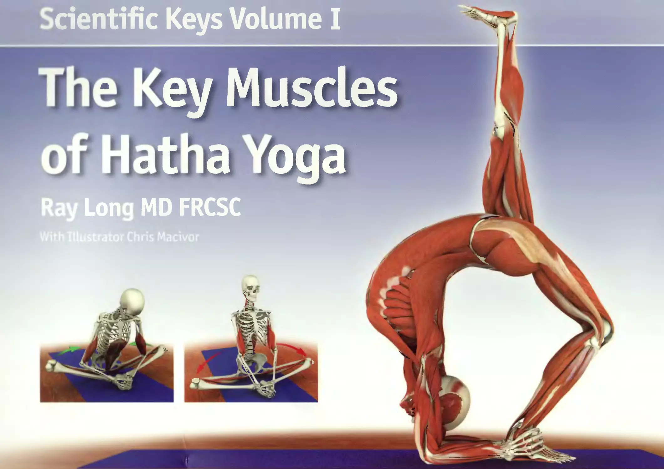
                    Scientific Keys Volume I
The Key Muscles
of Hatha Yoga
Ray Long MD FRCSC
With Illustrator Chris Macivor
Contents
Introduction 7
Fundamentals 8
I Locations on the Body 8
Skeleton 10
Joints 22
Ligaments 26
Muscles and Tendons 35
Movement 44
Part One - The Pelvic Girdle and Thighs 49
Chapter 1: Iliopsoas 57
Chapter 2: Gluteus Maximus 64
Chapters: Gluteus Medius 69
Chapter 4: Tensor Fascia Lata 74
Chapter 5a: Pectineus 79
Chapter 5b: Adductor Magnus 84
Chapter 6: External Rotators 91
Chapter 7: Quadriceps 96
Chapters: Hamstrings 103
Part Two - The Trunk 113
I Chapter 9: Abdominals 118
Chapter 10:	Back Muscles 128
Chapter 11:	Latissimus Dorsi 135
Chapter 12:	Trapezius 139
Chapter 13:	Pectoralis Major & Minor 144
Part Three - The Shoulder Girdle and Upper Arms 151
Chapter 14:	Rhomboids 157
 Chapter 15: Chapter 16:	Serratus Anterior 162 Deltoids 167
i Chapter 17:	Rotator Cuff 174
Chapter 18: I Chapter 19:	Biceps Brachii 185 Triceps Brachii 190
Chapter 20: Sternocleidomastoid 197
Chapter 21:	Lower Leg and Foot 202
Chapter 22:	Forearm and Hand 206
Chapter 23:	Myofascial and Organ Planes 210
Chapter 24:	The Breath Connection 212
Chapter 25:	Bandhas 220
Chapter 26:	Chakras 222
Putting It All Together 224
Appendix of Asanas 230
Index of Asanas 236
Index of Muscles 238
Introduction
uman anatomy and physiology is a vast subject, as
is the art of hatha yoga. Nevertheless, combining
knowledge from both fields is extremely
beneficial to the yoga practitioner. Athletes can
improve their performance and experience fewer injuries
through a basic understanding of their musculoskeletal
system. Similarly, yoga practitioners can benefit from the
application of Western science to their practice
development.
It is not necessary to memorize hundreds of muscles and
bones to experience the benefits of applying science to
yoga. What is necessary is the functional understanding of
a manageable number of key anatomic structures in their
settings as they relate to hatha yoga. Knowledge of these
structures can be applied immediately to optimize your
practice, break through blockages and avoid injuries.
This first volume presents key muscles in the context of
hatha yoga. For practitioners unfamiliar with the Western
scientific terminology of the body, the following section,
"Fundamentals," is recommended.

About the Author
Ray Long
Roy Long MD FRCSCis a board
certified orthopedic surgeon and
the founder of Bandha Yoga. Ray
graduated from The University
of Michigan Medical School with
post-graduate training at Cornell
University, McGill University,
The University of Montreal and
Florida Orthopedic Institute.
He has studied hatha yoga for
over twenty years, training
extensively with B.K.S. Iyengar
and other of the world's
leading yoga masters.
Chris Macivor
Chris Mocivoris a digital illustrator
and the visual coordinator of Bandha
Yoga. Chris is a graduate of Etobicoke
School of The Arts, Sheridan College
and Seneca College. His work has
spanned many genres from TV and
film to videogames and underwater
videography.
Patanjali, the patron saint of yoga, said that mastery
combines a balance of science and art. Knowledge of
science is like the colors on an artist's palette - the
greater the knowledge, the more colors available. The
body is the canvas and the asanas are the art we create.
Scientific
Keys Volume I
First Edition: Copyright 2005, Raymond A Long MD FRCSC
Second Edition: Copyright 2006, Raymond A Long MD FRCSC
Third Edition: Copyright 2006, Raymond A Long MD frcsc
Bandha Yoga Publications
All rights reserved
No part of this book may be reproduced, copied or transmitted in any form without the express written
permission of the publisher.
Always consult your healthcare provider before practicing yoga or any other exercise program.
Yoga must always be practiced under the supervision of a qualified instructor.
The author assumes no responsibility for injuries that may occur as a result of the practice of yoga.
Design: Ingrid Patricia Sanchez
www.BandhaYoga.com
Scientific keys
How to Use This Book
The images in this book are the keys. We
present each muscle in the context of
its function as an agonist, antagonist or
synergist. Note the interrelated views
of the muscle in each of its various
representations.
Relax and study one muscle at a time.
Actively apply what you have learned by
visualizing the muscles as you perform
the asanas. Consciously contract and
relax them, as detailed in the images.
This will consolidate your knowledge.
Review each studied muscle, first at
twenty four hours and then again at one
week. In this way you will master the
muscles and integrate them into your
yoga practice.
6

Fundamentals
Locations of Structures on the Body
The following terms are used to describe where structures Lie in relation to certain Landmarks on the body.
Medial: Closer to the midline of the body	Lateral: Away from the midline
Proximal: Closer to the trunk or midline	Distal: Away from the trunk or midline
Superior: Above or towards the head	Inferior: Below or away from the head
Anterior: Towards the front of the body Ventral: On the front of the body	Posterior: Towards the back of the body Dorsal: On the back of the body
Superficial: Towards the skin	Deep: Inside the body
Locations on the Body
These images demonstrate the terminology for identifying
body locations in yoga postures. Note that some of the terms
are interchangeable. For example, an anterior structure (such
as the chest) in utkatasana is also ventral.
1)	The sternum is medial to the shoulder.
2)	The shoulder is lateral to the sternum.
3)	The shoulder is proximal.
4)	The hand is distal.
’>) The head i. supetiot to the feet
6)	The fret ar* inferior to tlv head.
7)	The (hest Is .inter lot to the bar k.
8)	The back is posterior to the r hest.
9)	The abdomen is ventral.
10)	The lumbar region is dorsal.
11)	The abdominal muscles are superficial.
12)	The abdominal organs are deep.
tadosano
utkatasana
9
Skeleton
Bone is the dynamic living tissue that forms the body's structural
framework. The bones mass is composed of organic and inorganic
materials including calcium salts and connective tissue, as well
as cells and blood vessels within a calcium matrix. This combina-
tion gives bone a tensile strength near that of steel, yet maintains
a modicum of elasticity. By aligning the direction of the force
of gravity along the major axis of the bones, we can access this
strength in yoga postures.
Regular practice-pf yoga is beneficial for your bones because
healthy stresses are applied in a variety of unusual directions. This
strengthens bones, which remodel in response to stress by depos-
iting layers of calcium into the bone matrix. Like a physiological
yin/yang, lack of healthy stress on bones weakens them.
Bonej are also the body's reservoir for calcium, critical in a variety
of physiological functions including muscle contraction. The
concentration of calcium in the body is tightly regulated through a
complex interplay between the skeletal, endocrine and excretory
systems. This involves feedback loops between the parathyroid
gland, the kidneys, the intestines, the skin, the liver and the
bones.
10
living bone

Bone mass decreases in osteoporosis. This age related de-
crease is associated with the loss of estrogen in post-meno
pausal women. Studies have demonstrated that resistance
type exercise maintains bone mass. Accordingly, it is reason
able to conclude that the various healthy stresses that yoga
practice applies across the bones may aid preventing osteo
porosis.
The bones of the skeleton link together at the joints and act
as levers for the muscles that cross the joints. (onsciously
contracting and relaxing these skeletal muscles moves the
body into the various yoga postures.
Shapes of bones
The form or shape of a bone reflects its
function. Long bones provide leverage, flat
bones provide protection and a place for
broad muscles to attach, and short bones
provide for weight bearing functions.
Yoga accesses each bone's particular po-
tential, using long bones to leverage the
body deeper into postures, the flat bones
(and their accompanying core muscles) for
stability, and the short vertebral bodies to
bear weight. Examples of these bones are
illustrated here.
virabhadrasana II
И
Gravity and the Skeleton
The Sanscrit word for a yoga pose is asana. Sanscrit scholars variously
translate this word to mean "a comfortable or effortless position".
Yoga postures approach effortlessness when we align the long axis of
the bones with the direction of gravity. This decreases the muscular
force needed to maintain our postures.
For example, in utthanasana, the force of gravity aligns with the long
axis ofthe femur and tibia bones. Similarly, in siddhasana the force
of gravity aligns with the long axis ofthe spine.
Use muscular force to bring the bones into a position where they
carry the load. Once these positions are attained, muscular force is
no longer necessary (or greatly decreased).
utthanasana
siddhasana
12
Fundamental Bones
clavicle
scapula
sternum
humerus
radius
ulna
ilium
sacrum
pubis
femur
patella
fibula
tibia
13
Fundamental Bones
carpals
radius
ulna
trochlea
phalanges
metacarpals
radial head
lateral epicondyle
medial epicondyle
olecranon
14
hindfoot midfoot	forefoot
(calcaneus,talus)	(tarsals) (metatarsals, phalanges)
Shoulder and Hip
The hips and shoulders are ball and socket joints. Their form reflects their function
in that the deep socket (acetabulum) ofthe hip is designed to support weight
while the shallow socket (glenoid) ofthe shoulder is designed to provide maximum
range of motion for the arms. Yoga postures balance mobility and stability by
increasing the range of motion ofthe hipsand stabilizing the shoulder.
femoral head
lesser trochanter
ischial tuberosity
hip
acetabulum
greater trochanter
acromion
glenoid
humeral head
greater tuberosity
lesser tuberosity
bicipital groove
shoulder
The Axial and Appendicular Skeleton
The axial skeleton consists
ofthe spinal column,
cranium (skull), and rib
cage. The spinal column
surrounds and protects the
spinal cord, which is the
central energy channel,
orsushumna nadi. It is
the axis around which the
poses of yoga revolve. The
appendicular skeleton
connects us with the world.
The lower extremities
form our connection
to the earth. The upper
extremities, in association
with our senses, connect us
with each other.
axial skeleton
appendicular skeleton
16
The Shoulder Girdle
The shoulder girdle is the yoke that connects the upper
extremities with the axial skeleton. It is the seat of the
brachial plexus, a collection of nerves that in association with
the heart forms the basis for the fourth and fifth chakras. The
shoulder girdle is comprised of the following structures:
•	Scapula (shoulder blade)
	Scapulothoracicjoint
•	Clavicle
•	Sternoclavicular and Acromioclavicular joints
•	Humerus (upper-arm bone)
	Glenohumeraljoint
The Pelvic Girdle
The pelvic girdle is the yoke that connects the lower
extremities to the axial skeleton. It is the seat of the sacral
plexus, a collection of nerves that forms the basis for the
first and second chakras. The pelvic girdle is comprised of the
following structures:
•	Iliac bones
•	Sacroiliacjoint
•	Femur (thigh bone)
•	Hipjoint
17
Connecting the Appendicular and Axial Skeleton
nerve roots in back bend
Eka pada viparita dandasana demonstrates connecting the upper and
lower appendicular skeletons to create movement in the axial skeleton.
Note the inset image demonstrating stimulation of the spinal nerves in
this back bend.
eka pada viparita dandasana
18
The Vertebral Column
cervical
thoracic
lumbar
sacrum
coccyx
Spinal Curves
~7
I
We determine the spinal
curves by viewing them
from the side. Kyphosis is
a convex curve and lordo-
sis is a concave curve.
This illustration demon-
strates the four normal
curves in the spine:
1) cervical lordosis
2) thoracic kyphosis
3) lumbar lordosis
4) sacral kyphosis
tadasana
14
Scoliosis
scoliosis
Scoliosis is a lateral deviation and rotational deformity of the spine. The most common
form of scoliosis is called "idiopathic", meaning without an identifiable cause. Other forms
include congenital and neuromuscular scoliosis. Studies have suggested that idio-
pathic scoliosis is related to hormonal factors including the level of melatonin
produced. This form of scoliosis also has a hereditary component.
When the magnitude of the scoliotic curve progresses beyond 20 degrees,
there is a risk of continued progression after skeletal maturity. Very large scoli-
otic curves can impact respiration by restricting the ribcage.
Scoliosis affects the bone, cartilage and muscles of the spine.
Muscles on the concave side of a curve become chronically
shortened when compared with those on the convex side. Yoga
postures aid to counteract this process by stretching the
shortened muscles.
Scoliosis also affects the pelvic and shoulder girdles, as illustrated here.
For example, tilting the pelvic girdle creates a perception of limb length
discrepancy (one leg shorter than the other). Similarly, one arm may ap-
pear shorter than the other.
marichyosana
*oqa as therapy
h« «images of <3 twist, back bend and forward bend demonstrate how yoga postures
conn net .md stretch the back muscles. This lengthens chronically shortened muscles on the
< oiv avt? side of the scoliotic curve while strengthening them on the convex side. This assists
in Imi ricing perceived discrepancies in limb length and may also improve nerve conduction.
salabhasana tricing mukhaikapada paschimottcinusanu
Joints
As with the bones, the shape of the joints reflects
theirfunction (and theirfunction reflects their
shape). Joints come in a spectrum of shapes, de-
pending on the mobility or stability they require.
For example, the hip joint is a ball and socket
while the knee joint is a hinge. A ball and socket
type hip joint confers the greatest mobility in all
planes and is useful for activities such changing
direction while walking and running (or reach-
ing in various directions to grasp objects, as with
the shoulder). A hinge type knee joint provides
greater stability and is useful for propelling the
body forward (ordrawing an object towards the
body, as with the elbow).
Other joints such as the intervertebral between
the vertebrae allow for limited mobility between
individual vertebrae but great stability to protect
the spinal cord. Mobility ofthe spinal column
comes from combining the limited movement of
individual intervertebral joints as a whole.
22
compressive
lumbar spine
hip articular cartilage
i up joint capsule with
synovium (posterior view)
Articular Structure
The joint capsule is connective tissue sheathing
that surrounds and seals synovia I joints. It is sus-
ceptible to stretch injury when executing extreme
movements in yoga postures.
Synovial tissue lines the inside of the joint cap-
sule. This tissue produces synovia I fluid, a viscous
lubricant for the joint surface that decreases
friction during joint movement. Synovial fluid
circulates throughout the joint, transporting
nutrients to the articular cartilage and removing
debris from the joint space. The various contor-
tions resulting from yoga postures aid flex and
expand the joint capsule, stimulating circulation
of synovia I fluid.
Articular cartilage covers the joint surfaces, al-
lowing smooth gliding of one bone over the other.
In fact, articular cartilage is one of the smooth-
est surfaces known to man. Applying excessive
pressure to this fragile cartilage can injure it,
ultimately resulting in arthritis.
The meniscus deepens the articular surface and
broadens the contact area of the joint. This aids
to stabilize the joint and distributes the force of
gravity and muscular contraction over a greater
surface area. The meniscus is composed of fibro-
tarlilage, giving it a flexible rubbery consistency.
Joint reaction forces
Every action has an equal and opposite
reaction. Muscular contraction and grav-
ity create opposing forces across the joint
surfaces, known as joint reaction forces.
It is important to spread these forces over
the greatest possible joint surface area.
Joint congruency refers to the fit of a
joint's articular surfaces. A joint is congru-
ent when its surfaces fit together perfect-
ly. Movement out of congruency focuses
stress on a small surface area. A large
force focused on a small area of articular
cartilage can injure it, eventually causing
degenerative changes.
Some yoga postures have the capacity to
sublux or take a jointinto an incongruent
position. Avoid this by using the joints
with a greater range of motion while
protecting those joints with limited range
of motion.
24

Joint reaction forces - applied
For example, the ball and socket joint ofthe hip has greater range of mo-
tion than the hinge joint ofthe knee. Lotus posture (or padmasana) re-
quires a Large amount of external rotation ofthe hip joint to bring the foot
into position on the opposite leg. Obtaining this external rotation from the
knee joint creates incongruency because the knee is a hinge joint with lim-
ited capacity to rotate. This incongruency can result in the abnormal distri-
bution of joint reaction forces, injuring the intra-articular structures ofthe
knee. Therefore it is essential to first obtain full range of motion ofthe ball
and socket hip joint to protect the hinge knee joint, (see arrows)
<7/ dha padmasana
ligaments
Ligaments are fibrous connective tissue
structures that Link one bone to another at
the joint. They serve to stabilize the joint
while at the same time allowing mobility.
Ligaments vary in size and shape according
to their function. For example, the cruciate
ligaments of the knee are short, stout
structures that assist in maintaining the
knee as a hinge. The sacroiliac ligaments
are dense, broad and thick structures that
limit movement of the sacroiliac joint.
The shoulder ligaments are thin band-like
structures that are confluent with the
shoulder capsule, allowing for great range of
motion.
Ligaments are non-contractile but actively
participate in movement because they have
sensory nerves that transmit information
about joint position to the spinal cord and
brain.
26
I
Ligamentotaxis
Ligamentotaxis refers to the pull of
Ligaments on the bones to which they
are attached. This concept is used by
physicians to set pull broken bones
back into place and set them. It can
also be used during yoga practice as
illustrated here in the forward bend
utthanasana. Here the weight of the
upper body is transmitted to the pelvis
via the Ligaments of the back. This pulls
the pelvis forward and lifts the ischial
tuberosities, passively stretching
the hamstrings. Similarly, Ligaments
have some capacity for elastic recoil.
This recoil can be combined with the
momentum of the body as it raises from
postures such as back bend.
27

Pelvic and Hip Ligaments
The form ofthe pelvic and hip Ligaments reflects theirfunction. The pelvic
ligaments are thick and strong in support ofthe weight bearing function of
these joints. The hip ligaments are shaped to stabilize the hip while allowing
movement for walking and running.
posterior iliofemoral
ligaments
sacroiliac
ligaments
sacrotuberous
ligament
anterior iliofemoral
ligaments
Pelvis and Hip (anterior)
Pelvis and Hip (posterior)
28
Iliofemoral Ligaments
anterior iliofemoral
ligaments (relaxed)
Hip joint (flexed internally rotated)
The anterior iliofemoral ligaments are part of the hip joint and stabilize
it. These ligaments become taut when the femur extends and externally
rotates. They relax when the femur flexes and internally rotates. Tightness
in these ligaments limits extension of the hip in lunging poses and forward
splits. This limitation is overcome by tilting the pelvis forward and internally
rotating the femur.
Hip joint (extended, externally rotated)
Shoulder and Elbow Ligaments
Elbow (posterior)
The collateral ligaments of the elbow limit side to side
motion and maintain the joint as a hinge. The interosseous
membrane stabilizes the bones of the forearm.
annular ligament	interosseous
(lateral collateral ligament)	membrane
shoulder
Unlike the thick ligaments of the hip, the
glenohumeral ligaments of the shoulder are
thin structures. Their design allows greater
mobility of the joint.
acromioclavicular
ligaments
coracoclavicular
ligaments
The inferior glenohumeral ligament is the
most important of the three glenohumeral
ligaments. This ligament tightens when the
humerus abducts and externally rotates.
inferior glenohumeral
ligament
coracoacromial
ligament
transverse bicipital
ligament
30
The Muscular Stabilizers
of the Shoulder
The shape ofthe bones and thick ligaments stabilize the hip. Muscl js
stabilize the shoulder. The primary shoulder stabilizer is the rotator
cuff, the secondary stabilizers are the triceps and biceps. Yoga
postures such as arm balances and inversions strengthen these
muscles, balancing stability and mobility in the joint.
supraspinatus
infraspinatus
subscapularis
biceps (long head)
triceps (long head)
Rotator Cuff
(stabilizing shoulder joint)
Biceps and Triceps
(stabilizing shoulder joint)
31
VERTEBRAL UNIT
Spine Ligaments
The vertebral unit is comprised of two adjacent vertebral bodies and
the intervertebral disc. Movement between the vertebrae is possible
in several planes (including small amounts of rotation, flexion and
extension). The combination of motion across multiple vertebral
units culminates in spinal movement.
LUMBOSACRAL SPINE
sacrum
anterior longitudinal
ligament
spinous process
tranverse process
spinalis muscle
transversalis muscle
interspinous ligament
neural foramina
vertebral body
intervertebral disc
32
Trunk Ligaments
Ligaments attach bone to bone and also serve as attachments for
certain muscles. Below are three such ligaments that connect the uppei
body and trunk with the lower body.
ilioinguinal ligament
linea alba
lumbosacral fascia
Knee Ligaments
The patellar tendon connects the quadriceps muscle to the tibia for extension ofthe knee. The
collateral ligaments limit side to side motion ofthe knee and maintain its function as a hinge
joint. The anterior and posterior cruciate ligaments limit anterior and posterior translation
ofthe tibia on the femur (respectively). The menisci deepen and stabilize the knee joint. The
interosseous membrane stabilizes the bones ofthe lower leg.
patellar tendon
lateral collateral ligament
anterior cruciate ligament
posterior cruciate ligament
menisci
interosseous membrane
34
Muscles and Tendons
Muscles
The synergists of psoas assist in
flexing the hip.
The quadriceps are the agonists
that contract to extend the knees.
The hamstrings are the antagonists
stretched by this action.
36
Movements are determined by the varying forces acting across the joints. These
forces are produced by the muscles, and their effects on body position are
determined by the muscles' shape, origin (the attachment of the muscle to a
bone at the more fixed or proximal end), and insertion (the attachment of the
muscle to a bone at the end toward the part to be moved, or the more distal end).
Origin
Proximal attachment of the muscle to a bone.
Insertion
Distal attachment of the muscle to a bone.
Agonist, or prime mover
The muscle that contracts to produce a
certain action about a joint. For example, the
hamstrings are agonists when you flex your
knee.
Antagonist
A muscle that relaxes while the agonist
contracts. The antagonist produces the
opposite action about a joint. For example,
the quadriceps (at the front of the thigh) is the
antagonist to the hamstrings when you flex
your knee. When you extend your knee, the
quadriceps is the agonist and the hamstrings
the antagonists.
Synergist
A muscle that assists and fine-tunes the action
of the agonist and can be used to produce
the same action, although generally not as
efficiently.
I
I he or irjiri of the
rectus feiiioris is the
outer ior superior iliac
spine. The insertion is
the patella.
Muscles and Tendons
Tendons attach muscles to bones, transmitting the forces produced by the muscles, moving joints. Tendons also have
sensory nerves that communicate information about muscle tension and joint position to the brain.
1 endons and ligaments have limited capacity to stretch and do not contract. Practicing yoga improves tendon and
ligament flexibility, especially when performed in a heated room. Practitioners should not stretch tendons or ligaments
beyond their normal length as this can cause injury.
iliacus tendon
hamstring tendon
37
Muscle Shapes
Muscles come in a variety of shapes, reflecting their specific
function. These different shapes provide for maximal
mechanical efficiency during movement of the skeleton.
Similarly, muscles may curve over the bones to provide a
"half-pulley" effect which multiplies the force of contraction.
Some of the various muscle shapes include the following:
biceps bracialis
two headed fusiform
iliopsoas
multi headed convergent
(with half pulley)
stm’fent/c
transversalis
short square
1/7
t  Ins abdommus
38
Mono-articular and Poly-articular Muscles
mono-articular
For example, in the one-legged vrksasana the iliacus and gluteus medius represent monoarticular muscles because
they originate on the ilium and attach to the proximal femur, crossing (and moving) only the hip joint. Here the
iliacus and gluteus medius serve to stabilize the hip jointin the standing leg. The quadratus lumborum, psoas, rectus
femoris and sartorius represent polyarticular muscles because they all cross (and move) multiple joints. Here these
muscles contribute to flexing, abducting and externally rotating the non-standing leg.
When monoarticular muscles contract they primarily move only one joint. When polyarticular muscles contract they
can move multiple joints.
Muscles are also defined by the number of joints that they cross from their origin to their insertion. Monoarticular
muscles cross only one joint while polyarticular muscles cross more than one joint.
poly-articular
vrksasana
Muscle Structure and Function
CONTRACTED
RELAXED
STRETCHED
Muscle fibers are the functional contractile units of each skeletal
muscle. Fibers are grouped into fascicles which in turn are grouped
into bundles, thus forming the individual skeletal muscles.
Skeletal muscles are also composed of non-contractile elements. The non-
contractile elements include the connective tissue sheath surrounding the muscle bundles, fascicles and
individual fibers, as well as the myotendon junction.
Muscle fibers contract in response to afferant nerve stimuli (from the central nervous system). This is an active,
energy-dependent process involving the release of calcium at the cellular Level of the muscle fiber. Calcium then
forms cross bridging between the myofilaments (of the myofibril). This causes a "ratcheting" effect that results
in the shortening or contraction of the individual muscle fiber. The net effect of this process is shortening or
contraction of the entire muscle.
The force of this contraction is transmitted to the non-contractile fascial elements surrounding the muscle.
These fascial elements then transmit this force to the myotendon junction and on to the bones, moving the
joint.
Muscles exist in either a contracted, relaxed or stretched state. This is illustrated above in the inset showing the
cross bridging of myofilaments.
Types of Muscle Contraction
There are three types of muscle contraction:
Concentric (isotonic) contraction:
The muscle shortens while maintaining constant
tension through a range of motion.
Eccentric contraction
The muscle contracts while lengthening.
Isometric contraction
The muscle generates tension but does not
shorten, and the bones do not move.
41
Stretching Muscles
Static Stretching
Static stretching is the most common technique used in hatha yoga. There are two categories
of static stretching. The first is active static stretching. This involves contracting antagonist
muscles to stretch a target muscle. Contracting the quadriceps, iliopsoas and biceps during
the forward bend paschimottanasana is a form of active static stretching ofthe hamstrings.
Contracting antagonist muscles in active static stretching results in a phenomenon called
"reciprocal inhibition." During reciprocal inhibition, the central nervous system signals the
target muscle to relax.
Passive static stretching occurs when we relax into a stretch, using only the force of body
weight (or an externally applied weight) to stretch muscles. The restorative pose setubandha is
an example of passive static stretching ofthe iliopsoas muscle.
42
Facilitated Stretching
Yoga practitioners use facilitated stretching to deepen their postures. This type
of stretching involves contracting the muscle being stretched during an active
static stretching. This action triggers a reflex arc involving the Golgi tendon organ,
resulting in a profound relaxation of the target muscle when the contraction period
ends. This is also known as proprioceptive neuromuscular facilitation (PNF). It is
extremely important to consider the joint reaction forces when using facilitated
stretches, since the force the muscle generates is transmit ted to the joints. As a
general rule, gently contract the stretched muscle to avoid excessive joint reaction
forces. These images demonstrate facilitated stretching of the gluteus medius,
maximus and tensor fascia lata.
Dynamic Stretching
(Scientific Keys, Volume П covers the physiology of stretching in detail).
Yoga practitioners use dviidinicstietching during the vuiyasa type
practice. This type of stretching involves repetitive movement of the
body into increasingly deeper st ret< lies. Performing dynamic stretching
in the morning "resets" the resting muscle length for the day.
Movement
Movement Definitions
Motion ofthe musculoskeletal system necessarily involves
multiple joints, forces applied in many directions, and
movement in many planes. A convention exists to describe
the basic movements ofthe musculoskeletal system that can
be useful in analyzing the form and function ofthe asanas.
The six basic movements of the body take place in three planes.
Coronal plane: Divides the body into front and back. Movements along this
plane are called adduction and abduction. Adduction moves the extremity
towards the midline, abduction moves the extremity away from the
midline.
Sagittal plane: Divides the body into right and left. Movements along
this plane are called flexion and extension. Flexion usually moves the
extremity forward except at the knee where it moves backward. Extension
moves the extremity backward.
Transverse plane: Divides the body into upper and lower halves. Movement
along this plane is called rotation. Rotation is further classified as medial
rotation (toward the midline) or lateral rotation (away from the midline).
Medial and lateral rotation are also referred to as internal and external
rotation, respectively.
All movements ofthe body are composed of varying
contributions of these six elemental movements.
44
Pose with movements
The form of each asana reflects its function and vice versa. Here we use
virabhadrasana II to analyze the positions of the body in a yoga posture.
You can combine this analysis with knowledge of the muscle actions to
optimize the function of your poses.
5. The torso extends.
6. The arms abduct.
1.	The front knee flexes.
2. The front hip flexes.
3. The back hip extends.
4.	The back foot rotates internally.
7.	The forearms rotate internally.
8.	The neck and head rotate
45
Coupling of Joints
Movement of adjacent joints in different planes is called
coupled movement. For example, in the side bend of
utthita trikonasana, the vertebral column undergoes
a complex series of coupled movements, including
rotation, flexion, and extension at various levels.
Similarly, in the same pose, the position of the hip joint
of the forward leg involves a combination of flexion of
the femur (thigh bone) at the hip joint and forward tilt of
the pelvis.
46
Complex Movements
г',?ь
&
п
In reality, and especially in yoga postures, movement can rarely
be described in simple terms. Complex movements involve many
joints moving in various ways. Complex movements are also
described in terms of other characteristics, including coupling
of joints and open and closed chain movements.

Open and dosed chain movements
1) Open chain: Movements in which the distal end moves
freely are called open-chain movements (for example, the
deltoid abducting the upper arms in virabhadrasana II).
2) Closed chain: Movements in which the distal end (the
insertion) of the moving limb or body segment are fixed are
called closed-chain movements (for example, the iliopsoas
lowering the pelvis in virabhadrasana II).
Part
One
Pelvic Girdle & Thighs
External rotators of the hip
1	piriformis
2	gameLLus
3	obturator internus
4	obturator externus
5	quadratus femoris


1
iliopsoas
2
gluteus medius
3
gluteus maximus
4
sartorius
5
tensor fascia lata
6
pectineus
7
gracilis
8
adductor Longus
9
rectus femoris
10
quadriceps
11
biceps femoris
12
semitendonosus
13
semimembranosus
14
gastrocnemius
Movement: Hip
52
Flexion
Utthita Hasta Pcdangustasana
The following examples illustrate the elemental movements of the hip
and pelvis. Look closely to appreciate the "coupling" of the movements
of the hip joint and pelvis.
Extension
Vrishchikasana
Abduction
(movement away from the midline)
Supta Padangusthasana В

Adduction
(movement towards the midline)
Marichyasana C

Movement: Hip
Internal Rotation
Garudasana

Movement: Pelvis
Anterior Tilt
Utthanosana
55
Movement: Pelvis
Rotation
Garudasana

Chapter
Iliopsoas
Also known as the psoas muscle, the iliopsoas is
actually a combination of two large muscles:
the psoas major and the iliacus. The psoas
major muscle originates in the lower
back; theiliacus originates on the inside
of the pelvis. Both muscles combine to
form one tendon that attaches to the
inside of the proximal femur bone.
psoas major
iliacus
The iliopsoas is thus called polyarticular. This
means that it crosses over (and moves) more than
one joint. The iliopsoas also acts like a pulley as
it curves over the front rim of the pelvis on its
way to the femur. Like other pulley systems, this
serves to multiply the force generated when the
iliopsoas contracts. The iliopsoas thus moves
the bones of the lower back, pelvis and hip
in a coupled fashion. This means that when it
contracts, a combination of movements across
severaljoints is possible.
The iliopsoas first awakens during infancy when
we are learning to sit up and then to walk. Once
awakened, the iliopsoas becomes constantly
activein activities such as standing and walking.
In spite of this constant use, our awareness of the
iliopsoas quickly becomes unconscious. (Imagine
if we had to think every time we took a step!)
Hatha yoga can be used to reawaken oui
consciousness of this large and important muscle.
Once you awaken the iliopsoas, contract or relax it
to transform and deepen your asanas.
iliopsoas
Origin
1) Psoas major: Tranverse processes,
discs and bodies of Lumbar vertebrae one
through five; body of twelfth thoracic
vertebra.
2) ILiacus: lipper two thirds of the inside
surface of the iliac bone up to the inner Lip
of the iliac crest and anterior sacroiliac
joint.
Innervation & chakra illuminated
Lumbar nerves 1,2,3,4
Chakra: Second
The second chakra is illuminated by contracting and lengthening
the iliopsoas muscle. This is due to stimulation of the various
sensory nerves at its origin
and insertion, within the
muscle itself, and the skin
surrounding it.
insertion
Lesser trochanter (the smaller prominence
or knob) of the proximal femur.
liopsoas (d-e-o-SO-us)
ntagonists
Gluteus maximus: extends hip and
trunk resulting in lengthening and
stretching of the Iliopsoas, particularly
in backbends.
Hamstrings: extends the hip when
initiating backbends, can be used to
draw the opposite leg iliopsoas into a
deeper stretch in lunging postures.
Synergists
Tensor fascia lata: assists the iliopsoas in
fine-tuning hip flexion.
Sartorius: assists the iliopsoas in fine-
tuning hip flexion and external rotation.
Rectus femoris: assists the iliopsoas in
fine-tuning hip flexion, also assists the
gluteus maximus in accentuating stretch
of the iliopsoas during back-bending (by
extending the knee).
Pectirieus: assists the iliopsoas in fine-
tuning hip flexion and provides adduction
component to stabilize hip (also balances
abduction action of sartorius).
Synergy
This illustration uses virabhadrasana II to demonstrate the tensor fascia
lata, Sartorius, rectus femoris, and pectineus as synergists of the psoas.
Similarly, the extended back hip demonstrates how the gluteus maximus and
hamstrings act as antagonists to the psoas.
virabhadrasana II
Synergy
Fh s illustration uses eka pada viparita dandasana to demonstrate the
qlnteus maxirnus and hamstrings stretching the psoas and the synergists
of the psoas in the planted leg. Similarly, the flexed hip of the leg in the
air demonstrates the tensor fascia lata, sartorius, rectus femoris and
pectineus as synergists of the psoas.
eka pada viparita dandasana

Iliopsoas (il-e-o-SO-us)
Action
Open chain
(Origin fixed, insertion moving):
Flexes and laterally rotates
the femur at the hip. Ex.
Padangusthasana D
Closed chain
(Insertion fixed, origin moving):
Flexes the trunk, anteverts (tilts
forward) the pelvis, straightens
and supports the lumbar spine. Ex.
Virabhadrasana В
Awakening
Closed chain isometric resistance to
trunk flexing.
Open chain isometric resistance to
femur flexing.
Conscious contraction in standing
poses.
Eccenti ic contraction in lunging
poses.
Contracted
I Itt hit.1 trikonasana
opt imally contracts the
psoas major portion of
the iliopsoas muscle.
(mitraction in this posture
intwerts the pelvis,
his action draws the
hamstrings' origin (ischial
t uberosity) away from their
insertion (lowerleg), and
accentuates their stretch.
Stretched
Ushtrasana stretches the iliopsoas through contraction
of the hip and trunk extensors, including the gluteus
maximus. Stretch is accentuated by contraction of
the quadriceps (including the rectus femoris, which is
eccentrically contracted).
Iwisted variations of
utthita trikonasana
preferentially contract
theiliacus portion of the
Chapter
Gluteus Maximus
The gluteus maximus stands out as
the Largest and most posterior of
four muscles located on the outside
of the pelvis. The gluteus maximus
is a single muscle divided into two
insertions: one on the outside of the
proximal femur bone and one on a
strap-like structure on the outside of
the thigh called the iliotibial band.
Contracting the gluteus maximus
extends and outwardly rotates the
femur. Fibers attached to the iliotibial
band tense it and assist in moving the
knee. The gluteus maximus functions
as a mono- and polyarticular muscle.
Tightness of the gluteus maximus
limits forward bending at the hips,
such as in utthanasana.
Like the iliopsoas, the gluteus
maximus works unconsciously
during standing and walking. Many
important yoga postures awaken the
gluteus maximus including standing
poses, backbends and forward
bends. Tightness limits forward
bends and weakness limits back
bends.
Gluteus Maximus (GLOO-te-us MAK-si-mus)
Origin
Outer posterior surface of the
illium, posterior surface of
the sacrum and coccyx, and
I he aponeurosis of the erector
spinae muscles of the back.
Insertion
Gluteal tuberosity on lateral
surface of the proximal femur
below the greater trochanter.
Iliotibial barjd (inserts onto
Gerdys' tubercle on the front of
the proximaltibja).
Innervation & chakra illuminated
Inferior gluteal nerve (lumbar spinal nerve 5 and sacral spinal nerves 1 and 2).
Chakra illuminated: First.
65
Gluteus Maximus (GLOO-te-us MAK-si-mus)
Synergists
Semimembranosis, semitendenosis, biceps femoris, quadratic lumborum
and adductor magnus.
Antagonists
Iliopsoas, rectus femoris and pectineus.
Action
I xlends and externally rotates hip. Upper fibers
issist to abduct thigh. Assists to stabilize fully
extruded knee (via iliotibial band).
Open chain contraction extends and
externally rotates the hip joint.
Contracting the gluteus maximus
lifts and externally rotates the back
leg in virabhadrasana III. The fibers
inserting on the iliotibial band also
assist in stabilizing the straight knee.
Closed chain contraction extends trunk in
virabhadrasana II.
Awakening
The gluteus maximus can be eccentrically contracted in
padangustasana, stretching and strengthening it.
Closed chain contraction of the
gluteus maximus in ushtras.in.i
extends the trunk.
Gluteus Maximus (GLOO-te-us MAK-si-mus)
Contracted
Purvottanasana: The gluteus maximus contracts in this
asana. Its external rotation component is counteracted by
contraction of the gluteus medius (anterior fibers), tensor
fascia lata and adductor group. (Accentuate this by pressing
down the ball of the foot.)
Stretched
Utthanasana: The gluteus maximus
stretches in this and other asanas
flexing the trunk and hips.
Chapter
Gluteus Medius
The gluteus medius presents a medium-
sized, fan-shaped muscle located
forward of the gluteus maximus, which
partially covers it. The gluteus medius
inserts on the tip of the greater trochanter
(a protuberance on the proximal femur to
which muscles attach). The gluteus medius
covers the gluteus minimus.
Direction and placement of muscle fibers
determines the movement produced by
contraction. Anterior fibers internally
rotate and middle fibers abduct the
femur. Whenthefemurisfixedin place,
as in one-legged standing poses,
contracting the gluteus medius tilts
the pelvis, maintaining balance.
We are largely unaware of the gluteus
medius, though it is constantly
active and balances the pelvis when
standing and walking. Contract it
in backbends to counterbalance
the external rotation of the hips
produced by contraction of the gluteus
maximus.
Tightness in the gluteus medius limits
postures that require extensive external
rotation of the femur at the hip (e.g.
lotus posture). Weakness limits one-
legged standing poses.
Gluteus Medius (GLOO -te-us ME-de-us)
Origin
Outer surface of ilium below the iliac
crest and anterior to the gluteus
maximus origin.
Gluteus Minimus
This see-through image of the
gluteus medius illustrates the
position of the gluteus minimus,
which has a similar function.
Origin
Outer surface of the ilium below and
anterior to the gluteus medius origin.
Insertion
Superior surface of greater
trochanter of the proximal femur.
Insertion:
Anterior portion of the greater
trochanter.
Synergists
Gluteus minimus, tensor fascia Lata and piriformis.
Antagonists
Adductor group and quadratus femoris.
Innervation & chakra illuminated
Superior gluteal nerve (lumbar spinal nerves 4 and 5,
and sacral spinal nerve 1)
Chakra illuminated: First
Gluteus Medius (GLOO -te-us ME-de-us)
Action
Abducts and internally rotates hip.
Stabilizes pelvis during walking. Posterior
fibers may externally rotate thigh.
The gluteus medius contracts and abducts
the bentleginjanusirasana. Anterior fibers
also medially rotate the thigh, protecting
the knee.
The gluteus medius contracts and
abducts the straight leg, lifting it in
ardhachandrasana.
Awakening
Contracting the gluteus medius in
marichyasana IV accentuates the twist.
This isometric contraction awakens the
gluteus medius.
Contracting the gluteus medius in
the back leg of parivrtta trikonasana
accentuates the twist from the trunk by
rotating the femur.
72
Contracted
Urdhvadhanurasana: Contracting the anterior fibers of the gluteus medius
internally rotates the hips and releases stress at the sacroiliac joint created by
contracting the gluteus maximus (to extend the hip).
Stretched
Vatayanasana: Externally rotating the hip
stretches the gluteus medius (especially the
anterior fibers). All poses with a lotus component
to the hips (external rotation) accomplish this.
Chapter4
Tensor Fascia Lata
This small polyarticular muscle originates
from the iliac crest in front of the
gluteus medius, assisting it with internal
rotation of the hip. Inserting on the
iliotibial band, it also works with the
anterior fibers of the gluteus maximus to
extend the knee.
Tightness in the tensor fascia lata limits
postures that externally rotate the hip,
such as padmasana.
Tensor Fascia Lata (TEN-sor FASH-e-a LA-te)
Origin
anterior superior iliac spine.
Innervation & chakra illuminated
Anterior portion of the outside of the iliac crest, and the
Superior gluteal nerve (lumbar nerves 4 and 5, and sacral nerve 1).
Chakra illuminated: First.
Insertion
Iliotibial band (and from there to the anterolateral
proximal tibia).
Tensor Fascia Lata (TEN-sor FASH-e-a LA-te)
Antagonists
Hamstrings, adductor group and gluteus maximus (femoralinsertion).
Synergists
Quadriceps, iliopsoas and anterior portion of gluteus maximus
(iliotibial band insertion), gluteus medius.
Action
I lexes internally rotates and abducts the hip, and supports the femur
on the tibia during standing.
Open chain contraction in parsvottonasana and urdhvadhanurasana
turns the thighs inward and straightens the knee.
Tensor Fascia Lata (TEN-sor FASH-e-a LA-te)
Stretched
Padmasana stretches the tensor fascia Lata
Eccentrically contracting it in this pose
facilitates this, awakening the muscle.
Contracted
Contracting the tensor fascia lata stabilizes
the lifted leg in ardha chandrasana.
Chapter >a
Pectineus
The pectineus is the proximal muscle
in the adductor group. It is a flat
rectangular muscle originating from the
front of the pelvic girdle and inserting
on the inside of the proximal femur. It is
monoarticular.
Awareness of the pectineus
awakens its neighboring
adductor muscles, the brevis
and longus.
Weakness limits
gomukhasana B.
Contraction accentuates
moola bandha.
Tightness in the pectineus limits
the depth of poses like
baddhakonasana.
Pectineus (pec-ti-NEUS)
Origin
Pecten (a bulge) of the pubic bone
on the iliopubic ramus, lateral to the
pubic symphysis.
(front view)
Insertion
Pectineal line extending from the
lesser trochanter to the linea aspera
on the inside of the proximal femur,
(back view)
80
Innervation & chakra
illuminated
Femoral nerve (lumbar spinal nerves
3 and 4) +/_ obturator nerve (lumbar
spinal nerves 2, 3 and 4).
Chakra illuminated: Second.
Antagonists
Gluteus medius, gluteus minimus, tensor fascia lata and piriformis.
Synergists
Adductor group, illiopsoas and quadratus femoris.
Pectineus (pec-ti-NEUS)
Action
Adducts, flexes and internally rotates hip.
The pectineus contracts in
parivrttaikapada sirasana adducting both
femurs and assisting the iliopsoas, flexing
the forward hip. This same principle
applies in parivrtta trikonasana.
Awakening
Baddhakonasana awakens the pectineus.
Isometric eccentric contraction
accentuates this.
Closed chain contraction of the front leg
pectineus draws the pelvis (and trunk)
forward in parsvottanasana.
Stretched
Baddhakonasana: The pectineus is at
full stretch in the upright version of
Contracted
Bakasana: Contracting the adductor group stabilizes this asana.
Chapter 5b
Adductor Magnus
This is the largest and most posterior
of the adductor group. It origir
from the back of the pelvis and
inserts along the length of
the inside of the femur. A
hole or "hiatus" in the distal
region of its insertion allows
passage of the femoral blood
vessels.
Its posterior location means
that it functions to adduct and
extend the thigh backward.
It is a synergist to the gluteus
maximus, assisting it in back-
bending postures such as
urdhvadhanurasana. Tightness
limits poses such as front splits.
Weakness limits bakasana.
Contraction of the adductor
magnus accentuates moola
bandha.
pQStefiQI
84
Adductor Magnus (ad-DUK-tor MAG-nus)
Origin
Anterior section:
Ischiopubic ramus.
Posterior section:
Ischial tuberosity.
Innervation & chakra illuminated
Anterior fibers: Obturator nerve (lumbar spinal nerves 2, 3 and 4).
Posterior fibers: Tibial portion of sciatic nerve (lumbar spinal nerves 3, 4 and 5).
Chakra illuminated:
Upper part of first, lower part of second.
insertion
1) Anterior section: Linea
aspera on the back of
middle third of the femur.
2) Posterior section:
Medial epicondyle on
inside of the distal femur
above the knee joint.
Adductor Magnus (ad-DUK-tor MAG-nus)
Antagonists
Gluteus medius, gluteus minimus,
tensor fascia lata and piriformis.
Synergists
Adductor group and quadratus
femoris.
Action
Adducts hip. Posterior fibers extend
and externally rotate hip.
Contracting the adductor magnus
squeezes the thighs together in
parsva bakasana.
Awakening
Upavistha konasana stretches and
awakens the adductor magnus by
abducting and flexing the hip.
Contracting the adductor magnus
assists the gluteus maximus,
extending and externally rotating the
back leg in parivrtta parsvokonasana.
Baddhakonasa stretches and awakens
the adductor magnus through
isometric eccentric contraction.
Adductor Magnus (ad-DUK-tor MAG-nus)
Stretched
The adductor magnus and the entire adductor group
stretches in upavistha konasana (the more distal and
posterior muscles are preferentially stretched).
Contracted
The adductor magnus lifts the lower leg
accentuating the twist in parsvabakasana.
1
pectineus
2
longus
3
brevis
4
magnus
5
gracilis

Adductor Group
Tightness of the adductor group causes the knees to be higher in seated postures, such as baddhakonasana
and siddhasana. Higher knees means a higher center of gravity. Holding a seated posture where the center of
gravity is higher requires more muscular effort. Lowering the knees makes these postures easier to maintain.
Releasing tightness in the adductor group assists in this process.
Facilitated stretching of the adductor group is illustrated here. Begin by placing the legs into
baddhakonasana and then attempt to adduct them while resisting them with the elbows. Contract the
adductors isometrically for a few moments and then draw them out to length by lowering the knees.
baddhakonasana
Chapters pirif°rmis&
External Rotators
Quadratus Femoris
Piriformis (piri-FOR-mus):
Quadratus Femoris (KWA-drat-us fe-MOR-us):
This is a pyramid-shaped
muscle originating from the
inside of the pelvis at the
sacrum. The piriformis wraps
around the ilium and inserts
on the tip of the greater
trochanter on the proximal
femur.
This creates a pulley effect
multiplying the piriformis'
force - much like what
occurs with the iliopsoas as
it curves over the front of
the pelvis. The sciatic nerve
runs behind the piriformis
and can be irritated by
tightness or inflammation of this muscle, a phenomenon known as "piriformis
syndrome." The piriformis acts in open and closed chain fashions. When its
origin (the sacrum) is fixed, contraction produces external rotation and
abduction of the femur. When the femur is fixed, contraction tilts the pelvis
backward. Tightness in the piriformis limits internal rotation of the thigh in
certain seated twists and in twisted standing poses.
This is the most distal of
the external rotators. It is a
quadrangular-shaped muscle
originating from above the
ischial tuberosity and inserting
on the greater trochanter of
the proximal femur. It is a
synergist to the piriformis
in external rotation of the
femur. It is also an adductor
of the femur and opposes the
piriformis' capacity to abduct.
Combined contraction of the
two muscles externally rotates
the thigh.
Tightness in the quadratus
femoris limits internal rotation of the femur in certain seated twists and
twisted standing poses. Contraction accentuates the effect of other seated
twists and поп-twisted standing poses. Awakening the piriformis and
quadratus femoris muscles brings awareness to the neighboring gamelli and
obturators (the other external rotators of the hip).
Piriformis & Quadratus Femoris
Origin-Piriformis
Inside surface of sacrum and
sacrotuberous ligament.
Origin-Quadratus femoris
Lateral surface of ischial tuberosity.
Insertion
Piriformis: Tip of greater trochanter.
Quadratus femoris: Posterior surface
of femur at the level of the greater
trochanter.
Innervation & chakra illuminated
Piriformis: Nerve to piriformis (sacral nerves 1 and 2).
Quadratus femoris: Nerve to quadratus femoris (lumbar nerves 4 and 5, and
sacral nerve 1).
Antagonists-Piriformis
Adductor group and gluteus medius (anterior fibers).
Antagonists-Quadratus femoris
Gluteus medius (anterior fibers), gluteus minimus and tensor
fascia lata.
Synergists-Piriformis
Gluteus medius (lateral and posterior fibers), gluteus minimus and
tensor fascia lata.
Synergists-Quadratus femoris
Adductor group.
Piriformis & Quadratus Femoris
Action
The piriformis externally
rotates and abducts the hip. The
quadratus femoris externally
rotates and adducts the hip.
Closed chain of the piriformis
contraction tilts the pelvis
backward.
The external rotators position
the hips in external rotation for
padamasana.

Contracted
Supta Padangusthasana B: ALL external rotators
of the hip contract in this asana. The piriformis
also assists the Lateral fibers of the gluteus
medius, abducting the femur.
Stretched
Marichyasana III: Contracting the internal rotators of the hip
(tensor fascia Lata and anterior fibers of gluteus medius) in this
asana stretches the external rotators.
Chapter
Quadriceps
rectus femoris	vastus
intermedius
vastus medialis
vastus lateralus
The quadriceps muscle forms the front of the
thigh. Its name, derived from Latin, means "four
headed." It is a four-part muscle combining
to form the quadriceps tendon which inserts
on the patella (kneecap). The patellar tendon is a
functional continuation of the quadriceps tendon,
inserting on the front of the proximal tibia. The
patella is a "sesamoid" bone (stone-like). This
refers to a bone within a tendon. Acting as
a fulcrum, it increases the force produced
by contraction of the quadriceps when
straightening the knee.
The rectus femoris - unique in that it	j
originates from the front of the pelvis at	I
the anterior-inferior iliac spine - continues I
on the front of the thigh, covering the
vastus medialis and combining with the
other quadriceps to insert on the patella.
It works as a polyarticular muscle. Force В
produced by its contraction results in a
combination of two possible movements:
flexion of the hip and extension of
the knee. The other three heads of the
quadriceps are monoarticular and only act
to straighten the knee.
The quadriceps are key muscles in yoga.
Contracting them directly stretches the
hamstrings when seated or in standing
poses. They also straighten the knees in
backbends, lifting the body.
Quadriceps (KWA-dra-seps)
Origin
Vastus medialis
Proximal two-thirds of the
anterior femur.
Vastus intermedins
Lateral portion of proximal
femur in region of greater
trochanter (seen through the
vastus Lateralus).
Vastus lateralis
Lateral portion of proximal
femur in region of greater
trochanter.
Origin - Rectus femoris
Anterior inferior iliac spine.
Insertion - (alt)
Quadriceps tendon: Superior
surface of patella (and on to the
anterior proximal tibia via the
patellar tendon).
Quadriceps
Innervation & chakra illuminated
Femoral nerve (Lumbar nerves 2, 3 and 4).
Chakra illuminated:
Second.
Synergists	Antagonists
Iliopsoas, tensor fascia lata.	Hamstrings, gastrocnemius, sartorius
and gracilis.
Action
Extends knee.
Rectus femoris also flexes hip.
The quadriceps contract, extending the knee
and flexing the hip (rectus femoris) in utthita
trikonasana.
Awakening
The vastus lateralis, medialis and intermedius
contract straightening the knee in
urdhvadhanurasana. The rectus femoris stretches
and contracts (eccentric contraction).
99
Quadriceps
Contracted
Utthanasana:
The quadriceps contract in this forward bend,
lifting the patella, straightening the knee and
stretching their antagonists (the hamstrings).
Stretched
Trianga Mukhaikapada Paschimottanasana:
Bending the knee stretches the vastus Lateralis, mediaLis and
intermedius. The rectus femoris relaxes due to the flexed position
of the hip. The straight leg quadriceps contract stretching the
corresponding hamstings.
Knee Biomechanics
Contracting the knee flexors helps to avoid hyperextending the knee. For example,
pressing down the ball of the foot contracts the gastroctnemius, stabilizing the knee.
The flexors of the knee counterbalance the extension force of the quadriceps. These
images illustrate how the knee flexors and extensors oppose each other, stabilizing the
knee.
Contracting the quadriceps draws the patella upward and against the anterior femur
into a groove between the femoral condyles. The patella fits congruently into the
intercondylar groove, stabilizing the standing leg. In this way the patella acts as a
fulcrum for knee extension.
It is important to avoid hyperextension or "locking" of the knee during standing poses.
This can overstretch the hamstrings and creates unhealthy stress on the knee's articular
cartilage.
Sartorius (sar-TOR-e-us)
The sartorius is a Long strap-like muscle originating from the
anterior superior iliac spine and inserting on the upper medial
surface of the tibia. This muscle flexes, abducts and externally
rotates the thigh, as in siddhasana, padmasana, vrksasana
janu sirsasana. In fact, the Latin translation for sartorius is
"tailor", because tailors used to sit cross-legged. The femoral
nerve innervates the sartorius, stimulating the second chakra.
vrksasana
102
Chapters
Hamstrings
Semitendonosus (sem-e-ten-di-NO-sus)
Semimembranosus (sem-e-mem-bruh-NO-sus)
Biceps Femoris (Bl-seps fe-MOR-us)
These two muscles make up the inner
hamstrings. The semimembranosus has a
flattened wide belly. The semitendonosus
is fusiform in shape (tapered at both ends)
The biceps femoris is a two-
headed fusiform-shaped muscle.
One head originates from the
ischial tuberosity, the other
from the back of the femur. Both
heads combine forming a single
tendon insertion on the head
of the fibula bone at the Lateral
side of the knee. It can be
palpated as a cord in this region.
The biceps femoris flexes the
straight knee and outwardly
rotates the lower leg (in the
bent knee). Its rotary action
accentuates twisting postures
such as marichyasana III.
Tightness in this muscle Limits
forward bends and certain
standing poses, especially those
involving internal rotation of
the Leg.
with the distal end forming a Long
tendon. Both muscles originate from
the ischial tuberosity. They have
separate insertions on the proximal
tibia, one on the inside of the back
of the tibia (semimembranosus)
and one on the inside of the front
of the tibia (semitendonosus). The
semitendonosus insertion combines
with the sartorius and gracilis muscles
to form a broad duckfoot-like insertion
on the anterior tibia called the pes
anserinus.
The semimembranosus and
semitendonosus flex the straight knee,
inwardly rotating the Lower leg in the
bent knee. This rotary component
accentuates seated twists, but in
the opposite direction of the biceps
femoris. Contraction of these muscles
also assists the gluteus maximus in
extension of the thigh at the hip, as
in virabhadrasana III. Tightness in
this muscle Limits forward bends and
Semitendonosus
Semimebranosus
certain standing poses, especially
those involving external rotation of the Leg.
Hamstrings
Origin-Biceps femoris
Insertion-Biceps femoris
Long head:
Ischial tuberosity
(Long head origin
is shared with the
semitendinosus.)
Short head:
Linea aspera on upper two
thirds of posterior femur.
Head of fibula.
Origin-Semimebranosus & Semitendonosus
Insertion-Semimebranosus & Semitendonosus
Ischial tuberosity:
(Semitendinosus origin is
shared with the long head of
biceps femoris).
1) Semimembranosus:
Posteromedial surface of proximal
tibia. Some fibers join to form
oblique popliteal ligament and attach
to posterior medial meniscus.
2) Semitendonosus:
Lipper inner surface of proximal tibia
(contributes to pes anserinus).
Innervation & chakra
illuminated
Biceps femoris
1) Long head: Tibial portion of sciatic nerve
(sacral nerves 1 and 2).
2) Short head: Fibular portion of sciatic nerve
(lumbar nerve 5, and sacral nerves 1 and 2)
Semimebranosus & Semitendonosus Tibial nerve
(lumbar nerve 5 and sacral nerve 1).
Chakra illuminated: First.
Gluteus maximus (illustrated by green arrow)
extending hip and knee, stretching long head of
the biceps femoris (and gastrocnemius).
Hamstrings
Antagonists
Quadriceps and iliopsoas.
Front view (anterior)
Back view (posterior)
106
Gluteus maximus, sartorius, gracilis and gastrocnemius.
Action
Flexes knee and extends hip (Long head).
Externally rotates tibia in flexed knee.
The biceps femoris contracts, flexing the
knee and externally rotating the tibia in
marichyasana III. This external rotation
manifests as internal rotation of the hip,
accentuating the twist of the trunk.
Awakening
Adho Mukha Svanasana stretches
and awakens the biceps femoris.
Semimebranosus & Semitendonosus
Action
Flexes knee and extends hip.
Internally rotates the tibia in the
flexed knee.
The semimembranosus and
semitendonosus contract, bending
the knee and internally rotating the
tibia in marichyasana I. This internal
rotation manifests as external
rotation of the hip, accentuating the
twist of the trunk.
Awakening
The semimembranosus and
semitendonosus stretch and awaken in
supta padangusthasana B.
Hamstrings
Contracted
Iliopsoas Lunge: Contracting the hamstrings of the front Leg draws
the body forward, accentuating the stretch of the iliopsoas in
Stretched
Krounchasana: This asana stretches all hamstrings.
Contracting the iliopsoas on the side of the bent knee
tilts the pelvis forward, drawing the origin of the
hamstrings away from the insertion. This accentuates
the hamstring stretch.
Hamstrings
Marichyasana I
The seated twists are named in honor of the great sage
Maha Rishi. Their tortional action compresses and
expands the internal organs, flushing blood into the
veins. The veins have one-way valves that then direct this
blood to the heart.
All muscles having rotational action contribute to twists
including the rotator cuff, external rotators of the hip and
the hamstrings.
110
Marichyasana III
Twisting postures awaken the muscles of the trunk stimulating
sensory nerve conduction from the skin, myofacial layers and the
muscles themselves. This illuminates and drives the subtle energies
of the chakras upward though the sushumna nadi (spinal cord).
The semimembranosis and semitendonosis contract in
marichyasana I. The biceps femoris contracts in marichyasasna III.

Test your anatomy*
1
2
3
4
5
★Please see www.bandhayoga.com for answers...
Part
Two
The Trunk
1	pectoralis major
2	external oblique
3	rectus abdominus
4	pectoralis minor
5	intercosta Is
6	internal oblique

The Trunk
Left to right illustrates the muscles of the back from deep to superficial
splenius capitis
splenius cervicis
longissimus cervicis
muLtifidis
- semispina lis
(thoracis,capitis,cervicis)
iliocostalis
interspinous ligament
lumbosacral fascia
sacrotuberous ligaments
rhomboids
serratus posterior
latisimus dorsi
erector spinae
lumbosacral fascia
trapezius
interspinous ligament
levator scapulae
5
Movement-

Rotation (twist)
Parivrtta Trikonasana

Lateral Flexion (side bend)
Utthito Trikonasana
Chapter9
Abdominals
Rectus abdominus (REK-tus ab-DOM-i-nus)
Internal oblique (o-BLEEK)
External oblique (o-BLEEK)
Transversus abdominus
Rectus Abdominus
This is a long flat muscle that
is divided into four bellies
by horizontal fibrous bands,
giving it a "washboard"
appearance. It originates
bilaterally from the pubic
symphysis and pubic crest,
inserting on the xyphoid
process (at the bottom of the
sternum) and, more laterally,
the cartilage of the fifth, sixth
and seventh ribs.
Contracting the rectus
abdominus flexes the trunk forward, or, if the insertion is fixed, lifts the
pelvis. This is demonstrated in utthanasana and tolasana respectively.
Tightness in this muscle limits the depth of backbends such as
urdhvadhanurasana and purvottonasana.
1	Transversus Abdominis
2	Internal Oblique
3	External Oblique
4	Rectus Abdominus
Contracting the rectus abdominus also compresses the abdominal
contents, producing an "air bag" effect, which is thought to prevent
hyperextension of the lumbar spine, protecting it when extended (as in
backbends).
External Oblique
This is also a sheet-like muscle
with fibers running opposite to
the internal oblique. It is the
larger of the two obliques and
lies superficial. Its anterior fibers
are more superior, originating
from the front of the ribs,
crossing diagonally forward and
downward and inserting on the
linea alba. Its lateral fibers are
more posterior, originating from
the back of the ribs, crossing
downward and forward and
inserting on the structures at the
front of the pelvis.
Contraction of the external
oblique draws the same side
shoulder forward. This action
combines with contraction of the
contralateral (other side) internal oblique, accentuating twisting
poses. Tightness in this muscle limits these postures. Contraction
assists in compressing the abdominal contents and contributes to
the "air bag" effect, protecting the lumbar spine.
Internal Oblique
This is a thin sheet-like muscle located
on the side of the trunk. Its fibers cross
diagonally upward and forward from the
iliac crest, inserting on the lower ribs and
the linea alba (a band of fibrous tissue
running down the front of the abdomen).
Contraction of the internal oblique draws
the opposite shoulder forward and bends
che trunk laterally. This action accentuates
uwisting postures such as parivrtta
trikonasana. Contracting the internal
oblique also contributes to the "air bag"
effect described for the rectus abdominus.
Transversus Abdominus
The transversus abdominus is the deepest
ofthe abdominal muscles. Its fibers run
horizontally, originating from the iliac
crest, the inguinal ligament and the
thoracolumbar fascia and inserting on the
lower costal cartilages. Contracting the
transversus abdominus compresses the
abdomen and tones the abdominal organs.
This muscle is important for udyana
bandha and nali. Awaken and strengthen it
in navasana.
Abdominals
Origin-Rectus Abdominus
Symphysis pubis and pubic crest.
Origin-Internal Obligue
Lower borders of the Lateral 1/3 of
the inguinal ligament, iliac crest,
thoracolumbar fascia and tinea alba.
Origin-External Obligue
Ribs 5 through 12 and lower section of
latissimus dorsi.
Insertion-Rectus Abdominus
Zyphoid process, costal cartilages 5, 6 and 7.
Insertion-Internal Obligue
Linea alba and ribs 9 through 12.
Insertion-External Oblique
Linea alba, inguinal ligament and anterior
half of iliac crest.
Transversus Abdominus
Origin Insertion
Innervation & chakra
illuminated
Origin: iliac crest,
inguinal ligament and
thoracolumbar fascia;
Insertion: lower costal
cartilages
Intercostal nerves (thoracic nerves 7 through
12), iliohypogastric and ilioinguinal nerves
(thoracic nerve 12 and lumbar nerve 1).
Chakra illuminated: Third
21
Abdominals
Abdominals Antagonists
Erector spinae and quadratics
lumborum.
Obliques Antagonists
Same side muscles are rotational
antagonists.
Abdominals Synergists
Each other (for abdominal
compression).
Obliques Synergists
Opposite side muscles are rotational
synergists. They can assist each other,
turning the body.
122
Rectus Abdominus
Action
Flexes trunk and compresses abdomen.
Contracting the rectus abdominus draws
the trunk forward and deepens prasarita
padottanasana. Contracting the iliopsoas
and quadriceps accentuates this action.
Awakening
The rectus abdominus awakens in
navasana.
123

Action and Awakening
1) External Oblique: Unilateral (one-sided) contraction rotates same side
shoulder forward and laterally flexes trunk.
Bilateral contraction flexes trunk and compresses abdomen.
2) Internal Oblique: Unilateral contraction rotates opposite side shoulder
forward and laterally flexes trunk.
Bilateral contraction flexes trunk and compresses abdomen.
The upper side internal oblique and the lower side external oblique
contract in utthita trikonasana turning the trunk. Their opposites are
lengthened by this action.
Abdominals
The “Airbag" Effect
Contracting the abdominal muscles compresses the abdominal organs and provides additional support to the
muscles surrounding the lumbar spine. This mechanism comes into play when we lift a heavy object and "valsalva"
This concept can be applied during yoga postures. Only light contraction is necessary to benefit from this action.
Light contraction of the abdominals in back bends also opposes hyperextension of the lumbar spine and tones
the abdominals (through eccentric contraction). Contracting the abdominals in this way activates udyana bandha
(in the region of the solar plexus), illuminating the third chakra.


Twisting and Detoxification
Twisting postures create a "wringing" effect on the abdominal
organs. This helps to flush the liver and other organs, directing
blood and lymphatic fluid into the larger vessels of the
cardiovascular system, eliminating toxins.
The aodominal muscles are the core prime movers in the
twisting postures. Combine them with other muscular
synergists of the twist. For example, in twisting siddhasana, the
sternocleidomastoid, latissimus dorsi and triceps of one side
assist the biceps and hamstrings of the other side to accentuate
the twist.
Synergy
Combine the actions of various muscles to create synergy in your posture. Contract the
posture's synergists to lengthen the antagonists.
These illustrations demonstrate that contracting the rectus abdominus, iliopsoas,
quadriceps, deltoids and biceps in prasarita padottanasana stretch the erector spinae,
hamstring and gastrocnemius muscles.
Chapter 10
Back Muscles
Erector Spinae
(EE-rec-tor SPEE-neh)
This group has three sets of muscles
running parallel to the vertebral
column. The spinalis runs up the
center of the back from one vertebral
spinous process to the next. The
longissimus are more lateral and
run from one vertebral transverse
process to the next. The iliocostalis
are the most lateral and run from one
rib to the next. Contracting these
muscles straightens the spine, as in
tadanasa. Contracting the laterally
placed longissimus and iliocostalis
produces lateral bending as in utthita
trikonasana. Contracting one side or
the other produces a rotational effect
in twisting postures.
Forward bends such as utthanasana
and kurmasana stretch these muscles.
When they reach full length they
tilt the pelvis forward by pulling
on the back of the ilium. This tilt
draws the ischial tuberosity upward
and stretches the hamstrings.
Backbends such as urdhvadhanurasana
strengthen these muscles.
Quadratus Lumborum
(quad-RA-tus lum-BOR-um)
Deep to the erector spinae Lies the quadratics Lumborum, a
combination of five heads that form a square-shaped muscle. They
have a common origin from the posterior iliac crest, dividing into
four parts, inserting on the transverse processes of the lumbar
vertebrae and the posterior section of rib twelve. Contracting the
quadratus lumborum unilaterally flexes the trunk to the side in
utthita trikonasana. Contracting both sides extends the lumbar spine
in urdhvadhanurasana.
When the pelvis is fixed, contracting the quadratus lumborum draws
the ribcage downward. This action can be used to deepen respiration.
The quadratus Lumborum and psoas major wrap around the lumbar
spine and stabilize it. Contracting the quadratus lumborum,psoas
major and rectus abdominus protects the lumbar spine in backbends.
1) Quadratus Lumborum
2) Psoas major

Origin:
1)	Spinalis: Spinous processes of the vertebrae.
2)	Transversalis: Transverse processes of the vertebrae.
3)	Iliocostalis: Sacrum and ribs.
Insertion:
1)	Spinalis: Spinous processes of the vertebra above.
2)	Transversalis: Transverse processes of the vertebra above.
3)	Iliocostalis: Ribs above.
Back Muscles
Innervation & chakra illuminated
Lower thoracic and upper lumbar nerves.
Chakra illuminated: Third and fourth
Origin
Quadratus Lumborum
Medial iliac crest.
Insertion
Quadratus Lumborum
Lower border of rib 12 and transverse
processes of lumbar vertebrae 1 through 4.
Antagonists
Abdominals.
Synergists
Each other, latissimus dorsi and trapezius.
131
Back Muscles
Action
Extends, Laterally flexes, and assists in rotation of vertebral column.
The Laterally placed erector spinae and the deeper quadratus Lumborum
contract to twist the back and Lift the kidney region in poses such as
marichyasana III.
The erector spinae and quadratus lumborum Lift and straighten the
spine in tadasana.
Open chain contraction of the quadratus lumborum draws the ribs
downward during respiration.
Synergy
The erector spinae muscles are the prime movers in the back bend
purvottanasana. Combine contracting the erector spinae with the
synergists of this pose, including the quadriceps, gluteus maximus and
triceps. This combination stretches the rectus femoris, iliopsoas, rectus
abdominus, pectoralis major, triceps and anterior neck muscles.
133
Chapter И
Latissimus Dorsi
The Latissimus dorsi forms two-thirds of
the superficial back muscles, originating
from the posterior iliac crest, sacrum
and thoraco-Lumbar fascia, rotating 180°
before inserting on the inside of the
proximal humerus. This "twist" increases
the torque generated by contraction of
the Latissimus dorsi. This muscle draws
the arm down and toward the body
from the overhead position, internally
rotating the humerus. When the humerus
is fixed (as in certain twists or upward
dog), contraction of the Latissimus
draws the chest forward and opens it.
Tightness Limits overhead postures such
as virabhadrasana I, urdhvadhanurasana,
and adhomukhasvanasana.

Latissimus Dorsi (luh-TIS-uh-mus DOR-si)
Origin (back view)
Iliac crest, thoracolumbar fascia,
spinous processes of sacral vertebrae
1 through 5, lumbar vertebrae 1
through 5, and thoracic vertebrae 7
through 12, lower 3 ribs and inferior
angle of scapula.
Insertion (front view)
Floor of the bicipital groove of the
humerus
Innervation & chakra
illuminated
Thoracodorsal nerve (cervical nerves 6,
7 and 8).
Chakra illuminated: Fourth
Antagonists
Anterior deltoids, pectoralis major (clavicular portion) and long head of
biceps.
Synergists
Posterior deltoids and pectoralis major (sternocostal portion; extends
humerus). Long head of triceps.
Latissimus Dorsi (luh-TIS-uh-mus DOR-si)
Action
Extends the arm (from a flexed position). Internally rotates and
adducts the arm.
The latissimus dorsi contract, drawing the lower back upward
and opening the chest in urdhva mukha svanasana.
Awakening
The latissimus dorsi stretch in adho mukha svanasana.
The latissimus dorsi, in association with the pectoralis major, draws
the body forward and through the arms during the transition from
adho mukha svanasana to urdhva mukha svanasana.

Chapter 2|
Trapezius
This is a broad triangular-shaped muscle
originating from the center of the back,
extending from the lower thoracic spine
to the base of the skull, inserting on the
scapula and clavicle. Contraction of the
lower fibers draws the scapula downward.
Contraction of the upper fibers elevates
and rotates the scapula upward. This action
increases contact of the humeral head with
the glenoid in overhead movements (such as
full arm balance). Contraction of the middle
fibers adducts the scapula, assisting the
rhomboids to open the chest.
Tightness in the middle fibers limits
postures such as gomukhasana B. Weakness
in the lower and upper fibers limits arm
balances such as tolasana and vrksasana
respectively.
Trapezius (tra-PE-ze-us)
Origin
Base of skull, posterior ligaments
of neck and spinous processes of
cervical vertebra 2 through thoracic
vertebra 12.
(illustration shows upper, middle and
lower fibers of the trapezius)
Insertion (top vie\
Posterior aspect of lateral third of the
clavicle, medial margin of acromion
and superior spine of the scapula.
Innervation & chakra illuminated
Accessory nerve (cranial nerve 11, and cervical nerves 3 and 4).
Chakra illuminated: Fifth
Antagonists Lower Fibers
Upper fibers of trapezius, rhomboid major, minor and sternocleidomastoid.
Antagonists Upper Fibers
Lower fibers of trapezius, pectoralis minor, major and latissimus dorsi.
Trapezius (tra-PE-ze-us)
Synergists Lower Fibers
Pectoralis minor, major and latissimus dorsi.
147
Synergists Upper Fibers
Anterior and Lateral deltoids, rhomboid major, minor and sternocleidomastoid.
Action
The upper fibers of the trapezius
contract in urdhvadhanurasana
assisting the Lift of the upper body,
outwardly rotating the scapula and
drawing the glenoid into greater
contact with the humeral head.
Awakening
The middle and lower fibers of the
trapezius contract in tolasana, lifting
the body, retracting the scapular
inward and downward. Weakness
in this muscle limits the ability to
perform this pose.
Pectoralis Major & Minor
Pectoralis Minor
(pek-to-RA-lis):
This is a small three-
headed muscle lying
deep to the pectoralis
major, originating from
the third, fourth and fifth
ribs and inserting on the
coracoid process of the
scapula. The pectoralis
minor draws the scapula
downward and forward in
open chain movements.
Closed chain contraction of the pectoralis minor, while stabilizing
the scapula posteriorly (with the rhomboids), lifts the ribcage in
respiration.
Pectoralis Major
The pectoralis major presents as a large
flat muscle forming the front of the chest.
The larger sternocostal portion originates
from the body of the sternum, the smaller
clavicular portion originates from the medial
clavicle. Both portions combine to form
one tendon, inserting on the inside of the
proximal humerus.
Sequential closed chain contraction of the
pectoralis major draws the body forward
(as when moving from downward dog to
upward dog). Both portions adduct the
humerus (as in gomukhasana B). This is
also a key muscle in push-up poses such as
chaturanga dandasana. The sternocostal
portion stretches in overhead postures such
as urdhvadhanurasana and tightness limits
the depth of these poses.
Origin
Pectoralis major
Medial 1/3 of clavicle,
anterior aspect of sternum,
upper six costal cartilages and
aponeurosis of the external
oblique.
Origin
Pectoralis minor
Outer surface of ribs 2 through 5.
Insertion
Pectoralis major
Lateral Lip of bicipital
groove (sternal fibers more
proximally, clavicular fibers
insert more distally).
Insertion
Pectoralis minor
Coracoid process of the
scapula.
Pectoralis Major & Minor
Antagonists
Pectoralis major
Middle deltoid, supraspinatus, infraspinatus and biceps (long head).
Synergists
Pectoralis major
Latissimus dorsi and triceps (long head).
Antagonists
Pectoralis minor
Sternocleidomastoid and upper fibers
of the trapezius.
Innervation & chakra illuminated
Pectoralis major:
Clavicular portion: Lateral pectoral
nerve (cervical nerves 5 through 7)
Sternal portion: Medial pectoral
nerve (cervical nerves 8 through
thoracic nerve 1).
Pectoralis minor:
Medial pectoral nerve (cervical nerves
8 through thoracic nerve 1).
Chakra illuminated: Fifth
Synergists
Pectoralis minor
Rhomboid major, minor and
latissimus dorsi.
147
Pectoralis Major & Minor
Action
Adducts and internally rotates arm.
Flexes arm from an extended position.
Depresses arm and shoulder.
The pectoralis major and minor are stretched
and awakened in purvottonasana.
Awakening
Chaturanga Dandasana: The pectoralis major and minor
(in association with the serratus anterior) stabilize the
upper body in this pose.
Stretched and Contracted
The upper arm stretches the lower fibers
of the pectoralis major. The pectoralis
minor contracts drawing the scapula of
the lower arm forward. The rhomboids
of the lower arm contract to stabilize
the scapula, lifting the ribcage.
Eccentrically contracting the upper arm
pectoralis major facilitates this stretch
(seen herein gomukhasana B).

Test your anatomy
1
'Please see www.bandhayoga.com for answers...
Part
Three
Shoulder Girdle & Upper Arms
1	subscapularis
2	supraspinatus
3	infraspinatus
4	teres minor
5	teres major
Shoulder Girdle & Upper Arms
1	levator scapulae
2	anterior deltoid
3	lateral deltoid
4	biceps (long head)
5	biceps (short head)
6	coracobrachialis
7	subscapularis
8	serratus anterior
9	rhomboid minor
I	10	rhomboid major
11	supraspinatus
12	posterior deltoid
13	triceps (short head)
14	triceps (long head)
15	infraspinatus
16	teres minor
17	teres major
Movement: Scapula
Abduction (protraction)
Adduction (retraction)
Shoulder Girdle & Upper Arms
Movement: Scapula

Movement: Upper Arm
Л nrll //'/7/ЛИ
Shoulder Girdle & Upper Arms
Movement: Upper Arm
Adduction
Vatayanasana
Internal Rotation
Parsvottonasana
External Rotation
Gomukhasana В
Chapter 14
Rhomboids
Rhomboids and Levator Scapulae
The major and minor rhomboids are
flat rectangular muscles originating
from the vertebral spinous processes
and a ligament in the midline of the
back, inserting on the medial border
of the scapula. Contraction draws the
scapula towards the midline and opens
the chest. Postures such as garudasana
stretch the rhomboids. Contracting
the rhomboids stabilizes the scapula
and lifts the ribcage (in association
with closed chain contraction of the
pectoralis minor). The rhomboids are
direct antagonists of the serratus
anterior muscle. The levator scapulae
assist in lifting and rotating the
scapula.
Rhomboids (rom-BOID)
Origin
1) Major: Spinous processes of
thoracic vertebrae 2 through 5 and
supraspinous ligament.
2) Minor: Spinous processes of
cervical vertebra 7 and thoracic
vertebra 1, lower ligamentum nuchae
and supraspinous ligament.
Innervation & chakra
illuminated
Dorsal scapular nerve (cervical nerve 5).
Chakra illuminated: Fifth
Insertion
1) Major: Medial border from scapular
spine to inferior angle of the scapula.
2) Minor: Upper medial border of the
scapula.
Antagonists	Synergists
Serratos anterior (seen here through the scapula), trapezius (lower fibers) and Levator and t ilB ( fibers)
pectoralis major (sternocostal portion - see inset above).
Rhomboids (rom-BOID)
Action
Stabilizes, retracts (adducts) and rotates scapula downward.
Assists in opening chest.
Contracting the rhomboids opens the chest in marichyasana I and
virabhadrasana II.
Contracted & Stretched
The rhomboids contract in utthita trikonasana, opposing the
action of the serratus anterior (which also contracts). This
action stabilizes the scapula and turns the chest.
Chapter 15
Serratus Anterior
This multi-headed muscle forms
the lateral part of the chest wall,
giving it a "serrated" appearance.
It originates from the superior
borders of the upper nine ribs,
inserting on the medial border
of the scapula from the inside.
Contracting this muscle draws
the scapula forward and away
from the midline and relaxing it
allows the scapula to be drawn
toward the midline opening the
chest.
Weakness in the serratus anterior
limits postures such as chaturanga
dandasana, resulting in the "winging"
of the scapula, (in which the medial
borders lift off the back).

Serratus Anterior (ser-RA-tus an-TEER-I-or)
Origin
Outer surfaces and superior borders
of ribs 1 through 9.
Insertion
Costal aspect of medial border of the
scapula, (seen here through the chest)
Innervation & chakra illuminated
Long thoracic nerve (cervical nerves 5, 6 and 7).
Chakra illuminated: Fifth
Serratus Anterior (ser-RA-tus an-TEER-I-or)
Antagonists
Synergists
Rhomboid (major and minor) and trapezius (middle fibers).
Pectoralis (major and minor).
164
Action
Stabilizes and protracts (abducts) scapula, preventing the medial
border from lifting or "winging" during pushing movements.
Assists scapular rotation.
The serratus anterior contracts and prevents "winging"of the
scapula in chaturanga dandasana.
f
Serratus Anterior (ser-RA-tus an-TEER-I-or)
Contracted & Stretched
The serratus anterior muscles contract in utthita
trikonasana, drawing the scapula away from the midline
and extending the arms. This opposes the action of the
rhomboids (which also contract in this pose). Adjusting
the contraction of these opposing muscles assists in
turning and opening the chest in this pose.
Chapter 16
Deltoids
posterior deltoid
lateral deltoid
anterior deltoid
This is a three-part muscle with
anterior, lateral and posterior
sections, originating from the
clavicle, acromion and scapula
respectively, and inserting on
the lateral humerus. The anterior
section raises the arm forward.
The posterior section extends the
arm backward. These two sections
are thus antagonists, and contracting
one stretches the other. The lateral
section abducts the arm.
Tightness in the anterior section limits postures
in which the arm is extended backward, such
as purvottonasana. Tightness in the posterior
section limits overhead movements such as
urdhvadhanurasana and virabhadrasana I.
Tightness in the lateral section limits postures
involving cross body movements, such as
garudasana. Weakness in the deltoids limits
postures in which the body weight is supported by
the arms. Arm balances can be used to strengthen
the deltoids.
167
Deltoids
Origin
Anterior section: Anterior border of
the lateral 1/3 of the clavicle.
Lateral section: Lateral border of the
acromion process of the scapula.
Posterior section: Scapular spine.
Insertion
Deltoid tuberosity on lateral surface
of the humeral shaft.
Innervation & chakra
illuminated
Axillary nerve (cervical nerves 5 and 6).
Chakra illuminated: Fifth
Deltoids Anterior Section
Antagonists	Synergists
Deltoids (posterior section), latissimus dorsi and pectoralis major (sternocostal	Pectoralis major (clavicular portion),
portion).
Deltoids Lateral Section
Antagonists
Pectoralis major, latissimus dorsi and triceps (long head).
Synergists
Supraspinatus and biceps (long head).
Deltoids Posterior Section
Antagonists
Deltoids (anterior section), biceps (long head) and pectoralis major
(clavicular portion).
Synergists
Latissimus dorsi and triceps (long head)
Deltoids (DEL-toid)
Action
The Lateral section of the deltoids contracts
in virabhadrasana II, abducting the arms.
The supraspinatus muscle of the rotator
cuff initiates this action.
Awakening
The posterior section of the deltoids
contracts, extending the arms in
purvottonasana, and stretching the
anterior section of the deltoids, the biceps
brachii and the pectoralis major muscles.

Contracted & Stretched
The Lateral and posterior sections of the deltoids
stretch in vatayanasana. The pectoralis major
contracts to accentuate this action.
The anterior deltoid contracts in adho mukha
/rksasana, stretching the posterior deltoid, latissimus
dorsi and lower fibers of the trapezius muscles.
Contracted & Stretched
Chapter
Rotator Cuff
The rotator cuff is a combination of
four muscles: the subscapularis, the
infraspinatus, the teres minor and the
supraspinatus. The subscapularis and
infraspinatus have opposing actions and
function as antagonists. The teres minor is
a synergist of the infraspinatus and is not
covered in detail here.
The shoulder (glenohumeral) joint is
composed of a ball (the humeral head) and
a shallow saucer-like socket (the glenoid of
the scapula). The shoulder joint enjoys the
greatest mobility of all the
joints, but also has the least
stability and is the joint most
frequently dislocated. (Like
a yin-yang, greater mobility
means lesser stability.) The
rotator cuff encircles the
humeral head, stabilizing it
within the shoulder joint.
As with the deep pelvic
muscles, we are unaware
of the rotator cuff, even
though we use its muscles
constantly in our daily life.
Certain yoga postures awaken
our consciousness of these
muscles. Once awakened,
their contraction and
relaxation can be used to
refine other postures.
Infraspinatus
(in-fruh-spi-NA-t us)
This muscle originates from the back (dorsal)
surface of the scapula, inserting on the
Larger, more Lateral greater tuberosity
of the humerus. Contraction produces
external rotation of the upper arm. The
subscapularis and infraspinatus function
as classic antagonists. Tightness of the
infraspinatus limits internal rotation
of the humerus, especially in postures
such as parsvottanasana. Weakness
limits external rotation in poses such as
urdhvadhanurasana.
Rotator Cuff
Supraspinatus
soo-pruh-spi-NA-tus)
The supraspinatus originates from the back (dorsal) surface of the scapula and
inserts on the greater tuberosity of the humerus in front of the infraspinatus.
The supraspinatus initiates arm abduction. Injury to this muscle results in the
use of accessory muscles such as the trapezius and deltoids to accomplish this
action.
Of all the rotator cuff muscles the supraspinatus is the most frequently
injured, due to impingement of its tendon on the inferior surface of the
acromion process of the scapula. In yoga, impingement can occur in asanas
such as dog pose and urdhvadhanurasana. This problem can be avoided by
not us (back view)
externally rotating the
humerus and inwardly
rotating the scapula.
Tightness of the
supraspinatus limits
poses where the arm
crosses the chest (such
as in garudasana).
Injuries may limit
abduction of the
arm, resultingin a
"shrugged" shoulder
appearance to poses
where the arm is
abducted (such as
virabhadrasana II).
п
external rotation component of the
upper arms, such as urdhvadhanurasana.
Weakness limits poses such as
parsvottanasana.
Subscapularis
(sub-skap-u-LAR E-us)
The subscapularis originates from the
inside (ventral) surface of the scapula and
inserts on a knob-shaped structure called
the "lesser tuberosity" of the humeral
head. Contracting the subscapularis
internally rotates the humerus. Tightness
in this muscle limits poses with an
Rotator Cuff
Origin
Subscapularis
Subscapular fossa on anterior
surface of the scapula, (seen here
through the chest)
Origin-Infraspinatus
Infraspinous fossa of the scapula.
Insertion
Subscapularis
Lesser tuberosity of humerus and
capsule of the shoulder joint (lower
part).
Insertion
Infraspinatus
Middle facet of greater tuberosity
of the humerus and capsule of the
shoulder joint, (shown from above)
Origin
Supraspinatus
(back view)
Supraspinous fossa of scapula.
Insertion
Supraspinatus
(front view)
Upper part of greater tuberosity of
humerus and capsule of the shoulder
joint.
n nervation & chakra
illuminated
Subscapularis: Upper and lower subscapular
nerves (cervical nerves 5 and 6).
Infraspinatus: Suprascapular nerve (cervical
nerves 5 and 6).
Supraspinatus: Suprascapular nerve (cervical
nerves 5 and 6).
Chakra illuminated: Fifth
Subscapularis
O
Antagonists
Infraspinatus, deltoid (posterior section) and teres minor.
Synergists
Pectoralis major, latissimus dorsi and deltoid (anterior section).
Infraspinatus
Antagonists
Subscapularis, latissimus dorsi, pectoralis major and deltoid (anterior
section).
Synergists
Teres minor and deltoid (posterior section).
179
Supraspinatus
Antagonists
Pectoralis major. Latissimus dorsi and triceps (long head).
Synergists
Deltoid (lateral section) and biceps (long head).

Subscapularis & Infraspinatus
Contracted & Stretched
Gomukhasana B: The upper arm infraspinatus contracts, stretching
the subscapularis. The lower arm subscapularis contracts,
stretching the infraspinatus.
181
Impingement
The subacromial bursa is a sac-like fluid-filled structure that
facilitates the gliding of the rotator cuff under the acromion.
Impingement involves compression of the subacromial bursa
between the greater tuberosity of the humerus and the
acromion. This can result in shoulder pain.
Contracting the infraspinatus externally rotates the humerus
and draws the greater tuberosity away from the acromion.
Contracting the long head of the triceps rotates the acromion
towards the midline, away from the greater tuberosity.
Combining contraction of both of these muscles creates space
between the acromion and the greater tuberosity, assisting to
prevent impingement of the bursa.
Contract these muscles to externally rotate the humerus
and outwardly rotate the scapula when performing postures
involving overhead movements.
184

Chapter 18
Biceps Brachii
biceps
Both heads combine to form one tendon inserting on the
bicipital tuberosity of the radius. When the biceps contract,
the forearm rotates into supination (palm up) position. Further
contraction flexes the elbow.
Tightness in this muscle limits poses such as purvottonasana.
Weakness limits poses such as sarvangasana.
The brachialis muscle acts in synergy with the biceps, flexing
the elbow.
This is a two-headed fusiform muscle. The short head
originates from the coracoid (crow's beak) process of the
scapula, near the insertion of the pectoralis minor. When the
elbow is fixed, contracting the short head tilts the scapula
forward. The long head originates from the top of the glenoid
of the scapula, curving overthe humeral head and into the
bicipital groove, (a trough into which it is tethered by a
ligament). Contracting the long head with the elbow fixed
depresses the humeral head, stabilizing it in the joint.
5
Biceps Brachii (Bl-seps BRA-ke-I)
Origin
Long Head: Supraglenoid
tubercle.
Short Head: Tip of coracoid
process of the scapula.
Innervation & chakra
illuminated
Musculocutaneous nerve (cervical nerves 5 and 6)
Chakra illuminated: Fifth
Insertion
Radial tuberosity.
Antagonists
Triceps and deltoid (posterior section).
Synergists
Deltoid (anterior section) and pectoralis major (sternocostal portion).
Biceps Brachii (Bl-seps BRA-ke-I)
Contracted
The biceps contract, flexing the elbow to draw the
upper body forward in paschimottanasana. The force
produced by this action ultimately affects the position
of the pelvis, tilting it forward. This draws the ischial
tuberosity backward, stretching the hamstrings.
Stretched
The biceps stretch in purvottanasana. The
triceps and posterior section of the deltoids
contract to accentuate this action.
______________________________________________________________________________________________________________
——"— -----------------------—......... 	— 	f I I	-
I
Action & Awakening |
The biceps contract, flexing the elbow and supinating the forearm
in sarvangasana. This action stabilizes the back, strengthening
the biceps and brachialis.	l
Chapter 19
Triceps Brachii
The triceps brachii presents a three-headed muscle on the back
of the arm. The medial and short heads originate from the
humerus. The long head originates from the inferior border of
the glenoid. All three heads combine to form one distaltendon,
inserting on the olecranon process of the ulna (forearm bone).
Contraction of all three heads produces extension of the elbow
(as in downward facing dog pose). Contraction of the long head
with the forearm fixed rotates the scapula upward (by pulling on
its origin). This rotation increases contact of the humeral head
with the shallow glenoid, stabilizing the joint. This contraction
of the triceps also moves the acromion process medially and
away from the humeral head, preventing impingement of the
acromion on the humeral head. This protects the rotator cuff
muscles in poses like the backbend and dog.
Contraction of the triceps opens the front of the elbow
(antebrachial fossa) and relieves blockages in the minor chakra
of the elbow. Weakness in the triceps limits the ability to
perform the various arm balancings.
Triceps Brachii (TRI-seps BRA-ke-I)
Origin
1) Lateral Head: Upper half of the posterior surface of humeral shaft.
.') Medial Head: Posterior shaft of the humerus, distal to the radial groove.
long Head: Infraglenoid tubercle of the scapula, (looking into the armpit)
Insertion
Posterior surface of the olecranon process
of the ulna, (back view)
nnervation & chakra illuminated
Radial nerve (cervical nerves 7 and 8).
Chakra illuminated: Fifth
Triceps Brachii (TRI-seps BRA-ke-I)
Antagonists
Biceps and deltoid (anterior section).
Synergists
Latissimus dorsi and deltoid (posterior section).
I
Contracted
The triceps contract and stabilize
the upper arms and shoulders in
vrishchikasana (and similar asanas
such as pincha mayurasana).
Stretched
The upper and lower arm triceps
stretch in gomukhasana B.
Triceps Brachii (TRI-seps BRA-ke-I)
Action & Awakening
The triceps contract, extending the elbows in
urdhvadhanurasana.
The Long head of the triceps also upwardly rotates the
scapula, increasing contact between the humeral head
and the glenoid. This aids to prevent impingement of
the humeral head on the acromion.

Test your anatomy*
1
2	__________________
3	__________________
4	__________________
5	__________________
6	__________________
Please see www.bandhayacm rem for answers...
195
Test your anatomy*
★Please see www.bandhayoga.com for answers...
196
Antagonists
Trapezius and dorsal neck muscles.
Synergists
Longus colli and longus capitus.
199
Sternocleidomastoid (ster-no-kli-do-MAS-toid)
Action & Awakening
Bilateral contraction:
Flexes neck forward and draws chin downward.
Unilateral contraction:
Rotates and tilts head to face opposite side.
Closed chain contraction lifts ribcage during
respiration.
The sternocleidomastoid contracts, drawing the head
forward to the sternum in padmasana. This action lifts
the ribcage, accentuating jalandhara bandha.
Contracted
The Lower side sternocleidomastoid
contracts in utthita trikonasana.
Lengthening the upper side
sternocleidomastoid and turning the head.
Stretched
The dorsal neck muscles and upper trapezius
contract in purvottanasana, stretching both
sternocleidomastoid muscles.
201
Chapter 21
Lower Leg and Foot
The Lower Leg and foot form the foundation for many yoga
poses. For this reason it is important to have a functional
understanding of the major muscles of the lower leg and
foot. Minor chakras are present in the foot that contribute to
illuminating the first and second major chakras.
For ease of understanding it is useful to divide the many
muscles in this region into groups identified by their
function.
The major functions include flexing, extending, everting
and inverting the foot. In the foot itself the muscles are
categorized into flexors and extensors of the toes.
These illustrations demonstrate the major muscles
performing these actions.
202
of foot and toes
1Л
u-
О
1Л
E
Ф
X
1	tibialis anterior
2	extensor digitorum
longus
3	extensor hallucis longus
4	peroneus longus
5	peroneus brevis
6	abductor digiti minimi
(abducts little toe)
Lower Leg and Foot
eversion
flexion (plantar flexion)
1 gastrocnemius
! .	2 soleus
О
3 tibialis posterior
4 flexor digitorum
longus
Ф
Q
Ч-»	5 flexor hallucis
longus
</)
q 6 flexor hallucis
X	brevis
Ф
7 achilles tendon
Movement: Foot
Plantar Flexion
Purvottonasana
Eversion
Sarvangasana
Inversion
Utthita Trikonasana
Gastrocnemius
I he gastrocnemius is a two-headed fusiform muscle originating from
the backs of the femoral condyles and inserting on the calcaneus
(heel bone) via the Achilles tendon. Its primary action is plantar
flexion of the foot. The gastrocnemius also acts synergistically with
the hamstrings to flex the knee during the push-off phase of walking,
propelling the body forward.
Tightness in the gastrocnemius limits extension of the knee (as
with tightness in the hamstrings). Facilitated stretching of the
gastrocnemius is an effective method to break through limitations in
forward-bending postures where the knees straighten.
Use the forward bend paschimottanasana to bring the gastrocnemius
out to full length and then resist plantar flexion of the feet by pulling
them towards the head with the hands. Hold this for a few moments
and then extend the knees and draw the feet upward.
paschimottanasana
This view from the floor illustrates the polyarticular nature of the gastrocnemius and
how it originates from the posterior femoral condyles, crosses the knee, and inserts
on the calcaneus (via the achilles tendon).
Paschimottanasana illustrates stretching the gastrocnemius by contracting the
quadriceps to extend the knees. The hands dorsiflex the ankles.
205
Chapter 22
Forearm & Hand
The muscles of the forearm and hand connect the upper body with the lower body in yoga.
They also stabilize the body in balancing poses and inversions. Minor chakras are present in
the hand that contribute to illuminating the fourth and fifth major chakras.
For ease of understanding it is usefulto divide the many muscles in this region into groups
identified by their function.
The major functions include flexing and extending the wrist and complex movements of the
hand and fingers. For the purpose of this chapter we illustrate the flexors and extensors of
this region.
Extensors
1	extensor pollicis longus
2	extensor carpi radialis brevis
3	extensor carpi radialis longus
4	extensor digiti minimi
6	flexor carpi ulnaris
7	flexor digitorum profundus
8	pronator teres
9	flexor digitorum superficialis
5 extensor digitorum
10 flexor carpi radialis
Movement
Flexed
The fingers, wrist and forearms flex
to grip the feet and draw the body
deeper into forward bends.
Extended
Extending the wrist can be used
to form a lock behind the back in
twisting poses.
Forearm & Hand
1	palmaris longus
2	palmar arch
3	flexor digitorum profundus
4	intrinsic muscles
(adductors and abductors)
5	flexor digitorum superficialis
6	extensor and abductor pollicis
7	extensor digitorum
8	extensor digiti minimi
9	digital sheaths
supinator
pronator
quadratus
pronator teres
Movement
Pronation
The pronator teres and pronator
quadratic muscles of the forearm
contract, turning the palm down.
Supination
The biceps brachii and supinator
muscles contract turning the palm up.
209
Chapter >3
Myofascial &
Organ Planes
circulatory system
A connective tissue sheath encapsulates and separates
individual muscles and organs. A thin layer of body fluid
coats these sheaths, facilitating the gliding of muscles
over neighboring structures. This fluid is apparent in the
shiny appearance of muscles and organs during surgery.
The Myofascial Planes
The space between the muscles is
called the myofascial plane. Blood
vessels, nerves and lymphatics lie in
this space and within the connective
tissue sheaths themselves.
The One-way Valve
System
Massage stimulates nerves and mo-
bilizes fluids within the myofascial
and organ planes. Contraction and
relaxation of muscles during the yoga
practice has a similar effect on nerve
conduction and fluid transport. The
pumping action of the muscles pro-
pels body fluids through the one-way
valve system of the vessels.
Blood vessels and lymphatics have
one-way valves that direct the flow
of body fluids to the larger central
vessels. Toxins within the blood and
lymphatic fluid are then transported
to lymph nodes and organs such as
the liver, facilitating their removal.
Fascia
The fascial planes are a lattice-like
matrix of thin sheets of connective
tissue that cover the organs and
muscles. Sensory nerves are found
throughout the various fascial planes
and are stimulated by stretching the
fascia in yoga postures. This nerve
stimulation can evoke emotional and
energetic releases during the practice
ofyoga.
This image illustrates the fascial
planes and their movement in urdhva
mukha svanasana.

Chapter >4
The Breath Connection
Regions of the brain such as the brain-
stem are highly evolved for survival,
controlling complex functions such as
respiration with speed and precision that
is far beyond the comprehension of the
conscious mind. Great instinctive power is
stored in these regions of the brain. Ha-
tha Yogic breathing techniques "yoke" or
connect the conscious mind to the primal
instinctive regions of the brainstem.
Athletes and martial arts practitioners
access the breath's primal force by timing
moments of exertion with forced exhala-
tion. Yogis refine this by coordinating the
rhythm of the breath with movements in
the asanas, generally coupling inhalation
with expansion and exhalation with deep-
ening. Pranayama perfects this process.
12

Inhalation and Exhalation
The diaphragm is the prime mover for inhalation and exhalation. It is a thin half-dome shaped muscle that separates the thoracic abdominal cavities.
Contracting the diaphragm expands the chest, creating a negative inspiratory pressure in the thorax, and drawing air into the lungs through the
trachea. Contracting the diaphragm also gently massages the abdominal organs.
Unlike most other skeletal muscles, the diaphragm rhythmically contracts and relaxes under the control of the autonomic nervous system, via the
phrenic nerve. We are unaware of the diaphragm, unless we consciously think about its function.
Yogic breathing techniques such as pranayama involve consciously contracting the diaphragm and controlling the breathing, thereby connecting the
conscious and unconscious mind.
These images demonstrate the diaphragm contracting and relaxing. The lungs are elastic and expand when the diaphragm contracts during inhalation.
Like a balloon the lungs passively empty during exhalation as the diaphragm relaxes.
13
Ujayi Breath
When we breathe, the air passes through the nasal sinuses and pharynx into the trachea and on to the lungs, oxygenating the blood and
removing carbon dioxide. The pharynx and nasal passages are lined with blood-rich mucosa. The nasal sinuses create turbulence, increas-
ing the amount of air contacting the mucosa. This process warms the air before it passes into the lower parts of the respiratory tract.
The glottis is a muscular aperture below the pharynx and nasal passages. Opening and closing the glottis regulates the flow of air into the
lower respiratory tract. Normally we control the opening and closing of the glottis unconsciously.
Yogic breathing techniques involve consciously regulating airflow through the glottis. For example, we seal the glottis when performing
Nali so that the negative inspiratory pressure generated by contracting the diaphragm draws the abdominal contents upward instead of
drawing breath into the trachea.
Consciously narrowing the opening of the glottis increases the turbulence of the air passing through the nasal and pharyngeal cavities.
w..
This action increases the transfer of heat to the air from the blood-rich mucosal lining, raising the temperature of the air above normal.
Increasing air turbulence also creates an audible vibration similar to that of a flame leaping up from a fire. This process of increasing heat
and creating vibration with the air is known as Ujayi breathing and is fundamental to the practice of Pranayama or "Breath of Fire."
(See Scientific Keys, Volume II for details on Nali and Pranayama).
accessory Muscles of Breath
Accessing the force of the accessory muscles of breath expands
the lung volume and increases the turbulence of air in the respi-
ratory passageways. As with postural muscles, we are generally
not conscious of these accessory breath muscles until awakening
them consciously. Focusing on contracting these muscles brings
them under conscious control with profound effects. The follow-
ing pages illustrate this process in siddhasana, virabhadrasana II,
tadasana and utthanasana.
215
Thoracic Bellows
Begin awakening the accessory muscles of
breath by drawing the scapula towards the
midline. Hold this position and then attempt
to roll the shoulders forward by contracting
the pectoralis minor. This closed chain
contraction Lifts and opens the lower ribcage
like a bellows and expands the lung volume.
Begin by practicing in siddhasana and then
apply this technique to other postures such
as twists that constrict the volume of the
thoracic cavity.
216

Accessory
Muscles of
Breath
Straighten the lower back by
contracting the erector spinae and
quadratus lumborum. This draws the
Lower posterior ribcage downward.
Balance this action by gently
contracting the rectus abdominus.
This draws the lower anterior rib-
cage downward and compresses the
abdominal organs against the dia-
phragm, dynamizing its contraction
and strengthening it.
Draw the shoulder blades togeth-
er by contracting the rhomboids. This
opens the front of the chest.
4-J Maintain the contraction of the
rhomboids and simultaneously con-
tract the pectoralis minor and sterno-
cleidomastoid. This lifts and opens the
ribcage like a bellows.
Complete this process by pressing the
hands down on the knees to fully open
the chest (by contracting the Latissi-
mus dorsi).
Exhalation
Access the breath's primal force when moving into pos-
tures. Gently contract the rectus abdominus, transversus
abdominus and intercostal muscles during exhalation.
Applying this type of contraction rhythmically connects the
conscious and unconscious mind during movement.

Synergy
Train the accessory breathing muscles so that they work
synergistically to expand and contract the thorax during
movement.
Increase the lung volume during inhalation by contracting
the accessory breathing muscles in various combinations.
For example, combine the rhomboids with the pectoralis
minor, or the rectus abdominus with the quadratus lumbo-
rum (illustrated herein tadasana).
Expel the residual air in the lungs during exhalation by
contacting the rectus abdominus, transversus abdominus
and intercostal muscles.
Awakening the accessory breathing muscles is an extreme-
ly powerful technique. Begin with very gentle contraction
and progress slowly and with great care. Never force any
yoga technique, especially breathing. Always proceed with
caution under the guidance of an instructor.
219
Chapter
Bandhas1
Bandhas are "locks" occuring throughout
the body. The combination of opposing
muscles forms these "locks", stimulating
nerve conduction and illuminating the
chakras.
Moola bandha
Moola bandha contracts the muscles
of the pelvic floor lifting and toning
the organs of the pelvis including the
bladder and genitalia. The pelvic floor
muscles are recruited and awakened by
contracting associated muscles such as
the iliopsoas. This focuses the mind on the
first chakra.
220
Simultaneously contracting other muscle groups accentuates moola
bandha. For example, gently sgueezing the knees together (by contracting
the adductors) increases contraction of the pelvic floor muscles. Pressing
the hands together has the same effect. This phenomenon is known as
"recruitment."
Udyono bondho
Udyana bandha contracts the upper
abdominals in the region approximately two
inches below the solar plexus and focuses
the mind on the third chakra.
tiansversus abdominus
rectus abdominus
Jolondhoro bondho
Jalandhara bandha contracts the anterior
neck muscles, flexing the neck and drawing
the chin to the sternum. This focuses the
mind on the fifth chakra.
Chapter 16
Chakras
The chakras are the subtle energy centers of the body. Like pinwheels, the
chakras spin at the speed of light, emanating the colors of the spectrum, each
resonating with a particular frequency. These colors combine to form the auras
that surround each of us, connecting us with each other and with the cosmos.
There are seven to eight major and numerous minor chakras in the body. Their
locations correspond to regions of the body where nerves collect and electrical
activity is high, such as the brachial and sacral plexi (major chakras) and the
elbows and knees (minor chakras).
The flow of energy in the chakras can become blocked by life events through
the activity of the autonomic nervous system. For example, when we habitually
assume a defensive posture in response to negative stimuli, we block the flow
of energy in the chakras. Hatha Yoga counteracts this and re-illuminates the
chakras, stimulating them to spin freely.
Kundalini awakening refers to the "unblocking" of the flow of energy through
and between the chakras. This process can occur instantaneously from contact
with a master (inner or outer) who awakens the student's awareness of his
or her potential. Classically, this occurs through a touch but can occur with a
glance or even through the mere presence of the master. This is known as Shak-
tipata (the transmission of psycho-spiritual energy). As human consciousness
transitions from the Piscean to the Aquarian Age, more and more people are
spontaneously experiencing varying degrees of Kundalini awakening.
Kundalini awakening is akin to tapping into a high voltage line and requires
careful preparation. Hatha Yoga prepares the practitioner and awakens the
Kundalini at the same time.
>22
brociol plexus
sacral plexus
Putting it all
together
1. Position the body in lunge to begin stretching the iliopsoas.
Balance all of the forces acting throughout the musculoskeletal system.
Contract, relax and stretch the appropriate muscles and the bones will
align themselves automatically. Synergistically combine the asanas to
complete the process. (Discussed in Scientific Keys, Volume II).
This sequence illustrates the process of combining
muscles sequentially in the lunge pose:
2. Contract the front leg hamstrings, drawing the body deeper into
lunge, accentuating the iliopsoas stretch.
Easing into Downward Dog
1) This image illustrates downward facing dog pose with tight ham-
strings. Note how the pull of the hamstrings tilts the pelvis backwards
(retroversion). This pulls the lumbosacral fascia and back muscles so that
the lower back loses some of its natural arch.
c.) Bend the knees to release the hamstings and free
the lower back. Contract the iliopsoas to tilt the pel-
vis forward (anteversion). This action brings back the
natural arch of the lower back and draws the trunk to-
wards the thighs.
226
Contract the triceps to extend the elbows, straightening the arms.
4) Maintain the contraction of the ilopsoas to fix the pel-
vis in anteversion. Then contact the quadriceps to straight-
en the knees and draw the hamstrings out to full stretch
completing the pose.
Optimizing Siddhasana
The ancient Chinese oracle the I-Ching contains fundamental instruction for the practice of yoga in the hexagram
"Keeping Still" (52 Ken). This hexagram resembles a vertebral unit of the spine. The text contains instruction for
achieving stillness by stabilizing the spine from the sacrum to the skull.
This section shows how to optimize siddhasana by sequentially activating various muscles as follows:
Contract the psoas and quadratus lumborum
to stabilize the Lumbosacral spine and ground the
pelvis.
Contract the erector spinaeto straighten the
spine and move energy upward.
Draw the scapulae towards the midline by
contracting the rhomboids. This action opens the
chest. Balance this by closed chain contraction of
the pectoralis minor to lift the ribcage.
Optimizing Siddhasana
Contract the Latissimus dorsi to further open the chest. Lift the spine by
gently contracting the triceps, pressing the hands into the knees.
Complete and balance the pose by adding the rectus abdominus to activate
udyana bandha.
229
Appendix of Asanas
adho mukha svanasana
downward dog pose
adho mukha vrksasana
full arm balance
ardhachandrasana
halfmoon pose
baddhakonasana
bound angle pose
bakasana
crane pose
chaturanga dandasana
four Limb staff pose
dhanurasana
bow pose
eka pada viparita dandasana
one Legged inverted staff pose
230
garudasana
eagle pose
gomukhasana В
cows face pose
janu sirsasana
head to knee pose
kurmasana
tortoise pose
marichyasana I
sage pose
marichyasana III
sage pose
navasana
boat pose
padmasana
lotus pose
231
Appendix of Asanas
padangustasana
sleeping great toe in hand pose
padangustasana
great toe pose
parivrtta trikonasana
revolved triangle pose
parivrtta parsvokonasana
revolved side angled pose
parivrttaikapada sirasana
revolved head stand pose
parsva bakasana
revolved crane pose
parsvottanasana
intense side stretch pose
paschimottanasana
intense stretch to the west pose
232
prasarita padottanasana
wide feet intense stretch pose
purvottanasana
intense stretch to the east pose
salabhasana
locust
sarvangasana
shoulderstand
setubandha
bridge pose
siddhasana
the seer pose
supta padangusthasana В
lying great toe in hand pose
tadasana
mountain pose
233
Appendix of Asanas
tolasana
scale pose
upavistha konasana
front splits
urdhva mukha svanasana
upward facing dog pose
urdhvadhanurasana
backbend
ushtrasana
camel pose
utkatasana
fierce pose
utthanasana
standing forward bend
utthita trikonasana
triangle pose
234
vatayanasana
horse face pose
virabhadrasana I
warrior I
vrishchikasana
scorpion pose
vrksasana
tree pose
virabhadrasana II
warrior II
virabhadrasana III
warrior III
Index of Asanas
A
adho mukha svanasana (downward facing dog pose) 9,107,135,138,144,
175,190, 226, 227, 231
adho mukha vrksasana (full arm balance) 154,173, 231
ardhachandrasana (halfmoon pose) 72, 78, 231
В
baddhakonasana (bound angle pose) 79, 82, 83, 87, 90, 94, 231
bakasana (crane pose) 83, 84, 231
C
chaturanga dandasana (four Limb staff pose) 144,148,153,162,165, 231
D
dhanurasana (bow pose) 6
E
eka pada viparita dandasana (one Legged inverted staff pose) 18, 61
G
garudasana (eagle pose) 54, 56,157,167,175, 231
gomukhasana В (cows face pose) 79,139,144,149,156, 181,193, 231
J
janu sirsasana (head to knee pose) 72,102, 232
К
krounchasana (heron pose) 109
kurmasana (tortoise pose) 128, 232
M
marichyasana (sage pose) 18, 20,
marichyasana I (sage pose) 108,110, 111, 160, 232
marichyasana III (sage pose) 53, 95, 103,107, 111, 132, 232
marichyasana IV (sage pose) 72
N
navasana(boat pose) 123,232
P
padangustasana (great toe pose) 67, 232
padangustasana D 62
padmasana (Lotus pose) 25, 54, 69, 74, 78, 94,102, 200, 232
parivrtta parsvokonasana (revolved side angled pose) 87, 233
parivrtta trikonasana (revolved triangle pose) 72, 82,117,119, 233
parivrttaikapada sirasana (revolved head stand pose) 82, 233
parsva bakasana (revolved crane pose) 87, 88, 233
parsvottonasana (intense side stretch pose) 77, 82,156,174,175, 233
paschimottanasana (intense stretch to the west pose) 42,116,188, 205, 233
pincha mayurasana (feather pose) 193
prasarita padottanasana (wide feet intense stretch pose) 123,127, 233
purvottanasana (intense stretch to the east pose) 68,118,133,148,155,167,
172,185, 188,197, 201, 204, 233
S
salabhasana (locust) 21
sarvangasana (shoulderstand) 185,189, 204, 234
setubandha (bridge pose) 42
siddhasana (the seer pose) 12, 90,102, 126, 215, 216, 228, 229
supta padangusthasana В (lying great toe in hand pose) 53, 95,108, 234
т
tadasana (mountain pose) 9,19,128,132,155, 215, 219, 234
tolasana (scale pose) 118,139,143,154, 234
trianga mukhaikapada paschimottanasana (three limbed face to extended leg
pose) 21,100
U
upavistha konasana (front splits) 87,88, 234
urdhvadhanurasana (backbend) 55, 73, 77, 84, 99,116,118,128,135,143,
144,167,174,175,194, 234
urdhva mukha svanasana (upward faring dog pose) 135,138,144,175,194,
211, 234
ushtrasana (camel pose) 63,67,234
utkatasana (fierce pose) 9, 27, 112, 220
utthanasana (standing forward bend) 12, 27, 55, 64, 68,100,118,128, 215,
235
utthita hasta padangusthasana (extended hand to big toe pose) 52
utthita trikonasana (triangle pose) 46, 63, 99, 117,124,128,161,166,197,
201, 204, 235
V
vatayanasana (horse face pose) 73,156,173, 182, 235
virabhadrasana I (warrior I) 135,136,167
virabhadrasana II (warrior II) 11, 45,47, 60, 62, 67,153,155,160,172,175,
182,215,235
virabhadrasana III (warrior III) 67,103, 235
vrishchikasana (scorpion pose) 52,193, 235
vrksasana (tree pose) 39,102,139
237
Index of Muscles
A
abdominals (see also transversus abdominis, internal and external oblique,
rectus abdominus) 118-126,
(as antagonists) 131
(as synergists) 122
abductor digiti minimi 202
abductor pollicis 208
adductor group (see also pectineus, adductor brevis, adductor longus, adductor
magnus, gracilis) 68, 79, 84, 88, 89-90
(as antagonist) 71, 76, 93
(as synergist) 81, 86, 93
adductor brevis 79,89
adductor longus...51, 79, 89
adductor magnus 84-88, 89
(as synergist) 66, 84
В
biceps brachii 31, 38,152,172,185-189, 209, 225
(as antagonist) 42,137,146,171,192
(as synergist) 170,180,185
biceps femoris 51,103-105,107, 111
(as synergist) 66,126,127
brachialis 185,189
(as synergist) 185
C
capitis (see semispinalis)
cervicis (see semispinalis)
coracobrachialis 152
D
deltoids (posterior, lateral, anterior) 47,152,167-173,175,182,183,188
(as antagonist) 137,146,167,169,171,178,179,187,192
(as synergist) 127,137,142,178,179,180,187,192
diaphragm 213, 217
E
erector spinae (see also spinalis, longissimus and iliocostalis) 65,115,128-
129,132-133, 217
(as agonist) 135
(as antagonist) 122,127
extensor carpi radialis brevis 206
extensor carpi radialis longus 206
extensor digiti minimi 206, 208
extensor digitorum 206,208
extensor digitorum longus 202
extensor hallucis longus 202
extensor pollicis longus 206, 208
external oblique 113,118-120,122,124,
(as antagonist) 122
(as synergist) 122
external rotators 94, 95
F
flexor carpi radialis 206
flexor carpi ulnaris 206
flexor digitorum longus 203
flexor digitorum profundus 206, 208
flexor digitorum superficialis 206, 208
flexor hallucis brevis 203
flexor hallucis longus 203
238
G
gamelLi 91
gamellus 50
gastrocnemius 51,105, 203, 205
(as antagonist) 98,127
(as synergist) 106, 205
gluteus maximus 43, 51, 63, 64-70, 73, 74, 84, 87,103,105
(as antagonist) 59, 60, 61, 76
(as synergist) 76,106,133
gluteus medius...39, 43, 51, 68, 69-74, 95
(as antagonist) 81,86, 93
(as synergist) 76, 93
gluteus minimis 69, 70
(as antagonist) 81, 86, 93
(as synergist) 71, 93
gracilis (see also adductor group) 51,89, 98,103
(as antagonist) 106
H
hamstrings (see also biceps femoris, semitendonosus, semimembranosus) 42,
63, 96,101,103-111,128,188,194, 224, 226, 227
(as agonist) 36
(as antagonist) 36, 59, 60, 61, 76, 98,100,127
(as synergist) 126, 205
I
iliacus (see iliopsoas)
iliocostalis 114,128-129
iliopsoas 38, 39, 42, 47, 51, 57-63, 64, 82, 91,109,123,128,133, 220, 224,
225, 226, 227
(as antagonist) 42,66,106
(as synergist) 76, 81, 98,127
infraspinatus 31,151,152,174,175,176-177, 179,181,184
(as antagonist) 146,174,178
intercostals 113, 218, 219
internal oblique 113,118-120,122,124,
(as antagonist) 122
(as synergist) 122
L
latissimus dorsi 38, 115,135-138,173, 217
(as antagonist) 141,169,170,179,180
(as synergist) 126,131,142,146,147,171,178,192
levator scapulae 115,152,157
(as synergist) 159
longissimus cervicis 114,128,
longus capitus
(as synergist) 199
longus colli
(as synergist) 199
M
multifidis 114
0
obturator externis 50, 91
obturator internis 50,91
P
palmar arch 208
palmaris longus 208
pectineus 51, 79-83, 89
(as antagonist) 66
(as synergist) 59, 60, 61
239
Index of Muscles
pectoralis major 113,133,138,144-149,172,173,182
(as antagonist) 137,141,159,169,170,171,179,180
(as synergist) 137,142,164,169,178, 187
pectoralis minor 113,144-149,157,185, 216, 217, 219
(as antagonist) 141
(as synergist) 142,164
peroneus brevis 202
peroneus longus 202
piriformis 50, 91-95
(as antagonist) 81, 86
(as synergist) 71
pronator quadratus 208,209
pronator teres 206,208,209
psoas, psoas major (see iliopsoas)
Q
quadratus femoris 50, 91-95
(as antagonist) 71
(as synergist) 81, 86, 91
quadratus lumborum 39, 128,130,132, 217, 219
(as antagonist) 122
(as synergist) 66
quadriceps (see also rectus femoris, vastus intermedins, vastus medialis, vastus
lateralus) 51, 63, 96-101, 123, 225, 227
(as agonist) 36
(as antagonist) 36, 42,106
(as synergist) 76, 127, 133
R
rectus abdominis 38,113,118,119,120,123,128,133, 217, 218, 219, 221,
225
(as synergist) 127
rectus femoris (see also quadriceps) 39, 51, 63, 96, 97, 99,100,133
(as antagonist) 66
(as synergist) 59, 60, 61
rhomboid major 152,157-158
(as antagonist) 141,157,164
(as synergist) 142,147
rhomboid minor 152,157-158
(as antagonist) 141,157,164
(as synergist) 142,147
rhomboids 115, 144, 149, 157-161, 166, 217, 219
rotator cuff (see also subscapularis, supraspinatus, infraspinatus, teres minor,
teres major) 31,151,172,174-182,184,190
S
sartorius 39, 51,102,103,
(as antagonist) 98
(as synergist) 59, 60, 61,106
semimembranosus 51,103-105,108, 111
(as synergist) 66
semispinalis 114
semitendonosus 38, 51,103-105,108, 111
(as synergist) 66
serratus anterior 148,152,157,161,162-166,
(as antagonist) 159
serratus posterior 115
spinalis 32,128-129
splenius capitis 114
splenius cervicis 114
soleus 203
sternocleidomastoid 38,197-201, 217
(as antagonist) 141,147
(as synergist) 126,142
240
subscapularis 31,151,152,174-178,181
(as antagonist) 174,179
supinator 208, 209
supraspinatus 31,151,152,172,174-175,177,180,182,183
(as antagonist) 146
(as synergist) 170
T
tensor fascia Lata 43, 51, 68, 74-78, 95
(as antagonist) 81, 86, 93
(as synergist) 59, 60, 61, 71, 93, 98
teres major 151,152
teres minor 151,152,174
(as antagonist) 178
(as synergist) 174,179
thoracis (see semispinalis)
tibialis anterior 202
tibialis posterior 203
transversalis 32, 38,129
transversus abdominis 118-119,121, 218, 219, 221
trapezius 115,139-143,173,175,183, 201
(as antagonist) 141,147,159,164,199
(as synergist) 131,159
triceps brachii 31,133,152,184,188,190-194, 225, 227
(as antagonist) 170,180,187
(as synergist) 126,133,137,146,171
V
vastus intermedius (see also quadriceps) 96-97, 99,100
vastus Lateralus (see also quadriceps) 96-97, 99,100
vastus medialis (see also quadriceps) 96-97, 99,100
241
Scientific Keys Volume I
 The Key
Muscles of
Hatha Yoga
Ray Long MD FRCSC
With Illustrator Chris Macivor
www.BandhaYoga.com
SBN 0-9779614-1-9
9 0 0 0 0
9 780977 961412