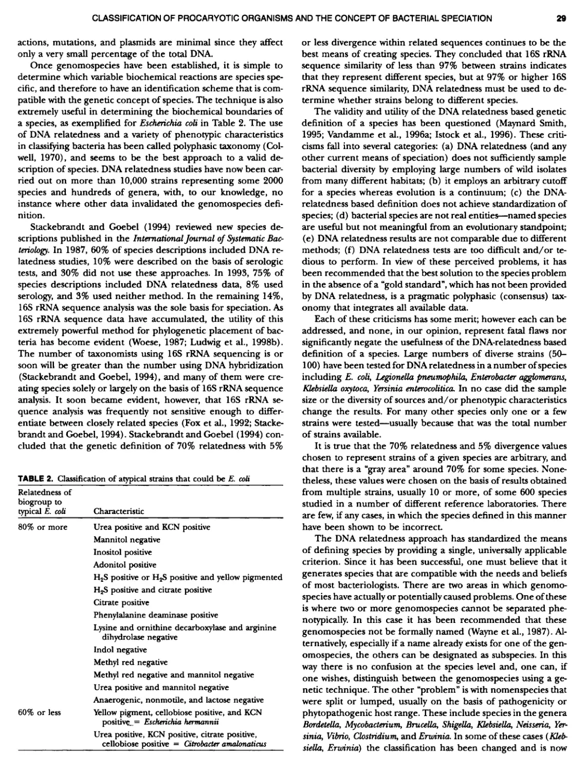
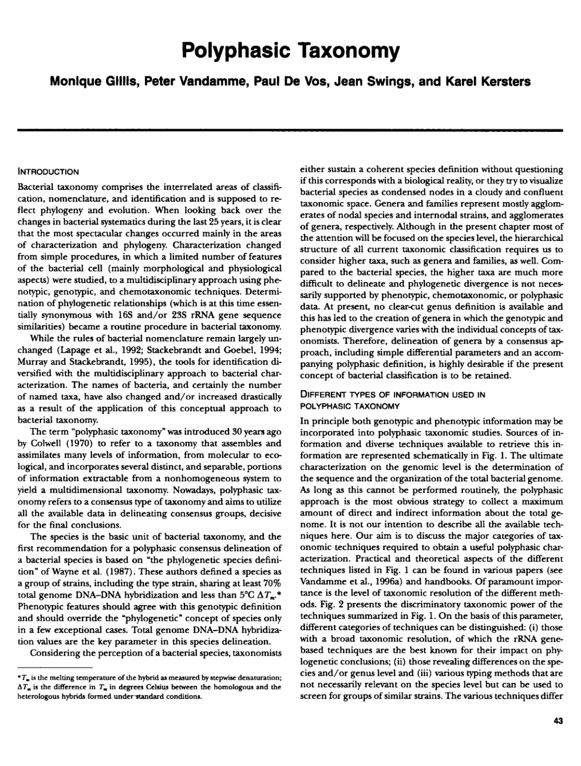
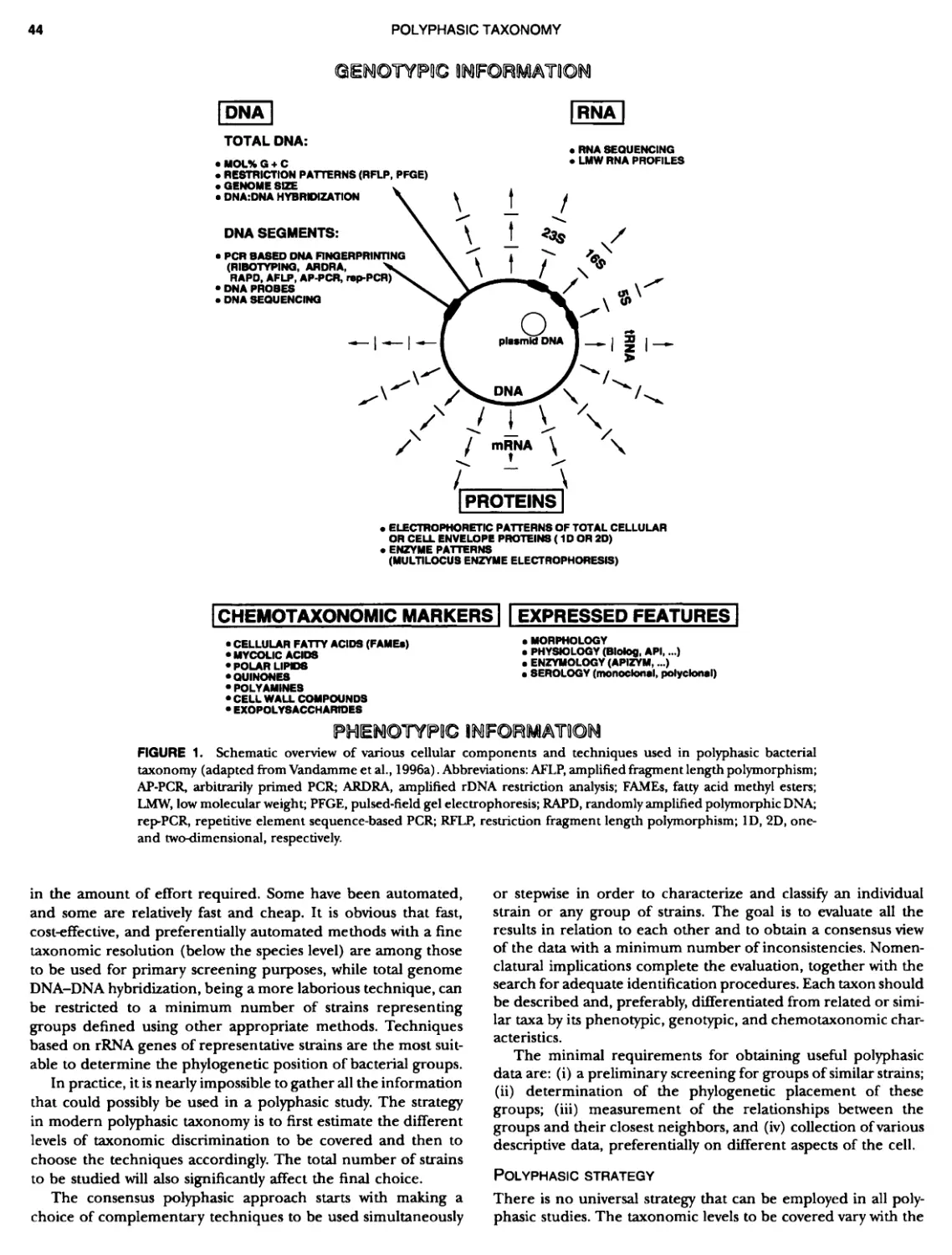
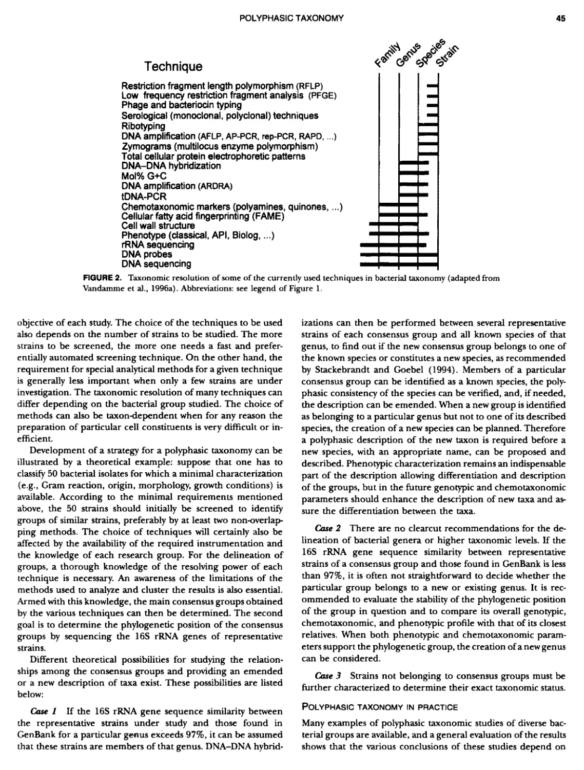
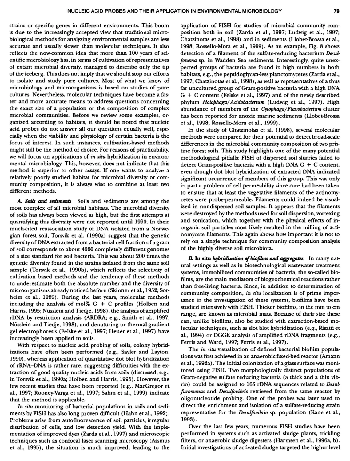
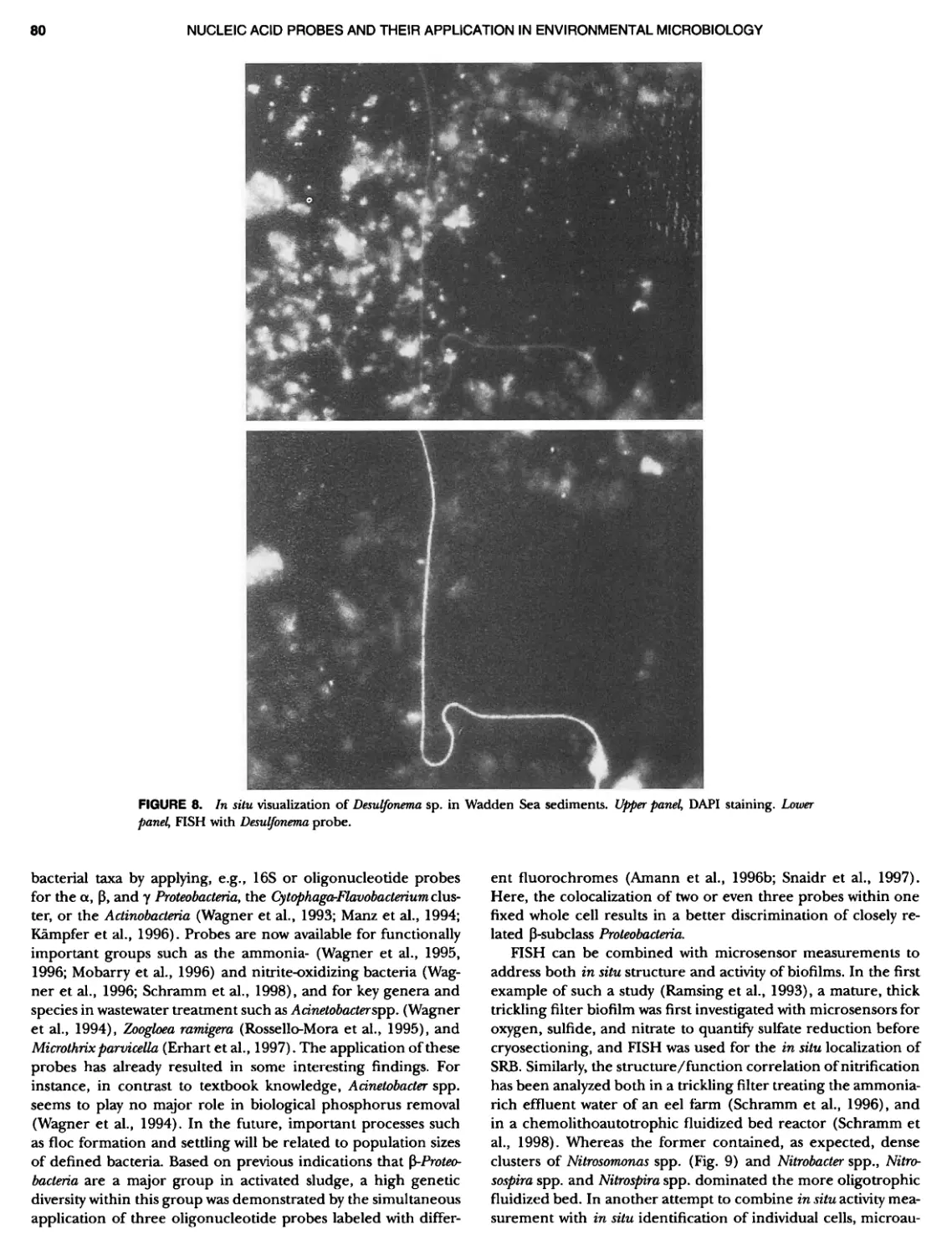
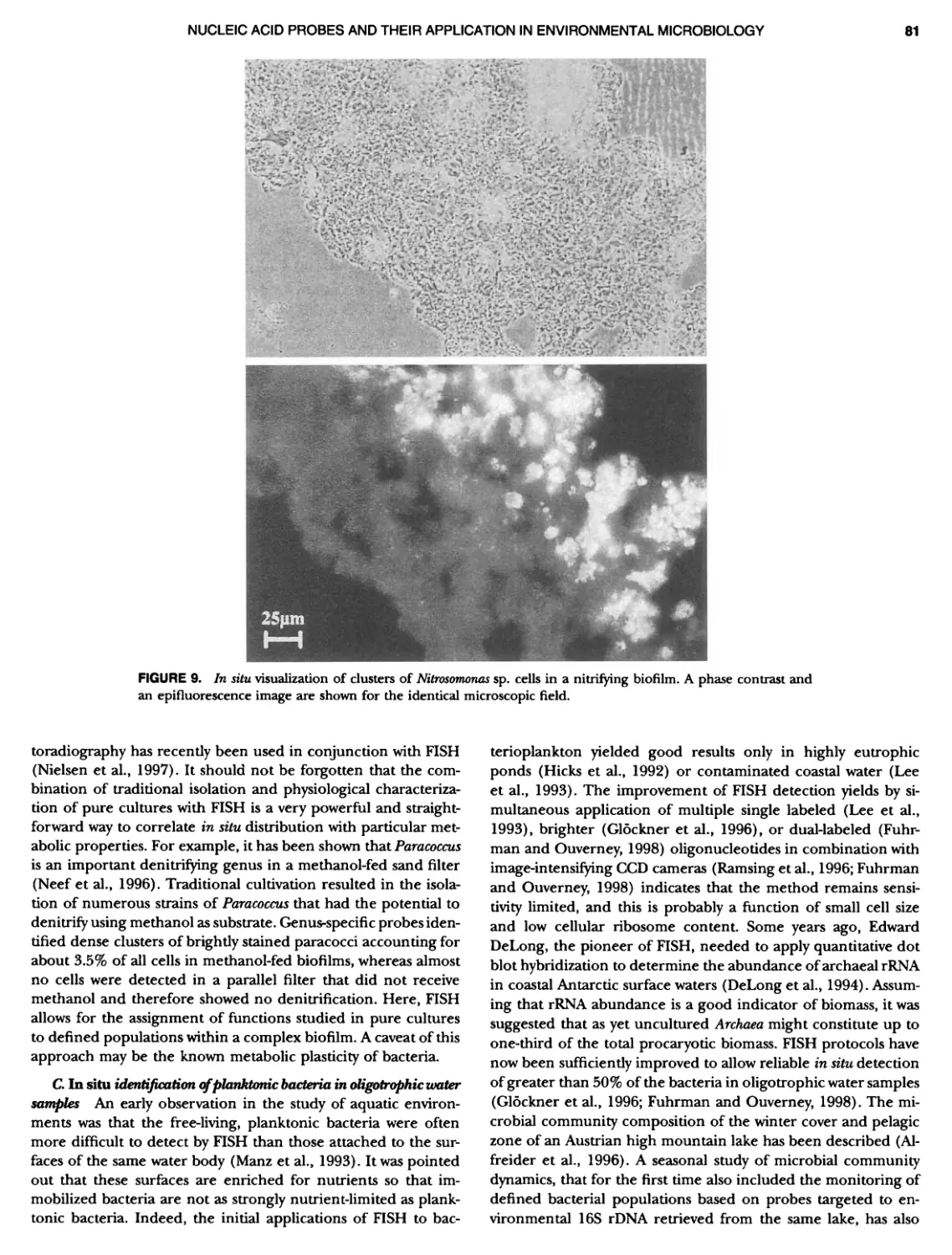
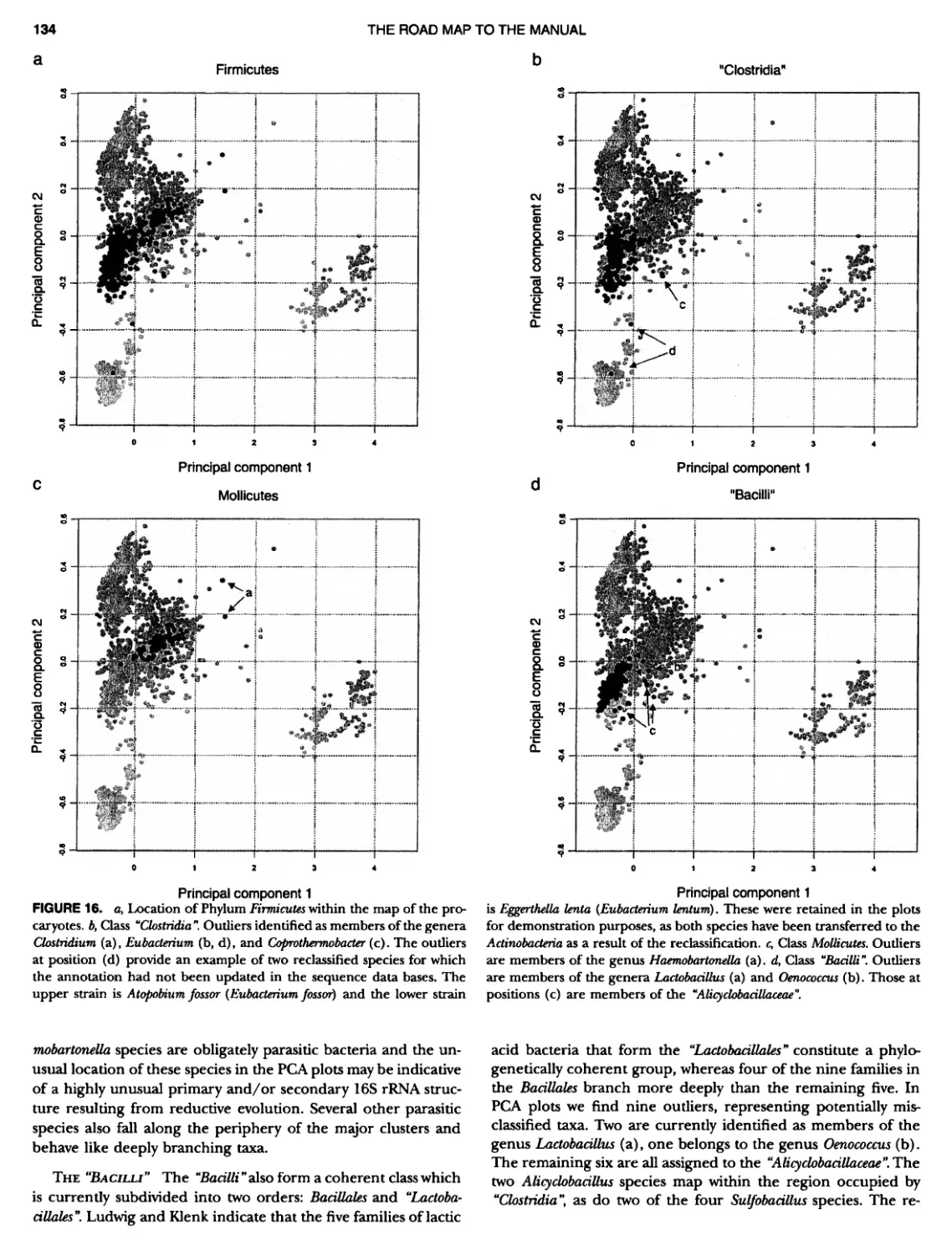
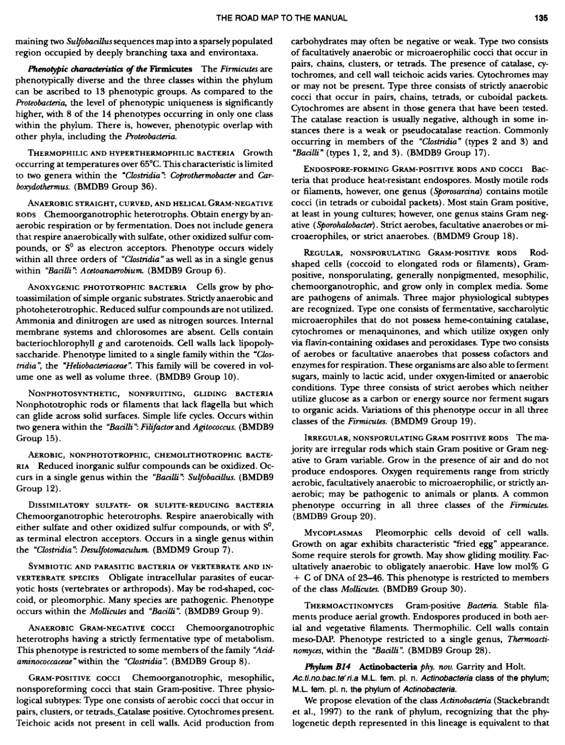
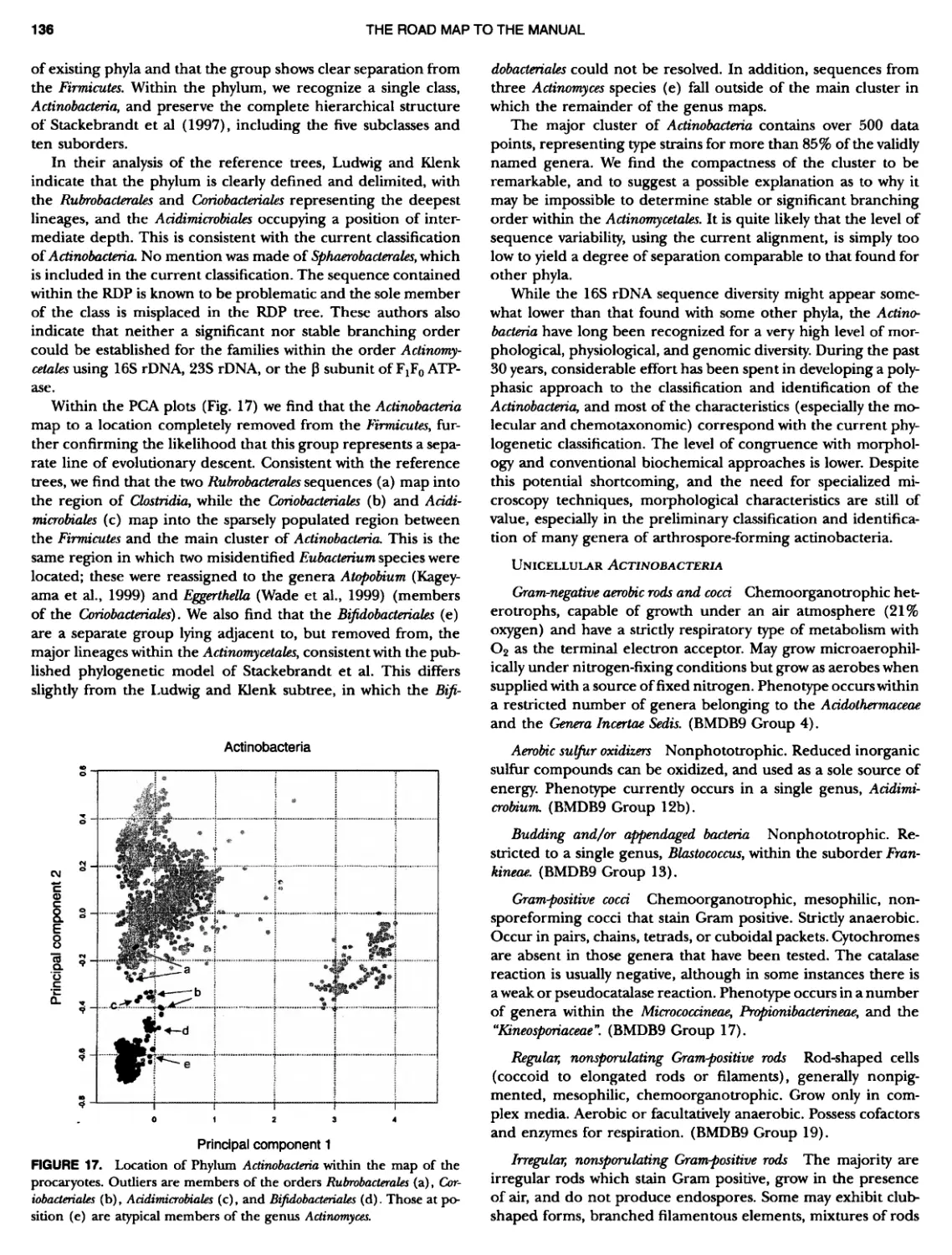
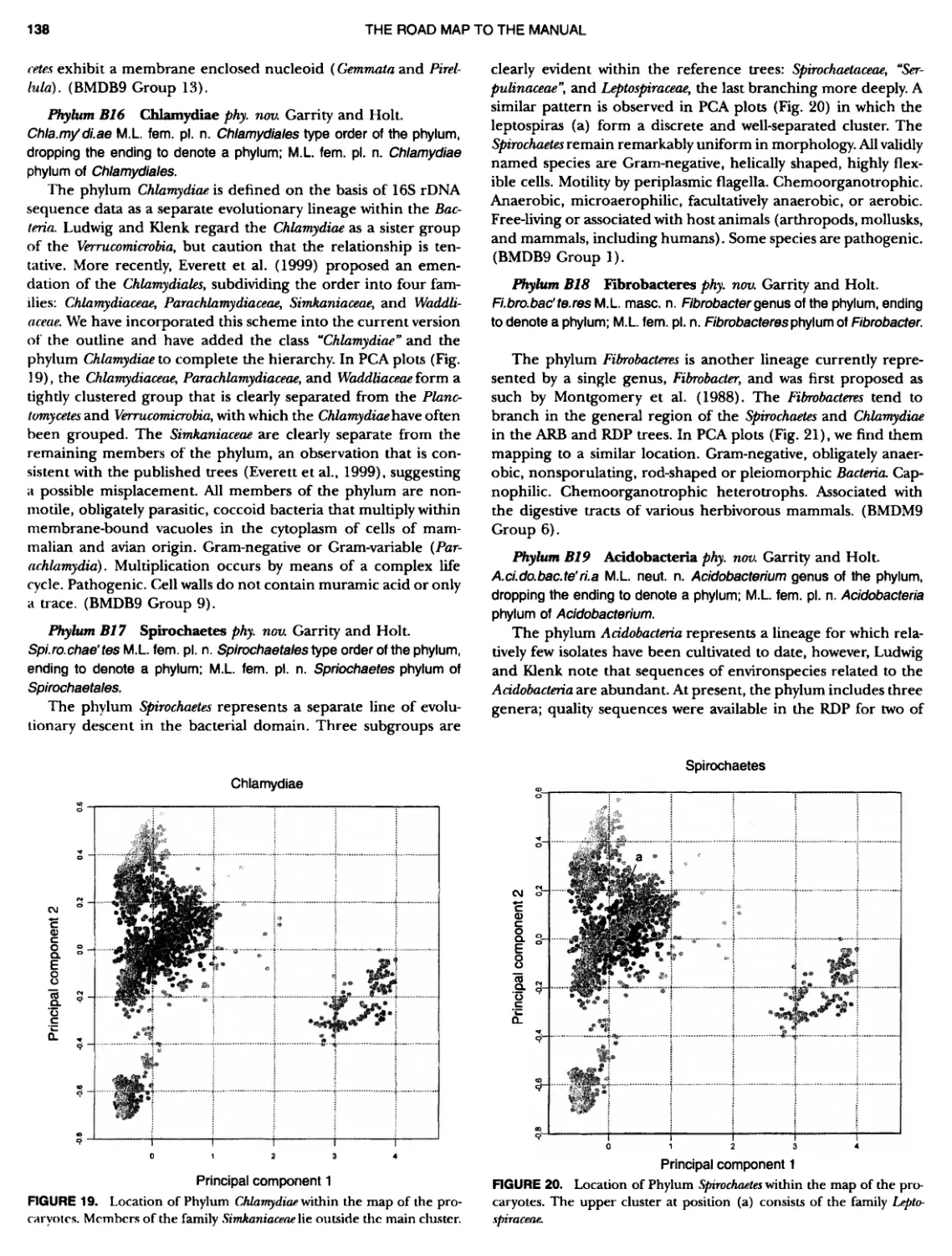
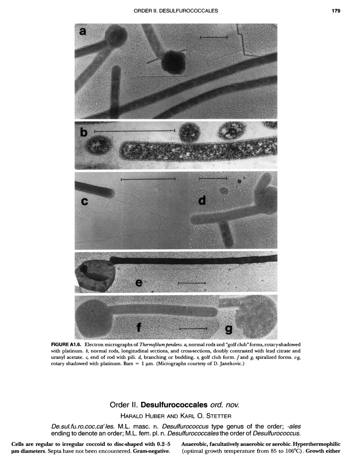
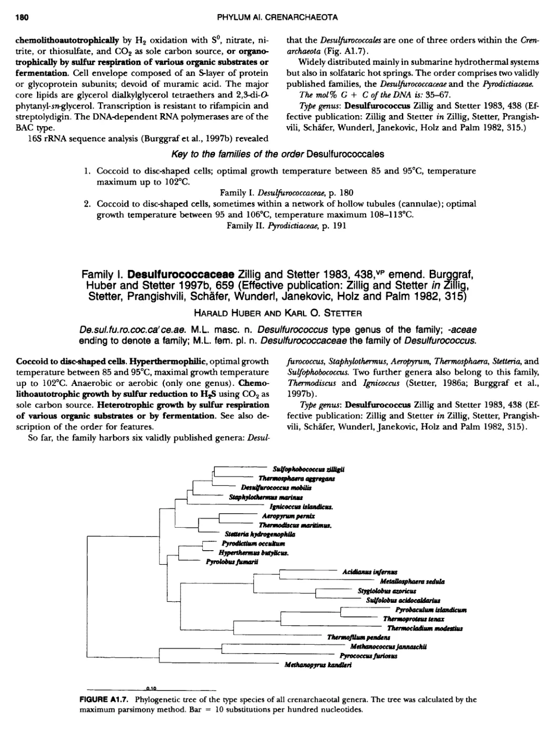
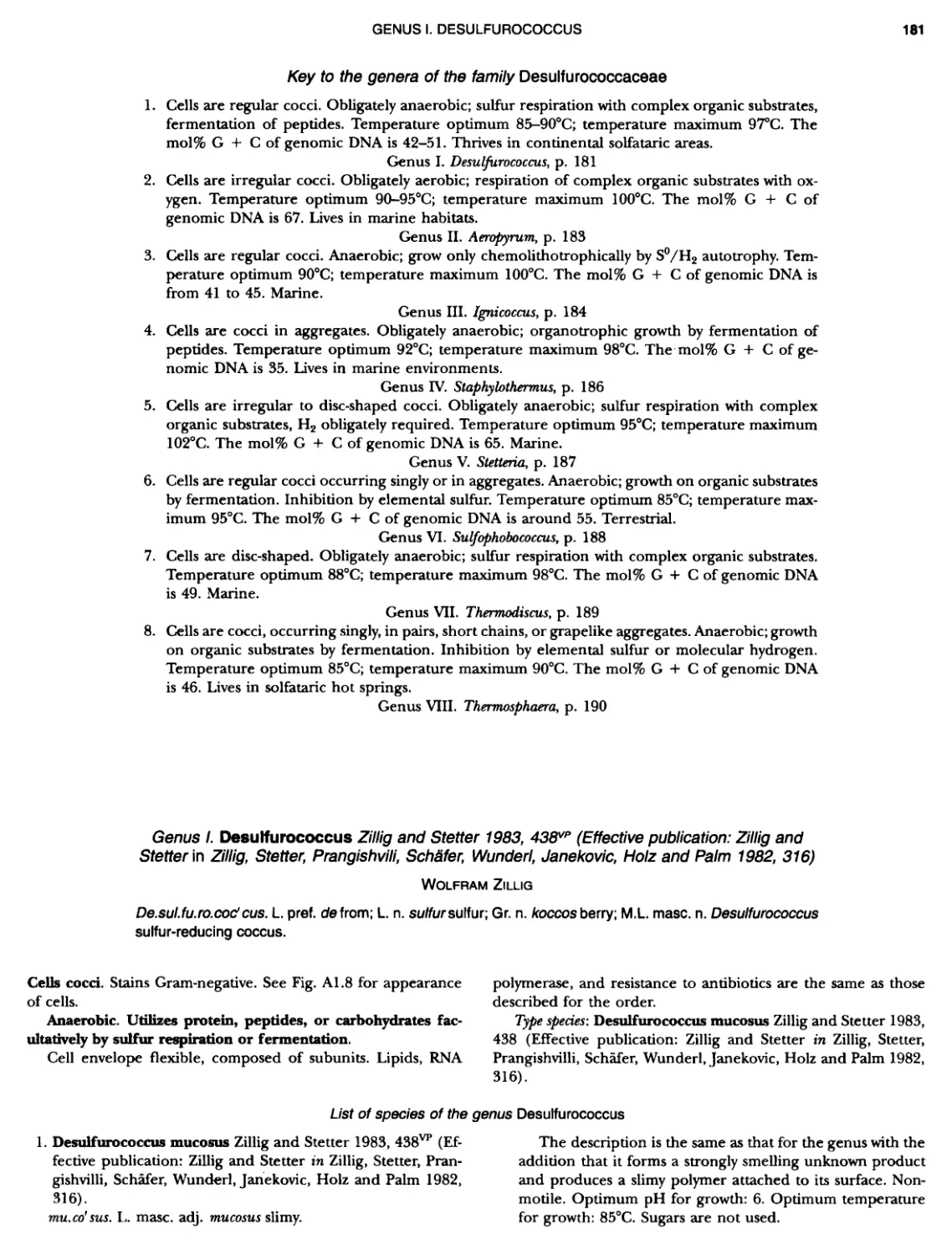
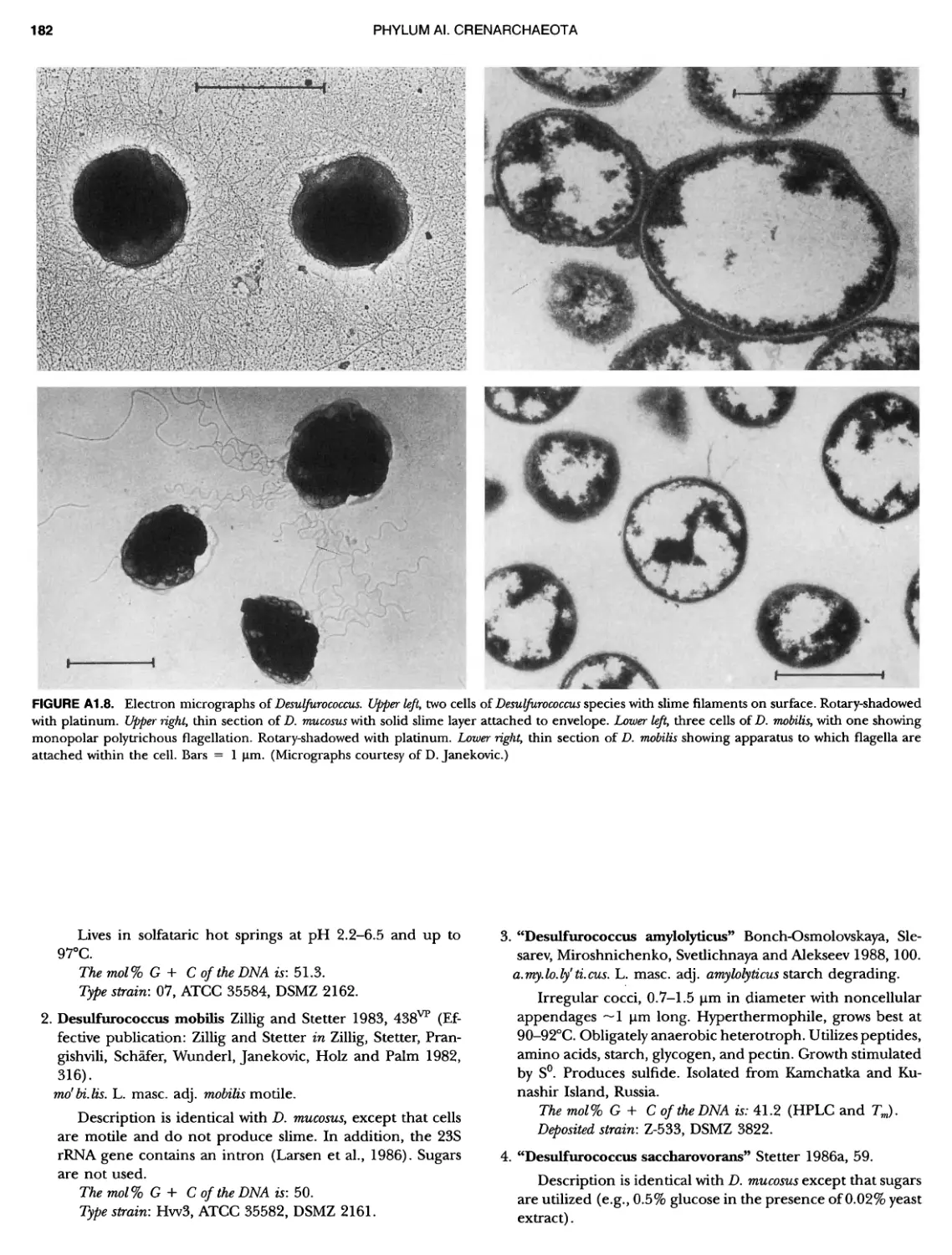
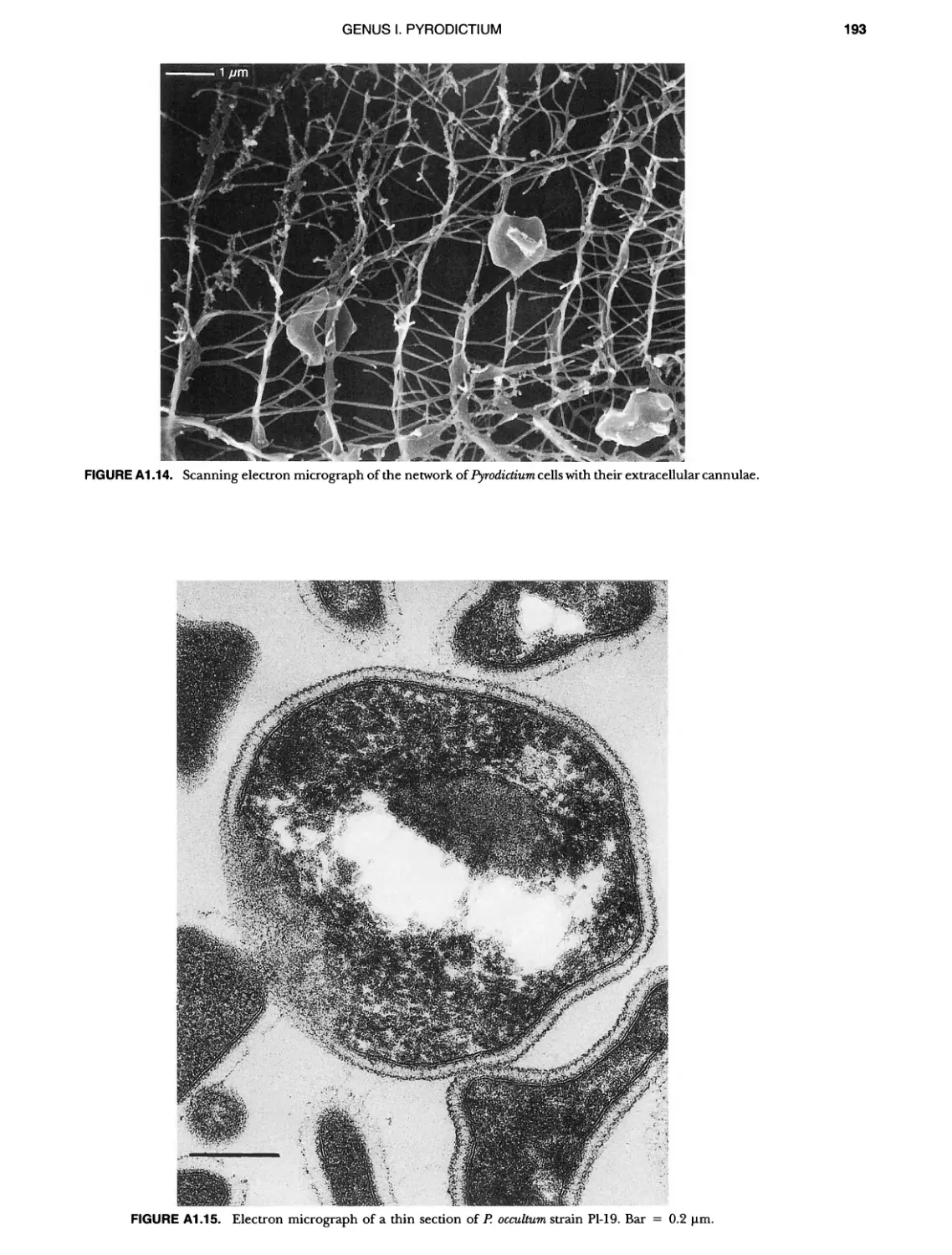
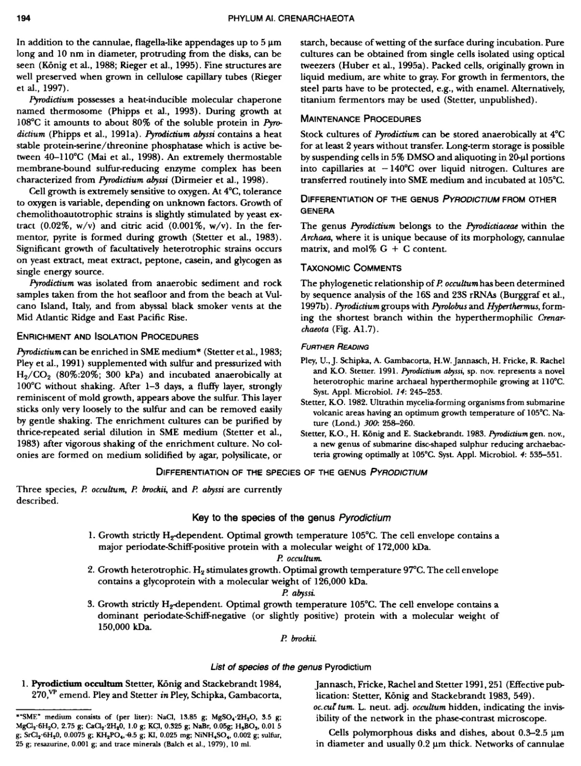
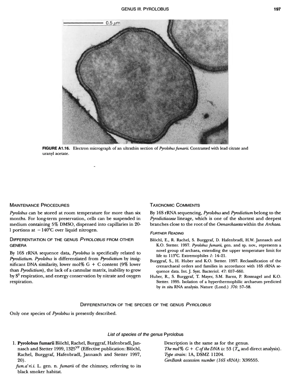
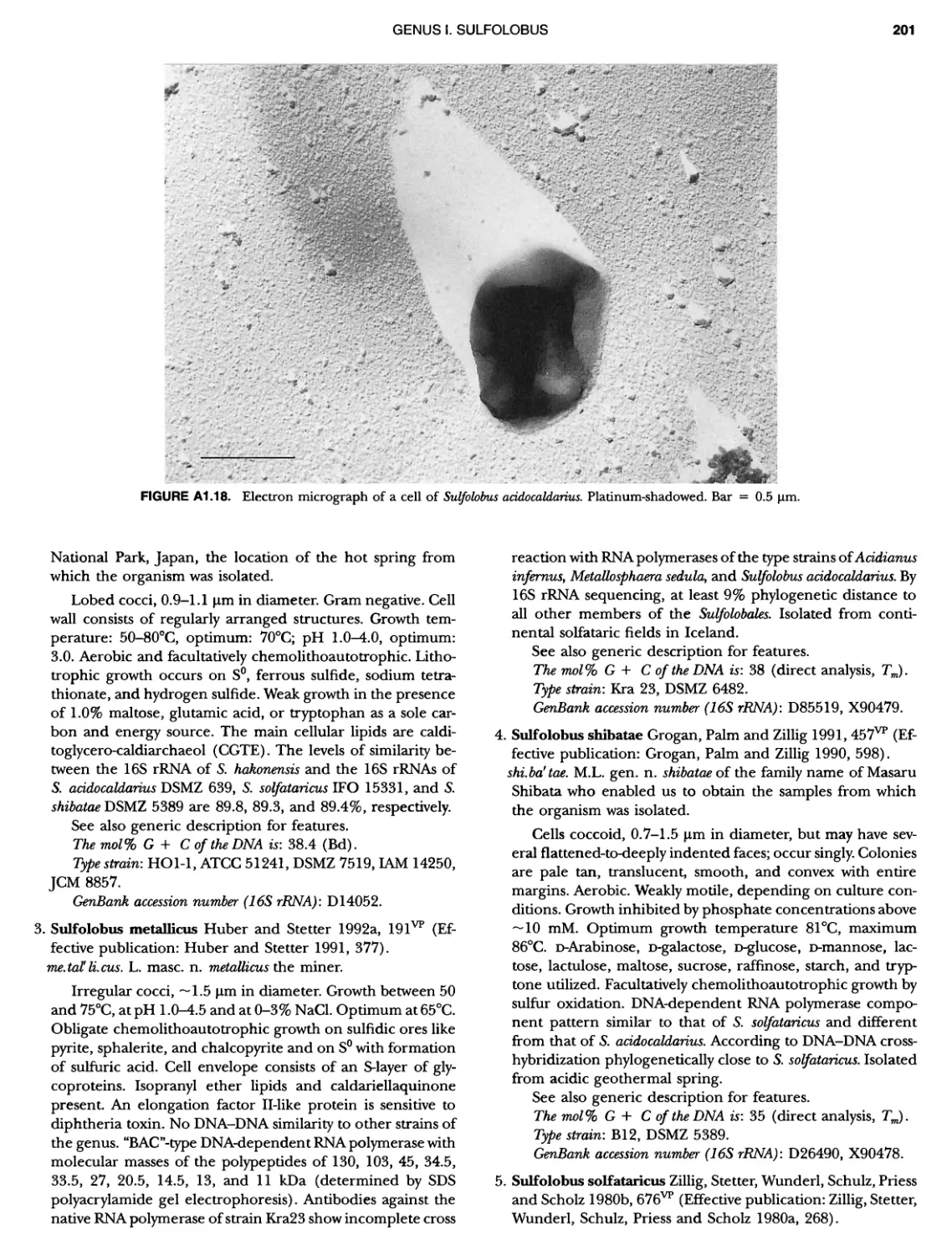
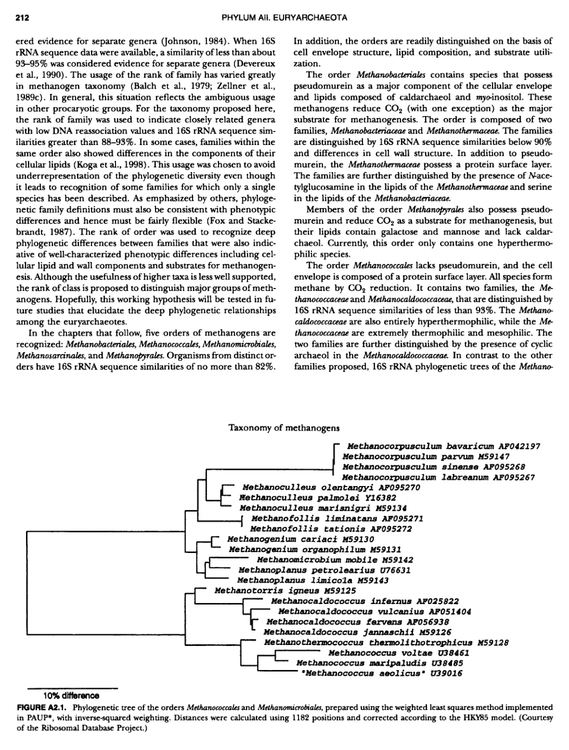
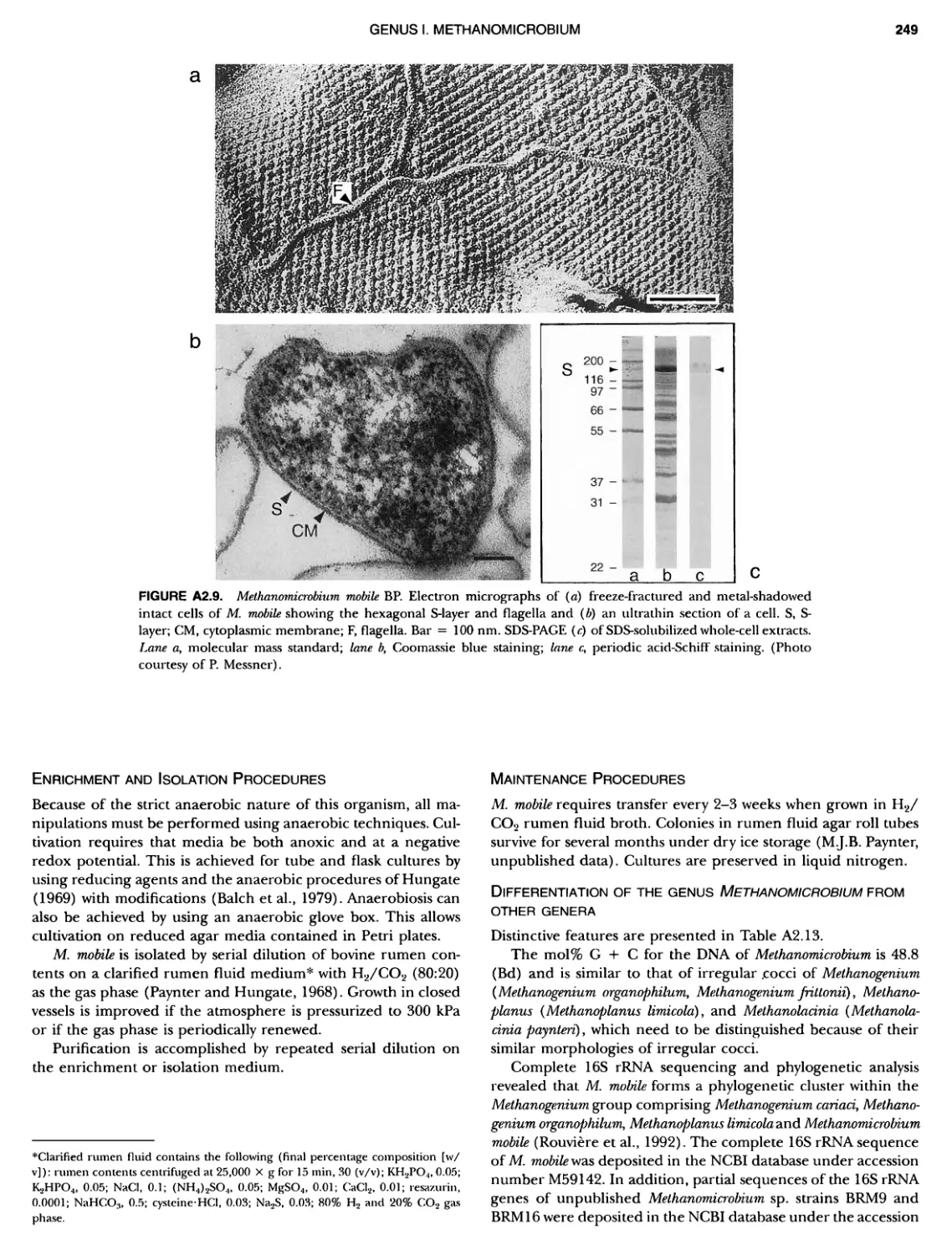
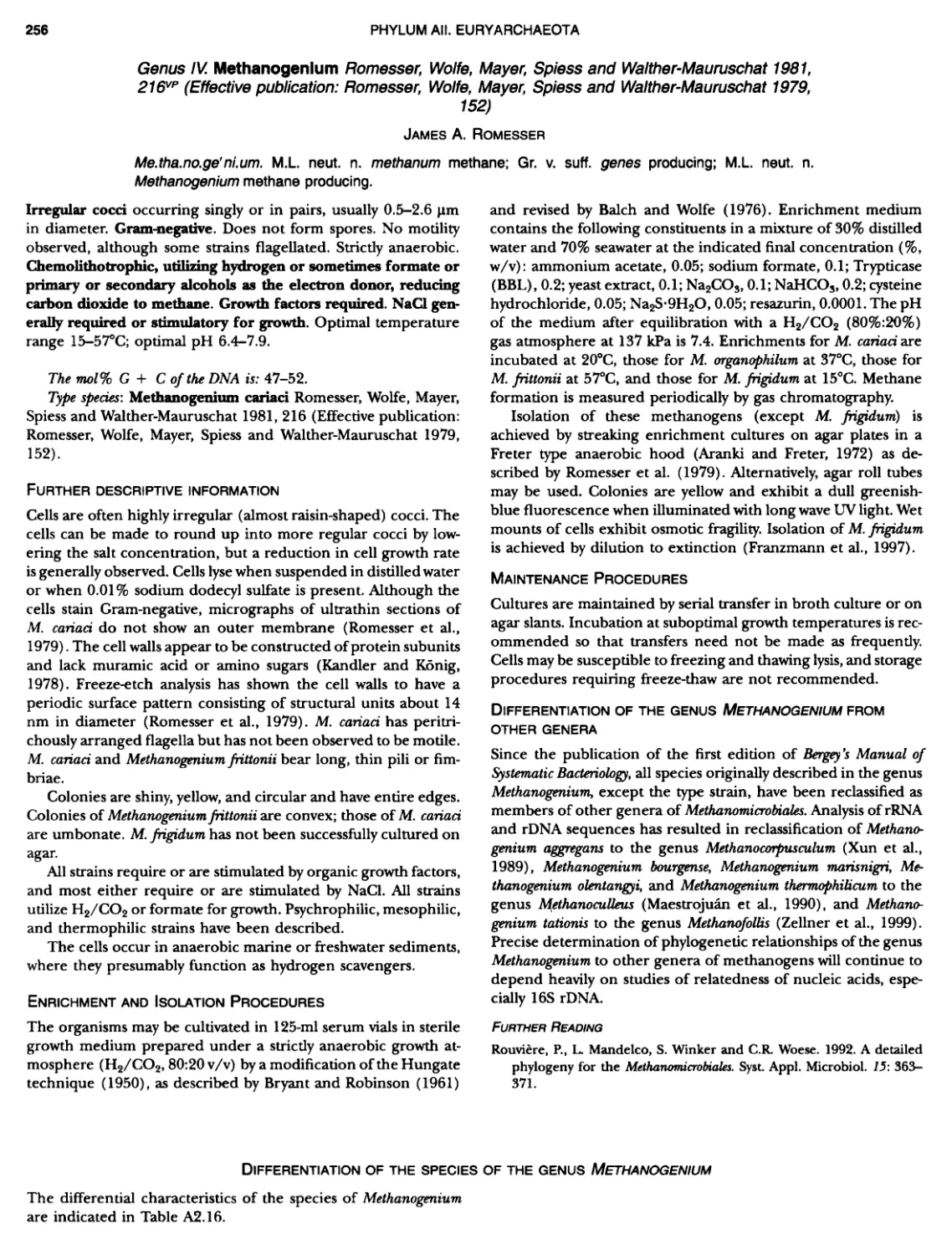
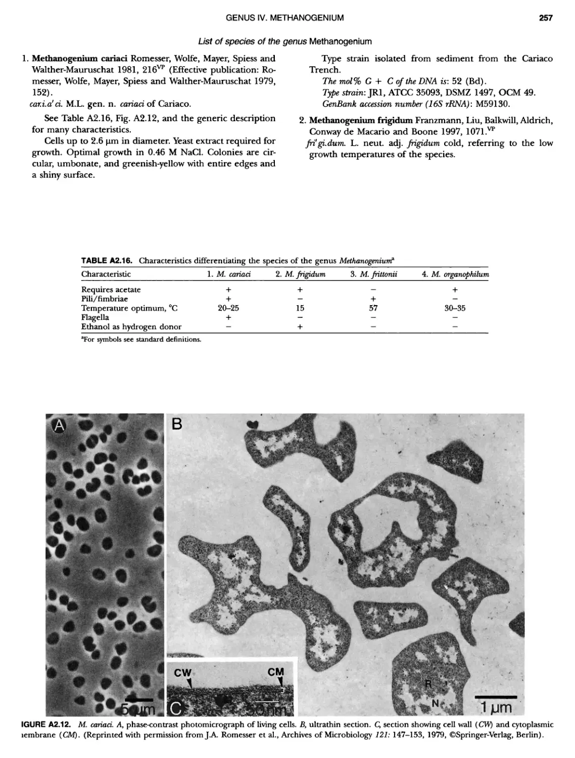
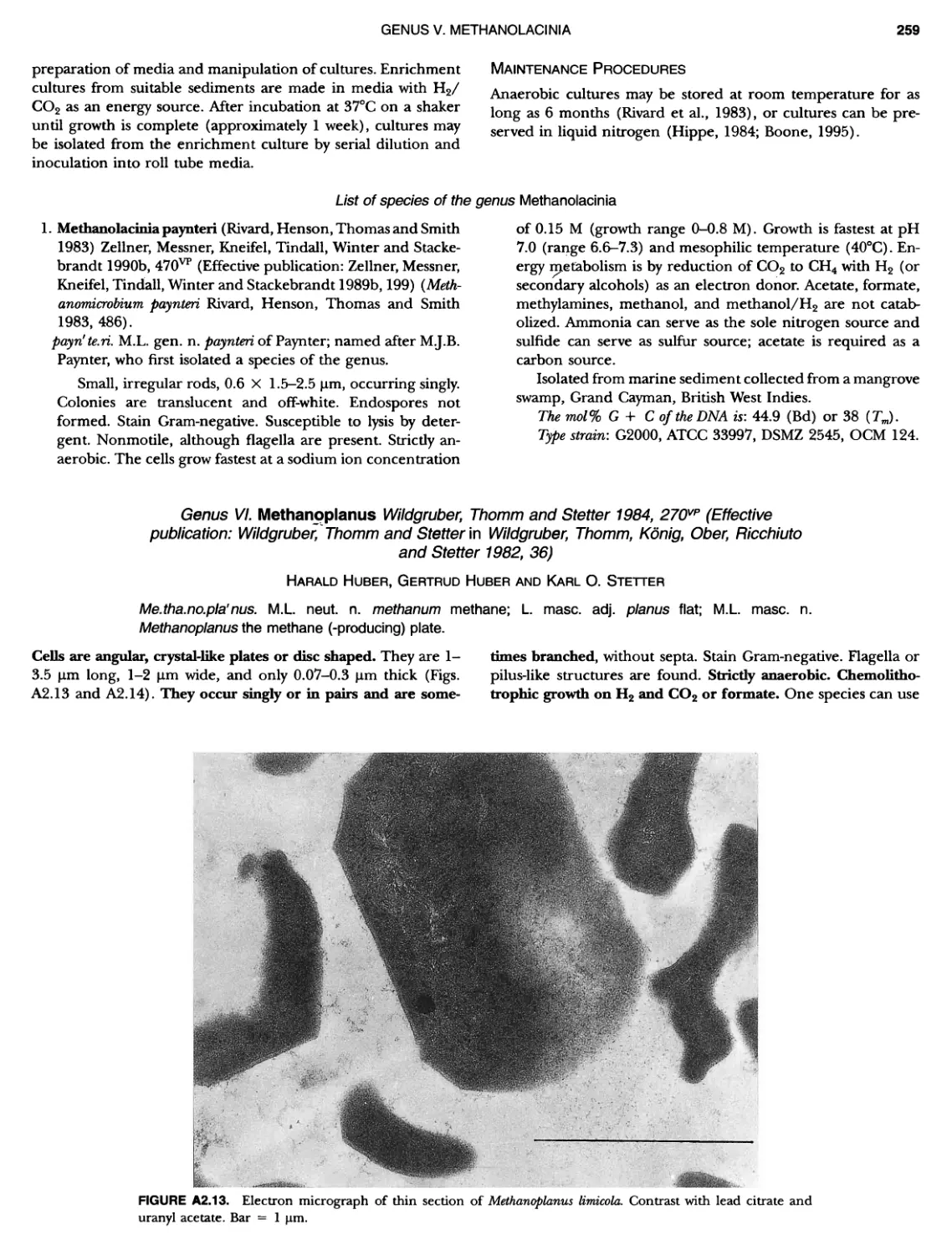
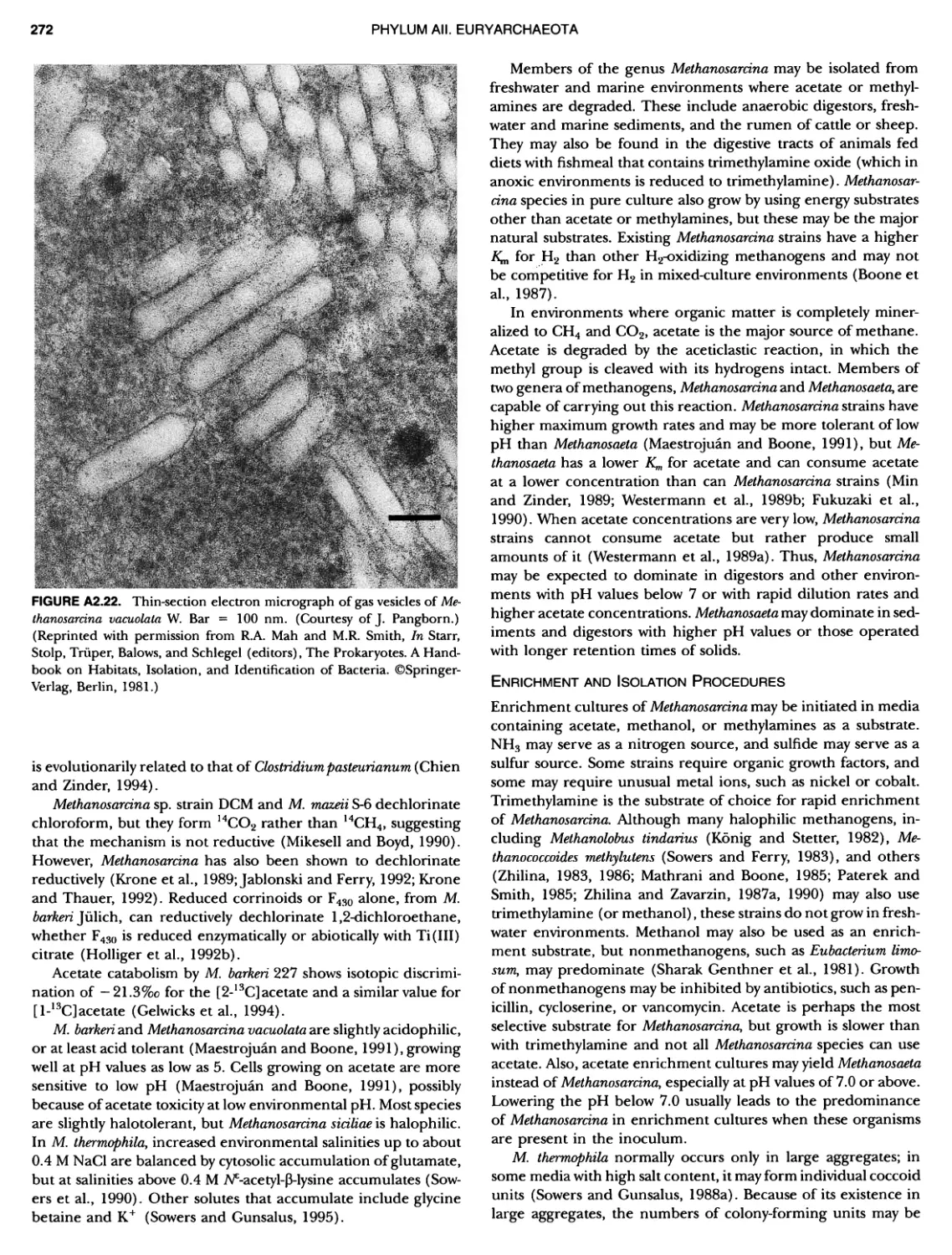
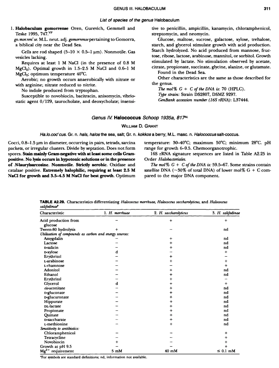
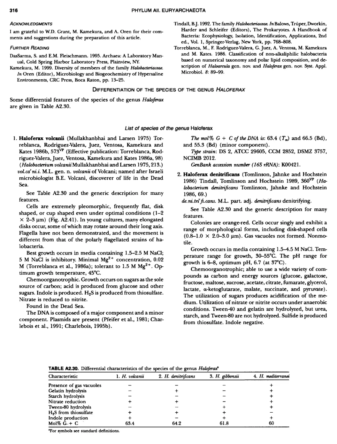
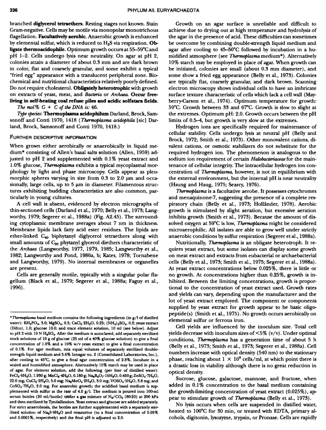
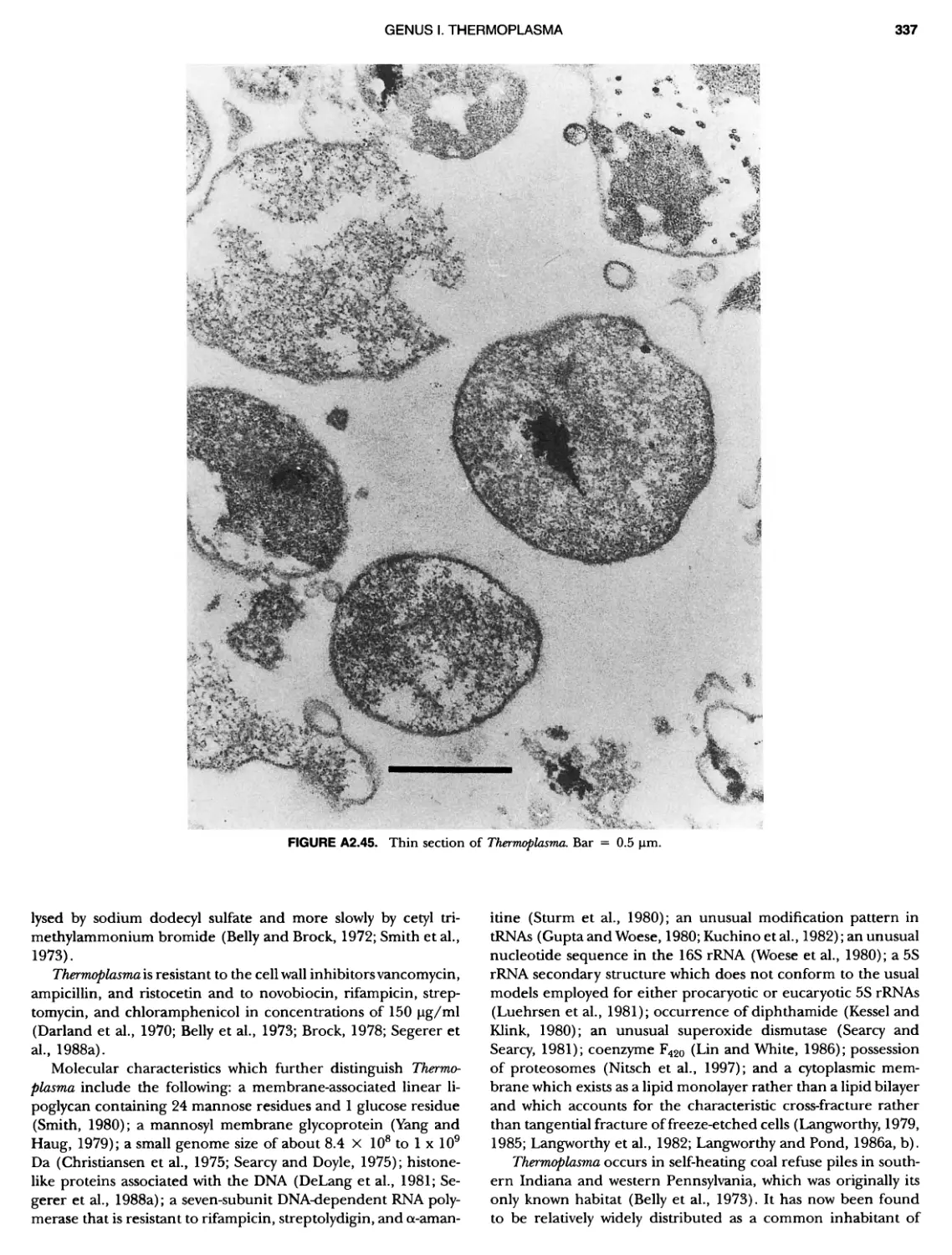
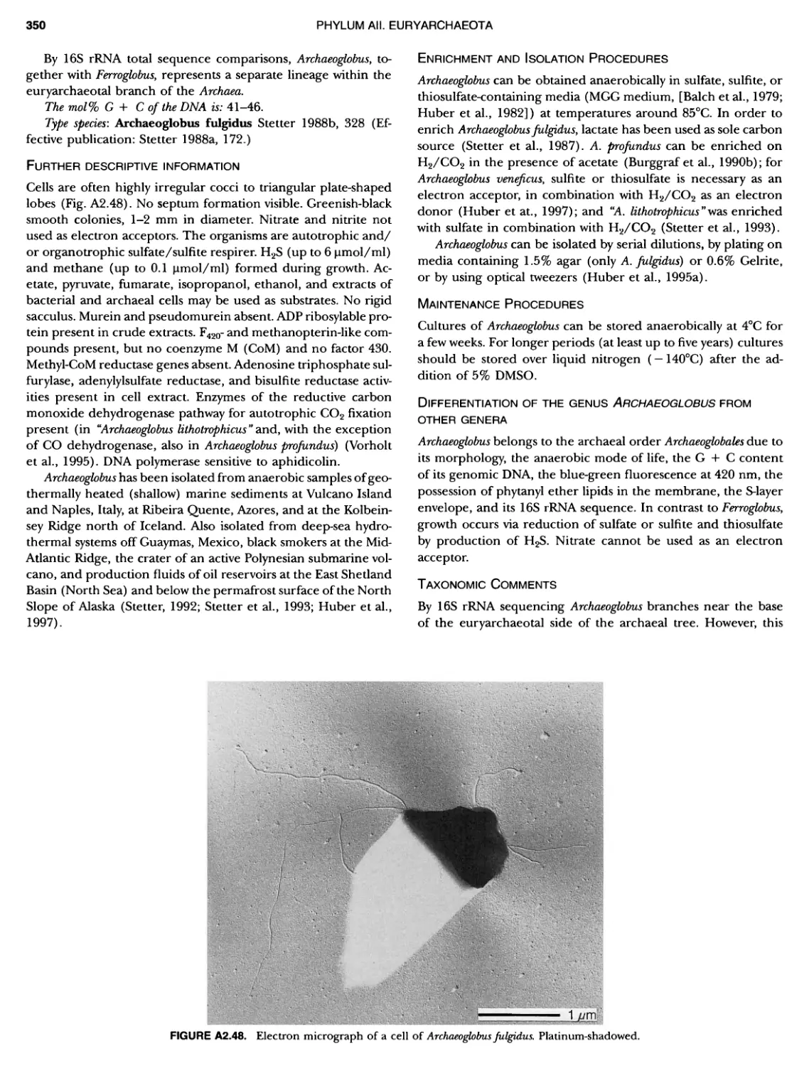
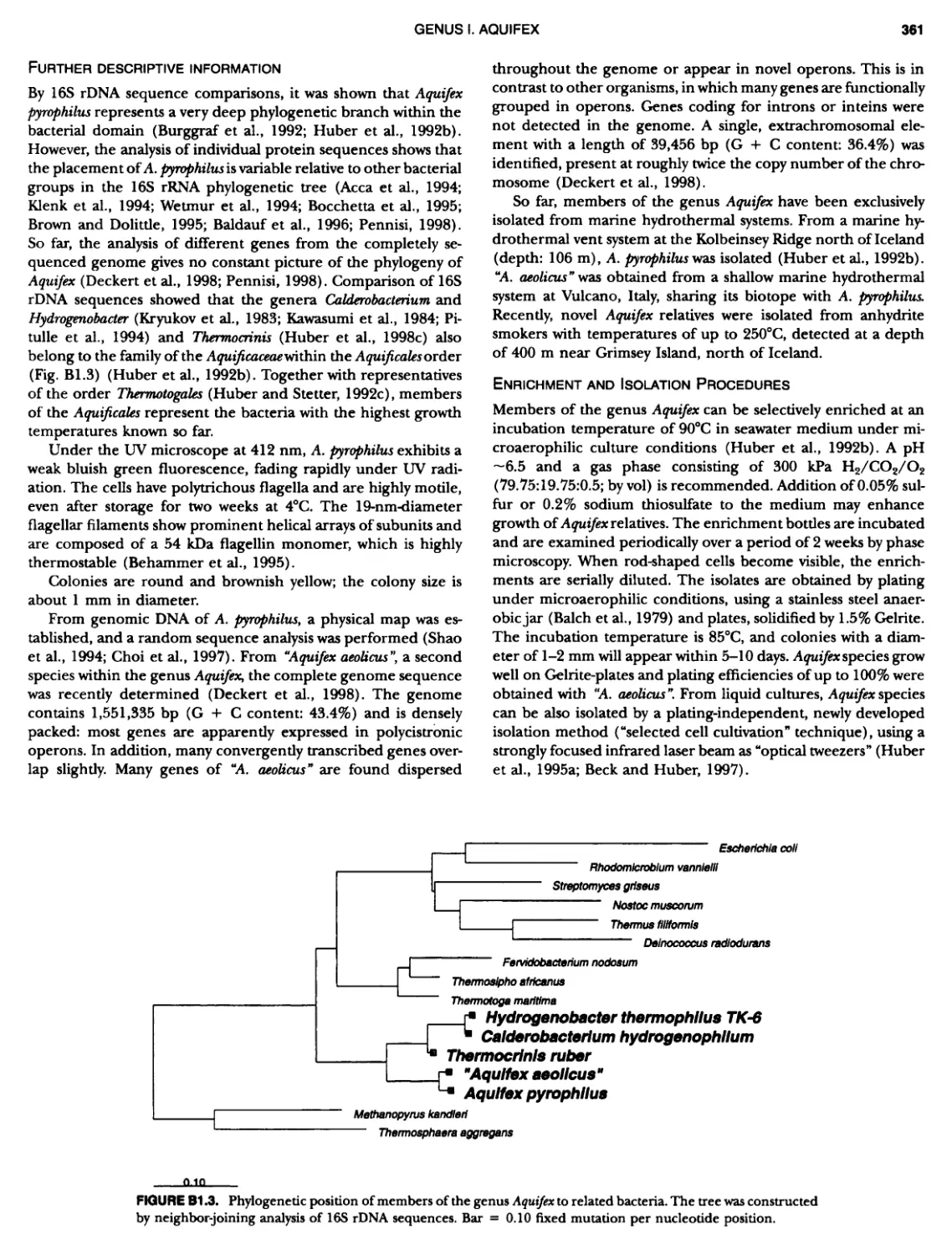
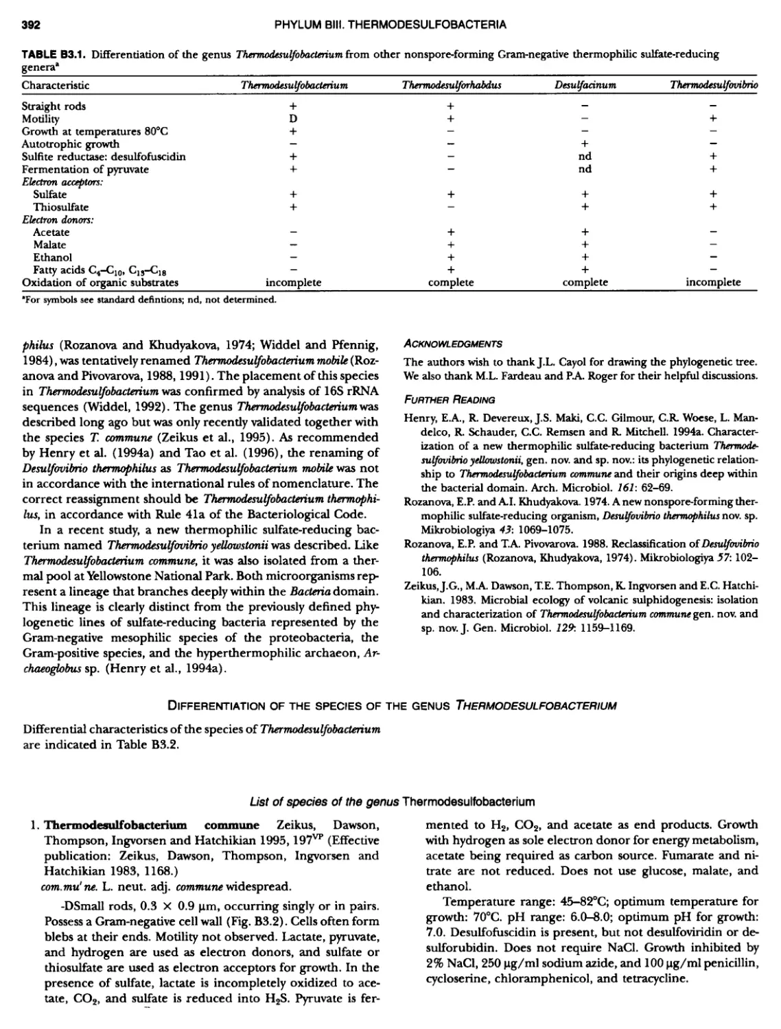
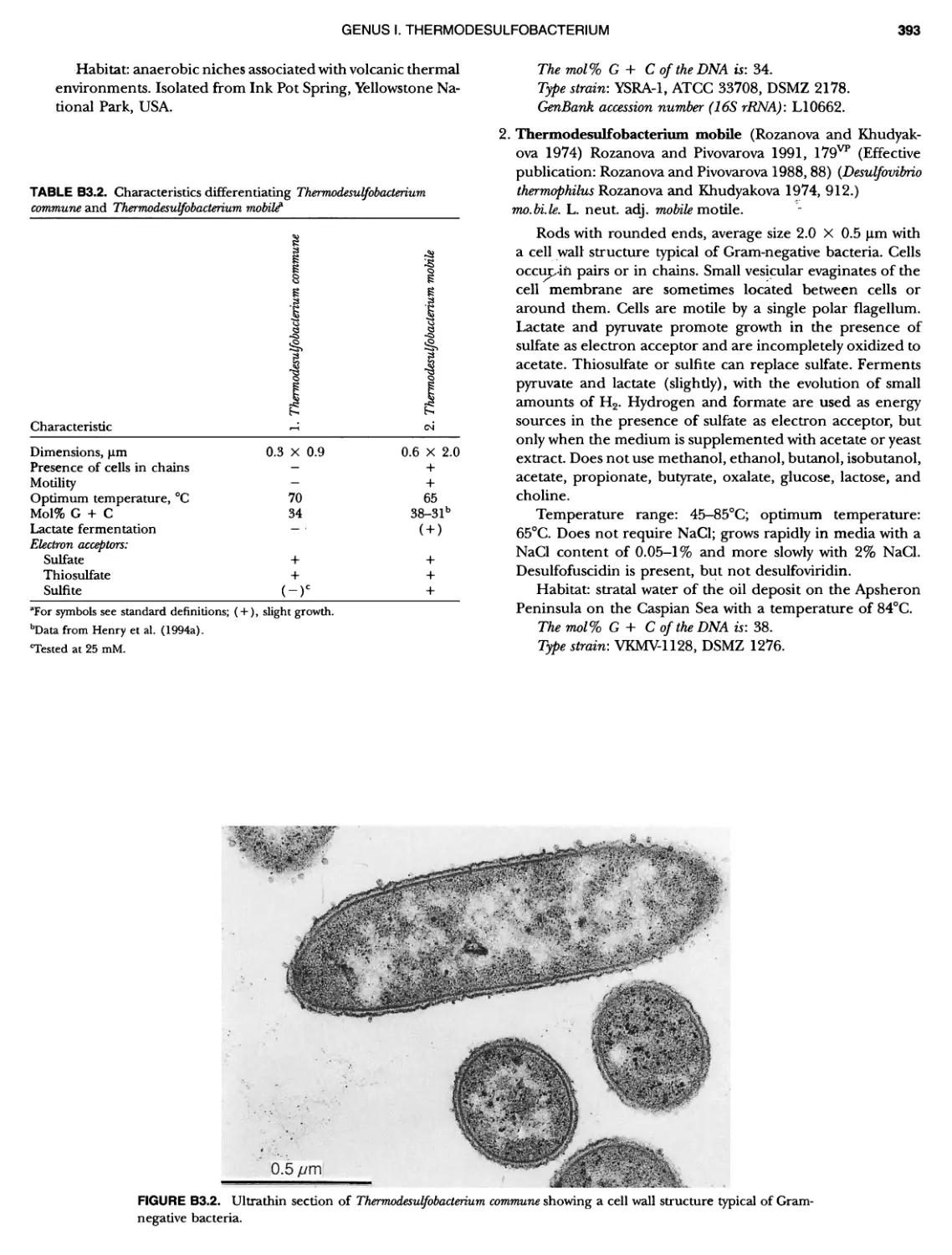
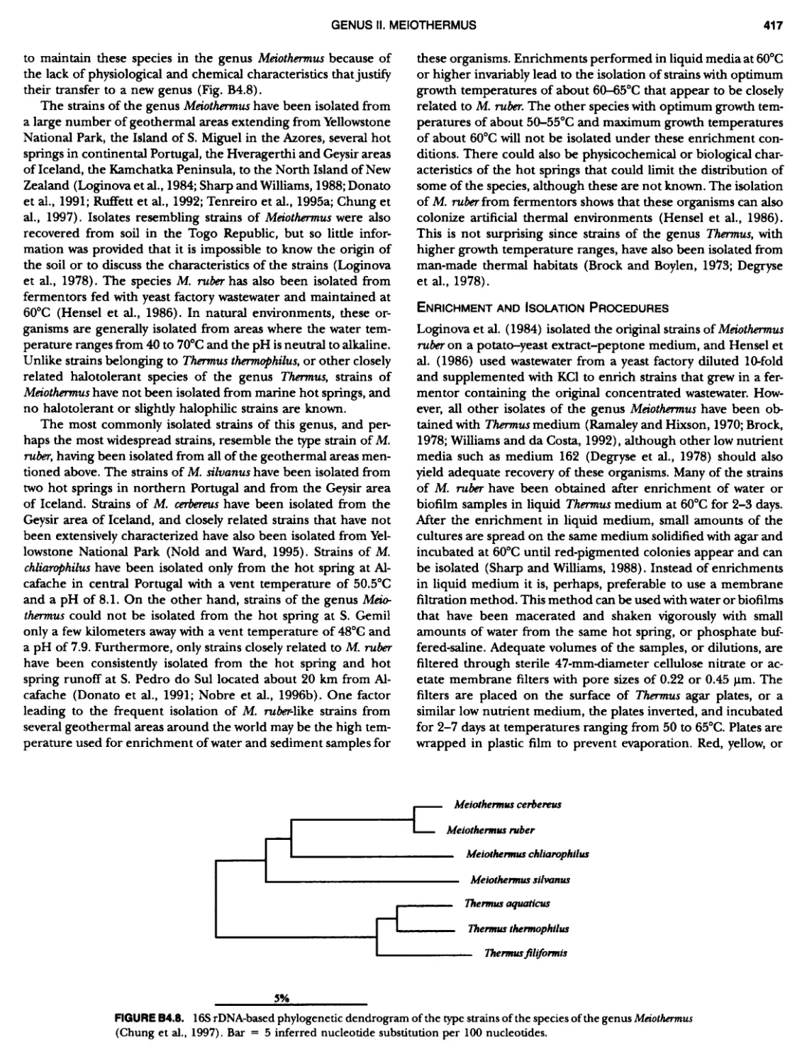
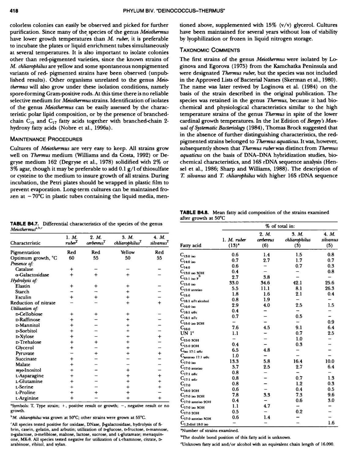
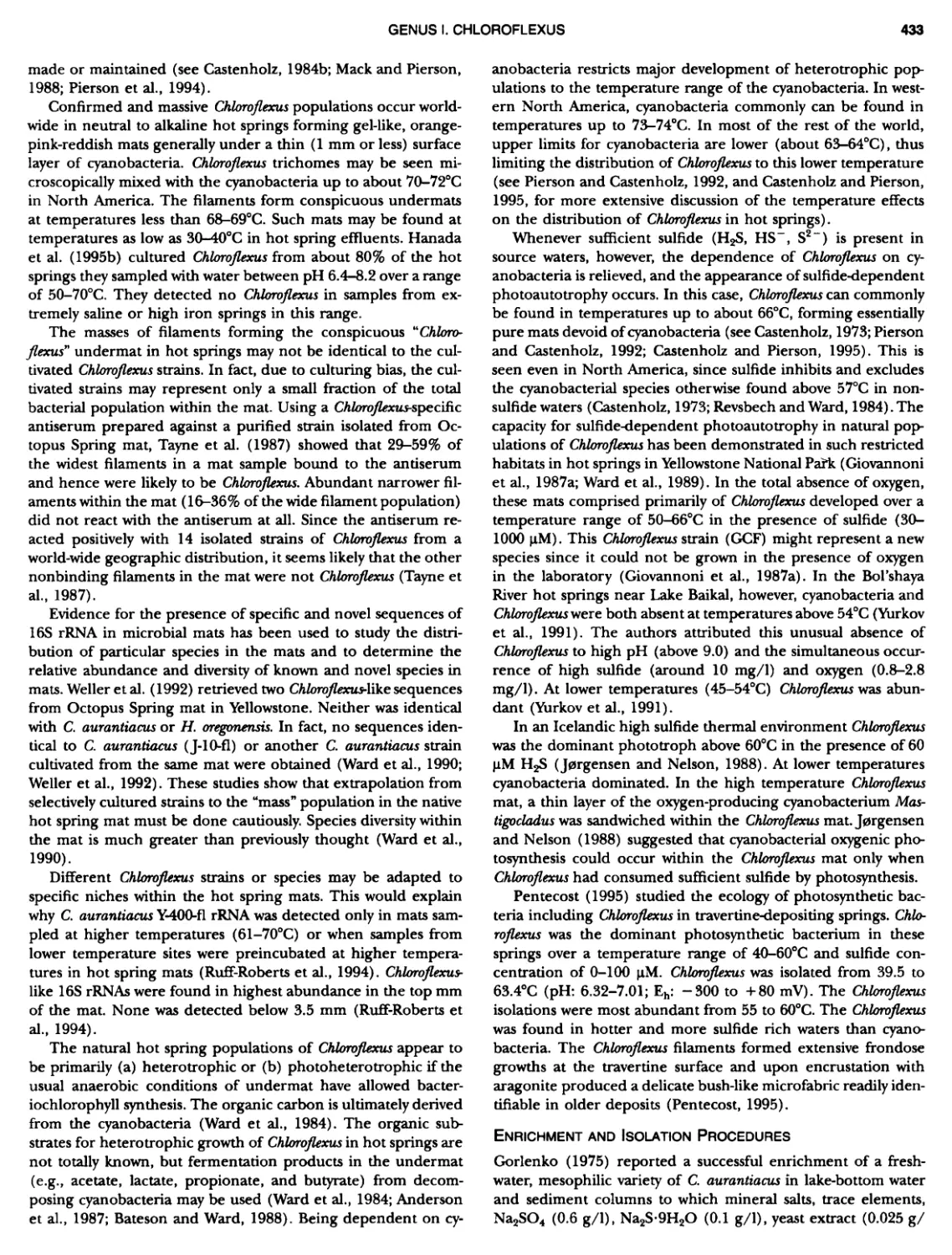
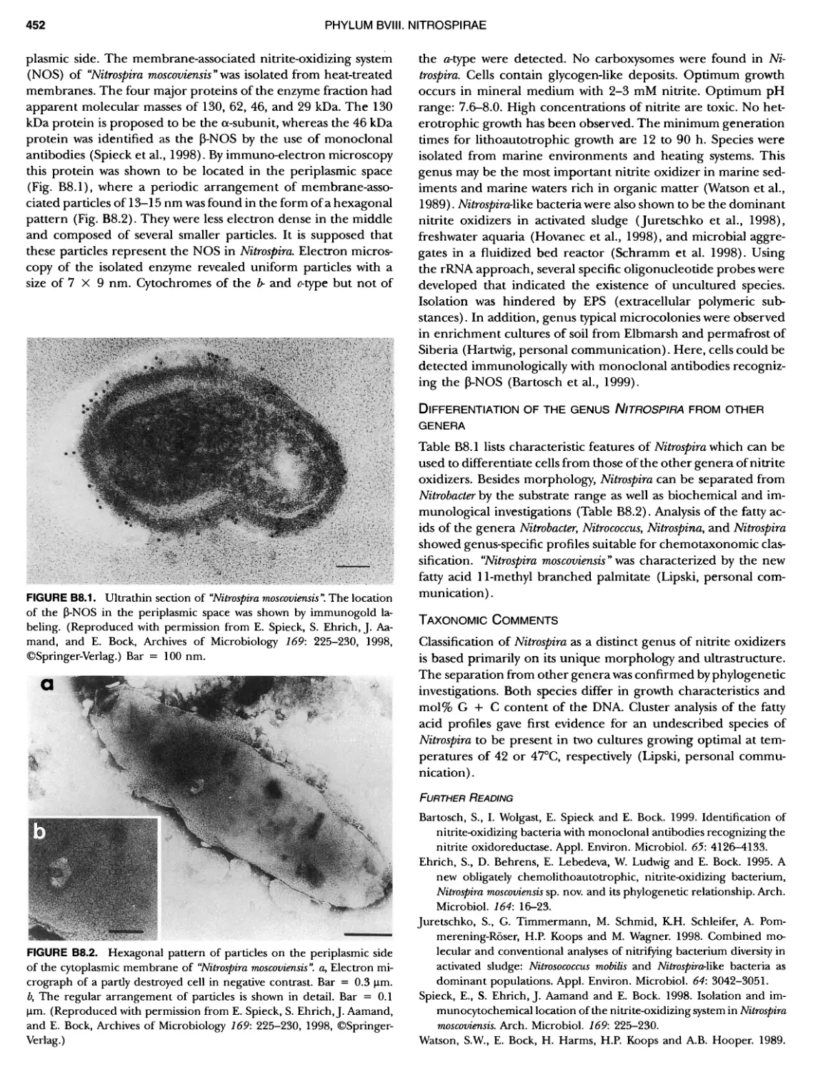
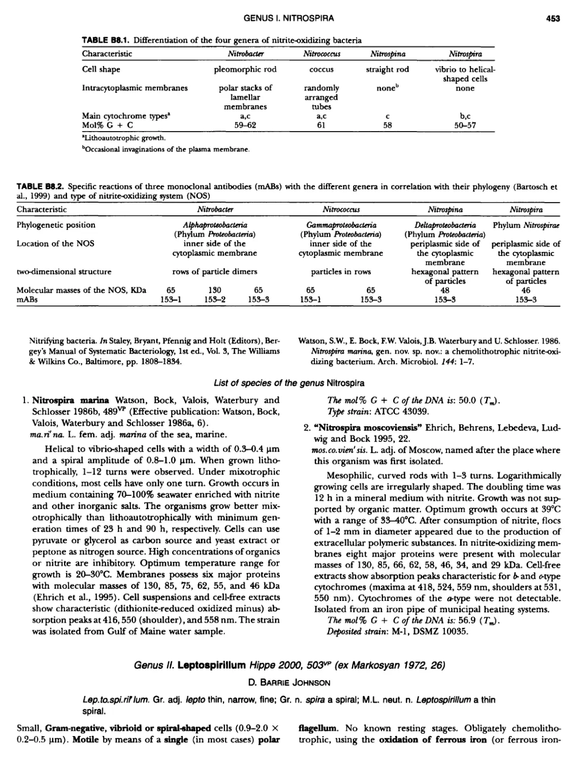
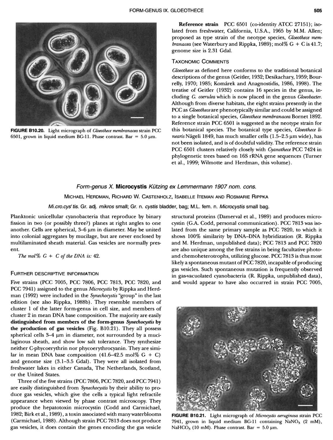
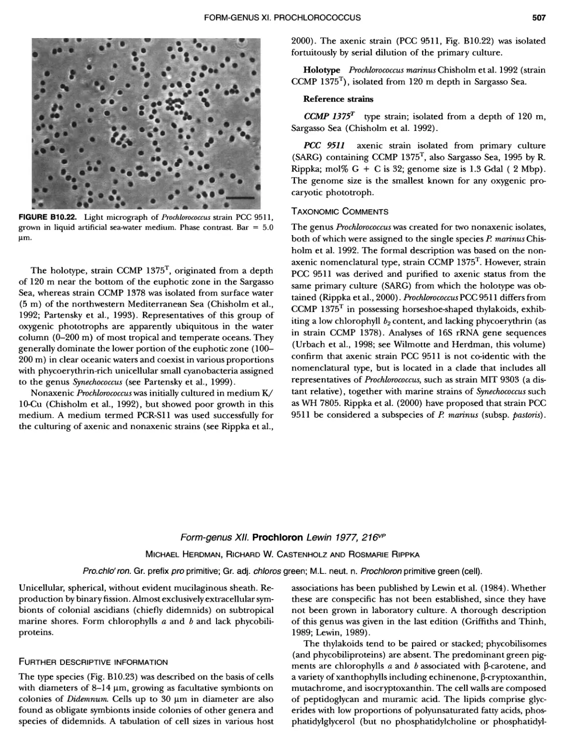
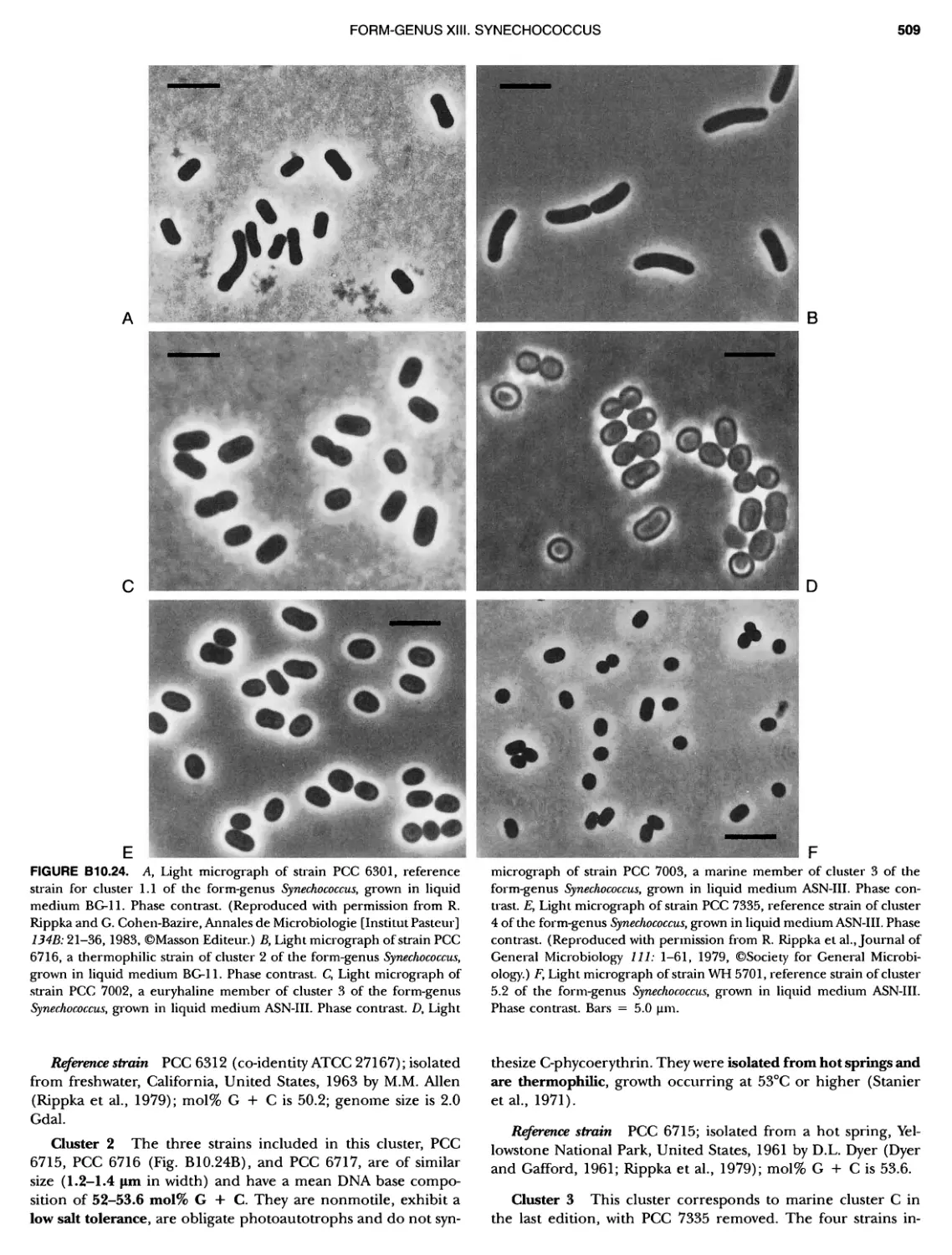
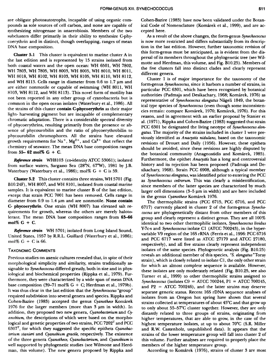
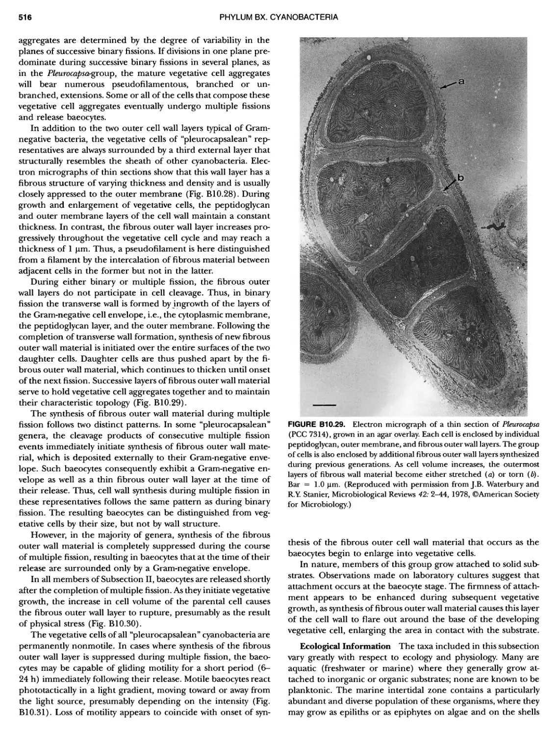
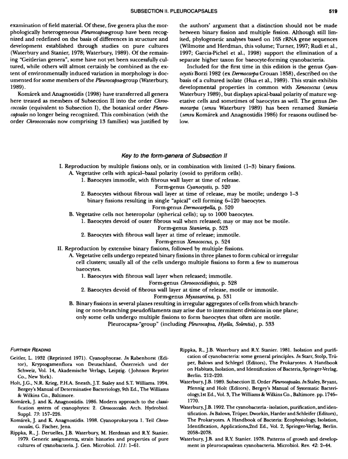
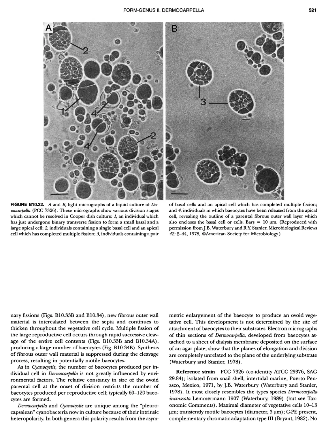
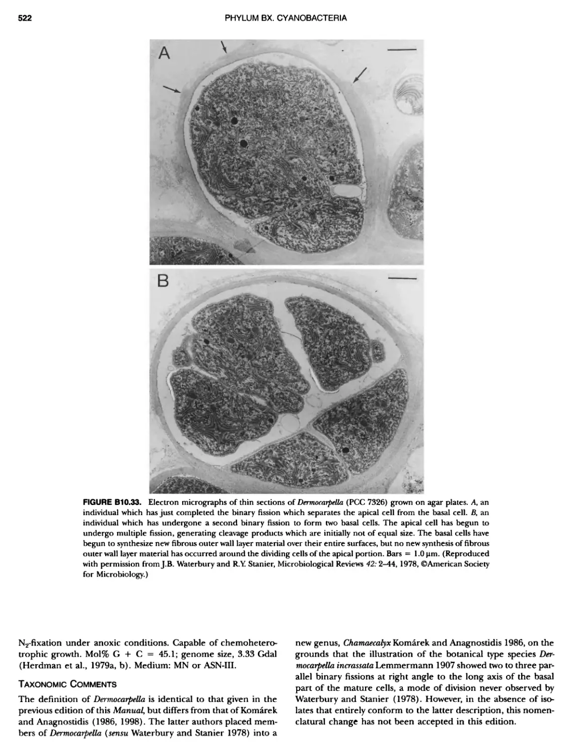
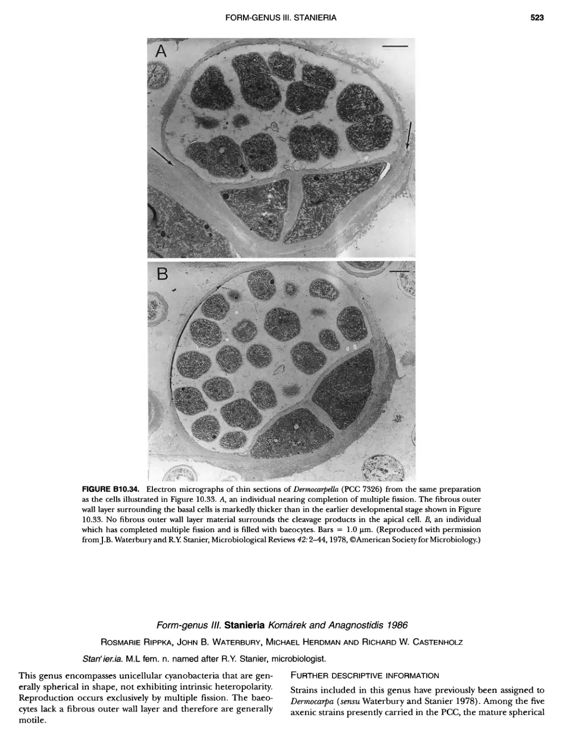
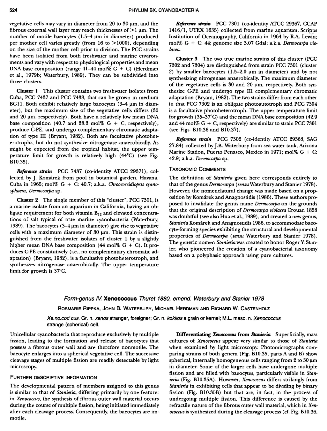
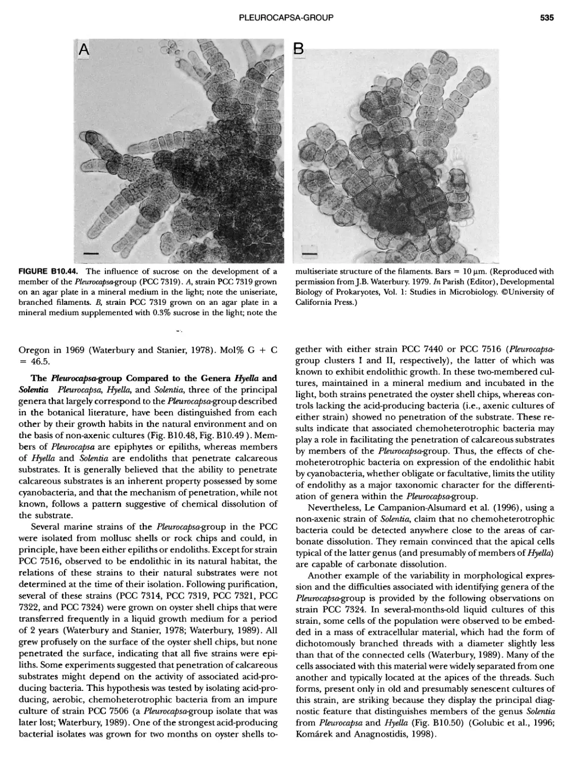
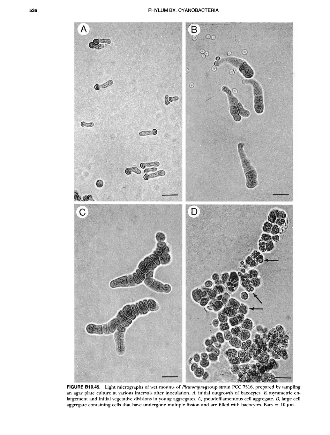
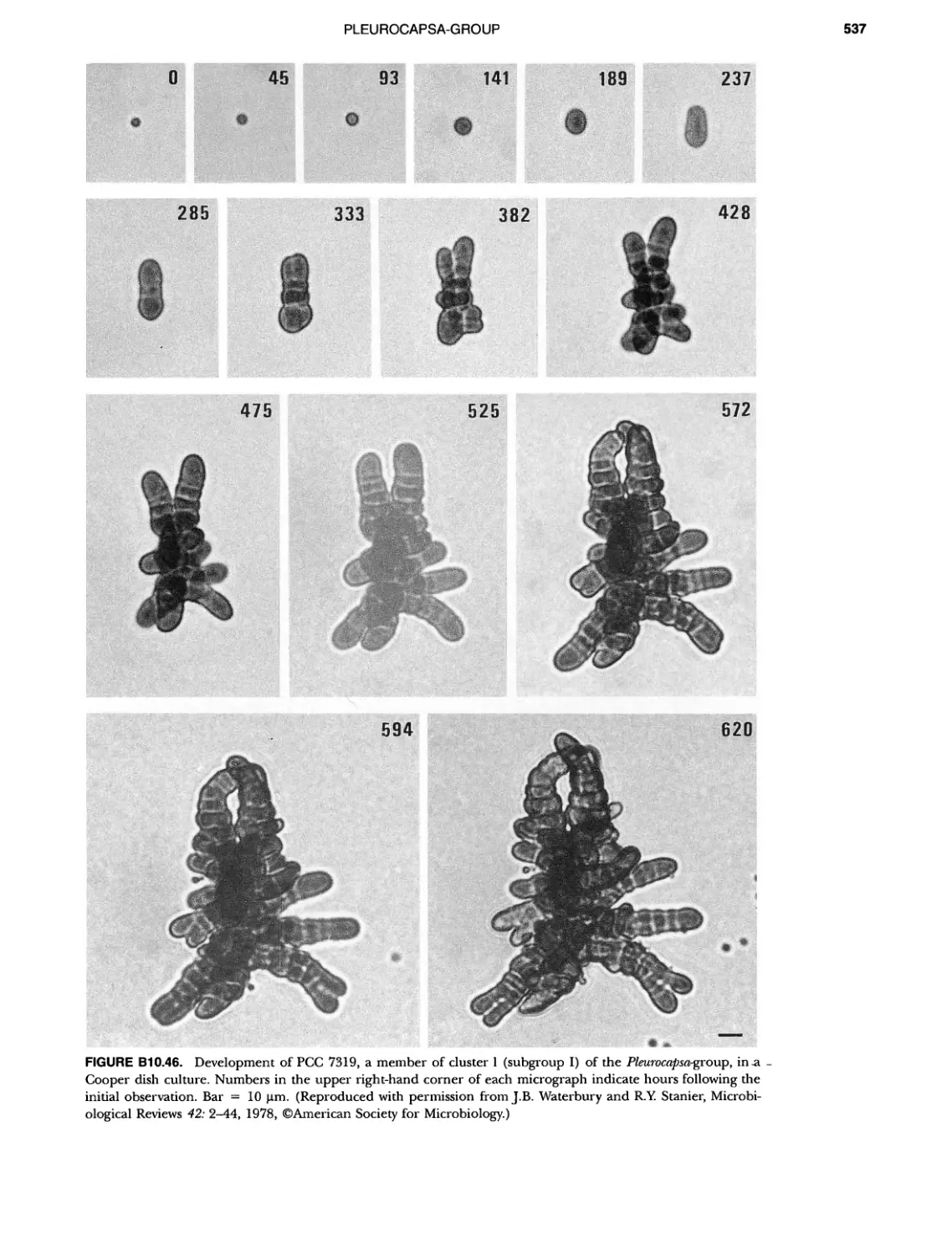
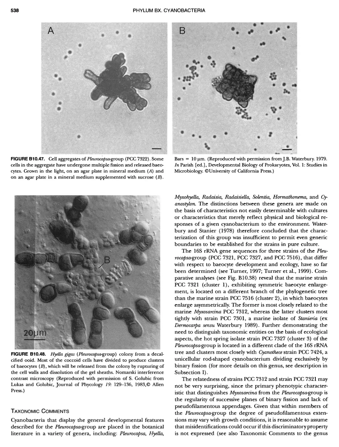
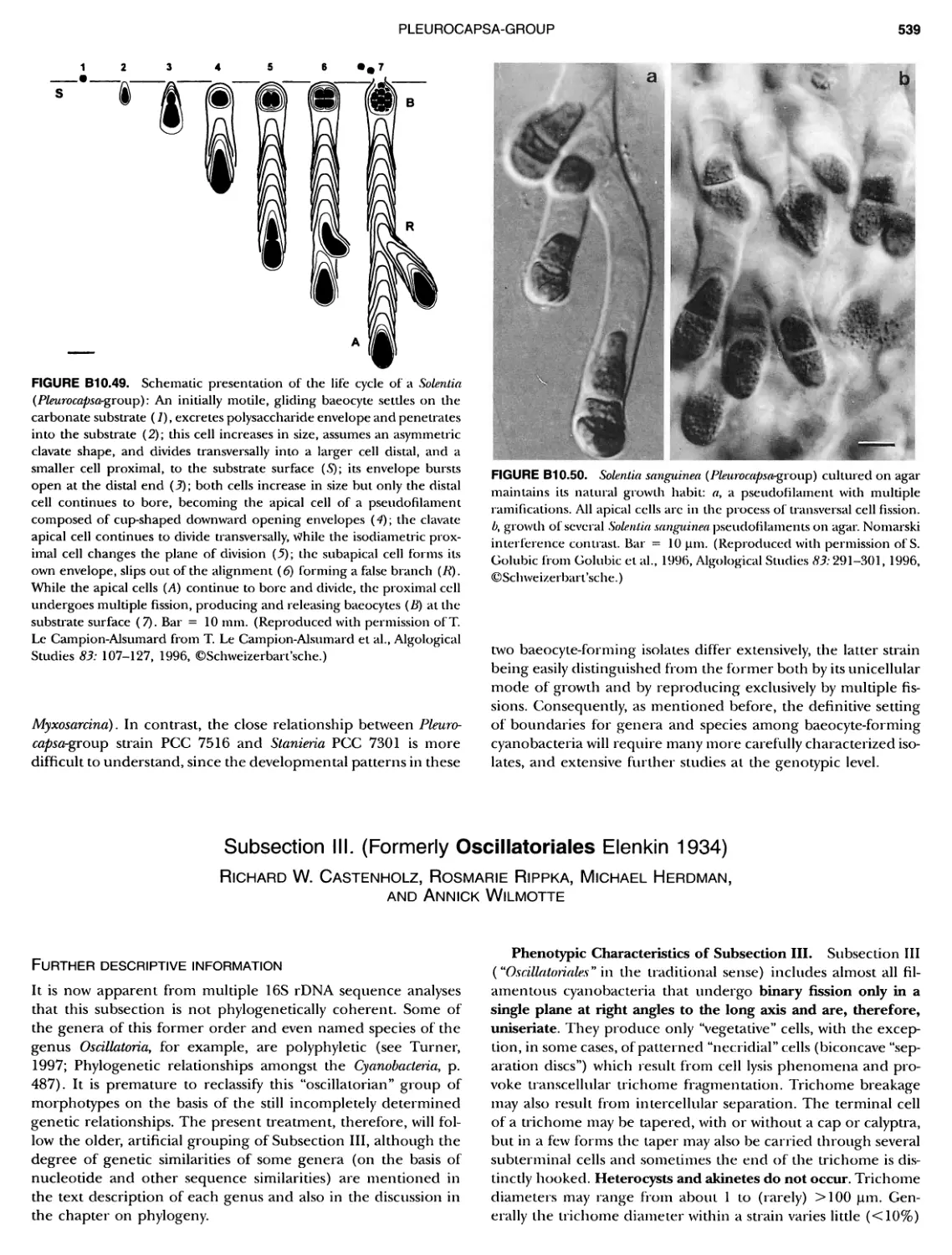
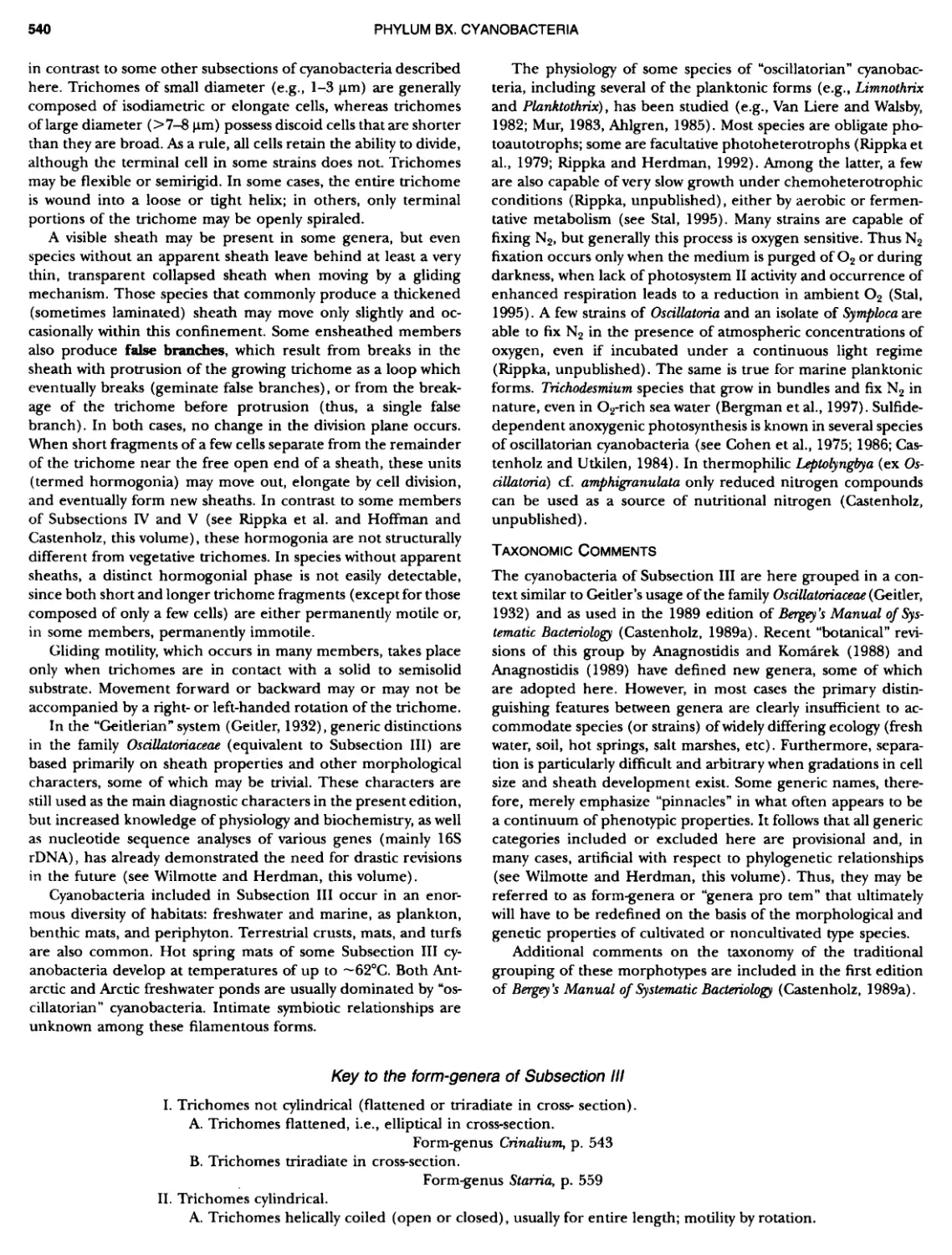
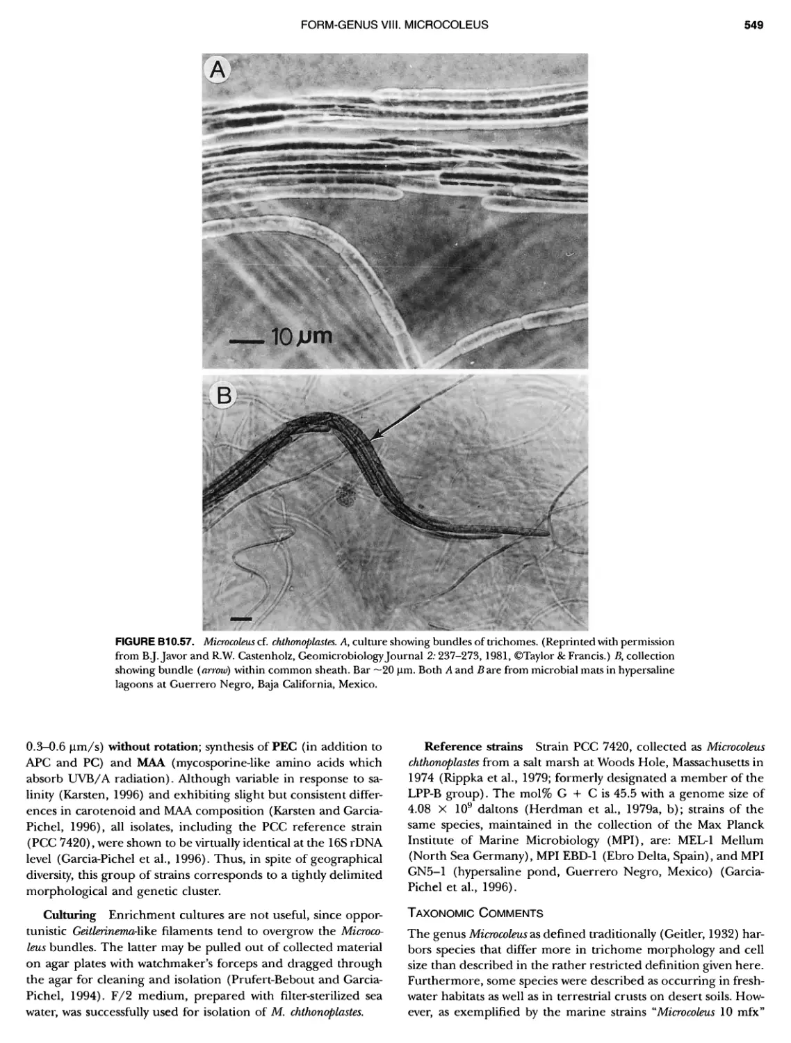
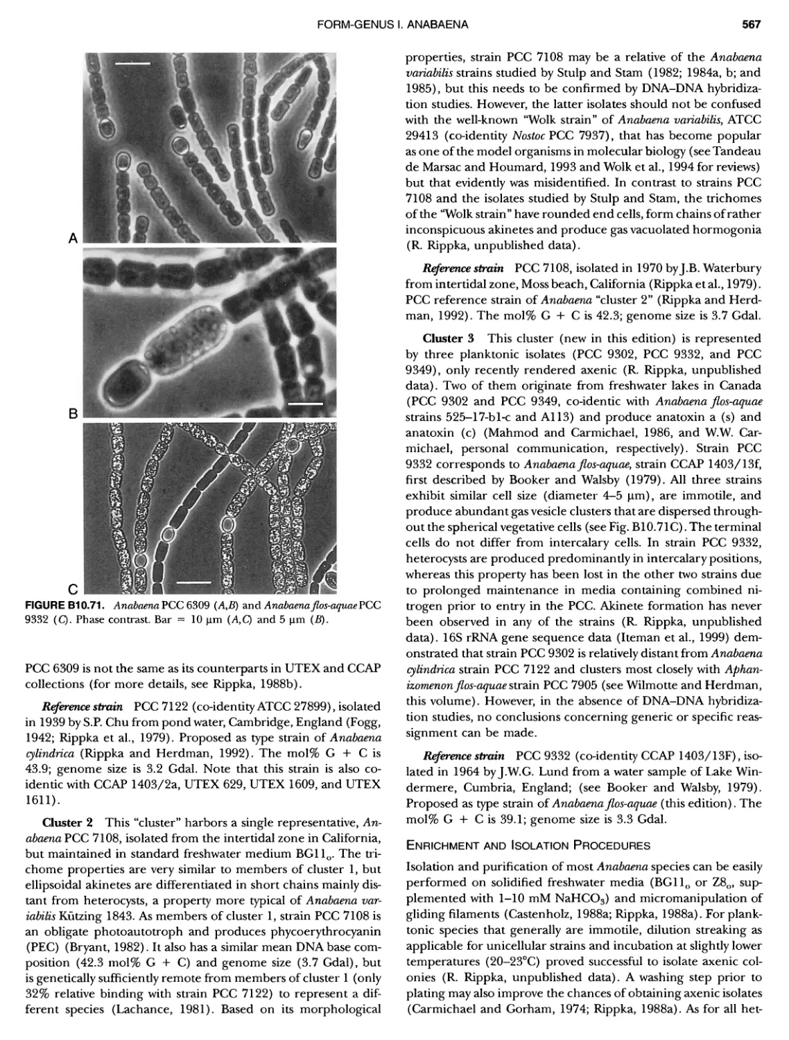
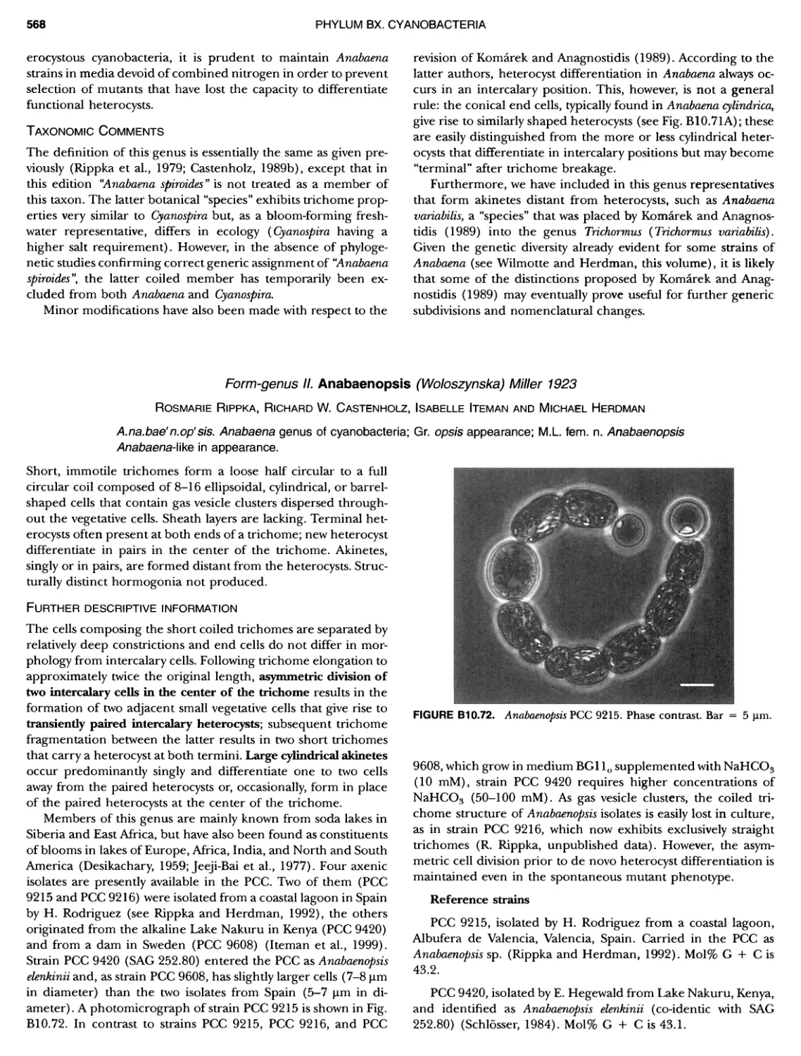
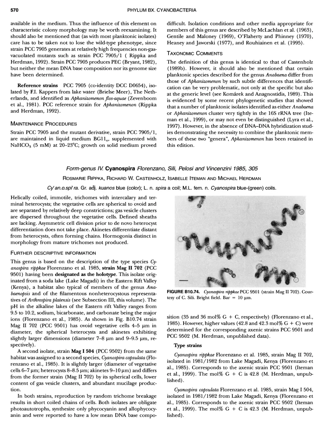
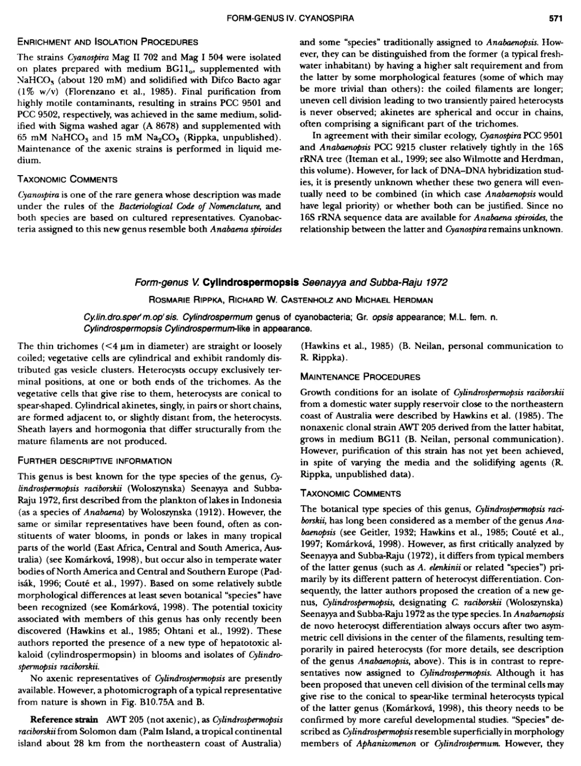
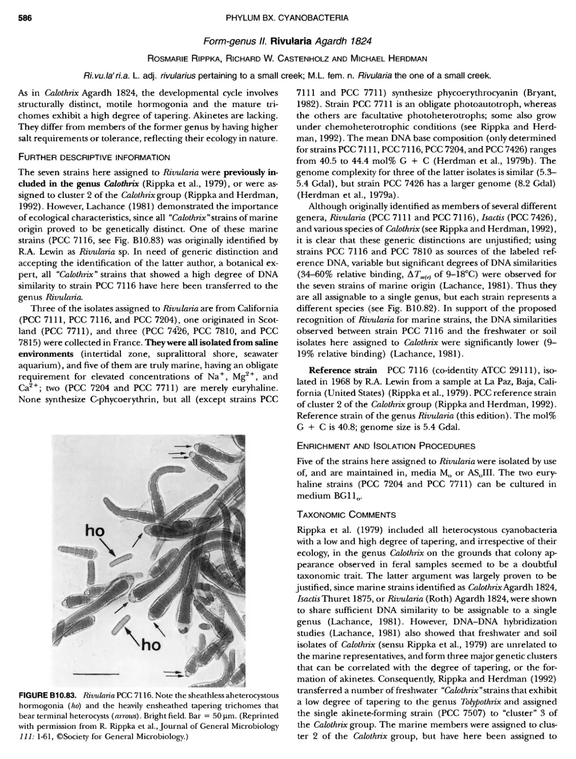
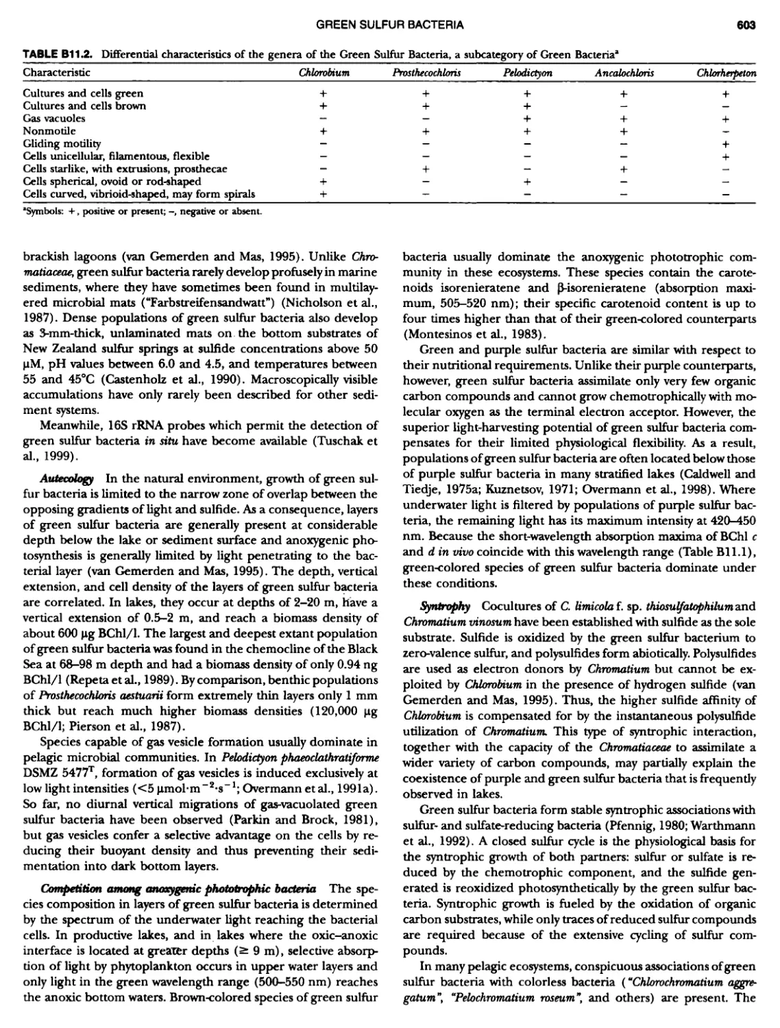
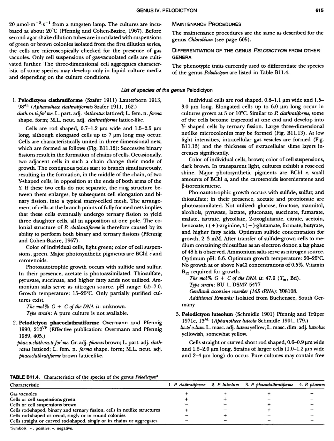
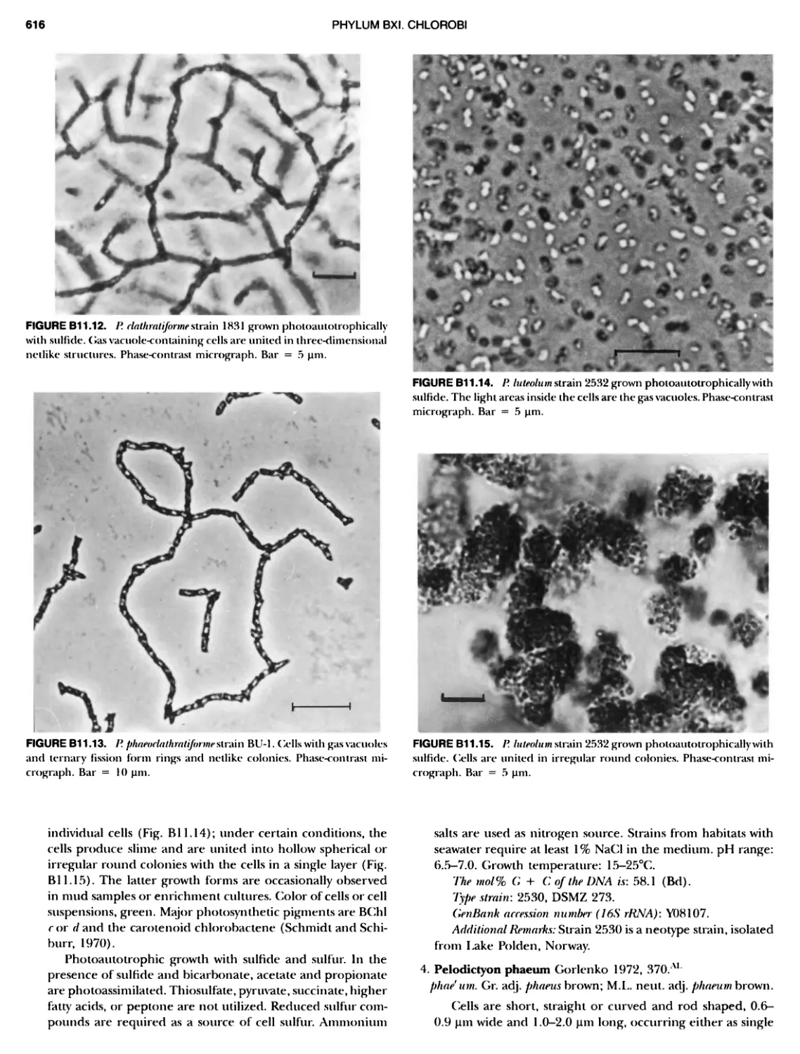
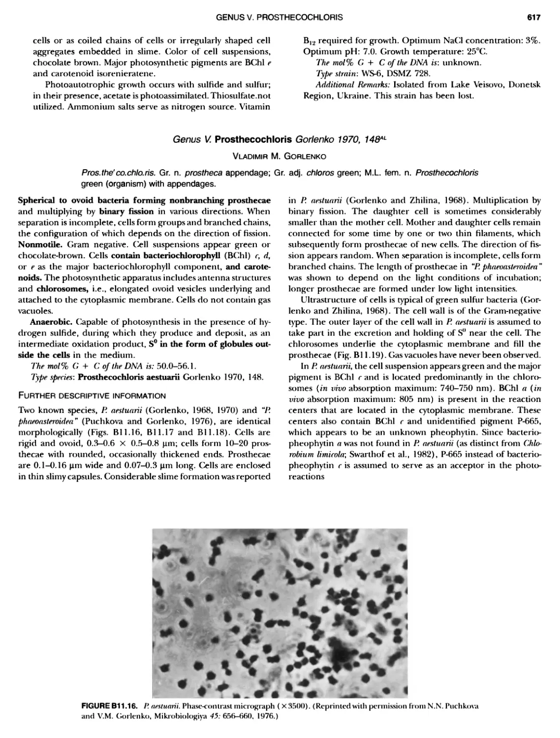
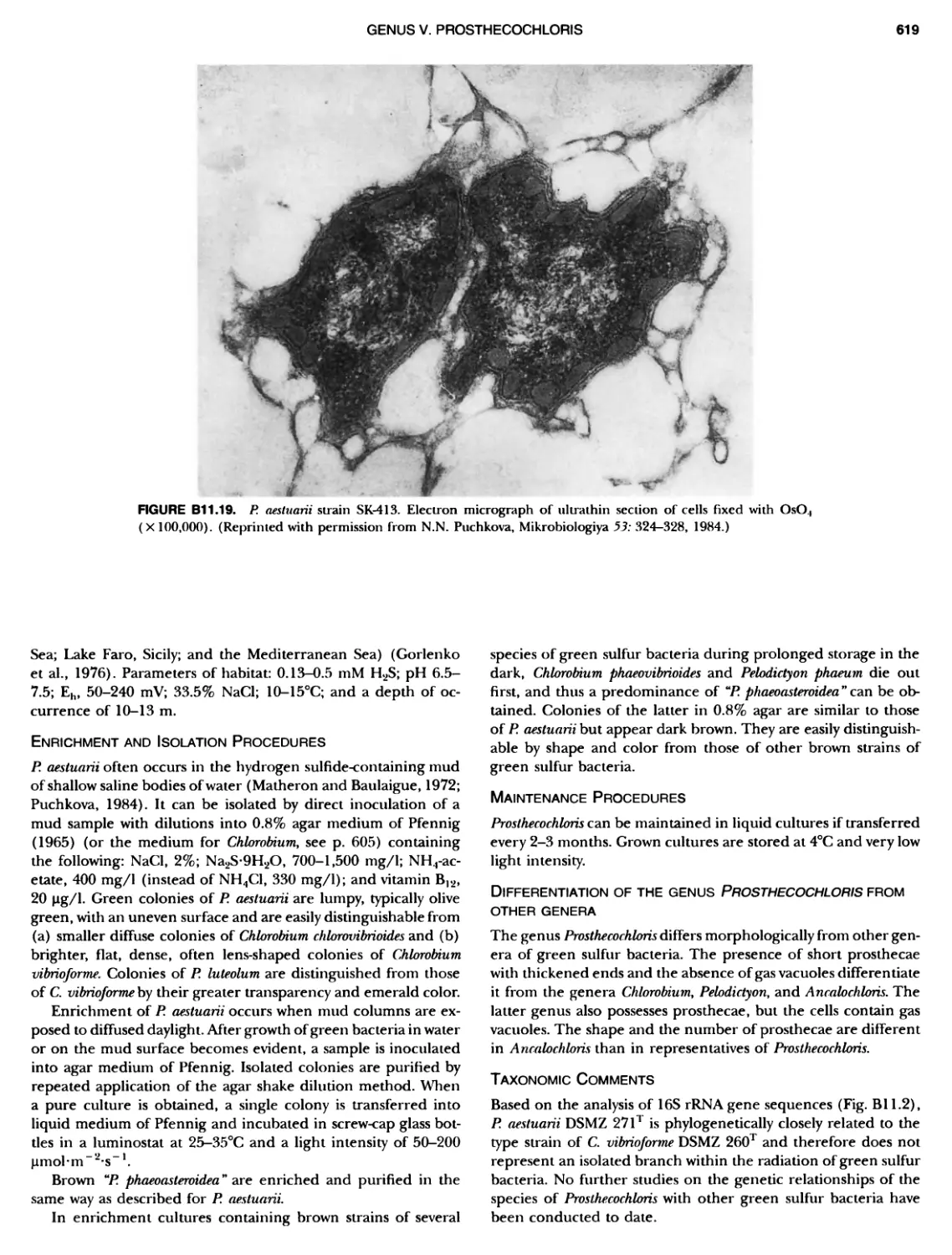
/


Автор: Boone D.R. Garrity G.
Теги: biology microbiology springer edition bacteriology cell microbiology cell anatomy manual of systematic bacteriology
ISBN: 0-587-98771-1
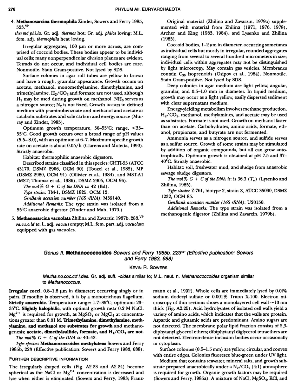
Год: 1984


Текст
BERGEY'S MANUAL� OF Systematic Bacterio ogy Second Edition Volume OneThe Archaea and the Deeply Branching andPhototrophic Bacteria
Springer New York Berlin Heidetberg Barcelona Hong Kong London Milan Paris Singapore Tokyo
BERGEY'S MANUAL� OF Systematic Bacteriology Second Edition Volume One The Archaea and the Deeply Branching and Phototrophic Bacteria David R. Boone Richard W. Castenholz EDITORS, VOLUME ONE George M. Garrity EDITOR-IN-CHIEF EDITORIAL BOARD James T. Staley, Chairman, David R. Boone, Vice Chairman, Don J. Brenner, Richard W. Castenholz, George M. Garrity, Michael Goodfellow, Noel R. Krieg, Fred A. Rainey, Karl-Heinz Schleifer WITH CONTRIBUTIONS FROM 105 COLLEAGUES Springer
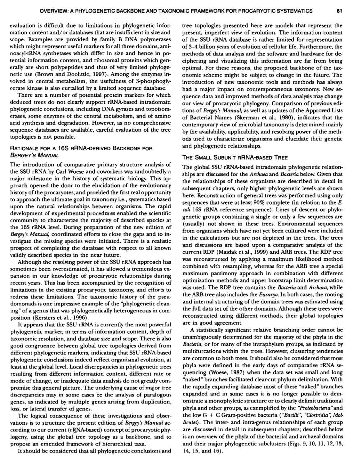
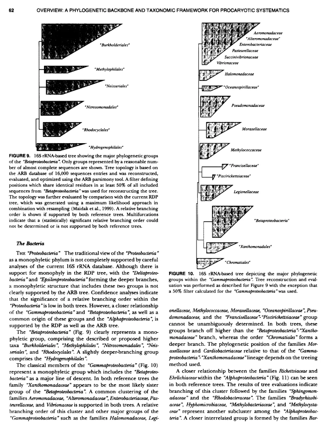
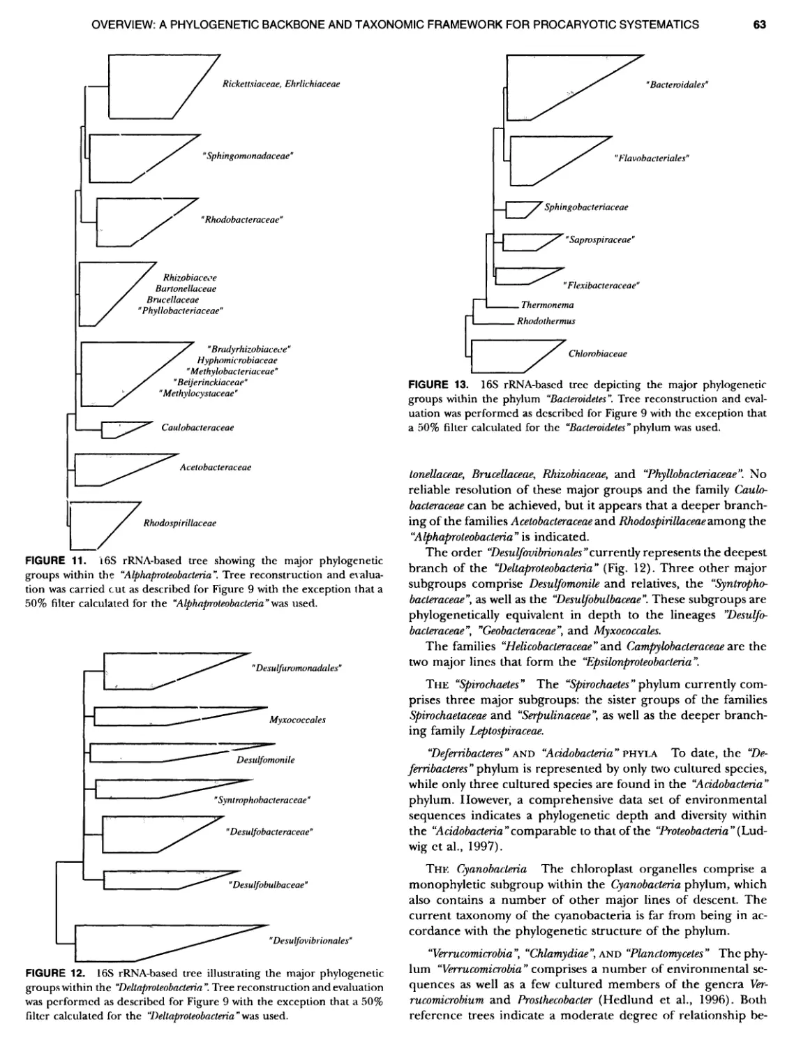
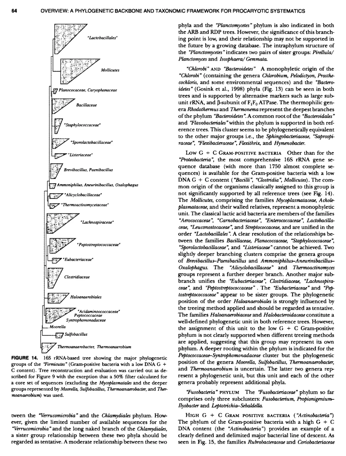
George M. Garrity Department of Microbiology and Molecular Genetics Bergey's Manual Trust Michigan State University East Lansing, MI 48824-1101 USA Library of Congress Cataloging-in-Publication Data Bergey's manual of systematic bacteriology / David R. Boone, Richard W. CastenhoL, editors, volume 1; George M. Garrity, editor-in-chief.— 2nd ed. p. cm. Includes bibliographical references and index. Contents: v. 1. The archaea and the deeply branching and phototrophic bacteria. ISBN 0-587-98771-1 (alk. paper) 1. Bacteria — Classification. I. Title: Systematic bacteriology. II. Boone, David R. III. Castenholz, Richard W. IV. Garrity, George M. QRSI.B46 2001 579.5'01'2 — dc21 2001020400 With 530 illustrations The following proprietary names of products are used in this volume: Casamino�' acids; Vector NTI�; XL10-Gold�; Gelrite�; Tryptone�', Phytagel�', bio-Trypcase�'; Trypticase�; Oxoid� purified agar. Printed on acid-free paper. First edition published 1984-1989 by Bergey's Manual Trust and Williams St: Wilkins, Baltimore. Production coordinated by Impressions Book and Journal Services, Inc., and managed by Frederick Bartlett, Theresa Kornak, and Catherine Lyons; manufacturing supervised by Jacqui Ashri. Typeset by Impressions Book and Journal Services, Inc., Madison, WI. Printed and bound by Maple-Vail Book Manufacturing Group, York, PA. Printed in the United States of America. 987654521 ISBN 0-587-98771-1 SPIN 10711544 Springer-Verlag New York Berlin Heidelberg A member of BerseismannSpringer Science+Business Media GmbH � 2001 Bergey's Manual Trust All rights reserved. This work may not be translated or copied in whole or in part without the written permission of the publisher (Springer-Verlag New York, Inc., 175 Fifth Avenue, New York, NY 10010, USA), except for brief excerpts in connection with reviews or scholarly analysis. Use in connection with any form of information storage and retrieval, electronic adaptation, computer software, or by similar or dissimilar methodology now known or hereafter developed is forbidden. The use of general descriptive names, trade names, trademarks, etc., in this publication, even if the former are not especially identified, is not to be taken as a sign that such names, as understood by the Trade Marks and Merchandise Marks Act, may accordingly be used freely by anyone. While the advice and information in this book are believed to be true and accurate at the date of going to press, neither the authors nor the editors nor the publisher can accept any legal responsibility for any errors or omissions that may be made. The publisher makes no warranty, express or implied, with respect to the material contained herein.
EDITORIAL BOARD AND TRUSTEES OF BERGEYS MANUAL TRUST James T. Staley, Chairman David R. Boone, Vice Chairman Don J. Brenner Richard W. Castenholz George M. Garrity Michael Goodfellow Noel R. Krieg Fred A. Rainey Karl-Heinz Schleifer John G. Holt, Emeritus John Liston, Fmetitus James W. Moulder, Fmeritus R.G.K. Murray, Emeritus Charles F. Niven, Jr., Emeritus Norbert Pfennig, Emeritus Peter H.A. Sneath, Emeritus Joseph G. Tully, Fmeritus Stanley T. Williams, Emeritus
Preface to the Second Edition of Bergey's Nianualof Systematic Bacteriology There is a long-standing tradition for the Editors of each succes- sive edition of Beryl's Manual to open their respective volumes with the observation that the new edition is a departure from the earlier ones. We shall not waver frorr this tradition, as the very nature of our Beld compels us to make this pronouncement. Sys- tematic bacteriology (or perhaps systematic procaryotic biology) is a dynamic field, driven by constant theoretical and methodo- logical advances that will ultimately lead to a more perfect and useful c1assification scheme. Since publication of the First Edition of the Systematics Manual, we have witnessed a major shift in how we view the relationships among Bactena and Archaea. While the possibility of a universally applicable natural classification was evident as the First Edition was in preparation, it is only recently that the sequence databases became large enough, and the taxonomic coverage broad enough, to make such an arrangement feasible. We have relied heavily upon these data in organizing the contents of this edition of Bergen''s Manual of Systematic Bacteriology, which will follow a phy- logenetic framework based on analysis of the nucleotide se- quence of the small ribosomal subunit RNA, rather than a phe- notypic structure. This departs from the First Edition, as well as the Eighth and Ninth Editions of the Determinative ManuaL While the rationale for presenting the content of this edition in such a manner should be evident to most readers, they should bear in mind that this edition, as have all preceding ones, represents a progress report rather than a final classification of procaryotes. The Editors remind the readers that the Systematics Manual is a peer-reviewed collection of chapters, contributed by authors who were invited by the Trust to share their knowledge and ex- pertise of specific taxa. Citation should refer to the author, the chapter title, and inclusive pages rather than to the Editors. The Trust is indebted to all of the contributors and reviewers, without whom this work would not be possible. The Editors are grateful for the time and effort that each expended on behalf of the entire scientific community. We also thank the authors for their good grace in accepting comments, criticisms, and editing of their manuscripts. We would also like to recognize the special efforts of Drs. Hans Truper and Brian Tindall for their assistance on matters of nomenclature and etymology and Dr. Aharon Oren for his critical reading of large portions of the ManuaL We would like to express our thanks to the Department of Microbiology and Molecular Genetics at Michigan State Univer- sity for housing our headquarters and editorial ofhce and for providing a congenial and supportive environment for microbial systematics. We would also like to thank Connie Williams not only for her expert secretarial assistance, but also for unflagging ded- ication to the mission of Bergey's Manual Trust and Dr. Denise Searles for her editorial assistance and diligence in verifying countless pieces of critical information, along with Heather Ev- erett, Alissa Wesche, and Mathew Winters for their assistance in fact-checking and compilation of the bibliography. A project such as the Systematics Manual also requires the strong and continued support of a dedicated publisher, and we have been most fortunate in this regard. We would also like to express our gratitude to Springer-Verlag for supporting our ef- forts and for the development of the Bergey's Document Type Definition (DTD). We would especially like to thank our Execu- tive Editor, Dr. Robert Badger, for his courage, patience, under- standing, and support; Catherine Lyons for her expertise in de- signing and developing our DTD, and Terry Kornak and Fred Bartlett for their efforts during the pre-production and produc- tion phases. We would also like to acknowledge the support of ArborText, Inc., for providing us with statewf-the-art SGML de- velopment and editing tools at reduced cost. Lastly, I would like to express my personal thanks to my fellow trustees for providing me with the opportunity to participate in this effort, to Drs. David Boone and Richard Castenholz for their enormous efforts as vol- ume editors and to my wife, Nancy, and daughter, Jane, for their patience, tolerance, and support. Comments on this edition are welcomed and should be di- rected to Bergey's Manual Trust, Department of Microbiology and Molecular Genetics, Giltner Hall, Michigan State University, East Lansing, MI, USA 48824-1101. Email: garrity@pilot.msu.edu George M. Garrity
Preface to the First Edition of Bergey's Manualof Systematic Bacteriology Ix Many microbiologists advised the Trust that a new edition of the Manual was urgently needed. Of great concern to us was the steadily increasing time interval between editions; this interva1 reached a maximum of 17 years between the seventh and eighth editions. To be usef'ul the Manual must reflect relatively recent information; a new edition is soon dated or obsolete in parts because of the nearly exponential rate at which new information accumulates. A new approach to publication was needed, and from this conviction came our plan to publish the Manual as a sequence of four subvolumes concerned with systematic bacteri- ology as it applies to taxonomy. The four subvolumes are divided roughly as follows: (a) the Gram-negatives of general, medical or industrial importance; (b) the Gram-positives other than acti- nomycetes; (c) the archaeobacteria, cyanobacteria and remain- ing Gram-negatives; and (d) the actinomycetes. The Trust be- lieved that more attention and care could be given to preparation of the various descriptions within each subvolume, and also that each subvolume could be prepared, published, and revised as the area demanded, more rapidly than could be the case if the Man- ual were to remain as a single, comprehensive volume as in the past. Moreover, microbiologists would have the option of pur- chasing only that particular subvolume containing the organisms in which they were interested. The Trust also believed that the scope of the Manual needed to be expanded to include more information of importance for systematic bacteriology and bring together information dealing with ecology, enrichment and isolation, descriptions of species and their determinative characters, maintenance and preserva- tion, all focused on the illumination of bacterial taxonomy. To reflect this change in scope, the title of the Manualwas changed and the primary publication becomes Bergey's Manual of Systematic Bacteriology. This contains not only determinative material such as diagnostic keys and tables useful for identification, but also all of the detailed descriptive information and taxonomic com- ments. Upon completion of each subvolume, the purely deter- minative information will be assembled for eventual incorpora- tion into a much smaller publication which will continue the original name of the Manual, Bery y's Manual of Determi native Bac teriology, which will be a similar but improved version of the pres- ent Shorter Beryl's ManuaL So, in the end there will be two pub- lications, one systematic and one determinative in character. An important task of the Trust was to decide which genera should be covered in the first and subsequent subvolumes. We were assisted in this decision by the recommendations of our Advisory Committees, composed of prominent taxonomic au- thorities to whom we are most grateful. Authors were chosen on the basis of constant surveillance of the literature of bacterial systematics and by recommendations from our Advisory Corn- mittees. The activation of the 1976 Code had introduced some novel problems. We decided to include not only those genera that had been published in the Approved Lists of Bacterial Names in Jan- uary 1980 or that had been subsequently validly published, but also certain genera whose names had no current standing in no- menclature. We also decided to include descriptions of certain organisms which had no forma1 taxonomic nomenclature, such as the endosymbionts of insects. Our goal was to omit no impor- tant group of cultivated bacteria and also to stimulate taxonomic research on "neglected" groups and on some groups of un- doubted bacteria that have not yet been cultivated and subjected to conventional studies. The invited authors were provided with instructions and ex- emplary chapters in June 1980 and, although the intended dead- line for receipt of manuscripts was March 1981, all contributions were assembled in January 1982 for the final preparations. The Manual was forwarded to the publisher in June 1982. Some readers will note the consistent use of the stem -var in- stead of -type in words such as biovar, serovar and pathovar. This is in keeping with the recommendations of the Bacteriological Code and was done against the wishes of some of the authors. We have deleted much of the synonymy of scientific names which was contained in past editions. The adoption of the new starting date of January 1, 1980 and publication of the Approved Lists of Bacterial Names has made mention of past synonymy ob- solete. We have included synonyms of a name only if they have been published since the new starting date, or if they were also on the Approved Lists and, in rare cases with certain pathogens, if the mention of an old name would help readers associate the organism with a clinical problem. If the reader is interested in tracing the history of a name we suggest he or she consult past editions of the Manual or the Index Bergsyana and its Supplement In citations of names we have used the abbreviation AL to denote the inclusion of the name on the Approved Lists of Bacterial Names and VP to show the name has been validly published. In the matter of citation of the Manual in the scientific liter- ature we again stress the fact that the Manual is a collection of authored chapters and the citation should refer to the author, the chapter title and its inclusive pages, not the Editor. To all contributors, the sincere thanks of the Trust is due; the Editor is especially grateful for the good grace with which the
PREFACE TO THE FIRST EDITION authors accepted comments, criticisms and editing of their man-uscripts. It is only because of the voluntary and dedicated eKorts of these authors that the Manual can continue to serve the sci-ence of bacteriology on an international basis. A number of institutions and individuals deserve special ac- knowledgment from the Trust for their help in bringing about the publication of this volume.. ~
Preface to the First Edition of Bergey's Manualof Determinative Bacteriology The elaborate system of classification of the bacteria into families, tribes and genera by a Committee on Characterization and Clas- sification of the Society of American Bacteriologists (1911, 1920) has made it very desirable to be able to p1ace in the hands of students a more detailed key for the identification of species than any that is available at present. The valuable book on "Deter- minative Bacteriology" by Professor F. D. Chester, published in 1901, is now of very little assistance to the student, and all pre- vious classifications are of still less value, especially as earlier systems of classification were based entirely on morphologic char- acters. It is hoped that this manual will serve to stimulate efforts to perfect the classification of bacteria, especially by emphasizing the valuable features as well as the weaker points in the new system which the Committee of the Society of American Bacte- riologists has promulgated. The Committee does not regard the classification of species offered here as in any sense final, but merely a progress report leading to more satisfactory classifica- tion in the future. The Committee desires to express its appreciation and thanks to those members of the society who gave valuable aid in the compilation of material and the classification of certain species. ~ ~ ~ The assistance of all bacteriologists is earnestly solicited in the correction of possib1e errors in the text; in the collection of descriptions of all bacteria that may have been omitted from the text; in supplying more detailed descriptions of such organisms as are described incompletely; and in furnishing complete de- scriptions of new organisms that may be discovered, or in di- recting the attention of the Committee to publications of such newly described bacteria. David H. Bergey, Chairman Francis C. Harrison Robert S. Breed Bernard W. Hammer Frank M. Huntoon Committee on ManuaL August, 1923.
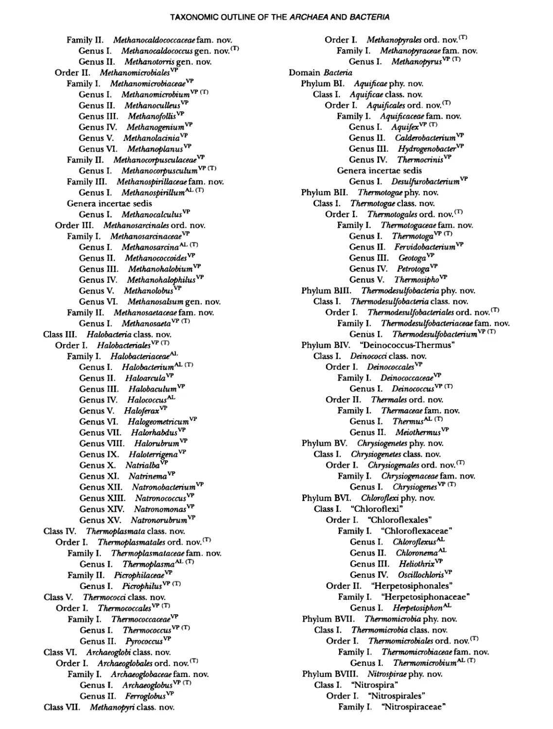
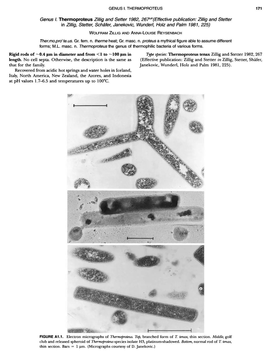
Contents vil ix xiXIX 27 111 11 11 DOMAIN ARCHAEA xiii Preface to the Second Edition of Bergey's Manual� of Systematic B acterlology ~ ~ ~ ~ ~ ~ ~ ~ ~ ~ ~ ~ ~ ~ ~ ~ ~ ~ ~ 0 ~ ~ ~ ~ ~ ~ ~ ~ ~ ~ ~ ~ ~ ~ ~ ~ ~ ~ ~ 0 Preface to the First Edition of Bergey's Manual� of Systematic B acteriology ~ ~ ~ ~ ~ ~ ~ ~ ~ ~ ~ ~ ~ ~ ~ ~ ~ ~ ~ ~ ~ ~ ~ ~ ~ ~ ~ ~ ~ ~ ~ ~ ~ ~ ~ ~ ~ ~ ~ ~ Preface to the First Edition of Bergey's Manual� of Determinative B acteriology ~ ~ ~ ~ ~ ~ ~ ~ ~ ~ ~ ~ ~ ~ ~ ~ ~ ~ ~ ~ ~ ~ ~ ~ ~ ~ ~ ~ ~ ~ ~ ~ ~ ~ ~ ~ 0 ~ ~ ~ C ' ~ OntrI~utOrS ~ ~ ~ ~ ~ ~ ~ ~ ~ ~ ~ ~ ~ ~ ~ ~ ~ ~ ~ ~ ~ ~ ~ ~ ~ ~ ~ ~ 0 ~ ~ ~ ~ ~ ~ ~ ~ ~ ~ ~ ~ ~ ~ The History of Bergey's Manual............................. On Using the Manual ..................................... Procaryotic Domains Classification of Procaryotic Organisms and the Concept of Bacterial S peciatlon ~ ~ ~ ~ ~ ~ ~ ~ ~ ~ ~ ~ ~ ~ ~ ~ ~ ~ ~ ~ ~ ~ ~ ~ ~ ~ ~ ~ ~ ~ ~ ~ ~ ~ ~ ~ ~ ~ ~ ~ ~ ~ Identification of Procaryotes Numerical Taxonomy ..................................... Polyphasic Taxonomy Overview: A Phylogenetic Backbone and Taxonomic Framework for Procaryotic Systematics Nucleic Acid Probes and Their Application in Environmental Icrobiology Bacterial Nomenclature Etymology in Nomenclature of Procaryotes Microbiai Ecology — New Directions, New Importance Culture Collections: An Essential Resource for Microbiology ...... Intellectual Property of Procaryotes The Road Map to the Manual ............................... PHYLUM Al Crenarchaeota ................. Class I. Thermoprotei Order I. Thermoproteales ....... Family I. Thermoproteaceae ... Genus I. Thermoproteus Genus li. Caldivirga ....... Genus I II. Pyrobaculum Genus IV. Thermocladium Family II. Thermofilaceae ..... Genus I. Thermofilum Order II. Desulfurococcales Family I. Desulfurococcaceae .. Genus I. Desulfurococcus Genus II. Aeropyrum Genus III. Ignicoccus ...... Genus I V. Staphylothermus Genus V. Stetteria Genus Vl. Sulfophobococcus 169 168 170 170 171 173 174 177 178 178 179 180 181 183 184 186 187 1
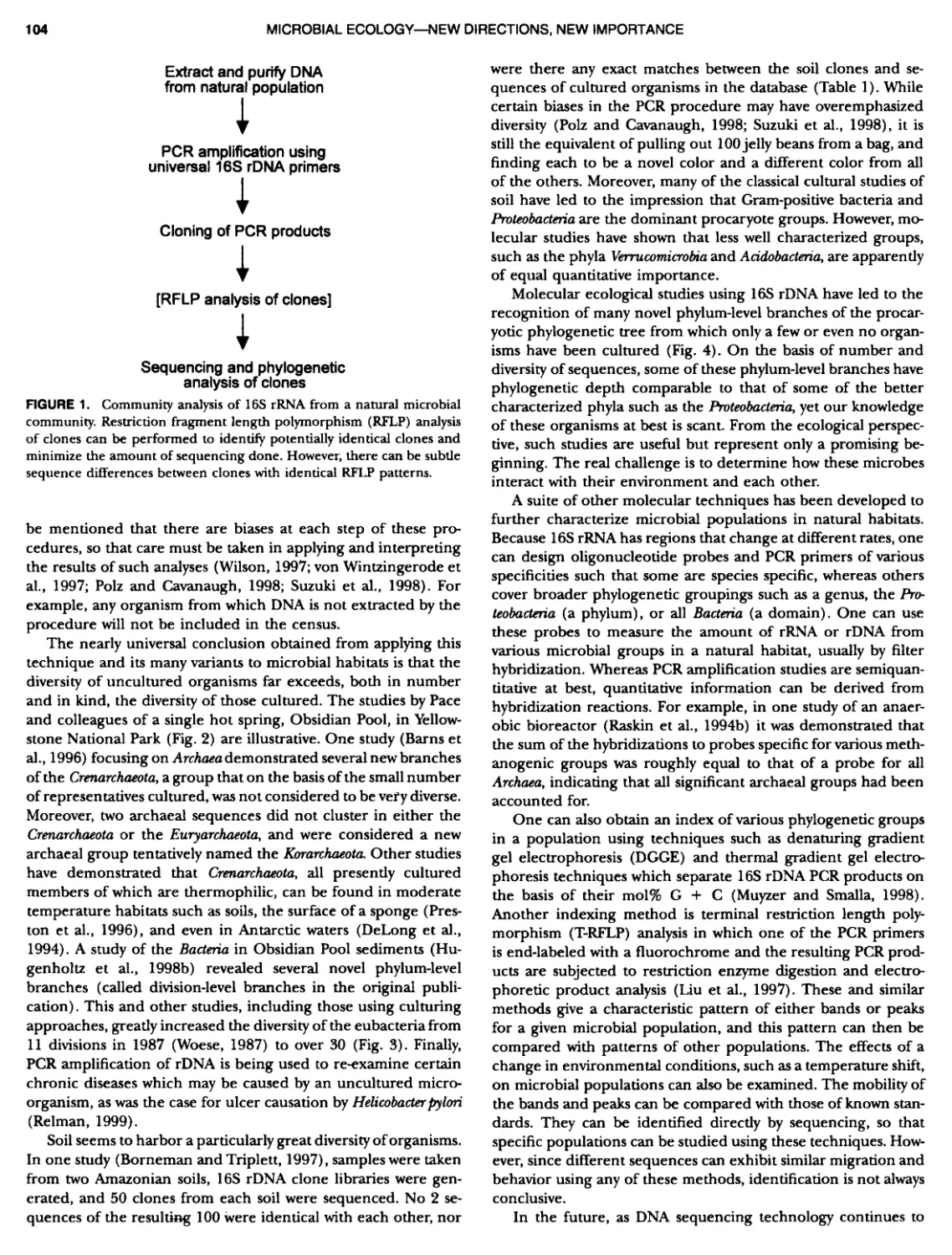
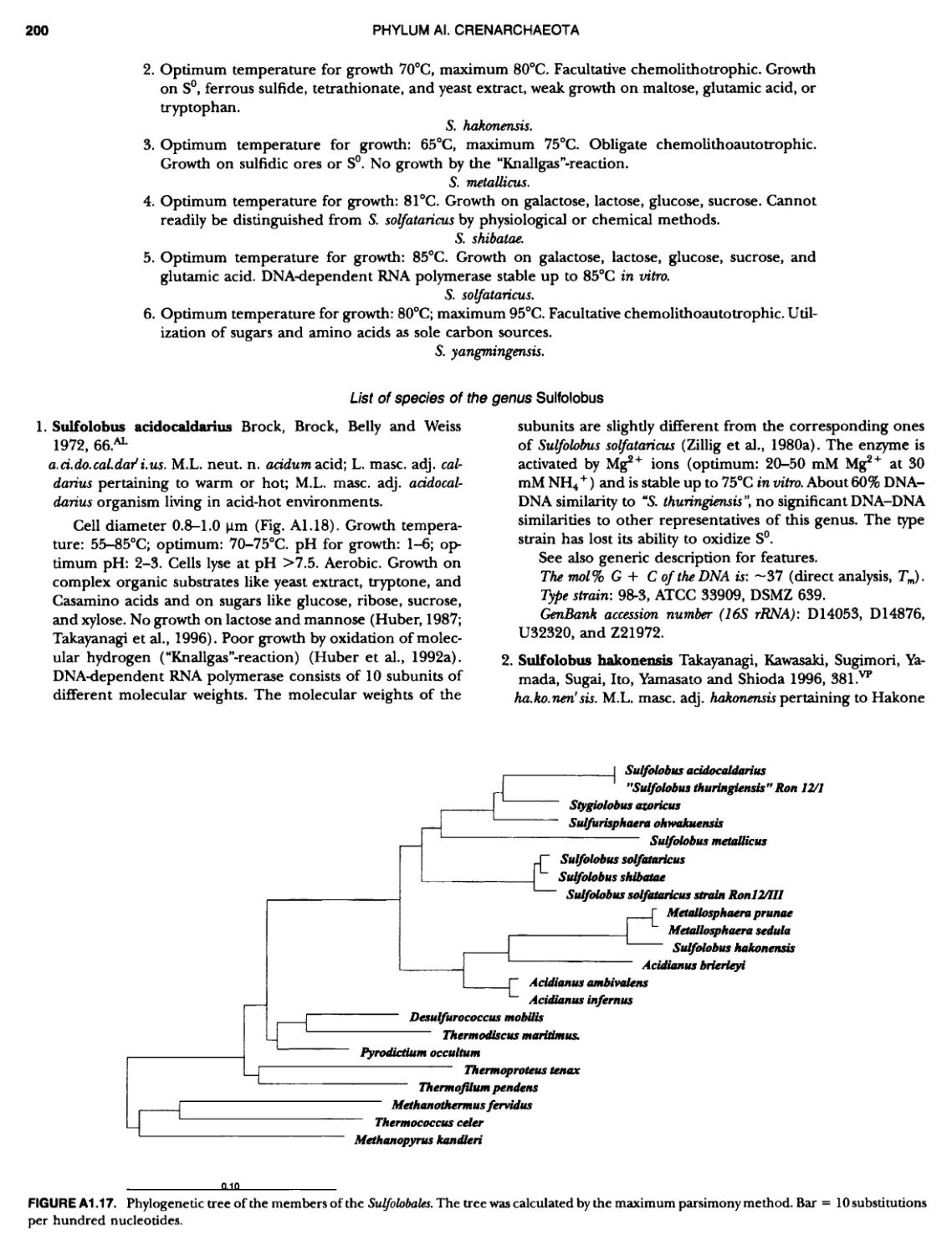
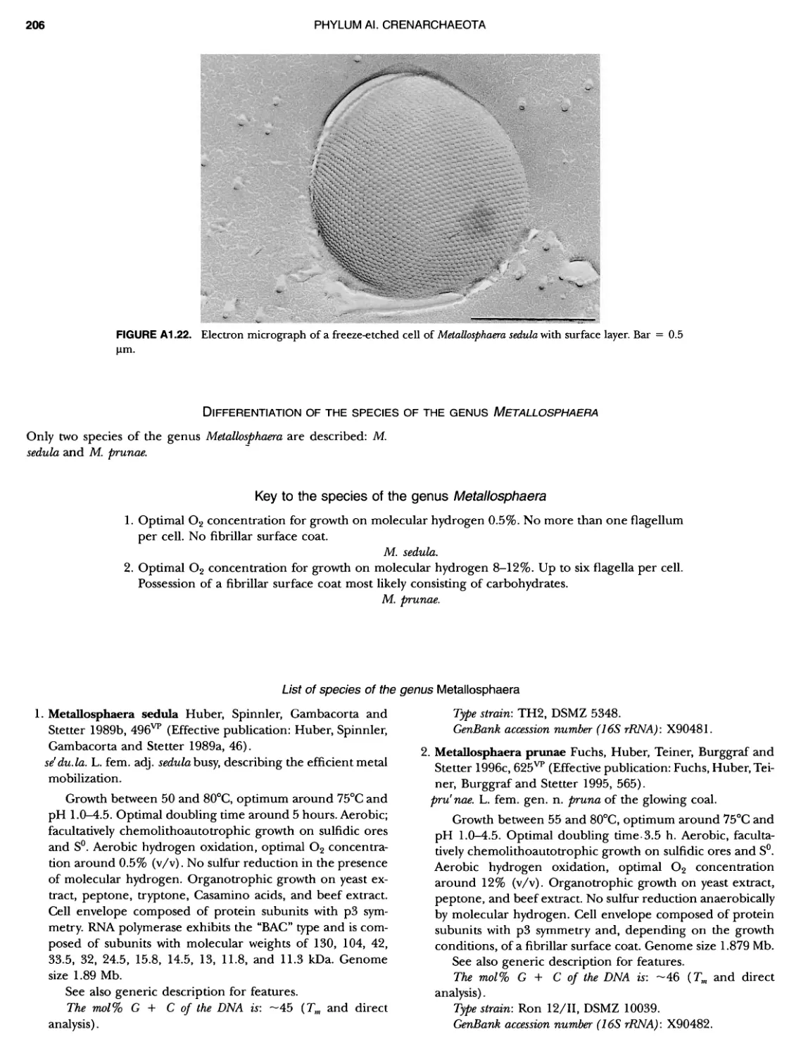
XIV CONTENTS 189 1 191 192 195 202 204 207 208 209 Iculus Genus Vll. Thermodiscus .. Genus Vill. Thermosphaera Family II. Pyrodictiaceae Genus I. Pyrodictium Genus II. Hyperthermus ... Genus I II. Pyrolobus Order III. Sulfolobales Family I. Sulfolobaceae Genus I. Sulfolobus Genus II. Acidianus Genus I II. Metallosphaera Genus IV. Stygiolobus Genus V. Sulfurisphaera Genus Vl. Sulfurococcus PHYLUNI All Euryarchaeota ..................... Taxonomy of INethanogenic Archaea..... Class I. Methanobacteria Order Methanobacteriales Family I. Methanobacteriaceae ..... Genus I. Methanobacterium Genus II. Methanobrevibacter.... Genus III. Methanosphaera Genus IV. Methanothermobacter Family II. Methanothermaceae Genus I. Methanothermus Class II. Methanococci ............... Order I. Methanococcales Family I. Methanococcaceae Genus I. Methanococcus Genus II. Methanothermococcus Family II. Methanocaldococcaceae .. Genus I. Methanocaldococcus Genus II. Methanotorris Order I I. Methanomicrobiales Family I. Methanomicrobiaceae Genus I. Methanomicrobium ..... Genus II. Methanoculleus....... Genus II I. Methanofollis Genus IV. Methanogenium ...... Genus V. Methanolacinia Genus Vl. Methanoplanus Family II. Methanocorpusculaceae Genus I. Methanocorpusculum ... Family III. Methanospirillaceae Genus I. Methanospirillum Genus Incertae Sedis I. Methanoca Order III. Methanosarcinales Family I. Methanosarcinaceae Genus I. Methanosarcina ....... Genus II. Methanococcoides Genus III. Methanohalobium..... Genus IV. Methanohalophilus .... Genus V. Methanolobus ........ Genus VI. Methanosalsum ...... Family II. Methanosaetaceae ~ ~ ~ ~ ~ ~ ~ ~ ~ ~ ~ ~ ~ ~ ~ ~ ~ ~ ~ ~ ~ ~ ~ ~ ~ ~ ~ ~ ~ ~ ~ ~ ~ ~ ~ ~ ~ ~ ~ ~ ~ ~ ~ ~ ~ ~ ~ ~ ~ ~ ~ ~ ~ ~ ~ ~ ~ ~ ~ ~ ~ ~ ~ ~ ~ ~ ~ ~ ~ ~ ~ ~ ~ ~ ~ ~ ~ ~ 211 211 213 214 214 215 218 226 230 233 233 235 236 236 236 241 242 243 245 246 247 247 251 253 256 258 259 262 262 264 264 267 268 268 269 276 279 281 283 287 289
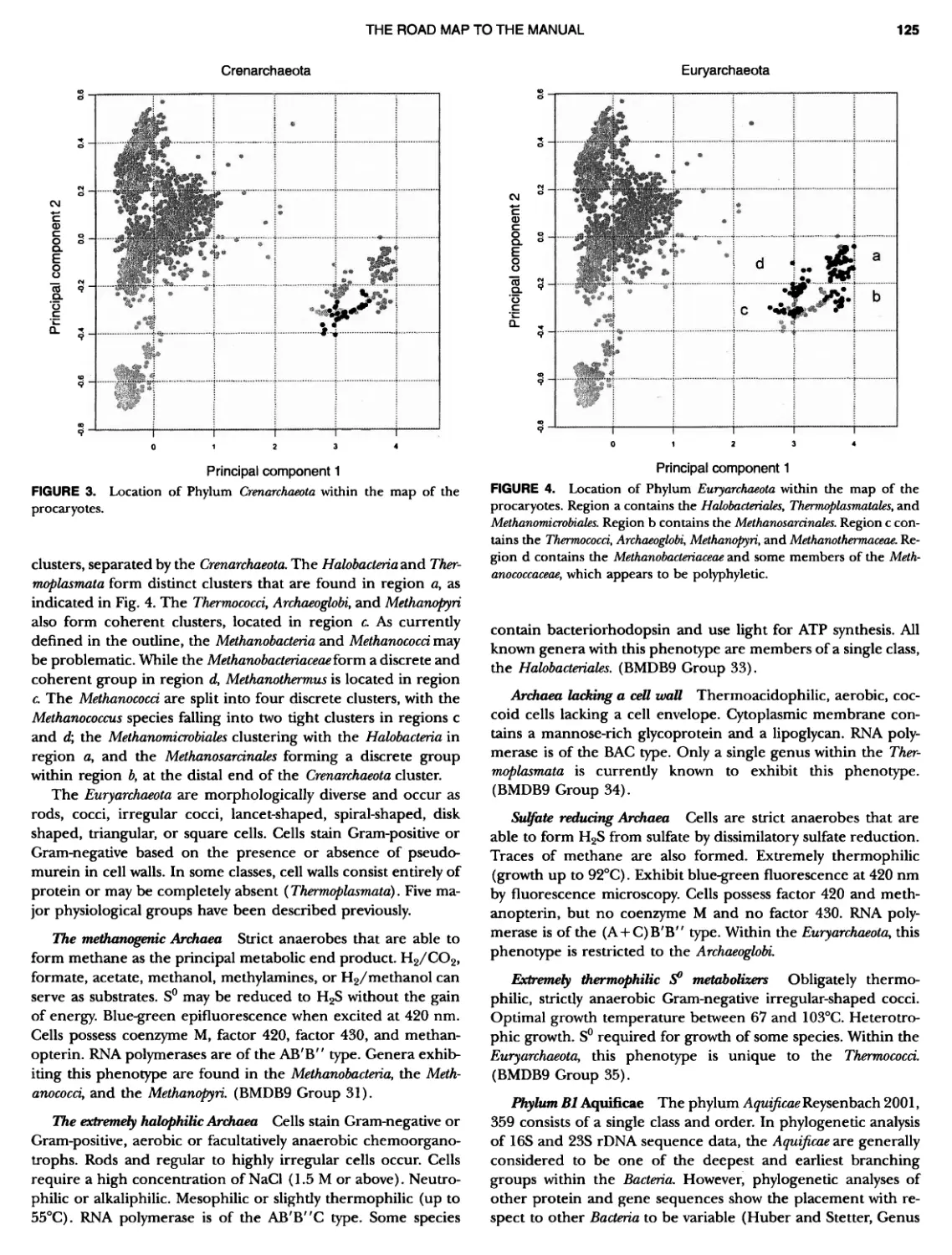
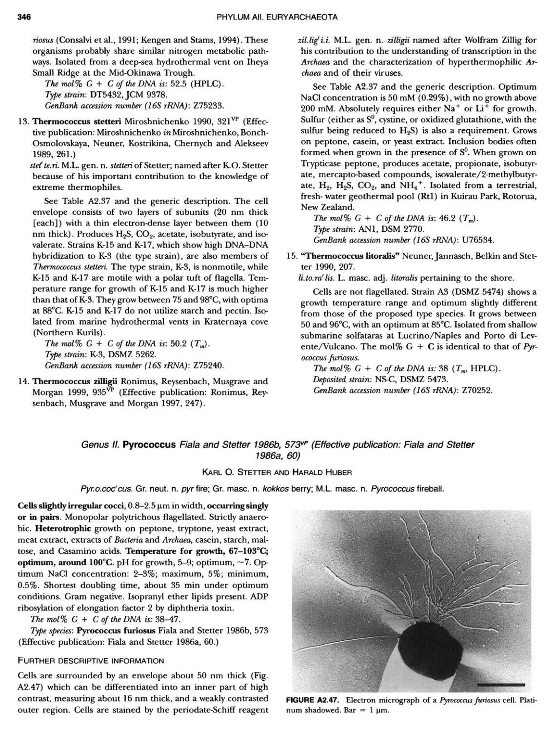
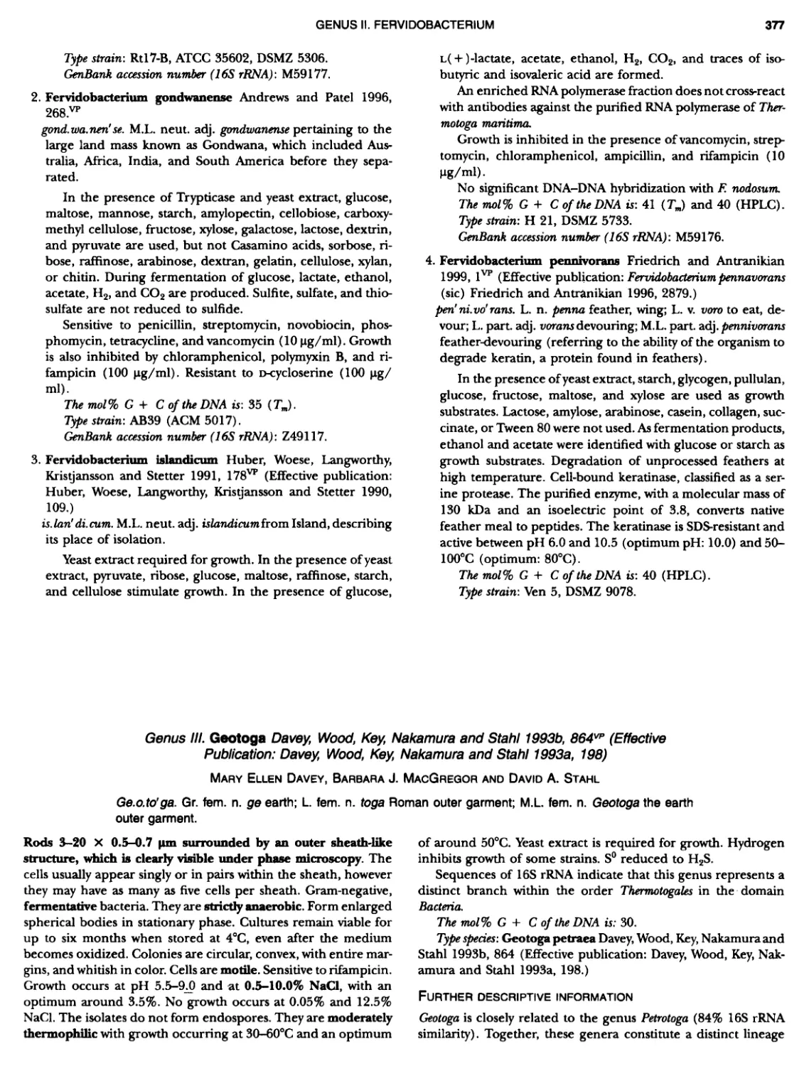
CONTENTS XV 359 359 359 360 360 362 363 364 366 ~ ~ I ~ ~ ~ ~ ~ ~ ~ ~ ~ ~ ~ ~ ~ ~ ~ ~ ~ ~ ~ ~ ~ ~ ~ ~ 369 369 369 370 370 ~ ~ ~ ~ ~ ~ ~ ~ ~ 0 ~ ~ ~ ~ ~ Genus I. Methanosaeta Class III. Halobacteria Order I. Halobacteriales ....... Family I. Halobacteriaceae ... Genus I. Malobacterium Genus II. Haloarcula Genus III. Halobaculum Genus IV. Halococcus Genus V. Haloferax Genus Vl. Malogeometricum Genus Vll. Malorubrum Genus Vill. Haloterrigena .. Genus IX. Natrialba Genus X. Natrinema Genus Xl. Natronobacterium Genus XII. Natronococcus Genus XIII. Natronomonas . Genus XIV. Natronorubrum Class IV. Thermoplasmata Order I. Thermoplasmatales .... Family I. Thermoplasmataceae Genus I. Thermoplasma... Family II. Picrophilaceae Genus I. Picrophilus...... Class IV. Thermococci Order I. Thermococcales Family I. Thermococcaceae Genus I. Thermococcus ... Genus II. Pyrococcus ..... Class Vl. Archaeoglobi .......... Order I. Archaeoglobales Family I. Archaeoglobaceae .. Genus I. Archaeoglobus... Genus I I. Ferroglobus Class Vll. Methanopyri .......... Order I. Methanopyrales....... Family I. Methanopyraceae Genus I. Methanopyrus DOMAIN BACTERIA PHYLUM Bl A quificae ~ ~ ~ ~ ~ ~ ~ O ~ ~ ~ ~ ~ ~ ~ ~ ~ 1 ~ ~ ~ ~ ~ ~ ~ ~ ~ ~ ~ S ~ Class I. Aquificae Order I. Aquificales Family I. Aquificaceae Genus I. Aquifex Genus II. Calderobacterium Genus III. Hydrogenobacter ........... Genus IV. Thermocrinis Genus Incertae Sedis I. Oesulfurobacteri um PHYLUM Bll Thermotogae .............. Class I. Thermotogae Order I. Thermotogales Family I. Thermotogaceae Genus I. Thermotoga .. 289 294 294 1 299 305 309 311 315 318 320 324 325 327 329 330 332 333 335 335 335 335 339 339 341 341 341 342 346 349 349 349 349 352 353 353 353 354
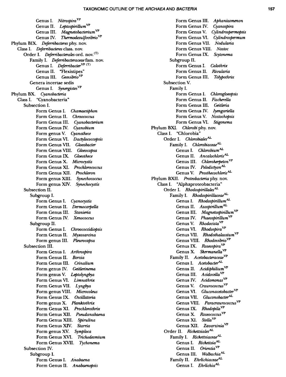
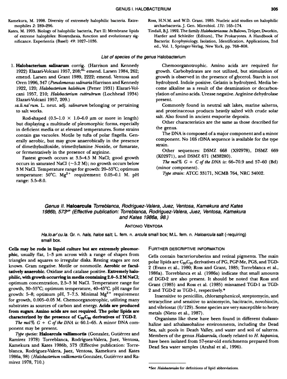
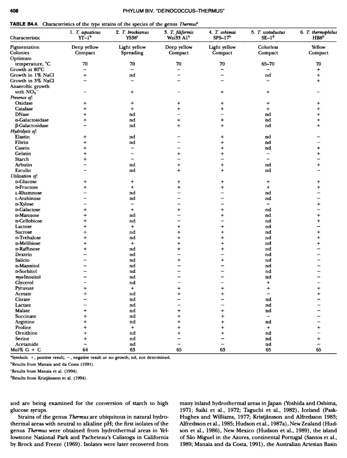
CONTENTS XVI 375 382 385 389 389 389 390 390 ~ ~ ~ ~ ~ ~ ~ ~ ~ ~ ~ ~ ~ ~ ~ ~ ~ ~ ~ ~ 1 21 421421422 Bacteria 447 447 447 447 448 ~ ~ ~ ~ ~ ~ ~ ~ ~ ~ Genus II. Fervidobacterium Genus III. Geotoga Genus IV. Petrotoga ....... Genus V. Thermosipho PHYLUM BIII Thermodesulfobacteria Class I. Thermodesulfobacteria Order I. Thermodesulfobacteriales Family I. Thermodesulfobacteriaceae Genus I. Thermodesulfobacterium PHYLUM BIV "Deinococcus-Thermus" ... Class I. Deinococci Order I. Deinococcales Family I. Deinococcaceae Genus I. Deinococcus Order II. Thermales Family I. Thermaceae Genus I. Thermus Genus II. Meiothermus PHYLUM BVChrysiogenetes ............ Class I. Chrysiogenetes ...... Order I. Chrysiogenales .... Family I. ChrysiogenaceaeGenus I. Chrysiogenes PHYLUM BVI Chio roflexi Class I. "Chloroflexi" Order I. "Chloroflexales".............. Family I. "Chloroflexaceae".......... Filamentous Anoxygenic Phototrophic Genus I. Chlorotlexus Genus II. Chloronema Genus III. Heliothrix ............. Genus IV. Oscillochloris Order II. "Herpetosiphonales" .......... Family I. "Herpetosi phonaceae" Genus I. Herpetosiphon .......... PHYLUM BVII Thermomicrobia Class I. Thermomicrobia Order I. Thermomicrobiales Family I. Thermomicrobiaceae Genus I. Thermomicrobium PHYLUM BVIII Nitrospirae .............................. Class I. "Nitrospira" Order I. "Nitrospirales" ................... Family I. "Nitrospiraceae" ............... Genus I. Nitrospira.................. Genus I I. Leptospirillum Genus III. "Candidatus Magnetobacterium" Genus IV. Thermodesulfovibrio ......... ~ ~ ~ ~ ~ ~ ~ ~ ~ ~ ~ ~ ~ ~ ~ ~ ~ ~ ~ ~ ~ ~ ~ ~ ~ ~ ~ ~ ~ ~ ~ ~ ~ ~ 395 395 395 395 396 403 403 404 414 427 427 427 427 427 429 437 438 451 451 451 451 451 453 457
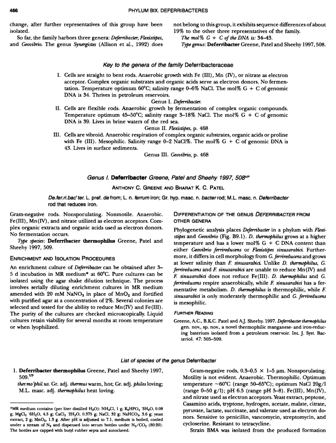
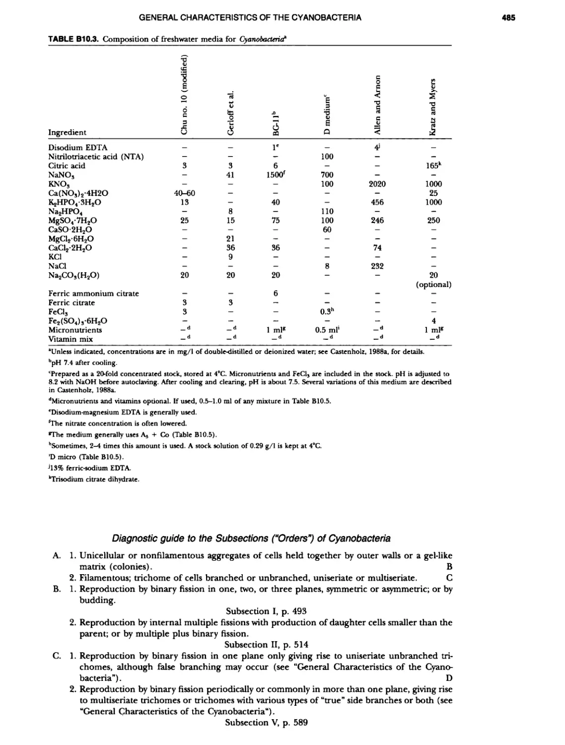
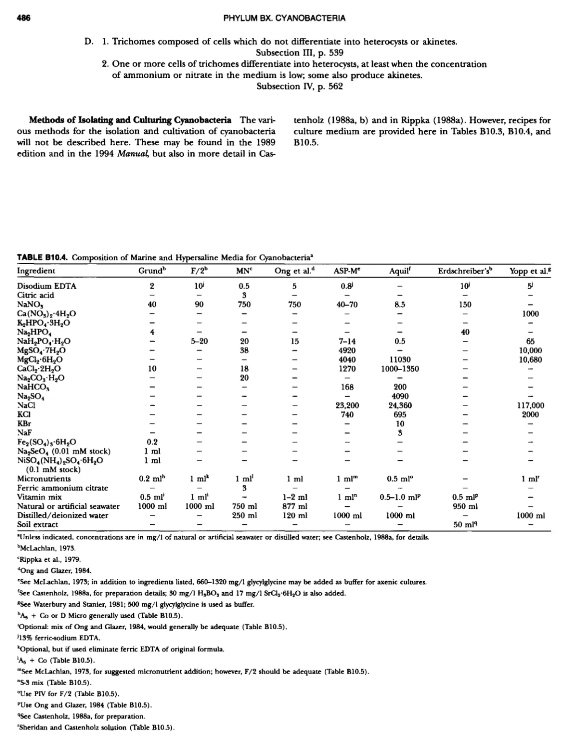
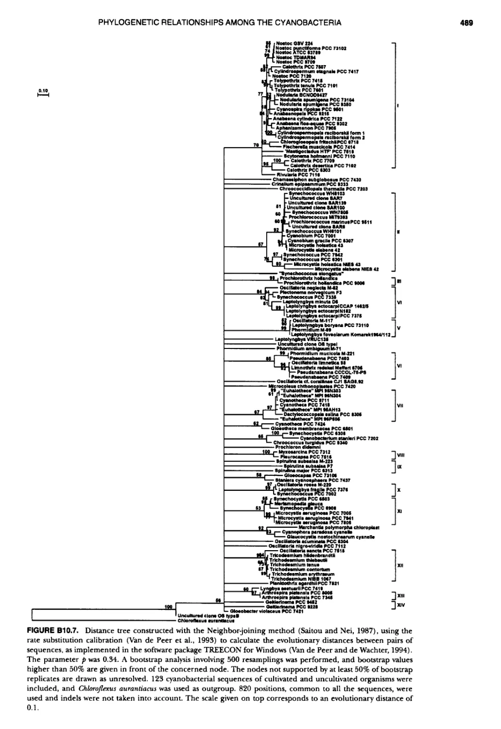
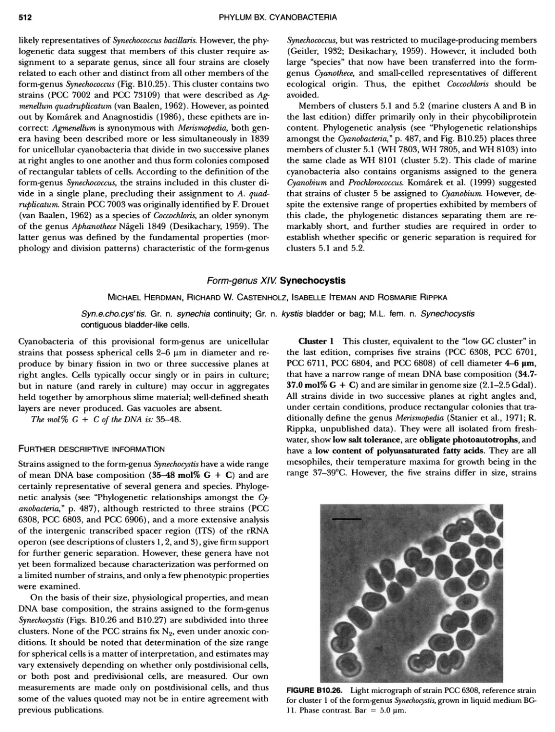
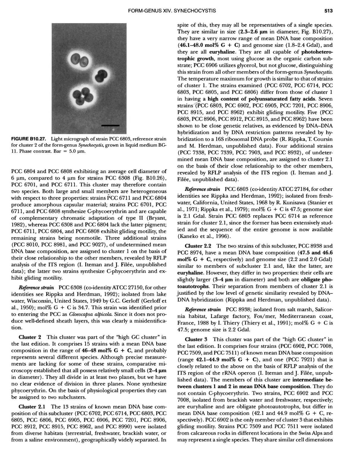
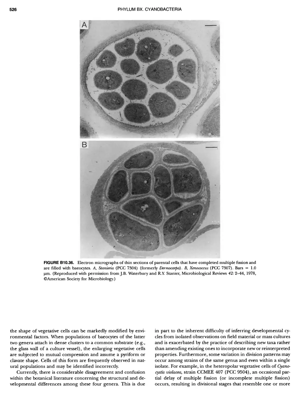
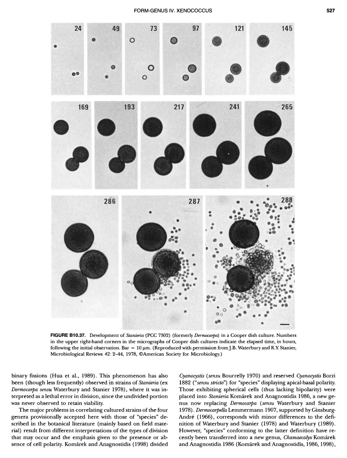
xvii CONTENTS PHYLUM BIX Deferrlbacteres Class I. Deferribacteres Order I. Deferribacterales ............. Family I. Deferribacteraceae ......... Genus I. Deferribacter Genus II. Flexisti pcs Genus III. Geovibrio ............. Genus Incertae Sedis I. Synergistes . 465 465 465 465 466 468 468 470 ~ ~ ~ ~ ~ ~ ~ ~ ~ ~ ~ ~ ~ ~ ~ 473 473 474 474 487 493 495 496 497 498 499 501 502 503 504 505 506 507 508 512 14 520 520 520 523 524 pie 528 528 1 533 539 542 543 543 544 544 546 547 548 550 553 554 554 Form-genus I. Arthrospira Form-genus I I. Borzia Eorm-genus III. Crinalium ...... Form-genus IV. Geitlerinemia Form-genus V. Leptolyngbya Form-genus Vl. Limnothrix ..... Form-genus Vll. Lyngbya Form-genus Vill. Microcoleus ... Form-genus IX. Oscillatoria Form-genus X. Planktothrix Eorm-genus XI. Prochlorothrix Form-genus XII. Pseudanabaena PHYLUM BX C yanobacteria ~ ~ ~ ~ ~ ~ ~ ~ ~ ~ ~ ~ ~ ~ ~ ~ ~ ~ ~ ~ ~ ~ ~ ~ ~ ~ ~ ~ ~ ~ ~ ~ ~ ~ ~ ~ ~ ~ ~ 0 ~ ~ ~ Oxygenic Photosynthetic Bacteria Oxygenic Photosynthetic Bacteria General Characteristics of the Cyanobacteria Phylogenetic Relationships Among the Cyanobacteria Based on 16S rRNA Sequences S ubsection I. ~ ~ ~ ~ ~ ~ ~ ~ ~ ~ ~ ~ ~ ~ 0 ~ ~ ~ ~ ~ ~ ~ ~ ~ ~ ~ ~ ~ ~ ~ ~ ~ ~ ~ ~ ~ ~ ~ ~ ~ ~ Eorm-genus I. Chamaesi phon Eorm-genus II. Chroococcus Form-genus III. Cyanobacterium ........................ Form-genus IV. Cyanobium Form-genus V. Cyanothece Form-genus Vl. Dactylococcopsis ....................... Form-genus Vll. Gloeobacter Form-genus Vill. Gloeocapsa .......................... Form-genus IX. Gloeothece Form-genus X. Microcystis ............................ Eorm-genus XI. Prochlorococcus Form-genus XII. Prochloron Form-genus XIII. Synechococcus Eorm-genus XIV. Synechocystis ubsection II. ~ ~ ~ ~ ~ 4 ~ ~ ~ ~ ~ ~ ~ ~ ~ ~ ~ ~ ~ ~ ~ ~ ~ ~ ~ ~ ~ ~ ~ ~ ~ ~ ~ 0 ~ ~ ~ ~ ~ ~ ~ Genera Reproducing by Multiple Fissions Only, or in Combination wit Limited (1-3) Binary Fissions Form-genus I. Cyanocystis ............................ Form-genus I I. Dermocarpella Form-genus III. Stanieria Form-genus IV. Xenococcus ........................... Genera in Which Extensive Vegetative Binary Fission Precedes Multi F iSSiOn ~ ~ ~ ~ ~ ~ ~ ~ ~ ~ ~ ~ ~ ~ ~ ~ ~ ~ ~ ~ ~ ~ ~ ~ ~ ~ ~ ~ ~ ~ ~ ~ ~ ~ ~ ~ ~ ~ ~ ~ ~ Form-genus I. Chroococcidiopsis Form-genus II. Myxosarcina ........................... Pleurocapsa-group gO ubsection III.
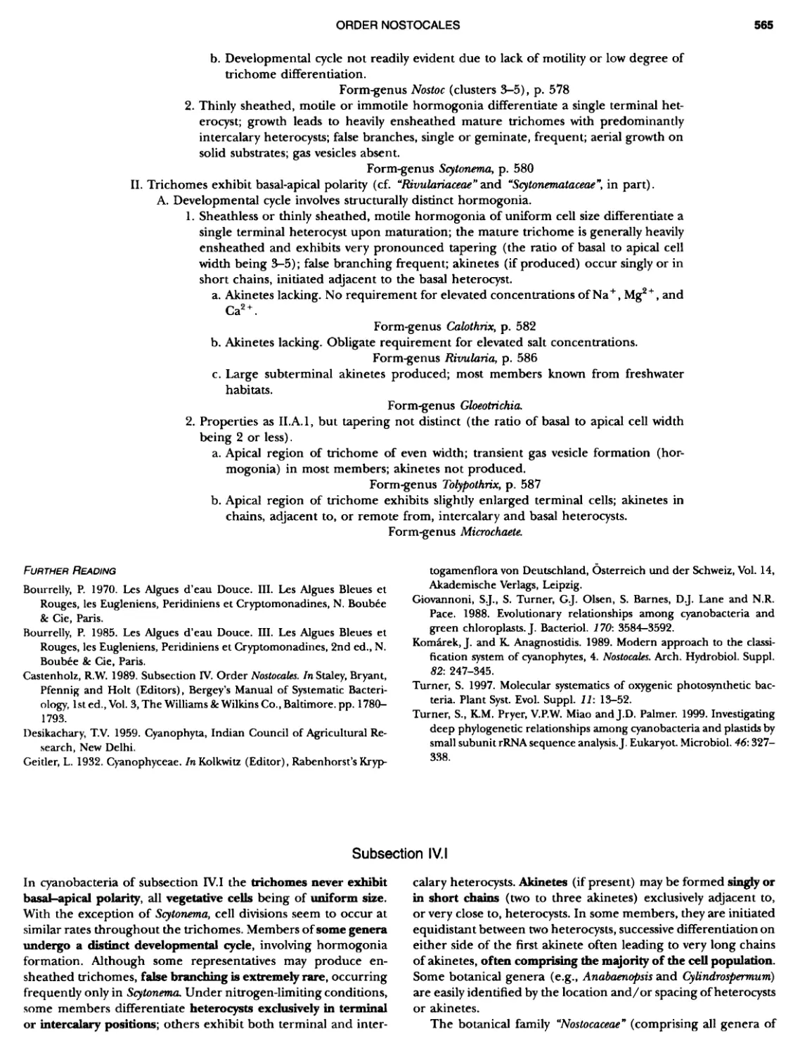
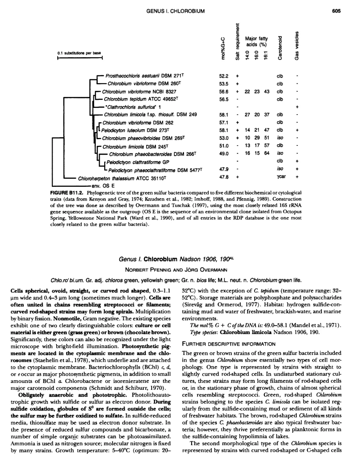
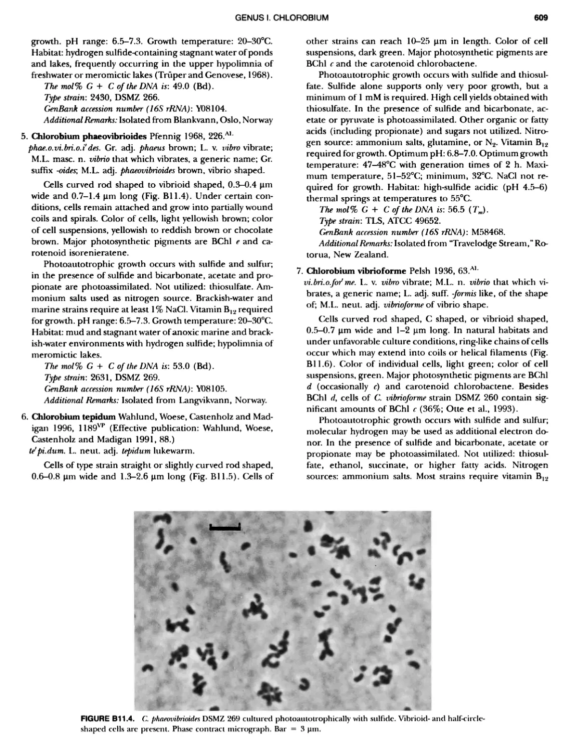
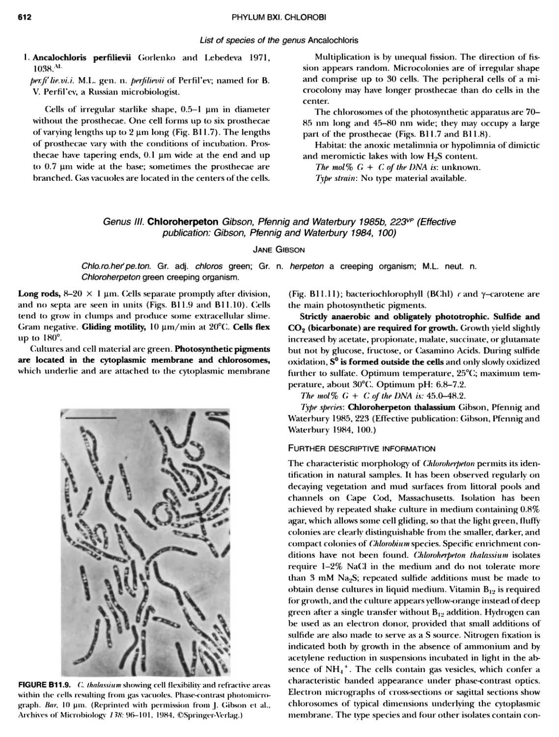
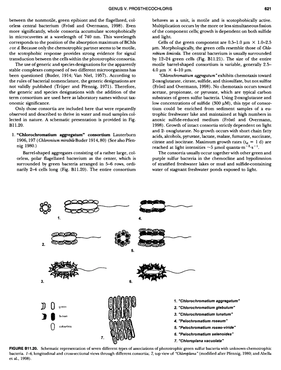
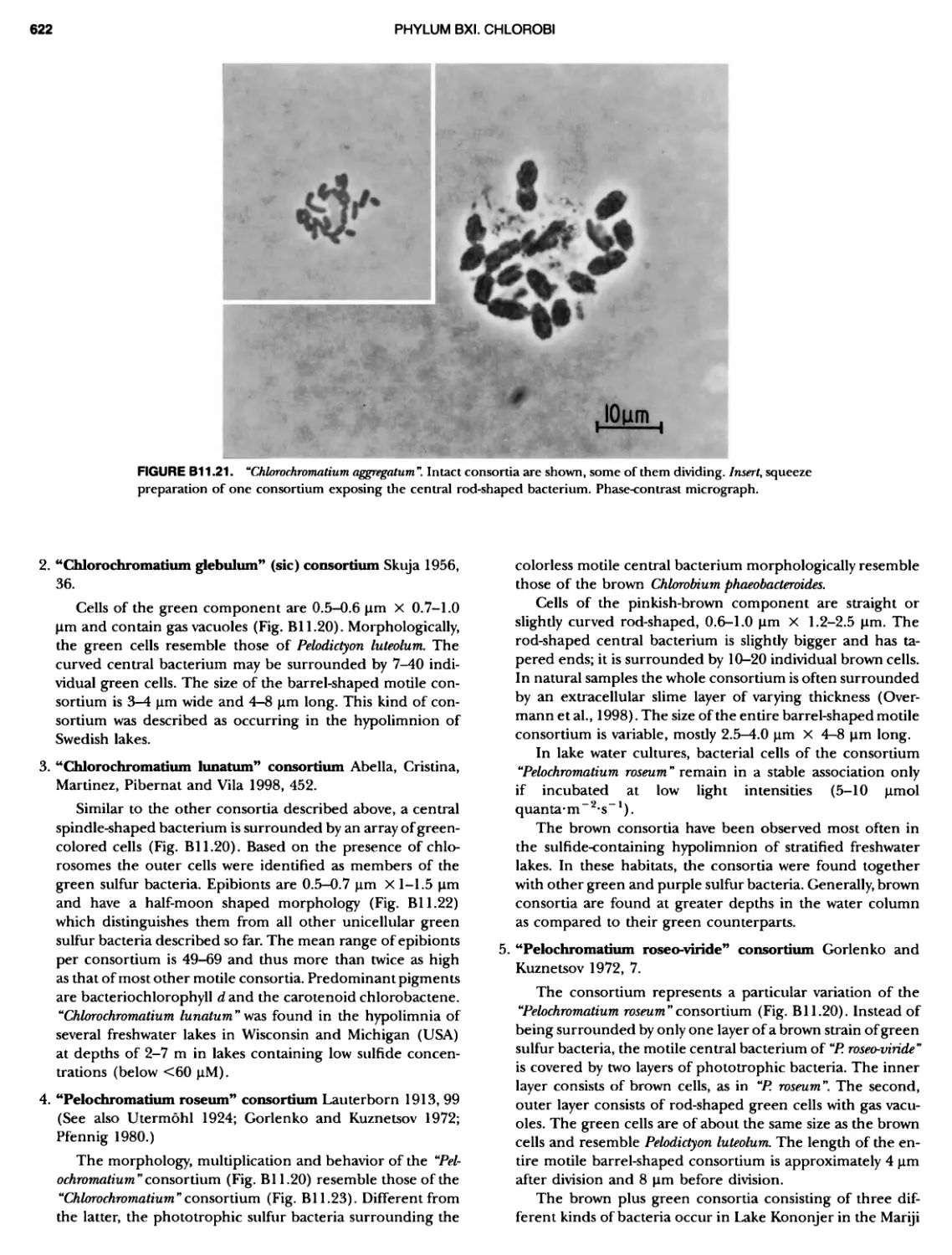
xviii CONTENTS 557 559 559 560 1 562 565 601 601 601 601 601 601 605 610 612 61 17 ~ ~ ~ ~ ~ ~ ~ ~ ~ ~ ~ ~ ~ ~ ~ ~ ~ ~ Green Sulfur Bacteria 620 625 625 625 625 626 629 629 630 631 639 703 ~ ~ ~ ~ ~ ~ ~ ~ ~ ~ ~ ~ ~ ~ ~ ~ ~ ~ ~ ~ ~ ~ Form-genus XIII. Spirulina Form-genus XIV. Starria ........ Form-genus XV. Sympioca ...... Form-genus XVI. Trichodesmium Eorm-genus XVII. Tychonema Subsection IV. Subsection IV.I Eorm-genus I. Anabaena Eorm-genus Ii. Anabaenopsis .... Form-genus III. Aphanizomenon .. Form-genus IV. Cyanospira Form-genus V. Cylindrospermopsis Form-genus VI. Cylindrospermum . Form-genus Vll. Nodularia Form-genus Vill. Nostoc Form-genus IX. Scytonema Subsection IV. II Form-genus I. Calothrix Form-genus II. Rivularia ........ Form-genus lil. Tolypothrix ...... Subsection V. Form-genus I. Chlorogloeopsis ... Form-genus II. Fischerella Form-genus III. Geitleria........ Eorm-genus IV. Iyengariella Form-genus V. Nostochopsis Form-genus Vl. Stigonema ...... PHYLUM BXI C hlorobi Class I. "Chlorobia ubclass I. ~ ~ ~ ~ ~ ~ ~ ~ ~ ~ ~ ~ ~ ~ ~ ~ ~ ~ ~ ~ ~ ~ ~ ~ ~ ~ ~ ~ ~ ~ ~ Order I. "Chlorobiales" ..................... Family I. "Chlorobiaceae" ................. Green Sulfur Bacteria Genus I. Chlorobium Genus II. Ancalochloris Genus II I. Chloroherpeton Genus IV. Pelodictyon Genus V. Prosthecochloris Addendum to the Green Sulfur Bacteria: Phototrophic Living In Consortia with Other Illcroorganiems. PHYLUM BXIII E irmicutes Class I. "Clostridia" Order I. "Clostridiales" Family Vl. "Heliobacteriaceae" Genus I. Heliobacterium ................ Genus II. Heliobacillus ................. Genus III. Heliophilum Genus IV. Heliorestis The Anoxygenic Phototrophic Purple Bacteria Bibliography Index of Scientific Names of Archaea and Bacteria .. 569 570 71 572 574 575 580 582 582 586 587 589 591 593 595 598 598 599
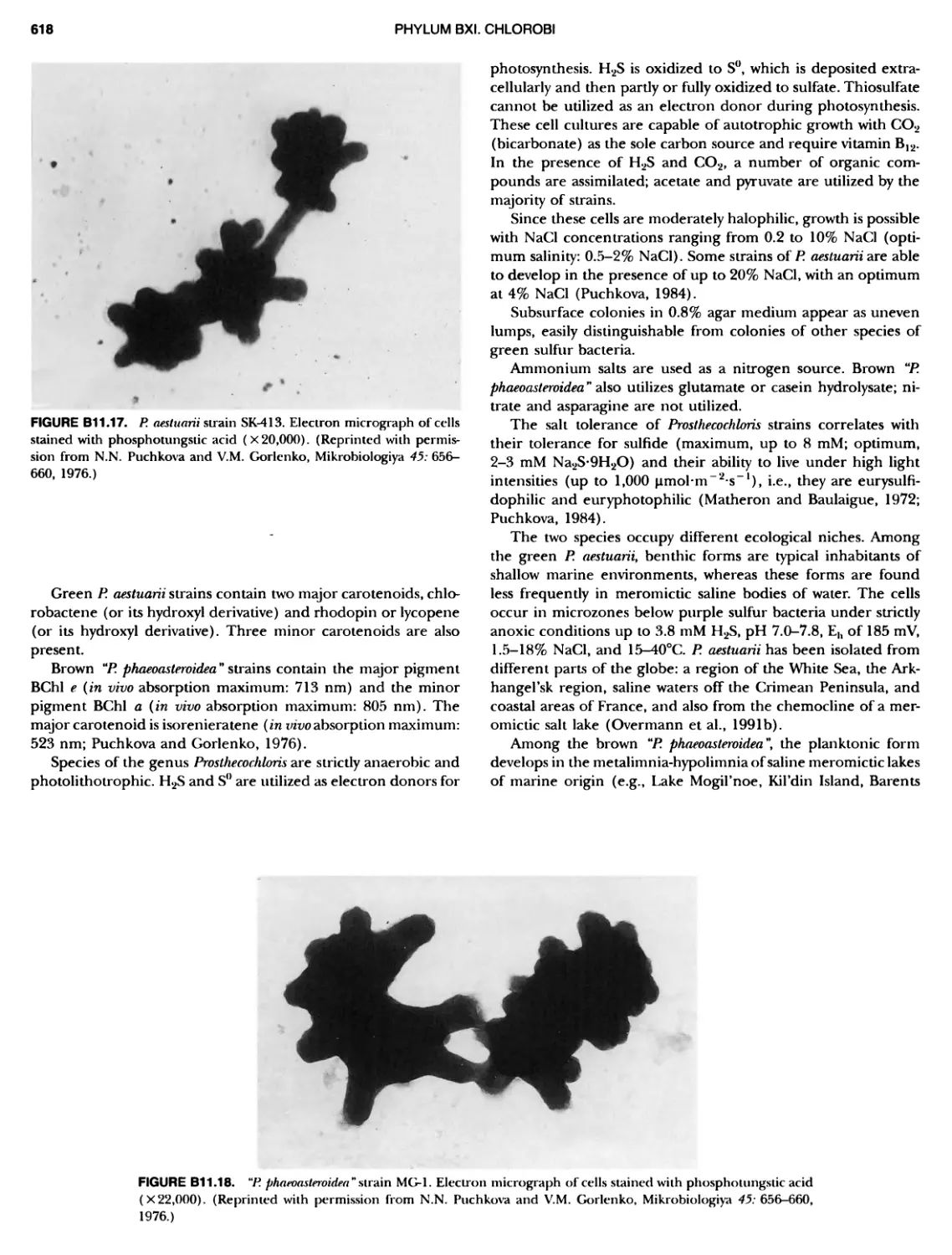
CohtrlbUtors XIX Milton J. Allison USDA, Agricultural Research Service — Midwest Area, National Animal Disease Center, Ames, IA 500104070, USA Rudolf Amann Nachwuchsgruppe Molekulare Okologie, Max Planck Institute fur Marine Mikrobiologie, Celsiusstrasse 1, D28359 Bremen, Ger-many Chad C. Baker Oregon Graduate Institute, P.O. Box 91000, Portland, OR 97291-1000, USA John R. Battista Department of Microbiology, Louisiana State University, Baton Rouge, LA 708034001, USA Eberhard Bock Inst. fur Allgemeine Botanik und Botanischer Garten, Universitat Hamburg, Ohnhorststrasse 18, Hamburg D-22609, Germany David R. Boone Department of Environmental Biology, Portland State University, Portland, OR 97207-0/51, USA Don J. Brenner Meningitis 8c Special Pathogens Branch Laboratory Section, Cen-ters for Disease Control 8t. Prevention, Atlanta, GA 30333, USA Frank Caccavo, Jr. Department of Microbiology, University of New Hampshire, Rud-man Hall/Spaulding, 46 College Road, Durham, NH 03824, USA Richard W. Castenholz Department of Biology, University of Oregon, Eugene, OR 97403-1210, USA Song C. Chong Department of Environmental Biology, Portland State University, Portland, OR 97207-0751, USA !Hilton S. da Costa Centro de Neurociencias, Departamento de Zoologia, Universi-dade de Coimbra, Apartado 3126, P-5004-517, Coimbra, Portugal Mary Ellen Davey Microbiology Department, Dartmouth Medical School, Room 202, Vail Bldg., North College Street, Hanover, NH 03755, USA Paul De Vos Department of Biochemistry, Physiology Bc Microbiology (WE 10V), K.L. Ledeganckstraat 35, B-9000, Gent, Belgium Wolfgang Eder Lehrstuhl fur Mikrobiologie, Universitat Regensburg, Universi-tatsstrasse 31, Regensburg 95053, Germany James G. Ferry Department of Biochemistry 8c Molecular Biology, The Pennsyl-vania State University, University Park, PA 168024500, USA Jean-Louis Garcia Laboratoire de Microbialogie, Universite de Provence, OR- STROM-ESIL-Case 925, 163, Avenue de Luminy, 13288 Marseille Cedex 9, France George IN. Garrlty Department of Microbiology and Molecular Genetics, Michigan State University, East Lansing, MI 48824-1101, USA Jane Gibson Section of Biochemis~, Molecular 8c Cell Biology, Division of Biological Sciences, Cornell University, Ithaca, NY 14853-0001, USA Monique Gillls Laboratorium voor Microbiologie en Microbiele Genetica (WE 10V), Rijksuniversiteit Gent, K.-L. Ledeganckstraat 35, B-9000 Gent, Belgium Vladimir M. Gorlenko Institute of Microbiology, Russian Academy of Sciences, Prospect 60-1etiya, Octyabrya 7 k.2, Moscow 117811, Russia William D. Grant Microbiology 8c Immunology, Leicester University, University Road, Leicester LE1 9HN, England Anthony C. Greene School of Biomolecular and Biomedical Science, Academic 1 Building, Logan Campus, Griffith University, Meadowbrook, Queensland 4131, Australia Doris Hafenbradl 10665 Sorrento Valley Road, San Diego, CA 92121, USA E. Claude Hatchikian IBSM-CNRS, Unite de Bioenergetique et Ingenierie des Protei-nes, 31, Chemin Joseph-Aiguier, 13402 Marseille Cedex 20, France Michael Herdman Physiologic Microbienne, Dept. B.M.G., CNRS-URA, 2172, Insti-tut Pasteur, 28 Rue du Docteur Roux, F-75724 Paris Cedex 15, France Lucien Hoffmann Laboratoire d'Algologie, de Mycologie et de Systematique Ex-perimentale, Institut de Botanique, B. 22, Universite de Liege, Sart Tilman, B-4000, Liege, Belgium John G. Holt Department of Microbiology and Molecular Genetics, Michigan State University, East Lansing, MI 48824-1101, USA Gertrud Huber Lehrstuhl fur Mikrobiologie, Universitat Regensburg, Universi-tatsstrasse 31, Regensburg, Germany Harald Huber Lehrstuhl fur Mikrobiologie, Universitat Regensburg, Universi-tatsstrasse 31, Regensburg, Germany Robert Huber Lehrstuhl fur Mikrobiologie, Universitat Regensburg, Universi-tatsstrasse 31, Regensburg, Germany
CONTRIBUTORS Yasuo Igarashi Department of Biotechnology, University of Tokyo, l-l-l Yayoi, Bunkyo-ku, Tokyo 113-8657, Japan Johannes F. Imhoff Institut fur Meereskunde, Universitat Kiel, Abt. Marine Mikro- biologie, Dusternbrooker Weg 20, D-24105 Kiel, Germany Masaharu Ishii Department of Biotechnology, The University of Tokyo, 1-1-1 Ya- yoi, Bunkyo-ku, Tokyo 118-8657,Japan Isabelle lteman Physiologic Microbienne, Dept. B.M.G., CNRS-URA 2172, Insti- tut Pasteur, 28 Rue du Docteur Roux, F-75724 Paris Cedex 15, France Takashi Itoh Japan Collection of Microorganisms, The Institute of Physical and Chemical Research, Riken, Hirosawa, Wako-shi, Saitama 351-0198, Japan Christian Jeanthon Universite de Bretagne Occidentale, Institut Universitaire Euro-peen de la Mer, 29680 Plousane, France D. Barrie Johnson School of Biological Sciences, University of Wales, Bangor LL57 2UW, United Kingdom Nlasahlro Kamekura Noda Institute for Scientific Research, 399 Noda, Noda-shi, Chiba-ken 278, Japan Toshiyuki Kawasumi Department of Food and Nutrition, Faculty of Home Economics, Women's University, 2-LI, Mejirodai, Bunkyo-ku, Tokyo, 112-8681,Japan Olga I. Keppen Department of Microbiology, Moscow State University, 119899 Moscow, Russia Karel Kersters Lab. voor Microbiologie, Rijksuniversiteit Gent, Vakgroep Bioch- ernie, Fysiologie en Microbiologie, K L. Ledeganckstraat 35, B- 9000, Gent, Belgium Jyoti Keswani 3157 Sylvan Circle, Morgantown, WV 26505, USA Hans-Peter Klenk Vp Genomics, Epidauros Biotechnology Inc., Am Neuland 1, D- 82347 Bernried, Germany Tetsuo Kobayashl Department of Applied Biological Sciences, Nagoya University, Lab. for Gene Regulation, School of Agricultural Sciences, Chi- kusa-ku, Nagoyawhi, Aichi 464-01, Japan Yosuke Koga Department of Chemistry, University of Occupational 8c Environ- mental Health, Fukuoka 807, Japan Torsten Krafft Am Grenzgraben 13, D-63067 OKenbach, Germany Noel R. Krleg Department of Biology, Virginia Polytechnic Institute Bc State L niversity, Blacksburg, VA 24061-0406, USA David P. Labeda USDA, National Center for Agricultural Utilization Research, Mi- crobial Properties Research, Peoria, IL 61604-3999, USA Thomas A. Langworthy Department of Microbiology, University of South Dakota School of Medicine, Vermillion, SD 57069-2390, USA Stbphane L'Haridon Universite de Bretagne Occidentale, Institut Universitaire Euro-peen de la Mer, 29680 Plousane, France Woltgang Ludwig Lehrstuhl fur Mikrobiologie, Technische Universitat Munchen, Am Hochanger 4, Freising, D-85350, Germany Barbara J. NlacGregor Civil Engineering Department, Northwestern University, Evans-ton, IL 60208, USA Joan Nl. Nlacy Department of Microbiology, LaTrobe University, Bundoora Vic-toria 3083, Australia Michael T. Madigan Department of Microbiology, Southern Illinois University, Mail Stop 6508, Carbondale, IL 62901-4399, USA Robert A. Mah Division of Environmental Health Science, UCLA School of Pub-lic Health, Los Angeles, CA 90024-1772, USA James S. Maki Department of Biology, WEHR Life Science Building, Marquette University, Milwaukee, WI 53201-1881, USA Terry J. IHcGenlty Department of Biological Sciences, University of Essex, Main Campus, Wivenhoe Park, Colchester, Essex CO4 5SQ, United Kingdom Roy D. Nleredith Ringoes Wertsville R, Hopewell, NJ 08525, USA Terry L. Miller Wadsworth Centre for Lab. 8c Research, New York State Depart-ment of Health, Albany, NY 12201-0509, USA Rafael Nlontalvo-Rodriguez University of Nebraska, Lincoln, NB, USA R.G.E. Murray Department of Microbiology Bc Immunology, The University of Western Ontario, London, Ontario N6A 5C1, Canada Takashi Nakase Japan Collection of Microorganisms, The Institute of Physical and Chemical Research, Riken, Wako-shi, Saitama 351-0198,Japan M. Fernanda Nobre Departmento de Zoologia, Universidade de Coimbra, Apartado 3126, P-3000 Coimbra, Portugal Norimichi Nornura Laboratory of Marine Microbiology, Division of Applied Biosci-ence, Graduate School of Agriculture, Kyoto University, Kyoto 606-8502,Japan Bernard Qlllvler Laboratoire de Microbiologie des Anaerobies, Universite de Prov-ence, CESS-ESIL ORSTOM, Case 921, 163 Avenue de Liminy, Marseille 13288 Cedex 9, France Aharon Oren Division of Microbial and Molecular Ecology, The Institute of Life Science, and the Moshe Shilo Minerva Center for Marine Biw geochemistry, The Hebrew University of Jerusalem, Givat Ram, Jerusalem 91904, Israel J5rg Overmann Institute fur Chemic und Biologic des Meeres (ICBM), Univer-sitat Oldenburg, Carl-von-Ossietzky-Strasse 9-11, Postfach 25 03, D-26111 Oldenburg, Germany Bharat K.C. Patel School of Biomolecular Bc Biomedical Sciences, Faculty of Sci-ence Bc Technology, GriIIith University, Nathan Campus, Bris-bane, Queensland 4111, Australia
CONTRIBUTORS XXI Girishchandra B. Patel National Research Council of Canada, Institute for Biological Sci- ences, Ottawa, Ontario K1A OR6, Canada Jerome J. Perry 3125 Eton Road, Raleigh, NC 27608-1113, USA Norbert Pfennig Primelweg 12, D-88662 Uberlingen, Germany Beverly K. Plerson Department of Biological Sciences, University of Puget Sound, 1500 N. Warner, Jones Hall ¹007, Tacoma, WA 98416, USA Fred A. Rainey Department of Microbiology, Louisiana State University, Baton Rouge, LA 70803, USA Anna-Louise Reysenbach Department of Environmental Biology, Portland State University, Portland, OR 972074751, USA Rosmarie Rippka Physiologic Microbienne, Dept. B.M.G., CNRS-URA 2172, Insti- tut Pasteur, 28 Rue du Docteur Roux, F-75724 Paris Cedex 15, France James A. Romesser Betz Dearborn, Inc. P.O. Box 4300, The Woodlands, TX 77380, USA Yoshihiko Sako Department of Applied Bioscience, Graduate School of Agricul- ture, Laboratory of Marine Microbiology, Kyoto University, Kyoto 606-8502, Japan Prlscilla C. Sanchez Museum of Natural History, University of the Philippines, Los Ba Atos, College, Laguna 4031, Philippines Abigail A. Salyers Department of Microbiology, University of Illinois-Urbana, Champaign, Urbana, IL 61801-5704, USA Karl-Heinz Schleifer Lehrstuhl fur Mikrobiologie, Universitat Munchen, Am Hochan- ger 4, D-85350 Freising, Germany Lindsay I. Sly Centre for Bacterial Diversity and Identification, Department of Microbiology, University of Queensland, St. Lucia, Brisbane, Queensland 4072, Australia Peter H.A. Sneath Department of Microbiology Sc Immunology, School of Medicine, University of Leicester, P.O. Box 138, Leicester LE1 9HN, En- gland Kevin R. Sowers Center for Marine Biotechnology, Maryland Biotechnology Insti- tute, Baltimore, MD 21202, USA Eva Spieck Inst. fur Allgemeine Botanik und Botanischer Garten, Universitat Hamburg, Ohnhorststrasse 18, Hamburg D-22609, Germany Stefan Spring Deutsche Sammlung von Mikroorganismen und Zellkulturen, GmbH, Mascheroder Weg 1b, D-58124 Braunschweig, Germany David A. Stahl Department of Civil Engineering/Technology Institute, North- western University, Evanston, IL 60208-3109, USA James T. Staiey Department of Microbiology, University of Washington, Seattle, WA 98195-0001, USA Karl O. Stetter Lehrstuhl fur Mikrobiologie, Universitat Regensburg, Universi- tatsstrasse 31, D-93053 Regensburg, Germany Ken-ichiro Suzuki Japan Collection of Microorganisms, The Institute of Physical and Chemical Research, Riken, Hirosawa, Wako-shi, Saitama 351- 0198,Japan Jean Swings Laboratorium voor Microbiologie, Universiteit of Gent, Vak- groep WE 10V, Fysiologie en Microbiologie, K.L. Ledeganckstraat 35, 8-9000, Gent, Belgium Xlnyu Tlan Institute of Microbiology, Academia Sinica, Beijing 100080, China Brian J. Tlndall Deutsche Sammlung von Mikroorgenismen und Zellkulteren, GmbH, Mascheroder Weg 1b, D-38124 Braunschweig, Germany Hans G. Truper Institute fur Mikrobiologie und Biotechnologie, Rheinsche Fried- rich-Wilhelms-Universitat, Mechenheimer Allee 168, W-53115 Bonn, Germany Peter Vandarnme Lab. voor Microbiologieen Microbiele Genetica, Universiteit of Gent, Faculteit Wetenschappen, K.L. Ledeganckstraat 55, B-9000 Gent, Belgium Antonio Ventosa Departamento de Microbiologia, Universidad de Sevilla y Paras- itologia, Facultad de Farmacia, Apdo. 874, 41080 Sevilla, Spain Russell H. Vreeland Department of Biology, West Chester University, West Chester, PA 19383, USA John B. Waterbury Woods Hole Oceanographic Institute, Woods Hole, MA 02543, USA William B. Whitman Department of Microbiology, University of Georgia, Athens, GA 30602-2605, USA Annlck Wllraotte Labo d'Algologique, Mycologie et Systematique Experimentale, Department de Botanique, B-22, Universite de Liege, ~000 Liege, Belgium Yi Xu Institute of Microbiology, Academia Sinica, Beijing 100080, China Gerhard Zellner Institute of Hydrology, GSF-National Research Center for Envi- ronment 8c Climate, Home address: Fuchsbergstrasse 7, D-85386 Eching, Germany Tatjana N. Zhlllna Institute of Microbiology, Russian Academy of Sciences, Prospect 60-letja Oktyabrya 7a, Moscow 117812, Russia Peljln Zhou Institute of Microbiology, Academia Sinica, Beijing 100080, China Wolfram Zllllg Max-Planck-Institut fur Biochernie, Am Klopferspitz 18a, D-82152 Martinsried, Germany Stephen H. Zinder Department of Microbiology, Cornell University, Ithaca, NY 14853-0001, USA
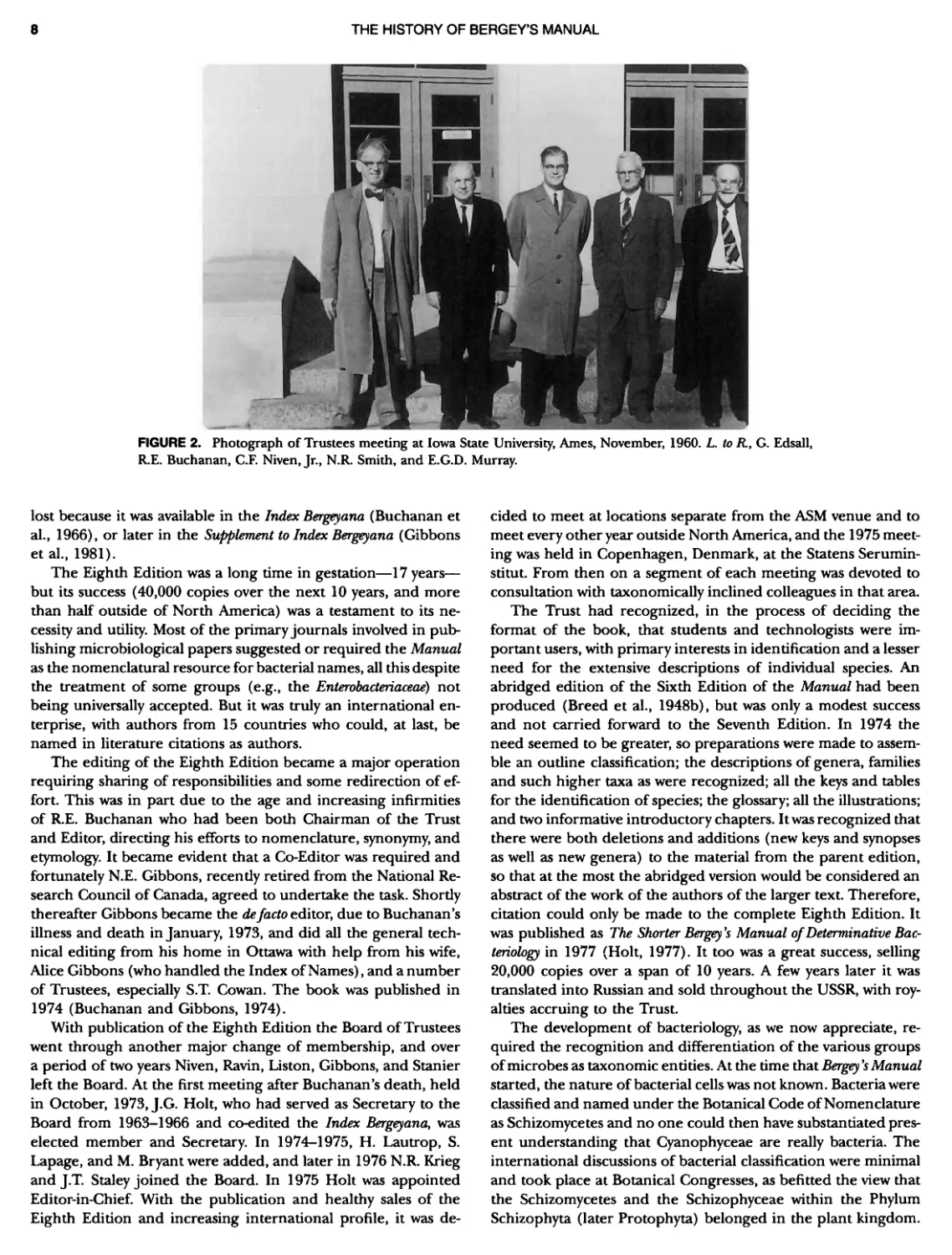
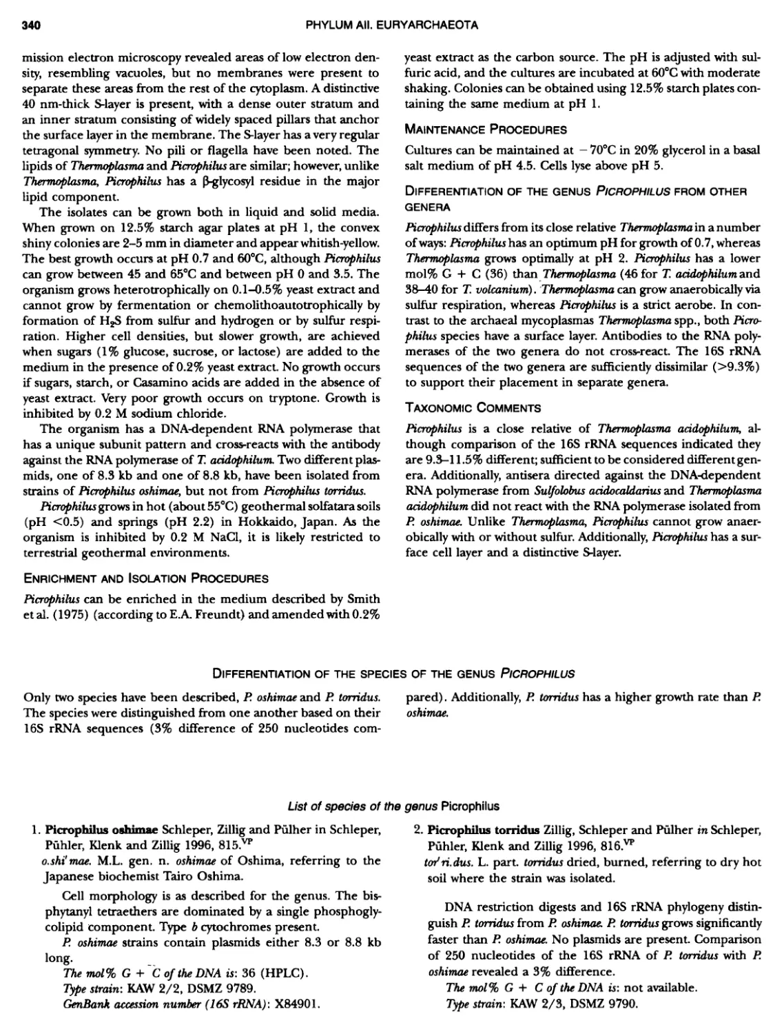
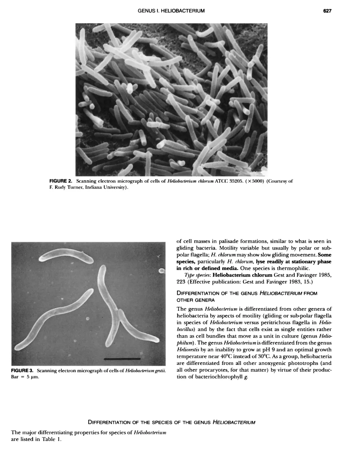
The History of Bergey's ManualR.G.E. Murray and John G. Holt I NTRODUCTION Bergey's Manual of Deterrrtinatiue Bacters'ology has been the major provider of an outline of bacterial systematics since it was initiated in 1923 and has provided a resource ever since to workers at the bench who need to identify bacterial isolates and recognize new species. It originated in the Society of American Bacteriologists (SAB) but it has since become a truly international enterprise directed by an independent Trust which was founded in 1936. It has gone through nine editions and has generated, as a more comprehensive resource, a unique compendium on bacterial sys- tematics, Bergey's Manual of Systematic Bactersology (Holt et al., 1984-1989), which now enters its second edition. A number of dedicated bacteriologists (Table 1) have formed, guided the development of, and edited, each edition of Beryl's ManuaL Many of these individuals have been well known for activity in their national societies and devotion to encouraging worldwide cooperation in bacteriology and particularly bacterial taxonomy. Some of them worked tirelessly on the international stage towards an effective consensus in taxonomy and common approaches to classification. This led to the formation in 1950 of an International Association of Microbiological Societies (IAMS) holding regular Congresses. The regulation of bacterial taxonomy became possible within IAMS through an Interna- tional Committee on Systematic Bacteriology (ICSB), thus rec- ognizing the need for international discussions of the problems involved in bacterial systematics. Eventually, the need for a Code of Nomenclature of Bacteria was recognized and was published in 1948 (Buchanan et al., 1948), and a Judicial Commission ( JC) was formed by ICSB to adjudicate conflicts with the Rules. Despite these efforts, an enormous number of synonyms and illegitimate names had accumulated by the 1970s and were an evident and major problem for the Editor/Trustees of Bergey's Manual and for all bacteriologists (Buchanan et al., 1966; Gibbons et al., 1981). A mechanism for recognizing useful, and abandoning useless, names was accomplished by the ICSB and the JC largely due to the insistent arguments of V.B.D. Skerman. Lists were made based on the names included in the Eighth edition of Bergey's Manual of Determinatiue Bacteriology (Buchanan and Gib- bons, 1974), because they had been selected by expert commit- tees and individual author/experts, together with the recom- mendations of subcommittees of ICSB. The results were (1) the published Approved Lists of Bacterial Names (Skerman et al., 1980); (2) a new starting date for bacterial names of January 1, 1980 to replace those of May 1, 1758; (8) freeing of names not on the Approved Lists for use in the future; and (4) definition in the Bacteriological Code (1976 revision; Lapage et al., 1975) of the valid and invalid publication of names. It is now evident that the care and thought of contributors to Berg@'s Manual over the years played a major part in stimulating an orderly nomen- clature for taxonomic purposes, in the development of a useful classification of bacteria often used as a basal reference, and in providing a continuing compendium of descriptions of known bacteria. The Manual started as a somewhat idiosyncratic assembly of species and their descriptions following the interests and prej- udices of the editor/authors of the early editions. Following the formation of the Bergey's Manual Trust in 1936 and the inter- national discussions of the ICSB at Microbiological Congresses, the new editions became more and more the result of a consensus developed by advisory committees and specialist authors for each TABLE 1. Members of the Board of Trustees David H. Bergey David R. Boone Robert S. Breed Don J. Brenner Marvin P. Bryant R.E. Buchanan Richard W. Castenholz Harold J. Conn Samuel T. Cowan Geoffrey Edsall George M. Garrity Norman E. Gibbons Bernard W. Hammer Francis C. Harrison A. Parker Hitchens John G. Holt Frank M. Huntoon Noel R. Krieg Stephen P. Lapage Hans Lautrop John Liston A.G. Lochhead James W. Moulder E.G. D. Murray R.G. E. Murray Charles F. Niven, Jr. Norbert Pfennig Arnold W. Ravin Karl-Heinz Schleifer Nathan R. Smith Peter H.A. Sneath James T. Staley Roger Y. Stanier Joseph G. Tully Jan Ursing Stanley T. Williams 1925-1957 1994- 1925-1957 (Chairman 1957-1956) 1979- 1975-1986 1951-1973 (Chairman 1957-1975) 1991- 1948-1965 late 1950s-1974 late 1950s-1965 1997- 1965-1976 1925-1954 1925-1934 1939-1950 1975- 1925-1954 1976-1991, 1996- 1975-1978 197W1979 1965-1976 (Chairman 1973-1976) late 1950s-1960 1980-1989 1934-1964 1964-1990 (Chairman 1976-1990) Late 1950s-1975 1978-1991 1962-1980 1989- 1950-1964 1978-1994 (Chairman 1990-1994) 1976- 1965-1975 1991-1996 1991-1997 1989- (Chairman 1994-)
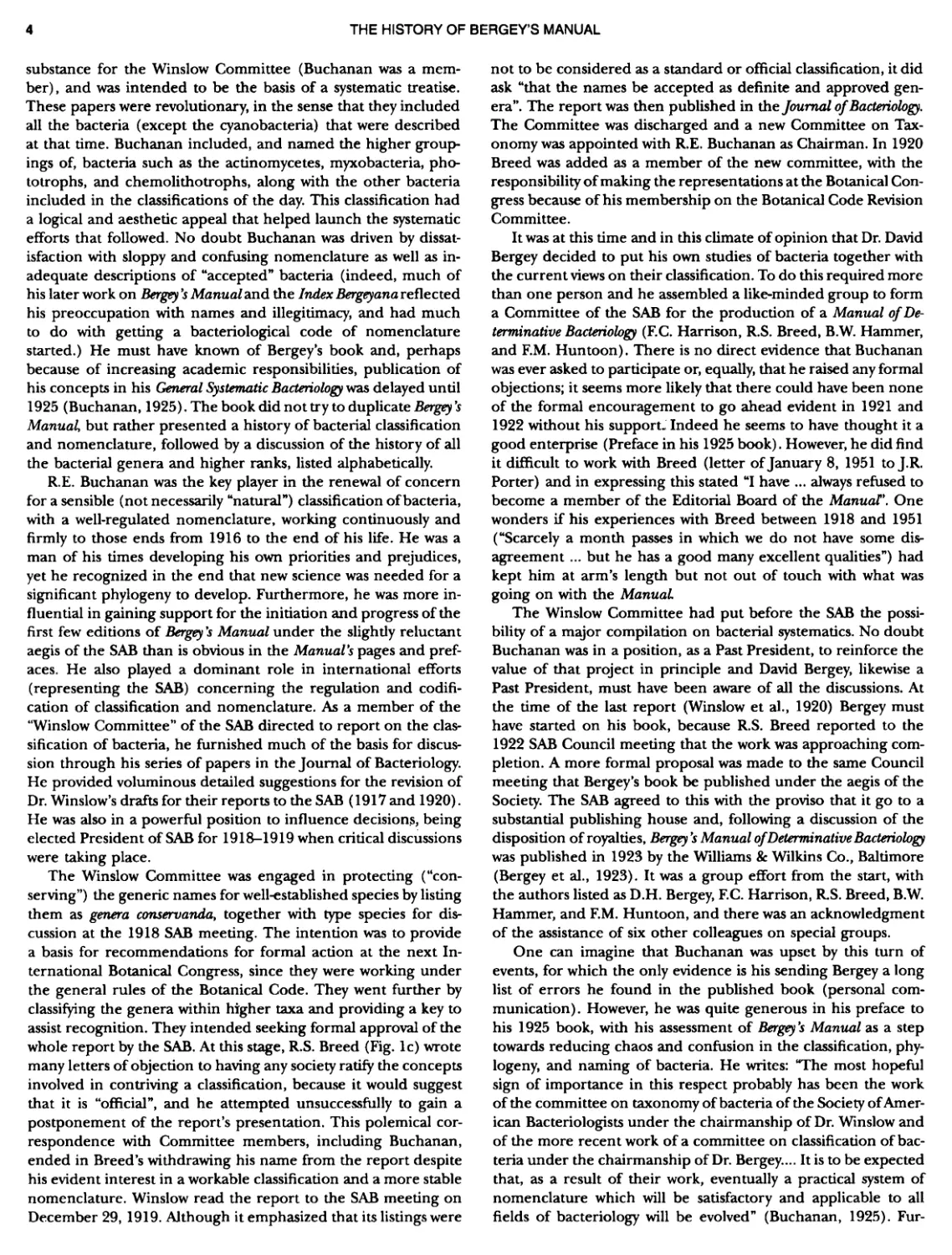
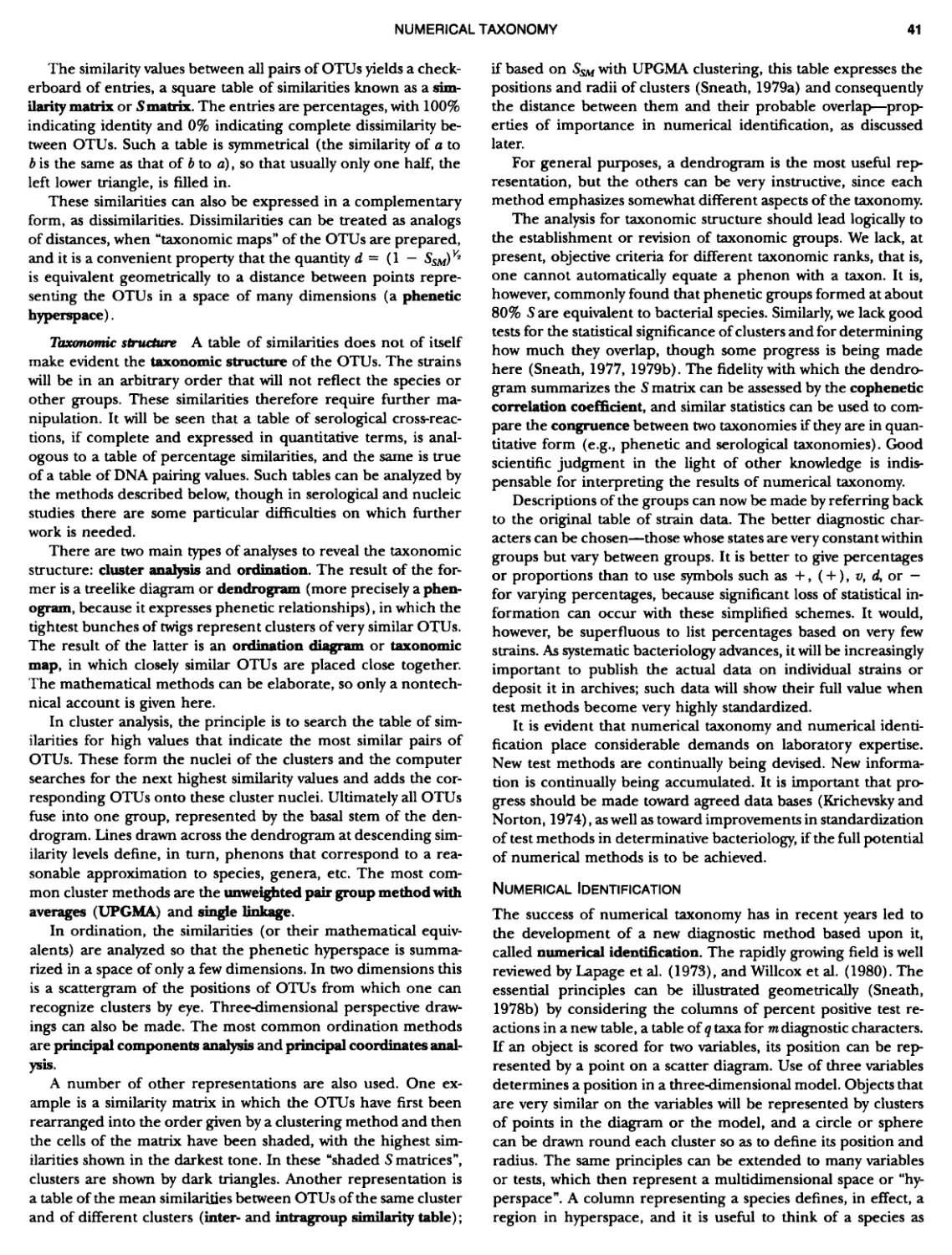
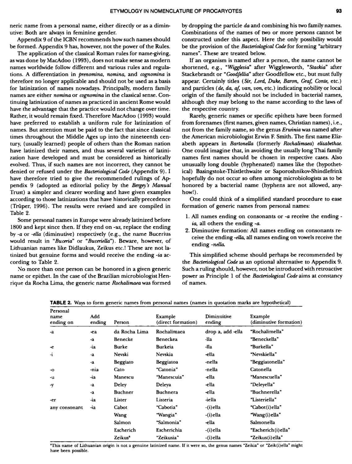
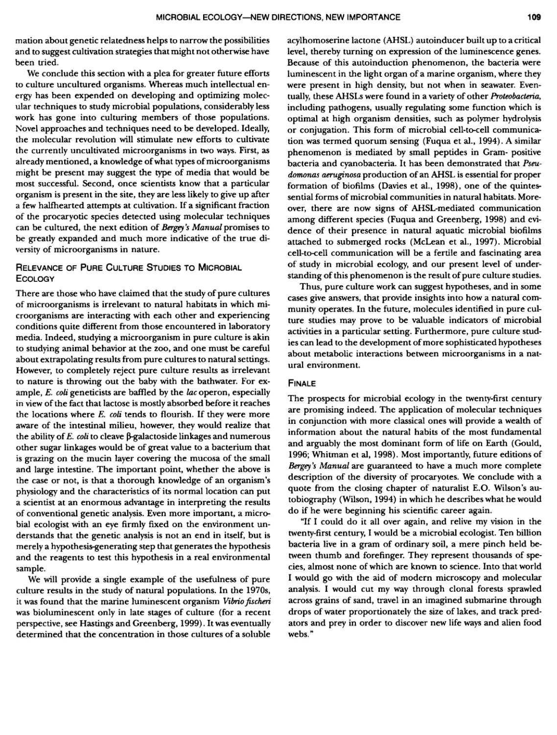
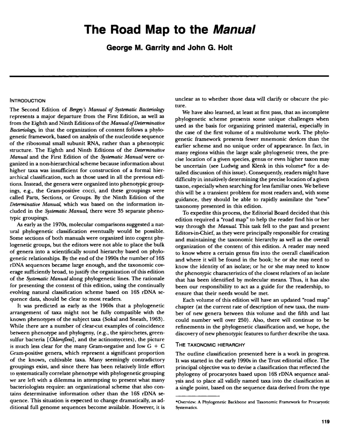
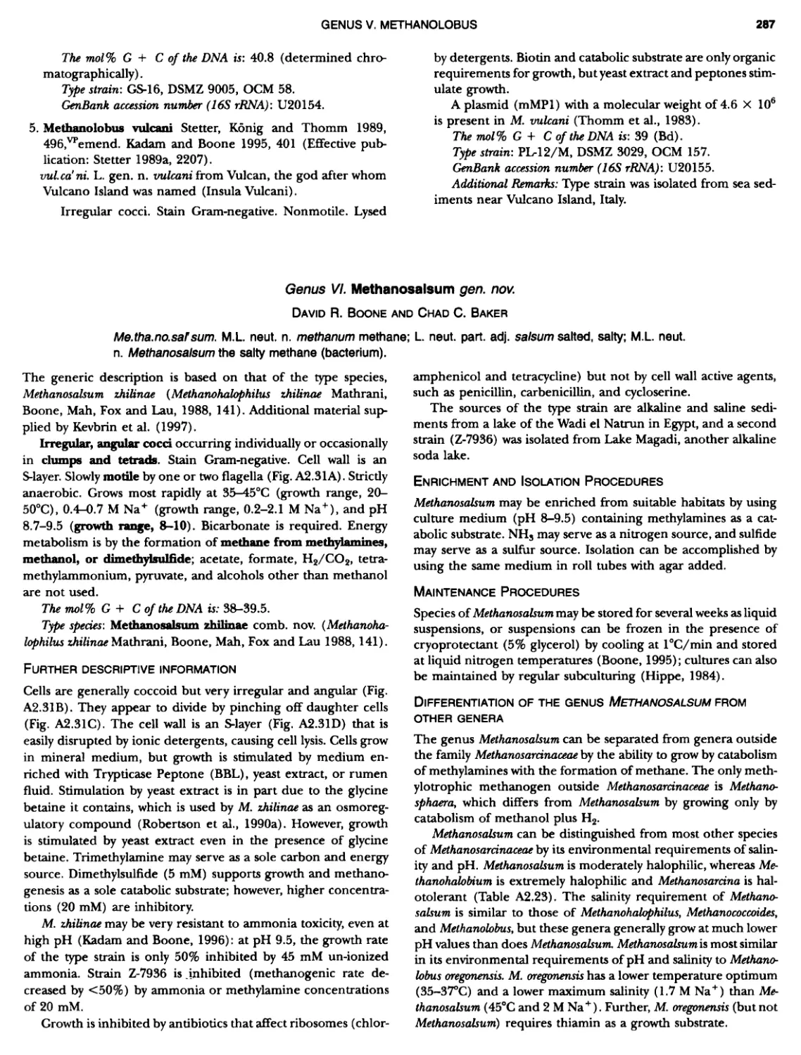
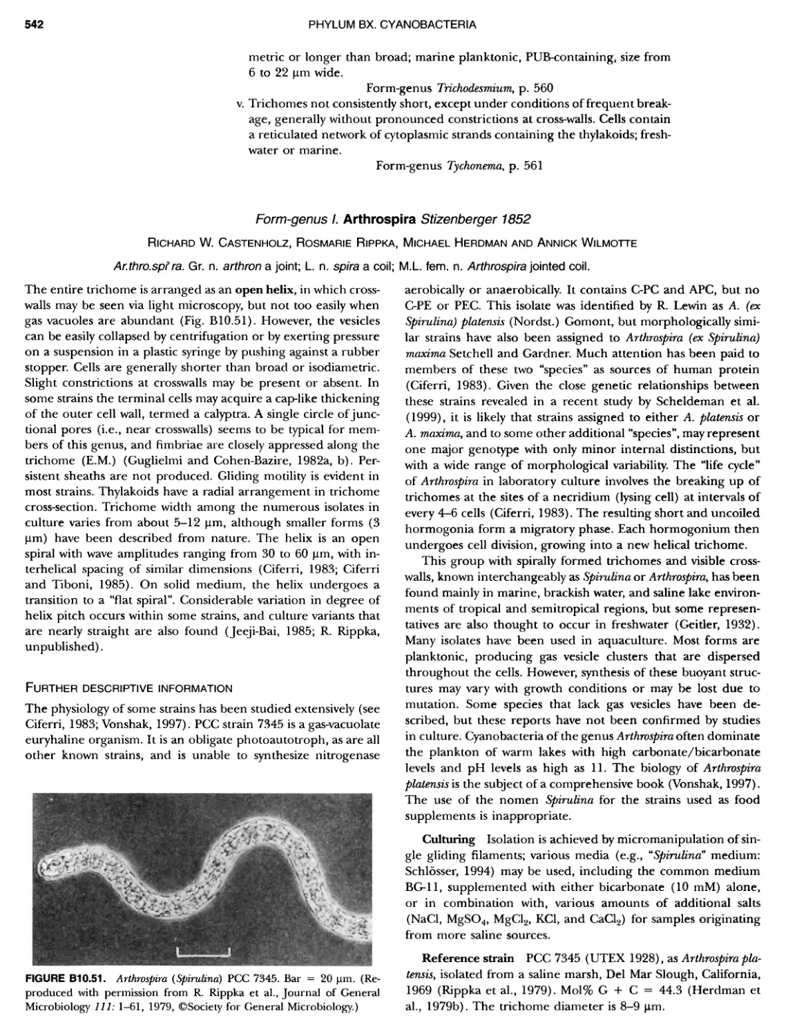
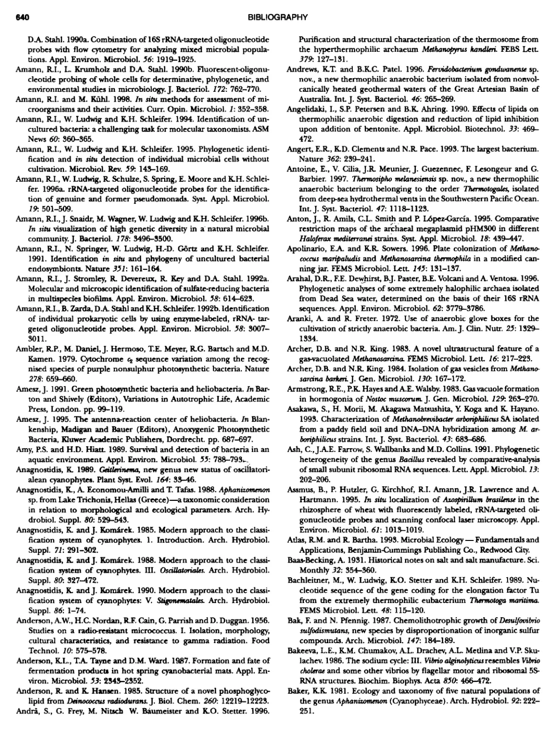
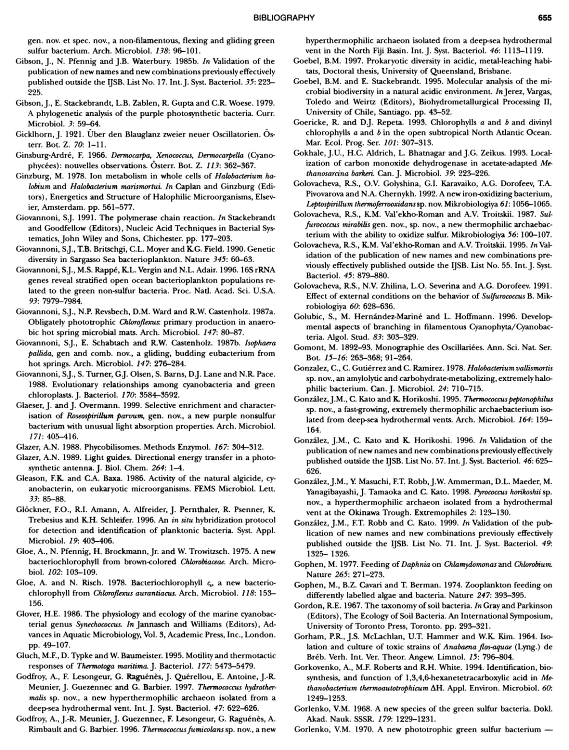
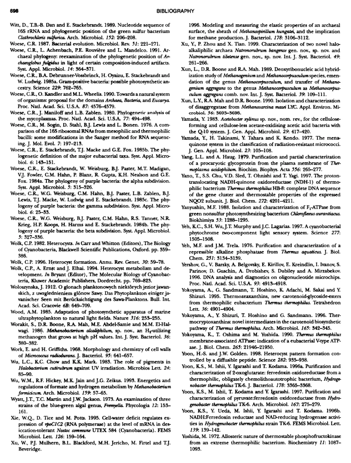
THE HISTORY OF BERGEY'S MANUAL part or chapter of the volumes. This did not happen all at once; it developed out of practice and trials, and it is stil1 developing as the basic sciences afFecting taxonomy bring in new knowledge and new understanding of taxa and their relationships. ANTECEDENTS OF BERGEY S MANUAL Classification of named species of bacteria did not arise quickly or easily (Buchanan, 1948). The Linnaean approach to naming life forms was adopted in the earliest of systems, such as Muller's use of Vibno and Monas (Muller, 1773, 1786), for genera of what we would now consider bacteria. There were few observations, and there was insuBicient discrimination in the characters avail- able during most of the nineteenth century to allow any system, even the influential attempts by Ehrenberg (1838) and Cohn (1872, 1875), to provide more than a few names that still survive (e.g. Spirillum, Spirochaeta, and Bacillus). Most descriptions could rest only on shape, behavior, and habitat since microscopy was the major tool. Muller's work was the beginning of the descriptive phase of bacteriology, which is still going on today because we now realize that the majority of bacteria in nature have not been grown or characterized. Early observations such as Muller's were made by cryptogamic botanists studying natural habitats, usually aquatic, and who usually gave Linnaean binomials to the objects they described microscopically. The mycologist H.F. Link (1809) de- scribed the first bacterium that we still recognize today, which he named Polyanyum uitellinum and is now placed with the fruit- ing myxobacteria. Bizio (1825) attempted to explain the occur- rence of red pigment formation on starchy foods such as polenta as the result of microbial growth and named the organism he found there Sermtia marcescens, a name now associated with the prodigiosin-producing Gram-negative rod. Perhaps one of the most significant observers of infusoria in the early nineteenth century was C.G. Ehrenberg, who described many genera of algae and protozoa and, coincidentally, some bacteria (Ehrenberg, 1838). He named genera such as Spirochaeta and Spirillum, still recognized today, and Bacterium, which became a catch-all for rod-shaped cells, and was made >~men rejiciendum in 1947. Logical classifications were attempted throughout the nine- teenth century and that of Ferdinand Cohn (1872, 1875), with his attempts to classify the known bacteria, was most influential. In his 1872 paper Cohn recognized six genera of bacteria (Mi- crococcus, Bacterium, Bacillus, Vibrio, Spirillum, and Spirochaeta) and later (1875) expanded the classification to include the cyano- bacteria while adding more bacterial genera (Sarnna, Ascococcus, Leptothrix, Beggiatoa, Cladothrix, Crenothrix, Streptococcus [not those recognized today], and Streptothrix) Buchanan (192.5) suggested that Cohn's 1875 classification could be the starting date for bacterial nomenclature instead of Linnaeus' Species Plantarum of 1753 and discussed various ideas for the proper starting date for bacterial nomenclature, anticipating by a quarter of a century the actual change in starting date proposed in the revised Bac- teriological Code (Lapage et al., 1975). The realization that cul- tivation was possible, and the development of pure culture tech- niques, extended enormously the capability to recognize and describe species by adding their growth characteristics and effects on growth media. The vague possibilities of pleomorphism gave way to a concept of fixity of species. All this was aided by the human preoccupation with health, the seriousness of infectious diseases, and the growing awareness of the association of partic- ular kinds of bacteria with particular diseases. The result was a rapid increase in the number of taxonomic descriptions and the recognition that similar but not identical species of bacteria were to be found both associated with higher life forms and more generally distributed in nature. Between 1885 and 1910 there were repeated attempts at clas- sification and arrangements based on perceived similarities, mostly morphological. There were genuine attempts to bring order out of chaos, and a preliminary publication often stimu- lated subsequent and repeated additions and revisions, but all these authors neglected the determinative requirements of bac- teriology. Some notable examples were Zopf (1885), Flugge (1886), Schroeter (1886), and Trevisan (1887, 1889) . Migula pro- duced his first outline in 1890 and new versions in 1894, 1895, 1897, and 1900; others followed, notably Fischer (1895), and importantly, because of a degree of nomenclatural regularity, Lehmann and Neumann published their atlas in 1896. The latter was probably the most successful of the systems and was used in successive editions until 1950, especially in Europe. All these were important in their time. However, a major influence in the sub- sequent development of Bergey's Manual in the environment of the Society of American Bacteriologists (SAB) was the work of F.D. Chester, who produced reports in 1897 and 1898 of bacteria of interest in agriculture, to be followed in 1901 by his Manual of Determinative Bacteriology Cheste.r had recognized that the lack of an organized assembly of descriptions and a scheme of clas- sification made the identification of isolates as known species and the recognition of new species an insurmountable task. An- other classification provided by OrlaJensen (1909, 1919) was infiuential because it represented an interpretation of "natural relationships", reflecting a more physiological approach to de- scription based on his own studies of the lactic acid bacteria encountered in dairy bacteriology. He delimited genera and spe- cies on the basis of characteristics such as metabolic byproducts, fermentation of various sugars, and temperature ranges for growth, in addition to morphology. Most classifications to that time reflected the idiosyncrasies of the authors and their areas of experience. What was yet to come was the ordering of assem- blies of all known bacteria, arranged with properties documented to facilitate determination and presenting continuing trials of hierarchical arrangements; it was in that format that Beryy 's Man- ual started. STEPS LEADING TO THE FIRST EDITION OF THE MANUAL Bergs 's Manual of Determinative Bacteriology arose from the interest and efforts of a group of colleagues in the Society of American Bacteriologists, who were fully aware of previous attempts to sys- tematize the information available on bacterial species and who recognized that the determination of bacterial identity was dif- ficult and required extensive experience. A committee was formed with C.-EA. Winslow as chairman and J. Broadhurst, R.E. Buchanan, C. Krumweide Jr., LA. Rogers, and G.H. Smith as members. Their discussions at the meetings of the SAB and their reports, which were published in the Journal of Bacteriology (Winslow et al., 1917, 1920), were signposts for future efForts in systematics. There were two "starters" for a ManuaL R.E. Bu- chanan (Fig. la), a rising star in the bacteriological firmament, and President of the SAB in 1918, working at Iowa State College, and D.H. Bergey (Fig. Ib), a senior and respected bacteriologist and President of the && for 1915, working at the University of Pennsylvania. Between 1916 and 1918 Buchanan wrote ten papers entitled "Studies on the nomenclature and classification of the bacteria" (Buchanan, 1916; 1917a, b, c; 1918a, b, c, d, e, f) which provided
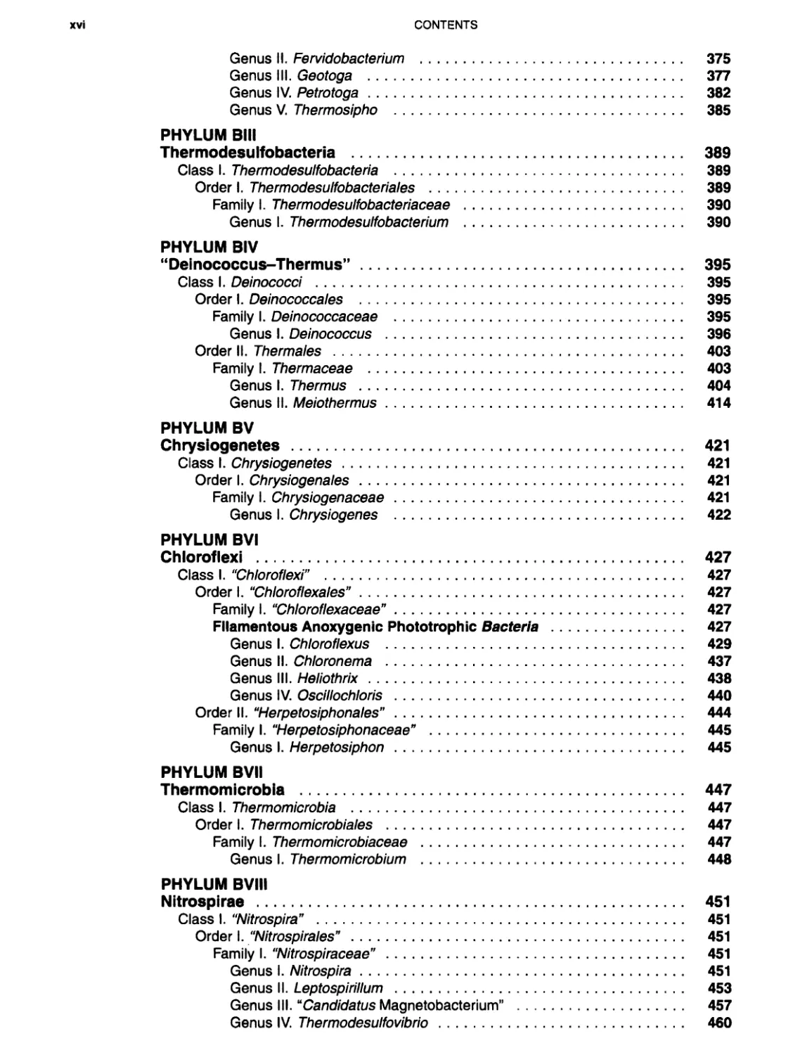
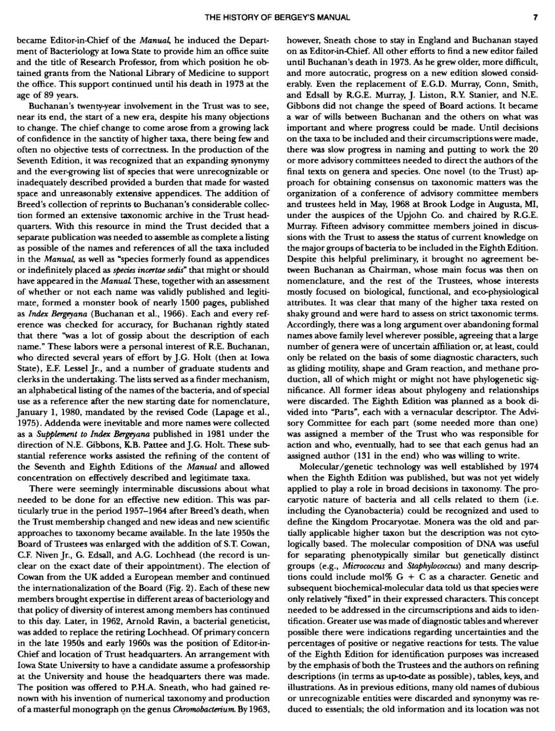
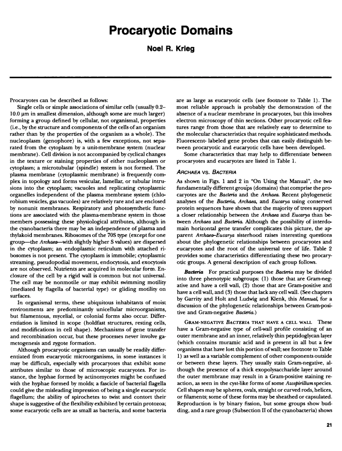
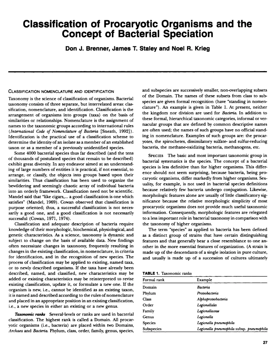
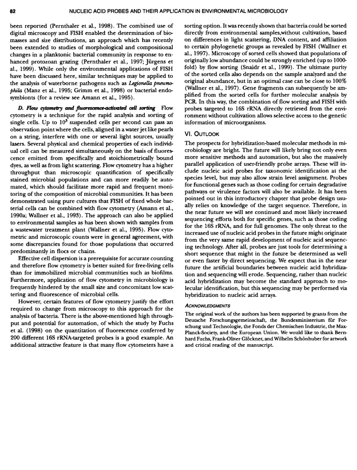
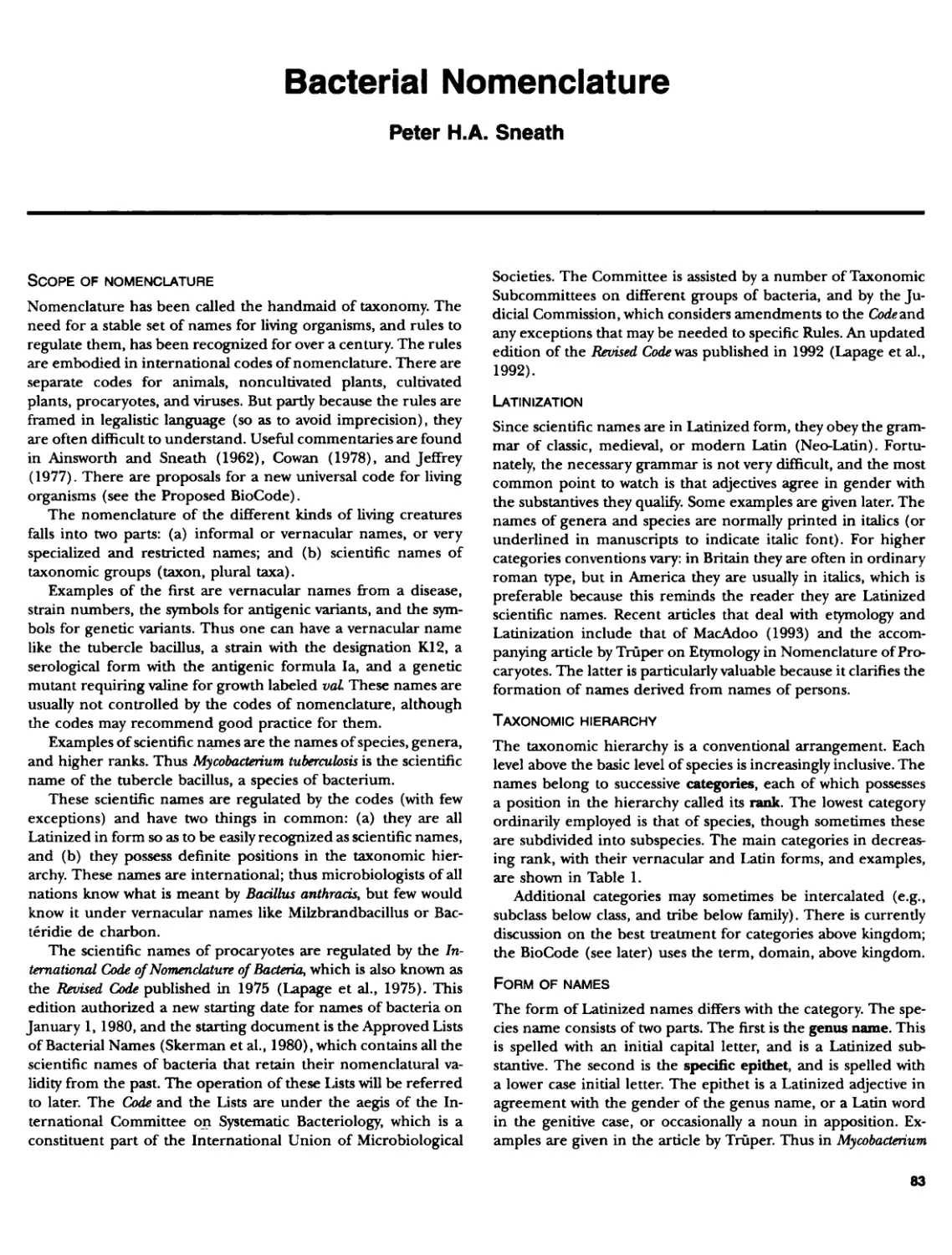
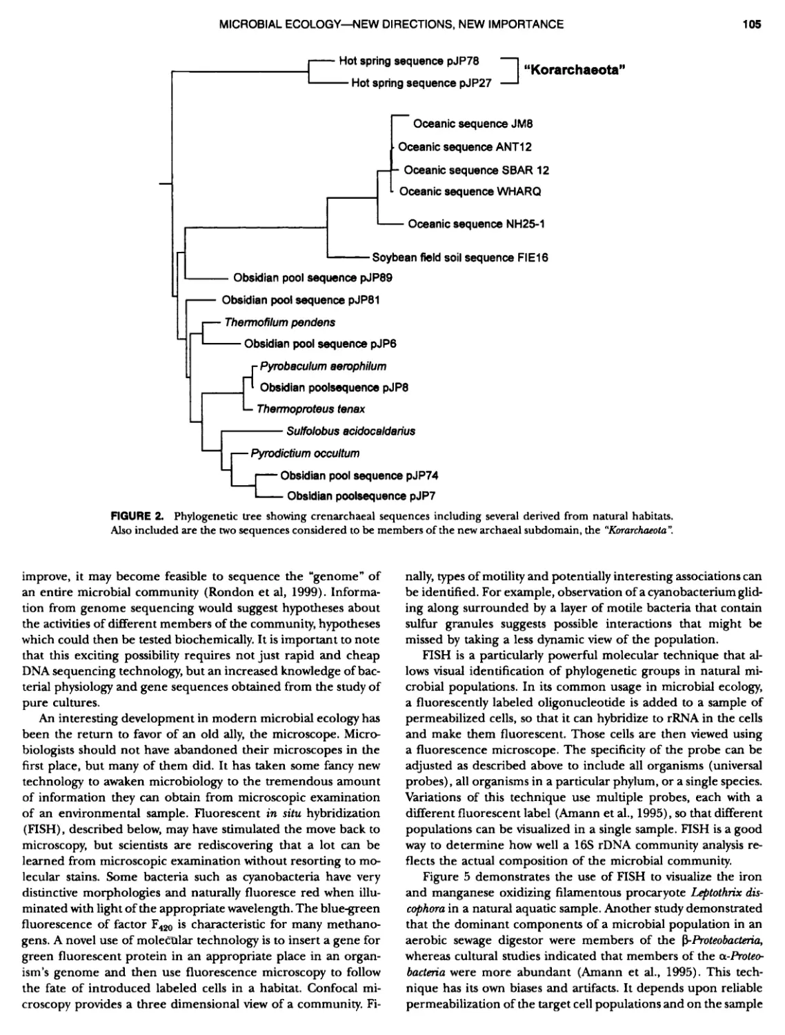
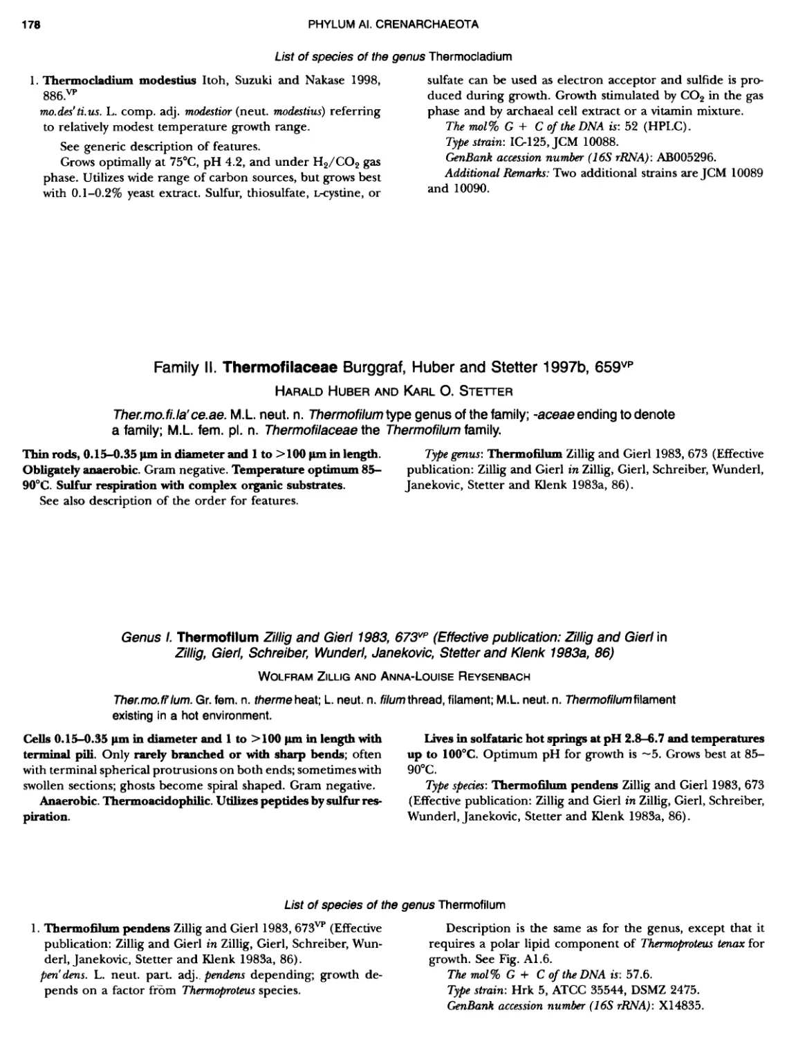
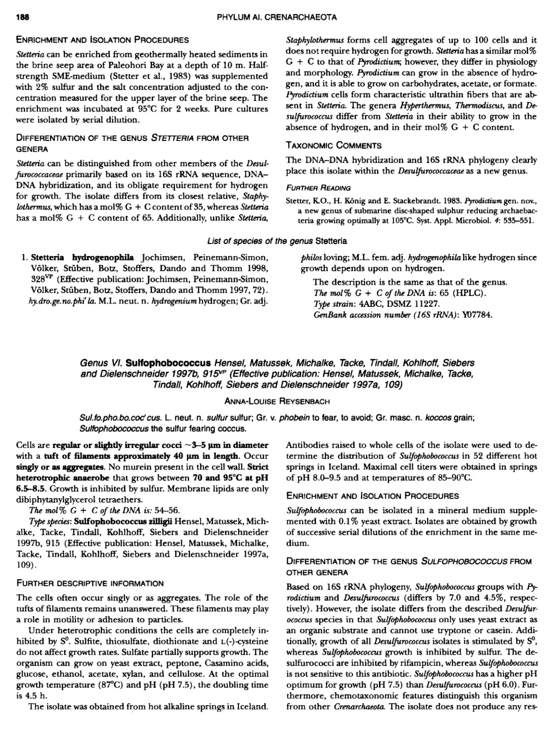
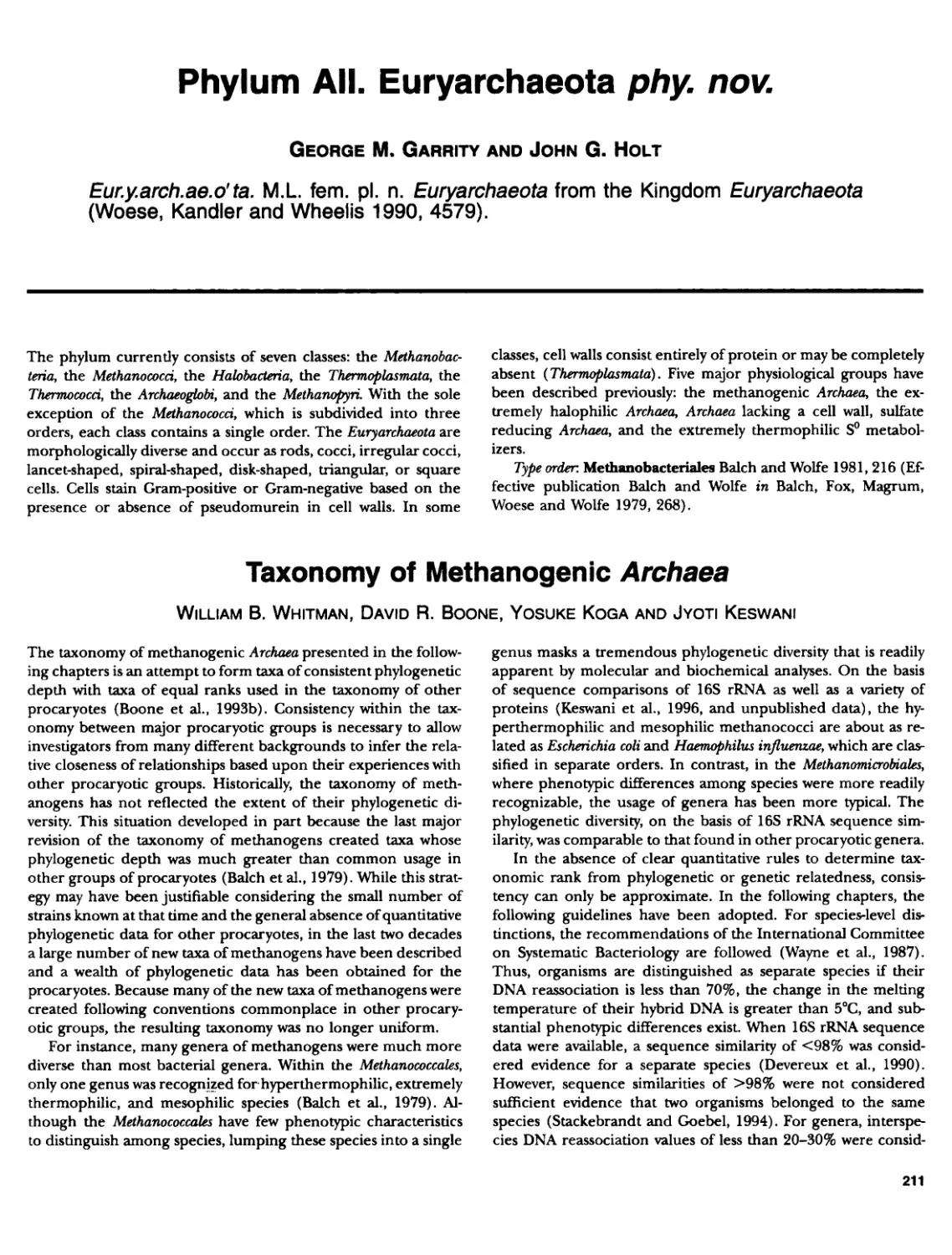
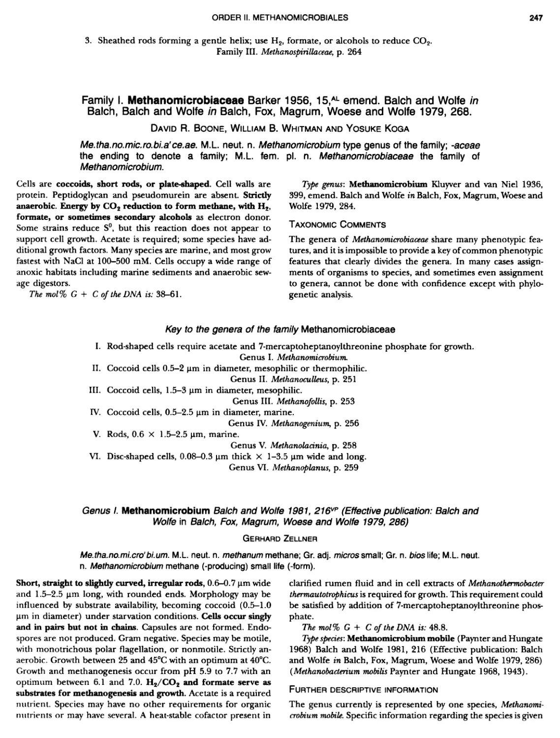
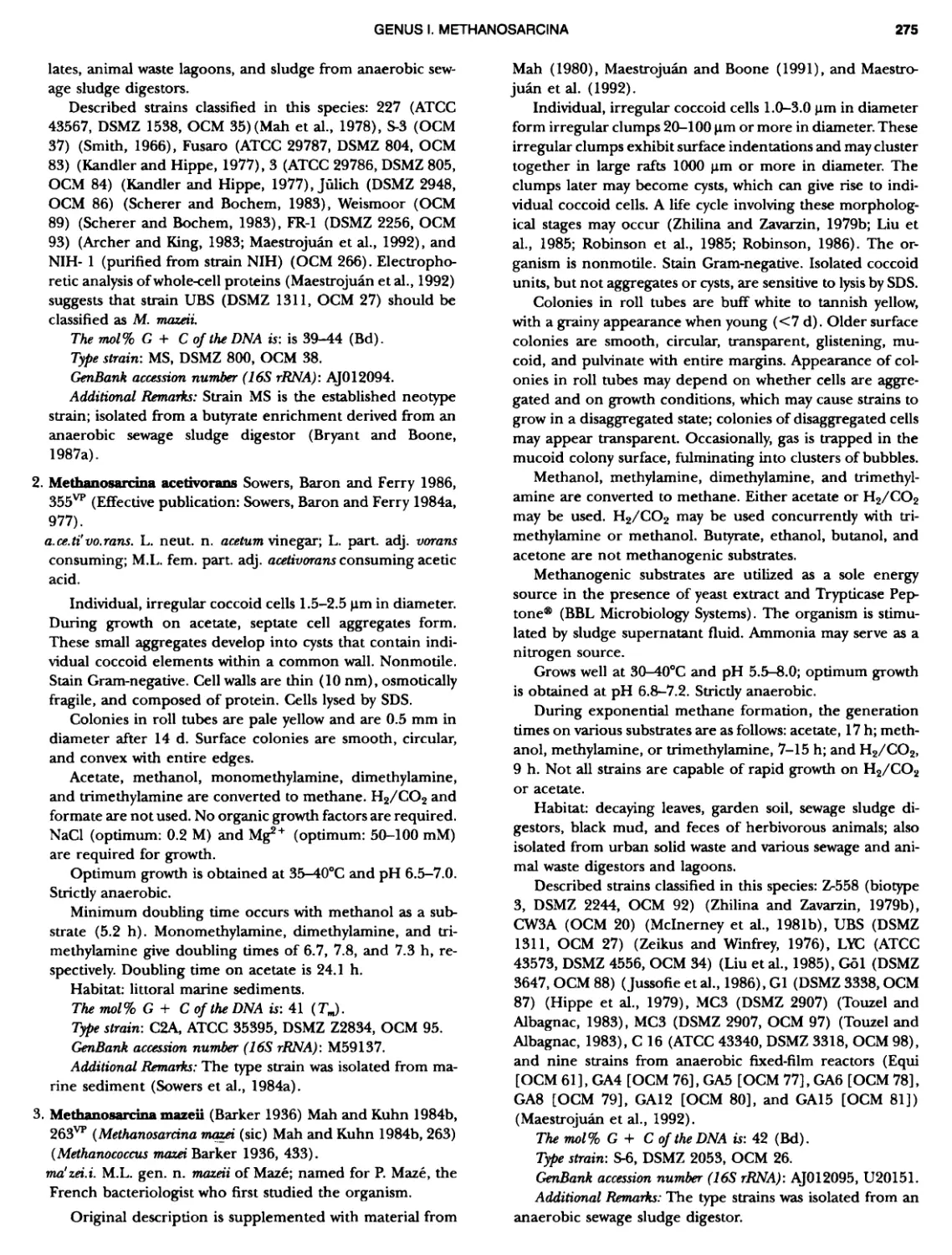
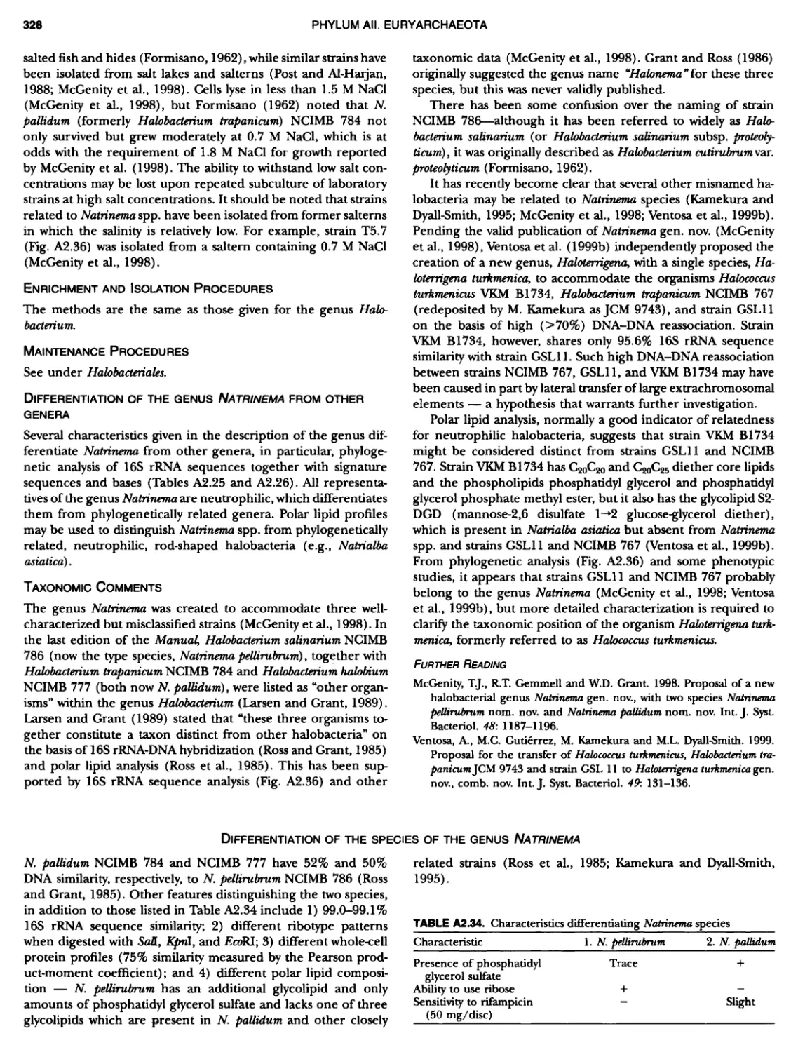
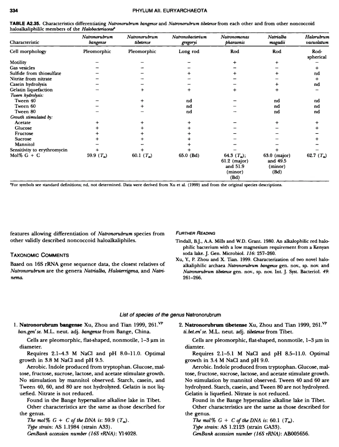
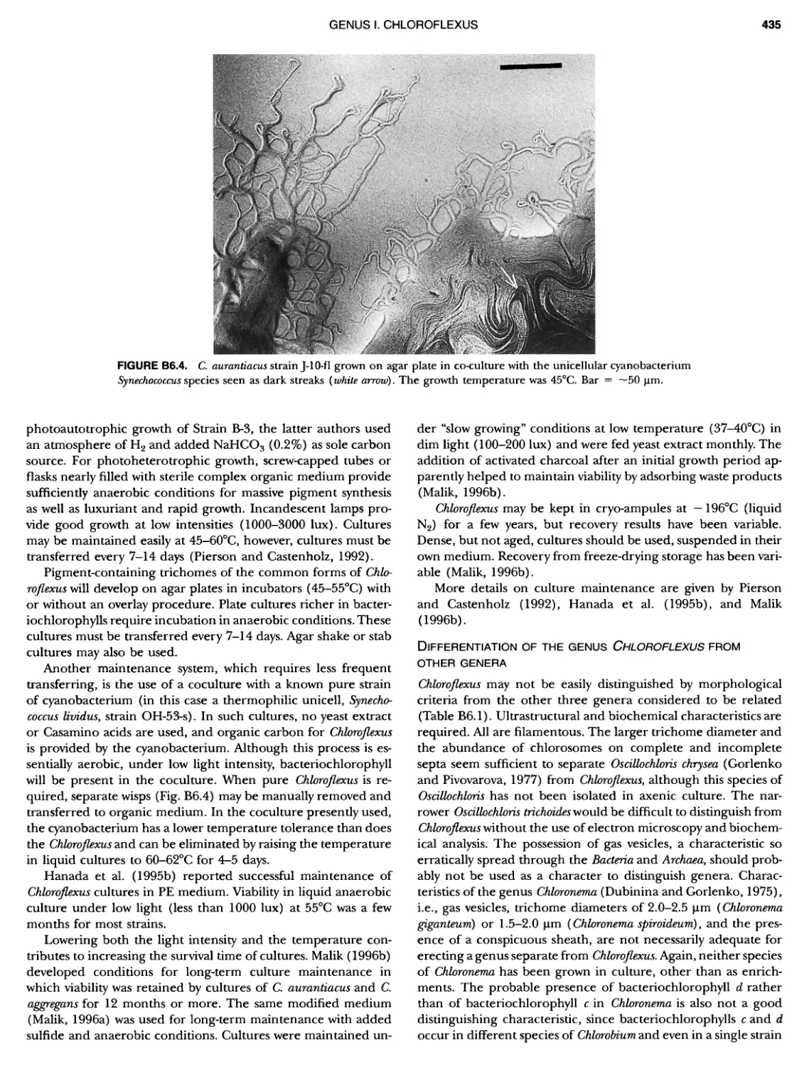
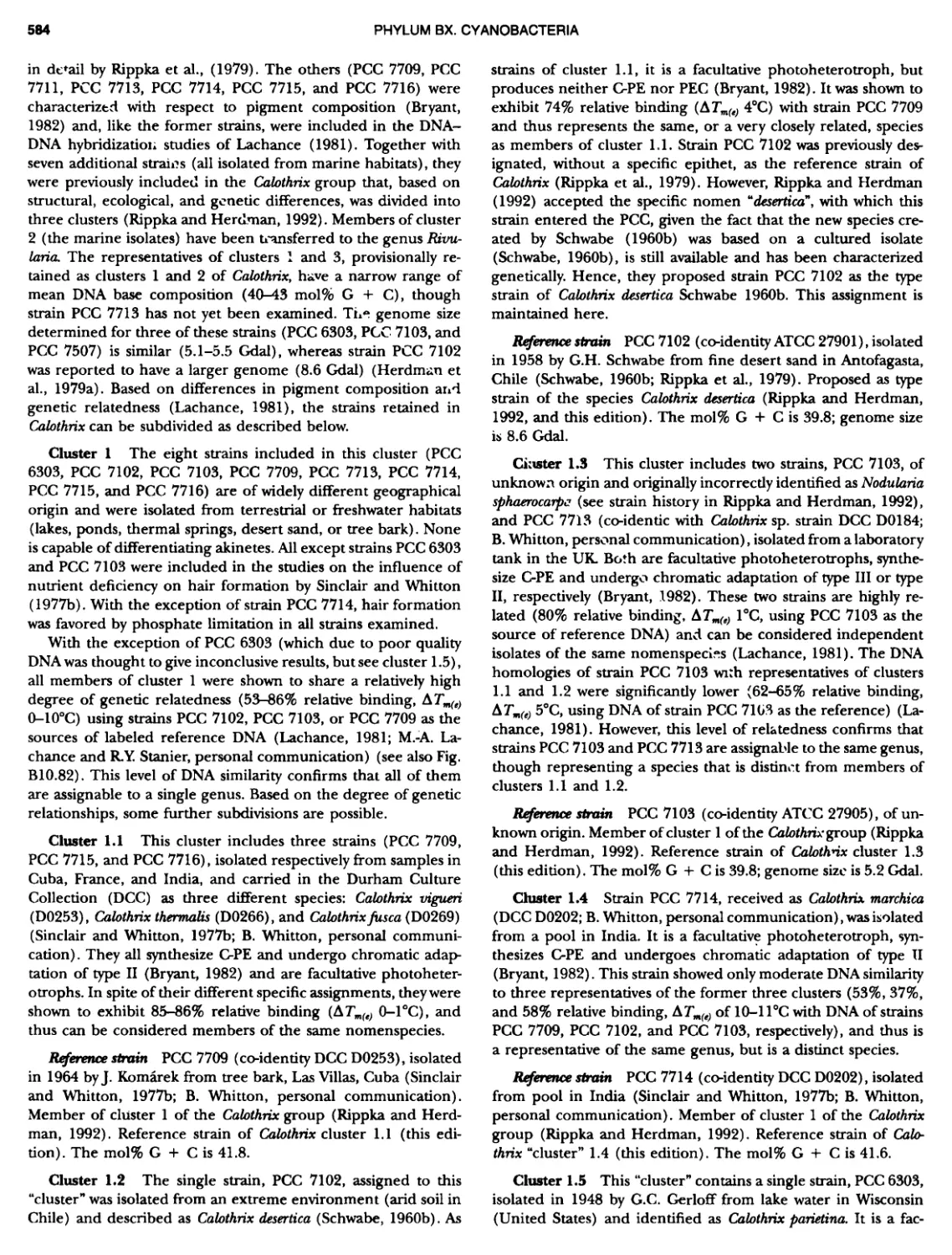
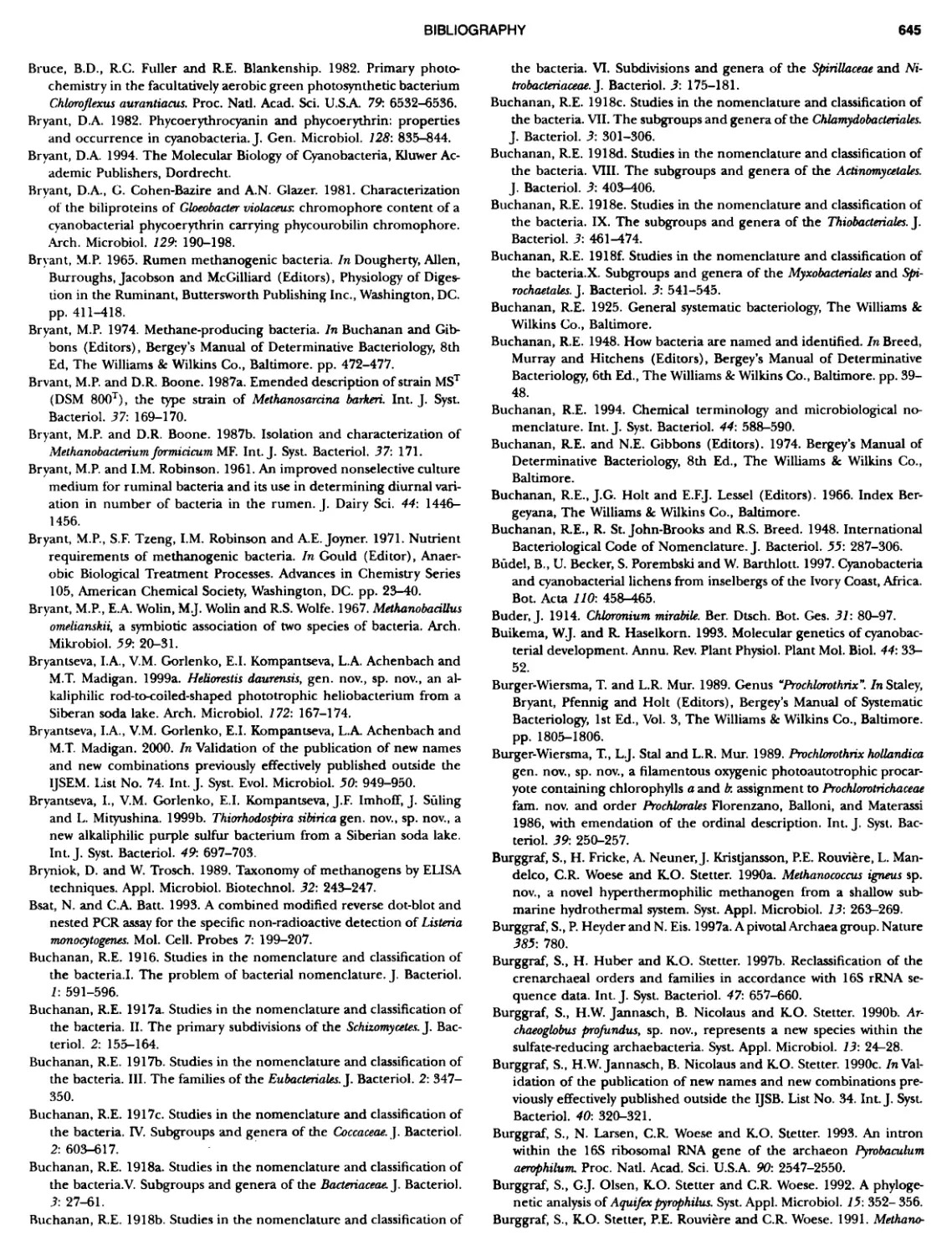
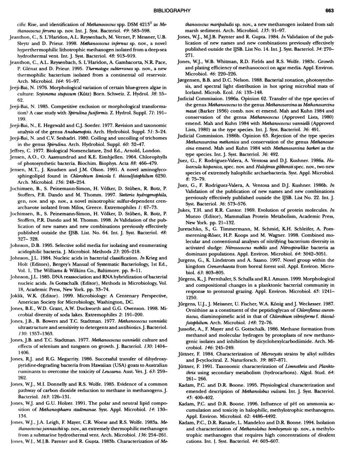
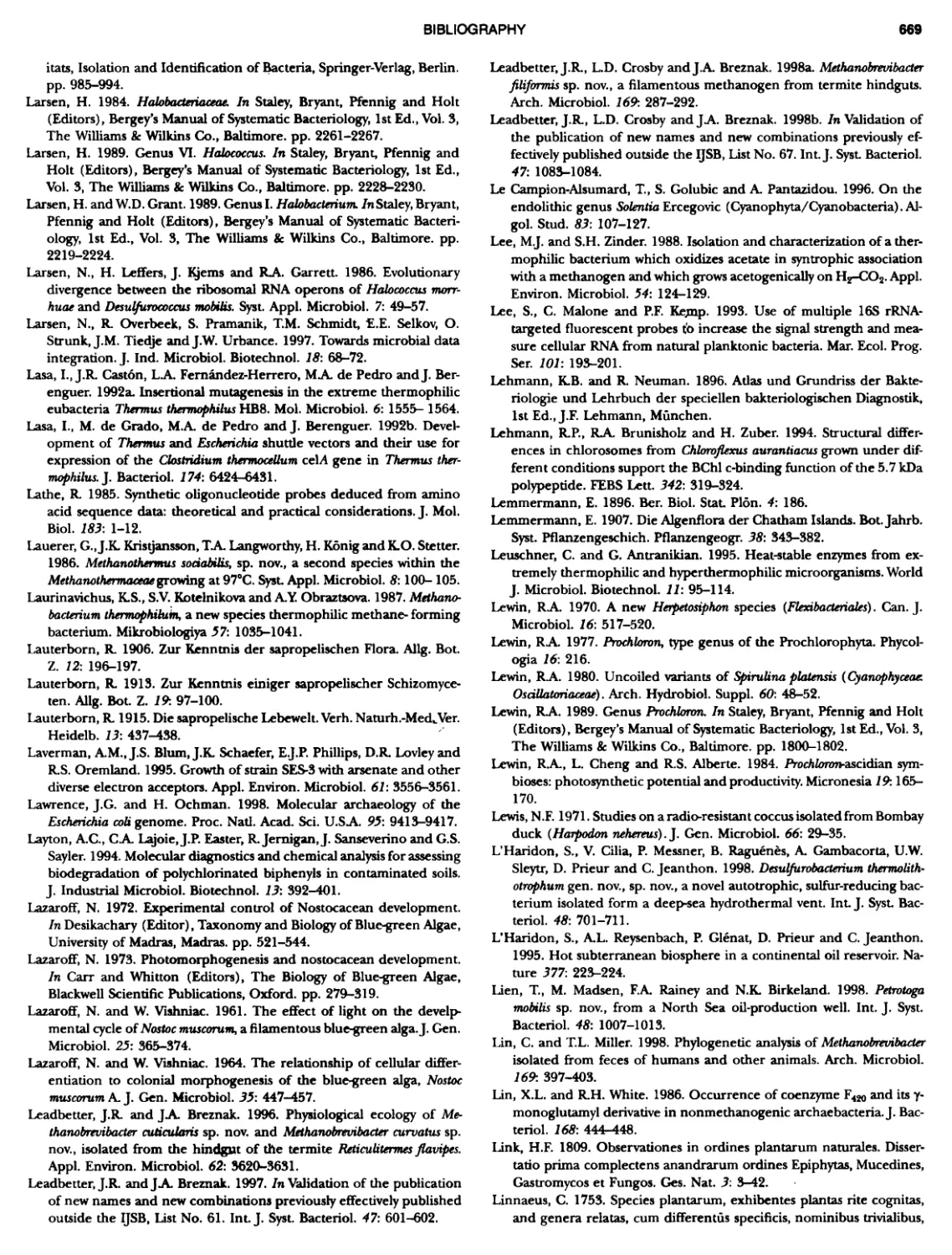
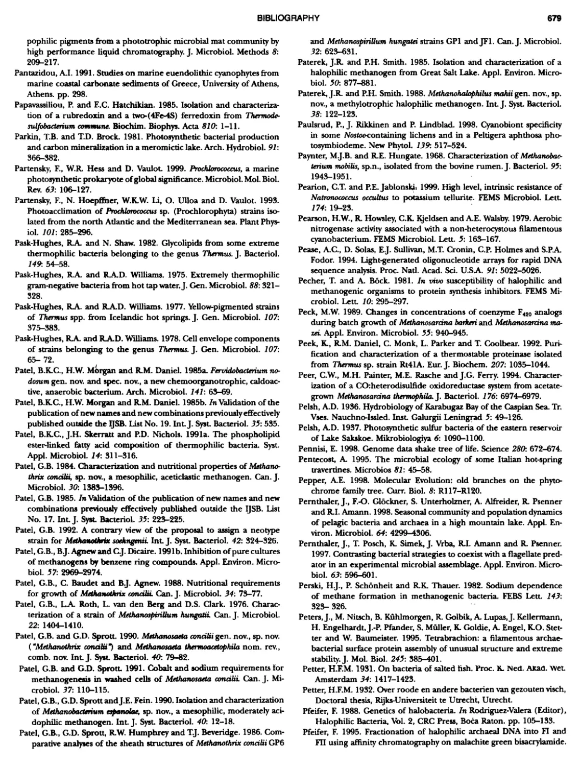
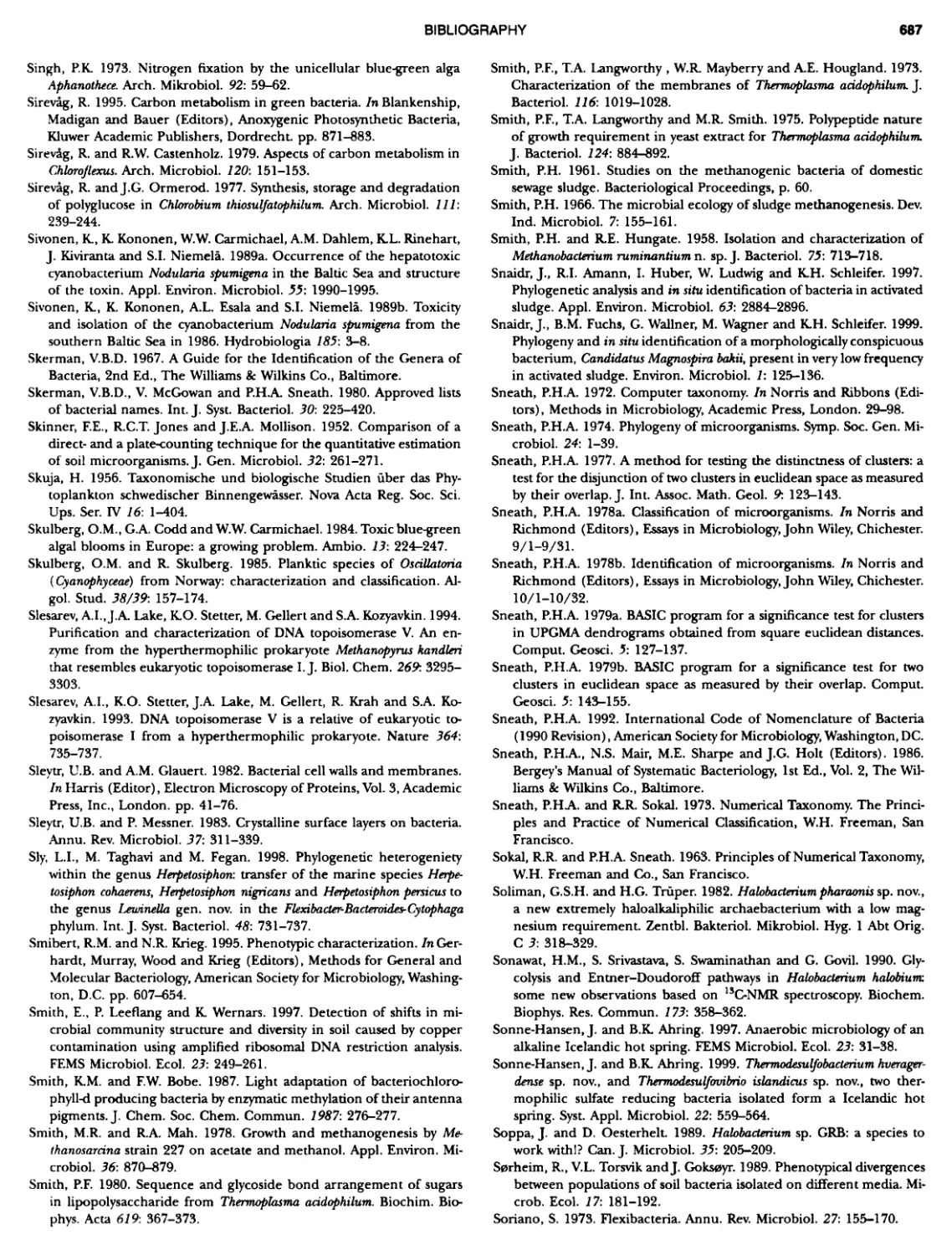
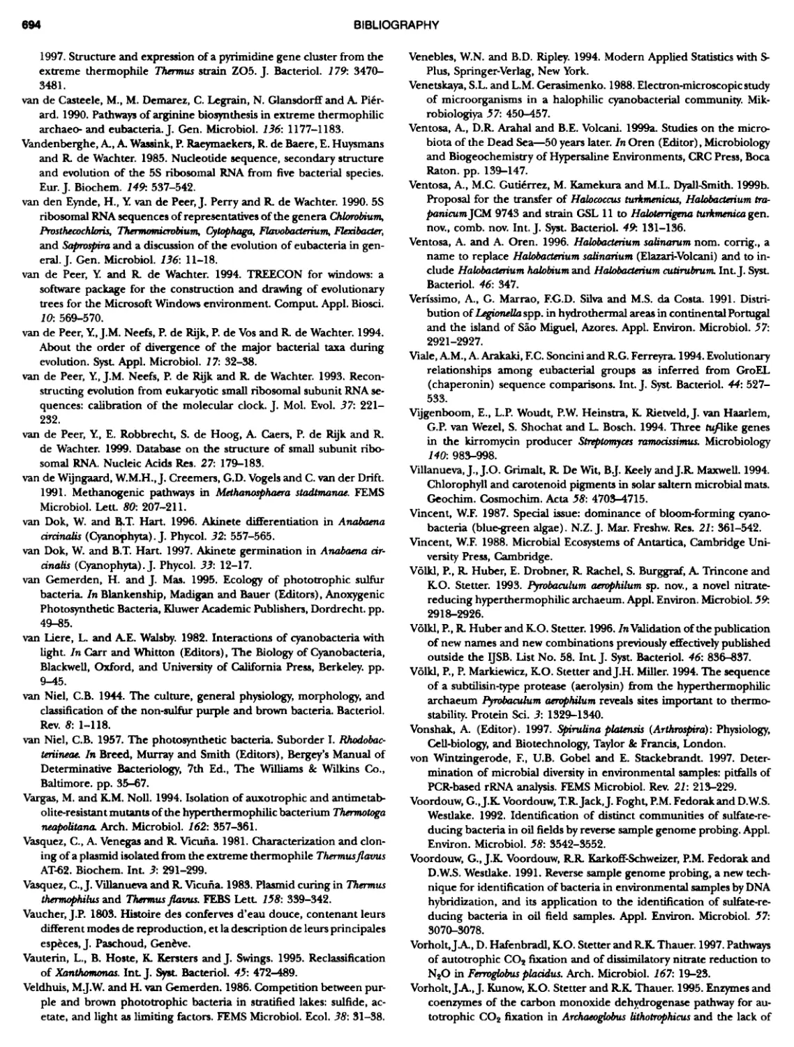
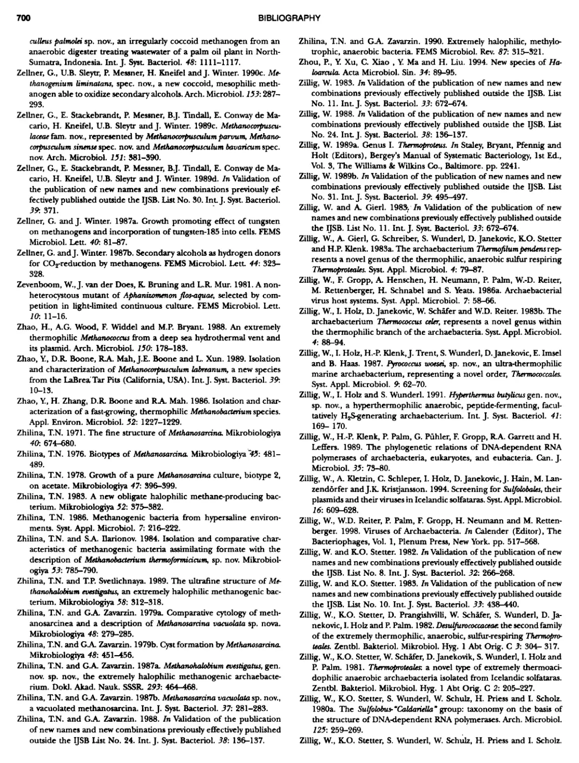
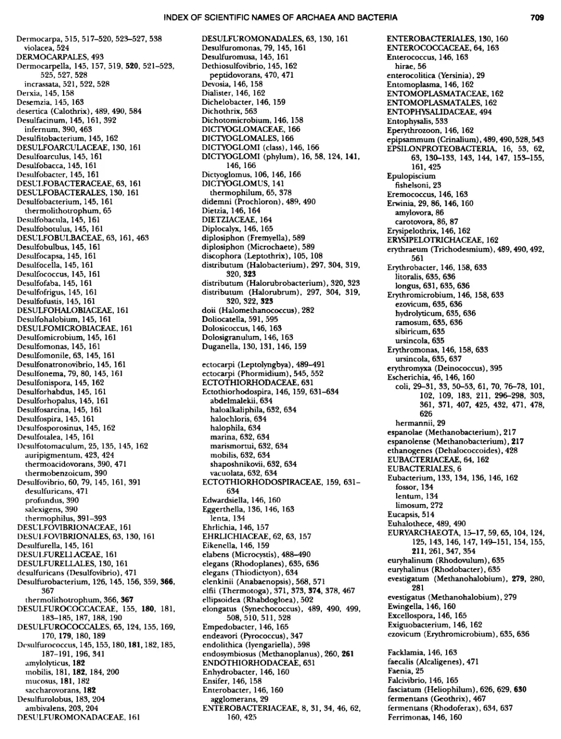
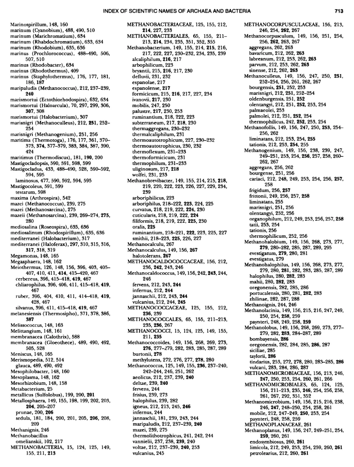
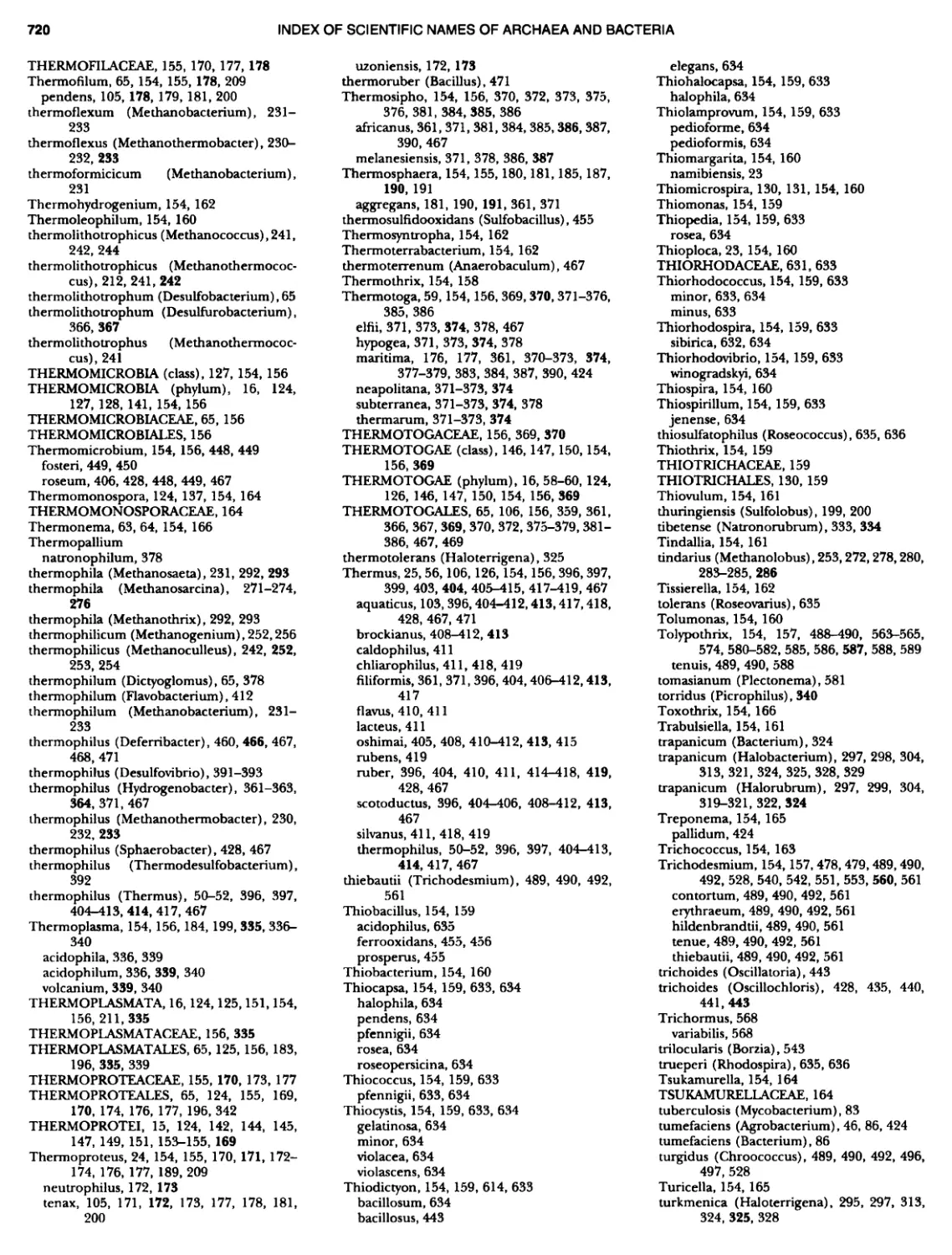
THE HISTORY OF BERGEY'S MANUAL FIGURE 1. A, Robert Earle Buchanan, 1883 — 1973; B, David Henricks Bergey, 1860 — 1937; Q Robert Stanley Breed, 1877 — 1956; D, Everitt G.D. Murray, 1890 — 1964. (Fig. 1C courtesy of American Society for Microbiology Archives Collection.)
THE HISTORY OF BERGEY'S MANUAL substance for the Winslow Committee (Buchanan was a mem- ber), and was intended to be the basis of a systematic treatise. These papers were revolutionary, in the sense that they included all the bacteria (except the cyanobacteria) that were described at that time. Buchanan included, and named the higher group- ings of, bacteria such as the actinomycetes, myxobacteria, pho- totrophs, and chemolithotrophs, along with the other bacteria included in the classifications of the day. This classification had a 1ogical and aesthetic appeal that helped launch the systematic efforts that followed. No doubt Buchanan was driven by dissat- isfaction with sloppy and confusing nomenclature as well as in- adequate descriptions of "accepted" bacteria (indeed, much of his later work on Beryy 's Manual and the Index Bergeyarta reflected his preoccupation with names and illegitimacy, and had much to do with getting a bacteriological code of nomenclature started.) He must have known of Bergey's book and, perhaps because of increasing academic responsibilities, publication of his concepts in his General Systematic Bacteriology was delayed until 1925 (Buchanan, 1925). The book did not try to duplicate Ber~ 's Manual, but rather presented a history of bacterial classification and nomenclature, followed by a discussion of the history of all the bacterial genera and higher ranks, listed alphabetically. R.E. Buchanan was the key player in the renewal of concern for a sensible (not necessarily "natural" ) classification of bacteria, with a well-regulated nomenclature, working continuously and firmly to those ends from 1916 to the end of his life. He was a man of his times developing his own priorities and prejudices, yet he recognized in the end that new science was needed for a significant phylogeny to develop. Furthermore, he was more in- fluential in gaining support for the initiation and progress of the first few editions of Bergey's Manual under the slightly reluctant aegis of the SAB than is obvious in the Manual's pages and pref- aces. He also played a dominant role in international efforts (representing the SAB) concerning the regulation and codifi- cation of classification and nomenclature. As a member of the "Winslow Committee" of the SAB directed to report on the clas- sification of bacteria, he furnished much of the basis for discus- sion through his series of papers in the Journal of Bacteriology. He provided voluminous detailed suggestions for the revision of Dr. Winslow's drafts for their reports to the SAB (1917 and 1920) . He was also in a powerful position to influence decisions, being elected President of SAB for 1918 — 1919 when critical discussions were taking place. The Winslow Committee was engaged in protecting ("con- serving") the generic names for wellwstablished species by listing them as genera cortservanda, together with type species for dis- cussion at the 1918 SAB meeting. The intention was to provide a basis for recommendations for formal action at the next In- ternational Botanical Congress, since they were working under the general rules of the Botanical Code. They went further by classifying the genera within higher taxa and providing a key to assist recognition. They intended seeking formal approval of the whole report by the SAB. At this stage, R.S. Breed (Fig. 1c) wrote many letters of objection to having any society ratify the concepts involved in contriving a classification, because it would suggest that it is "ofBcial", and he attempted unsuccessfully to gain a postponement of the report's presentation. This polemical cor- respondence with Committee members, including Buchanan, ended in Breed's withdrawing his name from the report despite his evident interest in a workable classification and a more stable nomenclature. Winslow read the report to the SAB meeting on December 29, 1919. Although it emphasized that its listings were not to be considered as a standard or official classification, it did ask "that the names be accepted as definite and approved gen- era". The report was then published in the Journal of Bacteriology The Committee was discharged and a new Committee on Tax- onomy was appointed with R.K. Buchanan as Chairman. In 1920 Breed was added as a member of the new committee, with the responsibility of making the representations at the Botanical Con- gress because of his membership on the Botanical Code Revision Committee. It was at this time and in this climate of opinion that Dr. David Bergey decided to put his own studies of bacteria together with the current views on their classification. To do this required more than one person and he assembled a like-minded group to form a Committee of the SAB for the production of a Manual of De- terminative Bacteriology (F.C. Harrison, R.S. Breed, B.W. Hammer, and F.M. Huntoon). There is no direct evidence that Buchanan was ever asked to participate or, equally, that he raised any forrnal objections; it seems more likely that there could have been none of the formal encouragement to go ahead evident in 1921 and 1922 without his support. Indeed he seems to have thought it a good enterprise (Preface in his 1925 book) . However, he did find it difficult to work with Breed (letter of January 8, 1951 to J.R. Porter) and in expressing this stated "I have ... always refused to become a member of the Editorial Board of the Manual". One wonders if his experiences with Breed between 1918 and 1951 (" Scarcely a month passes in which we do not have some dis- agreement ... but he has a good many excellent qualities" ) had kept him at arm's length but not out of touch with what was going on with the ManuaL The Winslow Committee had put before the SAB the possi- bility of a major compilation on bacterial systematics. No doubt Buchanan was in a position, as a Past President, to reinforce the value of that project in principle and David Bergey, likewise a Past President, must have been aware of all the discussions. At the time of the last report (Winslow et al., 1920) Bergey must have started on his book, because R.S. Breed reported to the 1922 SAB Council meeting that the work was approaching com- pletion. A more formal proposal was made to the same Council meeting that Bergey's book be published under the aegis of the Society. The SAB agreed to this with the proviso that it go to a substantial publishing house and, following a discussion of the disposition of royalties, Bergey 's Martual of Determinative Bttctenology was published in 1923 by the Williams Sc Wilkins Co., Baltimore (Bergey et al., 1925). It was a group effort from the start, with the authors listed as D.H. Bergey, F.C. Harrison, R.S. Breed, B.W. Hammer, and F.M. Huntoon, and there was an acknowledgment of the assistance of six other colleagues on special groups. One can imagine that Buchanan was upset by this turn of events, for which the only evidence is his sending Bergey a long list of errors he found in the published book (personal com- munication). However, he was quite generous in his preface to his 19'25 book, with his assessment of Ber~'s Manual as a step towards reducing chaos and convulsion in the classification, phy- logeny, and naming of bacteria. He writes: "The most hopeful sign of importance in this respect probably has been the work of the committee on taxonomy of bacteria of the Society of Amer- ican Bacteriologists under the chairmanship of Dr. Winslow and of the more recent work of a committee on classification of bac- teria under the chairmanship of Dr. Bergey... ~ It is to be expected that, as a result of their work, eventually a practical system of nomenclature which will be satisfactory and applicable to all fields of bacteriology will be evolved" (Buchanan, 1925). Fur-
THE HISTORY OF BERGEY'S MANUAL thermore, he emphasized the diKerences between practical (medical} and academic attitudes towards individual species and the requirements of a classification. He was then, as later, con- cerned that bacterial nomenclature was not regulated by an ap- propriate Code. He writes: "It seems to be selfwvident that until the bacteriologists can agree upon a code and follow it consis- tently, there is little hope or remedy for our present chaos". So it is not surprising that he contributed a section to the Fourth Edition (Bergey et al., 1934) discussing the International Botan- ical Code as a basis for a bacteriological code with modifications to make it more appropriate. The committee that organized the First Edition stated that they did not regard their classification of species "as in any case final, but merely a progress report leading to more satisfactory classifications in the future". Clearly there was some feeling in the UK and Europe that this classification was an imposition on the part of the SAB~. As a counter, the Third Edition (Bergey et al., 1930) included a box opposite the title page which declares that it is "Published ot the direction of the Society" which "disclaims any responsibility for the system of classification followed"; and states further that it "has not been formally approved by the Society and is in no sense official or standard" (italics are in the original). This shows that there had been, as indicated by the article by I.C. Hall in 1927 (Hall, 1927), some degree of conten- tion among members of the SAB with the decisions of the Com- mittee. Hall's objections to the presentations of the Committee of the SAB on characterization and classification of bacterial types starts with the final report (Winslow et al., 1920) being "presented only to a small minority of the members of the Society who happened to return from lunch in time to attend a business session of the twenty-first annual meeting, which was held in Boston more than four months before the publication of the report". He regrets lack of opportunity for scientific consideration and "practically no discussion because only a few knew what was coming". He evidently objected to physiological criteria and believed that mor- phology should define genera, families, and orders; furthermore he disputed the validity of habitat and believed that serological characterization was futile. He was prepared to use cultural and physiological properties as criteria for species. He sought "un- ambiguous criteria". He quotes others who disagreed with the Bergey's Manual approach including W.W.C. Topley, who also ex- pressed his distaste in his famous textbook ("Topley and Wilson" ) that was published in 1929. Bergp's Manual was launched and successful enough for the publisher to encourage further editions with corrections and additions in 1925 and 1950, for which Bergey had the support of the same four co-authors. There were problems ahead. By 1950 Bergey was aging and becoming somewhat frai1 so that he was concerned about the Manual's governance and future. He turned to Breed to an increasing degree for the overall editing and as a major contributor, but also to fight for financial support and for a degree of independence. The agreement co-signed by Bergey and Breed with the Society in 1922 had recommended that royalties "... be accumulated in a separate fund to be used to stimulate father work in this field" and Bergey himself felt that he had "donated" this fund to the Society for that purpose. ~As can be gathered from skeptical sentiments in the famous textbook by W.W.C Topley and G.s. wilson, PHnripks of Bacteriology and Immunity, 1st ed. (1929), Edward Arnold Ltd., London, and continued in large part to the Fifth Edition (1964) but not thereafter. THE STRUGGLE FOR FINANCIAL AND EDITORIAL INDEPENDENCE Breed's correspondence after 1930 with the powerful Secretary- Treasurers of the SAB (J.M. Sherman 1923-1934; I.L. Baldwin 1935-1942} seeking funds to assist the business of producing new editions became increasingly sharp and argumentative because this assistance was almost uniformly refused. The royalties were smal1 and the publisher did not pay any until the costs were covered; the result was that the Society felt they were exposed to risk with a property that they considered not likely to go on much longer. Sherman, in particular, strongly objected to Breed's rhetoric and proprietary attitude, yet he reluctantly agreed in 1933 to cede $900 (half the accumulated royalties) for Fourth Edition purposes. The Society felt that the funds were theirs (the contract was between the Society and Williams Bc Wilkins) and there might be others deserving of support from the fund. A request for funds by A.T. Henrici in 1935 brought the whole matter of ownership back into contention and into Baldwin's more diplomatic hands. At the same time Breed was asking for $1000 (essentially the remainder of royalties plus interest) and decisions had to be made during a flurry of correspondence with a repetitive non placet obligato from Sherman. There was also a Berge)I 's Manual Committee (Winslow, Buchanan and Breed) re- porting to the Council in support of a mechanism for funding the ManuaL In the end, and agreeably to all parties for different reasons, it was decided between Sherman and Baldwin that the SAB should cede the rights to the Manual, the royalties to come, and the accumulated fund to Dr. Bergey to do with as he would wish, and the Council agreed (December 28, 1935). In large part it was a gesture of respect for Dr. Bergey because both of them stated in letters that they did not expect the Manual to go through more editions, in which respect they were mistaken. In preparation for the Fourth Edition, and recognizing that Bergey was not well and that Harrison, Hammer, and Huntoon would not stay for long, Breed added E.G.D. Murray (Fig. 1d} to his corps of editors/authors, so that with Harrison still enlisted there were two Canadian members. With the Fourth edition pub- lished in 1954, from late 1935 until early 1956 was a time of negotiation. It is clear that Bergey, Breed, and Murray wanted an independent entity, while Buchanan with his own ideas was presenting a plan to Baldwin involving sponsorship by the Society, and Breed was trying unsuccessf'ully to make peace with Bu- chanan. Bergey, for his part, was (January, 1936) consulting with the SAB and advisors in preparation for developing a deed of trust for the future development of the Manual, and asking that there be no further controversy. His feeling about the whole sad tale was voiced on January 29, 1936: "The arrangement I have made wi11 be without hindrance from a group of persons who appear to have no kindly feeling toward advances in bacteriology in which they could not dictate every step". The Beg~'s Manual Trust was indentured on January 2, 1936 in Philadelphia, Penn- sylvania, and the Trustees were Bergey, Breed, and Murray. The only concession to the SAB, that continues to the ASM today, is that one of the Trustees is chosen as a representative who reports annually to the Society on the state of the Trust and its work. Mr. R.S. Gill, the representative of the Williams 8c Wilkins Co., informed Breed in December, 1954 that copies of the Fourth Edition were exhausted and sought agreement for a new edition; Breed prevaricated because the situation was not yet clear. How- ever, by 1957 he was seeking contributions from a number of colleagues for a future volume. Sadly, D.H. Bergey died on Sep-
THE HISTORY OF BERGEY'S MANUAL tember 5, 1937 at age 77, but the trustees retained his name on the masthead of the Fifth Edition published in 1939. Breed was now Chairman of the Trust and remarked in a letter to K.B. Fred and I.L. Baldwin (January 26, 1958) that Dr. Bergey, who was so interested in seeing the Manual revised, would have liked "... to know how well his plans are developing and how ... interested specialists are cooperating with us in making this new edition much better than anything we have had before". So a new way of producing the Manual with many contributors was now in place for elaboration in future editions. The first printing of '2000 copies of the Fifth Edition (Bergey et al., 1989) was sold out before the end of the year and 1000 more copies were printed. It was obvious that the Manual was needed and served a useful purpose, vindicating the optimism Bergey and Breed had main- tained in the face of opposition. Breed, Murray, and A.P. Hitch- ens (who was appointed to the Board of Trustees in 1989) had to organize a Sixth Edition, which needed to be completely re- vised and required much to be added. There were 1335 species descriptions in the Fifth Edition and the Sixth, when accom- plished, would have 1630. They were faced not only with the need to make changes in the outline classification but also to make decisions about the inclusion or exclusion of large numbers of dubious and inadequately described bacteria. Furthermore, the exigencies of World War II took some of the trustees and many of their contributors out of contention for the duration. Nevertheless, the Sixth Edition was published in 1948 (Breed et al., 1948a) and acknowledged the assistance of 60 contributors. Some of the incompletely described species appeared in appen- dices following the listings in genera and the book included an index of sources and habitats as an attempt to be helpful. A novelty, and an approach not to be fully realized until 35 years later in the Systematic Manual, was a section on the Myxobacteraks containing a preliminary discussion of the nomenclature and biological characteristics of members of that Order. For this, credit is given to J.M. Beebe, R.E. Buchanan, and R.Y. Stanier; it seems likely to those who knew all of them that this approach originated with Stanier. Additions to the Sixth Edition were sec- tions on the classification of Rickettsiales prepared by I.A. Bengs- ton and on the Virales or Filterable Viruses prepared by F.O. Holmes. The former was appropriate but the latter pleased very few, certainly preceded an adequate understanding that would have allowed for a rational classification, and never appeared again. The original Board of Trustees went through changes due to death and the enlargement of the Board. H.J. Conn, a colleague of Breed's at Cornell, was added in 1948 to join Breed, Murray, and Hitchens. The next year A.P. Hitchens died and was replaced by N.R. Smith, an expert on Bacillus species. R.E. Buchanan was added as a member in 1951 and began to take an active role in the affairs of the Trust. In 195'2 Breed expressed a desire to step down as Editor-in-Chief, he was 75, and the Board debated about his successor. Among those considered were E.G.D. Murray, who was about to retire from McGill University, L.S. McClung of In- diana University, and C.S. Pederson of Cornell, but no decision was made. In correspondence to Breed, Smith wrote that "No doubt, Dr. Buchanan would like to take over when you step aside ~ .. In fact one can read between the lines that 'no one besides Buchanan is capable of editing the Manual' ". This change, how- ever, did not come to pass as Breed stayed on until his death in 1956. Breed pursued actively the production of a Seventh Edition in the 1950s with the active support of Murray and Smith (Breed et al., 1957). The task was no less formidable, and there were many new authorities mounting increasingly pointed discussions about shortcomings in bacterial taxonomy in the dinner sessions that Breed arranged at the annual SAB meetings. It was to be the last edition in which the bacteria are classified as Schizo- mycetes within a Division of the Plantae, the Protophyta, pri- mordial plants. In fact, the Preface tells us, the opening statement describing the Schizomycetes as "typically unicellular plants", was hotly debated without attaining a change, yet there were some concessions to cytology in the rest of that description, particularly concerning nucleoids. Ten Orders were recognized, adding to the five in the Sixth Edition, and these now included Mycoplas matales and considerable division of the Order Eubacteriaks. The keys to the various taxa were improved for utility and, recognizing the many difhculties involved in determination, an inclusive key to the genera described in the book was devised by V.B.D. Sker- man and appended. This key, which was referred to as a com- prehensive key, was designed to lead the user by alternative routes to a diagnosis of a genus when a character might be variable. It proved to be extremely popular and useful with readers and was repeated as an updated version in the Eighth Edition. Overall, the substance of the Seventh Edition of the Manual was due to the efforts of 94 contributors from 14 different countries. The Manual was becoming an international effort; however, Breed complained that the slowness of communication between the USA and Europe hampered their eKorts. Breed did not see the fruits of his labors as Editor-in-Chief; he died February 10, 1956, with many of the contributions ar- ranged and the form of the book decided, but leaving a serious problem of succession. The position of Chairman of the Board of Trustees and Editor-in-Chief was decided, appropriately, and given to R.E. Buchanan whose interest in bacterial nomenclature and taxonomy, with direct and indirect involvement in the Man- ual, dated back to its origins. There was the immediate problem of finishing the editorial work on the Seventh Edition after Breed's death. E.F. Lessel Jr. had been working as a graduate student with Breed in Geneva, NYon the Manual, but was called into military service before the job was finished, and was sta- tioned at a camp in Texas. Upon taking over the Chairmanship, Buchanan contacted W. Stanhope Bayne Jones, of the Army's Once of the Surgeon General and Lessel's superior, to ask that Lessel be assigned to work on the completion of the Manual while in the service. Bayne Jones agreed and assigned Lessel to the Walter Reed Hospital in Washington, DC. Thus the last ed- itorial polishing of the book could take place without undue delays. After his service commitments were fulfilled Lessel went to Iowa State and finished his Ph.D. under Buchanan's direction and acted on occasion as recording secretary for Trust meetings. R.E. Buchanan for many years had held three important ad- ministrative posts at Iowa State (Bacteriology Department Head since 1912; Dean of Graduate College since 1919; and Director of the Agricultural Experiment Station since 1986), retiring from all three in 1948. After 1948 some of his energies went to com- piling and annotating the text for the 195'2 publication of the Bacteriological Code and starting the Inierrtational Bulktin of Bac teriological Nomenclature and Taxonomy. The International Bulktin received its initial monetary start in 1950 with a $150 gift from the Bergey's Manual Trust, to which Murray objected, saying "the Journal would be ephemeral." Fortunately he was wrong because the Bulletin later changed its name to the International Journal of Systematic Bacteriology and is still being published by ICSB (IAMS) with about 1200 pages in the 1997 volume. When Buchanan
THE HISTORY OF BERGEY'S MANUAL became Editor-in-Chief of the Manual, he induced the Depart- ment of Bacteriology at Iowa State to provide him an once suite and the title of Research Professor, from which position he ob- tained grants from the National Library of Medicine to support the ofhce. This support continued until his death in 1975 at the age of 89 years. Buchanan's twenty-year involvement in the Trust was to see, near its end, the start of a new era, despite his many objections to change. The chief change to come arose from a growing lack of confidence in the sanctity of higher taxa, there being few and often no objective tests of correctness. In the production of the Seventh Edition, it was recognized that an expanding synonymy and the ever-growing list of species that were unrecognizable or inadequately described provided a burden that made for wasted space and unreasonably extensive appendices. The addition of Breed's collection of reprints to Buchanan's considerable collec- tion formed an extensive taxonomic archive in the Trust head- quarters. With this resource in mind the Trust decided that a separate publication was needed to assemble as complete a listing as possible of the names and references of all the taxa included in the Manual, as well as "species formerly found as appendices or indefinitely placed as species incertae sedis" that might or should have appeared in the ManuaL These, together with an assessment of whether or not each name was validly published and legiti- mate, formed a monster book of nearly 1500 pages, published as Index Beqpyana (Buchanan et al., 1966). Each and every ref- erence was checked for accuracy, for Buchanan rightly stated that there "was a lot of gossip about the description of each name." These labors were a personal interest of R.E. Buchanan, who directed several years of effort by J.G. Holt (then at Iowa State), E.F. Lessel Jr., and a number of graduate students and clerks in the undertaking. The lists served as a finder mechanism, an alphabetical listing of the names of the bacteria, and of special use as a reference after the new starting date for nomenclature, January 1, 1980, mandated by the revised Code (Lapage et al., 1975). Addenda were inevitable and more names were collected as a Supplentsnt to Index Bergeyana published in 1981 under the direction of N.E. Gibbons, K.B. Pattee and J.G. Holt. These sub- stantial reference works assisted the refining of the content of the Seventh and Eighth Editions of the Manual and aHowed concentration on electively described and legitimate taxa. There were seemingly interminable discussions about what needed to be done for an effective new edition. This was par- ticularly true in the period 1957-1964 after Breed's death, when the Trust membership changed and new ideas and new scientific approaches to taxonomy became available. In the late 1950s the Board of Trustees was enlarged with the addition of S.T. Cowan, C.F. Niven Jr., G. Edsall, and A.G. Lochhead (the record is un- clear on the exact date of their appointment). The election of Cowan from the UK added a European member and continued the internationalization of the Board (Fig. 2). Each of these new members brought expertise in different areas of bacteriology and that policy of diversity of interest among members has continued to this day. Later, in 1962, Arnold Ravin, a bacterial geneticist, was added to replace the retiring Lochhead. Of primary concern in the late 1950s and early 1960s was the position of Editor-in- Chief and location of Trust headquarters. An arrangement with Iowa State University to have a candidate assume a professorship at the University and house the headquarters there was made. The position was offered to P.H.A. Sneath, who had gained re- nown with his invention of numerical taxonomy and production of a masterful monograph on the genus Chromobacterium. By 1963, however, Sneath chose to stay in England and Buchanan stayed on as Editor-in-Chief. All other efforts to find a new editor failed until Buchanan's death in 1975. As he grew older, more diKcult, and more autocratic, progress on a new edition slowed consid- erably. Even the replacement of E.G.D. Murray, Conn, Smith, and Edsall by R.G.E. Murray, J. Liston, R.Y. Stanier, and N.E. Gibbons did not change the speed of Board actions. It became a war of wills between Buchanan and the others on what was important and where progress could be made. Until decisions on the taxa to be included and their circumscriptions were made, there was slow progress in naming and putting to work the 20 or more advisory committees needed to direct the authors of the final texts on genera and species. One novel (to the Trust) ap- proach for obtaining consensus on taxonomic matters was the organization of a conference of advisory committee members and trustees held in May, 1968 at Brook Lodge in Augusta, MI, under the auspices of the Upjohn Co. and chaired by R.G.E. Murray. Fifteen advisory committee members joined in discus- sions with the Trust to assess the status of current knowledge on the major groups of bacteria to be included in the Eighth Edition. Despite this helpful preliminary, it brought no agreement be- tween Buchanan as Chairman, whose main focus was then on nomenclature, and the rest of the Trustees, whose interests mostly focused on biological, functional, and eco-physiological attributes. It was clear that many of the higher taxa rested on shaky ground and were hard to assess on strict taxonomic terms. Accordingly, there was a long argument over abandoning formal names above family level wherever possible, agreeing that a large number of genera were of uncertain aKliation or, at least, could only be related on the basis of some diagnostic characters, such as gliding motility, shape and Gram reaction, and methane pro- duction, all of which might or might not have phylogenetic sig- nificance. All former ideas about phylogeny and relationships were discarded. The Eighth Edition was planned as a book di- vided into "Parts", each with a vernacular descriptor. The Advi- sory Committee for each part (some needed more than one) was assigned a member of the Trust who was responsible for action and who, eventually, had to see that each genus had an assigned author (151 in the end) who was willing to write. Molecular/genetic technology was well established by 1974 when the Eighth Edition was published, but was not yet widely applied to play a role in broad decisions in taxonomy. The pro- caryotic nature of bacteria and all cells related to them (i.e. including the Cyanobacteria) could be recognized and used to define the Kingdom Procaryotae. Monera was the old and par- tially applicable higher taxon but the description was not cyto- logically based. The molecular composition of DNA was useful for separating phenotypically similar but genetically distinct groups (e.g., Micrococcus and Staphylococcus) and many descrip- tions could include mol% G + C as a character. Genetic and subsequent biochemical-molecular data told us that species were only relatively "fixed" in their expressed characters. This concept needed to be addressed in the circumscriptions and aids to iden- tification. Greater use was made of diagnostic tables and wherever possible there were indications regarding uncertainties and the percentages of positive or negative reactions for tests. The value of the Eighth Edition for identification purposes was increased by the emphasis of both the Trustees and the authors on refining descriptions (in terms as up-t~ate as possible), tables, keys, and illustrations. As in previous editions, many old names of dubious or unrecognizable entities were discarded and synonymy was re- duced to essentials; the old information and its location was not
THE HISTORY OF BERGEY'S MANUAL FIGURE 2. Photograph of Trustees meeting at Iowa State University, Ames, November, 1960. L. to R, G. Edsall, R.E. Buchanan, C.F. Niven, Jr., N.R. Smith, and E.G.D. Murray. lost because it was available in the Index Bergryarta (Buchanan et al., 1966), or later in the SuPPlement to Index Bergsyarta (Gibbons et al., 1981). The Eighth Edition was a long time in gestation — 17 years- but its success (40,000 copies over the next 10 years, and more than half outside of North America) was a testament to its ne- cessity and utility. Most of the primary journals involved in puW lishing microbiological papers suggested or required the Manual as the nomenclatural resource for bacterial names, all this despite the treatment of some groups (e.g., the Ertterobacteriaceae) not being universally accepted. But it was truly an international en- terprise, with authors from 15 countries who could, at last, be named in literature citations as authors. The editing of the Eighth Edition became a major operation requiring sharing of responsibilities and some redirection of ef- fort. This was in part due to the age and increasing infirmities of R.E. Buchanan who had been both Chairman of the Trust and Editor, directing his efforts to nomenclature, synonymy, and etymology. It became evident that a Co-Editor was required and fortunately N.E. Gibbons, recently retired from the National Re- search Council of Canada, agreed to undertake the task. Shortly thereafter Gibbons became the de facto editor, due to Buchanan's illness and death in January, 1978, and did all the general tech- nical editing from his home in Ottawa with help from his wife, Alice Gibbons (who handled the Index of Names), and a number of Trustees, especially S.T. Cowan. The book was published in 1974 (Buchanan and Gibbons, 1974). With publication of the Eighth Edition the Board of Trustees went through another major change of membership, and over a period of two years Niven, Ravin, Liston, Gibbons, and Stanier left the Board. At the first meeting after Buchanan's death, held in October, 1978, J.G. Holt, who had served as Secretary to the Board from 1965-1966 and co-edited the Index Bergeyana, was elected member and Secretary. In 1974 — 1975, H. Lautrop, S. Lapage, and M. Bryant were added, and later in 1976 N.R. Krieg and J.T. Staley joined the Board. In 1975 Holt was appointed Editor-in-Chief. With the publication and healthy sales of the Eighth Edition and increasing international profile, it was de- cided to meet at locations separate from the ASM venue and to meet every other year outside North America, and the 1975 meet- ing was held in Copenhagen, Denmark, at the Statens Serumin- stitut. From then on a segment of each meeting was devoted to consultation with taxonomically inclined colleagues in that area. The Trust had recognized, in the process of deciding the format of the book, that students and technologists were im- portant users, with primary interests in identification and a lesser need for the extensive descriptions of individual species. An abridged edition of the Sixth Edition of the Manual had been produced (Breed et al., 1948b), but was only a modest success and not carried forward to the Seventh Edition. In 1974 the need seemed to be greater, so preparations were made to assem- ble an outline classification; the descriptions of genera, families and such higher taxa as were recognized; all the keys and tables for the identification of species; the glossary; all the illustrations; and two informative introductory chapters. It was recognized that there were both deletions and additions (new keys and synopses as well as new genera) to the material from the parent edition, so that at the most the abridged version would be considered an abstract of the work of the authors of the larger text. Therefore, citation could only be made to the complete Eighth Edition. It was published as The Shorter Bergey's Manual of Determinative Bac teriology in 1977 (Holt, 1977). It too was a great success, selling 20,000 copies over a span of 10 years. A few years later it was translated into Russian and sold throughout the USSR, with roy- alties accruing to the Trust. The development of bacteriology, as we now appreciate, re- quired the recognition and differentiation of the various groups of microbes as taxonomic entities. At the time that Bergey's Manual started, the nature of bacterial cells was not known. Bacteria were classified and named under the Botanical Code of Nomenclature as Schizomycetes and no one could then have substantiated pres- ent understanding that Cyanophyceae are really bacteria. The international discussions of bacterial classification were minimal and took place at Botanical Congresses, as befitted the view that the Schizomycetes and the Schizophyceae within the Phylum Schizophyta (later Protophyta) belonged in the plant kingdom.
THE HISTORY OF BERGEY'S MANUAL This interpretation was maintained in Bergs's Manuat's up to and including the Seventh Edition (1957); however, it was stated in an introductory chapter that K.G.D. Murray "... felt most strongly that the bacteria and related organisms are so different from plants and animals that they should be grouped in a kingdom equal in rank with these kingdoms". As expressed by Stanier and Van Niel (1962) in their seminal paper "The concept of a bac- terium" it is "... intellectually distressing (for a biologist) to devote his life to the study of a group that cannot be readily and sat- isfactorily defined in biological terms ...". This marked the be- ginning of the useful and directive description of bacteria as cells of unique nature. With this approach it was clear that the cy- anobacteria were included and there was, at last, a satisfactory unity. This was to be slowly elaborated in the next three decades by the recognition of phylogenetic information recorded in mo- lecular sequences of highly conserved macromolecules, but in the meantime the Eighth Edition (1974) subscribed to the view based on cytological data that the bacteria (all the procaryotes) belong in a separate kingdom, the Procaryotae. This was not a surprising decision because two Trustees, Stanier and R.G.E. Mur- ray, were then involved in the description of bacteria as cells with unique features. tNTERNATIoNAL EFFoRTs TQ REGULATE TAxoNQMY The founders of Beryy's Manual were fully aware of the substra- tum of opinion, albeit not supported then by strong data, that the bacteria were a special form of life, requiring special methods and a diEerent approach to classification, not necessarily the same as that required by the Botanical Code. In fact, between 1927 and 1950 there was a considerable international corre- spondence between bacteriologists interested in taxonomy in the varied fields of application in agriculture, medicine, soil science, etc, expressing their concerns. The correspondence also con- cerned what should be done about discussing bacteria at the forthcoming Botanical Congress to be held in Cambridge, En- gland, in 1950, and about resolutions adopted by the Bacterio- logical Section of the Botanical Congress, of which J.M. Sherman had been Secretary, held in Ithaca, NY, in 1926. The resolutions were (I) exclusion of the requirement for a Latin diagnosis in bacteriological nomenclature; (2) greater emphasis on the "type concept"; (S) a special international and representative com- mittee was needed to coordinate the special nomenclatural in- terests of bacteriologists; and (4) that a permanent International Commission on Bacteriological Nomenclature should be formed. Sherman, then Secretary-Treasurer of SAB, wrote to Prof.J. Bri- quet of the Permanent International Committee on Botanical Nomenclature pointing out that the past two Congresses had authorized a bacteriological committee on nomenclature, that it should be organized, and that the Bacteriological Section had prepared a distinguished list of nominations for membership. The list included three of the major contributors to discussions of systematics in the && (Buchanan, Breed, and Harrison) and two of them were intimately involved with Ber~'s ManuaL A lively correspondence among the authorities resulted and much of it was stimulated by Breed writing to bacteriologists in Europe as well as America. He sums up an impression of the responses in a letter to the Secretary of the Botanical Congress, as follows: "... there is a general feeling that unless the Congress welcomes us into the ranks of botanists with the recognition of our peculiar and perplexing problems in the taxonomic field, we must organize an independent international group". At the same time he recognized the value of the work of Congresses in maintaining useful rules of nomenclature and reiterating the list of resolutions. The British correspondents were generally agree- able to bacteriological discussions but expressed sharp divisions as to associating or not with the botanists. Other players namely the newly formed International Society of Microbiology, and the Cambridge committee charged with organizing the bacteriolog- ical component of the 1930 Botanical Congress came on the scene in 1927. The former encouraged some thoughts of an independent base for microbiological congresses and taxonomy committees, while the latter questioned whether or not a Section of Bacteriology was desirable or even feasible, and asked H.R. Dean (Professor of Pathology at Cambridge University) to seek interest and act on it. Dean's correspondents in this matter were numerous and mostly British, but also included Breed, Bu- chanan, B. Issatchenko (USSR), and K.B. Lehmann (Germany) (letters regarding this information are now filed in The American Society for Microbiology Archives). The responses generally sup- ported a Section at the Congress but the overall opinions on continuing association with the botanists varied from the enthu- siastic (mostly general microbiologists) to outright contrary opin- ion (mostly medical bacteriologists). Paul Fildes wrote: "Person- ally I am of the opinion that bacteriology has nothing to gain by a close association with botany." And Sir John Ledingham, while agreeing with having general bacteriological discussions, thought in the future "If the botanists will not have us, maybe that is all to the good".J.W. McLeod wrote: "Frankly, I am not very enthu- siastic about a Section of Bacteriology at an International Botan- ical Congress especially if we are going to have an International Association of Microbiology". Other views crept into letters such as one from F. Lohnis: "I know that there exists within ... (the SAB) ... a small but very active minority extremely eager to ad- vance a scheme of classification and nomenclature that seems to me as to others quite contrary to international usage ... this minority has advanced its ideas in the U.S.A. and will probably try the same scheme at Cambridge in 1930 if there should be a separate Section of Bacteriology". Breed wrote Dean that there would be support in the SAB for a delegation and added a few remarks on differences with the botanists, including: "Our trou- bles, for example, do not concern type specimens kept in a her- barium. They are intimately concerned with the maintenance of type culture collections such as the English bacteriologists have been able to establish so splendidly at the Lister Institute". There were more meetings in 1929 of a subcommittee appointed to settle a program for the Bacteriology Section (Dean as Chairman, with Boycott, Topley, Ledingham, Paine, Thornton, Thaysen, and Murray) and charged to keep Briquet (Botanical Nomenclature Committee) informed of any discussion of bacteriological no- menclature that might take place Attitudes to studying and naming bacteria were rather differ- ent in the UK and Europe in the 1920s than was evident in the USA and Canada. The influential members of the SAB involved in Bergs's Manual seemed to be able to muster support for their views and seek consensus even if there were rumblings of dissent (q.v. Hall, 1927). In Europe many, like Orla Jensen, believed that individual bacteriologists of substance should prevail because they were the ones who knew their groups of bacteria and he objected to imposition from outside. Internationalism did not and does not come easily. The International Society for Microbiology (ISM), formed during an international conference on rabies sponsored by the Institute Pasteur in April, 1927, elected Prof. J. Bordet as Presi- dent and R. Kraus as Secretary-General. It was stated in the bro-
10 THE HISTORY OF BERGEY'S MANUAL chure that: "It will not only compose the Science of Bacteriology but a11 the sciences associated with Microbiology" and the con- cept was based on "the unanimous conviction that Science should unite Nations...". The idea that all Societies of Microbiology may join, and that National Committees may present individual mi- crobiologists as members, was expressed. So, the concept of an international association was born in Europe without anyone from North America among the founding members from 14 countries. There was interest: Harrison wrote to Dean suggesting that contact should be established between the ISM and the Bacteriological Section meeting at the Botanical Congress. Led- ingham wrote to Dean in June, 1928, to support a meeting of the Nomenclature Committee of the Pathological Society of Great Britain and Ireland with Breed and others who were vis- iting, "particularly with regard to joint action on this matter by the botanical bacteriologists and the new International Society for Microbiology. Possibly they might consent to turn the matter over entirely to the new International Society (if adequate guar- antees given)". It is not clear what group meeting resulted al- though hints were made. 1930 was the year of change because the First International Congress for Microbiology was held in Paris and by a vote agreed to follow the rules of nomenclature accepted by the International Congresses of Botany and Zoology "irt so far as ttteymay be apjlicable and appropriate" (italics as given by Breed, 1943). This opened the doors for a dedicated committee which would be in action at the following Congress (1936, in London, England), and set in train the development of an International Committee for Sys- tematic Bacteriology, the regulatory mechanisms that were to be so important to taxonomic decisions in years to come, and a bacteriological code of nomenclature. The Microbiology Con- gress and the Botanical Congress, prompted by its Bacteriology Section (and probably by a questionnaire circulated by Breed), both approved in plenary session that the starting date for bac- teriological nomenclature should be May 1, 1755, the date of publication of Species Pkrntarum by Linnaeus. No doubt, there was much going on behind the scenes and some degree of consensus about the ever contentious matters involved in bacterial taxonomy. However, it was clear that bac- terial taxonomy would be a matter of international concern from then on. THE ENLARGEMENT DF THE SGQPE QF THE MANUAL In the period following the death of R.E. Buchanan, John Liston took over as Chairman until 1976 when he retired and was re- placed by R.G.E. Murray. It was during this subsequent period, in the late 1970s, that plans were laid to expand the informational coverage of the ManuaL What started as a discussion of a new edition of the determinative manual developed into a plan to include much more information on the systematics, biology, and cultivation of each genus covered. Hans Lautrop had analyzed the content of the Eighth Edition and suggested a format that would allow authors to expound on further descriptive infor- mation, isolation and maintenance, and taxonomic problems. Other planned departures from past editions included the pro- fuse use of high quality illustrations and allowing publication of new names and combinations in the ManuaL It was also decided to preface the book with essays on general aspects of bacterial systematics such as modern genetic techniques, culture collec- tions, and nomenclature. This expanded coverage meant a large increase in the number of pages and it was decided to publish the book in four volumes, each containing a set of taxa divided along somewhat practical lines. The final arrangement consisted of volumes covering the Gram-negatives of medical importance, the Gram-positives of medical importance, the other Gram-neg- atives (including the Archaea and, for the first time, the Cyano- bacteria), and lastly, the Actinomycetes. This division allowed users to purchase separate volumes that suited their special pro- fessional requirements. This expansion demanded a more de- scriptive title and it was decided to call the book Berry's Manual of Systematic Bacteriology. Production of each volume was set up on a cascading schedule with completion planned for the mid 1980s. Trust members were chosen to edit each sub-volume, with the final editing being done in the Ames oKce. Obviously, such an undertaking was an expensive endeavor, beyond royalty in- come, and extra funding was provided by a grant from the Na- tional Library of Medicine of the US National Institutes of Health for volumes 1 and 2, and an advance on royalties from the pub- lisher. In the end the complete project cost around $400,000. Volume 1 was published in 1984 (Krieg and Holt, 1984), Volume 2 in 1986 (Sneath et al., 1986), and Volumes 3 and 4 in 1989 (Staley et al., 1989; Williams et al., 1989). The book was a truly international project in which 290 sci- entists from 19 countries (and 6 continents) participated, and as much of a success as the Trust and its authors could have expected. Each of the volumes sold between 10 and 23 thousand copies in the 1984 — 1996 period and more than half of the sales were outside of the USA. The total royalties add up to in excess of $450,000, making the Systematic Manual both a scientific and business success. The challenge now is to find the finances, en- ergies, and means to keep the Manual up to date, affordable and reasonably current. One of the mandates of the Trust is to further bacterial tax- onomy, and the modern Board of Trustees has taken other ini- tiatives besides the publication of books to promote the field. There has been monetary support, however small, for worthwhile causes, such as the aforementioned gift to launch the International Bulletin of Bacteriological Taxonomy and Nomenclature Also in 1980,. the Trust contributed $3000 towards the publication of the Ap- proved Lists of Bacterial Names (Skerman et al., 1980). Two ways have been found to honor people who have made important contributions to the field of bacterial systematics. In 1978 the Bergey Award was instituted as a joint effort by Williams Sc Wilkins and the Trust; the first award went to R.Y. Stanier and is an annual event. Table 2 lists the recipients of this award, which consists of $2,000 and expenses to allow travel to a meeting of the recipient's choice to receive the award. In the 1990s the Trust commissioned a medal, the Bergey Medal (Fig. 3), to be given to individuals who have made significant lifetime contributions to bacterial sys- tematics and to recognize the service of Trustees (Table 3). In 1982, the Board of Trustees decided to stimulate the involvement of more people in the affairs of the Trust, beyond the legal limit of nine regular members set in the By-Laws. It instituted the appointment of Ber~'s Manual Associates for five-year terms to contribute their scientific expertise to the needs of the Manual, the Trust and its Editors (Table 4). The Systematic Manual was produced during a time of signifi- cant advances in our understanding of relationships between bacterial taxa based on the comparison of molecular sequences in highly conserved nucleic acids and proteins. The work of Carl Woese and others dating from the 1970s began to provide solid, initially sparse but now burgeoning, information on the phylm genetic relationships of the bacteria and, indeed, all life forms. This new information had a potential impact on the organization
THE HISTORY OF BERGEY'S MANUAL TABI E 2. Recipients of the Bergey Award TABLE 3. Recipients of the Bergey Medal TABLE 4. Past and Present Bergey's Manual Associates (1982 — 1999) FIGURE 3. Obverse view of the Bergey Medal, 5 in. diam., See Table 3 for a list of recipients. N. Pfennig, P.H.A. Sneath, R.W. Castenholz, J.G. Tully, and J. Ursing, some of whom have since retired (Table 1 and Fig. 4). In 1990 Board Chairman R.G.E. Murray retired after a long and fruitful tenure and was replaced by P.H.A. Sneath, who served until 1994 when S.T. Williams took over the helm. It should be explained that the Board of Trustees has a retirement age of 70 (members call it the "Buchanan Amendment" ), which is no re- flection on the quality of service of retired Board members. See Fig. 5 for the current membership of the Board of Trustees and Editors of su&volumes of this Second Edition. One important change in the Trust operations has been the establishment of a permanent headquarters. In the late 1980s the Trust decided to move from Iowa State University where it had resided since 1958, and set out to find a permanent home for the Editorial Office that was not tied to the tenure of the Editor. After an active search such a horne was eventually found at Michigan State University which has a large, active Department of the taxa in the Manual, however, the Trust and its advisors decided to continue to organize the book on phenotypic grounds. First, because the bench workers needing to identify isolates have to use these characters and, secondly, because the phylogenetic data were accumulating slowly during the early 1980s. The Trust decided to continue with a phenotypic arrange- ment and indicate, where appropriate and data were sufficient, the phylogenetic placement of the taxon being discussed. Finally, enough progress has been made in the last 20 years for this Second edition to be phylogenetically organized, although there are still gaps and uncertainties in our knowledge. In the 1980s and early 1990s there was a large turnover in Board membership and leadership. New Board members in- cluded D.J. Brenner,J.W. Moulder, S.T. Williams, K.-H. Schleifer, Roger Y. Stanier John L.Johnson Morrison Rogosa Otto Kandler Carl R. Woese W.E.C. Moore Jozef De Ley William H. Ewing Patrick A.D. Grimont Lawrence G. Wayne Hubert A. Lechevalier M. David Collins Erko Stackebrandt Wolfgang Ludwig Wesley E. Kloos Friedrich Widdel Michael Goodfellow Karel Kersters Rosrnarie Rippka Barry Holmes David A. Stahl William B. Whitman 1979 1980 1981 1982 1983 1984 1985 1986 1987 1988 1989 1990 1991 1992 1993 1994 1995 1996 1997 1998 1999 2000 Eyvind A. Freundt R.G.E. Murray Riichi Sakazaki V.B.D. Skerman Dorothy Jones Norberto Palleroni Norbert Pfennig Thomas D. Brock Marvin P. Bryant John G. Holt Emilio Weiss Lillian H. Moore Ralph S. Wolfe George A. Zavarzin Kjell Bgvre Holger Jannasch Juluis P. Kreier Peter H.A. Sneath Wilhelm Frederiksen James W. Moulder Karl O. Stetter Hans G. Truper Martin Altwegg Paul Baurnann David R. Boone Richard W. Castenholz Rita R. Colwell Gregory A. Dasch Floyd E. Dewhirst Paul De Vos Takayuki Ezaki Monique Gillis Michael Goodfellow Pctcr Hirsch Lillian Holdeman-Moore Barry Holmes J. Michael Janda Dorothy Jones Lev V. Kalakoutskii Otto Kandler Karel Kersters Helmut Konig Micah I. Krichevsky L. David Kuykendall David P. Labeda Mary P. Lechevalier 1994 1994 1994 1994 1995 1995 1995 1996 1996 1996 l996 1997 1997 1997 1998 1998 1998 1998 1999 1999 1999 1999 Thomas McAdoo W.E.C. Moore Aharon Oren Norberto J. Palleroni Frederick A. Rainey Anna-Louise Reysenbach Morrison Rogosa Abigail Salyers Juri Schindler Karl-H. Schleifer Haroun N. Shah Leslie I. Sly Robert M. Smibert Erko Stackebrandt Karl O. Stetter James M. Tiedje Hans G. Truper Anne Vidaver Lawrence G. Wayne Robbin S. Weyant Friedrich Widdel Stanley T. Williams George A. Zavarzin
THE HISTORY OF BERGEY'S MANUAL FIGURE 4. Trustees at their meeting in Stmaford, England, September, 1985. L. to R, D. Brenner, P. Sneath, N. Krieg, J. Holt, J. Moulder, N. Pfennig, J. Staley, S. Williams, M. Bryant, and R. Murray. FIGURE 5. Current Trustees (with Emeritus Chairman P.H.A. Sneath) taken at Sun River, OR, August, 1997. L to R,J. Staley, S. Willia.ms, G. Garrity, J. Holt, K. Schleifer, D. Brenner, N. Krieg, R. Castenholz, D. Boone, and P. Sneath. of Microbiology and is the base for the NSF-funded Center for Microbial Ecology. In December, 1990, Holt and the Trust once and archives moved to East Lansing, Michigan. Holt subsequently retired as Editor-in-Chief in 1996 and a replacement was found who continued as a faculty member in the Department. The new Editor-in-Chief, George M. Garrity, assumed his duties in 1996. All of these changes were accompanied by an increasingly active publishing program. After publication of the last two vol-
THE HISTORY QF BERGEY'S MANUAL umes of the Systematic Manual in 1989, plans were made to pro- duce the Ninth Edition of the Detnminatiue ManuaL Based on a concept of N.R. Krieg, the format of the book was changed to a style between the Eighth Edition and the Shorter Manuat, the species descriptions are summarized in extensive tables. It was published in 1994 (Holt et al., 1994) in softcover and contained the determinative information from the Systnnatic book plus de- scriptions of new genera and species named since publication of the larger book. This Manual is intended to be a prime resource for bench workers and all who are engaged in diagnostic bac- teriology and the identification of isolates. The Trust published other books in the early 1990s, notably Stedman s/Bergey's Bacteria Words (Holt et al., 1992) (one of a series of wordbooks compiled for medical transcriptionist use), and provided the general ed- iting of the Second Edition of the CDC manual on the Identifi cation of Unusual Pathogenic Gram negati-ve Aerobic and Facultativety Anaerobic Bacteria (Weyant et al., 1996) . THE PUBLICATION PROCESS It is no mean task to produce and get into print a taxonomic compendium; it is a major and complex project for authors, editors, and not least the publisher. The Williams 8c Wilkins Co. of Baltimore was the publisher of the Manuals from 1925 to 1998, and over those years there was an extraordinarily effective part- nership between the Trust and the publisher which was mutually advantageous. The various editions of the Determinative Manual have been very successful in both the scientific and the com- mercial sense. The confidence of the publisher allowed them to provide financial support for the preparation of other ventures such as the Systematic Manual, which required some years of work and several editorial ofBces, adding to the up-front expenses. The great success of the published volumes vindicated and more than repaid the publisher's generous support of the enterprise. After major changes in the management of Williams 8c Wilkins and the merger of the company with another publisher, the Trust reexamined its publishing arrangements and entertained overs from other firms. In late 1998 a new publishing agreement was signed with Springer-Verlag of New York to publish this edition of the Systematic Manual,, ushering in a new era of cooperation between the Trust, representing the microbiological community, and its publisher, who is committed to disseminating high-quality and useful books to that community. Because of the number and complexity of the entries, the number of scientists involved in generating the text (or revising it, as is now more often the case), and the sheer number of indexable items, it has been obvious for years that some form of computer assistance would become essential. One of the long- term goals of the Trust and its publishers has been to produce an electronic version of the ManuaL There were a number of objectives associated with this project. One was the obvious pro- vision of a searchable CD-ROM version of the data contained in the ManuaL The other, not so obvious, was the ability to stream- line the process of updating new editions by supplying the phe- notypic data of each taxon in a database that can be easily up- dated by authors and to which new information (which is accru- ing at an alarming rate) can be added. The Trust editorial once is now using the latest computer technology in producing this and subsequent versions of its manuals, utilizing the power of Standard Generalized Markup Language (SGML) to facilitate the storage, retrieval, typesetting, and presentation of the informa- tion in both print and electronic form. Planning for this new edition of the Systematic Manual has been underway for the past four years and two major problems have faced the Board and its Advisory Committees. One is the rapid rate of description of new taxa, many of which are not adequately differentiated by phe- notypic characteristics. The other is the requirement that the book reflect the best of current science, including a phylogenetic classification based on semantides, particularly 16S rRNA. The phylogeny is incomplete but the gaps are being slowly filled. Problems occur when there is little correlation between the phy- logenetic classification and the phenotypic groupings that prove essential to the initiation of identification. Therefore, broadly based and informational descriptions remain an essential feature of the Manual as well as a text that stimulates research. We were most fortunate over the years to enjoy not only a cooperative and productive relationship with Williams 8c Wilkins, but also the friendly assistance of a series of liaison ofBcers who have represented the Company and its interests and concerns. Among these most helpful people were Robert S. Gill, Dick Hoo- ver, Sara Finnegan, and William Hensyl, whose abilities as facil- itators and as interpreters of the disparate requirements of Trust and Publisher were essential. We look forward to our new rela- tionship with Springer-Verlag which should be productive and benefit the entire microbiological community. The concept of the Beq~'s Manuah, i.e. encyclopedic taxo- nomic treatments of the procaryotic world that aid microbiolo- gists at all levels and in all su&disciplines, is alive and well. The vision of Bergey and Breed is being carried on by their successors and will continue we11 into the next millennium. ACKNowLEDGMENTs A consideration of the history of the Manuah and publications of the Bergey's Manual Trust would be incomplete without an acknowledgment of the contributions of a large number of individuals. One such person that we wish to thank for assistance is The American Society of Micro- biology Archivist, Jetf Karr, who sought and found correspondence and minutes that were of great use in preparing this manuscript. Many other people were often involved in complex operations going on in their place of work with no or limited forrnal recognition of their contribution, and frequently without recompense as in the case of wives of editors. Some individuals performed major tasks (e.g., Alice Gibbons, the whole index for the Eighth Edition). A long succession of helpers were involved over the 52 years the headquarters was at Iowa State, and their contributions were invaluable. Of special note was the long service to R.E. Buchanan of Eisa Zvirbulis, Mildred McConnell, and Vlasta Krakowska in Ames.J.G. Holt has been ably assisted by a series of excellent secretary/editorial assistants, especially Cynthia Pease in Ames, and Betty Caldwell and Con- stance Williams in East Lansing. Taxonomy and the production of useful compilations and classifications are "labors of love" involving both ded- ication and unremitting eEort of those so inclined AND the people around them.
On Using the Manual Noel R. Krieg and George M. Garrity One important goal of the Manual has always been to assist in the identification of procaryotes, but another goal, equally im- portant, is to indicate the relatedness that exists among the vari- ous groups of procaryotes. This goal seemed elusive until the late 1950s and early 1960s, with the realization that the DNA of an organism makes it what it is. Initially, overall base compositions of DNAs (mol% G + C values) were used to compare procaryotic genomes, and organisms for which mol% G + C values differed markedly were obviously not of the same species. If, however, two organisms had the same mol%%uo G + C value, they might or might not belong to the same species, and thus a much more precise method of comparison was needed. The development of DNA- DNA hybridization techniques fulfilled this need. The continua that often blurred the separation between groups defined by phenotypic characteristics did not usually occur with DNA-DNA hybridization. Organisms tended to be either closely related or not, because DNA-DNA duplex formation did not even occur if base pair misrnatches exceeded 10-20%. Thus DNA-DNA hy- bridization solved many of the problems that had long plagued bacterial taxonomy at the species level of classification. It, was almost useless, however, for estimating more distant relationships among procaryotes, i.e., at the generic level, family level, or above. An important development was the discovery by Doi and Igarashi (1965) and by Dubnau et al. (1965) that the ribosomal RNA (rRNA) cistrons in bacterial species were conserved (slower to change) to a greater extent than the bulk of the genome, probably because of their critical function for the life of a cell. This function would allow only slow changes in nucleotide se- quence to occur over long periods of time, relative to other genes that were not so critical for the cell. This in turn led to the idea that rRNA-DNA hybridization might be useful for deducing the broader relationships that DNA-DNA hybridization could not reveal. For instance, in 1973 a monumental study by Palleroni et al. showed that the genus Pseudmnonas consisted of five rRNA groups (tantamount to five different genera). In the 1970s, the idea — based on cellular organization — that there were only two main groups of living organisms, the pro- caryotes and the eucaryotes, was challenged by Woese and Fox, who compared oligonucleotide catalogs of the 16S rRNA (and the analogous eucaryotic 18S rRNA) from a broad spectrum of living organisms. These comparisons indicated that there were two fundamentally different kinds. of procaryotes: the Archaea (also called archaebacteria or archaeobacteria), and the Bacteria (also called eubacteria). Urcaryotes — i.e., that portion of eu- caryotes exclusive of mitochondria or chloroplasts, these being 15 ARRANGEMENT OF THE MANUAL endosymbionts undoubtedly derived from procaryotesdiffered from both the Bactena and the Archaea. These findings led Woese, Kandler, and Wheelis (1990) to the view that the Archaea, the Bacteria, and the eucaryotes (now called the Eucarya) evolved by separate major evolutionary pathways from a common ancestral form, although just where the deepest branchings occur is still not clear. Improvements in the methodologies of molecular biology have now made it possible to determine and compare sequences of the rDNA cistrons from a great number of procaryotes, and a comprehensive classification of procaryotes, based on relat- edness deduced from 16S rDNA sequences, is underway. It is hoped that such a phylogenetic classification scheme will lead to more unifying concepts of bacterial taxa, to greater taxonomic stability and predictability, to the development of more reliable identification schemes, and to an understanding of how bacteria have evolved. However, sequencing of the complete genomes of a number of procaryotes and comparison of various genes among the organisms has led to some reservations about whether 16S rDNA sequence analyses are completely reliable for reconstruct- ing evolutionary phylogenies. The study of genes other than rRNA genes has sometimes led to diferent phylogenetic arrange- ments. Some bacteria have been found to contain certain genes of the archaeal type, and some archaea have been reported to have certain genes of the bacterial type. These discrepancies might be due in part to a lateral transfer of genes by transfor- mation, transduction, or conjugation from one presentMay spe- cies to another, as distinguished from vertical transfer from an- cestral forms. Thus the location of the deep evolutionary branch- ings deduced from 16S rRNA gene sequences may not be as firm as once thought. On the other hand, acquisition of an eclectic assortment of genes might have occurred in very primitive life- forms that existed prior to the divergence of the three major evolutionary pathways. In any event, the present edition of the Manual provides the best available phylogenetic scheme based on 16S rRNA gene sequencing. Figure 1 shows the major groups of procaryotes and their relatedness to one another. The deeper branching points are not shown because of their uncertainty, and some crossover of branch points beneath the plane of pro- jection is likely. Each branch is the equivalent of either a class or an order. The arrangement shown is reasonably firm and is unlikely to change very much as more information is gathered. Within the Archaea, two phyla are recognized: "Crenarchaeota" and "Euryarchaeota". In the present classification, one class is ac- commodated in the "Crenarchaeota": "Thermoprotci ", and seven in the "Euryarchaeota": "Methanobactena", "Methanococci ", "Halobacte- r
ON USING THE MANUAL 16 BACTERIA DEEP-ROOTED OUPS ARCHAEA EURYAR| HAEOTA llethanobacterla XIHethanococcl nospirllia cterl ophos ptomy norno Atopobla yceCoryne&ocardla group rthrobactere li-LactobaciIN GRAWl-POSITIVE BACTERIA Mycoplasmas FIGURE 1. The major groups of procaryotes and the>r relatedness to one ano'".er, T tJ".er, The relative size of the oval discs is an approximate indicator of Th d f th gr ups i.e. their evolution from more primitive forms, is still debatable and the number of species in each group. The eeper origin o t ese group, ' therefore is represented only by dashed lines. (Courtesy of Peter H.A. Sneath.) last of the phyla depicted in the figure and consistently appear in close proximity to the Gram-positive bacteria, but represent a distinct phylogenetic lineage. These major groups are also shown in Fig. 2, in which boxes have been used to enclose the related groups. Only a few representative groups are shown in the figures because of space considerations. THE PHYLA ria", "Thenmoplasmata", "Thermococci ", "Archaeoglobi ", and "Meth anopyri ". The Bacteria have been grouped into 23 phyla, which are fur- ther subdivided into 28 classes. The deep-rooted Bacteria encom- pass nine phyla: the "Aquificae", "Thermotogae", "Thermodesulfo- bacteria", the "Deinococcus — Thermus" phylum, the single-species phylum "Chrysiogenetes" (not shown), "Chloroflexi ", "Thermomicro- bia", "Nitrospirae", and "Deferribacteres" Within the Gram-negatives, the "Proteobacteria" have been el- evated to a phylum and subdivided into five classes correspond- ing to the "Alphaproteobacteria" (Rhodospirilla, Rhizobia, Rickett- sias), the "Betaproteobacteria" (Neisserias), the "Cammaproteobac teria" (Pseudomonads), the "Deltaproteobacteri a" (Desulfos and My- xos), and the "Epsilonproteobacteria " (Campylobacters}; other Gram-negative phyla include the "Planctomycetes" (Planctomyces), the "Chlamydiae" (Chlamydias}, the "Chlorobi" (Chlorobia), the "Spirochaetes" (Spirochetes and Leptospiras), the "Fusobacteria" (Fusiforms), the "Verrucomicrobia" (not shown), the "Bacteroidetes" (Bacteroides, Flavobacteria, Sphingobacteria), the "Acidobactena" (not shown), "Fibrobacteres" (Fibrobacters), the Cyanobacteria (Cy- anobacteria), and "Dictyoglomi" (not shown) . Traditionally, the Gram-positive bacteria have been separated on the basis of rno1% G + C. The low G + C Gram-positives have been assigned to the phylum "Firmicutes" and include the Mycoplasrnas, Clostridia, Bacilli-Lactobacilli, and Syntrophos- pora branches shown in Fig. 1. The high G + C Gram-positive bacteria have been assigned to the "Actinobacteria "and encompass the Arthrobacters, the Myco/Coryne/Nocardia group, the Ato- pobias, and the Streptornycetes. The Cyanobacteria represent the The use of a phylogenetic schema for the organization and pre- sentation of the contents of the Manual represents a departure from the first edition, in which genera were grouped together based on a few readily determined phenotypic criteria. All ac- cepted genera have now been placed into a provisional taxo- nornic framework based upon the best available 16S rDNA se- quence data (~1000 nts and <0.01% ambiguities). At the close of 1999, 16S rDNA sequences for approximately two-thirds of the validly published type strains were publicly available. In those instances where 16S rDNA data for the type species were not available, placement of a genus into the framework was based on either phenotypic characteristics or data derived from a closely related species. Some validly named genera are known to be either paraphyletic or polyphyletic. In such cases, allocation of the genus within the taxonomic framework was determined by the phylogenetic position of the type species. The order in which taxa are presented is based, in part, on the topology of the RDP tree (Release 6.01, November 1997) with some notable exceptions. This is discussed in more detail in the article entitled "Roadmap to the Manual". It is generaIly agreed that procaryotes fall into two major lines
ON USING THE MANUAL 17 BACTERIA Deep-RootedGroups Cyanobacteria Gyanobacteria Atopobia G + ~ Streptomycetes IVI yco-Coryne-Nocardias Arthrobacters. etc. Gram-Positive Area Fusobacteria LOW G + C AREA Syntrophosporas Clostridia. Pe I tostreptococci Selenomonads Bacilli. Lactobacilli Gram Negative Area ARCHAEA CRENARCHAEOTA EURYARCHAEOTA FIGURE 2. Simplified arrangement of the major groups of procaryotes. Blackened boxes in the Bacteria group indicate a Gram-positive staining reaction for members of Bacteria; shaded boxes indicate lack of any cell wall in members of Archaea and Bactn~a. of evolutionary descent: the Archaea and Bacteria. These will be dealt with as domains. The domains have been further subdivided into phyla that represent the major procaryotic lineages and will serve as the main organizational unit in this edition of the Man- ual. At present, the Archaea have been subdivided into two phyla and the Bacteria into 23 phyla. With the exception of the Cyano- bacteria and the "Actinobacteria ", phyla are further subdivided into classes, orders, and families. In the case of the former, families are replaced by subsections, which may be further divided into subgroups. In addition, species generally do not appear as dis- crete entities. Rather, these are represented by strain designa- tions. Some genera are also referred to as Form-genera. In the case of "Actinobacteria", the taxonomic hierarchy is slightly mod- ified and includes subclasses and suborders. Readers should note
18 ON USING THE MANUAL that names above the class level are not covered by the Bacteri- ologcal Code and should be regarded as informal or colloquial names. Furthermore, as additional sequence data become avail- able, some phyla are likely to be combined while others may be split. ARTICLES Each article dealing with a bacterial genus is presented wherever possible in a definite sequence as follows: a. Name of the Genus Accep.ted names are in boldface, followed by the authority for the name, the year of the original de- scription, and the page on which the taxon was named and described. The superscript AL indicates that the name was included on the Approved Lists of Bacterial Names, puW lished in January 1980. The superscript VP indicates that the name, although not on the Approved Lists of Bacterial Names, was subsequently validly published in the International journal of Systematic Bacteriology Nam.es given within quotation marks have no standing in nomenclature; as of the date of preparation of the Manual they had not been validly pub- lished in the Interaational Journal of Systematic Bacteriology, al- though they had been "electively published" elsewhere. Names followed by the term "gen. nov." are newly proposed but will not be validly published until they appear in the International Journal of Systematic Bacteriology; their proposal in the Manual constitutes only "effective publication", not valid publication. b. Name of Author(s) The perso.n or persons who prepared the Bergey article are indicated. The address of each author can be found in the list of Contributors at the beginning of the ManuaL c. Synonyms. In some instances a list of some synonyms used in the past for the same genus is given. Other synonyms can be found in the Index Bergeyana or the Supplement to the Index Bergeyana. d. Etymology of the Genus Name. Etymologies are provided as in previous editions, and many (but undoubtedly not all) errors have been corrected. It is often di8icult, however, to deter- mine why a particular name was chosen, or the nuance in- tended, if the details were not provided in the original pub- lication. Those authors who propose new names are urged to consult a Greek and Latin authority before publishing, in order to ensure grammatical correctness and also to ensure that the name means what it is intended to mean. e. Capsule Description. This is a brief resume of the salient fea- tures of the genus. The most important characteristics are given in boldface. The name of the type species of the genus is also indicated. f. Further Descriptive Information. This portion elaborates on the various features of the genus, particularly those features hav- ing significance for systematic bacteriology. The treatment serves to acquaint the reader with the overall biology of the organisms but is not meant to be a comprehensive review. The information is presented in a definite sequence, as fol- lows: i. Colonial morphology and pigmentation ii. Growth conditions and nutrition iii. Physiology and metabolism iv. Genetics, plasmids, and bacteriophages v. Phylogenetic treatment vi. Antigenic structure vii. Pathogenicity viii. Ecology g. Enrichment and Isohztion. A few selected methods are pre-sented, together with the pertinent media formulations. h. Maintenance Procedures. Methods used for maintenance of stock cultures and preservation of strains are given. i. Procedures for Testing Special Characters This portion provides methodology for testing for unusual characteristics or per-forming tests of special importance. j. Differentiation of the Genus from Other Genera Th.ose character- istics that are especially useful for distinguishing the genus from similar or related organisms are indicated here, usually in a tabular form. k. Taxonomic Comments. This summarizes the availabIe infor- mation related to taxonomic placement of the genus and indicates the justification for considering the genus a distinct taxon. Particular emphasis is given to the methods of m~ lecular biology used to estimate the relatedness of the genus to other taxa, where such information is available. Taxonomic information regarding the arrangement and status of the various species within the genus follows. Where taxonomic controversy exists, the problems are delineated and the vari- ous alternative viewpoints are discussed. 1. Further Reading. A list of selected references, usually of a gen- eral nature, is given to enable the reader to gain access to additional sources of information about the genus. m. Differentiation of the Species of the Genus Those .characteristics that are important for distinguishing the various species within the genus are presented, usually with reference to a table summarizing the information. n. List of the Species of the Genus The citatio.n of each species is given, followed in some instances by a brief list of objective synonyms. The etymology of the specific epithet is indicated. Descriptive information for the species is usually presented in tabular form, but special information may be given in the text. Because of the emphasis on tabular data, the species descriptions are usually brief. The type strain of each species is indicated, together with the collection(s) in which it can be found. (Addresses of the various culture collections are given in the article entitled Culture Collections: An Essential Resource for Microbiology.) The 16S rRNA gene sequence used in phylogenetic analysis and for placement of the genus into the taxonomic framework is given, along with the GenBank accession number and RDP identifier for the aligned sequence. Additional comments may be provided to point the reader to other wellwharacterized strains of the species and any other known DNA sequences that may be relevant. o. Species Incertae Sedis. The List of Species may be followed in some instances by a listing of additional species under the heading "Species Incertae Sedis". The taxonomic placement or status of such species is questionable and the reasons for the uncertainty are presented. p. Literature Cited. All references given in the article are listed alphabetically at the end of the volume rather than at the end of each article. TABLES In each article dealing with a genus, there are generally three kinds of tables: (a) those that differentiate the genus from similar or related genera, (b) those that differentiate the species within the genus, and (c) those that provide additional information
ON USING THE MANUAL about the species (such information not being particularly useful for differentiation). Unless otherwise indicated, the meanings of symbols are as follows: +: 90% or more of the strains are positive d: 11-89% of the strains are positive —: 90% or more of the strains are negative D: different reactions occur in different taxa (e.g., species of a genus or genera of a family) v: strain instability (NOT equivalent to "d") Exceptions to the use of these symbols, as well as the meaning of additional symbols, are clearly indicated in footnotes to the tables. USE OF THE MANUAL FOR DETERMINATIVE PURPOSES Each chapter has keys or tables for differentiation of the various taxa contained therein. Suggestions for identification may be found in the article on Polyphasic Taxonomy. For identification of species, it is important to read both the generic and species descriptions because characteristics listed in the generic descrip- tions are not usually repeated in the species descriptions. The index is useful for locating the names of unfamiliar taxa and discovering what has been done with a particular taxon. Every bacterial name mentioned in the Manual is listed in the index. In addition, an up-t~ate outline of the taxonomic frame- work, along with an alphabetized listing of genera, is provided in the Roadmap to the Manual. The table also provides the reader with an indication to which section a genus either was, or would have been, assigned in the first edition. ERRORS, COMMENTS, SUGGESTIONS As indicated in the Preface to the first edition of Bergey's Manual of Determinative Bacteriology, the assistance of all microbiologists in the correction of possible errors in the text is earnestly solic-ited. Comments on the presentation wi11 also be welcomed, as well as suggestions for future editions. Correspondence should be addressed to: Editorial Office, Bergey's Manual Trust, Michigan State University East Lansing, MI 48824-1101 Telephone 517-432-2457; fax 517-432-2458; e-mail:garrity�msu.edu
Procaryotic DomainsNoel R. Krleg ARCHAEA VS. BACTERIA 21 Procaryotes can be described as follows: Single cells or simple associations of similar cells (usually 0.2— 10.0 pm in smallest dimension, although some are much larger) forming a group defined by cellular, not organismal, properties (i.e., by the structure and components of the cells of an organism rather than by the properties of the organism as a whole). The nucleoplasm (genophore) is, with a few exceptions, not sepa- rated from the cytoplasm by a unit-membrane system (nuclear membrane) . Cell division is not accompanied by cyclical changes in the texture or staining properties of either nucleoplasm or cytoplasm; a microtubular (spindle) system is not formed. The plasma membrane (cytoplasmic membrane) is frequently com- plex in topology and forms vesicular, lamellar, or tubular intru- sions into the cytoplasm; vacuoles and replicating cytoplasmic organelles independent of the plasma membrane system (chlo- robium vesicles, gas vacuoles) are relatively rare and are enclosed by nonunit membranes. Respiratory and photosynthetic func- tions are associated with the plasma-membrane system in those members possessing these physiological attributes, although in the cyanobacteria there may be an independence of plasma and thylakoid membranes. Ribosomes of the 70S type (except for one group — the Archrrm with slight— ly higher S values) are dispersed in the cytoplasm; an endoplasmic reticulum with attached ri- bosomes is not present. The cytoplasm is immobile; cytoplasmic streaming, pseudopodial movement, endocytosis, and exocytosis are not observed. Nutrients are acquired in molecular form. En- closure of the cell by a rigid wall is common but not universal. The cel1 may be nonmotile or may exhibit swimming motility (mediated by flagella of bacterial type) or gliding motility on surfaces. In organismal terms, these ubiquitous inhabitants of moist environments are predominantly unicellular microorganisms, but filamentous, mycelial, or colonial forms also occur. Differ- entiation is limited in scope (holdfast structures, resting cells, and modifications in cell shape). Mechanisms of gene transfer and recombination occur, but these processes never involve ga- metogenesis and zygote formation. Although procaryotic organisms can usually be readily differ- entiated from eucaryotic microorganisms, in some instances it may be difBcuIt, especially with procaryotes that exhibit some attributes similar to those of microscopic eucaryotes. For in- stance, the hyphae formed by actinomycetes might be confused with the hyphae formed by molds; a fascicle of bacterial flagella could give the misleading impression of being a single eucaryotic flagellum; the ability of spirochetes to twist and contort their shape is suggestive of the flexibility exhibited by certain protozoa; some eucaryotic cells are as small as bacteria, and some bacteria are as large as eucaryotic cells (see footnote to Table 1). The most reliable approach is probably the demonstration of the absence of a nuclear membrane in procaryotes, but this involves electron microscopy of thin sections. Other procaryotic cell fea- tures range from those that are relatively easy to determine to the molecular characteristics that require sophisticated methods. Fluorescent- labeled gene probes that can easily distinguish be- tween procaryotic and eucaryotic cells have been developed. Some characteristics that may help to differentiate between procaryotes and eucaryotes are listed in Table 1. As shown in Figs. 1 and 2 in "On Using the Manual", the two fundamentally diferent groups (domains) that comprise the pro- caryotes are the Bacteria and the Archaea. Recent phylogenetic analyses of the Bactena, Archaea, and Eucarya using conserved protein sequences have shown that the majority of trees support a closer relationship between the Archaea and Eucarya than be- tween Archaea and Bacteria Although the possibility of interdo- main horizontal gene transfer complicates this picture, the ap- parent ArchaemEucarya sisterhood raises interesting questions about the phylogenetic relationships between procaryotes and eucaryotes and the root of the universal tree of life. Table 2 provides some characteristics differentiating these two procary- otic groups. A general description of each group follows. Bacteria For practical purposes the Bacteria may be divided into three phenotypic subgroups: (1) those that are Gram-neg- ative and have a cell wall, (2) those that are Gram-positive and have a cell wall, and (5) those that lack any cell wall. (See chapters by Garrity and Holt and Ludwig and Klenk, this Manual, for a discussion of the phylogenetic relationships between Gram-posi- tive and Gram-negative Brrdnia ). GRAM-NEGATIvK BAGTERTA THAT HAvE A cELL wALL These have a Gram-negative type of cell-wall profile consisting of an outer membrane and an inner, relatively thin peptidoglycan layer (which contains muramic acid and is present in all but a few organisms that have lost this portion of wall; see footnote to Table 1) as well as a variable complement of other components outside or between these layers. They usually stain Gram-negative, al- though the presence of a thick exopolysaccharide layer around the outer membrane may result in a Gram-positive staining re- action, as seen in the cyst-like forms of some Azospirillurn specie. Ce11 shapes may be spheres, ovals, straight or curved rods, helices, or filaments; some of these forms may be sheathed or capsulated. Reproduction is by binary fission, but some groups show bud- ding, and a rare group (Subsection II of the cyanobacteria) shows
PROCARYOTIC DOMAINS 22 TABLE 1. Some differential characteristics of procaryotes and eucaryotes' Characteristic Procaryotes Eucaryotes +b +Usually ++ D D D D + D + + D D D D D Rare Common f CommonRareCommon DI Dh Dh D D D D Rare+ D + Usually 1+k Usually 1 (continued) Cytological features Nucleoplasm (genophore, nucleoid) separated from the cytoplasm by a unit-membrane system (nuclear membrane) Size of smallest dimension of cells (width or diameter): Usually 0.2-2.0 pm Usually 2.0 pm Mitochondria present Chloroplasts present in phototrophs Vacuoles, if present, enclosed by unit membranes Gas vacuoles present' Golgi apparatus present Lysosornes present Microtubular systems present Endoplasmic reticulum present Ribosome location: Dispersed in the cytoplasm Attached to endoplasmic reticulum Cytoplasmic streaming, pseudopodial movement, endocytosis, and exocytosis Cell division accompanied by cyclical changes in the texture or staining properties of either nucleoplasm or cytoplasm Diameter of flagella, if present: 0.01-0.02 pm -0.2 pm In cross-section, flagella have a characteristic "9 + 2" arrangement of microtubules Endospores present' Anti biotic sus~tibility Susceptible to penicillin, streptomycin, or other antibiotics specific for procaryotes Susceptible to cycloheximide or other antibiotics specific for eucaryotes Features based on chemical analysis Poly+hydroxybutyrate present (as a storage compound in cytoplasmic inclusions) Teichoic acids present (in cell walls) Polyunsaturated fatty acids possibly present (in membranes) Branchedwhain iso- or anteisofatty acids and cyclopropane fatty acids present (in membranes) Sterols present (in membranes) Diaminopimelic acid present (in cell walls) Muramic acid present (in ce11 walls) Peptidoglycan (containing muraxnic acid) present in cell walls Nutrition Nutrients acquired by cells as soluble small molecules; to serve as sources of nutrients, particulate matter or large molecules must first be hydrolyzed to small molecules by enzymes external to the plasma membrane Metabolic features Respiratory and photosynthetic functions and associated pigments and enzymes (e.g., chlorophylls, cytochromes), if present, are associated with the plasma membrane or invaginations thereof Chemolithotrophic type of metabolism occurs (inorganic compounds can be used as electron donors by organisms that derive energy from chemical compounds) Ability to fix Nq Ability to dissimilate NO~ to N>O or Nz Methanogenesis Ability to carry out anoxygenic photosynthesis Enzymic features Type of superoxide dismutase: Cu-Zn type Mn and/or Fe type Reproductive features Cell division includes mitosis, and a microtubular (spindle) system is present Meiosis occurs Mechanisms of gene transfer and recombination, if they occur, involve gametogenesis and zygote formation Motecular bio i toglcparoperties Number of chromosomes present per nucleoid Chromosomes circular Chromosomes linear Sedimentation constant of ribosomes: 70S SOS Sedimentation constants of ribosomal RNA: 16S, 25S, 5S 18S, 28S, 5.85S, 5S
PROCARYOTIC DOMAINS 23 TABLE 1. (continued) Characteristic Procaryotes Eucaryotes First amino acid to initiate a polypeptide chain during protein synthesis: Methionine D + N-Formylmethionine D Messenger-RNA binding site at AUCACCUCC at 3' end of 16S or 18S ribosomal RNA + Symbols; +, positive; —, negative; D, divers among organisms. A few bacteria (e.g., certain treponemes, mycoplasmas, HaemobnrtoneQa) may have a width as small as 0.1 pm; a few bacteria (e.g., Achrometium, Macnnnonas) may have a width greater than 10 pm. The largest known procaryote is a spherical, sulfur bacterium provisionally named "Thr'omargnrita namihcesis" and has a diameter of 1~ 750 pm. It is a member of the Z Profeobacteria and is related to the genus Thioploca Its cytoplasm occurs as a narrow layer surounding a large, central, liquid vacuole that contains nitrate. The organism has not yet been isolated. Another large procaryote is Epulopiscium fishehonr, a noncultured cigar-shaped bacterium that inhabits the intestinal tract of surgeonfish from the Red Sea; it can be larger than 80 X 600 pm, This organism is also viviparous, producing two live daughter cells within the mature cell. 'Gas vacuoles are not bounded by a unit membrane but by a protein. The vesicles composing the vacuoles can be caused to collapse by the sudden application of hydrostatic pressures, a feature essential to identify them. They can also be identified by electron microscopy. However, certain intracellular fibrils that may be microtubules have been reported in Spiroplasma, certain spirochetes, the cyanobacterium Anabaena, and in bacterial L f()rms. 'Bacterial endospores are usually resistant to a heat treatment of 80 C or more for IO min, however, some types of endospores may be killed by this heat treatment and may require testing at lower temperatures. 'Except in membranes of most mycoplasmas. gPresent in virtually all Gram-negative bacteria and in many Gram-positive bacteria. "Present in walled Bacteria except chlamydiae and planctornycetes; absent in Archaea. 'However, in cyanobacteria there may be an independence of cytoplasmic membrane and thylakoid membranes. ~Except in mitochondria, in which the Mn type occurs. "A few bacteria such as some Borrelia species have linear chromosomes. 'Except in mitochondria and chloroplasts, which have 70S ribosomes. multiple fission. Fruiting bodies and myxospores may be formed by the myxobacteria. Swimming motility, gliding motility, and nonrnotility are commonly observed. Members may be photo- trophic or nonphototrophic (both lithotrophic and heterotro- phic) bacteria and include aerobic, anaerobic, facultatively an- aerobic, and microaerophilic species; some members are obligate intracellular parasites. Arduua The Archaea are predo inantly te resm 1 and aquatic microbes, occurring in anaerobic, hypersaline, or hydro- thermally and geothermally heated environments; some also oc- cur as symbionts in animal digestive tracts. They consist of aer- obes, anaerobes, and facultative anaerobes that grow chemoli- thoautotrophically, organotrophically, or facultatively organo- trophically. Archaea may be mesophiles or thermophiles, with some species growing at temperatures up to 110'C. The major groups of the Archaea include (a) the methano- genic Archaea, (b) the sulfate-reducing Arrhaea, (c) the extremely BACTERIA LACKING A CELL WALL These Bactena are com- monly called the mycoplasmas. They do not synthesize the pre- cursors of peptidoglycan and are insensitive to P-lactam antibi- otics or other antibiotics that inhibit cell wall synthesis. They are enclosed by a unit membrane, the plasma membrane. The cells GRAM-PosITIvK BAcTERIA THAT HAvK A cKLL wALL These Bacteria have a cell-wall profile of the Gram-positive type; there is no outer membrane and the peptidoglycan layer is relatively thick. Some members of the group have teichoic acids and/or neutral polysaccharides as components of the wall. A few mem- bers of the group have cell walls that contain mycolic acids. Re- action with Gram's stain is generally, but not always, positive; exceptions such as Butynvibrio, which has an unusually thin wall and stains Gram-negative, may occur. Cells may be spheres, rods, or filaments; the rods and filaments may be nonbranching, but many show true branching. Cellular reproduction is generally by binary fission; some produce spores as resting forms (endospores or spores on hyphae). The members of this division include simple asporogenous and sporogenous bacteria, as well as the actinornycetes and their relatives. Gram-positive Bactena are gen- erally chemosynthetic heterotrophs and include aerobic, anaer- obic, facultatively anaerobic, and microaerophilic species; some members are obligate intracellular parasites. Only one group, the heliobacteria, is photosynthetic and although these have a Gram-positive type of cell wall they nevertheless stain Gram- neg- ative. Table 5 provides some characteristics that help to differentiate the Gram-positive Bacteria &om the Gram-negative Bactena. are highly pleomorphic and range in size from large deformable vesicles to very small (0.2 pm), filterable elements. Filamentous forms with branching projections are common. Reproduction may be by budding, fragmentation, and/or binary fission. Some groups show a degree of regularity of form due to the placing of internal structures. Usually, they are nonmotile, but some spe- cies show a form of gliding motility. No resting forms are known. Cells stain Gram-negative. Most require complex media for growth (highwsmotic-pressure surroundings) and tend to pen- etrate the surface of solid media forming characteristic "fried egg" colonies. The organisms resemble the naked L-forms that can be generated from many species of bacteria (notably Gram- positive Bacteria) but differ in that the mycoplasmas are unable to revert and make cell walls. Most species are further distin- guished by requiring both cholesterol and longwhain fatty acids for growth; unesterified cholesterol is a unique component of the membranes of both sterol-requiring and nonrequiring spe- cies if present in the medium. The mo1% G + C content of rRNA is 43 — 48 (lower than the 50-54 mol% of walled Gram- negative and Gram-positive Bacteria); the mol% G + C content of the DNA is also relatively low, 2~6, and the genome size of the mycoplasmas at 0.5-1.0 X 10 Da is less than that of other procaryotes. The mycoplasmas may be saprophytic, parasitic, or pathogenic, and the pathogens cause diseases of animals, plants, and tissue cultures.
PROCARYOTIC DOMAINS 24 Bactena Archaea Characteristic Gmenrl nunphological and svtaboSc fcahsres Strict anaerobes that form methane as the predominant metabolic end product horn H2/COR, formate, acetate, methanol, methylamine or Hz/methanol. Cells exhibit a blu~een epifluorescence when excited at 420 nm Strict anaerobes that form H>S from sulfate by dissimilatory sulfate reduction. Extremely thermophilic (some grow up to 110'C). Exhibit blu~een epifluorescence when excited at 420 nm Cells stain Gram negative or Gram positive and are aerobic or facultatively anaerobic chemoorganotrophs. Rods and regular to highly irrcgular cells occur. Cells require a high concentration of NaC1 (1.5 M or above). Neutrophilic or alkaliphilic. Mesophilic or slightly thermophilic (up to 55'C). Some species contain the red-purple photoactive pigment bacteriorhodopsin and are able to use light for generating a proton motive force Thermoacidophilic, aerobic, coccoid cells lacking a cell wall Obligately thermophilic, aerobic, facultatively anaerobic, or strictly anaerobic Gram-negative rods, filaments, or cocci. Optimal growth temperature between 70'C and 105'C. Acidophiles and neutrophiles. Autotrophic or heterotrophic. Most species are sulfur metabolizers CM uralls (if +ment) Contain muramic acid D D D D D +b Antibiotic sus~tsbility Susceptible to P lactam antibiotics I~ Membrane phospholipids consist of: long chain alcohols (phytanols) that are ether linked to glycerol to form C20 diphytanyl glycerol diethers or C~ dibiphytanyl diglycerol tetraethers Long chain aliphatic fatty acids that are ester linked to glycerol Pathway used in formation of lipids: Mevalonate pathway Malonate pathway Molendnr biological fcutum Ribothymine is present in the "common arm" of the tRNAs Pseudouridine or 1-methylpseudouridine is present in the common arm" of the tRNAs First amino acid to initiate a polypeptide chain during protein synthesis: Methionine AFormylmethionine Aminoacyl stem of the initiator tRNA terminates with the base pair "AU" Protein synthesis by ribosomes inhibited by: Anisomycin Kanamycin Chloramphenicol ADP-Ribosylation of the peptide elongation factor EF-2 is inhibited by diphtheria toxin Elongation factor 2 (EF-2) contains the amino acid diphthamide Some tRNA genes contain introns DNAMependent RNA polymerases are: Multicom ponent enzymes inhibited by rifampicin and streptolydigin Replicating DNA polymerases are inhibited by aphidicolin or butylphenylAGTP D Usually + + + + ' Symbols: +, positive; —, negative; D, di8'ers among organisms. "Except planctomycetes and chlarnydiae, which have a protein wall. TABLE 2. Some characteristics diEerentiating Bacteria from Archaea' halophilic Archaea, (d) the Archaea lacking cell walls, and (e) the extremely therxnophilic S -metabolizing Archaea. A unique biochemical feature of all Archaea is the presence of glycerol isopranyl ether lipids. The lack of murein (peptido- glycan-containing muramic acid) in cell walls makes Archae in- sensitive to P-lactam antibiotics. The "common arm" of the tRNAs contains pseudouridine or 1-methylpseudouridine instead of ri- bothymidine. The nucleotide sequences of 5S, 16S, and 23S rRNAs are very diKerent from their counterparts in Bacteria and Eu cary' Archaea share some molecular features with Estcarya: (a) the elongation factor 2 (EF-2) contains the amino acid diphthamide and is therefore ADP-ribosylable by diphtheria toxin, (b) amino acid sequences of the ribosomal "A" protein exhibit sequence homologies with the corresponding eucaryotic (L-7/L12) pro- tein, (c) the methionyl initiator tRHVA is not formylated, (d) some tRNA genes contain introns, (e) the aminoacyl stem of the ini- tiator tRXA terminates with the base pair "AU," (f) the DNA- dependent RNA polymerases are multicomponent enzyxnes and are insensitive to the antibiotics rifampicin and streptolydigin, (g) like the u-DNA polymerases of eucaryotes, the replicating DNA polymerases of Archaea are not inhibited by aphidicolin or butylphenyl-dGTP, and (h) protein synthesis is inhibited by an- isomycin but not by chloramphenicol. Autotrophic Archaea do not assimilate CO> via the Calvin cycle. In methanogens, autotrophic CO> is fixed via a pathway involving the unique coenzymes methanofuran, tetrahydromethanopterin, coenzyme F<20, HS-HTP, coenzyme M, HTP-SH, and coenzyme F~so whereas in Addianus and ThermoProteus, autotrophic COs is Axed via a reductive tricarboxylic acid pathway. Some methan- ogenic Archaea can fix N>. Gram stain results may be positive or negative within the same subgroup because of very different types of cell envelopes. Gram- positive-staining species possess pseudomurein, methanochon- droitin, and heteropolysaccharide cell walls; Gram-negative-stain- ing cells have (glyco-) protein surface layers. The cells may have a diversity of shapes, including spherical, spiral, plate or rod; unicellular and multicellular forms in filaments or aggregates also occur. The diameter of an individual cell may be 0.1-15 p,m, and the length of the filaments can be up to 200 pm. Multipli- cation is by binary fission, budding, constriction, fragmentation, or unknown mechanisms. Colors of cell masses may be red, pur- ple, pink, orange-brown, yellow, green, greenish black, gray, and white.
PROCARYOTIC DOMAINS 25 TABLE 3. Some characteristics differentiating Gram-positive bacteria having a cell wall from Gram-negative bacteria having a cell wall Gram-negative bacteria Gram-positive bacteria Characteristic Cytological futures An outer membrane is present (in the cell wall) in addition to the plasma (cytoplasmic) membrane Acid-fast staining Endospores present Filamentous growth with hyphae that show true branching Locomotion Gliding motility occurs Chemical featuns Percentage of the dry weight of the cell wall that is represented by lipid Teichoic or lipoteichoic acids present Lipopolysaccharide (LPS)' occurs (in the outer membrane of the cell wall) 2-KetwMeoxyoctonate (KDO) presentI Percentage of the dry weight of the cell wall represented by peptidoglycan Mycolic acids present Phosphatidylinositol mannosides present Phosphosphingolipids present Metabolic f etures Energy derived by the oxidation of inorganic iron, sulfur, or nitrogen compounds Enzymic features Citrate synthases: Inhibited by reduced nicotinamide adenine dinucleotide (NADH) Molecular weight: -250,000 -100,000 Succinate thiokinases, molecular weight of: 70,000-75,000 140,000-150,000 Db Dd D D Usually 4%'D Usually 11-229e + D UsuaHy <10% Usually 10% Dh D' D Usually +" Usually— Usually +' Usually— Usually— Usually + Usually— Usually + Usually + Usually— ' Symbols: +, positive; —, negative, D, differs among organisms. "Acid-fast staining occurs in the genus Mycobactnium, and in some Nocantia species. 'An exception may be the genus CoxieÃa Endospores occur in the genera BacNus, Clostridium, Desulfotomaculum, Spomsarrina, Z~oactinomycas, Spommusa, M~ctcrium, and Polyspera. 'Except for Mycobactenum, Cmyncbadenum, Nocanfia, and other genera whose walls contain rnycolic acids. 'LPS consists of Lipid A (a /linked ~lucosamine disaccharide to which phosphate residues are linked at positions 1 and 4 and fatty acids are linked to both the amino and hydroxyl groups of the glucosamines), a core polysaccharide (a short acidic heteropolysaccharide), and 0 antigens (side chains that are polysaccharide composed of repeating units). Iln many but not all Gram-negative bacteria, the core polysaccharide contains KDO which, if present, can serve as an indicator of the presence of LPS. "Mycolic acids occur in Coryncbactcrium, Nocardia, Mycobactnium, Bacteri~mema, Faenia, and Rhodococcus. 'Present in certain actinomycete and coryneforrn bacteria. 'F pl,i "Known exceptions include Aatobacter, Thongs, and cyanobacteria. 'One known exception is Therrnus.
C assification of Procaryotic Qrganisms and theConcept of Bacterial Speciation Don J. Brenner, James T. Staley and Noel R. Krieg CLASSIFICATION NOMENCLATURE AND IDENTIFICATION TABLE 1. Taxonomic ranks Formal rank Domain Example Bactena Proteobacteria A lphaproteobacteria Legionellales Legioneltaceae Leg onella Le~onella pneumophila Leg'onella pneumophila subsp. pneumophila Phylum Class Order Family Genus Species Subspecies 27 Taxonomy is the science of classification of organisms. Bacterial taxonomy consists of three separate, but interrelated areas: clas- sification, nomenclature, and identification. Classification is the arrangement of organisms into groups (taxa) on the basis of similarities or relationships. Nomenclature is the assignment of names to the taxonomic groups according to international rules (International Code of Nomenclature of Bacteria [Sneath, 1992]). Identification is the practical use of a classification scheme to determine the identity of an isolate as a member of an established taxon or as a member of a previously unidentified species. Some 4000 bacterial species thus far described (and the tens of thousands of postulated species that remain to be described) exhibit great diversity. In any endeavor aimed at an understand- ing of large numbers of entities it is practical, if not essential, to arrange, or classify, the objects into groups based upon their similarities. Thus classification has been used to organize the bewildering and seemingly chaotic array of individua1 bacteria into an orderly framework. Classification need not be scientific. Mandel said that "like cigars,... a good c1assification is one which satisfies" (Mandel, 1969). Cowan observed that classification is purpose oriented; thus, a successful classification is not neces- sarily a good one, and a good classification is not necessarily successful (Cowan, 1971, 1974). Classification and adequate description of bacteria require knowledge of their morphologic, biochemical, physiological, and genetic characteristics. As a science, taxonomy is dynamic and subject to change on the basis of available data. New findings often necessitate changes in taxonomy, frequently resulting in changes in the existing classification, in nomenclature, in criteria for identification, and in the recognition of new species. The process of classification may be applied to existing, named taxa, or to newly described organisms. If the taxa have already been described, named, and classified, new characteristics may be added or existing characteristics may be reinterpreted to revise existing classification, update it, or formulate a new one. If the organism is new, i.e., cannot be identified as an existing taxon, it is named and described according to the rules of nomenclature and placed in an appropriate position in an existing classification, i.e., a new species in either an existing or a new genus. Tamnomie reeks SeveraHevels nr ranks a e ns d in bacterial classification. The highest rank is called a Domain. All procar- yotic organisms (i.e., bacteria) are placed within two Domains, Archaea and Bactena. Phylum, class, order, family, genus, species, and subspecies are successively smaller, non-overlapping subsets of the Domain. The names of these subsets from class to sub- species are given formal recognition (have "standing in nomen- clature"). An example is given in Table 1. At present, neither the kingdom nor division are used for Bacteria In addition to these formal, hierarchical taxonomic categories, informal or ver- nacular groups that are defined by common descriptive names are often used; the names of such groups have no official stand- ing in nomenclature. Examples of such groups are: the procar- yotes, the spirochetes, dissimilatory sulfate- and sulfur-reducing bacteria, the methane-oxidizing bacteria, methanogens, etc. SpKcIEs The basic and most important taxonomic group in bacterial systematics is the species. The concept of a bacterial species is less definitive than for higher organisms. This differ- ence should not seem surprising, because bacteria, being pro- caryotic organisms, differ markedly from higher organisms. Sex- uality, for example, is not used in bacterial species definitions because relatively few bacteria undergo conjugation. Likewise, morphologic features alone are usually of little classificatory sig- nificance because the relative morphologic simplicity of most procaryotic organisms does not provide much useful taxonomic information. Consequently, morphologic features are relegated to a less important role in bacterial taxonomy in comparison with the taxonomy of higher organisms. The term "species" as applied to bacteria has been defined as a distinct group of strains that have certain distinguishing features and that generally bear a close resemblance to one an- other in the more essential features of organization. (A strain is made up of the descendants of a single isolation in pure culture, and usually is made up of a succession of cultures ultimately
28 CLASSIFICATION OF PROCARYOTIC ORGANISMS AND THE CONCEPT OF BACTERIAL SPECIATION derived from an initial single colony). Each species divers con- siderably and can be distinguished from all other species. One strain of a species is designated as the type strain; this strain serves as the name-bearer strain of the species and is the permanent example of the species, i.e., the reference specimen for the name. (See the chapter on Nomenclature for more de- tailed information about nomenclatural types). The type strain has great importance for classification at the species level, be- cause a species consists of the type strain and all other strains that are considered to be sufficiently similar to it as to warrant inclusion with it in the species. Any strain can be designated as the type strain, although, for new species, the first strain isolated is usually designated. The type strain need not be a typical strain. The species definition given above is one that was loosely followed until the mid-1960s. Unfortunately, it is extremely sub- jective because one cannot accurately determine "a close resem- blance", "essential features", or how many "distinguishing fea- tures" are sufhcient to create a species. Species were often de- fined solely on the basis of relatively few phenotypic or mor- phologic characteristics, pathogenicity, and source of isolation. The choice of the characteristics used to define a species and the weight assigned to these characteristics frequently reflected the interests and prejudices of the investigators who described the species. These practices probably led Cowan to state that "taxonomy... is the most subjective branch of any biological sci- ence, and in many ways is more of an art than a science" (Cowan, 1965) . Edwards and Kwing (1962, 1986) were pioneers in establishing phenotypic principles for characterization, classification and identification of bacteria. They based classification and identi- fication on the overall morphologic and biochemical pattern of a species, realizing that a single characteristic (e.g., pathogenicity, host range, or biochemical reaction) regardless of its importance was not a sufBcient basis for speciation or identification. They employed a large number of biochemical tests, used a large and diverse strain sample, and expressed results as percentages. They also realized that atypical strains, when adequately studied, are often perfectly typical members of a given biogroup (biovar) within an existing species, or typical members of a new species. Numerical taxonomic methods Gether improved the validity of phenotypic identiFication by further increasing the number of tests used, usually to 100 — 200, and by calculating coefficients of similarity between strains and species (Sneath and Sokal, 1975). Although there is no similarity value that defines a tax- ospecies (species determined by numerical taxonomy), 80% sim- ilarity is commonly seen among strains in a given taxospecies. Despite the additional tests and added sensitivity of numerical taxonomy, even a battery of 300 tests would assess only between 5-20% of the genetic potential of bacteria. It has long been recognized that the most accurate basis for classification is phylogenetic. Kluyver and van Neil (1956) stated that "many systems of classification are almost entirely the out- come of purely practical considerations... (and) are often ul- timately impractical..." They recognized that "taxonomic boundaries imposed by the intuition of investigators will always be somewhat arbitrary — especially at the ultimate systematic unit, the species. One must create as many species as there are organisms that differ in sufBciently fundamental characters" and they realized that "the only truly scientific foundation of classi- fication is in appreciating the available facts from a phylogenetic view". The data necessary to develop a natural (phylogenetic) species definition became available when DNA hybridization was utilized to determine relatedness among bacteria. DNA hybridization is based upon the ability of native (double- stranded) DNA to reversibly dissociate or be denatured into its two complementary single strands. Dissociation is accomplished at high temperature. Denatured DNA will remain as single strands when it is quickly cooled to room temperature after de- naturation. If it is then placed at a temperature between 25 and 30'C below its denaturation point, the complementary strains will reassociate to again form a double-stranded molecule that is extremely similar, if not identical, to native DNA (Marmur and Doty, 1961). Denatured DNA from a given bacterium can be incubated with denatured DNA (or RNA) from other bacteria and will form heteroduplexes with any complementary se- quences present in the heterologous strand-DNA hybridization. This is the method used to determine DNA relatedness among bacteria. Perfectly complementary sequences are not necessary for hy- bridization; the degree of complementary required for hetero- duplex formation can be governed experimentally by changing the incubation temperature or the salt concentration. Increasing the incubation temperature and/or lowering the salt concentra- tion in the incubation mixture increases the stringency of het- eroduplex formation (fewer unpaired bases are tolerated), whereas decreasing the temperature and/or increasing the salt concentration decreases the stringency of heteroduplex forma- tion. The percentage of unpaired bases within a heteroduplex is an indication of the degree of divergence present. One can approximate the amount of unpaired bases by comparing the thermal stability of the heteroduplex to the thermal stability of a homologous duplex. This is done by stepwise increases in tem- perature and measuring strand separation. The thermal stability is calculated as the temperature at which 50% of strand sepa- ration has occurred and is represented by the term "T~,>". The AT„values of heteroduplexes range from 0 (perfect pair- ing) to -20'C, with each degree of instability indicative of ap- proximately 1% divergence (unpaired bases). As DNA related- ness between two strains decreases, divergence usually increases. A number of different DNA-DNA and DNA-RNA hybridiza- tion methods have been used to determine relatedness among bacteria (Johnson, 1985). Two of these, free solution reassocia- tion with separation of single- and double-stranded DNA on hy- droxyapatite (Brenner et al., 1982) and the S-l endonuclease method (Crosa et al., 1973) are currently the most widely used for this purpose. These methods have been shown to be com- parable (Grimont et al., 1980). An in-depth discussion of DNA hybridization methods has been presented by Grirnont et al. (1980) and by Johnson (1985). Experience with thousands of strains from several hundred well-established and new species led taxonomists to formulate a phylogenetic definition of a species (genomospecies) as "strains with approximately 70% or greater DNA-DNA relatedness and with 5'C or less hT . Both values must be considered" (Wayne et al., 1987). They further recommended that a genomospecies not be named if it cannot be differentiated from other geno- mospecies on the basis of some phenotypic property. DNA re- latedness provides a single species definition that can be applied equally to all organisms and is not subject to phenotypic variation, mutations, or variations in metabolic or other plasmids. The ma- jor advantage of DNA relatedness is that it measures overall re- latedness, and therefore the effects of atypical biochemical re-
CLASSIFICATION OF PROCARYOTIC ORGANISMS AND THE CONCEPT OF BACTERIAL SPECIATION TABLE 2. Classification of atypical strains that could be E. coli Relatedness of biogroup to typical F. cori Characteristic 80% or more Urea positive and KCN positive Mannitol negative Inositol positive Adonitol positive HzS positive or HzS positive and yellow pigmented HsS positive and citrate positive Citrate positive Phenylalanine dearninase positive Lysine and ornithine decarboxylase and arginine dihydrolase negative Indol negative Methyl red negative Methyl red negative and mannito1 negative Urea positive and mannito1 negative Anaerogenic, nonmotile, and lactose negative Yellow pigment, cellobiose positive, and KCN positiv~ = Escherichia hermannii Urea positive, KCN positive, citrate positive, cellobiose positive = Citrobacter amalonaticus 60% or less actions, mutations, and plasmids are minimal since they affect only a very small percentage of the total DNA. Once genomospecies have been established, it is simple to determine which variable biochemical reactions are species spe- cific, and therefore to have an identification scheme that is com- patible with the genetic concept of species. The technique is also extremely useful in determining the biochemical boundaries of a species, as exemplified for Eschenchia coli in Table 2. The use of DNA relatedness and a variety of phenotypic characteristics in classifying bacteria has been called polyphasic taxonomy (Col- well, 1970), and seems to be the best approach to a valid de- scription of species. DNA relatedness studies have now been car- ried out on more than 10,000 strains representing some 2000 species and hundreds of genera, with, to our knowledge, no instance where other data invalidated the genomospecies defi- nition. Stackebrandt and Goebel (1994) reviewed new species de- scriptions published in the International Joisrnal of Systematic Bac teriology. In 1987, 60% of species descriptions included DNA re- latedness studies, 10% were described on the basis of serologic tests, and 50% did not use these approaches. In 1998, 75% of species descriptions included DNA relatedness data, 8% used serology, and 3% used neither method. In the remaining 14%, 16S rRNA sequence analysis was the sole basis for speciation. As 16S rRNA sequence data have accumulated, the utility of this extremely powerful method for phylogenetic placement of bac- teria has become evident (Woese, 1987; Ludwig et al., 1998b). The number of taxonomists using 16S rRNA sequencing is or soon will be greater than the number using DNA hybridization (Stackebrandt and Goebel, 1994), and many of them were cre- ating species solely or largely on the basis of 16S rRNA sequence analysis. It soon became evident, however, that 16S rRNA se- quence analysis was frequently not sensitive enough to differ- entiate between closely related species (Fox et al., 1992; Stacke- brandt and Goebel, 1994). Stackebrandt and Goebel (1994) con- cluded that the genetic definition of 70% relatedness with 5% or less divergence within related sequences continues to be the best means of creating species. They concluded that 16S rRNA sequence similarity of less than 97% between strains indicates that they represent diferent species, but at 97% or higher 16S rRNA sequence similarity, DNA relatedness must be used to de- termine whether strains belong to diferent species. The validity and utility of the DNA relatedness based genetic definition of a species has been questioned (Maynard Smith, 1995; Vandamme et al., 1996a; Istock et al., 1996). These criti- cisms fall into several categories: (a) DNA relatedness (and any other current means of speciation) does not sufficiently sample bacterial diversity by employing large numbers of wild isolates from many different habitats; (b) it employs an arbitrary cutoff for a species whereas evolution is a continuum; (c) the DNA- relatedness based definition does not achieve standardization of species; (d) bacterial species are not real entities — named species are useful but not meaningful from an evolutionary standpoint; (e) DNA relatedness results are not comparable due to different methods; (f) DNA relatedness tests are too difficult and/or te- dious to perform. In view of these perceived problems, it has been recommended that the best solution to the species problem in the absence of a "gold standard", which has not been provided by DNA relatedness, is a pragmatic polyphasic (consensus) tax- onomy that integrates all available data. Each of these criticisms has some merit; however each can be addressed, and none, in our opinion, represent fatal flaws nor significantly negate the usefulness of the DNA-relatedness based definition of a species. Large numbers of diverse strains (50- 100) have been tested for DNA relatedness in a number of species including E. coli, Legs'onelhz pneamoptula, Enterobacter agglomerans KebsieQa oxytoca, Yersinia enterocolitica In no case did the sample size or the diversity of sources and/or phenotypic characteristics change the results. For many other species only one or a few strains were tested — usually because that was the total number of strains available. It is true that the 70% relatedness and 5% divergence values chosen to represent strains of a given species are arbitrary, and that there is a "gray area" around 70% for some species. None- theless, these values were chosen on the basis of results obtained from multiple strains, usually 10 or more, of some 600 species studied in a number of different reference laboratories. There are few, if any cases, in which the species defined in this manner have been shown to be incorrect. The DNA relatedness approach has standardized the means of defining species by providing a single, universally applicable criterion. Since it has been successful, one must believe that it generates species that are compatible with the needs and beliefs of most bacteriologists. There are two areas in which genomo- species have actually or potentially caused problems. One of these is where two or more genomospecies cannot be separated phe- notypically. In this case it has been recommended that these genomospecies not be formally named (Wayne et al., 1987). Al- ternatively, especially if a name already exists for one of the gen- omospecies, the others can be designated as subspecies. In this way there is no confusion at the species level and, one can, if one wishes, distinguish between the genomospecies using a ge- netic technique. The other "problem" is with nomenspecies that were split or lumped, usually on the basis of pathogenicity or phytopathogenic host range. These include species in the genera Bordetella, Mycobacterium, Bracelks, Shigella, E/ebsiella, Neisseria, Yer- si nia, Vibrio, Clostridium, and Erurinia. In some of these cases (Keb- sielhz, Erwinia) the classification has been changed and is now
30 CLASSIFICATION OF PROCARYOTIC ORGANISMS AND THE CONCEPT OF BACTERIAL SPECIATION accepted. In the others, changes have not yet been proposed or, as in the case of Yersinia pestis and Yersinia pseudotuberculosis, which are the same genomospecies, the change was rejected by the Judicial Commission because of possible danger to public health if there was confusion regarding Y. Pestis, the plague bacillus. If one agrees that a true species definition is not possible, the genomospecies definition is still useful in providing a single, uni- versally applicable basis for designating species. To criticize DNA relatedness because results obtained using different methods may not be totally comparable seems some- what unjustified. When compared, the most frequently used methods have given similar results. Obviously, one should be careful in comparing data from various laboratories, especially when different methods are used. However, this is at least equally true for sequence data and phenotypic tests. It is true that large amounts of DNA are required for the DNA relatedness protocols now used for taxonomic purposes, and that it is necessary to use radioactive isotopes. As for the difficulty involved and the limitations in strains that can be assayed (it is not uncommon to do 40-80 DNA relatedness comparisons daily), surely these are not credible reasons to stop using the method. Efforts can and should be made to automate the system, to min- iaturize it, and to substitute nonradioactive compounds for the radioactive isotopes. With these improvements, the method will be available for use in virtually any laboratory. Even without them, one can argue that DNA hybridization is more affordable and practical than a consensus classification system in which several hundred tests must be done on each strain. It is noteworthy that bacterial species can be compared to higher organisms on a molecular basis using rno1% G + C range, DNA — DNA or DNA — rRNA relatedness, and similarity of 16S vs. 18S rDNA sequences (Staley, 1997, 1999). Thus, E coli ca.n be compared with its primate hosts based on the results of DNA- DNA hybridization. When this is done, it is apparent that the bacterial species is much broader than that of its hosts. For ex- ample, humans and our closest relative, the chimpanzee (Pan troglodytes), show 98.4% relatedness by this technique (Sibley and Ahlquist, 1987; Sibley et al., 1990). Indeed, even lemurs, which exhibit 78% DNA relatedness with humans, would be included in the same species as humans if the definition of a bacterial species was used. Furthermore, none of the primates would be considered to be threatened species using the bacterial defini- tion. Likewise, the range of mo1% G + C and the range of small subunit ribosomal RNA within E. coli strains shows a similar result, namely, that the bacterial species is much broader than that of animals (Staley, 1999). One consequence of the broad bacterial species definition is that very few species have been described, fewer than 5000, corn- pared with over a million animals. This has led some biologists to erroneously conclude that bacteria comprise only a minor part of the biological diversity on Earth (Mayr, 1998). In addition, with such a broad definition, not a single free-living bacterial species can be considered to be threatened with extinction (Staley, 1997). Therefore, biologists should realize, as mentioned earlier in this section, that the bacterial species is not at all equiv- alent to that of plants and animals. In summary, the genetic definition of a species, if not perfect, appears to be both reliable and stable. DNA relatedness studies have already resolved many instances of confusion concerning which strains belong to a given species, as well as for resolving taxonomic problems at the species level. It has not been replaced as the current reference standard. It should remain the standard, at least until another approach has been compared to it and shown to be comparable or superior. SUssPECIEs A species may be divided into two or more sub- species based on consistent phenotypic variations or on geneti- cally determined clusters of strains within the species. There is evidence that the subspecies concept is phylogeneticaIly valid on the basis of frequency distribution of AT values. There are pres- ently essentially no guidelines for the establishment of subspecies, which, although frequently useful, are usually designated at the pleasure of the investigator. Subspecies is the lowest taxonomic rank that is covered by the rules of nomenclature and has oKcial standing in nomenclature. INFRAsUssPEcIFIc RANKs Ranks below subspecies, such as biovars, serovars, phagovars, and pathovars, are often used to indicate groups of strains that can be distinguished by some spe- cial character, such as antigenic makeup, reactions to bacterio- phage, etc. Such ranks have no ofBcial standing in nomenclature, but often have great practical usefulness. A list of some common infrasubspecific categories is given in Table 3. HIGHER TAxA Classificatory relationships at the familial and higher levels are even less certain than those at the genus level, and descriptions of these taxa are usually much more general, if they exist at all. Families are composed of one or more genera that share phenotypic characteristics and that should be consis- tent from a phylogenetic standpoint (16S rRNA sequence clus- tering) as well as from a phenotypic basis. MAJOR DEVELOPMENTS IN BACTERIAL CLASSIFICATION A century elapsed between Antony van Leeuwenhoek's discovery of bacteria and Muller's initial acknowledgement of bacteria in TABLE 3. In&asubspecific designations Preferred name Applied to strains having: Synonym Biotype Biovar Special biochemica1 or physiologic properties Distinctive antigenic properties Pathogenic properties for certain hosts Ability to be lysed by certain bacteriophages Special morphologic features Serovar Pathovar Phagovar Serotype Pathotype Phage type Morphovar Morphotype GENUS All species are assigned to a genus, which can be functionally defined as one or more species with the same general phenotypic characteristics, and which cluster together on the basis of 16S rRNA sequence. In this regard, bacteriologists con- form to the binomial system of nomenclature of Linnaeus in which the organism is designated by its combined genus and species names. There is not, and perhaps never will be, a satis- factory definition of a genus, despite the fact that most new genera are designated substantially on the basis of 16S rRNA sequence analysis. In almost all cases, genera can be differenti- ated phenotypically, although a considerable degree of fiexibility in genus descriptions is often needed. Considerable subjectivity continues to be involved in designating genera, and considerable reclassification, both lumping and splitting, is still occurring at the genus level. Indeed, what is perceived to be a single genus by one systematist may be perceived as multiple genera by an- other.
CLASSIFICATION OF PROCARYOTIC ORGANISMS AND THE CONCEPT OF BACTERIAL SPECIATION a classification scheme (Muller, 1786). Another century passed before techniques and procedures had advanced sufficiently to permit a fairly inclusive and meaningful classification of these organisms. For a comprehensive review of the early development of bacterial classification, readers should consult the introductory sections of the first, second, and third editions of Bogey's Manual of Determinatiue Bactniology A.less detailed treatment of early clas- sifications can be found in the sixth edition of the Manual, in which post-1925 developments were emphasized. Two primary difEculties beset early bacterial classification sys- tems. First, they relied heavily upon morphologic criteria. For example, cell shape was often considered to be an extremely important feature. Thus, the cocci were often classified together in one group (family or order). In contrast, contemporary schemes rely much more strongly on 16S rRNA sequence simi- larities and physiological characteristics. For example, the fer- mentative cocci are now separated Erom the photosynthetic cocci, which are separated from the methanogenic cocci, which are in turn separated from the nitrifying cocci, and so forth; with the 16S rRNA sequences of each group generally clustered together. Secondly, the pure culture technique which revolutionized bac- teriology was not developed until the latter half of the 19th cen- tury. In addition to dispelling the concept of "polymorphism", this technical development of Robert Koch's laboratory had great impact on the development of modern procedures in bacterial systematics. Pure cultures are analogous to herbarium specimens in botany. However, pure cultures are much more useful because they can be (a) maintained in a viable state, (b) subcultured, (c) subjected indeFinitely to experimental tests, and (d) shipped from one laboratory to another. A natural outgrowth of the pure culture technique was the establishment of type strains of species which are deposited in repositories referred to as "culture col- lections" (a more accurate term would be "strain collections" ). These type strains can be obtained from culture collections and used as reference strains to dupliCate and extend the observa- tions of others, and for direct comparison with new isolates. Before the development of computer-assisted numerical tax- onomy and subsequent taxonomic methods based on molecular biology, the traditional method of classifying bacteria was to char- acterize them as thoroughly as possible and then to arrange them according to the intuitive judgment of the systematist. Although the subjective aspects of this method resulted in classifications that were often drastically revised by other systematists who were likely to make different intuitive judgments, many of the ar- rangements have survived to the present day, even under scrutiny by modern methods. One explanation for this is that the system- atists usually knew their organisms thoroughly, and their intuitive judgments were based on a wealth of information. Their data, while not computer processed, were at least processed by an active mind to give fairly accurate impressions of the relationships existing between organisms. Moreover, some of the characteris- tics that were given great weight in classification were, in fact, highly correlated with many characteristics. This principle of cor- relation of characteristics appears to have started with Winslow and Winslow (1908), who noted that parasitic cocci tended to grow poorly on ordinary nutrient media, were strongly Gram- positive, and formed acid from sugars, in contrast to saprophytic cocci which grew abundantly on ordinary media, were generally only weakly Gram-positive and formed no acid. This division of the cocci studied by the Winslows (equivalent to the present genus Mi crococcus (the saprophytes) and the genera Staphylococcus and Streptococcus (the parasites) has held up reasonably well even to the present day. Other classifications have not been so fortunate. A classic ex- ample of one which has not is that of the genus "Paracolobactrum". This genus was proposed in 1944 and is described in the Seventh Edition of Berg@'s Manual in 1957. It was created to contain certain lactose-negative members of the family Enterobacteriaceae. Because of the importance of a lactose-negative reaction in iden- tification of enteric pathogens (i.e., Salmonella and Shigr/la), the reaction was mistakenly given great taxonomic weight in classi- fication as well. However, for the organisms placed in "Paraco- lobactrum", the lactose reaction was not highly correlated with other characteristics. In fact, the organisms were merely lactose- negative variants of other lactose-positive species; for example "Paracolobactrum coliform" resembled E coli in .every way except in being lactose-negative. Absurd arrangements such as this even- tually led to the development of more objective methods of clas- sification, i.e., numerical taxonomy, in order to avoid giving great weight to any single characteristic. PSylogmetic Ghssifications We have already discussed the im- pact of DNA relatedness at the species level. Unfortunately, this method is of marginal value at the genus level and of no value above the genus level because the extent of divergence of total bacterial genomes is too great to allow accurate assessment of relatedness above the species level. At the genus level and above, phylogenetic classifications, especially as based on 16S rRNA se- quence analysis, have revolutionized bacterial taxonomy (see Overview: A Phylogenetic Backbone and Taxonomic Framework for Procaryotic Systematics by Ludwig and Klenk). Official C2 assificnsio'ns A significant number of bacteriologists have the impression that there is an "o%cial classification" and that the classification presented in Berg@'s Manual represents this "official classification". It is important to correct that misim- pression. There is no "official classification" of bacteria. (This is in contrast to bacterial nomenclature, where each taxon has one [and usually only one] valid name, according to internationally agreed-upon rules, and judicial decisions are rendered in in- stances of controversy about the validity of a name.) The closest approximation to an "ofBcial classification" of bacteria would be one that is widely accepted by the community of microbiologists. A classification that is of little use to bacteriologists, regardless of how fine a scheme or who devised it, will soon be ignored or significantly modified. The editors of Bevgey's Manual and the authors of each chapter make substantial efforts to provide a classification that is as accurate and up-to-date as possible, how- ever it is not and cannot be "official". It also seems worthwhile to emphasize something that has often been said before, viz. bacterial classifications are devised for microbiologists, not for the entities being classified. Bacteria show little interest in the matter of their classification. For the systematist, this is sometimes a very sobering thought. AcKNoWLEOGMENTS This chapter is dedicated to the memory of John L.Johnson, a consum- mate scientist, trusted colleague and friend, whose search for truth was uncompromising and unhindered by personal ego.
Identification of ProcaryotesNoel R. Krieg PURE CULTURES 33 THE NATURE OF IDENTIFICATION SCHEMES Identification schemes are not classification schemes, although there may be a superficial similarity. An identiFication scheme for a group of organisms can be devised only after that group has first been classified (i.e., recognized as being difFerent from other organisms). Identification of that group is based on one or more characteristics, or on a pattern of characteristics, which all the members of the group have and which other groups do not have. The particular pattern of characteristics used for identifying a bacterial group should not be found in any other bacterial group. Following classification of the group, a relatively few char- acteristics which, taken together, are unique to that group are selected. The identifying characteristics may be phenotypic, such as cell shape and Gram reaction or the ability to ferment certain sugars, or they may be genotypic, such as a particular nucleotide sequence. Although it is possible to identify specific organisms, and even individual cells, in a mixed culture, pure cultures are usually used for identification. Moreover, in most laboratories identification is still being done mainly on the basis of the phenotypic char- acteristics of the culture, although it may be aided by commercial multitest identification systems, usually involving 96 — well micro- titer plates, that are capable of determining a variety of char- acteristics easily and quickly. Phenotypic identification systems work reasonably well with pure cultures, but if the culture is not pure the results will be a composite from all of the diferent organisms in the culture and thus can be very misleading. In obtaining a pure culture, it is important to realize that the selection of a single colony from a plate does not necessarily assure purity. This is especially true if selective media are used; live but non-growing contaminants may often be present in or near a colony and can be subcultured along with the chosen organism. It is for this reason that non-selective media are pre- ferred for final isolation, because they allow such contaminants to develop into visible colonies. Even with non-selective media, apparently well-isolated colonies should not be isolated too soon; some contaminants may be slow growing and may appear on the plate only after a longer incubation. Another difficulty occurs with bacteria that form extracellular slime or that grow as a net- work of chains or filamen� contaminants often become firmly embedded or entrapped in such matrixes and are difficult to remove. In the instance of cyanobacteria, contaminants fre- quently penetrate and live in the gelatinous sheaths that sur- round the cells, making pure cultures difficult to obtain. In general, colonies from a pure culture that has been streaked on a solid medium are similar to one another, providing evidence of purity. Although this is generally true, there are ex- ceptions, as in the case of S ~ R variation, capsular variants, pigmented or nonpigmented variants, etc., which may be selected by certain media, temperatures, or other growth conditions. An- other criterion of purity is morphology: organisms from a pure culture generally exhibit a high degree of morphological simi- larity in stains or wet mounts. Again, there are exceptions, coc- coid body formation, cyst formation, spore formation, pleomor- phism, etc., depending on the age of the culture, the medium used, and other growth conditions. For example, examination of a broth culture of a marine spirillum after 2 or 5 days may lead one to believe the culture is highly contaminated with cocci, unless one is previously aware that following active growth such spirilla generally develop into thin-walled coccoid forms. Universal Systems for Identifying a Pare Culture Although the goal of identification is merely to provide the name of an isolate, most identification systems depend on first determining a nurn- ber of morphological, biochemical, cultural, antigenic, and other phenotypic characteristics of the isolate before the name can be assigned. An ideal universal system would be one that provides the name without having to determine these characteristics. In a sense, such a system would be a kind of "black box" into which the isolate, or an extract of it, is placed, to be followed some time later by a display of the name of the organism. One system that has proven extremely useful is automated cellular fatty acid (CFA) analysis (Onderdonk and Sasser, 1995). The system depends on saponifying the fatty acids with sodium hydroxide, converting them to their volatile methyl esters, and then separating and quantifying each fatty acid by gas-liquid chro- matography. A computer compares the resulting fatty acid profile with thousands of others in a huge database and calculates the best match or matches for the isolate. The computer can also indicate that an isolate does not closely match any other fatty acid profile, which can lead to discovery of new genera or species. The entire procedure is simple and takes about 2 h, and nu- merous specimens can be analyzed rapidly each day. One draw- back is that the isolate must be cultured under highly standard- ized conditions of media and temperature in order to provide a valid basis of comparison with other fatty acid profiles. Another drawback is that the system may not be able to differentiate spe- cies that are very closely related by DNA-DNA hybridization, for example, Escherichia coli and Shigella. Still another drawback is
34 IDENTFICATION OF PROCARYOTES that the system is extremely expensive to purchase or lease. Some commercial laboratories will perform the entire identification procedure on an isolate that is sent to them; this is helpful for one or a few isolates but becomes expensive if many isolates are to be identified. A second universal system, and the one of choice at present, is one in which all or most of the nucleotide sequence of the 16s rRNA gene of an unknown isolate is determined. DNA is isolated from the strain and then universal primers are used to amplify the 16S rDNA by the polymerase chain reaction (PCR). The sequence of the PCR product is compared with other se- quences stored in an enormous database. One such database is that used in the Ribosomal Database Project-II (RDP-II), which is a cooperative effort by scientists at Michigan State University and the Lawrence Berkeley National Laboratory. 16S sequencing is rapid enough to handle a large number of isolates in a short time; this service is provided by a number of institutions for medical and other types of isolates. Given a well-equipped se- quencing lab, 16S sequences can be obtained and analyzed within 48 hours. The main drawback with sequence-based identification is that of the need for sophisticated equipment, which is present in relatively few microbiology labs. Another drawback is that al- though sequence-based identification is very effective for assign- ing an isolate to its most likely genus, it may not be able to identify an isolate to the species level if the sequences for two or more related species have greater than 979o similarity. Tnufitional Identijication Schemes for Identifying a Pure Culture Phenotypic cluuacteristics chosen for an identification scheme should be easily determinable by most microbiology laboratories. Such characteristics should not be restricted to research labo- ratories or special facilities. Characteristics useful for identifica- tion are often not those that were involved in classification of the group. Classification might be based on a DNA-DNA hy- bridization study or on ribosomal RNA gene sequencing, whereas identification might be based on a few phenotypic characteristics that have been found to correlate well with the genetic infor- mation. Serological reactions, which generally have only limited value for classification, often have enormous value for identifi- cation. Slide agglutination tests, fiuorescent antibody techniques, and other serological methods can be performed simply and rapidly and are usually highly specific; therefore, they offer a means for achieving quick, presumptive identification ofbacteria. Their specificity is frequently not absolute, however, and confir- mation of the identification by additional tests is usually required. The goal of having easily determinable identifying character- istics may not always be possible, particularly with genera or spe- cies that are not susceptible to being identified by traditional phenotypic tests. For instance, the inability of Camjrylobacterspe cies to use sugars makes phenotypic identification of species of this genus much more dificult than, say, the species of the family Enterobacteriaceae. In such instances one may need to resort to less common phenotypic characteristics such as the ability to grow at a specific temperature, antibiotic susceptibilities, and the abil- ity to grow anaerobically with various electron acceptors such as trimethylamine oxide. There may even be a requirement for more sophisticated procedures, such as the use of cellular lipid patterns, DNA-DNA hybridization, or nucleic acid probes, in order to achieve an accurate identification. It may even be nec- essary to send the culture in question to a major reference facility that has the necessary equipment and technical expertise. Identification of a strain should depend on a pattern of several characteristics, not merely one or a very few characteristics. If one feature is given great importance, it is possible that some strains may be mutants that do not exhibit that particular char- acteristic yet do have the other identifying features. For instance, hippurate hydrolysis was given great emphasis in differentiating Campylobacter jejuni from other Campylobacter species, but later it was discovered that hippurate-negative strains may occur. At first, these hippurate-negative strains were incorrectly thought to be- long to a difFerent species, Camjylobacter coli, until DNA-DNA hybridization experiments showed that this was not correct. Identification should rely on relatively few characteristics com- pared to dassification schemes. Classification may involve hun- dreds of characteristics, as in a numerical taxonomy study but the prospect of inoculating hundreds of tubes of media in order to identify a strain is daunting. It may be possible, however, to use a large number of characteristics if they can be determined easily. To alleviate the need for inoculating large numbers of tubed media, a variety of convenient and rapid multitest systems have been devised and are commercially available for use in identifying particular groups of bacteria, particularly those of medical importance. A summary of some of these systems has been given by Smibert and Krieg (1995) and Miller and O' Hara (1995) but new systems are being developed continually. Each manufacturer provides charts, tables, coding systems, and char- acterization profiles for use with the particular multitest system being offered. It is important to realize that each system is for use in identifying only certain taxa and may not be applicable to other taxa. For instance, the commercial systems for identi- fying members of the family Enterobacteriaceae woul give results that would be meaningless for identifying Campylobacter species. Determination of the characteristics chosen for an identifi- cation scheme should be relatively inexpensive. Ordinary micro- biology laboratories may not be able to afFord expensive appa- ratus such as those required for cellular fatty acid profiles, 16S rRNA gene sequencing, or DNA probes. In regard to the latter, commercial kits for using DNA probes to identify particular taxa may be simple to use but may also be quite expensive. In general, such probes are best reserved for situations where it is essential to make a definitive identification because no other method will suKce. The identification scheme should give results rapidly. This is especially true in clinical microbiology laboratories, where the treatment of a patient often depends on a rapid (but accurate) identification, and sometimes even a presumptive identification, of a pathogen. Serological methods have long been used for rapid detection of antigens associated with a particular species. For instance, a swab of the throat of a person with suspected case of streptococcal pharyngitis can be treated to extract the Lance- Field Group A polysaccharide indicative of StrePtococcus Pyogenes. Anti-Group A antibodies can then be used in various ways, such as an ELISA test, to identify this antigen. Fluorescent antibodies can be used to obtain presumptive identification of individual cells in a mixture. For instance, cells of Streptococcus diogenes can be seen in a swab from streptococcal pharyngitis by using fluo- rescent Lancefield Group A antiserum, and cells of Vibrio cholerae can be seen in diarrheic stools of cholera patients by using fiuo- rescent 0 Group I antiserum. Antibodies are not always corn- pletely specific, however, and definitive identification usually re- quires isolation of the organism and determination of various identifying features. Need for standi@ dized test methods. One difhculty in devising identification schemes based on phenotypic characteristics is that
IDENTIFICATION OF PROCARYOTES 35 the results of characterization tests may vary depending on the size of the inoculum, incubation temperature, length of the in- cubation period, composition of the medium, the surface-to-vol- ume ratio of the medium, and the criteria used to define a "posi- tive" or "negative" reaction. Therefore, the results of character- ization tests obtained by one laboratory often do not match ex- actly those obtained by another laboratory, although the results within each laboratory may be quite consistent. The blind ac- ceptance of an identification scheme without reference to the particular conditions employed by those who devised the scheme can lead to error (and, unfortunately, such conditions are not always specified). Ideally, it would be desirable to standardize the conditions used for testing various characteristics, but this is eas- ier said than done, especially on an international basis. The use of commercial multitest systems offers some hope of improving standardization among various laboratories because of the high degree of quality control exercised over the media and reagents, but no one system has yet been agreed on for universal use with any given taxon. It is therefore advisable to sways include strains whose identity has been firmly established (type or reference strains, available from national culture collections) for compar- ative purposes when making use of an identification scheme, to make sure that the scheme is valid for the conditions employed in one's own laboratory. Need for definitioas of "positive" and "negative" reactions. Some tests may be found to be based on plasmid- or phage- mediated characteristics; such characteristics may be highly mu- table and therefore unreliable for identification purposes. Even with immutable characteristics, certain tests are not well suited for use in identification schemes because they may not give highly reproducible results (e.g., the catalase test, oxidase test, Voges- Proskauer test, and gelatin liquefaction are notorious in this re- gard). Ideally, a test should give reproducible results that are clearly either positive or negative, without equivocal reactions. In fact, no such test exists. The Gram reaction of an organism may be "Gram variable," the presence of endospores in a strain that makes only a few spores may be very diKcult to determine by staining or by heat-resistance tests, acid production from sug- ars may be difticult to distinguish from no acid production if only small amounts of acid are produced, and a weak growth response may not be clearly distinguishable from "no growth". A precise (although arbitrary) definition of what constitutes a "positive" and "negative" reaction is often important in order for a test to be useful for an identification scheme. Sequence of tests used in identifying an isolate. In identifying an isolate, it is important to determine the xnost general features Rat. For instance, it would not be wise to begin by determining that melibiose is fermented, gelatin is liquefied, and that nitrate is reduced. Instead, it is better to begin with more general fea- tures such as the Gram staining reaction, morphology, and gen- eral type of metabolism. It is important to establish whether the new isolate is a chemolithotrophic autotroph, a photosynthetic organism, or a chemoheterotrophic organism. Living cells should be examined by pha~ontrast microscopy and Gram-stained cells by light microscopy; other stains can be applied if this seems appropriate. If some outstanding morphological property, such as endospore production, sheaths, holdfasts, acid-fastness, cysts, stalks, fruiting bodies, budding division, or true branching, is obvious, then further efforts in identification can be confined to those groups having such a property. Whether or not the organ- isms are motile, and the type of motility (swimming, gliding) may be very helpful in restricting the range of possibilities. Gross growth characteristics, such as pigmentation, mucoid colonies, swarming, or a minute size, may also provide valuable clues to identification. For example, a motile, Gram-negative rod that produces a water-soluble fluorescent pigment is likely to be a Pseudomonas species, whereas one that forms bioluminescent col- onies is likely to belong to the family Vibrionaceae. The source of the isolate can also help to narrow the field of possibilities. For example, a spirillum isolated from coastal sea water is likely to be an Oceanospirillum species, whereas Gram- positive cocci occurring in grape-like clusters and isolated from the human nasopharynx are likely to belong to the genus StaPhy- lo coccus. The relationship of the isolate to oxygen (i.e., whether it is aerobic, anaerobic, facultatively anaerobic, or microaerophilic) is often of fundamental importance in identification. For ex- ample, a small microaerophilic vibrio isolated from a case of diarrhea is likely to be a Campylobacter species, whereas a Gram- negative anaerobic rod isolated from a wound infection may well be a member of the genera Bacteroides, Preootslkt, PorPhrymomonas, or Fusobacterium. Similarly, it is important to test the isolate for its ability to dissimilate glucose (or other simple sugars) to de- termine if the type of metabolism is oxidative or fermentative, or whether sugars are catabolized at all. Above all, common sense should be used at each stage, as the possibilities are narrowed, in deciding what additional tests should be performed. There should be a reason for the selection of each test, in contrast to a "shotgun" type of approach where many tests are used but most provide little pertinent information for the particular isolate under investigation. As the category to which the isolate belongs becomes increasingly delineated, one should follow the specific tests indicated in the particular diag- nostic tables or keys that apply to that category. The following summary is taken from "The Mechanism of Identification" by S.T. Cowan and/. Liston in the eighth edition of the Manual, with some modifications: 1. Make sure that you have a pure culture. 2. Work from broad categories down to a smaller, specific cate- gory of organism. 3. Use all the information available to you in order to narrow the range of possibilities. 4. Apply common sense at each step. 5. Use the minimum number of tests to make the identification. 6. Compare your isolate to type or reference strains of the per- tinent taxon to make sure the identification scheme being used is actually valid for the conditions in your particular laboratory. If, as may well happen, you cannot identify your isolate from the information contained in the Manual, neither despair nor immediately assume that you have isolated a new genus or spe- cies; many of the problems of microbial classification are the result of people jumping to this conclusion prematurely. When you fail to identify your isolate, check (a) its purity, (b) that you have carried out the appropriate tests, (c) that your methods are reliable, and (d) that you have used correctly the various keys and tables of the ManuaL Et has been said that the most frequent cause of mistaken identity of bacteria is error in the determi- nation of shape, Gram-staining reaction, and motility. In most cases, you should have little diKculty in placing your isolate into a genus; allocation to a species or subspecies may need the help of a specialized reference laboratory. On the other hand, it is always possible that you have actually
36 IDENTF ICATION OF P ROCARYOTES isolated a new genus or species. A comparison of the present edition of the Manual with the previous edition indicates that many new genera and species have been added. Undoubtedly, there exist in nature a great number of bacteria that have not yet been classified and therefore cannot yet be identified by existing schemes. However, before describing and naming a new taxon, one must be very sure that it is really a new taxon and not merely the result of an inadequate identification. USE OF PROBES FOR FOR IDENTIFICATION OF A PARTICULAR SPECIES DNA probes have made it possible to identify an isolate deFini- tively without relying on phenotypic tests. A probe is a single- stranded DNA sequence that can be used to identify an organism by forming a "hybrid" with a unique complementary sequence on the DNA or rRNA of that organism. Using probes as a "shot- gun" approach to identification of an isolate, however, is costly and time-consuming. In general, probes are mainly used to verify the identification of an isolate after the microbiologist already has fairly good clues as to its identity. Whether a probe consists of only a few nucleotides or many nucleotides, it must be specific for the particular species and must not bind to the DNA of other species. Also, the probe must have a label attached to it so that if it forms a hybrid duplex with a complementary sequence, that duplex can be readily detected. Labeling can be accomplished by incorporating a radioactive isotope such as P into the probe so that the hybrid duplex will be detectable by exposure to a photographic film. Because work- ing with radioisotopes is dangerous and requires safe radioactive waste disposal, nonradioactive labeling of probes has become popular. One commonly used method is to chemically link di- goxigenin to the probe. After the probe hybridizes to its target DNA an antiMigoxigenin antibody that has been chemically linked to alkaline phosphatase is used. After the antibody-enzyme conjugate binds to the digoxigenin on the probe, adamanty1-1,2- dioetane is applied as a substrate for the enzyme. The chemical reaction emits light which can be detected with photographic film. Some of the more convenient procedures for identifying an isolate depend on the use of two probes for a particular organism, a detector (or reporter) probe and a capture probe, which bind to diKerent regions of the same target DNA or RNA. (RNA is preferable because a bacterial cell has much more of it than DNA.) The detector probe has an antigenic group attached to it whereas the capture probe has a "tail" composed of a chain of similar nucleotides such as polyA or polyG. This tail allows the probe/target DNA hybrids to be removed by attachment to beads or plastic rods to which are bound the appropriate com- plementary chains of nucleotides (i.e., polyT or polyC). Detec- tion of the removed hybrids is then done by means of an anti- body/enzyme conjugate for the antigenic group on the detector probe, in which the enzyme attached to the antibody catalyzes a color-yielding reaction. DNA probes can even be used to identify individual cells in mixed cultures under a microscope. A specific DNA probe is conjugated to a fluorescent dye (e.g., see DeLong et al., 1989; Amann et al., 1990; Angert et al., 1998) and applied to cells on a slide. If hybridization occurs between the probe DNA and the DNA or rRNA of an appropriate cell, the cell will become fiuo- rescent when viewed under a fluorescence microscope. Methods have even been developed for rapid, nonradioactive, tn situ hy- bridization with bacteria in paraffinwmbedded tissues (Barrett et al., 1998). USE OF PROBES FOR IDENTIFICATION OF MULTIPLE SPECIES IN MIXED CULTURES The methods of molecular biology have now made possible the definitive identification of many diferent organisms in a mixed culture, as in a sample of feces, soil, or water. The basis for this is the fact that approximately 70% of the 16S rRNA genes (i.e., 16S rDNA) of all procaryotes is highly conserved (identical in sequence) whereas other regions are unique to particular genera or species. This has made possible the construction of "universal primers" that can bind to any rDNA so that the various 16S rDNAs present in a mixed culture can be amplified by the polymerase chain reaction (PCR). The resulting PCR products are cloned and the unique rDNAs separated. These are sequenced and the corresponding organism is identified by comparing the sequence to a large database of 16S rDNA sequences (for examples, see Wise et al., 1997; Hugenholtz et al., 1998). The technique of denaturing gradient gel electrophoresis (DGGE) has been found useful for separating the PCR products derived from mixed cul- tures (e.g., see Teske et al., 1996; Fournier et al., 1998). When applied to mixed cultures from environmental sources such as soil and water, analysis of the 16S rDNA sequences has indicated that many of the sequences cannot be matched with those from any known organisms (i.e., are not identifiable as any cultured, described organism). The results indicate that even the present edition of the Manual, large as it is, probably describes less than 1% of existing procaryotic species. IDENTIFICATION OF A PARTICULAR STRAIN OF A SPECIES It is often necessary to identify one strain among the various strains of a species. One example is the need to identify a par- ticular pathogenic strain so that the source of an outbreak of disease can be determined. For instance, one may wish to de- termine whether a strain of Legionella pneumophila isolated from an air conditioning system is the same strain as that isolated from a patient with Legionnaire's disease. As another example, in an ecological study one might be interested in learning whether a particular strain of Bacillus spIiaen'cus that has been isolated from one soil sample is present in soil samples from other areas. The following are various methods for differentiating one bacterial strain from another. Some are traditional methods; others are DNA fingerprinting methods based on the techniques of molec- ular biology. DNA fingerprinting is the most specific way available to identify individual strains of a species. &adikonal nsethods Antigenic Typing (Serotyping). DifFerent strains of a species may have different antigens. The antigens present in a particular strain can be determined by the use of specific antisera. As examples, Streptococcus pyogsnes is divided into >70 antigenic types based on M-proteins, Streptococcus pneumoniae is divided into >80 antigenic types based on capsular polysaccharides, and salmonellas are divided into >2000 serotypes based on 0 and H antigens. Phage Typing. Strains of a bacterial species may be subject to attack and lysis by numerous bacteriophages. Some phages may attack a particular strain while others do not. The pattern of lysis by various bacteriophages constitutes the phage type of a strain. For example, Salmonella lyphi can be divided into 33 phage types.
IDENTIFICATION OF PROCARYOTES 37 Antibiograms. Which of a large spectrum of antibiotics can in- hibit growth of a strain and which cannot constitutes a specific identifying pattern. fyNA ~ This method of identifying a bacterial strain can be done in various ways, as follows. DNA fingerprinting using a probe and agarose gel electropho- resis.The DNA is treated with a restriction endonuclease to cleave it into many small pieces of differing molecular weight, which are then separated on an agarose gel according to their molecular weight. The gel is treated with an alkali to convert the double-stranded DNA fragments into single-stranded frag- ments. The pattern of DNA &agments on the gel is transferred to a nitrocellulose membrane and a labeled DNA probe is added. The probe binds only to DNA fragments containing a base sequence complementary to that of the probe. After removing any unbound probe, the location of the bound probe is determined by overlaying the membrane with pho- tographic film, which will be exposed to either radiation or chemiluminescence. Ribotyping. Ribotyping is a variation of the DNA fingerprinting method in which the DNA probe that is applied to the mem- brane is complementary to the gene for rRNA. A bacterial chromosome contains genes for three kinds of rRNA (2SS, 16S, and 5S rRNA). These genes are transcribed from an rrn operon to yield a single large 30S precursor RNA molecule, which then undergoes a maturation process to yield the three different kinds of rRNA. Most operons in procaryotes occur only once on a chromosome but rrn operons occur more than once — from 2 to 14 per genome, depending on the species (Rainey et al., 1996). Ribotyping depends on the fact that the sequence of the DNA between the rrn operons varies from strain to strain in a bacterial species and, consequently, the sites for cleavage of this DNA by a restriction endonuclease will vary from one strain to another. If the DNA from each of two strains is treated with an appropriate endonuclease, the size of the resulting DNA &agments that contain an rrn operon will differ between the two strains and can be visualized by agarose gel electrophoresis. A universal DNA probe for 16S rDNA can be used to detect only the rnmontaining fragments on a membrane blot, and the pattern of these particular frag- ments will be unique for each strain. DNA Fingerprinting Using Pulsed Field Gel Electrophoresis (PFGE}. No probe is used in this method When .bacterial DNA is treated with a rare-cutting restriction endonuclease, many short fragments but only a few fragments of 1,000,000 bp or more are formed. Long DNA fragments cannot be sepa- rated on conventional agarose gel in the same way as short Eragments but instead they "worm" their way through the ma- trix, as if they were going through a narrow, winding tube, and all migrate at a similar rate away horn the cathode. Con- sequently, no banding pattern can be formed that could be used to characterize the bacterial strain. However, if the angle of the electric field suddenly changes, these DNAs must re- orient their long axes along the new direction of the Geld before they can continue to migrate. The higher the molecea ular weight of the fragment, the longer the time it takes for this reorientation to occur. Thus the longer the fragment, the longer it takes to migrate through the gel. A PFGE apparatus causes a periodic switching of the angle of the electric field and thus allows the long fragments to become well separated and form distinct bands. The bands can be visualized merely by soaking the gel in a solution of ethidium bromide, which binds to the Eragments and fluoresces under ultraviolet light. One problem with PFGE, however, is that the DNA must be treated very gently to avoid random mechanical breakage, because the only breakage must be that caused by the restric- tion endonuclease. Therefore, the intact bacterial cells are embedded in small blocks of low melting point agarose and lysed in situ before being treated with the restriction endo- nuclease. The blocks are then placed into a gel slab and sub- jected to pulsed-field gel electrophoresis at a low temperature for several hours. Randomly amplified polymorphic DNA (140'D} strain typing. RAPD strain identification is based on the PCR technique and the use of a single 10-base primer. Because the primer is short, there are usually many complementary sequences on the ge- nomic DNA to which the primer will bind. DNA polymerase adds other bases to the primer, creating short pieces of dou- ble-stranded DNA. The PCR technique then creates millions of copies of these pieces. The various sizes of DNA pieces are then separated electrophoretically on an agarose gel and viewed by staining with ethidium bromide. FURTHER READING Amann, R.I., L. Krumholz and D.A. Stahl. 1990. Fluorescentwligonucle- otide probing of whole cells for determinative, phylogenetic, and environmental studies in microbiology. J. Bacterial. 172: 762 — 770. Angert, E.R., K.D. Clements and N.R. Pace. 1993. The largest bacterium. Nature 362: 259-241. Barrett, D.M., D.O. Faigel, D.C. Metz, K. Montone and E.E. Furth. 1997. In situ hybridization for Helicobacter pylori in gastric rnucosal biopsy specimens: quantitative evaluation of test performance in comparison with the CLO test and thiazine stain.J. Clin. Lab. Anal. II: $74-579. Barrow, G.I. and R.KA. Feltham (Editors). 1995. Cowan and Steel's Man- ual for the Identification of Medical Bacteria, 3rd Ed., Cambridge University Press, Cambridge. Board, R.G., D. Jones and F.E. Skinner (Editors}. 1992. Identification Methods in Applied and Environmental Microbiology, Blackwell Sci- entific Publications, Oxford. DeLong, E.F., G.S. Wickharn and N.R. Pace. 1989. Phylogenetic stains: ribosomal RNA-based probes for the identification of single cells. Science. 243: 1360 — 1565. Forbes, B.A., D.F. Sahn and A.S. Weissfeld. 1998. Bailey and Scott's Di- agnostic Microbiology, 10th Ed., Mosby, St. Louis. Fournier, D., R. Lemieux and D. Couillard. 1998. Genetic evidence for highly diversified bacterial populations in wastewater sludge during biological leaching of metals. Biotechnol. Lett. 20: 27-51. Hugenholtz, P., C. Pitulle, K.L. Hershberger and N.R. Pace. 1998. Novel division level bacterial diversity in a Yellowstone hot spring. J. Bac- teriol. 180: 966-976. Logan, N.A. 1994. Bacterial systematics, Blackwell Scientific Publications, Oxford. Miller, J.M. and C.M. O' Hara. 1995. Substrate utilization systems for the identification of bacteria and yeasts, In Murray, Baron, Pfaller, Ten- over and Yolken (Editors), Manual of Clinical Microbiology, 6th Ed., American Society for Microbiology, Washington, D.C. pp. 105-109. Murray, P.R., E J. Baron, M.A. Pfallen, F.C. Tenover and R.H. Yolken (Editors). 1995. Manual of Clinical Microbiology, 6th Ed., American Society for Microbiology, Washington, D.C. Onderdonk, A.B. and M. Sasser. 1995. Gas-liquid and high-performance chromatographic methods for the identification of micoorganisms, In Murray, Baron, Pfaller, Tenover and Yolken (Editors), Manual of Clinical Microbiology, 6th Ed., American Society for Microbiology, Washington, D.C. pp. 123-129.
38 IDENTFICATION OF PROCARYOTES Podzorski, R. and D.H. Persing. 1995. Molecular detection and identi- fication of microorganisms, In Murray, Baron, Pfaller, Tenover and Yolken (Editors), Manual of Clinical Microbiology, 6th Ed., American Society for Microbiology, Washington, D.C. pp. 130-157. Rainey, F.A., N.L. Ward-Rainey, P.H. Janssen, H. Hippe and E. Stacke- brandt. 1996. Clostridium paradoxum DSM 7308 contains multiple 16S rRNA genes with heterogeneous intervening sequences. Microbiology (Reading). 142: 2087-2095. Smibert, R.M. and N.R. Krieg. 1995. Phenotypic characterization, In Ger- hardt, Murray, Wood and Krieg (Editors), Methods for General and Molecular Bacteriology, American Society for Microbiology, Washing- ton, D.C. pp. 607-654. Teske, A., P. Sigalevich, Y. Cohen and G. Muyzer. 1996. Molecular iden- tification of bacteria from a coculture by denaturing gradient gel electrophoresis of 16S ribosomal DNA fragments as a tool for isolation in pure cultures. Appl. Environ. Microbiol. 62: 4210-4215. Wise, M.G., TV. Matchers and LC. Shanties. 1997. Bacterial diversity of a Carolina bay as determined by 16S rRNA gene analysis: confirmation of novel taxa. Appl. Environ. Microbiol. 63: 1505-1514.
Numerical TaxonomyPeter H.A. Sneath 39 Numerical taxonomy (sometimes called taxometrics) developed in the late 1950s as part of multivariate analyses and in parallel with the development of computers. Its aim was to devise a con- sistent set of methods for classification of organisms. Much of the impetus in bacteriology came from the problem of handling the tables of data that result from examination of their physio- logical, biochemical, and other properties. Such tables of results are not readily analyzed by eye, in contrast to the elaborate mor- phological detail that is usually available from examination of higher plants and animals. There was thus a need for an objective method of taxonomic analyses, whose first aixn was to sort indi- vidual strains of bacteria into homogeneous groups (conven- tionally species), and that would also assist in the arrangement of species into genera and higher groupings. Such numerical methods aIso promised to improve the exactitude in measuring taxonomic, phylogenetic, serological, and other forms of rela- tionship, together with other benefits that can accrue from quan- titation (such as improved methods for bacterial identification; see the discussion by Sneath of Numerical Identification in this Manual) . Numerical taxonomy has been broadly successful in most of these aims, particularly in defining homogeneous clusters of strains, and in integrating data of different kinds (morphological, physiological, antigenic) . There are still problems in constructing satisfactory groups at high taxonomic levels, e.g., families and orders, although this may be due to inadequacies in the available data rather than any fundamental weakness in the numerical methods themselves. The application of the concepts of numerical taxonomy was made possible only through the use of computers, because of the heavy load of routine calculations. However, the principles can easily be illustrated in hand-worked examples. In addition, two problems had to be solved: the first was to decide how to weight different variables or characters; the second was to analyze similarities so as to reveal the taxonomic structure of groups, species, or clusters. A full description of numerical taxonomic methods may be found in Sneath (1972) and Sneath and Sokal (1973). Briefer descriptions and illustrations in bacteriology are given by Skerman (1967), Lockhart and Liston (1970), Sneath (1978a), Priest and Austin (1993), and Logan (1994). A thorough review of applications to bacteria is that of Colwell (1973). It is important to bear in mind certain definitions. Relation- ships between organisms can be of several kinds. Two broad classes are as follows. Similarity on Observed Properties. Similarity, or resem- blance, refers to the attributes that an organism possesses today, without reference to how those attributes arose. It is expressed as proportions of similarities and differences, for example, in existing attributes, and is called the phenetic relationship. This includes similarities both in phenotype (e.g., motility) and in genotype (e.g., DNA pairing). Relation~hip by Ancestry, or Evolutionary Relationship. This refers to the phylogeny of organisms, and not necessarily to their present attributes. It is expressed as the time to a common an- cestor, or the amount of change that has occurred in an evo- lutionary lineage. It is not expressed as a proportion of similar attributes, or as the amount of DNA pairing and the like, al- though evolutionary relationship may sometimes be deduced from phenetics on the assumption that evolution has indeed proceeded in some orderly and defined way. To give an analogy, individuals from different nations may occasionally look more similar than brothers or sisters of one family; their phenetic re- semblance (in the properties observed) may be high though their evolutionary relationship is distant. Numerical taxonomy is concerned primarily with phenetic relationships. It has in recent years been extended to phyloge- netic work, by using rather different techniques; these seek to build upon the assumed regularities of evolution so as to give, Erom phenetic data, the most probable phylogenetic reconstruc- tions. A review of the area is given by Sneath (1974). The basic taxonomic category is the species. It is noted in the chapter on "Bacterial Nomenclature" that it is useful to distin- guish a taxospecies (a cluster of strains of high mutual phenetic similarity) from a genospecies (a group of strains capable of gene exchange), and both of these from a nomenspecies (a group bearing a binomial name, whatever its status in other respects). Numerical taxonomy attempts to define taxospecies. Whether these are justified as genospecies or nomenspecies turns on other criteria. One may also distinguish a genomospecies, a group of strains that have high DNA-DNA relatedness. It should be em- phasized that groups with high genomic similarity are not nec- essarily genospecies: genornic resemblance is included in phe- netic resemblance; genospecies are defined by gene exchange. Groups can be of two important types. In the first, the pos- session of certain invariant properties defines the group without permitting any exception. All triangles, for example, have three sides, not four. Such groupings are termed monothetic. Taxo- nomic groups, however, are not of this kind. Exceptions to the most invariant characters are always possible. Instead, taxa are polythetic, that is, they consist of assemblages whose members share a high proportion of common attributes, but not necessary any invariable set. Numerical taxonomy produces polythetic groups and thus permits the occasional exception on any char- acter.
NUMERICAL TAXONOMY LOGICAL STEPS IN CLASSIFICATION The steps in the process of classification are as follows: 1. Collection of data. The bacterial strains that are to be clas- sified have to be chosen, and they must be examined for a number of relevant properties (taxonomic characters). 2. The data must be coded and scaled in an appropriate fashion. S. The similarity or resemblance between the strains is calcu- lated. This yields a table of similarities (similsIrity matrix) based on the chosen set of characters. 4. The similarities are analyzed for taxonomic structure, to yield the groups or clusters that are present, and the strains are arranged into phenons (phenetic groups), which are broadly equated with taxonomic groups (taxa). 5. The properties of the phenons can be tabulated for publi- cation or further study, and the most appropriate characters (diagnostic chartseters) can be chosen on which to set up identification systems that will allow the best identification of additional strains. It may be noted that those steps must be carried out in the above order. One cannot, for example, find diagnostic characters before finding the groups of which they are diagnostic. |'ur- thermore, it is important to obtain complete data, determined under well-standardized conditions. Dam for tmmrritatt haaaaatad The data needed for numerical taxonomy must be adequate in quantity and quality. It is a com- rnon experience that data from the literature are inadequate on both counts; most often it is necessary to examine bacterial strains afresh by an appropriate set of tests. ORGANIsMs Most taxonomic work with bacteria consists of examining individual strains of bacteria. However, the entities that can be classified may be of various forms — strains, species, genera — for which no common term is available. These entities, t in number, are therefore called operational taxonomic uaits (OTUs). In most studies OTUs will be strains. A numerical tax- onomic study, therefore, should contain a good selection of strains of the groups under study, together with type strains of the taxa and of related taxa. Where possible, recently isolated strains, and strains from different parts of the world, should be included. CHARAcrzas A character is defined as any property that can vary between OTUs. The values it can assume are character states. Thus, "length of spore" is a character and "1.5 pm" is one of its states. It is obviously important to compare the same character in different organisms, and the recognition that characters are the same is called the determination of homology. This may sometimes pose problems, but in bacteriology these are seldom serious. A single character treated as independent of others is called a unit character. Sets of characters that are eiated in some way are called character complexes. There are many kinds of characters that can be used in tax- onomy. The descriptions in the Manual give many examples. For numerical taxonomy, the characters should cover a broad range of properties: morphological, physiological, biochemical. It should be noted that certain data are not characters in the above sense. Thus the degree of serological cross-reaction or the per- cent pairing of DNA are equivalent, not to character states, but to similarity measures. NUMBER oF CHARAcTERs Although it is well to include a number of strains of each known species, numerical taxonomies are not greatly affected by having only a few strains of a species. This is not so, however, for characters. The similarity values should be thought of as estimates of values that would be ob- tained if one could include a very large number of phenotypic features. The accuracy of such estimates depends critically on having a reasonably large number of characters. The number, n, should be 50 or more. Several hundred are desirable, though the taxonomic gain falls off with very large numbers. QUALITY QF DATA The quality of the characters is also im- portant. Microbiological data are prone to more experimental error than is commonly realized. The average difFerence in rep- licate tests on the same strain is commonly about 5%. Efforts should be made to keep this figure low, particularly by rigorous standardization of test methods. It is very diKcult to obtain rea- sonably reproducible results with some tests, and they should be excluded from the analysis. As a check on the quality of the data, it is useful to reduplicate a few of the strains and carry them through as separate OTUs; the average test error is about half the percentage discrepancy in similarity of such replicates (e.g., 90% similarity implies about 5% experimental variation). CoDING oF THE REsULTs The test reactions and character states now need coding for numerical analysis. There are several satisfactory ways of doing this, but for the present purposes of illustration only one common scheme will be described. This is the familiar process of coding the reactions or states into positive and negative form. The resulting table, therefore, contains en- tries + and — (or I and 0, which are more convenient for computation), for t OTUs scored for n characters. Naturally, there should be as few gaps as possible. The question arises as to what weight should be given to each character relative to the rest. The usual practice in numerical taxonomy is to give each character equal weight. More specifi- cally, it may be argued that unit characters should have unit weight, and if character complexes are broken into a number of unit characters (each carrying one unit of taxonomic informa- tion), it is logical to accord unit weight to each unit character. The difFiculties of deciding what weight should be given before making a classification (and hence in a fashion that does not prejudge the taxonomy) are considerable. This philosophy de- rives from the opinions of the eighteenth-century botanist Adan- son, and therefore numerical taxonomies are sometimes referred to as Adansonian. Sitlilarsty The n X I table can then be analyzed to yield similarities between OTUs. The simplest way is to count, for any pair of OTUs, the number of characters in which they are iden- tical (i.e., both are positive or both are negative) . These matches can be expressed as a percentage or a proportion, symbolized as SsM (for simple matching coeKcient). This is the most com- mon coeKcient in bacteriology. Other coefficients are sometimes used because of particular advantages. Thus the Gower coefB- cient Sz accommodates both presence-absence characters and quantitative ones, the Jacquard coeKcient SJ discounts matches between two negative results, and the Pattern coeKcient Sz cor- rects for apparent differences that are caused solely by differ- ences between strains in growth rate and hence metabolic vigor. These coefficients emphasize different aspects of the phenotype (as is quite legitimate in taxonomy) so one cannot regard one or another as necessarily the correct coefficient, but fortunately this makes little practical difference in most studies. Various spe- cial similarity coeKcients can also be employed for electropho- retic and chemotaxonomic data.
NUMERICAL TAXONOMY 41 The similarity values between all pairs of OTUs yields a check- erboard of entries, a square table of similarities known as a sim- ilarity matrix or S matrix. The entries are percentages, with 100% indicating identity and 0% indicating complete dissimilarity be- tween OTUs. Such a table is symmetrical (the similarity of a to b is the same as that of b to a), so that usually only one half, the left lower triangle, is filled in. These similarities can also be expressed in a complementary form, as dissimilarities. Dissimilarities can be treated as analogs of distances, when "taxonomic maps" of the OTUs are prepared, and it is a convenient property that the quantity d = (1 — SsM) ' is equivalent geometrically to a distance between points repre- senting the OTUs in a space of many dimensions (a phenetic hyperspace) . tbsoaoadr ~ troemre A table of similarities does not of itself make evident the taxonomic structure of the OTUs. The strains will be in an arbitrary order that will not reflect the species or other groups. These similarities therefore require further ma- nipulation. It will be seen that a table of serological cross-reac- tions, if complete and expressed in quantitative terms, is anal- ogous to a table of percentage similarities, and the saine is true of a table of DNA pairing values. Such tables can be analyzed by the methods described below, though in serological and nucleic studies there are some particular difficulties on which further work is needed. There are two main types of analyses to reveal the taxonomic structure: cluster analysis and ordination. The result of the for- mer is a treelike diagram or dendrogram (more precisely a phen- ogram, because it expresses phenetic relationships), in which the tightest bunches of twigs represent clusters of very similar O>Us. The result of the i ner is an ordiaatioa diagram or taaonomie map, in which closely similar OTUs are placed close together. The mathematical methods can be elaborate, so only a nontech- nical account is given here. In cluster analysis, the principle is to search the table of sim- ilarities for high values that indicate the most similar pairs of OTUs. These form the nuclei of the clusters and the computer searches for the next highest similarity values and adds the cor- responding OTUs onto these cluster nuclei. Ultimately all OTUs fuse into one group, represented by the basal stem of the den- drogram. Lines drawn across the dendrogram at descending sim- ilarity levels define, in turn, phenons that correspond to a rea- sonable approximation to species, genera, etc. The most com- mon cluster methods are the unweighted pair group method with averatges (UPGMA) and single linkage. In ordination, the similarities (or their mathematical equiv- alents) are analyzed so that the phenetic hyperspace is summa- rized in a space of only a few dimensions. In two dimensions this is a scattergram of the positions of OTUs from which one can recognize clusters by eye. Three-dimensional perspective draw- ings can also be made. The most common ordination methods are principal components analysis and principal coordinates anal- )%1$. A number of other representations are also used. One ex- ample is a similarity matrix in which the OTUs have first been rearranged into the order given by a clustering method and then the cells of the matrix have been shaded, with the highest sim- ilarities shown in the darkest tone. In these "shaded S matrices", clusters are shown by dark triangles. Another representation is a table of the mean similarjges between OTUs of the same cluster and of different clusters (inter- and intragroup similarity table); if based on S~ with UPGMA clustering, this table expresses the positions and radii of clusters (Sneath, 1979a) and consequently the distance between them and their probable overlap — prop- erties of importance in numerical identification, as discussed later. For general purposes, a dendrograrn is the most useful rep- resentation, but the others can be very instructive, since each method emphasizes somewhat different aspects of the taxonomy. The analysis for taxonomic structure should lead logically to the establishment or revision of taxonomic groups. We lack, at present, objective criteria for different taxonomic ranks, that is, one cannot automatically equate a phenon with a taxon. It is, however, commonly found that phenetic groups formed at about 80% S are equivalent to bacterial species. Similarly, we lack good tests for the statistical significance of clusters and for determining how much they overlap, though some progress is being made here (Sneath, 1977, 1979b). The fidelity with which the dendro- gram summarizes the S matrix can be assessed by the cophenetic correlation coeKcient, and similar statistics can be used to com- pare the congruence between two taxonomies if they are in quan- titative form (e.g., phenetic and serological taxonomies). Good scientific judgment in the light of other knowledge is indis- pensable for interpreting the results of numerical taxonomy. Descriptions of the groups can now be made by referring back to the original table of strain data. The better diagnostic char- acters can be chosen — those whose states are very constant within groups but vary between groups. It is better to give percentages or proportions than to use symbols such as +, (+ ), v, d, or- for varying percentages, because significant loss of statistical in- formation can occur with these simplified schemes. It would, however, be superfiuous to list percentages based on very few strains. As systematic bacteriology advances, it will be increasingly important to publish the actual data on individual strains or deposit it in archives; such data will show their full value when test methods become very highly standardized. It is evident that numerical taxonomy and numerical identi- fication place considerable demands on laboratory expertise. New test methods are continually being devised. New informa- tion is continually being accumulated. It is important that pro- gress should be made toward agreed data bases (Krichevsky and Norton, 1974), as well as toward improvements in standardization of test methods in determinative bacteriology, if the full potential of numerical methods is to be achieved. NUMERICAL IDENTIFICATION The success of numerical taxonomy has in recent years led to the development of a new diagnostic method based upon it, called numerical identification. The rapidly growing field is well reviewed by Lapage et al. (1975), and Willcox et al. (1980). The essential principles can be illustrated geometrically (Sneath, 1978b) by considering the columns of percent positive test re- actions in a new table, a table of q taxa for m diagnostic characters. If an object is scored for two variables, its position can be rep- resented by a point on a scatter diagram. Use of three variables determines a position in a threeQimensional model. Objects that are very similar on the variables will be represented by clusters of points in the diagram or the model, and a circle or sphere can be drawn round each cluster so as to define its position and radius. The same principles can be extended to many variables or tests, which then represent a multidimensional space or "hyao perspace". A column representing a species defines, in effect, a region in hyperspace, and it is useful to think of a species as
42 NUMERICAL TAXONOMY heing represented by a hypersphere in that space, whose position and radius are specified by the numerical values of these per- centages. The tables form a reference library, or database, of properties of the taxa. The operation of numerical identification is to compare an unknown strain with each column of the table in turn, and to calculate a distance (or its analog) to the center of each taxon hypersphere. If the unknown lies well within a hypersphere, this will identify it with that taxon. Further, such systems have im- portant advantages over most other diagnostic systems. The nu- merical process allows a likelihood to be attached to an identi- fication, so that one can know to some order of magnitude the certainty that the identity is correct. The results are not greatly affected by an occasional aberrant property of the unknown, or an occasional experimental mistake in performing the tests. Fur- thermore, the system is robust toward missing information, and quite good identifications can be obtained if only a moderate proportion of the tests have been performed. Numerous applications of numerical identification are now heing made. Most commercial testing kits or automatic instru- ments for microbial identification are based on these concepts, and they require the comparison of results on an unknown strain with a database using computer software or with printed material prepared by such means. Research sponsored by the Bergey's ManuaP Trust (Feltham et al., 1984) shows that these concepts can be extended to a very wide range of genera. FURTHER READING Feltham, R.K.A., P.A. Wood and P.H.A. Sneath. 1984. A general-purpose system for characterizing medically important bacteria to genus level. J. Appl. Bacteriol. 57: 279 — 290. Lapage, S.P., S. Bascomb, W.R. Willcox and M.A. Curtis. 1973. Identifi- cation of bacteria by computer.l. General aspects and perspectives.J. Gen. Microbiol. 77: 275-290. Logan, N.A. 1994. Bacterial Systematics, Blackwell Scientific Publications, Oxford. Sneath, P.H.A. 1978. Identification of microorganisms, In Norris and Richmond (Editors), Essays in Microbiology, john Wiley, Chichester. 10/1 — 10/32. Willcox, W.R., S.P. Lapage and B. Holmes. 1980. A review of numerical methods in bacterial identification. Antonie van Leeuwenhoek J. Mi- crobiol. Serol. 06: 233-299.
Po yphasic Taxonomy Monique Glllts, Peter Vandamme, Paul De Vos, Jean Swings, and Karel Kersters INTRODUCTION Bacterial taxonomy comprises the interrelated areas of classifi- cation, nomenclature, and identification and is supposed to re- flect phylogeny and evolution. When looking back over the changes in bacterial systematics during the last 25 years, it is clear that the most spectacular changes occurred mainly in the areas of characterization and phylogeny. Characterization changed from simple procedures, in which a limited number of features of the bacterial cell (mainly morphological and physiological aspects) were studied, to a multidisciplinary approach using phe- notypic, genotypic, and chemotaxonomic techniques. Determi- nation of phylogenetic relationships (which is at this time essen- tially synonymous with 16S and/or 2SS rRNA gene sequence similarities) became a routine procedure in bacterial taxonomy. While the rules of bacterial nomenclature remain largely un- changed (Lapage et al., 1992; Stackebrandt and Goebel, 1994; Murray and Stackebrandt, 1995), the tools for identification di- versified with the multidisciplinary approach to bacterial char- acterization. The names of bacteria, and certainly the number of named taxa, have also changed and/or increased drastically as a result of the application of this conceptual approach to bacterial taxonomy. The term "polyphasic taxonomy" was introduced 30 years ago by Colwell (1970) to refer to a taxonomy that assembles and assimilates many levels of information, from molecular to eco- logical, and incorporates several distinct, and separable, portions of information extractable from a nonhomogeneous system to yield a multidimensional taxonomy. Nowadays, polyphasic tax- onomy refers to a consensus type of taxonomy and aims to utilize all the available data in delineating consensus groups, decisive for the final conclusions. The species is the basic unit of bacterial taxonomy, and the first recommendation for a polyphasic consensus delineation of a bacterial species is based on "the phylogenetic species defini- tion" of Wayne et al. (1987). These authors deFined a species as a group of strains, including the type strain, sharing at least 70% total genome DNA-DNA hybridization and less than O'C h,T .~ Phenotypic features should agree with this genotypic definition and should override the "phylogenetic" concept of species only in a few exceptional cases. Total genome DNA-DNA hybridiza- tion values are the key parameter in this species delineation. Considering the perception of a bacterial species, taxonomists ~ T is the melting temperature of the hybrid as measured by stepwise denaturation; b T is the di8'erence in T in degrees Celsius between the homologous and the heterologous hybrids formed under~dard conditions. either sustain a coherent species definition without questioning if this corresponds with a biological reality, or they try to visualize bacterial species as condensed nodes in a cloudy and confluent taxonomic space. Genera and families represent mostly agglom- erates of nodal species and internodal strains, and agglomerates of genera, respectively. A1though in the present chapter most of the attention will be focused on the species level, the hierarchical structure of all current taxonomic classification requires us to consider higher taxa, such as genera and families, as well. Com- pared to the bacterial species, the higher taxa are much more dificult to delineate and phylogenetic divergence is not neces- sarily supported by phenotypic, chemotaxonomic, or polyphasic data. At present, no clearwut genus definition is available and this has led to the creation of genera in which the genotypic and phenotypic divergence varies with the individual concepts of tax- onomists. Therefore, delineation of genera by a consensus ap- proach, including simple di8erential parameters and an accom- panying polyphasic definition, is highly desirable if the present concept of bacterial classification is to be retained. DIFFERENT TYPES OF INFORMATION USED IN POLYPHASIC TAXONOMY In principle both genotypic and phenotypic information may be incorporated into polyphasic taxonomic studies. Sources of in- formation and diverse techniques available to retrieve this in- formation are represented schematically in Fig. 1. The ultimate characterization on the genomic level is the determination of the sequence and the organization of the total bacterial genome. As long as this cannot be performed routinely, the polyphasic approach is the most obvious strategy to collect a maximum amount of direct and indirect information about the total ge- nome. It is not our intention to describe all the available tech- niques here. Our aim is to discuss the major categories of tax- onomic techniques required to obtain a useful polyphasic char- acterization. Practical and theoretical aspects of the different techniques listed in Fig. 1 can be found in various papers (see Vandamme et al., 1996a) and handbooks. Of paramount impor- tance is the level of taxonomic resolution of the di8erent meth- ods. Fig. 2 presents the discriminatory taxonomic power of the techniques summarized in Fig. 1. On the basis of this parameter, di6'erent categories of techniques can be distinguished: (i) those with a broad taxonomic resolution, of which the rRNA gene- based techniques are the best known for their impact on phy- logenetic conclusions; (ii) those revealing differences on the spe- cies and/or genus level and (iii) various typing methods that are not necessarily relevant on the species level but can be used to screen for groups of similar strains. The various techniques differ
POLYPHASIC TAXONOMY 44 CGIIEIMG7VPPQ PMIFGRIMIA7IQIM TOTAL ONA: ~ RNA SEQUENCING ~ LMW RNA PROFILES ~ MOL% G+ C ~ RESTRICTION PATTERNS (RFLP, PFGE) ~ GENOME SIZE ~ DNA:DNA HYSRNIZATION DNA SEGNIENTS: ~ PCR SASED DNA FINQERPRINll (RISOTYPINQ, ARDRA, RAPO, AFLP, AP-PCR, rap-PC ~ DNA PROSES ~ DNA SEQUENCING mRNA ~ ELECTROPHORETIC PATTERNS OF TOTAL CELLULAR OR CELL ENVELOPE PROTEINS ( 1D OR 20) ~ ENZYME PATTERNS (MULTILOCUS ENZYME ELECTROPHORESIS) CHEMOTAXONOMIC MARKERS ~ MORPHOLOGY ~ PHYSIOLOGY (Blolog, API, ...) ~ ENZYMOLOGY (APIZYM, ...) ~ SEROLOGY (monoclonel, polyclonel) ~ CELLULAR FATTY ACIDS (FAMEe) ~ MYCOLIC ACIDS ~ POLAR LIP�8 ~ aulNONES ~ POLYAMINES ~ CELL WALL COMPOUNDS ~ EXOPOLYSACCH ARID ES IPIHEIMG~IP'Pc IIMFGRMAPPQIM FIGURE 1. Schematic overview of various cellular components and techniques used in polyphasic bacterial taxonomy (adapted from Vandamme et al., 1996a) . Abbreviations: AFLP, amplified fragment length polymorphism; AP-PCR, arbitrarily primed PCR; ARDRA, amplified rDNA restriction analysis; FAMEs, fatty acid methyl esters; LMW, low molecular weight; PFGE, pulsed-field gel electrophoresis; RAPD, randomly amplified polymorphic DNA; rep-PCR, repetitive element sequence-based PCR; RFLP, restriction fragment length polymorphism; 1D, 2D, one- and two-dimensional, respectively. POLYPHASIC STRATEGY in the amount of effort required. Some have been automated, and some are relatively fast and cheap. It is obvious that fast, cost-effective, and preferentially automated methods with a Gne taxonomic resolution (below the species level) are among those to be used for primary screening purposes, while total genome DNA-DNA hybridization, being a more laborious technique, can be restricted to a minimum number of strains representing groups defined using other appropriate methods. Techniques based on rRNA genes of representative strains are the most suit- able to determine the phylogenetic position of bacterial groups. In practice, it is nearly impossible to gather all the information that could possibly be used in a polyphasic study. The strategy in modern polyphasic taxonomy is to first estimate the different levels of taxonomic discrimination to be covered and then to choose the techniques accordingly. The total number of strains to be studied will also significantly affect the final choice. The consensus polyphasic approach starts with making a choice of complementary techniques to be used simultaneously or stepwise in order to characterize and classify an individual strain or any group of strains. The goal is to evaluate all the results in relation to each other and to obtain a consensus view of the data with a minimum number of inconsistencies. Nomen- clatural implications complete the evaluation, together with the search for adequate identification procedures. Each taxon should be described and, preferably, differentiated from related or simi- lar taxa by its phenotypic, genotypic, and chemotaxonomic char- acteristics. The minimal requirements for obtaining useful polyphasic data are: (i) a preliminary screening for groups of similar strains; (ii) determination of the phylogenetic placement of these groups; (iii) measurement of the relationships between the groups and their closest neighbors, and (iv) collection of various descriptive data, preferentially on different aspects of the cell. There is no universal strategy that can be employed in all poly- phasic studies. The taxonomic levels to be covered vary with the
POLYPHASIC TAXONOMY 45 e e~. q+ /recto<~ Technique Restriction fragment length polymorphism (RFLP) Low frequency restriction fragment analysis (PFGE) Phage and bacteriocin typing Serological (monoclonal, polyclonal) techniques Ribotyping DNA amplification (AFLP, AP-PCR, rep-PCR, RAPD, ...) Zymograms (multilocus enzyme polymorphism) Total cellular protein electrophoretic patterns DNA — DNA hybridization Mol% G+C DNA amplification (ARDRA) tDNA-PCR Chemotaxonomic markers (polyamines, quinones, ...) Cellular fatty acid fingerprinting (FAME) Cell wall structure Phenotype (classical, API, Biolog, ...) rRNA sequencing DNA probes DNA sequencing F1GlJRE 2. Taxonomic resolution of some of the currently used techniques in bacterial taxonomy (adapted from Vandamrne et al., 1996a). Abbreviations: see legend of Figure l. POLVPHASIC TAXONOMV IN PRACTICE objective of each study. The choice of the techniques to be used also depends on the number of strains to be studied. The more strains to be screened, the more one needs a fast and prefer- entially automated screening technique. On the other hand, the requirement for special analytical methods for a given technique is generally less important when only a few strains are under investigation. The taxonomic resolution of many techniques can differ depending on the bacterial group studied. The choice of methods can also be taxonMependent when for any reason the preparation of particular cell constituents is very difficult or in- efficient. Development of a strategy for a polyphasic taxonomy can be illustrated by a theoretical example: suppose that one has to classify 50 bacterial isolates for which a minimal characterization (e.g., Gram reaction, origin, morphology, growth conditions) is available. According to the minimal requirements mentioned above, the 50 strains should initially be screened to identify groups of similar strains, preferably by at least two non-overlap- ping methods. The choice of techniques will certainly also be affected by the availability of the required instrumentation and the knowledge of each research group. For the delineation of groups, a thorough knowledge of the resolving power of each technique is necessary. An awareness of the limitations of the methods used to analyze and cluster the results is also essentia1. Armed with this knowledge, the main consensus groups obtained by the various techniques can then be determined. The second goal is to determine the phylogenetic position of the consensus groups by sequencing the 16S rRNA genes of representative strains. Different theoretical possibilities for studying the relation- ships among the consensus groups and providing an emended or a new description of taxa exist. These possibilities are listed below: Case l If the 16S rRNA gene sequence similarity between the representative strains under study and those found in GenBank for a particular genus exceeds 979o, it can be assumed that these strains are members of that genus. DNA-DNA hybrid- izations can then be performed between several representative strains of each consensus group and all known species of that genus, to find out if the new consensus group belongs to one of the known species or constitutes a new species, as recommended by Stackebrandt and Goebel (1994). Members of a particular consensus group can be identified as a known species, the poly- phasic consistency of the species can be verified, and, if needed, the description can be emended. When a new group is identified as belonging to a particular genus but not to one of its described species, the creation of a new species can be planned. Therefore a polyphasic description of the new taxon is required before a new species, with an appropriate name, can be proposed and described. Phenotypic characterization remains an indispensable part of the description allowing differentiation and description of the groups, but in the future genotypic and chemotaxonomic parameters should enhance the description of new taxa and as- sure the differentiation between the taxa. Case 2 There are no clearcut recommendations for the de- lineation of bacterial genera or higher taxonomic levels. If the 16S rRNA gene sequence similarity between representative strains of a consensus group and those found in GenBank is less than 97%, it is often not straightforward to decide whether the particular group belongs to a new or existing genus. It is rec- ommended to evaluate the stability of the phylogenetic position of the group in question and to compare its overall genotypic, chemotaxonomic, and phenotypic profile with that of its closest relatives. When both phenotypic and chemotaxonomic param- eters support the phylogenetic group, the creation of a new genus can be considered. Case 3 Strains not belonging to consensus groups must be further characterized to determine their exact taxonomic status. Many examples of polyphasic taxonomic studies of diverse bac- terial groups are available, and a general evaluation of the results shows that the various conclusions of these studies depend on
46 POLYP HASIC TAXONOMY the bacterial group(s) studied, on the techniques applied, and on the researcher involved (Vandamme et al., 1996a). In many cases the identification of consensus groups and the formulation of conclusions is not simple and may show various inconsistencies either with (i) earlier classification and nomenclature or (ii) within the new conclusions themselves. Some striking examples will be discussed briefly in Part A and the main problems will be summarized in Part B. 1. Polyphasic classification does not necessarily conform to spe- cial purpose classification practiced by specialists in the field, e.g., in the genera Agrobacteriusn (Kersters and De Ley, 1984) and Xanthoosonas (Vauterin et al., 1995). In the former ex- ample the polyphasic groups did not at all correspond with the named species, of which the type species A. tumefacien.s has a conserved status; this has led to the use of a "biovar" system to indicate the polyphasic groups. In Xanthomonas 14 new species corresponding to polyphasic groups have been created, partly replacing the former pathovar system. 2. Certain groups constitute very tight phylogenetic clusters that can be biochemically quite versatile e.g., Bordetella (Van- damme, 1998). 5. Occasionally members of very tight polyphasic groups are not classified accordingly e.g., Escherichia and Shigella species sharing more than 859o total DNA — DNA hybridization. For pragmatic reasons they remain classified in two separate gen- era (Brenner, 1984). 4. In contrast, biochemically restricted groups can be phylo- genetically extremely heterogeneous. For example, Campy- lobacter (Vandamme et al., 1991) and Capnocytophaga (Van- damme et al., 1996b) originally included a large number of taxa characterized by a minimal set of common phenotypic features. The ability of various techniques to distinguish be- tween members of these taxa at various taxonomic levels was reexamined leading to the development of molecular diag- nostic tests. 5. For the lactic acid bacteria, traditionally applied phenotypic classification schemes do not correspond with the phyloge- netic-based classification because of a large amount of se- quence variation in the 16S rRNA genes (Vandamme et al., 1996a). The traditional phenotypic analysis remains impor- tant for identification purposes because the phylogenetic data have not yet been translated into new identification strat- egies. 6. In several bacterial lineages, such as Comamonadaceae, mul- tiple subbranches (16S rRNA gene sequence similarities of 95-96%) have been identified, and some of them can be considered candidates for separate generic status (Willems et al, 1991a; Wen et al., 1999) . However, as with species, it is important that genera exhibit some phenotypic coherence. This discrepancy has resulted either in the combination of multiple subbranches into a single genus or in the creation of separate genera for individual sub-branches. 7. In many examples genotypic groups could not be described phenotypically and therefore remain unnamed within species e.g., in Cemamonas terrtgsna (Willems et al., 1991c) or within genera e.g., Acinetobacter, containing several unnamed ge- nomic species (Bouvet and Jeanjean, 1989; Nishimura et al., 1987). Additional methods for unambiguous identification are required. It should be stressed that in such cases the requirement for phenotypic differentiation as formulated by Wayne et al. (1987) makes it impossible to properly name genomic groups and can thus hinder the recognition of bio- logical diversity. 8. In some cases it is very difBcult to determine consensus groups because many strains occupy separate positions or the clusters are too narrow, due to the techniques used being too discriminating. Supplementary techniques, with slightly different levels of discriminating power, are recommended to improve the delineation of significant groups. 9. On occasion, a new genus has been created on the basis of significant phenotypic and physiological differences despite sharing more than 99% 16S rRNA gene similarity with an existing genus (Yurkov et al., 1997). 10. Molecular tools allow one to obtain various DNA sequences from diverse biotypes, providing an image of the total bac- terial populations and consortia. Most uncultured or "un- culturable" bacteria are only characterized by their 16S rRNA gene sequence, ITS, or 25S rRNA gene sequence, and many may represent new taxa that cannot yet be characterized poly- phasically (Hugenholtz et al., 1998a). Identical sequences may originate from different cells of the same strain, from different strains of the same species, or from strains of closely related species. Therefore, it is not appropriate to propose to classify them like cultured organisms and to propose bi- nomial species names. It is recommended to include such organisms temporarily in a new category, Candidatus as pro- posed by Murray and Schleifer (1994) and Murray and Stackebrandt (1995) . 11. During the last years, many new species containing a single strain have been described. Likewise, there are numerous genera consisting of only one species. This does not corre- spond with an ideal definition nor with the reality of nature. However, if only a single strain representing a new taxon can be isolated, the creation of single strain species illustrates the breadth of bacterial diversity. Attempts to obtain additional isolates should be encouraged. Part B The major sources of conflict in practicing consensus taxonomy are: (i) characteristics expressing variability among or- ganisms appear to be superior parameters for certain taxonomic ranks (DNA — DNA hybridization on the species level, rRNA gene sequencing on the genus and family level). However, the delin- eated groups cannot necessarily be revealed by the examination of phenotypic parameters. This has significant impact because of the need for a phenotypic description and differentiating phe- notypic features; (ii) the lack of guidelines or minimal standards for description of a (polyphasic) genus. 1. DNA — DNA hybridizations. Classical hybridization techniques are laborious, require considerable amounts of DNA, and are consequently not suitable for large scale use. Moreover, sev- eral methods that do not necessarily give the same quanti- tative results are now in use, making quantitative comparisons difficult. New, more rapid, miniaturized, and standardized methods that will quickly delineate species (Adnan et al., 1993; Ezaki et al., 1989} are under development The use of. a single, rapid, and standardized method requiring small amounts of DNA is recommended. Regardless of the method used, it remains difficult to apply the 709o DNA-DNA hy- bridization rule for species definition because this rule was proposed mainly on the basis of difFerences among species in the family Enterobacteriaceae. These species are phenotypi-
POLYPHASIC TAXONOMY 47 cally well studied and exhibit a high degree of phenotypic heterogeneity that does not always correspond with genotypic heterogeneity. In many other bacterial families, members of a single species share DNA hybridization values of 40-1001o, and evaluation of these lower values is often difBcult. 2. rRNA gene sequencing. The comparison of 16S and 25S rRNA gene sequences is indispensable in polyphasic taxon- orny and provides the phylogenetic framework for present day classification. The 16S rRNA gene sequence does not contain enough discriminating power to delineate species within certain groups, and additional DNA-DNA hybridiza- tions are often required (Stackebrandt and Goebel, 1994). Moreover, all 16S rRNA gene sequences employed in classi- fication should be used with care because high levels of se- quence variation have been observed, even between strains of the same species. This variation is attributed to inter~p- eron differences, as well as to other differences (Clayton et al., 1995; Young and Haukka, 1996}. For the reconstruction of phylogenetic trees, it is also important to include a wide range of related and unrelated reference organisms. Boot- strap analysis is highly recommended to determine the sig- nificance of the branching points. 5. Phenotypic analysis. According to Wayne et al. (1987}, phe- notypic data deserve special attention because of their impact on species delineation and because a description of new spe- cies requires a minimum number of phenotypic character- istics. Historically, phenotypic analysis was very important and many conventional tests have been used to describe and dif- ferentiate taxa. Taxonomic reports do not always provide much new phenotypic data, and data from older literature do not always reflect possible adaptations of strains or minor changes in test media and conditions. Nowadays few research groups perform extensive conventional phenotypic analysis because it is laborious and sometimes not reproducible. More and more commercialized, automated, and miniaturized methods, mostly conceived for particular bacterial groups are being used, resulting in the analysis of a restricted set of phenotypic properties and creating a dependence on the commercial dealer. A minimal phenotypic description may still be required in the long-term, but in the future genotypic and chemotaxonornic parameters should complete the de- scription of new taxa and facilitate difFerentiation between the taxa. Phenotypic coherence at the species level does not usually represent a problem. However, at higher (genus) lev- els phenotypic coherence cannot always be found in the vari- ous phylogenetic groups, and clear differentiation of these groups is often doubtful or even impossible. 4. Geaus delineation. Although there is a rather broad consen- sus among taxonomists that phylogenetic data are of superior value for the delineation of genera, the goal remains to de- fine genera polyphasically, to describe them, and to differ- entiate them from their neighbors. However, there are no rules to delineate genera, except that it is generally accepted that genera should reflect phylogenetic relationships. The phylogenetic divergence within genera can differ with the bacterial groups under consideration, although most tax- onomists do not accept very large (phylogenetically) heter- ogenous genera. The level of phylogenetic relatedness, as shown in an rRNA dendrogram, that corresponds to a given hierarchical line showing phenotypic coherence varies con- siderably (mostly between 4' and 10' 16S rRNA gene se- quence difference). Phenotypic coherence does not always correspond to delineated phylogenetic groups and vice versa. Any new genus needs to be described. The goal of pheno- typically differentiating genera from other closely related genera is regularly not fulfilled because the phenotypic data are often not complete or not comparable with results ob- tained from conventional tests described in the literature. We recommend, therefore, to also include chemotaxonomic and genomic data in order to improve the description and differentiation of taxa. Therefore, universal, comprehensive databases containing various kinds of molecular patterns, as well as phenotypic and chemotaxonomic data are required. To obtain reliable, reproducible, and exchangeable profiles which can be consulted, preferably on-line, the standardi- zation of the experimental conditions and the use of tools to correct for inevitable small experimental aberrations be- comes extremely important. Software programs for con- structing and consulting such databases should be developed. For any new genus, a type species and an appropriate name must be proposed. Named taxa are necessary for the rec- ognition of groups and for the practical use of bacterial clas- sification. We strongly recommend guidelines or minimal standards for delineating genera including (i) a phylogenetic parameter ex- pressed as percentage 16S rRNA gene sequence similarity and a high bootstrap value for the relevant branching points; (ii) a polyphasic description including phenotypic, genomic, and che- motaxonomic data to provide a comprehensive description and allow differentiation. CoNCLUSION The main conclusions concerning polyphasic taxonomy as it has been practiced widely during the last 20 years are as follows: 1. Replacing minimal numbers of characteristics by large num- bers of features and characterizing different aspects of bac- terial cells has resulted in more stable polyphasic classifica- tion systems. 2. Polyphasic species descriptions should (i) reflect phyloge- netic relationships, (ii) be based on total genome DNA-DNA hybridization to determine the genomic relationships be- tween representatives of groups and within these groups, and (iii) provide further descriptive genomic, phenotypic and chemotaxonomic information, as well as information on the infraspecific clonal structure as revealed by fine typing meth- odologies. In principle all methods studying a particular as- pect of the ce11 can be useful as sources of information. In practice a choice of complementary methods has to be made to tackle any taxonomic problem. 3. The polyphasic bacterial species is more complex than the species defined by Wayne et al. (1987} because more aspects are considered. Polyphasic practice differs according to the groups studied, and the Bnal impact of a particular category of characters may vary considerably. From polyphasic tax- onomy studies, the bacterial species appears as an assemblage of isolates originating &om a common ancestor population, in which the steady generation of genetic diversity has re- sulted in clones with different degrees of recombination. The polyphasic species is characterized by a degree of phenotypic consistency, by a significant degree of total genome DNA- DNA hybridization, and by over 971o 16S rRNA gene se- quence similarity. Usually, a minimal phenotypic description
48 POLYP HASIC TAXONOMY is required, supported by genomic and/or chemotaxonomic difFerentiating parameters. 4. Polyphasic taxonomy is purely empirical, follows no strict rules or guidelines, may integrate any type of information, and results in a consensus classification that reflects the phy- logenetic relationships, a guarantee for its stability. The aim is to reflect as closely as possible the biological reality. 5. The usefulness of polyphasic characterization is to enable the selection of an appropriate technique to be used for a quick and accurate identification. For many bacteria encountered in routine diagnostic laboratories, monophasic, mostly phe- notypic, identification will still be used, but other bacteria may require the utilization of more than one technique e.g., rRNA identification to determine the phylogenetic position and an appropriate fine technique (genomic, chemotaxo- nomic, and/or phenotypic tests) for the species level. Com- parison with a standardized, accessible, universal database is a conditio sine qua non. PERSPECTIVES l. One of the most interesting perspectives in bacterial system- atics is the technological progress to be expected in the near future and its enormous impact on polyphasic methodology. Data for large numbers of bacterial strains will be gathered even faster, but the challenge will be the processing of these enormous amounts of data into a classification system. Large sets of data can only be analyzed by computer- assisted tech- niques, and appropriate software programs are needed to agglomerate the most closely related strains and to represent the agglomerates. The application of fuzzy logic (Kosko, 1994), based on the idea that an isolate does not have to belong to a particular set of strains but can have a partial degree of membership in more than one set, may open new perspectives. 2. The accessibility of standardized genotypic, chemotaxo- nomic, and phenotypic features via universal databases is an- other goal, which can only be realized when complete stan- dardization of all techniques is achieved. New methodologies that can fuse difFerent databases are also required. 3. A further goal is the design of an accessible, cumulative, dynamic system allowing continuous recalculation of all ex- isting information into "new synthetic taxonomies of the mo- ment . 4. We will be dependent on these and other developments if we want to perform better in the discovery and description of bacterial biodiversity in nature. In such a system, non- clustered isolates and gene sequences of uncultured bacteria have their place and are available for comparison at the same level as recognized named taxa. In order to streamline la- beling of taxa, the simplification of the actual nomenclatural practice might be considered. 5. Other macromolecules potentially useful for phylogenetic comparison e.g., P-subunit of ATPase and elongation factor Tu (Ludwig et al., 1995}, chaperonin (Viale et al., 1994), various ribosomal proteins (Ochi, 1995), RNA polymerases (Zillig et al., 1989) and tRNAs (Hofle, 1990, 1991) should be further investigated to allow comparison of the results of phylogenetic analysis with the rRNA gene based dendro- grams. 6. More whole-genome sequences and insights into genomic organization are available for a variety of bacterial organisms and will also become accessible to microbial taxonomists. It will be a formidable challenge in the next century to use this information to evaluate our present view on polyphasic clas- sification. 7. Together with the comparison of sequences of particular genes or gene families, a better understanding of the evo- lution of bacterial genomes will become possible, shining a new light on the present (in}consistencies in bacterial sys- tematics. If horizontal gene transfer is indeed not a marginal phenomenon but an important mechanism of procaryote evolution (Lake et al., 1999), complete genome sequencing may yield major revelations about the evolutionary tree of life. ACKNOWLEDGMENTS We are indebted to the Fund for Scientific Research (Flanders, Belgium) for research and personnel grants (M.G., PD.V., K.K. and J.S.} and for funding Postdoctoral Research Fellow (P.V.) and Research Director (P.D.V.) positions.
Qverview: A Phylogenetic Backbone and TaxonomicFramework for Procaryotic Systematics Wolfgang Ludwig and Hans-Peter Klenk RECONSTRUCTION AND INTERPRETATION OF PHYLOGENETIC TREES INTRODUCTION 49 Despite its relatively short history, microbial systematics has never been static but rather constantly subject to change. The evidence of this change is provided by many reclassifications in which bacterial taxa have been created, emended, or dissected, and organisms renamed or transferred. The development of a pro- caryotic systematics that reflects the natural relationships be- tween microorganisms has always been a fundamental goa1 of taxonomists. However, the task of elucidating these relationships could not be addressed until the development of molecular methods (the analysis of macromolecules) that could be applied to bacterial identification and classification. Determination of genomic DNA G + C content, and chemotaxonomic methods such as analysis of cell wall and lipid composition, in many cases proved superior to classical methods based upon morphological and physiological traits. These tools provide information that can be used to di6'erentiate taxa, but do not allow a comprehensive insight into the genetic and phylogenetic relationships of the organisms. DNA — DNA reassociation techniques provide data on genomic similarity and hence indirect phylogenetic information, but the resolution of this approach is limited to closely related strains. DNA — DNA hybridization is the method of choice for delimiting procaryotic species and estimating phylogeny at and below the species level. The current species concept is based on two organisms sharing a DNA-DNA hybridization value of greater than 70% (Wayne et al., 1987). With improvement in molecular sequencing techniques, the idea of Zuckerkandl and Pauling (1965) to deduce the phylo- genetic history of organisms by comparing the primary structures of macromolecules became applicable. The Erst molecules to be analyzed for this purpose were cytochromes and ferredoxins (Fitch and Marguliash, 1967). Subsequently, Carl Woese and co- workers demonstrated the usefulness of small subunit (SSU) rRNA as a universal phylogenetic marker (Fox et al., 1977) . These studies suggested natural relationships between microorganisms on which a new procaryotic systematics could be based. The aims of this chapter are to provide a brief description of the methods used to reconstruct these phylogenetic relationships, to explore the phylogenetic relationships suggested by 16S rRNA and al- ternative molecular chronometers, and to present a justification for the use of the current 16S rRNA-based procaryotic systematics as a backbone for the structuring of the second edition of Bergey's Manual of Systematic Bacteriology. Sequence alignment The critical initial step of sequence-based phylogenetic analyses is undoubtedly the alignment of primary structures. Alignment is necessary because only changes at po- sitions with a common ancestry can be used to infer phylogenetic conclusions. These homologous positions have to be recognized and arranged in common columns to create an alignment, which then provides the basis for subsequent calculations and conclu- sions. Sequences such as SSU rRNA that contain a number of conserved sequence positions and stretches can be aligned using multiple sequence alignment software such as CLUSTAL W (Swofford et al., 1996). Furthermore, these conserved islands can be used a guide for arranging the intervening variable regions. The alignment of variable regions may remain dificult if deletions or insertions have occurred during the course of evolution. In addition, the homologous character of positions in variable regions is not necessarily indicated by sequence identity or similarity and hence can often not be reliably recognized. However, unctional homology, if detectable or predictable, can be used to improve the alignment. In the case of rRNAs, func- tional pressure apparently dictates the evolutionary preservation of a common core of secondary or higher order structure which is manifested by the potential participation of 67% of the residues in helix formation by intramolecular base pairing. The majority of these structural elements are identical or similar with respect to their position within the molecule as well as number and position of paired bases, or internal and terminal loops. The primary structure sequence alignment can be evaluated and im- proved by checking for potential higher structure formation (Ludwig and Schleifer, 1994). Furthermore, the character of the base pairing, G-C versus non-G-C, Watson-Crick versus non- Watson — Crick, may be used to refine an alignment. The pairing is a byproduct of thermodynamic stability and consequently has an impact on function. Therefore, adjustments to the alignment appear rational from an evolutionary point of view. However, the recognition of homologous positions in regions which are highly variable with respect to primary as we11 as higher order structure may still be dificult or even impossible. The principal problems of aligning rRNA sequences can be avoided by the routine user, if they take advantage of compre- hensive databases of aligned sequences (including higher order structure information) that can be obtained from the Ribosomal Database Project (Maidak et al., 1999), the compilations of small
OVERVIEW: A PHYLOGENETIC BACKBONE AND TAXONOMIC FRAMEWORK FOR PROCARYOTIC SYSTEMATICS TASI.K 1. Transformation of measured distances (lower triangle) into phylogenetic distances (upper triangle): applying the Jukes Cantor (Jukes and Cantor, 1969) transformation' Pseudomonasaeruginosa XfebsiellaPneumoniae Bacillus subtilis Escherichiacoli Thymusthermophi lus Geotogasubterranea Eschenchia coli Kfebsiella pneumoniae 5.1 Proteus vu(gart's 6.7 Pseudomonas aeruginosa 14.1 Bacillus subtilis 22 Thnwttss thermophilus 25.7 Geotoge subterranea 28.5 'The uncorrected distances were us.d for the correlate with the branch lengths in the tree. 15.6 15.1 17.6 28.5 28.2 29.9 29.2 27 5.2 55.8 56.4 57.8 54.5 30.4 32.4 26 25.8 26.6 25.5 6.7 15.7 21.8 25.5 28.8 15.7 22.4 24.7 29.7 20.2 24.2 27.6 22.6 25 26.5 reconstruction of the tree in Fig. 1. Given that the data are not ultrarnetric (see SwoEord et al, 1996), they do not directly and large subunit rRNAs at the University of Antwerp (De Rijk et a1., 1999; Van de Peer et al., 1999~>, or the M& project as a guide to inserting new sequence data. 'Ihe RDP offers alignment of submitted sequences as a service while the ARB program pack- age contains tools for automated alignment, secondary structure check, and confidence test.~ «Editmial Note: Software available from David SwoEord at the Laboratory of Mo-lecular Systematics, National Museum of Natural History, Smithonian Institu-tion, Washington, D.C. Available from Sinauer Associates of Sunderland, MS at www.sinauer.corn/formpurch.htm. «Editorial ¹tc: Software available from O. Strunk and W. Ludwig, Department of Microbiology, Technische Universitat, Munchen, Munich, Germany. ARB is a soft- ware environment for sequence data located at: www.micro.biologic.tu- muen chen.de/pub/ARB. &eeing tm%od's The number and character of positional dif- ferences between aligned sequences are the basis for the infer- ence of relationships. These primary data are ther: processed using treeing algorithms based on models of evolution. Usually, the phylogenetic analysis is refined by positional selection or weighting according to criteria such as variability or likelihood. The results of these analyses are usually visualized as additive trees. Terminal (the "organisms") and internal (the common "ancestors") nodes are connected by branches. The branching pattern indicates the path of evolution and the (additive) lengths of peripheral and internal branches connecting two terminal nodes indicate the phylogenetic distances between the respective organisms. There are two principal versions of presentation: ra- dial trees or dendrograms (Fig. 1). The advantage of radial tree presentation is that phylogenetic relationships, especially of only moderately related groups, can usually be shown more clearly, and that all of the information is condensed into an area which can be inspected "at a glance". However, the number of terminal nodes (sequences, organisms, taxa) for which the relationships can be demonstrated is limited. This number is not limited in den drograms. A number of different treeing methods or algorithms based on sequence data have been developed. Most of them are based on models of evolution. These models describe assumed rules of the evolutionary process concerning parameters such as (over- all) base frequencies or (number and weighting of) substitution types. A comprehensive review on methods for phylogenetic anal- yses, models of evolution, and the mathematical background is given by Swofford et al. (1996). The three most commonly used treeing methods, distance matrix, maximum parsimony, and maximum likelihood, operate by selecting trees which maximize the congruency of topology and branch lengths with the mea- sured data under the criteria of a given model of evolution. Distance treeing methods such as Neighbor Joining (Saitou and Nei, 1987) or the method of Fitch and Margoliash (Fitch and Margoliash, 1967) rely on matrices of distance values ob- tained by binary comparison of aligned sequences and calcula- tion of the Eraction of base differences. These treeing programs mostly perform modified cluster analyses by defining pairs and, subsequently, groups of sequences sharing the lowest distance values and connecting them into the framework of a growing tree. The tree topology is optimized by maximizing the congru- ence between the branch lengths in the tree and the correspond- ing inferred distances of the underlying matrix. Before treeing, the measured differences are usually trans- formed into evolutionary distance values according to models of evolution. The underlying assumption is that the real number of evolutionary changes is underestimated by counting the de- tectable differences in present day sequences. For example, the Jukes Cantor transformation (Jukes and Cantor, 1969) accounts for this underestimation by superelevation of the measured dis- tances (Table 1). Although the theoretical assumptions that pro- nde the basis for transforming the measured distance values into phylogenetic distances are convincing with respect to overall branch lengths, there is a certain risk of misinterpretation or overestimation of local tree topologies. An intrinsic disadvantage of distance treeing methods is that only part of the phylogenetic information, the distances, is used, while the character of change is not taken into account. However, there are methods available to perform more sophisticated distance calculations than simply counting the differences (Felsenstein, 1982). In contrast to distance methods, maximum parsimony-based treeing approaches use the original sequence data as input. Ac- cording to maximum parsimony criteria, tree reconstruction and optimization is based on a model of evolution that assumes pres- ervation to be more likely than change. Parsimony methods search for tree topologies that minimize the total tree length. That means the most parsimonious (Edgell et al., 1996) tree topology (topologies) require(s) the assumption of a minimum number of base changes to correlate the tree topology and the original sequence data. In principle, the problem of plesio- morphies (see below) can be handled more appropriately with parsimony than with distance methods, given that the most prob- able ancestor character state is estimated at any intern'l node of the tree. Long branch attraction is a disadvantage of the rvax- imum parsimony approach. The parsimony approach does infer branching patterns but does not calculate branch lengths per se. To superimpose branch lengths on the most parsimonious tree topologies additional methods and criteria have to be applied. Both PAUP* and ARB parsimony tools are able to combine the
OVERVIEW: A PHYLOGENETIC BACKBONE ANO TAXONOMIC FRAMEWORK FOR PROCARYOTIC SYSTEMATICS 51 Proteus vulgaris A Xlebsiella pn monas aeruginosa Baci7lus subtilis Thennus thermophilus Geotoga subterranea I.5 / 23 B Escherichia coli Nebsiella pneumoniae Proteus vulgaris domonas aeruginosa llus subtilis us thennophilus eotoga subterranea l0% RQURE 1. Additive trees. The same tree is shown as a radial tree (A) and a dendrogram (B). The tree was reconstructed by applying the neighbor joining method (Saitou and Nei, 1987) to a matrix of uncorrected binary 16S rRNA sequence differences for the organisms shown in the tree and a selection of archaeal sequences as outgroup references. Annwhauh indicate the branching of the archaeal reference sequences and the root of the trees. Bar = 10% sequence difference. The distance between two sequences (organisms) is the sum of all branch lengths directly connecting the respective terminal nodes or the sum of the corresponding horizontal branch lengths in the radial tree or the dendrogram, respectively. The numbers at the individual branches indicate overall percentage sequence divergence, followed by the number of different sequence positions (the length of the E coli 16S rRNA sequence [1542 nucleotides] was used as reference in all calculations). Note: the tree topology was not evaluated by applying different methods and parameters. reconstruction of topologies and the estimation of branch lengths. The most sophisticated of the three independent phyloge- netic treeing methods is maximum likelihood, where a tree to- pology is regarded as optimal if it reflects a path of evolution that, according to the criteria of given models of evolution, most likely resulted in the sequences of the contemporary organisms. The corresponding evolutionary models may include parameters such as transition jtransversion ratio, positional variability, char- acter state probability per position and many others. Given that the maximum likelihood approach utilizes more of the infor- mation content of the underlying sequences, it is considered to be superior to the other two treeing methods. An accompanying disadvantage is the need for expensive computing time and per- formance. Even if powerful computing facilities are accessible only a limited number of sequences can be handled within a reasonable time. Rapid development in the field of computing hardware suggests that this powerful method may become ap-plicable for larger data sets in the near future. The «se of filters Most commonly used programs for phylo- genetic treeing are capable of including 61ters or weighting masks that remove or weight down individual alignment columns while treeing, thus reducing the influence of highly variable po- sitions. Conservation profiles can be calculated by simply deter- mining the fraction of the most frequent character. More so- phisticated approaches define positional variability, the rate of change, or the likelihood of a given character state, with respect to an underlying tree topology according to parsimony criteria, or by using a maximum likelihood approach. The choice of phy- logenetic entities for which filters or masks should be generated depends on the group of organisms or the phylogenetic level
OVERVIEW: A PHYLOGENETIC BACKBONE AND TAXONOMIC FRAMEWORK FOR PROCARYOTIC SYSTEMATICS 52 TABLE 2. Phylogenetic information content of procaryotic small subunit rRNA' BacteriaScrnt;67% Archaea8crnt;67% Intra-domain similarity Variable Variable Conserved Conserved 36.8 37 63.2 63 Pos. 568 Pos. 974 Pos. 571 Pos. 971 Sequence conservation Potential information (bits) Number of characters Positional variability, % Corrected information (bits) 1948 1942 223.2 315.2 136.8 426.5 313.5 1 37 228.3 419.5 1506 1385 'The calculations were performed using the 16S rRNA sequence of E. coli (1542 nucleotides) as a reference. To avoid infiu-ences of sequencing, database, and alignment errors a 98% similarity criterion was applied to define "invariant" positions. Therefore, the term 'conserved' was used instead "invariant". Bits (of information) were calculated by multiplying the loga-rithm to the base two of the permissive character states (positional variability: different nucleotides per position) times the number of informative (variable) sites, Potential information was calculated as the maximum information content assuming positional variability of four. These values were corrected according measured positional variability. 489o Escherichia coli Klebsiella pneumoniae Proteus vulgaris seudomonas aeruginosa acillus subtilis rmus thermophilus Geotoga subterranea (the corresponding area in a tree) of interest. Tools for the generation of profiles, masks, or filters are implemented in the ARB software package or available from other authors (Swofford et al., 1996; Maidak et al., 1999). The removal of positions also means loss of information; therefore it is recommended to per- form treeing analyses of a given data set several times applying difFerent filters. This helps to visualize the robustness or weakness of a specific tree topology and to estimate whether or not variable positions have had a substantial influence. Filters or masks should only be calculated using comprehensive data sets of full se- quences; then these filters can also be applied to the analysis of partial sequences. The results of many years of tree reconstruc- tion have shown that positions should only be removed up to 60% positional conservation, to avoid the loss of too much in- formation. In most cases use of a 50/o conservation filter is ap- propriate. Cotijkknce tssh Different treeing methods handle data ac- cording to particular assumptions and consequently may yield different results. The many inconsistencies of real sequence data also prevent easy and reliable phylogenetic inference; therefore the careful evaluation of tree topologies is to be recommended. Besides the application of filters and weighting masks and the use of difFerent treeing approaches, resampling techniques can be used to evaluate the statistical significance of branching order. Bootstrapping or jackknifing (Swofford et al., 1996) are proce- dures that randomly sample or delete columns in sequence data (alignments) or distance values (distance matrix}. Usually 100- 1000 different artificial data sets are generated as inputs for tree- ing operations by these methods. For each data set the optimum tree topologies are inferred by the particular treeing method and, finally, a consensus tree topology is generated. In this con- sensus tree, bootstrap or jackknife values are assigned to the individual branches. These values indicate the number of treeing runs in which the subtree defined by the respective branch ap- peared as monophyletic with respect to all other groups. An example of a bootstrapped tree is shown in Fig. 2. Besides the bootstrap value, an area of low significance is indicated by circles centered on the individual (internal) nodes. These areas were estimated from the sampling values in relation to the corre- sponding (internal} branch lengths using the AIU3 software tools. No convincing significance can be expected if only a few residues provide information supporting the separation of branches or subtrees. Given that in most cases branch lengths indicate the degree of estimated sequence divergence, a subtree separated 10% FIGURE 2. Confidence tests on tree topology. 1000 bootstrap operations were performed for evaluation of the tree in Figure 1B. The numbers at the furcations indicate the fraction of (1000 bootstrapped) trees which support the separation of the respective subtree (branches to the right of the particular furcation) from all other branches or groups in the tree. Circles indicating an area of "unsharpness" were calculated as a function of bootstrap values and branch lengths using ARB. from the remainder of a phylogenetic tree by a short internal branch is highly unlikely to be assigned a high resampling value. The resampling techniques can only be used to estimate the robustness of a tree reconstructed by applying a single treeing method and parameter set. Thus for reliable phylogenetic con- clusions it is necessary to combine different treeing methods, as well as filters and weighting masks and resampling techniques. Even if appropriate software and powerful hardware are acces- sible, high quality tree evaluations may get rather expensive in working and computing time. An approach for estimating "upper bootstrap" limits without the need of expensive multiple treeings was developed and implemented in the ARB package. In the absence of resampling values, a critical "reading" of trees allows a rough estimate of the confidence of relative branching orders at a glance, assuming that a short branch length in most cases also indicates low significance of separation. $%y do ttsm divers Tree reconstruction can often be a frus- trating experience, especially for researchers not familiar with the theoretical principles of phylogenetic treeing, when the ap- plication of different treeing methods or parameters to a single data set results in difFerent tree topologies. This is not surprising since difFerent treeing methods are based on different models of evolution, and therefore the data are processed in different ways. Consequently, a perfect match of tree topologies cannot necessarily be expected even if identical data sets are analyzed
OVERVIEW: A PHYI OGENETtC BACKBONE ANO TAXONOMIC FRAMEWORK FOR PROCARYOTIC SYSTEMATICS 63 using identical parameters. None of the models reflect perfectly the reality of the evolutionary process. The assumption of in- dependent evolution of different sequence positions, for exarn- ple, does not hold true for the many functionally correlated residues such as base paired nucleotides in rRNAs. In addition, none of the treeing methods and software programs can really exhaustively test and optimize all possible tree topologies. For example, with only 20 sequences there would be 10 possible tree topologies to be examined. Other factors, such as data se- lection (the organisms and sequence positions included in cal- culations), the order of data addition to the tree, and the pres- ence of positions that have changed at a higher rate than the remainder of the data set, also influence tree topology. These instabilities do not usually concern the global tree topology but rather local branching patterns. LIMITATIONS OF TREE RECONSTRUCTION ttformottsm sontmt sf otseotorstt ononntsm The reconstrnction of gene or organismal history, based upon the degree of diver- gence of present day sequences, relies on the number and char- acter of detectable sequence changes that have accumulated dur- ing the course of evolution. Thus the maximum information content of rnolecules is defined by the number of characters (monomers), and the number of potential character states (dif- ferent residues}, per site. With real data, only a fraction of the sites are informative, as a reasonable degree of sequence con- servation is needed to demonstrate the homologous character of molecules or genes and to recognize a phylogenetic marker as such. For example, there are 974 (65.2%) variable and hence informative positions in the 16S rRNA genes of members of the Bactena, and 971 (659o) such positions in the Archae. Given that the maximum information content per position is defined by the number of possible character states i.e., the four nucleotides (the potential fifth character state, deletion or insertion, is not con- sidered here), there could be 1948 (Bacteria) or 1942 (Archae) bits of information (logarithm to base 2 of the number of possible character states times the number of informative positions} in the SSU rRNA. However, due to functional constraints and evo- lutionary selective pressure, the number of allowed character states varies from position to position. As shown in Table 2, there are only 407 (26.4/o,' Bacteria) or 801 (19.5%,' Archaea) positions in the investigated data set at which all four nucleotides are found, whereas only three different residues apparently are tolen crated at 209 (15.69o', Bacteria) or 255 (15.2go', Archaea) positions, and only two character states are realized at 558 (25.2%; Bactena) or 457 (28.5%; Archaea) positions. Thus the theoretical infor- mation content of 1984 (Bacteria) or 1958 (Archaea) bits in reality is reduced to 1506 (Bacteria) or 1585 (Archaea) The reduce.d information content draws attention to the need for careful se- quence alignment and analysis. The pmblem of pkiomsmphy Any homologous residue in pres- ent day sequences can only report one evolutionary event. The higher the number of permitted characters at a particular po- sition, the higher the probability that such an evolutionary event is directly detectable (by a difference). The majority of these events remain obscure since, especially at variable positions, iden- tical residues are probably the result of multiple changes during the course of evolution, simulating an unchanged position (ple- siomorphy). The effect of plesiomorphy on the topology of the resulting trees depends on the number of plesiomorphies sup- porting branch attraction and also on the treeing method used. Such plesiomorphic sites may cause misleading branch attraction, as shown in Fig. 5, where a short stretch of aligned real 16S rRNA sequences is used to visualize branch attraction. Plesiomorphies may also be responsible for the observation that long "naked" branches represented by only one or a few highly similar se- quences often "jump" in phylogenetic trees when the reference data set is changed or expanded. The positioning can usually be stabilized when further representatives of different phylogenetic levels of that branch become available. The rooting of trees may also be influenced by identities at plesiomorphic sites when single sequences are used as outgroup references. The influence of plesiomorphic positions can be reduced by using them at a lower weight for tree reconstruction, but is nevertheless still present. Partial sequence data There are several convincing arguments for the use of only complete sequence data in the reconstruction of phylogenetic trees. These include the limited information con- tent of the molecule, and the fact that diferent parts of the primary structure carry information for different phylogenetic levels (Ludwig et al., 1998b). Whenever partial sequences are added to a database of complete primary structures and phylo- genetic treeing approaches are applied to the new data set, the new sequences may influence the overall tree topology. The in- clusion of partial sequence data may impair phylogenetic trees or influence conclusions previously based on full data. Software which allows the addition of new data to a given data set, and placement of the new sequence according to optimality criteria in a validated tree without changing its topology, is now available. The AIU3 implementation of this software is capable of removing short partial sequences from a tree prior to the integration of a new highly similar but more complete sequence. Thus the more informative sequence is not "attracted" by a probably misplaced partial sequence. After finding the most similar sequences, the ARB tool compares the number of determined characters, re- moves the shorter version, and reinserts the data in the order of completeness. There are a number of recent publications pre- senting comprehensive trees based upon data sets which have been truncated to the regions comprised by included partial sequences. This procedure is not acceptable, given all the limine tations of partial sequence data and of the methods of analysis. Partial sequence data of appropriate regions of the gene may contain suKcient information for the identification of organisms. The determination and comparative analysis of partial sequences may be sufhcient to reliably assign an organism to a phylogenetic group if the database contains sequences from closest relatives. A fraction of the 5'-terminal region of the SSU rRNA (Eschenchia toli pos. 60-110) is one the most informative or discriminating regions for closely related organisms. Hence partial sequence data that include this region can be used to find the closest relative of an organism or to indicate a novel species. Short di- agnostic regions (15-20 nucleotides} of partial sequences can also be used as targets for taxon-specific probes or PCR primers that are commonly used for the sensitive detection and identi- fication of microorganisms (Schleifer et al., 1995; Amann et al., 1995; Ludwig et al., 1998a). Bushlike trees The majority of names and definitions of major phylogenetic groups, such as the phylum "Proteobacteria" and the corresponding classes ( "Alphaproteobacteria", "Betaproteobacteria", "Gammaproteobacteria", "Deltaproteobacteria", and "Epsom lonproteobac' tena"} originated in the early years of comparative rRNA se- quence analysis. At that time phylogenetic clusters could easily be delimited, given that the trees contained many long "naked"
OVERVIEW'. A PHYLOGENETIC BACKBONE AND TAXONOMIC FRAMEWORK FOR PROCARYOTIC SYSTEMATICS 54 Spec. 7 Spec. 7 Spec.7 Spec. 5 Spec. 5 Spec. 4 Spec. Syec. 1 Spec. 2 Syec. 3 Spec.1 Spec.s Syec.6 Spec.7 Syec.8 ALMAAMUUALULCCM AAGUUQOAUUAAUACCQC AAQQCCUGCUAAUACCQC LACAULUUCUALUACCUC AJRCAQGUQCUAAUACCOQ AAMUCMCUALUACC M AAQCCUAQCUAAUhCCQC MUUMCUALUACCM Spec. 2 Spec. 2 Spec. 2 PRESENTATION OF PHYLOGENETIC TREES 16S RRNA: THE BENcHMARK MDLEGULE FoR PROCARYOTE SYSTEMATICS RGURE 3. "Branch attraction". The trees show the efFects of separate (A, B) or combined (C) inclusion of sequences Spec. 4 and Spec. 5 on the tree topology. The rectangle highlights the region of the tree where major changes can be seen. The trees were reconstructed using the neigh- branches separating subtrees. This "phylogenetic clarity" was mainly an effect of the limited amount of available sequence data and has often been obscured by the rapid expansion in the num- ber of sequence database entries. Most of the long "naked" branches have expanded and the tree-like topology changed to a bush-like topology. It is probably only a matter of time before the missing links will be found for the remaining "naked" branches such as the Chlamydiales, the V'kxistipes", or branches assigned to cloned environmental sequences. In bush-like areas of a tree, the probability that a given branch will exchange positions with a neighboring branch decreases with the distance between the two branches. This indicates that the relative order of closely neighboring branches cannot be reliably reconstructed, although their separation from more distantly lo- cated lineages remains robust. As a consequence, delimitation of taxa often cannot be based on individual local branching or- der. The use of criteria or additional data for the definition of taxonomic units remains the subjective decision of the taxono- mist. In some cases, this leads to the definition of taxa that in- clude paraphyletic groups. The main purpose of drawing trees is to visualize the phyloge- netic relationships of the organisms or markers, and to allow the reader to recognize these relationships at a glance. It is often dificult to combine an easy-to-grasp presentation of phylogenetic relationships and associated information on the significance of branching patterns. There is no optimum solution to the prob- lem of "correct" presentation of trees; however, ways of address- ing this problem can be suggested. One acceptable procedure would be to present all the trees (which may differ locally) obtained from the same data set by the application of different treeing methods and parameters. However, this may prove more confusing than helpful for readers not experienced in phylogenetic treeing. A more user-friendly solution is to present only one tree topology and to indicate the significance of the individual branches or nodes. However, show- ing multiple confidence values at individual nodes, or depicting bor joining method on the aligned 16S rRNA sequence fragments shown (D). The column of residues responsible for the attraction of Spec. 5 branch and the Spec. ~ subtree in A as well as the attraction of Spec. 4 branch and the Spec. 1 — 5 subtree in B is marked by arrows. areas af confidence by shading or circles around the nodes, may make the tree unreadable, especially in areas of bush-like to- pology. In many cases use of a consensus tree is advantageous. Some programs for consensus tree generation are able to present local topologies as multifurcations at which a relative branching order is not significantly supported by the results af tree evaluations. A fairly acceptable compromise is to use a consensus tree, and to visualize both a detailed branching pattern where stable to- pologies can be validated, and multifurcations that indicate in- consistencies or uncertainties. Such a multifurcation indicates missing information on that particular era of evolution rather than multiple events resulting in a high diversification within a narrow span of evolutionary time. This type of presentation is certainly more informative for the reader than a choice of various tree topologies, each showing low statistical significance (Ludwig et al., 1998b). None of the modes of presentation described above can be applied to bush-like topologies and yield meaningful results. Al- though individual branches are likely to change their positions only locally within a large bush-like area, depending on the meth- ods and parameters applied for treeing, there is no way to split up such an area by several multifurcations. No methods are cur- rently available for the calculation of confidence values for the next, second, third and so on neighboring nodes, and highlight- ing areas of unsharpness makes the tree difticult to read. A le- gitimate solution is to base the calculations on the fu11 data set but to hide some of the branches for presentation purposes, and show a tree topology containing a smaller number of significantly separated branches. Thus, while the tree would be based on a11 available information, only that part of the tree topology of in- terest for the particular phylogenetic problem is shown clearly laid out (Ludwig et al., 1998b). In principle, all the requirements of a phylogenetic marker mol- ecule are fulfilled in SSU rRNAs to a greater extent than in almost
OVERVIEW: A PHYLOGENETIC BACKBONE AND TAXONOMIC FRAMEWORK FOR PROCARYOTIC SYSTEMATICS 55 all other described phylogenetic markers (Woese, 1987; Olsen and Woese, 1993; Olsen et al., 1994b; Ludwig and Schleifer, 1994; Ludwig et al., 1998b). Besides functional constancy, ubiquitous distribution, and large size (information content), genes coding for SSV rRNA exhibit both evolutionarily conserved regions and highly variable structural elements. The latter characteristic re- sults from different functional selective pressures acting upon the independent structural elements. This varying degree of se- quence conservation allows reconstruction of phylogenies for a broad range of relationships from the domain to the species level. A comprehensive SSU rRNA sequence data set (currently more than 16,000 entries) is available in public databases (Ludwig and Strunk, 1995*; Maidak et al., 1999; Van de Peer et al., 1999) in plain or processed (aligned) format, and is rapidly increasing in size. A significant fraction of validly described procaryotic species are represented by 16S rRNA sequences from type strains or closely related strains. As with any new technique in the field of taxonomy, it took time to establish comparative sequencing of SSU rRNA (genes) as a powerful standard method for the identification of micro- organisms and defining or restructuring procaryotic taxa ac- cording to their natural relationships. Rapid progress in sequenc- ing and in vitro nucleic acid amplification technology led to the replacement of an expensive, sophisticated, and tedious meth- odology, available only to specialists, by rapid and easy-to-apply routine techniques. As a result, analysis of the genes coding for SSU rRNA is one of the most widely used classification techniques in procaryotic identification and systematics. It is widely accepted that SSU rRNA analysis should be integrated into a polyphasic approach for the new description of bacterial species or higher taxa. SOME DRAWBACKS OF 16S RRNA GENE SEQUENCE ANALYSIS F netianal eanrlrairnt Depending on innctional importance, the individual structural elements of rRNAs cannot be freely changed. It is therefore assumed that sequence change in the rRNAs occurs in jumps rather than as a continuous process. The divergence of present day rRNA sequences may document the succession of common ancestors and their present day descen- dants, but a direct correlation to a time scale cannot be postu- lated. MgdtiPle genes It has been known since the early days of comparative rRNA sequence analysis that the genomes of micro- organisms may contain multiple copies of some genes or operons. However, until recently it was commonly assumed that there are no remarkable difFerences between the rRNA gene sequences of a given organism. A significant degree of sequence divergence among multiple homologous genes within the same organism, such as has been found in Clostgidium paradoxugn (Rainey et al., 1996) and Poenibacillus Polymyxo (¹bei et al., 1996), would call any sequence-based interorganism relationships into question. The underestimation of this problem may be attributed to the fact that such differences are not easy to recognize using se- quencing techniques which depend on purified rRNA or am- plified rDNA, and can be mistaken for artifacts. Only frame shifts resulting from inserted or deleted residues can be readily rec- "FMitorial Note: Software available from O. Strunk and W. Ludwig, Deparunent of Microbiology, Technische Universitat, Munchen, Munich, Germany. ARB is a soft- ware environment for sequence data located at: www.mikro.biologic.tu- muenchen.de/pub/ARS. ognized. New techniques, such as denaturing gradient gel elec- trophoresis (DGGE) (Nubel et al., 1996), allow sequence varia- tion in PCR-amplified rDNA fragments to be detected. The rap- idly progressing genome sequencing projects have also provided detailed information on the topic of intraorganism rRNA het- erogeneities. Different organisms vary with respect to the pres- ence and degree of intercistron primary structure variation, and most differences concern variable positions and afFect base- paired positions (Engel, 1999; Nubel et al., 1996). Although some projects to systematically investigate interoperon differences have been initiated, no comprehensive survey of the spectrum of mi- crobial phyla has been performed. Current and future investi- gations will show whether regularities or hot spots for inter- operon differences can be defined in general or in particular for certain phylogenetic groups. This knowledge can then be used to remove or weight such positions for phylogenetic recon- struction s. 1n~tion of high 16S rRNA gene sequence simiVarity Or- ganisms sharing identical SSU rRNA sequences may be more diverged at the whole genome level than others which contain rRNAs differing at a few variable positions. This has been shown by comparison of 16S rRNA sequence and genomic DNA-DNA hybridization data (Stackebrandt and Goebel, 1994}. In the in- terpretation of phylogenetic trees, it is important to note that branching patterns at the periphery of the tree cannot reliably reflect phylogenetic reality, Given the low phylogenetic resolving power at these levels of close relatedness (above 97% similarity), it is highly recommended to support conclusions based on SSU rRNA sequence data analysis by genomic DNA reassociation stud- ies (Stackebrandt and Goebel, 1994). COMPARATIVE ANALYSES OF ALTERNATIVE PHYLOGENETIC MARKERS Other genes have been investigated as potential alternative phy- logenetic markers, to determine whether SSU rRNA-based phy- logenetic conclusions can describe the relationships of the or- ganisms, or merely reflect the evolutionary history of the re- spective genes. For sound testing of phylogenetic conclusions based on SSU rRNA data, the sequences used must originate from adequate phylogenetic markers. The principal require- ments for such markers are ubiquitous distribution in the living world combined with functional constancy, sufhcient information content, and a sequence database which represents diverse or- ganisms, containing at least members of the major groups (phyla and lower taxa} as defined based upon SSU rRNA. How many alternative phylogenetic fnagi&rs ggre out therek Com- parative analysis of the completed genome sequences suggests that there are only a limited number of genes that occur in all genomes and which also share suAicient sequence similarity to be recognized as ortho- or paralogous. Analysis of the first eight completely sequenced genomes (six Bacteria, one Archaeo, and one yeast) showed that only 110 clusters of orthologous groups (COGs) were present in all genomes (Tatusov et al., 1997; Koonin et al., 1998; updated in www.ncbi.nlm.nih.gov/COG/) and only eight additional genes were ubiquitous in procaryotes. Another 126 COGs were found in the remaining five microbial genomes, excluding the mycoplasmas, which have a reduced genomic com- plement. The majority of the universally conserved COGs (65 out of 110) belong to the information storage and processing proteins, which appear to hold more promise for future phylo- genetic analysis than the metabolic proteins. However, about half
56 OVERVIEW: A PHYLQGENETIC BACKBONE AND TAXONOMIC FRAMEWORK FQR PROCARYOTIC SYSTEMATICS of these information processing COGs contain ribosomal pro- teins, which are small and therefore not suKciently informative for the inference of global phylogenies. This leaves us with about 40-100 genes that fulfill the basic requirements of useful phy- logenetic markers. It has been proposed that many genes involved in the pro- cessing of genetic information (components of the transcription and translation systems) exhibit concurrent evolution due to their housekeeping function (Olsen and Woese, 1997). It appears logical that these key systems would be optimized early and then conserved to confer maximum survival and evolutionary benefit on the organism. Although the databases of alternative phylogenetic markers are small relative to that of the SSU rRNA, some of the other requirements for markers, including representation of phylo- genetically diverse organisms, are met by, for example, LSU rRNA, elongation factor Tu/1u, the catalytic subunit of the pro- ton translocating ATPase, recA, and the hsp 60 heat shock protein. For some other markers fulfillment of the ubiquity requirement can not be assessed because of the limited state of the sequence databases. SOME DRAWBACKS OF ALTERNATIVE PHYLOGENETIC MARKERS Lateral gene transter and ywe dnplitsrtion Comparative analyses of the 18 published complete microbial genome sequences does not reveal a consensus picture of the root of the tree of life (Klenk et al., 1997b) or of the relative branching order of the early lineages within the domains. This contradicts the marked sepa- ration of the primary domains based on morphology, physiology, biochemical characteristics, and overall genome sequence data. A monophyletic origin of the domain Archaea has been put in question by some authors (Gupta, 1998), but genomic evidence for monophyly of this group has also been reported (Gaasterland and Ragan, 1999). This contradiction has led to the assumption that lateral gene transfer and/or gene duplications, often fol- lowed by the loss of one or more gene variants in difFerent line- ages, has occurred in some potential marker molecules, espe- cially genes coding for proteins involved in central metabolism (Brown and Doolittle, 1997). Obviously, such genes or markers cannot be used for testing major phylogenies deduced from SSU rRNA data. The usefulness of many proteins as potential phylogenetic markers is curtailed by the presence of duplicated genes in cer- tain organisms. The degree of sequence divergence in these du- plicated markers ranges from the interdomain level, as shown for the catalytic subunit of vacuolar and F,FO-ATPases of Entero- coccus hydrae, to the species level, exemplified by EF-Tu of Strep- tomyces ramocissimus. When conserved proteins are used as phy- logenetic markers for inferring intradomain phylogenies, one has to take care that orthologous genes (common origin) rather than paralogous genes (descendants of duplications) are com- pared. The recognition of paralogous genes is a central problem in phylogenetic analyses, especially when only limited data sets are available as in the case of the catalytic subunit of the proton- translocating ATPase. Although the sequence similarities be- tween bacterial F>FO type, and archaeal and eucaryal vacuolar type, ATPase subunits are rather low (around 20%), it was initially assumed that the corresponding subunits (P and A or a and B) are homologous molecules (Iwabe et al., 1989; Ludwig et al., 1995). The presence of an F,F0 type ATPase P-subunit gene has been shown for a11 representatives of the domain Bacteria inves- tigated thus far (Ludwig et al., 1995; Neumaier, 1996). However, the finding that Thermus and other members of the "Deinococcus- Thermus" phylum contain vacuolar type ATPases (Tsutsumi et al., 1991; Neumaier, 1996) threatened this ATPase-based phyloge- netic picture. It was 1ater found that genes for subunits of vac- uolar type ATPases exist in many (but not all) bacterial species from different phyla in addition to the corresponding F>FO type ATPase subunit genes (Kakinuma et al., 1991; Neumaier, 1996). It is commonly accepted that FtFO type ATPase subunits a and P resulted from an early gene duplication and should be re- garded as paralogous. The same is assumed for the vacuolar type ATPase subunits A and B. The findings described above suggest additional early gene duplications probably leading to the an- cestors of FtFp and vacuolar type ATPase (subunits). Whereas a and P, or A and B subunits, coexist in all cases investigated so far, this is not the case for the F,FO and vacuolar type paralogs. The available data indicate that the former would have become the essential energy-gaining version in the bacterial domain, the latter in the archaeal and eucaryal domains. During the course of evolution, the other member of the duplicate pair apparently changed its function (Kakinuma et al„1991) and may have lost its essential character. Therefore, the nonessential copy could have been lost by many (even closely related) organisms during the course of evolution. The "Deinococcus — Thermus" phylum, in which only vacuolar type ATPases have been found, might be an exception. Assuming an early diversification of the bacterial phyla, the functional diversification of the duplicated ATPases could have occurred during this era of evolution. The ancestor of the members of the "Deinococcus — Themus" phylum may have lost the F>FO version early in evolution. However, early lateral gene transfers as postulated by some authors (Hilario and Go- garten, 1995) cannot be excluded. There are other examples of gene duplications and premature phylogenetic misinterpretations, as documented by the history of glyceraldehyde-5-phosphate dehydrogenase (GAPDH) based phylogenetic investigations (Martin and Cerff, 1986; Brinkmann et al., 1987; Martin et al., 1995; Henze et al., 1995). Besides these early gene duplications, there are also indications of more recent events, such as the EF-Tu of Streptomyces, hsp60 of Rhixobium, or recA of Myxococcus. Paralogous genes occurring as a result of gene duplication or lateral gene transfer can only be recognized as such in organisms which have preserved more than one version of the (duplicated) gene. And even then it may remain difficult or even impossible to decide which genes can be regarded as orthologous. Obviously, only the orthologous gene, which rep- resents the functiona11y essential compound, can be used for inferring or evaluating phylogenies. Thus, whenever new poten- tial phylogenetic markers are investigated and major discrep- ancies with rRNA-based conclusions are found, a comprehensive data base should be established, accompanied by an extensive search for potential gene duplications. Limited information content Based on currently available se- quence data, the LSU rRNA is the only marker which carries more phylogenetic information than the small subunit rRNA. There are more than twice as many informative residues in the large subunit rRNA (Ludwig et al., 1998b). In the case of protein markers, the amino acid sequences are preferred over the coding gene sequences for phylogenetic analysis. Proteins provide the function, and consequently the amino acid sequences are the targets of evolutionary selective pressure. In contrast, the DNA sequence differences, especially at third base positions, are under
OVERVIEW: A PHYLOGENETIC BACKBONE AND TAXONOMIC FRAMEWORK FOR PROCARYOTIC SYSTEMATICS 57 ALTERNATIVE GENE TREES Spec. 7 Spec. 7 Spec. 7 Spec. 8 .8 Spec. 8 Spec. S Spec. 1 GGGAGAUUA Spec. 2 CGGUGAUUA Spec. 3 GGGUGUCUA Spec. 4 UCGUGGCUA Spec.5 UGGUAGCUA Spec.6 CGGCGGCUA Spec.7 GGCCUGCUA Spec.8 CAGGUGCUA Spec. S Spec. 4 Spec. 1 Spec. 1 Spec. 1 Spec. 2 Spec. 2 pressure of the codon preferences of the particular organism. Most of the proteins recognized as useful phylogenetic markers comprise less informative primary structure sites than the rRNA markers. For example, EF-Tu/-ln and ATPase catalytic subunit protein primary structures contain 311 and 559 informative res- idues, respectively. This deficiency could be partly compensated for by the 20 possible character states (amino acids) per position. However, in real data the number of allowed character states- the positional variability — is reduced due to functional con- straints. The current data sets (EF-Tu/-la and ATPase catalytic subunit) do not contain positions at which more than 15 different amino acids occur, and the largest fractions of positions (18%— 20%, 11% — 12%, 9% — 12%) are represented by positions with only 2, 3, or 4 different residues, respectively. CoafIicling tree topologier Identical tree topologies cannot be expected from phylogenetic analysis of different markers. Given the low phylogenetic information content of each of the markers, and the wide grid of resolution, it is unlikely that independently evolving markers have preserved information on the same eras of evolutionary time. In principle, one would expect that this missing phylogenetic information would yield reduced resolution but not change the topology of the tree. However, the latter is often the case, as shown in Fig. 4. A small stretch of aligned real 16S rRNA sequences was used to generate the tree in Fig. 4A. If it is assumed that this tree illustrates the phylogenetic truth and that the information for the common origin of Spec. 4 and Spec. 5 was lost during the course of evolution, one would expect a reduction in resolution. Removing the alignment column (marked by an arrowhead) responsible for this relationship should result in shortening or deleting of the common branch of Spec. 4 and Spec. 5, producing a multifurcation as shown in Fig. 48. However, due to branch attraction by residues at other alignment positions the branches of Spec. 4 and Spec. 5 are separated as shown in Fig. 4C, misleadingly simulating a different history. Consequently, local differences of resolution and topol- ogy in trees derived from alternative phylogenetic markers do not necessarily indicate a difFerent path of evolution. Large subunit rRlVA As alluded to above, the LSU rRNA may be the most informative alternative phylogenetic marker. The pri- FIGURE 4. Missing phylogenetic information. If the tree ia A reflects the true phylogeny, a tree topology showing a rnultifurcation for Spec. 4 and Spec. 5 as well as the other subtrees as shown in B would be correct if the phylogenetic information on the monophyletic origin of Spec. 4 mary structure of this molecule is at least as conserved as that of the SSU rRNA, and it contains more and longer stretches of informative positions. The spectrum of the LSU rRNA database is superior to that of all other alternative (protein) markers. Given that both rRNAs are involved in the translation process, it can be assumed that a similar selective pressure has been ex- erted on both genes. Consequently, LSU rRNA should be more useful for supporting rather than evaluating SSU rRNA-based conclusions. The internal structure (branching orders of the ma- jor lineages) of the intradomain trees can also be evaluated, given the availability of representative data sets for both molecules. The overall topologies of trees based upon the sequences of small and large subunit rRNA genes are in good agreement (De Rijk et al., 1995; Ludwig et al., 1998b). A 25S rRNA-based bacterial phyla tree is shown in Fig. 5, the corresponding 16S rRNA-based tree in Fig. 6. Slight local differences between trees reconstructed from both genes with the same method and parameters have been documented (De Rijk et al., 1995; Ludwig et al., 1995). This finding does not really cast doubt on the SSU rRNA-based branching patterns but rather underlines the previously men- tioned limitations of phylogenetic markers. The LSU rRNA might be the better phylogenetic marker, providing more information and greater resolution, but the major drawback of this molecule is the currently limited database. Unfortunately, this database has not grown as fast as that for the SSU rRNA. Elongation factors The elongation factors are also intrinsic components of the translation process but are functionally dif- ferent from the rRNAs. It is generally assumed that the different classes of elongation (and probably initiation) factors are par- alogous molecules resulting from early gene duplications. At present, a reasonable data set is available for EF-Tu/la. In gen- eral, EF-Tu/la-based domain trees (Fig. 7) globally support rRNA<erived branching patterns (Ludwig et al., 1998b). How- ever, some general problems of protein markers are also exhib- ited by EF-Tu/ln sequences. As with the rRNA markers, no sig- nificant relative branching order for the major intradomain lines of descent can be determined. No major contradictions, e.g., members of a given phylum defined by rRNA sequences clus- tering among representatives of another phylum, were seen be- tween rRNA and EF-Tu/1o. tree topologies. However, in detailed Spec. 2 and Spec. 5 was not preserved in present day sequences. This can be simulated by exclusion of the column marked by arrowheads in D. The loss of this information produces the misleading tree topology of C as a result of branch attraction.
OVERVIEW: A PHYLOGENETIC BACKBONE AND TAXONOMIC FRAMEWORK FOR PROCARYOTIC SYSTEMATICS 58 "Vernccom"Planctomycetes" bacteria" "Chio tes" "Bacte CMoniflexi " Thermous" "A'oteo l0% RGURE 5. 23S rRNA based tree depicting the major bacterial phyla. The triangles indicate groups of related organisms, while the angle at the root of the group roughly indicates the number of sequences available and the edges represent the shortest and longest branch within the group. The tree was reconstructed, evaluated and optimized using the ARB parsimony tool. Only sequence positions sharing identical residues in at least 50% of alI bacterial sequences were included in the calculations. All available almost complete homologous sequences from Archaea and Eucarya were used as outgroup references to root the tree (indicated by the arround) . Multifurcations indicate that a relative branching order could not be defined. "Bactericide FIGURE 6. 16S rRNA based tree showing the major bacterial phyla. Tree reconstruction was performed as described for Figure 5. Tree layout of this and subsequent trees was according to the description for Figure 5.
OVERVIEW: A PHYLOGENETIC BACKBONE AND TAXONOMIC FRAMEWORK FOR PROCARYOTIC SYSTEMATICS Plane llicNtes Chlamydia 'Proteob 10% "Thermotogae" FIGURE 7. Elongation factor Tu based tree illustrating relationships among the major bacterial phyla. The tree was reconstructed from amino acid sequence data, and evaluated and optimized using the MU3 parsimony tool. The tree is shown as unrooted, and only positions sharing identical residues in at least 50% of all sequences were included in the calculations. trees local topological difFerences have been demonstrated (Lud- wig et al., 1995). The reduced phylogenetic information content of EF-Tu (656 bits versus 1506 bits in the SSU rRNA; Ludwig et al., 1998b) may be responsible for the fact that the monophyletic status of some phyla such as the "Proteobacteria" is not supported by the protein-based trees. The separation of subgroups such as proteobacterial classes, however, is globally in agreement with the rRNA-based trees. Interdomain sequence similarities for the rRNAs are 50% and higher, allowing the rooting and (at least to some extent) struc- turing of the lower branches for a given domain tree versus the other two. The interdomain protein similarities of the elongation factors are low (not more than 50%), making a reliable rooting or structuring of the bacterial tree dificult. The elongation factor database also contains examples of paralogy resulting from gene duplications or lateral gene transfer (Vijgenboom et al., 1994). RNA pof)otmmees The DNA-directed RNA polym erases (RNAPs) are essential components of the transcription process in all organisms, and the genes for the largest subunits (P and P' in Bactena; A', A" and B in "Crenarchoeota"; B' and B" in "Euryarchoeoto') are highly conserved and ubiquitous. The public databases contain RNAP sequences for about 40 species of Bac- teria and 10 species of Archaea. The genes coding for RNAPs are located next to each other on the chromosomes of both Bacteria and Archaea, and contain 2500 (Archaea) to 2400 (Bacterta) amino acids that can be clearly aligned for phylogenetic purposes (Klenk et al., 1994). No paralogous genes are known for RNAPs. In general, for the Bacteria the intradomain topology of the trees derived from both RNAP large subunits supports the 16S rRNA- based tree in almost all details, with only one major discrepancy: the position of the root of the domain. Intensive rooting exper- iments with a variety of archaeal and/or eucaryotic outgroups does not place the root of the Bacteria close to the extreme ther- mophiles (Aquifex or Therototogo species) as in the rRNA tree, but next to Mycoplasma (Klenk et al., 1999). Since the placement of a root within a phylogenetic tree is not critical for most taxo- nomic purposes, it can be concluded that rRNAs and RNAPs in general support the same intradomain branching pattern for the Bactena. Proton trowslocettingATPttse The catalytic subunit of proton- translocating ATPase is another example of a protein marker for which a reasonable data set is available, at least with respect to the spectrum of bacterial phyla (Ludwig et al., 1995, 1998b; Lud- wig and Schleifer, 1994; Neumaier et al., 1996). This marker should be more appropriate than elongation factors or RNA polymerases for testing the validity of rRNA-based trees for or- ganismal phylogeny, as the ATPase has nothing in common func- tionally with transcription or translation except its own synthesis. In general, the F,Fo ATPase P-subunit data support the rRNA- based tree (Fig. 8), but the information content and resolving power is reduced. Again, local differences in branching patterns have been shown, and the monophyletic structure of some phyla, defined by rRNA analysis, is not supported (Ludwig et al., 1995). A correct rooting of the ATPase P-subunit-based bacterial do- main tree with the paralogous catalytic subunit of the vacuolar type ATPase (Hilario and Gogarten, 1998; Ludwig et al., 1998b) is not possible as the overall sequence similarities between the two paralogs are not higher than 259o. There are not sufficient data available for the F>FO ATPase e- subunit (most likely the paralogous pendant of the P-subunit) to allow effective comparison with the rRNA data. However, the currently available a-subunit data set does not indicate great dif-
OVERVIEW: A PHYLOGENETIC BACKBONE AND TAXONOMIC FRAMEWORK FOR PROCARYOTIC SYSTEMATICS 60 Pirellula "CA ctinobacteria" "Gammaproteobacteria acilli" Desulfo Mollic utes 5tig "Alphaproteo siphon Aquifex 10% FIGURE 8 FiFs ATPase P-subunit-based tree depicting the major bacterial phyla. The tree is shown as unrooted. Tree reconstruction was performed as described for Figure 7. ferences in phylogenetic conclusions inferred from the two data sets. There are also insufticient data for the paralogous subunits A and B of the vacuolar type ATPase; however, a clear separation of the Eucarya from the Bacteria and Archaea is seen when the currently available data set is analyzed. The bacterial and archaeal lines appear intermixed at the lowest level of the corresponding subtree. At present, this intermixing cannot be proven or cor- rectly interpreted (Neumaier, 1996). There is low significance for any branching pattern at this level of (potential} relatedness. Furthermore, only a few positions, which currently cannot be tested for plesiomorphy, are responsible for this intermixing. In addition, functional constancy can not be assumed for eucaryal and archaeal versus bacterial vacuolar type ATPases, and lateral gene transfer cannot be excluded (Hilario and Gogarten, 1998). recA protein Most of the bacterial phyla are represented by one or a few sequences in the recA protein sequence data base (Wetmur et al., 1994; Eisen, 1995; Karlin et al., 1995}. Compar- ative analysis of these data again supports the rRNA-based view of bacterial phylogeny. A homologous counterpart for the ar- chaeal and eucaryal phyla has not yet been identified. A signifi- cant relative branching order of phyla cannot be defined. Al- though monophyly of the "Proteobacteria" or the Gram-positive bacteria with a low DNA G + C content is not observed, no major contradictions to the rRNA-based phylogeny have been reported. The higher phylogenetic groups ("Proteobacteria", Cy anobactena, "Actinobacteria", Chlamydiaies, "Spirochaetes", "Deinococ cus Thermus", "Bacteroid— etes", as well as "Aquijicae") are separated from each other as in the rRNA<erived phylogeny. However not surprisingly local differences in detailed branching patterns were found. There is one major discrepancy: phylogenetic analysis of Aci- diphilium using recA sequence data does not show it to cluster within the "Alphaproteobacteria" as is found with rRNA analyses. Two recA genes, which differ remarkably in sequence, have been found in Myxococcus xanthus and may indicate the occurrence of gene duplications or lateral gene transfer. Therefore it is possible that such phenomena have occurred in the evolution of recA in A cidiphili um. hsp60 heat shock pr»teine Sequences for hsp60 chaperonin have been determined for a wide spectrum of bacterial phyla (Viale et al., 1994; Gupta, 1996; 1998). A distant relationship has been postulated for hsp60, the eucaryotic TCP-1 complex, and the archaeal Tf-55 protein (Brown and Doolittle, 1997). However, given the low similarities, the homologous character of hsp60 and the TCP-1 complex or the Tf-55 protein cannot be dem- onstrated unambiguously. Trees based upon the currently available hsp60 sequence data set support rRNA-based trees in that the different phyla are well separated from one another, and in cases where several se- quences are available for a given phylum, subclusters resemble the rRNA<erived phylogeny. For example, the "Gammaproteobac teria" and "Betaproteobacteria" are more closely related to one an- other than to the "Alphaproteobacteria" sister group in both hsp60 and rRNA analyses. However, the use of hsp60 as a phylogenetic marker molecule is again complicated by the existence of du- plicated genes as, for example, among Rhixobium species. Other supporting and nonsupporting protein markers The hsp70 (70 kDa heat shock protein)-based tree globally supports rRNA- based clustering. The phyla appear to be separated and even the branching order of the classes of the "Proteobacteria" ( "Alphapro- teobacteria", "Gammaproteobacteria", "Betaproteobacteria") is corrob- orated. The major concern associated with the hsp70-derived phylogeny is the intermixed rooting of bacterial and archaeal major lines of descent (Brown and Doolittle, 1997; Gupta, 1998}. No significant branching order can be defined for the intermixed lines, and, as discussed above for the ATPase phylogeny, these findings may reflect missing resolution at the interdomain level. At first glance, many other proteins (reviewed by Brown and Doolittle, 1997} seem to support the intradomain tree structures of rRNA-based phylogenies. However, meaningful comparative
OVERVIEW: A PHYLOGENETIC BACKBONE AND TAXONOMIC FRAMEWORK FOR PROCARYOTIC SYSTEMATICS 61 evaluation is difficult due to limitations in phylogenetic infor- mation content and/or databases that are insufficient in size and scope. Examples are provided by family B DNA polymerases which might represent useful markers for all three domains, ami- noacyl-tRNA synthetases which differ in size and hence in po- tential information content, and ribosomal proteins which gen- eraIly are short polypeptides and thus of very limited phyloge- netic use (Brown and Doolittle, 1997). Among the enzymes in- volved in central metabolism, the usefulness of 3-phosphogly- cerate kinase is also curtailed by a limited sequence database. There are a number of potential protein markers for which deduced trees do not clearly support rRNA-based intradomain phylogenetic conclusions, including DNA gyrases and topoisom- erases, some enzymes of the central metabolism, and of amino acid synthesis and degradation. However, as no comprehensive sequence databases are available, careful evaluation of the tree topologies is not possible. RATIONALE FOR A 16S RRNA-DERIVED BACKBONE FOR BERGEY'S MANUAL The introduction of comparative primary structure analysis of the SSU rRNA by Carl Woese and coworkers was undoubtedly a major milestone in the history of systematic biology. This ap- proach opened the door to the elucidation of the evolutionary history of the procaryotes, and provided the first real opportunity to approach the ultimate goal in taxonomy i.e., systematics based upon the natural relationships between organisms. The rapid development of experimental procedures enabled the scientific community to characterize the majority of described species at the 16S rRNA level. During preparation of the new edition of Bergs)t 's Manual, coordinated efforts to close the gaps and to in- vestigate the missing species were initiated. There is a realistic prospect of completing the database with respect to all known validly described species in the near future. Although the resolving power of the SSU rRNA approach has sometimes been overestimated, it has allowed a tremendous ex- pansion in our knowledge of procaryotic relationships during recent years. This has been accompanied by the recognition of limitations in the existing procaryotic taxonomy, and efforts to redress these limitations. The taxonomic history of the pseu- dornonads is one impressive example of the "phylogenetic clean- ing" of a genus that was phylogenetically heterogeneous in com- position (Kersters et al., 1996). It appears that the SSU rRNA is currently the most powerful phylogenetic marker, in terms of information content, depth of taxonomic resolution, and database size and scope. There is also good congruence between global tree topologies derived from different phylogenetic markers, indicating that SSU rRNA-based phylogenetic conclusions indeed reflect organismal evolution, at least at the global level. Local discrepancies in phylogenetic trees resulting from different information content, different rate or mode of change, or inadequate data analysis do not greatly com- promise this general picture. The underlying cause of major tree discrepancies may in some cases be the analysis of paralogous genes, as indicated by multiple genes arising from duplication, loss, or lateral transfer of genes. The logical consequence of these investigations and obser- vations is to structure the present edition of Bergey's Manual ac- cording to our current (rRNA-based) concept of procaryotic phy- logeny, using the global tree topology as a backbone, and to propose an emended framework of hierarchical taxa. It should be considered that all phylogenetic conclusions and tree topologies presented here are models that represent the present, imperfect view of evolution. The information content of the SSU rRNA database is rather limited for representation of 3-4 billion years of evolution of cellular life. Furthermore, the methods of data analysis and the software and hardware for de- ciphering and visualizing this information are far from being optimal. For these reasons, the proposed backbone of the tax- onomic scheme might be subject to change in the future. The introduction of new taxonomic tools and methods has always had a major impact on contemporaneous taxonomy. New se- quence data and improved methods of data analysis may change our view of procaryotic phylogeny. Comparison of previous edi- tions of Bergey's Manual, as well as updates of the Approved Lists of Bacterial Names (Skerman et al., 1980), indicates that the contemporary view of microbial taxonomy is determined mainly by the availability, applicability, and resolving power of the meth- ods used to characterize organisms and elucidate their genetic and phylogenetic relationships, THE SMALL SUBUNIT RRNA-BASED TREE The global SSU rRNA-based intradomain phylogenetic relation- ships are discussed for the Archaea and Bactena below. Given that the relationships of these organisms are described in detail in subsequent chapters, only higher phylogenetic levels are shown here. Reconstruction of general trees was performed using only sequences that were at least 90% complete (in relation to the E, coli 16S rRNA reference sequence). Lines of descent or phylo- genetic groups containing a single or only a few sequences are (usually) not shown in these trees. Environmental sequences from organisms which have not yet been cultured were included in the calculations but are not depicted in the trees. The trees and discussions are based upon a comparative analysis of the current RDP (Maidak et al., 1999) and A1U3 trees. The RDP tree was reconstructed by applying a maximum likelihood method combined with resampling, whereas for the && tree a special maximum parsimony approach in combination with different optimization methods and upper bootstrap limit determination was used. The RDP tree contains the Bacteria and Archaea, while the ARB tree also includes the Eucarya. In both cases, the rooting and internal structuring of the domain trees was estimated using the full data set of the other domains. Although these trees were reconstructed using diferent methods, their global topologies are in good agreement. A statistically significant relative branching order cannot be unambiguously determined for the majority of the phyla in the Bacteria, or for many of the intraphylum groups, as indicated by multifurcations within the trees. However, clustering tendencies are common to both trees. It should also be considered that most phyla were defined in the early days of comparative rRNA se- quencing (Woese, 1987) when the data set was small and long "naked" branches facilitated clear-cut phylum delimitation. With the rapidly expanding database most of these "naked" branches expanded and in some cases it is no longer possible to dem- onstrate a monophyletic structure or to clearly delimit traditional phyla and other groups, as exemplified by the "Proteobacteria "and the low G + C Gram-positive bacteria (" Bacilli", "Clostridia", Mol- licutes). The inter- and intra-genus relationships of each group are discussed in detaiI in subsequent chapters; described below is an overview of the phyla of the bacterial and archaeal domains and their major phylogenetic subclusters (Figs. 9, 10, 11, 12, 15, 14, 15, and 16).
OVERVIEW: A PHYLOGENETIC BACKBONE AND TAXONOMIC FRAMEWORK FOR PROCARYOTIC SYSTEMATICS 62 monadaceae monadaceae" riaceae les" es" eae aproteobacteria" ?he Baden'a nadales" FIGURE 9. 16$ rRNA-based tree showing the major phylogenetic groups of the "Betaproteobacteria". Only groups represented by a reasonable num- ber of almost complete sequences are shown. Tree topology is based on the ARB database of 16,000 sequences entries and was reconstructed, evaluated, and optimized using the ARB parsimony tool. A filter defining positions which share identical residues in at least 50% of all included sequences from "Betaproteobacteria" was used for reconstructing the tree. The topology was further evaluated by comparison with the current RDP tree, which was generated using a maximum likelihood approach in combination with resarnpling (Maidak et al., 1999). A relative branching order is shown if supported by both reference trees. Multifurcations indicate that a (statistically) significant relative branching order could not be determined or is not supported by both reference trees. THE "Proteobacteria" The traditional view of the "Proteobactena" as a monophyletic phylum is not completely supported by careful analyses of the current 16S rRNA database. Although there is support for monophyly in the RDP tree, with the "Deltaproteo- bacteria" and "Epsilonproteobacteria" forming the deeper branches, a monophyletic structure that includes these two groups is not clearly supported by the AIU3 tree. Confidence analyses indicate that the significance of a relative branching order within the "Proteobacteria" is low in both trees. However, a closer relationship of the "GammaProteobacteria" and "BetaProteobacteria", as well as a common origin of these groups and the "A/phaproteobacteria", is supported by the RDP as well as the ARB tree. The "Betaproteobacteria" (Fig. 9) clearly represents a mono- phyletic group, comprising the described or proposed higher taxa "Burhholderiales", "MethyloPhilaies", "Nitrosomonadales", "Neis senales", and "Rhodocyclales". A slightly deeper-branching group comprises the "Hydrogen ophilales" The classical members of the "Gammaproteobacteria" (Fig. 10) represent a monophyletic group which includes the "Betaproteo- bactena" as a major line of descent. In both reference trees the family "Xanthomonadaceae" appears to be the most likely sister group of the "Betaproteobacteria" Acommon cl.ustering of the families Aeromonadaceae, "Altert~onadaceae", Enterobacteri aceae, Pas- teureQaceae, and Vibrionaceae is supported in both trees. A relative branching order of this cluster and other major groups of the 'Y ammaproteobacteria" such as the families Halomonadaceae, Legi FIGURE 10. 16S rRNA-based tree depicting the major phylogenetic groups within the "Gammaproteobacteria". Tree reconstruction and eval- uation was performed as described for Figure 9 with the exception that a 50% filter calculated for the "Gammaproteobacteria "was used. oneQaceae, Methylococcaceae, Moraxellaceae, "Oceanospin llaceae", P'sets domonadaceae, and the "Francisellaceae"- "Piscirickettsiaceae" group cannot be unambiguously determined. In both trees, these groups branch off higher than the "BetaProteobactena" "Xantho-- monadaceae" branch, whereas the order "Chromatiales" forms a deeper branch. The phylogenetic position of the families Mor- axellaceae and Cardiobacteriaceae relative to that of the "Gamma- proteobacteria"-"Xanthomonadaceae" lineage depends on the treeing method used. A closer relationship between the families Rickettsiaceae and Ehrlichiaceae within the "AlPhaProteobactena" (Fig. 11) can be seen in both reference trees. The results of tree evaluations indicate branching of this cluster followed by the families "Sphingomon adaceae" and the "Rhodobacteraceae". The families "Bradyrhimbi- aceae", Hyphomicrobiaceae, "Methylobactniaceae", and "Methylocysta ceae" represent another subcluster among the "Alphaproteobac teria". A closer interrelated group is formed by the families Bar-
OVERVIEW: A PHYLOGENETIC BACKBONE AND TAXONOMIC FRAMEWORK FOR PROCARYOTIC SYSTEMATICS 63 Rickettsiaceae, Ehrlichiaceae "Bacteroidalesa i ngomonadaceaeu acleriales" obacteraceae" radyrhi zobi ace@ca omicrobi aceae obacleri aceaeo aceaeu eae" raceae Ifuromonadales" Myxococcales nile eraceae" teraceae" baceae" esulfoi>ibri onales" FIGURE 11. 16S rRNA-based tree showing the major phylogenetic groups within the "AlpLzproteobacteria". Tree reconstruction and es alua- tion was carried cut as described for Figure 9 with the exception that a 50% filter calculated for the "Alphaproteobacteria" was used. FIGURE 12. 16S rRNA-based tree illustrating the major phylogenetic groups within the "Deltaproteobacteria ". Tree reconstruction and evaluation was performed as described for Figure 9 with the exception that a 50% filter calculated for the "Deltaproteobacteria" was used. FIGURE 13. 16S rRNA-based tree depicting the major phylogenetic groups within the phylum "Bacteroidetes". Tree reconstruction and eval- uation was performed as described for Figure 9 with the exception that a 50% filter calculated for the "Bacteroidetes" phylum was used. tonellaceae, Brucellaceae, Rhizobiaceae, and "Phyllobacteriaceae". No reliable resolution of these major groups and the family Caulo- bacteraceae can be achieved, but it appears that a deeper branch- ing of the families Acetobacteraceae and Rhodospirillaceaeamong the "Alphaproteobacteri a" is indicated. The order "Desulfouibrionales "currently represents the deepest branch of the "Deltaproteobacteria" (Fig. 12). Three other major subgroups comprise Desulfomonile and relatives, the "Syntropho- bacteraceae", as well as the "Desulfobulbaceae" These su.bgroups are phylogenetically equivalent in depth to the lineages "Desulfo- bacteraceae", "Geobacteraceae", and Myxococcales. The families "Helicobacteraceae" and Campylobacteraceae are the two major lines that form the "Fpsilonproteobacteria" THE "Spirochaetes" The "Spirochaetes" phylum currently com- prises three major subgroups: the sister groups of the families SPirochaetaceae and "SerPulinaceae", as well as the deeper branch- ing family Leptospiraceae "Deferribacteres" Awn "Andobacteria" pHn.A To date, the "De ferribacteres" phylum is represented by only two cultured species, while only three cultured species are found in the "Acidobacteria" phylum. Irowever, a comprehensive data set. of environmental sequences indicates a phylogenetic depth and diversity within the "Acidobacteria "comparable to that of the 7'roteobacteria" (Lud wig et al., 1997). THE Cyanobacteria The chloroplast organelles comprise a monophyletic subgroup within the Cyanobacteria phylum, which also contains a number of other major lines of descent. The current taxonomy of the cyanobacteria is far from being in ac- cordance with the phylogenetic structure of the phylum. "Verrucomicrobia", "Chlamydiae", AND "Planctomycetes" The phy- lum "Verrucomicrobia" comprises a number of environmental se- quences as well as a few cultured members of the genera Ver- rucomicrobium and Prosthecobacter (Hedlund et al., 1996). Both reference trees indicate a moderate degree of relationship be-
64 OVERVIEW: A PHYLOGENETIC BACKBONE AND TAXONOMIC FRAMEWORK FOR PROCARYOTIC SYSTEMATICS aci llales" Mollicutes hanaceae aceae" acillus aci llus, Oxalophagus eae" raceae" occaceae" coccaceae" e ae rmoanaerobi um FIGURE 14. 16S rRNA-based tree showing the major phylogenetic groups of the "Finnicutes" (Gram-positive bacteria with a low DNA G + C content). Tree reconstruction and evaluation was carried out as de- scribed for Figure 9 with the exception that a 50% filter calculated for a core set of sequences (excluding the Mycoplasmataks and the deeper groups represented by Mooretta, Sulfobacittus, Thermoanaerobacter, and Ther moanaerobium) was used. tween the "Verrucomicrobia" and the Chlamydiales phylum. How- ever, given the limited number of available sequences for the "Verrucomicrobia" and the long naked branch of the Chlamydiales, a sister group relationship between these two phyla should be regarded as tentative. A moderate relationship between these two phyla and the "Planctomycetes" phylum is also indicated in both the && and RDP trees. However, the significance of this branch- ing point is low, and their relationship may not be supported in the future by a growing database. The intraphylum structure of the "Planctomycetes" indicates two pairs of sister groups: Pirellula/ Planctomyces and Isosphaera/ Gemmate "Chlmobi" Ar'D "Bacteroidetes" A monophyletic origin of the "Chlorobi" (containing the genera Chlorobium, Pelodictyon, Prosthe cochknis, and some environmental sequences) and the "Bactero- idetes" (Gosink et al., 1998) phyla (Fig. 13) can be seen in both trees and is supported by alternative markers such as large su& unit rRNA, and P-subunit of FIFo ATPase. The thermophilic gen- era Rhodothennus and Thnmonema represent the deepest branches of the phylum "Bacteroidetes". A common root of the "Bacteroidales" and 'Flavobactetiales" within the phylum is supported in both ref- erence trees. This cluster seems to be phylogenetically equivalent to the other major groups i.e., the Sphingobacteriaceae, "SaProsPi raceae", "Flexi bacteraceae", Flexithrix, and Hymenobacter. Low G + C GRAM-PosITIvE BAGTERIA Other than for the "Proteobacteria", the most comprehensive 16S rRNA gene se- quence database (with more than 1750 almost complete se- quences) is available for the Gram-positive bacteria with a low DNA G + C content ( "Bacilli ", "Clostridia", Mollicutes) . The com- mon origin of the organisms classically assigned to this group is not significantly supported by all reference trees (see Fig. 14). The Mollicutes, comprising the families Mycoplasmataceae, Achoie- plasmataceae, and their walled relatives, represent a monophyletic unit. The classical lactic acid bacteria are members of the families "Aerococcaceae", "Carnobactenaceae", "Enterococcaceae", LactobaciQa- ceae, "Leuconostocaceae", and Streptococcaceae, and are unified in the order "Lactobacillales". A clear resolution of the relationships be- tween the families Bacillaceae, Planococcaceae, "Staphylococcaceae", "Sporolactobacillaceae", and "Listeriaceae" cannot be achieved. Two slightly deeper branching clusters comprise the genera groups of BretnbacillusPaeniba-cillus and AmmoniPhilusAneuriniba-cillus- OraloPhagus. The "Alicyclobacillaceae" and Therrttoactirtomyces groups represent a further deeper branch. Another major sub- branch unifies the Eubacteriaceae", Clostridiaceae, "LachnosPira ceae", and "PePtostrePtococcaceae" . The "Eubacteriaceae" and "PeP- tostreptococcaceae" appear to be sister groups. The phylogenetic position of the order Haloanaerobiales is strongly influenced by the treeing method applied and should be regarded as tentative. The families Haloanaerobiaceae and Halobacteroidaceae constitute a well-de6ned phylogenetic unit in both reference trees. However, the assignment of this unit to the low G + C Gram-positive phylum is not clearly supported when different treeing methods are applied, suggesting that this group may represent its own phylum. A deeper rooting within the phylum is indicated for the PeptococcaceaeSyntrophomonada-ceae cluster but the phylogenetic position of the genera MooreQa, Sulfobacillus, Thermoanaerobacter, and Thermoanaerobium is uncertain. The latter two genera rep- resent a phylogenetic unit, but this unit and each of the other genera probably represent additional phyla. "Fusobacteria" pHYLUM The "Fusobacteriaceae" phylum so far comprises only three subclusters: Fusobacterium, ProPionigenium- Ilyobacter and LePtotrichiaSebaldella- HIGH G + C GRAM PosITIvE BAcTERIA ( Actinobacteria") The phylum of the Gram-positive bacteria with a high G + C DNA content (the "Actinobacteria") provides an example of a clearly de6ned and delimited major bacterial line of descent. As seen in Fig. 15, the families Rubrobacteraceae and Conobacteriaceae
OVERVIEW: A PHYLOGENETIC BACKBONE ANO TAXONOMIC FRAMEWORK FOR PROCARYOTIC SYSTEMATICS 65 Halobacteri ales nebacterineae hanomicrobi ales ermopla.smatales" sporangineae eriales Bifidobacteriaceae cetaceae ales les" rvpionibacterineae sporaceae ineae ineae fclDcocclneae imicrobiaceae riaceae 10% FIGURE 1S. 16S rRNA-based tree depicting the major phylogenetic groups within the "Actinobacterig" (Gram-positive with a high DNA mol% G + C content). Tree reconstruction and evaluation was performed as described for Figure 9 with the exception that a 50% filter calculated for this phylum was used. currently represent the deepest branches of the phylum, whereas the family Acidimicrobiuceae occupies an intermediate position be- tween the former two and the remaining major subgroups of the phylum. There is some support for a common origin of the Btfsdobacteriaceae and Actinomycetaceae, and for the clustering of the families ProPionibacteriaceae and Micromortosporaceae. No sig- nificant or stable branching order for these and other subgroups such as Corynebacteriaceae, Frarthirteae, Pseudorsocardiar~ae, StrePto- mycetaceae, and Streptosporangineae could be achieved. OTHER PHvm The "NitrosPirae" phylum contains a limited number of organisms, namely representatives of the genera Ni- FIGURE 16. 16S rRNA-based tree showing the major phylogenetic groups within the Archaeo. Tree reconstruction and evaluation was carried out as described for Figure 9 with the exception that tree optimization was performed independently for the "Euryarchcwota "and "Crenarchaeota" using a 50% filter in each case. trosPira, LePtosPirillum, Thermodesulfovibrio, and Magnetobacterium. Similarly, only a limited number of organisms and environmental sequences represent the phylum of the green non-sulfur bacteria, which includes the families 'Y'hlorofle.xaceae", "Herpetosiphonaceae", and "Thermomicrobiaceae". Two major subgroups, the Deinococca- ceae and the "Thermaceae", have been identified within the "Dein- ococcus Therm— us" phylum The o.rders "Thermotogaks" and "Aquifi caks" constitute two of the deeper branching phyla within the bacterial domain. The existence of additional phyla is suggested by the phylo- genetic position of organisms such as Dictyoglomus thelvnophilum and Desulfobacterium thermolithotrophum, and of some environmen- tal sequences. However, the phylum status of these lineages can- not be evaluated at this time, due to the paucity of available sequence data. lyse Archaea Two major lines of descent (phyla) have been delineated within the Archaea: the 'Kuryarchaeota", and the "Cren- archaeota". Within the "Furyarchaeota", the orders Halobacteriales, Methanomicrobiales, and "Thermoplasmataks "share a common root. A relationship between the first two orders is suggested in both reference trees (Fig. 16), and the order Methanobactert'aks is in- dicated as the next deepest branch. A stable and significant tree topology resolving the relationship between these four orders and the orders "Archaeoglobales", Methanococcaks, Thenmococcaks, and "Methanopyraks" cannot be deduced from the current da- tabase. The orders Sulfolobaks and "Desulfurococcaks" appear to be sis- ter groups within the "Crenarchaeota", while a monophyletic struc- ture of the Thermoproteaks is somewhat questionable. The genus Thermoftlum tends to root outside the Thermoproteales group, how- ever the significance of this branching is low and the database does not contain sufficient entries to allow careful evaluation of this outcome. A third archaeal phylum, 'Korarchaeota", has been postulated on the basis of two partial environmental 16S rRNA sequences, but representatives of this lineage have not yet been isolated in pure culture. Consequently, the phylum status as well as phylo- genetic position of the lineage can currently not be assessed.
d Karl-Heinz Schlei er Rudolf Amann an o enous parameters on th p eex ressed "g g p g I 'd 'fi b d o ter an d more reliable identi ca e endent of t e cu ltivation conditions. p ic methods shou itable only r 'fi artificial classi ca ' h- DNA- RNA h„b id- g g ny 19 s an ' ed si nificant insig 3' and Stackebrandt 198 . icre a' %%u similarity o 0 to e into species. Mea, ic aci ro ntifin fasti iou, K erto uncu ur vaila i i o se and nu-tabases, e e 'cal advances in mo gl merous o er 'n reaction (PCR; ai ' p g' p ossible to review a rinci les p it a we will, therefore, o ssar for acid robing, descn e acid robes, directe es' 'g ' pp ' f the use o nuc an i tl„b ~M n roaches to Environmen boo on pp nders, 1996). Examples or (Pickup and Saunders, P I. INTRODUCTiON . Al ost every month ered the molecular age. mo Micro i M biology has entere cterial enome sequen ' e e nce is publishe e.g., 1998), and it is now rounn ano er h determination an co it of 1 i ido f }1 11 b dA h Th h h RNA fB t 'd e 16S r o in rinci e, typ f i f nucleic aci seq in informa i folio es. One is sequ d b two techniques. h bridization with be accesse y nal sis; the second is y ri compara 'v tive sequence ana ysis; uc eic ' n be de- 1 ' 'd b be to their coun e 'g P' B t}1' }1 11 d}1 . In most cases, this is bridization, on rimary structure. n as DNA-DNA 1 nomists at times w e ts. Taxonomists bacteriai taxono still di cu i%cult and availa le on lo soon after the rs first hybridization ex- 196o d adopte erformed in the early s a enerally p peri „fo s iversi necessar ods, r . Th t aditional i en i ' o 'd tification to be r i ol zation of pure cu t p ic characteriza 'o he influ-ase often ielded unclear resu were slow and often yie e g Label Probe Hybrid to a target molecuIe. Target a nucleic acid probe to a targe FIGURE 1. Specific y h bridization of a nuc eic 67 ~ b8s Bll tj Thsjr Ap>jj<8 ' NUt,ISING Acj< I M' robjojoQY Environmenta
68 NUGLEIG AGID PROBES AND THEIR APPLIGATION IN ENVIRONMENTAL MICROBIOLOGY nucleic acid probes in identification of bacteria are given by Amann et al. (1996a) and Schleifer et al. (1995). II. BASIC PRINCIPLES OF NUCLEIC ACID PROBING This part is intended to provide the groundwork for the rest of this chapter by explaining the principles of nucleic acid hybrid- ization in a coherent way. It may also be used as a glossary for the specialized terminology. In every cell there are two types of nucleic acids: deoxyribo- nucleic acid or DNA and ribonucleic acid or RNA. Whereas the former is the storage medium of genetic information, RNA mol- ecules occur as ribosomal RNA (rRNA), messenger RNA (mRNA), or transfer RNA (tRNA) and are involved in the trans- lation of genotypic information into the expressed characters. DNA usually forms a duplex of two antiparallel strands of poly- nucleotides that are fully complementary to each other. This means the base adenine (A) on one strand is opposing a thymine (T) on the other strand and cytosine (C) base pairs with guanine (G). The non-covalent bonding between the base pairs is me- diated by hydrogen bonds which can be broken by physical or chemical means, resulting in the denaturation of the DNA rnol- ecule into single strands. The process is fully reversible and upon cooling or neutralization the DNA will reassociate to form the original duplex. In RNA the base thymine is replaced by uracil (U). Internal base complementarities cause the single stranded RNA to fold into secondary structures that may, in addition to the canonical base pairs G-C and A-U, also be stabilized by non-canonical pairs such as G-U or G-A. It was first shown by Hall and Spiegelman (1961) that RNA may bind to or hybridize with denatured DNA, resulting in DNA-RNA duplex structures. The term hybridization is also used to describe the binding of a labeled single-stranded nucleic acid, the probe, to an unlabeled single-stranded nucleic acid, the target. When the degree of probe binding is plotted against temperature or the concentration of the naturing agent, the resulting profile is sigmoid (Fig. 2). Hybridization may also occur between two strands that are not fully complementary. In this case, canonical base pairs or matches stabilize a certain num- ber of mismatches. The resulting hybrid will be less stable than a Ally complementary hybrid and show a lower temperature of dissociation (T~) of the probe as compared to the fully comple- mentary target nucleic acid. The T„ is defined as that tempera- ture at which 50% of the maximally bound probe has dissociated from the immobilized target. It is similar to the melting tem- perature, or T of double stranded DNA. Parameters such as temperature and composition of the hybridization buEer have a strong influence on the kinetics and specificity of hybridization. The combined e8'ect of these parameters is often referred to as the stringency of hybridization. As outlined in Fig. 2, deterrni- nation of the optimal stringency of hybridization for a given probe is of utmost importance for the specificity of a hybridiza- tion assay and its capacity to discriminate between target and nontarget nucleic acids. The optimum hybridization temperature is usually close to but below the Tz. If the stringency of hybridization is too low, the probe specificity may be compromised. If the stringency is too high, the sensitivity of the hybridization assay will be de- creased. (II. DEVELOPMENT OF NUCLEIC ACID PROBES The development of a new nucleic acid probe for the identifi- cation of a given strain, species, or a defined group of rnicro- organisms can be accomplished by two different approaches. In addition to an approach that is based on comprehensive nucleic acid sequence collections, it is possible to generate nucleic acid probes for bacterial identification without prior knowledge of the target nucleic acid. In the following, the two approaches are discussed separately in the sections Empirical Probe Selection and Rational Probe Design. A. Empirical probe seleditm In a very simple hybridization format it is possible to use the total chromosomal DNA of a given strain after radioactive or nonradioactive labeling as a probe for the screening of total DNA. Such genomic DNA probes have been referred to as whole-cell probes (Stahl and Amann, 1991; Schleifer et al., 1995). Genomic DNA of the strain of interest is, e.g., labeled with photobiotin (Forster et al., 1985) and hybrid- ized to DNA of reference strains that have been immobilized in microtiter plates (Ezaki et al., 1989). This assay is similar to, but much faster than, the traditional DNA — DNA hybridization, in which DNA must be released and purified horn a large number of reference strains before it is subjected to pairwise hybridiza- tions. Whole cell probes are, however, ill-defined and by their nature will always contain a fraction of sequences that are highly conserved, such as the rRNA genes. A considerable amount of nonspecific hybridization with less related species is therefore common (Grimont et al., 1985; Hyypia et al., 1985) and often can not be prevented, even when highly stringent hybridization conditions are used (Ezaki et al., 1989). Another straightforward possibility for using a natural nucleic acid fraction is the application of isolated RNA as a hybridization probe. This fraction consists mainly of rRNA, which when com- pared to the other RNAs (e.g., mRNA, tRNA) is more abundant and less rapidly degraded. It is important to clarify the essential difference between a chromosomal DNA-DNA hybridization and a DNA-(16S or 25S) rRNA hybridization. In the latter case, the probe is well known and characterized and therefore, sensu stri cto, is not an example of empirical probe selection. With the satu- ration hybridization method (De Ley and De Smedt, 1975) the differences in the melting temperature of homologous and het- erologous pairs of DNA and rRNA are determined as AT„(e) values. These allow for the determination of intra- and inter- generic relationships of bacteria (De Ley, 1992). The limits of this widely accepted taxonomic method originate from the spe- cific nature of the 16S and 23S rRNAs which are usually too conserved to allow for the difFerentiation of strains within one species or even closely related species. Furthermore, DNA-rRNA hybridization does not allow reconstruction of relationships above the level of families or orders (De Ley, 1992). Random DNA fragments have the potential to be strain-spe- cific. They can be selected from recombinant genomic DNA li- braries by screening randomly chosen clones for specificity. Those c)ones that do not hybridize with closely related non-target strains are further evaluated. While this approach has been used frequently (for a review see e.g., Schleifer et al., 1995) it is rather time-consuming. Several strategies to enrich for DNA fragments with unique sequences have been developed. In the format of subtractive hybridization (Schmidhuber et al., 1988), DNA re- striction fragments of the strain of interest are hybridized with biotinylated DNA fragments of a closely related strain. DNA that is similar in both strains is removed by binding to immobilized avidin, leaving behind a fraction that is enriched for DNA frag- ments unique to the strain of interest. Subtracter DNA may be obtained from one or several related organisms and is always
NUCLEIC ACID PROBES AND THEIR APPLIGATIQN IN ENVIRONMENTAL MICROBIOLOGY probe binding [sensitivity] 50~/0 temperature [stringency] specificity low low FIGURE 2. Theoretical dissociation profiles of a nucleic acid probe h.om a perfectly matched (bold line) and an imperfectly matched immobilized target nucleic acid (broken line). Probe binding (y-axis), which is directly pro- portional to the sensitivity, is shown over temperature (x-axis) which represents only one parameter that defines the hybridization stringency. Note that T~, the temperature of dissociation of the perfect hybrid is higher than the Tz of the imperfect hybrid. It is the primary goal of probe design to maximize h,T~, the difference between the temperature of dissociation of a probe from target and non-target nucleic acid. The bar below the dissociation profiles indicates hybridization temperatures/stringencies with high and low specificity of discrimination of target and non-target nucleic acid. used in excess. The limitations of this technique originate from its reliance on a single removal system which might not be suf- ficiently effective, and from problems with cloning the small quantities of DNA fragments resulting from this approach. Re- cently, an improved method that combines subtractive hybrid- ization with PCR arnplification and several removal systems and thereby largely overcomes these problems was described (Bjour- son et al., 1992). A further simplification of the method was introduced by Wassill et al. (1998) and led to the successful iden- tification of individual strains of lactococci. After a random DNA probe has been selected for further use, the target sequence should be localized on either the chromo- some or a plasmid, and in the latter case, the copy number of the plasmid should be established. Since plasmids are often mo- bile and might be lost, plasrnid-targeted probes have been se- lected for identification of bacteria in only a few cases (e.g., Totten et al., 1985). Like probes targeted to species-specific re- petitive sequences (e.g., Clark-Curtiss and Docherty, 1989), plas- mid-targeted probes have the advantage of enhanced sensitivity, attributable to the increased number of target sites per cell. How- ever, a clear disadvantage of randomly selected DNA fragments is that the biological function may remain unknown even after a sequence has been determined. Bacterial genome data clearly show that comparative sequence analysis still fails to assign de- fined functions to a substantial fraction of the sequences accu- mulated thus far (e.g., Fleischmann et al., 1995; Cole et al., 1998) . Nevertheless, a complete sequence contains plenty of target sites for strain-specific probes. These probes are of importance in the monitoring of biotechnological strains such as starter cultures in food microbiology or production strains in amino acid fermen- tation. B Directed probe dm.ign The exponentially increasing number of nucleic acid sequences in various databases has prompted a move to directed probe design, which usually starts with the se- lection of a target site. Good targets might, for example, be genes coding for well described virulence factors that allow for the diiferentiation of virulent and avirulent strains (Moseley et al., 1982). Other DNA probes have been targeted to genes specifying surface epitopes (Korolik et al., 1988) or antibiotic resistance (Groot Obbink et al., 1985). Because of rapid advances in mo- lecular techniques, it is now easier and faster to screen for the presence of a gene of interest than to show its function. One has to realize, however, that detection of a gene by hybridization does not necessarily prove that it is present in a functional form, without deleterious mutations, or that it is expressed. The latter problem can be addressed by switching to mRNA as a target. In the following, an example of a directed probe design that goes one step beyond the use of cloned DNA probes to target known molecules will be discussed in detail. The directed design of short oligonucleotide probes exploits defined signatures, e.g., single point mutations, that are initially detected in sequence databases by comparative analysis. This type of probe design is possible for any gene for which a reasonably large sequence da- tabase exists. By far the most commonly used target molecule, in this respect, is the 16S rRNA. The general steps in the design and optimization of an oligonucleotide probe are described in the following sections: i. generation of a nucleic acid sequence database (see Nucleic acid sequence databases) ii. design of probe (see Computer-assisted probe design) iii. synthesis and labeling of the probe (see Synthesis and la- beling of oligonucleotide probes) iv. preparation of the target nucleic acid (see Some conunon formats of oligonucleotide hybridization) v. optimization of the hybridization conditions (see Optimizing the hybridization conditions) vi. evaluation of the probe specificity and sensitivity (see Eval- uation of probe specificity and sensitivity)
70 NUCLEIC ACID PROBES AND THEIR APPLICATION IN ENVIRONMENTAL MICROBIOLOGY Before starting the design of new probes, it is recommended that one checks whether suitable probes have already been de- veloped and published. Rapid growth in the number of such probes precludes the provision of a compilation of available oli- gonucleotide probes, even if we restrict ourselves to rRNA-tar- geted probes as was done previously (Amann et al., 1995). Several probes databases are available, but are updated infrequently. Readers interested in rRNA-targeted probes might want to start with the Oligonucleotide Probe Database (OPD) which was ac- cessible via the World Wide Web at the time of publication (http:/ /www.cme.msu.edu/OPD; Alm et al., 1996). In addition to the probe sequences and references, this database also provides in- formation on optimal hybridization conditions and cvalues. An- other special feature is the integration of OPD and the Ribosomal Database Project (RDP; Maidak et al., 1999), which allows the end user to reevaluate probe specificity against the constantly increasing rRNA sequence databases. 1. NUcLEIc AGID sEQUENGE DATABAsEs Several large nucleic acid databases exist that are readily accessible via the Internet. For the design of 16S rRNA-targeted oligonucleotide probes spe- cialized databases, offered by the Ribosomal Database Project (RDP) (Maidak et al., 1999) or the University of Antwerp (Van de Peer et al., 1999), are an ideal starting point. Both databases have collected more than 16,000 sequences of small subunit rRNA molecules. This includes the 16S rRNA of approximately 2750 of the validly described species of Bactena and Archaea and numerous 18S rRNA sequences of Eucarya. It is likely that cov- erage of the 16S rRNA sequences of the cultured procaryotes will be almost complete in the near Rture. The value of these databases for the identification of microorganisms can not be overestimated. For a scientist interested in the design of a new probe, the initial question is the availability of the target sequence. Is there a full or partial 16S rRNA sequence of the microbial strain of interest in the public databases? Have additional strains of the same and closely related species also been sequenced? If a corn- plete detection system, consisting of multiple probes, is to be developed for a genus or an even wider taxonomic entity, how well do the available sequences cover this group? Are corre- sponding sequences for those organisms that must be discrimi- nated against available? A critical examination of the database will frequently reveal a need to perform additional sequencing. Today, this is largely facilitated by direct sequencing of PCR prod- ucts. Conserved primers for the 5' and 3' end of the 16S rRNA gene exist (Giovannoni, 1991), which enable amplification of *The direct sequencing of a 16S rDNA PCR product of a pure culture assumes that it contains only one copy or, in the more frequent case of genomes with several rRNA operons (e.g., E. cos has seven, B. mdtilis ten rRNA operons), identical copies of the gene coding for the 16S rRNA. This, however, is not always the case. Mi- CIOheterOgeneitieS betWeen the diEerent rRNA OPerOnS Of baCteria eXiSt (¹bel et al., 1996; Rainey et al., 1996). For the archaeon Haloareula numsmortui, which contains two rRNA operons, an exceptionaOy high sequence heterogeneity of 5% has been shown (Mylvaganam and Dennis, 1992). This clearly demonstrates that the "one organis~ne rRNA sequence" hypothesis that applies for many organisms might be an oversimplification in some instances. This has various consequences, one being that a group of closely related sequences recovered from an environment may not represent a group of separate, phylogenetically highly related strains, but rather the sequence heterogeneity of the 16S rDNA contained within one strain. Such small sequence difFerences can be distinguished, e.g., by denaturing gradient gel electrophoresis (DGGE). Thus, DGGE of PCR-amplified 16S rRNA gene frag- ments from a single strain can result in multiple bands. Oligonucleotide probes targeted to the rRNA microheterogeneities allow one to analyze the expression of the different operons (Nubel et al., 1996). almost full length 16S rRNA genes from most, but not all (Mar- chesi et al., 1998), procaryotes. When starting from a pure cul- ture, the resulting rDNA PCR product can be directly sequenced. Since a high~uality rRNA database is a prerequisite for reliable probe design, double-stranded sequencing should be performed on almost full length 16S rDNA sequences.* Early in the design of a probe it is important to consider the intended application. If it is merely to screen isolates obtained from a specific set of samples, isolated on standardized media, the specificity requirements are more relaxed since one only needs to discriminate among those bacteria that are culturable under the selected conditions. However, if the probe is designed for in situ identification of a given microbial population in a complex environmental sample, it must be kept in mind that we have currently cultivated and described only a minority of the extant bacteria (Amann et al., 1995). It might, in such cases, be highly advisable to initially generate a 16S rRNA gene library of the community of interest to get at least a first impression of the natural diversity at the site of interest. Several environmental samples have already been investigated in this manner (e.g., Gio- vannoni et al., 1990, 1996; DeLong, 1992; Fuhrman et al., 1992; Barns et al., 1996; Snaidr et al., 1997; Hugenholtz et al., 1998b). The results not only indicate a huge microbial diversity but also add important 16S rRNA sequence information to the databases from organisms that have hitherto not been cultured. In the near future such "environmental" rRNA sequences (Barns et al., 1996) will outnumber sequences of well described, pure cultures and contribute significantly to our ability to perform directed probe design based on a reliable database. 2. COMPUTER-ASSISTED PROBE DESIGN The principle behind directed design of oligonucleotide probes is the identification of sites at which all target sequences are identical and maximally different from all nontarget sequences. The process can be best described as a systematic search of a number of aligned se- quences. In an alignment, sequences are arranged in such a way that homologous positions are written in columns. In a difference alignment, only those nucleotides that deviate from the upper- most sequence, which is usually the sequence of the target or- ganism, are given as letters. A window of the width of the in- tended oligonucleotide probe is then shifted from left to right with the aim of identifying a region at which all nontarget se- quences contain one or several mismatches (Fig. 8). If several such sites are identified, the number, quality, and location of mismatches provide the basis for a ranking of the potential target sites. The primary goal is to maximize the difference in the tem- peratures of dissociation of the probe Rom the target and the nontarget sequences (ATd). In our example (Fig. 3) the third option would be the best. It contains not only more, but also stronger, mismatches. It has been shown by Ikuta et al. (1987) that for 19 base pair oligonucleotide-DNA duplexes containing different single mismatches, e.g., the destabilizing effect of A-A, T-T, C-A is more pronounced than that of the only slightly de- stabilizing C' T, C' A base pairs. An important fine tuning of the ATd may be achieved by shifting the probe target position in a way that the strong mis- matches are located in the center. It has been shown previously that mismatches at the end of an oligonucleotide are generally less destabilizing than internal mismatches (Szostak et al., 1979). It is very difBcult to differentiate a single terminal mismatch, whereas a strongly destabilizing A-A or T-U mismatch at position 11 of a 17mer results in a significant reduction of binding to
NUCLEIC ACID PROBES AND THEIR APPLICATION IN ENVIRONMENTAL MICROBIOLOGY 71 Probe 3 ' -5 ' Target 5'-3' UCGC GGGCCUCUUGCCAUCGGAUGUGCCCAGAU ~ s ~ r r C ~ ~ ~ i ~ 0 T 0 ~ i ~ ~ ~ ~ 0 i ~ ~ 0 ~ ~ ~ Non-Target ~ ~ ~ ~ e C e ~ ~ t ~ ~ ~ ~ ~ ~ ~ ~ ~ ~ ~ ~ ~ ~ ~ ~ ~ ~ ~ 0 0 ~ 0 ~ C ~ 0 ~ i ~ 0 T 0 ~ ~ ~ ~ ~ ~ ~ ~ ~ ~ ~ ~ ~ ~ ~ Probe 3 ' -5 ' Target 5'-3' UCGCAAGACCAAAG GCCAUCGGAUGUGCCCAGAU ~ ~ i T 0 ~ ~ ~ ~ ~ ~ 0 ~ ~ ~ ~ ~ ~ ~ ~ Non-Target ~ i I T 0 ~ ~ ~ ~ ~ 0 ~ ~ ~ ~ ~ ~ ~ ~ ~ Probe 3 ' -5 ' Target 5'-3' UCGCAAGACCAAAGAGGGGC GAUGUGCCCAGAU Non-Target FIGURE 3. Rational probe design with the help of a difference alignment of target and non-target sequences. Note the differences in the number and the location of mismatches between the probe and the non-target sequences. 31% and 22%, respectively, of the binding to target rRNA without a mismatch (Manz et al., 1992). In the same study it was shown that the single mismatch discrimination could be significantly enhanced by competitor oligonucleotides. Competitors are un- labeled derivatives of the probe that are fully complementary to the known nontarget sequence. They are mixed with the labeled probe and efficiently prevent its hybridization to the nontarget sequence without significantly decreasing the homologous hy- bridization, thereby increasing probe specificity. The advantage of large databases is that such competitors can be designed in a directed way, preventing known potential unspecificities in ad- vance. The variation in destabilizing effect of differently located mismatches during fluorescence in situ hybridization (FISH) with 16S rRNA-targeted oligonucleotide probes has also been evalu- ated (Neef et al., 1996). Given the high number of 16S rRNA sequences now available, probe design must be performed with the aid of computer pro- gralns such as the PROBE DESIGN tool of ARB* or the DE- SIGN PROBE tool of the RDP (Maidak et aL, 1999) . Since rRNA sequences are patchworks of evolutionarily conserved regions, signature sites can be identified for any taxon between the level of the domains Archaea, Bacteria, Eucarya, and single species. In this respect, PROBE DESIGN has the advantage that designation of target groups is done within a phylogenetic tree, assuring that «Editorial Note: Software available from O. Strunk and W. Ludwig: ARB: a software environment for sequence data. www.mikro.biologic.tu-muenchen.de (Department of Microbiology, Technische Universitat, Munchen, Munich, Germany.) monophyletic assemblages are targeted. The relatively high de- gree of conservation of the rRNA molecules usually does not allow for the design of subspecies- and strain-specific rRNA-tar- geted probes. Modern probe design tools generate an ordered list of potential probe target sites that take into account the above mentioned key parameters, number, quality and location of mis- matches. The ranking of target sites should be according to an estimated h, T~ between the target and nontarget organisms. For group-specific probes, i.e., those targeted to families, orders or classes, it may be necessary to allow for incomplete coverage of the target group and few non-target hits. It is in the very nature of the evolutionary process that mu- tations in a sequence site that is characteristic for a particular phylogenetic group may occur. A good signature site might be present in only 95% of all members of the group and, because of high microbial diversity, organisms might exist that are not members of the group but have the identical probe target site (Fig. 4A). A multiple probe approach was developed to address precisely such problems. By an intelligent combination of several probes, identifications can be made with a high degree of con- fidence. If possible, two or even three probes to separate sig- nature sites of one target sequence are constructed (Amann, 1995a). If they bind to the same cells or colonies or to the same fragment or fraction of DNA, the possibility of false positives is virtually eliminated (Fig. 4B). Multiple probe systems can also be built from nested probe sets (Stahl, 1986) in which the first probe targets, e.g., a signature at the genus level, the second one at the species level, and the third is specific for selected strains
72 NUCLEIC ACID PROBES AND THEIR APPLICATION IN ENVIRONMENTAL MICROBIOLOGY FIGURE 4. The multiple probe approach. The target group is repre- sented by a circle. The specificities of the probes are described as squares. A, a probe detects most of the strains in the target group, but also some outside organisms. B, simultaneous application of three probes targeted to the same group. Note the diIIerent levels of gray indicating those subgroups that are detected by one, two, or three probes. C, nested set of probes focusing on increasingly narrow subgroups. of this species (Fig 4C). If in agreement, the results of such a top- to-bottom approach (Amann et al., 1995) support each other, giving higher confidence to the final identification. For nested sets of probes used to quantify abundance of genera and species in mixed samples, it should also be possible to perform bookkeeping (Devereux et al., 1992) in the sense that the sum of all species-specific probes should be identical to the value obtained with the genus-specific probe. Here, the application of multiple probes allows for the identification of missing species within known genera. This directed screening for hitherto un- known bacteria that are genotypically different from, but related to, known species, might be of considerable interest in biotech-nology. S. SYvTHRsIS AND LABELING oF oLIGoNUCLEOTIDE PROBEs Based on the results of the probe design, oligonucleotides are chemically synthesized on a solid support. Today solid phase syn- thesis is a fast and reliable technique that is frequently performed by service units or private companies. The cost of oligonucleotide synthesis has become affordable, with a product of 0.2 p,mol, sufficient for thousands of hybridization assays usually costing less than $50. For standard assays the nucleic acid probe must be labeled prior to hybridization. There are two principal types of detection systems: (i) direct systems in which a label that can be directly detected is covalently attached to the probe and (ii) indirect systems in which the initial modification of the probe is detected via the secondary, non-covalent binding of a labeled reporter protein. This second step can, for example, be the specific de- tection of the vitamin biotin by labeled (strept)avidine (Langer et al., 1981) or an antigen-antibody reaction such as the detection of the hapten digoxigenin (Kessler, 1991). A rather complete compilation of labels and detection systems can be found else- where (Kessler, 1994). The labeling of oligonucleotides during solid phase synthesis by the incorporation of modified nucleotides or the direct at- tachment of labels to the 5' end using phosphoamidite chemistry is becoming more commonplace. Alternatively, labels or reporter molecules can be attached after the synthesis by enzymatic or chemical means. T4 polynucleotide kinase catalyzes, e.g., the P- or " P-labeling of oligonucleotides at the 5'-end with y-labeled nucleotide triphosphate (Maxam and Gilbert, 1980). Terminal transferase may be used to elongate the 3' end with labeled nucleotide triphosphates (Ratcliff, 1981). Chemical labeling of oligonucleotides after synthesis can be accomplished via primary aliphatic amino groups incorporated into the oligonucleotides during synthesis. Detailed protocols are available for the attach- ment of activated fluorescent dye molecules such as fluorescein or rhodamine via 5' aminolinkers (Amann, 1995b) or the co- valent labeling of oligonucleotide probes with enzymes such as alkaline phosphatase (Jablonski et al., 1986) or horseradish per- oxidase (Urdea et al., 1988). The choice of detection system and the quality of labeling are of prime importance for the sensitivity of a hybridization assay. It is highly recommended to check any new batch of labeled oligonucleotides for expected length, homogeneity, and com- pleteness of labeling by polyacrylamide gel electrophoresis (Sam- brook et al., 1989). If, because of incomplete labeling and pu- rification, a probe batch still contains an equimolar amount of unlabeled oligonucleotide of identical sequence, the resulting competition for target sites would reduce the hybridization signal by one half of the maximum. Another factor that strongly inOu- ences the sensitivity of a hybridization assay is, of course, the amount, purity, and accessibility of the target nucleic acid. Since the preparation of target nucleic acids is highly dependent on the hybridization format used, this aspect will be discussed sub- sequently (Section 4) in the context of some common formats of oligonucleotide hybridization. 4. QPTIMIzivc; I HE HYBRIDIzATioN coNDITIONS Unfavora.- ble hybridization conditions may lead to the failure of even a well designed and highly purified probe. These conditions en- compass the hybridization buffer, the temperature, and time of hybridization. The two main components of any hybridization
NUCLEIC ACID PROBES AND THEIR APPLIGATIQN IN ENVIRONMENTAL MICROBIOLOGY 73 buffer are monovalent cations, added in the form of salts, and a buAer system. Hybridizations require a pH close to neutral. The monovalent cations are important for the speed of hybrid formation and the stability of the resulting duplexes. The time required for hybrid formation is also directly influenced by the complexity of the probe, of which probe length is a good indi- cator. The higher the probe complexity, the longer the time necessary for hybridization. As a rule of thumb, oligonucleotide hybridizations take 1 h, compared to 5 h or more for polynu- cleotide probes. However, the most important characteristic that needs to be determined during the optimization of hybridization conditions is the melting point (T„) of the probe-target hybrid, or in the case of an oligonucleotide probe, its temperature of dissociation (T„). For long hybrids the melting point can be quite accurately estimated based on the mol% G + C content of the DNA (Stahl and Amann, 1991). There are also formulae for the estimation of T~ values, the simplest being that of Suggs et al. (1981), which applies for hybridization in 6X SSC (0.9 M sodium chloride; 0.09 M trisodium citrate; pH 7.0): Tz = 4>o + c + 2N„+~ (Suggs et al., 1981) Here N„+ „and NA+~ are the numbers of G and C and of A and T which are assumed to add 4 and 2'C, respectively, to the thermal stability. Other more elaborate formulae, such as that of Lathe (1985), are available. They usually include the three parameters previously identified as key determinants of the Td, the concentration of monovalent cations (M) in the hybridization buffer, and the length (n) and base composition (% G+C) of the oligonucleotide probe: T~ = 81.5 + 16.6 logM + 0.41(mol% G+ C) — 820/n (Lathe, 1985) All of these relationships have been empirically derived from experimental data. It must be noted that for oligonucleotides, the influence of the exact base sequence on the thermal stability is profound. The formulae should, therefore, only be used during probe design as an attempt to obtain a probe with a Tz within a certain range. As soon as the oligonucleotide has been syn- thesized, the Tz must be experimentally determined before the probe is used for the identification of microorganisms. For 16S rRNA-targeted oligonucleotide probes, several procedures have been described. In the original protocols, replicates of extracted, filter-immobilized total nucleic acid (Stahl et al., 1988; Raskin et al., 1994b) were hybridized at a relatively low temperature of 40'C with P-labeled, rRNA-targeted oligonucleotide probes. The replicates were subsequently washed at successively higher temperatures (e.g., 40, 45, 50... 70'C) in 0.1% SDS-1X SSC for 30 min, and the amount of remaining radioactivity quantified. Alternatively, in the more economical elution technique, the same piece of membrane is transferred after hybridization through a series of washing steps at increasing temperature. Here, the amount of P released in each washing step is quan- tified in scintillation vials and the total amount of released activity is plotted against temperature (Raskin et al., 1994b). In order to determine the optimum wash temperature, dissociation pro- files for target and nontarget organisms need to be completed. Only then can conditions that fully discriminate nontarget nu- cleic acids and simultaneously yield good binding of the probe to the target nucleic acid be defined. Probes are specific only under certain conditions. Changing the temperature of washing is, of course, not the only way to control the stringency of a hybridization assay. The temperature of hybridization is another obvious possibility, and, TABLE 1. Hybridization and washing buft'ers with corresponding stringencies for use in FISH Concentration [rnM] ofmonovalent cations in a washbuffer containing X rnM NaCl,0.01% SDS, 5 mM Na>EDTA, 10mM Tris/HCl; pH 7.4 % Formamide [v/v] in ahybridization bufFer containing900 mM NaCl, 0.01% SDS, 10mM Tris/HC1; pH 7.4 0 5 10 15 20 25 50 40 45 50 55 60 65 900 656 450 318 225 159 112 80 56 40 28 20 14 10 even at a constant temperature, the stringency of hybridization can be changed either by the addition of denaturing agents such as formamide or dimethylsulfoxide, or by varying the concen- tration of the duplex stabilizing monovalent cations. During hy- bridization of fixed whole microbial cells, high temperatures could have detrimental effects on the morphology. Therefore, formamide has been used to change the stringency of hybrid- ization without altering the hybridization temperature of 46'C. This is done with the assumption that an addition of forrnamide of 1% is equivalent to an increase in hybridization temperature of 0.5'C (Wahl et al., 1987). The hybridization is followed by a slightly more stringent washing step at 48'C. In order to prevent production of excess amounts of potentially harmful waste, the stringency of the wash bu8'er is adjusted by lowering the con- centration of monovalent cations rather than by the addition of formamide. This adjustment cannot be performed in the hy- bridization buffer since this would decrease the speed of hybrid- ization. Based on the 0.5'C assumption of Wahl et al. (1987) and the salt term of the formula of Lathe (1985), Table 1 gives forma- mide concentrations and the salt concentrations that should yield comparable stringency. By quantifying the fluorescence of the probe of interest after hybridization to selected target and nontarget cells, T„values and optimum hybridization stringencies for whole cell hybridi- zations can be determined in a similar way as the optimum wash temperatures for immobilized extracted rRNA (Wagner et al., 1995; Neef et al., 1996) . It has also been shown that the cvalues of a probe hybridized against extracted rRNA and against whole fixed cells are very similar (Amann et al., 1990b). One has to keep in mind, however, that the buffers used for T„determi- nations are frequently very different. A Td of 59'C in a buffer containing 2X SSC — 0.1% SDS with a concentration of monova- lent cations of roughly 390 mM is equivalent to a Td in 1X SSC— 0.1% SDS (195 mM) of 54'C. In the 900 mM NaC1 buffer fre- quently used for in situ hybridization (Neef et al., 1996) the same Tz would be 65'C. If the temperature is kept at 46'C and the stringency is increased by adding formamide, half maximal bind- ing would be at a concentration of 58% formamide (65 — 46 = 19'C equivalent to 58% formamide). Long term experience has shown that these correlations are quite robust. However, it should be stressed that it is best to determine Td values in the
74 NUCLEIC ACID PROBES AND THEIR APPLIGATIQN IN ENVIRONMENTAL MICROBIOLOGY actual format in which the probe is going to be used. If T< values from separate determinations do not match, one should consider that the thermal stability of sequence identical duplexes increases from DNA-DNA to DNA-RNA and RNA-RNA (Saenger, 1984). As a rule, the Tz of a deoxyoligonucleotide hybridized against rRNA is about 2'C higher than against DNA. Furthermore, even though each probe has a defined T~, the optimal hybridization conditions are dependent on the hybrid- ization format and the needs of the researcher. For slot blot hybridizations of total nucleic acids, the wash temperature fre- quently matches the Tz so as to reduce nonspecific binding. For FISH, in which discrete cells are stained, a weak nonspecific binding does not interfere, as long as it does not exceed the natural background fluorescence. In this format, which tends to be sensitivity-limited, the optimal stringency of hybridization is usually at the highest formamide concentration that still yields full fluorescence. 5. EvALUATIoN oF PRoBE sPEGIFIGITY AND sENsITIvITY Spec- ificity and sensitivity are key aspects of any identification method. In the process of generating a new probe, specificity has already been controlled on two levels: initially during probe design and subsequently in the optimization of hybridization conditions us- ing selected target and nontarget reference strains. However, even when the hybridization conditions have been properly de- termined, it may still be too early to apply the newly designed probe for determinative purposes. Questions that should first be considered are whether all strains available for a given species are indeed detected by a species-specific probe. Since 16S rRNA sequences are not usually available for all strains of interest, the best approach is to check the newly designed probe against a panel of reference strains. Subsequently, one should consider which bacteria need to be discriminated and whether 16S rRNAs from those bacteria have been sequenced. If this is not the case, it must be demonstrated that these strains do not hybridize at the optimized hybridization conditions. Finally, hybridization assays should always incorporate proper controls, including at the minimum a positive and a negative control to evaluate the specificity and sensitivity of hybridization. IV. FoRMATs QF OLIGDNUcLEDTIDE HYBRIDlzATloN Numerous hybridization assays exist. A full coverage is beyond the scope of this article and readers interested in a mope com- plete listing are referred to other recent reviews (e.g., Schleifer et al., 1993). We will restrict ourselves to hybridization with oli- gonucleotide probes and to a few commonly used assays that allow a rapid identification of microorganisms. For the scope of this chapter, it might be sufficient to discriminate, on one hand, standard from reverse formats and, on the other hand, assays that require extracted nucleic acids f'rom those that detect target nucleic acids at their original location within microbial cells. A. Doth!ot/shtdht and oker wasbniwAmwi hyhidisatian fim mats These assays are all based on the immobilization of target nucleic acids that have been extracted from the samples of in- terest. Critical steps for these types of assays are the cell lysis, purification of the nucleic acids, denaturation, and imrnobili- zation of the target nucleic acid on nitrocellulose or nylon mem- branes. 1. QUANTITATIVE SLOT-BLOT HYBRIDIZATION Dot-blot and slot-blot refer to the technique of using a vacuum chamber with round (dot) or longitudinal (slot) holes for the defined appli- cation of target nucleic acid solutions to membranes (Kafatos et al., 1979). In contrast to simply spotting samples onto mem- branes, which is suKcient for qualitative screening of multiple organisms, blotting evenly immobilizes each target nucleic acid on the same, defined area, facilitating quantitation. One partic- ular method, quantitative slot blot hybridization with rRNA-tar- geted oligonucleotide probes, was introduced to studies in mi- crobial ecology by Stahl et al. (1988). This assay was designed to be directly applicable to diverse environmental samples without the need to cultivate the popu- lations of interest. The choice of rRNA as a target molecule allows the use of highly disruptive cell lysis methods, which would dam- age high molecular weight DNA. This is an important advantage, since little is known about the samples a priori and quantitation relies on the eKcient and representative recovery of nucleic acids from physiologically diverse bacteria (e.g., thin-walled Gram-neg- ative bacteria vs. thick-walled Gram-positive bacteria). DNA is much more sensitive to shearing than RNA, therefore many of the DNA-based methods that are dependent on the retrieval of relatively intact nucleic acids require the use of less harsh lysis protocols. Consequently, those methods might fail to recover certain groups of bacteria present in high abundance in the community under investigation. One such example was the com- plete absence of 16S rDNA clones of Gram-positive bacteria with a high DNA G + C content in a PCR based gene library obtained from municipal activated sludge known to contain significant numbers of bacteria belonging to this phylogenetic group (Snaidr et al., 1997). In this case the freeze-thaw lysis method applied might have been ine8'ective in releasing DNA from this important part of the natural microbial community. For quantitative slot blot hybridizations, total nucleic acid is recovered from the sample of interest by mechanical disruption with zirconium beads. The lysis is performed at low pH in the presence of equilibrated phenol and sodium dodecylsulfate to minimize nucleic acid degradation. Subsequently, nucleic acids are further purified by sequential extraction with phenol/chloro- form and chloroform followed by ethanol precipitation. After spectrophotometric quantitation, the RNA is denatured with 2% glutaraldehyde and applied to a nylon membrane using a slot blot device. Air drying and baking is used to f'urther immobilize the nucleic acids. The membranes are prehybridized in a burr containing Denhardt's solution (Denhardt, 1966) before a syn- thetic oligonucleotide probe (5'-end labeled with P using pol- ynucleotide kinase and [7- P]ATP) is applied. Denhardt's so- lution saturates free nucleic acid binding sites on the membrane that would otherwise increase the background by nonspecifically binding the labeled probe. Membranes are usually hybridized in rotating cylinders to prevent drying during the 40'C incubation, which lasts for several hours. The subsequent 30 min washing step is then performed at, or close to, the Tz determined for each probe. The membranes are dried and the amount of ra- dioactivity bound to each slot is quantified by phospor imaging or autoradiography combined with densitometry. Average signals obtained from triplicates of a particular sample (e.g., with a ge- nus-specific probe) are normalized for differences in the total amount of immobilized rRNA by comparison to the average sig- nal obtained from replicates of the very same sample with a universal probe that binds to the rRNA of all organisms. Several applications of this technique have been published (e.g., Stahl et al., 1988; Raskin et al., 1994a; b).
NUCLEIC AGIO PROBES ANO THEIR APPLICATION IN ENVIRONMENTAL MICROBIOLOGY 75 2. CQLQNY HYBRIDIzATIQN In the special case of colony hy- bridization (Grunstein and Hogness, 1975); nucleic acids are released directly onto filters on which colonies were either di- rectly grown or transferred by replica plating or filtration. This method was originally developed for the rapid screening of cloned DNA fragments to search for specific genes. Colony hy- bridization can also be used for the identification of culturable bacteria (e.g., screening of primary plate isolates obtained from environmental samples) (Sayler et al., 1985; Festl et al., 1986). It must, however, be considered that Gram-positive bacteria need considerably harsher lysis methods than Gram-negative bacteria. Gram-positive bacteria may be resistant to the frequently used alkaline lysis method and may therefore yield false negative re- sults (Jain et al., 1988b). It has been shown that pretreatment of cells with 10% sodium dodecylsulfate improves the in situ lysis of a variety of Gram-positive bacteria (Betzl et al., 1990; Hertel et al., 1991). Under optimal conditions 1 in 10 colonies may be detected (Sayler et al., 1985). However, problems may arise from bacteria that show rapidly spreading growth, such as Bacillus cereus subsp. mycoides. In addition, only those bacteria that readily form colonies on the media employed can be identified. Media are always selective and allow the analysis of only a poorly defined subfraction of the microbial cells present in a given environment. These drawbacks are not so important if colony hybridization is used to follow the fate of defined, rapidly growing bacterial strains, so it can be used successfully for these applications. For the sake of brevity, we will provide only one example for several diEerent areas: PCB-degrading bacteria were monitored in soil by detecting specific catabolic genes (Layton et al., 1994). Heavy metal resistant bacteria have been screened and enumerated (Barkay et al., 1985). More recently, colony hybridization was used to differentiate subspecies and biovars of Lactococcus lectis with a gene fragment from the histidine biosynthesis operon (Beimfohr et al., 1997). The survival of genetically modified mi- croorganisms has been studied in aquatic environments (Amy and Hiatt, 1989) and in mammalian intestines (Brockmann et al., 1996), and colony hybridization has also been used to monitor the maintenance and transfer of genes (Jain et al., 1987b). B Ihvene hy.hidhalion fonna' In reverse hybridization for- mats the labeled target nucleic acid is analyzed using an array of immobilized probes. In contrast to the standard hybridization assays, multiple nucleic acid probes, rather than the target, are deposited or even synthesized on a support. Subsequently, the sample of interest, rather than the probe, is labeled and hybrid- ized against the array. This approach was initially used by Saiki et al. (1989) for the genetic analysis of PCR-amplified DNA with immobilized sequence-specific oligonucleotide probes. Mean- while, it has found numerous application for the identification of bacteria, e.g., Listena monocytoymes (Bsat and Batt, 1995), clos- tridia (Galindo et al., 1993), and lactic acid bacteria (Ehrmann et al., 1994). Reverse sample genome probing was introduced to environ- mental microbiology by Voordouw et al. (1991). This method follows the same concept as reverse hybridization, but uses im- mobilized genomic DNAs from reference strains to probe envi- ronmental DNA that is radioactively labeled by nick translation. Here, the quality of the identification obtained following the incubation of a labeled total chromosomal DNA probe is largely dependent on the number and types of bacterial standards spot- ted on the master filter, and is therefore restricted by our current ability to retrieve representative pure cultures. Furthermore, since this assay represents essentially a massively parallel but clas- sical DNA-DNA hybridization, the basic principle of this method must be considered. Because of potentially large differences in the mol% G + C content of DNA, which can range from 25- '75%, a given incubation temperature might be optimal or re- laxed for one type of DNA but highly stringent for another DNA. This directly influences the extent of binding and thereby the potential of this approach for accurate quantitation. The degree of DNA-DNA hybridization is high only between closely related species and quite low between less closely related species. As a consequence, the DNA of fairly closely related reference strains must be immobilized in order to detect even numerically abun- dant populations. So far the method has been restricted to the well characterized group of sulfate-reducing bacteria (e.g., screening of enrichments and isolates obtained &om oil fields; Voordouw et al., 1992). Supports for probe immobilization range from nylon mem- branes (e.g., Ehrmann et al., 1994) to microtiter plates (Galindo et al., 1995). Recently, Guschin and coworkers (1997) used oli- gonucleotide microchips as genosensors for determinative and environmental studies in microbiology. These microchips con- tain an array of deoxyribonucleotide oligomers that were im- mobilized after synthesis and purification within a polyacryl- amide gel matrix bound to the surface of a glass slide. Oligo- nucleotide microchips were originally developed for rapid se- quence analysis of genomic DNA by hybridization with oligo- nucleotides (Mirzabekov, 1994) and have proven to be suitable for analysis of mutations and gene polymorphisms (Yershov et al., 1996) . Yet another fascinating possibility is the highly parallel synthesis of thousands of oligonucleotides. Here, photolithog- raphy is used to generate miniaturized arrays of densely packed oligonucleotides on a glass support (Fodor et al., 1991; Pease et al., 1994). These probe arrays, or DNA chips, are then used in hybridizations in which the analyzed nucleic acid is fluorescently labeled. Subsequently, the fluorescence arising from areas cov- ered by the different oligonucleotides is quantified by laser scan- ning microscopy. Fluorescence signals from complementary probes were reported to be 5-35 times stronger than those arising from probes with one or two mismatches (Pease et aL, 1994). In the near R.ture, it should be possible to immobilize thou- sands of species-specific probes or sets of nested probes tailored to the specific needs of microbiologists. Whereas light-generated DNA chips appear perfect for routine applications with a large commercial market (e.g., clinical microbiology), the postsyn- thesis loading of multiple oligonucleotide probes on suitable sup- ports such as microchips or membranes could also be cost ef- fective for more specialized applications. Along these lines, it is noteworthy that simultaneous transcriptional profiling on all open reading frames of the yeast Saccharomyces cerevisiae has been reported recently (Hauser et al., 1998). C. In situ hybridixahon In situ hybridization, defined in a strict sense, is a localization technique that identifies nucleic acids in cells that remain at the site where they live. In a somewhat wider definition, microbiologists are using the term to describe the detection of target nucleic acids within fixed whole cells, al- though early attempts were made to discriminate between true in situ and whole cell hybridization (Amann et al., 1990b). The in situ identification of fixed whole bacterial cells using fluores- cently labeled, rRNA-targeted oligonucleotides originally de- scribed by DeLong and coworkers (1989) has, over the last de- cade, found numerous applications in microbiology (for review
76 NUCLEIC ACID PROBES AND THEIR APPLICATION IN ENVIRONMENTAL MICROBIOLOGY see Amann et al., 1995; Amann and Kuhl, 1998) . Ribosomal RNA is not the only target for in situ hybridization, but for obvious reasons the most common one. Its stability and high copy number makes rRNA a much easier target than, e.g., mRNA. This does not mean that in situ mRNA detection in single cells has not yet been achieved (e.g., Hahn et al., 1993; Honerlage et al., 1995; Wagner et al., 1998), but that it has not yet been used for routine applications, as is the case for rRNA-targeted in situ hybridization probes. The basic steps of fluorescence in situ hybridization are out- lined in Fig. 5. In principle, all the points that need to be considered for specific and sensitive detection of extracted target nucleic acids also apply to in situ hybridization. However, a couple of additional points are critical for in situ hybridization, especially for avoiding false negative results. 1. PERMEABILIZATION OF TARGET GELID FOR NUCLEIC ACID PROBES A prerequisite for successful in situ hybridization is that the probe molecules can get to the target molecules. For this, cell components such as the cell wall, membranes, and, if present, capsular material or other extracellular polymeric substances must be permeable for the probe molecules to enter. This is easier when smaller probes are used. Oligonucleotides are, in this regard, better than polynucleotides and small fluorescent labels with a molecular weight below 1 kDa are better than large enzyme labels such as horseradish peroxidase (Amann et al., 1992b; Schonhuber et al., 1997}. Furthermore, since intact mem- branes are generally impermeable to standard oligonucleotides, a fixation step is required. Fixation is usually accomplished by treatment of the sample with crosslinking aldehyde solutions (paraformaldehyde, formalin) and/or denaturing alcohols (for detail see Amann, 1995b}. This step also kills the cells. Even Fixation $ Hybridization Washing Detection FIGURE 5. Principal steps of fluorescence in situ hybridization. The dots and asterisks indicate two diA'erent fluorescent dye molecules that are linked to two specific oligonucleotide probes. though several fairly general fixation protocols have been de- scribed (Amann, 1995b; Amann et al., 1995), care should be taken to ensure that the procedure is optimized for the target cells, both so that their morphological integrity is not compro- mised and so that the cell walls do not become so strongly cross- linked that probe penetration is hindered. Thick-walled Gram- positive bacteria need different fixation protocols than Gram- negative bacteria (Roller et al., 1994; Erhart et al., 1997). Fur- thermore, diffusion of the probe requires a certain time, which is a function of the distance between the probe and the target. Therefore, larger aggregates need either to be dispersed, e.g., by sonication (Llobet-Brossa et al., 1998) or sectioned to preserve the natural organization (Ramsing et al., 1995; Schramm et al., 1996) . The impermeability of the thick peptidoglycan layer of many Gram-positive bacteria to horseradish peroxidase-labeled oligo- nucleotides has recently been exploited to estimate the state of the cell wall in individual bacteria using FISH (Bidnenko et al., 1998). The authors reasoned that the expression of intracellular, peptidoglycan-hydrolyzing enzymes, such as autolysins or phage- encoded lysins, should permeabilize the ce11 waHs for probe entry and thereby make the cells detectable by this method. The con- cept worked for strains of Lactococcus lactis infected with the vir- ulent bacteriophage bIL66. Whereas only few cells hybridized in an exponentially growing culture, after infection the frequency of hybridizing cells increased sharply to 90%. In contrast, FISH with peroxidase-labeled oligonucleotide probes cannot be used to estimate the state of the cell wall of the Gram-negative E. coli, which without Rrther lysis is fully permeable for probes of that size. 2. IN sITU AccKssIBILJTY QF PRQBE TARGET sITEs Ever since rRNA-targeted FISH was first performed it was obvious that some target sites yield stronger signals than others (Amann et al., 1995; Frischer et al., 1996). For denatured extracted nucleic acids, it is assumed that target molecules are completely sing1e-stranded and that different target sites are equally accessible for different nucleic acid probes. This is not the case for in situ hybridization with rRNA-targeted oligonucleotides. Here, the target molecules, which are integral parts of the ribosome, remain in the cell. Consequently, both rRNA-protein and intramolecular rRNA- rRNA interactions may influence the accessibility of the target sites. It is therefore not surprising that 200 fluorescein-labeled oligonucleotides, targeting the 16S rRNA of Escherichia coli with a spacing of less than 10 nucleotides (Fuchs et al., 1998), showed large differences in their capacity to fluorescently stain the very same cells (Fig. 6). A good choice of accessible target sites yielding bright fluo- rescent signals is of critical importance for the sensitivity of in situ identification. Since the highermrder structure of the rRNA molecules and the ribosome are quite conserved, the in situ ac- cessibility map of the E. coli 16S rRNA should be helpful for the selection of target sites in other organisms. Nevertheless, varia- tions in in situ accessibility between different species will exist (Fuchs et al., 1998) . Therefore, in the event that a newly designed probe that works on extracted nucleic acid does not yield good signals in situ, it is recommended to use one of the well-estab- lished, strongly fluorescing, general probes (e.g., the EUB588; Amann et al., 1990a) to determine whether this problem is cell- or probe-related and could possibly be solved by switching to a different target site.
NUCLEIC ACID PROBES AND THEIR APPLICATION IN ENVIRONMENTAL MICROBIOLOGY I 100 90 ~o 80 g 70 60 50 40 30 20 10 0 0 100 200 300 400 500 600 700 800 900 1000 1100 1200 1300 1400 1500 E. coli posltlonRQURE 6. In situ accessibility of the 16S rRNA of E. cori. 3. IMPRovING THE sKNsITIvITY QF IN s?TU HYBRIDIzATIoN When discussing the sensitivity of in situ hybridization, one has first to realize that even though an individual cell can be iden- tified (Fig. 7), this cell first needs to be brought into the micro- scopic field of observation. In a marine sediment containing ~10" cells/cm and a very high fraction of autofluorescent par- ticles, the detection limit might be no better than 0.1% or 10 cells/cm . However, given a relatively clean water sample and the right equipment, it should also be possible to detect <I cell/ cm' ~ Another frequently encountered problem is that bacterial cells from the environment have low signals after in situ hybrid- ization with fluorescently monolabeled oligonucleotide probes (Amann et al., 1995). The fluorescence conferred by a rRNA- targeted probe will be sensitive to changes in the cellular rRNA content of the target cells. The linear relationship between the growth rate of Salmonella typhimurium (Schaechter et al., 1958) and cellular ribosome content is well known. This correlation also applies to other bacteria (Poulsen et al., 1998; Wallner et al., 1995) and might be the reason why small, starving cells with little to no growth are so difficult to detect by FISH with rRNA- targeted probes. On the other hand, if this correlation is really true for cells in the environment, then it should be possible to determine or, at least, to estimate in situ growth rates of individual cells based on quantitation of probewonferred fluorescence. This has been attempted for sulfate-reducing bacteria in a biofilm using digital microscopy (Poulsen et al., 1995). However, there is a large difference between the highly controlled growth con- ditions in Schaechter's experiments (Schaechter et al., 1958) and those experienced by environmental bacteria which might have to cope with rapid changes in the physical and chemical envi- ronment. Since ribosome synthesis is energetically costly, ribo- some degradation, as a rapid first response to the slowing of the growth rate, would be very waste&1. Indeed, during periods of starvation of up to several months, bacteria maintain cellular ribosome pools in excess of their current needs (Flardh et al., 1992; Wagner et al., 1995). Consequently, in strongly fluctuating environments such as, e.g., sediments in the intertidal zone, the cellular ribosome content should not be used to estimate actual growth rates. Nevertheless, the FISH signal of a cell is ecologically meaningful since it reflects the potential of the cell to synthesize protein. There have been several attempts to combine FISH with short term incubation of environmental samples with nutrients and/ or antibiotics, with the aims of increasing the ribosome content of environmental cells or demonstrating viability. Oligotrophic biofilms in potable water were incubated for 8 h with a mixture of carbon sources and an antibiotic preventing cell division (Kalmbach et al., 1997) prior to FISH in a modification of the direct viable count technique (Kogure et al., 1979; 1984). The number of cells detectable by FISH increased from 50% to 80%, clearly demonstrating viability of the majority of the cells. In a similar approach, marine water samples were incubated for ap- proximately one hour with chloramphenicol (Ouverney and Fuhrman, 1997). Again, an increase in detection yield from 75% to almost 100% was observed. It should be noted here that even though both studies described precautions taken to prevent changes in total cell number or microbial composition during the incubation of the samples, the treatments had effects, e.g., on the cellular ribosome content. These methods cannot there- fore, in a strict sense, be regarded as in situ hybridizations, and it might, in any case, be helpful to also investigate parallel samples after direct fixation. Recently, technical improvements that result in more sensitive FISH have been reported. These include the use of more sensitive fluorescent dye molecules such as CV3 (Glockner et al., 1996), dual labeling of oligonucleotide probes (Wallner et al., 1995, Fuhrman and Ouverney, 1998), the application of tyramide sig- nal amplification (Schonhuber et al., 1997), and detection of the probe-conferred fluorescence by highly sensitive cameras (e.g., Ramsing et al., 1996; Fuhrman and Ouverney, 1998). Still, a cer- tain fraction of particles detected by binding of the DNA stain DAPI (Porter and Feig, 1980) and identified as cells by cell mor- phology cannot be detected by these improved methods. It has been suggested that some of these cells might represent "ghosts" lacking nucleoids (Zweifel and Hagstrom, 1995). Since many of these DAPI stained spots that remain undetected by FISH are at the limit of resolution of light microscopy, the possibility that they originate from large virus particles can also not be excluded. Furthermore, problems with probe penetration, and the possi- bility that even the most general 16S rRNA probe target sites contain mutations, should be considered. Like any other method for the identification of microorgan- isms, FISH has specific limitations. In addition to those we have
NUCLEIC ACID PROBES AND THEIR APPLICATION IN ENVIRONMENTAL MICROBIOLOGY 78 1 A@Ill FIGURE 7. In situ identification of E. coli and the yeast Saccharomyces cerevisiae by in situ hybridization with two differently labeled rRNA-targeted oligonucleotide probes. Phase contrast and two epifluorescence images are shown for the same microscopic field. V. APPLICATIONS OF NUCLEIC ACID PROBING IN ENVIRONMENTAL MICROBIOLOGY already discussed, a further limitation is the lack of automation, as cells are frequently still counted manually. It should be stressed that FISH is the method of choice for determination of numbers of individual cells or localization of cells. However, for simple yes/no answers or rough estimates, this method may still be too complicated and time-consuming. Nucleic acid probes have, in the last decade, revolutionized en- vironmental microbiology. In each of the monthly issues of Ap- plied and Environmental Microbiology, there are numerous examples in which hybridization or PCR assays are used to monitor defined
NUCLEIC ACID PROBES AND THEIR APPLICATION IN ENVIRONMENTAL MICROBIOLOGY strains or specific genes in different environments. This boom is due to the increasingly accepted view that traditional micro- biological methods for analyzing environmental saxnples are less accurate and usually slower than molecular techniques. It also reflects the nowxommon idea that more than 100 years of sci- entific microbiology has, in terms of cultivation of representatives of extant microbial diversity, managed to describe only the tip of the iceberg. This does not imply that we should stop our efforts to isolate and study pure cultures. Most of what we know of microbiology and microorganisms is based on studies of pure cultures. Nevertheless, molecular techniques have become a fas- ter and more accurate means to address questions concerning the exact size of a population or the composition of complex microbial communities. Before we review some examples, or- ganized according to habitats, it should be noted that nucleic acid probes do not answer all our questions equally well, espe- cially when the viability and physiology of certain bacteria is the focus of interest. In such instances, cultivation-based methods might still be the method of choice. For reasons of practicability, we wi11 focus on applications of in situ hybridization in environ- mental microbiology. This, however, does not indicate that this method is superior to other assays. If one wants to analyze a relatively poorly studied habitat for microbial diversity or com- munity composition, it is always wise to combine at least two different methods. A. Soih and sedimentr Soils and sediments are among the most complex of all microbial habitats. The microbial diversity of soils has always been viewed as high, but the first attempts at quantifying this diversity were not reported until 1990. In their muchxited reassociation study of DNA isolated from a Norwe- gian forest soil, Torsvik et al. (1990a) suggest that the genetic diversity of DNA extracted from a bacterial cell fraction of a gram of soil corresponds to about 4000 completely different genomes of a size standard for soil bacteria. This was about 200 times the genetic diversity found in the strains isolated from the same soil sample (Torsvik et al., 1990b), which reflects the selectivity of cultivation based methods and the tendency of these methods to underestimate both the absolute number and the diversity of microorganisms already noticed before (Skinner et al., 1952; Sor- heim et al., 1989). During the last years, molecular methods including the analysis of mol% G + C profiles (Holben and Harris, 1995; Niisslein and Tiedje, 1998), the analysis of amplified rDNA by restriction analysis (ARDRA; e.g., Smith et al., 1997; ¹sslein and Tiedje, 1998), and denaturing or thermal gradient gel electrophoresis (Felske et al., 1997; Heuer et al., 1997) have increasingly been applied to soils. With respect to nucleic acid probing of soils, colony hybrid- izations have often been performed (e.g., Sayler and Layton, 1990), whereas application of quantitative dot blot hybridization of rRNA-DNA is rather rare, suggesting diKculties with the ex- traction of good quality nucleic acids from soils (discussed, e.g., in Torsvik et al., 1990a; Holben and Harris, 1995). However, the few recent studies that have been reported (e.g., MacGregor et al., 1997; Rooney-Varga et al., 1997; Sahm et al., 1999) indicate that the method is applicable. In situ monitoring of bacterial populations in soils and sedi- ments by FISH has also long proven difficult (Hahn et al., 1992) . Problems arise from autofluorescence of soil particles, irregular distribution of cells, and low detection yield. With the imple- mentation of improved dyes (Zarda et al., 1997) and microscopic techniques such as confocal laser scanning microscopy (Assmus et al., 1995), the situation is much improved, leading to the application of FISH for studies of microbial community com- position both in soil (Zarda et al., 1997; Ludwig et al., 1997; Chatzinotas et al., 1998) and in sediments (Llobet-Brossa et al., 1998; Rossello-Mora et al., 1999). As an example, Fig. 8 shows detection of a filament of the sulfate-reducing bacterium Desul- fonema sp. in Wadden Sea sediments. Interestingly, quite unex- pected groups of bacteria are found in high numbers in both habitats, e.g., the peptidoglycan-less planctomycetes (Zarda et al., 1997; Chatzinotas et al., 1998), as well as representatives of a thus far uncultured group of Gram-positive bacteria with a high DNA G + C content (Felske et al., 1997) and of the newly described phylum Holophaga/Acidobactcrium (Ludwig et al., 1997). High abundance of members of the Cytophaga/Flauobactcnum cluster has been reported for anoxic marine sediments (Llobet-Brossa et al., 1998; Rossello-Mora et al., 1999). In the study of Chatzinotas et al. (1998), several molecular methods were compared for their potential to detect broad-scale diKerences in the microbial, community composition of two pris- tine forest soils. This study highlights one of the many potential methodological pitfa11s: FISH of dispersed soil slurries failed to detect Gram-positive bacteria with a high DNA G + C content, even though dot blot hybridization of extracted DNA indicated significant occurrence of members of this group. This was only in part a problem of cell permeability since care had been taken to ensure that at least the vegetative filaments of the actinomy- cetes were probe-permeable. Filaments could indeed be visual- ized in nondispersed soil samples. It appears that the filaments were destroyed by the methods used for soil dispersion, vortexing and sonication, which together with the physical effects of in- organic soil particles most likely resulted in the milling of acti- nomycete filaments. This again shows how important it is not to rely on a single technique for community composition analysis of the highly diverse soil microbiota. 8. In situ hybridimhon af biofilms and tqgrslgntes In many nat- ural settings as well as in biotechnological wastewater treatment systems, immobilized communities of bacteria, the so-called bio- films, are the main mediators of biogeochemical reactions rather than free-living bacteria. Since, in addition to determination of community composition, in situ localization is of prime impor- tance in the investigation of these systems, biofilms have been studied intensively with FISH. Thicker biofilms, in the mm to cm range, are known as microbial mats. Because of their size these can, unlike biofilms, also be studied with extraction-based mo- lecular techniques, such as slot blot hybridization (e.g., Risatti et al., 1994) or DGGE analysis of amplified rDNA fragments (e.g., Ferris and Ward, 1997; Ferris et al., 1997). The in situ visualization of defined bacterial biofilm popula- tions was first achieved in an anaerobic fixed-bed reactor (Amann et al., 1992a). The initial colonization of a glass surface was moni- tored using FISH. Two morphologically distinct populations of Gram-negative sulfate reducing bacteria (a thick and a thin vib- rio) could be assigned to 16S rDNA sequences related to Desut- f'uromonas and Desulfovitmo retrieved from the same reactor by oligonucleotide probing. One of the probes was later used to direct the enrichment and isolation of a sulfate-reducing strain representative for the Desulfovibno sp. population (Kane et al., 1993). Over the last few years, numerous FISH studies have been performed in systems such as activated sludge plants, trickling filters, or anaerobic sludge digesters (Harmsen et al., 1996a, b). Initial investigations of activated sludge targeted the higher level
NUCLEIC ACID PROBES AND THEIR APPLICATIQN IN ENVIRONMENTAL MICROBIOLOGY 80 FIGURE 8. In situ visualization of Desulfonema sp. in Wadden Sea sediments. Upper panel, DAPI staining. Lower panel, FISH with Desuljonema probe. bacterial taxa by applying, e.g., 16S or oligonucleotide probes for the u, p, and 7 Proteobacteria, the CytophagaFlavobacterium -clus- ter, or the Actinobacteria (Wagner et al., 1993; Manz et al., 1994; Kampfer et al., 1996). Probes are now available for functionally important groups such as the ammonia- (Wagner et al., 1995, 1996; Mobarry et al., 1996) and nitrite-oxidizing bacteria (Wag- ner et al., 1996; Schramm et al., 1998), and for key genera and species in wastewater treaunent such as Acinetobacterspp (Wagner. et al., 1994), 7wogloea ramigera (Rossello-Mora et al., 1995), and Microthrix Parvi cella (Erhart et al., 1997) . The application of these probes has already resulted in some interesting findings. For instance, in contrast to textbook knowledge, Acinetobacter spp. seems to play no major role in biological phosphorus removal (Wagner et al., 1994). In the future, important processes such as floe formation and settling will be related to population sizes of defined bacteria. Based on previous indications that PProteo-- bacteria are a major group in activated sludge, a high genetic diversity within this group was demonstrated by the simultaneous application of three oligonucleotide probes labeled with differ- ent fluorochromes (Amann et al., 1996b; Snaidr et al., 1997). Here, the colocalization of two or even three probes within one fixed whole cell results in a better discrimination of closely re- lated P-subclass Proleobactena. FISH can be combined with microsensor measurements to address both in situ structure and activity of biofilms. In the first example of such a study (Ramsing et al., 1995), a mature, thick trickling filter biofilm was first investigated with microsensors for oxygen, sulfide, and nitrate to quantify sulfate reduction before cryosectioning, and FISH was used for the in situ localization of SRB. Similarly, the structure/function correlation of nitrification has been analyzed both in a trickling filter treating the ammonia- rich effluent water of an eel farm (Schramm et al., 1996), and in a chemolithoautotrophic fluidized bed reactor (Schramm et al., 1998). Whereas the former contained, as expected, dense clusters of Nilrosomonas spp. (Fig. 9) and 1Vitrobacter spp., Nitro- sospira spp. and Nitrospira spp. dominated the more oligotrophic fluidized bed. In another attempt to combine in situ activity mea- surement with in situ identification of individual cells, microau-
NUCLEIC ACID PROBES AND THEIR APPLICATION IN ENVIRONMENTAL MICROBIOLOGY 81 FIGURE 9. In situ visualization of clusters of Nitrosomonas sp. cells in a nitrifying biofilm. A phase contrast and an epifluorescence image are shown for the identical microscopic field. toradiography has recently been used in conjunction with FISH (Nielsen et al., 1997). It should not be forgotten that the com- bination of traditional isolation and physiological characteriza- tion of pure cultures with FISH is a very powerful and straight- forward way to correlate in situ distribution with particular met- abolic properties. For example, it has been shown that Paracoccus is an important denitrifying genus in a methanol-fed sand filter (Neef et al., 1996). Traditional cultivation resulted in the isola- tion of numerous strains of Paracoccus that had the potential to denitrify using methanol as substrate. Genus-specific probes iden- tified dense clusters of brightly stained paracocci accounting for about 5.5% of all cells in methanol-fed biofilms, whereas almost no cells were detected in a parallel filter that did not receive methanol and therefore showed no denitrification. Here, FISH allows for the assignment of functions studied in pure cultures to defined populations within a complex biofilm. A caveat of this approach may be the known metabolic plasticity of bacteria. C. In situ identiftctstioN of planktonic bachvia in oligotroptti c runter samples An early observation in the study of aquatic environ- ments was that the free-living, planktonic bacteria were often more difficult to detect by FISH than those attached to the sur- faces of the same water body (Manz et al., 1995). It was pointed out that these surfaces are enriched for nutrients so that im- mobilized bacteria are not as strongly nutrient-limited as plank- tonic bacteria. Indeed, the initial applications of FISH to bac- terioplankton yielded good results only in highly eutrophic ponds (Hicks et al., 1992) or contaminated coastal water (Lee et al., 1995). The improvement of FISH detection yields by si- multaneous application of multiple single labeled (Lee et al., 1995), brighter (Glockner et al., 1996), or dual-labeled (Fuhr- man and Ouverney, 1998) oligonucleotides in combination with image-intensifying CCD cameras (Ramsing et al., 1996; Fuhrman and Ouverney, 1998) indicates that the method remains sensi- tivity limited, and this is probably a function of small cell size and low cellular ribosome content. Some years ago, Edward DeLong, the pioneer of FISH, needed to apply quantitative dot blot hybridization to determine the abundance of archaeal rRNA in coastal Antarctic surface waters (DeLong et al., 1994). Assum- ing that rRNA abundance is a good indicator of biomass, it was suggested that as yet uncultured Archaea might constitute up to one-third of the total procaryotic biomass. FISH protocols have now been sufficiently improved to allow reliable in situ detection of greater than 50% of the bacteria in oligotrophic water samples (Glockner et al., 1996; Fuhrman and Ouverney, 1998). The mi- crobial community composition of the winter cover and pelagic zone of an Austrian high mountain lake has been described (Al- freider et al., 1996). A seasonal study of microbial community dynamics, that for the first time also included the monitoring of defined bacterial populations based on probes targeted to en- vironmental 16S rDNA retrieved from the same lake, has also
82 NUCLEIC ACID PROBES AND THEIR APPLICATION IN ENVIRONMENTAL MICRO81OLOGY been reported (Pernthaler et al., 1998). The combined use of digital microscopy and FISH enabled the determination of bio- masses and size distributions, an approach which has recently been extended to studies of morphological and compositional changes in a planktonic bacterial community in response to en- hanced protozoan grazing (Pernthaler et al., 1997; Jurgens et al., 1999). While only the environmental applications of FISH have been discussed here, similar techniques may be applied to the analysis of waterborne pathogens such as Legion@lb pneumo- phila (Manz et al., 1995; Grimm et al., 1998) or bacterial endo- symbionts (for a review see Amann et al., 1995). D. Slow eyloncslry asd Jhormossceadiuahd mii sorling Plow cytometry is a technique for the rapid analysis and sorting of single cells. Up to 10 suspended cells per second can pass an observation point where the cells, aligned in a water jet like pearls on a string, interfere with one or several light sources, usually lasers. Several physical and chemical properties of each individ- ual cell can be measured simultaneously on the basis of fluorew cence emitted from specifically and stoichiometrically bound dyes, as well as from light scattering. Flow cytometry has a higher throughput than microscopic quantification of specifically stained microbial populations and can more readily be auto- mated, which should facilitate more rapid and frequent moni- toring of the composition of microbial communities. It has been demonstrated using pure cultures that FISH of fixed whole bac- terial cells can be combined with flow cytometry (Amann et al., 1990a; Wallner et al., 1993). The approach can also be applied to environmental samples as has been shown with samples from a wastewater treatment plant (Wallner et al., 1995). Flow cyto- metric and microscopic counts were in general agreement, with some discrepancies found for those populations that occurred predominantly in floe or chains. Kffective ce11 dispersion is a prerequisite for accurate counting and therefore flow cytometry is better suited for free-living cells than for immobilized microbial communities such as biofilms. Furthermore, application of flow cytometry in microbiology is frequently hindered by the small size and concomitant low scat- tering and fluorescence of microbial cells. However, certain features of flow cytometry justify the effort required to change from microscopy to this approach for the analysis of bacteria. There is the above-mentioned high through- put and potential for automation, of which the study by Fuchs et al. (1998) on the quantitation of fluorescence conferred by 200 different 16S rRNA-targeted probes is a good example. An additional attractive feature is that many flow cytometers have a sorting option. It was recently shown that bacteria could be sorted directly from environmental samples, without cultivation, based on differences in light scattering, DNA content, and aKliation to certain phylogenetic groups as revealed by FISH (Wallner et al., 1997). Microscopy of sorted cells showed that populations of originally low abundance could be strongly enriched (up to 1000- fold) by flow sorting (Snaidr et al., 1999). The ultimate purity of the sorted cells also depends on the sample analyzed and the original abundance, but in an optimal case can be close to 1001o (Wallner et al., 1997). Gene fragments can subsequently be am- plified from the sorted cells for further molecular analysis by PCR. In this way, the combination of flow sorting and FISH with probes targeted to 16S rRNA directly retrieved from the envi- ronment without cultivation allows selective access to the genetic information of microorganisms. VI. OUTLOOK The prospects for hybridization-based molecular methods in mi- crobiology are bright. The future will likely bring not only even more sensitive methods and automation, but also the massively parallel application of user-friendly probe arrays. These will in- clude nucleic acid probes for taxonomic identification at the species level, but may also allow strain level assignment. Probes for functional genes such as those coding for certain degradative pathways or virulence factors will also be available. It has been pointed out in this introductory chapter that probe design usu- ally relies on knowledge of the target sequence. Therefore, in the near future we will see continued and most likely increased sequencing efforts both for specific genes, such as those coding for the 16S rRNA, and for full genomes. The only threat to the increased use of nucleic acid probes in the future might originate from the very same rapid development of nucleic acid sequenc- ing technology. After all, probes are just tools for determining a short sequence that might in the future be determined as well or even faster by direct sequencing. We expect that in the near future the artificial boundaries between nucleic acid hybridiza- tion and sequencing will erode. Sequencing, rather than nucleic acid hybridization may become the standard approach to mo- lecular identification, but this sequencing may be performed via hybridization to nucleic acid arrays. AcKNOwLEDGMENTS The original work of the authors has been supported by grants from the Deutsche Forschungsgemeinschaft, the Bundesministerium fur For- schung und Technologie, the Fonds der Chemischen Industrie, the Max- PlanckSociety, and the European Union. We would like to thank Bern- hard Fuchs, Frank-Oliver Glockner, and Wilhelm Schonhuber for artwork and critical reading of the manuscript
Bacterial NomenclaturePeter H.A. Sneath SCOPE OF NOMENCLATURE Nomenclature has been called the handmaid of taxonomy. The need for a stable set of names for living organisms, and rules to regulate them, has been recognized for over a century. The rules are embodied in international codes of nomenclature. There are separate codes for animals, noncultivated plants, cultivated plants, procaryotes, and viruses. But partly because the rules are framed in legalistic language (so as to avoid imprecision), they are often difficult to understand. Use&1 commentaries are found in Ainsworth and Sneath (1962), Cowan (1978), and Jeffrey (1977). There are proposals for a new universal code for living organisms (see the Proposed BioCode). The nomenclature of the different kinds of living creatures falls into two parts: (a) informal or vernacular names, or very specialized and restricted names; and (b) scientific names of taxonomic groups (taxon, plural taxa). Examples of the first are vernacular names Erom a disease, strain numbers, the symbols for antigenic variants, and the sym- bols for genetic variants. Thus one can have a vernacular name like the tubercle bacillus, a strain with the designation K12, a serological form with the antigenic formula Ia, and a genetic mutant requiring valine for growth labeled vaL These names are usually not controlled by the codes of nomenclature, although the codes may recommend good practice for them. Examples of scientific names are the names of species, genera, and higher ranks. Thus Mycobacterium tuberculosis is the scientific name of the tubercle bacillus, a species of bacterium. These scientific names are regulated by the codes (with few exceptions) and have two things in common: (a) they are all Latinized in form so as to be easily recognized as scientific names, and (b) they possess definite positions in the taxonomic hier- archy. These names are international; thus microbiologists of all nations know what is meant by Bacillus anthracis, but few would know it under vernacular names like Milzbrandbacillus or Bac- teridie de charbon. The scientific names of procaryotes are regulated by the In- ternational Code of Nomertclature of Bacteria, which is also known as the Revised Code published in 1975 (Lapage et al., 1975). This edition authorized a new starting date for names of bacteria on January 1, 1980, and the starting document is the Approved Lists of Bacterial Names (Skerman et al., 1980), which contains all the scientific names of bacteria that retain their nomenclatural va- lidity from the past. The operation of these Lists will be referred to later. The Code and the Lists are under the aegis of the In- ternational Committee on Systematic Bacteriology, which is a constituent part of the International Union of Microbiological Societies. The Committee is assisted by a number of Taxonomic Subcommittees on different groups of bacteria, and by the Ju- dicial Commission, which considers amendments to the Code and any exceptions that may be needed to specific Rules. An updated edition of the Revised Code was published in 1992 (Lapage et al., 1992) . LATINIZATION Since scientific names are in Latinized form, they obey the gram- mar of classic, medieval, or modern Latin (Neo-Latin). Fortu- nately, the necessary grammar is not very difficult, and the most common point to watch is that adjectives agree in gender with the substantives they qualify. Some examples are given later. The names of genera and species are normally printed in italics (or underlined in manuscripts to indicate italic font). For higher categories conventions vary: in Britain they are often in ordinary roman type, but in America they are usually in italics, which is preferable because this reminds the reader they are Latinized scientific names. Recent articles that deal with etymology and Latinization include that of MacAdoo (1993) and the accom- panying article by Truper on Etymology in Nomenclature of Pro- caryotes. The latter is particularly valuable because it clarifies the formation of names derived from names of persons. TAXONOMIC HIERARCHY The taxonomic hierarchy is a conventional arrangement. Each level above the basic level of species is increasingly inclusive. The names belong to successive categories, each of which possesses a position in the hierarchy called its rank. The lowest category ordinarily employed is that of species, though sometimes these are subdivided into subspecies. The main categories in decreas- ing rank, with their vernacular and Latin forms, and examples, are shown in Table 1. Additional categories may sometimes be intercalated (e.g., subclass below class, and tribe below family). There is currently discussion on the best treatment for categories above kingdom; the BioCode (see later) uses the term, domain, above kingdom. FORM OF NAMES The form of Latinized names differs with the category. The spe- cies name consists of two parts. The first is the genus name. This is spelled with an initial capital letter, and is a Latinized sub- stantive. The second is the specific epithet, and is spelled with a lower case initial letter. The epithet is a Latinized adjective in agreement with the gender of the genus name, or a Latin word in the genitive case, or occasionally a noun in apposition. Ex- amples are given in the article by Truper. Thus in Mycobactenum
BACTERIAL NOMENCLATURE TABLE 1. The ranking of taxonomic categories Example Bac~a Actinobactena Category Domain Phylum in zoology or Division in botany and bacteriology Class Subclass Order Suborder Actinobactena Actinobacteridae Actinomy cetaks A cti nomy cineae Acti nomy cetaceae A ctinomy ces Actinomyces bovis Family Genus Species PURPOSES OF THE CODES OF NOMENCLATURE The codes have three main aims: 1. Names should be stable, 2. Names should be unambiguous, 3. Names should be necessary. These three aims are sometimes contradictory, and the rules of nomenclature have to make provision for exceptions where tuberculosis, the epithet tuberculosis means "of tubercle", so the species name means the mycobacterium of tuberculosis. The spe- cies name is called a binomial name, or binomen, because it has two parts. When subspecies names are used, a trinominal name results, with the addition of an extra subspecific epithet. An example is the subspecies of Lactobacillus casei that is called Lactobacillus casei subsp. rhamnosus In this name, casei is the spe- cific epithet and rhamnosus is the subspecific epithet. The exis- tence of a subspecies such as rhamnosus implies the existence of another subspecies, in which the subspecific and specific epithets are identical, i.e., Lactobacillus casei subsp. casei. One problem that frequently arises is the scientific status of a species. It may be difficult to know whether an entity difFers from its neighbors in certain specified ways. A useful terminology was introduced by Ravin (1965) . It may be believed, for example, that the entity can undergo genetic exchange with a nearby spe- cies, in which event they could be considered to belong to the same genospecies. It may be believed the entity is not pheno- typically distinct from its neighbors, in which event they could be considered to belong to the same taxospecies. Yet, the con- ditions for genetic exchange may vary greatly with experimental conditions, and the criteria of distinctness may depend on what properties are considered, so that it may not be possible to make clear-cut decisions on these matters. Nevertheless, it may be con- venient to give the entity a species name and to treat it in no- menclature as a separate species, a nomenspecies. It follows that all species in nomenclature should strictly be regarded as no- menspecies. They are, of course, usually also taxospecies. Genus names, as mentioned above, are Latinized nouns, and so subgenus names (now rarely used) are conventionally written in parentheses after the genus name; e.g., Bacillus (Aerobscillus) indicates the subgenus Aerobacillus of the genus Barillus. As in the case of subspecies, this implies the existence of a subgenus Bacillus (Bacillus) Above the genus level most names are plural adjectives in the feminine gender, agreeing with the word Procaryotae, so that, for example, Brucellaceae means Procaryotae Brucellaceae. they clash. The principles are implemented by three main de- vices: (a) priority of publication to assist stability, (b) establish- ment of nomenclatural types to ensure the names are not am- biguous, and (c) publication of descriptions to indicate that dif- ferent names do refer to di8'erent entities. These are supported by subsidiary devices such as the Latinized forms of names, and the avoidance of synonyms for the same taxon (see Synonyms and Homonyms later in this section). PRIORITY OF PUBLICATION To achieve stability, the first name given to a taxon (provided the other rules are obeyed) is taken as the correct name. This is the principle of priority. But to be safeguarded in this way a name obviously has to be made known to the scientific com- munity; one cannot use a name that has been kept secret. There- fore, names have to be published in the scientific literature, to- gether with sufhcient indication of what they refer to. This is called valid pubHcation. If a name is merely published in the scientific literature, it is called effective publication; to be valid it also has to satisfy additional requirements, which are sum- marized later. The earliest names that must be considered are those pub- lished after an official starting date. For many groups of organ- isms this is Linnaeus' Species Plantarum of 1755, but the difficulties of knowing to what the early descriptions refer, and of searching the voluminous and growing literature, have made the principle of priority increasingly hard to obey. The Code of nomenclature for bacteria, therefore, established a new starting date of 1980, with a new starting document, the Approved Lists of Bacterial Names (Skerman et al., 1980). This list contains names of bacterial taxa that were recognizable and in current use. Names not on the lists lost standing in nomen- clature on January 1, 1980, although there are provisions for reviving them if the taxa are subsequently rediscovered or need to be reestablished. To prevent the need to search the volumi- nous scientific literature, the new provisions for bacterial no- menclature require that for valid publication new names (in- cluding new names in patents) must be published in certain o%cial publications. Alternatively, if the new names were effec- tively published in other scientific publications, they must be announced in the oi%cial publications to become validly pub- lished. Priority dates from the oKcial publication concerned. At present the only official publication is the International journal of Systematic Bacteriology (now the International Journal of Systematic and Evolutionary Microbiology) . NOMENCLATURAL TYPES To make clear what names refer to, the taxa must be recognizable by other workers. In the past it was thought sufhcient to publish a description of a taxon. This has been found over the years to be inadequate. Advances in techniques and in knowledge of the many undescribed species in nature have shown that old de- scriptions are usually insufhcient. Therefore, an additional prin- ciple is employed, that of nomenclatural types. These are actual specimens (or names of subordinate taxa that ultimately relate to actual specimens). These type specimens are deposited in museums and other institutions. For procaryotes (like some other microorganisms that are classified according to their prop- erties in artificial culture) instead of type specimens, type strssus are employed. The type specimens or strains are intended to be typical specimens or strains that can be compared with other material when classification or identification is undertaken,
BACTERIAL NOME NGLATURE 85 hence the word "type". However, a moment's thought will show that if a type specimen has to be designated when a taxon is first described and named, this will be done at a time when little has yet been found out about the new group. Therefore, it is im- possible to be sure that it is indeed a typical specimen. By the time a completely typical specimen can be chosen, the taxon may be so well known that a type specimen is unnecessary; no one would now bother to designate a type specimen of a bird so well known as the common house sparrow. The word "type" thus does not mean it is typical, but simply that it is a reference specimen for the name. This use of the word "type" is a very understandable cause for confusion that may well repay attention by the taxonomists of the future. For this reason, the Code discourages the use of terms like serotype and recommends instead terms formed from -var, e.g., serovar. In recent years other type concepts have been suggested. Nu- merical taxonomists have proposed the hypothetical median or- ganism (Liston et al., 1965), or the centroid; these are mathe- matical abstractions, not actual organisms. The most typical strain in a collection is commonly taken to be the centrotype (Silvestri et al., 1962), which is broadly equivalent to the strain closest to the center (centroid) of a species cluster. Some workers have suggested that several type strains should be designated. Gordon (1967) refers to this as the "population concept". One strain, however, must be the ofBcial nomenclatural type in case the species must later be divided. Gibbons (1974b) proposed that the official type strain should be supplemented by reference strains that indicated the range of variation in the species, and that these strains could be termed the "type constellation". It may be noted that some of these concepts are intended to define not merely the center but, in some fashion, the limits of a species. Since these limits may well vary in different ways for different characters, or classes of characters, it will be appreciated that there may be difficulties in extending the type concept in this way. The centrotype, being a very typical strain, has often been chosen as the type strain, but otherwise these new ideas have not had much application to bacterial nomenclature. Type strains are of the greatest importance for work on both classification and identification. These strains are preserved (by methods to minimize change to their properties) in culture col- lections from which they are available for study. They are obvi- ously required for new classificatory work, so that the worker can determine if he has new species among his material. They are also needed in diagnostic microbiology, because one of the most important principles in attempting to identify a microorganism that presents difficulties is to compare it with authentic strains of known species. The drawback that the type strain may not be entirely typical is outweighed by the fact that the type strain is by definition authentic. Not all microorganisms can be cultured, and for some the function of a type can be served by a preserved specimen, a photograph, or some other device. In such instances, these are the nomenclatural types, though it is commonly considered wise to replace them by type strains when this becomes possible. Mo- lecular sequences are increasingly being used as important as- pects of organisms, and sometimes they assume the functions of nomenclatural types, although they are not yet explicitly men- tioned in the Coda Authors should, however, bear in mind the limitations of sequences for distinguishing very closely related organisms. Sometimes types become lost, and new ones (neotypes) have to be set up to replace them; the procedure for this is described in the Code. In the past it was necessary to define certain special classes of types, but most of these are now not needed. Types of species and subspecies are type specimens or type strains. For categories above the species, the function of the type — to serve as a point of reference — is assumed by a name, e.g., that of a species or subspecies. The species or subspecies is tied to its type specimen or type strain. Types of genera are type species (one of the included species) and types of higher names are usually type genera (one of the included genera). This principle applies up to and including the category, order. This can be illustrated by the types of an example of a taxonomic hierarchy shown in Table 2. The type specimen or type strain must be considered a mem- ber of the species whatever other specimens or strains are ex- cluded. Similarly, the type species of a genus must be retained in the genus even if all other species are removed from it. A type, therefore, is sometimes called a nominifer or name bearer; it is the reference point for the name in question. DESCRIPTIONS TABLE 2. An example of taxonomic types Category TRXOD Family Genus Pseudomonadaceae Pseud omonas Pseudomonas aeruy'nosa Pseud omonas Pseudomonas aerugi nosa ATTC 10145 Species The publication of a name, with a designated type, does in a technical sense create a new taxon, insofar as it indicates that the author believes he has observations to support the recog- nition of a new taxonomic group. But this does not aEord evi- dence that can be readily assessed from the bald facts of a name and designation of a type From .the earliest days of systematic biology, it was thought important to describe the new taxon for two reasons: (a) to show the evidence in support of a new taxon, and (b) to permit others to identify their own material with it- indeed this antedated the type concept (which was introduced later to resolve difficulties with descriptions alone). It is, therefore, a requirement for valid publication that a description of a new taxon is needed. However, just how full the description should be, and what properties must be listed, is difficult to prescribe. The codes of nomenclature recognize that the most important aspect of a description is to provide a list of properties that dis- tinguish the new taxon from others that are very similar to it, and that consequently fulfill the two purposes of adducing evi- dence for a new group and allowing another worker to recognize it. Such a brief differential description is called a diagnosis, by analogy with the characteristics of diseases that are associated with the same word. Although it is difficult to legislate for ade- quate diagnoses, it is usually easy to provide an acceptable one; inability to do so is often because insufficient evidence has been obtained to support the establishment of the new taxon. It is generally unwise to propose a new taxon unless one can provide at least a few properties that distinguish it with good reliability from closely similar taxa. The Code provides guidance on descriptions, in the form of recommendations. Failure to follow the recommendations does not of itself invalidate a name, though it may well lead later workers to dismiss the taxon as unrecognizable or trivial. The code for bacteria recommends that as soon as minimum stan-
86 BACTERIAL NOMENCLATURE dards of description are prepared for various groups, workers should thereafter provide that minimum information; this is in- tended as a guide to good practice, and should do much to raise the quality of systematic bacteriology. For an example of mini- mum standards, see the report of the International Committee on Systematic Bacteriology Subcommittee on the Taxonomy of Mollicutes (1979). CLASSIFICATION DETERMINES NOMENCLATURE The student often asks how an organism can have two di6erent names. The reason lies in the fact that a name implies acceptance of some taxonomy, and on occasion no taxonomy is generally agreed upon. Scientists are entitled to their own opinions on taxonomies; there are no rules to force the acceptance of a single classification. Thus opinions may be divided on whether the bacterial genus Pectobactenum is suKciently separate from the genus Erwinia The soft-rot bacterium was originally called Bacterium carotouorum in the days when most bacteria were placed in a few large genera such as Bacillus and Bacterium. As it became clear that these un- wieldy genera had to be divided into a number of smaller genera, which were more homogeneous and convenient, this bacterium was placed in the genus Ertoinia (established for the bacterium of fireblight, Ertoinia amylovora) as Ertoinia carotovoro. When fur- ther knowledge accumulated, it was considered by some workers that the soft-rot bacterium was su%ciently distinct to merit a new genus, Pectobacterium. The same organism, therefore, is also known as Pectobacterium carotovorum. Both names are correct in their respective positions. If one believes that two separate genera are justified, then the correct name for the soft-rot bacterium is Pectobacterium carotovorum. If one considers that Pectobactenum is not justified as a separate genus, the correct name is Erwinia carotovora. Classification, therefore, determines nomenclature, not no- menclature classification. Although unprofitable or frivolous changes of name should be avoided, the freezing of classification in the form it had centuries ago is too high a price to pay for stability of names. Progress in classification must reflect progress in knowledge (e.g., no one now wants to classify all rod-shaped bacteria in Bacillus, as was popular a century ago). Changes in name must reflect progress in classification; some changes in name are thus inevitable. CHANGES OF NAME Most changes in name are due to moving species from one genus to another or dividing up older genera. Another cause, however, is the rejection of a commonly used name because it is incorrect under one or more of the Rules. A much-used name, for ex- ample, may not be the earliest, because the earliest name was published in some obscure journal and had been overlooked. Or there may already be another identical name for a difFerent microorganism in the literature. Such problems are now rare because of the Approved Lists and the lists of new names in the International Journal of Systematic Bactenology (see Proposal of New Names). Changes can be very inconvenient if a wellwstablished name is found to be illegitimate (contrary to a Rule) because of a technicality. The codes of nomenclature therefore make pro- vision to allow the organizations that are responsible for the codes to make exceptions if this seems necessary. A name thus retained by international agreement is called a conserved name, and when a name is conserved the type may be changed to a more suitable one. When a species is moved from one genus into another, the specific epithet is retained (unless there is by chance an earlier name that forms the same combination, when some other epithet must be chosen), and this is done in the interests of stability. The new name is called a ne~ combination. An example has been given above. When the original Bactenum carotovorum was moved to Erurinia, the species name became Erurinia carotovora The gender of the species epithet becomes the same as that of the genus Erwinia, which is feminine, so the feminine ending,- a, is substituted for the neuter ending, -um. NAMES SHOULD BE NECESSARY The codes require that names should be necessary, i.e., there is only one correct name for a taxon in a given or implied tax- onomy. This is sometimes expressed by the statement that an organism with a given position, rank, and circumscription can have only one correct name. NAMES ARE LABELS, NOT DESCRIPTIONS In the early days of biology, there was no regular system of names, and organisms were referred to by long Latin phrases that de- scribed them briefly, such as Tulipa minor lutea italica folio latiore, "the little yellow Italian tulip with broader leaves". The Swedish naturalist Linnaeus tried to reduce these to just two words for species, and in doing so he founded the present binominal system for species. This tulip might then become Tulipa lutea, just "the yellow tulip". Very soon it would be noted that a white variant sometimes occurred. Should it then still be named "the yellow tulip" ? Why not change it to "the Italian tulip" ? Then someone would find it in Greece and point out that the record from Italy was a mistake anyway. Twenty years later an orange or yellow form would be found in Italy after all. Soon the nomenclature would be confused again. After a time it was realized that the original name had to be kept, even if it was not descriptive, just as a man keeps his name Fairchild Goldsmith as he grows older, and even if he becomes a farmer. The scientific names or organisms are today only labels, to provide a means of referring to taxa, just like personal names. A change of name is therefore only rarely justifie, even if it sometimes seems inappropriate. Provisions exist for replacement when the name causes great confusion. CITATION OF NAMES A scientific name is sometimes amplified by a citation, i.e., by adding after it the author who proposed it. Thus the bacterium that causes crown galls is Agrobucterium tumefacietLt (Smith and Townsend) Conn. This indicates that the name refers to the organism first named by Smith and Townsend (as Bacterium tu mefaciens, in fact, though this is not evident in the citation) and later moved to the genus Agrobacterium by Conn, who therefore created a new combination. Sometimes the citation is expanded to include the date (e.g., Rhirobium, Frank 1889), and more rarely to include also the publication, e.g., Proteus nu~anii Rauss 1936 Journal of Pathology and Bacteriology Vol. 42, p. 185. It will be noted that citation is only necessary to provide a suitable reference to the literature or to distinguish between inadvertent duplication of names by difFerent authors. A citation is not a means of giving credit to the author who described a taxon; the main actions of citation would be served by the bibliographic reference without mentioning the author's name. Citation of a name is to provide a means of referring to a name, just as a name is a means of referring to a taxon.
BACTERIAL NOMENCLATURE 87 SYNONYMS AND HOMONYMS A homonym is a name identical in spelling to another name but based on a different type, so they refer to different taxa under the same name. They are obviously a source of confusion, and the one that was published later is suppressed. The first published name is known as the senior hoxnoaym, and later published names are junior homonyxns. Names of higher animals and plants that are the same as bacterial names are not treated as homonyms of names of bacteria, but to reduce confusion among microor- ganisms, bacterial names are suppressed if they are junior hom- onyms of names of fungi, algae, protozoa, or viruses. A synonym is a name that refers to the same taxon under another scientific name. Synonyms thus come in pairs or even swarms. They are of two kinds: l. Objective synonyxns are names with the same nomenclatural type, so that there is no doubt that they refer to the same taxon. These are often called nomenclatural synonyms. An example is Erwinia carotovora and Pectobactenum carotouoncm; they have the same type strain, American Type Culture Col- lection strain 15713. 2. Subjective synonyms are names that are believed to refer to the same taxon but that do not have the same type. They are matters of taxonomic opinion. Thus Pseudomonas geni- culate is a subjective synonym of Pdsmmnonas fl r uns aftosr us worker who believes that these taxa are sufBciently similar to be included in one species, P. Iluorescens. They have different types, however (American Type Culture Collection strains ¹19574 and 1%25, respectively), and another worker is en- titled to treat them as separate species if he or she so wishes. There are senior and junior synonyms, as for homonyms. The synonym that was first published is known as the seaior synonym, and those published later are junior synonysjas. Junior synonyms are normally suppressed. PROPOSAL Of NEW NAMES The valid publication of a new taxon requires that it be named. The Code insists that authors should make up their minds about the new taxon; if they feel certain enough to propose a new taxon with a new name, then they should say they do so propose; if they are not sure enough to make a definite proposal, then the name of their taxon will not be aKorded the protection of the Code. They cannot expect to suggest provisional names=or possible names, or names that one day might be justified — and then expect others to treat them as definite proposals at some unspecified future date. How can a reader possibly know when such vague conditions have been fulfilled? If a taxon is too uncertain to receive a new name, it should remain with a vernacular designation (e.g., the marine form, group 12A). If it is already named, but its affinities are too un- certain to move it to another genus or family, it should be left where it is. There is one exception, and that is that a new species should be put into some genus even if it is not very certain which is the most appropriate, or if necessary a new genus should be created for it. Otherwise, it will not be validly published, it will be in limbo, and it will be generally overlooked, because no one else will know how to index it or how to seriously consider it. If it is misplaced, it can later be moved to a better genus. Names of procaryotic genera should not end in -myces, -phyces, -phyta, or -virus to avoid confusion with mycology, botany, or virology. The formation of names is considered at length by Triiper in the accompanying section on Etymology in Nomenclature of Pro- caryotes. This gives advice on Latinization. He recommends that names should be short and easy to pronounce and should be formed from Latin or Greek roots where possible. He discusses the difficulties of forming names of taxa from the names of persons. Authors should refrain from naming taxa after them- selves. The basic needs for publication of a new taxon are four: (a) the publication should contain a new name in proper form that is not a homonym of an earlier name of bacteria, fungi, algae, protozoa, or viruses; (b) the taxon name should not be a synonym of an earlier taxon name; (c) a description or at least a diagnosis should be given; and (d) the type should be designated. A new species is indicated by adding the Latin abbreviation sp. nou, a new genus by gm. nov., and a new combination by comb. nov. The most troublesome part is the search of the literature to cover the first two points. This is now greatly simplified for bacteria, because the new starting date means that one need search only the Approved Lists of Bacterial Names and the issues of the International Journal of Systematic Bacteriology from January, 1980, onward for all validly published names that have to be consid- ered. This task is made easier by the periodic cumulative up- dating of names in the Internatiortfsl Journal of SysternaticBacteriology (e.g., Moore and Moore, 1989) and by the increasing availability of electronic online listings (e.g., Euzeby at Web site www- sv.cict.fr/bacterio/ and by the DSMZ, Braunsweig, Germany). However, the new name has to be published in thatjournal, with its description and designation of type, or, if published elsewhere, the name must be announced in that journal to render it validly published. THE PROPOSED BIOCODE In recent years there has been growing awareness in botany and zoology of the problems for nomenclature from the huge num- bers of new organisms that are being discovered. The diferent biological disciplines, therefore, have started the process of uni- fying the nomenclature of all living organisms, and a proposal for a universal BioCode is being actively pursued. A draft has been published (Greuter et al., 1998), which is now being studied by the organizations responsible for the codes for animals, plants, microorganisms, cultivated plants, and viruses. The aim of the BioCode is to introduce changes for names of taxa published at some date after January 1, 2000. These proposals are at present only recommendations until the reforms are complete and widely accepted. The present codes of nomenclature will continue to operate in their own subject areas but will be revised to implement the provisions of the BioCode. The International Union of Microbiological Societies (which is the body ultimately responsible for the Bacteriological Code) is, in principle, in favor of this development, but the prac- tical implementation will take some time. Nevertheless, it would be wise for microbiologists to take account of the main proposals. Registration of new names for all organisms will be introduced by mechanisms similar to those in the Bacteriological Code. The main differences from that Code can be summarized as follows: 1. Phylum will replace division (the category below kingdom and above class). 2. Provision is made for numerous intercalations, with prefixes supra-, sub-, and infra-. 8. Nomenclature types will not be living specimens, although type strains in the form of viable but metabolically inactive organisms are acceptable.
BACTERIAL NOMENCLATURE 4. Generic homonyms will be prohibited across all organisms. At present generic names of animals can be the same as those of plants (thus, Pieris is a genus of butterflies and a genus of ericaceous plants). Whether this is practicable remains to be seen. It will be easier to achieve when lists of genus names of plants and animals are more complete and are available in electronic form. The two serial publications, Index Zoolo- gicus and Index Nomina Genencorum Plantarum, are widely avail- able to check animal and plant genus names. The Bacterio- logical Code already prohibits homonyms among procaryotes, fungi, algae, protozoa, and viruses, as noted earlier. 5. There will be some complex rules on the use of synonyms extending above the genus to the rank of family. These are unfamiliar to bacteriologists, and it is not clear how readily they will be accepted. 6. There will be changes in the formal usage of certain terms. Thus, effective pubheation in bacteriology will become simply pubtication and valid publication will become establishment by registration Legitimate names will become acceptable names. Syn- onyms will be homotypic and hetervtypi c instead of objective and subjective, respectively. Priority will become precedence, and se. nior and junior names will become earner and later names. 7. Prohibition of genus names ending in -myces, -phyces, -phyta, and -virus has been mentioned earlier. It is evident that revision of the Bacteriological Code will be required to achieve the aims of the BioCode, although it will often be possible to make exceptions for bacteriological work. It is to be hoped that such revision will ultimately lead to a version expressed in language familiar to bacteriologists and illustrated by examples from this discipline.
Etymo ogy in Nomenclature of ProcaryotesHans G. Triiper I. INTRODUCTION 89 A. Inbeductwy rmsarh When I was invited to write this chapter I felt flattered. I have always been interested in names, in ety- mology and semantics. The invitation was probably due to more than 25 years of active membership in the International Com- mittee for Systematic Bacteriology (ICSB) and in the Editorial Board of the International Journal of Systematic Bacteriology, and there especially my self-adopted task of watching the correctness of new Latin names by offering advice in etymology and questions of procaryote nomenclature. What I write hereafter is an outfjow of the experiences I have gathered in these tasks including cor- respondence in etymological (often intertwined with nomencla- tural) matters with hundreds of colleagues. Therefore, I shall try to write this chapter &om the viewpoint of the microbiologist- as a user; for the user — rather than writing it ex cathedra as a classicist might want to do. Further, what I write here are my own opinions on these matters and they are not meant to oKend anyone who has other or better insights. B. The Latin/Gtmh lhesaurus of tetnW and word elemenhSci-' entific terminology, both in technical terminology and in no- menclature, has to fulfill requirements other than those of ev- eryday language. These requirements have been excellently de- scribed by the late Fritz C. Werner (1972), a German zoologist. The first requirement is that every term must unambiguously circumscribe a clearly conceivable idea and that every name stands for a special object or a special group of objects charac- terized by determined features. The second requirement is that the total number of diferent words and word combinations must exceed the large number of discernible objects and abstract concepts, thereby ensuring that names are unambiguous. This is a real challenge as the number of objects, processes and concepts is continuously growing both in depth and breadth because of new scientific and social de- velopments, and changes in nature due to human activities. As more scientists from a wider range of nationalities par- ticipate in these developments, it is important that scientific terms and names fulfill a third requirement, namely universal comprehensibility. These three requirements — unambiguousness, a large num- ber of possible combinations, and universal usage — are met, to a high degree, by the fact that the terminology of natural sciences and medicine is largely based on the lexicon of classical Greek and Latin. The fact that these so called "dead" languages no longer undergo natural and living changes makes their word material a thesaurus that has been used and may be used further for contemporary needs. Consequently one has more or less arbitrarily given these classical words and word elements certain new meanings. Using a living and constantly changing language in this way would promptly lead to problems and misunderstand- ings. Firstly, the use of ancient word material allows the naming of the many new and — in their numbers — permanently increasing objects and concepts for which there are no respective words in contemporary spoken languages; even circumscriptions and combinations of words would hardly su%ce. Latin and Greek oKer a wealth of word elements and ways to form words that remain inexhausted thus far and are likely to serve our needs for a long time in the future, although scientists have not always been careful or reasonable in their "creations". By mixing Greek and Latin elements, by dropping syllables, repositioning letters, contracting words and creating arbitrary formations, the antique wealth of words has been changed, at times rather significantly. Furthermore, many other languages have contributed, and the names of scientists and other persons have been latinized. What Werner (1972) did not emphasize was the fact that Latin remained the international language (linguo franca) of philos- ophy, religion, law, sciences, and politics throughout the Euro- pean Middle Ages and the Renaissance and for philosophical and scientific publications up into the nineteenth century. Its usage, although limited to these circles, led to an enormous increase in vocabulary, usually adopted from other European or oriental languages (e.g., Arabic). It also needs to be mentioned here that Latin has remained the spoken language in the center of the Catholic Church, the Vatican, and is likely to be so into the future. This is particularly well documented by the fact that the Liberia Editoria Vaticana takes all efforts to integrate new Latin words coined for modern objects and concepts into the written and spoken Latin of the Vatican. The Lexicon Recentis Latinitatis, that appeared 1992 in Italian and 1998 in German, contains about 15,000 new Latin words, "Rom astronaut to za- baione", word combinations and circumscriptions of the fields of sciences, technology, religion, medicine, politics, sports, and even common idiomatic terms. The thesaurus of words, enlarged this way, is thus no longer identical with that of either classical language but represents "something new" that has developed along historical lines and follows special contemporary laws of language. All of the statements made by Werner (1972) apply to general scientific and medical terminology as well as to biological no- menclature. And they apply especially to the scientific nomen- clature of procaryotes (eubacteria and archaebacteria) and vi- ruses because these — in contrast to most animals, plants, and
90 ETYMOLOGY IN NOMENCLATURE OF PROCARYOTES larger fungi — do not have popular or vernacular names in any living language because of their usual invisibility. Nomenclature ("the system of names used in a branch of learning or activity") is an indispensable tool for correct infor- mation in our fast growing scientific world with its rapidly de- veloping information networks. The binomial nomenclature used in biosystematics goes back to 1735 when the Swedish bot- anist Carolus Linnaeus (Karl von Linne, 1707 — 1778, ennobled 1757) published his famous "Systema Naturae" in Latin accord- ing to the scholarly habits of his times. By introducing the species concept and the use of Latin and Greek for the names of living beings, Linnaeus laid down the principles of modern biological systematics as well as nomencla- ture. In our "age of informatics" one could certainly think of other ways to name the vast number of plants, fungi, animals, protists and procaryotes, perhaps by a number and/or letter code. For the human brain, however, names are still easier to memorize and work with as part of a system, as long as they are readable and pronounceable. For the scientific names of procaryotes the International Code of Nomenclature of Bacteria (ICNB, Bactniological Code), issue of 1992, is the compulsory compendium of governing Rules. It is the task of the accompanying chapter on nomenclature by P.H. A. Sneath to explain the Bacteriological Code (ICNB), whereas this chapter is intended to deal with etymology. Etymology means "origin and historical development of a word, as evidenced by study of its basic elements, earliest known use, and changes in form and meaning" or "the semantic derivation and evolution of a word". "Etymology" is derived from Greek etymon, "the truth" and thus aims at the true, the literal sense of a word. Etymology is a necessary element in biological nomenclature as it explains the existing (i.e., so far given) names and helps to form new names. For the average microbiologist, "etymology" is that part of a species or genus description that stands first, de- scribes the accentuation, origin and meaning of the name, con- tains a lot of strange abbreviations and is often considered as superfiuous or nasty. I shall come to appropriate examples at the end of the chapter. In 1993, the late professor of classical languages, Thomas Qzro MacAdoo of Blacksburg, VA, U.S.A., wrote a marvelous chapter on "Nomenclatural literacy" (MacAdoo, 1993) with the intention of helping bacteriologists form correct names. MacAdoo carefully described and examplified the five Latin declensions, the Greek alphabet and its Latin equivalents, the Greek declensions and their Latin equivalents, adjectives and participles, compounding in Latin and Greek, and the latinization of modern proper names. It cannot and will not be my task to equal this excellent and scholarly piece of work, as it contains an introduction to the two classical languages and requires a basic knowledge of, at least, Latin grammar. I highly recommend reading, or better studying, MacAdoo's paper. But I am afraid that I cannot agree with him on the way personal names should be latinized nowadays. (Ad- ditional literature recommended as etymological help for the formation of new bacterial names is marked by an asterisk in the further reading list.) C. Rensnciatioa and accenSwlias For many bacterial names the current common pronunciation difFers from the pronunci- ation that is correct according to Latin rules (cf. common text books for Latin). It is unfortunately strongly influenced by the speaker's mother tongue, a clear indication that Latin is no longer the lingua Panca of the scientific world. Whereas native speakers of languages that are written close to phonetics, such as Italian, Spanish, Portuguese, Dutch, or German, usually pro- nounce Latin close to its spelling, native speakers of French and especially of English (languages pronounced rather differently from their spelling) often pronounce Latin according to the pronunciation rules of their languages, i.e., further away from the written form. These diO'erences in pronunciation are not generally that important as diff'erences in spelling, because the name in question is often understood despite differences in pro- nunciation. Substantially helpful here, however, could be to pro- nounce at least the vowels as they are pronounced in Spanish and Italian, languages whose pronunciations stayed close to their Latin origin. International science will have to live with this prob- lem until the day when all languages are written according to phonetic rules. In many Central European high schools Latin pronunciation has gone back to the times of Caesar and Augustus when the Romans always pronounced the letter c as the sound k. As a consequence students pronounce, e.g., Caesar "Kaesar" (origin of the German word Kaiser which means emperor) or Cicero "Kikero". In bacteriology this leads to alternate pronunciations of Acinctobacter, Acetobactcr, etc. (as akinetobakter, aketobakter, etc.) by some younger European microbiologists. I consider it a pity that, for scientific terms used mainly in chemistry and physics, the writing of Greek k remained (keratin, kinetics) whereas in biological nomenclature it has usually, but not always, been latinized to c (Triceratops, Acinctobaclcr). Fortu- nately, classical Latin already introduced the Greek z for trans- literated Greek words, and Medieval Latin introduced the letter j for the consonantic i. Meanwhile several names of bacteria starting with J have been proposed (e.g., Janthinobacter and the specific epithet jctuni). It makes sense to use the j in Latin names as the first letter of a word or word element when it is followed by a vowel. One significant problem with pronunciation is that of some personal or geographical names used in generic names or spe- cific epithets, e.g., the bacterial generic name Buttiauxella, named after the French microbiologist Buttiaux (pronounced: "buttio"). This generic name and specific epithets like "bordeauxensis", "lei- ccstercnsis", or "worccstcrcnsis" may be pronounced fully (as Latin would require) or pronounced as though they were spelled "but- tioella", "bordoensis", "lesterensis", "woosterensis". I am afraid that we will have to leave the decision of pronunciation in such cases to the single scientist, as a rule for such "problems" seems rather di%cult to conceive. Frequently accentuation of Latin names appears to pose prob- lems, especially when Greek word elements are involved. Here, the correct classical accentuation is often not used in bacterial names, e.g., the accepted accentuation of the name Pseudomonas is pseudo-mo'-nas, whereas the classical Greeks would have ac- centuated the word pseudo'-mo-nas. An almost universal guide- line for accentuation of generic names is, that the syllable next to the last bears the accent. Although this holds for most specific epithets as well, we do tend to encounter other accentuations more often. The practical sense of natural scientists should pre- vail and the present common usage of accentuation in bacterial names should be the guideline. II. FDRMAT10N oF GENERlc NAMEs AND sPEGIFlc EPITHETs Since Linnaeus, biological species bear binomial names, con- sisting of a genus (kind) and a species (appearance) name. The latter, if taken by itself, is called "specific epithet". A complete
ETYMOLOGY IN NOMENCLATURE OF PROCARYOTES 91 species name thus consists of the genus name and the specific epithet. In principle the language of biological nomenclatural names is Latin. In nomenclature, words of Greek origin as welI as those of any other origin are handled as Latin, i.e., they have to be "latinized". Only those bacterial names contained in the Approved Lists of Names (Skerman et al., 1980) and the Validation Lists that regu- larly appear in the International Journal of Systematic Bacteriology have standing in nomenclature. Regularly updated nonofficia lists of legitimate bacterial names (except for cyanobacteria de- scribed under the Botanical Code) are published by the German Culture Collection DSMZ, Braunschweig, Germany, twice a year. Dr j.P. Euzeby, Toulouse, France, provides an even more detailed nonofficial list electronically on the Web site www- sv.cict.fr/ bacterio/. A. Cornpourtd names Compound names are formed by com- bining two or more words or word elements of Latin and/or Greek origin into one generic name or specific epithet. In most cases two word elements are used (e.g., Thio/bacillus, thio/parus), but up to four elements may be found (e.g ,Ecto/th.io/rhodo/spira). In principle the formation of such combined or compound names is not at all difBcult. There are four basic rules to be followed: 1. Except for the last word element, only the stems are to be used. 2. The connecting vowel is ~ when the preceding element is of Greek, it is -i- when the preceding element is of Latin origin. 3. A connecting vowel is dropped when the following element starts with a vowel. 4. Hyphens are not allowed. In order to avoid later changes, these recommendations (cf. Bactenological Code, Appendix 9 [Lapage et al., 1992]; Truper, 1996) should be strictly followed, i.e., they should be considered as rules without exceptions. The reader may protest here and mention, e.g., Lactobacillus as being against this ruling. Lactibacillus would indeed be the correct name, however, the name Lactobacillus is much older than the Bacteriological Code and has become a well established name. The ending phile (or phil-ic) i-n English is often added to words of Latin origin connected by ~ (e.g., acidophile, francophile, anglophile, nucleophile, lactophile etc.). This is due to the mean- ing of -phile, "friendly to", which commands the dative case. In the most common Latin declension, the second, the dative is formed by adding an -o to the stem (acidophile, friendly to whom/what? friendly to acid) . Therefore in bacteriology we have a number of older compound names of Latin origin with the connecting vowel ~. By unknowingly taking over such originally dativeMerived word elements ending on -o, names like Lactoba- cillus came into existence. Such cases prove that Appendix 9 of the Baclenological Code (Lapage et al., 1992) does not have the power of a Rule yet. In the future new name formations of that kind should be avoided. There are numerous mistakes with respect to compound names. Sometimes authors want to express that their new or- ganism was isolated from a certain part of an animal's body, e.g., from the throat of a lion; throat is pharynx (Greek word stem: pharyng ), lion is leo (Latiii word stem -leon ) These stems may -be. correctly combined in two ways: "pharyngoleonis" or "leoniphar yngis" Unfortunately the a.uthors chose kopharyngis, which may be corrected to the latter. This example demonstrates the dif- ferent connecting vowels as well. Two more examples may em- phasize the importance of word stems: so Obesumbacterium should be corrected to Obesibacterium, as the Latin stem of the first com- ponent is obes-, and the connecting vowel must be -i-. The generic name Carbophilus was formed the wrong way, because the stem of the first component is carbon-; the correct name would be Carboniphilus Fo.r those scientists without training in Latin, a good Latin dictionary indicates the genitive of a noun thereby allowing them to identif'y the stem of a Latin noun. Typically, the genitive usually shows the stem (e.g., carbo, carbonis, the coal) well. MacAdoo (1995) gives a very useful overview on word stems and declensions for non-classicists. An excellent pocket book on word elements (stems) of Latin and Greek origin for usage in scientific terms and names was published by Werner ( 1972). However, it has only appeared in German to date. An English translation would be of great value for biologists world wide. Other typical, yet well established misnomers whose con- necting vowels were not dropped include Acetoancerobium, Cu- priavidus, Haioanaerobacter, Haloanaerobium, Haloarcula, Pseudoal teromonas, Streptoalloteichus, Thermoactinomyces, Thermoanaerobacter, Thermoanaerobacterium, not to speak of numerous equally mal- formed specific epithets. B Gene.ric names The name of a genus (or subgenus) is a Latin noun (substantive) in the nominative case. If adjectives or participles are chosen to form generic names they have to be transformed into substantives (nouns) and handled as such. Both Latin and Greek recognize three genders of nouns: mas- culine, feminine, and neuter. Adjectives associated with nouns follow these in gender. For the correct formation of specific epithets (as adjectives) it is therefore necessary to know the gen- der of the genus name or of its last component, respectively. The more frequent last components in compound generic names of masculine gender are: -arcus, -bacillus, -bacter, -coccus, ferax, fex, ger, -glob-us, -myces, -oi-des, philu-s, planes-, sinus, st'ph-o-,- -viHo, and -vora@, of feminine gender: -arcula, -bacca, -cystis, -ella, ia, ilkt, in-a, -musa, -m-onas, opsi-s, ph-aga, pi la, r-habdu-s (sic), sarcina, -spha-era, -spira, spina, -spora, -thrix, and -toga; of ne-uter gender: bacterium, bactrum, bacu-lum, bium-, - fitame-ntum, Plum,- genium, micr-obium, n-ema, plasma, spiri-llum, -spo-rangium, and- -tomaculum. C. Specific epithets As demanded by Rule 12c of the Bacteri ological Code, the specific (or subspecific) epithet must be treated in one of the three following ways: 1. as an adjective that must agree in gender with the generic name. 2. as a substantive (noun) in apposition in the nominative case. 3. as a substantive (noun) in the genitive case. Correct examples of these three ways are Staphylococcus aureus (adjective: "golden" ), Desulfovibno gigas (nominative noun: "the giant"), and Escheri chia coli (genitive noun: "of the colum/colon" ), respectively. 1. ADJEGTIvEs AND pARTIGIpLEs As spEGIFIc EpITHETs Latin adjectives belong to the first, second, and third declension. Those of the first and second declension have difFerent endings in the three genders, whereas in the third declension the situation is much more complicated, as there are adjectives that don' t change with gender, others that do, and those that are identical in the masculine and feminine gender and different in the neu-
ETYMOLOGY IN NOMENCLATURE OF PROCARYOTES 92 Masculine Feminine English translation Neuter jirst and second declension: bonus' aureus' miser bona aurea rnisera good golden wretched bonum aureum miser um pigra rubra piger ruber pigrum rubrum fat, lazy red pulcher pulchra putns celeris beautiful rotten rapid pulch rum third declension: puter celer putre cele re facilis' facilior maior minor simplex egens facilis facilior rnaior minor simplex egens facile facilius maius minus simplex egens easy easier more less simple needy 'Most common types. ter. Table 1 gives some representative examples. Note also that comparative adjectives are listed. I recommend always checking an adjective in the dictionary before using it in the formation of a name. Participles are treated as if they were adjectives, i.e., they fall under Rule 12c, (2), of the Bacteriological Code. Infinitive (also named "present") participles in the singular do not change with gender. According to the four conjugations of Latin they end on ans -(e.g., vorans devouring, from vorare to devour), ens (-e.g., delens destroying, from dekre to destroy, deko I destroy), ens -(e.g., kgens reading, from kgere to read, kgo I read), -iens (e.g., capiens, from capere to seize, capio I seize), iens -(e.g., audiens, from audire to listen, audio I listen). Note that the knowledge of the ending of the first person singular in the present is decisive' Perfect participles change their endings with gender and are handled like adjectives of the first and second declension, e.g., voratus, vorata, voratum devoured, deletus, deleta, deletum destroyed, lectus, lecta, lectum (irregular) read, captus, capta, captum (irregu- lar) seized, auditus, audita, auditum, listened/heard. 2. NOMINATIVE NOUNS IN APPOSITION AS SPECIFIC EPITHETS While the above mentioned first and third ways to form specific epithets are generally well understood and usually do not pose problems, the formation of epithets as substantives in apposition has obviously been misunderstood in several cases. So, for in- stance, when the name Mycoplasma kocaptivus was proposed for an isolate from a lion held in captivity, the authors, probably unintentionally, called their bacterium "the captive lion", whereas they wanted rather to explain the origin of their isolate "from a captive lion". Thus "captivikonis" would have been the correct epithet. A nominative noun in apposition does notjust mean that any nominative noun may be added to the generic name to auto- matically become its acceptable epithet. In grammar, apposition means "the placing of a word or expression beside another so that the second explains and has the same grammatical construc- tion as the first"; i.e., the added nominative noun has an ex- planatory or specifying function for the generic name, like in general English usage "the Conqueror" has for "William" in "Wil- liam, (called) the Conqueror". Thus Desulfmnbrio gigas may be understood as Desulfovibno dictus gigas and translated as "Desul TABLE 1. Examples of Latin adjectives fovibrio, called the giant", which, with reference to the unusual cell size of this species, makes sense. Because all specific epithets ending with the Latin suffixes- cola (derived from incola, "the inhabitant, dweller" ) and ci-da ("the killer" ) fulfill the above-mentioned requirement, they are to be considered correct. Most legitimate specific epithets formed in bacteriology as nominative nouns in apposition so far have been mentioned and, where necessary, corrected recently (Truper and de'Clari, 1997, 1998) . Although they are not explicitly ruled out by the Bacteriological Code, I have not yet encountered tautonyms, i.e., specific epithets identical with and repeating the genus name, in bacterial no- menclature (such as in zoology Canis canis, the dog). In order to avoid confusion, it would be wise to abstain from proposing such names. 3. GENITIvE NoUNs As sPEGIFIc EPITHETs The formation of specific epithets as genitive nouns rarely poses problems, as the singular genitive of substantives (nouns) is usually given in the dictionaries. If the plural genitive is preferred, as, e.g., in Rhizobium Leg- uminosarum ("of legumes" ), one has to find out the declension of the noun, as plural genitives are different in different declen- sions. This question will be addressed below. D. Formation of bacterial names pym personal names Persons may be honored by using their name in forming a generic name or a specific epithet. This is an old custom in the whole area of biology. The Bactenological Code, however, strongly recommends to refrain from naming genera (including subgenera) after per- sons quite unconnected with bacteriology or at least with natural science (Recommendation 10a) and in the case of specific epi- thets to ensure that, if taken from the name of a person, it recalls the name of one who discovered or described it, or was in some way connected with it (Recommendation 12c). It is good style to ask the person to be honored by a scientific name for permission (as long as she/he is alive). Authors should refrain from naming bacteria after themselves or co-authors after each other in the same publication, as this is considered im- modest by the majority of the scientific community. The Bacteriological Code provides only two ways to form a ge-
ETYMOLOGY IN NOMENCLATURE OF PROCARYOTES TABLE 2. Ways to form generic names from personal names (names in quotation marks are hypothetical) Personal name ending on Example (diminutive formation) Example Diminuitive (direct formation) ending Add ending Person "Rochalimella" "Beneckella" "Bur kella" "Nevskiella" drop a, add -ella -lla da Rocha Lima Rochalimaea Benecke Burke Nevski Beneckea Burkeia Nevskia -lla ->a -ella "Beg giatonella" Caton ella -nella -n ella Beggiatoa "Catonia" Beggiato Cato -nia -0 "Man escuella" "Man escuia" -ia Manescu -U "Deleyella" "Buchnerella" mila Deleya Buchnera Deley Buchner -ella "Listeriella" -iella Listeria "Cabotia" -ia Lister Cabot -er "Cabot(i) ella" "Wang(i) ella" Salmonella -(i) ella -(i) ella -ella any consonant -ia "Wan gia" "Salmonia" Wang Salmon "Escherich (i) ella" "Zeikus(i) ella" Esche rich Zeikus' Escherichia "Zeikusia" -(i) ella -(i) ella 'This name Of Lithuanian origin is not a genuine latinized name. If it were so, the genus names "Zeikia" or "Zeik(i)ella" might have been possible. neric name from a personal name, either directly or as a dimin- utive: Both are always in feminine gender. Appendix 9 of the ICBN recommends how such names should be formed. Appendix 9 has, however, not the power of the Rules. The application of the classical Roman rules for name-giving, as was done by MacAdoo (1993), does not make sense as modern names worldwide follow different and various rules and regula- tions. A differentiation in prenomina, nomina, and cognomina is therefore no longer applicable and should not be used as a basis for latinization of names nowadays. Principally, modern family names are either nomina or cognomina in the classical sense. Con- tinuing latinization of names as practiced in ancient Rome would have the advantage that the practice would not change over time. Rather, it would remain fixed. Therefore MacAdoo (1993) would have preferred to establish a uniform rule for latinization of names. But attention must be paid to the fact that since classical times throughout the Middle Ages up into the nineteenth cen- tury, (usually learned) people of others than the Roman nation have latinized their names, and thus several varieties of latini- zation have developed and must be considered as historically evolved. Thus, if such names are not incorrect, they cannot be denied or refused under the Bacteriological Code (Appendix 9). 1 have therefore tried to give the recommended rulings of Ap- pendix 9 (adopted as editorial policy by the Bergs's Manual Trust) a simpler and clearer wording and have given examples according to those latinizations that have historically precedence (Truper, 1996). The results were revised and are compiled in Table 2. Some personal names in Europe were already latinized before 1S00 and kept since then. If they end on -us, replace the ending by -a or -ella (diminutive) respectively (e.g., the name Bucerius would result in "Buccria" or "BucerieQa") Beware, .however, of Lithuanian names like Didlaukus, Zeikus etc.. These are not la- tinized but genuine forms and would receive the ending -ia ac- cording to Table 2. Xo more than one person can be honored in a given generic name or epithet. In the case of the Brazilian microbiologist Hen- rique da Rocha Lima, the generic name Rochalimaea was formed by dropping the particle da and combining his two family names. Combinations of the names of two or more persons cannot be constructed under this aspect. Here the only possibility would be the provision of the Bacteriological Code for forming "arbitrary names". These are treated below. If an organism is named after a person, the name cannot be shortened, e.g., "Wiggksia" after Wigglesworth, "Stackia" after Stackebrandt or "Goodfellia" after Goodfellow etc., but must fully appear. Certainly titles (Sir, Lord, Duke, Baron, Graf, Conte, etc.) and particles (de, da, af, van, von, etc.) indicating nobility or local origin of the family should not be included in bacterial names, although they may belong to the name according to the laws of the respective country. Rarely, generic names or specific epithets have been formed from forenames (first names, given names, Christian names), i.e., not from the family name, so the genus Erunnia was named after the American microbiologist Erwin F. Smith. The first name Eliz- abeth appears in Bartonella (formerly Rochalimaea) elizabethae One could imagine that, in avoiding the usually long Thai family names first names should be chosen in respective cases. Also unusually long double (hyphenated) names like the (hypothet- ical) Basingstoke-Thistlethwaite or SaporoshnikovShindlefrink hopeFully do not occur so often among microbiologists as to be honored by a bacterial name (hyphens are not allowed, any- how! ) . One could think of a simplified standard procedure to ease formation of generic names from personal names: l. All names ending on consonants or -a receive the ending- ia, all others the ending -a. 2. Diminutive formation: All names ending on consonants re- ceive the ending -ella, all names ending on vowels receive the ending -nella. This simplified scheme should perhaps be recommended by the Bacteriological Code as an optional alternative to Appendix 9. Such a ruling should, however, not be introduced with retroactive power as Principle I of the Bacteriological Code aims at constancy of names.
ETYMOLOGY IN NOMENCLATURE OF PROCARYOTES TASLE 8. Formation of specific epithets from personal names in the adjectival form (examples given are hypothetical) Add the endings for gender Ending of name Example: family name masculine feminine neuter Grant Kondratieva -ianum -num -anum -RIlum -anum -anuIIl -anum -ianus -nus -RIl us -anus -anus -anus -Rnus -iana consonant -na Lee -ana -ana -ana -ana -ana Bianchi Guerrero Manescu Bergey TASLK 4. Formation of specific epithets from personal names as genitive nouns (hypothetical epithets in quotation marks) Ending of name Add for female Example (female person) Add for male Example (male person) -e (first declension) Catarina, "catarinae" -e (cIassic) Komagata, komagatae Volta, voltae Thomalla, "thoInallai" Julia, -juliaeae" Mateka, "matekaiae" Poralla, "porallaei" Ventosa, "ventosaii" -iae ~ ~ -11 Stille, "stillei" Suzuki, "suzukii" Hesse, "hesseae" Kinski, "kinskiae" Cleo, "cleoniae" -ae -ae -niae -nii Guerrero, "guerreronu" Otto, ottonis -nis Manescu, "manescuii" Feresu, "feresuiae" -iae ~ ~-11 Deley, deleyi Stutzer, stutzeri Stanier, stanieri Macy, "macyae" Miller, "millerae" -ae -ae -er any other letter ~ ~-11 -1ae Gordon, "gordoniae" Pfennig, pfennigii Zeikus, "zeikusii" To form specific epithets from personal names there are, in principle, two possibilities: the adjective form and the genitive noun form. The adjective form has no means of recognizing the sex of the honored person, which, in principle is not necessary for nomenclatural purposes. The personal names receive appro- priate endings according to the gender of the generic name as indicated in Table 3. Thus an adjective epithet is formed that has the meaning of "pertaining/belonging to... (the person) ". When the genitive of a latinized personal name is formed for a specific epithet, the sex of the person to be honored may be taken into consideration as indicated in Table 4. On the basis of classical, medieval, and modern usage any of the forms of latinization listed in Table 4 may be chosen. As evident from Table 4 the formation of specific epithets from personal names as genitive nouns poses certain problems only with names ending on -a and -o. Classical Roman names of male persons like Agrippa, Caligula, Caracalla, Galba, Seneca, etc. (predominantly cognomina) were used in the first declension like the masculine nouns poeta (the poet), nauta (the sailor), or agricoh (the land dweller, farmer), regardless of the fact that most of the nouns in this declension are of feminine gender. If bacteria would have been named after these gentlemen, their specific epithets were agrippae, caligulae, caracaQae, galbse, and sencese, respectively. I think that Volta, Mig- ula, and Komagata are dignified successors in this row. If authors consider it necessary to indicate the sex of the person to be honored, there are several choices, in the following exemplified by the Japanese name Nakamura: 1. Mr. Nakamura is latinized to Nakamuraus, resulting in a spe- cific epithet "nakamurai". 2. Mr. Nakamura is latinized to Nakamuraeus (like Linnaeus or my ancestors Nissaeus and Molinaeus), resulting in a specific epithet "nakamuraei". 3. Respectively, Ms. Nakarnura may be latinized to Nakamuraea resulting in a specific epithet "nakamuraeae". 4. Mr. Nakamura is latinized to Nakamuraius, as in MacAdoo's opinion it should be normative (MacAdoo, 1993), resulting an a specific epithet "nakamuraiP. 5. Respectively, Ms. Nakamura is latinized to Nakamuraia, re- sulting in a specific epithet "nakamuraiae". By now the reader will understand that possibilities 2-5, al- though permissible or even recommended by MacAdoo (1993), look and sound rather awkward and are likely to produce nu- merous misspellings. Therefore I strongly suggest to use the clas- sical version and version 1 only. Roman names ending on -o usually followed the third de- clension, i.e., the genitive is formed by adding the ending -nis, which also reveals that such words have stems ending on n, e.g., Nero/Neronis, Cicero/Ciceronis, or the noun ko/konis (the lion). Medieval Latin followed this custom. So, for the medieval German emperors named Otto the genitive Ottonis was used in writing, which was all in Latin at that time. Therefore it makes
ETYMOLOGY IN NOMENCLATURE OF PROCARYOTES 95 sense to treat Spanish, Italian, Portuguese, Japanese, Chinese, Ukrainian, Indonesian, as well as all other names that end on- o the same way. Several European names are derived from classical Greek and end on -as, such as Thomas, Andreas, Aeneas, Cosmas, etc.. In their genitive form, they receive the ending -ae Thomae, An- dreae, Aeneae, Cosmae, etc. Although one could argue for a Latinization to Thomasius, Andreasius, etc., to form the specific epithets thomasii, andreasii, etc., I would recommend the use of the classical ending -ae. E. Fortnation af bachmal names Pom geographiaal names Au- thors often consider it necessary to indicate the geographical origin, provenance, or occurrence of their isolates in the re- spective specific epithets. Such epithets are simply constructed by adding the ending ensis (-masculine or feminine gender) or ense (n-euter gender) to the geographical name in agreement with the latter's gender. If the name of the locality ends on -a or -e or -en these letters are dropped before adding ensis/-ense -(e.g., jenensis from Jena, halknsis from Halle, bremeltsisfrom Bremen) . Sometimes authors make the mistake of adding iensis/ iense. This -is only correct if the locality's name ends on ia (-e.g., California leads to califor niensis) The adv.ice given above guarantees that such mistakes will not happen. Specific local landscape names such as tundra, taiga, puszta, prairie, jungle (from Sanskrit j angala), steppe and savanna may be dealt with in the same way (tundrensis, taigensis, Pusstensis, prairiensis, j angalensis, steppensis and savannensis, respectively). Epithets on the basis of geographical names may not be formed as substantives in the genitive case, as if they were derived from personal names (e.g., the city of Austin, Texas, cannot lead to "austinii" but must lead to "austinensis"). Quite a number of localities in the Old World (Europe, Asia, Africa) have classical Greek, Latin, and medieval Latin names and adjectives derived from these: europaetts, aegyptius, apicanus, asiaticus, ibericus, italtcus, romanus (Rome), getrmanicus, britannicus, hibi eru nc(Isreland), indicus (India), arabicus (Arabia), gallicus (France), pokmicus, hungaricus, graecus (Greece), helknicus (Hel- las, classical Greece), hispanicus (Spain), rhenanus (Rhineland), frt'sius (Friesland), saxonicus (Saxony), bavaricus (Bavaria), breton icus (Brittany), baltieus (Baltic Sea), mediterraneus (Mediterranean Sea), etc. Since the discovery of the other parts of the world by Euro- pean sailors and travelers, European geographers have continued to give Latin names to "new" continents and countries, so ad- jectives like americanus, ncbanus, mexicanus, etc. were introduced. Wherever older adjectives exist they may be used as specific ep- ithets to indicate geographical origins. European and Mediterranean cities and places of classical times may have had very diEerent names than those in current useage: e.g., Lucentum (Alicante, Spain), Argentoratum (Stras- bourg, France), Lutetium (Paris, France), Traiectum (Utrecht, Netherlands), Ratisbona (Regensburg, Germany), Eboracum (York, U.K.), Londinium (London, U.K.), Hafnia (Copenhagen, Denmark). Microbiologists are free to demonstrate their knowl- edge of these ancient names but may use epithets derived from the present names of such places, e.g., alicantensis, strasburgensis, pansensis, utrechtensis, yorhensis, regensburgensis (MacAdoo, 1993). Many localities (mostly lakes, rivers, seas, valleys, islands, capes, rocks or mountains, but also some towns or cities) have names that consist of two words, usually an adjective and a sub- stantive (noun), e.g., Deep Lake, Black Sea, Dead Sea, Red River, Rio Grande, Rio Tinto, Long Island, Blue Mountain, Baton Rouge etc., or of two substantives, e.g., Death Valley, Lake Win- dermere, Loch Ness, Martha's Vineyard, Ayers Rock, Woods Hole, Cape Cod etc. Although such epithets would be correct in the sense of the Bacteriological Code, formation of specific epithets from such localities' names may pose a problem, because the use of the adjectival suffix -ensis, -ense may lead to rather strange looking or awkward constructions, such as "deeplakensis" or "bluemountainense". If the name of a locality lends itself to trans- lation into Latin, specific epithets may alternatively we11 be formed as genitive substantives by forming the genitives of the two components and concatenating them without hyphenation, e.g., like the existing ones lacusprofundi (of Deep Lake), mansnign (of the Black Sea), mansmortui (of the Dead Sea), or (of two nouns) vallismortis (of Death Valley) . Note that in Latin the basic noun comes first, the determining word (adjective or noun) sec- ond. If possible one should avoid the inclusion of articles such as the, el, i4 k, la, de, den, het, der, die, das, or their plurals los, les, las, ils, gli, le, de, die, etc. as they are used for locations in several languages, e.g., La Jolla, La Paz, El Ferrol, El Alamein, Le Havre, The Netherlands, Die Schweiz, Den Haag, Los Angeles, etc. Ar- ticles would unnecessarily elongate names without adding infor- mation. F Fonna' an a.fnsmer fw wadena living is auoa'a!ion argymbkgig uriS oSer biota An enormous reservoir of bacteria for future research is the microflora that is more or less tightly associated with other biota. I predict that at least two mi11ion new species (Triiper, 1992) will be described for the gut flora of various ani- mal species. Also the plant microfloras have so far been mainly investigated with respect to nitrogen fixation and diseases of economically important plants. To date, little has been done to investigate the phytopathogens that attack economically unimportant plants or weeds. It is to be expected that microbiologists working in these fields will want to give new isolates names that relate to their hosts or associates.i.e., Latin nomenclatural names of animals, fungi, plants, and protists have been, and to a much larger extent, will be used. This area of bacterial namegiving is unfortunately full of traps. Clearly, naming a bacterium after a host animal bearing a tautonym (such as Picus picus, the woodpecker) is easier than having to choose between generic name and a different specific epithet of the host. It is therefore important to know what these mean and how they were formed (adjective, substantive in gen- itive, etc.), in order to avoid nasty, ridiculous, or embarrassing mistakes. The following example may demonstrate this situation: Cer- tainly a bacterium isolated from the common house fly Musca domestica should not receive the epithet domesticus, a, um ("per--- taining to the house"); its epithet should rather be muscae (of the fly) or muscicola (dwelling in/on the fly) the latter being a nominative noun in apposition. The domestica associated with Musca is an adjective. If we the- oretically consider it an independent noun meaning "the one pertaining to the house" one could, of course, form the genitive from it and thus produce a bacterial epithet domesticae. In this example, however, that would not make much sense as too many things "pertain to a house". But formally it would not violate the Rules of the Bacteriological Code.
ETYMOLOGY IN NOMENCLATURE QF PRQCARYOTES The easiest way of forming such specific epithets is the use of the genitive case of the generic name of the eucaryote in question, e.g., suis, equi, bovis, muscae, muris, aquilae, jalconis, gypis, elephantis (of the pig, horse, cow, fly, mouse, eagle, falcon, vulture, elephant), or: jagi, quercus (fourth declension genitive, spoken with long u), castaneae, aesculi, rosae, liliae (of the beech, the oak, chestnut, horse chestnut, rose, lily). Alternatively the genitive of the plural is recommended, es- pecially if several species of the eucaryotic genus house the bac- terial species in question. The formation of the plural genitive needs the knowledge of the stem and declension of the word. The following examples may be of some principal assistance: 1. First declension: aru-m (muscarum, of fiies; rosarum, of roses) 2. Second declension: orum -(equorum, of horses; pinorum, of pines) S. Third declension: -um (ieonum, of lions; canum, of dogs) 4. Fourth declension: -um (quercuum, of oaks) 5. Fifth declension: -rum (scabierum, of different forms of sca- bies, a skin disease) Be aware of irregular forms such as bos (the cow), genitive: bovis, plural genitive bourn! Use dictionaries and look up the de- clension in MacAdoo (1995)! G. Names takenPom languages other than Latin or Greek Besides names of persons or localities, many words from languages other than Latin or Greek have been used in bacterial names and certainly will be in the future. Here a few examples may suffice to demonstrate the width and variety of such cases: During late medieval and renaissance times alchemy became rather fashionable among European scientists and many Arabic words entered into the terminology that would eventually be used in chemistry. One of these, which is often used in bacterial names, is "alkali" (Arabic al-qaliy, the ashes of saltwort) from which the element kalium (K, English: potassium) received its name. As the -i at the end of the word belongs to the stem it is wrong to speak and write of alcalophilic instead of alkaliphilic microbes. Latinized names of bacteria containing this stem should there- fore be corrected to, e.g., Alhabgenes, alhaliphilus, etc., and new ones should be formed correctly! A rather common mistake occurs with the English suffix- philic (e.g., hydrophilic — friendly to water, water-loving). This is clearly an English transformation of the Latin philus, a, -um-- (originating from Greek philos, friendly). All names formed thus far ending on phihcus, a, -u-m are wr-ong and should, in my opinion, be changed to philus, a, -um as -soon -as possible. Here, however, Rule 57a (accordance with the rules of Latin) would have to be weighed against Rule 61 (retaining the original spell- ing) of the Bacteriological Code. National foods or fermentation products often do not have equivalent Latin names and if typical microorganisms found in them or causing their fermentations are described, they have been (and may be) named after them, e.g., sake, tofu, miso, yogurt, kvas, kefir, pombe, pulque, aiva, etc. However, these names cannot be used unaltered as specific epithets in the form of nominative substantives in apposition (Truper and de'Clari, 1997). They must be properly Latinized. The best way to do so is to form a neuter substantive from them by adding -um (e.g., saheum, tojuum, he/rum, pombeum, etc.) and use the genitive of that (ending: -i) in the specific epithet (e.g., sahei, tojui, henri, pombei, etc.) Another point worth mentioning is the "unnecessary" usage of words from languages other than Greek or Latin. For instance, the formation of the epithet simbae from the East African Swahili word simba, lion, for a Mycoplasma species was not necessary be- cause in this genus the corresponding Latin epithet leonis (of the lion) had not been used before. H. Formation of hackney'al names Pom names ojelemmts and corn pounds used in chemistry and pharmacy The almost unlimited biochemical capacities of bacteria is another rather inexhaustible source for new names. Many generic names, as well as specific epithets, have been formed from names of chemical elements, compounds and even pharmaceutical and chemical products or their registered or unregistered trade names. The late Robert E. Buchanan (1960, reprinted 1994) listed numerous examples of such generic names and specific epithets. Based on the classical Latin/Greek thesaurus and enriched by numerous Arabic words, the pharmaceutical sciences have, since the Middle Ages, developed a Neo-Latin terminology for chem- icals of all categories. The vast majority of names of chemicals are latinized as neuter nouns of the second declension with nominatives ending -um, genitives in -i. The following groups belong in this category: 1. Most of the chemical elements with the exception of carbon (L. carbo, carbonis), phosphorus (L. phosphorus, phosphori), and sulfur (L. suljur, suljuris) have the ending -(i) um); nitrogen may also be called axotum besides nitrogmium, calcium may also be called cake (genitive: calcis). 2. Chemical and biochemical compounds ending on ide (ani-- ons), in, ane, ene, o-n-e, o-l (o-nly -non-alcoholic compounds), ose (sug-ars), an (polysaccha-rides), ase (enzy-mes) (-um is added, or the eat the en-d is replaced by -um, respectively). 5. Acids are named by acidum (L. neuter noun, acid), followed by a descriptive neuter adjective, e.g., sulfurous acid acidum suljurosum, sulfuric acid acidum suljuricum, acetic acid acidum aceti curn. The second largest category of chemicals are treated as neuter nouns of the third declension: these end on -ol (the alcohols), -al (aldehydes), er (ethers, esters), -and -yl (organic radicals); latinization does not change their names at the end, whereas the genitive is formed by adding -is. Anions ending in -ite and -ate are treated as masculine nouns of the third declension. The English ending -ite is latinized to- is, with the genitive -itis, e.g., nitrite becomes nitris, nitritis. The English ending -ate is latinized to -as, with the genitive -atis, e.g., nitrate becomes nitras, nitratis. Only few chemicals have names that are latinized in the first declension as feminine nouns, ending on -a with a genitive on ae. Besides che-micals that always had names ending on a(like- urea), these are drugs found in classical and medieval Latin, such as gentian (gentiana) and camphor (camphora), further modern drugs, whose Latin names were formed by adding -a, like the French ergot becoming ergota in Latin. The most important group of this category are alkaloids and other organic bases, such as nucleic acid bases and amino acids with English names on -inc. In NeoLatin this ending is -ina, with the genitive inae, e.g., betaina, a-e; atropina, a-e; adenina, a-e; alan- ina, -ae; etc. For their use in bacterial generic names and specific epithets word stems and genitives of latinized chemical names are the basis. In principle they are then treated like any other word elements.
ETYMOLOGY IN NOMENCLATURE OF PROCARYOTES 97 I. Arbitrwy names Either genus names or specific epithets "may be taken from any source and may even be composed in an arbitrary manner" (Bacteriologica Code, Rule 10a and Rule 12c). They must, however, be treated as Latin. These "rubber" paragraphs open up a box of unlimited possibilities for people whose Latin is exhausted. But in view of the million names that will have to be formed in the future they are a simple necessity, whether Latin purists like them or not. Examples for arbitrary generic names are Cedecea, Afipia, and in the near future "Vipia" and "Descmzics", that were derived from the abbreviations CDC (Center for Disease Control), AFIP, VPI (Virginia Polytechnical Institute), and DSMZ (Deutsche Samm- lung von Mikroorganismen und Zellkulturen), respectively. Ex- amples for arbitrary specific epithets are, e.g., (Salmonella) etousae, derived from the abbreviation ETOUSA (European Theater of Operations of the U.S. Army), and (Bacteroides) thetcsiolcsomicron, formed fTom the three Greek letter names theta, iota, and omicron. More recently, the new genus Simkania was described. The name is a latinized contraction of the first and the family name of the microbiologist Simona Kahane. Certainly an arbitrary name, short, elegant and easy to pronounce, points to future possibilities of bacterial name-giving. Authors should aim at such easily spelled and pronounced short names, when they take ad- vantage of arbitrary name-giving. III. SOMIE CASE HISTORIES OF MALFORMED NAMES From the viewpoint of classical Latin many of the existing bac- terial names are, plainly said, lousy in their grammar and ety- mology. However, under the Rules of the Bacteriological Code they are acceptable. A few case histories of wrong bacterial names are worth mentioning in a chapter on etymology because of their scurrility. Acetobacter xylinus: This specific epithet goes back to Brown 1S86, who described a Bacterium xylinum. Several subsequent changes of the genus (Trevisan 1889, Bacillus xylinus' Ludwig 1898, Acctobactcrium xylinum; Pribram 1988, Uluincs salina) prove by the change in gender that the epithet is an adjective. Because before 1951 (Bcsctcriologiccsl Code, Opinion 8), the gender of names ending in -bacter was not fixed as masculine, Acetobacter xySnum (Holland 1920 and Bergey et al. 1925) (all names and dates before 1950 cited were taken from Index Beryyana, Buchanan et al., 1966) was not wrong either. As a consequence of Opinion 8 the species should be named Acctobacter xylinus The Approve.d Lists of names (Skerman et al., 1980), however, listed the organism as Acetobactcr aceti subspecies xylinumt Yamada (1988) revived the species status and correctly called it A. syrus. The compiler of Validation List 14 (International Journal of Systematic Bacteriology, 1984) incorrectly put a sic after xylinus and changed it to the neuter form xylinum! (The Latin expression sic is used to point out a mistake or other peculiarity.) Unexpectedly the previous authors obeyed this falsifying change and even tried to give the neuter epithet justification by explaining it as a nominative noun in apposition (xylum, M.L. neut.n. cotton). uAcetobacter, called the cotton" makes little sense and certainly does not meet the re- quirements of a nominative noun in apposition (cf. Truper and de'Clari, 1997), Finally, Euzeby (1997) corrected the name to A. xylinus. Methanobrevibactcr csrbonphilus: In 1975 the new species Me. thanobacterium arbophiiicum was described. The organism was iso- lated from rotting trees and the authors wanted to express "friendly to trees" by the epithet. In Latin, tree is arbor, genitive arboris, ie., the stem is clearly arbor-, not arbo-. The second error was that the English ending -philic was latinized to philicum in- stead of correctly to -philum. Although this was first pointed out to the authors in 1976, they did not correct the epithet them- selves. Then, in a review paper, Balch et al. (1979) rearranged the rnethanogenic procaryotes and transferred the species to the genus Methanobreuibacter as M. csrboriPhilus (the correct form of the epithet). It was again the compiler of the Validation List No. 6 (International Journal of Systematic Bacteriology, 1981), who created a new wrong form of the epithet, arbori jhilicus! Although im- mediately informed of his error, he did not correct it. And so this wrong epithet still occurred in BeqIcy's Manual of Systematic Bactenology, Vol. 3 (1989). To my knowledge it has not been cor- rected'* Some time ago an author wanted to create the specific epithet "nakuPumuans" and explained this word as derived from the Maori word nakuPumua, breaking protein down to fragments. Becoming informed that there was neither need to use another language than Latin, nor any specific connection between the Maori and protein degradation the author decided to call the isolate proteoclasticum Accep.ting such name formations in pro- caryote nomenclature would mean giving up Latin as the basic language of biological nomenclature. As long as names can be formed from the Latin/Greek thesaurus at our hands, names fTom other languages should be avoided. In another instance, an author wanted to propose a specific epithet in honor of a colleague and formed an epithet ending in -ines. As this is not within the Rules, I advised him to choose either an epithet ending on ii (genitiv-e noun) or on icsnus (ad-- jective). His answer was that he did not like the former and felt that the latter sounded like an insult to the colleague to be honored! Another colleague correctly formed the generic name Acidi- anus (accentuation: a.cid.ia'nus) from the Latin neuter noun acidum, acid and the Latin masculine noun Ianus, the Roman god with the two faces, by which he wanted to indicate the ability of the organism to both oxidize and reduce elemental sulfur. With this spelling the epithet promptly became mispronounced (a.ci.di.a'nus) suggesting a difFerent meaning and causing sug- gestive jokes. Here the use of the consonantic i, (i.e., j) would have sufficed to suppress the misinterpretation: Acidijanuswould be the choice. These examples also show that nobody is fTee from making mistakes. During my work in this field I have made several, and sometimes even given wrong advice, quite to my embarrassment afterwards. IV. PRAcTIGAL ETYMoLoGY lN DEscRIPTloNs QF GENERA AND SPECIES As mentioned before, for the average microbiologist "etymology" is a kind of nasty linguistic exercise necessary for the description of a new genus or species. In reality he/she has to "create" a new name; the organism has been isolated and determined by the author, not "created"t The better and more modest wording would be, to "propose" a new name. On the basis of six examples of such "etymologies" I shall try to explain how these are composed. l. Eschenichia cok Esch.er.i'chi.a (better: E.sche.ri'chi.a) M.L. fern.n. Escherichia, named after Theodor Escherich, who iso- «Edt'toria/ Note: As of January 2000, this name still appears on the Approved List. No action to correct the name has been taken.
9B ETYMOLOGY IN NOMENCLATURE OF PROCARYOTES lated the type species of the genus. co'li Gr.n. colon large intestine, colon; M.L. gen.n. coli of the colon. 2. Rhodospirillum rubrum: Rho.do.spi.ril'lum Gr.n. rhodon, the rose; M.L. dim neut.n. Spirillum, a bacterial genus; M.L. neut n. Rhodospirillum, a red Spirillum. (Etymology of the latter: Gr. n. spira, spiral, M.L. dim. neut n. Spirillum, a small spiral.) rub'rum.L. neut. adj. rubrum, red. S. Azotobacter paspah: A.zo.to.bac'ter French n. azote, nitrogen; M.L. masc.N. bacter, the equivalent of Gr. neut.n. bactrum, a rod or staKM.L. masc n. Azotobacter, nitrogen rod. pas.pal'i (better: pas.pa'li). M.L. gen n. paspali, named for Paspalum, generic name of a grass. 4. Pseudomonas fluorescens: Pseu.do.mo'nas (seldom: Pseu.do'mo.nas). Gr. adj. pseudos, false; Gr.n. monas, a unit; M.L. fern.n. Pseudomonas, false monad. flu.o.res'cens.M.L. v. fluorescere (fluoresco), fluoresce; M.L. part adj. fluorescens, flu- orescing. 5. Desulfiuibrio gignr. De.sul.fo.vi'brio (or: De.sul.fo.vib'rio). L. pref. de, from; L.n. sulfur, sulfur; L.v. oibrare, vibrate; M.L. masc.n. Vibrio, that which vibrates, a bacterial generic name; M.L. masc.n. Dssulfovibrio, a vibrio that reduces sulfur com- pounds. (Note: If we were meticulous, the name should ei- ther be "Desulfativibrio" referring to sulfate, or "Desulturivibrio" referring to sulfur. As Desulfomay cov-er both, in this case it is certainly the best name for the genus!) gi'gas.L. nom.n. gigas, the giant. Thermoananvbium aotearoense Ther.mo.an.ae.ro'bi.um. Gr. adj. thermos, hot; Gr. pref. an-, without; Gr.n. aer, air; Gr.n. bios, life; M.L. neut.n. Thermoanaerobium, life in heat without 6. air. a.o.te.a.ro.en'se. Maori n. Aotearoa, New Zealand; L. neut. suffix -ense, indicating provenance; M.L. neut. adj. aoteasoense, from or pertaining to Aotearoa (New Zealand). From these examples several regularities can be deduced: l. After the name or epithet the "etymology" starts with an indication of accentuation. The word is broken into a row of syllables interrupted by periods. The accent-bearing syl- lable is indicated by an accent sign behind it (note: never before it.) instead of a period. The classical Latin language did not develop explicit rules about breaking up words into syllables,' the Romans broke written words the way they were spoken, and logically split compound words between com- pounds. As the rules for breaking words into sy11ables are diferent for cMerent modern languages, in my opinion, one should continue to follow the Roman custom rather than the rules for any modern language. 2. The accentuation is followed by the etymology proper of the name. The abbreviations commonly in use indicate the lan- guage of origin (Gr. classical Greek, L. classical Latin, M.L. modern Latin), the type of word or word element (adj. ad- jective, n. noun/substantive, v. verb, part. adj. participle used as adjective, dim. diminutive, pref. prefix, suK suffix), the case (gen. genitive, nom. nominative, the latter being seldom indicated) and the gender of nouns or adjectives (fern. fem- inine, masc. masculine, neut. neuter). S. The word elements are explained in the sequence they occur in the name. Then, like a summary, the language, gender, and the word type of the complete name or epithet is given, followed by the Latin name and its translation. The abbreviation M.L. is very often misunderstood as medi- eval Latin. I personally would therefore prefer a ruling that M.L. would really mean medieval Latin and that modern Latin, better Neo-Latin, would be abbreviated N.L. V. RECOMMENDATIONS (FROM THE VIEWPOINT OF LANGUAGE) FOR FUTURE EMENDATIONS OF THE BACTERIOLOGICAL CODE We should not aim for pure classical Latin in biological nomen- clature but rather develop the current Latin/Greek thesaurus further by following the Rules of the ICNB or the respective codes of nomenclature applicable to other fields of biology. This is in reality what has happened since Linnaeus' time. In my opinion the ICNB has excellent provisions to do so. This is already doc- umented by the low number of Opinions that had to be issued by the Judicial Commission of the ICSB during the last ten years. For several years the development of a uniform code of no- menclature for all biological taxa has been underway, enlisting the participation of well-known taxonomists From bacteriology, botany, mycology, phycology, protozoology, virology, and zoology. This efFort has received the support of the International Unions of Biological and Microbiological societies, IUBS and IUMS (Hawksworth and NcNeill, 1998). These activities reflect the gen- eral scientific need to assess the total extent of biodiversity on Earth, in order to facilitate conservation and, perhaps, prevent further extinction of the biota. For this purpose a unified system of biological names has been considered indispensable. Drafts of the Future universal "BioCode" have been published, the latest (fourth) draft by Greuter et al. (1998). As soon as the BioCode is accepted by the taxonomic committees of the difFerent bio- logical disciplines involved, the Bacteriological Code will have to be revised to conform with any new recommendation. Changes in etymological rulings should be expected. Unfortunately the rec- ommendations for latinization (Articles 87-39) are not yet for- malized, therefore comments and recommendations cannot be overed at this time. Besides the cases mentioned in the text above, where certain changes or simplifications have been recommended, there are pe<~ pz+rs <n may rpia<Zp fQ ~Q„Z ~Z$ f>p+ppJ' development with respect to etymology: 1. Stronger emphasis should be put on short and easily pro- nounceable names. 2. Words from languages other than Latin or Greek should be banned as long as an equivalent exists in Greek or Latin or can be constructed by combining word elements h.om these two languages, and as far as they are not derived from names of geographical localities or local foods or drinks (e.g., sake, kefir, kvas, pombe, tofu, miso, yogurt, etc.), for which no Latin/Greek names exist. 3. Formation of bacterial names on the basis of latinized names of chemical compounds should be regulated under the Code. Here the recommendations of Buchanan (1994), as ex- plained above, should be the basis. 4. The principal ban on ordinal numbers (adjectives) for the formation of bacterial names (ICNB, Rule 52, -2-) on)y makes sense for those numbers above ten because of their length. Therefore, this part of Rule 52 should be abandoned. 5. In the transliteration of the Greek letter k to the Latin letter c the k sound is lost when the vowels e, i, or y follow. Instead the c is pronounced as a sharp s as in English. Therefore, to preserve the k sound before e, i, and y, the letter K should be kept even in the Latin transliteration (example: Akineto- bactgr as in kinetics instead of Acinstobacter).
ETYMOLOGY IN NOMENCLATURE OF PROCARYOTES 6. Authors should reh.ain from naming bacteria after them- selves or coauthors after each other in the same publication, as this is considered immodest by the majority of the scientific community. 7. Generic names and specific epithets formed from personal names can only contain the name of one person, not a com- bination or contraction of the names of two or more persons. 8. In the future, bacteriologists (including those that work on archaebacteria and cyanobacteria) should avoid names that end on -myces or -phyces in order to avoid confusion with mycology and phycology, i.e., with eucaryote nomenclature. Articles 25-28 of the future BioCode (Greuter et al., 1998) will forbid procaryote names ending in -myces, phyta-, -phyces, etc. or in -virus. 9. In the etymology given with the description of a taxon, there should be an indication whether a Latin name is from clas- sical Latin ("L.") or Greek ("G."), from a medieval Latin ("M.L.") source or formed as Neo-Latin ("N.L."). This will save time for those who want to look up such names and words in dictionaries, and it will end ambiguous interpreta- tion of M.L. as either "modern" Latin or medieval Latin. Already Buchanan (1960, reprinted 1994) prefered "Neo- Latin" over "modern" Latin. ACKNOLVLEDQMENTS I wish to thank Eckhard Bast (Bonn), Jean P. Euzeby (Toulouse), Lan- franco de'Clari (Lugano), Roy Moore (Ulster), and Bernhard Schink (Konstanz), for their extremely helpful correspondence and discussions on etymology of bacterial names, and the Fonds der Chemischen In- dustrie for financial support. FURTHER READING *Bailly, A. (Editor).1950. Dictionnaire Grec-Francais, Hachette, Paris. Balch, W.E., G.E. Fox, LJ. Magrum, C.R. Woese and R.S. Wolfe 1979.. Methanogens: reevaluation of a unique biological group. Microbiol. Rev. 43: 260-296. *Brown, W.B. 1956. Composition of Scientific Words - A Manual of Meth- ods and a Lexicon of Materials for the Practice of Logotechnics, Brown, pp. 882 Buchanan, R.E. 1994. Chemical terminology and microbiological no- menclature. Int. J. Syst. Bacteriol. 44: 588-590. Buchanan, R.E., J.G. Holt and E.F. Lessel (Editors). 1966. Index Ber- geyana, The Williams 8c Wilkins Co., Baltimore. Calonghi-Badellino, G. (Editor) .1966. Dizionario della lingua latina, Ro- senberg and Sallier, Torino. Diefenback, L 1857. Glossarium Latino-Germanicum Mediae et Infir- mae Aetatis, Frankfurt. Egger, C. (Editor).1992. Lexicon Recentis Latinitatis (Italian/Latin), Libraria Editoria Vaticana, Rome. Euzeby, J.P. 1997. Revised nomenclature of specific or subspecific epithets that do not agree in gender with generic names that end in -bacter. Int. J. Syst. Bacteriol. 47: 585. ~Farr, E.R., J.A. Leussink and FA. Stafleu. 1979. Index Nominum Ge- nericorum (Plantarum), Sheltema and Holkema, Utrecht. ~Feihl, S., C. Grau, H. OEen and A. Panella (Editors). 1998. Neues Latein Lexikon, translated from Italian, Libraria Editoria Vaticana, Bonn. Greuter, W., D L .H.awksworth, J. McNeill, MA. Mayo, A. Minelli, P.H.A. Sneath, B.J. Tindall, P. Trehane and P. Tubbs. 1998. Draft Biocode (1997): the prospective international rules for the scientific naming of organisms. Taxon. 47: 129-150. Habel, E. and F. Grobel. 1989. Mittellateinisches Glossar, Schoningh Verlag, Paderborn. Hawksworth, D.L. and J. McNeill. 1998. The International Committee on Bionomenclature (ICB), the draft BioCode (1997), and the IUBS resolution on bionomenclature. Taxon. 47: 125-156. *Lapage, S.P., P.HA. Sneath, E.F. Lessel, V.B.D. Skerman, H.P.R. Seeliger and WA. Clark (Editors). 1992. International Code of Nomenclature of Bacteria (1990) Revision. Bacteriological Code, American Society for Microbiology, Washington, D.C. Lewis, C.T. and C. Short (Editors). 1907. A New Latin Dictionary, Amer- ican Book Company, New York. ~Liddell, H.G., R. Scott, H.S.Jones and R. McKenzie (Editors). 1968. A Greek- English Lexicon, Oxford University Press, Oxford. ~MacAdoo, T.O. 1995. Nomenclatural literacy, In Goodfellow, M. and A.G. O'Donnell (Editors), Handbook of New Bacterial Systematics, Academic Press Ltd., London. pp. 559-560. Noel, F. (Editor).1855. Dictionarium Latin~allicum, Le Normant, Paris. Simpson, D.P. (Editor).1959. Cassell's New Latin Dictionary, Funk and Wagnalls, New York. Skerman, V.B.D., V. McGowan and P.H.A. Sneath. 1980. Approved lists of bacterial names. Int. J. Syst. BacterioL 30: 225-420. *Steam, W.T. (Editor).1985. Botanical Latin, David and Charles, Devon. Truper, H.G. 1992. Prokaryotes: an overview with respect to biodiversity and environmental importance. Biodivers. Conserv. 1: 227 — 256. *Truper, H.G. 1996. Help! Latin! How to avoid the most common mis- takes while giving Latin names to newly discovered prokaryotes. Mi- crobiologia 12: 47~75. Truper, H.G. and L. de'Clari. 1997. Taxonomic note: Necessary correc- tion of specific epithets formed as substantives (nouns) "in apposi- tion". Int. J. Syst. Bacteriol. 47: 908-909. Truper, H.G. and L. de'Clari. 1998. Taxonomic note: erratum and cor- rection of further specific epithets formed as substantives (nouns) "in apposition". Int. J. Syst. Bacteriol. 48: 615. ~Werner, F.C. (Editor).1972. Wortelemente Lateinisch&riechischer Fa- chausdrucke in den Biologischen Wissenschaften, 3rd ed., Suhrkamp Taschenbuch Verlag, Berlin. *Woodhouse, S.C. (Editor) .1979. English-Greek Dictionary: A Vocabulary of the Attic Language, Routledge and Kegan Paul, London. Yamada, Y. 1985. Acetobacter xylinus sp. nov., nom. rev., for the cellulose- forming and cellulose-less acetat~xidising acetic acid bacteria with the +10 system. J. Gen. Appl. Microbiol. 29: 417-420. *Yancey, P.H. 1945. Origin from mythology of biological names and terms. Bios. 16: 7-19.
Microbial Ecology — New Directions, New ImportanceStephen H. Zlnder and Abigail A. Salyers INTRODUCTION: MICROBIAL ECOLOGY — THE CORE THAT LINKS ALL BRANCHES OF MICROBIOLOGY 101 Microbial ecology is the study of microorganisms in their natural habitats. In these habitats, they are rarely in pure culture and are usually interacting with other microorganisms, are sometimes interacting with host organisms, and are always interacting with their physicochemical environment. These conditions are usually very diEerent from those used to grow microorganisms in pure culture in the laboratory. Since Ber~'s Manual is a compendium of properties of pure cultures of procaryotes, it might appear that a discussion of microbial ecology is inappropriate. However, ecological studies have a profound effect on our understanding of pure cultures, and this impact will become more important in the twenty-first century. This chapter will not give a compre- hensive overview of microbial ecology, but will, instead, discuss the relevance of microbial ecology to pure culture studies and vice versa. Microbial ecology has a long history that reaches back to An- tony van Leeuwenhoek's microscopic observations of microbial populations in various habitats including rainwater, dental plaque, and feces. Until the late nineteenth century, when the techniques developed by Louis Pasteur and Robert Koch allowed new approaches to be taken, the microscope was essentially the only tool available to study microorganisms, and only natural populations of microorganisms could be studied. Many of Pas- teur's early studies on fermentations, spontaneous generation, and the distribution of microorganisms in air had an ecophy- siological bent, describing phenomena such as the eKect of ox- ygen on species composition and metabolism. Pasteur eventually used the techniques and concepts he developed in these studies to investigate pathogenesis. Pure culture microbiology began in the late nineteenth cen- tury with the development of isolation techniques, particularly the use of semisolid agar media by Robert Koch. Koch's postulates demanded isolation as an essential step in proving microbial causation of a disease. Koch and his followers, the "microbe hunt- ers", took center stage in microbiology in the first half of the twentiethwentury. They isolated and characterized nearly a11 of the important pathogens, leading to the almost complete elim- ination of infectious diseases from the Western world through better sanitation and use of vaccines and antibiotics, an achieve- ment that is certainly one of the great triumphs of twentieth- century science. In the latter half of the twentieth century, molecular biologists took center stage in microlith)logy, working mainly with Eschenchia coli. They defined genes and operons, mapped their positions on the chromosome, and studied the regulation of their ex- pression. These studies culminated in the recent determination of the entire genome sequence of E. coji K12, as well as those for over 25 other microorganisms at the time this volume went to press, with many more microbial genome sequences in the ofBng. The molecular characterization of E. coli and other bac- teria is also a major landmark of twentieth-century science. Thus, the twentieth century could be considered the "Age of the Pure Culture". Working in a reductionist style with pure cul- tures was extremely successful and therefore very seductive. Many microbiologists carne to believe that only work with pure cultures or with macromolecules could be good science, and forgot the communities from which their microbe had been taken. During the twentieth century, most microbial ecologists worked mainly at agricultural and technical schools. Their work was often directed towards applied areas such as soil microbiology related to agriculture or the environment. The dearth of re- searchers, the prevalence of applied rather than fundamental research, relatively low levels of funding, and, as will be described presently, formidable technical difficulties in studying microbial ecology, all contributed to this field lagging behind pure culture microbiology. Starting in the 1980s, a series of unpleasant surprises on the disease front brought clinical microbiologists face to face with the fact that microbial ecology was indeed central to their in- terests. The emergence of new diseases such as Lyme disease, AIDS, and ehrlichosis, and the reemergence of old diseases such as cholera in South America and tuberculosis in the United States, demonstrated that changing human practices (human ecology) could create new windows of opportunity for microbes and that understanding the way that they moved into new niches (microbial ecology) was critical for controlling further spread of diseases. Large outbreaks of salmonellosis and E. cori 0157:H7 raised anew questions concerning the factors that control colo- nization of animals by these pathogens, and whether the normal microbiota of animals and plants could be manipulated to de- crease colonization opportunities. Increased concern about an- tibiotic resistance, the study of which had long been dominated by molecular biologists, led to the realization that a11 of the prac- tical questions about how to control the spread of resistance were centered instead on the ecology of resistance — how genes were spreading in various microbial communities. In general, the re- alization that it was better to prevent disease than intervene after the disease had established a foothold gave new impetus to un- derstanding where diseas~ausing organisms are normally found, how they fit into their normal ecological niches, and how
102 MICROBIAL ECOLOGY — NEW DIRECTIONS, NEW IMPORTANCE they adapt to new niches that in some cases were quite different from their usual ones. Another humbling finding was that, despite decades of inten- sive research on E. coli K12, a function could not be ascribed to 38% of the genes in its genome (Blattner et al., 1997). The unknown genes probably encode functions that help E. coS to live in habitats as diverse as the human intestinal tract and fresh- water creeks and to make a living under conditions more de- manding than those experienced growing in Luria broth or even in a chemostat. Moreover, evidence has been obtained that a considerable fraction (at least 18%) of the genes in the E. coli K12 genome were transferred from other organisms (Lawrence and Ochman, 1998) . Finally, several E. cob strains have genomes several hundred million bases larger than that of strain K12 (Bergthorsson and Ochman, 1995), indicating that we have much to learn about this species which we thought we knew so well. Thus, functional genomics also leads us into microbial ecol- ogy. Meanwhile, environmental microbiologists, who had identi- fied themselves all along as microbial ecologists, began to turn away from characterizing steps in pathways using biochemical analysis of pure cultures, and returned to asking questions about how such pathways operated in nature. At one time such ques- tions would have seemed futile because of the complexity of microbial communities and the suspicion that there remained many uncultivated microbes. Microbial ecologists were also be- ginning to look at familiar environments in new ways. Bacterio- phages began to be recognized as important predators of bacteria in some settings. Horizontal gene transfer assumed new promi- nence as ecologists began to realize that bacteria in a complex community could interact sexually as well as metabolically, and that gene transfer can even occur between eubacteria and ar- chaea (Doolittle and Logsdon, 1998). The discovery of syn- trophic interactions, in which two microbes work together to carry out a reaction that is thermodynamically impossible for one organism (Schink, 1997), opened up a new dimension in metabolic interactions. Indeed, the paper originally describing the resolution of "MethanobaciQus omeSanskt'i" into two syntrophic organisms (Bryant et al., 1967) was considered to be one of the 100 most important in twentiethwentury microbiology (Joklik, 1999). Report after report appeared of microbes that could carry out reactions previously thought to be improbable, if not un- possible: anaerobic breakdown of aromatic (Evans et al., 1991) and aliphatic (Aeckersberg et al., 1998) hydrocarbons, "fermen- tation" of inorganic sulfur compounds (Bak and Pfennig, 1987), utilization of chlorinated organics as respiratory electron accep- tors (Mohn and Tiedje, 1992), and methanogenesis in aerobic methaneoxidizing bacteria, using enzymes from the methano- genesis pathway of archaea in the reverse direction (Chistoser- dova et al., 1998). Microbiologists from many different areas have begun to re- discover microbial communities and to recall that the conditions under which microbes normally live are very diEerent from those used to grow them in the laboratory. The pendulum began to swing back to a position where pure culture studies were declared by some to be unscientific and inappropriate, and community analysis became the imperative (Caldwell, 1994). While this rep- resents an extreme position that few would advocate, microbial communities have been neglected too long, and the time is ripe for their study. Moreover, as new technologies have been intro- duced, another need has become evident — the need for more sophisticated models and theories about community structure and interactions among members of the community. Just as the availability of genome sequences has challenged scientists work- ing on individual microbes to find creative new ways to use this information, the availability of molecular tools for analyzing mi- crobial communities calls for conceptual advances that will make maximal use of new technologies. CLASSiCAL MICROBiAL ECOLOGY Microbial ecology, as practiced through most of the twentieth century, employed nonmolecular biological tools to study natural microbial populations. These consist mainly of activity measure- ments, biomass measurements, microscopy, and cultivation tech- niques (Atlas and Bartha, 1993). When applied to procaryotes, all of these techniques suffer from limitations that are mainly due to the small size of these organisms and the complexity of their environments. For example, a single 1-mm crumb of soil contains microhabitats that are aerobic, anaerobic, wet, dry, or- ganic-rich, organic-poor, acidic, and basic. Thus methodological problems arising &om the microenvironment are particularly formidable. As an example of this, consider some commonly used ways of measuring various activities in microbial populations. A com- pound, sometimes isotopically labeled, is added to the environ- ment, If the compound is at its natural concentration, a chemical transformation can be measured to estimate the rate of that process. Alternatively, the metabolic potential for that process can be determined if a higher concentration is used. A problem with these methods when applied to microbial populations is that they are essentially bulk measurements. For example, one can measure the rate of sulfate reduction in a sample, but several different populations of sulfate reducers may be contributing to that rate. Kinetic measurements and analysis or inhibition studies may provide more fine structure information about the process, and clever use of microelectrodes (Fossing et al., 1995) or mi- croautoradiography (Krumholz et al., 1997) can give spatial in- formation on an appropriate scale for that process. Still, more often than not, the information we obtain from these studies is of low resolution. Moreover, natural microbial populations are often perturbed during sampling, by such processes as mixing or simply placing them in a vial, so that delicate spatial relation- ships are destroyed. Microbial biomass can be estimated by a variety of bulk tech- niques in which some cell constituent such as organic matter, protein, or chlorophyll is extracted and quantified from micro- bial populations. Measurement of amount of ATP and other nu- cleotides can give an estimation of the active biomass (Karl, 1980). More specific methods based on quantifying lipids, in- cluding those considered "signatures" of various microbial groups such as archaea and eubacterial methylotrophs, have been developed (Hedrick et al., 1991), but their suitability for appli- cation to complex natural microbial habitats is uncertain. Microscopy remains an extremely important technique in mi- crobial ecology, especially since it brings the researcher down to the scale of the microenvironment. Particularly useful are fluo- rescent microscopical methods, such as staining with nucleic acid specific stains such as acridine orange or DAPI (Amann et al., 1995). A problem with microscopic observation of procaryotic cells is that they are, with certain exceptions, morphologically nondescript to our eyes, so that we cannot simply identify them by looking at them the way we can plants and animals. Even using electron microscopy, many procaryotes are indistinguish- able. In some cases, fluorescent antibodies have been useful in identifying microbes in situ, but the antibody specificity must be
MICROBIAL ECOLOGY — NEW DIRECTIONS, NEW IMPORTANCE 103 carefully assessed to correlate serotype with taxonomic group (Macario et al., 1991b). The culture of microorganisms is a cornerstone of microbial ecology. Enrichment culture techniques were developed by Bei- jerinck and Winogradsky at the beginning of the twentieth cen- tury and used by them and their followers to cultivate a variety of metabolically diverse organisms from natural habitats. In a manner similar to application of Koch's postulates, organisms that carried out in pure culture processes detected in natural habitats such as nitrogen fixation, pesticide degradation, or pyrite oxidation were isolated. Despite success in applying cultural techniques to the study of microbial ecosystems, it has long been known that the number of organisms obtained from most natural habitats using cultural techniques is usually one to several orders of magnitude lower than that seen under the microscope, a phenomenon termed the "great plate count anomaly" (Staley and Konopka, 1985). The potential causes of this anomaly will be discussed below, but it should be mentioned here that it was not clear at the time whether the relatively low viable counts were mainly a matter of poor recovery of known organisms, or whether there were entire microbial groups which were not being cultured. Two sets of classical microbial ecological studies in the twen- tieth century are particularly notable: those of Robert Hungate on gastrointestinal habitats and those of Thomas Brock on hot springs. Hungate studied the microbiota of the termite gut and the animal rumen from the 1940s until the 1980s (Hungate, 1979). He developed a novel set of anaerobic culture techniques for growing fastidious anaerobes, and enunciated the concept that growth media must simulate the microbial habitat to pro- mote growth of the organisms; for example, by adding sterilized rumen fluid as a nutrient supplement and assuring that the me- dium, as closely as possible, matched the physicochemical char- acteristics of the rumen. These studies led to the isolation and characterization of a large variety of previously unknown fastid- ious anaerobes, most of which had complex nutritional require- ments, indicating nutritional interdependence of rumen popu- lations. Once isolated, organisms in the rumen fluid, such as cellulose degraders were enumerated, to determine whether their numbers were suKciently high to account for a significant fraction of the activity measured directly in the rumen. Hungate and his disciples made the rumen an example of how a microbial habitat can be studied quantitatively. Indeed, in some studies of rumen and colon populations, nearly half of the directly counted organisms were cultured. Brock studied microbial populations in hot springs, mainly in Yellowstone National Park, in the 1960s to 1970s (Brock, 1995). In the initial isolation studies, he also applied principles of hab- itat simulation by using a medium containing relatively low con- centrations of organic nutrients, a mineral composition similar to that of the hot spring, and an incubation temperature of 70— 75'C. Whereas many of his predecessors used rich media, in- cubated at temperatures near 60'C, and invariably obtained Ba- cillus stearothennoPht Ius and its relativ'es, Brock and colleagues ob- tained Thermus aquaticus, which grew at temperatures up to 78'C, and produced a thermostable DNA polymerase that made pos- sible automation of the polymerase chain reaction. T. aquatincs was the beginning of a flood of thermophiles isolated by Brock and later by Karl Stetter (Stetter, 1995) and others. In other studies, Brock and colleagues applied several physiological and microscopy techniques to study microbial populations in the hot springs. These included studies in which the effects of tempera- ture on processes such as CO2 fixation were examined in natural populations, and studies in which microautoradiography was em- ployed to examine growth and metabolism of individual cells. Growth of organisms in boiling water was demonstrated by ex- amining colonization of microscope cover slips in situ, a tech- nique used by Arthur Henrici in the 1980s to culture (but not isolate) organisms such as Caulobacter from aquatic habitats. Cover slips which were repeatedly treated with germicidal ultra- violet radiation had considerably fewer organisms present, dem- onstrating that the organisms were mainly growing on the slides rather than passively attaching to the cover slips. Electron mi- crographs of these populations showed unusual ultrastructures that today are easily recognized as archaeal. These boiling water organisms eluded culture until it was realized that most were anaerobes or microaerophiles (Stetter, 1995). THE WOESEAN REVOLUTION AND ITS IMPACT ON MICROBIAL ECOLOGY As is amply described elsewhere in this volume, Carl Woese's studies on molecular phylogeny had a revolutionary effect on microbial systematics. In terms of understanding microbial di- versity and evolution, Woese's phylogeny exposed the fallacy of assuming that the number of named species is any indication of diversity, a notion put forth by Mayr (1998); for example, that there are millions of animal species and only thousands of bac- terial ones. This fallacy should have already been evident con- sidering that if macrobiologists created species by the same cri- teria of genetic relatedness as microbiologists (two members of the same species have at least 709o DNA — DNA hybridization and 5'C difference in melting temperature of heteroduplexes, equiv- alent to 4 — 5% sequence divergence [Stackebrandt and Goebel, 1994]), humans and chimpanzees (-1.6% sequence divergence) would be members of the same species (Sibley et al., 1990; Staley, 1997, 1999). Early microbiologists were well aware of the meta- bolic diversity of the microbial world, but this sense of microbial diversity had been lost during the era when molecular biology first came to dominate microbiology. Woese's phylogeny made the extent of diversity in the microbial world more apparent by displaying genetic diversity in a way that had high visual impact and showed clearly how much diversity exists in the microbial world. About ten years after the Woesean revolution in microbial systematics began, it started to have an equally profound effect on microbial ecology. The development, mainly by Norman Pace and his colleagues (Pace, 1997; Hugenholtz, et al., 1998a), of techniques to retrieve rRNA gene sequences from nature has enabled researchers to identify organisms in natural habitats without the need for culturing them. Many of the features of 16S rRNA that make it a good taxo- nomic tool, especially its universal distribution and the fact that it contains regions with various degrees of sequence conserva- tion, also make it a powerful tool for ecological analysis. In one of the most straightforward methods using 16S rRNA (Fig. I), the DNA is extracted from a mixed microbial population, and primers directed at universally conserved regions of the 16S rRNA gene (rDNA) are used to amplify these genes using the polymerase chain reaction (PCR). The resulting population of rDNAs are then cloned and sequenced (Fig. I). The different 16S rDNA clones can be analyzed phylogenetically by comparison to the databases of known 16S rRNA genes. Thus, a semiquan- titative census or community analysis of the organisms present in a habitat can be obtained without culturing them. It should
MICROBIAL ECOLOGY — NEW DIRECTIONS, NEW IMPORTANCE Extract and purif'y DNA from natural population PCR amplification using universal 16S rDNA primers Cloning of PCR products [RFLP analysis of clones] Sequencing and phylogenetic analysis of ctones FIGURE 1. Community analysis of 16S rRNA from a natural microbial community. Restriction fragment length polymorphism (RFLP) analysis of clones can be performed to identify potentially identical clones and minimize the amount of sequencing done. However, there can be subtle sequence differences between clones with identical RFLP patterns. be mentioned that there are biases at each step of these pro- cedures, so that care must be taken in applying and interpreting the results of such analyses (Wilson, 1997; von Wintzingerode et al., 1997; Polz and Cavanaugh, 1998; Suzuki et al., 1998). For example, any organism from which DNA is not extracted by the procedure will not be included in the census. The nearly universal conclusion obtained from applying this technique and its many variants to microbial habitats is that the diversity of uncultured organisms far exceeds, both in number and in kind, the diversity of those cultured. The studies by Pace and colleagues of a single hot spring, Obsidian Pool, in Yellow- stone National Park (Fig. 2) are illustrative. One study (Barns et al., 1996) focusing on Archaea demonstrated several new branches of the Crenarchaeota, a group that on the basis of the small number of representatives cultured, was not considered to be vei'y diverse. Moreover, two archaeal sequences did not cluster in either the Crenarchaeota or the Euryarchaeota, and were considered a new archaeal group tentatively named the Korarchaeota. Other studies have demonstrated that Crenarchaeota, all presently cultured members of which are thermophilic, can be found in moderate temperature habitats such as soils, the surface of a sponge (Pres- ton et al., 1996), and even in Antarctic waters (DeLong et al., 1994). A study of the Bacteria in Obsidian Pool sediments (Hu- genholtz et al., 1998b) revealed several novel phylum-level branches (called division-level branches in the original publi- cation). This and other studies, including those using culturing approaches, greatly increased the diversity of the eubacteria from 11 divisions in 1987 (Woese, 1987) to over 30 (Fig. 5). Finally, PCR amplification of rDNA is being used to reexamine certain chronic diseases which may be caused by an uncultured micro- organism, as was the case for ulcer causation by Helicobrtcter Pylor (Relman, 1999). Soil seems to harbor a particularly great diversity of organisms. ln one study (Borneman and Triplett, 1997), samples were taken from two Amazonian soils, 16S rDNA clone libraries were gen- erated, and 50 clones from each soil were sequenced. No 2 se- quences of the resuluag 100 were identical with each other, nor were there any exact matches between the soil clones and se- quences of cultured organisms in the database (Table 1). While certain biases in the PCR procedure may have overemphasized diversity (Polz and Cavanaugh, 1998; Suzuki et al., 1998), it is still the equivalent of pulling out 100 jelly beans from a bag, and finding each to be a novel color and a diferent color from all of the others. Moreover, many of the classical cultural studies of soil have led to the impression that Gram-positive bacteria and Proteobacteria are the dominant procaryote groups. However, mo- lecular studies have shown that less well characterized groups, such as the phyla Verrucomicrobia and Acidobactena, are apparently of equal quantitative importance. Molecular ecological studies using 16S rDNA have led to the recognition of many novel phylum-level branches of the procar- yotic phylogenetic tree from which only a few or even no organ- isms have been cultured (Fig. 4). On the basis of number and diversity of sequences, some of these phylum-level branches have phylogenetic depth comparable to that of some of the better characterized phyla such as the Proteobacteria, yet our knowledge of these organisms at best is scant. From the ecological perspec- tive, such studies are useful but represent only a promising be- ginning. The real challenge is to determine how these microbes interact with their environment and each other. A suite of other molecular techniques has been developed to further characterize microbial populations in natural habitats. Because 16S rRNA has regions that change at different rates, one can design oligonucleotide probes and PCR primers of various specificities such that some are species specific, whereas others cover broader phylogenetic groupings such as a genus, the Pro- teobacterirt (a phylum), or all Bacteria (a domain). One can use these probes to measure the amount of rRNA or rDNA from various microbial groups in a natural habitat, usually by filter hybridization. Whereas PCR amplification studies are semiquan- titative at best, quantitative information can be derived from hybridization reactions. For example, in one study of an anaer- obic bioreactor (Raskin et al., 1994b) it was demonstrated that the sum of the hybridizations to probes specific for various meth- anogenic groups was roughly equal to that of a probe for all Archaea, indicating that all significant archaeal groups had been accoun ted for. One can also obtain an index of various phylogenetic groups in a population using techniques such as denaturing gradient gel electrophoresis (DGGE) and thermal gradient gel electro- phoresis techniques which separate 16S rDNA PCR products on the basis of their mol% G + C (Muyzer and Smalla, 1998). Another indexing method is terminal restriction length poly- morphism (T-RFLP) analysis in which one of the PCR primers is end-labeled with a fluorochrome and the resulting PCR prod- ucts are subjected to restriction enzyme digestion and electro- phoretic product analysis (Liu et al., 1997). These and similar methods give a characteristic pattern of either bands or peaks for a given microbial population, and this pattern can then be compared with patterns of other populations. The effects of a change in environmental conditions, such as a temperature shift, on microbial populations can also be examined. The mobility of the bands and peaks can be compared with those of known stan- dards. They can be identified directly by sequencing, so that specific populations can be studied using these techniques. How- ever, since diKerent sequences can exhibit similar migration and behavior using any of these methods, identification is not always conclusive. In the future, as DNA sequencing technology continues to
MICROBIAL ECOLOGY — NEW DIRECTIONS, NEW IMPORTANCE 105 i uence p JP78 "Korarchaeota" equence p JP27 Oceanic sequence JM8 eanic sequence ANT1 2 ceanic sequence SBAR 12 ceanic sequence WMARQ ceanic sequence NH25-1 n field soil sequence FIE16 JP74 pJP7 FIGURE 2. Phylogenetic tree showing crenarchaeal sequences including several derived from natural habitats. Also included are the two sequences considered to be members of the new archaeal subdomain, the "Korarchaeota". improve, it may become feasible to sequence the "genome" of an entire microbial community (Rondon et al, 1999). Informa- tion from genome sequencing would suggest hypotheses about the activities of different members of the community, hypotheses which could then be tested biochemically. It is important to note that this exciting possibility requires not just rapid and cheap DNA sequencing technology, but an increased knowledge of bac- terial physiology and gene sequences obtained from the study of pure cultures. An interesting development in modern microbial ecology has been the return to favor of an old ally, the microscope. Micro- biologists should not have abandoned their microscopes in the first place, but many of them did. It has taken some fancy new technology to awaken microbiology to the tremendous amount of information they can obtain from microscopic examination of an environmental sample. Fluorescent in situ hybridization (FISH), described below, may have stimulated the move back to microscopy, but scientists are rediscovering that a lot can be learned from microscopic examination without resorting to mo- lecular stains. Some bacteria such as cyanobacteria have very distinctive morphologies and naturally fluoresce red when illu- minated with light of the appropriate wavelength. The blue-green fluorescence of factor F~20 is characteristic for many methano- gens. A novel use of molecular technology is to insert a gene for green fluorescent protein in an appropriate place in an organ- ism's genome and then use Quorescence microscopy to follow the fate of introduced labeled cells in a habitat. Confocal mi- croscopy provides a three dimensional view of a community. Fi- nally, types of motility and potentially interesting associations can be identified. For example, observation of a cyanobacterium glid- ing along surrounded by a layer of motile bacteria that contain sulfur granules suggests possible interactions that might be missed by taking a less dynamic view of the population. FISH is a particularly powerful molecular technique that al- lows visual identification of phylogenetic groups in natural mi- crobial populations. In its common usage in microbial ecology, a fluorescently labeled oligonucleotide is added to a sample of permeabilized cells, so that it can hybridize to rRNA in the cells and make them fluorescent. Those cells are then viewed using a fluorescence microscope. The specificity of the probe can be adjusted as described above to include all organisms (universal probes), all organisms in a particular phylum, or a single species. Variations of this technique use multiple probes, each with a different fluorescent label (Amann et al., 1995), so that different populations can be visualized in a single sample. FISH is a good way to determine how well a 16S rDNA community analysis re- flects the actual composition of the microbial community. Figure 5 demonstrates the use of FISH to visualize the iron and manganese oxidizing filamentous procaryote Leptothrir dis- cophora in a natural aquatic sample. Another study demonstrated that the dominant components of a microbial population in an aerobic sewage digestor were members of the PProteobacteria,- whereas cultural studies indicated that members of the e-Proteo- bactnia were more abundant (Amann et al., 1995). This tech- nique has its own biases and artifacts. It depends upon reliable permeabilization of the target cell populations and on the sample
MICROBIAl ECOLOGY — NEW DIRECTIONS, NEW IMPORTANCE GIg~ nno . Op5 Cytophagales Copfo 'coccus <he<<0 pt 0.10 Archaea FIGURE 3. Phylum level tree for the eubacteria in 1999 compared with that of 1987. The width of the phylum lines is proportional to the phylogenetic depth within them, and unfilled lines represent uncultured phyla. From Hugenholtz et al. (1998a). TABLK 1. Phylogenetic assignments of clones derived from PCR amplification of SSU rDNA horn two soil samples taken fTom the Amazon river region' Taxon (Phylum) Mature forest Pasture 1 0 7 1 11 5 9 6 2 1 1 2 12 4 10 10 2 0 4 4 7 'Although the primers used were universal, no eucaryotes were detected. Data from Borneman and Triplett (1997). having a low background fluorescence. Moreover, the target cells must contain at least 10,000 ribosomes, a condition that may not be met by slow growing populations often encountered in natural Cnmarchaeota Chio roplasts Verrucomi crobia Planctomycetes Low molgo G + C Gram-positive (Firmicutes) Banllus Clostridium High mo19o G + C Gram-positive (Actinobecteria) CytoPhagu Flauobaetetium Baeteroides Acidobacteria Proteobactnia Alpha subdivision Beta subdivision Delta subdivision Epsilon subdivision Unclassified habitats. Finally, hybridization conditions must be optimized to give the desired specificity. A part of the molecular revolution that remains relatively un- derdeveloped is the use of molecules other than nucleic acids to characterize microbial communities. For example, clinical mi- crobiologists have long used antibodies to identify pathogens. This approach might be useful for detecting enzyme expression. If the enzyme is encoded by a regulated gene, its expression indicates that the microbes are experiencing a certain set of conditions. Some microbial ecologists are beginning to exploit another type of technology for monitoring gene expression: tagging a gene with a reporter group such as the gene encoding green fluorescent protein, and monitoring expression of the gene by measuring fluorescence emitted by the microbe in its natural environment. A drawback to this approach is that the organism whose gene expression is to be monitored must be genetically manipulable in order for the reporter gene to be introduced into the bacterial genome. Moreover, addition of the reporter gene to the organism's natural complement of genes may reduce its fitness in the environment. Nonetheless, the reporter gene approach looks very promising for the future. There are also natural "reporter" traits. For example if an organism growing in pure culture changes shape during starvation, its shape in an environmental sample can give some indication of the degree of starvation it is experiencing. As previously mentioned, the
MICROBIAL ECOLOGY — NEW DIRECTIONS, NEW IMPORTANCE 107 Cultivated g Uncuitlvated P Proteobacteria Actinobacteria Low 8+C grampoeitives Cytophagales Acidobacteriurn Verrucomicrobia Green non-suNurbacteria OP11 100 50 100 50 Percentage representation in ARB database FIGURE 4. Relative representation of selected bacterial phyla of 16S rDNA sequences &om cultivated and uncultivated organisms. From Hu- genholtz et al. (1998a). THE CHALLENGE QF UNcULTURED SPEGIEs lt is clear that we have cultured only a very small proportion of the procaryotic species in nature. These studies have left us with thousands of what Howard Gest (1999) has termed 'virtual bac- teria", sequences without a corresponding isolated organism. The increase in number of these sequences shows no signs of abating. Clearly, we would know much more from isolates of these or- ganisms than from their 16S rDNA sequences alone. It has been suggested by some that many of these "uncultured" organisms are "unculturable". There are reports that in natural habitats some microorganisms enter a state, often upon starvation, called "viable, but nonwulturable". That is, they cannot be grown using standard culture media but are still viable in their environment or, in the case of pathogens, are still able to cause disease (Col- well, 1993). This concept is controversial (Bogosian et al., 1998), strength of fluorescence &om in situ hybridization probes may reflect the number of ribosomes and thus the nutritional state of the microbe (Amann et al., 1994). Methods for direct PCR amplification of genes from bacteria that have been fixed on a slide and permeabilized may soon be sufficiently sensitive to allow single copy genes to be detected, providing important clues about the microbe's metabolic potential. For example, if a bacterium tested positive with primers designed to amplify ribulose bis-phos- phate carboxylase, the microbe probably is capable of auto- trophic growth, fixing carbon dioxide by the Calvin cycle. As more and more genome sequences appear, it should become easier to identify highly conserved regions of genes associated with a particular microbial activity and to design primers to am- plify these genes on slides. By incorporating fluorescent nucle- otides in the reaction mixture, the organism containing the gene of interest becomes visible under a fluorescence microscope. but that controversy is not relevant to this discussion, Even as- suming that the concept is completely valid, there is still no reason to equate uncultured with unculturable. We believe that a free-living uncultured organism is culturable until proven oth- erwise. Our concern with this semantic point is that equating the terms uncultured and unculturwble legitimizes not attempt- ing to culture organisms represented by novel rDNA sequences, since if they are nonculturable anyway, why bother trying? The idea of unculturability is bolstered by the previously men- tioned "great plate count anomaly", since direct microscopic counts are so often much higher than viable counts. There are several other reasonable explanations for this phenomenon, how- ever. No single growth medium, even if well designed, can nec- essarily culture from a sample aI1 of the organisms present, that could include aerobes, anaerobes, heterotrophs, chemolitho- trophs, and phototrophs. Moreover, microorganisms in natural habitats are often associated with particles, microcolonies, or bio- films, which must be dispersed to provide accurate counts. In all too many studies, inappropriate growth media have been used to culture organisms Rom natural habitats. In the most common examples, overly rich media, often designed for cul- turing microorganisms from the human body, are used in at- tempts to culture organisms Rom soil and water. In addition, most microbiologists are accustomed to growing pure cultures on high concentrations of soluble substrates in batch culture to obtain high organism densities. In many natural habitats, soluble organic compounds are found at vanishingly small steady-state concentrations, and exposure to high nutrient concentrations can be toxic to these starved organisms. Better results can be obtained using appropriately habitat-simulating media. For ex- ample, one study (Button et al., 1998) showed that viable counts representing up to 60%o of direct microscopic counts could be obtained from seawater if a growth medium consisting of ster- ilized seawater without organic amendment was used for a most- probable-number enumeration technique in which a fluores- cence-activated cell sorter with a detection limit of 104 cells/m! was used for detection of tubes positive for growth. Addition of as little as 5 mg/1 Casamino acids led to complete inhibition of growth of the natural population. Eventually a hydrocarbon ox- idizing organism, less susceptible to this inhibition once in cul- ture, was isolated from these and similar samples (Dyksterhouse et al., 1995; Button et al., 1998). Organic contaminants in agar, especially in "bacteriological grade agars" which are brown from contaminants, are inhibitory to some organisms. Better results can be obtained with more purified agar preparations, other gelling agents such as bacterial polysaccharides, or silica gel, which was used by Winogradski to isolate nitrifiers. Many organisms require organic nutrients in their growth media, and supplying and identifying those nutrients can pose a challenge. Typical nutrients added to media include vitamins, amino acids, and nucleic acid precursors, or complex nutrient sources such as yeast extract (which, incidentally, lacks vitamin B,s). Examples of unconventional nutrients required by micro- organisms include polyamines such as putrescine (Cote and Gherna, 1994), the peptidoglycan precursor diaminopimelic acid (Cote and Gherna, 1994), cresol (Stupperich and Eisinger, 1989), and the methanogenic cofactors coenzyme M and coen- zyme B (Kuhner et al., 1991). Some organisms require lipids or lipid precursors such as mevalonic acid, an isoprenoid precursor. Certain rumen anaerobes require branched-chain volatile fatty acids as precursors of branched-chain amino acids, but corre- sponding amino acids sometimes do not support growth because
MICROBIAL ECOLOGY — NEW DIRECTIONS, NEW IMPORTANCE 108 FIGURE 5. Phase contrast (left) and fluorescence in situ hybridization (FISH) (right) micrographs of natural populations from an iron/man- ganese layer in swamp water. The probe used was specific for LePtothnx discophora, a sheathed rod with a morphology similar to the cells indicated they are not transported into the cell. Often, good sources of unknown nutrients are extracts or supernatants of natural pop- ulations or mixed laboratory cultures containing that organism. For example, rumen fluid, a supernatant of rumen contents, is used to cultivate rumen bacteria, and an extract of a mixed cul- ture was used to isolate an organism which utilizes chlorinated ethenes (Maymo-Gatell et al., 1997). These extracts and super- natants help simulate the habitat from which the organism was derived, although the steady state concentrations of essential nu- trients in those preparations may not support significant growth in batch culture. Care must also be taken to simulate the physico- chemical environment of an organism (Breznak and Costilow, 1994), which includes factors such as pH, oxidation-reduction potential, and concentrations of various solutes. Finally, more sophisticated factors, such as cytokines (Mukamolova et al., 1998), may be needed for the culture of a given microorganism. Enrichment culture is a powerful tool for isolation of novel metabolic types of organisms, especially ones that carry out a given process rapidly. However, enrichment may not always yield the most numerous organism carrying out that process. As an example, a thermophilic cyanobacterium named Synechococcus liv- idus was routinely enriched from undiluted samples of hot spring photosynthetic mats in Yellowstone National Park. S. lividus was morphologically identical to the cyanobacteria seen in micro- scopic examinations of hot spring mat material. However, mo- lecular community analysis showed that several other cyanobac- teria with 16S rRNA sequences highly divergent from that of S. lividus were present in the mat material (Ward et al, 1998). More- over, according to hybridization and DGGE analyses (Ferris et al., 1996), S. Iividuswas only a minor component of the microbial community. Dilutions of Octopus Spring mat material, contain- ing -10' cyanobacterial cells per ml, into liquid growth medium showed that S. lividus dominated in low dilutions, whereas in dilutions which received 25 — 1000 cells, much slower growing strains predominated. Two strains were isolated from these high dilutions: one of these had a 16S rDNA sequence which matched one of the major sequences from the mat, while the other had a sequence not found in the mat material, an instance of culture with arrows. Note that a much thicker filamentous rod is also stained while many other organisms present are not. When the hybridization was done at higher temperature (greater stringency), the thicker rods were not stained. From Ghiorse et al. (1996). techniques finding an organism not found using the molecular approach. Thus, one conclusion from these studies was that S. lividus was a "weed", able to grow rapidly in low dilutions in enrichments, and thereby overtake the slower growing strains which predominated in the mat material. Simply diluting the material prior to enrichment yielded some more ecologically important strains. Molecular techniques can provide information that aids in the culturing of target organisms. One can sometimes infer the physiology of the target organism from that of its relatives. For example, early in Brock's studies of Yellowstone National Park hot springs, large pink tufts of a filamentous bacterium were noted in the outflow of Octopus Spring, but the organism eluded culture. Subsequent molecular ecological studies (Reysenbach et al., 1994), including FISH analysis, showed that the 16S rDNA sequence of the dominant organism fell within the AquifexHy-- drogenobacter group, members of which can grow microaerophil- ically on hydrogen. This information was used to isolate Ther- mocrinis ruber (Huber et al., 1998c). One can also use molecular techniques to determine optimal culture conditions for growth of an organism. Do its numbers increase under aerobic or an- aerobic conditions, with one nutrient present or with another? FISH can allow visualization of an organism, for example, one member of the Korarchaeota was shown to be a long crooked rod (Burggraf et al., 1997a). In another case, FISH analysis showed that a large coccoid organism in an anaerobic enrichment cul- ture was associated with a particular archaeal rDNA sequence. Although the organism was not numerically dominant, laser tweezers were used to move single cells of this organism into sterile microcapillary tubes from which they were then trans- ferred to culture medium and isolated (Huber et al., 1995a). It should be mentioned that knowing the 16S rDNA sequence of a microorganism may not always allow one to predict whether it possesses a particular phenotype. Thus, the fact that an un- cultivated microbe is found to be closely related to a cultivated photosynthetic organism at the rDNA sequence level does not mean that the uncultivated microbe is necessarily photosynthetic. Yet, as demonstrated in the case of Thnmocrinis, sequence infor-
MICROBIAL ECOLOGY — NEW DIRECTIONS, NEW IMPORTANCE 109 mation about genetic relatedness helps to narrow the possibilities and to suggest cultivation strategies that might not otherwise have been tried. We conclude this section with a plea for greater future efforts to culture uncultured organisms. Whereas much intellectua1 en- ergy has been expended on developing and optimizing molec- ular techniques to study microbial populations, considerably less work has gone into culturing members of those populations. grovel approaches and techniques need to be developed. Ideally, the molecular revolution will stimulate new efforts to cultivate the currently uncultivated microorganisms in two ways. First, as already mentioned, a knowledge of what types of microorganisms might be present may suggest the type of media that would be most successful. Second, once scientists know that a particular organism is present in the site, they are less likely to give up after a few halfhearted attempts at cultivation. If a significant fraction of the procaryotic species detected using molecular techniques can be cultured, the next edition of Bergs's Manual promises to be greatly expanded and much more indicative of the true di- versity of microorganisms in nature. RELEVANCE OF PURE CULTURE STUDIES TO MICROBIAL EGQLOGY There are those who have claimed that the study of pure cultures of microorganisms is irrelevant to natural habitats in which mi- croorganisms are interacting with each other and experiencing conditions quite different from those encountered in laboratory media. Indeed, studying a microorganism in pure culture is akin to studying animal behavior at the zoo, and one must be careful about extrapolating results from pure cultures to natural settings. However, to completely reject pure culture results as irrelevant to nature is throwing out the baby with the bathwater. For ex- ample, E. coli geneticists are baftled by the lac operon, especially in view of the fact that lactose is mostly absorbed before it reaches the locations where E. coli tends to flourish. If they were more aware of the intestinal milieu, however, they would realize that the ability of E. coli to cleave P-galactoside linkages and numerous other sugar linkages would be of great value to a bacterium that is grazing on the mucin layer covering the mucosa of the small and large intestine. The important point, whether the above is the case or not, is that a thorough knowledge of an organism's physiology and the characteristics of its normal location can put a scientist at an enormous advantage in interpreting the results of conventional genetic analysis. Even more important, a micro- bial ecologist with an eye firmly fixed on the environment un- derstands that the genetic analysis is not an end in itself, but is merely a hypothesis-generating step that generates the hypothesis and the reagents to test this hypothesis in a real environmental sample. We will provide a single example of the usefulness of pure culture results in the study of natural populations. In the 1970s, it was found that the marine luminescent organism Vibrio jbcheri was bioluminescent only in late stages of culture (for a recent perspective, see Hastings and Greenberg, 1999). It was eventually determined that the concentration in those cultures of a soluble acylhomoserine lactone (AHSL) autoinducer built up to a critical level, thereby turning on expression of the luminescence genes. Because of this autoinduction phenomenon, the bacteria were luminescent in the light organ of a marine organism, where they were present in high density, but not when in seawater. Even- tually, these AHSLs were found in a variety of other Proteobacteria, including pathogens, usually regulating some function which is optimal at high organism densities, such as polymer hydrolysis or conjugation. This form of microbial cell-to-cell communica- tion was termed quorum sensing (Fuqua et al., 1994). A similar phenomenon is mediated by small peptides in Gram- positive bacteria and cyanobacteria. It has been demonstrated that Pseu- domonas aerugnosa production of an AHSL is essential for proper formation of biofilms (Davies et al., 1998), one of the quintes- sential forms of microbial communities in natural habitats. More- over, there are now signs of AHSL-mediated communication among different species (Fuqua and Greenberg, 1998) and evi- dence of their presence in natural aquatic microbial biofilms attached to submerged rocks (McLean et al., 1997). Microbial cell-towell communication will be a fertile and fascinating area of study in microbial ecology, and our present level of under- standing of this phenomenon is the result of pure culture studies. Thus, pure culture work can suggest hypotheses, and in some cases give answers, that provide insights into how a natural com- munity operates. In the future, molecules identified in pure cul- ture studies may prove to be valuable indicators of microbial activities in a particular setting. Furthermore, pure culture stud- ies can lead to the development of more sophisticated hypotheses about metabolic interactions between microorganisms in a nat- ural environment. FINALE The prospects for microbial ecology in the twenty-first century are promising indeed. The application of molecular techniques in conjunction with more classical ones will provide a wealth of information about the natural habits of the most fundamental and arguably the most dominant form of life on Earth (Gould, 1996; Whitman et al, 1998). Most importantly, future editions of Berg@'s Manual are guaranteed to have a much more complete description of the diversity of procaryotes. We conclude with a quote from the closing chapter of naturalist E.O. Wilson's au- tobiography (Wilson, 1994) in which he describes what he would do if he were beginning his scientific career again. "If I could do it all over again, and relive my vision in the twenty-first century, I would be a microbial ecologist. Ten billion bacteria live in a gram of ordinary soil, a mere pinch held be- tween thumb and forefinger. They represent thousands of spe- cies, almost none of which are known to science. Into that world I would go with the aid of modern microscopy and molecular analysis. I would cut my way through clonal forests sprawled across grains of sand, travel in an imagined submarine through drops of water proportionately the size of lakes, and track pred- ators and prey in order to discover new life ways and alien food webs,"
Culture Co Iections: An Essential Resource forMicrobio ogy David P. Labeda ROLE OF CULTURE COLLECTIONS HISTORICAL P ERSP ECTIVE ROLE IN PRESERVATION OF MiCROBIAL BIODIVERSITY lid Collections of microbial strains have existed since bacteriologists were first able to isolate and cultivate pure cultures of micro- organisms, and have always been an important aspect of micro- biology, whether used as a source of strains for teaching purposes or as an archive of reference material for research, taxonomic, or patent purposes. Although the field of microbiology is assum- ing an increasingly molecular emphasis, the need for culture collections has not diminished. Culture collections still provide a significant degree of continuity with the past through the pres- ervation and distribution of microbial strains described or cited in publications, and collections often maintain novel microor- ganisms awaiting future exploitation by biotechnology. The history of microbial culture collections has been reviewed in detail by numerous authors over the years (Porter, 1976; Malik and Claus, 1987), but there is consensus that the first culture collection established specifically to preserve and distribute strains to other researchers was that of Professor Frantisek Eral in Prague during the 1880s (Martinec et al., 1966). Upon the death of Professor Eral in 1911, this collection was acquired by Professor Ernst Pribham, who transferred it to the University of Vienna and issued several catalogs listing the holdings of the collection. Part of this collection was brought to Loyola University in Chicago by Pribham in the 1950s. Many of these strains were subsequently transferred to the American Type Culture Collec- tion upon Pribham's death, but others remained in the collection at Loyola University (Porter, 1976). The Vienna portion of the Pribham Collection was apparently lost during World War II (Martinec et al., 1966). The next oldest culture collection, the Centraalbureau voor Schimmelcultures (CBS), was founded in 1906 and is still active in Baarn and Delft, The Netherlands (Ma- lik and Claus, 1987). It has served to rescue the collections of many European laboratories. The large number of filamentous fungi collected by Charles Thorn at USDA in Washington, DC, in the early part of this century formed the nucleus of the col- lections of these microorganisms at both the American Type Culture Collection and the Agricultural Research Service (NRRL) Culture Collection in Peoria, Illinois, when the strains were transferred in the 1940s (Kurtzman, 1986). Both of these collections have also served as repositories for many other or- phaned culture collections from individuals and institutions, such as the N.R. Smith BacilLus Collection or the U.S. Army Quar- termaster Collection. The major culture collections throughout the world, some of which are listed in Table 1, have as their primary commission the preservation and distribution of germplasm that has been demonstrated to have significance to the microbiology commu- nity. The importance of a particular strain may be as a reference for medical or taxonomic research, as an assay organism for testing or screening, or as an essential component of a patent application for a product or process in which it is involved. Al- ternatively, the strain may be placed in a collection with reference to the publication in which it was cited as part of the investigation. This latter form deposition is essential on account of the inherent transience of researchers and their research programs, making it possible for later investigators to repeat or advance published research that would be impossible in the absence of the strains involved. As mentioned above, the many national reference and service collections have succeeded in preserving, for later gen- erations of microbiologists, many of the private and specialized collections of microorganisms that may represent an entire ca- reer of one microbiologist. In other cases, however, the acqui- sition of a collection of strains may well result from a change in the direction of the research program in a scientist's laboratory. Active culture collections represent centers of expertise in the methods of preservation of microbial germplasm and collection management practices, by virtue of their day-tray activities in these areas. As such, they are an invaluable resource for training others in these important activities. The major culture collections of the world also serve as centers for excellence in research in systematics and taxonomy. In large part the identification and characterization of strains is an in- tegral function of collections, and the availability of a large col- lection of strains is essential for this type of research. Culture collections that have contract identification services are also con- tinually searching for faster and more reliable methods to char- acterize unknown strains for their clients. In many cases, the strains maintained in any collection will directly reflect the tax- onomic interests of the curators, in terms of the depth and breadth of particular taxonomic groups. The Convention on Biodiversity, also known as the Rio Treaty (Convention on Biological Diversity Secretariat, 1992), has re- sulted from the recent global emphasis on conservation of bio- diversity and, although dealing more specifically with higher or-
112 CULTURE COLLEGTlONS: AN ESSENTIAL RESOURCE FOR MICROBIOLOGY TABLE 1. Some of the world's major bacterial culture collections Collection Address American Type Culture Collection 10801 University Boulevard, Manassas, VA 20110- 2209 USA Telephone: 705-565-2700; Fax: 705 — 565 — 2701 Web site: www.atcc.org/ Belgian Coordinated Collections of Microorganisms Laboratorium voor Microbiologie, Universiteit Gent (RUG) K.L. Ledeganckstraat 55, B-9000 Gent BELGIUM Telephone: 52-9-264 51 08; Fax: 52-9-264 55 46 E-mail:bccm.lmg�rug.ac.be Web site: mvw.belspo.be/bccm/ Deutsche Sammlung von Mikroorganismen und Zellkulturen GmbH (German Collection of Microorganisms and Cell Cultures) Mascheroder Weg 1b, D-58124 Braunschweig GERMANY Telephone: 49-551 — 2616 Ext. 0; Fax: 49 551 — 2616 Ext. 418 E-mail: help@dsmz.de Web site: www.dsmz.de ATCC BCCM�/LMG DSMZ IFO Institute for Fermentation, Osaka 17-85 Juso-Honmachi 2~home, Yodogawa-ku, Osaka, 552 JAPAN Telephone: 81-MOO-6555; Fax: 81~500-6814 Web site: wwwsoc.nacsis.ac.jp./ifo/index. html Agricultural Research Service Culture Collection National Center for Agricultural Utilization Research 1815 North University Street, Peoria, IL 61604 USA Telephone: 309-681-6560; Fax: 509-681-6672 E-mail:nrrl@mail.ncaur.usda.gov Web site: nrrl.ncaur.usda.gov Japan Collection of Microorganisms RIKEN Hirosawa, Wakoehi, Saitama, 551-01 JAPAN Telephone: 81-48-462-1111; Fax: 81-48-462-4617 Web site: www.jcm.riken.go.jp National Collections of Industrial and Marine Bacteria, Ltd. 25 St. Machar Drive, Aberdeen, AB24 SRY Scotland, UNITED KINGDOM Telephone: 44-0-1224 275552; Fax: 44 — 0-1224 487658 E-mail:ncimb@abdn.ac.uk Web site: www.ncimb.co.uk ARRL JCM NCIMB National Collection of Type Cultures PHLS Central public Health Laboratory 6l Colindale Avenue, London, NW9 5HT UNITED KINGDOM Telephone: 44-181-2004400; Fax: 44-181-2007874 Web site: www.ukncc.co.uk ganisms such as plants and animals, suggests in situ conservation of genetic resources through the establishment of protected ha& itats. The very nature of microorganisms makes this concept somewhat untenable, and thus culture collections should play a major role in the cataloging and ex situ preservation of microbial germplasm. Moreover, although the Rio Treaty encouraged the establishment of means af conserving genetic resources in the country of origin, this may not be economically or technologically feasible because of the costs and training involved in the de novo establishment of a culture collection (Kirsop, 1996). The estab- lished national culture collections are steed with experienced personnel well versed in the preservation of microorganisms. The relative shortage worldwide of trained microbial taxonomists magnifies this problem, and since the large established collec- tions are centers of excellence in systematics, this is additionally supportive of their potential role in conservation of microbial biodiversity. TYPE STRAINs A mission critical function of the culture collections is the pres- ervation and distribution of type strains as a primary reference for taxonomic research. The importance of type strains to mi- crobial systematics has been reiterated in virtually every edition of Berg@'s Manual. Type strains represent the primary reference for taxonomic characterization, whether it is identification of unknown strains, rewharacterization of known taxa, or description of new taxa. The advent of molecular phylogenetic characterization and anal- ysis based on sequence determination of the 16S ribosomal RNA gene, or other conserved genes, does not diminish the impor- tance of culture collections; for after all, type strains represent a significant part of the "foliage" on the procaryotic tree of life. Sequence databases have largely been constructed using the type strains of microorganisms held in the international reference and service culture collections. Phylogenetic trees from 16S rRNA gene sequences serve as an indication of the evolutionary relationship among strains, but may underestimate the actual differences between strains. Type strains are thus still necessary for evaluation of subtle phenotypic differences between strains and are essential if other gene sequences are to be determined. The deposition of type strains of new taxa in one or more of the internationally recognized permanent culture collections, in conjunction with description and valid publication, was a rec- ommendation under Rule 50 of the Bacteriological Code through the 1992 Revision (Sneath, 1992). The International Committee on Systematic Bacteriology, upon the recommendation of the Judicial Commission, emended Rule SO of the code to change this recommendation to an absolute requirement (International Committee of Systematic Bacteriology, 1997). Under this revised rule, a taxon cannot be considered validly published and hence a valid name unless it has been deposited and is available for distribution from a recognized culture collection. Moreover, de- scriptions of new taxa are not accepted for publication in the International Journal of Systematic and Evolutionary Microbiology, the oKcia1 organ of the International Committee on Systematic Bac- teriology, unless type strains have been deposited. Thus, it is the role and responsibility of the permanent culture collections to preserve and distribute to the scientific community type material for all of the validly published taxa. The skill in strain preser- vation in the major culture collections is such that, barring a major disaster, it is unlikely that type strains held there will be lost, and the frequent distribution and replication of type ma- terial among permanent collections is another form of protection against such a loss. Should the type strain of a taxon be lost for any reason, however, the procedure for defining a neotype strain is outlined in the Bactenohgical Code, and a culture of this strain must be deposited in one or several of the permanent collections. NETWORKING AND DATABASES With the advent of the Internet, networking of culture collection information has become commonplace. The first compilation of
CULTURE COLLECTIONS: AN ESSENTIAL RESOURCE FOR MICROBIOLOGY 113 information regarding the culture collections of the world, based on an international survey, was provided in the first edition of the World Directory of Culture Collections (Martin and Skerman, 1972). This was replaced ten years later by the updated second edition (McGowan and Skerman, 1982). A computerized data- base of this information was maintained as the World Data Centre for Culture Collections of Microorganisms (WDCM) at the University of Queensland until 1986, when WDCM was moved to RIKEN, Japan, the site of the Japan Collection for Microorganisms. The WDCM was subsequently moved again in 1996 to the National Institute of Genetics in Japan. Currently 498 culture collections are registered in the WDCM database. The culture collection and strain information compiled and held at the WDCM have been available to microbiologists throughout the world via the World Wide Web since 1994, with approximately 30,000 average accesses per month (H. Sugawara, personal communication). There also has been an explosion in the number of on-line cat- alogs for culture collections now available on the Internet (Can- hos et al., 1996). The WDCM website (wdcm.nig.ac jp) provides a useful starting point on the Internet to begin a search for this on-line culture collection information. The collection database at WDCM is useful for deciphering the siglas (e.g., ATCC, DSMZ, JCM, NRRL, etc.) used by collections and identifying the location of the collection and contact information. The STRAINS data- base allows searching for taxonomic names and provides an in- dication of which culture collections throughout the world have a strain or strains available for a particular species. The global interest in the study of microbial diversity and biotechnological utilization of microorganisms has greatly ac- celerated the placement and interrelating of the collection data with databases related to genomics and physiological properties of microorganisms. The efforts toward the total integration of microbial data using the Internet has been well reviewed by Can- hos et al. (1996), Larsen et al. (1997), and Sugawara et al. (1996), and so will not be discussed here.
Intellectual Property of ProcaryoteaRoy D. INeredlth PATENTS 115 Procaryotes and their macromolecular components are protect- able as intellectual property, which is a composite legal field of mostly federal laws on patents, trademarks, and copyrights. Pat- ents cover scientific inventions evidencing practical application, and provide exclusive rights for a limited period. Trademarks, as well as tradenames and trade dress, are labels designed to identify to the public particular goods or services, and function to preserve the reputation of a business and to prevent confusing similarity. Copyrights protect original works fixed in any tangible medium of expression, and may be applicable to nucleotide or amino acid sequences. All three of these kinds of intellectual property possess the common characteristic of enabling the owner to obtain an injunction against unlicensed use, and to seek monetary damages. Except where specifically noted, the present essay covers only federal laws of the United States. An invention is patentable if it is new, useful, and not an obvious variation of what is known. What is held to be new under the law is roughly any invention without its anticipation existing in the public domain, i.e., there is no closely similar invention by another, whether published or publicly known. An invention must be useful to be patented, and this requirement of utility includes some practical application with at least some initial evi- dence that the invention will work as stipulated, e.g., a DNA sequence capable of expressing a structural protein of medical value with an experiment showing such expression in one host cell. Applications for perpetual motion machines are deemed incredible and lack such utility. Other features of the require- ment of utility relate to statutory subject matter, and prevent patenting of mathematical equations, methods of doing business, evolutionary trees, and the like. Finally, a patentable discovery must not be an obvious variant of what is known, the standard of obviousness being defined with reference to a person of or- dinary skill in the art. What is an obvious discovery and therefore not patentable under the law is similar in scope to a balanced expert's view of what is obvious in his or her field of expertise. The patent law on procaryotes in the United States has un- dergone rapid development ever since a well publicized decision of the U.S. Supreme Court in 1980. This decision was partly responsible for substantial increases in investment and business development in the commercial application of recombinant DNA methods. In Diamond v. Chakrabarty, a patent claim to a micro- organism per sewas held to be patentable as appropriate statutory subject matter (No. 79-1464, 1980). The U.S. Patent and Trade- mark Once (USPTO) had rejected a patent claim to a recom- binant Pseudomonas species capable of metabolizing camphor and octane, two components of oil. The practical application of the microorganism for the clearance of oil spills was not an issue, but the USPTO held the patent claim to be inappropriate stat- utory subject matter. On appeal after intermediate appellate re- view, the U.S. Supreme Court held that the patent law permits patenting of "anything under the sun that is made by man." The decision was split 5-4, a hint of potential weakness as binding precedent for future legal decisions of the U.S. Supreme Court. However, no challenges have since been made to overturn or substantially modify Diamond v. Chakrabarty. Patenting of the macromolecular components of a procary- otic cell, or of any other cell, largely follow classic guidelines and case law of chemical entities. Since Diamond v. Chakrabarty, the patent practitioner has available an increasing body of case law relating to polynucleotides or proteins of defined amino acid sequence, vectors, plasmids, and so on. This development is largely consistent with older case law on defined synthetic mol- ecules of an organic or inorganic nature. Patent practitioners can now provide advice of a more certain nature to inventors, providing more opportunities for business development. Patent law in the United States and Europe divers in various respects. First, a patent claim filed in the United States on a living organism per se cannot be properly rejected on moral or ethical grounds, unless the subject matter is repugnant, e.g., claims to a virulent strain of Yersinia pestis intended for biological warfare. In contrast, the laws in Europe may prevent patenting of recombinantly altered living organisms wholly confined to the laboratory (e.g., transgenic mice having exclusive uses related to research and drug development). At the time of this writing, the European Union has not resolved the issues, so such ethical concerns may continue as long standing impediments to obtain- ing coverage in Europe for patent claims to certain kinds of organisms per se. The European Patent Convention now permits patent claims to microorganisms alone. A second important difference is the effect of an inventor's own publication. In the United States, there is a one-year grace period for filing a patent application after the publication date of the invention in a scientific paper, or abstract. In contrast, Europe, Japan, and many other countries have the rule of ab- solute novelty, which requires the filing of a patent application before publishing. For valuable inventions, an inventor is well advised to follow the absolute novelty rule to obtain non-U.S. patent protection. A third important difference relates to priority of invention. In the United States, priority depends on the first to invent, not the first to file. In Europe, Japan, Canada, Australia, and many other countries, priority depends on the first to file, prompting
116 INTELLECTUAL PROPERTY OF PROCARYOTES a rush to the patent once. The consequence in practice allows an inventor filing in the United States to prove a date of invention earlier than the filing date, in a patent interference proceeding. Notwithstanding these di6erences, most patent applications hav- ing substantial commercial value should be filed diligently after the invention is actually reduced to practice, so that the inventor is both the first to invent and the first to file (Meredith, 1997). Enforcement of a patent arises after allowance and issue by the USPTO, when the invention is patented. The patent owner possesses the legal right to prevent any third party from making, using, selling, or ofFering for sale his or her patented invention in the United States. Alternatively, the patent owner may license the invention on terms and conditions acceptable to the patent owner and his or her licensee(s). To enforce the patent, the patent owner files an action of patent infringement in federal court, which has substantially uniform procedure throughout the United States. Such litigation is often expensive, timewonsuming, and not prone to settlement. However, when there is valid patent infringement, the owner may be adequately compensated for lost profits. The ethics of patenting living things primarily relates to sev- eral concerns. First, patenting living things may be inherently unethical because it involves ownership of a living thing. This view ignores long standing law allowing ownership of living things, even bailment law on cattle from medieval English com- mon law. Second, patenting living things may encourage inap- propriate commercial exploitation. This second view has merit to the extent that it is morally wrong to commercially exploit a particular technology. For example, there is a consensus that it is unethical to promote the commercial exploitation of methods for altering the human germ line by in vitro recombinant DNA. Perhaps it is worth pointing out that the patentee does not seek to patent a living thing with the intention to inflict cruel and unusual punishment. Patents also have limited terms, now 20 years after filing, in most countries including the United States. Third, patenting living things discourages research by suppress- ing publication, and therefore it may be unethical. This third view correctly points out that delay of scientific publications sometimes occurs before the filing of a patent application, but proper planning typically avoids this situation. These ethical con- cerns are not likely to be settled in the near future. TRADEMARKS AND RELATED CONCEPTS Many commercial products and kits for laboratory use are trade- marked, and are well known to any laboratory scientist. Examples include the SORVALL� centrifuge, Vector NTI'5l' molecular bi- ology software, or Epicurian Coli� XL10-Gold� cells for high transformation frequency. Trademarks distinguish particular goods or services from others in the marketplace. They function to preserve business reputation by preventing copying of the trademark in the sale of similar goods or services. Tradernarking usually occurs years after filing of a patent ap- plication, e.g., just before commercial launch. In the drug in- dustry, it is common practice to trademark the same drug with several diKerent trademarks in the course of commercial devel- opment. A trademark is a word, symbol, or device used to identify the goods of a manufacturer or distributor and to distinguish them from the goods or services of others. The mark must not be confusingly similar to those already registered or in use. Trade- names and trade dress are related concepts, and are also im- portant intellectual property rights. A tradename is the name of a corporation, business or other organization, e.g., Genome- SystemsInd"-�. Trade dress involves the total image of a product and may include features such as size, shape, color, graphics, and so on. To obtain enforceable rights, the corporate sponsor is well advised to undertake a trademark search, and seek professional advice &om legal counsel. It is not uncommon to wait too long, with the result that a mark, name, or trade dress already in use by internal marketing personnel must be substituted with a new one. If advertising investment is made to establish an exclusive association of a unique mark with a particular product, the long term value of the mark will be substantially greater. Enforcement against unlicensed use is carried out in federal court, in much the same way as patent enforcement. The owner may obtain an injunction and monetary damages. The laws of trademarks, tradenarDes, and trade dress are a mosaic of federal and state law, as well as numerous additional iterations in foreign countries. CopYRIGHTS There is some scholarly authority that DNA sequences, and by implication amino acid and RNA sequences, are copyrightable (Kayton, 1982). Others disagree (Cooper, 1997). Copyrighted subject matter gives the author or owner exclusive rights to re- produce the copyrighted work or its derivative. Courts have re- cently extended copyright protection to computer software. By analogy, it seems reasonable to extend copyright law to natural or synthetic sequences having biological significance, particularly synthetic sequences created by the experimental scientist. Copyrightable subject matter is defined as "origina1 works of authorship fixed in any tangible medium of expression" (17 U.S C Section 102(a) ). Original includes any new collection and as- sembling of preexisting materials, e.g., nucleotides or amino ac- ids to be polymerized into a sequence. The work must be fixed in a tangible medium. Court cases find that floppy disks, CD- ROM, and RM4 are examples of a tangible medium, suggesting that a DNA strand or protein would be similarly held to be a tangible medium. An idea, e.g., a mathematical formula, is not copyrightable, just like patent law. One advantage of copyrighting polynucleotide or amino acid sequences is the ease of registration. Mere registration in the U.S. Copyright Once may be sufhcient to confer an enforceable right. There is no lengthy examination of the copyright, although registration may be refused. By contrast, patent claims are not enforceable until after examination, allowance, and issue by the USPTO. Examination, allowance, and issue of a patent applica- tion often takes more than several years. CONCLUSION Procaryotes and their macromolecular components are protect- able as intellectual property. An explosion of commercial interest in this scientific area over the past two decades has created a body of case law allowing legal counsel to give better defined advice to prospective inventors, universities, and corporate spon- sors. However, many issues in the intellectual property of cells remain to be clarified by the courts, legislative bodies, and reg- ulatory agencies. This sketch outlines the current state of the intellectual prop- erty of procaryotes in 1998. It will be appreciated that, as a gen- eral matter, the law is an ever evolving collection of rules that are sometimes subject to substantial modification, even reversal. In this respect, the law diKers fundamentally from scientific ad-
INTELLECTUAL PROPERTY OF PROCARYOTES 417 vances, which are timeless and always true. The microbiologist seeking to bring an invention to the marketplace is well advised to seek legal counsel early in the process of business and com- mercial development, particularly for patent protection. ACKNOWLEDGMENTS The Board would like to acknowledge the wise counsel and friendship of Rick Meredith who passed away in 1998. FURTHER READING No. 79-1464. Diamond v. Chakrabarty, 447 U.S. 505, 65 L.Ed.2d 144 (Supreme Court). Cooper, I.P. 1997. Biotechnology and the Law, Clark Boardman Callaghan Co., New York. Goldbach, K., H. Vogelsang-Wenke and F.-J. Zimmer. 1997. Protection of Biotechnological Matter under European and German Law: A Hand- book for Applicants, VCH-Law Books, Wenheim. Kayton, I. 1982. Copyright in living genetically engineered works. Geo. Wash.L. Rev. 50: 191-218. Meredith, R. 1997. Winning the Race To Invent. Nat. Biotechnol. 15: 285-284. Meredith, R 1995. Good news for inventors: A lawyer's view. Soc. Ind. Microbiol. 45: 75-74. Miller, A.R 1995. Copyright protection for computer programs, data- bases, and computer~enerated works: Is anything new since CONTU? Harv.L. Rev. 106: 978-1075.
The Road Map to the NIanualGeorge M. Garrity and John G. Holt I NTRODUCTION THE TAXONOMIC HIERARCMY 119 The Second Edition of Bergtsy's Manual of Systematic Bacteriology represents a major departure from the First Edition, as well as from the Eighth and Ninth Editions of the Manual ofDetenninative Bacteriology, in that the organization of content follows a phylo- genetic framework, based on analysis of the nucleotide sequence of the ribosomal small subunit RNA, rather than a phenotypic structure. The Eighth and Ninth Editions of the Determinative Manual and the First Edition of the Systematic Manual were or- ganized in a non-hierarchical scheme because information about higher taxa was insuKcient for construction of a formal hier- archical classification, such as those used in all the previous edi- tions. Instead, the genera were organized into phenotypic group- ings, e.g., the Gram-positive cocci, and these groupings were called Parts, Sections, or Groups. By the Ninth Edition of the Determinative Manua4 which was based on the information in- cluded in the Systsrnatic Manual, there were 55 separate pheno- typic groupings. As early as the 1970s, molecular comparisons suggested a nat- ural phylogenetic classification eventually would be possible. Some sections of both manuals were organized into cogent phy- logenetic groups, but the editors were not able to place the bulk of genera into a scientifically sound hierarchy based on phylo- genetic relationships. By the end of the 1990s the number of 16S rDNA sequences became large enough, and the taxonomic cov- erage sufficiently broad, to justify the organization of this edition of the Systematic Manual along phylogenetic lines. The rationale for presenting the content of this edition, using the continually evolving natural classification scheme based on 16S rDNA se- quence data, should be clear to most readers. It was predicted as early as the 1960s that a phylogenetic arrangement of taxa might not be fully compatible with the known phenotypes of the subject taxa (Sokal and Sneath, 1963). While there are a number of clearwut examples of coincidence between phenotype and phylogeny, (e.g., the spirochetes, green- sulfur bac:.eria [Chlamflenj, and the actinomycetes), the picture is much less clear for the many Gram-negative and low G + C Gram-positive genera, which represent a significant proportion of the known, cultivable taxa. Many seemingly contradictory groupings exist, and since there has been relatively little effort to systematically correlate phenotype with phylogenetic grouping we are left with a dilemma in attempting to present what many bacteriologists require: an organizational scheme that also con- tains determinative information other than the 16S rDNA se- quence. This situation is expected to change dramatically, as ad- ditional full genome sequences become available. However, it is unclear as to whether those data will clarify or obscure the pic- ture. We have also learned, at least at first pass, that an incomplete phylogenetic scheme presents some unique challenges when used as the basis for organizing printed material, especially in the case of the first volume of a multivolume work. The phylo- genetic framework presents fewer mnemonic devices than the earlier scheme and no unique order of appearance. In fact, in many regions within the large scale phylogenetic trees, the pre- cise location of a given species, genus or even higher taxon may be uncertain (see Ludwig and Klenk in this volume* for a de- tailed discussion of this issue). Consequently, readers might have difficulty in intuitively determining the precise location of a given taxon, especially when searching for less familiar ones. We believe this will be a transient problem for most readers and, with some guidance, they should be able to rapidly assimilate the "new" taxonomy presented in this edition. To expedite this process, the Editorial Board decided that this edition required a "road map" to help the reader find his or her way through the ManuaL This task fell to the past and present Editors-in-Chief, as they were principally responsible for creating and maintaining the taxonomic hierarchy as well as the overall organization of the content of this edition. A reader may need to know where a certain genus fits into the overall classification and where it wi11 be found in the book; he or she may need to know the identity of an isolate; or he or she may need to know the phenotypic characteristics of the closest relatives of an isolate that has been identified by molecular means. Thus, it has also been our responsibility to act as a guide for the readership, to ensure that their needs would be met. Each volume of this edition will have an updated "road map" chapter (at the current rate of description of new taxa, the num- ber of new genera between this volume and the fifth and last could number well over 250). Also, there will continue to be refinements in the phylogenetic classification and, we hope, the discovery of new phenotypic features to further describe the taxa. The outline classification presented here is a work in progress. It was started in the early 1990s in the Trust editorial office. The principal objective was to devise a classification that reflected the phylogeny of procaryotes based upon 16S rDNA sequence anal- ysis and to place all validly named taxa into the classification at a single point, based on the sequence data derived from the type «Overview: A Phylogenetic Backbone and Taxonomic Framework for Procaryotic Systematics.
120 THE ROAD MAP TO THE MANUAL strain, type species, or type genus. We acknowledge that some workers may raise objections to such an approach, as there are a number of existing genera (e.g., Clostzidium) that are para- phyletic, with species appearing in multiple locations within phy- logenetic trees. Such instances indicate the need for taxonomic revision, as the species appearing in clades apart from the type strain are clearly misclassified. Authors of individual treatments have been requested to provide readers with detailed discussions of the relevant taxonomic and phylogenetic issues, and resolu- tions or proposals as to how such matters are best resolved where they arise. Initially, the RDP (Ribosomal Database Project) tree was used to guide the placement of genera within the outline. However, 16S rDNA sequences for the majority of type strains were not avai1able until recently. As a result, provisional placement, based on phenotypic similarity to sequenced strains, was the only op- tion. As new sequence data became available for existing species, placement in the outline was changed accordingly. Also, as new taxa were described they were added to the classification. During this time P.H.A. Sneath and R.G.E. Murray were charged with the task of devising a hierarchical classification based upon the phylogenetic trees. All these efforts were combined into the out- line presented in this edition. In October, 1997, in collaboration with the Center for Micro- bial Ecology at Michigan State University, the Trust hosted a two- day meeting to discuss progress on updating the RDP tree and to compare the evolving classification with the ARB tree main- tained by Ludwig and Strunk at the Technical University of Mu- nich. A panel of 16 internationally recognized experts in pro- caryotic phylogeny and taxonomy was assembled to discuss known problems within these two large-scale phylogenetic trees and the ramifications of those problems on the further devel- opment of a natural classification for Bacteria and Archaeo Place- ment of taxa within the two phylogenetic trees was thoroughly reviewed and areas of uncertainty and discordance were high- lighted. In addition, a number of other technical issues, having direct or indirect bearing on the development of a workable taxonomy, were raised. These included a lack of control over the quality and authenticity of some sequences, the actual identity of the organisms 6'om which the sequences were obtained, a lack of published documentation on the calculations and algorithms used in the construction of the RDP tree, and the impact of sequence alignment methods on the resulting phylogenies. This effort lead to a significant, albeit slow, improvement in the num- ber and quality of sequences in the trees. More recently, it has also led to experimentation in alternative methods of visualizing very large sets of sequence data. There were, of course, nomenclatural problems that needed to be addressed. First of all, the Bacteriological Code (Lapage et al., 1992) does not cover taxa above the rank of Class, so we have had to follow other Codes for naming these higher ranks. Secondly, the trees are of little help in determining the limits of ranks above Order or Class. Thirdly, there is no recognition of rank above Kingdom, yet most phylogeneticists state that the living world is contained in three groups in a rank above King- dom (variously called Domain or Empire), namely the Archaea, Bacteria, and Eucarya. After considerable deliberation, the Trust has concluded that the rank of Domain should be incorporated into the hierarchy. Furthermore, the rank of Kingdom would not be used to avoid possible conflicts with other codes. Within the classification, the Archaea and Bacteria are divided into Phyla. The phyla are, in turn, successively divided into classes, orders (except for the Cyanobacteria which use the rank of subdivision), families, and genera. In the Actinobactena, subclasses and suborders are also recognized. At the close of 1999, 4514 validly named pro- caryotic species had appeared either in the Approved List of Bacterial Names (Skerman et al., 1980) or in Validation Lists 1- 71,* In addition, 910 synonymies had been recorded as a result of taxonomic emendments. These are summarized in Table l. Adoption of a hierarchical classification presents several other issues. By definition, each species must be a member of succes- sively higher ranks (six of which will be recognized for the ma- jority of taxa in this edition of the Systematics Manual). Yet, there is considerable reluctance among many workers to place new species and genera into higher taxa, especially at the interme- diate levels (family, order, and class). In compiling the outline we have had to deal with situations where new species were var- iously assigned to a class or domain without being ascribed mem- bership in any of the intervening taxa. This may be attributed to a lack of clear rules for delineating higher taxa. It may also reflect the inherent limitations of the 16S rRNA gene for defining higher taxonomic structure, especially when contemporary phy- logenetic techniques, that rely solely on tree graphs, are used to analyze small and inherently biased data sets. We have also ob- served a general lack of consistency in defining the boundaries of genera based on 16S rDNA sequence analysis. This is particu- larly problematic in "bushy" areas of the phylogenetic trees where uncertainty of branching order is high and clear demarcation of taxonomic groups is impossible in the absence of other sup- porting data. In dealing with such problems, we have "filled- in" the missing taxa so that the hierarchy is complete. Names of higher taxa are based largely on priority, except in instances where such a strategy might lead to unnecessary confusion (e.g., Helicobacteraceae rather than Wolinellaceae). Each of these higher taxa has also been scru- tinized for phylogenetic coherence so as to avoid paraphyletic or polyphyletic taxa, wherever possible. However, since 16S rDNA sequences are not yet available for all validly named species, some such instances are likely to occur. Considerable effort has been spent in confirming the placement of genera within higher taxa. We also acknowledge that some existing taxa are "problematic" and contain misidentified species. There are also areas within the phylogenetic trees that are ambiguous. While corrections have been made to taxa appearing in Volume 1 of this edition, further corrections will have to await publication of subsequent volumes, when authors address these issues in detail. Despite these limitations, use of the well established phylogeny based on the 16S rRNA gene provides a marked improvement over the earlier artificial classifications. The technique (16S rDNA sequencing) is universal in applicability and will soon pro- vide a single type of data that will be available for all validly named species. Given the rapid advancements in sequencing technology, we expect that other gene sequences (e.g., 23S rRNA gene) will follow in the near future and help in placement of "problem taxa". Therefore, readers must recognize that the current clas- sification is fluid and as each new "road map" is published there ~Lists 1-71 were published in the International Journal of Systematic Bacteriology 27 (1977) 506; 29 (1979) 79, 456; 30 (1980) 601, 676; 31 (1981) 215, 582; 32 (1982) 266, 584; 33 (1985) 438, 672, 896; 34 (1984) 91, 270, 355, 50S; 35 (1985) 223, 375, 555; 36 (1986) 554, 489, 573; 37 (1987) 179; 38 (1988) 156, 220, 328, 449; 39(1989) 98, 205, 371, 495; 40 (1990) 105, 212, 520, 470; Wl (1991) 178, 551, 456, 580; 42 (1992) 191, 527, 511, 656; 43 (1995) 1S8, 598, 624, 864; 44 (1994) 182, 570, 595, 852; 45 (1995) 197, 418, 619, 879; 46 (1996) 562, 625, 856, 1189; 47 (1997) 242, 601, 915, 1274; 48 (199S) 327. 627, 651. 1085; <9 (1999) 1, 541, 955, 1525.
121 THE ROAD MAP TO THE MANUAL TABLE 1. Summary of taxonomic scheme employed in the Second Edition of Bergey's Manual of Systematic Bactenohgy Taxonomic rank' Archaea Baderia Total 12S525 77 14 182 8715007 1 2 8 0 12 0 21 69 217 Domains 2 Phyla 25 Classes 40 Subclasses 5 Order/subsection 89 Su borders 14 Families 205 Genera 941 Species 5224 'At the close of 1999 there were 4514 validly named species and 849 validly named genera of Bacteria and Anhaca. The increased numbers that will appear in the second edition of Beg@'s Manual of Systematic Bacteriohgy are attributed to jndgsioa of Cyaygygmkgg, which are comred by she 2kemieaP Cock; ~ spnonymier (opec~ and subjeca've). MAPPING THE TAXONOMIC SPACE will be changes in the placement of some taxa. If it is true that we have described only about 10% of the extant procaryotes, then it is highly likely that this current classification will expand and change. We are in a period of rapid isolation and description of new procaryotic taxa which rivals the expansion of the field in the late 1800s. We hope that this current "natural history" approach will provide the basis for a more meaningful and pre- dictive classification of procaryotes in the future. One of the greatest difhculties we experienced while construct- ing and updating the outline was to easily visualize the higher taxonomic structure of the procaryotes based on 16S rDNA se- quence analysis. While it was clear that such structure should exist, based on similarity in the topology of many regions of the ARB and RDP trees (e.g., the separation of Archaea and Bacteria and the consistent presence of deeply branching taxa in the Bacteria), the vahdity of many of the intermediate taxa appearing in the outline was less obvious. Summary trees, drawn in various ways (Barnes et al., 1996; Hugenholtz et al., 1998a; Ludwig and Klenk), suggested the presence of 25~ major lineages within the procaryotes. However, such trees yield relatively little infor- mation about either the number of member taxa or their relat- edness. On the other hand, while these relationships can be examined in larger trees, such trees obscure the spatial relation- ships among the taxa, especially when the groups of interest may be separated by 10s or even 100s of pages. To that end, we sought alternative methods of exploring the sequence data for evidence of taxonomic structure and to independently confirm placement of genera within the taxonomic outline. Following the 1997 meeting on phylogenetic trees, P.HA. Sneath used principal coordinate analysis to prepare a two-di- mensional projection of the major procaryotic groups. The anal- ysis was based upon branch lengths (evolutionary distances) of type strains appearing in Version 6.01 of the RDP tree (See Fig. 1 in Krieg and Garrity's "On Using the Manual" ). His analysis supported the clear separation of the procaryotic domains. It also supported separation of the deeply branching Bacteria and oxygenic phototrophic Bacteria from the Gram-negative and Gram-positive Bacteria, There was, however, proportionately less separation among the many phyla of the Gram-positive Bacteria than was observed among the Gram-negative Bactena. Although it is unclear how one can utch a plot to assert evolutionary relationships, it seems quite reasonable to infer that points that reside close together in these plots are more likely to have a recent common ancestry than those that do not plot closely to- gether, assuming that the plot of those two principal coordinates accounts for a significant portion of the variance within the data. To show the relationship between the planar projection of evo- lutionary data and phylogenetic trees, Sneath drew imaginary branches below the plane, along a third axis, time. Sneath's analysis suggested that ordination techniques might provide a useful alternative to phylogenetic trees, especially for uncovering higher order taxonomic structure within very large sets of sequences (>1000). To that end, a series of experiments were conducted by Garrity and Lillburn using principal com- ponent analysis as a means of exploring aligned 16S rDNA se- quence data. While their approach differed from Sneath's in several ways, the results showed remarkable consistency. Garrity and Lilburn's strategy was to calculate evolutionary distances for aligned, full-length 16S rDNA sequences (a subset of sequences contained in Version 7.1 of the RDP) to 184 ref- erence sequences representing type strains on which families were based in the Bergey's outline. The rationale for this approach was to use these reference sequences as benchmarks, much like those employed in the production and validation of topographi- cal maps. The sequences (n = 4502, length ) 1350 nts, ambi- guities ~ 3%) were masked to exclude positions that were not conserved and matrices of evolutionary distances were calculated in PAUP (Version 4.02b, Swofford, 1999; Sinauer Associates, Mas- sachusetts) using maximum likelihood methods, as that approach was found to yield the best separation of taxa without significantly distorting either close or distant relationships. The parameters used in the maximum likelihood estimates were comparable to the F84 model used in fastDNAml or DNAml (Tateno et al., 1994b; Kishino and Hasegawa, 1989). The resulting matrix of vectors was then subjected to a principal component analysis (PCA) in S-Plus 2000 (Mathsoft, Seattle). Like principal coordinate analysis, PCA provides a means of visualizing high dimension data in a lower dimensional space by finding the uncorrelated single linear combinations of the origi- nal variables that explain most of the under1ying variability within the data (Mardia et al., 1979; Venables and Ripley, 1994). PCA has been widely used in taxonomic and ecological studies in the past (Sneath and Sokal, 1973; Dunn and Everitt, 1982), it is a we11 understood method, and is suited to answering questions about higherwrder structure within data and uncovering outliers (e.g., misclassified taxa). PCA also offers several advantages. Un- like many "treeing" algorithms used in phylogenetic analysis, PCA is a computationally eAicient method, allowing the rapid analysis of data sets with thousands of taxa. The reliability of PCA scatter plots (created by plotting scores for each principal component)
THE ROAD MAP TO THE MANUAL 122 Principal component 1Archaea Bacteria N C o o 0 O tQ 0 c CL 9 Principal component 1 FIGURE 1. a, Map of the procaryotes based on a principal component analysis of evolutionary distances derived from full-length 16S rDVA se- quences in the RDP (Release 7.01). Evolutionary distances for aligned, full-length 16S rDNA sequences (n = 4o02, length >1550 nts, ambiguities ~3/p) were estimated, using maximum likelihood methods, to 184 ref- erence strains on which families were based in the Bergey's outline. The resulting matrix was then subjected to a principal component analysis and the principal component scores plotted for principal component 1 vs. principal component 2. b, Screeplot of principal component analysis data can be easily tested by estimating the cumulative residual variance explained by the principal components; ranked in descending order. If the first two or three account for >85% of the total variance (Mardia et al., 1979; Venables and Ripley, 19941, the plot is generally considered a good depiction of the relationships among the taxa. Furthermore, if the underlying principal com- ponent scores are available for further analysis, one can readily CV o c Q) 0 CL E 0 O O. O Q 9 rv OJ c Q o Q ci E 0 O g rv g. 9 6 C CL 9 Map of all the Procaryotic16S sequences Comp. \ Comp. 2 Comp. 3 Comp. I Comp. 5 Comp. 8 Comp. 1 Comp. 8 Comp. I Comp. 10 Principal component 1 reveals that the first two principal components account for 86.2% of the total variance within the evolutionary data matrix used to compute the principal components. r., Location of type strains of validly named species of the Archaea within the map of the procaryotes. The selected strains are highlighted in black, whereas non-ape and bacterial strains are shaded. This scheme of presentation will be used throughout to highlight different groups of interest. d, Location of the Bacteria within the map of the pro- caryotes. create plots identifying the location of subsets of the original taxa projected back into the original coordinate system. This allows one to work in a fixed space of constant dimension and orien- tation, overcoming one of the more common problems of PCA: recomputation of principal components for subsets, leading to diferent views of the data. Despite the obvious utility of PCA and other ordination techniques, it does not appear that these
THE ROAO MAP TO THE MANUAL 123 methods have been applied previously to the exploration of evo-1utionary data. Proof of prinnpk A PCA plot of all of the sequence data is presented in Fig. la. A screeplot (Mardia et al., 1979; Venebles and Ripley, 1994) of the cumulative variance revealed that the first component accounted for >75.0% of the total variance, with second and third components accounting for 13.2% and 4.2%, respectively (Fig. lb). These results were quite surprising as they indicate that the dimensionality of the evolutionary data, derived from the 16S rDNA sequences, could be significantly reduced with very little loss of information. This finding was confirmed in a series of experiments using successively smaller numbers of randomly selected reference sequences ("benchmarks") in the data vectors. Essentially no impact on the overall topology in PCA plots was observed, until the number of reference points (benchmarks species) dropped below 10. Subsequently, the un- derlying data were modified, adding the names of the higher taxa to which each species (sequence) belonged and the phe- notypic group in which it best fit. These names and categories were then used to create different views, in which subsets could be visualized against the background of all taxa and compared to known placement within the RDP and AI& trees. This tool was used to validate the taxonomic outline and highlight mis- identified species (sequences). The locations of the domains Ar chorea and Bacteria are shown in Fig. 1c and ld. Points that cor- respond to sequences from type strains appear in black. Those which correspond to nomenspecies or environspecies are shaded. PHENOTYPIC GROUPS WITMIN TME PROCARYOTES In the Ninth Edition of the Determinative Manual, approximately 590 genera were subdivided into 35 major phenotypic groups. These phenotypic groups were based upon readily recognizable characters that could be used for the presumptive identification of species that are routinely encountered in a wide variety of ecological niches. As stated earlier, the objective of the Ninth Edition of the Manual of Determinative Bacteriology was utilitarian, and readers were advised that no attempt at creating a natural classification had been made. Since publication of the Determinative Manual in 1994, use of molecular methods of identification has become increasingly common, often to the exclusion of traditional methods of phe- notypic profiling. Molecular probes, based largely on conserved regions of the genome (principally 16S and 2SS rRNA), have come into routine use and provide a universally applicable tech- nique to aid in the detection and identification of bacteria in virtually any sample, without the need to cultivate. However, de- spite these advances, there remains a need for incorporation of both phenotypic and genotypic data in formal descriptions and in identification schemes for procaryotes. This is discussed in considerable detail by Gillis et al. in their introductory essay, "Polyphasic Taxonomy," in this volume. Phenotypic information can also play a role in the separation of closely related taxa, especially in ambiguous regions of phylogenetic trees where 16S rDNA sequence data proves inadequate for resolution of such taxa (Ludwig and Klenk). A prime example is provided by the "Acidaminococcaceae", a well defined group of cocci possessing a Gram-negative cell wall that is found within the low G + C Gram- positive phylum Firmicutes. To that end, we have undertaken the task of mapping each of the genera into broad phenotypic groups based on those that were used in the last edition of the Determinative ManuaL We recognize that some of the original categories have proven to be unnecessarily broad and now include Gram-negative and Gram- positive staining phyla or Archaea and Bactena. To address these shortcomings, we have subdivided those groups along phyloge- netic lines. We have also added several categories to accornmo- date newly recognized phenotypes. Assignment of a phenotype to each genus was based upon information provided either in this volume or from the original published descriptions. In in- stances where phyla will appear in subsequent volumes, the phe- notypic categories should be viewed as "working descriptions" of higher taxa. Fig. 2 summarizes the reIationships between phe- notypic groupings and the 25 phyla defined above. Table 2 pro- vides the reader with an alphabetical listing of the genera that will be included in this edition of the Systematics Manual, the phylum and class to which each genus has been (or likely will be) assigned, the phenotypic group, and the volume in which a detailed treatment will appear. THE PROCARYOTIC PHYLA As indicated in Table 1, the 2 procaryotic domains have been subdivided into 25 phyla, 2 of which occur within the Archaea. The remaining 25 phyla are ascribed to the Bactena. The follow- ing are brief, working descriptions that are intended to provide readers with some indication of the relationship between the taxonomic outline and phylogenies proposed by the RDP and ARB trees, along with known problems. It also provides the read- ers with an indication of where each taxonomic group will appear in this or subsequent volumes of the Systematics ManuaL Although the contents will be presented in a phylogenetic context, some practical considerations were necessary in the final layout of the individual volumes. Twelve phyla will be presented in this volume and 10 in Volume 5, while Volumes 2, 5, and 4 wi11 each cover a single phylum (the Protcobactcria, the Firmicutcs, and the Acti nobacteria, respectively). Furthermore, Volume 1 will deviate slightly from the phylogenetic model as the phototrophic species will be presented together, as a phenotypically coherent group. Phototrophic species within the Proteobacteria and Firmicutes will also be presented (and updated) in Volumes 2 and S, respectively, in their proper phylogenetic context. In addition, readers are cautioned that the numbering scheme used in the outline is, to some extent, arbitrary, especially at the lower levels. As the branching order of species within genera is oftentimes ambiguous and the data set is known to be incomplete, the use of phylogenetic trees as a guide to the ap- pearance of taxa in the Systematics Manual proved to be unten- able. Thus, we have had to adopt a more workable and all-in- clusive strategy. The type genus will always appear first within a family, and all other genera within each family (if more than one genus is included) will appear in alphabetical order. Readers are reminded that the numbering of subordinate taxa will be subject to change as new taxa are described and existing taxa split apart. Likewise, the fact that the Archaea and deeply branching Bacteria are presented first is based 1argely on the earlier versions of the RDP tree. There will be some deviation from the RDP ordering of taxa in subsequent volumes for a variety of practical reasons. Phy/titn AI Crenarchaeota phy. nou. Garrity and Holt. Cren arch ae dta M.L.. fer.n. .pl. n. Crenarchaeota from the Kingdom Crenarchaeota (Woese, Kandler and Wheelis 1990, 4579). «ECktorial Note: names of phyla are given the feminine gender and plural number, in conformance with Rule 7 of the International Code of NomencLature of Barteria (1990 revision) .
THE ROAD MAP TO THE MANUAL 124 3 A1 A2 81 82 83 84 85 86 87 I 89 810 811 FIGURE 2. Occurrence of major phenotypic groups within the 25 pro- caryotic phyla. The group number refers to the phenotypic group used in the Ninth Edition of Bergey's Manual of Determinative Bacteriology (Holt et al., 1994). Phylum Al Crenarchaeota-, A2 Euryarchae-ota, Bl Aqusflc-ae, B2 Thermotogae, B3Thermodesu-lfobacteria, B4"Deinococc-us Thermus", -B5-Chrysi ogenetes, B6Chtorofle-xi, B7 Thn7no-microbia, BBNitrospi-raeB9Defe,rnbactere-s, BlO-Cyanobacteria, B11-Chlorobi, B12-Proteobacteria, B13-Firmicutes, B14-Acti- nobacterta, B15Ptanctomycet-es, B16Chlamydiae-, B17 Spirochaetes, B-1SFibro-- bacteres, B19-Acidobacteria, B20-Bacteroidetes, B21-Fusobacteria, B22-Verruco- microbia, B23-Di ctyoglomi. Phenotypic Group 1-Spirochetes, Group 2-Aer- obic/microaerophilic, motile, helical/vibrioid Gram-negative Bacteria, Group 3-Nonmotile or rarely motile, curved Gram-negative Bacteria, Group 4-Gram-negative aerobic/microaerophilic rods and cocci, Group 5-Facultatively anaerobic Gram-negative rods, Group 6-Anaerobic, straight, curved, and helical Gram-negative rods, Group 7-Dissimilatory sulfate- or sulfite-reducing Bacteria, Group 8-Anaerobic Gram-negative cocci, Group 9-Symbiotic and parasitic Bacteria of vertebrate and invertebrate species, Group 10-Anoxygenic phototrophic Bacteria, Group 11-Oxygenic photo- The phylum consists of a single class, the Thermoprotei, which is well supported by 16S rDNA sequence data. It is subdivided into three orders: the Thermoproteales, the Desulfurococcaies, and the Sulfolobaies At present, .Ludwig and Klenk indicate good sup- port for the Sulfolobales and Desulfurococcaies. In PCA plots (Fig. 3), member species for which high quality sequences were avail- able map to a position intermediate between the major sub- groups within the Euryarchaeota. Morphologically diverse, including rods, cocci, filamentous forms, and disk-shaped cells. Stain Gram-negative. Motility ob- served in some genera. Obligately thermophilic, with growth oc- curring at temperatures ranging from 70 to 113'C. Acidophilic. Aerobic, facultatively anaerobic, or strictly anaerobic chemoli- thoautotrophs or chemoheterotrophs. Most metabolize S . Che- rnoheterotrophs may grow by sulfur respiration. RNA polymerase Group-35 Group-32 Group-36 Group-31 Group-33 Group-34 Group-37 Group-l Group-6 Group-10 Group-15 Group-12 Group-7 Group-11 Group-5 Group-13 Group-2 Group-Q Group-16 Group-1i Group-3 Group-8 Group-1 Group-17 Group-18 Group-1Q Group-20 Group-30 Group-28 Group-22 Group-26 Group-2i Group-27 Group-23 Group-25 Group-2Q Group-21 812 813 81< 815 816 817 818 81Q 820 BP~ 822 823 P trophic Bacteria, Group 12-Aerobic chemolithotropic Bacteria and associ- ated genera, Group 13-Budding and/or appendaged Bactena, Group 14- Sheathed Bacteria, Group 15-Nonphotosynthetic, nonfruiting, gliding Bac- teria, Group 16-Fruiting gliding Bacteria: The myxobacteria, Group 17- Gram-positive cocci, Group 18-Endospore-forming Gram-positive rods and cocci, Group 19-Regular, nonsporulating, Gram-positive rods, Group 20- Irregular, nonsporulating, Gram-positive rods, Group 21-Mycobacteria, Group 22-Nocardioform actinomycetes, Group 23-Actinomycetes with multilocular sporangia, Group 24-Actinoplanetes, Group 25-Streptomyces and related genera, Group 26-Maduromycetes, Group 27-Thermomonospora and related genera, Group 28-Thermoactinomyces, Group 29-Other acti- nomycete genera, Group 30-Mycoplasmas, Group 31-The methanogens, Group 32-Archaeal sulfate reducers, Group 33-Extremely halophilic Ar- chaea, Group 34-Archaea lacking a cell wall, Group 35-Extremely ther- mophilic and hyper thermophilic S -metabolizers, Group 36-Hyperther- mophilic nonS metabolizing Archaea, Group 37-Thermophilic and hy- perthermophilic Bacteria. of the BAC type. In the Determinative Manual, the known cren-archaeotes were assigned to Group 56. Phylum A2 Euryarchaeota phy nov Garrity and Holt.. Eur y arch.ae.o'ta M.L. fern. pl. n. Euryarchaeota from the Kingdom Eu- ryarchaeota (Woese, Kandler and Wheelis 1990, 4579). The phylum currently consists of seven classes: the Methano- bacteria, the Methanococci, the Haiobacteria, the Thermoplasmata, the Thermococci, the Archaeoglobi, and the Methanopyri. With the sole exception of the Methanococci, which is subdivided into three orders, each class contains a single order. In phylogenetic anal- yses, Ludwig and Klenk indicate that the Methanobacteria, Meth- anomicrobiales, Halobacteria, and Thermoplasmata share a common root. The relationships among the remaining classes are more ambiguous. In PCA plots, the Euryarchaeota split into four sub-
THE ROAD MAP TO THE MANUAL 125 Euryarchaeota Crenarchaeota cs N Q o o E O O cs CQ 9 O L Q 9 CV I o 0 Q E 0 O cs CO Q O C Q 9 Principal component 1 FIGURE 3. Location of Phylum Crenarchaeota within the map of the procaryotes. clusters, separated by the Crenarchaeota. The Halobacteria and Ther- moplasmata form distinct clusters that are found in region a, as indicated in Fig. 4. The Thermococci, Archaeoglobi, and Methanopyri also form coherent clusters, located in region c. As currently defined in the outline, the Methanobactena and Methanococci may be problematic. While the Methanobacteriaceaeform a discrete and coherent group in region d, Methanothermus is located in region c. The Methanococci are split into four discrete clusters, with the Methanococcus species falling into two tight clusters in regions c and d; the Methanomicrobiaks clustering with the Ha&bacteri in region a, and the Methanosarcinaks forming a discrete group within region b, at the distal end of the Crenarchaeota cluster. The Euryarchaeota are morphologically diverse and occur as rods, cocci, irregular cocci, lancet-shaped, spiral-shaped, disk shaped, triangular, or square cells. Cells stain Gram-positive or Gram-negative based on the presence or absence of pseudo- murein in cell walls. In some classes, cell walls consist entirely of protein or may be completely absent (Thermoplasmata) Five ma-. jor physiological groups have been described previously. The methanogenic Archaea Strict anaerobes that are able to form methane as the principal metabolic end product. Hs/COs, formate, acetate, methanol, methylamines, or Hs/methanol can serve as substrates. S may be reduced to H2S without the gain of energy. Blue-green epifluorescence when excited at 420 nm. Cells possess coenzyme M, factor 420, factor 430, and methan- opterin. RNA polymerases are of the AB'B" type. Genera exhib- iting this phenotype are found in the Methanobacteria, the Meth- anococci, and the Methanopyri (BMDB9 Group 3.1). The extremely halophilic Archaea Cells stain Gram-negative or Gram-positive, aerobic or facultatively anaerobic chemoorgano- trophs. Rods and regular to highly irregular cells occur. Cells require a high concentration of NaCl (1.5 M or above). Neutro- philic or alkaliphilic. Mesophilic or slightly thermophilic (up to 55'C). RNA polymerase is of the AB'B"C type. Some species Principal component 1 FIGURE 4. Location of Phylum Euryarchaeota within the map of the procaryotes. Region a contains the Halobacteriales, ThermoPlasmatales, and Methanomi crobiales. Region b contains the Methanosarcinales. Region c con- tains the Thefmococci, Archaeoglobi, Methanopyri, and Methanothermaceae. Re- gion d contains the Methanobacteriaceae and some members of the Meth- anococcaceae, which appears to be polyphyletic. contain bacteriorhodopsin and use light for ATP synthesis. All known genera with this phenotype are members of a single class, the Halobacteriales (BMDB9 .Group 33). Archaea lacking a cell wall Thermoacidophilic, aerobic, coc- coid cells lacking a cell envelope. Cytoplasmic membrane con- tains a mannose-rich glycoprotein and a lipoglycan. RNA poly- merase is of the BAC type. Only a single genus within the Ther- moplasmata is currently known to exhibit this phenotype. (BMDB9 Group 34). Sulfate reducing Archaea Cells are strict anaerobes that are able to form H2S from sulfate by dissimilatory sulfate reduction. Traces of methane are also formed. Extremely thermophilic (growth up to 92'C). Exhibit blue-green fluorescence at 420 nm by fluorescence microscopy. Cells possess factor 420 and meth- anopterin, but no coenzyme M and no factor 430. RNA poly- merase is of the (A+C)B'B" type. Within the Euryarchaeota, this phenotype is restricted to the Archaeoglobi 1htremcly thcrmophilic S metabolixers Obligately thermo- philic, strictly anaerobic Gram-negative irregular-shaped cocci. Optimal growth temperature between 67 and 103'C. Heterotro- phic growth. S required for growth of some species. Within the Euryarchaeota, this phenotype is unique to the Thermococci. (BMDB9 Group 35). Phylum BI Aquificae The phylum Aquificae Reysenbach 2001, 359 consists of a single class and order. In phylogenetic analysis of 16S and 23S rDNA sequence data, the Aquificae are generally considered to be one of the deepest and earliest branching groups within the Bacteria. However, phylogenetic analyses of other protein and gene sequences show the placement with re- spect to other Bacteria to be variable (Huber and Stetter, Genus
THE ROAD MAP TO THE MANUAL 126 Aquifex). One genus, Desulfurobacterium, is provisionally placed within the phylum and may represent another, currently unde- fined phylum (Ludwig and Klenk; Reysenbach, Phylum Aquifi- cae). In PCA plots (Fig. 5), the Aquificae are represented by a single sequence from the type strain, Aquifex pyrophilus Re.adily identifiable, high quality sequences were not publicly available for members of the other genera at the end of 1999. All members of the Aquificae are Gram-negative nonsporulating rods or fila- ments. Thermophilic, with optimum growth in the range of be- tween 65 and 85'C. Chernolithoautotrophic or chemoorgano- trophic growth. Many species grow anaerobically and are capable of nitrate reduction. However, both microaerophilic and aerobic species also occur. (BMDB9 Group 36*) Phylum B2 Thermotogae The phylum Thermotogae Reysen- bach 2001, 369 consists of a single class and order. In phyloge- netic analysis, this phylum consistently branches deeply, along with the Aquificae. In PCA plots (Fig. 6), the type strains for member species fall in the same general vicinity as the Aquificae and are widely spaced, suggesting a high level of variability. All Thermotogae are Gram-negative, nonsporulating, rod-shaped bac- teria that possess a characteristic sheath-like outer layer or "toga". meso-Diaminopimelic acid not present in peptidoglycan. Strictly anaerobic heterotrophs, utilizing a broad range of organic com- pounds for growth. Thiosulfate and/or S reduced. Growth in- hibited by H2. (BMDB9 Group 36). Phylum B3 Thermodesulfobacteria Phy. nov. Garrity and Holt. Ther'mo.de'sul.fo.bac'teria M.L. fern. pl. n. Thermodesulfobacteriales type order of the phylum; dropping the ending to denote a phylum; M.L. fern. pl. n. Thermodesulfobacteria the phylum of Thermodesulfobacter- iales. The phylum Thermodesulfobacteria is currently represented by a single species which branches deeply in the ARB tree. High Aquificae Thermotogae Principal component 1 FIGURE 5. Location of Phylum Aquificae within the map of the procar- yotes. ~Fclitorial Note: Neither Aquifex nor any other genus within the phylum appears in the ninth edition of Bergey's Manual of Determinative Bacteriology. N 0 o O E 0 O g N 9 O C CL 9 quality sequence data were not available from the RDP at the time of publication; therefore, the location of this phylum in PCA plots is unkown. Gram-negative, rod-shaped cells possessing an outer membrane layer which forms protrusions. Thermo- philic, strictly anaerobic, chernoheterotrophs exhibiting a dissi- milatory sulfate-reducing metabolism. (BMDB9 Group 36). Phylum B4 "Deinococcus — Thermus" The "Deinococcus — Ther- mus" phylum represents a deep branching line of descent that is defined largely on the basis of 16S rDNA signature nucleotides (Battista and Rainey, Family Deinococcaceae, and da Costa, Nobre and Rainey, Genus Thermus). The phylum is subdivided into two orders, each of which contain a single family. In PCA plots (Fig. 7), member species group together into a coherent, elongated cluster, with Dei nococcus and Meiothermus species overlapping. Two major phenotypes, which are consistent with the phylogenetic branching, are observed. Deinococcaks Gram-positive, spherical to rod shaped cells witha complex, multi-layered cell wall. Mesophilic to thermophilic. Nonmotile. Chemoorganotrophic with a respiratory metabolism. Resistant to ionizing radiation. (BMDB9 Group 17). Thermaks Gram-negative straight rods or filaments. Non- motile, nonsporulating. Thermophilic. Aerobic heterotrophs or chemoheterotrophs with a strictly respiratory metabolism. Some species capable of utilizing nitrate or nitrite as a terminal electron acceptor. (BMDB9 Group 4). Phylum B5 Chrysiogenetes phy. nov. Garrity and Holt. Chrysi.o'ge.ne.tes M.L. fern. pl. n. Chrysiogenales type order of the phylum; dropping the ending to denote a phylum; M.L. fern. pl. n. Chry- siogenetes the phylum of Chrysiogenales. The phylum Chrysiogenetes is currently represented by a single species, which was reportedly distinct from members of other phyla. Exact placement is currently uncertain, but a distant re- lationship to Deferribacteres is likely. In PCA plots (Fig. 8), Otry CV C C 0 O 0 E 0 O CV GJ e O C 0 CP Principal component 1 FIGURE 6. Location of Phylum Thermotogae within the map of the pro- caryotes.
THE ROAD MAP TO THE MANUAL 127 Deinococcus-Therrnus Chrysiogenetes Chloroflexi Principal component 1 FIGURE 7. Location of Phylum "Deinococcus-Thermus" within the map of the procaryotes. siogenes is located in a densely populated region, in close prox- imity to Geovibrio as well as the "Clostridia", "Bacilli", and the "DeltaProleobactena". Gram-negative, motile, curved, rod-shaped cells. Mesophilic, exhibiting anaerobic respiration in which ar- senate serves as the electron acceptor. (BMDB9 Group 6). Pitylum B6 Chloroflexi phy. nov Garrity .and Holt. Chio.ro.flex'i M.L. masc. n. Chloroflexus genus of the phylum; dropping the ending to denote a phylum; M.L. fern. pl. n. Chloroflexi phylum of Chloroflexus. The phylum Chloroflexi is a deep branching lineage of Bacteria The single class within Chloroflexi subdivides into two orders: the "Chkrroflexaies" and the "HerPetosiphonaies ". In phylogenetic trees, the Chloroflexi tend to group with the Therraomicrobia; however, PCA plots (Fig. 9) show a clear separation, in keeping with the marked differences in phenotype. Gram-negative, filamentous Bactena exhibiting gliding motility. Peptidoglycan contains r:or- nithine as the diamino acid. Lipopolysaccharidewontainingouter membrane not present. "CaLoRoFLExALEs Contain bacteriochlorophyll and carot- enoids. Obligate or facultative anoxygenic phototrophs. Photo- synthetic pigments borne in chlorosomes. All genera are facul- tatively aerobic and preferentially utilize organic substrates in both phototrophic and chemotrophic metabolism. (BMDB9 Group 10). "HERPETosIPHoNALES" Lack bacteriochlorophylls, but ca- rotenoids present. Aerobic, chemoheterotrophic metabolism. (BMDB9 Group 15). Phylum By Thermomicrobia phd nov. Garrity and Holt. Ther mo mi oro'bi aM.L.. fe.m. pl. .n. Thermomiorobiales type order of the phylum; dropping the ending to denote a phylum; M.L. fern. pl. n. Ther- momicrobia the phylum of Thermomicrobiales. The phylum Thermomicrobia consists of a single known rep- resentative that branches deeply in the RDP and AI& trees and is distantly related to the Chlorofleri Although Ludw.ig and Klenk Al C I C 0 o O E 0 0 C ~ ~ Principal component 1 FIGURE B. Location of Phylum Chrysiogenetes within the map of the procaryotes. tV 5I C C 0 o Ci E 0 V O C Q Principal component 1 FIGURE 9. Location of Phylum Chloroflexi within the map of the pro- caryotes. argue that the Thermomicrobia are monophyletic with the Chlo- roflexi, we have provisionally placed them into a separate phylum. Relatively little work has been done with this organism since the First Edition of the Systematics Manualwas published, and no new relatives have been reported. Gram-negative, short, irregularly shaped nonmotile rods. In PCA plots (Fig. 10), the type strain maps to a sparsely populated region in which other deeply branching taxa and environspecies are found. Vionsporulating. No diamino acid present in peptidoglycan in significant amount.
128 THE ROAD MAP TO THE MANUAL The rmomicrobia Nitrospirae N C I C 0 O 0 O 9 6 C ~ ~ CL 9 Defer ribacteres ~ ~ CL 9 Principal component 1 FIGURE 10. Location of Phylum Thermomicrobia within the map of the procaryotes. Hyperthermophilic, optimum growth temperature 70-75'C. Ob-ligately aerobic and chemoorganotrophic. (BMDB9 Group 4a) Phylum BS Nitrospirae Phy nou G.arrity and Holt. Ni.tro.spI" rae M.L. fern. n. Nitrospira genus of the phylum; dropping the ending to denote a phylum; M.L. fern. pl. n. Nitrospirae the phylum of Nitrospira. The phylum Nitrospirae is based mainly on phylogenetic grounds. At present, it consists of a single class, order and family of Bacteria and environtaxa that branch deeply in the ARB and RDP trees; member taxa consistently group together. In PCA plots (Fig. 11) the Nitrospirae cluster in a densely populated re- gion in close proximity to the "Clostridia", "Bacilli ", and the "Del taproteobacteria" Gram-negati.ve, curved, vibrioid or spiral-shaped cells. Metabolically diverse, most genera are aerobic chemolith- otrophs including nitrifiers, dissirnilatory sulfate reducers, and magnetotactic forms. One genus (Thermodesulfovibrio) is ther- mophilic, and obligately acidophilic and anaerobic. (BMDB9 Groups 12 and 7). Hcylum B9 Deferribacteres phd nov. Garrity and Holt. De.ferrI.badte.res M.L. fern. pl. n. DeferrIbacterales type order of the phylum; dropping the ending to denote a phylum; M.L. fern. pl. n, De- ferribacteres the phylum of OeferrI'bacterales. The phylum Deferribscteres is a distinct lineage within the Bac tena based on phylogenetic analysis of 16S rDNA sequences. At present the members of this phylum are organized into a single class, order, and family. The relationships within the phylum may, however, be more distant and warrant further subdivision in the future. In PCA plots (Fig. 12), the member species are widely separated along the Y axis and are in close proximity to the "Deltagroteobactena" and ¹trospirae While such separa.tions are consistent for members of a phylum, it is quite probable that the individual strains belong to different classes and/or orders. Fur- ther subdivision will await the inclusion of additional sequence data into the models. Chemoorganotrophic heterotrophs that respire anaerobically with terminal electron acceptors including Principal component 1 FIGURE 11. Location of Phylum Mtrospirae within the map of the pro- caryotes. Principal component 1 FIGURE 12. Location of Phylum Deferribacures within the tnap of the procaryotes. Fe (II), Mn (IV), S, Co (III), and nitrate. Placement of one genus, Synergt'stes, is provisional. (BMDB9 Group 6). Phylum B10 Cyanobacteria Although the Cyanobacteria rep- resent a major lineage within the Bactena and include the chlo- roplasts as a distinct and highly diverse subgroup, the nomen- clature of taxa ascribed to this phylum is governed by the Botan- ical Code of Nomenclature rather than the Bacteriological Code The. phylum consistently branches close to the low G + G Gram- positive Bacteria (the Firmicutes). In PCA plots (Fig. 15a), the
THE ROAD MAP TO THE MANUAL 129 Cyanobacteria I C 0 cI E 0 O CD C ~ ~ CL Principal component 1Chloroplasts 0 CO ~ 9. CD C ~ ~ lL 0 1 2 3 4Principal component 1FIGURE 13. a, Location of Phylum Cyanobacteria within the map of theprocaryotes. b, Location of a selected set of chloropasts in reference toprocaryotic 16S rDNA sequences. Cyanobacteria are located in a region densely populated by the Mollicutes. Chloroplasts (Fig. 15b) also map to the same general location, but span a much broader area and include some of the most distant outliers, presumably because of unique stem and loop structures (B. Maidak, personal communication). At pres- ent, the taxonomy of this group is in a state of flux. In a separate chapter in this volume, Wilmotte and Herdman (see "Phyloge- netic Relationships Amongst the Cyanobacteria Based on 16S rRNA Sequences" in this volume) discuss the manifold discrep- ancies between the current 16S rDNA-based phylogeny and the distinctive phenotypes of these bacteria. Grmn-negative unicel- lular, colonial, or filamentous oxygenic and photosynthetic Bac- teria exhibiting complex morphology and life cycles. The prin- cipal characters that define all members of this phylum are the presence of two photosystems (PSII and PSI) and the use of H>O as the photoreductant in photosynthesis. Although facultative photoheterotrophy or chemoheterotrophy may occur in some species or strains, all known members are capable of photoau- totrophy (using COs as the primary source of cell carbon). Li- popolysaccharide outer membrane present. Thick peptidoglycan layer (2 — 200 nm). Contain chlorophyll a. Phycobiliproteins (al- lophycocyanin, phycocyanin, and sometimes phycoerythrin) may or may not be present. The Cyanobacteria are subdivided into five subsections that are equivalent to orders. The current classifi- cation scheme is structured according to phenotypic character- istics rather than the 16S rDNA phylogeny as such a scheme is not yet possible. (BMDB9 Group 11). SUBsEcTIoN I Unicellular or nonfilamentous aggregates of cells held together by outer walls or a gel-like matrix (colonies). Reproduction by binary fission in one, two, or three planes, sym- metric or asymmetric; or by budding. SvasKCTioN II Unicellular or nonfilamentous aggregates of cells held together by outer walls or a gel-like matrix (colonies). Reproduction by internal multiple fissions with production of daughter cells smaller than the parent; or by multiple plus binary fission. SUBsKcTIQN III Filamentous; trichome of cells, branched or unbranched, uniseriate or multiseriate. Reproduction by binary fission in one plane only, giving rise to uniseriate unbranched trichomes, although false branching may occur. Trichomes com- posed of cells, which do not differentiate into heterocysts or akinetes. SvasKc Tiox IV Filamentous; trichome of cells, branched or unbranched, uniseriate or multiseriate. Reproduction by binary fission in one plane only, giving rise to uniseriate unbranched trichomes, although false branching may occur. One or more cells of trichomes differentiate into heterocysts when the con- centration of ammonium or nitrate in the medium is low; some also produce akinetes. SUBsKcTIoN V Filamentous; trichome of cells, branched or unbranched, unisenate or multiseriate. Reproduction by binary fission periodically or commonly in more than one plane, giving rise to multiseriate trichomes or trichomes with various types of side branches or both. Phylum Bll Chlorobi phy nou Garrity and Holt. Chio.ro'bi M.L. neut. n. Chlorobium genus of the phylum; dropping the ending to denote a phylum; M.L. fern. pl. n. Chlorobi phylum of Chlo- robium. The Chlorobi share a common root with Bacteroidetes in both the RDP and ARB trees. In PCA plots (Fig. 14), the Chlorobl are found in closest proximity to the "Flavobaderia", a class within the Bacteroidetes. At present, the phylum contains a single class, order, and family. Gram-negative, spherical, ovoid, straight, or curved rod-shaped Bacteria Strictly anaerobic, obligately photo- trophic. Cells grow preferentially by photoassimilation of simple organic compounds. Some species may utilize sulfide or thio- sulfate as an electron donor for CO> accumulation. Sulfur glob- ules accuxnulate on the outside of the cells when grown in the presence of sulfide and light, and sulfur is rarely oxidized further to sulfate. Most genera are able to grow as chemoheterotrophs under microaerobic or aerobic conditions. Ammonia and di-
THE ROAD MAP TO THE MANUAL 130 Chlorobi 0 O CD I L Principal component 3 RGURE 14. Location of Phylum Chlorobi within the map of the procary- otes. nitrogen used as the nitrogen source. Most genera require one or more growth factors; the most common are biotin, thiamine, niacin, and p-aminobenzoic acid. Phylum B12 Proteobacteria phy nou. G.arrity and Holt. Pro.te.o.bac.te'ri.a M.L. fern. pl. n. ProteobacterI'a class ProteobacterI'a elevated to phylum. Encompassing over 1500 validly named species, most of which are cultivable, and assigned to 384 genera, the Proteobactena cur- rently represent the largest, phylogenetically coherent group within the Bacteria. The group was originally proposed as the Class Proteobacteria by Stackebrandt et al. (1988) and contained four informally named subclasses; we have elevated the class to the rank of phylum. At the time Stackebrandt et al. proposed the class Proteobacteria, it was generally agreed that the group had undergone a rapid evolution and generated numerous branches in which physiologically and morphologically diverse forms grouped together. Despite this incongruity, the Proteobacteriawere generally thought to be monophyletic. More recently, the con- sensus has changed somewhat, largely as a result of additional sequences having been added to the phylogenetic models. At present, five major lines of descent are recognized within the phylum. However, it now appears that the Proteobacteria may not be monophyletic (Ludwig and Klenk) and the separation be- tween the "Betaproteobacteria" and "Gammaproteobacteria" is much less clear, with the former appearing as a subgroup of the latter. The "AlphaProteobacteria", the "DeltaProteobactena", and the "EP- silonproteobacteria" remain distinct lineages. In PCA plots (Fig. 15a f), the ov-erlap among the "Betaproteobacteria" and "Gamma proteobacteria" is also apparent, as is the separation of the "Al IrhaProteobacteria", the "Deltaproteobacteria", and the "EPsilonProteo- bacteria". The "Alphaproteobacteria" Within our classification, the "Al- PhaProteobacteria" have been further subdivided into six orders: RhodosPirillaks, Rickettsi ales, "Rhodobacterales ", "SPhingomonadaks ", Caulobacteraks, and "Rhixobiaks". This divers slightly from the ARB tree, which shows a separation of the families Rhodospiril- laceae and Acetobacteraceae into separate clades; we have treated these as families of the same order. Within the Rickettsiales, se- quences from species of two genera, Wolbachia (a) and Polynu ckobacter (b), appear as outliers, separated from the other mem- bers of the order, which cluster tightly. These taxa map into the region occupied by the "Betaproteobactena" and "Gammaproteobac tena" and might be misplaced or misidentified strains. A similar pattern is observed with one species of Azotobacter (c) within the Rhodospirillaks Otherwise, the orders form discrete clusters that together form an essentially complete coverage of a region of the map that overlaps only slightly with the other four classes of Proteobacteria. The "Betaproteobacteria" The "Betaproteobacteria" have also been subdivided into six orders: "Burhholderiaks", "HydrogenoPhi lales", "MethyloPhilaks", "Neisseriales", "Nitrosomonadales", and "Rho- docyclaks". Within the "BuAholderiaks", one species of Duganella (a) appears to be misidentified or misplaced. The "Hydrogeno- philales" also appear to be somewhat problematic, as all of the member species appear as outliers in the PCA plots. Ludwig and Klenk indicate that this group branches more deeply within the ARB tree. The "Gammaproteobacteria" The "GammaProteobactena" are subdivided into 15 orders within our classification: "Chromatiales", "Xanthomonadaks", "Cardiobacteriaks", "Thiotnchaks", "Legionella- ks", "Methykcoccales", "OceanosPirillales", Pseudomonadaks, "Altero- monadaks", "Vibnonaks", "Aeromonadaks", "Enterobacteriaks", and "Pasteurellaks". While this is generally in good agreement with the ARB and RDP trees, Ludwig and Klenk note a common clustering of the last five orders and show a deeper division be- tween the Pasteurellaceae and Moraxellaceae, equivalent to separate orders. In PCA plots, the PasteureQaceaeand Moraxellaceaeappear in adjacent regions of a densely populated area, proximal to the "Alteromonadaks ", "Vi brionaks", "Aeromonadales", Enterobacteriales and "Pasteurellaks". These authors also note that the relative branching order within the "Gammaproteobacteria" cannot be un- ambiguously determined. We believe that this is consistent with our observation of nearly confluent coverage of taxonomic space. Within the "Gammaproteobacteria", the orders appear relatively co- herent in our classification. Some outliers exist, notably Thio- microspira (a) which appears in the region of the 'Zpsikmproteo- bacteria", and Nitrosococcus and Halorhodosfrira species (b) which are somewhat removed &om the main cluster. Some species as- signed to the genus Pseudomonas also appear to be misplaced. The order "Cardiobacteriaks "may also be problematic as it appears to be polyphyletic. Zhe "Deltaproteobacteria" The taxonomy of the "DeltaPro- teobacteria "is currently in a state of flux and some rearrangement within this class prior to publication of the second volume of this edition is likely. At present, the class is subdivided into seven orders: "Desulfurellales ", "Desulfouibrionaks ", "Desulfobacteraks ", "De- sulfuromonadales ", "Syntrophobacterales ", "Bdellouibri onaks", and My xococcaks The "Des.ulfobacteraks" may be paraphyletic, with the "Desulfoarculaceae" (Family III) appearing on a separate branch in the ARB tree. This family contains the genus Nitrospina, which appears in different locations in the ARB and RDP trees. In PCA plots, the "Desulfobacteraks "appear as a discontinuous group. One additional order, the "Bdellovibrionaks", may be misplaced. At present, this group (which is represented by a single sequence)
THE ROAD MAP TO THE MANUAL 131 Alphaproteobacteria Proteobacteria 0$ g O L 'C Principal component 1 Principal component 1 Gamrnaproteobacteria Beta proteobacteria CV � 1 i Principal component 1 Principal component 1 Epsilon proteobacteria Deltaproteobacteria CV L tD L 0 0 0 O P Principal component 1 Principal component 1 RGURE 15. a, Location of Phylum Proteobacteria within the map of the procaryotes. b, Class "AlphaProteobacteria" — Outliers are identified as mem- bers of the genera Wolbachia (a), Polynuckobacteria (b), and Azotobacter (c). c, Class "Betapmteobacteria" — Two of the outliers (a) are identified as mem- bers of the genus Dmganella The remaining outliers are members of the E 8 es P O C 'L r CV R E 8 O 'L Q. P Hydrogenophi laks. d, Class "Gammaproteobacteria" — Outliers are identified as members of the genera Thiomicrospira (a), Nitrococcus and Halorhodospira (b). e, Class "Deltaprotenbacteria" — Outlier identified as a member of the genus Bdeltovi brio (a) . f, Class "Epsi tonproteobacteri a" Out lie rs iden— tified as members of the genera Helicobacteria (a) and Wolinella (b).
132 THE ROAD MAP TO THE MANUAL appears within the region occupied by the "Epsilonproteobacteria" (a) The "Epsilonproteobacteria" The "Epsiknproteobacteria" rep- resent a more recently recognized line of descent within the Pmteobacteria and encompass two families within a single order. The group is well supported in phylogenetic trees and appears as a well separated and tightly clustered group in PCA plots. Three outliers appear in the plot. One (a) is a potentially mis- identified species ascribed to the genus Helicobacter, while the other two represent strains of Wolinella (b). it'tootttitie ~ ef lde Proteotteeterte Ae noted above, the Proteobacteria are phenotypically diverse and the five classes are heterogeneous in this regard. In the Ninth Edition of the Determinative Manual, the Proteobacteria were distributed among 12 of the phenotypic groups, and only 5 of those groups are unique to any one of the current classes. GRAM-NEGATIVE AEROBIC/MICROAEROPHILIC RODS AND COCCI Predominantly chemoorganotrophic heterotrophs, but some may grow autotrophically by using H2 as an electron donor. Ca- pable of growth under an air atmosphere and have a strictly respiratory type of metabolism with Os as the terminal electron acceptor. Some genera are also capable of anaerobic respiration using alternate terminal electron acceptors. Occur in soil, fresh water, or marine environments, within plant roots, or in the re- productive organs, intestinal tract, and oral cavity of humans and animals. Some are pathogenic for animals or humans. Two major subtypes occur within this group. The first includes aerobic rods and cocci that can grow in the presence of air (21% oxygen). Some genera are microaerophilic under nitrogen-fixing condi- tions but grow as aerobes when supplied with a source of fixed nitrogen. The second subtype consists of microaerophilic straight rods that cannot grow in the presence of air (21% oxygen). Oxygen-dependent growth exhibited at reduced 02 concentra- tions and in the absence of alternate electron acceptors. Some genera can respire anaerobically using nitrate, fumarate, or other electron acceptors. Common among the "Alphaproteobactena", "Be- taproteobacteria ", "Gammaproteobacteria ", and "Deltaproteobacteria ". (BMDB9 Group 4). ANAEROBIC STRAIGHT, CURVED, AND HELICAL GRAM-NEGATIVE RODs Chemoorganotrophic heterotrophs. Obtain energy by an- aerobic respiration or by fermentation. Do not include genera that respire anaerobically using sulfate, other oxidized sulfur compounds, or S as electron acceptors. This phenotype is quite common in the "Betapro teobacteria ", the "Gammaproteobacteria ", and the "Deitaproteobacteria" (BMDB9 Gro.up 6) . ANoxYGENIc PHQToTRQPHIc BAGTERIA Bacteria that contain bacteriochlorophyll and can use light as an energy source. When growing under illumination the organisms are anaerobic and do not evolve Oz during photosynthesis. Some are also capable of growing in the dark by respiring with oxygen. Do not contain phycobiliproteins. Three subtypes occur within the &oteobacteri a. The first is characterized by cells able to grow with sulfide and sulfur as the sole electron donor for photosynthetic CO> assim- ilation. Grow well under photoautotrophic conditions. Sulfur globules accumulate inside the cells when grown in the presence of sulfide and light and may be hxrther oxidized to sulfate. Con- tain bacteriochlorophyll a or b and carotenoids. May require vitamin B,s for growtlI .This subtype is restricted to the Gam maproteobacteria". (BMDB9 Group 10). The second subtype is characterized by cells that grow pref- erentially by photoassimilation of simple organic compounds. Some species may utilize sulfide or thiosulfate as an electron donor for CO> accumulation. Sulfur globules form only on the outside of the cells when grown in the presence of sulfide and light and sulfur is rarely oxidized further to sulfate. Most genera are able to grow as chemoheterotrophs under microaerobic or aerobic conditions. Ammonia and dinitrogen used as the nitro- gen source. Most genera require one or more growth factors; the most common are biotin, thiamine, niacin, and p.aminoben- zoic acid. This subtype occurs in the "Alphaproteobacteria" and "Betaproteobactena ". The third subtype is defined by cells that grow chemoheter- otrophically under aerobic conditions. No growth occurs under anaerobic conditions in the light. Metabolism is predominantly respiratory. Cells contain bacteriochlorophyll a and carotenoids. This subtype is restricted to some species within the "Alphapro- teobacteria". NON-PHOTOSYNTHETIC, NON-FRUITING GLIDING BACTERIA Non-phototrophic rods or filaments that lack flagella but which can glide across solid surfaces. Simple life cycles. Sheaths may occur in some genera. Myxospores produced by some genera. Four subtypes are observed. Subtype one consists of single-celled, rod-shaped, gliding bacteria that are often pleomorphic. Che- moorganotrophic, aerobic, and facultatively or obligately anaer- obic. Restricted to some members of the "Gammaproteobacteria" Subtype two consists of cells that occur as flattened, filamentous, gliding bacteria. Chemoorganotrophic, aerobic, saccharolytic. Found in oral cavity of warm-blooded vertebrates. Restricted to the "Betaproteobacteria" Subtype .three consists of sulfur oxidizing, gliding bacteria. Cell size varies widely. Most are filamentous. Sulfur deposited internally in the presence of H2S and often from thiosulfate. Chemoorganotrophic and chemolithotrophic nutri- tion known; possibly mixotrophic nutrition for some genera. Res- piratory metabolism. This subtype is restricted to the "Gamma- proteobacteria". The fourth subtype consists of other gliding forms that do not fall into the three subtypes described above Such. forms occur in small number within the "Betaproteobacteria" and "Gammaproteobacteria" (BMDB.9 Group 15). AEROBIC CHEMOLITHOTROPHIC BACTERIA AND ASSOCIATED CENERA Nonphototrophic. Three subtypes found within the Proteobacteria: nitrifiers, which utilize reduced inorganic nitrogen compounds (ammonia and nitrite) as energy sources for growth; sulfur oxidizers, which either oxidize or utilize reduced inorganic sulfur compounds as the sole source of energy; and obligate hydrogen oxidizers, which utilize hydrogen gas as the energy source for growth rather than organic sources of carbon. Nitri- fiers occur in the "Betaproteobacteria", the "Gammaproteobacteria", and the "Epsilonproteobacteria". Sulfate oxidizers are found solely within the "Betaproteobacteria" and obligate hydrogen oxidizers occur in all but the "Epsilonproteobactena". (BMDB9 Group 12). FAGULTATIvELY ANAERQBIc GRAM-NEGATIvE RQDs Chemo organotrophic heterotrophs, but some may grow autotrophically by using H2 as an electron donor. Capable of growing under an air atmosphere by a respiratory type of metabolism; also capable of growing anaerobically by fermentation. Occur either free-liven ing or in association with animal, human, or plant hosts. Some are pathogenic. Initially, this phenotype was restricted to three families generically defined as enteric bacteria. However, the phe-
THE ROAD MAP TO THE MANUAL 133 notype occurs more broadly and is found in all but the "Epsilon-proteobacteria" .(BMDB9 Group 5). BUDDING AND/OR APPKNDAGKD NONPHOTOTROPHIC BACTERIA This phenotype is further subdivided into two subtypes, based on the presence of absence of prosthecae and the mode of cell division. Both forms occur within the "Alphapmteobacteria", while only nonprosthecate forms occur within the "Gammapmteobac teria". (BMDB9 Group 15). AEROBIC/MICROAEROPHILIC, MOTILE, HELICAL/VI BRIO ID GRAM-NEGATIvE BAcTERIA Vibrioid or helical cells. Motility by polar flagella exhibiting a characteristic corkscrew-like motion when swimming. Aerobic or microaerophilic. Respiratory type of rnetabolisrn with Oz as the terminal electron acceptor. Anaerobic respiration may occur with fumarate or nitrate as alternate elec- tron acceptor. Most genera are chemoorganotrophic but some may grow autotrophically with Hz as the electron donor. Occur in soil, fresh water, and marine environments, in association with plant roots, or in the reproductive organs, intestinal tract, and oral cavity of humans. Some species are predatory on other mi- croorganisms. This phenotype occurs in all classes of the Proteo- bacteria (BMDB9 Group 2). SYMBIOTIC AND PARASITIC BACTERIA OF VERTEBRATE AND IN- vERTEBRATE sPEcIEs Obligate intracellular parasites of eucar- yotic hosts (vertebrates or arthropods). May be rod-shaped, coc- coid, or pleomorphic. Multiply by binary fission. Cell walls con- tain muramic acid. Some are pathogenic to humans or other vertebrate and invertebrate hosts. This phenotype is found among members of the "Alphapmteobactena" and the "Gammapro- teobactena" (BMDB9 .Group 9) . FRUITING GLIDING BAcTERIA Chernoorganotrophic, strictly aerobic bacteria that lack flagella but can glide across solid sur- faces. Under conditions of nutrient deprivation, cells aggregate to form fruiting bodies, composed of modified slime and cells, which are often brightly colored and of macroscopic dimensions. Fruiting bodies vary in complexity, from simple mounds to com- plex structures consisting of sporangia of characteristic shape and dimensions which may be sessile or borne singly or in groups on simple or branched stalks. Within the fruiting bodies, myxo- spores or microcysts are produced. A narrowly defined pheno- type that occurs within a single order of the "Deltaproteobactena", the Myxococcaks. (BMDB9 Group 16). SHEATHED BAc TERIA Nonphototrophic. Aerobic. Do not ex- hibit gliding motility. Characterized by a filamentous pattern of growth in which the cells are enclosed with a transparent sheath. Sheaths may appear yellow to dark brown, owing to the depo- sition of iron and manganese oxides. Single cells may be motile by polar or subpolar flagella or they may be nonmotile. An un- common phenotype that is found in two genera of "Betaproteo- bacteria". (BMDB9 Group 14). NONMOTILE OR RARELY MOTILE, CURVED GRAM-NEGATIVE BAC- TERIA Chemoorganotrophic heterotrophs. Saprophytic. Four morphotypes were originally defined, one of which is observed in a single genus, Ancylabacter, within the "Alpbaproteobacteria". Curved or C-shaped cells that may form rings by overlapping cell ends. Coils and helices may occur. Gas vacuoles may occur. Aer- obic. (BMDB9 Group 5). Phylunt 813 Firmicutes phy. noII. Gibbons and Murray 1978,~ emend. Garrity and Holt. Fir.mi.cu tes M.L. fern. pj. n. Firmicutes named for the division Firmicutes. Originally described by Gibbons and Murray (1978) as the division Firmicutes and encompassing all of the Gram-positive Bac- teria; we propose conservation of the name for the phylum con- taining the Gram-positive Bactena with a low DNA mol%%uo G + C content. At the end of 1999, the phylum included 154 genera which have been subdivided into three classes: the "Clostridia", the MoQicutes, and the Bacilli". Ludwig and Klenk note that while the Mollicutes appear to be a monophyletic group in reference trees, a common origin of the classical "low G + C Gram posi- tives" is not significantly supported by either the ARB or RDP trees W.ithin PCA plots (Fig. 16aM), we find that the Finnicutes occupy a large area between the Proteobacteria and the Actinobac- teria along the second principal component, and between the Proteobacteria and Archaea along the first principal component. The "Clostridia " and "BaciQi" fall into a densely populated region and overlap significantly. There is also a slight overlap with the "Deltaproteobactena" in the large scale "maps" but the groups can be clearly separated by recalculation of principal components for the subset of sequences limited to the Proteobacteri a and Finny'cutes. The Mollicutes fall into a separate region of the plots, overlapping slightly with the Cyanobactena. The phylum is phenotypically di- verse and has been the subject of a number of recent rearrange- ments. While the current taxonomic scheme appears to be rela- tively stable, it is expected that a number of rearrangements and refinements will occur prior to publication of the third volume of this series, which will deal with the Firmicutes exclusively. Ex- amination of PCA plots at the order, family, and genus level reveals that most taxa form coherent clusters. There are, however, a number of instances in which outliers occur at the genus and family level. This is typically indicative of misplaced or misnamed taxa or sequences. THE CLosTRIDIA The class "Clostridia" is subdivided into three orders: the Clostndiaks, the "Thermoanaerobacteriahs", and the Haloanaerobiaks. The Clostri diales are coherent in PCA plots, with only a few outliers. One of the outliers is a Clostridium species that is most likely misidentified (a) and appears adjacent to a Eubacterium species that is also potentially classified in error (b). Two other outliers were clearly in error and are left uncorrected in these figures for demonstration purposes. Both were "iden- tified" as Eubacten'um species (d), on the basis of sequence an- notations, but clearly mapped into the region occupied by deeply branching Actinobactena. The annotations were, however, out of date and did not reflect reassignment to other genera. The "Ther- moanaerobacteriales" appears to be polyphyletic The type .genus stands alone. Coprothermobacter (c) plots into a sparsely populated region occupied by deeply branching phyla and the remaining genera fall into a distinctly diferent region. The Haloanaerobiaks is also problematic in that members of the genus Acetohalobium map into three distinct places within the PCA plots. We suspect that the genus is either malformed or contains unidentified strains or sequences. THE MOLLIcUTEs The Mollicutes represent a well formed class that is subdivided into four orders, in which the current taxonomy and phylogeny are largely in good agreement. The Molli cutes form a broad and essentially coherent cluster with only three outliers. Two of the outliers are species of Haemobartonella (a) and are particularly noteworthy as they fall into a sparsely populated region of the map which is occupied by a few se- quences derived from environ taxa and atypical chloroplasts. Hae
THE ROAD MAP TO 7HE MANUAL Firmicutes "Clostridia" CV C � C Of P C ~ ~ P Principal component 1Mollicutes Principal component 1 "Bacilli" CV C � C O of CV C � C 0 o O 0 O P U C P OI cg P 0 C ~ ~ P Principal component 1 FIGURE 46. a, Location of Phylum Firmicutes within the map of the pro- car yotes. b, Class "Clostridia". Outliers identified as members of the genera Clostridium (a), Euboclcrium (b, d), and Coprotbenmobocter (c). The outliers at position (d) provide an example of two reclassified species for which the annotation had not been updated in the sequence data bases. The upper strain is Atopobium fossor (Eubactcrium fossor) and the lower strain mobartonella species are obligately parasitic bacteria and the un- usual location of these species in the PCA plots may be indicative of a highly unusual primary and/or secondary 16S rRNA struc- ture resulting from reductive evolution. Several other parasitic species also fall along the periphery of the major clusters and behave like deeply branching taxa. THE "BAclml" The "Bacilli" also form a coherent class which is currently subdivided into two orders: Bacillaks and "Lactoba- cillaks". Ludwig and Klenk indicate that the five families of lactic 6l I g a d E O V P O C ~ ~ P Principal component 1 is Fggerthella lenta (Eubacterium lentum). These were retained in the plots for demonstration purposes, as both species have been transferred to the Actinobacteria as a result of the reelasiifiaatio. c, Class Mollicutes. Outliers are members of the genus Haemobartonella (a). d, Class "Bacilli". Outliers are members of the genera Lactobacillus (a) and Oenococcus (b). Those at positions (c) are members of the "Alicyclobacillaceae". acid bacteria that form the "Lactobacillaks" constitute a phylo- genetically coherent group, whereas four of the nine families in the Bacillaks branch more deeply than the remaining five. In PCA plots we find nine outliers, representing potentially mis- classified taxa. Two are currently identified as members of the genus Lactobacillus (a), one belongs to the genus Oenococcus (b). The remaining six are all assigned to the "Alicyclobacillaceae". The two Alicyclobacillus species map within the region occupied by "Clostridia", as do two of the four Sulfobacillus species. The re-
THE ROAD MAP TO THE MANUAL 135 maining two Sulfobacillus sequences map into a sparsely populated region occupied by deeply branching taxa and environtaxa. pheeertrpie a eerariraer af ihepirmieeiea The Firratrerraare phenotypically diverse and the three classes within the phylum can be ascribed to 15 phenotypic groups. As compared to the Proteobactena, the level of phenotypic uniqueness is significantly higher, with 8 of the 14 phenotypes occurring in only one class within the phylum. There is, however, phenotypic overlap with other phyla, including the Proteobacteria. THERMOPHILIC AND HYPERTHERMQPHILIC BACTERIA Growth occurring at temperatures over 65'C. This characteristic is limited to two genera within the "Clcstridia": Coprothermobacter and Car boxydothermus. (BMDB9 Group 56). ANAEROBIC STRAIGHT, CURVED, AND HELICAL GRAM-NEGATIVE RoDS Chemoorganotrophic heterotrophs. Obtain energy by an- aerobic respiration or by fermentation. Does not include genera that respire anaerobically with sulfate, other oxidized sulfur com- pounds, or S as electron acceptors. Phenotype occurs widely within all three orders of "Clostridia" as well as in a single genus within "Bacilli ": Acetoanaerobium. (BMDB9 Group 6). ANoxYGENIc PHoToTRQPHIc BACTERIA Cells grow by pho- toassimilation of simple organic substrates. Strictly anaerobic and photoheterotrophic. Reduced sulfur compounds are not utilized. Ammonia and dinitrogen are used as nitrogen sources. Internal membrane systems and chlorosomes are absent. Cells contain bacteriochlorophyll g and carotenoids. Cell walls lack lipopoly- saccharide. Phenotype limited to a single family within the "Clos tridia", the "Heliobacteriaceae". This family will be covered in vol- ume one as well as volume three (BMD.B9 Group 10). NQNPHOTOS YNTHETIC, NONFRUITING, GLIDING BACTERIA Nonphototrophic rods or filaments that lack flagella but which can glide across solid surfaces. Simple life cycles. Occurs within two genera within the "Bacilli ': Filifactor and Agitococcus (BMDB9. Group 15). AEROBICs NONPHOTOTROPHIC, CHEMOLITHOTROPHIG BACTE- RIA Reduced inorganic sulfur compounds can be oxidized. Oc- curs in a single genus within the "Bacilli ": Sulfobacillus (BMDB9. Group 12). 0 IS SIMILATORY SULFATE- OR SULFITE- REDUCING BACTERIA Chemoorganotrophic heterotrophs. Respire anaerobically with either sulfate and other oxidized sulfur compounds, or with S, as terminal electron acceptors. Occurs in a single genus within the "Clostridia": Desttlfotomaculttm. (BMDM9 Group 7). SYMBIOTIC AND PARASITIC BACTERIA OF VERTEBRATE AND INee vERTEBRATE sPEcIEs Obligate intracellular parasites of eucar- yotic hosts (vertebrates or arthropods). May be rod-shaped, coc- coid, or pleomorphic. Many species are pathogenic. Phenotype occurs within the Mollicutes and "Bacilli" (BMDB9 Gro.up 9). ANAERoBIG GRAM-NEGATIvE coccI Chemoorganotrophic heterotrophs having a strictly fermentative type of metabolism. This phenotype is restricted to some members of the family "Arid- aminococcaceae within the "C10stridia". (BMDB9 Group 8). GRAM-POSITIvE coccI Chemoorganotrophic, mesophilic, nonsporeforming cocci that stain Gram-positive. Three physio- logical subtypes: Type one consists of aerobic cocci that occur in pairs, clusters, or tetrads. Catalase positive. Cytochromes present. Teichoic acids not present in cell walls. Acid production from carbohydrates may often be negative or weak. Type two consists of facultatively anaerobic or microaerophilic cocci that occur in pairs, chains, clusters, or tetrads. The presence of catalase, cy- tochromes, and cell wall teichoic acids varies. Cytochromes may or may not be present. Type three consists of strictly anaerobic cocci that occur in pairs, chains, tetrads, or cuboidal packets. Cytochromes are absent in those genera that have been tested. The catalase reaction is usually negative, although in some in- stances there is a weak or pseudocatalase reaction. Commonly occurring in members of the "Clostn'dia" (types 2 and 8) and "Bacilli" (types 1, 2, and 8). (BMDB9 Group 17). ENDosPoRK-FoRMING GRAM-PosITIvE RoDs AND coccI Bac- teria that produce heat-resistant endospores. Mostly motile rods or filaments, however, one genus (Sporosarcina) contains motile cocci (in tetrads or cuboidal packets). Most stain Gram positive, at least in young cultures; however, one genus stains Gram neg- ative (Sporohalabacter) Strict a.erobes, facultative anaerobes or mi- croaerophiles, or strict anaerobes. (BMDM9 Group 18). REGULAR, NoNspoRULATING GRAM- PosITIvE RoDs Rod- shaped cells (coccoid to elongated rods or filaments), Gram- positive, nonsporulating, generally nonpigrnented, mesophilic, chemoorganotrophic, and grow only in complex media. Some are pathogens of animals. Three major physiological subtypes are recognized. Type one consists of fermentative, saccharolytic microaerophiles that do not possess hem~ontaining catalase, cytochromes or menaquinones, and which utilize oxygen only via flavinwontaining oxidases and peroxidases. Type two consists of aerobes or facultative anaerobes that possess cofactors and enzymes for respiration. These organisms are also able to ferment sugars, mainly to lactic acid, under oxygen-limited or anaerobic conditions. Type three consists of strict aerobes which neither utilize glucose as a carbon or energy source nor ferment sugars to organic acids. Variations of this phenotype occur in all three classes of the Firmictttes. (BMDM9 Group 19). IRREGULAR, NoNsPQRULATING GRAM PoslTIvE RoDs The ma- jority are irregular rods which stain Gram positive or Gram neg- ative to Gram variable. Grow in the presence of air and do not produce endospores. Oxygen requirements range from strictly aerobic, facultatively anaerobic to microaerophilic, or strictly an- aerobic; may be pathogenic to animals or plants. A common phenotype occurring in all three classes of the Firmi cutes. (BMDB9 Group 20). MYcoPLAsMAs Pleomorphic cells devoid of cell walls. Growth on agar exhibits characteristic "&ied egg" appearance. Some require sterols for growth. May show gliding motility. Fac- ultatively anaerobic to obligately anaerobic. Have low mol% G + C of DNA of 23-46. This phenotype is restricted to members of the class Mollicutes (BMDB9 Gro.up 30). THERMQAcTINQMYGEs Gram-positive Bacteria. Stable fila- ments produce aerial growth. Endospores produced in both aer- ial and vegetative filaments. Thermophilic. Cell walls contain meso-DAP. Phenotype restricted to a single genus, Thnmoacti- nomyces, within the "Bacilli" (BMDB9 Group 28.). Phylum B14 Actinobaeteria phy. nov. Garrity and Holt. Ac.ti.no.bac.te'ri.a M.L. fern. pl. n. Actinobacteria class of the phylum; M.L. fern. pl. n. the phylum of Actinobacteria. We propose elevation of the class Actinobactena (Stackebrandt et al., 1997) to the rank of phylum, recognizing that the phy- logenetic depth represented in this lineage is equivalent to that
136 THE ROAD MAP TO THE MANUAL of existing phyla and that the group shows clear separation from the Firmicutes. Within the phylum, we recognize a single class, Actinobacteria, and preserve the complete hierarchical structure of Stackebrandt et al (1997), including the five subclasses and ten suborders. In their analysis of the reference trees, Ludwig and Klenk indicate that the phylum is clearly defined and delimited, with the RubrobacteraIes and Coriobacteriaks representing the deepest lineages, and the Acidt'microbiales occupying a position of inter- mediate depth. This is consistent with the current classification of Acti nobactena. No mention was made of Sphaerobacleraks, which is included in the current classification. The sequence contained within the RDP is known to be problematic and the sole member of the class is misplaced in the RDP tree. These authors also indicate that neither a significant nor stable branching order could be established for the families within the order Aclinomy- cetaks using 16S rDNA, 23S rDNA, or the P subunit of F,FO ATP- ase. Within the PCA plots (Fig. 17) we find that the Actinobacteria map to a location completely removed from the Firmicutes, fur- ther confirming the likelihood that this group represents a sepa- rate line of evolutionary descent. Consistent with the reference trees, we find that the two Rubrobacteraks sequences (a) map into the region of Clostridia, while the Coriobactcriaks (b) and Acidi microbiaks (c) map into the sparsely populated region between the Firmicutes and the main cluster of Actinobacteria. This is the same region in which two misidentified Eubacterium species were located; these were reassigned to the genera Atopobium (Kagey- ama et al., 1999) and Eggerthella (Wade et al., 1999) (members of the Coriobactcriaks). We also find that the Bifidobacteriaks (e) are a separate group lying adjacent to, but removed from, the major lineages within the Acti nomycetaks, consistent with the pub- lished phylogenetic model of Stackebrandt et al. This differs slightly from the l.udwig and Klenk subtree, in which the Bifi Actinobacteria CV C I C Cl 0 O 9 U ~ ~ Q 9 Principal component 1 FIGURE 47. Location of Phylum Actinobacteria within the map of the procaryotes. Outliers are members of the orders Rubrobacteraks (a), Cor- iobacceriales (b), Acidimicrobiaks (c), and Bifidobacteriales (d). Those at po- sition (e) are atypical members of the genus Actinomyces. dobacteriaks could not be resolved. In addition, sequences from three Actinomyces species (e) fall outside of the main cluster in which the remainder of the genus maps. The major cluster of Actinobactena contains over 500 data points, representing type strains for more than 85'%%uo of the validly named genera. We find the compactness of the cluster to be remarkable, and to suggest a possible explanation as to why it may be impossible to determine stable or significant branching order within the Actinomycetaks. It is quite likely that the level of sequence variability, using the current alignment, is simply too low to yield a degree of separation comparable to that found for other phyla. While the 16S rDNA sequence diversity might appear some- what lower than that found with some other phyla, the Actino- bacteria have long been recognized for a very high level of rnor- phological, physiological, and genomic diversity. During the past 50 years, considerable effort has been spent in developing a poly- phasic approach to the classification and identification of the Actinobactcria, and most of the characteristics (especially the mo- lecular and chemotaxonomic) correspond with the current phy- logenetic classification. The level of congruence with morphol- ogy and conventional biochemical approaches is lower. Despite this potential shortcoming, and the need for specialized mi- croscopy techniques, morphological characteristics are still of value, especially in the preliminary classification and identifica- tion of many genera of arthrospore-forming actinobacteria. UNICELLULAR ACTINOBACTERIA Gram-negative aerobic rods and cocci Chernoorganotrophic het- erotrophs, capable of growth under an air atmosphere (21% oxygen) and have a strictly respiratory type of metabolism with Oz as the terminal electron acceptor. May grow microaerophil- ically under nitrogen-fixing conditions but grow as aerobes when supplied with a source of fixed nitrogen. Phenotype occurs within a restricted number of genera belonging to the Acidothermaceae and the Genera Inccrtae Sedis (BMD.B9 Group 4). Aerobic sulfur oxidisers Nonphototrophic. Reduced inorganic sulfur compounds can be oxidized, and used as a sole source of energy. Phenotype currently occurs in a single genus, Acidimi- crobium. (BMDB9 Group 12b). Budding and/or appendaged bacteria Nonphototrophic. Re- stricted to a single genus, Blastococcus, within the suborder Fran- kineae (BMDB9 Grou.p 15). Grampositive cocci Chemoorganotrophic, mesophilic, non- sporeforming cocci that stain Gram positive. Strictly anaerobic. Occur in pairs, chains, tetrads, or cuboidal packets. Cytochromes are absent in those genera that have been tested. The catalase reaction is usually negative, although in some instances there is a weak or pseudocatalase reaction. Phenotype occurs in a number of genera within the Micrococcineae, Propionibacterineae, and the "Kineosponaceae". (BMDB9 Group 17). Regular, nonsporulating Gram+ositive rods Rod-shaped cells (coccoid to elongated rods or filaments), generally nonpig- mented, mesophilic, chemoorganotrophic. Grow only in com- plex media. Aerobic or facultatively anaerobic. Possess cofactors and enzymes for respiration. (BMDB9 Group 19). Irregular, nonsporulating Grampositive rods The majority are irregular rods which stain Gram positive, grow in the presence of air, and do not produce endospores. Some may exhibit club- shaped forms, branched filamentous elements, mixtures of rods
THE ROAD MAP TO THE MANUAL 137 or filamentous and coccoid forms, and some may have a rod- coccus cycle. May stain Gram negative to Gram variable. Oxygen requirements include strictly aerobic, facultatively anaerobic to microaerophilic, or strictly anaerobic. Some are pathogens of animals or plants. (BMDB9 Group 20). Mycobacteria Aerobic, nonmotile, nonsporing, slow-growing, rod-shaped Bacteria that are characteristically acid-fast. Gram stain reaction is weak. Branched filaments are formed occasion- ally. No aerial rnycelium is formed. Only one genus was ascribed to this group: Mycobacterium. Some species are pathogenic to ani- mals and humans. (BMDB9 Group 21). THE SPQRoAcTINQMYcETEs Nocardioform actinomycetes Gram-positive Bacteria. Morpholog- ically diverse. Cells of some genera occur as straight to curved rods. Others produce a mycelium with rudimentary to extensive branching that fragments into bacillary or coccoid elements. Some genera may produce an aerial mycelium and chains of arthrospores. Genera are distinguished mainly on the basis of chemotaxonomic markers. Aerobic or facultatively anaerobic. Chemoorganotrophic. Most are free-living saprophytes. Some genera are pathogenic to animals and humans. Four subtypes are recognized. Type one consists of Corynebacterineae, exclusive of Mycobacterium. Type two consists of Pseudonocardineae. Type three consists of Nocardioidaceae. Type four consists of Promicro- monosporaceae. (BMDB9 Group 22). Actinomycetes with multilocularsporangia Gram-positive Bactena Filaments divide by formation of longitudinal and transverse septa to produce a thallus composed of coccoid elements. Spores may be motile or nonmotile. Restricted to the Frankineae and Dermatophilaceae. (BMDB9 Group 25). Acti noplanetes Gram positive -Bacteria. Chemoorganotrophic. Stable filaments formed with little or no aerial growth. Some genera produce motile spores which are borne in sporangia. Others may produce nonmotile spores either singly or in short chains. Cell walls contain meso-DAP and glycine. Arabinose and xylose present in whole cell hydrolysates. This phenotype is lim- ited to members of the Micromonosporaceae other than the type genus. (BMDB9 Group 24). Streptomyces and related genera Gram-positive Bacteria. Most are strict aerobes. Chemoheterotrophic. Filamentous growth, of- ten accompanied by extensive aerial growth with long chains of spores. Cell walls contain L-DAP and glycine. Although the most commonly encountered phenotype because of the shear abun- dance of Streptomyces species in culture and in nature, it is limited to a few genera. (BMDM9 Group 25). Maduromycetes Gram-positive Bacteria. Chemoheterotrophic. Aerobic. Stable filaments formed with varying amounts of aerial growth. Arthrospores borne on aerial mycelia in chains of varying length or in sporangia. Cell walls contain meso-DAP and cell hydrolysates contain madurose. Two subtypes recognized. Type one contains actinomycetes that form a branched, nonfrag- menting, vegetative rnycelium that bears an aerial mycelium that can differentiate into two or more arthrospores or sporangia bearing one to many spores. Spores may be motile or nonmotile. Type two consists of actinomycetes bearing extensively branched, nonfragmenting hyphae that forra a dense vegetative mycelium. Aerial mycelium may be moderate or absent. Chains of arthro- spores of varying length arise from the aerial mycelium and may be straight, flexuous, or form irregular spirals. (BMDM9 Group 26). Thermomonospora and related genera Gram-positive Bacteria Produce stable filaments with varying amounts of aerial growth. Spores borne singly, in chains or in sporangium-like structures. Cell walls contain meso-DAP. (BMDM9 Group 26). Other sporoactinomycete genera Gram-positive Bacteria Che- moorganotrophic. Exhibit filamentous growth, produce long chains of arthrospores on the aerial mycelium and do not pro- duce mycolic acids. (BMDM9 Group 29). Phylum B15 Planctomycetes phy. nou Garrity and Holt. Plane.to.myce'tes M.L. fern. pl. n. Planctomycetales type order of the phylum, ending to denote a phylum; M.L. fern. pl. n. Planctomycetes phylum of Planctomycetales The Planctomycetes phylum branches deeply within the bacte- rial radiation in the ARB and RDP trees and has consistently shown a distant relationship to the Chlamydiae. While the precise location within either the ARB or RDP trees remains uncertain, Ludwig and Klenk note that the phylum consistently subdivides into two sister groups, one consisting of Planctomyces and Pirellula, and the second containing of Gemmata and Isosphaera Both groups are currently ascribed to the family Planctomycetaceae within the order Planctomycetates (Schlesner and Stackebrandt, 1986). Within PCA plots (Fig. 18), Planctomyces and Pirellula clus- ter together closely and both split into two subgroups that each overlap. The Gemmata (a) species map very close to Planctomyces/ Pirellula whereas Isosphaera (b) maps to a sparsely populated re- gion some distance from the other genera. The Planctomycetesare Gram-negative Bacteria that reproduce by budding. Cells are spherical to ovoid or bulbiforrn. Cells may produce one or more rnultifibrillar appendages that may terminate in holdfasts. Cell envelope lacks peptidoglycan. Some members of the Planctomy- Planctomycetes N C � C Q O 0 ~ ~ 0 C 0 9 Principal component 1 FIGURE 1B. Location of Phylum Plnnctomycetes within the map of the procaryotes. Outliers are members of the genera Isosphaera (a) and Gem- mata (b).
138 THE ROAD MAP TO THE MANUAL Spirochaetes Chlamydiae CV C O CL E 0 o CL o Q OJ C Q C Q o 0 s ~ ~ { aA ~ ~0 ~ e . ssg+ect t4 e$ 0 P ~ ~ o C r e . e 2 3 < etet exhibit a membrane enclosed nucleoid (Gemmate and Pirel-lula). (BMDB9 Group 13). Phylum B16 Chlamydiae phd nov Garrity and Holt. Chla m.y'di ae M.L. fern. pl. n. Chlamydiales type order of the phylum, dropping the ending to denote a phylum; M.L. fern. pl. n. Chlamydiae phylum of Chlamydiales. The phylum Chlamydiae is defined on the basis of 16S rDNA sequence data as a separate evolutionary lineage within the Bac- teria. Ludwig and Klenk regard the Chlamydiae as a sister group of the Verrucomicrobia, but caution that the relationship is ten- tative. More recently, Evereu et al. (1999) proposed an emen- dation of the ChlamydiaIes, subdividing the order into four fara- ilies: Chlamydiaceae, Parachlamydiaceae, Simkaniaceae, and Waddli- nceae. We have incorporated this scheme into the current version ot' the outline and have added the class "Chlamydiae" and the phylum Chlomydiae to complete the hierarchy. In PCA plots (Fig. 19), the Chlamydiaceae, Parachlamydiacertc, and W dttdlia caefeorm a tightly clustered group that is clearly separated from the Planc- tomycetes and Verrucomi crobi a, with which the Chlamydiaehave often been grouped. The Simkaniaceae are clearly separate from the remaining members of the phylum, an observation that is con- sistent with the published trees (Kverett et al., 1999), suggesting a possible misplacement. All members of the phylum are nor- m>otile, obligately parasitic, coccoid bacteria that multiply within membrane-bound vacuoles in the cytoplasm of cells of mam- tnalian and avian origin. Gram-negative or Gram-variable (Par rtchlamydia). Multiplication occurs by means of a complex life cycle. Pathogenic. Cell walls do not contain muramic acid or only a trace. (BMDB9 Group 9). Phylam B17 Spirochaetes phy. nov. Garrity and Holt. Spi ro chae'fe.s .M.L. fern. pl. n. Spirochaefales ty pe order of the phylum, ending to denote a phylum; M.L. fern. pl. n. Spriochaetes phylum of Spirochaetales. The phylutn Spirochaetes represents a separate line of evolu- tionary descent in the bacterial domain. Three subgroups are Principal component 1 FlGURE 19. Location of Phylum Chlamydiae within the map of the pro- caryotcs. Mc rnbcrs of the family Simkaniaceae lie outsi6e the main cluster. clearly evident within the reference trees: Spirochacfaceoe, "Ser- pulinaceac", and Lcptospiraccae, the last branching more deeply. A similar pattern is observed in PCA plots (Fig. 20) in which the leptospiras (a) form a discrete and well-separated cluster. The SPirochaefcs remain remarkably uniform in morphology. All validly named species are Gram-negative, helically shaped, highly flex- ible cells. Motility by periplasmic flagella. Chemoorganotrophic. Anaerobic, microaerophilic, facultatively anaerobic, or aerobic. Free-living or associated with host animals (arthropods, rnollusks, and mammals, including humans). Some species are pathogenic. (BMDB9 Group 1). l%ylum B18 Fibrobacteres phy. nov. Garrity and Holt. Fi.bro.bac'te.res M.L. masc. n. Fibrobacter genus of the phylum, ending to denote a phylum; M.L. fern. pl. n. Fibrobacteres phylum of Fibrobacter. The phylum Fibrobacteres is another lineage currently repre- sented by a single genus, Fibrobacter, and was first proposed as such by Montgomery et al. (1988). The Fibrobacteres tend to branch in the general region of the Spirochaetes and Chlamydiae in the AIU3 and RDP trees. In PCA plots (Fig. 21), we find them mapping to a similar location. Gram-negative, obligately anaer- obic, nonsporulating, rod-shaped or pleiornorphic Bacteria Cap- nophilic. Chemoorganotrophic heterotrophs. Associated with the digestive tracts of various herbivorous mammals. (BMDM9 Group 6). Phylum B19 Acidobacteria phd nov. Garrity and Holt. A.ci.do.bac.te'ri.a M.L. neut. n. Acidobacterium genus of the phylum, dropping the ending to denote a phylum; M.L. fern. pl. n. Acidobacteria phylum of Acidobacterium. The phylum Acidobacteria represents a lineage for which rela- tively few isolates have been cultivated to date, however, Ludwig and Klenk note that sequences of environspecies related to the Ari dobacteria are abundant. At present, the phylum includes three genera; quality sequences were available in the RDP for two of Principal component 1 RGURE 20. Location of Phylum Spirochaetes within the map of the pro- caryotes. The upper cluster at position (a) consists of the family Lepto- sPimceap.
THE ROAD MAP TO THE MANUAL 139 Fibrobacteres Acidobacteria CV � C Q D ei E 0 O cv g P O C ~ ~ O c Q o 0 E 0 es c5 g P O ~ ~ P Bacteroidetes N � Q O 0 E Q O cv P P Principal component 1 FIGURE 21. Location of Phylum Fibrobacteres within the map of the pro- caryotes. these PCA pl.ots (Fig. 22) reveal that Acidobacterium capsulatum maps to a region in close proximity to Sporohalobacter, within the Haiobacteroidarsae (I'irmicutes) Geothrix, .the other genus for which a 16S rDNA sequence was available, mapped to a position re- moved from Acidobacterium, in a sparsely populated region be- tween the "Deltaproteobacteria " and the "Gammaproteobacteria" Al-. though the members of the phylum are currently ascribed to a single class, order, and family, it is possible that further subdi- vision is warranted once additional data becomes available for inclusion into the models. Two phenotypic groups are currently recognized in genera assigned to this phylum. Type one consists of Gram-negative, aerobic, nonsporeforming, rod-shaped Bacte. ria. Chemoorganotrophic heterotrophs. Mesophilic. Acid toler- ant. The second group consists of Gram-negative, anaerobic, rod- shaped Bacteria which obtain energy by anaerobic respiration or by fermentation. Chemoorganotrophic heterotrophs. (BMDB9 Groups 4 and 6). Phylum B20 Bacteroidetes phd nov. Garrity and Holt. Bac.te.roi.de'tes M.L. fern. pl. n. Bacteroidaceae family of the phylum, ending to denote a phylum; M.L. fern. pl. n. Bacteroidetes phylum of Bacteroidaceae. As discussed above, the Bacteroidetes share a common root with the Chlorobi in the && and RDP trees. We have opted to treat these as separate phyla at present, as the branching occurs at a depth that is equivalent to that observed for several other phyla. Within the Bacteroidetes there are three distinct lineages that have been accorded the rank of class: the "Bacteroidetes, the "Flavo- bacteria", and the "Sphingobactcria In PCA plots .(Fig. 23), the class "Bacteroidetes" forms a relatively coherent group with only three outliers. One is a species of Bactcroides (a) and is likely misidentified, as it maps into the region of the Clostridiales. A second is a species of Porphyromonas (b), which maps into the sparsely populated region where deeply branching taxa are gen- erally located. The "Flavobacteria" map to the same general vicin- ity, with two outliers, which are misidentified strains of Flavobac Principal component 1 FIGURE 22. Location of Phylum Acidobacteria within the map of the procaryotes. The type genus of the phylum is the lower point. The upper point is the type strain of Holophaga. Principal component 1 RGURE 23. Location of Phylum Bacteroidetes within the map of the procaryotes. Outliers are identified as members of the genera Bacteroides (a), PorPhyromonas (b), Flavobacterium and Fkctobacillus (c), and Rhodo- thermus (d). tcrium (c), appearing in the region of the "Betaproteobacteria" and "Gammaproteobactcria" Asecond clus.ter of outliers is also ob- served and consists of all of the species of Blattabacterium. Blat- tabacterium species are insect syrnbionts and we note that syrn- bionts and parasites often map some distance away horn phy- logenetically related, nonsyrnbiotic or nonparasitic species in PCA plots. The "Sphingobacleria" appear to be more heteroge-
THE ROAD MAP TO THE MANUAL neous and cluster more loosely. Three outliers also occur within the class. One is likely to be a misidentified species of Fkctobacillus (c) as it maps into the region of the "Betaprntenbacterin "and "Gam- maproteobactnia". The other two outliers are species of Rhndn- thcrmus (d), which Ludwig and Klenk indicate is one of the two deepest branching genera within the phylum. The Bacteroidctes are phenotypically diverse, and overlap sig- nificantly with members of other phyla. Member species can be ascribed to the following broad categories. GRAM- NEGATIvF AERo sic/MIGRQAERoPHILIc RoDs Pre- dominantly chemoorganotrophic heterotrophs, but some may grow autotrophically by using Hz as an electron donor. Capable of growth under an air atmosphere and have a strictly respiratory type of metabolism with O2 as the terminal electron acceptor. Some genera are also capable of anaerobic respiration using alternate terminal electron acceptors. Occur in soil, fresh water, or marine environments, within plant roots, or in the reproduc- tive organs, intestinal tract, and oral cavity of humans and ani- mals. Some are pathogenic for animals or humans. Two subtypes observed. Subtype one consists of aerobic rods that can grow in the presence of air (21% oxygen). Some genera are microaero- philic under nitrogen-fixing conditions but grow as aerobes when supplied with a source of fixed nitrogen. Subtype two consists of microaerophilic straight rods that cannot grow in the presence of air (21% oxygen). Oxygen<ependent growth exhibited at re- duced 02 concentrations and in the absence of alternate electron acceptors. Some genera can respire anaerobically using nitrate, fumarate, or other electron acceptors. Occurs predominantly in the "Flavobacteria" and "Sphingobacteria". (BMDB9 Group 4). ANAERoslc GRAM-NFGATIvE RQDs Chemoorganotrophic heterotrophs. Obtain energy by anaerobic respiration or by fer- mentation. Do not include genera that respire anaerobically with sulfate, other oxidized sulfur compounds, or S as electron ac- ceptors. The phenotype occurs mainly within the Bacteroidetcs. One genus of Flavobacteria", Psychromonas, also exhibits this phe- notype. (BMDM9 Group 6). XONPHOTOSYNTHKTIC, NONFRU ITING, GLIDING BACTERIA Gram-negative, single~elled, rod-shaped gliding Bacteria. Often pleomorphic. Lack flagella, but motile by gliding across solid surfaces or nonmotile. Chemoorganotrophic, aerobic, and fac- ultatively or obligately anaerobic. Nonphototrophic. Simple life cycles. Found mainly within species ascribed to the "Sphingnbac tena", however also occurs with some species of Bacteroides and "Flavnbacteria". (BMDM9 Group 15). SYMsloTIC BAcTERIA oF INVERTEsRATE spEcIES Obligate in- tracellular parasites of eucaryotic hosts (vertebrates or arthro- pods). Gram-negative, rod-shaped Restricted .to a single genus of Bactcroidetes, Blattabacterium. (BMDB9 Group 9). SHFATHED BAcTERIA Nonphototrophic. Aerobic. Do not ex- hibit gliding motility. Characterized by growing as filaments, the cells of which are enclosed with a transparent sheath. Sheaths may appear yellow to dark brown, owing to the deposition of iron and manganese oxides. Single cells may be motile by polar or subpolar flagella or they may be nonmotile. Phenotype re- stricted to a small number of genera within the "Sphingnbactenn" (BMDB9 Group 14). NONMOTILE OR RARELY MOTILE, CURVED, GRAM-NEGATIVE BAcTERIA Chemoorganotrophic heterotrophs. Saprophytic. Morphologically distinctive. Curved or C-shaped cells that may form rings by overlapping cell ends. Coils and helices may occur. Gas vacuoles do not occur. Aerobic, aerotolerant, or anaerobic. Occur in soil, fresh water, or marine environments. (BMDB9 Group 5). Phylum B21 Fusobacteria phy. nov. Garrity and Holt. Fu.so.bac.te'ri.a M.L. neut. n. Fusobacterium genus of the phylum, drop- ping the ending to denote a phylum; M.L. fern. pl. n. Fusobacteria phylum of Fusobacterium. The Fusobacteria represent a line of descent within the AI& and RDP trees that, according to Ludwig and Klenk, contains three subclusters. Our PCA analysis (Fig. 24) shows the group to be quite heterogeneous, with a main cluster located in a region adjacent to the Lcptospiraceae, but the remaining genera widely dispersed across the region occupied by the Firmicutes and Bac- teroidetes. It is unclear at this time whether this is attributable to the inclusion of sequences derived from misidentified or mis- placed species in our model. At present, we recognize a single class, order, and family. Phenotypically, Fusobacteria are homo- geneous and are characterized as anaerobic, Gram-negative rods with a chemoorganotrophic heterotrophic metabolism. (BMDB9 Group 6). Phylum B22 Verrucomicrobia phy. nnv. Hedlund, Gosink and Staley 1997, 35, emend. Garrity and Holt Ver.ru.co.mi.cro'bi.a M.L. fern. pl. n. Verrucomicrobiales type order of the phylum, dropping ending to denote a phylum; M.L. fern. pl. n. Ver- rucomicrobia phylum of Verrucomicrobiales. The phylum Verrucomicrobiawas recently proposed by Hedlund et al. (1997) as a new division within the bacterial domain. We will retain this taxon, however, it will be referred to as a phylum rather than a division. The Verrucomicrobia represent another dis- tinct lineage within the phylogenetic reference trees and contains a number of environ species as well as a small number of cultured species assigned to two genera: Verrucomicrobium and Prostheco- bacter, which we ascribe to the family Verrucomicrobiaceae. In most Fusobacteria N C 0) 0 CL. E 0 O c5 CL. O C ~ ~ 0 1 2 3 Principal component 5 FIGURE 24. Location of Phylum Fusobartena within the map of the pro- caryotes.
THE ROAD MAP TO THE MANUAL 141 Ver rucomicrobia Dictyoglomi CV C 0) Q cs cf 0 CV g 9 C P P ACKNOlVLEDGMENTS Principal component 1 RGURE 25. Location of Phylum Verrucomicrobia within the map of the procaryotes. instances, Verrucomierobia have shown a moderate degree of re- lationship to the P/anctomycetes and Chlamydiae; however, signifi- cance of the common branching is generally low and the rela- tionships among these three phyla are likely to change as ad- ditional species are characterized. Within PCA plots (Fig. 25), the Vnvucomicrobia map to a region adjacent to the boundaries of the Planctomycetes. Within the same region, we also find a single representative of the Chlamydiae, Simkania negeuensis, which is an outlier of that phylum. Phenotypically, members of the Verrucomicrobia are Gram-neg- ative Bacteria that possess peptidoglycan containing diaminopi- melic acid. Some species are capable of producing prosthecae and fimbriae. Aerobic or facultatively aerobic. Chemohetero- trophic metabolism. Mesophilic. Multiply by binary fission or asymmetrically by budding. Buds may be produced at the tip of a prostheca or on the cell surface. (BMDB9 Group 13). l%ylssns B23 Dictyoglomi phd non Garrity and Holt. Dic tyo glo'mi L. .n. .Dictyoglumus genus of the phylum, dropping the ending to denote a phylum; M.L. fern. pl. n. Dictyoglomi phylum of Dlc- tyoglomus The phylum Dictyogtomi is currently represented by a single species. In the ARB and RDP reference trees, Dictyoglomi behaves as a deeply branching group and is found in the vicinity of the Thermomicrobia and "Deinococcus-Thermus" phylum. In PCA Principal component 1 RGURE 28. Location of Phylum Di+oglomi within the map of the pro- caryotes. plots (Fig. 26), Dictyoglomi maps to the sparsely populated region beneath the Firmicutes. Within this region we also find members of the "Acidaminococcczceae", with Succiniclasticum appearing as the nearest neighbor. Gram-negative, rod-shaped, extremely ther- mophilic Bacteria. Nonsporulating. Obligately anaerobic, che- moorganoheterotrophic with a fermentative metabolism. Single cells may aggregate into spherical, membrane bound structures of up to several hundred cells. We extend our thanks to Dr. Timothy Lilburn of the Ribosomal Database Project, and Peter Sneath of the University of Leichester, who provided invaluable assistance in building and testing evolutionary models and statistical concepts that were vital in compilation of this chapter. We also would like to acknowledge Drs. Wolfgang Ludwig and Karl-Heinz Schlei- fer of the Technical University of Munich for their numerous and help&1 discussions on resolving discrepancies between the Bergey's outline and the phylogenetic reference trees. Special thanks go to Dr. Denise Searles of Bergey's Manual Trust for her assistance in building and maintaining the Bergey's outline and researching the history of each type strain, and to Matthew Winters for his assistance in testing and refining our SGML implementation of the outline. We would also like to extend our thanks to Drs. Fred Rainey, Joseph Tully, Floyd Dewhirst, Philip Hugenholtz, Thomas Schmidt, Norman Pace, Bonnie Maidak, David Boone, Erko Stackcbrandt, Ross Overbeek, Niels Larsen, Oliver Strunk, and Gary Ol- sen for their helpful discussions and constructive comments.
THE ROAD MAP TO THE MANUAL 142 TABLE 2. Phenotypic grouping of the procaryotic phyla Volume Phylum Class BMDB9 Genus Agitococcus Agrobacterium Agrococcus Agromonas Agromyces Ahrensia A kahgenes Alcanivorax Alicydobaci llus AQochromatium Alhriococcus AQomonas ANorhizobium Alterococcus Altnemonas Alysiella Amaricoccus Aminobacter Aminobacterium Aminomonas Ammottifex Ammoniphilus Amoebobacter Amphibacillus Amycolatopsis Anabaena Anabaenqbsis Anaeroarcus (continued) AbiotmpPhia Acetitomaculum Acetivibrio Acetoanaerobium Acetobacter Acetobactenum Aeetofilamentum Acetogenium Acetohalobium Acetomicrobium Acetonema Acetothermus AehoiePlasma Achromatium Achromobacter Acidanunobacter Acidaminococcus Acidi anus Acidtmicrobium AcidiPhilium Acidobacterium AcidoceQa Acidomonas Acidothermus Ari dovorax Ari netobacter Acti nobacillus Actinobaculum Asti stobisPora Acti nocoraQia Actinokineospora Actinomadura Actinomyces Actinqblanes Acti nopolyspora Actinosynnema Aegyptianella Aero coccus Aeromicrobium Aeromonas AeroPy rum A Pia Firmicutes Firmicutes Firmicutes Firmicutes Proteobacteria Firmt'cutes Bacteroidetes Firmtcutes Firmicutes Bacteroidetes Finnicutes Bactmridetes Firmicutes Proteobacteria Proteobactena Firmicutes Firmi cutes Crenarchaeota Actinobacteria Proteobacteria Acidobacteria Proteobacteria Proteobactena Actinobacteria Proteobacteria Proteobacteria Proteobacteria Actinobacteria Actinobacteria Actinobacteria Actinobacteria Actinobacteria Actinobacteria Actinobacteria Actinobacteria Acti nobactena Proteobacteria Firmicutes Actinobacteria Proteobacteria Crenarchaeota Proteobacteria Firmi cutes Proteobactena Actinobacteria Proteobacteria Actinobacteria Proteobactena Proteobacteria Proteobaderia Firmicutes Proteobacteria Firmicutes Proteobacteria Proteobactena Proteobacteria Proteobactena Proteobacteria Proteobacteria Proteobactena Firmicutes Firmi cutes Firmi cutes Firmi cutes Proteobacteria Firmicutes Actinobacteria Cyanobacteria Cyanobacteria Firmicutes "Bacilli "Clostndia" "Clostridia" "Bacilh" "AlphaProteobactena" "Clostridia "Bacteroidetes "Clostridia" "Clostridia" "Bacteroidetes" "Clostri dia" "Bacteroidetes" MoQicutes "Gammaproteobacteria" "Betaproteobacteria" "Clostridia" "Clostridia Thesmoprotes Actinobacteria "Alphaprvteobacteria" "Acidobacteria" "AlphaProteobacteria" "AlphaProteobacteria" Actinobacteria "Betaproteobacteria" "Gammaproteobacteria" "Gammaproteobacteria" Actinobactena Acti nobacteri� Actinobacteria Actinobacteria Actinobacteria Actinobacteria Actinobactena Actinobacteria Acti nobacteri "AlPhaPmteobacteria" BaciQi" Actinobacteria "GammaProteobacteria" T~tei "Alphaproteobacteria" "Bacilli "Alphaproteobactena" Actinobacteria "Alphaproteobactena" Actinobacteria "Alphaproteobactena" "Betapmteobacteria "Gammaproteobacten'a" "Baci Qi" "Gammaproteobacteria" "Bacilli " "Gammaproteobacteria" "Alphaproteobacteria" "Gammaproteobactena" "GammaProteobacteria" "BetaProteobacteria" "Alphaproteobactena" "Alphaproteobacteria" "Clostridi a" "Clostri dia" "Clostridia" "Bacilli" "Gammaproteobacteria" "BaciQi" Actinobacteria "Cyanobacteria" "Cyanobactena" "Clostridia" 3 3 3 3 2 3 5 3 3 5 3 5 3 2 2 3 1 4 2 5 2 2 4 2 2 2 4 4 4 4 4 4 4 4 4 2 3 4 2 1 2 3 2 4 2 4 2 2 2 3 2 3 2 2 2 2 2 2 2 3 3 3 3 2 3 4 1 1 4 Group-17 Grou~20 Groups Groups Group@a Group-20 Group-6 Group-6 Group-6 Group@ Groups Group@ Group-30 Group-15.3 Grou~a Groups Group8 Group-35.1 Group-12.2 Group-4a Group-4a Group-4a Group-4a Group-4a Grou~a Grou~a Group-5.3 Group-20 Grou p-22.2 Group-27 Group-22.2 Group-26.2 Group-20 Group-24 Group-22.2 Group-27 Group-9 Group-17 Group-20 Group-5.2 Group-35.1 Group-4a Group-15.5 Group4a Group-17 Group4a Group-20 Group4a Grou~a Group-4a Group-18 Group-10.1 Grou~17 Group-5 Grou~a Group-5.1 Group-4a Group-15.2 Grou~a Group4a Grou~ Groups Grou~ Grou~l8 Group-10.1 Group-18 Group-22.2 Group-11.4a Group-11 Grou~6
7HE ROAD MAP TO THE MANUAL Class Phylum BMDB9 Volume Genus (continued) TABLE 2. (continued) Anaerebacter Anaerobaculum AnaerobiosPirillum Anaerobranca Artaerofilum Anaeremusa Anae+lasma Anaemrhabdus Anaerosi nus Anaerevibrio Anaplasma Ancalochkris An calomicrobium Ancylobacter Aneurinibacillus Angiococcus Angulomicrobium Antarctobacter APhanizomenon Aquabacter Aquabacterium Aycamicrobium Aquaspirillum Aquifex Arcanobactenum Archaeoglobus Archangium Arcobacter A hd ArsenoP bonus Arthrobacter Arthrosgra Asteroieplasma Asti ccacaulis Atqbobium Aureobacterium Azoarcus Azomonas Axorhixobium AzosPirillum Azotobacter Bacillus Bactnvides Balneatrix BartoneNa Bdellovi brio Beggiatoa Beijerinckia Bergeyelh Beuteabergia Bifido bacterium Bi lophi kt Blasto bacter Blasto chins Blasto coccus Blastomonas Blattabacterium Bogwiella Bordetella Borreka Bwana Bosea Brachy bacteriu Brachymonas Bra chyspira Bradyrhixobium Brenneria Brevibad llus &evibacterium Brevinema Firmicutes Firmicutes Proteobacteria Firmicutes Firmicutes Firmi cutes Firmi cutes Bacteroidetes Firmicutes Firmicutes Proteobacteri a Chlorobi Proteobacteria Proteo bacteria Firmscutes Proteobactena Proteobacteria Proteobactena Cyaaobacteri a Pfoteobacteria Proteobacteria Proteobacteria Proteobacteria Aquificae Actinobacteria Euryarchaeota Pfoteobacteria Proteobacteria Pfoteobacteria Proteobactena Actinobacteria Cyanobacteria Firmicutes Proteobacteria Actinobacteria Actinobacteri a Pfoteobacteria Proteobacteria Proteobacteria Proteobacteria Pfoteobacteria Firmicutes Bacteroidetes Proteobacteria Proteobacteria Proteobacteria Proteobacteria Proteobaderia Bacter0idetes Actinobacteria Actt'nobacteria Proteo bacteria Proteobacteria Pfoteobacteria Actinobacteria Proteobacteria Bacteroidetes Actinobacteria Pmteobacteria SPi rochaetes Cyanobacteria Proteobacteria Actinobacteria Proteobacteri a Spirochaetes Proteobacteria Proteobacteria Firmicutes Ac6nobacteria Sptrochaetes "Clostridia" "Clostridia" "Gammaproteobacteria" "Clostridia" "Clostridia" "Clostridia" Mollicutes "Bacteroidetes" "Clostridia" "Clostridia" "AlPhaProteobactena" "Chlorobia" "AlPhaproteobacteria" "Alphaproteobacteria" "BaciS" "Deltaproteobacteria" "Alphaproteobacteria" "ALPhajroteobacteri a" "Cyanobactena "Alphaproteobacteria" "Betaproteobacteria" "Alphaproteobacteria" "BetaProteobacteria" Aquificae Acti nobacteria Archaeoglobi "Deltajroteobacteria" "Epsi lonprvteobacteria" "Gammaproteobacteria" "Gammaproteobacteria" Actinobacteria "Cyanobacteria" Mollicutes "Alphaproteobacteria" Actinobacteria Acti nobacteri "BetaPmteobacteria" "GammaProteobacteria" "Alphajroteobacteria" "Alphaproteobacteria" "Gamma' teobacteria" "BaciS" "Bacteroidetes" "Gammapmteobacteria" "Alphaproteobacteria" "Deltaproteobacteria "Gammaproteobacteria "AlphaProteobacteria" "Flavobacteria" Actinobacteria Actinobacteria "Deltaproteobacteria" "Alphapmteobacteria" "Alphaproteobacteria" Actinobacteria "Alphapro teobacteria" "Flavobacteria" A ctinobacteria "Betaproteobacteria" "Spi rochaetes" "Cyanobacteria" "Alphaproteobacteria" Actinobacteria "BetaProteobacteria" "SPirochaetes" "Alphaproteobacteria" "Gammapmteobacteria" "BaciQi Actinobacteria "SPirochaetes " 2 5 5 2 1 2 2 2 2 2 1 2 2 2 2 1 4 1 2 2 2 2 4 1 5 2 4 4 2 2 2 2 2 3 5 2 2 2 2 2 5 4 4 2 2 2 4 2 4 2 5 1 2 4 2 5 2 2 4 5 Group-18 Group6 Group-6 Group-20 Group-19 Group6 Group-30 Group6 Group6 Group6 Group-9 Group-10.5 Grou~l 5.1 Group-3 Group-18 Group-16 Group-13.3 Group-15.5 Group-11.4a Group4a Group-4a Group4a Group-2 Group-56 Group-20 Group-52 Group-16 Group-2 Group4a Group-5.1 Group-20 Growl 1.3 Group-30 Grou~15.1 Group-19 Group-20 Group4a Group4a Group4a Group-2 Group4a Group-18 Group4b Group-2 Group4a Group-2 Group-15.3 Group4a Group4a Group-17 Grou~20 Group-6 Group-13.5 Group-10.3 Group-15.5 Group-13.3 Group 9 Group-20 Group4a Gr ouzel Group-11.3 Group4a Group-20 Group4a Grou~l Group4a Group-5.1 Group-18 Group-20 Group-1
144 THE ROAD MAP TO THE MANUAL BMDB9 Voluxne Class Phylum (continued) TABLE 2. (continued) Brevundimonas Brochothrix Brucella Buchnera Budvicia Bun%olden'a Buttiauxella Bu tynvibrio Caedibacter Coldmked Caldicellulosiruptor Caldivitga Caloramator Calothrix Calymmatobacterium Campylobacter Capet ocytophaga GarboPhi lus CarboxydotheIrmus Cankobacterium Carnimonas Carnobacterium Cary' hanon CateQatospora Catenococcus Cateatt ioplanes CatoneNa Caulobacter Cede cea Cellulomonas Gelluioph ago Cellvibrio Cert tipeda Cetobactenum Chanumiphon Chelatohacter Chelatococcus Chitinqbhagu Cldamydia Chlamydophtla Chlorobium Chhmojkxus Chlosogtoeopsis Chloroherpetort Chloronema Chondromyces Chromatium Chromobacterium Ch rorno halobacter Chroococri diqbsis Chroococcus Chryseobacterium Chryseomonas Chrysiogenes Citrobacter Clavibacter Clevelandina Clostridium Collinsella CohueQia Coma monas Coprococcus Copmthermobacter Coriobacterium Corynebacterium Couchiqb lanes Cocudria Coxie@a Craurococcus Crenothnx Proteobacteria Firmicutes Proteobacten'a Proteobactena Proteobacteria Proteobaderia Proteobacteria Firmicutes Proteobacteria Aqutftcae Finnicutes Crenarchaeota Firmicutes Cyanobacteria Proteobacteria Proteobacteria B Braids'u Proteobacteria Firmicutes Proteobacteria Proteobacteria Firmi cutes Firmicutes Actinobacteria Proteobacteria Actinobacteria Firmicutes Proteobacteria Proteobacteria Actinobacteria 8actertmdetes Proteobacteria Finnicutes Fusobactena Cyanobacteria Proteobactena Proteobacteria Bactemidetes Chlamydiae Chlamydiae Chlorobi Chlorojkxi Cyanobacteria Chlorobi Chhrojkxi Proteobacteria Proteobacteria Proteobacteria Proteobacteria Cyan obacteria Cyanobacteria Bacteroidetes Proteobactena Chrysiogenetes Proteobacteria Actinobacteria Spirochaetes Firmicutes Actinobacteria Proteo bacteri Proteobacteri a Firmi cutes Firmi cutes Actinobacteria Actinobacteria Actinobacteria Proteobacteria Rnteobacteria Proteobacteria B "Alphaprvteobactena" "BaciIh" "Alphaproteobactena" "Gammaprvteobacteria" "Gammaproteobacteria" Betapreteobacteria" "GammaProteobacteria" "Clostridia" "Alphaproteobacteria" Aqutftcae "Clostridia" Thermoprotei "Clostridia" "Cyanobacteria" "Gammaproteobacteria" "Epsi lonproteobacteria" Flavobacteria" "A lphaproteobacteria" "Clostridia" Gammaproteobacteria" "Gammaproteobacteria" "Bari Qi "Bacilli" Acti nobacteria "Betaproteobacteria" Actinobacteria "Clostridia" "AlPhaprvteobactena" "GammaProteobactena Actinobacteria "Flavobacteria" "Gammaproteobacteria" "Clostndia" "Fusobacteria" "Cyanobactena" "Alphaproteobacteria" "Alphaproteobacteria" "Sphi rtgo bacteria" "Chlamydiae" "Chlamydiae" "Chkeobia" "Chloiojkxi" "Cyanobacteria" "Chlorobia" "Chlorojiexi" "Deltapmteobacteria "Gammapnyteobacteria" "Betaproteobacteria" "Gammajmteobacteria" "Cyanobacteria "Cyanobacteria" "Flavobacteria" Gammaproteobacteria" Chrysiogenetes Gammaproteobacteria" Actinobacteri a "Spirochaetes" "Clostridia" Actinobacteria "Gammapmteobacteria" "Betaproteobactena" "Clostridia" Clostridia Actinobacteria Actinobacteria Actinobacteria "A!phaproteo bacteria" "Gammaproteobacteit'a" "Alphaproteobacteria" "Sphingo bacteri a" 2 2 2 2 2 2 5 2 1 1 1 2 2 2 5 2 2 5 4 2 4 5 2 2 4 5 2 1 2 2 5 5 5 1 1 1 1 1 2 2 2 2 1 1 2 1 2 4 4 2 2 4 4 4 2 2 2 Group4a Group-19 Group4a Group4a Group-5.1 Group4a Group-5.1 Group4 Group 9 Group-56 Groupers Group-55.2 Group-18 Group-11.4c Group-5.4 Group-2 Group-15.1 Group4a Group-56 Group-5.4 Group4a Group-19 Group-19 Group-24 Group4 Group-24 Group@ Group-15.1 Group-5.1 Group-20 Group-15.1 Group-2 Group@ Groupers Group-1 1.1 Group4a Group4a Group-15.1 Group-9.2 Group-9.2 Group-10.5 Group-10.6 Group-11.5 Group-10.5 Group-10.6 Group-16 Group-10.1 Group-5.4 Group4a Group-11.2 Group-1 1.1 Group-4a Group4a Group-6 Group-5.1 Group-20 Group-1 Group-18 Group-20 Group-5.2 Group4a Group-16 Group-56 Group-20 Group-20 Group-24 Group-9.1 Group-9.1 Group4a Group-14
THE ROAD MAP TO THE MANUAL Class Phylum Volume Genus BMDB9 (continued) TABLE 2. (continued) Crinalium Cns~ra Cry obacteriu Cryptobactcrium Cryptosporangium Cupriavidus Curtobacterium Cyanobactcnum Cyanobi um Cyanocystis Cyanospira Cyanothece Cyclobacterium Cyclochzsticus cysnd~ ' Cy li ndrospermum Cystobacter Cytophags Dactylococcopsis Daclylosporangium Defenibacter Defluvibacter Dehalobacter Deinoco ccats Dekya Deljha Demetria Dendmspon~bacter Dennacoccus DermatoPhilus Dennocarpefks Denaa Descnuia Desulfaci num Desuljitobacterium Dendfoarculus Desulfobacca Desuifobacter Desulfobadesium Desulfobacula Desulfobotulus Desulfobulbus Desulfo caps a Desulfocella Desulfococcus Desu%faha Desulfofrigus Desulfofustis Desulfohalobium Desu Lfomscrobium Dendfossumas Desulfomonik Dendfonatronovibrio Des ulfonewa Desulfonispora Desuiforhabdus Des ulforhopalus Desulfosarcsna Desulfospi ra Desulfosporossnus Desu%taka Desulfotomaculum Desulfovibno Desuifurella Desulfurobacterium Desulfu rococcvs Desulfuromonas Desuljurmnus a' Dethiosulfovibno Cyanobacteria Spirochaetes Actinobacteria Actinobacteria Actinobacteria Proteobacteria Acdnobacteria Cyanobacteria Cyanobacteria Cyanobacteria Cyanobacteria Cyanobacteria Bacteroidetes Proteobacteria Cyanobacteria Cyanobacteria Proteobacteria Bacteroidetes Cyanobacteria Actinobacteria Deferrshacteres Proteobacteria Finnicutes "Dnnococcus — Thennus" Proteobacteria Proteobacteria Actinobacteria Firmicutes Actinobacteria Actinobacteria Actinobacteria Cyanobacteria Proteobacteria Firmicutes Proteobacteria Firmicutes Proteobacteria Proteobacteria Proteobacteria Proteobactena Proteobactena Proteobacteria Proteobacteria Proteobactena Proteobacteria Pro teobactena Proteobacteria Proteobacteria Proteobacteria Proteobacteria Pmteobacteria Proteo baden a Proteobacteria Proteobactena Proteobacteria Firminctes Proteobacteria Proteobacteria Proteobacteria Proteobactena Finnicutes Proteobacteria Firmicutes Proteobacteria Proteobacteria A pgsfscae Cmnarchaeota Proteobactena Proteobacteria Firminctes "Cyanobacteria" "Spirochaetes" Actinobacteria Actinobacteria Acti nobacteri� "Betaproteobacteria" Actinobacteria "Cyanobacteri a" "Cyanobacteria" "C yano'bsctena" "Cyanobacteria" "Cyanobactena" "Sphingvbacteria" "Gammaproteobacteria" "Cyanobacteria" "Cyanobacteria" "DeltaProteobacteria "Sphi ngobacteria" "Cyanobacteria" Actinobacteria Defeni hacteres "AlPhaProteobacteria" "Clostridia" Deinococci "Gammaproteobacteria" "BetaPmteobacteria" Actinobacteria "Clostridia" Actinobacteria Actinobacteria Actinobacteria "Cyanobactesia" "Alphaproteobacterut" "BaciS" "Deltaproteobacteria" "Clostridia" "Deltaproteobacteria" "Deltaproteobacteria" "DeltaProteobacteria" "Deltaproteobacteri a" "Deltaproteobactnia" "Deltaproteobacteri a" "DeltaProteobacteri a "Deltaproteobacteria" "DeltaProteobacteri a "Deltaproteobacteria" "DeltaProteobacteria" "Deltaproteobacteria" "Deltaproteobacteria sr "Deltaproteobacteria" "Deltaproteobacteria " "Deltaproteobacteri a" "Deltaproteobacteria" "Deltaproteobacteria "DeltaProteobacteria" "Clostridt'a" "Deltaproteobacteria" "Deltaproteobacteria "DeltaProteobacteria" "Deltaproteobacteria" "Clostridia" "Deltaproteobacteria" "Clostndia" "Deltaproteobacteria" "Deltaproteobacteria" Astuificae Themnnqbrotei "Deltaproteobacters'a "Deltaproteobacteri a" "Clostndia" 1 4 4 4 2 4 1 1 1 1 1 2 1 1 2 5 1 4 1 2 1 2 2 4 4 4 4 1 2 2 3 2 2 2 2 2 2 2 2 2 2 2 2 2 2 2 2 2 2 2 3 2 2 2 2 3 2 2 2 1 1 2 2 Group-11.3 Group-1 Group-20 Group-19 Group-25 Group4a Group-20 Group-11 Group-11 Group-11 Group-11 Group-11.1 Group-5 Group4a Group-11 Group-11.4a Group-16 Group-15.1 Grou~11.1 Group-24 Group-6 Group4a Group6 Group-17 Group4a Group4a Group-25 Group-18 Group-20 Group-17 Group-23 Group-11. 2 Group-4a Group-19 Group-7.3 Group-19 Group-7.3 Group-7.3 Group-7.3 Group-7.3 Group-7.3 Group-7.3 Group-7.2 Group-7.2 Group-7.3 Group-7.3 Grou~7.2 Group-7.3 Group-7.2 Group-7.3 Group-7.2 Group-7.2 Group-7.3 Group-7.3 Group-7.3 Group-18 Group-7.3 Group-7.2 Group-7.3 Group-7.3 Group-18 Group-7.2 Group-7.1 Group-7.2 Group-7.4 Group-56 Group-35.3 Group-7.4 Group-7.3 Group6
146 THE ROAD MAP TO THE MANUAL BMDB9 Glass Volume Phylum Genus (continued) TABLE 2. (continued) Devosia Dialister Dichelobacter Dichotomicmbium Di ctyogtomus Dietzia Di jlocatyx Dolosicoccus Dolosip'anulum &cgnneQa EctothiorhodosPira Edxuardsiella Eggerthella Ehrlichia Eiken ella EmPedobacter Enhydrobacter Ensifer Enterobacter Enterococcus Entomoplasma EPerythnrzoon Eremo coccus &winia ErysiPelothrix Erythrobacter Erythnnnicrobium Erythromonas Escherichia Eu bacterium Euingella ExceQospora Exiycobactenum Facklamia Falcivibrio Ferrimonas Ferroglo bus Fervidobacterium Fibrobacter Fili bacter Fi hfactor Filomicrobium Fischerella Flammeoviqpz Flavimonas Flavobactenum Fkctobarillus Fkxibacter Fkxistipes Fkxithrix Formivibno Francisella Frankia Frateuria FriedmannieQa Frigon bacteri Fun dibacter Fusibacter Fusobactenum GaSonella GardnereQa Geitkria Geitkrinema Gelidibacter Gemella Gemmata Gemma v Gem mobacter Geo bacter ~1 it Proteobacteria Firmi cutes Proteobacteri a Proteobactena Di ctyogfomi Actinobacteria SPirochaetes Firmicutes Finnicutes Proteobacteria Proteobacteria Proteobacteria Actinobacteria Proteobacteria Proteobacteria B ~ Proteobacteri a Proteobacteria Proteobacteria Firmi cutes Firmicutes Firminctes Finnicutes Proteobacteria Firmi cutes Proteobacteria Proteo bacteri Proteobacteria Proteobacteria Firmicutes Proteobacteria Actinobacteria Firmicutes Rnnicutes Actinobactena Proteobacteria Euryarchaeota Thennotogue Fibrobacteres Firmicutes Firmicutes Proteobacteria Cyanobacteria Bactenndetes Proteobacteria t Bacteroidetes Bacteroidetes Defenihacteres Bacteroidetes Proteobacterux Proteobacteria Acti nobacteri a Proteobactena Actinobacteria Actinobactena Proteobacteria Firmicutes Fuso bacteri Proteobacteria Act! nobacteria Cyanobacteria Cyanobacteria Bacteroidetes Firmicutes Planctomycetes Proteobacteria Proteobacteria Proteobacteria Actinobactena "AlPhaProteobacteria" "Clostndia" "Gammaprvteobacteria "AlphaProteobactena "Dictyoglomi " Actinobactena "SPirochaetes" "BaciQi" Bucilh" "Betaproteobacteria" "Gammapmteobactena" "Gammaproteobacten a"' Actinobactena Alphaproteobacteria" "Betaproteobactena" "Ravobacteria" "Gammaproteobacteria" "ALP haPmteobachma "Gammaproteobactena" "BaciQi" MoScutes MoScutes "BaciQi "Gammaproteobacteria" MoQicutes "Alphaproteobacteria" "Alphaprvteobacteria" "Alphaproteobacteria "GammaProteobacteria" "Clostridia" "Gammaproteobacteria" Actinobacteria Bacilli " "BaciQi Actinobactena "Gammaproteobactena" Archaeogiobi Z7u;,rmotogm "Fibrobacteres" BaciQi "Clostndia re "AlPhaproteobacteria" Cyanobacteria" "Sphingobacteria "Gammaproteobacteria "Flavobacteria "Sphingobacteria" "SPhi ngobactena" Defenibacteres "SPhingobactena" "Betaprvteobacteria" "Gammaproteobacteria Acti nobacteria "Gammaproteobacteria" Actinobactena Actinobacteria "Gammaproteobactena" "Clostndia" "Fusobacteria" "BetaPmteobacteria" Actinobacteria "Cyanobactena "Cyanobacteria" "Flavobacteria "BaciS" "Planctomycetad a" "AIPhaProteobactena" "AlPhaproteobacteria" "Deltaproteobactena" Actinobacteria 2 5 2 2 5 4 5 5 5 2 2 2 4 2 2 5 2 2 2 5 5 5 2 5 2 2 2 2 5 2 4 5 4 2 1 1 5 5 2 1 5 2 1 5 2 4 2 4 4 2 2 4 1 1 5 5 5 2 2 2 4 Grou a Group6 Group% Group-15.1 Group-56 Group-22.1 Group-1 Group-}7 Group-19 Group4a Group-10.2 Group-5.1 Group-19 Group-9.1 Group-5.4 Gr outa Group-5.2 Group4a Group-5.1 Group-17 Group-50 Group-9.1 Group-17 Group-5.1 Group-19 Group4a Group-10.7 Group-10.7 Group-5.1 Group-20 Group-5.1 Group-26 Group-20 Group-17 Group-20 Group-5.2 Group-52 Group-% Group% Group-15 Group-18 Group-15.1 Group-11.5 Group-15.1 Group4a Group4a Group-3 Group-15.1 Grou~ Group-15.1 Grou+ Group-4a Group-23 Group-4a Group-22.5 Group-20 Group-2 Groug19 Group@ Group-12.2 Group-20 Group-11.5 Group-11.5 Group-4a Group-17 Group-15.2 Group-15.5 Group4a Groups Group-25
THE ROAD MAP TO THE MANUAL 147 Phylum BMDB9 Class Volume Genus (continued) TABLE 2. (continued) Geothrix Geotoga Geoeibrio Glaciecola Globicatella Gloeobacter Gloeocapsa Gloeothece Gluconacetobacter Gluconobacter Gly corny ces Gordonia Gracili bacillus Haemobartonella HaemoPhilus H faina HaLiscomenobacter Hallella Haloanaerobacter Haloanaerobium Haloarcula Halobacillus Halobacterium Halobacteroides Halobaculum HaloceQa Halochromatium Halococcus Halaferax Halageometricum Halomonas Halorhabdus HalorhodosPira Halorubrum Haloterrigena Halothermothrix Helco coccus Heli cobacter Heliobacillus Heliobacterium Heliophi lum Hehothrix Herbaspirillum Herbidospora Herpetosiphon Htppea Hirschia Holdemania Hollandina HolaPhaga Holospora Hydrogen obacter Hydrogen ophaga Hydrogenophi lus Hydrogen ovibri Hymenobacter HyPerthermus HyPhomicrobium Hyphomonas Ideonella Ignaoigranum Igneococcus Ilyobacter Intrasporany'um Iodobacter Isochromatt'um Isospha era IyengarieQa Jani hacler Jan thi nabacteriss Acidobacteria 7 hermotogae Defeni bacteres Proteobacteria Firmi cutes Cyanobacteria Cyanobacteria Cyanobacteria Proteobacteria Proteobacteria Actinobacteria Acti nobacteri� Firmicutes Firmicutes Proteobacteria Proteobacteria Bactervidetes Bacteroidetes Firmi cutes Firmi cutes Euryarchaeota Firmicutes Euryarchaeota Firmicutes Euryarchaeota Firmicutes Proteobacteria Euryarchaeota Euryarchaeota Euryarchaeota Proteobactena Euryarchaeota Proteobacteria Euryarchaeota Euryarchaeota Firmicutes Firmi cutes Proteobacteria Firmicutes Firmicutes Firmicutes Chbroflexi Proteobacteri a Actinobacteria Chlarofi exi Proteobacteria Proteobacteri a Firmt'cutes SPirochaetes Acidobacteria Proteobacteria Aqssi jlicae Proteobacteri a Proteobacteria Proteobacteria Bacteroidetes Crenarchaeota Proteobacteria Proteobacteria Proteobacteria Firmicutes Crenarchaeota Fusobacteria Actinobacteria Proteobacteria Proteobacteria Planctomycetes Cyanobacteria Acti nobacteri Proteobacteria "Acidobacteria" 7 hermotogae Defeni bacteres "Gammaproteobacteria" "Bacilli" "Cyanobactena" "Cyanobacteria" "Cyanobacteria" "Alphaproteobacteria" "AlPhapreteobacteria" Actinobacteria Acti nobacteria "Bacillt'" MoNcutes "GammaProteobacteria" "Gammaproteobacteria" "SPhingabacteria" "Bacteroidetes" "Clostridia" "Clostridia" Halobacteria "BaciQi " Halobacteria "Clostridia" Halobacteria "Clostridia" "Gammapmteobacteria" Halobacteria Halobacteria Halobacteria "Gammaproteobacteria" Halobacteria "Gammaprvteobacteria" Halobacteria Halobacteria "Clostridia" "Clostridia" "Epsilonprvteobacteria" "Clostridia" "Clostridt'a" "Clostridia" "Chkvaflexi" "Betaproteobacteria" Actinobacteria "Chlorojlexi " "Deltaprvteobacteria" "AlPhaproteobacteria" MoÃicutes "SPirochaetes" "Acidobacteria" "AlPhaproteobacteria" Aqssi jlicae "Betaproteobacteria" "Betaproteobacteria" "Gammaprvteobacteria" "Sphingo bacteria" Therm oprotei "Alphaproteobacteria "AlPhaproteobacteri a" "Betaproteobactena" "Bacilli " T~tei "Fusobacteria" Actinobacteria "Betaproteobacteri a" "Gammaproteobacteria" "Planctomycetacia" "Cyanobacteria" Actt'nobacteria "Betaproteobacteria" 1 1 2 1 1 1 2 2 4 4 5 3 2 2 5 5 3 5 1 1 3 1 2 1 1 1 2 1 2 1 1 5 3 2 5 3 1 2 4 1 2 2 3 5 5 2 1 2 2 2 5 1 2 2 2 1 5 4 2 2 5 1 4 2 Groups Group-36 Groups Group-5.2 Group-19 Group-11.1 Group-11.1 Group-11.1 Group-4a Grou~a Group-29 Group-22.1 Group-18 Group-9.1 Group-5.5 Group-5.1 Grou~14 Grou~ Group4s Grou~ Group-33 Group-18 Group-53 Groups Group-34 Groups Group-10.1 Group-33 Group-55 Group-53 Group4a Group-33 Group-10.3 Group-33 Group-33 Group-6 Group-17 Group-2 Group-10.4 Group-10.4 Group-10.4 Group-10.6 Group-2 Group-26 Group-15.5 Group-7.4 Group-13.1 Group-20 Group-1 Group-6 Group-9 Group-36 Group4a Group-12.1b Group4a Group4a Group-35.5 Group-15.1 Group-1 8.1 Group4a Group-17.2 Group-35.1 Group@ Group-25 Group-5.4 Grou~10.1 Group-13.2 Grou~11 Group-19 Group4a
148 THE ROAD MAP TO THE MANUAL BMDB9 Class Volume Phylum Genus (continued) TABLE 2. (continued) Johnson ella Eibdelosporangium Kineococcus Kineospona KingeQa Kitasatospora Kfebsiella Xfuyv era Kocuria Kribbella Kurthia Kutxneria Kytococeus Labrys Lachnospira Lactobacillus Lactococcus Lactosphaera Lamprobacter Lamprecystis Lampropedia Lautmpia Laarsonia Leclercia Legionella Les Jsonia Leminorella Lent@ca Leptolyngbya Leptonema Leptospi ra Leptospirillnm Leptothrix Leptotri chia Leu cobacter Leu con ostoc Leucothrix Leuri nella Limnothrix Listeria Listonella Lonepinella Luteococcus Lyngbya Lysobacter Lyticum Macrococcus Macromonas Magnetobactenum Magvwo~rillum Malonomonas Mannheimia Maricauks Marichroemtium Mari nilabili Mari nobacte Marinobacterium Marinococcus Marinomonas Marinospirillum Mega monas Megusphaera Meiothermus Melissococncs Melittangium Meniscus Mesqbhilobacter Mesqb Lasma Mesorhuobium Finni cutes Actinobacteria Actinobacteria Actinobacteria Adinobacteria Proteobacteria Acti nobacteri Proteobacteria Proteobacteria Actin obacteria Acti nobacteri Firmicutes Actinobacteria Actinobacteria Proteobacteria Firmi cutes Finnicutes Finnicutes Firmicutes Pro teobacteria Proteobacteria Proteobacteria Proteobacteria Proteo bacteria Proteobacteria Proteobacteria Actinobacteria Proteobacteria Actinobactena Cyanobacteri a Spirochaetes Spirochaetes Ni trodi rae Proteobacteria Fusobacteria Actinobacteria Firmtcutes Proteobacteria Bacteroidetes Cyanobacteria Firmicutes Proteobacteria Proteobacteria Actinobacteria Cyanobacteria Proteobacteria Proteobacteria Firmicutes Proteobacteria Nitmspirae Proteobacteria Proteobacteria Proteobacteria Proteobacteria Proteobacteria Baderoidetes Proteobacteria Proteobacteria Firmicutes Proteobactena Proteobacteria Bacteroidetes Firmicutes "Dei nococcus — Thermus" Firmi cutes Proteobacteria Bacteroidetes Proteobacteria Firmicutes Pfoteobacteria "Clostridia" Actinobacteria Actinobacteria Actinobacteria Actinobacteria "Betapmteobacteria" Acti nobacteria "GammaProteobacteria" "Gammapmteobacteria" Actt'nobacteria Acti nobacteria "Bacilli " Actinobacteria Actinobacteria "A lPhaProleobacteria" "Clostridia" "BaciN" "BaciS" "BaciS" "GammaProteobacteria" "Gammapmteobacteria" "Gammaproteobactena" "Betaproteobacteria" "DeltaPmteobacteria" "Gammaproteobacteria "GammaProteobacteria" Actinobacteria "Gammaproteobacteria" Actinobacteria "Cyanobacteri a" "Spirochaetes" "Spirochaetes" "NitrosPira" "Betaproteobacteria" "Fusobactena" Actinobacteria "BaciS" "Gammaproteobactena" "Sphi ngobacteri a" "Cyanobacteria" "Bacilli " "Gammaproteobactena" "Gammaproteobactena" Actinobacteria "Cyanobacteria" "Gammaproteobacteria" "ALPhaproteobacteria" "BaciQi" "Gammaproteobacteria "Ni trospt'ra" "ALPhaproteobacteria" "Deltaproteobacteria" "Gammaprgteobacteria" "AlPhaproteobacteria "Gammaprvteobacteria" "Bacteroidetes" "Gammaproteobactena te "Gammaprvteobacteria" "Bacilli Gammaproteobactena" "Gammaproteobacteria" "Bacteroidetes" "Clostridia Deinococci "Bacilli" "Deltaprvteobactena" "Sphingobacteria" "Gammaproteobacteria" Mori cutes "AlPhaproteobacteria" 3 4 4 4 4 2 4 2 2 4 4 3 4 4 2 3 3 3 3 2 2 2 2 2 2 2 4 2 4 1 0 1 2 5 4 3 2 5 1 3 2 2 4 1 2 2 3 2 1 2 5 2 2 2 5 2 2 3 2 2 5 3 1 3 2 5 2 3 2 Group-6 Group-20 Group-22.2 Group-17 Group-25 Group-4a Group-25 Group-5.1 Group-5.1 Group-17 Group-25 Group-19 Group-29 Group-17 Group-13.1 Group6 Group-19 Group-17 Group-17 Group-10.1 Group-10.1 Group4a Group-4a Group4a Group-5.1 Group4a Group-20 Grou~5.1 Group-27 Group-11.3 Group-1 Group-1 Group-12.2 Grou~l4 Group6 Group-19 Group-17 Group-15.5 Group-14 Grou~l 1.3 Group-19 Group-5.2 Group-5.3 Group-17 Group-11.3 Group-15.1 Group-9 Group-17 Group-12. 1 a Group-12.5 Group-2 Group6 Group-5.3 Group-15.1 Group-10.1 Group-15.1 Group-4a Group-4a Group-17 Group-2 Group-2 Group-6 Group-8 Group4a Group-17 Group-16 Group-5 Group4a Group-30 Group4a
THE ROAD IVIAP TO THE MANUAL BMDB9 Volume Class Phylum Genus (continued) TABLE 2. (continued) Metallosphaera Methanobacterium Methanobrevi bacter Methanocalculus Methanocaldococcus Methanococcoides Methanococcus Methanocoq&sculum Methanoculleus Methcn%llis Methanopnium Methanohalobium MethanohabPhi !us Methanolacinia Methanolobus Methanomicrobium Methanqblanus Methanoppms Methanosaeta Methanosalsum Afethanosarcina Methanosphaera MethanospiriQum Methanothermobacter Methanothermococcus Methanothermus Methanotorris Methylobaci Qus Methylobacter Methylobacterium Methylocaldum Methylococcus Methylocystis Methylomicrobium Methylomonas MelhyloPh age Methylophilus MethykiPilo Methylorhabdus Methylosinus Methylosphaera Methylovorus Mi cavibrio Mi crobacterium Mi crobispora MicrobgSifer Micrococcus Mi crocoleus Microcystis Microlunatus Mi cromonospora MicroPruina Mi crosci lla Microsphaera Mi crotetrasPora Microviqncla MitsuokeKz Mobiluncus Modestobacter MoeQerella Moorella Moraxella Mme neQa MoriteQa Moro coccus Mycobacterium Mycqblana Mycoplasma Myroides Myxococcus Crenarchaeota Euryarchaeota Euryarchaeota Euryarchaeota Euryarchaeota Euryarchaeota Euryarchaeota Euryarchaeota Euryarchaeota Euryarchaeota Euryarchaeota Euryarchaeota Euryarchaeota Euryarchaeota Euryarchaeota Euryarchaeota Euryarchaeota Euryarchaeota Euryarchaeota Euryarchaeota Euryarchaeota Euryarchaeota Euryarchaeota Euryarchaeota Euryatchaeota Euryarchaeota Euryarchaeota Proteobacteria Proteobacteria Proteobacteri a Proteobacteri a Proteobacteria Proteobactena Proteobacteria Proteobacteri a Proteobacteria Proteobacteria Proteobaderia Proteobacteria Proteobacteria Proteobacteria Proteobacteria Proteobacteri a Actinobacteria Acti nobacteria Proteobacteria Actinobacteria Cyanobacteria Cyanobacteria Actinobacteria Actinobacteria Actinobacteria Bacteroidetes Acti nobacteria Actinobacteria Proteobactena Firmicufes Actinobaderia Actinobacteria Proteobactena Firmicutes Pmteobacteria Proteobacteria Proteobacteria Proteobactena Actinobactena Proteobacteria Firmi cutes Bacteroidetes Proteobacteria Thermoprotn Methanobactena Methanobacteria Methanococci Methanococri Methanococci Methanococci Methanococci Methano cocci Methano cocci Methanococci Methanococci Methano cocci Methano cocci Methano cocci Methanococci Methanococci MethanoPyri Methanococci Methanococci Methano cocci Methanobacteria Methano cocci Methanobacteria Methanococci Methanobacteria Meth ano cocci "Betaproteobactena" "Gammapnteobacteria "Alphaproteobacteria" "Gammaproteobactena" "Gammaproteobacteria" "Alphaproteobacteria" "Gammaproteobacteria" "Gammaproteobactena" "GammaProteobacteria" "Betapmteobacteria "Alphaprvteobacteria" "Alphaproteobactena "Alphaprvteobactena" "Gammapmteobactena" "Betaprvteobacteria "Deltaproteobacteria" Actinobacteria Actinobacteria Gem maPr"otcobactrna" Actinobactena "Cyanobactena" "Cyanobacteria" Actinobacteria Actinobacteria Actinobacteria "Sphinggbactena" Actinobactena Actinobacteria "Betaproteobactena" "Clostridia" Actinobacteria Actinobacteria "Gammapmteobacteria" "Clostndia" "GammaPreteobacteria" "Gammapmteobactena "Gammaproteobacteria" "Gammaproteobacteria Actinobacteria Alphaproteobacteria" Molli cutes Flavobacteria" "Deltaproteobacteria 1 1 1 I 1 1 1 1 1 1 1 1 1 1 1 1 1 1 1 1 1 1 1 1 1 1 2 2 2 2 2 2 2 2 2 2 2 2 2 2 2 2 4 4 2 4 1 1 4 4 4 5 4 4 2 3 4 4 2 3 2 2 2 2 4 2 3 5 2 Group-55.1 Group-51.1 Group-51.1 Group-51 Group-31 Group-31.3 Group-51.2 Group-31.2 Group-51.2 Group-51.2 Group-51.2 Group-31.3 Group-51.3 Group-51.2 Group-51.3 Group-31.2 Group-31.2 Group-51 Group-31 Group-31 Group-31.3 Group-51.1 Group-31.2 Group-51 Group-51 Group-51.1 Group-31 Group4a Group4a Group4a Group-4a Group4a Group4a Group-4a Group-4a Group4a Group4a Group4a Group4a Group4a Group4a Group4a Group-2 Group-20 Group-26.1 Group4a Group-17 Group-11.3 Group-11.1 Group-17 Group-24 Group 17.1 Group-15.1 Group-17 Group-26.1 Group4a Grou p6 Group-20 Group-20 Group-5.1 Group-18 Group-4a Group-5.1 Group4a Group-4a Group-21 Group4a Group-50 Group4a Group-16
150 THE ROAD MAP To THE MANUAL Class Phylum Genus BMDB9 Volume (continued) TABLE 2. (continued) Myxosarci na Xannocystis Xatrialba Xatrinema ¹troni ella Natronincola Natronobacteri um Natrono coccus Natronomonas ¹tronorubrum Neisseria ¹orickettsia Neptunomonas Nesterenkonia Nevskia Nitrobacter Nitrococcus Nitrosococcus Nitrosomonas Nttrosospira NitrosPina Nitrospira Nocardia No cardioid es Nocardt'opsis Nodularia Nonomuria Nostoc Nossochopsi s Obesumbacterium Oceanospirillum Ochrobactncm Octadecabacter Oenococcus Oerskovia Oligella OligotroPha Orenia Orientia Orni thinicoccus Ornithobacterium Osci Qatoria Oscillochloris Osci llospira Oxalobacter Oxa jophagsss Oxobacter Paenibacillus Pantoea Parachlamydia Paracoccus Para era urococcus Pasteurella Pasteuria Pectinatus Pectobacterium Pedi ococcus Pedobacter Pedomi crobium Pekxaria Pelistega Pelohacter Pelodictyon Peptococcus Peptostrqbtococcus Persicobacter Petrotoga Pfennigi a Phaeospiri llum Phascolarctobacterium Cyanobacteria Proteobacteria Euryarchaeota Fury archaeota Firmicutes Fi rmicutes Euryarchaeota Euryarchaeota Euryarchaeota Euryarchaeota Proteobacteria Proteobacteria Proteobacteria Actinobacteria Proteobacteria Proteobacteria Proteobacteria Proteobacteria Pmteobacteria Proteobacteria Proteobacteria Nitrospirae Acti nobacteria Actinobacteria Actinobacteria Cyanobacteria Actinobacteria Cyanobacteria Cyanobcscseria Proteobacteria Proteobacteria Proteobacteria Preteo bacteri Ft'rmicutes Actinobacteria Pfoteobacteria Proteobacteria Firmicutes Proteobacteria Actinobacteria Bacteroidetes Cyanobacteri a Chhroflexi Firmi cutes Proteo bacteri Firmicutes Finnicutes Firmi cutes Proteobacteria Chlamydiae Preteo bacteria Proteobactena Proteobacteria Firmicutes Firmicutes Proteobacteri a Firmi cutes Bacteroidetes Proteobacteria Acti nobacteri Proteobacteria Proteobacteria Chtombi Firmicutes Firmi cutes Bacteroidetes Therm otogae Proteobacteri a Proteobacteria Firmicutes "Cyanobacteria" "Deltapmteobacteri a" Halobacteria Halobacteria "Clostridia" "Clostridia rs Halobactena Halobacteria Halobacteria Halobacteria "BetaProteobacteria" "AlphaProteobacteria "Gammaproteobactena" Acti nobacteri "Gammaproteobacteria" "AlphaProteobacteria "Gammapmteobacteria" "GammaProteobacteria" "Betapmteobacteria "BetaProteobacteria" "Deltaproteobactena" "Nitrospiira" Actinobacteria Acti nobacteri Actinobacteria "Cyanobacteria" Actinobacteri a "Cyanobacteria" "Cyanobacteria" Gammaprvteobacteria" "Gammaprvteobacteria" "Alphaproteobactena" "AlPhaProteobacteria" "Bacilli™ Actinobacteria "Gammaproteobacteria" "Alphaproteobacteria" "Clostridia" "AlPhaProteobacteria" Actinobacteria "Flavobacteria" "Cyanobacteria" "Chloroflexi " "Banlli " "Betapmteobacteria" "Bacilli " "Clostridia" "Banlli" "Gammaproteobactena" "Chlamydiae" "AlPhaProteobacteria" "Alphaproteobactena" "Gammaproteobacteria" "Bacilli " "Clostridi a" "Gammapryteobacteria" "BanS" "Sphingobacteria" "ALPhaProteobaderm" Acti nobacteri "Bet apmteobacteria "Deltaproteobacteria" "Chlorobia" "Clostridia" "Clostridia "Sphi ngo bacteria" Thermotogae "Gammaproteobacteria" "Alphaproteobacteria" "Clostridia" 1 2 1 1 5 5 1 I 1 1 2 2 2 4 2 2 2 2 2 2 2 1 4 4 4 1 4 1 1 2 2 2 2 3 4 2 2 3 2 4 5 1 1 3 2 5 5 5 2 5 2 2 2 3 3 2 3 5 2 4 2 2 1 5 3 a 1 2 2 5 Group-11.2 Group-16 Group-35 Group-35 Group6 Group-18 Group-55 Group-55 Group-33 Group-55 Group-4a Group-9 Group4a Group-17 Group-13.3 Group-12.5a Group-12.5a Group-12.5b Group-1 2.5b Group-12.5b Group-12.5a Group-12.5a Group-22.1 Group-22.5 Group-27 Grou~11.4a Group-26 Group-11.4a Group-11.5 Group-5.1 Group-2 Group4a Group4a Group-17 Group-22.4 Group-4a Group4a Group6 Group-9 Group-20 Group4a Group-11.5 Group-10.6 Group-18 Group6 Group-18 Group-18 Group-18 Group-5.1 Group-9 Group-4a Group4a Group-5.5 Group-9 Group6 Group-5a Group-17 Group-4a Group-15.1 Group4a Group4a Group-6 Group-10.5 Group-17 Group-17 Group-15.1 Group-36 Group-10.1 Group-10.5 Group-6
THE ROAD MAP TO THE MANUAL Volume Class Phylum BMDB9 Genus (continued) TABLE 2. (continued) Phenylobacterium Phocoenobacter Photobacterium Photorhabdus Phyllobacterium Rcrvphilus Pili melia Pi lloti na Pirellula Pisciri ckettsia Planctomyces Planktothri x Pksnobispora Plan ococcus Planomonospora Planopolygora Planotetraspora Plesio mon as PleurocaPsa Polaribacter Polaromonas Poly any'um Poly nucleobacter Pm/by robacter P~hyromonas Pragia Prauserplla PrevoteQa Prochlorococcus Pro chloron Pro chlorothri Prolinoborus Pro micromonospora Propionibacter Propionibactenum Propioniferasc Propionigenium Propionispira Propionivibrio Prosthecobacter Prosthecochloris Prosthecomi crobium Proteus Protomonas Providencia Pseudami nobacter Pseud oalteromonas Pseudobutyrivibrio Pseudocaedi bacter Pseudo mon as Pseudonocardia Psncdoramibacter Pseudoxanthomonas Pslianabaena Psychrobacter Psychroflescus Psy chromonas Psychroseqbens Qrobaculum Ptrococcus PtroCkctium Ppolobus Quinelks RahneQa Ralstonia Rarobacter Rathayibacter Renibacterium Rhabdochromatium Rhixobacter Proteobacteria Proteobacteria Proteobacteri a Proteobacteria Proteobacteri a Euryarchaeota Acti nobacteria Spirochaetes Planctomycetes Proteobacteria Planctomycetes Cyanobacteria Actinobacteria Firmi cutes Acti nobacteri Acti nobacteri Actinobacteria Proteobacteria Cyanobacteria Bactereidetes Proteobacteria Proteobacteria Proteobacteria Proteobacteria Bacteroidetes Proteobacteria Acti nobactena Bacteroidetes Cyanobacteria Cyanobactena Cyanobacteria Proteobaderia Acti nobacteria Proteobacteria Actinobacteria Acti nobacteri Fusobacteria Firmicutes Proteobacteruz Verrucomicrobia Chlorobi Proteobacteria Proteobacteria Proteobacteria Proteobacteria Proteobacteria Proteobacteria Firmi cutes Proteobacteria Proteobacteria Acti nobacteria Firmicutes Proteobactma Cyanobacteria Proteobacteria Bacteroidetes Bacteroidetes Bacteroidetes Crenarchaeota Euryarchaeota Crenarchaeota Crenarchaeota Firmi cutes Proteobacteri a Proteobacteria Actinobacteria Acti nobactena Acti nobacteri Proteobacteria Proteobacteria "A/phaproteobacteria" "GammaProteobacteria" "Gammaproteobacteri a" "Gammaproteobacteria" "AlPhaProteobacteria" Thermoplasmata Actinobacteria "Spirochaetes " "Planctomycetacia" "Gammaproteobacteria" "Planctomycetacia" "Cyanobacteria Actinobacteria "Bacilli " Actinobacteria Actinobacteri a Actinobacteri a "Gammaproteobacteria" "Cyanobacteria" "Flavobacteri a" "Betaproteobactena" "Deltaproteobacteria" "A/phaproteobacteria" "Alphaproteobacteria" "Bacteroidetes" "Gammaproteobacteria" Acti nobacteria "Bacteroidetes" "Cyanobacteria" "Cyanobacteria" "Cyanobacteria" "BetaProteobacteria Actinobacteria "Betaproteobacteri a" Actinobacteria Acfinobacteria "Fusobactma" "Clostridia" "Betaproteobacteria" Verrucomicrobiae "Chlombia" "A/PhaProteobacteria" "Gammaproteobacteria" "Alphaproteobactena" "Gammaproteobacteria" "Alphapm teobactena" "Gammaproteobacteria" "Clostndia" "Alphaproteobacteria" "Gammaproteobactena" Actinobacteria "Clostridia" "Gammaproteobactma" "Cyanobacteria" "Gammaproteobacteria" "Flavobacteria" "Flavobactena" "Flavobacteria" Thermoprotei Thermo cocci T mmoprotei Thermoprotei "Clostridia" "Gammaproteobacteria" "Betaproteobacteria" Actinobacteria Acti nobacteri Actinobacteria "GammaProteobacteria" "Gammaproteobacteria" 2 2 2 2 2 1 4 5 2 5 1 4 4 4 4 2 1 5 2 2 2 2 2 5 1 1 1 2 4 2 4 4 5 1 2 2 2 2 2 2 5 2 2 4 2 1 2 5 5 1 1 1 1 3 2 2 4 4 4 2 2 Group-4a Group-5.5 Group-5.2 Group-5.1 Group@a Group-55.3 Group-24 Group-1 Group-13.2 Group-9 Group-15.2 Group-11.5 Group-26.1 Group-17 Group-26.1 Group-26 Group-26.1 Group-5.2 Group-11.2 Group-4a Group-4a Group-16 Group-9 Group-4a Group-6 Group-5.1 Group-22.2 Group-6 Group-1 1.3 Group-11.6 Group-11.6 Group-4a Group-22.4 Group-6 Group-20 Group-20 Group-6 Group6 Group% Group-15.1 Group-10.5 Group-15.1 Group-5.1 Group-4a Group-5.1 Group-4a Group-4a Group% Group-9 Group-4a Group-22.2 Group-19 Group-4a Group-11.6 Group-4a Group-15.1 Group-6 Group-5 Group-55.2 Group-55,5 Group-35.5 Group-55.5 Groups Group-5.1 Group-4a Group-20 Group-20 Group-19 Group-10.1 Group-4a
152 THE ROAD MAP TO THE MANUAL Genus Class Volume BMDB9 Phylum (coetiezud) TABLE 2. (continued) Rhixobium Rhixomonas Rhodanobacter Rhodobacter Rhodobium Rhodonsta Rhodococcus Rhodocyclus Rhodoferax Rhodomicrobium RhodoPila Rhodoplanes ~ wdoeonas Rhodospira Rhodospiri Qum Rhodothalassium Rhodothermus Rhodovibrio Rhodanclum Rickettsia Rickettsiella Riemerella RikeneQa Rivularia Roseateles Roseburia Roseivivax Roseobacter Roseo coccus Rosecwionas Roseosjnra Roseovarius Rothia Rubnmonas Rubrivivax Rubrobacter Ruegeria Rugamonas Ruminobacter Rumi nococcus Runella Saccharobacter Saccharococcus Saccharomonospora Saccharopolyspora Saccharothrix Sagittula Salibaci llus Salini coccus Salini vibrio Salmonella Sandaracinobacter Sanguibacter Saprospira Sarcina Schwartxia Scy tonema Sebalde@a Selen mmmm as Seliberia Serpens Seqbulina Serra6a Shewan ella Shigella Simkania Simonsiella Sinorhixobium Sbmnanella Sk~nania Proteobacteria Proteobacteria Proteobacteria Proteobacteria Proteobactena Proteobacteria Acti nobacteria Proteobacteria Proteobacteria Proteobacteria Proteobacteria Proteobacteria Proteobactena Proteobactena Proteobacteria Proteobacteria Bacteroidetes Proteobacteria Proteobacteria Proteobacteria Proteobacteria Bacteroidetes Bacteroidetes Cyanobacteria Proteobacteria Firmicutes Proteobacteria Proteobactena Proteobacteria Proteobactena Proteobacteria Proteobacteria Actinobacteria Proteobacteria Proteobacteria Actinobactma Proteobacteria Proteobacteria Proteobacteria Firmi cutes Bacteroidetes Proteobacteria Firmi cutes Actinobacteria Actinobacteria Actinobacteria Proteobacteria Firmicutes Firmicutes Proteobacteria Proteobacteria Proteobacteria Actinobacteria Bacteroidetes Firmicutes Firmicutes Cyanobacteria Fusobactena Firmicutes Proteo bacteria Proteobactena Spirochaetes Proteobacteria Proteobactena Proteobacteria Chlamydiae Proteobactena Proteobacteria Proteobactena Actinobacteria "AlPhaPn)teobacteria" "AlPhaPnteobacteria" "GammaProteobactena" "ALPhaProteobacteria" "ALPhaProteobactena" "AlphaPnteobactena" Actinobacteria "Betaproteobacteria" "Betapn)teobacteria" "AlPhaPn)teobacteria" "AlPhaProteobactena" "AlPhaProteobacteria" A/PhaProteobactena" "Alphaproteobacteria" "AlPhaProteobacteria" "AlPhaproteobacteria" "Sphingobaesefia" "Alphaproteobactena" "A/PhaProteobacteria "A/PhaProteobactena "GammaProteobactena" "Flavobactena" "Bactemidetes" "Cyanobacteria rs "Betaproteobacteria "Clostridia" "A/phaproteobacteria" "AlPhaproteobacteria" "A/Phaproteobactena "A/PhaProteobacteria" "AlphaProteobacteria "AIPhaproteobactena" Acti nobacteria "AlPhaproteobacteria "Betaproteobactena" Acti nobacteri "AlPhaPmteobacteri a "Gammapn)teobacteria" "Gammaproteobacteria" "Clostridia" "Sphingobacteria" "Gammaproteobacteria" "Bacilli" Actinobacteria Acti nobactena Actinobactena "Alphaproteobacteria" "Bacilli "Bacilli " "Gammapmteobactma" "Gammaproteobactma" "Alphaproteobacteria" Acti nobacteria "Sphingobacteria" "Clostndia" "Clostridia" "Cyanobacteria" "Fusobacteria" "Clostndia" "AlPhaproteobactena" "Gammaproteobacteria" "SPirochaetes" "Gammaproteobacteria" "GammaProteobactena" "Gammaproteobactena" "Chlamydiae "Betaproteobactma" "AlPhaproteobacteria" "A/phaproteobactena" Actinobacteria 2 2 2 2 2 2 4 2 2 2 2 2 2 2 2 2 5 2 2 2 2 5 5 1 2 3 2 2 2 2 2 2 4 2 2 4 2 2 2 5 2 4 4 4 2 3 2 2 2 4 5 5 1 2 2 5 2 2 2 5 2 2 2 4 Group4a Group4a Group4a Group-10.5 Group-10.5 Group-10.5 Group-22.1 Group-10.5 Group-10.3 Group-10.5 Group-10.3 Group-10.3 Group-10.3 Group-10.3 Group-10.3 Group-10.5 Group-4a Group-10.8 Group-10.5 Group-9 Group-9 Group-4a Grou~ Group-11 Group-10.5 Grou~ Group-4a Group-4a Group-4a Group-4a Group-10.3 Group-15.5 Group-20 Group-4a Group-10.3 Group-20 Grou~a Group-4a Group-6 Group-17 Group-5 Group-5.1 Group-17 Group-22.2 Group-22.2 Group-29 Grou~a Group-18 Group-17 Group-5.2 Group-5.1 Group-10.7 Group-20 Group-15.5 Group-17 Group-6 Group-11.4b Group% Group-6 Group-15.3 Group-4a Group-I Group-5.1 Group-5.2 Group-5.1 Group-9 Group-15.2 Group-4a Group-10.5 Group-22.2
THE ROAD MAP TO THE MANUAL Phylum Genus Class BMDB9 Volume (continued) TABLE 2. (continued) Slackia Smi thella Sodas SPhaerobacter Sphaerotilus Sphi ngobactenum Sphi ngo monas Spirithptanes Spirillospora Spirillum irochaeta Spiroplasma Spirosoma Spiruhna Sporichthya Sporobacter Sporobacterium Sporocy tophaga Spomhalobacter Sporoksctobaci Qtts $poromusa Sporosarci na Sporotomaculum Staleya Stanieria Staphylococcus Staphylothermus Stappia Starria Stella Stenotrophomonas Stettena Stigmatella Stigonema Stomatococcus Streptoalloteichus Streptobacillus Streptococcus Streptomyces Streptosporangium Streptovertinlhum Sty giolobus Succiniclasti curn Succinimonas Succinisptra Succinieibrio Sulfttobacter Sulfobacitlus Sulfolobus Su lfophobococctts Sulfurisphaera Suiftsrococcus Sulfa rospirillum SuttereQa Suttonella Symbiotes Symploca Synechococcns Synechocystis Synergistes Syntrophobacter Syntrophobotulus Syntrophococcus Syntrophomonas Syntrophospora Syntrophus TatumeQa TayloreQa Tecti bacter Telluria Actinobacteria Proteobactena Proteobacteria Actinobacteri a Proteobacteria Bacteroidetes Proteobacteria Acti nobacteria Actinobacteria Proteobactena Spi rochaetes Firmicutes Bacteroidetes Cyanobactena Actinobacteria Finnicutes Firmicutes Bacteroidetes Finnicutes Firmicutes Firmicutes Firmicutes Firmicutes Proteobacteria Cyanobacteria Ft'rmicu tes Crenarchaeota Proteobacteria Cyanobacteria Proteobacteria Proteobacteria Crenarchaeota Proteobactena Cyanobacteria Actinobacteria Actinobacteria Fusobactena Firmicutes Actinobacteria Actinobacteria Actinobacteria Crenarchaeota Firmicutes Proteobactena Firmicutes Proteobacteria Proteobacteria Finnicutes Crenarchaeota Crenarchaeota Crenarchaeota Crenarchaeota Proteobacteria Proteobacteria Proteobacteria Proteobacteria Cyanobacteria Cyanobacteria Cyanobacteria Deferri bacteres Proteobacteria Firmicutes Firmi cutes Firmicutes Firmicutes Proteobactena Proteobacteria Proteobactena Proteobacteria Proteobactena Actinobacteria "Deltaproteobacteria" "Gammaproteobacteria" Actinobacteria "Betaproteobacteria" "Sphingobacteria " "Alphaproteobacteria" Acti nobacteri Actinobactma "Betapro teobacteria " "Spirochaetes" Mollicutes "Sphingobacteria" "Cyanobactena" Acti nobacteri "Clostridia " "Clostridia " "Sphingobacteria" "Clostndia" "Bacilli " "Clostridia" "Bacilli "Clostridia" "Alphaproteobacteria" "Cyanobacteria" "Bacilli " Therm oprotei "Alphaproteobacteria" "Cyanobacteri a" "Alphaproteobactena" "Gammaproteobacteria" Therm oprotei "Deltaproteobacteria" "Cyanobacteria" Actinobacteria Actinobacteria 'Fusobacteria" "BaciS" Actinobacteria Actinobacteria Actinobacteria Thermoprotei "Clostridia " "Gammaproteobacteria rs "Clostridia" "Gamma')teobactena" "Alphaproteobacteria" "Bacilli " Thermoprotei Thermoprotei Thermoprotei Thermoprotei "Epsilonproteobacten'a" "Betaproteobacteri a" "Gammaproteobacteria" "Alphaproteobacteria" "Cyanobacteria" "Cyanobactena" "Cyanobacteria" Deferribacteres "Deltaproteobactena" "Clostridia" "Bacilli" "Clostridia" "Clostridia" "Deltaproteobacteria rs "Gammaproteobacteria" rsBetaproteobacteria" "Alphaproteobactena" "Betaproteobacteria" 4 2 2 4 2 5 2 4 4 2 5 5 1 4 3 5 5 5 3 5 5 2 1 5 1 2 1 2 2 1 2 1 4 5 5 4 4 4 1 3 5 5 2 2 5 1 1 l 1 2 2 2 2 1 1 l 1 2 5 3 3 2 2 2 2 2 Group-20 Group6 Group-4a Group-20 Group-l4 Group-4a Group-4a Group-24 Group-26.1 Group-2 Group-1 Group-50 Group-5 Group-1 1.3 Group-25 Group-18 Group-18 Group-15.1 Group-18 Group-18 Group6 Group-18 Group-18 Group-4a Group-11.3 Group-17 Group-55.5 Group4a Group-11.3 Group-15.1 Group-4a Group-52 Group-16 Group-11.5 Group-17 Group-27 Group-5.4 Group-17 Group-25 Group-26.1 Group-25 Group-55.1 Group-6 Group6 Group-6 Groups Group-4a Group-12.2 Group-55.1 Group-56 Group-55.1 Group-35.1 Group-2 Group6 Group-4a Group-9 Group-11.5 Group-11.1 Group-11.1 Group6 Group-6 Group@ Groups Group6 Group-18 Group-6 Group-5.1 Group4b Group-9 Group-4a
154 THE ROAD MAP TO THE MANUAL BMD89 Volume Class Phylum ra Thiothnx 17Morndum Tindallia Tissic alla Tolumonas T byporhnx Temthrix Trnbulsiella (continued) TAME 2. (continued) Tcrrabacter Ter7lscoccus Tcssaracoccus T raearra Thmurn P~ 7 Th la 7il la TA I P~ ll' Thensobifida Th rnarbhba ?kensochnnmtium P~i The naococcus blhnann Zhnaabmebhanl ~rM hl T naafablf I' Then' odiscus Ti&nsofihcm Therm ohydrogesium ArrariaPhh Thcnaomicmbium 77 Thnarbhna Thensoproretcs Thcnaosipho T nrrrpaabb ~ha Thcnsothrix Therm otogu ThdFNQCS bbhbars 'Wio~a ThiococAcs Thiocystis Thiodiceyorb ThiohalocaPsa Thio m Pbaaaarnl ?RowlicnuPira Thiopedia 77 alArbabl T brio ?Hchodcsmium ?3skamumQa TuriceQa Qchosena Actinobactetia Actinobectnia Actinobacteria Finnicutes Proteobacteria Firmicutes Firmicutes Firmicutes Firmicutes Firmicutes Firmt'cutes Actinobacteria Actinobacteria Firmicutes Protcobactgria Crenarchacota Euryarchaeota Abjsbficae Actinobectcria Thebfmodesslfobacteria Prvteobactena NiW+rae Cmwrchacota Crenarchacota Firmicutcs Proteobecteria Thegnnomicrvbia Actinobacteria B rrnabna Euryarchaeota C~l ?'hcrmotogge Crcnarchacota Firmicutes F&mi cutes Proteobacteria Thejrmotogne "Deinococncs — Thermus Protcobactena Protcobactena Proteobaderia Protcobecteria Proteobacteria paaha Protcobactena Protcobactcna Proteobactcna Paaha Proteobactena Protcobactetia Proteobactena Proteobectena Proteobactcna Protcobactcna Proteobactetia Firmicutes Firmicutes Proteobectcna Cyanobacteria B~ Proteobactena Spi 7ochaeres Firmicutes Cyanobecteria Ac¹nobacteria Ac¹nobactcria Cyanobactetia Acti nobacteri Actinobactena Actinobacteria "Bacilli " "Betapnteobacteria" "Clostridia "Baalli" Clostridia" "Clostridia" Clostridia "Bacilli " Actinobectena Actinobecteria Clostridia" "Gammapmteobacteria" T henspowrei Thermococci Abpcijicae Actinobacteria T~lfn n "DeltaPmteobac~a" "Nitrgspira" Thermopbotei ThermoPbotei Clostndia" "Gammaproteobacteria ThermomicroSa Actinobecteria "SPhi sgo bactefi a" ThermoPhsmafa ThermoProtei Thermotogne Thermoprorei "Clostridia" "Clostridia "Betaproteobacteria Th~mtogae Deinococci "BetaProteobactetia "Gammapmteobactena" G Aaaha "Gammapnteobacteria "Gammaproteobacteri a "Gammaproteo bacteri "G prrrahaah" GammaProteobacteria" "Gem maproteobacteri "Gammaproteobaderia" "BetaProteobacteria" "Gammaproteobacteria" Gammaproteobactena Gammaproteobactcria" "Gem maproteobacteria Gammaprotcobectetia" "GammaProteobactetia "Gammaproteobactcna" "Gammaproteobacteria "Epsilonpn)teobaderia " "Clostridia "Clostridia" Gammaproteobacteria Cyanobacteria" "Sphisgo bacteria" GammaProteobactena" "Spirochaetes "Bacilli "Cyanobactena" Actinobecteria Actinobacteria Cyanobacteria" 4 4 4 5 2 5 5 3 3 5 5 4 4 5 2 1 1 1 4 1 2 1 1 1 5 2 1 4 1 1 1 1 5 2 1 1 2 2 2 2 2 2 2 2 2 2 2 2 2 2 2 2 2 2 2 2 5 5 2 1 5 2 5 5 1 4 1 Group-20 Group-17 Group-17.2 Group-17 Group@a Group-56/37 Group-28 Group-20 Group@ Group-20 Group-4a Group-27 Group-26.1 Group-19 Group-10.1 Group-55.2 Group-55.5 Group-36 Group-22.2 Group-86 Group-7.5 Group-/.2 Group-55.5 Group-35.2 Group-20 Group@a Group@a Group-27 Group-15.1 Group-84 Group-55.2 Group-56 Group-55.5 Group6 Group-19 Group-12.1b Group-56 Group@a Group-12.1b Group-1 2.1a Group-10.1 Group-10.1 Group-10.1 Group-10.1 Group-10.1 Group-10.1 Group-12.la Group-12.1b Group-12.1b Group-10.1 Group-15.5 Group-10.1 Group-12.5a Group-10.1 Group-12.1a Group-10.1 Group-15.5 Group-12.1a Group-19.1 Group@ Group-5.1 Group-11.5 Group-15.5 Group-5.1 Group-1 Group-17 Group-11.5 Group-22.1 Group-20 Group-11.5
TAXONOMIC OUTLINE OF THE ARCHAEA AND BACTERIA 155 TABLE 2. (continued) Phylum Class Genus Volume BMDB9 Taxonomic Outline of the Archaea and Bacteria Readers are advised that the taxonomic scheme presented here is a worh-in~gress and is based on data available in March 2000. SorIterearrangement and emendment is expected to occur as new data become available and subsequent volumes go to press Genus III. Qrolobus Order III. Sulfolobales Family I. Sulfolobaceae Genus I. Sulfolobus Genus II. Andianus Genus III. Metallosphaera Genus IV. Stygiolobus Genus V. Sulfurisphaera Genus VI. Sulfurococcus Phylum AII. Euryarchaeota phy. nov. Class I. Methanobacteria class. nov. Order I. Methanobacteriaks Family I. Methanobactenaceae Genus I. Methanobacterium Genus II. Methanobrevibacter Genus III. Methanosphaera Genus IV. Methanothennobacter Family II. Methanothmnaceae Genus I. Methanothermus Class II. Methanococci class. nov. Order I. Methanococcales Family I. Methanococcaceae Genus I. Methanococcus Genus II. Methanothen~coccus gen. nov. Domain Archaea Phylum AI. Crenarchaeota phy. nov. Class I. Ther7noprotei class. nov. Order I. Thermoproteales Family I. Thermoproteaceae Genus I. Thermoproteus Genus II. Cakfivirga Genus III. Pyrobaculum Genus IV. Thermocladium Family II. Thermofilaceae Genus I. Thervnoftlum Order II. Desulfurococcales ord. nov. Family I. Desulfurococcaceae Genus I. Desulfurococcus Genus II. Aeropyrum Genus III. Igni coccus Genus IV. Staphylothermus Genus V. Stetteria Genus VI. Sulfophobococcus Genus VII. Thermodiscus gen. nov. Genus VIII. Thermosphaera Family II. Pyrodictiaceae Genus I. Pyrodictium Genus II. Hyperthermus UreaPlasma Vagococcus Vampirovibrio Variovorax Veillonella Verrucomi crobium Verrucosispora Vibrio Virgibacillus Vitreosci Qa Vogesella Waddlia Weeks ella Weissella Wi~ksworthi a WiSamsia Wolbachia Wolinella Xanthobacter Xanthomonas Xenococcus Xenorhabdus Xylella Xytophi tus Yersinia Yokene@a 7avaninia Zoogloea Zymobacter Zymomonas 7ymophilus Fi rmicutes Fi rmi cuter Pro teobactena Proteobacteria Firmicutes Verrucomicrobia Actinobacteria Proteobacteria Firmicutes Proteobacteria Proteobacteria Chlamydiae Bacteroidetes Firmicutes Proteobacteria Acti nobacteria Proteobacteria Proteoba curia Proteobacteria Proteobacteria Cy anobacteria Proteobacteria Proteobacteri a Proteobacteria Proteobacteria Proteobacteria Proteobacteria Proteobacteria Proteobacteria Proteobacteria Firm! cutes Mollicutes "BacilS" "Deltaproteobactena" "Betaproteobacteria" "Clostridia" Verruwmicrobiae Acti nobacteri "Gammaproteobacteria" "Bacilli " "Betaproteobacteria" "Betaproteobacteria" "Chlamydiae" "Flavobacteria" "Bacilli " "Gammaproteobacteria" Acti nobacteri "Alphaproteobacteria" "Epsikmproteobacteria" "Alphaproteobactma" "Gammaproteobacteria" "Cyanobacteria" "Gammaproteobacteria" "Gammaproteobacteria" "Gammaproteobacteria" "Gammaproteobacteria" "Gammaproteobacteria" "Alphaproteobacteria" "BetaProteobacteria" "Gammaproteobacteria" "Alphaproteobacteria" "Closgridia" Group-50 Group-17 Group-2 Group-4a Group@ Group-15.1 Group-24 Group-5.2 Group-18 Group-15.5 Group@a Group-9 Group-4a Group-17 Group-5.1 Group-20 Group-9 Group-2 Group@a Group-4a Group-11.2 Group-5.1 Group-4a Group@a Group-5.1 Group-5.1 Group@a Group-4a Group-5.1 Group-5.4 Group6
TAXONOMIC OUTLINE OF THE ARCHAEA AND BACTERIA Family II. Methanocaldococcaceae fam. nov. Genus I. Methanocaldococcus gen. nov. Genus II. Methanotorris gen. nov. Order II. Methanomicrobiales Family I. Methanomicrobiaceae Genus I. Methanomicrobium Genus II. Methanoculleus Genus III. Methanofo/lis Genus IV. Methanogenium Genus V. Methanolacinia Genus VI. Methanoplanus Family II. Methanocorpusculaceae Genus I. Methanocorpusculum Family III. Methanospirillaceaefam. nov. Genus I. Methanospirillum Genera incertae sedis Genus I. Methanocalculusv Order III. Methanosarrinales ord. nov. Family I. Methanosarcinaceae Genus I. Methanosarcina Genus II. Methanococcoides Genus III. Methanohalobium Genus IV. MethanohaloPhilus Genus V. Methanolobus Genus VI. Methanosalsum gen. nov. Family II. Methanosaetaceae fam. nov. Genus I. Methanosaeta Class III. Halobactena class. nov. Order I. Halobacteri ale Family I. Halobacteri accus Genus I. Halobacterium Genus II. Haloarcula Genus III. Halobaculum Genus IV. Halococcus Genus V. Haloferax Genus VI. Halogeometri curn Genus VII. Halorhabdus Genus VIII. Halorubrum Genus IX. Haloterrigena Genus X. ¹trialba Genus XI. Natrinema Genus XII. Natronobacterium Genus XIII. Natronococcus Genus XIV. ¹tronomonas Genus XV. Natronorubrum Class IV. Thermoplasmata class. nov. Order I. Thermoplasmatales ord. nov.' ' Family I. Thermoplasmataceae fam nov.. Genus I. Thermoplasma Family II. Picrophilaceae Genus I. Picrophilus Class V. Thermococci class. nov. Order I. Thermococcales Family I. Thermococcaceae Genus I. Thermococcus Genus II. Pyrococcus Class VI. Archaeoglobi class. nov. Order I. Archaeoglobaies ord. nov.' ' Family I. Archaeoglobaceae Earn nov.. Genus I. Archaeogkbus Genus II. Ferroglobus Class VII. Methanopyri class. nov. Order I. Methanopyralesord. nov.' ~ Family I. Methanopyraceae farn n.ov. Genus I. Methanopyrus Domain Bacteria Phylum BI. Aqutficae phy. nov. Class I. Aquificae class. nov. Order I. Aqurficales ord. nov.t r Family I. Aquificaceae fam n.ov. Genus I. Aquifex Genus II. Calderobacterium Genus III. Hydrogenobacter Genus IV. Thermocnnis Genera incertae sedis Genus I. Desulfu robacterium Phylum BII. Thermotogae phy. nov. Class I. Thermotogae class. nov. Order I. Thermotogales ord. nov. Family I. Thennotogaceae fam. nov. Genus I. TheIrmotoga~ ' Genus II. Fervidobacterium Genus III. Geotoga Genus IV. Petrotoga Genus V. Thermosipho Phylum BI II. Thermodesu%bacteri a phy. nov. Class I. Thermodesulfobacteria class. nov. Order I. Thermodesulfobacteriales ord. nov.' Family I. Thennodesulfobacteriaceaefam nov.. Genus I. Thermodesulfobactenum Phylum BIV. "Deinococcus-Therrnus" Class I. Deinococri class. nov. Order I. Deinococcales Family I. Deinococcaceae Genus I. Deinococcus Order II. Thermales ord. nov. Family I. Thermaceae farn. nov. Genus I. Thermus Genus II. Meiothermus Phylum BV. Chrysiogenetes phy. nov. Class I. Chrysiogenetes class. nov. Order I. Chrysiogenales ord. nov. Family I. Chrysiogenaceae fam. nov. Genus I. Chrysiogenes Phylum BVI. Chloroflexi phy. nov. Class I. "Chloroflexi" Order I. "Chloroflexales" Family I. "Chloroflexaceae" Genus I. Chloroflecus Genus II. Chloronema Genus III. Heliothrix Genus IV. Oscillochlons Order II. "Herpetosiphonales" Family I. "Herpetosiphonaceae" Genus I. Herpetosiphon Phylum BVII. Thermomicrobia phy. nov. Class I. Thermomi crobia class. nov. Order I. Thermomicrobi ales ord. nov. Family I. Thermomicrobiaceae fam. nov. Genus I ~ Thermomicrobium Phylum BVIII. Nitrospirae phy nov.. Class I. "Nitrospira" Order I ~ "Nitrospirales" Family I. "Nitrospiraceae"
TAXONOMIC OUTLINE OF THE ARCHAEA AND 8ACTERIA 157 Genus I. Nitrospira" Genus II. Leptospirillum Genus III. Magnetobacterium Genus IV. T~slfow b '0' Phylum BIX. Defenibacleres phy. nov. Class I. Defenibacteres class. nov. Order I. Deferribacterales ord. nov.' ~ Family I. Deferribacteraceae fam no.v. Genus I. Deferri bacter Genus II. "Flexistipes" Genus III. Geovibrio Genera incertae sedis Genus I. Synerpstes Phylum BX. Cyanobacteria Class I. "Cyanobacteria" Subsection I. Form Genus I. Charaaesiphom Form Genus! I. Chroococcus Form Genus III. Cyanobacterium Form Genus IV. Cyanobium Form genus V. Cyanothece Form Genus VI. Dactylococcopsis Form Genus VII. Gloeobacter Form Genus VIII. Gloeocapsa Form Genus IX. Gloeothece Form Genus X. Mi crogstis Form Genus XI. Prochlorococcus Form Genus XII. Prochloron Form genus XIII. Synechococcus Form genus XIV. Synechocystis Subsection II. Subgroup I. Form Genus I. Cyanocystis Form Genus II. DermocarpeQa Form Genus III. Stanieria Form Genus IV. Xenococcus Subgroup II. Form Genus I. Chroococcidiopsis Form Genus II. Myxosarci na Form genus III. Pteurocapsa Subsection III. Form Genus I. ArthrosPira Form Genus II. Borzoi a Form Genus III. Crinalium Form genus IV. Geitlennema Form genus V. Leptolyngbya Form Genus VI. Limnothrix Form Genus VII. Lyrtgbya Form genus VIII. Mi crocoleus Form Genus IX. Oscillatona Form genus X. Planktothrix Form Genus XI. Prochlorothrix Form Genus XII. Pseudanabaena Form Genus XIII. Spirulina Form Genus XIV. Starria Form genus XV. Symptoca Form Genus XVI. Tri chodesmium Form Genus XVII. Tychonema Subsection IV. Subgroup I. Form Genus I. Anabaena Form Genus II. Anabaenopsis Form Genus III. APhanizomenon Form Genus IV. Cyanospira Form Genus V. Cylindrosperraopsis Form Genus VI. Cylindrospermum Form Genus VII. Nodularia Form Genus VIII. Nostoc Form Genus IX. Scytonema Subgroup II. Form Genus I. Calothrix Form Genus II. Rivularia Form Genus III. Tolypothrix Subsection V. Family I. Form Genus I. Chtorogtoeopsis Form Genus II. Fischerella Form Genus III. Geitlena Form Genus IV. IyengarieQa Form Genus V. Nostochopsis Form Genus VI. Stigonema Phylum BXI. Chlorobi phy. nov. Class I. "Chlorobia" Order I. Chlorobiales Family I. Chlorobiaceae Genus I. Chlorobium Genus II. Ancalochlons Genus III. Chlmoherpeton Genus IV. Pelodictyon Genus V. Prosthecochlmis Phylum BXII. Proteobacteria phy. nov. Class I. "Alphaproteobacteria" Order I. Rhodospiri llales Family I. Rhodospirillaceae Genus I. Rhodospiri llum Genus II. Azospirillum Genus III. MagnetosPiri llum Genus IV. Phaeospirillum Genus V. Rhodocista Genus VI. Rhodospira Genus VII. Rhodothalassium Genus VIII. Rhodouibrio Genus IX. Roseospira Genus X. Skermanella Family II. Acetobacteraceae Genus I. Acetobacter Genus II. Acidiphilium Genus III. Acidocella Genus IV. Aridomonas Genus V. Craurococcus Genus VI. Gluconacetobacter Genus VII. Gluconobacter Genus VIII. Paracraurococcus Genus IX. Rhodopila Genus X. Roseococcus Genus XI. Stella Genus XII. Zavarzinia Order II. Rickettsiales Family I. Rickettsiaceae Genus I. Rickettsia Genus II. Orientia Genus III. Wolbachia Family II. Ehrlichiaceae Genus I. Ehrkchia
ass TAXONOMIC OUTLINE OF THE ARCHAEA AND BACTERIA Genus II. Aegyptiartella Genus III. Artaplasma~ Genus IV. Cowdria Genus V. Neorickettsia Family III. "Holosporaceae" Genus I. Holospora Genus II. Caedi bacter Genus III. Lyticum Genus IV. Polynucleobacter Genus V. Pseudocaedi bacter Genus VI. Symbi otes"' Genus VII. Tecti bacter Order III. "Rhodobacterales" Family I. "Rhodobacteraceae" Genus I. Rhodobacter Genus II. Ahrensia Genus III. Amari coccu Genus IV. Antarctobacter Genus V. Gemmobacter Genus VI. Hirschia Genus VII. Hyphomonas Genus VIII. Maricaulis Genus IX. Octadecabacter Genus X. Paracoccus Genus XI. Rhodovulum Genus XII. Roseivivax Genus XIII. Roseobacter Genus XIV. Roseovari us Genus XV. Rubrimonas Genus XVI. Ruegeria Genus XVII. Sagittula Genus XVIII. Stakya Genus XIX. Stappia Genus XX. Sulfitobactcr" Order IV. "Sphingomonadales" Family!. "Sphingornonadaceae" Genus I. Sphi rtgomonas Genus II. Blastomonas Genus III. Erythrobacter Genus IV. Erythromi crobium Genus V. Erythromonas Genus VI. Porphyrobactcr Genus VII. Rhixomonas Genus VIII. Sandarari nobacter Genus IX. Zymomonas Order V. Caulobacterales Family I. Caulobacteraceae Genus I. Caulobacter Genus II. Asti ccacaulis Genus III. Breuundimonas Genus IV. Phenylobacterium Order VI. "Rhizobiales" Family I. Rhimbiaceae Genus I. Rhizobium Genus! I. Agrobactenum Genus III. AQorhixobium Genus IV. Carbophilus Genus V. Chelatobacter Genus VI. Ensifer Genus VII. Sinorhixobium Family II. Bartonellaceae Genus I. Bartonella Family III. BruceQaceae Genus I. Brucella Genus II. Mycoplarta Genus III. Ochrobactrum Family IV. "Phyllobacteriaceae" Genus I. Phyllobacterium Genus II. Aminobacter Genus III. Aquamicrobium Genus IV. Defluuibactcr~ Genus V. Mesorhixobium Genus VI. Pseudaminobacter Family V. "Methylocystaceae" Genus I. Methylocystis Genus II. Methylopila Genus III. Methylosinus Family VI. "Beijerinckiaceae" Genus I. Beijerinckia Genus II ~ Chelatococcus Genus III. Derxia Family VII. "Bradyrhizobiaceae" Genus I. Bradyrhizobium Genus II. Aftpia Genus III. Agromonas Genus IV. Blaslobacter Genus V. Bosea Genus VI. Nitrobacter Genus VII. Oligotropha Genus VIII. Rhodopseudomortas Family VIII. Hyphomicrobiaceac Genus I. Hyphomicrobittm" Genus II. Ancalomicrobium Genus III. Ancylobacter Genus IV. Angulomicrobium Genus V. Aquabacter Genus VI. Azorhizobium Genus VII. Blastochlons Genus VIII. Devosia Genus IX. Dichotomicrobium Genus X. Filomicrobium Genus XI. Gemmiger Genus XII. Labrys Genus XIII. Methylorhabdus Genus XIV. Pedomicrobium Genus XV. Prosthecomi crobium Genus XVI. Rhodomicrobium Genus XVII. RhodoPlanes Genus XVIII. Sehberia Genus XIX. Xanthobacter Family IX. "Methylobacteriaceae" Genus I. Methylobacterium Genus II. Protomonas Genus III. Roseomonas Family X. "Rhodobiaceae" Genus I. RAodobium Class II. "Betaproteobacteria" Order I. "Burkholderiales" Family I. "Burkholderiaceae" Genus I. Burkholderia Genus II. CuprMvidus Genus III. Lautropia Genus IV. TheInnothrix Family II. "Ralstoniaceae"
TAXONOMIC OUTLINE OF THE ARCHAEA AND BACTERIA Genus I. Ralstoni a Family III. "Oxalobacteraceae" Genus I. Oxalobacter Genus II. DuganeQa Genus III. Herbaspirillttm Genus IV. Janthinobacterium Genus V. Telluria Family IV. Alcaligenaceae Genus I. Alcaligenes Genus II. Achromobacter Genus III. Bordetella" Genus IV. Pelistega Genus V. Sutterella Genus VI. Taylorella Family V. Comamonadaceae Genus I. Comamonas Genus II. Ari dovorax Genus III. Aquabacterium Genus IV. Brachymonas Genus V. Delftia Genus VI. Hydrogeaophaga Genus VII. Ideonella Genus VIII. Leptothrix Genus IX. Polarommuzs Genus X. Rhodoferax Genus XI. Roseateles Genus XII. Rubnvivax Genus XIII. Sphaerotilus Genus XIV. Thiomonas Genus XV. Vari ovora Order II. "Hydrogenophilales" Family I. "Hydrogenophilaceae" Genus I. Hydrogeaophilus Genus II. Thiobacillus Order III. "Methylophilales" Family I. "Methylophilaceae" Genus I. Methylophilus Genus II. Methylobari Qus Genus III. Methylovorus Order IV. "Neisseriales" Family I. Neisseriaceae Genus I. Neisseria Genus II. Alysi ella Genus III. Aquaspirillum Genus IV. Catenococcus Genus V. Chromobacterium Genus VI. Eikenella Genus VII. Formivibrio Genus VIII. Iodobacter Genus IX. Kingella Genus X. Microvirgula Genus XI. Prolinoborus Genus XII. SimonsieQa Genus XIII. Vitreoscilla Genus XIV. VogeseQa Order V. "Nitrosomonadales" Family I. "Nitrosomonadaceae" Genus I. Nitrosomonas Genus II. Nitrosospira Family II. Spirillaceae Genus I. Spirillum Family III. Galhonellaceae Genus I. GaQt'onella Order VI. "Rhodocyclales" Family I. "Rhodocyclaceae" Genus I. Rhodocyclus Genus II. Azoarcus Genus III. Propionibacter Genus IV. Propiortivibrio Genus V. Thauera Genus VI. Zoogloea Class III. "Gammaproteobacteria" Order I. "Chromatiales" Family I. Chromatiaceae Genus I. Chromatium Genus II. Allochromatium Genus III. Amoebobacter Genus IV. Halochromatium Genus V. Isochromatium Genus VI. Lamprobacter Genus VII. Lamprocystis Genus VIII. Marichromatium Genus IX. Nitrosococcus Genus X. Pfeanigia Genus XI. Rhabdochromatium Genus XII. Thermochromatium Genus XIII. Thiocapsa Genus XIV. Thiococcus Genus XV. Thiocystis Genus XVI. Thiodictyort Genus XVII. Thi ohalocaPsa Genus XVIII. Thiolamprovum Genus XIX. Thiopedia Genus XX. Thi orhodococcus Genus XXI. Thiorhodovi bri Genus XXII. Thiospirillum Family II. Ectothiorhodospiraceae Genus I. Ectothiorhodospira Genus II. Arhodomonas Genus III. Halorhodospira Genus IV. ¹trococcus Genus V. Thiorhodospira Order II. "Xanthomonadales" Family I. "Xanthomonadaceae" Genus I. Xanthomonas Genus II. Frateuria Genus III. Lysobacter Genus IV. Neoski a Genus V. Pseudoxanthomonas Genus VI. Rhodanobacter Genus VII. Steaotrophomonas Genus VIII. Xylella Order III. "Cardiobacteriales" Family I. Cardi obacteriaceae Genus I. Cardiobacterium Genus II. Dichelobacter Genus III. Suttonella Order IV. "Thiotrichales" Family I. "Thiotrichaceae" Genus I. Thiothrix~ Genus II. Achromatium Genus III. Beggiatoa Genus IV. Leucothrix Genus V. Macromonas
TAXONOMIC OUTLINE OF THE ARCHAEA AND BACTERIA Genus VIGenus VIGenus VIGenus IXFamily II. Genus I. Genus IIGenus IIGenus IVGenus VFamily III. Genus I. Order V. "LeFamily I. LegsGenus I. Family II. Genus I. Genus IIOrder VI. "MFamily I. MGenus I. Genus IIGenus IIGenus IVGenus VGenus VIOrder VII. "0Family I. Genus I. Genus IIGenus IIGenus IVGenus VGenus VIFamily II. Genus I. Genus IIGenus IIGenus IVGenus VGenus VIOrder VIII. PFamily I. PGenus I ~Genus IIGenus IIGenus IVGenus VGenus VIGenus VIGenus VIGenus IXGenus XGenus XGenus XGenus XIGenus XGenus XFamily II. Genus I. Genus IIGenus II Thiobactenum I. Thiomargarita II. Thioploca Thi os/i ra "Piscirickettsiaceae" Piscirickettsia Cycloclasticus I. Hydrogen oui brio Methylophaga Thiomicrospi ra "Francisellaceae" Fran cisella gionellales" onellaceae Legionella "Coxiellaceae" Coxiella Rickettsielkz ethylococcales" ethylocomzceae Methylococcus Methylobacter I. Methylocaldum Methylomi crobium ~ Methylomonas MethylosPhaera ceanospirillales" 'Oceanospirillaceae" Oceanosp jrillum Balneatrix I. Fundibacter Marinomonas Marinospirillum NePtunomonas Halomonadaceae Halomonas~ Alcanivorax I. Carnimonas Chromohalobacter Deleya~ Zymobacter~ scud omonadales sou~ Mae~ PseudoshÃlas Azomonas I. Azotobacter Cellvibrio Chryseomonas Flavi monas I. Lampropedia II. MesoPhilobacter Morococcus Oligella I. Rhino bacter II. Ruga mon as II. Serpens IV. ThennoleoPhilum V. XyloPhilus Moraxelhzceae Mora xella Acinetobacter I. Psychrobacter Order IX. "Alteromonadales" Family I. "Alteromonadaceae" Genus I. Alteromonas Genus II. ColmeQia Genus III. Ferrimonas Genus IV. Glaciecola Genus V. Marinobacter Genus VI. Mari nobacterium Genus VII. Microbulbtfer Genus VIII. Mori tella Genus IX. Pseudoalteromonas Genus X. Psychromonas Genus XI. Shewanella Order X. "Vibrionales" Family I. Vibrionaceae Genus I. Vibrio Genus II. Allomonas Genus III. Enhydrobacter Genus IV. Listonella Genus V. Photobacterium Genus VI. Salinivi bri Order XI. "Aerornonadales" Family I. Aeromonadaceae Genus I. Aeromonas Genus II. Tolumonas Family II. Sucri nivibrionaceae Genus I. Sucrinivibrio Genus II. Anaerobiospirillum Genus III. Ruminobacter Genus IV. Succinimonas Order XII. "Enterobacteriales" Family I. Enterobacteriaceae Genus I. Escherichia Genus II. Alterococcus Genus III. ArsenoPhonus Genus IV. Brenneri a Genus V. Buchnera Genus VI. Budviria Genus VII. Buttiauxella Genus VIII. Calymmatobacteri um Genus IX. Cedecea Genus X. Citrobacter Genus XI. EdwardsieQa Genus XII. Enterobacter Genus XIII. Erwinia Genus XIV. Eari ngella Genus XV. Hafnia Genus XVI. Kkbsi ella Genus XVII. K/uyvera Genus XVIII. Leclercia Genus XIX. Lemi norella Genus XX. Moellerella Genus XXI. MorganeQa Genus )OGI. Obesumbacterium Genus XXIII. Pantoea Genus XXIV. Pectobacteri um Genus XXV. Photorhabdus Genus XXVI. Plesiomonas Genus XXVII. Praya Genus XXVIII. Proteus Genus XXIX. Providenria Genus XXX. Rahnelkz
TAXONOMIC QUTLJNE OF THE ARCHAEA AND 8ACTERIA Genus XXXI. Saccharobacter Genus XXXII. Salmonella Genus XXXIII. Serratia Genus XXXIV. Shigella Genus XXXV. Sodahs Genus XXXVI. Tatumella Genus XXXVII. Trabulsiella Genus XXXVIII. Wigglesworthi a Genus XXXIX. Xenorhabdus Genus XL. Yersini a Genus XLI. Yokenella Order XIII. "Pasteurellales" Family I. Pasteurellaceae Genus I. Pasteurella Genus II. Acti nobacillus Genus III. Haemophilus Genus IV. Lonepinella Genus V. Mannheimia Genus VI. Phocoenobacter Class IV. "Deltaproteobacteria" Order I. "Desulfurellales" Family I. "Desulfurellaceae" Genus I. Desulfurella Genus II. Hippea Order II. "Desulfovibrionales" Family I. "Desulfovibrionaceae" Genus I. Desulfovibrio Genus II. Bilophila~ Genus III. Lawsonia Family II. "Desulfornicrobiaceae" Genus I. Desulfomicrobium Family III. "Desulfohalobiaceae" Genus I. Desulfohalobium Genus II. Desulfomortas Genus III. Dcsulfonatronovibrio Order III. "Desulfobacterales" Family I. "Desulfobacteraceae" Genus I. Dcsulfobacter Genus II. Desulfobacterium Genus III. "Desulfobacula" Genus IV. "Desulfobotulus" Genus V. Desulfocella Genus VI. Desulfococcus Genus VII. Dcsulfofaba Genus VIII. Desulfofrt'gus Genus IX. Desulfortcma Genus X. Desulfosarcina Genus XI. Desulfospira" Genus XII. Desulfotalea Family II. "Desulfobulbaceae" Genus I. Desulfobulbus Genus II. Desulfocapsa Genus III. Desulfofustis Genus IV. Desulforhopalus Family III. "Desulfoarculaceae" Genus I. "Desulfoarculus" Genus II. Nitrospirta Genus III. Desulfobacca Genus IV. Desulfomonile Order IV. "Desulfuromonadales" Family I. "Desulfuromonadaceae" Genus I. Desulfu romonas Genus II. Dcsulfu romusa Family II. "Geobacteraceae" Genus I. Geobacter Family III. "Pelobacteraceae" Genus I. Pelobacter Genus II. Makmomonas Order V. "Syntrophobacterales" Family I. "Syntrophobacteraceae" Genus I. Syntrophobactcr Genus II. Dcsulfacinum Genus III. Desulforhabdus Genus IV. Thermodesulforhabdus Family II. "Syntrophaceae" Genus I. Syntrophus Genus II. Smi thella Order VI. "Bdellovibrionales" Family I. "Bdellovibrionaceae" Genus I. Bdellovi bri Genus II. Micavibrio Genus III. Vampirovibrio Order VII. Myxococcales Family I. Myxococcaceae Genus I. Myxococcus Genus II. Anyococcus Family II. Archangiaceae Genus I. Archangium Family III. Cystobacteraceae Genus I. Cystobacter Genus II. Melittanyum Genus III. Stigmatella Family IV. Polyany'aceae"' Genus I. Polyany'um Genus II. Chondromyces Genus III. ¹nnocystis Class V. "Epsilonproteobacteria" Order I. "Campylobacterales" Family I. Campylobactcraceae Genus I. Campylobacter Genus II. Arcobacter Genus III. Sulfurospirillum Genus IV. Thiovulum Family II. "Helicobacteraceae" Genus I. Heli cobacter Genus II. Wolinella Phylum BXIII. Firmi cutes phy. nov. Class I. "Clostridia" Order I. Clostridi ale Family I. Clostridiaceae Genus I. Clostridium Genus II. Acetivibrio Genus III. Ari daminobacter Genus IV. Anaerobacter Genus V. Caloramator Genus VI. Natronincola Genus VII. Oxobacter Genus VIII. Sarcina Genus IX. Sporobactcr Genus X. Thermobrachium Genus XI. Tindalha Family II. "Lachnospiraceae" Genus I. Lachnospira Genus II. Aceti tomaculum
TAXONOMIC OUTLINE OF THE ARCHAEA AND BACTERIA Genus III. Anaerofilum Genus IV. Butyrivibrio Genus V. Catonella Genus VI. Coprococcus Genus VII. Johnsonelta Genus VIII. Pseudobutyri vibrio Genus IX. Roseburi a Genus X. Ruminococcus Genus XI. Sporobactenum Family III. "Peptostreptococcaceae" Genus I. PePtostreptococcus"' Genus II. Filifactor Genus III. Fusibacter Genus IV. Helcococcus Genus V. Tissierella Family IV. "Eubacteriaceae" Genus I. Eubacterium Genus II. Acetobacteri um Genus III. Pseudoramibacter Family V. Peptococcaceae Genus I. Peptococcsss Genus II. Anaeroarcus Genus III. Anaerosinus Genus IV. Anaerovibrio Genus V. Carboxydothermus Genus VI. Centipeda Genus VII. Dehalobacter Genus VIII. Dendrosporobacter Genus IX. Desuljitobacterissm Genus X. Desulfonispora Genus XI. Destslfosporosinsss Genus XII. Desulfotomaculssm Genus XIII. Mitsuokella Genus XIV. Propionispira Genus XV. SssccinisPira Genus XVI. SyntroPhobotulus Genus XVII. Thennoterrabacterium Family VI. "Heliobacteriaceae" Genus I. Heliobacterium Genus II. Hehobari llus Genus III. Heliophilum~ Family VII. "Acidaminococcaceae" Genus I. Acidaminococcus Genus II. Acetonema Genus III. Anaeromusa Genus IV. Dialister Genus V. Megasphaera Genus VI. Pectinatus Genus VII. Phascolarctobacterium Genus VIII. Quinella Genus IX. Schmartua Genus X. Selenomonas Genus XI. Sporomusa Genus XII. Succiniclasti curn Genus XIII. Veillonella Genus XIV. Zymophilus Family VIII. Syntrophomonadareae Genus I. Syntrophomortas Genus II. Acetogenium Genus III. Aminobacterium Genus IV. Aminomonas Genus V. Anaerobaculum Genus VI. Anaerobranca Genus VII. Caldicellulosiraptor Genus VIII. Dethiosulfovibrio Genus IX. Syntrophospora Genus X. Thermaerobacter Genus XI. Thermanaerovibno Genus XII. Thermohydrogenium Genus XIII. Thermosyrstropha Order II. "Thermoanaerobacteriales" Family I. "Thermoanaerobacteriaceae" Genus I. Thermoanaerobacterium Genus II. Ammonifex Genus III. Coprothenrtobacter~ Genus IV. Moorella Genus V. Sporotomaculum Genus VI. Thermoanaerobacter Genus VII. Thermoanaerobium Order III. Haloanaerobiales Family I. Haloanaerobiaceae Genus I. Haloanaerobium Genus II. Halocelhx Genus III. Halothermothrix Genus IV. ¹troniella Family II. Halobacteroidaceae Genus I. Halobacteroides Genus II. Acetohalobium Genus III. Haloanaerobacter Genus IV. Orenia Genus V. Sporohalobacter Class II. Molli cutes Order I. Mycoplasmatales Family I. Mycoplasmataceae Genus I. MycoPlasma Genus II. Eperythrmoon Genus III. Haemobartonella Genus IV. Ureaplasma Order II. Entomoplasmatales Family I. EntomoPlasmataceae Genus I. Entomoplasma Genus II. Mesoplasma Family II. Spiroplasmataceae Genus I. Spiroplasma Order III. A choleplasmatales Family I. Acholeplasmataceae Genus I. AcholePkssma Order IV. Anaeroplasmatales Family I. AnaeroPlasmataceae Genus I. Anaeroplasma Genus II. Asteroleplasma Genera incertae sedis Family I. "Erysipelotrichaceae" Genus I. Erysipelothrix Genus II. Holdemania Class III. "Bacilli" Order I. Bari llales Family I. Bari llaceae Genus I. Bacillus Genus II. Amjhibacillus Genus III. Exiguobacteri um Genus IV. Gracihbari llus Genus V. Halobacillus Genus VI. Saccharococcus
TAXONOMIC OUTLINE OF THE ARCHAEA AND BACTERIA Genus VII. Sali bacillus Genus VIII. Viry'bacillus Family II. Planococcaceae Genus I. Planococcus Genus II. Fi hbacter Genus III. Kurthia Genus IV. Sporosarcina Family III. Caryophanaceae~ Genus I. Caryophanon Family IV. "Listeriaceae" Genus I. Listeria Genus II. Brochothrix Family V. "Staphylococcaceae" Genus I. Staphylococctu Genus II. Gemella Genus III. Macrococcus Genus IV. Salinicoccus Family VI. "Sporolactobacillaceae" Genus I. SPorolactobacillus Genus II. Mari nococcu Family VII. "Paenibacillaceae" Genus I. Paenibacillus Genus II. Ammoniphilus Genus III. Aneurinibacillus Genus IV. Breuibacillus Genus V. Oxalophagus" Genus VI. Thermobacillus Family VIII. "Alicyclobacillaceae" Cereus 2'.,H.~+A4wmVPm Genus II. Pasteuria Genus III. Sulfobacillus Family IX. 'Thermoactinomycetaceae" Genus I. Thermoactinomyces Order YI. "Lactobac llales' Family I. Lactobaci Qaceae Genus I. Lactobacillus Genus II. Pedi ococcus Family I!. "Aerococcaceae" Genus I. Aerococcus Genus II. Abiotrophia ~~~ JJJ J)nIncirnrms.t ~ Genus IV. Eremococcus Genus V. Facklamia Genus VI. Globicatella Genus VII. Ignavigranum Family III. "Carnobacteriaceae" Genus I. Carnobacterium Genus l!. Ayfococcm Genus III. Alloiococcus Genus IV. Desemzia Genus V. Dolosigranulum Genus VI. Lactosphaera Genus VII. Tri chococcus Family IV. "Enterococcaceae" Genus I. Enterococcus Genus II. Mekssococcus Genus III. Tetragenococcus Genus IV. Vagococcus Family V. "Leuconostocaceae" Genus I. Leuconostoc Genus II. Oenococcus Genus III.. WeisseQa Family VI. Streptococcaceae Genus I. Streptococcus Genus II. Lactococcus .Genera incertae sedis Genus I. Acetoanaerobium Genus II. Oscillospira Genus III. Syntrophococnu " Phylum BXIV. Acti nobacteri phy. nov. Class I. Actinobacteria Subclass I. Aridimicrobidae Order I. Acidimi crobi ales Suborder I. "Acidirnicrobineae" Family I. Acidimicrobiaceae Genus I. Acidimicrobium Subclass II. Rubrobacteridae Order I. Rubrobacterales Suborder I. "Rubrobacterineae" Family I. Rubrobacteraceae Genus I. Rubrobacter Subclass III. Coriobactendae Order I. Cori obacterial Suborder I. "Coriobacterineae" Family I. Cori obacteriacea Genus I. Cori obacteriu Genus II. Atopobium Genus III. Collinsella Genus IV. Cryptobacterium Genus V. Eggerthella Genic, Vl Vln.rk.i n~ Subclass IV. Sphaerobacteridae Order I. Sphaerobacterales Suborder I. "Sphaerobacterineae" Family I. Sphaerobacteraceae" t~v I~ Subclass V. Acti nobacteridae Order?. Acti nomycetales Suborder I. Actinomyci neae Family I. Actinomycetaceae Genus I. Acti nomyces Genus II. Actinobaculum Genus III. Arcanobactenum Genus IV. Mobi luncus Suborder VI. Mi nococri neae Family I. Mi crococcaceae Genus I. Micrococcus Genus II. Arthrobacter Genus III. BogorieQa Genus IV. Demetri a Genus V. Eocuria Genus VI. Leucobacter Genus VII. Nesterenkonia Genus VIII. Renibacterium Genus IX. Rothia Genus X. Stomatococcus Genus XI. Terracoccus Family II. Breuibacteriaceae Genus I. Brevi bacterium Family III. CeQulomonadaceae Genus I. Cellulomonas Genus II. Oerskovia Genus III. Rarobacter Family IV. Dennabacteraceae
TAXONOMIC OUTLINE OF THE ARCHAEA AND 8ACTERIA Genus I. Dermabacter Genus II. Brachy bacterium Family V. Dermatophilaceae~ Genus I. Dermatophilus~ Genus II. Dermacoccus Genus III. Kytococcus Family VI. Intrasporangiaceae Genus I. Intrasporangium Genus II. Janibacter Genus III. Ornithinicoccus Genus IV. Sanguibacter Genus V. Terrabacter Family VII. Jonesiaceae Genus I. Jonesia Family VIII. Microbacteriaceae Genus I. Mi crobacterium Genus II. Agococcus Genus III. Agromyces Genus IV. Aureobacterium Genus V. Clavibacter Genus VI. Cryobacterium Genus VII. Curtobacterium Genus VIII. Frigvribacterium Genus IX. Leifsonia Genus X. Rathayibacter Family IX. "Beutenbergiaceae" Genus I. Beutenbergia Family X. Promicromonosporaceae Genus I. PromicromonosPora Suborder VII. Corynebacterineae Family I. Corynebacteriaceae Genus I. Corynebacterium Family II. Dietuaceae Genus I. Dietzia Family III. Gordoniaceae Genus I. Gordonia Genus II. Skermania Family IV. Mycobacteriaceae Genus I. Mycobactenum Family V. Nocardiaceae Genus I. Nocardia Genus II. Rhodococcus Family VI. TsukamureQaceae Genus I. TsukamureQa Family VII. "Williamsiaceae" Genus I. Wilhamsia Suborder VIII. Micromonosporineae Family I. Micromonosporaceae Genus I. Micromonospora Genus II. Acti noplanes Genus III. Catelhztospora Genus IV. Catenuloptanes " Genus V. Couchioplanes Genus VI. Dactylosporangium Genus VII. Pi li melia Genus VIII. SPin lliPlanes' Genus IX. VerrucosisPora Suborder IX. Propionibacterineae Family I. Propionibacteriaceae Genus I. Propi onibacteri um Genus II. Luteococcus Genus III. Microlunatus Genus IV. Propioniferax Genus V. Tessaracoccus Family II. ¹cardioidaceae Genus I. Nocardioides Genus II. Aeromicrobium Genus III. Friedmanniella Genus IV. KribbeQa Genus V. Micropruina Suborder X. Pseudonocardineae Family I. Pseudonocardiaceae Genus I. Pseudonocardia Genus II. Actinopolyspora Genus III. Amycolatopsis Genus IV. Kibdelosporangittm Genus V. Kutzneria Genus VI. Prauserella Genus VII. Saccharomonospora Uenus Vill. Saccharopotyspora' Genus IX. Streptoalloteichus~ Genus X. Thermobispora Genus XI. Thermocrispum Family II. Acti nosy nnemataceae Genus I. Acti nosynnema Genus II. Acti nokineospora Genus III. Lent@ca Genus IV. Saccharothrix Suborder XI. Streptomyci neae Family I. Streptomycetaceae Genus I. Streptomyces Genus II. Kitasatospora Genus III. Streptovertici ilium Suborder XII. Streptosporangineae~ Family I. Streptosporangiaceae Genus I. Streptosporangittm Genus II. Herbidospora Genus III. Mi crobispora Genus IV. Microtetraspora Genus V. Nonomuraea Genus VI. Planobi sPora Genus VII. Planomonospora Genus VIII. Planopolyspora Genus IX. Planotetraspora Family II. Nocardiopsaceae Genus I. Nocardiopsis Genus II. Thermobifida Family III. Thermomonosporaceae Genus I. Thermomonospora Genus II. Actinomadura Genus III. Spiri llospora Suborder XIII. Frankineae Family?. Frankiaceae Genus I. Frankia Family II. Geodermatophilaceae Genus I. Geodermatophi lus Genus II. Blastococcus Genus III. Modestobacter Family III. Microsphaeraceae Genus I. Mi crosphaera Family IV. Spori chthyaceae Genus I. Sponchthya Family V. Acidothermaceae Genus I. Acidothermus
TAXONOMIC OUTLINE OF THE ARCHAEA AND BACTERIA 165 Family VI. "Kineosporiaceae" Genus I. Kirteosporia Genus II. Cryptosporartgium~ Genus III. Kineococcus Suborder XIV. Glycomyci neae Family I. Glycomycetaceae Genus I. Glycomyces Order II. Btftdobacteriales Family I. Bifidobactenaceae Genus I. Bifutobacterium Genus II. Falcivi bri Genus III. Gardnerella Genera Incertae sedis Genus I. Actinobispora Genus II. ActinocoraQia Genus III. Excellospora Genus IV. Pelnaria Genus V. Turicella Phylum BXV. Planctomycetes phy. nov. Class I. "Planctomycetacia" Order I. Planctomycetales Family I. Planctomycetaceae Genus I. Planctomyces Genus II. Gemmata Genus III. Isosphaera Genus IV. Pirellula Phylum BXVI. Chlamydiae phy. nov. Class I. "Chlarnydiae" Order I. Chlamydiales Family I. Chlamydiaceae Genus I. Chlamydia Genus II. Chlamydophila Family II. Parachlamydiaceae Genus I. Parachlamydia Family III. Simkaniaceae Genus I. Simkania Family IV. Waddliaceae Genus I. Waddlia Phylum BXVII. Spirochaetes phy. nov. Class I. "Spirochaetes" Order I. Spirochaetales Family I. Spirochaetaceae Genus I. Spirochaeta Genus II. Borrelia Genus III. Bmvinema Genus IV. Clevelandina Genus V. Cnstispira~ Genus VI. Diplocalyx Genus VII. Hollandina Genus VIII. Pillotina Genus IX. Treponema Family II. "Serpulinaceae" Genus I. Brachyspira Genus II. Serpulina Family III. Leptospiraceae Genus I. Leplortema Genus II. Leptospira Phylum BXVIII. Fi brobacteres phy. nov. Class I. "Fibrobacteres" Order I. "Fibrobacterales" Family I. "Fibrobacteraceae" Genus I. Fibrobacter Phylum BXIX. Acidobacteria phy. nov. Class I. "Acidobacteria" Order I. "Acidobacteriales" Family I. "Acidobacteriaceae" Genus I. Acidobacterium Genus II. Geothrix Genus III. Holophaga Phylum BXX. Bacteroidetes phy. nov. Class I. "Bacteroides" Order I. "Bacteroidales" Family I. Bacteroidaceae Genus I. Bacteroides Genus II. Acetoftiamentum Genus! II. Acetomicrobium Genus IV. Acetothermus Genus V. Anaerorhabdus Genus VI. Megamonas Family II. "Rikenellaceae" Genus I. Rikenella Genus II. Marinilabilia Family III. "Porphyromonadaceae" Genus I. Porphyromonas Family IV. "Prevotellaceae" Genus I. Prevotella Class II. "Flavobacteria" Order I. "Flavobacteriales" Family I. Flavobacteriaceae Genus I. Flavobacterium Genus II. Bergeyella Genus III. Caprtocytophaga Genus IV. Cellulophaga Genus V. Chryseobacterium Genus VI. Coenonia Genus VII. Empedobacter Genus VIII. Gelidibacter Genus IX. Ornithobactenum Genus X. Polari bacter Genus XI. Psychroflexus Genus XII. Psychroserpens Genus XIII. Riemerella Genus XIV. Weeksella Family II. "Myroidaceae" Genus I. Myroides Family III. "Blattabacteriaceae" Genus I. Blattabacterium Class III. "Sphingobacteria" Order I. "Sphingobacteriales" Family I. Sphingobacteriaceae Genus I. Sphirtgobacterium Genus II. Pedobacter Family II. "Saprospiraceae" Genus I. Saprospira Genus II. Haliscomenobacter Genus III. Lewinella Family III. "Flexibacteraceae" Genus I. Flexi bacter Genus II. Cyclobacteri um Genus III. Cytophaga Genus IV. Flectobaci llus Genus V. Hymenobacter Genus VI. Meniscus Genus VII. Microscilla
166 TAXONOMIC OUTLINE OF THE ARCHAEA AND BACTERIA Genus VIII. Runella Genus IX. Spirosoma Genus X. Sporocytophaga Family IV. "Flarnmeovirgaceae" Genus I. Flammeovirga Genus II. Flexithrix Genus III. Persicobacter Genus IV. Thermonema Family V. Crenotrichaceae Genus I. Crenothrix Genus II. Chitinophaga Genus III. RAodothermus Genus IV. Toxothri x Phylum BXXI. Fusobactena phy. nov. Class I. "Fusobacteria" Order I. "Fusobacteriales" Family I. "Fusobacteriaceae" Genus I. Fusobacterium Genus II. Ilyobacter Genus III. Leptotrichia Genus IV. Propionigenium Genus V. Sebaldella Genus VI. Streptobaci llus Family II. Incertae sedis Genus I. Cetobacterium Phylum BXXII. Verrucomicrobia phy. nov. Class I. Verrucomicrobiae Order I. Verrucomicrobiaks Family I. Verrucomi crobiaceae Genus I. Verrucomicrobi um Genus II. Prosthecobacter Phylum BXXIII. Dictyoglomi phy. nov. Class I. "Dictyoglomi" Order I. "Dictyoglomales" Family I. "Dictyoglornaceae" Genus I. Dictyoglomus
Phylum Al. Grenarchaeota phy. notr. GEORGE M. GARRITY AND JOHN G. HOLT Cren.arch.ae.o'ta. M.L. fern. pl. n. Crenarchaeota from the Kingdom Crenarchaeota (Woese, Kandler and Wheelis 1990, 4579). Class I. Thermepletei c/ass. nov.ANNA-LOUISE REYSENBACH Ther mo.pro.'te i M.L. .f.em. pl. n. Thermoproteales type order of the class; dropping the ending to denote a class; M.L. fern. pl. n. Thermoprotei the class of The rmoproteales. Key to orders of the c/ass Thermoprotei i. Rods and filaments, varying in diameter from 0.15 to 0.6 pm and up to 100 pm long. Often swollen spherical protrusions at ends of cells. Best growth at 75-100'C. Moderately acidophilic. Optimum pH 4.5-7. Order I. Thermoprotcales, p. 170 2. Irregular disc-shaped or coccoid cells ranging horn 0.3 to 2.5 pm in diameter. Hyperthermophilic, best growth above 85'C. Moderately acidophilic. Optimum pH 4.5-7. Order II. Desulfu rococcales, p. 179 3. Irregular coccoid cells, vary from 0.8 to 2.0 pm in diameter. Obligately or facultatively chemo- lithoautotrophic. Aerobic, or facultatively or obligately anaerobic. Grows best at 65-85'C. Acido- philic. Optimum pH 2-4.5 Order III. Sulfolobalts, p. 198 The phylum consists of a single class, the Thermoprotei, which is well supported by 16S rDNA sequence data. It is subdivided into three orders: the Thcrmoprotea!8s, the Desulfurococcales, and the Sulfolobaks. Morphologically diverse, including rods, cocci, 61a- mentous forms, and disk-shaped cells. Stain Gram-negative. Mo- tility observed in some genera. Obligately thermophilic, with growth occurring at temperatures ranging from 70 to 113'C. Rod, disc, or spherical cells of varied size and shape; rods are from -0.1 to 0.5 X 1 to -100 pm long. Coccoid forms are horn -0.5 to 5 pm in diameter and may be pleomorphic. Gram-neg- ative and thermophilic. Grows best from 65 to >100'C. Obligate anaerobes, facultative aerobes, or obligate aerobes. Chemo- lithoautotrophs, facultative chemolithoautotrophs, or organo- trophs. Sulfur reduction or oxidation, or sulfur respiration of Acidophilic. Aerobic, facultatively anaerobic, or strictly anaero- bic~hemolithoautotrophs or chemoheterotrophs. Most metab- olize S . Chemoheterotrophs, may grow by sulfur respiration. RNA polymerase of the BAC type. Type order Therm .portoeal s eZillig and Stetter 1982, 267, emend. Burggraf, Huber and Stetter 1997b, 659 (Effective pub- lication: Zillig and Stetter irt Zillig, Stetter, Schafer, Janekovic, Wunderl, Holz and Palm 1981, 224). various organic substrates. Occur worldwide in acidic thermal springs and rnudpots, and in submarine thermal environments. Type order. Thermoproteales Zillig and Stetter 1982, 267, emend. Burggraf, Huber and Stetter 1997b, 659 (Effective pub- lication: Zillig and Stetter irt Zillig, Stetter, Schafer, Janekovic, Wunderl, Holz and Palm 1981, 224).
PHYLUM AI. CRENARCHAEOTA 1VO Order I. Thermoproteales Zillig and Stetter 1982, 267,"' emend.Burggraf, Huber and Stetter 1997b, 659 (Effective publication: Zillig andStetter in Zillig, Stetter, Schafer, Janekovic, Wunderl, Holz and Palm1981, 224)HARALD HUBER AND KARL O. STETTER Thermo pr.o te .a'.les M.L. masc. n. Thermoproteus type genus of the order; al-es ending to denote an order; M.L. fern. pl. n. Thermoproteales the order of Thermoproteus Key to the families of the order Thermoproteales 1. Rigid rods 0.4-0.5 )sm in diameter and 1.5-20 lsm in length. Hs/S autotrophy. Family I. Thermoproteaceae. 2. Thin rods 0.15-0.35 Itm in diameter and 1-100 pm in length. No Hs/S respiration. Family II. Thermofiksceae, p. 178 Family I. Thermoproteaceae Zillig and Stetter 1982, 267,"' emend. Burggraf, Huber and Stetter 1997b, 659 (Effective publication: Zillig and Stetter in Zillig, Stetter, Schafer, Janekovic, Wunderl, Holz and Palm 1981, 224) HARALD HUBER AND KARL O. STETTER Thermo pro te a'.ce a.e .M.L. .ma.sc. n. Thermoproteus type genus of the family; aceae e-nding to denote a family; M.L. fern. pl. n. Thermoproteaceae the family of Thermoproteus Key to the genera of the family Thermoproteaceae 1. Obligately anaerobic; sulfur respiration with complex organic substrates. Temperature optimum 85-90'C; temperature maximum: 97'C. Genus I. Thermoproteus, p. 171 2. Grows anaerobically or microaerobically. Optimum growth at 85'C and pH 3.7-4.2. Genus I!. Caldivirga, p. 173 3. Anaerobic or facultatively anaerobic; growth on organic substrates by sulfur, nitrate, nitrite, or oxygen respiration. Temperature optimum 100'C; temperature maximum 104'C. Genus III. Pyrobaculum, p. 174 4. Anaerobic or microaerobic; growth on organic substrates by fermentation. Sulfur or thiosulfate required. Temperature optimum around 75'C. Genus IV. Thermochzdium, p. 177 CeHs are rods about 0.1-0.5 pm in diameter and 1 to almost 100 Itm in length. Septa have not been encountered. Terminal spheres (" golf clubs" ) observed under normal (aerobic) phase contrast microscopy (Zillig et al., 1981). "Golf clubs" cannot be seen under anaerobic conditions at growth temperatures (Horn et al., 1999) . Gram-negative. Anaerobic to facultatively anaerobic. Hyperthermophilic (optimal growth temperature 75-100'C) . Grow either chemolithoautotrophically by gaining energy from the reaction H~ + S ~ H2S using CO~ as sole carbon source or by sulfur respiration of various organic substrates yielding CO2 and HzS. Some genera are able to gain energy by respiration using 02, nitrate, or nitrite as electron acceptors. Cell envelope S-layer composed of protein or glycoprotein subunits in dense packing hexagonal arrangement, devoid of muramic acid. Lipids contain glycerol ethers of polyisoprenoid C40 and, in lesser Rigid rods, 0.4-0.5 X 1.5-20 pm. Energy produced chemoli- thoautotrophicaHy by reduction of elemental sulfur with Hz form- ing HzS and using CO2 as sole carbon source. Oqpmotrophic growth by sulfur respiration utilizing various organic substrates or respiration with O~ or nitrate as electron acceptors. See also description of the order for features. amounts, C2„alcohols. Transcription is resistant to rifampicin and streptolydigin. RNA polymerases exhibit a complex BAC- component pattern. 16S rRNA sequence analysis (Burggraf et al., 1997b) revealed that the Thermoproteales, along with the Sulfolobaies and the De suifurococcales comprise the three orders within the crenarchaeal branch of the Archaea. Widely distributed in solfataric hot springs and submarine hydrothermal systems. The order comprises two validly described families, the Ther- moproteaceae and the Thensofilaceoe. The mol% G + C of the DNA is: 46-57 Type gertus: Thermoproteus Zillig and Stetter 1982, 267 (Ef- fective publication: Zillig and Stetter in Zillig, Stetter, Schafer, Janekovic, Wunderl, Holz and Palm 1981, 225). The family harbors four genera: Thermoproteus, Caldioirga, py- robaculum, and Thermodadium. Type genus: Thermoproteus Zillig and Stetter 1982, 267 (Ef- fective publication: ZiBig and Stetter in Zillig, Stetter, Schafer, Janekovic, Wunderl, Holz and Palm 1981, 225).
GENUS I. THERMOPROTEUS 171 Genus I. Thermoproteus Zillig and Setter 1982, 267"'(Effective publication: Zillig and Stetterin Zillig, Stetter, Schafer, Janekovic, Wunderl, Holz and Palm 1981, 225) WOLFRAM ZILLIG AND ANNA-LOUISE REYSENBACH Thermo.pro'te us .G.r. fern. n. therme heat; Gr. masc. n. proteus a mythical figure able to assume different forms; M.L. masc. n. Thermoproteus the genus of thermophilic bacteria of various forms. FIGURE A1.1. Electron micrographs of Thermoproteus. Top, branched form of T. tenax, thin section. Middle, golf club and released spheroid of Thermoproteus species isolate HS, platinum-shadowed. Bottom, normal rod of I'. tenax, thin section. Bars = 1 ltm. (Micrographs courtesy of D Janekovic.). Rigid rods of -0.4 pm in diameter and from <1 to -100 pm in length. No cell septa. Otherwise, the description is the same as that for the family. Recovered from acidic hot springs and water holes in Iceland, Italy, North America, New Zealand, the Azores, and Indonesia at pH values 1.7 — 6.5 and temperatures up to 100'C. Type species: Thermoproteus tenax Zillig and Stetter 1982, 267 (Effective publication: Zillig and Stetter in Zillig, Stetter, Shafer, Janekovic, Wunderl, Holz and Palm 1981, 225).
PHYLUM AI. CRENARCHAEOTA 172 FIGURE A1.2. Top, branched form of T. tenax. Middk, golf club of T. tenax. Bottom, budding form of T. tenax. All platinum-shadowed. Bars = 1 um. (Micrographs courtesy of D. Janekovic.) Key to the species of the genus Thermoproteus 1. Grows best at pH 5. Host to four rod-shaped DS — DNA bacteriophage. T. tenax. 2. Grows best at pH 6.5. Resistant to T. tenax bacteriophage. T neutroPhilus. 3. Grows best at pH 5.6. "T. uzoniensis". List of species of the genus Thermoproteus 1. Thermoproteus tenax Zillig and Stetter 1982, 267 (Effective publication: Zillig and Stetter in Zillig, Stetter, Shafer,Jane- kovic, Wunderl, Holz and Palm 1981, 225). te'nax. L. masc. adj. tenax tenacious, resistant
FIGURE A1.3. Electron micrographs of viruses of T tenax .left .to right, 'ITV3, 'ITV2 and 'I IVI. Bar on the teft = 1 ttm. Bar on the right = 1 ttm for the middle and right sections of the figure. Negatively stained. (Micrographs courtesy of D. Janekovic.) Facultative chemolithotroph, which can utilize acetate in- stead of CO2 as carbon source, gaining energy by the for- mation of HzS from H2 and S (Schafer et al., 1986). Grows optimally at pH 6.5 but also at a pH as high as pH 7.0. In other respects resembles T. tenax but not as a host to its viruses. The mol% G + C of the DNA is: not available. Type strain: DSMZ 2338. GenBanh accession number (16$ rRNA): AB009618. 3. "Thermoproteus uzoniensis" Bonch-Osmolovskaya, Mirosh- nichenko, Kostrikina, Chernych and Zavarzin 1990, 559. u.zo.ni en.sis. M.L. masc. adj. uzoniensis inhabiting hot springs and soil of the Uzon caldera. 2. Thermoproteus neutrophilus Zillig 1989b, 496 " (Effective publication: Zillig 1989a, 2241). neu tro phi'lus L. .ma.sc. adj. .neuterneutral; Gr. adj. philosloving, preferring; M.L. masc. adj. neutrophilus preferring neutral pH. Genus II. Caldivirga Itoh, Suzuki, Sanchez and Nakase 1999, 1162"TAKASHI ITOH, KEN-ICHIRO SUZUKI, PRISCILLA C. SANCHEZ AND TAKASHI NAKASE Cal.di.vir'ga. L. adj. caldus hot; L. n. virga rod; M.L. fern. n. Caldivirga a hot rod. ENRICHMENT AND ISOLATION PROCEDURES Caldioirga can be enriched using the medium for Sutfolobus, sup- plemented with yeast extract (0.2 g/1), resazurin (1 mg/l), the vitamin mixture of Balch et al. (1979) (10 ml/l), and sulfur powder (5.0 g/I). The pH should be adjusted to 4.0 — 5.0. The medium is reduced with Na2S 9H20. Growth is observed after one week at 85'C under a N2 atmosphere. Strains are then iso- lated by serial dilutions as described by Itoh et al. (1998). DIFFERENTIATION OF THE GENUS CALOIVIRGA FROM OTHER GENERA Caldioirga is assigned to the family Thermoproteaceae on the basis of 16S rRNA sequence analysis, cell morphology, and the pres- ence of cyclic glycerol diphytanyl tetraether lipids. Caldivirga ma- quilingensis can be distinquished from other species in this family by a lower mol% G + C content (43.0%). It grows at more acidic pH than Thermoproteus or Pyrobaculum species and at a higher temperature than Thermocladium modestius. Cell shapes typical of genus (Figs. Al.l and A1.2). Non- motile. Pili attached laterally and/or terminally. Grows chem- olithoautotrophically with H2 and S" as energy sources and CO9 as carbon source, or by sulfur respiration of different compounds serving as carbon sources, including glucose, starch, glycogen, fumarate, and amino acids, but not lactate, acetate, and pyruvate. Malate replaces sulfur as electron ac- ceptor. Growth on organic substrates stimulated by yeast ex- tract, at concentrations of 0.2 — 0.5 g/l. Forms CO2. Fermen- tation pathways are unknown. Grows best at about pH 5 and 90'C but grows even up to 96'C. The DNA-dependent RNA polymerase is stable at 95'C. Strain Kra 1 is from Krafia, Iceland, and is host to four different rod-shaped viruses (Fig. A1.3) of a previously un- known type containing double-stranded DNA (Janekovic et al., 1983; Zillig et al., 1986a). The mol% G + C of the DNA is: 55.5. Type strain: Kra 1, ATCC 35583, DSMZ 2076. Straight or slightly curved rods, occasionally bent or branching, either singly or extensively. Most cells are 0.4 — 0.7 X 3 — 20 pm. Often, spherical bodies protrude at the end of the cells, or lat- erally. Cells multiply by branching or budding, and divide by constriction. Pili attach to cells terminally or laterally. Nonmotile. Grows anaerobically or microaerobically. Optimum growth at high temperature (85'C) and weakly acidic conditions (pH 3.7— 4.2). Resistant to chloramphenicol, kanamycin, oleandomycin, and streptomycin. Sensitive to erythromycin, novobiocin, and rifampicin. Possesses at least five cyclized glycerol-bisphytanyl- glycerol tetraethers. The 16S rDNA is typical of a chrenarchaeote in the signature sequence analysis. Phylogenetically represents an independent lineage in the family Thermoproteaceae Isolated. from acidic hot springs. Monospecific at present. The mol% G + C of the DNA is: 43. Tj~pe species: Caldivirga maquilingensis Itoh, Suzuki, Sanchez and Nakase 1999, 1162. The nonmotile cells are straight or slightly curved rods 0.3 — 0.4 X 1 — 20 pm, occasionally branching or with spherical bulbs. The cell envelope consists of hexagonal subunits, cov- ered with a layer of less well-defined structure. The isolate grows anaerobically from 74 to 102'C (optimum tempera- ture: 90'C) and at a pH of 4.6 — 6.8 (optimum pH 5.6). Cells utilize peptides, and can simultaneously reduce S . Fermen- tation products include acetate, isobutyrate, and isovalerate. The mol% G + C of the DNA is: 56.5 (T„,). DePosited strain: Z-605, DSMZ 5263.
PHYLUM AI. CRENARCHAEOTA 174 List of species of the genus Caldivirga Genus III. Pyrobaculum Stetter, Huber and Kristjansson 7988, 227," emend. Huber, Volkland Stetter in Volkl, Huber, Drobner, Rachel, Burggraf, Trincone and Stetter 1993, 2924(Effective publication: Stetter, Huber and Krist)ansson in Huber, Krist)ansson and Stetter1987b, 100) ROBERT HUBER AND KARL O. STETTERPyro.ba'cu.lum. Gr. neut. n. pyr fire; L. neut. n. baculum stick; M.L. neut. n. Pyrobaculum the fire stick. FURTHER DESCRIPTIVE INFORMATION 1 jtjm' 1. Caldivirga maquilingensis Itoh, Suzuki, Sanchez and Nakase 1999, 1162. " ma.oui.lin.gen'sis. L. adj. suff. -ensis belonging to; M.L. adj. pertaining to Mount Maquiling, a volcano in the Phillippines. Description is the same as the genus. Anaerobic or microaerobic. Heterotrophic. Growth is stimulated significantly by archaeal cell extracts or a vitamin mixture. Strain IC-167 grows optimally at 85'C and pH 3.7— 4.2, with a doubling time of 8 h. Growth occurs from 60 to 92'C, and between pH 2.3 and 5.9 when buffered with 10 mM trisodium citrate. Alternatively, grows up to pH 6.4 by Rod-shaped, almost rectangular cells. During growth, the rods form aggregates arranged in a V-shape with various angles. X- and raft-shaped aggregates occur. Rods form terminal spheres. Cells usually 0.5 — 0.6 X 1.5 — 8 pm. Gram negative. No murein present. No spores formed. No cell septa. Cells surrounded by a single or double S-layer of protein subunits, arranged on a p6 lattice. Isoprenyl ether lipids present in the membrane. Motile due to flagellation. Facultatively aerobic or strictly anaerobic. Hyperthermophilic, optimum growth temperature: 100'C; max- imum: 104'C; minimum: 74 C. Elongation factor G is ADP-ri- bosylated. Facultative or obligate heterotrophs. Based on 16S rDNA analysis, Pyrobaculum represents a genus within the order Thermoproteales The mal% G + C of the DNA is: 45 — 52. Type species: Pyrobaculum islandicum Stetter, Huber and Krist- jansson 1988, 221 (Effective publication: Stetter, Huber and Krist- jansson in Huber, Kristjansson and Stetter 1987b, 100). Within the 16S rDNA phylogenetic tree, the genus Pyrobaculum belongs to the Crenarchaeota within the Archaea domain (Huber et al., 1987b; Woese et al., 1990; Huber and Stetter, 1992b; Burg- graf et al., 1993; Volkl et al., 1993). Pyrobaculum belongs to the Thermoproteales with closest relationship to members of the genus Thermoproteus (Zillig et al., 1981; Burggraf et al., 1997b). Cells of Pyrobaculum are cylinder-shaped, rigid rods with nearly rectangular ends (Figs. A1.4 and A1.5). At the end of the ex- ponential growth phase, about 1 — 10% of the rods form terminal spherical bodies, similar to the "golf clubs" of Thermoproteus (Zillig et al., 1981). In the stationary phase, the terminal spheres of Pyrobaculum aerophilum tend to enlarge and several rods convert completely into spheres during further incubation. At high ni- trate concentrations or pH values ~8.0, cultures of P aerophilum. contain about 50% coccoid cells. Pyrobaculum species show mo- tility, which can be strongly enhanced by heating a microscopic slide to about 90'C. using 10 mM MFS as buffer agent. Grows at low salinity (0— 0.75% NaC1). Utilizes glycogen, gelatin, beef extract, pep- tone, tryptone, and yeast extract as carbon sources. Requires sulfur, thiosulfate, or sulfate as possible electron acceptors. Produces sulfide during growth. The 16S rDNA may contain introns. The type strain was isolated from a hot spring at Mt. Maquiling, Laguna, the Philippines. The mol% G + C of the DNA is: 43. Type strain: IC-167, ANMR 0178, JCM 10307, MCC-UPLB 1200. GenBanh accession number (16S rRNA): AB013926. The cell envelope of Pyrobaculum organotrophum is composed of two distinct hexagonally arrayed crystalline protein layers. Be- tween the two layers, fibrils appear to be sandwiched. The outer layer has a p6 symmetry and a lattice spacing of 20.6 nm (Phipps et al., 1991b). In contrast, the cell wall of Pyrobaculum islandicum and P aerophilum c.onsists of one S-layer with a p6 symmetry and is connected via a spacer to the cytoplasmic membrane (Phipps et al., 1990; Volkl et al., 1993). An additional surface coat of fibrillar material covers the S-layer (Phipps et al., 1990; Rieger et al., 1997). Thin sections of P aerophilum re.vealed a novel type of cell-to-cell connection in the form of thin tubes approximately 15 — 20 nm in diameter (Rieger et al., 1997). The complex lipids of the Pyrobaculum species consist mainly of phosphoglycolipids type I and type II. Aminophospholipids are absent (Trincone et al., 1992; Volkl et al., 1993). The core lipids are mainly composed of acyclic and cyclic glycerol diphytanyl glycerol tetraethers with FIGURE A1.4. Platinum-shadowed cell showing flagella of Pyrobaculum cceroPIulum.
GENUS III. PYROBACULUM 175 Q.5 pm FIGURE A1.5. Ultrathin-section of Pprobaculum oerophilum. Electron acceptor KNOq KNOq Oq Yeast extract Meat extract Tryptone Peptone Gelatin Casein Casamino Acids Starch Sucrose Glucose Lactose Ribose Pyruvate Propion ate Acetate Sodium thiosulfate Sulfite Sulfur Sulfur — yeast extract Hydrogen None +++ +++ +++ +++ + +++ +++ +++ +++ ++ + ++ +++ ++ ++ ++ nd nd nd nd + + nd nd + + + + + ++ nd 2. Pprobaculum 3. Pprobaculum aeroPhilum organotmphum 1. Pyrobaculumishzndicum Characteristic 5.0 — 7.06 5.0-7.06 5.8-9.07 pH range pH optimum Temperature range, 'C Temperature optimum, 'C NaCl range, % NaCl optimum, % Optimal doubling time, min 78 — 102 75-104 74-102 102 <0.6 nd 1000 — 5.61.5 100<0.9nd 690 180 150 '"nd, not determined. one to four pentacyclic rings. In addition, traces of glycerol di- phytanyl glycerol diethers are present (Trincone et al., 1992; Volkl et al., 1993). In P. islandicum and P. organotrophum, fully saturated menaquinones have been identified (Tindall, 1989; Tindall et al., 1991b). P organo.trophum and P. islandicum are strictly anaerobic. Both species can grow heterotrophically, using sulfur or sulfur com- pounds as electron acceptor, forming H>S and CO>. The com- plete oxidation of organic compounds to CO> proceeds via the citric acid cycle (Selig and Schonheit, 1994). In contrast to the strictly heterotrophic, P organotr.ophum, P. islandicum can grow chemolithoautotrophically with Hz, CO>, and sulfur. P aerophilu.m is facultatively anaerobic, growing by aerobic respiration or dis- similatory nitrate reduction, using organic or inorganic com- pounds as substrates (Table Al.l). During autotrophic growth under aerobic conditions, hydrogen or thiosulfate serves as elec- tron donor (Table Al.l). The optimal oxygen concentrations for aerobic growth during autotrophic and organotrophic nutrition are around 0.6% and 1% Oz, the upper limit is between 3 and 5% Oz. Under anaerobic conditions, P aerophilum .grows by ni- trate respiration, forming nitrite and traces of NO. Hydrogen or thiosulfate serves as electron donor during autotrophic growth with nitrate as electron acceptor (Table Al.l). Furthermore, P aerophilum grows in the presence of organic substrates by nitrite reduction, forming Nz and traces of N>O and NO as final prod- ucts. N>O is not used as an alternative electron acceptor. Growth of P aerophilum is i.nhibited on plates or in liquid medium, when S is added (Table Al.l). For growth of the Pyrobaculum species under anaerobic conditions, the anaerobic technique according to Balch and Wolfe (1976) is used. The physiological growth conditions of the different Pyrobaculum species are listed in Table Al.2. For detailed growth conditions see also Huber and Stetter (1992b). High cell densities of P aerophilum are o.btained when the organism is cultivated in cellulose capillary tubes (Rieger et al., 1997). A fosmid-based genomic map was constructed from the ge- nome of P aerophilum. So far., 95% of the genome sequence has been determined and 474 putative genes have been identified (Fitz-Gibbon et al., 1997). The sequencing of the entire 2.22 Mb genome of P aeroPhilum by thre.e groups under the leadership of the California Institute of Technology is in progress and the data should be available in 2000. Within the single 23S rRNA-encoding gene of P. organotro- Phum, two introns occur. The RNA products circularize after ex- cision from the 23S rRNA and are stable in the cell (Dalgaard and Garret, 1992). Furthermore, a 713-bp intron identified in the 16S rRNA gene of P aerophilum also circu.larizes upon exci- sion. The intron contains an open reading frame, whose protein translation shows no certain homology with any known protein sequence (Burggraf et al., 1993). TABLE A1.1. Substrate utilization of Pyrobaculum oerophilum' Substrate (0.05%, final concentration) '"nd, not determined; + + +, 10 or more cells/ml; + +, 5 X 10 -9 X 10 cells/ ml; +, 1 X 10 — 4 X 10 cells/ml; —, no growth. TABLE A1.2. Diagnostic and descriptive features of the Pyrobaculum species' Cells of P islandicum and P org.anotrophum are re.sistant to strep- tomycin, phosphomycin, chloramphenicol, penicillin G, and van- comycin (100 pg/ml). Growth of P. islandicum and P. organotro. Phum is reversibly inhibited in the presence of rifampicin (100 Itg/ml) . So far, members of Pyrobaculum have been obtained mainly from continental solfataras with low salinity. P organotrophumwa«. originally isolated from a strongly gassed spring hole at Gran- dalur (Hveragerthi, Iceland), while P islandicum was obtain.ed
PHYLUM AI. CRENARCHAEOTA 176 from an outflow of superheated water of an overpressure valve at the Krafla geothermal power plant (Iceland). Further mem- bers of Pyrobaculum were isolated from Pisciarelli Solfatara (Italy) and from the Ribeira Quente solfatara field (Azores). As an ex- ception, the marine P ae.rophilumwas isolated from a hot, strongly gassed marine water hole at the tidal zone of Maronti Beach in Italy, Naples. MAINTENANCE PROCEDURES Due to their extremely high growth temperatures, the three Py- robaculum species can be selectively enriched, using an incubation temperature of 100'C. For the enrichment and isolation of the Pyroboculum species, the anaerobic technique of Balch and Wolfe (1976) is suitable. For the enrichment of relatives of P. organo- trophum and P isksnd.icum, a low salt medium with pH 6, con- taining sulfur, yeast extract, and peptone, and a gas phase of 500 kPa H2/CO2 (80:20; v/v) is recommended under conditions de- scribed in Table A1.2 (Huber et al., 1987b; Huber and Stetter, 1992b). A still more selective enrichment of P. islandicum can be achieved when the organic compounds are omitted from the medium. Isolates of P. islandicum can be obtained by plating un- der anaerobic conditions, using a stainless-steel anaerobic jar (Balch et al., 1979) and plates, solidified by 20% starch, con- taining thiosulfate instead of sulfur. Tiny colonies will be formed after anaerobic incubation for about 7 days at 85'C. Colonies of P. islandicum are greenish black. For the selective enrichment of P oerophilu.m, half strength seawater medium, pH 7, supple- mented with nitrate and a gas phase of 300 kPa Hz/CO2 (80:20; v/v) can be used under conditions described in Table A1.2 (Volkl et al., 1993) . Isolates are obtained, using plates solidified by 0.6% Gelrite and a stainless-steel anaerobic jar for incubation at 85'C (Balch et al., 1979). After about 2 weeks, round grayish-yellow colonies will be visible. High plating eKciencies approaching 100% can be obtained on Gelrite plates at 92'C under aerobic DIFFERENTIATION OF THE SPECIES OF THE GENUS PYROBACULUM For general characteristics of the species see generic description. Other characteristics of the Pyrobaculum species are listed in Ta-bles A1.1 and A1.2. List of species of the genus Pyrobaculum 1. Pyrobaculum Ialandicum Stetter, Huber and Kristjansson 1988, 221 (Effective publication: Stetter, Huber and Krist- jansson in Huber, Kristjansson and Stetter 1987b, 100). is.lo,n'di.curn. M.L. neut. adj. islandicum of Iceland, describing the place of its first isolation. Facultative chemolithoautotroph. Chemolithoautotrophic growth on H~, CO2, and sulfur, forming H2S. S strictly re- quired as electron acceptor for autotrophic growth. Hetero- trophic growth on yeast extract, peptone, meat extract, and cell homogenates of Bacteria (e.g., Thctmotoga maritima) or Archaeo (e.g., Staphylothermus marinus) in the presence of sul- fur, sulfite, thiosulfate, t. ( — )-cystine, or oxidized glutathione. Dr;Lanthionine, fumarate, tetrathionate, dimethyl sulfone, and sulfate are not used as electron acceptors. No growth on gabrIose, gJijcose, maltose, starch, glvcogen, formamide. methanol, ethanol, Gasamino acids, formate, malate, propi- onate, L(+ )-lactate, or acetate. Highly thermostable glutamate dehydrogenase present, which requires NAB+ as a coenzyme for L-glutamate de- amination (Kujo and Ohshima, 1998). A dissimilatory siro- ENRICHMENT AND ISOLATION PROCEDURES conditions and an incubation time of only 4 days (Volkl et al., 1993). Pyrobaculum species can be also isolated by the "selected cell cultivation" technique, using a strongly focused infrared laser beam as "optical tweezers" (Huber et al., 1995a; Beck and Huber, 1997) . Pyrobaculum species can be stored for at least 3 months at 4'C under anaerobic conditions. Storage in liquid nitrogen at — 150'C in the presence of 5% DMSO is possible for long-term preservation. No loss of cell viability of Pyrobaculum was observed after storage over a period of more than 4 years. DIFFERENTIATION OF THE GENUS PYROBACULUM FROM OTHER GENERA Pyroboculum can be phylogenetically differentiated from other genera within the Thermoprotealos, by comparison of the 16S rDNA sequences. Pyrobaculum can be distinguished from ThcrmoProteus by its inability to form filaments, its growth at 100 C, negligible DNA similarity, and a 10% lower mol% G + C content in DNA (P. islandicum and P. organotroPhum). P. aeroPhilum can be further differentiated from ThermoProteus (and from P. isksnCkcum and P. organotrophum) in its physiological and metabolic properties. ACKNOWLEDGMENTS We would like to thank R. Rachel for electron microscopy. FURTHER REAOING Huber, R. and K.O. Stetter. 1992. The order ThermoPmtealss. In Balows, Truper, Dworkin, Harder and Schleifer (Editors), The Prokaryotes. A Handbook of Bacteria: Ecophysiology, Isolation, Identification, Ap- plications, 2nd ed., Vol. I, Springer-Verlag, New York, pp. 677-683. Stetter, K.O. 2000. Volcanoes, hydrothermal venting, and the origin of life. In Marit and Ernst (Editors), Volcanoes and the Environment, Cambridge University Press, Cambridge, in press. heme-sulfite-reductase-type protein has been identified, which has an u2@ structure and contains high-spin siroheme, nonheme iron and acid-labile sulfide (Molitor et al., 1998). Contains two new tRNA nucleosides, 3-hydroxy-At [ [9-t)-n- ribofuranosyl-9H-purin-6-yl amino] carbony]-norvaline and 3- hydroxy-Ã[ [9+D-ribofuranosyi-9H 2-methyl-thiopurin4- yl) amino]-carbonyl]-norvaline (Reddy et al., 1992). 65% DNA similarity to P. organotrophum. The mol% G + C of the DNA is: 45 (T ) and 47 (HPLC). TyPe strain: GEO3, DSMZ 4184. GcnBanh accession number (16$ rRNA): L07511. 2. Pyrobaculum aerophilum Volkl, Huber and Stetter 1996, 836 (Effective publication: Volkl, Huber and Stetter in Volkl, Huber, Drobner, Rachel, Burggraf, Trincone and Stet- ser 3993, 2924). a.e.iv'>hi.lunar,. Gr. n. aero air; Gr. matc. adj. phiks loving; M.L. neut. adj. aerophilum air-loving, because of its ability to use oxygen for growth in contrast to all other described species of the genus Pyrobaculum.
GENUS III. PYROBACULUM Genus I V. Thermocladlum Itoh, Suzuki and Nakase 1998, 886"ANNA-LOUISE REYSENBACH Ther.mo.cia'di.um. Gr. adj. thermos hot; Gr. dim. n. cladion small branch; M.L. neut. n. Thermocladium a hot twig, indicating branching cells in a hot environment. FURTHER DESCRIPTIVE INFORMATION DIFFERENTIATION OF THE SPECIES OF THE GENUS THERMOCLAOIUM Nutrition is described in the generic description and Ta- ble A1.1. Anaerobically grown cell masses exhibit a deep green color; aerobically grown cell masses show a brownish yellow color and exhibit a catalase-positive reaction. A subtilisin-type protease ("aerolysin") with activity be- tween 80 and 130'C was identified (Volkl et al., 1994). Cy- tochrome oxidases with novel prenylated hemes as cofactors are present (Lubben and Morand, 1994). The mol% G + G of the DNA is: 52 (HPLC, T ). Type strain: IM2, DSMZ 7523. GenBanh accession number (16$ rRNA): L07510. 3. Pyrobaculum organotrophum Stetter, Huber and Kristjansson 1988, 221 (Effective publication: Stett'er, Huber and Krist- jansson in Huber, Kristjansson and Stetter 1987b, 101.) Cells are straight or slightly curved rods about 0.5 X 5-20 Itm. Spherical swollen protrusions (1-3 Itm diameter) may exist at the ends of cells. Nonmotile. Multiply by branching aud budding and can divide by constriction. Graxn-negative. Anaerobic or mi- croaerobie heterotrophic thennophile growing between 60 and 80'G and pH $.0 and 5.9. Best growth occurs at 75'C at pH 4.2. Possesses at least five tetraether core lipids with cyclopentane rings in the isopranyl chains. Sensitive to rifampicin but not to chloramphenicol, kanamycin, or oleandomycin. The mol% G + C of the DNA is: 52. 1)pe spect'es: Thermociadium modestius Itoh, Suzuki and Na- kase 1998, 886. The cits of Thrrsmrtmhhm esemhie those of rirsrmoprorssr in shape and size. The branching and "golf club" appearance pro- duced by the spherical bodies at the ends of the cells are con- sistent with other members of the Thermoproteates The cells are. very variable in length and frequently can reach lengths of 50 pm. In early stationary growth phase, up to 20% of the cells may be branched. The cell membranes contain no fatty acids, but large amounts of at least five different types of tetraether lipids and small amounts of diether lipids. Thermocladium is an obligate heterotroph that grows best on yeast extract and gelatin. The isolate grows moderately on gly- cogen, starch, beef extract, Casamino acids, malt extract, pep- tone, and tryptone. Sulfur, thiosulfate, and L~stine are good electron acceptors, and little growth is observed with sulfate, nitrate, or FeClz. A vitamin solution or archaeal extract enhances growth. The organism can grow at reduced oxygen concentra- tion. Only one species is described, the type species Thermocladium modesti us. orga n.o t.ro'phum .Gr. neut. n. organon tool (pertaining to or-ganic chemical compounds in M.L.); Gr. masc. n. trophos one who feeds; M.L. neut. adj. organotrophum feeding on organic material. Strictly heterotrophic. Growth on peptone, yeast extract, meat extract, and cell homogenates of Bactert'a (e.g., Ther motoga maritima) or Archaea (e.g., Staphylothermus marinus) in the presence of sulfur, t.( — )-cystine or oxidized glutathione. DL-Lanthionine, tetrathionate, thiosulfate, sulfite, dimethyl sulfone, fumarate, or sulfate is not used as electron acceptors. No growth on glycogen, starch, maltose, galactose, glucose, Casamino acids, formamide, methanol, ethanol, formate, malate, propionate, t.(+)-lactate, or acetate. 65'%%uo DNA similarity to P. islandicum. The mol'%%uo G + C of the DNA is: 46 (T ) and 48 (HPLC). Type stratn: H10, DSMZ 4185. Strains of the organism were isolated from thermal springs, solfataric soils, and muds from nine different solfataric areas in Japan, including Nojimnsen, Fukushima; Kawayumnsen, Hok- kaido; and Tamagawa-onsen, Akita. ENRICHMENT AND ISOLATION PROCEDURES Thermocladium can be isolated using the medium for Sulfolobus, supplemented with 0.02% yeast extract and 0.5% sulfur and at pH 5. An H2/COs (4:1 v/v) gas headspace is used and the me- dium is reduced with Na2S 9Hq0. Growth is observed after aweek at 70'C and the enrichments are often mixed rods and cocci. Initial purificatiens of the strains were achieved by serial dilusm tions. DIFFERENTIATION OF THE GENUS THERMOCLADIUM FROM OTHER GENERA Based on DNA-rRNA cross-hybridization, Thermocladium is phy-logenetically distinct from its relative Thermoproteus tenax In ad-. dition, phylogenetic analysis using 16S rRNA sequences groups Thermoctadium with Thermoproteus and Pyrobaculum, but as a dis-tinct lineage. The organism branches more frequently and it has a lower optimal temperature for growth (75'C) than the other members of the Theymoproteaceae TAXONOMIC COMMENTS Although Thermoc&dium has many features in common with the members of the Thermoproteaceae, such as branching and cell size, its phylogenetic position places it as a distinct lineage, much like the Thermojilaceae As more isolates a.re obtained it is possible Thermocladium may be reclassified into a family of its own, alsm though DNA-DNA hybridization data do not support such a distinction.
PHYLUM AI. CRENARCHAEOTA 178 List of species of the genus Thermocladium Family II. Thermofilaceae Burggraf, Huber and Stetter 1997b, 659"'HARALD HUBER AND KARL O. STETTER Ther mo fi Ia'ce ..ae M..L. n.eut. n. Thermofilum ty pe genus of the family; aceae -ending to denote a family; M.L. fern. pl. n. Thermofilaceae the Thermofilum family. Genus I. Thermofilum Zillig and Gierl 1983, 673"' (Effective publication: Zillig and Gierl inZillig, Gierl, Schreiber, Wunderl, Janekovic, Stetter and Klenk 1983a, 86) WOLFRAM ZILLIG AND ANNA-LOUISE REYSENBACH Ther.mo.fl lum. Gr. fern. n. therme heat; L. neut. n. filum thread, fiiament; M.L. neut. n. Thermofl'lum filament existing in a hot environment. List of species of the genus Thermofilum 1. Thermoeladium modestius Itoh, Suzuki and Nakase 1998, 886.~ mo de.s'ti us .L. comp. adj. modestior (neut. modestius) referring to relatively modest temperature growth range. See generic description of features. Grows optimally at 75'C, pH 4.2, and under H2/CO> gas phase. Utilizes wide range of carbon sources, but grows best with 0.1-0.2% yeast extract. Sulfur, thiosulfate, L-cystine, or Thin rods, 0. 1~. $5 tun in diameter and 1 to > 100 tun in length. Obligately anaerobic. Gram negative. Temperature optimum 85-90'C. Sulfur respiration with complex orymie substrates. See also description of the order for features. Cells 0.1~.$5 itm in diameter and 1 to >100 itm in length with terminal pili. Only rttrely branched or with sharp bends; often with terminal spherical protrusions on both ends; sometimes with swollen sections; ghosts become spiral shaped. Gram negative. Anaerobic. Thermoacidophilic. Utilizes peptides by sulfur res- piration. l. Thermofilum pendens Zillig and Gierl 1983, 673 (Effective publication: Zillig and Gierl in Zillig, Gierl, Schreiber, Wun- derl, Janekovic, Stetter and Klenk 1983a, 86). pen'dens. L. neut. part. adj.. pendens depending; growth de- pends on a factor from Thermoproteus species. sulfate can be used as electron acceptor and sulfide is pro- duced during growth. Growth stimulated by CO2 in the gas phase and by archaeal cell extract or a vitamin mixture. The mol% G + C of the DNA is: 52 (HPLC). Type strat n: 'IC-125, JCM 10088. GenBanh accession number (1 6S rRNA): AB005296. Additional Remarhs: Two additional strains are JCM 10089 and 10090. Type genus: Thermofilum Zillig and Gierl 1983, 673 (Effective publication: Zillig and Gierl in Zillig, Gierl, Schreiber, Wunderl, Janekovic, Stetter and Klenk 1983a, 86). Lives in solfatarie hot springs at pH 2.8-6.7 and temperaturesup to 100'C. Optimum pH for growth is -5. Grows best at 85-90'C. Type species: Thermofilum pendens Zillig and Gierl 1983, 673(Effective publication: Zillig and Gierl in Zillig, Gierl, Schreiber,Wunderl, Janekovic, Stetter and Klenk 1983a, 86). Description is the same as for the genus, except that it requires a polar lipid component of Thermoproteus tenax for growth. See Fig. A1.6. The mol% G + C of the DNA is: 57.6. Type strain: Hrk 5, ATCC 35544, DSMZ 2475. GenBanh accession number (16S rRNA): X14835.
ORDER II. DESULFUROCOCCALES 179 FIGURE A1.6. Electron micrographs of Thermofilum pendens a, normal rods and ."golf club" forms, rotary-shadowed with platinum. b, normal rods, longitudinal sections, and cross-sections, doubly contrasted with lead citrate and uranyl acetate. c, end of rod with pili. d, branching or budding. e, golf club form. f and g, spiralized forms. cg, rotary shadowed with platinum. Bars = 1 ltm. (Micrographs courtesy of D. Janekovic.) Order I I. Desulfurococcales ord nov. HARALD HUBER AND KARL O. STETTER De sul fu ro co.c c.a'l.es .M..L. mas.c. n. Desulfurococcus type genus of the order; ales-ending to denote an order; M.L. fern. pl. n. Desulfurococcales the order of Desulfurococcus Cells are regular to irregular coccoid to disc-shaped with 0.2 — 5 pm diameters. Septa have not been encountered. Gram-negative. Anaerobic, facultatively anaerobic or aerobic. Hyperthermophilic (optimal growth temperature from 85 to 106'C). Growth either
PHYLUM Al. GRENARCHAEOTA chemolithoautotrophically by Hs oxidation with S, nitrate, ni- trite, or thiosulfate, and CO2 as sole carbon source, or organo- trophically by suH~ respiration of various organic substrates or fermentation. Cell envelope composed of an S-layer of protein or glycoprotein subunits; devoid of muramic acid. The major core lipids are glycerol dialkylglycerol tetraethers and 2,3-di-O- phytanyl-sn-glycerol. Transcription is resistant to rifampicin and streptolydigin. The DNA<ependent RNA polymerases are of the BAC type. 16S rRNA sequence analysis (Burggraf et al., 1997b) revealed Key to the families of the order Desulfurococcales 1. Coccoid to disc-shaped cells; optimal growth temperature between 85 and 95'C, temperature maximum up to 102'C. Family I. Desulfurococcaceae, p. 180 2. Coccoid to disc-shaped cells, sometimes within a network of hollow tubules (cannulae); optimal growth temperature between 95 and 106'C, temperature maximum 108-113'C. Family II. Pyrodictiaceae, p. 191 Family I. Desulfurococcaceae Zillig and Stetter 1983, 438,"' emend. Bur graf, Huber and Stetter 1997b, 659 (Effective publication: Zillig and Stetter in illi, Stetter, Prangishvili, Schafer, Wunderl, Janekovic, Holz and Palm 1982, 315 HARALD HUBER AND KARL O. STETTER De sul fu .ro .co.c c.e'ce .ae M..L.. masc. n. Desulfurococcus type genus of the family; acea-e ending to denote a family; M.L. fern. pl. n. Desulfurococcaceae the family of Desulfurococcus tteINIyrtuurre scgfgge NS dgOlSCQg Ppnkucahua hrlmsdicgsg AcneopnAme tcnax ThnssocIad&ua saxha6us FIGURE A1.7. Phylogenetic tree of the type species of a11 crenarchaeotal genera. The tree was calculated by the maximum parsimony method. Bar = 10 substitutions per hundred nucleotides. Goccoid to disc-shaped cells. Hyperthermophilic, optimal growth temperature between 85 and 95'C, maximal growth temperature up to 102'C. Anaerobic or aerobic (only one genus). Chemo- lithoautotrophic growth by sulfur reduction to HP using COs as sole carbon source. Heterotrophic growth by sulfur respiration of various organic substrates or by fermentation. See also de- scription of the order for features. So far, the family harbors six validly published genera: Desul- that the Desulfurococcales are one of three orders within the Cren- archaeota (Fig. A1.7). Widely distributed mainly in submarine hydrothermal systems but also in solfataric hot springs. The order comprises two validly published families, the Desulfurococcaceae and the Pyrodictiaceae. The mot% G + C of the DNA is: 55-67. Type genus: Desulfurococcus Zillig and Stetter 1985, 458 (Ef- fective publication: Zillig and Stetter in Zillig, Stetter, Prangish- vili, Schafer, Wunderl, Janekovic, Holz and Palm 1982, 515.) furococcus, StaPhylothermus, AeroPyrum, ThermosPhaera, Stettena, and Sulfophobococcus. Two further genera also belong to this family, Thermodiscus and Ignicoccus (Stetter, 1986a; Burggraf et al., 1997b) . Type genus: Desulfurococcus Zillig and Stetter 1985, 458 (Ef- fective publication: Zillig and Stetter in Zillig, Stetter, Prangish- vili, Schafer, Wunderl, Janekovic, Holz and Palm 1982, 515).
GENUS I. DESULFUROCOCCUS 181 Key to the genera of the family Desulfurococcaceae 1. Cells are regular cocci. Obligately anaerobic; sulfur respiration with complex organic substrates, fermentation of peptides. Temperature optimum 85 — 90'C; temperature maximum 97 C. The mol'%%uo G + C of genomic DNA is 42 — 51. Thrives in continental solfataric areas. Genus I. Desulfurococcus, p. 181 2. Cells are irregular cocci. Obligately aerobic; respiration of complex organic substrates with ox- ygen. Temperature optimum 90-95'C; temperature maximum 100'C. The mo1% G + C of genomic DNA is 67. Lives in marine habitats. Genus II. Aeropyrum, p. 183 3. Cells are regular cocci. Anaerobic; grow only chemolithotrophically by S /H2 autotrophy. Tem- perature optimum 90'C; temperature maximum 100'C. The mo1% G + C of genomic DNA is from 41 to 45. Marine. Genus III. Ignicoccus, p. 184 4. Cells are cocci in aggregates. Obligately anaerobic; organotrophic growth by fermentation of peptides. Temperature optimum 92'C; temperature maximum 98'C. The mol% G + C of ge- nomic DNA is 55. Lives in marine environments. Genus IV. Staphylothermus, p. 186 5. Cells are irregular to disc-shaped cocci. Obligately anaerobic; sulfur respiration with complex organic substrates, H2 obligately required. Temperature optimum 95'C; temperature maximum 102'C. The mo1% G + C of genomic DNA is 65. Marine. Genus V. Stetteria, p. 187 6. Cells are regular cocci occurring singly or in aggregates. Anaerobic; growth on organic substrates by fermentation. Inhibition by elemental sulfur. Temperature optimum 85'C; temperature max- imum 95'C. The mol% G + C of genomic DNA is around 55. Terrestrial. Genus VI. Sulfophobococcus, p. 188 7. Cells are disc-shaped. Obligately anaerobic; sulfur respiration with complex organic substrates. Temperature optimum 88'C; temperature maximum 98'C. The mol% G + C of genomic DNA is 49. Marine. Genus VII. ThermoChscus, p. 189 8. Cells are cocci, occurring singly, in pairs, short chains, or grapelike aggregates. Anaerobic; growth on organic substrates by fermentation. Inhibition by elemental sulfur or molecular hydrogen. Temperature optimum 85'C; temperature maximum 90'C. The mol% G + C of genomic DNA is 46. Lives in solfataric hot springs. Genus VIII. Thermosphaera, p. 190 Genus I. Desulfurococcus Zillig and Stetter 1983, 438' (Effective publication: Zillig andStetter in Zillig, Stetter, Prangishvili, Schdifer, Wunderl, Janekovic, Holz and Palm 1982, 318) WOLFRAM ZILLIG De.sul.fu.ro.cod cus. L. pref. de from; L. n. sulfur sulfur; Gr. n. koccos berry; M.L. masc. n. Desulfurococcus sulfur-reducing coccus. List of species of the genus Desulfurococcus GeHs cocci. Stains Gram-negative. See Fig. A1.8 for appearance of cells. Anaerobic. Utilizes protein, peptides, or carbohydrates fac- ultatively by su1fur respiration or fermentation. Cell envelope flexible, composed of subunits. Lipids, RNA 1. Desulfurococcus mucosus Zillig and Stetter 1983, 438 (Ef- fective publication: Zillig and Stetter in Zillig, Stetter, Pran- gishvilli, Schafer, Wunderl, Janekovic, Holz and Palm 1982, 316) . mu.co'sus. L. masc. adj. mucosus slimy. polymerase, and resistance to antibiotics are the same as those described for the order. Type species: Desulfurococcus mucosus Zillig and Stetter 1983, 438 (Effective publication: Zillig and Stetter in Zillig, Stetter, Prangishvilli, Schafer, Wunderl, Janekovic, Holz and Palm 1982, 316) . The description is the same as that for the genus with the addition that it forms a strongly smelling unknown product and produces a slimy polymer attached to its surface. Non- motile. Optimum pH for growth: 6. Optimum temperature for growth: 85'C. Sugars are not used.
PHYLUM AI. CRENARCHAEOTA 182 FIGURE A1.8. Electron micrographs of Desulfurococcus Upper left, two c.ells of Desulfurococcus species with slime filaments on surface Rotary.-shadowed with platinum. Upper right, thin section of D mucosus with s.olid slime layer attached to envelope. Lower left, three cells of D mobilis, with on.e showing monopolar polytrichous flagellation. Rotary-shadowed with platinum. Lower right, thin section of D. mobilis showing apparatus to which flagella are attached within the cell. Bars = 1 ltm. (Micrographs courtesy of D. Janekovic.) Lives in solfataric hot springs at pH 2.2 — 6.5 and up to 97'C. The mol% G + C of the DNA is: 51.3. Type strain: 07, ATCC 35584, DSMZ 2162. 4. "Desulfurococcus saccharovorans" Stetter 1986a, 59. Description is identical with D. mucosus except that sugars are utilized (e.g., 0.5% glucose in the presence of 0.02% yeast extract) . 2. Desulfurococcus mobilis Zillig and Stetter 1983, 438 (Ef- fective publication: Zillig and Stetter in Zillig, Stetter, Pran- gishvili, Schafer, Wunderl, Janekovic, Holz and Palm 1982, 316) . mo'bi.lis. L. masc. adj. mobilis motile. Description is identical with D. mucosus, except that cells are motile and do not produce slime. In addition, the 23S rRNA gene contains an intron (Larsen et al., 1986). Sugars are not used. The mol % G + C of the DNA is: 50. Type strain: Hvv3, ATCC 35582, DSMZ 2161. 3. "Desulfurococcus amylolyticus" Bonch-Osmolovskaya, Sle- sarev, Miroshnichenko, Svetlichnaya and Alekseev 1988, 100. a.my.lo.ly'ti.cus. L. masc. adj. amylolyticus starch degrading. Irregular cocci, 0.7 — 1.5 pm in diameter with noncellular appendages -1 pm long. Hyperthermophile, grows best at 90 — 92 C. Obligately anaerobic heterotroph. Utilizes peptides, amino acids, starch, glycogen, and pectin. Growth stimulated by S . Produces sulfide. Isolated from Kamchatka and Ku- nashir Island, Russia. The mol% G + C of the DNA is: 41.2 (HPLC and T ). Deposited strain: Z-533, DSMZ 3822.
GENUS II. AEROPYRUM 183 Genus II. Aeropyrum Sako, Nomura, Uchida, Ishida, Morii, Koga, Hoaki and Maruyama1996, 1075~~ YOSHIHIKO SAKO AND NORIMICHI NOMURA Ae ro .py.'rum Gr. n. aer air; Gr. neut. n. pyr fire; M.L. masc. n. Aeropyrum air fire, referring to the hyperthermophilic respirative character of the organism. ENRICHMENT AND ISOLATION PROCEDURES MAINTENANCE PROCEDURES FURTHER DESCRIPTIVE INFORMATION FIGURE A1.9. Electron micrograph of an ultrathin section of A. pernix strain Kl. Bar = 0.5 pm. Cells are coccoid, and highly irregular in shape. Cells are usually about 0.8 — 1.0 pm in diameter. Cells are surrounded by a cell envelope (S-layer-like structure) about 25 nm wide covering the cytoplasmic membrane (Fig. A1.9). Gram negative. Highly mo- tile. Cells are frequently surrounded by pililike appendages. Strictly aerobic, with molecular oxygen used as the terminal elec- tron acceptor. Heterotrophic hyperthermoneutrophiles. Growth occurs between 70 and 100'C (optimum, 90 — 95'C), at pH 5 — 9 (optimum, pH 7.0), and at 1.8 — 7.0' salinity (optimum, 3.5% salinity). Various proteinaceous complex compounds serve as substrates during aerobic respiration. On the basis of 16S rRNA analysis, the genus Aeropyrum is closely related to the members of the families Pyrodictiaceae and Desuifurococcaceae The mol'%%uo G + C of the DNA is: 56. Type species: Aeropyrum pernix Sako, Nomura, Uchida, Ishida, Morii, Koga, Hoaki and Maruyama 1996, 1076. Cells are resistant to ampicillin, vancomycin, and cycloserine and sensitive to chloramphenicol. Only di-sesterterpanyl (CssCss) glycerol ether lipids are present in the cell membrane. No fatty acid is detected. The 23S rRNA of Aeropyrum Pernix shares a sec- ondary structural feature with those of the members in the Pr- rodictiaceae and Desulfurococcaceae that might be a defining sig- nature. The helix at positions 1445 — 1465 (E coli numbe.ring) in the 23S rRNA secondary structure characterized by a G bulge six nucleotides 5' from the capping loop and an AG bulge six nucleotides 3' from the capping loop. This motif was not found elsewhere in the Archaea domain. The complete genome se- quence has been determined (Kawarabayashi et al., 1999). A. pernix possesses two family B DNA polymerases (Cann et al., 1999) . Enrichment of AeroPyrum Pernix can be achieved by using "JX" medium* (Sako et al., 1996). After 2 days of incubation at 90'C, coccoid organisms become visible. After the cell density of an enrichment culture reaches approximately 10 cells/ml, pure cultures can be obtained by a dilution-to-extinction technique in "JXT" medium, prepared by adding 0.1% (w/v) Nasssos 5H20 to JX medium. Cultures of AeroPyrum Pernix are routinely grown for 3 days at 90'C and then transferred into fresh JXT medium. Stock cultures can be stored at — 80'C for at least 6 months, without transfer. DIFFERENTIATION OF THE GENUS AEROPYRUM FROM OTHER GENERA On the basis of its hyperthermophily and strict aerobic mode of life, Aeropyrum is distinct from other archaea. Several members within the Sulfolobales and Thermoplasmatales are able to grow aerobically, depending on the culture conditions. Sulfolobusspp are irregular coccoid shape, and capable of growing under aer- obic conditions, either heterotrophically, or chemolithoauto- trophically by the oxidation of S (Segerer and Stetter, 1989a). Among them, Sulfolobus solfataricus DSMZ 1616 grows optimally at 87'C (Zillig et al., 1980a). Acidianus and Desulfurolobusspp are. *"JX" medium consists of (per liter of artificial seawater or filtrated natural sea-water): yeast extract, lg; and peptone, lg; pH 7.0-7.2 (adjusted with HC1 at room temperature).
PHYLUM AI. CRENARCHAEOTA ACKNOWLEDGMENTS FURTHER READING List of species of the genus Aeropyrum Genus ill. Ignicoccus Huber, Burggraf and Stetter 2000, 2098"HARALD HUBER AND KARL O. STETTERI'g.ni.coc'cus. L. masc. n. ignis fire; Gr. masc. n. koccos berry; M.L. rnasc. n. Ignicoccus the fireball. FURTHER DESCRIPTIVE INFORMATION ENRIcHMENT AND IsoLATIoN PRocEDUREs facultatively aerobic S oxidizers (Zillig et al., 1986b; Segerer and Stetter, 1989b). MetallosPhaera sedula is a strict aerobe, either by heterotrophy or chemolithoautotrophy (Huber et al., 1989a). Thermoplasma spp. are facultative aerobes (Langworthy and Smith, 1989). However, all of them have distinctly lower mol'%%uo G+C contents, which range from Sl to 46, and grow optimally below pH 4.0, whereas AeroPyrum pernix has a mo1% G + C content of 56 and grows within a pH range from 5.0 to 9.0. Furthermore, this genus is clearly differentiated from any known member of the Archoea by the idiosyncratic lipid composition and the low level of similarity of the 16S rRNA sequence. l. Aeropyrum pernix Sako, Nomura, Uchida, Ishida, Morii, Koga, Hoaki and Maruyama 1996, 1076. per'nix L. .masc. adj. penux nimble, indicating high motility in microscopic inspection. Cells are irregular, coccoid. Vigorous motility is evident by light microscopy either at room temperature or at 90'C. Packed cells are brown. Growth occurs between 70 and 100'C (optimum, 90-95'C), at pH 5-9 (optimum, pH 7.0), and at 1.8-7.0% salinity (optimum, 5.5% salinity). Cells lyse by low osmotic shock (below 1.5'%%uo salinity). Batch cultures are grown in cotton-plugged 2-liter Erlenmeyer flasks containing 500 ml of JXT medium and incubated at 90'C in an air bath rotary shaker with vigorous shaking (180 rpm). Thiosulfate stimulates growth. Oxygen serves as a possible electron ac- ceptor. During growth, H2S is not produced either in the Irregular cocci, about 1-$ ism in diameter; monopolar polytri- chous flagella. Gram negative. Occurring singly and in pairs. Cell envelope consists of cytoplasmic membrane, a periplasm (20— 550 nm wide), and a sheath resembling an outer membrane. Cells contain phytanyl di- and tetraether lipids. No rigid sacculus. Murein, pseudomurein, or S-layer absent. Strictly anaerobic. Che- molithoautotrophic growth in the presence of H> and GO< with sulfur as electron acceptor. Sulfate, sulfite, thiosulfate, tetrathion- ate, nitrate, and oxygen are not used as electron acceptors. HIS formed during growth. Growth between 70 and 98'G, pH opti- mum -6, NaC1 optimum -2'%%uo. Ampicillin, rifampicin, and van- comycin resistant. Growth stimulated by addition of meat extract or tryptone (0.1% w/v). Based on 16S rDNA sequence comparison, member of the family Desulfurococcaceae, within the phylum Crenarchaeota (Burg- graf et al., 1997b). The mol'%%uo G + C of the DNA is: 41-45. Type sPecies: Ignicoccus islandicus Huber, Burggraf and Stetter 2000, 2099. Cells are irregular in shape. The cytoplasm is densely packed and surrounded by a membrane (-6-8 nm wide). The periplasm has a variable width, ranging from 20 to 550 nm. It contains numerous round or elongated vesicles, 50-60 nm in diameter and up to 500 nm in length, each surrounded by a membrane. In sections, the outermost part of the cell envelope occasionally We wish to thank Tadaaki Yoshida, Kureha Chemical Industry Co. Ltd., Japan, for his help in ultrathin sectioning. Sako, Y., N. Nomura, A. Uchida, Y. Ishida, H. Morii, Y. Koga, T. Hoaki and T. Maruyama. 1996. Aeropyrum Prix gen. nov., sp. nov., a novel aerobic hypertherznophilic archaeon growing at temperatures up to 100'C. Int. J. Syst. Bacteriol. 46: 1070-1077. presence or absence of thiosulfate. By 16S rRNA sequence comparisons, the evolutionary distances (estimated changes per 100 nucleotides) to Pyrodictium occultum and Desulfurococ cus mobilis are 7.9 and 11.0, respectively. The single copy of the rRNA operon (16S-25S) of Aeropyrum pernix strain Kl contains three introns (Nomura et al., 1998) . The core lipids consist solely of di-sesterterpanyl (CssCss) glycerol ether lip- ids. The polar lipids consist of five components, but only two are predominant: phosphoglycolipid (archaetidyl [glucosyl] inositol; 91'%%uo) containing inositol and glucose, and phos- pholipid (archaetidyl inositol; 9%%uo) (Morii et al., 1999). The mol'%%uo G + C of the DNA is: 56 (genome sequencing). Type strain: Kl, JCM 9820. GenBanh accession number (16S rRNA): AB008745, D85259. has a weak double-layer appearance. This "sheath" is frequently found to be fractured into two leaflets in freeze-etch/freeze- fracture experiments and, therefore, most likely represents a (lipid) membrane. A twMimensional crystalline arrangement, as observed in S-layer sheets of most Archaea and many Bacteria, is not observed. The core lipids of isolate Kol8 are acyclic 2,3- di-&phytanyl-s~lycerol and glyceroldialkyl glycerol tetrae ther in a relative ratio of about 1:1. The organisms are obligate anaerobes, growing chemoli- thoautotrophically by sulfur reduction using molecular hydrogen as an electron donor. In the presence of H2 and S growth is stimulated by the addition of meat extract, tryptone (each 0.1%), and glucose (0.05'%%uo). No growth is observed on organic sub- strates such as meat extract, yeast extract, peptone, Casamino acids, gelatin, starch, formate, acetate, and glucose, when cul- tures are pressurized with hydrogen-free gas (Ns/COs; 80:20; 200 kpa). Sulfur cannot be replaced by oxygen, thiosulfate, tetra- thionate, sulfite, sulfate, or nitrate as electron acceptor. Cells lyse at NaCl concentrations in the medium above 6.0%. Ignicoccus was isolated from hot sediments at the Kolbeinsey Ridge north of Iceland and from active black smoker walls from the East Pacific. Ignicoccus can be enriched anaerobically in A SME medium (Stet- ter et al., 1985; Pley et al., 1991) at pH 5.5 using elemental sulfur and a Hs/COs atmosphere (80:20; 300 kpa) at 90'C. After 2-3
GENUS III. IGNIGOCCUS MAINTENANCE PROCEDURES TAXONOMIC COMMENTS DIFFERENTIATION OF THE SPECIES OF THE GENUS IGNICOCCUS Key to the species of the genus Ignicoccus 1. Growth temperature from 70'C and 98'C; at NaCl concentrations from 0.3% to 5.5'%%uo. The mol'%%uo G + C content of the DNA is 41. I. isLandicus.2. Growth temperature between 75'C to 98'C; at NaC1 concentrations from 1.0'%%uo to 5.0%. The mo1%G + C content of the DNA is 45. 1. pactftcus List of species of the genus Ignicoccus days irregular cocci appear and high amounts of H2S can be detected. Ignicoccus can be isolated by serial dilution or by using optical tweezers (Huber et al., 1995a). For long-term preservation (at least 8 years), cell cultures con- taining 5'%%uo DMSO should be stored over liquid nitrogen ( — 140'C) or freeze dried. DIFFERENTIATION OF THE GENUS IGNICOCCUS FROM OTHER GENERA Igni coccus belongs to the archaeal family of the Desulfurococcaceae due to the coccoid cell shape, the lack of a cell sacculus, the negative Gram reaction, the optimal growth temperature of around 90'C, and its phylogenetic position based on analysis of 16S rDNA sequences. They can be difFerentiated from the other members of the Desulfurococcaceae by their unique cell wall ar- chitecture (Baumeister and Lembcke, 1992) and the extremely limited spectrum of utilized electron donors and acceptors: mo- lecular hydrogen is the only electron donor, and no acceptors other than S can be used. Therefore, the organisms are obligate hydrogen/sulfur autotrophs, producing HsS. Some organic com- ponents stimulate growth by decreasing the shortest doubling times and yielding higher final cell densities. However, they can- Two species of the genus Ignicoccus exist: I. islandicus, and 1. pa A ASS. l. Ignicoccus islandicus Huber, Burggraf and Stetter 2000, 2099. is.la'n.di.eus. M.L. masc. adj. ishxndicus Icelandic, describing the location of its first isolation. Slightly irregular cocci, about 1.2-5 pm in diameter; monopolar polytrichous flagella. Gram negative. Occurring singly and in pairs. No rigid sacculus; murein or pseudo- murein and S-layer absent. Growth from 70 to 98'C, pH 5.8- 6.5, and 0.8-5.5%%uo NaC1; optima: 90'C, pH 5.8, 2'%%uo NaC1. Strictly anaerobic. Chemolithoautotrophic; growth in the presence of H2 and CO> with sulfur as an electron acceptor. No chemoorganotrophic growth on meat extract, yeast ex- tract, peptone, Casamino acids, gelatin, starch, formate, ac- etate, and glucose. Sulfate, sulfite, thiosulfate, nitrate, and oxygen are not used as electron acceptors. HsS (up to 25 ltmol/ml) is formed during growth. Growth stimulated by addition of 0.1'%%uo (w/v) meat extract, tryptone, or glucose (0.05%, w/v). No significant DNA-DNA similarity to Ignicoc cus pactficus. Isolated from the Kolbeinsey Ridge, north of Iceland. not be used as sole energy sources. In contrast, all other anaer- obic members of the Desulfurococcaceae known so far grow either by fermentation of sugars or complex organic compounds (e.g., Staphylothermus, Sulfophobococcus, and Thermosphaera) or by sulfur respiration of organic compounds, yielding H2S in addition to organic acids or alcohols (e.g. Desulfurococcus, Thermodiscus, or Stettena) (Huber and Stetter, in press*). This physiological sep- arateness is confirmed by 16S rDNA sequence analyses, where the new isolates exhibit phylogenetic distances of at least 6'%%uo to any other representative of the Desulfurococcaceae On the basis of 16S rDNA sequence analysis, the Ignicoccusspecies represent a deep branch within the Desulfurococcaceae. They ex- hibit phylogenetic distances of between 6'%%uo and 10'%%uo to any of the other members of this family (Huber et al., in press). FURTHER REAOING Burggraf, S., H. Huber and K.O. Stetter. 1997. Reclassification of the crenarchaeal orders and families in accordance with 16S rRNA se- quence data. Int. J. Syst. Bacterioi. 47: 657-660. Huber, H., S. Burggraf, T. Mayer, I. Wyschkony, M. Biebl, R. Rachel and K.O. Stetter. 2000. Ignicoccus gen. nov., a novel genus of hyperther- mophilic, chemolithoautotrophic Archaea, represented by two new species Ignicoccus islandicus sp. nov. and Ignicoccus pactficussp nov. Int.. J. Syst. Evol. Microbiol. 50: (in press). See also generic description for features. The mol '%%uo G + C of the DNA is: 41 ( T and direct analysis) . Type strain: Kolg, DSMZ 18165. GenBanh accession number (16$ rRNA): X99562. 2. Ignicoccus pacificus Huber and Stetter 2000, 2099. pa.ci'ft.cus. M.L. masc. adj. pactftcus from the Pacific, describ- ing the site of its first isolation. Slightly irregular cocci, -1-2 pm in diameter; monopolar polytrichous flagella. Gram negative. Occurring singly and in pairs. No rigid sacculus; murein or pseudomurein and S- layer absent. Growth from 75 to 98'C, pH 4.5-7.0, and 1.0- 5.0'%%uo NaC1; optima: 90'C, pH 6, 2% NaCl. Strictly anaerobic. Chemolithoautotrophic growth in the presence of Hz and CO> with sulfur as an electron acceptor. No chemoorgano- trophic growth on meat extract, yeast extract, peptone, Cas- amino acids, gelatin, starch, formate, acetate, and glucose. «Fditona/ Note: Encyclqbcdia of Life Sciences will be published in 2001. The chapter on Crmarchacota may be viewed at www.els.net.
PHYLUM AI. CRENARCHAEOTA 186 See also generic description for features. The mol% G + C of the DNA is: 45 (T„,). Type strain: LPC33, DSMZ 13166. GenBanh accession number [16S rRNA): AJ271794. Genus Ii/ Staphylothermus Stetter and Fiala 1986, 573vP (Effecttve publication: Stetter andFiala in Fiala, Stetter, Jannasch, Langworthy and Madon 1986, 112) KARL O. STETTER Sta.phy.lo.ther'mus. Gr. fern. n. staphyle bunch of grapes; Gr. fern. n. therme heat; M.L. masc. n. Staphylothermus grape (-forming) thermophile. FURTHER DESCRIPTIVE INFORMATION FIGURE A1.10. Electron micrograph of an aggregate of cells of StaphyloIhermus mcrinus. Platinum shadowed. Sulfate, sulfite, thiosulfate, tetrathionate, and nitrate are not used as electron acceptors. H2S formed during growth. Am- picillin, rifampicin, and vancomycin resistant. No significant DNA — DNA similarity to Ignicoccus islandicus. Isolated from Black Smoker samples at 9'N and 104'W in the Pacific. Cells slightly irregular cocci occurring singly, in pairs, as short chains, and as aggregates of up to 100 cells. Width about 0.5 — 15 ltm, depending on the culture conditions (Fig. A1.10). Strictly anaerobic. Heterotrophic growth in the presence of sulfur on peptone, tryptone, yeast extract, meat extract, and extracts of bacteria or archaea. H2S, CO2, acetate, and isovalerate are formed as metabolic products. Hyperthermophilic. Optimum nutrition (0.1% yeast extract [w/v] + 0.5% peptone [w/v]), op- timum temperature is 92'C and maximum, 98 C. In minimal medium (0.02% yeast extract [w/v] + 0.004% Methanosarcina extract [v/v]), optimum temperature is 85'C and maximum, 92'C. Resistant to vancomycin, kanamycin, streptomycin, and chloramphenicol ~ Gram negative. Cell envelope strongly stained by ruthenium red. Elongation factor 2 is ADP-ribosylated by diph- theria toxin. Phytanyl glycerol diether, dibiphytanyl diglycerol tetraether and a yet unknown ether lipid component are present. The mol% G + C of the DNA is: 35. Type species: Staphylothermus marinus Stetter and Fiala 1986, 573 (Effective publication: Stetter and Fiala in Fiala, Stetter,Jan- nasch, Langworthy and Madon 1986, 112.) At low substrate concentrations (0.005 — 0.02% yeast extract [w/ v]), large aggregates are mainly formed; at high substrate con- centrations (0.1% yeast. extract [w/v] + 0.5% peptone [w/v]), single cells, pairs and, very rarely, aggregates of up to 5 cells can be seen. Cells are 0.5 — 1 pm in diameter. At high yeast extract concentrations (0.2%), giant cells with diameters of up to 15 imam appear. Each contains one to a few highly contrasted dark gran- ules (seen by phase-contrast microscopy) which most likely con- sist of glycogen, due to their brownish staining reaction with the Lugol reagent (J-KJ solution). The S-layer is formed by a poorly ordered meshwork of branched, filiform morphological subunits resembling dandelion seed heads ("tetrabrachion") (Peters et al., 1995). In the presence of oxygen, the titer of viable cells is reduced by 50% within 7 min at 85'C; at 4'C the half-life of survival was about 20 h, thus indicating that the organism is extremely sen- sitive to oxygen during growth. S. marinus was isolated from anaerobic samples taken from
GENUS IV. STAPHYLOTHERMUS 187 ENRICHMENT AND ISOLATION PROCEDURES TAXONOMIC COMMENTS FURTHER REAOING MAINTENANCE PROCEDURES DIFFERENTIATION OF THE SPECIES OF THE GENUS STAPHYLOTHERMUS Only one species, S. marinus, is currently known. List of species of the genus Staphylothermus See the generic description for the features. The mol% G + C of the DNA is: 35 (T ). Type strmn: Fl, ATCC 49053, DSMZ 3639. GenBctnh accession number l16S rRNA): X99560. Genus V. Stetterla Jochimsen, Peinemann Simon, -Volker, Stuben, Botz, Stoffers, Oando andThomm 1998, 328"' (Effective publication: Jochimsen, Peinemann Simon, V-t)lker, Stuben,Botz, Stoffers, Dando and Thomm 1997, 72) ANNA-LOUISE REYSENBACH Stet.te'rla. IVI.L. fern. stetteria of Stetter, named after Karl Otto Stetter. FURTHER DESCRIPTIVE INFORMATION the beach of Vulcano, Italy, and from an abyssal "black smoker" vent at the East Paci6c Rise. S. marinus can be enriched anaerobically at 85'C in SME* me- dium (Stetter et al., 1983) supplemented with yeast extract and peptone. After about one week, coccoid cells in aggregates often appear. Purification by repeated serial dilutions in fresh medium or from single cells selected under the laser microscope (" optical tweezers") (Huber et al., 1995a). Cultures of S. marinus are grown for 3 days at 85'C. Storage is possible anaerobically at 4'C, conditions at which cells remain viable for at least 2 years. For long-term storage, in the presence of 5% DMSO, 20 pl portions of cultures are kept frozen in cap- illaries over liquid nitrogen at — 140'C. DIFFERENTIATION OF THE GENUS STAPHYLOTHERMUS FROM OTHER GENERA The genus Staphylothermus differs from all other genera of the Desulfurococcaceoe (Burggraf et al., 1997b) by its 16S rRNA se- quence and its very low mol% G + C content of 35. From the other genera adapted to marine environments (Aeropysum, Stet tefia, Thermodiscus, Ignicoccus) and from the terrestrial genus De sulfurococcus, Staphylothermus is differentiated by its growth in ag- 1. Staphylothermus marinus Stetter and Fiala 1986, 573~ (Ef- fective publication: Stetter and Fiala in Fiala, Stetter, Jan- nasch, Langworthy and Madon 1986, 112.) ma.ri'nus. L. masc. adj. marinus of the sea. Gram-negative irregular and disc-shaped cocci about 0.5 — 1.5 pm in diameter. Motile with a single flagellum. S-layer with protein subunits arranged in hexagonal array. Heterotrophic growth by oxidizing complex organics (yeast extract, peptone, tryptone, Casamino acids, and cell-free extracts of Thennococcus celeror Pyro- coccus furiosus) in the presence of Hs and S or thiosulfate. HsS is produced during growth with S . CO~ stimulates growth. Growth between 68 and 102'C and at pH 1.0-5.8, best at 95'C, pH 6. The optimal NaCI concentration for growth is 2-3.5% (range 0.5-6%) . Isolated from shallow marine submarine hydro- ri ereeri springs ee Srierieeri Say ri Mhee, Ceeeee. The mol% G + C of the DNA is: 65. ~"SME" medium consists of (per liter): NaCl, 15.85 g; MgSO4 2H20, 5.5 g; MgCI2 6H>O, 2.75 g; Ca02 2H>0, 1.0 g; KC1, 0.525 g; NaBr, 0.05g; HsBOs, 0.01 5 g; SrC12 6H20, 0.0075 g; KH>PO<, 0.5 g; KI, 0.025 mg; NiNH4SO4, 0.002 g; sulfur, 25 g; resazurine, 0.001 g; and trace minerals (Balch et al., 1979), 10 ml. gregates. From the terrestrial Sulfophobococcus and Thermosphaera, in addition to its 16S rRNA sequence, Staphylothermus is differ-entiated by its sulf'ur requirement. By 16S rRNA total sequencing, S. marinus is a member of the Desulfurococcaceoe and represents a deeply branching lineage that presumably is not rapidly evolving based on the short branch length (Kjems et al., 1992; Burggraf et al., 1997b). Burggraf, S., H. Huber and K.O. Stetter. 1997. Reclassification of the crenarchaeal orders and families in accordance with 16S rRNA se- quence data. Int. J. Syst. Bacteriol. 47: 657-660. Fiala, G., K.O. Stetter, H.W. Jannasch, TA. Langworthy and J. Madon. 1986. SlaPhylot~nus marinus, sp. nov., represents a novel genus of extremely thermophilic submarine heterotrophic archaebac teria growing up to 98'C. Syst. Appl. Microbiol. 8: 106-115. Kjems, J., N. Larsen, J.Z. Dalgaard, RA. Garrett and K.O. Stetter. 1992. Phylogenetic relationships amongst the hyperthermophilic archaea determined horn partial 25S rRNA gene sequences. Syst. Appl. Mi- crobiol. 15: 205-208. Peters, Jes M. Nitsch, B. Kuhlmorgen, R. Golbik, A. Lupas,J. Kellermann, H. Engelhardt J.P. Pfander, S. Muller, K. Goldie, A. Engel, K.O. Stetter and W. Baumeister. 1995, Tetrabrachion: a filamentous archaebac- terial surface protein assembly of unusual structure end extreme sta- bility. J. Mol. Biol. 245: 385-401. Type species: Stetteria hydrogenophila Jochimsen, Peinemann- Simon, Volker, Stuben, Botz, Stompers, Dando and Thomm 1998, 328 (Effective publication: Jochimsen, Peinemann-Simon, Volker, Stuben, Botz, Stoffers, Dando and Thomm 1997, 72). Cells of Stetteria form cytoplasmic protrusions during stationary growth phase, when grown on low sulfur (0.5%), or if sulfur is replaced by thiosulfate. Growth of the isolate is obligately de- pendent upon S and hydrogen. Produces H2S. The isolate grows ee'eeeeoptee~ ee repe6e eeeeteeee ' eee ee ee e R, Acetate and ethanol are among the products of metabolism when grown in SME* medium. In the absence of CO2 in the medium, the isolate forms CO2 during growth. Stetteria has only been isolated from the geothermally active area at Paleohori Bay at Milos along the South Aegean volcanic arc.
PHYLUM AI. GRENARGHAEOTA ENRICHMENT AND ISOLATION PROCEDURES TAXONOMIC COMMENTS FURTHER READING List of species of the genus Stetteria Genus Vl. Sulfophobococcus Hensei, Matussek, Michaike, Tacke, Tindaii, Kohlhoff Siebers and Oielenschneider 1997b, 91&' (Effective publication: Hensei, Matussek, Michaike, Tacke, Tindail, Kohihoff Siebers and Oieienschneider 1997a, 109) ANNA-LOUISE REYSENBACH Sul.fo.pho.bo.coc'cus. L. neut. n. sulfur sulfur; Gr. v. phobein to fear, to avoid; Gr. rnasc. n. koccos grain; Sulfophobococcus the sulfur fearing coccus. ENRICHMENT AND ISOLATION PROCEDURES FURTHER DESCRIPTIVE INFORMATION Stetteria can be enriched Rom geothermally heated sediments in the brine seep area of Paleohori Bay at a depth of 10 m. Half- strength SME-medium (Stetter et al., 1983) was supplemented with 2% sulfur and the salt concentration adjusted to the con- centration measured for the upper layer of the brine seep. The enrichment was incubated at 95'C for 2 weeks. Pure cultures were isolated by serial dilution. DIFFERENTIATION OF THE GENUS STETTERIA FROM OTHER GENERA Stetteria can be distinguished from other members of the Desul- furococcaceae primarily based on its 16S rRNA sequence, DNA- DNA hybridization, and its obligate requirement for hydrogen for growth. The isolate differs from its closest relative, Staphy lothermus, which has a mol% G + C content of 55, whereas Stettena has a mol'%%uo G + C content of 65. Additionally, unlike Stetteria, 1. Stetteria hydrogenophila Jochimsen, Peinemann-Simon, Volker, Stuben, Botz, Stoffers, Dando and Thomm 1998, 528 (Effective publication: Jochimsen, Peinemannkimon, Volker, StGben, Botz, Stoffers, Dando and Thomm 1997, 72). hy.droge.nophi'la. M.L. neut. n. hydrogeniumhydrogen; Gr. adj. Cells are regular or slightly irregular cocci -$-5 inn in dhuneter with a tuft of filaments approxinuttely 40 ltm in length. Occur singly or as aggregates. No murein present in the cell wall. Strict heterotrophic anaerobe that grows between 70 and 95'G at pH 6.~.5. Growth is inhibited by sulfur. Membrane lipids are only dibiphytanylglycerol tetraethers. The mol'%%uo G + C of the DNA is: 54 — 56. Type species: Sulfophobococcus zilligii Hensel, Matussek, Mich- alke, Tacke, Tindall, Kohlhoff, Siebers and Dielenschneider 1997b, 915 (Effective publication: Hensel, Matussek, Michalke, Tacke, Tindall, Kohlhoff, Siebers and Dielenschneider 1997a, 109) . The cells often occur singly or as aggregates. The role of the tufts of filaments remains unanswered. These filaments may play a role in motility or adhesion to particles. Under heterotrophic conditions the cells are completely in- hibited by S . Sulfite, thiosulfate, diothionate and L(-)wysteine do not affect growth rates. Sulfate partially supports growth. The organism can grow on yeast extract, peptone, Casamino acids, glucose, ethanol, acetate, xylan, and cellulose. At the optimal growth temperature (87'C) and pH (pH 7.5), the doubling time is 4.5 h. The isolate was obtained from hot alkaline springs in Iceland. StaPhylothermus forms cell aggregates of up to 100 cells and it does not require hydrogen for growth. Stetteri a has a similar mol'%%uo G + C to that of Pyrodictium; however, they differ in physiology and morphology. Pyrodictium can grow in the absence of hydro- gen, and it is able to grow on carbohydrates, acetate, or formate. Pyrodictium cells form characteristic ultrathin fibers that are ab- sent in Stetteria. The genera Hyperthermus, Thermodiscus, and De sulfurococcus differ from Stetteria in their ability to grow in the absence of hydrogen, and in their mol'%%uo G + C content. The DNA-DNA hybridization and 16S rRNA phylogeny clearly place this isolate within the Desulfurococcaceae as a new genus. Stetter, K.O., H. Konig and E. Stackebrandt. 1988. Ppodictiursgen. nov., a new genus of submarine disc-shaped sulphur reducing archaebac- teria growing optimally at 105'C. Syst. Appl. Microbiol. 4: 555-551. Philos loving; M.L. fern. adj. hydrogen oPhila like hydrogen since growth depends upon on hydrogen. The description is the same as that of the genus. The mol% G + C of the DNA is: 65 (HPLC). Type strain: 4ABC, DSMZ 11227. GenBanh accession number (16$ rRNA): Y07784. Antibodies raised to whole cells of the isolate were used to de-termine the distribution of Sulfophobococcus in 52 different hot springs in Iceland. Maximal cell titers were obtained in springs of pH 8.0-9.5 and at temperatures of 85 — 90'C. Sulfophobococcus can be isolated in a mineral medium supple-mented with 0.1% yeast extract. Isolates are obtained by growth of successive serial dilutions of the enrichment in the same me-dium. DIFFERENTIATIoN oF THE GENUs SULFoPHoBococcus FRoM OTHER GENERA Based on 16S rRNA phylogeny, Sulfophobococcus groups with Pt- rodictium and Desulfurococcus (differs by 7.0 and 4.5%, respec- tively). However, the isolate differs from the described Desulfur ococcus species in that Sulfophobococcus only uses yeast extract as an organic substrate and cannot use tryptone or casein. Addi- tionally, growth of all Desulfurococcus isolates is stimulated by S, whereas Sulfophobococcus growth is inhibited by sulfur. The de- sulfurococci are inhibited by rifampicin, whereas Sulfophobococcus is not sensitive to this antibiotic. SulfoPhobococcus has a higher pH optimum for growth (pH 7.5) than Desulfurococcus (pH 6.0) . Fur- thermore, chemotaxonomic features distinguish this organism from other Crenarchaeota. The isolate does not produce any res-
GENUS VI. SULFOPHOBOCOCCUS 189 TAXONOMIC COMMENTS DIFFERENTIATION OF THE SPECIES OF THE GENUS SULFOPHOBOCOCCUS List of species of the genus Sulfophobococcus Genus V/I. Thermodiscus gen. nov.KARL O. STETTER Ther.mo.dl's'cus. Gr. fern. n. therme heat; L. masc. n. discus disc; M.L. masc. n. Thermodiscus the hot dISC. 1pm FIGURE A1.11. Electron micrograph of a Thermodiscus cell. Platinum shadowed. piratory lipoquinones (present in genera of ThermoProteus and Pyrobaculum, and members of the Sulfolobales). Sulfophobococcus contains predominantly tetraether lipids, with no evidence of pentacyclic rings associated with the tetraethers. The 16S rRNA phylogeny groups Sulfophobococcus with Desulfur ococcus. A 95.5% similarity in sequence suggests that the isolate Only one species is described; the type species Sulfophobococcus ziQigii. 1. Sulfophobococcus zilligii Hensel, Matussek, Michalke, Tacke, Tindall, Kohlhoff, Siebers and Dielenschneider 1997b, 915 (Effective publication: Hensel, Matussek, Michalke, Tacke, Tindall, Kohlhoff, Siebers and Dielenschneider 1997a, 109). zil.lig'i.i. M.L. gen. n. zilligii of Zillig, in honor of Wolfram Zillig. See generic description of features. Regular to slightly irregular cocci (3 — 5 p,m in diameter) Cells are irregular dish- to disc-shaped cocci, varying in diameter from about 0.3 to 3 p,m (Fig. Al.l 1). The discs show a thickness of only about 0.1-0.2 pm. Cells occur singly. Gram negative. No cannulae formed. Pili-like structures about 0.01 pm in diameter and up to 15 pm long present, sometimes connecting two in- should be incorporated as a member of the Desulfurococcales H.ow- ever, the distinctive physiological and chemotaxonomic diKer- ences of these two genera will require a reassessment of the description of the Desulfurococcales F.urthermore, the SulfoPhobo- coccus 16S rRNA contains a number of deviations from the known crenarchaeotal sequences at positions where all Crenarchaeota have conserved bases. Examples are at positions 45/470, 1017/ 1028, and 1052/1088. with tufts of filaments. Forms aggregates and adsorbs to amor- phous particles in the medium. Optimal growth at 85'C and pH 7. Strictly anaerobic fermentative growth on yeast extract. No growth on starch, peptone, Casamino acids, glucose, eth- anol, acetate, xylan, or cellulose. Growth inhibited by S . The mol% G + G of the DNA is: 54.7 ~ 0.8 (T,„). Type strain: Kl, DSMZ 11193, JCM 10309. GenBanh accession number (16$ rRNA): X98064. dividual cells. Flagella and motility absent. Cell envelope com- posed of protein subunits in hexagonal array, about 33 nm in diameter. Isoprenoid ether-linked lipids are present (Langwor- thy, personal communication) . Strictly anaerobic. Optimum growth temperature: 90'C; maximum: 98'C; minimum: 75'C.
PHYLUM AI. CRENARCHAEOTA MAINTENANCE PROCEDURES FURTHER DESCRIPTIVE INFORIVIATION TAXONOMIC COMMENTS ENRICHMENT AND ISOLATION PROCEDURES FuRTHER REAOIIVG DIFFERENTIATION OF THE SPECIES OF THE GENUS THERMODISCUS List of species of the genus Thermodiscus Genus V/Il. Thermosphaera Huber, Oyba, Huber, Burggraf and Rachel 1998b, 38"ROBERT HUBER AND KARL O. STETTER Ther.mo.sphae'ra. Gr. fern. n. therme heat; L. fern. n. sphaera sphere; L. fern. n. Thermosphaera the hot sphere. FURTHER DESCRIPTIVE INFORMATION Growth occurs at pH 5 — 7; optimum pH -5.5. Optimum NaCl concentration: — 2%. Heterotrophic. Hss may be formed by S reduction. The mol'%%uo G + C of the DNA is: 49. Type species: Thermodiscus maritimus sp. nov. Growth by sulfur respiration in artificial seawater (Stetter et al., 1983) in the presence of S and 0.02'%%uo yeast extract. Hs may stimulate growth (in some isolates). Sometimes growth by fer- mentation of yeast extract, in the presence and absence of S (Stetter, 1986a). The cell envelope contains large amounts of a periodate-Schi8-'positive protein with an apparent molecular weight of about 84,000 kDa. Crude extracts carry a protein which is ADP-ribosylated by diphtheria toxin (Stetter, 1986a). Thermodiscus can be enriched by using SME medium~ supple- mented with 0.02'%%uo yeast extract and S (Stetter et al., 1983) under strict anaerobic conditions. Incubation at pH 5.5 at 90'C under shaking (gas phase: Hs/COs; 80'%%uo.20'%%uo', 3 bar). Thermo- discus can be isolated by repeated serial dilution. Alternatively, pure cultures can be obtained from single cells using optical tweezers (Huber et al., 1995a). Only one species, Thermodiscus maritimus, is currently known. 1. TherInochscus marithnus sp. nov. ma.ri'ti.mus. L. masc. adj. maritimus belonging to the sea; de- scribing its habitat. See the generic description for the features. Goccoid cells with a diameter of 0.2-0.8 pm. They occur singly, in pairs, in short chains and in grapeghe cell aggregates np to 10 Itm in diameter with several 100 individuals (Fig. Al.12). Cell envelope composed of a cytoplasmic membrane covered by an amorphous layer. No surface layer protein detectable. Acyeiic and cyclic glycerol diphytanyl tetrttethers in the membrane. No spores formed. Flagella present. Obligate anaerobe. Hyperther- mophilic, optimum growth temperature: 85'C; maximum: 90'C; minimum: 67'C. Optimal pH: 6.5-7.2. Heterotrophic growth on yeast extract, peptone, gelatin, amino acids, heat-treated xylan, amino acids, and glucose. H>, CO>, isovalerate, and acetate formed as fermentation products. Molecular hydrogen or sulfur inhibits growth. ~"SME" medium consists of (per liter): NaC1, 15.85 g; MgSO, 2H>O, 5.5 g; MgC1>-6HIO, 2.75 g; CaC12.2Hq0, 1.0 g; KCl, 0.525 g; NaBr, 0.05g; HsBO,, 0.01 5 g; SrC12 6H20, 0.0075 g; KH2PO4, 0.5 g; KJ, 0.025 mg; NiNH4SO,, 0.002 g; sulfur, 25 g; resazurine, 0.001 g; and trace minerals (Balch et al., 1979), 10 ml. Cultures of Thermodiscus can be stored anaerobically at 4'C for at least one year. For long-term storage, cells are suspended in 5'%%uo DMSO, 20 isl portions are kept frozen at — 140'C in capillaries over liquid nitrogen. DIFFERENTIATION OF THE GENUS THERMODISCUS FROM OTHER GENERA Thermodiscus belongs to the Desulfurococcaceae based on its 16S rRNA sequence and metabolic properties. The genus Thermodis- cus differs from all other genera of the Desulfurococcaceae by its 16S rRNA sequence (Burggraf et altt 1997b) and its disk-shaped cell morphology (Stetter, 1986a). By 16S rRNA sequence comparisons, Therm~discus is among the Desulfurococcaceae and is specifically related to AeroPyrum Pernix (Burggraf et al., 1997b) . Burggraf, S., H. Huber and K.O. Stetter. 1997. Reclassification of the crenarchaeal orders and families in accordance with 16S rRNA se- quence data. Int. J. Syst. Bacteriol. 47: 657-660. Stetter, K.O. 1986. Diversity of extremely thermophilic archaebacteria. In Brock (Editor), Thermophiles: General, Molecular and Applied Microbiology, John Wiley & Sons, New York, pp. 39-74. Isolated from a hot marine sediment at the beach of Vul- cano Island, Italy. The mol '%%uo G + C of the DNA is: 49 (T and direct analysis) . Deposited strain: S2. GenBanh accession number (16$ rRNA): X99554. By 16S rDNA analysis, Thermosphaera represents a new genus within the Desulfurococcaceae. The mol'%%uo G + C of the DNA is: 46. TyPe species: Thermosphaera aggregans Huber, Dyba, Huber, Burggraf and Rachel 1998b, 56. By 16S rDNA sequence comparisons, Thermosphaera aggregans was identified as a member of the Crenarchaeota within the Archaea domain (Woese et al., 1990). Within this phylogenetic branch, T. aggregans belongs to the Desulfurococcaceae (Huber et alar 1995a; Burggraf et al., 1997b) with closest relationships to the genera Desulfurococcus (Zillig et al., 1982), Staphylothermus (Fiala et al., 1986) and Sulfophobococcus (Hensel et alar 1997a). The cell aggregates of T. aggregans are very rigid and cannot be disintegrated into single cells by enzyme treatment (cellulase, proteinase K, trypsin), supersonic treatment, or mechanical stir- ring. The aggregates exhibit a weak bluish-green fluorescence at 420 nm, fading rapidly under UV radiation.
GENUS VIII. THERMOSPHAERA 191 ENRICHMENT AND ISOLATION PROCEDURES MAINTENANCE P ROCEDURES FURTHER READING List of species of the genus Thermosphaera Description is the same as for the genus. The mol% G + C of the DNA is: 46 (T,„). Tvpe strain: M11TL, DSMZ 11486. GenBanh accession number (16$ rRNA): X99556. Family II. Pyrodictiaceae Burggraf, Huber and Stetter 1997b, 659"HARALD HUBER AND KARL O. STETTER Pyr.o.dic ti.a.ce.ae. M.L. neut. n. Pyrodictium type genus Of the family; -aceae ending to denote a family; M.L. fern. pl. n. Pyrodictiaceae the Pyrodictium family. Key to the genera of the family Pyrodictiaceae 1. Cells are discs within a network of cannulae. Obligately anaerobic; Hz/S autotrophy and sulfur respiration with complex organic substrates. Temperature optimum: 105 C; temperature maxi- mum: 110'C. Genus l. Pyrodictium FIGURE A1.12. Platinum-shadowed cell aggregate of Thennosphaera ag- gegans. T. agpegans grows optimally under anaerobic conditions at 85'C, pH 6.5, and in the absence of exogenous sodium chloride. The optimal doubling time at 85'C is 110 min. The apparent activation energy for growth is about 149 kJ '. No growth on meat extract, bovine heart infusion, peptone, amylose, glycogen, cellulose, cellobiose, maltose, raffinose, pyruvate, and acetate. 1. Thermosphaera aggregans Huber, Dyba, Huber, Burggraf and Rachel 1998b, 36. ag'gre gans L. v. aggrega.re ref.erring to the ability of the cells to form grapelike aggregates. Coccoid to disc-shaped cells, Pyrodictium species form a network of cannulae. Hyperthermophilic, maximal growth temperature between 108 and 113'C. Grows either chemolithoautotrophically by gaining energy from the reduction of S or thiosulfate to H2S using CO2 as sole carbon source or by fermentation. Some genera gain energy by respiration using Oz or nitrate as electron accep- T. agpegans was isolated from Obsidian Pool, a terrestrial hot spring in Yellowstone National Park, WY, USA. T. agpegans was originally enriched and obtained in pure culture by a newly developed procedure, which allowed the isolation of a 16S rDNA sequence-predicted, hyperthermophilic archaeum from a natural environment for the first time. This procedure is a combination of i n situ 16S rDNA sequence analysis, specific cell hybridization within enrichment cultures, and "selected cell cultivation" by the use of a laser microscope (" optical tweezers"; Barns et al., 1994; Huber et al., 1995a; Beck and Huber, 1997). T. aggregans can be stored in liquid nitrogen at — 140'C in the presence of 5%%uo DMSO. DIFFERENTIATION OF THE GENUS THERMOSPHAERA FROM OTHER GENERA Based on 16S rDNA sequence data, T. aggregans can be distin- guished from the genera Staphylothermus, Desulfurococcus, and Sul fophobococcus T a.ggregan.s can be further distinguished from Sul fophobococcus on the basis of different conserved bases in the 16S rDNA sequence. T aggregan.s differs from Desulfurococcus and Sta phylothermus by the lack of significant DNA similarity, the presence of cyclic tetraether lipids in its membrane and the absence of a regular cell surface lattice. Huber, R., S. Burggraf, T. Mayer, S.M. Barns, P. Rossnagel and K.O. Stetter. 1995. Isolation of a hyperthermophilic archaeum predicted by in situ RNA analysis. Nature (Lond.) 376: 57 — 58. Stetter, K.O. 2000. Volcanoes, hydrothermal venting, and the origin of life. In Marit and Ernst (Editors), Volcanoes and the Environment, Cambridge University Press, Cambridge, in press. tors. Three genera are described: Pyrodictium, Hyperthermus and Pyrolobus Type genus: Pyrodictium Stetter, Konig and Stackebrandt 1984, 270, emend. Pley and Stetter in Pley, Schipka, Gambacorta,Jan- nasch, Fricke, Rachel and Stetter 1991, 251 (Effective publica- tion: Stetter, Konig and Stackebrandt 1983, 549).
PHYLUM AI. CRENARCHAEOTA 192 2. Cells are irregular cocci. Obligately anaerobic; growth by fermentation of complex organic ma- terial. Temperature optimum between 95 and 106'C; temperature maximum 108'C. Genus II. Hyperthermus, p. 195 3. Cells are irregular cocci. Facultatively anaerobic; obligately chemolithoautotrophic; growth by Hs oxidation with S, thiosulfate or (low concentrations of) oxygen. Temperature optimum 106 C; temperature maximum 113'C. Genus III. Pyrolobus, p. 196 Genus I. Pyrodictiurn Stetter, Konig and Stackebrandt 7984, 270," emend. Pley and Stetterin Pley, Schipka, Gambacorta, Jannasch, Fricke, Rachel and Stetter 1991, 251 (Effectivepublication: Stetter, Konig and Stackebrandt 1983, 549)KARL O. STETTER Pyr.o.dic ti.um. Gr. neut. n. pyr fire; Gr. neut. n. diktyon network; M.i. neut. n. Pyrodictium fire-loving network. FURTHER DESCRIPTIVE INFORIVIATION FIGURE A1.13. Electron micrograph of an ultrathin section of Pyrodictium cells, cultivated inside the lumen of cellulose capillary tubes (prepared by high-pressure freezing and freeze-substitution). Cells disk- to dish-shaped, highly variable in diameter, ranging from 0.3 to 2.5 pm, frequently with ultraflat areas, about 0.1 — 0.2 Itm thick (Fig. A1.18). Produce tubule-shaped structures ("can- nulae"), 0.025 Itm thick, which form networks connecting the cells (Fig. A1.14). Strictly and facultatively chemolithotrophic. H>S is formed from molecular sulfur and hydrogen. No mobility observed. Cell envelope composed of protein subunits (Fig. A1.15) in hexagonal array. Isopranoid di- and tetraether lipids present (Langworthy and Pond, 1986a). Elongation factor 2 is ADP-ribosylated by diphtheria toxirr (F. Klink, personal com- munication). Strictly anaerobic. Optimum temperature: 97- 105'C; primum: 110'C; minimum: 80 — 82'C. Growth occurs at pH 5 — 7; optimum pH: -5.5. Optimum NaC1 concentration: -1.5%. Vigorous shaking prevents cell growth. The mol% G + C of the DNA is: 60 — 62. Type species: Pyrodictium occultum Stetter, Konig and Stacke- brandt 1984, 270, emend. Pley and Stetter in Pley, Schipka, Gam- bacorta, Jannasch, Fricke, Rachel and Stetter 1991, 251 (Effective publication: Stetter, Konig and Stackebrandt 1988, 549). Cells are fragile and irregular in shape. They occur singly. Cell division by binary fission (Horn et aL, 1999) . The S-layer consists of subunits 21 nm in diameter. Cells can be disintegrated by sodium dodecyl sulfate (SDS, 1% w/v). The cell envelope of Pyrodictium contains a major protein that stains positive with the periodate-Schiff reagent and therefore is most likely a glycopro- tein. The networks composed of cannulae and cells can be seen in the light microscope with high-intensity dark field illumination and in the electron microscope (Stetter, 1982a). The cannulae often form plectenchyme-like bundles. Cannulae are hollow cyl- inders with a diameter of about 25 nm, composed of glycoprotein subunits in helical array (Rieger et al., 1995). The thickness of the wall is 2 — 3 nm. A triple-layered unit membrane was not found. The cannulae are up to 150 Itm in length (Horn et al., 1999). They cannot be disintegrated by SDS. Granules of sulfur are seen frequently, sticking to the surface of the cannulae. Precipitations of zinc and sulfur (Stetter et al., 1988), most likely zinc sulfide, can be seen on the cannulae, the same as on the cell envelope.
GENUS I. PYRODICTIUM 193 1 pfn 'ba. FIGURE A1.14. Scanning electron micrograph of the network of Ptrodictium cells with their extracellular cannulae. a, t. QT4c ' ~ ply ' ~ Tj' s ~ . FIGURE A1.15. Electron micrograph of a thin section of P. occultum strain Pl-19. Bar = 0.2 pm.
PHYLUM Al. CRENARCHAEOTA 194 MAINTENANCE PROCEDURES TAXONOMIC COMMENTS ENRICHMENT AND ISOLATION PROCEDURES FuRTHER READING DIFFERENTIATION OF THE SPECIES OF THE GENUS PYRODICTIUM Key to the species of the genus Pyrodictium l. Growth strictly H~ependent. Optimal growth temperature 105'C. The cell envelope contains a major periodate-SchiK-positive protein with a molecular weight of 172,000 kDa. P. occultum. 2. Growth heterotrophic. Hz stimulates growth. Optimal growth temperature 9TC. The cell envelope contains a glycoprotein with a molecular weight of 126,000 kDa. P. agssL 3. Growth strictly H2<ependent. Optimal growth temperature 105'C. The cell envelope contains a dominant periodate-Schiff-negative (or slightly positive) protein with a molecular weight of 150,000 kDa. P. brockii. List of species of the genus Pyrodictium In addition to the cannulae, flagella-like appendages up to 5 pm long and 10 nm in diameter, protruding from the disks, can be seen (Konig et al., 1988; Rieger et al., 1995). Fine structures are well preserved when grown in cellulose capillary tubes (Rieger et al., 1997). Ppvdictium possesses a heat-inducible molecular chaperone named thermosome (Phipps et al., 1995). During growth at 108'C it amounts to about 809o of the soluble protein in Pyro- dictium (Phipps et al., 1991a). Qvndictium aQssi contains a heat stable protein-serine/threonine phosphatase which is active be- tween 40-110'C (Mai et al., 1998). An extremely thermostable membrane-bound sulfur-reducing enzyme complex has been characterized from Pyrodictium assi (Dirmeier et al., 1998). Cell growth is extremely sensitive to oxygen. At 4'C, tolerance to oxygen is variable, depending on unknown factors. Growth of chemolithoautotrophic strains is slightly stimulated by yeast ex- tract (0.029o, w/v) and citric acid (0.001%, w/v). In the fer- mentor, pyrite is formed during growth (Stetter et al., 1985). Significant growth of facultatively heterotrophic strains occurs on yeast extract, meat extract, peptone, casein, and glycogen as single energy source. Pyrodictium was isolated from anaerobic sediment and rock samples taken from the hot seafloor and &om the beach at Vul- cano Island, Italy, and h'om abyssal black smoker vents at the Mid Atlantic Ridge and East Pacific Rise. Pyrodictium can be enriched in SME medium* (Stetter et al., 1985; Pley et al., 1991) supplemented with sulfur and pressurized with Hs/COs (809o.209o, 500 kPa) and incubated anaerobically at 100'C without shaking. After 1 — 5 days, a fluffy layer, strongly reminiscent of mold growth, appears above the sulfur. This layer sticks only very loosely to the sulfur and can be removed easily by gentle shaking. The enrichment cultures can be purified by thrice-repeated serial dilution in SME medium (Stetter et al., 1985) after vigorous shaking of the enrichment culture. No col- onies are formed on medium solidified by agar, polysilicate, or Three species, P. occuizum, P. brockii, and P. abyss' are currently described. 1. Pyrodictium occultum Stetter, Konig and Stackebrandt 1984, 270, emend. Pley and Stetter in Pley, Schipka, Gambacorta, *"SME" medium consists of (per liter): NaCl, 15.85 g; MgSO4 2H>O, S,5 g; MgC12 6H20, 2.75 g; CaC12 QHIto, 1.0 g; KGl, 0.595 g; NaBr, 0.05g; HQOZ, 0.01 5 g; SrC1~.6H>0, 0.0075 g; KH2PO4,-9.5 g; KI, 0.025 mg; NiNH4SO4, 0.00'2 g; Sulfur, 25 g; resazurine, 0.001 g; and trace minerals (Balch et al., 1979), 10 ml. starch, because of wetting of the surface during incubation. Pure cultures can be obtained from single cells isolated using optical tweezers (Huber et al., 1995a). Packed cells, originally grown in liquid medium, are white to gray. For growth in fermentors, the steel parts have to be protected, e.g., with enamel. Alternatively, titanium fermentors may be used (Stetter, unpublished). Stock cultures of Pyrodictium can be stored anaerobically at 4'C for at least 2 years without transfer. Long-term storage is possible by suspending cells in 59o DMSO and aliquoting in 20-pl portions into capillaries at — 140'C over liquid nitrogen. Cultures are transferred routinely into SME medium and incubated at 105'C. DIFFERENTIATION OF THE GENUS PYRODICTIUM FROM OTHER GENERA The genus Pyrodhctium belongs to the Pymdictiaceae within the Archaea, where it is unique because of its morphology, cannulae matrix, and mol% G + C content. The phylogenetic relationship of P. occultum has been determined by sequence analysis of the 16S and 25S rRNAs (Burggraf et al., 1997b) . Qrodictium groups with Qmiobus and HyPerthennus, form- ing the shortest branch within the hyperthermophilic Cmnar- chaeota (Fig. A1.7). Pley, U., J. Schipka, A. Gambacorta, H.W. Jannasch, H. Fricke, R. Rachel and K.O. Stetter. 1991. Pyrodictium sbyssi, sp. nov. represents a novel heterotrophic marine archaeal hyperthermophile growing at 110'C. Syst. Appl. Microbiol. 14: 245-255. Stetter, K.O. 1982. Ultrathin mycelia-forming organisms from submarine volcanic areas having an optimum growth temperature of 105'C. Na- ture (Lond.) 300: 258-260. Stetter, K.O., H. Konig and K. Stackebrandt. 1985. Ppvdictium gen. nov., a new genus of submarine disc-shaped sulphur reducing archaebac- teria growing optimally at 105'C. Syst. Appl. Microbiol. 0: 555-551. Jannasch, Fricke, Rachel and Stetter 1991, 251 (Effective pub- lication: Stetter, Konig and Stackebrandt 1985, 549). oc.cul'turn. L. neut. adj. occultum hidden, indicating the invite ibility of the network in the phasewontrast microscope. Cells polymorphous disks and dishes, about 0.5-2.5 pm in diameter and usually 0.2 pm thick. Networks of cannulae
GENUS I. PYRODICTIUM 195 Genus ll. Hypettbermue Zt'llig, Molz and Wunderl 1991, 170"ANNA-LOUISE REYSENBACH AND WOLFRAM ZILLIG Hy per.ther'mus. Gr. prep. hyper above; Gr. fern. n. therme heat; M.L. masc. n. Hyperthermus an organism existing in a very hot environment. ENRICHMENT AND ISOLATION PROCEDURES FURTHER DESCRIPTIVE INFORMATION formed. Growth by hydrogen-sulfur autotrophy or, in the Presence of 0.02% yeast extract, on Hs/COs and S20ss Optimal growth temperature around 105'C. Growth between 0.2 and 129o NaC1 and pH 4.5 and 7.2. The cell envelope contains a major periodate-SchiK-positive protein with a mo- lecular weight of 172,000 kDa. The m019o G + C of the DNA is: about 62 (T„and direct analysis) . Type strain: PI 19, DSMZ 2709. GenBanh accession number (16S rRNA): M21087. 2. Pyrodictium abyaai Pley and Stetter 1991, 580~ (EfFective publication: Pley and Stetter in Pley, Schipka, Gambacorta, Jannasch, Fricke, Rachel and Stetter 1991, 252). ab y'ss .i L.. .gen. n. abyssi immense depth, living at great depth within the oceans. Cells are polymorphous disk-shaped, mainly about 1-2 pm in diameter with flat protrusions. Growth in liquid cultures in colonies ("tiny white balls" ), about 1 mm in diameter, consisting of cannulae (about 0.025 imam in width) and cells. Grayish flakes formed in the late growth phase. Cell envelope composed of regularly arrayed subunits, about 0.015 p.m in diameter. The cell envelope contains a glycoprotein with a molecular weight of 126,000 kDa. Growth from 80 to 110'C (optimum: 97' C), 0.7 to 4.290 NaCl (optimum: -2% NaCl), and pH 4.7 to 7.1 (optimum: pH 5.5). Strictly heterotrophic. Proteins, carbohydrates, cell extracts, acetate, and formate fermented. Hs stimulates growth. Fermentation products (on yeast + meat extract + Hs) are isovalerate, isobutyrate, bu- tanol, and CO>. HsS formed from S or SzOs in the pres- Cetta are bregular eei about 1.5 tarn to tbameter. Rep ottute by constriction. Gram negative. Envelope is a highly regular hex- agonal S-layer. Projections that resemble pili are present. Obh- gately anaerobic heterotrophic hyperthermophile growing be- tween 7R and 108'C. Best growth occurs between 95 and 106'C at pH 7.0 and in the presence of 1.7% NaCl. Utilizes a mixture of peptides as carbon and energy sources. The mol9o G + C of the DNA is: 55.6. Type species: Hyperthermus butylicus Zillig, Holz and Wunderl 1991, 170. The cells of Hyperthermus resemble those of Sulfolobus in shape, but are slightly larger (1.5 pm). Clumping of the cells occurs if the cultures are not agitated. Many pili-like projections are pres- ent on the cell surface. The Slayer has a distinct hexagonal pattern. At high temperatures the cells often contain vacuoles, sometimes immediately below the Slayer. The boundary between the vacuoles and the cytoplasm is sharp, but is not a typical bilayered membrane. The predominant lipids are tetraethers. The RNA polymerase of Hyperthermus resembles that of other crenarchaeotes. Hyperthermus is an obligate heterotroph that grows by fer- mentation of peptide mixtures including tryptone, Trypticase, ence of Hs. Core lipids consist of glycerol-dibisphytanylgly- ceroltetraether and C20C20<iether. Isolated from the wall of an abyssal active "black smoker" oK Guaymas, Mexico and from a submarine hot vent at the Kolbeinsey ridge north of Iceland. The mal 90 G + C of the DNA is: about 60 (T and direct analysis) . 7i)ape strain: AV2, DSMZ 6158. GenBanh accession number (1 6S rRVA): X99559. 8. Pyrodictium broddi Stetter, Konig and Stackebrandt 1984, 270, emend. Pley and Stetter in Pley, Schipka, Gambacorta, Jannasch, Fricke, Rachel and Stetter 1991, 252 (Effective pub- lication: Stetter, Konig and Stackebrandt 1988, 549). block'i.i. M.L. gen. n. brockii of Brock; named for T. D. Brock for his pioneering work on the extreme thermophiles. Cells polymorphous disks and dishes, about 0.5-2.5 pm in diameter and usualIy 0.2 pm thick. Networks of cannulae are formed. Growth by hydrogen-sulfur-autotrophy or, in the presence of 0.0290 yeast extract, on Hs/COs and SOs . No growth with S20> as electron acceptor. In the presence of S and Hs/COs the growth yield is significantly stimulated by 0.2% yeast extract. Optimal growth temperature around 105'C. Growth at 0.2-12% NaCl and pH 4.5-7.2. The cell envelope contains a periodate-Schiff-negative or slightly posi- tive protein, with a molecular weight of about 150,000 kDa as the dominant component. The mal 9o G + C of the DNA is: about 62 (T and direct analysis). Type strain: Sl, DSMZ 2708. peptone, and gelatin. Growth on tryptone is stimulated in the presence of S and H2 with the resultant formation of H2S. Fer- mentation products from growth on tryptone are CO2, n butano1, acetic acid, propionic acid, phenylacetic acid, and hydroxyphen- ylacetic acid and in low amounts propylbenzene, acetophenone, and hydroxyacetophenone. The organism was isolated from a sandy, hydrothermally heated and gassed seafloor with temperatures up to 114'C in 10 m depth, off the coast of Sao Miguel Island in the Azores. Hyperthermus has been enriched using the medium employed to isolate Pyrodicti um (Stetter et al., 1988), with the addition of trace minerals (Balch and Wolfe, 1976) and 2.5 mg KCl, 2 mg NiSO4 6H20, and 0.5g NH4Cl. Tryptone (6 g/1) was added as the carbon source. The medium was reduced with H2S and the headspace was 800 kPa CO>, with or without 200 kPa of H2. The organism can be isolated using K9A40 gellan gum (Merck) . Eight grams of gum are dissolved in a liter of boiling water. The gum is poured into glass Petri dishes after 1 g CaSO~ is added. After the medium is solidified, the gum is soaked with a saturated solution of So in 1 M (NH4)2S. Colloidal sulfur is precipitated with 1M H2SO4, and after washing with water, the plates are equilibrated overnight with the culture medium. The gels are dried at 57'C and stored in anaerobic incubation vessels in the
PHYLUM AI. CRENARCHAEOTA DIFFERENTIATION OF THE SPECIES OF THE GENUS HYPERTHERMUS Only one species is described. List of species of the genus Hyperthermus Genus III. Pyrolobus Blbchl, Rachel, Burggraf, Hafenbradl, Jannasch and Stetter 1999, 132~ (Effective publication: B/t)chl, Rachel, Burggraf, Hafenbradl, Jannasch and Stetter 1997, 20) KARL O. STETTER Pyro Io'bus G.r. neut.. n. pyr fire; Gr. mesc. n. lobos lobe; M.L mesc. n. Pyrolobus the "fire lobe" (the hyperthermophilic lobe}. ENRICHMENT AND ISOLATION PROCEDURES FURTHER DESCRIPTIVE INFORMATION presence of H2S. The enrichments are plated and incubated at 99'C for 40 — 60 h at which time single, small amber colonies become visible. DIFFERENTIATION OF THE GENUS HYPERTHERMUS FROM OTHER GENERA Based on DNA — rRNA cross-hybridizations Hyperthermus is phy- logenetically distinct from its relatives in the Sulfolobales and from Thermococcales and Thermoprotectks Ad.ditional phylogenetic anal- ysis using 16S rRNA sequences has placed Hyperthermus close to 1. Hyperthermus butylicus Zillig, Holz and Wunderl 1991, 170~ bu ty'li c.us .M.L.. masc. adj. butylicus butylic, referring to the production of butanol. See generic description of features. Grows by fermentation of proteolysis products. The prod- ucts are CO2, n-butanol, acetic acid, propionic acid, and phe- nylacetic acid. Growth stimulated by S and Hz. Optimal Cells regularly to irreguhsrly lobed cocci, about 0.7-2.5 lim in diameter (Fig. A1.16). Gram-negative. No spores formed. hn- motile. S-layer of the cell envelope consists of protein subunits arranged in a p4 lattice. Membrane contains glyceroMialhyl-glyc- erol-tetraether. Growth between 90 and 11$'C (optimum 106'C), at pH 4.~.5 (optimum pH 5.5) and at 1-49o NaCl (optimum 1.79o). Exponential cultures survive a one-hour autoclaving at 121'C. Facultatively aerobic. Obligately chemolithoautotrophic. Growth by anaerobic and microaerophilic H2 oxidation, with NO>, S>O>, and Oz as electron acceptors. NO~ is reduced to NH4+. Growth inhibition by S . By 16S rRNA sequence analysis, specifically related to the Q- rodi ctiaceae. Tlie mol9o G + C of the DNA is: 53. 7ype species: Pyrolobus fumarii Blochl, Rachel, Burggraf, Haf- enbradl, Jannasch and Stetter 1999, 1525 (Effective publicauon: Blochl Rachel Burggraf, Hafenbradl,Jannasch and Stetter 1997, 20). Cells occurring singly and in short chains. White colonies, about 1 mm in diameter, formed on plates solidified by Gelrite. Center- to-center spacing of neighboring S-layer protein complexes of 18.5 nm. Nonhydrolyzed lipids contain a main spot on TLC stain- ing blue (instead of violet) by anisaldehyde. Most extreme hy- perthermophile known so far. No growth at 85'C or below. Pyrodictium into the Pyrodhctiaceae (Burggraf et al., 1997b) as also indicated by the nature of the lipids (see species description). The distinct hexagonal S-layer of Hyperthermus differ significantly from Desulfurococcus sp. (tetragonal symmetry) and from Pyro- dictium (different lattice constant and structure). Additionally, the mol9o G + C of Hyperthermus is 49o lower than that of ~ rodictium. The RNA polymerase of Hyperthermus resembles that of the Thermoproteaks Sulfolobctks, Thermococcales, and Thermoplas mataks in containing an unsplit B component. growth with 0.69o tryptone, 1.7% NaC1, pH 7, and between 95 and 107'C. No growth below 75'C and very slow growth occurs at 108'C. The membrane lipids are mainly bisisopranyl tetraether lipds containing two or more cyclopentane rings in the isopranyl chains. The mol'fo G + C of the DNA is: 55.6 (HPLC). Type strain: DSMZ 5456. GenBrsnk accession number (16S rRNA): X99555. Pressure of 25,000 kpa tolerated. No stimulation of growth by organic compounds. Growth inhibition by acetate, pyruvate, glu- cose, and starch. Membrane-associated hydrogenase with an op- timal reaction temperature of 119'C present in cells grown on Hs/Nos . Crude extracts of cells contain a heat-shock protein that serologically cross-reacts strongly with the thermosome of Pyrodictium occultum. No significant DNA-DNA similarity to Pyrodictium. Pyrotobus was isolated from abyssal submarine hydrothermal systems like the walls of active black smokers at the "TAG" site, Mid Atlantic Ridge, at the East Pacific Rise and at the Guaymas vents. Pyrolobus can be enriched anaerobically at 110'C in half strength SME* medium ('A SME) in the presence of NaNO> and with Hs/Cos as the gas phase (Blochl et al., 1997). Pyro&bus can be isolated by plating on ~A SME solidified by Gelrite and incubated anaerobically at 102'C. White colonies formed after 7 days. In addition, pure cultures of Qrolobus can be obtained from single cells isolated with optical tweezers (Huber et al., 1995a). «"SME" medium consists of (per liter): NaC1, 15.85 g; MgSO4-2H20, 5.5 g; MgC12 6H20, 2.75 g; CaC12 2H20, 1.0 g; KC1, 0.525 g; NaBr, 0.05g; H>BOs, 0.01 5 g; SrC1> 6H>0, 0.0075 g; KHIPO4, 0.5 g; KI, 0.025 mg; NiNH4SO4, 0.002 g; sulfur, 25 g; resazurine, 0.001 g; and trace minerals (Balch et al., 1979), 10 ml.
GENUS III. PYROLOBUS 197 0.5 ym FIGURE A1.16. Electron micrograph of an ultrathin section of Pyrolobusfumarii Contrasted .with lead citrate and uranyl acetate. TAXONOMIC COMMENTS MAINTENANCE PROCEDURES FURTHER READING DIFFERENTIATION OF THE SPECIES OF THE GENUS PYROLOBUS List of species of the genus Pyrolobus Qrolobus can be stored at room temperature for more than six months. For long-term preservation, cells can be suspended in medium containing 5% DMSO, dispensed into capillaries in 20-l portions at — 140 C over liquid nitrogen. DIFFERENTIATION OF THE GENUS PYROLOBUS FROM OTHER GENERA By 16S rRNA sequence data, Pyrolobus is specifically related to Pyrodictium. Pyrolobus is differentiated from Pyrodictium by insig- nificant DNA similarity, lower mol% G + C content (99o lower than Qrodictium), the lack of a cannulae matrix, inability to grow by S respiration, and energy conservation by nitrate and oxygen respiration. Only one species of Pyrolobus is presently described. 1. Pyrolobus fumarii Blochl, Rachel, Burggraf, Hafenbradl Jan- nasch and Stetter 1999, 1525 (Effective publication: Blochl, Rachel, Burggraf, Hafenbradl, Jannasch and Stetter 1997, 20) . fum.a'ri.i. L. gen. n. fumarii of the chimney, referring to its black smoker habitat. By 16S rRNA sequencing, Pyrolobus and Pyrodictium belong to the Pyrodictiaceae lineage, which is one of the shortest and deepest branches close to the root of the Crenarchaeota within the Archaea. Blochl, E., R. Rachel, S. Burggraf, D. Hafenbradl, H.W. Jannasch and K.O. Stetter. 1997. Pyrolobus fumarii, gen. and sp. nov., represents a novel group of archaea, extending the upper temperature limit for life to 113'C. Extremophiles 1: 14 — 21. Burggraf, S., H. Huber and K.O. Stetter. 1997. Reclassification of the crenarchaeal orders and families in accordance with 16S rRNA se- quence data. Int. J. Syst. Bacteriol. 47: 657 — 660. Huber, R., S. Burggraf, T. Mayer, S.M. Barns, P. Rossnagel and K.O. Stetter. 1995. Isolation of a hyperthermophilic archaeum predicted by in situ RNA analysis. Nature (Lond.) 376: 57 — 58. Description is the same as for the genus. The mol 9o G + C of the DNA is: 5S ( T„and direct analysis) . TyPe strain: 1A, DSMZ 11204. GenBank accession number (16S rRNA): X99555.
PHYLUM A'I. CRENARCHAEOTA Order III. Sulfolobales Stetter 1989d, 496"' (Effective publication: Stetter1989c, 2250)HARALO HUBER ANO KARL O. STETIER Sul.io Io..ba'les M..L. masc. n. Sulfolobus type genus of the order; a-les ending to denote an order; M.L. fern. pl. n. Sulfolobales the order of the Sulfolobus Family I. Sulfolobaceae Stetter 19891, 496"' (Effective publication: Stetter1989c, 2250)HARALO HUBER ANO KARL O. STETTER Sul fo Io .ba.'c.e ae .M.L.. masc. n. Sulfolobus type genus of the family; acea-e ending to denote a family; M.L. fern. pl. n. Sulfolobaceae the Sulfolobus family. Key to the genera of the family Sulfoiobaceae 1. Strictly aerobic growth. Cells can oxidize H2S to S; S or tetrathionate to sulfuric acid. Somestrains grow on sulfidic ores, producing soluble metal sulfates or oxidizing ferrous iron. Moststrains are facultative heterotrophs. The mol% G + C of the DNA is between 34 and 42. Genus I. Sulfolobggs, p. 1982. Cells are able to grow under aerobic conditions by oxidation of H2S or S producing H>SO4. Furthermore, H2 or ferrous iron can be oxidized. Anaerobically, cells reduce S to HzS with H2as electron donor (Hs/S autotrophy). Some strains are facultative heterotrophs. The mo1% G+ C of the DNA is around 31. Genus II. Acidianus, p. 202 3. Cells are obligate aerobes and oxidize HzS, S, and tetrathionate to sulfuric acid, and H2 to H>O. Soluble metal sulfates are produced by oxidation of sulfidic ores. All strains are facultative het- erotrophs. Organotrophic growth on complex organic substrates. The mo19o G + C of the DNA is around 45. Genus III. MetallosPhaera, p. 204 4. Cells are strict anaerobes and reduce S to HsS with Hs as electron donor (Hs/S autotrophy). Obligate chemolithoautotrophs. The mol% G + C of the DNA is around 38. Genus IV. Stygg'olobus, p. 207 5. Cells are facultative anaerobes. Aerobic growth on complex organic substrates (no data on sulfur oxidation available). Anaerobic growth with S and Hs (production of H2S). The mo19o G + C of the DNA is 55. Genus V. Syglfurgsphaera, p. 208 6. Cells are obligate aerobes and oxidize S and sulfidic ores to sulfuric acid. Soluble metal sulfates are produced by oxidation of sulfidic ores. All strains are facultative heterotrophs, organotrophic growth on complex organic substrates and sugars. The mo19o G + C of the DNA is around 45. Genus VI. Syglfurococcus, p. 209 Genus I. Sulfolobus Brock, Brock, Belly and Weiss 1972, 684'HARALD HUBER AND KARL O. STETTER Sul.fo.lo'bus. L. neut. n. sulfur sulfur; Gr. masc. n. tobos lobe; M.L. masc. n. Sulfolobus lobed sulfur-oxidizing organism. Celh eoccoid, 'rregularly lobed. Ealreme Ihenaoacidophgm: op. timum pH for growth around 2, temperature optima between 60 and 90'C. Aerobic, facultatively anaerobic, or obligately an- Description of the family is the same as for the order. Type genus: Sulfolohus Brock, Brock, Belly and Weiss 1972, 66. Calle coccoid, highly ineguhr with a diameter from about 0.7 to 2 Itm, usually occtsrring singly. Gram negative. Immotile or motile by possession of one or more flagella. Obligate aerobe. Facultatively chemolithoautotrophic. Lithotrophic growth via ox- idation of sel6dic ores, su18de, S, or tetrathionate resulting in the production of sulfuric acid. No growth by anaerobic sulfur aerobic. Facultatively or obligately chernolithoautotrophic S me- tabohzers. TyPe genus: Sulfolobus Brock, Brock, Belly and Weiss 1972, 66. reduction. Aerobic hydrogen oxidation by some species. Organ- otrophic growth on complex ortpmic material (e.g., beef extract, peptone, or yeast extract), sugars, or amino acids. Optimal growth temperatures between 65 and 85'G. Growth between pH I and 5.5 (Brock et al., 1972; Brock, 1978). Based on 16S rRNA sequence data, the Sggifolobus species are distributed over several
GENUS I. SULFOLOBUS MAINTENANCE PROCEDURES FURTHER DESCRlPTIVE lNFORMATION TAXONOMIC COMMENTS FURTHER READING ENRICHMENT AND ISOLATiON PROCEDURES DIFFERENTIATION OF THE SPECIES OF THE GENUS SULFOLOBUS Key to the SPeCieS Of the genuS SulfOIObuS 1. Optimum temperature for growth: 70 — 75'C, maximum 85'C. Growth on glucose, sucrose, man- nose, tryptophan, and glutamic acid. No growth on galactose and lactose. DNA-dependent RNA polymerase stable up to 75'C in vitro. S. acidocaQarius. branches within the Sulfolobales (Sulfolobaceae), indicating that they do not represent one cluster (genus) within this order (fam- ily) (Fuchs et aL, 1996a). The mol% G + C of the DNA is: 54-42. Type species: Sulfolobus acidocaldarius Brock, Brock, Belly and Weiss 1972, 66. Cells are highly irregular in shape, often lobed, but occasionally spherical. No septum formation visible. Cell envelope composed of glycoprotein subunits with pS symmetry. Cell membrane con- sists of isopranyl ether lipids, 959o tetraethers with cyclopentane rings (glycerolMialkylglycerol tetraethers, GDGT and glycerol- dialkyl-nonitol tetraethers, GDNT) and 5% diethers. In addition, unique quinones are found like caldariellaquinone and sulfo- lobusquinone. Flagella, piluslike, and pseudopodium-like struc- tures are described. Several species and strains of the genus Sulfolobus are mixo- trophic or obligately heterotrophic. Sulfur can be replaced by Fe + ions or by molecular hydrogen as an electron donor. In the absence of oxygen, sulfur oxidation occurs by using Fe ions or Mo04 ions as electron acceptors. In contrast to Acid- ianus, Sulfurisphoera, or Stygiolobut, Sulfolobus does not grow an- aerobically by S reduction. DNAMependent RNA polymerase shows the "BAC" type with incomplete immunochemical cross-reaction between the species of the genus and with enzymes of MetaQosphaera and Acidianus. ADP-ribosylation of an elongation factor II-like protein by diph- theria toxin. No significant DNA-DNA relatedness to represen- tatives of the genera Metallosphoera, Acidianus, or Stygiolobus Sulfolobus was isolated from acidic continental solfatara fields including Yellowstone National Park (Wyoming, USA), New Mex- ico, Solfatara Crater and Pisciarelli Solfatara, Naples, Italy, Dom- inica, El Salvador, New Zealand, Iceland, Japan, the Azores, and Sumatra. It has also been isolated &om hot deposits from heaps at the open cast mining area near Ronneburg, Germany, which indicates a worldwide distribution. On the surface of boiling mudholes, oily glimmering films contain large amounts of Sul- folobuslike cells (u-p to 10 /ml). Several viruses have been isolated from different strains, like the lemon-shaped SSV1 (representing the FuseQooiridae) from Sulfolobus shibotae (Grogan et al., 1990) . It can be induced by UV light and contains double-stranded DNA of 15 kb length. Further viruses (SIRV1 and SIRV2, belonging to the Rudiviridoe) have been found in "S. islondicus" (not yet described) which is closely related to S. solfataricus and S. shibatoe (Zillig et al., 1998). Sulfolobus can be enriched in a modified Allen medium (Allen, 1959; Brock et al., 1972) using S, sulfidic ores, complex organic Six species of the genus Sulfolobus are described: S. acidocaldarius, S. hohonensis, S. metallicus, S. shibotoe, S. solfataricus, and S. yang mingensis. Two further species are so far not validly published: substrates, or molecular hydrogen as energy sources. Incubation should be carried out under aerobic conditions at pH 2 at tem- peratures around 75'C. Sulfolobus can be isolated by plating under microaerobic conditions on Gelrite, starch or polysilicate plates or by using optical tweezers (Huber et al., 1995a). Sulfolobus cultures can be stored at 4'C or at room temperature for a few weeks. It is recommended that the culture media be neutralized before storage by the additon of CaCO>. For longer periods (up to at least 5 years) cultures can be stored over liquid nitrogen ( — 140'C) in 59o DMSO. DIFFERENTIATION OF THE GENUS SULFOLOBUS FROM OTHER GENERA Sulfolobus is assigned to the order Sulfolobales on the basis of the cell morphology, an optimal growth temperature between 70 and 80'C and a pH optimum around 2. In contrast to Acidianus, Sulfurisphowa, and Stygiolobus, growth does not occur under an- aerobic conditions. The mol9o G + C content of 55-88 separates Sulfolobus from Metallosphoera and Sulfurococcus Therm.oplasma, an- other group of thermoacidophilic Archaea, differs by the lack of a protein cell envelope, a mo1% G + C content of 46, and its inability to grow by sulfur oxidation. By 16S rRNA sequencing, the species of the genus Suifolobus are spread over several branches within the Sulfolobales, indicating that phylogenetically they do not represent a genus (Fig. A1.17) (Fuchs et al., 1996a). A close relationship was determined only between Sulfolobus solfataricus and Sulfolobus shibatae (0.49o phy- logenetic distance), but these organisms show no significant DNA-DNA similarity. The closest relative of Sulfolobut acidocal darius is Stygiolobus azoricus, an obligate anaerobe. Sulfolobus hah onentis is related to both Metallosphaera species, although the mol9o G + C content of its DNA is significantly lower than that of Metaljosphaera. Sulfolobus metallicus represents a separate line- age within the Sulfolohxtes with at least 99o phylogenetic distance to all other Sulfolobales species. Brock, T.D. 1978. Thermophilic Microorganisms and Life at High Tem- peratures, Springer-Verlag, Heidelberg. Segerer, A. and K.O. Stetter. 1992. The order Sulfotoboles. In Balows, Tru- per, Dworkin, Harder and Schleifer (Editors), The Prokaryotes: A handbook of Bacteria: Ecophysiology, Isolation, Identification, Ap- plications, 2nd Ed., Springer-Verlag, New York, pp 684-701. Fuchs, T., H. Huber, S. Burggraf and K.O. Stetter. 1996. 16S rDNA-based phylogeny of the archaeal order Sulfotobotes and reclassification of Desuljurolobus ambivatetLt as Acidionus ambivatetLt comb. nov. Syst. Appl. Microbiol. 19: 5~0. "S. isktndicus" (Zillig et al., 1994) and "S. thuringiensis (Fuchs et. al., 1996a).
PHYLUM AI. CRENARCHAEOTA 2. Optimum temperature for growth 70'C, maximum 80'C. Facultative chemolithotrophic. Growth on S, ferrous sulfide, tetrathionate, and yeast extract, weak growth on maltose, glutamic acid, or tryptophan. S. hakonensi s.S. Optimum temperature for growth: 65'C, maximum 75'C. Obligate chemolithoautotrophic.Growth on sulfidic ores or S . No growth by the "Kna11gas"-reaction.S. metalBncs.4. Optimum temperature for growth: 81'C. Growth on galactose, lactose, glucose, sucrose. Cannotreadily be distinguished from S. solfataricus by physiological or chemical methods.S. shibatae.5. Optimum temperature for growth: 85'C. Growth on galactose, lactose, glucose, sucrose, andglutamic acid. DNA-dependent RNA polyrnerase stable up to 85'C in vitro.S. solfataricus6. Optimum temperature for growth: 80'C; maximum 95'C. Facultative chemolithoautotrophic. Util-ization of sugars and amino acids as sole carbon sources.S. yanpningensis. List of species of the genus Sulfolobus 2. Sulfolobus hakoneasis Takayanagi, Kawasaki, Sugimori, Ya- mada, Sugai, Ito, Yamasato and Shioda 1996, 581. ite.genes'sts. M.L. ase. adj. hetesnsas pertaining to 'Haanne bus acidocaldurius lobus thurhgfensis" Ron le l IlweJbcensis ulfolobus metaNicus tuncus shan Eon12/III MctuNosphaera prunae Metallosphaera sedula Sulfolobus hakonensis cdianus brkrhg4 anoppFus FlGURE Ap.)7. phylogenetic tree of the members of the Sgg%tobales The tree was calculated b.y the maximum parsimony method. Bar = 10 substitutions per hundred nucleotides. 1. Sulfolobus acidocaldarius Brock, Brock, Belly and Weiss 1972, 66.~ a.ci.do.caldar'i.us. M.L. neut. n. acidum acid; L. masc. adj. cal- darius pertaining to warm or hot; M.L. rnasc. adj. acidocal- darius organism living in acid-hot environments. Cell diameter 0.8-1.0 atm (Fig. A1.18). Growth tempera- ture: 55-85'C; optimum: 70-75'C. pH for growth: 1-6; op- timum pH: 2-3. Cells lyse at pH >7.5. Aerobic. Growth on complex organic substrates like yeast extract, tryptone, and Casamino acids and on sugars like glucose, ribose, sucrose, and xylose. No growth on lactose and mannose (Huber, 1987; Takayanagi et al., 1996). Poor growth by oxidation of molec- ular hydrogen ("Knallgas"-reaction) (Huber et al., 1992a). DNAMependent RNA polyxnerase consists of 10 subunits of di6erent molecular weights. The molecular weights of the subunits are slightly different from the corresponding ones of Sulfolobus solfataricus (Zillig et al., 1980a). The enzyme is activated by Mg + ions (optimum: 20-50 mM Mg + at 50 mM NHd+ ) and is stable up to 75'C in uitro. About 60% DNA- DNA similarity to "S. thuringiensis", no significant DNA-DNA similarities to other representatives of this genus. The type strain has lost its ability to oxidize S . See also generic description for features. The mol'fo G + C of the DNA is: -57 (direct analysis, T„). 1)Pe strain: 98-5, ATCC 53909, DSMZ 659. GenBanh accession number (16S rRVA): D14055, D14876, U52520, and Z21972.
GENUS I. SULFOLOBUS 201 FIGURE A1.18. Electron micrograph of a cell of Sulfolobus ocidocoldorius Plati.num-shadowed. Bar = 0.5 Itm. National Park, Japan, the location of the hot spring from which the organism was isolated. Lobed cocci, 0.9-1.1 pm in diameter. Gram negative. Cell wall consists of regularly arranged structures. Growth tern- perature: 50 — 80'C, optimum: 70 C; pH 1.0 — 4.0, optimum: 3.0. Aerobic and facultatively chemolithoautotrophic. Litho- trophic growth occurs on S, ferrous sulfide, sodium tetra- thionate, and hydrogen sulfide. Weak growth in the presence of 1.0% maltose, glutamic acid, or tryptophan as a sole car- bon and energy source. The main cellular lipids are caldi- toglycero-caldiarchaeol (CGTE). The levels of similarity be- tween the 16S rRNA of S. haA:onensis and the 16S rRNAs of S. acidocaldarius DSMZ 659, S. solfataricus IFO 15881, and S. shibatae DSMZ 5389 are 89.8, 89.3, and 89.4%, respectively. See also generic description for features. The mol9o G + C of the DNA is: 88.4 (Bd) . Type strain: HO1-1, ATCC 51241, DSMZ 7519, IAM 14250, JCM 8857. GenBank accession number (16S rRNA): D14052. S. Sulfolobus meta11icus Huber and Stetter 1992a, 191 (Ef- fective publication: Huber and Stetter 1991, 877). me.tal'li.cus. L. masc. n. metaQicus the miner. Irregular cocci, -1.5 pm in diameter. Growth between 50 and 75'C, atpH 1.0 — 4.5 and at 0 — 3% NaC1. Optimum at 65 C. Obligate chemolithoautotrophic growth on sulfidic ores like pyrite, sphalerite, and chalcopyrite and on S with formation of sulfuric acid. Cell envelope consists of an S-layer of gly- coproteins. Isopranyl ether lipids and caldariellaquinone present. An elongation factor II-like protein is sensitive to diphtheria toxin. No DNA — DNA similarity to other strains of the genus. "BAC"-type DNA-dependent RNA polymerase with molecular masses of the polypeptides of 130, 103, 45, 34.5, 88.5, 27, 20.5, 14.5, 18, and 11 kDa (determined by SDS polyacrylamide gel electrophoresis). Antibodies against the native RNA polymerase of strain Kra23 show incomplete cross reaction with RNApolymerases of the type strains of Acidianus infernus, Metallosphaera sedula, and Sulfolobus acidocaldarius By. 16S rRNA sequencing, at least 9% phylogenetic distance to all other members of the Sulfolobales Isolated .from conti- nental solfataric fields in Iceland. See also generic description for features. The mol% G + C of the DNA is: 88 (direct analysis, T ). Type strain: Kra 28, DSMZ 6482. GenBank accession number (16S rRNA): D85519, X90479. 4. Sulfolobus shibatae Grogan, Palm and Zillig 1991, 457 (Ef- fective publication: Grogan, Palm and Zillig 1990, 598). shi.ba'tae. M.L. gen. n. shibatae of the family name of Masaru Shibata who enabled us to obtain the samples from which the organism was isolated. Cells coccoid, 0.7 — 1.5 pm in diameter, but may have sev- eral flattened-to-deeply indented faces; occur singly. Colonies are pale tan, translucent, smooth, and convex with entire margins. Aerobic. Weakly motile, depending on culture con- ditions. Growth inhibited by phosphate concentrations above -10 mM. Optimum growth temperature 81 C, maximum 86 C. D-Arabinose, D-galactose, D-glucose, D-mannose, lac- tose, lactulose, maltose, sucrose, raffinose, starch, and tryp- tone utilized. Facultatively chemolithoautotrophic growth by sulfur oxidation. DNA-dependent RNA polymerase compo- nent pattern similar to that of S. solfataricus and different from that of S. acidocaldarius. According to DNA — DNA cross- hybridization phylogenetically close to S. solfataricus Isolated. from acidic geothermal spring. See also generic description for features. The molfo G + C of the DNA is: 85 (direct analysis, T ). Type strain: B12, DSMZ 5589. GenBank accession number (16S rRNA): D26490, X90478. 5. Sulfolobus solfataricus Zillig, Stetter, Wunderl, Schulz, Priess and Scholz 1980b, 676 (Effective publication: Zillig, Stetter, Wunderl, Schulz, Priess and Scholz 1980a, 268).
202 PHYLUM AI. CRENARCHAEOTA Genus II. Acldianus Segerer, Neuner, Kristi'ansson and Stetter 1986, 561"'HARALD HUBER AND KARL O. STETTER A cid I an.us L.masc.. adj. acidus acid; L. masc. n. Ianus a mythical Roman figure with two faces looking in opposite directions; M.L. masc. n. Acidianus acidic bifaced (bacterium). ENRICHMENT AND ISOLATION PROCEDURES FURTHER DESCRIPTIVE INFORMATION MAINTENANCE PROCEDURES so! fa ta .ri c.us .M.L. masc .adj. solfataricus living in solfatara habitats. Cells are 0.8-2.0 pm in diameter. Temperature for growth: 50-87'C; optimum: 85'C. pH for growth: 2-5.5; optimum pH: -4.5. Cells lyse at pH >7.5. Organotrophic growth on com- plex substrates like yeast extract, tryptone, and Casamino acids and on sugars like galactose, but also on glucose, ribose, sucrose, lactose, mannose, and xylose (Huber, 1987; Takay- anagi et al., 1996). Poor growth by oxidation of molecular hydrogen ("Knallgas"-reaction) (Huber et al., 1992a). DNA- dependent RNA polymerase consists of 11 subunits of dif- ferent molecular weights. The molecular weights of the sub- units are slightly different from the corresponding ones of S. acidocaldarius (Zillig et al., 1980a). The enzytne is activated by Mg + ions (optimum: 3 mM Mg + at 50 mM NH~+) and is stable up to 85'C in vitro. Low DNA similarity to S. shibatae (around 20-259o). Type strain has lost its ability to oxidize SO See also generic description fpr features. The mol% G + C of the DNA is: -55 (direct analysis, T„). CeHs coccoig highly irregular, about 0.5 — 2 p,m in diameter, oc- curring singly almost exclusively. Gram negative. Neither motility nor flagella was detected. Cell envelope composed of protein subunits with p5 symmetry. Isopranyl ether lipids, calditol, cal- dariellaquinone and su1folobusquinone present. Elongation fac- tor EF-G is ADP-ribosylated by diphtheria toxin. Facultatively an- aerobic. Lithotrophic growth aerobically via S oxidation to sul- furic acid or by Hs oxidation ("Knallgas" reaction). Under an- aerobic conditions reduction of S with Hz to HsS. Thermoaci- dophBic, growth above 45'C at pH 1-6. Optimal NaCl concen- tration for growth in culture media around 0.29o, maximum around 49o. Based on 16S rRNA sequence data, Acidianus clusters with the genus Metallosphaera (Fuchs et al., 1996a). The mol9o G + C of the DNA is: 51. Type species: Acidianus infernus Segerer, Neuner, Kristjansson and Stetter 1986, 562. Cells are highly irregular in shape: lobed, disk- to dish-shaped or appearing as polyhedrons; occasionally spherical (Figs. A1.19 and A1.20). No septa formation visible. Cell envelope consists of protein subunits about 25 nm in width with p3 symmetry. The organisms are autotrophic and mixotrophic. Some strains are able to grow heterotrophically on complex organic substrates (e.g., yeast extract, peptone) in the presence of oxygen. No growth on sugars or amino acids. Optimal 02 concentration for hydrogen oxidation around 49o. Anaerobic growth via S oxi- dation takes place in the presence of molybdate. No growth at very low redox potentiaL/arourid — 500 mV) in the absence of either sulfur or hydrogen or both. Anaerobica1ly grown cells are significantly more resistant to storage and disintegration proce- dures than are aerobically grown ce11s. Type strain: ATCC 55091, DSMZ 1616. GenBanh accession number (16S rRVA): D26490, X90478. 6. Sulfolobus yangmingensis Jan, Wu, Chaw, Tsai and Tsen 1999, 1815. yang. ming.en sis. M.L. adj. yangmigpnsis pertaining to the Yang-Ming National Park, Taiwan, Republic of China, from where the organism was isolated. Lobed, spherical cells, 1.1 ~ 0.2 pm in diameter. Ther- moacidophilic. Optimum pH 4.0 (range: 2.0-6.0). Optimum temperature 80'C (range: 65-95'C). Growth on amino acids (except cysteine), D-arabinose, n-xylose, D-fructose, 0-galac- tose, glucose, D-mannose, I;rhamnose, lactose, maltose, su- crose, ra%nose, sorbitol, cellobiose, and trehalose. Weak growth on D-ribose. The major cellular lipids are calditogly- cerocaldarchaeol and caldarchaeol. Isolated from an acidic, muddy hot spring. The mol9o G + C of the DNA is: 42. Type strain: YM I. GenBanh accession number (16S rRVA): AB010957 The DNA-dependent RNA polymerase exhibits incomplete immunochemical cross-reaction with antibodies against the en- zyme of Sulfolobus acidocaldarius No sig.nificant DNA-DNA sim- ilarity to representatives of the genera Sulfolobus, Metallosphaera, and Stygiolobus The geno.me sizes of the difFerent species range from 1.83 to 1.88 Mb. Acidianus was isolated from aerobic and anaerobic samples taken from acidic solfatara springs and mudholes at Solfatara Crater, Naples, Italy, Yellowstone National Park, Wyoming, USA and from solfatara Fields in Iceland, The Azores and Java (In- donesia). It also occurs in geothermally heated acidic marine environments at the beach of Vulcano Island, Italy. Acidianus can be enriched anaerobically in a modified Allen me- dium (Allen, 1959; Brock et al., 1972) at pH 2 using S and an Hs/COz (80%.209o) atmosphere (300 kPa) at the appropriate temperature. After 2 — 5 days mature cultures are transferred into aerobic medium and further incubated. Acidianus can be isolated by plating under aerobic or anaer- obic conditions on plates with 0.5-19o colloidal sulfur containing 0.89' Gelrite or 109o starch or by using optical tweezers (Huber et al., 1995a). Since anaerobically grown cells are more resistant to storage pro- cedures than are aerobically grown cells, stock cultures of Acid- ianus can be routinely stored anaerobically at 4'C for a few months after raising the pH of the medium to 5.5 with sterile CaCOs. For longer periods (at least 2 years) cultures should be stored over liquid nitrogen ( — 140'C) or freeze dried.
GENUS II. ACIDIANUS 203 FIGURE A1.19. Electron micrograph of a cell of Acidianus infernus Platinu.m-shadowed. Bar = 1 Itm FIGURE A1.20. Electron micrograph of freeze-etched dividing cell of Acidianus infernus Bar = 0.5 Itm.. FURTHER READING TAXONOMIC COMMENTS DIFFERENTIATION OF THE GENUS ACIOIANUS FROM OTHER GENERA Acidianus belongs to the archaeai order of the Sulfolobales based on its morphology, the thermoacidophilic mode of life, the low mol% G + C content of its genomic DNA, the mode of aerobic energy conservation by S oxidation producing sulfuric acid, and the presence of caldariellaquinone and calditol. In contrast to the representatives of the genera Sulfolobus and Metallosphaera, growth occurs also under anaerobic conditions via reduction of S with molecular hydrogen producing H>S. Aerobic growth by some species on sulfidic ores results in metal-mobilization. The mo1% G + C content of 31 separates Acidianus from all other genera of the Sulfolobales By 16S rRNA sequencing, the closest relatives of the Acidianus species are the representatives of Metallosphaera The 16S rRNA. sequences of A. i nfernus and A. ambivalens (formally Desulfurolobus ambivalens) exhibit a phylogenetic distance of only 0.7% (Fuchs et ai., 1996a). In agreement with this result a similarity of about 50% was determined in DNA — DNA hybridization experiments (Huber et al., 1987a). Therefore, Desulfurolobus ambivalens was reclassified as Acidi anus ambivalens (Fuchs et al., 1996a). Fuchs, T., H. Huber, S. Burggraf and K O. Stetter. 1996. 16S rDNA-based phylogeny of the archaeal order Sulfolobales and reclassification of Desulfurolobus ambi valens as Acidi anus amtnvatens comb. nov. Syst. Appl. Microbiol. 19: 56 — 60. Segerer, A., A. Neuner, J.K. Kristjansson and K.O. Stetter. 1986. Acidianus infernus gen. nov., sp. nov., and Acidianus brier@i comb. nov.: facul- tatively aerobic, extremely acidophilic thermophilic sulfur-metaboliz- ing archaebacteria. Int. J. Syst. Bacteriol. 36: 559 — 564. Segerer, A., K O. Stetter and F. Klink. 1985. Two contrary modes of chemolithotrophy in the same archaebacterium. Nature (Lond.) 313: 787-789. Zillig, W., S. Yeats, I. Holz, A. Bock, M. Rettenberger, F. Gropp and G. Simon. 1986. Desulfurolobus ambivalens, gen. nov., sp. nov., an auto- trophic archaebacterium facultatively oxidizing or reducing sulfur. Syst. Appl. Microbiol. 8: 197 — 203.
PHYLUM AI. CRENARCHAEOTADIFFERENTIATION OF THE SPECIES OF THE GENUS ACIDIANUS Key to the species of the genus Acidianus 1. Optimum temperature for growth 85 — 90'C; maximum 96'C. Aerobic growth strictly lithotrophic by sulfur oxidation. A. in ernus.2. Optimum temperature for growth 80'C; maximum 87'C. Aerobic growth stricQy lithoautotrophicby sulfur oxidation. A. ambivakns.3. Optimum temperature for growth 70'C; maximum 75'C. Aerobic growth lithotrophically by ox-idation of S or organotrophically by oxidation of complex organic substrates (e.g., yeast extract) .A. brierleyi List of species of the genus Acidianus Genus III. INetallosphaera Huber, Spinnler, Gambacorta and Stetter 1989b, 498v' (Effectivepublication: Huber, Spinnler, Gambacorta and Stetter 1989a, 45) HARALD HUBER AND KARL O. STETTER Me.tal.lo.sphae'ra. L. neut. n. metallum ore; L. fern. n. sphaera sphere; M.L. fern. n. Metallosphaera the metal-mobilizing sphere. Three species of the genus Acidianus are described: A. infernus, A. brierleyi, and A. ambi valens The latter was previously positioned in the genus Desulfurolobus (which no longer exists). 1. Acidianus infernus Segerer, Neuner, Kristjansson and Stetter 1986, 562. in.feF'nus. L. masc. adj. infernus emerged from Hades, refer ring to the locus typicus at the Solfatara Crater, where Dante placed the gate to hell in his Divina Commedia. Growth between 65 and 96'C, optimum around 90'C; pH for growth 1-5.5; optimum around 2. Optimal doubling time around 2.5 hours. Obligately chemolithoautotrophic. Aero- bic hydrogen oxidation, optimal Oz concentration around 4% (v/v). No organotrophic growth on yeast extract, pep- tone, tryptone, Casamino acids, and beef extract. The cell width depends on the culture conditions, with aerobically grown cells being about 1.5 times larger than anaerobically grown ones of the same growth phase. Cells lyse at pH values >8.5. Genome size 1.829 Mb. About 50% DNA similarity to A. ambivakns. See also generic description for features. The mol% G + C of the DNA is: -31 (T and direct analysis) . Type strain: So4a, DSMZ 3191. GenBanh accession number (16S rRNA): D38773, D85505, and X89852. 2. Acidianus ambivalens (Zillig and Bock in Zillig, Yeats, Holz, Bock, Rettenberger, Gropp, and Simon 1986b) Fuchs, Huber, Burggraf and Stetter 1996b, 836 (Effective publication: Fuchs, Huber, Burggraf and Stetter 1996a, 59) (Desulfurolobus ambivalens Zillig and Bock in Zillig, Yeats, Holz, Bock, Ret- tenberger, Gropp and Simon 1986b, 202). am.bi.va'kns. L. masc. adj. ambo both; L. part. valens being able; M.L. part. adj. ambivalens ambivalent. Growth up to 87'C, optimum around 80'C; pH for growth 1 — 3.5.; optimum around 2.5. Optimal doubling time around Cells regular to irregular cocci with a diameter of 0.6-1.5 ltm (Fig. A1.21) . Gram negative. Motile by possession of one or more 4 h. Obligate chemolithoautotrophic. Genome size 1.855 Mb. About 50% DNA similarity to A. infernus. Cells contain 3 plasmids. The major plasmid, 7.7 kb in size, amplified upon reductive growth with the formation of virus-like particles resembling the Sulfolobus virus-like particles SSV1. See also generic description for features. The mol% G + C of the DNA is: -32.7 (T and direct analysis). Type strain: Leil, DSMZ 3772. GenBanh accession number (16S rRVA): D38774, D85506, and X90484. 3. Acidianus brierleyi (Zillig, Stetter, Wunderl, Schulz, Priess and Scholz 1980a) Segerer, Neuner, Kristjansson and Stetter 1986, 562 (Sulfolobus brierleyi Zillig, Stetter, Wunderl, Schulz, Priess and Scholz 1980a, 268). brier'ieyi. M.L. gen. n. bnerkyi of Brierley, named for J. A. Brierley who isolated the organism. Temperature for growth between 45 and 75'C, optimum around 70'C; pH for growth 1-6; optimum 1.5-2. Faculta- tively chemolithoautotrophic. Aerobic hydrogen oxidation. Organotrophic growth on yeast extract, peptone, tryptone, Casamino acids, and beef extract. Cells lyse at pH values >8.5. DNAMependent RNA polymerase requires Mg + ions, con- sists of nine subunits of different molecular weight, and is resistant to rifampicin and streptolydigin. Genome size 1.88 Mb. No significant DNA similarity to A. ambivakns and A. infernus. See also generic description for features. The mol% G + C of the DNA is: -31 (T and direct analysis) . Type strain: DSMZ 1651. GenBanh accession number (16S rRNA): X90477. flagella. Obhgate aerobic. Facultatively chemolithoautotrophic. Lithotrophic growth on sul6dic ores like pyrite, sphalerite, and
GENUS III. METALLOSPHAERA 205 FIGURE A1.21. Electron micrograph of a cell of Metallosphaera sedula with a flagellum. Platinum-shadowed. Bar = 1@m. MAINTENANCE PROCEDURES FURTHER DESCRIPTIVE INFORMATION TAXONOMIC COMMENTS FURTHER READING many. ENRICHMENT AND ISOLATION PROCEDURES chalcopyrite, and on S resulting in the production of sulfuric acid. Aerobic hydrogen oxidation. Organotrophic growth on beef extract, peptone, and yeast extract. No growth by anaerobic sul- fur reduction. Optimal growth temperature around 75'C between pH 1 and 4.5. Based on 16S rRNA sequence data, the MetaQ0- sphaera species are members of the family Sulfolobaceae and form a cluster with the representatives of the genus Acidianus (Fuchs et al., 1996a). The mol% G + C of the DNA is: 45. Type species: Metallosphaera sedula Huber, Spinnler, Gamba- corta and Stetter 1989b, 496 (Effective publication: Huber, Spinn- ler, Gambacorta and Stetter 1989a, 46). Cells are regular or irregular in shape, sometimes lobed. No septum formation visible. Cell envelope composed of protein subunits with p8 symmetry (Fig. A1.22). Cell membrane consists of isopranyl ether lipids. Flagella exhibit diameters between 10— 13 nm. RNA polymerase shows the "BAC" type with incomplete immunochemical cross-reaction with antibodies against the en- zyme of Sulfolobus acidocaldarius ADP-ribosylation .of an elonga- tion factor II-like protein by diphtheria toxin. Genome sizes from 1.879 to 1.89 Mb. No significant DNA — DNA hybridization to rep- resentatives of the genera Sulfolobus, Acidianus, and Stygiolobus Metallosphaera was isolated from acidic solfatara springs and mud- holes at Pisciarelli Solfatara, Naples, Italy, and from hot deposits from heaps at the open cast mining area near Ronneburg, Ger- Metallosphaera can be enriched in a modified Allen medium (Al- len, 1959; Brock et al., 1972) using S, sulfidic ores (highly se- lective), complex organic substrates, or molecular hydrogen as energy sources. Incubation should be carried out under aerobic conditions at pH 2 and a temperature around 75 C. Metallo- sphaera can be isolated by plating on media containing 0.8% Gel- rite, or polysilicate or by using optical tweezers (Huber et al., 1995a). MetallosPhaera can be stored at room temperature or at 4'C for a few weeks. For longer periods (up to at least 2 years) cultures should be stored over liquid nitrogen ( — 140'C). It is not nec-essary to neutralize the culture media before storage. DIFFERENTIATION OF THE GENUS METALLOSPHAERA FROM OTHER GENERA Metallosphaera belongs to the Sulfolobaies by its morphology, the pH optimum around 2, and an optimal growth temperature around 75 C. In contrast to the representatives of the genera Acidianus and Stygolobus, growth does not occur under anaerobic conditions. The mol% G + C content of 45 separates Metallo- sphaera from the genus Sulfolobus Efficient me.tal-mobilization from sulfidic ores. By 16S rRNA sequencing, the Metallosphaera species are related to the representatives of the genus Acidianus (around 9.5% phy- logenetic distance). The two species of MetallosPhaera are closely related (only 0.1% phylogenetic distance) but show no significant DNA — DNA similarity. Fuchs, T., H. Huber, S. Burggraf and K O. Stetter. 1996. 16S rDNA-based phylogeny of the archaeal order Su%tobates and reclassification of Desutfurotobus ambivalerss as Acidionus ombivolens comb nov. Syst. Appl.. Microbiol. 19: 56 — 60. Fuchs, T., H. Huber, T. Teiner, S. Burggraf and K.O. Stetter. 1995. Me- tallosphaera prunae, sp. nov., a novel metal-mobilizing, thermoacido- philic archaeum, isolated from a uranium mine in Germany. Syst. Appl. Microbiol. 18: 560 — 566. Huber, G., C. Spinnler, A. Gambacorta and K.O. Stetter. 1989. Metallo- sphaera sedula gen. and sp. nov. represents a new genus of aerobic, metal-mobilizing, thermoacidophilic archaebacteria. Syst. Appl. Mi- crobiol. 12: 38-47.
PHYLUM AI. CRENARCHAEOTA 206 RGURE A1.22. Electron micrograph of a freeze-etched cell of Metallosphaera sedula with surface layer. Bar = 0.5 pm. DIFFERENTIATION OF THE SPECIES OF THE GENUS METALLOSPHAERA Key to the species of the genus Metallosphaera 1. Optimal 02 concentration for growth on molecular hydrogen 0.5%. No more than one flagellum per cell. No fibrillar surface coat. M. sedula. 2. Optimal 02 concentration for growth on molecular hydrogen 8 — 12%. Up to six flagella per cell. Possession of a fibrillar surface coat most likely consisting of carbohydrates. M. prunae List of species of the genus Metallosphaera Type strain: TH2, DSMZ 5548. GenBank accession number (16S rRNA): X90481. Only two species of the genus Metallosphaera are described: M. sedula and M. prunae 1. Metallosphaera sedula Huber, Spinnler, Gambacorta and Stetter 1989b, 496 (Effective publication: Huber, Spinnler, Gambacorta and Stetter 1989a, 46). se'du. La. L. fern. adj. seduLa busy, describing the efficient metal mobilization. Growth between 50 and 80'C, optimum around 75 C and pH 1.0 — 4.5. Optimal doubling time around 5 hours. Aerobic; facultatively chemolithoautotrophic growth on sulfidic ores and S . Aerobic hydrogen oxidation, optimal 02 concentra- tion around 0.5% (v/v). No sulfur reduction in the presence of molecular hydrogen. Organotrophic growth on yeast ex- tract, peptone, tryptone, Casamino acids, and beef extract. Cell envelope composed of protein subunits with p3 sym- metry. RNA polymerase exhibits the "BAC" type and is com- posed of subunits with molecular weights of 130, 104, 42, 33.5, 32, 24.5, 15.8, 14.5, 13, 11.8, and 11.3 kDa. Genome size 1.89 Mb. See also generic description for features. The mol% G + C of the DNA is: -45 (T„, and direct analysis). 2. Metallosphaera prunae Fuchs, Huber, Teiner, Burggraf and Stetter 1996c, 625 (Effective publication: Fuchs, Huber, Tei- ner, Burggraf and Stetter 1995, 565). pru'nae L. fern. gen. n.. pruna of the glowing coal. Growth between 55 and 80 C, optimum around 75'C and pH 1.0 — 4.5. Optimal doubling time 3.5 h. Aerobic, faculta- tively chemolithoautotrophic growth on sulfidic ores and S . Aerobic hydrogen oxidation, optimal 02 concentration around 12% (v/v). Organotrophic growth on yeast extract, peptone, and beef extract. No sulfur reduction anaerobically by molecular hydrogen. Cell envelope composed of protein subunits with p3 symmetry and, depending on the growth conditions, of a fibrillar surface coat. Genome size 1.879 Mb. See also generic description for features. The mol% G + C of the DNA is: -46 (T„, and direct analysis). Type strain: Ron 12/11, DSMZ 10039. GenBank accession number (16S rRNA): X90482.
GENUS IV. STYGIOLOBUSGenus IV. Stygiolobus Segerer, Trincone, Gahrtz and Stetter f997, 497HARALD HUBER AND KARL O. STETTER 207 Stygi.o.lo'bus. L. masc. n. stygius from hell; Gr. masc. n. lobos lobe; M.L. masc. n. Stygiolobus lobed organism from Hades (its habitat was the gate to hell in Dante's Divina Commedia) MAINTENANCE PROCEDURES FURTHER DESCRIPTIVE INFORMATION TAXONOMIC COMMENTS ENRICHMENT AND ISOLATION PROCEDURES FIGURE A1.23. Electron micrograph of a cell of Stygiolobus azoricus with a single flagellum. Platinum-shadowed. Bar = 1 pm. Cells coccoid, highly irregular with a diameter from about 0.5 to 2 pm. Gram negative. Motile by possession of flagella. Obh- gately anaerobic, chemolithoautotrophic. Growth by reduction of S with molecular hydrogen, production of H>S. Optimal growth temperature around 80 C between pH 1 and 5.5. Based on 16S rRNA sequence data, Stygiolobus clusters with Sulfolobus acidocal darius (Fuchs et al., 1996a). The mol% G + C of the DNA is: 88. Type species: Stygiolobus azoricus Segerer, Trincone, Gahrtz and Stetter 1991, 497. Cells are highly irregular in shape, either strongly lobed or with sharp edges and bends (Fig. A1.28) . No septum formation visible. Cell envelope composed of protein subunits with p3 symmetry. Neither muramic acid nor mesMiaminopimelic acid present. Cell membrane contains glycerol-dibisphytanyl-nonitol tetraether lip- ids and sulfolobusquinone, but no caldariellaquinone. Incom- plete cross-reaction of protein fractions with polyclonal antibod- ies against the RNA polymerase of Sulfolobus acidocaldarius and the native (NiFe) hydrogen uptake hydrogenase from Acidianus brierleyi Comple.te immunochemical cross-reactions with poly- clonal antibodies against histone-like proteins HSNP C' and DBNP B from Sulfolobus aoidocaldarius. ADP-ribosylation of the elongation factor EF-G sensitive to diphtheria toxin. Genome size only about 1.55 Mb. No significant DNA — DNA hybridization to representatives of the genera Sulfolobus, Acidianus, and Metallo- sphaera Stygiolobus w.as isolated from hot acidic solfataric springs, mud and soil near Furnas and Ribeira Grande on Sao Miguel Island, Azores. Stygiolobus can be enriched in a modified Allen medium (Allen, 1959; Brock et al., 1972) under strict anaerobic conditions (Balch et al., 1979) using S as electron acceptor and an atmosphere consisting of H2/CO2 (e.g., 80:20, v/v; 800 kPa). Incubation should be carried out at pH 2 and a temperature around 80 C. However, a parallel enrichment of Acidianusinfernus or Acidianus ambivakns can not be excluded. Sty@,olobus can be isolated by repeated end point dilution. Stygolobus can be stored at 4'C in anaerobic media for at least one year after the pH is increased to pH 5.5 by adding sterile CaCO>. For longer periods cultures should be stored over liquid nitrogen ( — 140'C) . DIFFERENTIATION OF THE GENUS STYGIOLOBUS FROM OTHER GENERA The genus Stygiolobus belongs to the Sulfolobales based on its mor- phology, the pH optimum of -2 and an optimal growth tern- perature around 80 C. In contrast to the representatives of the other genera of the Sulfolobales, it grows obligately anaerobic. Sulfur cannot be oxidized to sulfuric acid but is reduced with molecular hydrogen to HsS (S /Hs lithotrophy). The mo1% G + C content of 38 separates Stygolobus from the genus Acidianus. No significant DNA — DNA similarity is observed to the other ther- moacidophilic genera. By 16S rRNA sequencing, the closest relative of Stygiolobus is Sul- folobus acidocaldari us with a phylogenetic distance of 5%. However, due to the great distances of the representatives of the genus Sulfolobus to each other (see also Genus Sulfolobus), no closer relationship is obvious to the other Sulfolobus species (Fuchs et al., 1996a).
PHYLUM Al. CRENARCHAEOTA FURTHER READING DIFFERENTIATION OF THE SPECIES OF THE GENUS STYGIOLOBUS List of species of the genus Styglolobus Genus V. Sutfurisphaera Kurosawa, Itoh, Iwai, Sugai, Uda, Kimura, Horiuchi and Itoh 1998,455 ~ ANNA-LOUISE REYSENBACH Sul.fu.ri.sphae'ra. L. neut. n. sulfur sulfur; L. fern. n. sphaera sphere; M.L. fern. nu Sulfurisphaera sulfur-metabolizing spherical cells from sulfataric fields. ENRICHMENT AND ISOLATION PROCEDURES MAINTENANCE PROCEDURES FURTHER DESCRIPTIVE INFORMATION TAXONOMIC COMMENTS DIFFERENTIATION OF THE SPECIES OF THE GENUS SULFURISPHAERA List of species of the genus Sulfurisphaera Fuchs, T., H. Huber, S. Burggraf and K.O. Stetter. 1996. 16S rDNA-based phylogeny of the archaeal order Sulfotoboles and reclassifscanon of Desulfurotobus ambivalens as Acidiapsus ambivalens comb. nov. Syst. Appl. Microbiol. 19: 56-60. Only one species, Stygiolobus azoricus, is currently known. 1. Stygiolobus azoricus Segerer, Trincone, Gahrtz and Stetter 1991, 497. a.zo'ri.cus. M.L. masc. adj. azorincs from the Azores, referring to the place of isolation. Exponentially growing cells are about 0.5-1.8 pm wide and occur singly almost exclusively. At elevated pH of ~4, aggregates of 2-50 cells occur. Cells are frequently sur- rounded by pilus or fimbria-like appendages. Growth be- Cells are irregular cocci, 1.2 — 1.5 pm in diameter. Gram-negative. Facultatively anaerobic heterotroph. Envelope surrounding the cell membrane. Cells contain caldarchaeol and calditoglycero- caldarchaeol as the core lipids. No pili or flagella present. Growth occurs between 65 and 9g G and pH 1.0 and 5.0. The organisms occur in acidic solfataras. The mol% G + C of the DNA is: 32.9. Tips species: Sulturisphasra oiuuscusasis Kuroeawa, Iso h, Iwai, Sugai, Uda, Kimura, Horiuchi and Itoh 1998, 455. Transmission electron microscopy revealed a distinctive 24 nm thick envelope surrounding the cell membrane. This structure is similar to that reported for Acidianus infernus, Stygiolobus azo- ricus, Metallosphaera sedula, and Sulfolobus acidocaldarius The isolates can be grown both in liquid and solid media, however growth on 0.6% gellan gum plates is much slower. The organism grows optimally at 84'C and pH 2.0 with a doubling time of 5.9 h. Sulfurisphaera is able grow on proteinaceous, com- plex substrates such as yeast extract or tryptone, however, growth does not occur on simple sugars or amino acids. Poor growth does occur under anaerobic conditions by oxidation or reduction ofS. Sulfurisphaera grows in hot acidic (about 70-80'C, pH 3) springs in Ohwaku Valley, Hakone, japan. Only one species has been described; Sulfurisphaera ohwahuensis. i. Schturisphaara oh ahuausis Kurosawa, Iioh, Iwsi, Susai, Uda, Kimura, Horiuchi and Itoh 1998, 455. oh.wa.A,u,.en'sis. M.L. fern. adj. ohwakuensisfrom Ohwaku Valley, referring to the place of isolation. Segerer, A.H., A. Trincone, M. Gahrtz and K.O. Stetter. 1991. Stygiohbus azoricus gen. nov., sp. nov., represents a novel genus of anaerobic, extremely thermoacidophilic archaebacteria of the order Su%toboles. Int. J. Syst. Bacteriol. 41: 495-501. tween 57 and 89'C (optimum -80'C) and between pH 1.0 and 5.5 (optimum pH 2.5-3). Optimal doubling time around 4.5 h. Obligately chemolithoautotrophic growth by means of S /Hs lithoautotrophy. Low amounts of yeast extract (0.005-0.02%, w/v) stimulate growth. Genome size 1.543 Mb. See also generic description for features. The mol9o G + C of the DNA is: -38 (T ). Type strain: FC6, DSMZ 6296. GenBanh accession number (16S rRNA): X90480. Sulfurisphowa can be enriched using a medium containing (NHs)sSOs, KHsPOs, MgSO4, CaCls and yeast extract, pH 3.0 and incubation at 80'C for up to 3 days. Isolated colonies can be obtained by inoculating a liquid culture onto gellan gum plates and incubating the plates for about 8 days. Aerobic cultures can be maintained at — 80'C in 209o glycerol in basal medium for at least 10 months. DIFFERENTIATIoN oF THE GENUs SULFURISPHAERA FRoM OTHER GENERA DNA-DNA hybridization and 16S rRNA analysis places Sulfuri sphaera as a distinct genus from its physiological relatives, the facultative anaerobic acidophiles like Acidianus. Based on 16S rRNA sequence similarity, Sulfunsphaera is 87-909o similar to members of the genus Acidianus, and about 949o to its closest phylogenetic relatives Sulfolobus acidocaldarius and StsIgio-lobus azorscus However, the mo19o G + C of Sulfunsphaera most closely resembles other facultative anaerobic isolates of the Sul-folobales, namely Acidianus spp. Irregular cocci are 1.2-1.5 pm in diameter. Cell envelope about 24 nm wide covers the cell membrane. Facultative an- aerobe. Organotrophic growth on yeast extract or tryptone.
GENUS V. SULFURISPHAERA 209 The mol9o G + C of the DNA is: 32.9 (HPLC). Type strain: TA-1, IFO 15161. GenBanh accession number (16S rRNA): D85507. Genus Vl. Sulfurococcus Golovacheva, Val'ekho-Roman and Troitski 1995, 880~ (Effectivepublication: Golovacheva, Val'ekho Ro-man and Troitski 1987, 91) ANNA-LOUISE REYSENBACH Sul.fu.ro.coc'cus. L. neut. n. sulfur sulfur; Gr. n. koccos berry; M.L. masc. n. Sulfurococcus sulfurwxidizing COCCUS. FURTHER DESCRIPTIVE INFORMATION ENRICHMENT AND ISOLATION PROCEDURES TAXONOMIC COMMENTS Cells grow at 69-92'C; optimum temperature, 84'C. The pH range for growth is 1.0-5.0; optimum pH -2.0. Isolate was obtained from muddy water &om an acidic hot spring in Ohwaku Valley, Hakone, Japan. CeHs coccoid, occurring singly or in pairs, 0.8-2.5 ltm in diasn- eter. Reproduce by binary fission and budding. Grime-negative. Cell wall lacks murein. S-layer with protein subunits arranged in hexagonal array. PBi and capsule present. Aerobic. Facultative lithotroph. Lithotrophy via oxidation of sulfur to sulfuric acid. Heterotrophic growth by oxidizing complex organics (e.g., yeast extract, peptone, sugars, amino acids) . ThermoacidophiTic, grow- ing between 46 and 85'C and at pH 1.0 and 5.8. The mol% G + C of the DNA is: 43-46. Type species: S lufruo o cccusmirabilis Golovacheva, Val'ekho- Roman and Troitski 1995, 880 (Effective publication: Golovach- eva, Val'ekho-Roman and Troitski 1987, 91). Regardless of growth conditions the cells are nonmotile cocci, varying in size between 0.8 p,m and 1.2 pm. Occasional large (3 lim) cells may be present and the cells may acquire an angular shape after three days of incubation. The cell wall consists of a 7.5 — 8 nm thick S-layer. The six proteins are arranged in a hex-agonal structure. When grown on a medium with S, S. yeQoxu-stonensis cells are surrounded by a 0.2&.4 pm capsule of un-characterized composition. Pili (about 10 nm) occur in growing cultures. Complex ester bonds are absent in the cytoplasmic membrane. Large electron dense polyphosphate granules and electron transparent granules of unknown function are present in the cytoplasm of S. yellowstonensis cells. The distinguishing characteristic of Sulfurococcus is its ability to reproduce by binary fission and budding. The mode of bud-ding is reminiscent of that found in Thermoproteus, Thermoftlum, Pyrodictium and Therinodiscus. One to five buds develop on the cell surface or on hyphae-like appendages that are about 10 nm in diameter and can be 5-4 pm in length. The cells that arise are small ( — 0.3 ltm in diameter). A similar mode of reproduction has not been reported for the close relative, Sulfolobus. S. mirabilis forms punctate, spherical, and transparent colonies when grown on agar media. Sulfurococcus is a strict aerobe, capable of oxidizing S to sul-furic acid under autotrophic and mixotrophic conditions. In ad-dition, S. yelLowstonensis is able to oxidize Fe + and sulfide min-erals such as pyrite, chalcopyrite, and sphalerite. Sulfurococcus grows well on many different organic substrates at concentrations of 0.1 — 0.2%. Preferred substrates include fructose, sucrose, glu-cose, peptone, yeast extract, casein hydrolyzate, and phenylala-nine. The mol% G + C of the DNA of the two species of Sulfuro-coccus is distinct from Sulfolobus (mo19o G + C is 36-38) and closer to Metallosphaera sedula (mold G + C is 43.7-46.7). The DNA — DNA similarity (1 — 7%) confirms the closer relationship of Sulfurococcus yellotostonensis with M. sedula strain TH-2. The 5S rRNA sequence comparisons place the two species of Sulfurococcus within the same clade of the Sulfolobales lineage. The 5S rRNA sequences of Sulfurococcus yelknostonensis and Sulfurococcus mirabihs are 95.4% similar, and the sequence similarity between Sulfuro- coccus and Sulfolobus solfatancus is 75-79%. Sulfurococcus has been isolated from acidic continental geo- thermal springs in Yellowstone National Park, USA, and in the crater of the Uzon volcano in Kamchatka, Russia. Glucose uptake and respiration in S. mirabiks shared the tem- peratures for optimal and maximal growth; however, uptake and respiration continue to occur until 30'C (15'C lower than the minimum temperature for growth). Temperatures lower than 30'C appear to destabilize the intracellular pH, and can cause cell death (Golavacheva et al., 1991). Sulfurococcus can be isolated in a modified AB medium of Allen and Brock (Allen, 1959; Brock et al., 1972) containing 0.01— 0.02% yeast extract and under either heterotrophic or auto- trophic conditions. Enrichment cultures are agitated in the pres- ence of air and incubated between 60 and 80'C. Pure cultures can be obtained on agar plates with modified AB medium. DlFFERENTlATION OF THE GENUS SULFUROCOCCUS FROM OTHER GENERA Sulfurococcus differs from the related genus Sulfolobus by its higher mo19o G + C content, 5S rRNA nucleotide sequence, and its ability to reproduce by budding. The 5S rRNA sequence comparisons places Sulfurococcus within the Sutfolobales From both .5S rRNA data and DNA-DNA hy- bridization results, Sulfurococcus is a distinct genus from Sulfolobus However, the physiological characteristics of the two genera are very similar, and confirmation of separate genera using 16S rRNA sequences may be required. Additionally, the primary character- istic used to distinguish Sulfurococcus from other members of the Sulfolobales was its ability to reproduce by budding. Although there is no report of budding in Sulfolobus, it is possible that this aspect of the morphology of this genus has been overlooked, and that budding is not restricted to Sulfu rococcus. The two species of Sulfurococcus are more clearly defined, based on their ability to oxidize sulfide minerals and Fe +, different 5S rRNA se- quences, low DNA — DNA homologies, and different optimum temperatures for growth.
PHYLUM Al. CRENARCHAEOTA 210 FURTHER READING DIFFERENTIATION OF THE SPECIES OF THE GENUS SULFUROCOCCUS List of species of the genus Suifurococcue Golovacheva, R.S., K.M. Val'ekhwRoman and A.V. Troitskii. 1987. Sul- furococcus mirabilis gen. nov., sp. nov., a new thermophilic archaebac- terium with the ability to oxidze sulfur. Mikrobiologiya 56: 100-107. Golovacheva, R.S., N.V. Zhilina, L.O. Severina and A.G. Dorofeev. 1991. Effect of external conditions on the behavior of Sulfurococcus B. Mi- krobiologiya 60: 628-636. Only two species are described; Sulfurococcus mirabi Ns and Sulfuro- coccus yelLowstonensis. These have been distinguished on the basis 1. Sulfurococcus adrabilis Golovacheva, Val'ekho-Roman and Troitski 1995, 880~ (Effective publication: Golovacheva, Val'ekho-Roman and Troitski 1987, 91). mi.ra.bi'lis. L. masc. adj. mirabilis wonderful. Cells occur singly or in pairs, 0.8 — 1.2 pm, some cells up to 2.5 p,m. Oxidizes S to sulfuric acid. Temperature for growth: 50-86'C; optimum 70 — 75'C. pH for growth: 1 — 5.8; optimum: pH 2.0 — 2.6, cells tolerate pH up to 6.2 and lyse at pH 7.0. 5S rRNA differs from Sulfolobus solfataricus at 23 nu- cleotide positions. See also generic description of features. The mol9o G + C of the DNA is: 43-46 (Bd, HPLC). Type strain: INMI AT-59. 2. Sulfurocoecus yellowatonensis Karavaiko, Golyshina, Troitski,Val'ekho-Roman, Golovacheva and Pivovarova 1995, 880 Karavaiko, G.I., O.V. Golyshina, A.V. Troitskii, K.M. Val'ekho-Roman, R.S. Golovacheva and TA. Pivovarova. 1994. Sutfuroeoccus yellooutonii sp. nov., a new species of iron- and sulfurwxidizing thermoacidophilic archaebacteria. Microbiology 63: 579-587. Severina, L.O., N.V. Zhilina and V.K. Plakunov. 1991. Some characteristics of glucose transport system in extreme thermoacidophilic archae- bacteria belonging to the genus Sulfurococcus M.icrobiology 60: 285- 289. of their 5S rRNA structure, DNA-DNA hybridization results, and ability to oxidize Fes+. (Effective publication: Karavaiko, Golyshina, Troitski, Val'ekho-Roman, Golovacheva and Pivovarova 1994, 586). yel.lamsto.nen'sis. M.L. masc. gen. n. yellomstonensis of Yellow- stone, named after Yellowstone National Park, where organ- ism was isolated. Spherical cells 0.8-1 pm, occur singly, in pairs, chains of 5 — 5 cells or grapelike microcolonies. Lithotrophic growth on Fe +, S, sulfide minerals. Temperature for growth: 40-80'C; optimum: 60'C. pH for growth: 1.0-5.5; optimum: 2-2.6. 12% DNA — DNA hybridization with S. mirabilis. 5S rRNA di8'ers from S. mirabilis in 7 nucleotide positions. See also generic description of features. The mol% G + C of the DNA is: 44.6 (HPLC). Type strain: Str6kar.
Phylum All. Euryarchaeota phy. nov. GEORGE M. GARRlTY AND JOHN G. HOLT Eur yarch.ae.o'ta. M.L. fern. pl. n. Euryarchaeota from the Kingdom Euryarchaeota (Woese, Kandler and Wheelis 1990, 4579). Taxonomy of INethanogenic ArchaeaWILLIAM B. WHITMAN, DAVID R. BOONE, YOSUKE KOGA AND JYOTI KESWANI 211 The phylum currenQy consists of seven classes: the Methanobac- teria, the Methanococri, the Halobacten a, the 'Thermoplasmata, the Thcrvnococri, the Archacoglobi, and the Methanopyri. With the sole exception of the Methanococci, which is subdivided into three orders, each class contains a single order. The Euryarchaeota are morphologically diverse and occur as rods, cocci, irregular cocci, lancet-shaped, spiral-shaped, disk-shaped, triangular, or square cells. Cells stain Gram-positive or Gram-negative based on the presence or absence of pseudomurein in cell walls. In some The taxonomy of methanogenic Archaea presented in the follow- ing chapters is an attempt to form taxa of consistent phylogenetic depth with taxa of equal ranks used in the taxonomy of other procaryotes (Boone et al., 1993b). Consistency within the tax- onomy between major procaryotic groups is necessary to allow investigators from many different backgrounds to infer the rela- tive closeness of relationships based upon their experiences with other procaryotic groups. Historically, the taxonomy of rneth- anogens has not reflected the extent of their phylogenetic di- versity. This situation developed in part because the last major revision of the taxonomy of methanogens created taxa whose phylogenetic depth was much greater than common usage in other groups of procaryotes (Balch et al., 1979). While this strat- egy may have been justifiable considering the small number of strains known at that time and the general absence of quantitative phylogenetic data for other procaryotes, in the last two decades a large number of new taxa of methanogens have been described and a wealth of phylogenetic data has been obtained for the procaryotes. Because many of the new taxa of methanogens were created following conventions commonplace in other procary- otic groups, the resulting taxonomy was no longer uniform. For instance, many genera of methanogens were much more diverse than most bacterial genera. Within the Methanococcaks, only one genus was recognized for hyperthermophilic, extremely thermophilic, and mesophilic species (Balch et al., 1979). Al- though the Methanococcaks have few phenotypic characteristics to distinguish among species, lumping these species into a single classes, cell walls consist entirely of protein or may be completely absent (Thermoplasmata). Five major physiological groups have been described previously: the methanogenic Archaea, the ex- tremely halophilic Archaea, Archaea lacking a cell wall, sulfate reducing Archaea, and the extremeIy thermophilic S metabol- izers. Type order. Methanobacteriales Balch and Wolfe 1981, 216 (Ef- fective publication Balch and Wolfe in Balch, Fox, Magrum, Woese and Wolfe 1979, 268). genus masks a tremendous phylogenetic diversity that is readily apparent by molecular and biochemical analyses. On the basis of sequence comparisons of 16S rRNA as well as a variety of proteins (Keswani et al., 1996, and unpublished data), the hy- perthermophiIic and mesophilic methanococci are about as re- lated as Escherichia coli and Haemophilrss inflrsenzac, which are clas- sified in separate orders. In contrast, in the Methanmnicrobiaks, where phenotypic differences among species were more readily recognizable, the usage of genera has been more typical. The phylogenetic diversity, on the basis of 16S rRNA sequence sim- ilarity, was comparable to that found in other procaryotic genera. In the absence of clear quantitative rules to determine tax- onomic rank from phylogenetic or genetic relatedness, consiw tency can only be approximate. In the following chapters, the following guidelines have been adopted. For species-level dis- tinctions, the recommendations of the International Committee on Systematic Bacteriology are followed (Wayne et al., 1987). Thus, organisms are distinguished as separate species if their DNA reassociation is less than 70%, the change in the melting temperature of their hybrid DNA is greater than 5'C, and sub- stantial phenotypic differences exist. When 16S rRNA sequence data were available, a sequence similarity of <989o was consid- ered evidence for a separate species (Devereux et al., 1990). However, sequence similarities of >98% were not considered sufBcient evidence that two organisms belonged to the same species (Stackebrandt and Goebel, 1994). For genera, interspe- cies DNA reassociation values of less than 20-509o were consid-
PHYLUM AII. EURYARCHAEOTA 212 Taxonomy of rnethanogens 10% dNsrsnce RGQRE A2.f. Phylogenetic tree of the orders Methanococcales and Methanomicrobiales, prepared using the weighted least squares method implemented in PAUP*, with inverse-squared weighting. Distances were calculated using 1182 positions and corrected according to the HKY85 model. (Courtesy of the Ribosomal Database Project.) ered evidence for separate genera Johnson, 1984). When 16S rRNA sequence data were available, a similarity of less than about 93-95% was considered evidence for separate genera (Devereux et al., 1990). The usage of the rank of family has varied greatly in methanogen taxonomy (Balch et al., 1979; Zellner et al., 1989c). In general, this situation reflects the ambiguous usage in other procaryotic groups. For the taxonomy proposed here, the rank of family was used to indicate closely related genera with 1ow DNA reassociation values and 16S rRNA sequence sim- ilarities greater than 88-98%. In some cases, families within the same order also showed differences in the components of their cellular lipids (Koga et al., 1998). This usage was chosen to avoid underrepresentation of the phylogenetic diversity even though it leads to recognition of some families for which only a single species has been described. As emphasized by others, phyloge- netic family definitions must also be consistent with phenotypic differences and hence must be fairly flexible (Fox and Stacke- brandt, 1987). The rank of order was used to recognize deep phylogenetic differences between families that were also indic- ative of wellwharacterized phenotypic differences including cel- lular lipid and wall components and substrates for methanogen- esis. Although the usefulness of higher taxa is less well supported, the rank of class is proposed to distinguish major groups of meth- anogens. Hopefully, this working hypothesis wi11 be tested in fu- ture studies that elucidate the deep phylogenetic relationships among the euryarchaeotes. In the chapters that follow, five orders of methanogens are recognized: Methanobacteriales, Methanococcaks, Methanomi nimbi aks, Methanosarcinales, and Methanopyraks. Organisms from distinct or- ders have 16S rRNA sequence similarities of no more than 82%. In addition, the orders are readily distinguished on the basis of cell envelope structure, lipid composition, and substrate utili- zation. The order Methanobactenaks contains species that possess pseudomurein as a major component of the cellular envelope and lipids composed of caldarchaeol and myo-inositol. These methanogens reduce COs (with one exception) as the major substrate for methanogenesis. The order is composed of two families, Methanobacteriaceae and Methanothermaceae. The families are distinguished by 16S rRNA sequence similarities below 90% and differences in cell wall structure. In addition to pseudo- murein, the Methanothermaceae possess a protein surface layer. The families are further distinguished by the presence of Aace- tylglucosamine in the lipids of the Methanothermaceae and serine in the lipids of the Methanobactenaceae. Members of the order Methanopyrales also possess pseudo- murein and reduce CO2 as a substrate for methanogenesis, but their lipids contain galactose and mannose and lack caldar- chaeol. Currently, this order only contains one hyperthermo- philic species. The order Methanococcaks lacks pseudomurein, and the cell envelope is composed of a protein surface layer. All species form methane by CO> reduction. It contains two families, the Me- thanococcaceae and Methanocaldococcaceae, that are distinguished by 16S rRNA sequence similarities of less than 95%. The Methano- caldococcaceae are also entirely hyperthermophilic, while the Me- thanococcaceee are extremely thermophilic and mesophilic. The two families are further distinguished by the presence of cyclic archaeol in the Methanocaldococcaceae. In contrast to the other families proposed, 16S rRNA phylogenetic trees of the Methano- Methanocoryuscul um bavaricum AP042197 Methanoco~usculum pawns M59147 Methanocoryuscul um sinense AF095268 Nethanocoryusculum labreanum AF095267 entangyi AF095270 ol ei Y1 6382 snigri N59134 dnatans AF095271 ionia AF095272 N59130 hflum N59131 um mobile M59142 learius U76631 o'La N59143 125 occus infernus AF025822 ococcus vtslcanius AP051404 ccus fmmens AF056938 ccus gazmsschff H59126 occus Chanrelf thotrophicus M59128 thanococcus vol tae 038461 occus narfpaludis 038485 anococcus aeolicus' U39016
CLASS I. METHANOBACTERIA 213 Key to the Methanogenic Archaea 1. Rod-shaped, lancet-shaped, or coccoid methanogens that reduce CO< or sometimes methyl com- pounds with H2, formate, or secondary alcohols as the electron donors; cell waIls contain pseu- domurein; lipids contain caldarchaeol and my~inositol in the absence of glycerol as a polar group of the lipid. Order Methanobacteriaks, p. 214 2. Coccoid methanogens from marine environments that grow by catabolizing Hz/CO> or formate; cell walls are protein S- layers; lipids contain Xacetylglucosamine and hydroxyarchaeol, caldar- chaeol, or cyclic archaeol in the absence of myo- inositol. Order Methanococcaks, p. 256 8. Coccoid, rod-, or spiral-shaped methanogens that catabolize Hz/COs, formate, and sometimes secondary alcohols; cell walls are glycoprotein S-layers, sometimes with an exterior sheath; lipids contain aminopentanetetrols, galactose, and glycerol as polar groups. Methanomicmbiaks, p. 246 4. Hyperthermophilic rod-shaped methanogens; cell walls contain pseudomurein; caldarchaeol, hy- droxyarchaeol, and cyclic archaeo1 lipids are absent. Methanopyrales p. 858 5. Pseudosarcinal, coccoid, or sheathed rod-shaped methanogens which catabolize methyl groups or acetate; sometimes Hs/COs is catabolized, but never formate; cell walls contain an S-layer and sometimes other material; lipids usually contain hydroxyarchaeol and contain glycerol or galactose as polar groups in the absence of aminopentanetetrols. Methanosarcinaks, p. 268 Class I. Methanobacterla c/ass. nov.DAVID R. BOONE Me tha no bac.te'.ri.a .M..L. fern. pl.. n. Methanobacteriales type order of the class; dropping the ending to denote a class; M.L. fern. pl. n. Methanobacteria the class of Methanobacteriales caldococcaceae suggest that this taxon is not monophyletic, and Methanotorris (Methanococcus) igneus appears to form a lineage separate from the other species (see Fig. A2.1). This distinction is also supported by the presence of hydroxyarchaeol and the absence of caldarchaeol in this group. While the classification of Methanotorris within a new family is difFicult to justify without additional strains, phenotypic differences and phylogenetic dis- tance suggest that the creation of a new family for members of this genus may be necessary in the future. The order Methanomicrobiaks is composed of three families, Methanomicrobiaceae, Methanocorpuscuksceae, and Mefhanospirilla- ceae. Members of the order all form methane by CO2 reduction and possess cell envelopes containing a glycoprotein surface layer. The lipids all contain galactose, aminopentanetetrols (which are unique to this taxon), and glycerol. The families are distinguished by 16S rRNA sequence similarities of less than 89%. The Methanospirilktceae are further distinguished by their spiril- Description is the same as that of Methanobacteriaks, the sole order of the class. The ntol% G + C of the DNA is: 28-61. lum morphology and complex cellular envelope. The Methano- corpusculaceae are further distinguished by their very small size and the presence of diaminopropane as the major polyamine in the species tested. Lastly, the Methanosarcinales are composed of two families, Me- thanosarcinaceae and Methanosaetaceae. Members of this order con- tain lipids composed of hydroxyarchaeol (with one exception), my~inositol, and ethanolamine. The families are distinguished by 16S rRNA sequence similarities below 88%. The family Me- thanosarcinaceae produces methane Rom methyl group contain- ing compounds, although many species can also reduce CO> to methane and split acetate to methane and CO2 in the aceticlastic reaction. The cell envelope is formed of a surface layer protein, although complex heteropolysaccharides may also be present. Lipids also contain glycerol. The Methanosaetaceae utilize only ac- etate as a substrate for methanogenesis and possess a sheath. Galactose is a component of their lipids. Type order. Methanobacteriales Balch and Wolfe 1981, 216 (Ef- fective publication Balch and Wolfe in Balch, Fox, Magrum, Woese and Wolfe 1979, 268).
PHYLUM AII. EURYARCHAEOTA 214 Order I. Methanobacteriales Balch and Wolfe 1981, 216"' (Effectivepublication Balch and Wolfe in Balch, Fox, Magrurn, Woese and Wolfe1979, 268)DAVID R. BOONE, WILLIAM B. WHITMAN AND YOSUKE KOGAMe th.a no..bac te .ri .a'.les M..L. neut. n. Methanobacterium type genus of the order; al-esending to denote an order; M.L. fern. pl. n. Methanobacteriales the order ofMethanobacteri um Key to the families of the order Methanobacteriales I. Little or no growth at or above 70'C. Family I. Methanobacteriaceae. II. No growth below 60'C; fastest growth above 70'C. Family II. MethanotheInnaceae, p. 253 Eamily I. Methanobacteriaceae Barker 1956, 15'", emend. Balch and Wolfe inBalch, Eox, Magrum, Woese and Wolfe 1979, 267DAVID R. BOONE, WILLIAM B. WHITMAN AND YOSUKE KOGA Me tha no ba.c te .ri a.'ce .ae .M..L. n.eu.t. n. Mefhanobacterium type genus of the family; aceae-ending to denote a family; M.L. fern. pi. n. Mefhanobacteriaceae the family of Methanobacterium Key to the genera of the family Methanobacteriaceae I. Cells are short to long rods or filarnents; no growth above 60'C. Genus I. Methanobactenum, p. 215 II. Cells are short rods and lancet-shaped; grow by reducing CO2. Genus II. Methanobreuibacter, p. 218 III. Cells are cocci, growing by reducing methanol with H~. Genus III. Methanosphaera, p. 226 IV. Cells are rods or 61aments growing above 60'C. Genus IV. Methanothermobacter, p. 250 Cells are short, lancet-shaped cocci to long, filamentous rods. Nonmotile. Cells typically stain as Gram-positive, although some strains are reported to be Gram-variable. Cell wall structure may appear to be typically Gmm-positive when viewed by electron microscopy of thin sections, but the wall does not contain mur- amic acid. Pseudomurein is the predomintmt peptidoglycan poly- mer. Cell membranes are composed of lipids containing caldar- chaeol and myo-inositol. The Methanobacteriaks are very strict an- aerobes, and they all grow by oxidizing Hz. Formate, secondary alcohols, or CO may also be oxidized. CO2 is the normal electron acceptor, but some cells reduce methanol instead; sulfur may also be reduced to sulfide, but sulfide production does not lead Cells are short, lancet-shaped cocci to long, filamentous rods. Nonmotile. Cells typically stain as Gram-positive, although some strains are reported to be Gram-variable. Cell wall structure may appear to be typically Gram-positive when viewed by electron microscopy of thin sections, but the wall does not contain mur- amic acid. Pseudomurein is the predominant peptidoglycan poly- xner. Cell membranes are composeck of arelhaeol, caldarchaeol, and (in one genus) hydroxyarchaeol. Phospholipid head groups include glucose, myoiniot sl, os.crine, and (in some genera) eth- anolanune. The Methanobacteriaceae are very strict anaerobes, and all grow by oxidizing H>. Formate or CO may also be oxidized. CO~ is the normal electron acceptor, but some cells reduce meth- anol instead; sulFur may also be reduced to sulfide, but sulfide to growth. They form a highly specialized physiologica1 group, which does not catabolize carbohydrates, proteinaceous material, or orlpuuc compounds other than methanol, some secondary alcohols, formate, or CO. They are widely distributed in nature, being found in anaerobic habitats such as aquatic sediments, soil, anaerobic sewage digestors, and the gastrointestinal tracts of ani- mals, and in ecosystems where geothermally produced H~ ac- cumulates. The mol% G + C of the DNA is: 23-61. Type genus: Methanobacterium Kluyver and van Niel 1956, 599, emend Balch, Fox, Magrum, Woese and Wolfe 1979, 284. production does not lead to growth. Cells contain coenzyme M, factor F~20, factor F4>0, and methanopterin. They form a highly specialized physiological group that does not catabolize carbo- hydrates, proteinaceous material, or organic compounds other tlum methanol, some secondary alcohols, formate, or CO. Little or no growth occurs at temperatures above 70'C. Widely distrib- uted in nature, being found in anaerobic habitats such as aquatic sediments, soil, anaerobic sewage digestors, and the gastrointes- tinal tracts of animals, and in ecosystems where geothermally produced H> accumulates. The mol go G + C of the DNA is: 25-61. Type genus: Methanobacterium Kluyver and van Niel 1956, 599, emend Balch, Fox, Magrum, Woese and Wolfe 1979, 284.
GENUS I. METHANOBACTERIUM 215 Genus I. Methanobacterlum Kluyver and van Nlel 1936, 399,~ emend .Balch and Wolfe inBalch, Fox, Magrum, Woese and Wolfe 1979, 284 DAVID R. BOONE Me.tha.no.bac.te'ri.um. M.L. masc. n. methanum methane; Gr. rnasc. n. bakterion a small rod; M.L. neut. n. Methanobacterium methane {-producing) rod. 85. FURTHER DESCRIPTIVE INFORMATION MAINTENANCE PROCEDURES ENRICHMENT AND ISOLATION PROCEDURES Curved, crooked, or straight rods, long to filamentous, about 0.5-1.0 pm in width. Endospores not formed. Usually stain Gram- positive, and cell wall structure appears as Gram-positive in elec- tron micrographs, but cell wall diEers chemically from pepti- doglycan. Nonmotile. Fimbriae may be present. Strictly anaero- bic. Optimum growth temperatures 37-45'C. Energy metabolisrn by reduction of CO2 to CH4, H2 is electron donor for this re- duction, and some strains can also use formate, secondary al- cohols, and CO. Ammonia, or for some strains dinitrogen, can serve as sole nitrogen source, and sulfide can serve as sulfur source. The mal% G + C of the DNA is: 52-61. Type species: Methauobacterium formicicum Schnellen 1947, Rods are long and form chains or often filaments. The cell wall is a single, thick layer external to the cytoplasmic membrane and composed of pseudomurein (14andler and Konig, 1978; Konig et al., 1982). The members of this genus generally stain Gram- positive, although results from Methanobacterium fonnicicurrs and Methariobacterium bryantii are variable Intra.cytoplasmic membra- nous bodies in thin- section electron micrographs (Zeikus and Wolfe, 1972; Zeikus and Bowen, 1975a) may be artifacts of the fixation process (Aldrich et al., 1987). Cells possess fimbriae (Doddema et al., 1979). Growth occurs by CO> reduction coupled to H2 oxidation. Some strains can use formate (Benstead et al., 1991), secondary alcohols, or CO (Kluyver and Schnellen, 1947). Formate and Hz can be interconverted by formate-using strains (Wu et al., 1995). Elemental sulfur may be reduced to sulfide, but this reaction does not support cell growth (Stetter and Gaag, 1985). Several species are autotrophic, and the mechanism of CO~ fixation is probably reduction of two CO> molecules to acetate by a modified Ljungdahl-Wood pathway (Simpson and Whitman, 1995). Nei- ther the reductive pentose cycle, the serine pathway, nor the hexose-phosphate pathway can be demonstrated (Ferry et al., 1976; Taylor et al., 1976; Daniels and Zeikus, 1978; Stupperich and Fuchs, 1981). An incomplete reductive tricarboxylic acid cycle may be present (Sprott et al., 1995), and gluconeogenesis from acetate occurs via pyruvate. Agar colonies are tannish white or grayish white, round, dif- fuse, and somewhat filamentous. Members of the genus Methanobacterium may be found in rnethanogenic environments such as anaerobic digestors, &esh- water sediments, marshy soils, and the rumen of cattle or sheep. H2/CO> or formate is used as a methanogenic substrate, even in environments in which the concentrations of H~ and formate are very low. Nevertheless, H> and formate may turn over very rapidly and thus be quantitatively important substrates. Members of the genus Methanobacterium may be isolated directly from their methanogenic environments by inoculation of roll tube media. Enrichment techniques may increase the numbers of Hz-using methanogens such as Methanobactenum species rela- tive to organisms unable to grow on Hz. However, some non- methanogens such as acetogenic bacteria can grow under anoxic conditions with H2 as a substrate, and these would also be en- riched. When enrichment cultures are transferred several times, the high H2 concentrations in such cultures may allow Methano- bacterium strains to be outcompeted by these fastergrowing non- methanogens. The incorporation of antibiotics such as penicillin or cycloserine may be necessary for isolation of methanogens from environments or enrichment cultures that contain large numbers of nonmethanogenic H2-using bacteria. Formate may be used to enrich or isolate formate-using strains of Methanobac- tnium such as M. fonnicicum. Some strains can also grow on sec- ondary alcohols (Widdel, 1986; Zellner and Winter, 1987b; Bleicher et al., 1989). Because species of Methanobacterium are autotrophic or have limited growth factor requirements, media for isolation or en- richment need have little or no organic additions. Extracts &om the natural habitat or rumen fluid additions to mineral media may meet all the nutritional requirements. Unusual metal ions such as nickel, cobalt, tungsten, or molybdenum are required. Ammonia should be included as a nitrogen source and sulfide as a sulfur source. The most successful culture techniques involve the use of roll tubes, although plating methods may be used (Edwards and McBride, 1975; Balch and Wolfe, 1976). Agar plates are prepared and inoculated within an anaerobic chamber and then sealed in anaerobic jars for incubation. MS medium (Boone et al., 1989) with Hz added as a catabolic substrate is suitable for maintenance of cultures of Methanobac- terium. This may be preferable to MS mineral medium because the presence of yeast extract and peptones allows turbid growth of nonmethanogenic contaminants, should they occur, making their presence more readily discernible. Methanobacterium may be stored after lyophilization (Hippe, 1984), as aqueous suspensions at liquid nitrogen temperatures (Hippe, 1984; Boone, 1995; Tum- bula et al., 1995), or cultures can be maintained by regular sub- culturing (Boone, 1995). O'IFFERENTIATION OF THE GENUS METHANOBACTERIUM FROM OTHER GENERA Members of the genus Methanobacterium can be distinguished from other genera of H2-utilizing rnethanogens by their mor- phology, their lack of motility, and their inability to grow at high temperatures. The other rod-shaped, H~-utilizing methanogens are in the genera Methanothermobacter, Methanothermus, Methano- pyrus, Methanobreuibacter, Methanospirillum, Methanomicrobium, and Methanolucinia Methanothermobacter, Methanothermus, and Meth- anopyrus are thermophiles or hyperthermophiles and can be dis- tinguished from Methanobacterium by their higher growth tem- peratures. Methanobreuibacter, Methanomicrobium, and Methanola- cinia rods are generally much shorter than those of Methanobac- ten'um. Methariospirillurri grows in long chains enclosed within a sheath, but the regularity of the gentle spiral curvature is easy
PHYLUM AII. EURYARCHAEOTA 216 FURTHER READING TAXONOMIC COMMENTS DIFFERENTIATION OF THE SPECIES OF THE GENUS METHANOBACTERIUM List of species of the genus Methanobacterium to distinguish h'om the irregularly crooked Methanobactenum mor- phology. Also, Methanospirillum, Methanomicrobium, and Methano- lacinia are motile, whereas Methanobacterium is not. Fimbriae occur and have been found only in this genus and in Methanothermobacter (Doddema et al., 1979). The taxonomy set forth in this Manual proposes the transfer of all the thermophilic species formerly in Methanobactenum into Strains of Methanobactenum cannot be accurately assigned to spe- cies based on phenotype. The species descriptions below are based on a limited number of strains, often a single strain. Thus, 1. Methanobacteriaun formicicum Schnellen 1947, 85. for mi'ci .curn .M.L.. neut. n. acidum formicum M.L. neut. adj. formicicum pertaining to formic acid. Original description supplemented by material from Myl- roie and Hungate (1954), Smith (1966), Langenberg et al. (1968), Zeikus and Bowen (1975a), and Bryant and Boone (1987b) . Cells are long, slender, crooked rods with blunt, rounded ends, often forming chains or filaments. Cells are 0.4-0.8 X 2-15 pm. Each strain grows with a relatively constant diameter but with a length that may vary. Fimbriae present (Doddema et al., 1979). Surface colonies are white to gray, flat, and filamentous. Deep colonies are profusely filamented spheroids. In roll tubes with Hs/CO2, colonies appear after 3-5 d, attaining a diameter up to 5 mrn within two weeks. Appearance of liquid cultures depends on the strain: cultures may be uniformly turbid or growth may occur as highly granular clumps which do not break up even with vigorous agitation (Langenberg et al., 1968). Some strains may be autotrophic; acetate and cysteine may be stimulatory. The maximum specific growth rate is 0.055/ h, and maximum H2 uptake rate is 0.15 mol h 'g ' (Lund- back et al., 1990). Habitat: may be numerous in anaerobic digestors or an- aerobic freshwater sediments; may be present in low numbers in the rumen of cattle or as endosymbionts in anaerobic protozoa (van Bruggen et al., 1984; Magingo and Stumm, 1991) . Isolated from a sewage sludge digestor. The mol9o G + C of the DNA is: 41-42 (Bd). TyPe strain: MF, ATCC 53274, DSMZ 1555, OCM 55. Additional Rmanb: Described strains appearing to belong in this species include JF-1 (DSMZ 2659, OCM 105) (Schauer and Ferry, 1980) and DSMZ 5656, which was not named in the original publication (van Bruggen et al., 1984) but was later referred to as strain MS-l. 2. Methanobacterium alcaliphilum Worakit, Boone, Mah, Abdel- Samie and El-Ha1%ragi 1986, 381. the new genus Methanothermobacter. This proposal was prompted by analysis of 16S rDNA sequences that indicate a deep phylo- genetic separation of the former mesophilic and thermophilic species of Methanobacterium (Boone et al., 1995b; Wasserfallen et al., 2000). Balch, W.E., G.E. Fox, LJ. Magrum, C.R. Woese and R.S. Wolfe. 1979. Methanogens: reevaluation of a unique biological group. Microbiol. Rev. 43: 260-296. the constancy of the phenotypes is not known, and the pheno-typic differences among the species indicated in Table A2.1 may not be dependable for assignment of new strains to species. al.ca.li'phil. umM.L. n. alcali (from Arabic al end; qaliy soda ash); Gr. adj. philos loving; M.L. neut. adj. alcaliphilum liking alkaline environments. Original material supplemented by Boone et al. (1986). Cells are long rods, 0.5-0.6 X 2-25 pm, occurring indi- vidually or in pairs, more rarely in short chains or filaments. Cells stain Gram-negative. Nonmotile. Colonies grow to 1.0— 1.5 mm in diameter. Deep colonies in roll tubes are cream colored, small (di- ameter, 0.2 mm), irregular spheroids. Surface colonies are yellowish to cream colored, smooth, opaque, raised, convex, and circular with entire margins. Hs/COs is the sole substrate for growth and methano- genesis. Growth factors present in peptone or yeast extract are required for growth. Acetate, when present, is assimilated. Habitat: alkaline lake sediments. Optimal growth occurs between pH 8.1 and 9.1 and near 57'C. Isolated from an alkaline lake of the Wadi el Natrun in Egypt. The mol% G + C of the DNA is: 57 (Bd). TyPe strain: WeN4, DSMZ 5587, OCM 11. Additional Renal@: Described strains classified in this spe- cies include WeN1 (ATCC 45576, DSMZ 5457, OCM 8), WeN2 (ATCC 45577, DSMZ 3458, OCM 9), and WeN5 (ATCC 45578, DSMZ 5459, OCM 10) (Boone, et al., 1986). 5. Methanobacterium bryantii Boone 1987, 175 (ex Balch and Wolfe in Balch, Fox, Magrum, Woese and Wolfe 1979, 284). bry.an'ti.i. M.L. gen. n. bryantii of Bryant; named for M.P. Bryant for his pioneering work on methanogens and for the separation and characterization of this organism from the "Methanobaci llus omelianskii" syntrophic coculture. Original description of the type culture by Bryant et al. (1967); description supplemented with material of Bryant et al. (1971) and Langenberg et al. (1968). Cells are slender rods with blunt, rounded ends. They often form chains and filaments which are irregularly crooked. Cells are 0.5-1.0 p,m wide, and chains and filaments are 10-15 pm long. Gram stain results are variable. Non- motile. Fimbriae are present.
217 GENUS I. METHANOBACTERlUM TABLE A2.1. Morphological and physiological characteristics of the species of the genus Methanobacterium' 1. Mformi eieum 2. M.alcaliPhilum Characteristic + + nd nd nd nd nd nd Ild NG' NG' nd NT nd Vit + + YE, P + + Vit + nd Ild YE, P + + + + 30-54 + + 54.5 57 MorPhology: Long rods Filaments Ultrastructure appears as Gram-positive Gram stain results Motility Fimbriae Catabolic substrates: HR/COB Formate Secondary alcohols Methanol or methyl amines Acetate Gmwth requimnents: Chemoautotrophic Acetate stimulates growth Compounds stimulating growth Conditions supj~ing most raput growth: Growth temperature 30-45'C Growth pH 5-6.5 Growth pH 6.5-7.0 Growth pH 7.0-7.5 Growth pH 8 — 9 Mo1%G+ C 'For symbols see standard definitions; YE, yeast extract; P, peptones; Vit, vitaxnins. "nd, not determined. 'VG, alcohols are oxidized, but do not result in growth. "NT, not tested; cells grew in vitamin-free medium that contained acetate. Surface colonies, which can reach 1 — 5 mm in diameter, are flat with diKuse to filamentous edges and have a char- acteristic gray to light gray-green appearance. Deep colonies are rounded and filamentous. Cells tend to clump in liquid culture. Ammonia is used as a nitrogen source. Acetate, cysteine, and B vitamins are highly stimulatory for growth. Habitat: anaerobic digestors and sediments. Isolated from the coculture "Methanobacillus omelianskii", which was originally isolated from an anaerobic digestor. The mol% G + C of the DNA is: 55-58 (Bd). Type strain: M.o.H., ATCC 55274, DSMZ 865, OCM 110. GenBanh accession number (16$ rRNA): M59124. Additional Remarhs: The type strain is an established neo- type (Boone, 1987). 4. Methanobactertum espanolense corrig. Patel, Sprott and Fein 1990, 17 (Methanobaetenum espanolae (sic) Patel, Sprott and Fein 1990, 17). es.pa.no.len'se. M.L. neut. adj. espanolense from Espanola, On- tario, Canada. Rods, 0.8 X 5 — 9 pm, often in chains and with some fil- arnents up to 22 pm long. Stain Gram-positive. Nonmotile. Flagella absent. Surface colonies are circular, slightly raised, creamy to light yellow, with entire margins and 0.5-1.0 mm in diameter after 7-14 d of incubation. Originally enriched and isolated from primary sludge of a bleachwraft mill. The mol% G + C of the DNA is: 54 ( T„) . Type strain: GP9, OCM 178. 5. M 4. M. 5.M. 6.M. '7.M 8. M bryontii espanotense ivonooii patustre subterraneum uliginosum 5. Methanobacterium ivanovii Jain, Thompson, Conway de Ma- cario and Zeikus 1988a, 156 (Effective publication: Jain, Thompson, Conway de Macario and Zeikus 1987a, 81). i.va.no'vi.i. M.L. gen. n. ivanovii of Ivanov, after the Russian scientist M.V. Ivanov for his pioneering interest in environ- mental biogeochemistry and microbial rnethanogenesis in deep subsurface environments. Original material supplemented by material from Belyaev et al. (1987, 1985) and Bhatnagar et al. (1984, 1986). Cells are slightly curved rods with rounded ends, 0.5-0.8 X 1 — 15 pm, occurring singly or in short chains. Stain Gram- positive. Nonmotile. Surface colonies yellowgreenish, smooth, and round, 5- 6 mm in diameter after 14 d of incubation. Grows with am- monia or dinitrogen (Magot et al., 1986; Souillard and Si- bold, 1986) as a nitrogen source. Sulfide, cysteine, or me- thionine serves as a sulfur source. The type strain was originally designated strain 51 (Belyaev et al., 1987) and strain Ivanov (Belyaev et al., 1987; Jain et al., 1987a) and was isolated from an oil-bearing sandstone rock core obtained from 1650 m deep in the Bondyuzshkoe oil field on the Kama River of the Tartar Republic of the former Soviet Union. The mol% G + C of the DNA is: 57 (Tg Type strain: 51, DSMZ 2611, OCM 140. 6. Methanobacterium palustre Zellner, Bleicher, Braun, Kneifel, Tindall, Conway de Macario and Winter 1990a, 470 (Ef- fective publication: Zellner, Bleicher, Braun, Kneifel, Tindall, Conway de Macario and Winter 1989a, 8). pa lus'tre M.L. neut. ad.j. palu.stre muddy, indicating its occur- rence in a muddy environment.
PHYLUM AII. EURYARCHAEOTA 218 Genus II. Methanobrevlbacter Balch and Wolfe 1981, 216" (Effective publication: Balchand Wolfe in Balch, Fox, Magrum, Woese and Wolfe 1979, 284) TERRY L. MILLER Me.tha.no.bre. vi.bad ter. M.L. neut. n. methanum methane; L. masc. adj. brevis short; M.L. masc. n. bacter equivalent of Gr. neut. n. bakterion rod, staff; M.L. masc n. Meth.anobrevibacfer short methane (-producing) I'Od. % Sequence SimilarityRFM — 2 RFM-I RFM — 3 PS DH1 Species 93.9 95.8 95.9 97 95.9 M. ruminantium M1 M. arboriPhilus DH1 M. euruatus RFM — 2 M. cuticulans RFM — 1 M flliformis RFM — 5 M. smithii PS 94.4 95.7 95.7 95.2 94.5 95 94.1 94.2 93.6 94.5 FURTHER DESCRIPTIVE INFORIVIATION Cells are rods 0.5 X 2.5 — 5 pm; occasionally form filaments up to 65 pm long. Cells stain Gram-positive. Nonmotile. Hs/CO>, formate, 2-propanol/COs, and 2-butanol/CO2 are catabolic substrates. No organic growth factors are re- quired. Ammonia serves as sole nitrogen source; sulfide serves as sole sulfur source. Isolated from marshy soil. The mol9o G + C of the DNA is: 54 (T„). Type strain: F, DSMZ 3108, OCM 238. 7. Methanobacterium subterraneum Kotelnikova, Macario and Pedersen 1998, 365. sub.ter.ra'ne.um. L. adj. neut. subterraneum underground, below the earth/soil surface. Small, thin rods, 0.1-0.15 X 0.6-1.2 pm, often in aggre- gates but not in chains. Nonmotile. Substrates used for growth and methane production include H2/COs and for- mate, but not rnethylarnines, acetate, pyruvate, dimethyl sul- fide, methanol or other alcohols plus CO2. Grows autotroph- ically in mineral medium without any organic additions. Growth inhibited by yeast extract (2 g/1), Casamino acids (1 g/1), isobutyric acid (5 mg/1), n.butyric acid (5 mg/1), Na2SeOs (2 mg/1), ZnClz (2 mg/1), CoClz (2 mg/1), NiClz (20 mg/1), and MnClz (20 mg/1) . Vitamins are not essential for growth. Optimum growth temperature 20-40'C (range: 5.6-45'C). Optimum pH 7.8-8.8 (range: 6.5-9.2) . Optimum NaCl concentration 0.2 M (range: 0.2-1.4M). Isolated from Oval rods or cocci to short rods, usually occurring in pairs or chains; about 0.5-0.7 pm in width and 0.8-1.4 pm in length. Rarely, filaments are formed. Nonsporing, Gram-positive. CeH walls are composed of pseudomurein. Nonmotile. Stnct anaer- obes. Optimum temperature, 57-40'C; maximum, -45'C; min- imum, -SO'C. Energy for growth is obtained by reduction of COs to CH4 by using H> and sometimes formate as the electron donor. Ac- etate, methanol, methylammea, or other orhaaic componada are not used as electron donors for CHd formation. NH4+ is a major source of cell nitrogen. One or more complex vitamins are required for growth. Acetate may be a major source of cell car- bon. The mol% G + C of the DNA is: 27.5-51.6. TyPe sPecies: Metlnmohrerihacter reminentinm (Smith and Hungate 1958) Balch and Wolfe 1981, 216 (Effective publication: Balch and Wolfe in Balch, Fox, Magrum, Woese and Wolfe, 1979, 284) (Methanobacterium ruminantium Smith and Hungate 1958, 717) . M. ruminantium, Methanobneuibacter smithii, and Methanobreuibacter arboriphilus were established on the basis of difFerences in 16S rRNA oligonucleotide catalog values (Balch et al., 1979). The 16S rRNA gene sequences of six of the seven species of the genus granitic rock groundwater from the Aspo hard rock laboraee tory tunnel, south-eastern Sweden. Strain A8p was isolated Rom granitic groundwater at a depth of 68 rn. Reference strains 5067 and C2BIS (DSM 11075) were isolated from gra- nitic groundwater at depths of 409 and 420 m, respectively. The mol% G + C of the DNA is: 54.5 ~ 0.5 (T ) Type strain: A8p, DSM 11074. GenBanh accession number (1 6$ rrfNA): X99044 8. Methanobacterium uliginosum Konig 1985, 575 (Effective publication: Konig 1984, 1480). u.li.gi.no'sum. M.L. neut. adj. uliginosum occurring wet, since it occurs in marshy soil. Cells are rods 0.2-0.6 X 1.9-5.8 pm; some spherical cells may be produced at the ends of the rods, and they may remain attached or be released. Cells stain Gram- positive. Nonmotile. Hs/COs is the sole catabolic substrate. Ammonia serves as sole nitrogen source; sulfide probably serves as sole sulfur source, although this was not tested in the absence of L- cysteine. Isolated by inoculation of enrichment cultures with marshy soil followed by treatment with antibiotics and pu- rification by dilution in liquid medium. The mol % G + C of the DNA is: 29.4 (T ) or 33.8 (deter- mined by direct nucleotide analysis). Type strain: P2St, ATCC 55997, DSMZ 2956, OCM 176. have been determined. A comparison of the sequence similarities of six species is shown in Table A2.2. Cells are coccobacillary with tapered ends to short rods with rounded ends (Fig. A2.2). They occur singly but more often in pairs or short chains and may appear in long chains. Cell walls are composed of pseudomurein (Konig et al., 1982). Pseudo- murein is composed of N-acetyl amino sugars, x.-amino acids, and neutral sugars. The glycan moieties of M. ruminantium and M. smithii contain D-glucosamine, Dgalactosamine, and r.-talosami- nuronic acid. M. arboriphilus (strains DH1 and AZ) contains only D~alactosamine and D-talosaminuronic acid. The peptide moiety TABLE A2.2. 16S rRNA gene sequence similarity values for Methanobrevibacter species' 'Values are based on the percent differences among 1164 unambiguously aligned nucleotides (Leadbetter and Breznak,1996; J.R. Leadbetter and J.A. Breznak, per-sonal communication) .
GENUS li. METHANOBREVIBACTER 219 of all species contains z;alanine, z.glutamate, and z;lysine; how- ever, z;threonine can partially or completely replace r;alanine in the wall of M. ruminantium. M. smithii (strain PS) cell wall contains ornithine as an additional component of the peptide moiety. M. ruminantium and M. orboriphilus (DH1) have high phosphate lev- els in their cell walls. The lipid composition of strains of M. ruminantium, M. smithii, and M. arbmiphilus have been examined (Tornabene and Langworthy, 1979; Tornabene et al., 1979; Morii et al., 1988). The lipids of the species differ primarily in the composition of the isoprenoid hydrocarbon neutral lipid. Morphological and ultrastructural features of the type strains of six species are shown in Figs. A2.2, A2.5, and A2.4. M. ruminantium and M. smithii require acetate as a major source of cell carbon. Acetate is a precursor of 60% of the cell carbon of M. ruminantium (Bryant et al., 1971). CO> can serve as the sole carbon source of M. arboriphilus, but one or more B- complex vitamins are required for growth. M. ruminantium re- quires 2-methylbutyrate, 2-mercaptoethanesulfonic acid (coen- zyme M), and a mixture of amino acids (Bryant et al., 1971; Taylor et al., 1974). Trace metal requirements have not been determined, although M. smithii was shown to require nickel (Diekert et al„1981). All of the species grow well with Hs/COs as energy sources. M. ruminantium and M. smithiican use forrnate as an energy source, but growth is usually slow and cultures do not grow to the extent observed with Hs and COs. M. arbmphilus does not usually grow with formate, although a sewage sludge isolate and a rice paddy isolate that are morphologically and immunologically similar to M. arhmphilus grow with formate as the sole energy source (Morii et al., 1985; Asakawa et al., 1995). Methanobrevi bacter curvatus, Methanobrevibacter cuticularis, and Methanobreuibacterfiliformis do not use formate as an energy source (Leadbetter and Breznak, 1996; Leadbetter et al., 1998a). Some features of the biosynthetic capabilities of Methanobnevi- bacter have been studied by assessing the incorporation of ' C- labeled precursors into cell rnacrornolecules. M. smithii incor- porates acetate and CO> into cell carbon by reductive carbox- ylation of acetate to form pyruvate (Choquet et al., 1994b). M. smithii and M. arboriphilus use the reductive TCA cycle for syn- thesis of the glutamate family of amino acids (Sprott et al., 1995) . M. smithii synthesizes ribose via the oxidative branch of the pen- tose phosphate pathway (Choquet et al., 1994b). M. smithii syn- thesizes hexose by the reverse of the Embden-Meyerhof-Parnas pathway (Chouquet et al., 1994b). M. smithii incorporates either CO., or formate into the C-2 and C-8 of purines (Choquet et al., 1994a). The ' C-labeling studies indicate that Methanobmribacter synthesizes amino acids by conventional pathways. M. smithii and M. arboriphilus are resistant to many antibiotics that inhibit eubacterial membrane function or cell wall, RNA, or protein synthesis (Hilpert et al., 1981; Pecher and Bock, 1981). Methanobreuibacter species occur in ruminant, human, and other animal gastrointestinal tracts, termite hindgut, human oral cavity, municipal sewage sludges, decaying woody tissues, and rice paddy soil. ENRICHMENT AND ISOLATION PROCEDURES All enrichments and isolations must be carried out under strictly anaerobic conditions. Isolation and cultivation procedures are based on the techniques developed by Hungate (1969). Serum bottle modifications of the Hungate technique (Miller and Wo- lin, 1974; Balch and Wolfe, 1976) allow the use of syringes for additions and incubation unde~ elevated gas pressures to increase the availability of the energy sources, Hs/COs. Liquid cultures with H2 and CO> are incubated with rotation or shaking. An- aerobic glove boxes based on the design of Aranki and Freter (1972) facilitate the isolation and handling of pure cultures. M smithii can be enumerated and isolated semiselectively from human fecal samples by plating on medium l of Balch et al. (1979) supplemented with 0.1% additional NH4Cl, 10% ru- men fluid, 2% agar (modified medium 1), and with clindamycin and cephalothin (Miller and Wolin, 1982). Many eubacteria are inhibited by these antibiotics. Methanogens are insensitive, owing to their unique rnacromolecular properties. The roll tubes are incubated statically at 57'C under 80% Hs and 20% COs gas phase, The presence of methanogens is con6rmed by gas chro- matographic analysis for CH4 in the headspaces of the roll tubes. A single methanogenic colony can produce detectable CH4. Col- onies are picked from the roll tubes having the most dilute in- ocula and detectable methane and are subcultured in liquid rne- dium. The purity of cultures is established by noting that all cells show F4~0 fluorescence when viewed with epifluorescence mi- croscopy and show lack of growth in complex media with energy sources other than H2 and CO2 or formate. The above-mentioned antibiotic medium may also be used to enrich or enumerate Methanobrevibacter species in rumen contents and animal feces (Miller et al., 1986a, b). The antibiotic medium is not specific for Methanobreuibacter and may be useful for enriching and/or enumerating other methanogens in other ecosystems. MAINTENANCE PROCEDURES M. ruminantium, M. smithii, and M. arbmiphalus are maintained for short periods (weeks) on agar slants of modified medium 1 without antibiotics (see above). A broth culture (24-48 h, 0.1- 0.5 ml) is inoculated into a tube containing 10 ml of reduced agar medium and 1 ml of reduced liquid medium. Inoculated tubes are regassed and pressurized to 2 atm with 80% H2 and 20% CO2 and incubated statically and horizontally at 57'C. After growth, the head space is regassed with Hs/COs, and the culture is stored at 4'C. Cultures are directly transferred to fresh slant tubes every two weeks. M. ruminantium, M. smithii, and M arbmiphilus are preserved for longer periods (months) by preparing agar — liquid cultures on the basis of biphasic culture techniques (Krieg and Gerhardt, 1981; Miller and Wolin, 1985a; Miller et al., 1986b). Double- strength modified medium 1 without antibiotics and containing 5% Difco agar is prepared and dispensed into serum bottles under an atmosphere of 80% N~ and 20% CO>. After autoclaving, double-strength reducing agent is added, the headspace of the bottle is replaced with 80% H~ and 20% CO>, and the bottle is laid on its side. When the agar solidifies, an amount of reduced single-strength broth medium is added in the ratio of 1 volume of liquid medium to 5 volumes of solid medium. An inoculum equivalent to 10% of the liquid volume is added, and the bottles are pressurized to 2 atm with 80% Hz and 20% CO2. The cultures are incubated at 37'C with gentle rocking and regassed and pres- surized 1 — 2 times daily until an OD (optical density) of >2.0 (1- cm cuvette) is obtained. After outgrowth, biphasic culture bottles are regassed and repressurized with 80% Hs and 20% COs, pre- cooled for 1 h at 4'C, and stored at — 76'C. Cultures are removed every &12 months, rapidly thawed un- der warm running water, and transferred by using a 10% inoc- ulum into reduced single-strength broth medium. Cells of M. smithii remain viable after 1-2 years of storage at — 76'C. Addition of sterile glycerol (20%, v/v) to biphasic cultures of Methano- breuibacter species did not enhance viability and resulted in growth
PHYLUM AII. EURYARCHAEOTA 220 1.0 p,m $.0 ILm 1.0 ILm 1+> pm FIGURE A2.2. Morphology of Methanobrevibacter ruminantium, M. smithii, and M. arboriphilus. The three species were grown in liquid modified medium I (see text) with 2 atm Hz/CO> (80%:20%) at 57 C. Cultures were regassed and repressurized 1 — 2 times daily for 2 — 4 days. Final OD660 (d = 1 cm) was M. ruminantium, 1.5 (72 h); M. smithii, 5.6 (96 h); M arboriphitus, 1.2 (48 h). Part 1. Negative stain preparation, 2% sodium phosphotungstate, pH 7 (2% NAPTA) of M. ruminantium M1. The multiple septa of the cells are penetrated by the stain. Part 2. Scanning electron micrograph (SEM) of M. ruminantium Ml. Cells from culture fluid were collected on a filter with mild vacuum and fixed in situ with 2% glutaraldehyde in 0.09 N sodium cacodylate, pH 7.2, for 50 min followed by rinsing with the same buffer and fixing in 1% osmium in veronal acetate buffer (Kellenberger et al., 1958) for 1 h. After rinsing in double distilled water and dehydration in a graded ethanol series, the material was critical-point- dried from liquid CO> and sputter-coated with gold. The division septa are apparent; cell ends appear more tapered than those seen by negative staining (part 1), and remnants of wall material from a recent cell division are observed (arrows). Part 3. Negative stain (2% NAPTA) of M. smithii, strain PS. Multiple septa are penetrated by the stain. Part 4. SEM of M. smithii, strain PS (prepared as for Part 2). Some cell septa are visible, and the cell ends are rounder than M. ruminantium (part 2). Part 5. Negative stain (2% NAPTA) of M. arboriphitus DHl. The cell surface is smooth. Part 6. SEM of M. arboriphitus, strain DH1. Septa are not present. The ends of the cells are slightly truncated. lags of 1 — 2 d when transfers were made into a liquid medium. This method of culture storage is routinely used by the author to stock a wide variety of other genera and species of methan- ogens, including thermophilic species. Biphasic cultures have also been used to obtain sufficient cells for cell wall and DNA analyses (Miller and Wolin, 1985a; Miller et al., 1986b; Konig, 1986) . DIFFERENTIATION OF THE GENUS METHANOBREVIBACTER FROM OTHER GENERA In 1979, the genus Methanobrevibacterwas phylogenetically differ- entiated from other genera by comparison of oligonucleotide catalog values of 16S rRNA (Balch et al., 1979). The genus is currently phylogenetically differentiated from other genera on the basis of differences in 16S rRNA gene sequence. Methano-
221 GENUS II. METHANOBREVIBACTER /3 0.1 pm CW 0.5 pm 0.5 pm 0.1 pm cw 0.5 pro CW 0.5 pm 0.5 pm FIGURE A2.3. Ultrastructure features of Methanobrevibacter ruminantium, M. smithii, and M. arboriphitus Cultures were grown as described in the legend to Fig. A2.2. Part 7. Thin section of M. ruminantium Ml, prepared as described by Samsonoff et al. (1970). The thick cell wall is invaginated at multiple septum sites. Part 8. Thin section through the cell wall of M. ruminantium Ml. The cell wall is composed of three layers as previously reported by Zeikus and Bowen (1975a): a thin electron dense inner layer (1), a thicker less electron dense middle layer (2), and a rough irregular outer layer (3). Part 9. Freeze-etched preparation of M. ruminantium Ml. Cells were harvested by centrifugation, frozen in freon-22, stored in liquid N>, and fractured in a Balzers 560M freeze-fracture device. After 1 min of etching at 110'C, the samples were shadowed with carbon platinum. No organized structural patterns can be seen in the cell wall (cw). Fractures through the cytoplasmic membrane (M) revealing the protoplasmic face (Branton et al., 1975) are frequent. The encircled arrow indicates the direction of the shadow. Part 10. Thin section of M. smithii PS (prepared as for part 7). Membranous structures (M) are frequently seen near the nucleoid and in some instances extend to the cytoplasmic membrane (not shown). Part 11. Thin section through the cell wall of M. smithii PS. The wall appears as a single thick electron-dense layer with a rough irregular outer surface (arrow). Part 12. Freeze-etched preparation of M. smithii PS (prepared as for part 9). No organized structural pattern is seen in the cell wall (cw). Most fractures are through the cytoplasm (c) and only occasionally occur through the cytoplasmic membrane (M), revealing the protoplasmic face. The encircled arrow indicates the direction of the shadow. Part 13. Thin section of M. arboriphitus DH1 (prepared as for part 7). The cell ends are slightly truncated (arrows). Part 14. Thin section through the cell wall of M. arboriphitus DH1. The cell wall appears as a single layer which is more electron dense toward the outer surface. Part 15. Freeze-etched preparation of M. arboriphilus DHl (prepared as for part 9). The cell wall (cw) has no apparent organized structural pattern. Fractures occur frequently through the cytoplasm (c) but not through the cytoplasmic membrane. The encircled arrow indicates the direction of the shadow.
222 0.5 pm 0.4 pm 0.2 pm FIGURE A2.4. Morphology of Methanobrevibacler curvatus (Part I), M cuticularis (Part 2), and M. jiliformis (Part 3). The three species were grown in liquid JF1 medium with 0.1% yeast extract with Hs/CO„(80%:20%) until late exponential phase (J. Breznak, personal communication). (Parts 1 and 2 are reprinted with permission from J.R. Leadbetter and J.A. Breznak, Applied and Environmental Microbiology 62: 5620-5651, (1996), �American Society for Microbiology, Washington, D.C. Part 5 is reprinted with permission from J.R. Leadbetter et al., Archives of Microbiology 169: 287 — 292, 1998, �Springer-Verlag, Berlin.) ACKNOWLEDGMENTS FURTHER READING List of species of the genus Methanobrevibacter brevibacter gives a positive Gram reaction, and all other genera, except Methanosarcina, Methanobacterium, and Methanosphaera, are Gram-negative. Methanobrevibacter is distinguished from Methano- sarrina on the basis of morphology, energy sources for growth, and the presence of pseudomurein in Methanobrevibacter cell walls. Methanobrevibacter is differentiated from MethanobacterIum on the basis of morphology. Methanobrevibacter is differentiated from Methanosphaera on the basis of morphology and energy sources for growth. The differentiation of species of Methanobrevibacteron the basis of phenotypic differences is unsatisfactory because of the lack of distinguishing morphological, biochemical, and physiological characteristics. There are phenotypic differences among strains, for example, bile sensitivity, formate utilization, and require- ments for acetate, coenzyme M, 2-methylbutyrate, and probably amino acids. However, the limited number of markers and the lack of information about their distribution among strains of Methanobrevibacter species require the use of more powerful mo- lecular tools for establishing phylogenetic relationships. Se- quence analysis of the 16S rRNA gene may reveal phylogenetic relationships of isolates to existing species. However, Methano- brevibacter isolates from different animal intestinal ecosystems 1. Methanobrevibacter ruminantium (Smith and Hungate 1958) Balch and Wolfe 1981, 216 (Effective publication: Balch and Wolfe in Balch, Fox, Magrum, Woese and Wolfe 1979, 284) (Methanobacterium ruminantium Smith and Hun- gate 1958, 717"'). share >97% 16S rRNA gene sequence similarity but little ge-nomic DNA similarity (Lin and Miller, 1998). In such cases, ge-nomic DNA reassociation studies are essential for differentiating new organisms at the species level (Lin and Miller, 1998). I thank W.A. Samsonoff for electron microscopic analyses of M. rumi-nantium, M. smithii, and M. arboriphilus and for the electron micrographs presented in this description. Balch, W.E., G.E. Fox, LJ. Magrum, C.R. Woese and R.S. Wolfe. 1979. Methanogens: reevaluation of a unique biological group. Microbiol. Rev. 43: 260-296. Ferrari, A., T. Brusa, A. Rutili, E. Canzi and B. Biavati. 1994. Isolation and characterization of Methanobrevibacter oralis sp. nov. Curr. Micro- biol. 29: 7 — 12. Leadbetter, J.R. and J.A. Breznak. 1996. Physiological ecology of Me thanobrevibacter cuticularis sp. nov. and Methanobreuibacter curvatus sp. nov., isolated from the hindgut of the termite Reticulitermes jlavipes Appl. Environ. Microbiol. 62: 3620 — 5631. Zeikus, J.G. and D.L. Henning. 1975. Methanobacterium arbophilicum sp. nov. an obligate anaerobe isolated from wetwood of living trees. An- tonie Leeuwenhoek 41: 545 — 552. ru.mi.nan'ti.um. L. part. adj. ruminans, ruminantis ruminating; M.L. neut. pl. n. ruminantia ruminants; M.L. pl. gen. rumi- nantium of ruminants. Only one strain of the species (Ml or DSMZ 1093), iso- lated from bovine rumen contents by Bryant (1965), has been
GENUS II. METHANOBREVIBACTER 223 phylogenetically characterized. The following features are based on studies of this strain. Short oval rod or coccobacillus with tapered ends 0.7 pm in width and 0.8-1.7 p,m in length. Cells occur predominantly in pairs in young cultures and in chains in older cultures. Strong Gram-positive reaction, even in relatively old cultures. Nonmotile. Flagella appear to be absent by negative staining or freeze fracture procedures (W.A. Samsonoff, personal communication) . Langenberg et al. (1968) first described the coccoid appearance and the presence of large numbers of cross- walls, presumably because the cells are constantly di- viding (Fig. A2.2, parts I and 2). The cell wall is composed of three layers (Fig. A2.5, part 8). Surface colonies on organically complex agar medium in roll tube cultures with Hz/COs gas phase are translucent, convex, and circular with entire margins and are frequently light yellow. They may be visible after -5 d of incubation at 57'C and can reach a diameter of 5 — 4 mm, depending on the number of colonies and the availability of the energy source. Colonies in deep agar are lenticular. M. ruminantium requires acetate as a major source (60%) of cell carbon (Bryant et al., 1971). In addition to one or more B vitamins, it requires 2-mercaptoethanesulfonic acid (coenzyme M), 2-methylbutyric acid, and a mixture of amino acids (Bryant, 1965; Bryant et al., 1971; Taylor et al., 1974). Coenzyme M transport is energy dependent and is inhibited by 2-bromoethanesulfonic acid (Balch and Wolfe, 1979b). Radioisotopic studies indicate that 2-methylbutyric acid is a precursor of isoleucine via a reductive carboxylation pathway (Robinson and Allison, 1969). The amino acids cannot re- place NH4+ as the major source of cell nitrogen. Growth is inhibited in modified medium 1 containing 2% oxgall and 0.1% sodium deoxycholate (T.L. Miller, unpublished data). H2 and CO2 serve as energy sources. Cells contain methan- ofuran and tetrahydromethanopterin (Jones et al., 1985). Growth with formate as an energy source is slow, and cultures do not grow to the same optical density as with H2 and CO>. Formate is probably first converted to Hz and CO~ via a for- mate dehydrogenase coupled to a hydrogenase. A magne- sium- and ATP-dependent methyl-coenzyme M methyl- reductase system has been demonstrated in cell-free extracts (Gunsalus and Wolfe, 1978; Romesser and Wolfe, 1982). Rabbit antisera and the corresponding antigen prepara- tions of strain M1 do not cross-react with antigens and an- tisera, respectively, of other species of the genus or other members of the family (Conway de Macario et al., 1982c). There is limited information on the antibiotic sensitivity of the strain. Clindamycin (2 )ig/ml) and cephalothin (8 iig/ ml) are not lethal in liquid or solid media. Bacitracin (100 )sg/ml) inhibits growth in liquid medium (T.L. Miller, un- published data). Growth and CH4 production are inhibited by 2-bromoethanesulfonate unless its molar concentration is exceeded by that of coenzytne M (Balch and Wolfe, 1979a). The strain isolated by Smith and Hungate (1958) was pres- ent in rumen contents in concentrations of 10 -10 per ml of rumen contents in grass- and/or alfalfa-fed steers. It is no longer in extant culture. Both coenzyme M-requiring and coenzyme M-nonrequiring Methanobreuibacter strains are pres- ent in high concentrations in bovine rumen contents (Lovley et al., 1984 and Miller et al., 1986a). The taxonomic rela- tionship of the Methanobrevibacter species isolated in these studies to the M. rumiriantium type strain or to each other has not yet been clearly established (Miller et al., 1986a). The mol9o G + C of the DNA is: 50.6 (Bd). Type strain: Ml, DSMZ 1095. Additional Remarks: The 16S rRNA sequence has not been deposited with GenBank. The Ribosomal Database Project designation for the 16S rRNA gene sequence is Mbb.rumina. 2. Methanobrevibacter arboriphilus corrig. (Zeikus and Hen- ning 1975) Balch and Wolfe 1981, 216 (Effective publica- tion: Balch and Wolfe in Balch, Fox, Magrum, Woese and Wolfe 1979, 284) (Methariobreoibacter arboriphilicus [sic] Balch and Wolfe 1981, 216) (Methanobacteriisrri arbophilicum Zeikus and Henning 1975, 550 ). arbo ri ptu'.lu.s L. .gen. n. arbor tree; Gr. adj. philos loving; M.L. masc. adj. arboriphilus tree-loving. Five strains of M. arboriphilus are presently in pure culture: strains DH1 (DSMZ 1125), DC (DSMZ 1556), AZ (DSMZ 744), A2 (DSMZ 2462), and SA (DSMZ 7056). Strains DHI and DC have identical 16S rRNA oligonucleotide catalog sirn- ilarity values (Balch et al., 1979). The 16S rRNA oligonucle- otide catalog of strain AZ has a similarity index value of 0.84 with strains DHl and DC. The 16S rRNA gene sequence of strain DHI has been determined. Strain AZ has <70% ge- nomic DNA reassociation values with the other strains of the species and may represent a difFerent species. Some physiological features of the strains are summarized in Table A2.4. Cells of DH1 grown in liquid culture are short rods with rounded ends, 0.5 p,rn in width and 1.2-1.4 pm in length. Some cells may have a slightly truncated end (Fig. A2.5, part 15). They occur singly or in pairs. In agar medium, cells are elongated, often as much as 12 times the length of cells grown in liquid medium (Zeikus and Henning, 1975). Cells in liquid culture tend to clump together and are not easily dispersed by vigorous shaking or vortexing. Gram positive. Nonmotile. Cells of strain AZ were reported to have a single polar fla- gellum (Doddema et al., 1979). Flagella appear to be absent from cells of strain DH1 by negative stain or freeze fracture procedures (W.A. Samsonoff, personal communication) . Multiple division septa are not usually present (Fig. A2.2, parts 5 and 6). The cell wall appears as a single electron- dense layer (Fig. A2.8, part 14). Surface colonies in organically complex agar medium in roll tubes with H2 and CO> gas phase are roughly round, diH'use, or filamentous, and creamy white to yellow or dark brown. Mature colonies do not exceed 5 mrn in diameter. Carbon dioxide is the major and possibly the sole source of cell carbon; however, one or more B vitamins are required for good growth. Growth is stimulated by Trypticase pep- TABLE A2.3. Genomic DNA reassociation of Methanobrevibacter smithii strains' % Reassociation with labeled DNA from Strain PS B181 PS ALI B181 100 88 78 112 100 91 110 113 100 'Data are from Miller and Wolin (1986). DNA reassociation was measured using the direct-binding nitrocellulose membrane method ( Johnson, 1984) .
PHYLUM AII. EURYARCHAEOTA 224 Characteristic DH1 DC SA 27.5 25.8 27.7 26.2 Mo1% G + C (Bd) Mol% G + C (Tg Semlogy: DH1 antiserum DC antiserum AZ antiserum A2 antiserum 16S rRNA Sab' Fogy source: H2/CO2 Formate 29.6 '25.9 31.6 25.5 nd 26.4 nd nd nd 3+ nd Strain DH1 DC 4+ nd 0.84 4+ nd nd I nd 1 Yes No Yes No Yes No Yes Yes Yes Yes tones, yeast extract, and rumen fluid. Growth of strains DH1, DC, and AZ is inhibited in modified medium 1 containing 2% oxgall and 0.1% sodium deoxycholate (T.L. Miller, un- published data). Strain SA grows in media containing 0 to 0.1 M NaC1. H2 and CO2 may be the sole or preferred energy sources. Methane formation from Hz and CO2 by cell sus- pensions of strain AZ is dependent on sodium ions (Perski et al., 1982). Growth with formate as a sole energy source has been reported for strains A2 and SA (Morii et al., 1985; Asakawa et al., 1995). The other strains do not use formate as an energy source. Extracts of DH1 oxidize CO with re- duction of benzyl viologen, but CO cannot substitute for H2 as the electron donor for COz reduction to CH4 (Daniels et al., 1977). Strains DH1 and AZ synthesize coenzytne M (Balch and Wolfe, 1979a). Cell extracts of strain AZ have formyl- methanofuran dehydrogenase, rnethylene-tetrahydrorne- thanopterin (H4MPT) dehydrogenase, methylene-H4MPT reductase, and heterodisulfide reductase activities (Schworer and Thauer, 1991). Strain DH1 has a reductive tricarboxylic acid pathway (Sprott et al., 1995). Strain DH1 contains cor- rinoids (Krzycki and Zeikus, 1980). Strain AZ has a low poly- amine content (Scherer and Kneifel, 1985). Strains A2 and SA have the tetraether type of lipids (Asakawa et al., 1995). The lack of detectable immunologic cross-reactivity be- tween reciprocal rabbit antisera and antigens between strains DH1, DC, and AZ and the existence of distinct irnrnunovars indicate strain differences (Table A2.4; Conway de Macario et al., 1982a). Strain A2 weakly cross-reacts with strain DC antiserum but not with strain AZ or DH1 antisera (Morii et al., 1985). Cells of strain SA cross-react with strain A2 anti- serum (Asakawa et al., 1995). Strain DH1 is inhibited (2 ltg/ml) by chloramphenicol and ansiomycin (Pecher and Bock, 1981). The following an- tibiotics produce zones of inhibition (15-25 mm) with strain AZ: bacitracin, gardimycin, enduracidin, chloramphenicol, gentamicin, and lasalocid (Hilpert et al., 1981). The genomic DNA reassociation values of strains DH1, DC, AZ, A2, and SA are shown in Table A2.5. The low degree of genornic DNA reassociation of strain AZ with other strains of the species suggests that it is a member of a diferent species (Asakawa et al., 1995). The type strain was isolated from enrichments of decaying cottonwood tissue (Zeikus and Henning, 1975). Strains AZ TABLE A2.4. Physiological features of Methanobrevibacter arboriPhilus' Strain ' Symbols: —, no reaction; nd, not determined. " Data from Conway de Macario et al. (1982b) and Asakawa et a1. (1995). " Relative to DHl (See Balch et al., 1979). and A2 were isolated from enrichments of anaerobic sewage sludge (Zehnder and Wuhrmann, 1977; Morii et al., 1985). Strain DC was isolated in the laboratory of R.S. Wolfe from an anaerobic sewage sludge enrichment provided by D. Cas- tignetti (D. Castignetti, personal communication). Strain SA was isolated from rice paddy soil (Asakawa et al., 1995). The mal% G + C of the DNA is: 25.5-51.6 (Bd or T ). Type strain: DH1, DSMZ 1125. Additional Remarks: Reference strains include DC (DSMZ 1556), AZ (DSMZ 744), A2 (DSMZ 2462), and SA (DSMZ 7056). The 16S rRNA sequence has not been deposited with GenBank. The Ribosomal Database Project designation for the 16S rRNA gene sequence is Mbb.arbori. 3. Methanobrevibacter curvatus Leadbetter and Breznak 1997, 601 (EfFective publication: Leadbetter and Breznak 1996, 5629) . cur@a'tus. L. particip. curvatus bent, referring to the curved shape of the cell. Curved rods with slightly tapered ends, 0.54 by 1.6 pm in size, occurring singly or in pairs. Nonmotile. Gram-positive- like by staining and cell wall ultrastructure. Cells have polar fibers of 3 by 500 nrn. No endospores formed. Strict anaerobe. Catalase positive, oxidase negative. Me- tabolizes Hz and CO2, yielding CH4 as the sole product. Meth- anol, methanol plus Hz, CO, formate, acetate, ethanol, iso- propanol, trimethylamine, dimethylarnine, theobromine, theophylline, trimethoxybenzoate, lactate, pyruvate, and glu- cose are not metabolized. Optimum temperature is 50'C (range 10 — 50'C). Opti- mum pH is 7.1 — 7.2 (range 6.5-8.5) . Complex nutritional sup- plements, e.g., 40% (v/v) clarified rumen fluid and nutrient broth (Difco) are required for growth. Strain RFM-2 was isolated from hindgut contents of the termite Reticulitermes flavipes (Kollar) (Rhinotermitidae). The mol Fo G + C of the DNA is: not known. Type strain: RFM-2, DSMZ 11111. GenBank accession number (16S rRNA): U62555. 4. Met&mobrevibacter cuticularis Leadbetter and Breznak 1997, 601 (EfFective publication: Leadbetter and Breznak 1996, 5629). cu.ti.cu.la'ris. L. fern. n. cuticula dim. skin; L. fern. adj. cuti- cularis referring to the cuticular surface of the termite hind- gut epithelium, which is colonized by this organism. Straight short rods with slightly tapered ends, 0.4-1.2 pm in size, occurring singly, in pairs, or in short chains. Non- TABLE A2.5. Genomic DNA reassociation of Methanobrnnbacter arboriPhilus strains' % Reassociation with labeled DNA from DHl 100 39 (72) 76 76 DC 74 (60) 42 (70) 86 92 AZ 51 (58) 100 40 55 A2 60 55 100 88 SA 75 57 108 100 'Data are from Asakawa et al. (1993) and (in parentheses) &om Miller and Wolin (3986). DNA reassociation was measured using the direct-binding nitrocellulose membrane method (Johnson, 1984).
GENUS II. METHANOBREVIBACTER 225 motile. Cells stain Gram-positive. TEM of thin sections shows that the cell wall lacks an outer membrane and resembles that of Gram-positive members of the Bacteria. No endospores are formed. Strict anaerobe. Oxygen tolerant. Catalase positive, oxi- dase negative. H2 and CO2 are the preferred energy sources. Formate is a poor substrate for methanogenesis. Methanol, methanol plus H2, CO, acetate, ethanol, isopropanol, tri- methylamine, dimethylamine, theobrornine, theophylline, trimethoxybenzoate, lactate, pyruvate, and glucose are not metabolized. Optimum temperature is 57'C (range 10-57'C). Opti- mum pH is 7.7 (range 6.5-8.5). Yeast extract, a source of amino acids, and -2.0% clarified rumen fluid are markedly stimulatory to growth. Strain RFM-1 is resistant to rifhmycin SV and cephalothin (10 lsg/ml each). Strain RFIUI-1 was isolated from hindgut contents of the termite Reticulitennes flavipes (Kollar) (Rhinotermitdae). The mol 9o G + C of the DNA is: not known. Type strain: RFM-1, DSMZ 11159. GenBanh accession number (16S rRNA): U41095. 5. Methanobrevibacter 61iforxnis Leadbetter, Crosby and Brez- nak 1998b, 1085 (Effective publication: Leadbetter, Crosby and Breznak 1998a, 291). fi.li fm'mis. L. neut. n. filum a thread; L. fern n. form.a shape; M.L. masc. adj. filiformis thread shaped. Filament-forming rods with slightly tapered ends, 0.25- 0.28 pm in width by up to several hundred pm in length. Septation within filaments typically occurs at -4-lsm intervals. Rarely occurs as single 4-p,m-long cells. Nonmotile. Gram- positive-like by staining and by cell wall ultrastructure. No endospores are formed. Strict anaerobe. Catalase positive. Metabolizes H> and CO> to CH~. Methanol, methanol/Hs, formate, CO, acetate, eth- anol, isopropanol, trimethylamine, dimethylamine, theobro- mine, theophylline, trimethoxybenzoate, lactate, pyruvate, and glucose are not metabolized. Optimum temperature is 50'C (range 10-88.5'C). Opti- mum pH is 7.0-7.2 (range 6.0-7.5). Yeast extract (>0.01%) is required for growth. Growth is inhibited in media. with 1 mM cysteine or sulfide as a reducing agent, but not by 1 mM dithiothreitol. The type strain was isolated horn hindgut contents of the termite Reticulitermes jlavipes (Kollar) (Rhinotermitdae) col- lected in Woods Hole, Massachusetts, USA. It was not part of the hindgut flora of R fiaoipes collected in Dansville, Michi- gan. The mol% G + C of the DNA is: not known. Type strain: RFM-S, DSMZ 11501. GenBanh accession number (16S rRNA): U82322. 6. Methanobrevibacter oralis Ferrari, Brusa, Rutili, Canzi and Biavati 1995, 880 (EfFective publication: Ferrari, Brusa, Ru- tili, Canzi and Biavati 1994, 11). o.ra'Iis. M.L. masc. adj. omlis of the mouth. The phylogenetic relationship of this species to the other species of the genus is not known. Cells are short, owe rods with tapered ends, 0.4-0.5 pm in width and 0.7 — 1.2 pm in length, occurring most frequently in pairs or short chains. Cells give a Gram-positive reaction when less than 4 d old. Ultrathin sections show a tristratified cell wall that is highly invaginated. Nonmotile. Surface colonies are 0.5-1.0 mm in diameter, have entire margins, and are creamy to light yellow in color. Strict anaerobe. Growth occurs with Hz and CO~. Formate, methanol, and acetate are not used as substrates for me- thanogenesis. The optimum sodium chloride concentration is between 0.01 and 0.1 M. There is no growth above 0.2 M. Fecal extract is required for growth, and a branched chain volatile fatty acid mixture is highly stimulatory. Optimum growth temperature is 35-88'C (range 25- 39'C). Optimum pH is 6.9-7.4 (range 6.2-8.0). The type strain was isolated from human subgingival plaque. Belay et al. (1988a) reported the isolation of meth- anogens from dental plaque that were antigenically similar to M. smithii. The description of the cell wall of strain ZR (Ferrari et al., 1994) is similar to that of M. ruminantium (Fig. A2.8, part 7 and part 8). Strain ZR has not been examined for its antigenic reactivity with antisera against other mem- bers of the genus. Genomic DNA of type strain ZR was re- ported not to hybridize with genomic DNA from M. rumi- nantium strain Ml, M. smithii strain PS, or M. arboriphilus strain DH1 in a dot blot assay with nonradioactive genomic DNA (Ferrari et al., 1994). Quantitative genomic DNA reas- sociation studies of strain ZR with species of the genus has not been examined. The mol% G + C of the DNA is: 28 (T ). Type strain: ZR, DSMZ 7256. 7. Methanobrevibacter amithii Balch and Wolfe 1981, 216 (Ef- fective publication: Balch and Wolfe in Balch, Fox, Magrum, Woese and Wolfe 1979, 284). smith' i.i. M.L. gen. n. smithii of Smith; named after P.H. Smith, who isolated the type strain. The species description is based on characteristics of the type strain PS and two strains from human feces. The two strains, 8181 and ALI, share a single nucleotide base diKer- ence in the 16S rRNA gene sequence with strain PS and are identical to each other (Lin and Miller, 1998). All three strains share greater than 90% genomic DNA similarity as measured by DNA reassociation studies (Miller and Wolin, 1986; Lin and Miller, 1998). Cells are short oval rods or coccobacilli with tapered ends, 0.6-0.7 p,m in width and -1.0 pm in length. Cells occur most frequently in pairs or in chains of 4-6 cells. Gram positive. Nonmotile. A strain cited as "PS1", but confirmed to have been strain PS, was reported to have a single polar flagellum (Doddema et al., 1979; H.D. Doddema and G.D. Vogels, per- sonal communication). Other investigators have found that flagella are absent from cells of strain PS or human fecal strains that are morphologically, physiologically, and immu- nologically indistinguishable from strain PS (Miller et al., 1982; M. Edwards and W.A. Samsono8', personal cornmuni- cation). Multiple septa are frequently observed on the cell surface (Fig. A2.2, parts 8 and 4), but septum formation is not as extensive as that observed with M. ruminantium cells. The cell wall appears as a single electronMense thick layer (Fig. A2.4, part 11). Surface colonies in roll tube cultures on complex rumen fluidcontaining medium are translucent, effuse to low con- vex, usually circular or elliptical with entire margins, and light to dark tan, often with a tiny brown center. They can reach
PHYLUM All. EURYARCHAEOTA Genus III. INetbanoepbaera Miller and Wolin 1985b, 535"' (Effective publication: Miller andWolin 1985a, 121) TERRY L. MILLER Me.tha.no.sphae'ra. M.L. neut. n. methanum methane; L. fern. n. sphaera a sphere; M.L. fern. n. Methanosphaera methane-producing sphere. FURTHER DESCRIPTIVE INFORMATION a diameter of 2 — 3 mm in roll tubes with few colonies and excess energy source. One or more B vitamins are required for stimulatory to growth, and acetate is required as a major source of cell carbon (Bryant et al., 1971). Nickel is required for growth (Diekert et al., 1981). Other trace metal requirements have not been investigated. Growth of the type strain or human fecal strains is not inhibited in modi6ed medium 1 contain- ing 2% oxgall and 0.1% sodium deoxycholate, with H2 and CO2 as energy sources (Miller et al., 1982). NH4+ is the sole source of cell nitrogen, and H~S may serve as the sole source of cell sulfur (Bryant et al., 1971). Hs and COz are the pre- ferred energy sources. Growth on formate is poor. Cell ex- tracts do not have CO dehydrogenase activity (Bott et al., 1985). A F4so- dependent formate dehydrogenase oxidizes formate to CO and reduced F42o (Tzeng et al., 1975a). A hydrogenase is F420- linked, and biosynthetic reducing power may be generated via F4zp/NADPH oxidoreductase (Tzeng et al., 1975b). These enzymatic reactions were the first dem- onstration of the function of F~o in electron transfer reac- tions in methanogens. M. smithii also contains the cofactors 2-mercaptoethanesulfonic acid (Balch and Wolfe, 1979a) and factor 450 (Diekert et al., 1981). Cells have corrinoids (Krzycki and Zeikus, 1980) . Strain PS also has methanofuran and tetrahydromethanopterin (Jones et al., 1985) . The poly- amine content is low (Scherer and Kneifel, 1985). Some fea- tures of the metabolic pathways of strain PS were determined by analysis of the incorporation of ' C-precursors into cellular components (Sprott et al., 1995; Choquet et al., 1994a, b). Strain PS synthesizes e-ketoglutarate from oxalacetic acid via succinate by the reducing reactions of the incomplete tri- carboxylic acid pathway. M. smithii incorporates acetate and CO~ into cell carbon by reductive carboxylation of acetate to form pyruvate. It synthesizes ribose via the oxidative branch of the pentose phosphate pathway. Hexose is formed by the reverse of the Embden-Meyerhof- Parnas pathway. The pseudomurein cell wall of strain PS contains orni- thine. However, the cell wall of strain ALI lacks ornithine, Round ceHs, usually occurring in pairs, tetrads, and clusters, about 1.0 pm in diameter. Resting cells, such as spores, are not known. Gram positive. Nonmotile. Very strict anaerobe. CeBmdls are composed of pseudomurein. Optimum temperature: near 37'C. Optimum pH: 6.5-6.9. Ghemoorganotrophic. Energy for growth is obtained by using 1 mol of Hz to reduce I mol of metissnol to I mol of CH4. Methane is not produced from methanol in the absence of Hz. Carbon dioxide, carbon monoxide, sulfate, Eamarate, choline, and nitrate do not substi- tute for methanol. Methane is not produced from acetate, meth- ylamines, or formate, with or without H2. No growth or methane is obtained with ethanol arl'd Hz. Easily visible pigments are not indicating that ornithine is not a reliable marker of the spe- cies (Konig, 1986). Antisera and the corresponding antigen preparations of strain PS do not eros>react with antigens and antisera, respectively, of other species in the genus (Conway de Macario et al., 1982c) . PS antisera strongly cross-react with human fecal strains (Conway de Macario et al., 1982b; Miller and Wolin, 1982; Miller et al., 1982). The following antibiotics produce zones of inhibition (20- 40 mm): bacitracin, gardimycin, enduracidin, chloramphen- icol, and lasalocid (Hilpert et al., 1981). In rumen fluid me- dium, monensin causes a delayed growth response (Chen and Wolin, 1979). Bacitracin (10 isg/ml) completely inhibits growth in liquid modified medium I (T.L. Miller, unpub- lished data). The type strain was isolated from an anaerobic sewage sludge enrichment with formate as the exogenously added energy source (Smith, 1961). M. smithii is the dominant meth- anogen in feces of humans who harbor rnethanogens in their large bowels (Nottingham and Hungate, 1968; Miller and Wolin, 1982) . Concentrations range from extremely low num- bers (a few cells per gram of dry feces) to as high as 10' per gram of dry fecal matter and in some individuals can be equal to 10% of the total concentration of viable anaerobic bacteria (Weaver et al., 1986). Methanobretnbacter species have been isolated h'om feces of several different animals, but to date, M. smithii appears to be unique to the human large bowel ecosystem (Miller and Wolin, 1986; Miller et al., 1986b; Weaver et al., 1986; Lin and Miller, 1998). The range of mo1% G + C of three human fecal isolates is 28.8-29.5 (T ) (Miller et al., 1986b; T.L. Miller, unpub- lished data). DNA reassociation studies show a high level of similarity (>94%) between the two fecal strains and the type strain (Table A2.5; Lin and Miller, 1998). The mot% G + C of the DNA is: 50.0-51.0 (T Bd) Type strain: PS, DSMZ 861. GenBank accession number (16S rRNA): U55255. Additional Remarks: Reference strains include ALI (DSMZ 2575) and B181 (DSMZ 11975). produced, snd cytochromes are absent. Corrinoids are present. Carbon dioxide and acetate are required for growth NH4+. and one or more axnino acids may be major sources of cell ni- trogen. One or more 8 vitamins may be required for, or stim- ulatory to, growth. The mol% G + C of the DNA is: 25-26. Type species: Methanosphaera stadtmanae Miller and Wolin 1985b, 555 (Effective publication: Miller and Wolin 1985a, 121.) The genus is currently represented by two species, Methanosphaera stadtmanae and Methanosphaera canicular. They are distinguished from each other based on a lack of genomic DNA reassociation.
GENUSIII. METHANOSPHAERA 227 All enrichments and isolations require stringent anaerobic con- ditions (Miller and Wolin, 1974). Selective enrichment proce- dures have not yet been developed. The type species was present in enrichments of human fecal material with methanol as the exogenously added methanogenic substrate and an initial gas phase of 80% Ns and 20% COs and was subsequently isolated from subcultures of the enrichment with methanol and 80% Hz and 20% COz (Miller and Wolin, 1988). M. stadtmanae was iso- lated from an individual who harbored Methunobrevibacter smithii as the numerically dominant methanogen morphotype (Miller and Wolin, 1983, 1985a). MethanosPhaera probably uses methanol produced by other organisms that degrade pectin in the intew tinal habitat. MAINTENANCE PROCEDURES M. stadtmanae is maintained in anaerobic agar/liquid biphasic culture in the phosphate-burred, complex rumen fluid medium described by Miller and Wolin (1985a) with a ratio of 1 volume of single-strength liquid medium to 4 volumes of double-strength agar medium. The methanol concentration is 1.2% in the agar phase and 0.6% in the liquid phase with a gas phase of 208 kPa H2/COs (80:20). An inoculum (OD ~0.7, diameter = 1.8 cm) equivalent to 10'%%uo of the liquid phase is added, and the bottle is incubated at 87'C with gentle shaking. After outgrowth, the biphasic cultures are regassed and pressurized to 205 kPa with Hs/COz, prechilled for 1 h at 4'C, and frozen at — 76'C. Biphasic cultures preserved by this method remain viable for up to one year of storage in the frozen state. ACKNOWLEDGMENTS DIFFERENTIATION OF THE GENUS METHANOSPHAERA FROM OTHER GENERA FURTHER READING Methanosphaera is currently the only described methanogen that is restricted to methanol and H~ as its sole source of methano- genic substrates (Miller and Wolin, 1988, 1985a). Methanosarcina barheri (strain Fusaro, DSMZ 804) grows and produces methane by direct reduction of 1 mol of methanol with 1 mol of Hs (Muller et al., 1986). However, Methanosarcsna is not restricted to this mechanism of methanogenesis and can use a variety of substrates for methanogenesis and growth, including methanol without Hz, methylamines, acetate, and Hz and CO>. List of species of the genus Methanosphaera 1. Methanosphaera stadtmanae Miller and Wolin 1985b, 535 (Effective publication: Miller and Wolin 1985a, 121) (Me- thanosphaera stadtmaniae [sic] Miller and Wolin 1985a, 121). stadt.man'ae. M.L. gen. n. stadtmanae of Stadtman; named in honor of T.C. Stadtman for her important contributions to the microbiology and biochemistry of methanogenesis. Morphological characteristics are shown in Fig. A2.5. A distinctive cleavage furrow is observed in dividing cells. Nu- merous electronMense inclusions that are stable in the elec- tron beam are usually, but not always, located near the cell wall (Fig. A2.5, parts c-e). The cell envelope consists of a single (18-20-nm-thick) electron<ense layer (Fig. A2.5, parts c and e); its surface is smooth and lacks any organized struc- ture. The peptide moiety of the pseudomurein contains I.— glutamate, L-lysine, and L-serine. Flagella are absent from ENRICHMENT AND ISOLATION PROCEDURES In 1985, Methanosphaera was phylogenetically differentiated from Methanosarcina and other genera of methanogens by the oligonucleotide catalog sequence of its 16S rRNA (Miller and Wolin, 1985a; C. Woese, personal communication). Several char- acteristics indicate that M. stadtmanae is closely related to the family Methanobacteriaceae. The 16S rRNA oligonucleotide catalog relationships showed a familial relationship to the Methanobac- tenaceae, although the S„o index (0.45) indicated that it did not belong to either of the recognized genera of the family, Me- thanobacterium or Methanobreoibacter. M. stadtmanae shares 91' 16S rRNA gene sequence similarity with Methanobacterium formi cicum and Methanobreoibacter ruminantium (based on data in the Ribosomal Database Project; Maidak et al., 1997). The latter two organisms are the type species of the genera of the Methanobac tersaceae. The cell envelope of MethanosPhaera contains pseudo- murein, the majnr polymer of the cell envelopes of the family (Konig et al., 1982; Konig, 1986). Cell envelopes of other families of methanogens do not contain pseudomurein. The amino acid composition of the pseudomurein of Methanosphaera is distin- guished from that of other members of the family by the presence of serine (Konig, 1986). The subunit pattern of the methyl re- ductase of M. stadtmanae is similar to that of Methanothermobacter thermautotrophicus (Methanobacterium thermoautotrophicum), Me thanobactenum formici curn, and Methanobreoibacter smithii (Rouviere and Wolfe, 1987). Immunological fingerprinting showed a re- lationship between M. stadtmanae and Methanothermobacter ther- mautotroPhicus but no relationship to any other member of the Methanobacteriaceae or any member of the other families of meth- anogens (Conway de Macario and Macario, 1986). I thank Dr. WA. SamsonoE of the Wadsworth Center for providing the electron microscopic analysis. Biavati, B., M. Vasta and J.G. Ferry. 1988. Isolation and characterization of Methanosphaera cunincli sp. nov. Appl. Environ. Microbiol. 54: 768- 771. Miller, T.L. and M J. Wolin. 1983. Oxidation of hydrogen and reduction of methanol to methane is the sole energy source for a methanogen isolated from human feces. J. Bacteriol. 159: 1051-1055. Miller, T.L. and MJ. Wolin. 1985. MethanosPhaera stadtmaniae, gen. nov., sp. nov.: a species that forms methane by reducing methanol with hydrogen. Arch. Microbiol. 141: 116-1/2. cells treated with negative stains or by freeze fracture pro- cedures (W.A. Samsonoff, personal communication). The lipid composition was investigated by Jones and Holzer (1991). The major component of the neutral lipids is a tail- to-tail linked Cso isoprenoid, 2,6,11,15-tetramethylhexade- cane, the first finding of a tail-to-tail-linked C20 isoprenoid hydrocarbon in any biological system. Smaller amounts of squalene, dihydrosqualene, and tricyclic terpenes are also found. The polar lipids consist primarily of diphytanyl di- glycerol tetraether and traces of dibiphytanyl diglycerol te- traether. M. stadtmanae contains large quantities and a broad distribution of Ct4 to Csp fatty acids, including an unusual component, 1-hydroxy-nonadecanoic acid. Surface colonies in complex rumen fluid medium are opaque to translucent, efFuse, circular, or elliptical with entire margins and are light tan. Colonies in uncrowded roll tubes
PHYLUM AII. EURYARCHAEOTA 1a 0.5 pm 0.2 m 0.2 pm FIGURE A2.5. Morphology and ultrastructure of Methanosphaera stadtmanae. The organism was grown in a liquid semi-defined medium with 1.0% methanol and 202 kPa H2/CO> (80%:20%) and cells were prepared for electron microscopy as described by Miller and Wolin, (1985a). (a) Scanning electron micrograph illustrating the spherical cellular morphology and the cleavage furrow seen in rapidly dividing cells. (b) Freeze — fracture through dividing cells; the outer surface of the wall appears smooth; the encircled arrow indicates the direction of the shadow. (c) Thin section through dividing cells reveals multiple septa and numerous electron-dense bodies. (d) High- magnification freeze fracture through inclusion bodies illustrating their presence in unfixed material. (e) High- magnification thin section through the electron-dense inclusion bodies near the cell wall, which consists of a single electron dense layer. (Reprinted with permission from T.L. Miller and M.J. Wolin, Archives of Microbiology 141: 116-122, 1985, �Springer-Verlag, Berlin). with excess methanol and hydrogen may reach a diameter of 2 mm. Methanol and hydrogen are stoichiometrically converted to methane (Miller and Wolin, 1983). Growth in a complex medium, with Hz in excess, increases as the methanol con- centration is increased, up to -0.45 M (1.4%). Higher con- centrations of methanol are inhibitory. The growth yield is 4 g (dry weight) of cells/mol of methane. No growth or methane production occurs with CO alone or with H2. CO inhibits growth and methane production from methanol and H2. The optimum pH is 6.5 — 6.9. The temperature range is 30 — 40'C. No growth occurs at 25'C or at 45 C. Thiamine is required for growth, and biotin is stimulatory. Coenzyme M, vitamin B,2, or other B vitamins are not required for growth. Acetate and CO> are required for growth. Radioisotopic in- corporation studies indicate -50% of the cell carbon is de- rived from COs and about 50% is derived from acetate (Miller and Wolin, 1983; Miller et al., 1995). Methanol and formate are not significant sources of cell carbon, although small amounts can be incorporated into cell carbon (Miller et al., 1995). Growth is not inhibited in a complex rumen fluid medium containing 2% oxgall and 0.1% sodium deoxycho- late. NH~+ and isoleucine are essential for growth, and leu- cine is stimulatory to growth. Leucine is transported into the cell by a Na+-dependent active transport system (Sparling et al., 1993a). Isoleucine competitively inhibited [' C]leucine uptake, suggesting that isoleucine is transported into the cell by the same mechanism. 2-Methylbutyric and isovaleric acids do not substitute for isoleucine and leucine, respectively. Sul-
GENUS III. METHANOSPHAERA fur and trace metal requirements have not been determined. The organism can be grown in a chemically defined medium containing acetate, CO~, thiamine, leucine, isoleucine, NH,+, minerals, and trace metals (Miller et al., 1995). The biochemistry of methanogenesis from methanol has been examined in M. stadtmanae. The inability of this organ- ism to reduce CO> to methane is explained by the lack of methanofuran, the Clcarrier cofactor involved in the first two reductive steps in the pathway of CO2 rnethanogenesis (van de Wijngaard et al., 1991). In addition, the enzymes responsible for reduction of COs (formylmethanofuran de- hydrogenase, methylenetetrahydromethanopterin dehydro- genase, and methylenetetrahydromethanopterin reductase) have very low specific activities in M. stadtmanae (Schworer and Thauer, 1991; van de Wijngaard et al., 1991) . The specific activity of heterodisulfide reductase is sufficient to account for the rates of in vivo methane formation from methanol (Schworer and Thauer, 1991). M. stadtmanae contains high concentrations of methyl-coenzyme M reductase (Rouviere and Wolfe, 1987). Van de Wijngaard et al. (1991) provided evidence that a methylated enzyme-bound corrinoid was identical to the Co-methyl derivative of 5-hydroxybenzimi- dazolylcobamide. The results suggest that the intermediates in methanol reduction are an enzyme-bound methylated cor- rinoid and methyl-coenzyme M. Washed cell suspensions of M. stadtmanae coupled the reduction of methanol to ATP synthesis by a proton motive force (Sparling et al., 1995b). The mechanism by which H2 reduces the bound methyl group to methane is not completely understood. Membrane- bound F<20Mependent hydrogenase activity was absent, but methyl viologen- dependent hydrogenase activity was present in the membranes (Deppenmeier et al., 1989). M. stadtmanae also has corrinoid-containing membrane proteins (Dangel et al., 1987). The pathways of biosynthesis of cellular monomers were established by using ' C-enriched precursors of cellular car- bon (Choquet et al., 1994a, b; Miller et al., 1995). Acetate and CO2 are assimilated into cellular carbon by reductive carboxylation of acetate to form pyruvate. The ' C-labeling studies provide evidence that the organism synthesizes hex- ose by the reverse reactions of the Embden-Meyerhof-Parnas pathway. Pentoses are synthesized by the oxidative branch of the pentose-phosphate pathway. The carbon skeletons of al- anine, serine, and cysteine arise from pyruvate. a-Ketoglu- tarate is formed from succinate via the symmetrical inter- mediate succinate which is formed by the reducing reactions of the incomplete tricarboxylic acid pathway. Histidine is formed horn 5-phosphoryl-l-pyrophosphate, ATP, and glu- tamine. All other amino acids are formed from pyruvate and CO2 by conventional biosynthetic pathways. The methyl group of acetate is the precursor of the hydroxymethyl of serine, the methyl of methionine, C7 of histidine, and C2 and C8 of the purine ring. Serine is the direct donor of C1 units for biosynthetic reactions in M. stadtmanae. The labeling of the carbon atoms of the purine ring and pyridines are consistent with the pathways found in eubacteria. Solid NMR specu oscopy uf ' C-labeled wlxulc cclb of i'lf. stcmfhnanac pro- vtheh mal'Kt pvl6ent.'0 'irpA 'AYE 'tretut$ cfi 'u?piikpiR. vi 6~- rived from the methyl of acetate via the hydroxymethyl group of serine (Bank et al., 1996). The chemical nature of the C- 1carrier cofactor in M. stadtmanae is not known. Typical de- rivatives of tetrahydromethanopterin are not found in de- tectable levels in extracts (Keltjens and Vogels, 1988; van de Wijngaard et al., 1991). The following antibiotics do not inhibit growth in liquid culture medium (ltg/ml): trimethoprim (10), methotrexate (10), sulfanilamide (500), cephalothin (1.7), and clindamy- cin (6.7). Metronidazole (1 ltg/ml) and bacitracin (10 ltg/ ml) completely inhibit growth. Monensin (10 ltg/ml) and bromoethanesulfonate (1 mM) cause growth lags of -5 d, after which cultures grow to turbidities similar to those ob- tained in controls without added inhibitors (Miller and Wo- lin, 1985a). The type strain was isolated from enrichments of human feces (Miller and Wolin, 1983, 1985a). Similar morphotypes have been observed in human and some animal feces but are usually far outnumbered by the numerically dominant H2/COs-using Methanobrevibacter morphotype (Miller and Wolin, 1986; Miller et al., 1986b; T.L. Miller, unpublished data). In the intestinal habitat, the organism probably uses methanol produced by other intestinal organisms that de- grade pectin. The moi% G + C of the DNA is: 25.8 (Tg. TyPe strain: MCBS, DSMZ 5091. GenBanh accession number (16S rRNA): M59159. 2. Methanosphaera mniculi Biavati, Vasta and Ferry 1990, 470 (Effective publication: Biavati, Vasta and Ferry 1988, 768). cu.ni'cu.li. L. masc. n. cuniculus rabbit; L. gen. n. cuniculi of the rabbit. The species is currently represented by one isolate, strain 1R7. The following description is based on the study by Bia- vati et al. (1988). CeIls are round, 0.6 to 1.2 pm in diameter, Gram-positive, nonmotile, and usuaOy occur in pairs. Cell walls are com- posed of pseudomurein and contain serine. The optimum temperature is 35-40'C, and the optimum pH is 6.8. No growth occurs at 25 or 45'C. The organism is anaerobic. Surface colonies are circular or elliptical, yellowish, and have entire edges. The diameters of colonies are -1 mm after 10 d of incubation. Both hydrogen and methanol are required for growth. No growth occurs with Hs/CO> alone or under a Ns/CO> at- mosphere in media containing methanol or acetate. No growth occurs under a Hs/CO> or Ns/CO> atmosphere in media containing ethanol, isopropanol, methylamines, or formate. Acetate is required for growth. Vitamin and other nutritional requirements are not known. Strain 1R7 was isolated from enrichrnents containing van- comycin (100 isg/ml) that were sequentially subcultured to media containing clindamycin (6.7 isg/ml) and cephalothin (1.7 pg/ml), and then penicillin (40 pg/ml). Cells of this strain react strongly with polyvalent rabbit antisera raised against M. stadtmanae MCB3 and show no reaction with anti- Methanothermobacter thermautotrophicus LH and GC1 polyvalent antisera at the S probe concentration. Using the S1 proce- dure described by Johnson (1985), the average genomic DNA similarity hetween strain 1R7 anR M. c5zdtmnnrw MCBS was 299~ (Kiavati et. al 1988). The type strain was isolated from enrichments of the con- tents of rabbit rectum. The mol% G + C of the DNA is: 25 (T ). 7)pe strain: 1R7, DSMZ 4108.
PHYLUM AII. EURYARCHAEOTA Genus IV. INetbanothermobacter Wasserfallen, NDlling, Pfister, Reeve and Conway deMacario 2000, 51" DAYID R. BooNE Me.tha.no.ther.mo.ha@ter. M.L. neut. n. methanum methane; Gr. adj. thermos hot; M.L. masc. n. bacter equivalent of Gr. neut. n. bakterion rod, staff; M.L. masc. n. Methanothermobacter thermophilIc methane rod. Curved or crooked slender rods, moderately long to filamentous, 0.5-0.5 pm wide. Endospores not formed. CeHs stain Gram-posi- tive, and ultrastructure appears typically Gram-positive, but cell walls are composed of pseudornurein. Nonmotile. CeHs produce fimbriae. Strictly anaerobic. Fastest growth between 55'C and 65'C. Energy xnetabohsm by reduction of CO> to CH4, with H> as electron donor; some cells can also use formate as an electron donor. Sulfur is reduced to sulfide, but this reaction does not yield energy for growth. Ammonia is the so1e nitrogen source, and sulfide may serve as sulfur source. The mol% G + C of tlu' DNA is: 82-61. TyPe sPecies: Methanothermobacter therttaautotrophicus (Zei- kus and Wolfe 1972) Wasserfallen, ¹lling, Pfister, Reeve and Conway de Macario 2000, 51 (Methanobacterium thermoautotrophi cus Zeikus and Wolfe 1972, 715) . FURTHER DESCRIPTIVE INFORMATION cter curvatus U62533 vibacter fi li formis U82322 Nethanosphaera s tad Cmanae M59139 i vanovi i AF095261 bryan ti i M59124 al ustre AF095263 terraneum X99044 fervi dus 859145 10% dNerence RGURE A2.6. Phylogenetic tree of Methanobacterium and Methanothenmobader based on analysis of their 16S rRNA gene sequences. (Courtesy of the Ribosomal Database Project.) Methanottuvmobacter is a phylogenetically coherent genus of rneth- anogens formerly classified in the genus Methanobactert'um (Fig. A2.6. The phylogenetic separation of Methanothenmobacter Rom Methanobactenum is mirrored by the difference in optimum tem- perature (see Taxonomic Comments). Rods are usually long, often forming filaments and sometimes chains. However, Methartothenrtobacter uelfeii cultures may occur as coccoid cells. The cell wall is a single thick layer external to the cytoplasmic membrane, morphologically similar to that of Gram-positive bacteria but composed of pseudomurein rather than peptidoglycan (Kandler and Konig, 1985). Gram-stained preparations appear to be Gram-positive, although results for some strains are variable. Cells are not motile and do not possess flagella, but they possess fimbriae (Doddema et al., 1979) that are 4-5 nm in diameter and resistant to proteases, lipase, and nucleases. Intracytoplasmic membranous bodies have been ob- served in electron micrographs (Zeikus and Bowen, 1975a), but these may be artifacts of sample preparation (Aldrich et al., 1987). Lipids are largely diether lipids characteristic of Archaea, but membranes contain some fatty acids, mainly palmitic and stearic but with smaller amounts of myristic and 18:1 acids (Pugh and Kates, 1994) . Methanothermobocter thermautotrophicus membrane lipids contain gentiobiosylarchaeol (a diether glycolipid), and serine-, inositol-, and ethanolaminecontaining diether and tetra- ether phospholipids and phosphoglycolipids (archaetidyl-t; ser- ine, caldarchaetidyl-r;serine, gentiobiosylcaldarchaetidyl-r;ser- ine, 1D-1-(archaetidyl)-myo inositol, lo-1 (caldarchaetidyl)-myo. inositol, 1D-1-(gentiobiosylcaldarchaetidyl)-myo-inositol, and ar- chaetidylethanolamine) (Nishihara et al., 1987, 1989). Cells obtain energy for growth by the reduction of CO2 to CH4,' H2 or sometimes formate serves as an electron donor. The F4>o-reducing hydrogenase is membrane bound (Braks et al., 1994). Methanothermobocter marburgensis produces a flavoprotein during growth with iron limitation (Wasserfallen et al., 1995). Two distinct coenzyme M reductases are present (Rospert et al., 1990; Brenner et al., 1992). One of the isozymes (mcdI) is pro- duced only early during batch growth (Pihl et al., 1994) or in continuous cultures with high concentrations of Hz, and the other (melt) is produced during growth with low H2 concentra- tion (Morgan et al., 1997). Growth yields (YcH~) were higher during growth with low H2 concentration (mni) (Morgan et al., 1997). The methyl reductase of Methanothermobacter thermautotro- phicus catalyzes reductive dechlorination of 1,2Michloroethane (Holliger et al., 1992a). Elemental sulfur may also be reduced to sulfide, but this re- action does not appear to support growth (Stetter and Gaag, 1988). Most strains are autotrophic, but M. thermoPhilus requires coenzyme M for growth (Laurinavichus et al., 1987). Acetate is synthesized by the Ljungdahl-Wood pathway first described for homacetigenic clostridia (Simpson and Whitman, 1995). Tryp- tophan synthesis is regulated at the transcriptional level (Gast et al., 1994). Ammonia serves as a sole nitrogen source, but am- monia can be inhibitory at concentrations above 8-4 g/l (Hen-
GENUS IV. METHANOTHERMOBACTER 231 driksen and Ahring, 1991). Sulfide can serve as a sulfur source. Cells are strictly anaerobic, although M. marburgensis contains superoxide dismutase (Meile et al., 1995). M. thermautotrophicus and M. marburgensis each can adapt to salinities up to 0.5 M with little effect on growth rate; they synthesize organic solutes, in- cluding cyclic-2,~iphosphoglycerate and glutamate (Ciulla et al., 1994b) . M. marburgensis also forms 1,5,4,6-tetracarboxyhexane (Gorkovenko et al., 1994) and lm~lutamate at low salinities and forms a novel solute at salinities greater than 0.5 M (Ciulla et al., 1994b). Halotolerance of M marbutgensis, but not M. ther mautotrophicus, is stimulated by yeast extract (Ciulla et al., 1994b). Molybdate is not required by M. wolfeii and Methanothermobacter marburgensis, which have a formylmethanofuran dehydrogenase isozyme that can use tungstate rather than molybdate (Schmitz et al., 1992a, b; Bertram et al., 1994) . Methanosarcina barheri and other methanogens that do not possess this isozyme require mo- lybdate (Schmitz et al., 1992b, 1994). The physical maps of M. wolfeii and M. marburgensis are very similar, although the restriction patterns showed major discrep- ancies (Stettler and Leisinger, 1992). Mobile genetic elements are carried as plasmids or integrated into the chromosome (¹l- ling et al., 1995a). The M. marburgensisstrains Marburg and ZH5 contain closely related plasmids (pME2001 and pME2200, re- spectively) (Stettler et al., 1994). Phage yM1 transduces M. mar- burgensis at a rate of about 10 (Meile et al., 1990), and a related prophage is present in M. wolfeii (Stettler et al., 1995). Virulent phages /F1 and /F5 infect some M. thermautotrophicus strains, but not strain THF. These do not infect M. marburgensis or M. wolfeii (strains CB12, SF-4, and HN4) (¹lling et al., 1995a). Phage yM1 is virulent for M. marbur~vnsis but not for other spe- cies of Methanothermobacter (Meile et al., 1989). ENRIcHMENT AND IsoLATIoN PRocEDUREs Methanothermobacter is easily enriched or isolated in mineral me- dia with Hz/COz as a catabolic substrate. MS mineral medium with Hz added is suitable for enrichment of Methanothermobacter, though growth is faster with 5 rnM acetate added as an anabolic substrate. MS mineral medium is MS medium (Boone et al., 1989) with peptones, yeast extract, and coenzyme M omitted and with the concentration of NazS.9H>O increased to 0.5 g/1. Few other organisms can grow in such media at 60'C, so enrichment steps may not be necessary. MAINTENANGE PRDGEDUREs Methanothermobacter grows well in MS medium (Boone et al., 1989) with Hz added as an electron donor for catabolic CO2 reduction or in MS mineral medium with H2 added. Methano- thermobacter cultures can be maintained by biweekly transfer to fresh medium, by lyophilization, or by freezing the cultures in liquid nitrogen (Boone, 1995). DIFFERENTIATION OF THE GENUS METHANOTHERMOBACTER FROM OTHER GENERA Methanothermobacter is the only genus of rod-shaped methanogens that grows on Hs/COs optimally at 60-65'C. Other thermophilic rod-shaped methanogens are Methanosaeta thermophila (which grows on acetate but not on Hs/CO>) and Methanothermus (which grows optimally at temperatures above 80'C). TAxoNoMIG CDMMENTs The thermophilic species that until recently belonged to the genus Methanobacterium are, as a group, phylogenetically coherent TABLE A2.6. DNA hybridization of Methanothermobocter strains' DNA sequence similarity (9o) Strain M. defluvii M. thermojkxus 100 46 59 47 42 29 26 25 46 100 45 44 ss 55 22 10 M. defluoii ADZ M thermojlexus IDZ M thennautotmghicus H M. thennautotroPhicus Z-245 M. thermautotmphicus DSMZ 3266 M thermautot~hicus AC60 M. wolfni DSMZ 2790 M. thermoPhilum M 'Data from Kotelnikova et al., 1995a. and distinct from the other members of the genus and should be considered as members of a separate genus (Boone et al., 1995b; Wasserfallen et al., 2000). The first edition of Begs's Manual of Systematic Bacteriology described four thermophilic spe- cies of Methanobacterium (Boone and Mah, 1989). Two of these, Methanobacterium thermoautotrophicum and Methanobacterium wolfei, were transferred to Methanothermobacter as Methanothermobacter thermautotrophicus and Methanothermobacter wolfeii, respectively (Wasserfallen et al., 2000). The two other thermophilic Methano- bacterium species described in the previous edition (Methanobac terium thermoformicicum and Methanobacterium thermalcaliphilum) are subjective synonyms of Methanothermobacter thermautotrophicus (Methanobacterium thermoautotrophicum) (Touzel et al., 1992; Ko- telnikova et al., 1995a; Wasserfallen et al., 2000). Since the publication of the previous edition, four new species of thermophilic Methanobacterium have been described. Three of these (Methanobacterium defluvii, Methanobacterium thermophilum, and Methanobacterium thermoflexum) were not transferred to Me- thanothermobacter because Wasserfallen et al. (2000) considered them to be junior subjective synonyms of M. thermautotrophicus The basis for this synonymy was similarity of phenotypes and of 16S rDNA sequences. However, DNA — DNA hybridization data of Kotelnikova et al. (1995b) suggest that these species are phylo- genetically distinct (Table A2.6) and should be considered as separate species, and the 16S rRNA data of Wasserfallen et al. (2000) indicate that they are sufficiently similar to be placed in the same genus, if not species. Therefore, it is proposed that these species be transferred into the genus Methanothermobacter. DNA — DNA hybridization values between Methanobacterium ther- moflexum or Methanobacterium defluvii and other strains of ther- mophilic Methanobaclerium and Methanothermobacter (including the type strain of Methanothermobacter thermautotrophicus) are low (22-479o), supporting the status of these two species as separate from M. thermautotrophicus. Although this DNA-DNA hybridiza- tion study (Kotelnikova et al., 1995b) included no direct com- parison between Methanobacterium thermophilum and M. thermau totrophicus, DNA from M. thermautotrophicus gave significantly dif- ferent results than the equivalent experiments with DNA from Methanobacterium thermophilum (Kotelnikova et al., 1995b), indi- rectly suggesting significant differences between the DNA se- quences of M. thermautotrophicus and those of Methanobacterium thermophilum. A fourth new thermophilic species, Methanobactenum thermag- gregans (Blotevogel and Fischer, 1985), has a type strain obtained from cattle manure by dilution. The highest dilution of manure into culture medium containing penicillin G (50 mg/I) was re- peatedly grown and transferred into the same medium until the culture appeared to be axenic. This culture is not derived Rom a single cell or colony-forming unit, so the data from the reported
PHYLUM Alt. EURYARCHAEQTA 232 Methanobacterium fall into three monophyletic groups repre-sented by M. thermautotrophicus, M. wolfeii, and M. marburgensis . The M. thermautotrophicus group also includes Methanobacterium thermophilum, Methanobacterium defluvii, and Methanobacterium ther moflescum. DIFFERENTIATION OF THE SPECIES OF THE GENUS METHANOTHERMOBACTER List of species of the genus Methanothermobacter 2000). Strain Marburg is now the type strain of M. marbur g8R$$5. The mol9o G + C of the DNA is: 50 or 52 (Bd) or 49 (T„). T)Ipe strain: AH, ATCC 29096, DSMZ 1055. GenBank accession number (16S rRNA): X68720. 2. Methanothermobacter defluvii comb. nov. (Methanobacterium defluvii Kotelnikova, Obraztsova, Gongadze and Laurinavi- chus 1995b, 454). de flu'vi i L. neut. .g.en. n. defluvii of sewage. 3. Methaaothermobacter marburgensis Wasserfallen, Nolling, Pfister, Reeve and Conway de Macario 2000, 52. mar.burgen'sis. M.L. masc. adj. marburgensis from Marburg. Cells are slender, cylindrical, straight or curved rods, 0.4- 0.6 X 5 — 6 pm, and frequently occur in pairs or chains up to 30 pm long. Colonies on agar plates are 1-4 mm in diameter, white or slightly yellowish, and convex. One plasmid is pres- ent, and some strains are infected by lytic phages. Habitat: mesophilic, sewage sludge digestors. Isolated from a mesophilic anaerobic sewage sludge di- gestor. TABLE A2.7. Diagnostic and descriptive features of Methanothefmobacter species 6. M. soolfeii 5. M. thermoflencs 1. M. therrnophilus 2. M. defluvii 5. M. marbuslgmsis 4. M. thermautotvqbhscus Characteristic Mob hology: Rods Filaments Gram stain results Catabolic substrates: H2/CO2 Formate Gmcuth requirements: Autotrophic Groroth factors: Acetate Yeast extract Coenzyme M Peptones pH range 7-8 + + + + + + + + + + + + + + + + + d 'For symbols see standard definitions. tests are difticult to interpret. Therefore, M. thermaggregans was not transferred to the genus Methanothermobacter and is not listed as a species in this ManuaL Phylogenetic and phenotypic analysis (Wasserfallen et al., 2000) indicates that thermophilic strains formerly classified in The differential characteristics of the species of the genus Me-thanothermobacter are indicated in Table A2.7. 1. Mettuuiothermobacter thermautotrophicus (Zeikus and Wolfe 1972) Wasserfallen, Nolling, Pfister, Reeve and Conway de Macario 2000, 51 (Methanobacterium thennoautotrophicus Zeikus and Wolfe 1972, 715). ther mau t.o tro'p.h.i cus G.r. a.dj. thermos hot; Gr. pref au.to self; Gr. adj. trophikos one who feeds; M.L. masc. adj. ttuvmautotro- phicus thermophilic and autotrophic. Cells are slender, cylindrical, irregularly crooked rods, 0.35-0.6 X 5 — 7 p,m, with frequent filaments 10-120 pm in length. Deep colonies in roll tubes are tannish white, roughly spherical, di8'use, and filamentous. At least two strains (THF and Z-245) have plasmids with a significant amount of similarity (Nolling et al., 1992), in- cluding a methyltransferase gene (¹lling and de Vos, 1992). Habitat: thermophilic, anaerobic, sewage sludge digestors. Isolated from an anaerobic sewage sludge digestor. Described strains classified within this species: strain Yf (ATCC 29185, DSMZ 1850); strain Yl'B (ATCC 55610) (Zei- kus et al., 1980); strain Z-245 (DSMZ 5720) (Zhilina and Ilarionov, 1984); strain THF (Lee and Zinder, 1988); strain FTF (DSMZ 5012); strain FF1 (Nolling et al., 1991); strain FF5 (Nolling et al., 1991); and strain JW510 (DSMZ 1910). Strain Marburg (DSMZ 2155, OCM 82) has often been clas- sified as M. thermautotrophicus (Methanobacterium thermoauto- trophicum) in spite of the fact that phylogenetic analysis dem- onstrates that strain Marburg is not closely related to the type strain (strain AH) (Brandis et al., 1981; Wasserfallen et al., Cells are curved or crooked rods 0.4 X 5-6 pm. Habitat: anaerobic sewage sludge digestors. The mol% G + C of the DNA is: 62.2 (Tg. Type strain: ADZ, DSMZ 7466, OCM 570, VKM B- 1962. GenBank accession number (16S rRNA): X99046.
GENUS IV. METHANOTHERMOBACTER 233 Family II. Methanothermaceae Stetter 1982b, 266"' (Effective publication:Stetter in Stetter, Thomm, Winter, Wildgruber, Huber, Zillig, Janekovic, Konig,Palm and Wunderl 1981, 176} KARL O. STETTER Me tha no the.r ma.'ce .ae M.L. .mas.c. n. Methanothermus type genus of the family; aceae- ending to denote a family; M.L. fern. pl. n. Methanothermaceae the Methanothermus family. Genus I. Methanothermus Stetter 1982b, 287"' (EA'ecti ve publication: Stetter in Stetter,Thomm, Winter, Wildgruber, Huber, Zillig, Janekovic, Konig, Palm and Wunderl 1981, 177) KARL O. STETTER Me.tha.no.ther'mus. M.L. neut. n. methanum methane; Gr. fern. n. therma heat; M.L. masc. n. Methanothermus methane- (producing) thermophile. Described strains classified in this species include Hver- agerdi (DSMZ 5590) (Roth et al., 1986) and strain ZH5 (Stet- tler et al., 1994). The mol% G + C of the DNA is: 47.6 (T„). 7)Pe strain: DSMZ 2155, OCM 82. GenBanh accession number (16$ rRNA): X15564, X05482. 4. Methanothermobacter thermoflexus (Kotelnikova, Obraz- tsova, Gongadze and Laurinavichus 1995b) comb nov. (Me- thanobacterium thermoflexum Kotelnikova, Obraztova, Gon- gadze and Laurinavichus 1995b, 454). thermo fl'xus Gr. .adj. thermos hot; L. masc. pred. adj. flexus bent or crooked; M.L. masc. adj. thermoflexus heat-(loving) and bent or crooked. Cel1s are crooked rods, 0.4 X 7-20 pm, forming chains. Colonies in roll tubes are yellowish, 0.5-1.0 mm in diameter, with entire margins. Habitat: anaerobic, sewage sludge digestors. The mol% G + C of the DNA is: 55 (T ). Type strain: IDZ, DSMZ 7268, OCM 571, VFM B- 1965. GenBanh accession number (1 6S rRNA): X99047. 5. Methanothermobacter thermophilus comb. nov. (Methano- bacterium thermophilum Laurinavichus, Kotelnikova and Ob- raztsova 1987, 520). thermo'phi.lus. Gr adj. .thermos hot; Gr. adj. philos loving; M.L. masc. adj. thermophilus thermophilic. Cells are slender, cylindrical, irregularly crooked rods, 0.36 X 1,4-6.5 pm, with filaments up to 50 pm in length. Rod-shaped cells, 0.3-0.4 X 1 — 5 pm, occurring singly and in short chains. Gram positive. The cell envelope contains pseudomurein and an outer S-layer. Two tufts of polar flagella. Strictly anaerobic. Chemolithotrophic. Utilize H2 and CO2 to form methane. S is reduced to HsS (Stetter and Gaag, 1985) . No growth below 60'C. Free-living in hot anaerobic environments within solfatara fields. Comparison with members of the Methanobactenaceaeby means Straight to slightly curved rods, usually 0.5-0.4 X 1-5 ltm, oc- curring singly and in short chains. Motile by bipolar polytrichous flagellation. The cell envelope consists of a double layer, an inner layer of pseudomurein sudan outer Slayer (Sleytr and Messner, (F4so) fluorescence is absent or weak. Habitat: thermophilic, anaerobic sewage sludge digestors. The mol% G + C of the DNA is: 44.7 (T ). TyPe strain: M, DSMZ 6529, VFM B-1786, OCM 251. GenBanh accession number (16$ rRNA): X99048 . 6. Metlumothermobacter wolfeii (Winter and Lerp in Winter, Lerp, Zabel, Wildenauer, Konig and Schindler 1984) Was- serfallen, Nolling, Pfister, Reeve and Conway de Macario 2000, 51 (Methanobacterium wolfei Winter and Lerp in Win- ter, Lerp, Zabel, Wildenauer, Konig and Schindler 1984, 465). wol'fei.i. M.L. gen. n. wolfeii of Wolfe; named for R.S. Wolfe for his pioneering research on the biochemistry of methan- ogenesis. Cells are rods, 0.4-0.6 X 2.5-6 p,m, with rounded ends; some filaments occur. Cells occur singly, in pairs, or in chains. Surface colonies are 0.5 — 2 mm in diameter after 5-6 d, con- vex or raised with entire margins, smooth, moist, translucent, and white to yellowish gray. Isolated from a mixture of sewage sludge and river sedi- ment. Described strains classified in this species: strain CB12 (ATCC 45574, DSMZ 5664, OCM 56) (Zhao et al., 1986), strain SF-4, and strain HN4 (¹lling et al., 1995b). The mol% G + C of the DNA is: is 61 (T„). Type strain: ATCC 45096, DSMZ 2970, OCM 154. GenBanh accession number (16S rRNA): X89406. of 16S rRNA sequencing revealed that they are phylogenetically distant (Burggraf et al., 1991; Burggraf et al., 1997b). So far, one genus with two species is described (Stetter et al., 1981; Lauerer et al., 1986). The mol% G + C of the DNA is: 55. Type genus: Metlumothermus Stetter 1982b, 267 (Effective pub- lication: Stetter in Stetter, Thomm, Winter, Wildgruber, Huber, Zillig, Janekovic, Konig, Palm and Wunderl 1981, 177). 1985) of protein subunits (Fig. A2.7). At the poles, flagellum-containing channels are visible, leading radially through the pseudomurein. Gram positive. Strictly anaerobic. Op@mum tem-perature: 80-88'C; maximum, -97'C; minimum, 55-60'C.
PHYLUM AII. EURYARCHAEOTA 234 ENRICHMENT AND ISOLATION PROCEDURES Chemolithotrophic, growing on hydrogen and CO2. The end product is methane. Formate, acetate, methanol, and methyla- mines cannot serve as substrates (Stetter et al., 1981). In the presence of sulfur, hydrogen, and CO2, large amounts of HzS are formed, and sulfur granules attached to the cells are visible (Stetter and Gaag, 1988). The molgo G + C of the DNA is: 53. Type species: Methanothermus fervidus Stetter 1982b, 267 (Ef- fective publication: Stetter in Stetter, Thomm, Winter, Wild- gruber, Huber, Zillig,ganekovic, Konig, Palm and Wunderl 1981, 177) . M. jervidus can be enriched anaerobically in medium 1 (Balch et al., 1979) at pH 6.5 in stoppered serum bottles with a gas phase of H2/CO2 (80%:20%) at -70'C. No growth occurs above pH 7. M. fervidus can be isolated by plating on medium 1 solid- ified by polysilicate and by incubation with H2/CO2 in a pressure cylinder (Balch et al., 1979) at 85'C. After 8 d, round, smooth, opaque, slightly grayish colonies 1 — 3 mm in diameter appeared. No growth on agar (Stetter et al., 1981). MAINTENANCE PROCEDURES FURTHER DESCRIPTIVE INFORMATION Stock cultures of Methanothermus, stable for at least 4 months without transfer, were obtained by 6 h of growth (5% inoculation) followed by renewing the gas phase of 200 kPa of H2/COs (80%.20%) and storage at 4'C. For longer periods cultures should be stored over liquid nitrogen ( — 140'C) in the presence of 5% dimethylsulfoxide. Cells are rigid..They occur singly, in pairs, and in short chains. At 97'C, irregular giant cells with large diameters (up to -2 pm) appear. At 65'C and below, the rods become curly and occur in clumps. Cells divide by binary fission. The S-layer of the envelope can be removed by pronase or sodium dodecyl sulfate. The pseu- domurein sacculus consists of Ãacetylglucosamine, Ãacetylga- lactosamine, Ãacetyltalosaminuronic acid, glutamic acid, ala- nine, and lysine (Stetter et al., 1981). Cells of Methanobactenum formicicum show a slight serological cross-reaction with Methano- thermus fervidus cells (Macario and Conway de Macario, 1983). The RNA polymerase shows a subunit pattern with a spacing similar to that of Methanothermobacter thermautotrophicus (strains AH and W; Stetter et al., 1980). However, no serological cross- reaction could be observed in the Ouchterlony immunodiKusion test (Stetter et al., 1981). Methanothermus species could be isolated only from samples of Icelandic solfatara fields at Kerlingarfjoll and Hveragerthi with a pH of 6.5 and a temperature of 85'C. DIFFERENTIATION OF THE GENUS METHANOTHERMUS FROM OTHER GENERA Methanothermus belongs to the order Methanobacteriaks due to its positive Gram reaction, the presence of a pseudomurein cell wall, and its rod shape. It divers from the genera Methanobacterium and Methanobrevibacter by the lack of serological cross-reaction of its RNA polymerases, the high temperature optimum, and the existence of an S-layer (Sleytr and Messner, 1983) outside of the cell wall. It differs from the genus Methanopyrus by its lower growth temperature and the lack of 2,5-di-O-geranylgeranyl-sn- glycerol (Hafenbradl et al., 1993). FIGURE A2.7. Thin section of Methanothermus fervidus contrasted with lead citrate and uranyl acetate Elec.tron micrograph. Bar = 1 pm. (Reprinted with permission from K.O. Stetter, 1984. Microbial growth on C, compounds. In Proceedings of the Fourth International Symposium, �American Society for Microbiology, Washington, D.C, p. 178.)
GENUS I. METHANQTHERMUS 235 TAXONOMIC COMMENTS FURTHER READING DIFFERENTIATION OF THE SPECIES OF THE GENUS METHANOTHERMUS Key to the species of the genus Methanothermus 1. Cells occur singly or in short chains. Temperature optimum 80-85'C. M. fervidus. 2. Cells grow in large clusters. Optimum growth at 88'C. M. sociabilis. List of species of the genus Methanothermus Class II. Methanococci c/ass. novDAYID R. BDDNE Me.tha no coc'ci .M..L. fern.. pl. n. Methanococcales type order of the class; dropping the ending to denote a class; M.L. fern. pl. n. Methanococci the class of Methanococcales Key to the orders of the c/ass Methanococci I. Coccoid organisms from marine environments; protein cell walls; Lipids contain N-acetylglucos- amine and hydroxyarchaeol or cyclic archaeol. Cells grow autotrophically by using H~ or formate to reduce CO>. Order I. Methanococcales, p. 256 II. Coccoids, rods, or sheathed rods that use H~, formate, or alcohols to reduce CO>. Lipids contain aminopetanetetrols, galactose, and glycerol. Order II. Methanomicrobiaks, p. 246 By 16S rRNA sequencing, the Methanothermus species are related to members of the genus Methanobacterium within the order Me-thanobacteriales (Burggraf et al., 1991). Burggraf, S., K.O. Stetter, P. Rouviere and C.R. Woese. 1991. Methanoppms kandleri: an archaeal methanogen unrelated to all other known meth- anogens. Syst. Appl. Microbiol. 14: 546-351. Only two species of Methanothermus are presently known, M. fer vidus and Methanothermus soeiabihs. 1. Metlumothermus fervidus Stetter 1982b, 267 (Effective publication: Stetter in Stetter, Thomm, Winter, Wildgruber, Huber, Zillig, Janekovic, Konig, Palm and Wunderl 1981, 177). fet'vi dus L.. m.asc. adj. fervidus fervent, because of its growth in almost-boiling water. See the generic description for features. The mal% G + C of the DNA is: 55 (T„and direct analysis). Type strain: V24S, DSMZ 2088, JCM 10508. GenBank accession number (16S rRNA): M59145. Cell shapes include cocci and many varieties of coccoid shapes, rods, and sheathed rods including spirals. Most cells have a pro- tein cell wall, some walls contain an acidic heteropolysaccharide, and some cells are surrounded by a sheath. Peptidoglycan and pseudomurein are absent. Lipids are largely isoprenoid hydro- carbons ether-linked to glycerol. Cells are strictly anaerobic. They obtain energy by formation of methane. Many form methane by CO2 reduction, with Hz, formate, or alcohols as electron donors. Some form methane by reduction or dismutation of methyl com- Lauerer, G.,J.K. Kristjansson, TA. Langworthy, H. Konig and K.O. Stetter. 1986. Methanothermus sociabilis, sp. nov., a second species within the Mtg t~g ' g t97'0.57t.Appt.gg 01 1.5:100-105. Stetter, K.O., M. Thomm, J. Winter, G. Wildgruber, H. Huber, W. ZIllig, D.Janecovic, H. Konig, P. Palm and S. Wunderl. 1981. Methanothermus fervidus, sp. nov., a novel extremely thermophilic methanogen isolated from an icelandic hot spring. Zentbl. Bakteriol. Mikrobiol. Hyg. C2: 166-178. 2. Mettumothermus sociabilis Stetter 1986b, 575~ (Effective publication: Stetter in Lauerer, Kristjansson, Langworthy, Konig and Stetter 1986, 104). so. ci.a'bi. Bs. L. masc. adj. sociabilis social, because of its growth in large aggregates. Cells grow in large clusters. Optimum growth: -88'C. The DNA-DNA similarity with M. fervidus is -65%. The lipid pat- tern is different from that of M. fervidus The mol fo G + C of the DNA is: 55 (T„and direct analysis). Type strain: Kfl-F1, DSMZ 5496. GenBank accession number (16$ rRVA): AF095275. pounds including methyl amines, methyl sulfides, or methanol, and some split acetate to CH4 and CO>. Some strains reduce S, but this reaction does not appear to support cell growth. Cells occupy a wide range of anoxic habitats including aquatic sedi- ments, anaerobic sewage digestors, and the gastrointestinal tracts of animals. Type order. Methanococeales Balch and Wolfe 1981, 216 (Ef- fective publication: Balch and Wolfe in Balch, Fox, Magrum, Woese and Wolfe 1979, 285).
PHYLUM All. EURYARCHAEOTA III. Coccoids, pseudosarcinae, or sheathed rods that catabolize methyl groups (methanol, methyl amines, or methyl sulfides); some cells can grow on acetate or on H2/COs. Lipids contain hy- droxyarchaeol or cyclic archael and glycerol or galactose in the absence of aminopentanetetrols. Order III. Methanosarcinaks, p. 268 Order I. Methanococcales Balch and Wolfe 1981, 216"' (Effectivepublication: Balch and Wolfe in Balch, Fox, Magrum, Woese and Wolfe1979, 285)WILLIAM B. WHITMAN, DAVID R. BOONE AND YOSUKE KOGA Me tha .no .coc .ca'.les M..L. masc. n. Methanococcus type genus of the order; ales -ending to denote an order; M.L. fern. pl. n. Methanococcales the Mefhanococcus order. Key to families of the order Methanococcales I. Temperature optimum for growth 70'C and below. Family I. Methanococcaceae. II. Temperature optimum for growth above 70'G. Family II. Methanocaldococcaceae, p. 242 Family I. Methanococcaceae Balch and Wolfe 1981, 216"' (Effectivepublication: Balch and Wolfe in Balch, Fox, Magrum, Woese and Wolfe 1979,285) emend. WILLIAM B. WHITMAN, DAVID R. BOONE AND YOSUKE KOGA Me tha no co.c ca.'ce .ae .M.L. .ma.sc. n. Methanococcus ty pe genus of the family; aceae en-ding to denote a family; M.L. fern. pl. n. Methanococcaceae the Methanococcus family. Key to the genera of the family Niethanococcaceae I. Temperature optimum for growth below 50'C. Genus I. Methanococcus. II. Temperature optimum for growth 50 C and above. Genus II. Methanothermococcus, p. 241 Genus I. Methanococcus Kluyver and van Niel 1936, 400, emend. Barker 1936, 430,"' Mahand Kuhn 1984b, 264 (Nom. Cons ,Opinion 62 of .the Jud Comm 1986.a, 491) .emend WILLIAM B. WHITMAN Me.tha.no.coc'cus. M.L. neut. n. methanum methane; Gr. n. kokkos a spherical cell; M.L. masc. n. Methanococcus methane coccus. Strictly anaerobic methane-producing archaeon. Hydrogen or formate may be electron donors. The cell wall is composed of protein. The order includes two families, Methanococcaceae Balch and Wolfe 1981, and Methanocakjococcaceae. Irn'.gular cnrri ~tb. a. protein. cell mall gentile. hy rneanr nf Ela gella. Obligately anaerobic methane-producing archaeon which uses carbon dioxide as an electron acceptor. Hydrogen and for- mate are electron donors. Temperature range varies from me- sophilic to thermophilic. Selenium is stimulatory to growth. The 4ve~bz cacc~, K,G — f 0 ym. im d~ame.<ex dw~~g bN~~<ed ~~~<4. Cells from older cultures or colonies are extremely irregular. Cells lose integrity during Gram staining and lyse completely within 10 s when suspended in either distilled water or 0.0l% Type genus: Methanococcus Kluyver and van Niel 1986, 400, emend. Barker 1986, 480, Mah and Kuhn 1984b, 264 (Nom. Cons., Opinion 62 of the Jud. Comm. 19S6a, 491) emend. gs ncaa are MeCkqmncg~c (~~~r ~g ~tan. g~$ LQ$+r 8'~~-Barker 1936) Mah and Kuhn l984b and Methanothermococcus. TyPe genus: Methanococcus Kluyver and van Niel 1986, 400, emend. Barker 1986, 480, Mah and Kuhn 1984b, 264 (Nom. Cons., Opinion 62 of the Jud. Comm. 1986a, 491) emend. se6m ~ 4c Ac cq<s~K~w. Ma6La, Q maana ~C ~bar mCw eC ~aUa.. Obligately anaerobic. Mesophilic (temperature optima: 55- 40'C). pH optimum between 6 and S. NaC1 required for growth, optimal concentrations 0.5-4% (w jv) . Obligately metlnntogenic;
GENUS I. METHANOCOCCUS 237 TABLE A2.8. Diagnostic characteristics of the species of the genus Methanococcus' Characteristic 4. "M. aeolicus" 1. M. vannielii 2. M. mariPaludis 3. M. volta' Cell diameter:~1.4 pm>1.4 pmAutotrophicgrowthGrowth withNq as soleN sourceMol%G+ C +33-35 + 52 52.5 'Symbols: +, property of the species; —, not a property of the species; nd, not determined. Hz and formate serve as electron donors. Acetate, methanol, and methylamlnes are not subltrates for methanogenesis. Alcohols, including isopropanol, are not substrates for methanogenesis. With one exception, all species grow autotrophically in mineral medium. Amino acids and acetate are stimulatory for some spe- cies. Nitrogen sources include ammonium, Nz gas and alanine. Storage materials include glycogen. Organisms are found in salt- marsh, marine, and estuarine sediments. The mol% G + C of the DNA is: 29-84. Type species: Methanococcus vaanieB1 Stadtman and Barker 1951b, 269 (Nom. Cons., Opinion 62 of the Jud. Comm. 1986a, 491) . FURTHER DESCRIPTIVE INFORMATION Cell morphology In growing cultures, cells are slightly ir- regular and uniform in size, between 1 and 2 p,m in diameter. Pairs of cells are common. In stationary cultures, colonies or enrichment cultures, cell shape is very irregular, and large cells up to 10 Itm in diameter are observed (Jones et al., 1977). In wet mounts, a few cells in a preparation may slowly swell and burst (Ward, 1970). Cells found on the edge of a slide, where drying may occur, are much larger, more regular, and more trans- parent than cells found in the center of the slide. Cells from older cultures are mechanically fragile and rupture during vig- orous stirring or upon harvesting by some continuous centri- fugation devices. Cells are also osmotically &agile and lyse rapidly in distilled water. Cell integrity is maintained in 2% NaCl (w jv). Cells lyse rapidly in 0.01% sodium dodecylsulfate and contain a protein cell wall or S-layer. In M. vannielii and M. voltae, the outer cell surface is composed of hexagonally ordered structures (Jar- rell and Koval, 1989). The pattern of flagellation has been described. M vannielii contains two tufts or bundles of fiagella at the same pole. Colony morphology Colonies are round, entire, and slightly convex. The surface is smooth or shiny. Color varies from pale green to yellow, depending on the species and the growth con- ditions. Colony size is 1 — 2 mm after 5 d. With transmitted light, colonies are translucent with either irregular or circular dark centers. However, because the colony morphology depends greatly on the culture conditions, it is not a reliable characteristic for classification. Growth conditions All species grow rapidly on H2/CO2 in pressurized culture tubes (Balch and Wolfe, 1976). Under op- timum conditions, generation times are <5 h. All species grow with formate. Acetate, methanol, and methylamines are not sub- strates for methanogenesis. All strains that have been tested are unable to utilize alcohols such as ethanol, isopropanol, isobu- tanol, and cyclohexanol as electron donors for CO> reduction (Zellner and Winter, 1987b). An unnamed isolate of Methano- coccus has also been reported to utilize methylfurfural com- pounds as a substrate for methanogenesis (Boopathy, 1996). However, Boopathy (1996) found that the tested Methanococnts maripaludis strain did not utilize methylfurfural. Other growth conditions are described in Table A2.8. Mineral requirements In addition to NaCl, high concentra- tions of magnesium salts are stimulatory or required by the meth- anococci (Whitman et al., 1982, 1986; Corder et al., 1985; Jones et al., 1988b). Calcium is required by M voltae (Whitman et al., 1982). Selenium is stimulatory to all species which have been tested (Jones and Stadtman, 1977; Whitman et al., 1982; Jones et al., 1988b). Iron, nickel, and cobalt are required or stimulatory for M. voltae (Whitman et al., 1982), and tungsten and nickel are required or stimulatory for M. vannielii ( Jones and Stadtman, 1977; Diekert et al., 1981). Carbon sources Except for M. voltae, the methanococci will grow in mineral medium with sulfide as the sole reducing agent and carbon dioxide as the sole carbon source (Whitman et al., 1986). Under these conditions, acetate is stimulatory to I mori- paludisbut not to M. vannieNi and "M aeolicus "M v.oltae requires acetate, isoleucine, and leucine for growth (Whitman et al., 1982) . Isovalerate and 2-methylbutyrate can substitute for leucine and isoleucine, respectively. Pantoyllactone and pantoic acid, which are formed from pantothenate during autoclaving, are also stimulatory to growth. Glycogen has been identified as a storage product in most methanococci (Konig et al., 1985; Yu et al., 1994) . Sulfur and nitrogen sources Sulfide is suKcient as a sulfur source for all methanococci. Elemental sulfur is also reduced to sulfide (Stetter and Gaag, 1988; Whitman, unpublished data). Cysteine, dithiothreitol, and sulfate do not substitute for sulfide (Whitman et al., 1982, 1987). Some strains of M. maripaludis utilize thiosulfate as a sulfur source (Rajagopal and Daniels, 1986) . Ammonium is sufBcient as a nitrogen source for all meth- anococci and is required by M. voltae even during growth with amino acids (Whitman et al., 1982). Ns gas and alanine are ad- ditional nitrogen sources for I maripaludis M. vttnnieliica.nnot utilize Nz gas or amino acids as nitrogen sources, but it will utilize purines (DeMoll and Tsai, 1986; Whitman, 1989). Lipid, polyamine, and compatible solute content A number of compounds are useful chemotaxonomic markers. The core lipid of the methanococci is composed of archaeol and hydrox- yarchaeol (Koga et al., 1998). The polar head groups are com- posed of glucose, Ãacetylglucosamine, serine, and ethanolamine
238 PHYLUM AII. EURYARCHAEOTA (in some species). The most abundant polyamine is spermidine (Kneifel et al., 1986). The compatible solute common in the methanothermococci, P-glutamate, has not been detected (Rob- ertson et al., 1990b). Ernxaunololical data Rabbit antisera to formalin-treated whole cells of methanococci cross-react weakly with other mem- bers of the genus but not other methane-producing bacteria (Conway de Macario et al., 1981; Jones et al., 1985b; Bryniok and Trosch, 1989). In a similar procedure, goat antisera to the type strain seldom cross-reacted to members of other species. How- ever, it also did not cross-react to every member of the species (Keswani et al., 1996), and care must be taken during interpre- tation of immunological data. Antibiotics and other inhibitors Like other rnethanogens, methanococci are generally resistant to low concentrations of many common antibiotics (Jones et al., 1977). Some antibiotics that are inhibitory at low concentrations are: adriamycin, chlor- amphenicol, efrapeptin, leucinostatin, metonidazole, monensin, pleuromutilin, pyrollnitrin, and virginiamycin (Elhardt and Bock, 1982; Bock and K;mdler, 1985). Methanococci are also sensitive to low concentrations of organic tinwontaining com- pounds such as: phenyltin, tripropyltin, and triethyltin (Boopathy and Daniels, 1991). Pathogenicity The methanococci have not been reported to be associated with disease. Habitats To date, methanococci have only been isolated from marine environments. M. vannielii was isolated from the shore of the San Francisco Bay (Stadtman and Barker, 1951b). The type strain of M. voltae was isolated &om sediments from the mouth of the Waccasassa estuary in Florida (Ward, 1970). The type strain of M. maripaludis was isolated from salt-marsh sediments near Pawley's Island, South Carolina (Jones et al., 1985b). Additional strains of both M. voltae and M. maripaludis have been isolated from salt-marsh sediments in Georgia and Florida (Whitman et al., 1986; Keswani et al., 1996) . An unnamed methanococcal isolate has also been obtained from the biofilm of a ship hull (Boopathy and Daniels, 1992). ENRICHMENT AND ISOLATION PROCEDURES Methanococci may be easily isolated after enrichment under Hs/ CO2 (80:20) in pressurized tubes or bottles (Miller and Wolin, 1974; Balch and Wolfe, 1976). Because of their rapid growth, methanococci frequently outgrow other H2-utilizing methano- gens found in marine sediments. Therefore, this enrichment is somewhat specific, and enrichment cultures that take 1onger than 5 d to develop seldom contain methanococci. The enrichments are transferred to medium containing antibiotics (0.2 mg/ml penicillin G, erythromycin, and streptomycin sulfate) before plat- ing on agar plates or roll tubes (Jones et al., 1985b, c). In some cases, it is necessary to include antibiotics in the solid medium to prevent growth of spreading bacteria over colonies of meth- anococci. Isolated colonies are picked with a syringe needle and transferred to liquid medium. Purity may be demonstrated by microscopic examination, by restreaking on agar medium and by the absence of growth in mineral medium supplemented with 1% yeast extract under Ns/CO> (80:20). MAINTENANGE PRocEDUREs A useful medium for rapid growth of methanococci consists of (in g/l): KCl, 0.54; NaCI, 22; MgSO4 7H>O, 5.5; MgCls 6H>O, 2.8; NH~Cl, 0.5; CaCls 2HsO, 0.14; KsHPO~, 0.14; NaHCOs, 5; Fe (NH4) s(SO4) s 6HsO, 0.01; and 10 ml/1 of trace metal solution (in g/1): nitrilotriacetic acid, neutralized with KOH, 1.5; MnSO4.2HsO, 0.1; Fe (NH~) s (SO4) s 6HsO, 0.2; CoC12 6HsO, 0.1; ZnSO4 7H>O, 0.1; CuSO4 5H>O, 0.01; NiCl> 6H>O, 0.025; Na2SeO>, 0.2; Na>Mo04 2H>O, 0.1; and NaIWO4 2H>O, 0.1. An oxygen indicator, resazurin (1 mg/l), may also be added prior to boiling the medium under Ns/COs (80:20). After boiling, 2- mercaptoethanesulfonate, 0.5 g/1, is added to reduce the me- dium. After dispensing the medium anaerobically and sterilizing, the medium may be stored for several months in an anaerobic chamber (Coy Laboratories, Ann Arbor, Michigan). Just prior to inoculation, one part of sterile 2.5%%uo NasS 9HsO (w/v) is added to 50 parts of medium. For M. ooltae, the mineral medium must be supplemented with either yeast extract, 2 g/1, or NaCHsCOO SH>O, 1.4 g/1, t:isoleucine, 0.5 g/1, t;leucine, 0.5 g/1, and pantoyllactone, 1.3 mg/l (Whitman et al., 1986). Because the methanococci lyse shortly after the cessation of growth in liquid media, stock cultures are grown below the tem- perature optimum (80'C) and stored at room temperature for up to 5 weeks. Strains of all mesophilic methanococci have been stored with little loss in viability for up to 5 years in 25%%uo glycerol at — 70'C (Whitman et al., 1986; Tumbula et al., 1995). Cultures of 10 years or older have been routinely revived. Tube cultures are first concentrated by centrifugation and resuspended in a one-fifth volume of medium containing yeast extract and 25% glycerol (v/v). Portions of the cell suspension are transferred to sterile 1-ml screw-top glass vials in the anaerobic chamber. The vials are then stored at — 70'C without anaerobic precautions. To revive the cultures, 0.2 ml of the cell suspension are allowed to thaw in the anaerobic chamber and transferred to fresh me- dium. Methanococci have also been stored by freezeMrying (Hippe, 1984) and freezing in glycerol (Winter, 1988). D'IFFERENTIATION OF THE GENuS MErHANOCOCCUS FROM OTHER GENERA Several morphologically and nutritionally similar species of Me- thanogmium and Methanomicrobium have also been obtained from marine environments (Romesser et al., 1979; Rivard and Smith, 1982; Ferguson and Mah, 1985; Rivard et al., 1988). Methano- cocci may be distinguished from these other species by a faster growth rate, a requirement for higher concentrations of NaCl for optimal growth, the lack of organic growth requirements (except M. voltae) and lower mol% G + C of the DNA. However, identification of isolates based solely on morphology and growth characteristics is equivocal, and the use of salt or mineral re- quirements has been particularly deceptive (see below). Thus, antigenic cross-reactivity (Conway de Macario et al., 1981) and 16S rRNA sequencing (Keswani et al., 1996) are helpful for final identification of new isolates. Species of methanococci may also be distinguished by one-dimensional sodium dodecyl sulfate-poly- acrylamide gel electrophoresis (SDS-PAGE) of cellular proteins (Whitman, 1989; Keswani et al., 1996). Cultures are grown to an absorbance of 1.0 cm at 600 nm, and 5-ml cultures are har- vested by centrifugation. The cells are resuspended in 0.1 ml of mineral medium or a salt solution prepared without reducing agents. This cell suspension may be stored at — 20'C prior to electrophoresis. After thawing, the suspension is vortexed until evenly dispersed, and 15 pl are added to 60 p,l of sample buffer containing sodium dodecyl sulfate and 2-mercaptoethanol. A 35- pl portion of this mixture is electrophoresed on a 12% poly- acrylamide gel. The protein profile on SDS-PAGE is sufBciently
GENUS I. METHANOCOCCUS 239 TAXONOMIC COMMENTS FURTHER READING List of species of the genus Methanococcus distinctive to distinguish species of mesophilic methanococci from each other or from other methanogenic bacteria like Me-thanogenium species (data not shown). In the 8th Edition of Bergcy's Manual (Bryant, 1974), the genus Methanococcus included methane-producing cocci which did not I'orm regular packets (i.e., Methanosarcina) or chains (i.e., some species of Methanobacterium, now Methanobrevibacter) At t.hat time, the type species, Methanococcus mallei, was not available in pure culture, and only one other species, M. vannielii, was known. Upon analysis of the partial sequence of the 16S rRNAs of meth- anogens (Balch et al., 1979) and isolation of an archaeon with the phenotype of M. mazei (Mah, 1980), it became apparent that M. mazei was related to the Methanosarcinaceae and that M. van- nieliiwas related to a new species of Methanococcus, M. voltae. Thus, it was proposed that M. vannielii become the new type species for the genus and M. mazei be reclassified as Methanosarcina mazeii (Balch et al., 1979; Mah and Kuhn, 1984b; Judicial Commission, 1986a). This proposal was adopted in the last edition of Bergcy's Manual of Systematic Bacteriology, and the genus Methanococcus in- cluded five phenotypically and phylogenetically related species (Whitman, 1989). Likewise, species that more closely resemble M. mazei were placed in the Methanosarcinaceae. Thus, Methanococcus halophilus Zhilina 1984, which utilizes methylamines for methane synthesis, was not classified with the Methanococcaceae (Zhilina, 1983). Sim- ilarly, Methanococcus frisius Blotevogel, Fischer and Lupkes 1986, resembles Methanosarci na mazeii by nutritional and morphological criteria (Blotevogel et al., 1986) and was also classified with the Methanosarcinaceae. Although the remaining methanococcal species were more closely related to each other than other methane-producing bac- teria, they were not a closely-knit group (Whitman, 1989). In particular, the 16S rRNA of M. jannaschii was different enough from the other methanococci to justify creation of a new genus (Jones et al., 1983a). Because the description of M. jannaschii was based on a single isolate and its phenotype was similar to other methanococci, the creation of a new genus was not un- dertaken at that time. Subsequently, the isolation of additional hyperthermophilic methanococci and the nearly complete se- quencing of the 16S rRNAs of many methanococci has led to the recommendation that the Methanococcus genus be further subdivided into four genera, thus reducing the genetic diversity within the group (Boone et al., 1993b). This informal proposal would leave the mesophilic species in the genus Methanococcus and place the thermophiles and hyperthermophiles in novel gen- era. The 16S rRNA sequence similarities (>91%) and DNA hy- bridizations (>3%) among the remaining, mesophilic species were still somewhat lower than found in many other bacterial and archaeal genera, suggesting that organisms in this group were still rather diverse (Keswani et al., 1996). However, in the absence of additional species, little benefit in subdividing this genus exists at this time. 1. Methanococcus vannielii Stadtman and Barker 1951 b, 269. van.nie'li.i. M.L. gen. n. vannielii of van Niel; named for C.B. van Niel, the bacteriologist who developed the carbon di- oxide reduction theory of methane formation. The present descriptions of some methanococci are based on single isolates. Therefore, the described phenotypic character- istics may not be truly representative of the species. This difficulty is illustrated by a description of methanococci isolated from sed- iments of salt marshes (Whitman et al., 1986). It was not possible to identify natural isolates by their sodium or magnesium re- quirements, which were somewhat variable. Although strains re- lated to M. voltae were recognized by their requirement for iso- leucine, leucine, and acetate, the larger number of autotrophic isolates could not be identified by their nutritional properties or their mol% G + C. Subsequent comparison of these isolates by SDS-PAGE proved very useful (Keswani et al., 1996). Moreover, these results indicate that Methanococcus maripaludis and Methane coccus deltae, which was largely described on the basis of differ- ences in salt requirements with M. maripaludis (Corder et al., 1983), are more similar to each other than are other species in this genus and probably are subjective synonyms. By 16S rRNA sequencing and other criteria, the methanococci can be separated into four species, only three of which have been validly published (Keswani et al., 1996). The deepest branching taxon is "Methanococcus aeolicus", an isolate which has been de- scribed parenthetically in the literature and appears to warrant species status (Schmid et al., 1984; Whitman et al., 1986; Whit- man, 1989). "Methanococcus aeolicus" is a mesophilic, coccal- shaped methane-producing archaeon which readily lyses in 0.01% sodium dodecyl sulfate or distilled water. Cells are 1.5— 2.0 pm in diameter. Growth is autotrophic with hydrogen or formate as the electron donors. Ammonium and N> gas can serve as nitrogen sources. Sodium and magnesium salts are required for growth. "M. aeolicus" may be readily distinguished from other mesophilic methanococci by SDS — PAGE. The next deepest branch is M. voltae. Described strains include a cluster closely related to the type strain and one additional genospecies related to strain AS (Keswani et al., 1996). Although the DNA hybridization between the type strain PS and strain AS was 35-38%, the phenotypic characteristics were insufficient to distinguish them from each other. Similarly, the described strains of M. maripaludis include three genospecies represented by strains C5, C6, and Ci. These genospecies possess 61-69% DNA hybridization to strain JJ, the type strain, and 64-81% DNA hy- bridization to each other. In part because of its high DNA hy- bridization (83%) with strain JJ, M deltae ARC is considered a subjective synonym of M. maripaludis and is named M. maripaludis h,RC. Boone, D.R., W.B. Whitman and P. Rouviere. 1995. Diversity and tax- onomy of methanogens. In Ferry (Editor), Methanogenesis: Ecology, Physiology, Biochemistry, and Genetics, Chapman 8t: Hall, New York, pp. 55-80. Jarrell, K.F. and S.F. Koval. 1989. Ultrastructure and biochemistry of Methanococcus voltae. Crit. Rev. Microbiol. 17: 53 — 87. Whitman, W.B 1985. Methanogenic bacteria. In Woese and Wolfe (Edi- tors), The Bacteria, Vol. VIII: Archaebacteria, Academic Press, Or- lando, pp. 5-84. Cell morphology, nutrition, and physiology are described in Tables A2.8 and A2.9 and in the generic description. The mol% G + C of the DNA is: 31-33 (Bd and liquid chromatography) .
240 PHYLUM AII. EURYARCHAEOTA TABLE A2.9. Descriptive characteristics of the species of the genus Methanococcus' Characteristic 1. M. vannielii 2. M. maripaludis 3. M. voltae 4. "M. aeolincs" +0.9 — 1.3 + 1.3 +1.3-1.7 + 1.7 + + + + + + + + + + na na + + d + + + + + ~20-45 6.~ 0.6-2 0.3-5 33-35 + + ~20-45 6.5 — 8 0.6 — 2 0.3 — 5 32.5 ~20-456.5 — 81 — 20.3-529-32 ~20-456.5 — 81 — 21to>532 'Symbols: +, property of the species; —, not a property of the species; d, property of some strains of the species; na, not applicable. Type strain: JJ, DSMZ 2067. GenBank accession number (16S rRNA): U58484. Other Organisms Irregular coccus Cell diameter, pm Substrates for methane synthesis: H2/CO2 (80:20) Formate Acetate Methylamines Growth requt'result: Acetate Isoleucine Leucine Ca (1 rnM) Growth stimulatory: Selenium Pantoyllactone Acetate Amino acids Sulfur sources: Sulfide Elemental sulfur Thiosulfate Sulfite Sulfate Nitrogen sources: NHs N2 Alanine Temperature range, 'C pH range NaC1 optimum, % (w/v) NaC1 range, % (w/v) Mol% G + C Type strat n: SB, ATCC '55089, DSMZ 1224. GenBank accession number (16S rRNA): M56507. Additional Remarks: The type strain was not named in the publication in which it was originally described. Subsequently, it has become known as strain SB. 2. Methtmococcus maripaiudis Jones, Paynter and Gupta 1984, 270 (Eff'ective publication: Jones, Paynter and Gupta 1985b, 91). ma ri pa lu'dis L. n.. m.ar.e sea; .L. n. palus marsh; M.L. pl. gen. n. man'paludis of the sea marsh. Cell morphology, nutrition, and physiology are described in Tables A2.8 and A2.9 and in the generic description. The mol% G + C of the DNA is: 55 (Bd and liquid chro- matography) . 1. "Methanococcus aeolicus" Mentioned parenthetically in Schmid et al. (1984), this bacterium has not been full> described. However, DNA hy- bridization data and 16S rRNA sequence analysis suggest that it represents a new species of Methanococcus (Keswani et al., 1996) . The SDS-PAGE pattern is distinctive. Nutritional prop- erties closely resemble other autotrophic methanococci (Whitman et al., 1986). It is capable of autotrophic growth and is not stimulated by acetate or amino acids. Hydrogen gas and formate are substrates for methanogenesis; acetate, methylamines, and methanol are not. Nitrogen sources in- 5. Metlumococcus voltae Balch and Wolfe 1981, 216~ (Effective publication: Balch and Wolfe in Balch, Fox, Magrum, Woese and Wolfe 1979, 285). vol'tae. M.L. gen. n. voltae of Volta; named for the Italian phys- icist A. Volta for discovery of the combustible nature of gas from anaerobic sediments. Cell morphology, nutrition, and physiology are described in Tables A2.8 and A2.9 and in the generic description. The mal fo G + C of the DNA is: 50-51 (Bd and liquid chromatography) . Type strain: PS, ATCC 55275, DSMZ 1557. GenBank accession number (16S rRNA): M59290. elude ammonium and Ns gas. The type strain has not been designated. '?,AtIetkctttrtcfccw Aelttt X~uler UAt>lt Jerkin and Xrca 1988. 221 (Eff'ective publication: Corder, Hook, Larkin and Frea 1985, 52). del'tae. M.L. gen. n. deltae from the Greek delta. The cell morphological, nutritional, and other properties closely resemble M. maripaludis JJ Considered as a subj.ective synonym of M maripaludis (Keswani et al., 1996).
G ENUS I I. METHANOTHERMOCOCCUSGenus II. Methanothermococcus gen .nov.WILLIAM B. WHITMAN 241 Me.tha.no.ther.mo.cod cus. M.L. neut. n. methanum methane; Gr. adj. thermos hot; Gr. n. kokkos a spherical cell; M.L. masc. n. Methanothermococcus a coccus producing methane at thermophilic growth temperatures. FURTHER DESCRIPTIVE INFORMATION ENRICHMENT AND ISOLATION PROCEDURES Characteristic M. thermohthotroPhus + 1.5 + + + + + + + + + + 60-65 17-70 5.1-7.5 4.9-9.8 2 — 4 0.6-9.4 52.5 not a Drooertv of the species. Symbols: +. property of the species: Regular to irregular coccus, 1.5 pm in diameter. Cells stain Gram- negative and lyse rapidly in dilute solutions of sodium dodecyl- sulfate. Motile, by means of a polar tuft of flageHa. Obligately anaerobic. Thermophilic (temperature optimum: 60-65'C). pH optimum between 5.1 and 7.5. NaCl required for growth, optimal concentration 2 — 4%%uo (w jv). Obligatelymethtmogenic; Hs and for- mate serve as electron donors. Acetate, methanol, and methyl- amines are not substrates for methanogenesis. Alcohols, includ- ing isopropanol, are not substrates for methanogenesis. Grows autotrophically in mineral medium. Organic carbon sources are not stimulatory. Nitrogen sources include ammonium, nitrate and Nz gas. Sulfur sources include sulfide, elemental sulfur, and other oxidized sulfur-containing compounds such as thiosulfate, sulfite, and sulfate. Isolated from shallow, sandy, geothermally heated marine sediments and marine oil-reservoir water. The mol'%%uo G + C of the DNA is: 51 — 54. Type species: Methanothermococcus thermolithotrophicus comb nov. .(Methanococcus thermoli thotrophi cus Huber, Thomm, Konig, Thies and Stetter 1982, 50). Cell morphology In growing cultures, cells are slightly ir- regular and uniform in size, between 1 and 2 pm in diameter. Pairs of cells are common. Cells on the edge of a slide, where drying may occur, are much larger, more regular, and more trans- parent than cells from the center of the slide. Cells contain a protein cell wall or S-layer and a single tuft of about 20 flagella. Colonial morphology Colonies are circular, smooth, and yel- lowish in color. Colony size is 1 mm. Growth conditions Grows rapidly on Hs/COs (80:20) in pres- surized culture tubes or on formate. For the two strains described, the growth properties varied somewhat (Huber et al., 1982; Nil- sen and Torsvik, 1996) . The temperature range for growth is 17- 70'C, and the temperature optimum is 60 — 65'C. An increase in the hydrostatic pressure increases the growth rate slightly, but does not increase the temperature range (Bernhardt et al., 1988) . The pH range for growth is 4.9 — 9.8. The range of NaCl concen- trations for growth is 0.6-9.4'%%uo, and the optimum concentration is 2-4'%%uo. Under optimal conditions, generation times are < I h. Other growth conditions are described in Table A2.10. Sulfur and nitrogen sources Sulfide is suRicient as a sulfur source for methanothermococci. Elemental sulfur is reduced to sulfide with inhibition of methanogenesis (Stetter and Gaag, 1983). Oxidized sulfur compounds including thiosulfate, sulfite, and sulfate are also sulfur sources (Daniels et al., 1986). In the presence of very low concentrations of sulfide, organic sulfur sources such as methanethiol, ethanethiol, propanethiol, buta- nethiol, methylsulfide, ethylsulfide, dimethylsulfide, and carbon disulfide serve as supplemental sulfur sources (Rajagopal and Daniels, 1986). Ammonium is suRicient as a nitrogen source. N2 gas and ni- trate are also assimilated as nitrogen sources (Belay et al., 1984, 1990). Lipid, polyamine, and compatible solute content Several compounds are useful chemotaxono~ic markers. The core lipid of the methanothermococci is composed of archaeol, caldar- chaeol, and hydroxyarchaeol (Koga et al., 1998). The polar head groups are composed of glucose, Ãacetylglucosamine, and ser- ine. The most abundant polyamine is spermidine, and small amounts of putrescine and spermine are also present (Kneifel et al., 1986; Hamana et al., 1998) . Although the abundance varies with salt concentration in the growth medium, methanother- mococci contain glutamate, glutamate, and Aacetyl+lysine as compatible solutes (Robertson et al., 1990b, 1992b). Sensitivity to antibiotics and other inhibitors Like other methanogens, methanothermococci are generally resistant to low concentrations of many common antibiotics. Organotin com- pounds including phenyltin, tripropyltin, and triethyltin are in- hibitory at low concentrations (ICM = 5)sM; Boopathy and Dan- iels, 1991). Pathogenicity The methanothermococci have not been re- ported to be associated with disease. Habitats Methanothermococci have been isolated from shal- low, geothermally heated marine sediments and oil-field reser- voir water (Nilsen and Torsvik, 1996). Methanothermococci may be isolated after enrichment under H2jCO> (80:20) in pressurized tubes or bottles at 50-60'C (Huber et al., 1982; Nilsen and Torsvik, 1996). The antibiotics TABLE A2.10. Descriptive characteristics of the species of the genus Methanothermo coccus' Irregular coccus Cell diameter, pm Substrates for methane synthests: H2/CO2 (80:20) For mate Acetate Methylamines Autotrophic growth Sulfur sources: Sulfide Elemental sulfur Thiosulfate Sulfite Sulfate Nitrogen sources: NO% NHs N2 Temperature optimum, C Temperature range, 'C pH optimum pH range NaCl optimum, % (w/v) NaCl range, % (w/v) Mol%G+ C
PHYLUM All. EURYARCHAEOTA 242 MAINTENANCE PROCEDURES TAXONOMIC COMMENTS FURTHER READING List of species of the genus Methanothermococcus Eamily I I. INethanocaldococcaceae fam nov.. WILLIAM B. WHITMAN, DAVID R. BOONE AND YOSLIKE KOGA Me tha no cal do c.oc c.a'c.e a.e .M.L. .masc.. n.. Methanocaldococcus type genus of the family; aceae ending -to denote family; M.L. fern. pl. n. Methanocaldococcaceae the MethanOCaldOCOCCuS family. (30 lig/ml penicillin G and 50 lig/ml vancomycin) may be in-cluded to facilitate isolation. Plating can be performed at ele-vated temperature on 2% agar or Gelrite gellan gum. A useful medium for rapid growth of methanothermococci con- sists of (in g/1): KC1, 0.84; NaC1, 22; MgSO< 7H>O, 5.5; MgCls 6HsO, 2.8; NH4C1, 0.5; CaCls 2HsO, 0.14; KsHPO4, 0.14; NaHCOs, 5; Fe(NH4) 2(SO4)2. 6H20, 0.01; and 10 ml/1 of trace metal solution (in g/l): nitrilotriacetic acid, neutralized with KOH, 1.5; MnSO4 2H>O, 0.1; Fe(NH4) s(SO4) s 6H>0, 0.2; CoC12 6H>O, 0.1; ZnSO4 7H20, 0.1; CuSO4.5H20, 0.01; NiClz 6H20, 0.025; Na2SeOs 0.2; Na~Mo04 2H,O, 0.1; and Na>WO~.2H>O, 0.1. An oxygen indicator, resazurin (I mg/I), may also be added prior to boiling the medium under Ns/COs (80:20). After boiling, 2-mercaptoethanesulfonate, 0.5 g/1, is added to reduce the medium. After dispensing the medium an- aerobically and sterilizing, the medium may be stored for several months in an anaerobic chamber (Coy Laboratories, Ann Arbor, Michigan). Just prior to inoculation, one part of sterile 2.5% Na>S 9H>O (w/v) is added to 50 parts of medium. The pH of the medium prior to growth is 6.8. Because the methanothermococci lyse rapidly shortly after the cessation of growth in liquid media, stock cultures are grown below the optimum temperature. Cultures can be stored at 4'C for six months. DIFFERENTIATION OF THE GENUS METHANOTHERMOCOCCUS FROM OTHER GENERA Methanothermococcus is distinguished from the mesophiles Me- thanococeus and the hyperthermophiles Methanocaldococcus and Methanotorris by its moderate thermophily. It diEers from most other moderately thermophilic methanogens (such as Methano- thermobacter) by its coccoid morphology. So far, Methanoculleus thermophilicus is the only other moderately thermophilic coccus 1. Metlmmothermococcus thermolithotrophicus comb. nov. (Me- thanococcus thermolithotrophicus Huber, Thomm, Konig, Thies and Stetter 1982, 50). thermo.li.tho.tro'phi.cue. Gr. fern. n. therme heat; Gr. masc. n. lithos stone; Gr. masc. n. trophos one who feeds; M.L. masc. adj. thermolithotrophicus grows lithotrophically at elevated tem- peratures. Irregular coccus with a protein cell wall. Obligately anaerobic methane-producing archaeon which uses carbon dioxide as an electron acceptor. Hydrogen is the electron donor. Hyperther- described (Rivard and Smith, 1982; Ferguson and Mah, 1988). M thermophilicus requires acetate for growth, grows in low con- centrations of NaCl, has a mol% G + C of 56-60, and is easily distinguishable by these properties. Antigenic cross-reactivity (Conway de Macario et al., 1981; Bryniok and Trosch, 1989; Nil- sen and Torsvik, 1996), 16S rRNA sequencing (Keswani et al., 1996), and sodium dodecyl sulfate-polyacrylamide gel electro- phoresis (SDS-PAGE) of cellular proteins (Nilsen and Torsvik, 1996) are also helpful for final identification of new isolates. In the first edition of Bergey 's Manual of Systematic Bacteriology, the type species of Methanothermococcus, M. thermolithotrophicus, was included in the genus Methanococcus on the basis of its mor- phology, 16S rRNA sequence, simple wall structure, and simple nutritional properties (Whitman, 1989). This classification, which was based in part on rRNA catalogs, did not take into account the quantitative differences in 16S rRNA sequences be- tween mesophilic, thermophilic, and hyperthermophilic species. With the availability of nearly complete 16S rRNA sequences, it was recommended that the genus Methanococcus be Eorther sub- divided into four genera to reduce the genetic diversity within the group (Boone et al., 1995b). The genus Methanococcus was restricted to mesophiles. The moderate thermophiles with 16S rRNA sequence similarities of <91% to the type strain of Me thanococcus were placed in new genera. The moderate thermo- philic genus, Methanothermococcus, was further distinguished from the hyperthermophilic Methanocaldococcus and Methanotorns gen- era based on its low 16S rRNA sequence similarity (<91%) and difference in growth temperature. Boone, D.R., W.B. Whitman and P. Rouviere. 1993. Diversity and tax- onomy of methanogens. In Ferry (Editor), Methanogenesis: Ecology, Physiology, Biochemistry, and Genetics, Chapman Sc Hall, New York, pp. 35-80. Cell morphology, nutrition, and physiology are described in Table A2.10 and in the generic description. The mal% G + C of the DNA is: 31-84 (T and liquid chromatography). Type strain: SN-I, DSMZ 2095. GenBanh accession number (16S rRNA): M59128. mophilic. Selenium may be stimulatory to growth. The genera are Methanocaldococcus and Methanotorris. Type genus: Methanocaldococcus gen. nov.
FAMILY II. METHANOCALDOCOCCACEAEKey to the genera of the family Methanocaldococcaceae I. Motile and selenium required or stimulatory for growth. Genus I. Methanocaldococcus. II. Nonmotile and selenium not required for growth. Genus II. Methanotorris, p. 245 Genus I. Methanocaldococcus gen n.ovWILLIAM B. WHITMAN Me.tha.no.cal.do.coc'cus. M.L. neut. n. methanum methane; L. adj. caldus hot; Gr. n. kokkos a spherical cell; M.L. masc. n. Methanocaldococcus a coccus producing methane at hyperthermophilic growth temperatures. ENRICHMENT AND ISOLATION PROCEDURES MAINTENANCE PROCEDURES Regular to irregular coccus, 1-5 pm in diameter. Cells stain Gram- negative and lyse rapidly in dilute solutions of sodium dodecyl- sulfate. Motile, by means of polar tufts of flagella. Obligately anaerobic. Hyperthermophilic (temperature optima: 80-85'C). pH optimum between 5.2 and 7.6. NaC1 required for growth, optimal concentration 2-4% (w/v) . Obligately methanogenic; Hs serves as the electron donor. Formate, acetate, methanol, and methylamines are not substrates for methanogenesis. Grows au- totrophically in mineral medium; complex carbon sources are sometimes stimulatory. Nitrogen sources include ammonium, ni- trate, and N2 gas. Sulfur sources include sulfide and S . Isolated from deep-sea hydrothermal vents and surrounding sediments. The mol% G + C of the DNA is: Sl — SS. Type species: Methanocaldococcus jannaschii comb. nov. (Me- thanococcus jannaschii Jones, Leigh, Mayer, Woese and Wolfe 198Sa, 260). FURTHER DESCRIPTIVE INFORMATION CeH morphology Irregular cocci, 1-5 pm in diameter. Pairs of cells are common. Cells contain a protein cell wall or S-layer with hexagonal spacing and two or three flagellar tufts. Growth conditions Grows rapidly on Hs/COs (80:20) in pres- surized culture tubes (Jones et al., 198Sa; Zhao et al., 1988; Jeanthon et al., 1998, 1999b). The temperature range for growth is 48-91'C, and the temperature optimum is 80-85'C. The pH range for growth is 5.2-7.6. The optimal pH for growth is 6.0- 6.5. The range of NaCl concentrations for growth is 0.5-5.6%, and the optimum concentration is near 2 — 5%. Under optima1 conditions, generation times are <1 h. Other growth conditions are described in Table A2.11. Sulfur and nitrogen sources Sulfide is sufficient as a sulfur source for methanocaldococci. Elemental sulfur is reduced to sulfide with inhibition of methanogenesis (Jones et al., 198Sa; Zhao et al., 1988; Jeanthon et al., 1998, 1999b). Thiosulfate and sulfate are not utilized as sulfur sources. Ammonium is sufficient as a nitrogen source, and nitrates are also assimilated in the species tested. Lipid, polyamine, and compatible solute contents So far, only M. jannaschii has been examined for these compounds. Its core lipid is composed of archaeol, caldarchaeol, and cyclic archaeo1 (Koga et al., 1998). The polar head groups are composed of glucose, ¹cetylglucosamine, ethanolamine, and serine. The composition of the lipid also varies with the growth temperature; caldarchaeol, cyclic archaeol, and phospholipids become more abundant at higher temperatures (Sprott et al., 1991). The most abundant polyamine is spermine, and small amounts of sper- midine and traces of putrescine are present (Hamana et al., 1998). The most abundant compatible solute is glutamate (Robertson et al., 1990b). Sensitivity to antibiotics and other inhibitors Like other methanogens, methanocaldococci are generally resistant to low concentrations of many common antibiotics. In particular, resis- tance to ampicillin and kanamycin have been noted. Sensitivity to chloramphenicol (75 Itg/ml) and rifampicin (50 pg/ml) has also been observed for all species except M fervens (Jeanthon et aL, 1999b). Pathogenicity The methanocaldococci have not been re- ported to be associated with disease. Habitats Methanocaldococci have been isolated from chim- ney material and sediments from deep-sea hydrothermal vents in both the Pacific and Atlantic Oceans. Methanocaldococci may be isolated after enrichment under Hs/ COs (80:20) in pressurized tubes or bottles at 80'C (Jeanthon et al., 1998) . Isolated colonies are obtained by plating at elevated temperature on 0.8% Phytagel (Sigma Chemical, Co., St Louis,. MO). Cultures can be stored at 4'C in culture medium or at — 80'C in medium plus 20% glycerol (Jeanthon et al., 1998). A typical medium consists of (per liter of distilled water): Sea salts, SO g; NH4C1, 1 g; KH>PO4, 0.35 g; NaHCO>, 1 g; PIPES, 3.46 g; yeast extract, 2 g; cysteine-HCI, 0.5 g; trace element solution (Widdel and Bak, 1992), 1 ml; tungstate, SO mg; selenate, 0.5 mg; vitamin solution (Widdel and Bak, 1992), 1 ml; thiamine solution (Wid- del and Bak, 1992), 1 ml; vitamin B>s, 0.05 mg; growth-stimulating factors (Pfennig et al., 1981), I ml; and resazurin, 1 mg. The pH is adjusted to 6.5 before autoclaving, and the medium is reduced by the addition of NasS 9HsO to a final concentration of 0.05% (w/v). The medium is then pressurized to 200 kPa with Hs/COs (80:20 v/v). Strictly anaerobic procedures are used throughout the preparation of the medium and cultivation. DIFFERENTIATION OF THE GENUS METHANOCALDOCOCCUS FROM OTHER GENERA MethanocaEdococcus is distinguished from the mesophile Methano- coccus and the thermophile Methanothermococcus by its hyper- thermophily. It is distinguished from the other hyperthermo- philic coccus, Methanotorns, by its growth requirement for sele- nium, the presence of flagellar tufts, and its 16S rRNA sequence.
PHYLUM AII. EURYARCHAEOTA TABLE A2.11. Descriptive characteristics of the species of the genus Methanocaldococcus' 4. M. vulcanius 2. M. femens 5. M. infernus Characteristic 1. M. jannaschii + 1 — 5 3 +1-55 + 1 — 2 nd + 1.5 2 + nd + + + + + + nd + + nd nd ++8549-896.55.2-72.51.2 — 5.033 ++8055-916.55.2 — 72.50.6-5.651 +nd855~665.2-751.0-5,051 +nd8548-926.55.5-7.650.5-5.053 'Symbols: +, property of the species; —, not a property of the species; nd, not determined. Restriction fragment length polymorphism of the gene encoding the 16S rRNA has proven useful for distinguishing species of Methanocaldococcus from each other as well as from strains of Methanotorns ( Jeanthon et al., 1999a) . It is difficult to distinguish species of Methanocaldococcus on phenotypic properties alone. Of the four species currently described, only M. fervens is resistant to rifampicin, and only M. jannaschii is not stimulated by yeast extract. TAXONOMIC COMMENTS In the first edition of Beqrey's Manual of Systematic Bacteriology the type species of Methanocaldococcus, M. jannaschii, was included in the genus Methanococcus on the basis of its morphology, 16S rRNA sequence, simple wall structure, and simple nutritional proper- ties (Whitman, 1989). This classification, which was based in part on rRNA catalogs, did not take into account the quantitative differences in 16S rRNA sequences between mesophilic, ther- FURTHER READING List of species of the genus Methanocaldococcus 1. Methanocaldococcus jannaschii comb. nov. (Methanococcus jannaschii Jones, Leigh, Mayer, Woese and Wolfe 1985a, 260). jan na'sebi i M.L.. gen. .n.. jannaschii of Jannasch; named for the marine microbiologist H.W. Jannasch. Cell morphology, nutrition, and physiology are described in Table A2.11 and in the generic description. The mol% G + C of the DNA is: Sl(Bd). Type strain: JAL-1, DSMZ 2661. GenBanh accession number (16S rRNA): M59126. 2. Methanocaldococcus fervens comb. nov. (Methanococcus fer vens Jeanthon, L'Haridon, Reysenbach, Corre, Vernet, Mess- ner, Sleytr and Prieur 1999b, 588.) Irregular coccus Cell diameter, pm Number of flagellar tufts Substrates for methane synthesis: H2/COg For mate Acetate Methylamine Autotrophic growth Growth requirement: Selenium Tungsten Growth stimulatory: Yeast extract Sulfur sources: Sulfide Elemental sulfur Thiosulfate Sulfate Nitrogen sources: NHs NO31 Temperature optimum, 'C Temperature range, 'C pH optimum pH range NaCl optimum, % NaCl range, % Mol%G+ C mophilic, and hyperthermophilic species. With the availability of nearly complete 16S rRNA sequences, Boone et al. (1995b) rec- ommended that the genus Methanococcus be further subdivided into four genera to reduce the genetic diversity within the group. The genus Methanococcus was thus restricted to mesophiles. The thermophile M. thermolithotrophicus was placed in a new genus Methanothermococcus. The hyperthermophiles were placed in two new genera, Methanocaldococcus and "Methanoignis" (called Me- thanotorris here, see below), on the basis of their low 16S rRNA sequence similarity ((95%) to each other and their differences in nutritional and other phenotypic properties. Boone, D.R., W.B. Whitman and P. Rouviere. 1995. Diversity and tax- onomy of methanogens. In Ferry (Editor), Methanogenesis: Ecology, Physiology, Biochemistry, and Genetics, Chapman Sc Hall, New York, pp. 55 — 80. fer'vens. L. part. adj. fervens boiling hot, referring to its high growth temperature. Cell morphology, nutrition, and physiology are described in Table A2.11 and in the generic description. The mol% G + C of the DNA is: 55 (T ). Type strain: AG86, DSMZ 4215. GenBanh accession number (16S rRNA): AF056958. S. Methanocaldococcus infernus comb. nov. (Methanococcus in fernus Jeanthon, L'Haridon, Reysenbach, Vernet, Messner, Sleytr and Prieur 1998, 917). in fer'nus. L. masc. adj. infernus referring to the place of iso- lation, deep-sea hydrothermal vents.
GENUS I. METHANOCALDOCOCCUS Genus II. Methanotorris gen no.v.WILLIAM B. WHITMAN Me.tha.no.tor'ris. M.L. neut. n. methanum methane; L. masc. n. torris fire; M.L. masc. n. Methanotorris the methane-producer at fiery temperatures. ENRICHMENT AND ISOLATION PROCEDURES FURTHER DESCRIPTIVE INFORMATION Characteristic M. igneus + + + + +8845-915.75.0-7.51.80.9-5.431 Cell morphology, nutrition, and physiology are described in Table A2.11 and in the generic description. The moI% G + C of the DNA is: 85 (T„). Type strat n: 'ME, DSMZ 11812. GenBanh accession number (16S rRNA): AF025822. 4. Methanocaldococcus vulcanius comb. nov. (Methanococcus tjttl canius Jeanthon, L'Haridon, Reysenbach, Corre, Vernet, Messner, Sleytr and Prieur 1999b, 588). Regular to irregular coccus, 1.3 — 1.8 p,m in diameter. Nonmotile. Obligate anaerobic. Hyperthermophilic (temperature optimum: 88'C). pH optimum 5.7. NaCl required for growth, optimal con- centration 1.8% (w/v). Obligate methanogen; Hs serves as the electron donor. Formate, acetate, methanol, and methylamines are not substrates for methanogenesis. Grows autotrophically in mineral medium. Complex carbon sources are not stimulatory. Nitrogen sources include ammonium. Sulfur sources include sul- fide and elementa1 sulfur. Isolated from deep-sea and shallow marine hydrothermal systems. The mol% G + C of the DNA is: 81. Type species: Methtmotorris igneus comb. nov. (Methanococcus igneus Burggraf, Fricke, Neuner, Kristjansson, Rouviere, Man- delco, Woese and Stetter 1990a, 268). Cell morphology Irregular cocci, 1.3-1.8 p,m in diameter. Pairs of cells are common. Cells contain a protein cell wall or S- layer. Although nonmotile, a few flagellar-like structures are ob- served by electron microscopy. Growth conditions Grows rapidly on Hs/CO> (80:20) in pres- surized culture tubes (Burggraf et al., 1990a). The temperature range for growth is 45-91'C, and the temperature optimum is 88'C. The pH range for growth is 5.0 — 7.5. The optimal pH for growth is 5.7. The range of NaCl concentrations for growth is 0.9-5.4%, and the concentration optimum is near 1.8%. Under optimal conditions, generation times are about 0.5 h. Other growth conditions are described in Table A2.12. Sulfur and nitrogen sources Sulfide is sufhcient as a sulfur source for methanothermococci. Elemental sulfur is reduced to sulfide with inhibition of methanogenesis (Burggraf et al., 1990a). Ammonium is sufficient as a nitrogen source. Nitrate and other potential nitrogen sources have not been tested. Lipid and compatible solute content The core lipid is com- posed of archaeol, caldarchaeol, and cyclic archaeol (Koga et al., 1998). The polar head groups are composed of glucose, ¹ce- tylglucosamine, ethanolamine, and serine. The compatible sol- utes include glutamate, glutamate, and di-myo-inositol-l, I '- phosphate (Robertson et al., 1990b; Ciulla et al., 1994a). Sensitivity to antibiotics and other inhibitors Like other rnethanogens, Methanotorns is generally resistant to low concen- trations of many common antibiotics. In particular, resistance to ampicillin, kanamycin, and rifampicin has been noted. Sensitivity oui.ca'ni.us. L masc. adj. vulcanius referring to Vulcanus, the Roman fire god, and to the place of isolation, the deep-sea hydrothermal vents. Cell morphology, nutrition, and physiology are described in Table A2.11 and in the generic description. The mol% G + C of the DNA is: Sl (T ). Type strain: M7, DSMZ 12094. GenBanh accession number (16S rRNA): AF051404. to chloramphenicol (75 Isg/ml) has also been observed (Jean-thon et al., 1999b). Pathogenicity The genus Methanotorrt's has not been reported to be associated with disease. Habitats Methanotorris was originally isolated from a shallow marine hydrothermal system near Iceland (Burggraf et al., 1990a). Additional strains have been isolated from chimney ma- terial and sediments from deep-sea hydrothermal vents in the Guaymas Basin and the Mid-Atlantic Ridge (Jeanthon et al., 1999a) . Methanotorris may be isolated after enrichment under Hs/Cos (80:20) in pressurized tubes or bottles at 80'C ( Jeanthon et al., 1998). Isolated colonies are obtained by plating at elevated tem- perature on 0.8% Phytagel (Sigma Chemical, Co., St. Louis, MO). TABLE A2.12. Descriptive characteristics of the species of the genus Methanotorris Irregular coccus Cell diameter, 1 — 2 pm Substrates for methane synthest s: H2/COp (80:20) For mate Acetate Methylamines Autotrophic growth Growth requirement: Selenium Tungsten Sulfur sources: Sulfide Elemental sulfur Thiosulfate Sulfate Nitrogen sources: NHq Temperature optimum, 'C Temperature range, 'C pH optimum pH range NaC1 optimum, % (w/v) NaC1 range, % (w/v) Mo1%G+ C 'Symbols: +, property of the species; —, not a property of the species.
PHYLUM All. EURYARCHAEOTA MAINTENANCE PROCEDURES TAXONOMIC COMMENTS FURTHER READING List of species of the genus Methanotorris Order II. Methanomicrobiales Balch and Wolfe 1981, 216"' (Effective publication: Balch and Wolfe in Balch, Fox, Magrurn, Woese and Wolfe 1979, 286). DAVID R. BOONE, WILLIAM B. WHITMAN AND YOSUKE KOGA Me fha.no mic..ro bi a.'les M..L.. neu.t. n. Mefhanomicrobium type genus of the order; ales- the ending to denote an order; M.L. fern. pl. n. Mefhanomicrobiales the order of Methanomicrobium Key to the families of the order Methanomicrobiales 1. Coccoid organisms that grow by using Hz, formate, or alcohols to reduce CO2, dificult to dis-tinguish from Methanocorpuscuksceae (below) except by phylogenetic analysis.Family I. Methanomicrobiaceae, p. 2472. Coccoid organisms that grow by using Hz, formate, or sometimes secondary alcohols to reduceCOs; difficult to distinguish from Methanomicrobiaceae (above) except by phylogenetic analysis.Family II. Methanocorpusculaceae, p. 262 Cultures can be stored at 4'C in culture medium or at — 80'C in medium plus 20% glycerol ( Jeanthon et al., 1998). Typically, a medium consists of: Sea salts, 50 g; NH4Cl, 1 g; KH>PO4, 0.55 g; NaHCOs, 1 g; PIPES, 3.46 g; yeast extract, 2 g; cysteine-HCl, 0.5 g; trace element solution (Widdel and Bak, 1992), 1 ml; tungstate, 50 mg; selenate, 0.5 mg; vitamin solution (Widdel and Bak, 1992), 1 ml; thiamine solution (Widdel and Bak, 1992), 1 ml; vitamin Bi2, 0.05 mg; growth-stimulating factors (Pfennig et al., 1981), I ml; resazurin, I mg; and distilled water to yield one liter final volume. The pH is adjusted to 6.5 before autoclaving, and the medium is reduced by the addition of Na>S 9H20 to a final concentration of 0.05% (w/v). The medium is then pres- surized to 200 kpa with Hs/CO> (80:20 v/v). Strict anaerobic procedures are used throughout the preparation of the medium and cultivation. DIFFERENTIATION OF THE GENUS METHANOTORRIS FROM OTHER GENERA Methanotorris is distinguished from the mesophile Methanococcus and the thermophile Methanothermococcus by its hyperthermo- phily. It is distinguished from the other hyperthermophilic coc- cus, Methanocaldococcus, by the absence of a growth requirement for selenium, the absence of motility and flagellar tufts, and its 16S rRNA sequence. Restriction fragment length polymorphism 1. Metlumotorris igneus comb. nov. (Methanococcus igneus Burg- graf, Fricke, Neuner, Kristjansson, Rouviere, Man delco, Woese and Stetter 1990a, 268). ig'ne.us. L. masc. adj. igneus belonging to the fire, denoting its high growth temperature. Cell shapes include cocci and many varieties of coccoid shapes, rods, and sheathed rods including spirals. Most cells have a pro- tein cell wall, and some cells are surrounded by a sheath. Pep- tidoglycan and pseudomurein are absent. Lipids contain galac- tose, aminopentanetetrols, and glycerol as polar head groups. Cells are strictly anaerobic. They obtain energy by formation of methane. Many form methane by CO2 reduction, with H2, for- mate, or alcohols as electron donors. Some strains reduce S, but of the gene encoding the 16S rRNA has proven useful for dis-tinguishing Methanotorris from species of Methanocaldococcus (Jeanthon et al., 1999a). The genus Methanotorns was created to recognize the genetic diversity within the hyperthermophilic Methanocaldococcaceae. Al- though the 16S rRNA sequence similarity to species of Methano- caldococncs is in the range of 92-95%, most phylogenetic treeing algorithms suggest that Methanotorris represents an independent clade. Within the genus Methanocaldococcus, the 16S rRNA se- quence similarity is greater than 95%. Therefore, placement of M. igvmcs in a separate genus distinguishes it &om these closely related species. This treatment follows the recommendation of Boone et al. (1995b), who subdivided the genus Methanococcus into four genera. However, the proposed genus name "Methano- ignts" was incorrectly formed, the correct latinization being "Methanignis". To preserve the prefix "methano-", the genus name Methanotorris is proposed here. Boone, D.R., W.B. Whitman and P. Rouviere. 1995. Diversity and tax- onomy of methanogens. In Ferry (Editor), Methanogenesis: Ecology, Physiology, Biochemistry, and Genetics, Chapman 8t. Hall, New York, pp. 55-80. Cell morphology, nutrition, and physiology are described in Table A2.12 and in the generic description. The mol% G + C of the DNA is: 51 (T ). Type strain: Kol 5, DSMZ 5666. GenBanh accession number (16S rRNA): M59125. this reaction does not appear to support cell growth. Cells occupy a wide range of anoxic habitats including aquatic sediments, an- aerobic sewage digestors, and the gastrointestinal tracts of ani- mals. TyPe genus: Methanomicrobium Kluyver and van Niel 1956, 399, emend. Balch and Wolfe in Balch, Fox, Magrum, Woese and Wolfe 1979, 284.
ORDER II. METHANQMICROBIALES 247 3. Sheathed rods forming a gentle helix; use H2, formate, or alcohols to reduce CO>. Family III. Methanospirillaceae, p. 264 Family I. Nlethanomicrobiaceae Barker 1956, 15,"' emend. Balch and Wolfe inBalch, Balch and Wolfe in Balch, Fox, Magrum, Woese and Wolfe 1979, 268.DAVID R. BOONE, WILLIAM B. WHITMAN AND YOSUKE KOGA Me tha .no .mi.c ro .bi .a'c.e ae .M..L. neut. n. Methanomicrobium type genus of the family; ac-eae the ending to denote a family; M.L. fern. pl. n. Methanomicrobiaceae the family of Methanomicrobium. TAXONOMIC COMMENTS Key to the genera of the family Methanomicrobiaceae I. Rod-shaped cells require acetate and 7-mercaptoheptanoylthreonine phosphate for growth. Genus I. Methanomicrobium. II. Coccoid cells 0.5-2 pm in diameter, mesophilic or thermophilic. Genus II. Methanoculleus, p. 251 III. Coccoid cells, 1.5-3 pm in diameter, mesophilic. Genus III. Methanofollis, p. 258 IV. Coccoid cells, 0.5 — 2.5 pm in diameter, marine. Genus IV. Methanogenium, p. 256 V. Rods, 0.6 X 1.5 — 2.5 pm, marine. Genus V. Methanolacinia, p. 258 VI. Disc-shaped cells, 0.08 — 0.3 pm thick X 1-3.5 pm wide and long. Genus VI. Methanoplanus, p. 259 Genus I. Methanomicrobium Balch and Wolfe 1981, 216" (Effective publication: Balch andWolfe in Balch, Fox, Magrum, Woese and Wolfe 1979, 286) GERHARD ZELLNER Me.tha.no.mi.cro bi.um. M.L. neut. n. methanum methane; Gr. adj. micros small; Gr. n. bios life; M.L. neut. n. Mefhanomicrobium methane (-producing) small life (-form). FuRTHER DESCRIPTIVE INFORMATION Cells are coccoids, short rods, or platewhaped. Cell walls are protein. Peptidoglycan and pseudomurein are absent. Strictly anaerobic. Energy by CO2 reduction to form methane, with Hz, formate, or sometimes secondary alcohols as electron donor. Some strains reduce S, but this reaction does not appear to support cell growth. Acetate is required; some species have ad- ditional growth factors. Many species are marine, and most grow fastest with NaCl at 100-500 mM. Cells occupy a wide range of anoxic habitats including marine sediments and anaerobic sew- age digestors. The mol% G + C of the DNA is: 58-61. Short, straight to slightly curved, irregular rods, 0.6-0.7 p,m wide and 1.5 — 2.5 pm long, with rounded ends. Morphology may be influenced by substrate availability, becoming coccoid (0.5-1.0 p.m in diameter) under starvation conditions. Cells occur singly and in pairs but not in chains. Capsules are not formed. Endo- spores are not produced. Gram negative. Species may be motile, with monotrichous polar fiagellation, or nonmotile. Strictly an- aerobic. Growth between 25 and 45'C with an optimum at 40 C. Growth and methanogenesis occur from pH 5.9 to 7.7 with an optimum between 6.1 and 7.0. Hs jCOs and formate serve as substrates for methanogenesis and growth. Acetate is a required nutrient. Species may have no other requirements for organic nutrients or may have several. A heat-stable cofactor present in Type genus: Methanomicrobium Kiuyver and van Niel 1986, 399, emend. Balch and Wolfe in Balch, Fox, Magrum, Woese and Wolfe 1979, 284. The genera of Methanomicrobiaceae share many phenotypic fea- tures, and it is impossible to provide a key of common phenotypic features that clearly divides the genera. In many cases assign- ments of organisms to species, and sometimes even assignment to genera, cannot be done with confidence except with phylo- genetic analysis. clarified rumen fluid and in cell extracts of Methanothermobacter thennautotrophicus is required for growth. This requirement could be satisfied by addition of 7-mercaptoheptanoylthreonine phos- phate. The mol% G + C of the DNA is: 48.8. Type spect'es: Metlumomicrobium mobile (Paynter and Hungate 1968) Balch and Wolfe 1981, 216 (Effective publication: Balch and Wolfe in Balch, Fox, Magrum, Woese and Wolfe 1979, 286) (Methanobacterium mobilis Paynter and Hungate 1968, 1948). The genus currently is represented by one species, Methanomi- crobium mobile. Specific information regarding the species is given
248 PHYLUM AII. EURYARCHAEOTA in the species description. Another strain previously described as Methanomicrobium paynteri (Rivard et al., 1988) was reclassified as member of a new genus, Methanolacinia paynteri (Zellner et al., 1989b). Cells of M. mobik are rod-shaped and motile by means of a single polar flagellum (Fig. A2.8A), but cells may become pleo- -1 pm F08 IlN FIGURE A2.8. Methanomicrobium mobik BP. Scanning electron micro- graphs of (A) rod-shaped cell of M. mobile BP with single polar flagellum and (B and C) variety of pleomorphic, irregularly coccoid cells of M. mobile BP. (Photo courtesy of H. Diekmann). morphic and irregularly coccoid and lose their flagella (Fig. A2.8B) depending upon nutritional status. The pleomorphic cells appear in media containing suboptimal concentrations of required nutrients or when starvation occurs, and they have a tendency to lyse (Tanner, 1982; Rivard et al., 1985). A rigid cell wall is lacking for M. mobik. Muramic acid is absent, and murein and pseudomurein are not present (Kandler and Konig, 1978). The cell envelope is sensitive to sodium dodecyl sulfate (SDS) and trypsin (Kandler and Konig, 1978). The cell envelope of M. mobile consists of a cytoplasmic membrane and an S-layer of hex- agonally arranged glycoprotein subunits (Fig. A2.9a and b). The center- to-center spacing of the morphological units of the S- layer lattice of M. mobik is -14.5 nm. The apparent NI„of the glycoprotein subunits, as indicated by a positive PAS-staining re- action, is 160,000 (Fig. A2.9C). Lipophilic core components of polar lipids extracted from M. mobile cells include diphytanylglycerol diether, bisdiphytanyldi- glycerol tetraether, and several unidentified novel core lipids (Grant et al., 1985; Zellner et al., 1989b; Koga et al., 1993). M. mobik has a complex pattern of core ether lipids with C2pC2p, C~OC~„, and unidentified ether core lipids (Grant et al., 1985). Motility of M. mobik may be difficult to observe. Brief exposure to air, or growth under less than optimum nutritional conditions (M J.B. Paynter, unpublished), results in very weak tumbling mo- tility which is difficult to distinguish from Brownian motion. A heat stable factor is required for growth and methanogen- esis. It is present in bovine rumen fluid, mixed rumen bacteria, and Methanothermobacter thermautotrophicus, from which it has been partially purified (Tanner, 1982). The mobile factor was isolated and chemically characterized (Tanner and Wolfe, 1988), and it was demonstrated that 7-mercaptoheptanoylthreonine phos- phate could satisfy the growth requirement of M. mobile for heat- stable factor (mobile factor) (Kuhner et al., 1991). Colonies of M. mobik grown on H2/CO2 in a medium con- taining clarified rumen fluid are barely visible after 4 d of in- cubation at 39'C. Growth for 15 d results in surface colonies that are 0.7 — 1.0 rnm in diameter, with entire edges, translucent, col- orless to pale yellow, smooth, and convex. Deep colonies are lenticular. Rabbit antiserum prepared against freeze-dried cells of M. mobik does not react in the indirect immunofluorescence assay with methanogens of other families or with Methanospiri llum hun gateii. The anti-M. mobile antibody probe shows a weak positive reaction with Methanogenium cari ac (Macario and Conway de Ma- cario, 1985). Cells of Methanolacinia paynteri also have a weak positive reaction with the anti-M. mobile antibody probe but not with antisera to methanogens belonging to other families (Rivard et al., 1983). Methanomicrobium has been isolated from bovine rumen (Payn- ter and Hungate, 1968). Methanogens antigenically related to M. mobile strain BP were also detected with anti-M. mobikantibody probe in anaerobic methane-producing wastewater bioreactors (Macario et al., 1991a; Zellner et al., 1996, 1997). In a wood- fermenting anaerobic bioreactor, 89 — 97%%uo of all immunologically detectable methanogens were weakly related (antigenically) to M. mobile BP (Macario et al., 1991a). M. mobile was found to be light sensitive (Olson et al., 1991). M. mobile reached a final optical density OD66p of 0.21 in the absence of light, while no growth was detected in the presence of light (General Electric blue fluorescent, 8 Wjm ). The inhi- bition was attributed to a wavelength of light between 370 and 430 nm.
GENUS I. METHANOMICROBIUM 249 1 e r ~ g e r ~ p 1 I C FIGURE A2.9. Methonomicrobizcm mobile BP. Electron micrographs of (a) freeze-fractured and metal-shadowed intact cells of M. mobile showing the hexagonal S-layer and flagella and (b) an ultrathin section of' a cell. S, S- layer; CM, cytoplasmic membrane; F, flagella. Bar = 100 nm. SDS-PAGE (r) of SDS-solubilized whole-cell extracts. I.ane a, molecular mass standard; kine b, Coomassie blue staining; lnne c; periodic acid-Schiff staining. (Photo courtesy of P. Messner). ENRICHMENT AND ISOLATION PROCEDURES MAINTENANCE PROCEDURES Because of the strict anaerobic nature of this organism, all ma- nipulations must be performed using anaerobic techniques. Cul- tivation requires that media be both anoxic and at a negative redox potential. This is achieved for tube and fiask cultures by using reducing agents and the anaerobic procedures of Hungate (1969) with modifications (Balch et al., 1979). Anaerobiosis can also be achieved by using an anaerobic glove box. This allows cultivation on reduced agar media contained in Petri plates. M. mobile is isolated by serial dilution of bovine rumen con- tents on a clarified rumen fluid medium': with H2/CO2 (80:20) as the gas phase (Paynter and Hungate, 1968). Growth in closed vessels is improved if the atmosphere is pressurized to 300 kPa or if the gas phase is periodically renewed. Purification is accomplished by repeated serial dilution on the enrichment or isolation medium. *Clarified rumen fluid contains the following (final percentage composition [w/ v]): rumen contents centrifuged at 2~,000 X g for 15 min, 30 (v/v); KH„PO, 0.05; K;HPO~, 0.05; NaC1, 0.1; (NH,,)2SO., 0.05; MgSO, 0.01; CaC12, 0.01; resajul ili, 0.0001; NaHCO, 0.5; cysteine.HC1, 0.03; Na. S, 0.03; 80% H. and 20% CO. gas phase. M. mobile requires transfer every 2 — 8 weeks when grown in H2/ CO., rumen fiuid broth. Colonies in rumen fiuid agar roll tubes survive for several months under dry ice storage (M J.B. Paynter, unpublished data). Cultures are preserved in liquid nitrogen. DIFFERENTIATION OF THE GENUS METHANOMICROBIUM FROM OTHER GENERA Distinctive features are presented in Table A2.13. The mol% G + C for the DNA of Methanomicrobium is 48.8 (Bd) and is similar to that of irregular cocci of Melhanogenium (Methanogenium organophilum, Methanogenium frittonii), Melhano planus (Methanoplanus limicola), and Methanolacinia (Methanola cinia paynleri), which need to be distinguished because of their similar morphologies of irregular cocci. Complete 16S rRNA sequencing and phylogenetic analysis revealed that M. mobile forms a phylogenetic cluster within the Methanogenium group comprising Methanogenium cariaci, Methano- genium organophilum, Methanoplanus limicola and Methanomicrobium mobile (Rouviere et. al., 1992). The complete 16S rRNA sequence of M mobile was deposited in the NCBI database under accession number M59142. In addition, partial sequences of the 16S rRNA genes of unpublished Methanomicrorilum sp. strains BRM9 and BRM16 were deposited in the NCBI database under the accession
PHYLUM AI I. EURYARCHAEOTA TABLE A2.13. Differential characteristics of genera of the family Methanominvbiaceac Methanolacinia Methanomicrobium Methanoculteus Methanofollis MethanoPlanus Methanogenium Characteristic + + + + + + + + + + + + + + + + 106,000-117,000 155,000 (155,000) + + + + + 110,000-145,000 + + + 118,000-120,000 101,000-138,000 160,000 + + D D D + + D D D + + + + + + D D D + D D D D nd nd D D D D + D + D + + + + + D + + + D + D + + D + 10/<70 20/45 16/41 20/45 0/62 25/45 49b 47-52 58/45 49 — 62 54-60 'For symbols, see standard definitions; nd, not determined. bbd. numbers X99138 and X99159, respectively. Both strains were isolated from rumen of Southern Hemisphere cattle (G.N.Jaris, E.R.B. Moore and K.N. Joblin, unpublished). FURTHER REAOING Conway de Macario, E., M J. Wolin and A J.L. Macario. 1981. Immunologyof archaebacteria that produce methane gas. Science 214: 74-75.Payuter, MJ. aud R.E. Hungate. 1968. Characterization of Methanobac List of species of the genus Methanomicrobium 1. Methanomierobium mobile (Paynter and Hungate 1968) Balch and Wolfe 1981, 216 (Effective publication: Balch and Wolfe in Balch, Fox, Magrum, Woese and Wolfe 1979, 286) (Methanobactenum mobilis Paynter and Hungate 1968, 1945) . mo'bi.k. L. neut. adj. mobile motile, movable. Characteristics are as described for the genus. Cells are 0.7 X 1.5-2.0 p,m. The polar flagellum is 81 nm wide and up to 12.5 p,m long. Lipophilic core components of polar lipids extracted from M. mobile cells include diphytanylgly- cerol diether, bisdiphytanyldiglycerol tetraether, and several unidentified novel core lipids. M. mobile has a complex pat- tern of core ether lipids with C20Cgo C~C~ and unidentified ether core lipids. Substrate for growth and methanogenesis is Hz jCOs or formate. H2 or formate can serve as an electron donor for reduction of CO> to CH4. Acetate, propionate, butyrate, iso- butyrate, valerate, isovalerate, caproate, succinate, glucose, pyruvate, methanol, ethanol, propanol, 2-propanol, and bu- tanol are not substrates. Cell moqbhology: Short, straight rods Irregular coccus Ring-shaped cells Disk-shaped cells Gell diameter, pm: 1.0 1.5 2.0 S.O M, of S-layer glycoprotein subunit Substrate; H2/CO2 Formate 2 — Propanol/CO2 2 — Butanol/CO~ Cyclopentanol/CO2 Growth repcimnents: Yeast extract Peptone Clarified rumen fluid Acetate 0.1 M NaCl Growth promotion by tun gstate Optimal growth temperature, 'C, (min/max) Mol%G+ C terium mobilis, sp. nov., isolated from the bovine rumeu.J. Bacteriol. 95: 1945-1951. Rouviere, P.F., L. Mandelco, S. Winker and C.R. Woese. 1992. A detailed phylogeny for the Methanominobiaks. Syst. Appl. Microbiol. 15: 563- 571. Zellner, G., P. Messner, H. Kueifel, B J. Tindall,J. Winter and E. Stacke- brandt. 1989. Methanolannia gen. nov., incorporating Methanomicro- bium paynteri as Methanotacinia paynteri comb nov. J. Ge.n. Appl. Mi- crobiol. 35: 185-202. Acetate, isobutyrate, 2-methylbutyrate, isovalerate, tryp- tophan, pyridoxine, thiamine, and biotin are required for growth; p.aminobenzoate is not required but is stimulatory. A heat-stable factor is required for growth and methan- ogenesis, which is present in bovine rumen fluid, extract of mixed rumen bacteria, and Methanothermobacter thermautotro- phicus. 7-Mercaptoheptanoylthreonine phosphate could ful- fill the growth requirement of M. mobile for heat-stable factor (mobile factor). Growth occurs from 50 to 45'C with an optimum at 40'C; no growth at 28 or 50'C. The pH range is 5.9-7.7 with an optimum at 6.1-6.9 in bicarbonate or acetate buffers. Tris- h)'drochloride (0.1 M) buff'er is completely inhibitory, and phosphate (0.1 M) reduces methanogenesis by 50%. The organism was isolated from bovine rumen. The mol% G + C of the DNA t's: 48.8 (Bd). Type strain: BP, ATCC 55094, DSMZ 1559. GenBanh accession number (16S rRNA): M59142.
GENUS II. METHANQCULLEUSGenus II. Methanoculleus MaestroI'udn, Boone, Xun, Mah and Zhang 1990, 121"SONG C. CHONG AND DAVID R. BOONE 251 Me.tha.no.cul'le.us. M.L. neut. n. methanum methane; L. n. culleus bag; M.L. masc. n. Methanoculleus methane producing. ENRICHMENT AND 'ISOLATION PROCEDURES MAINTENANCE PROCEDURES FURTHER DESCRIPTIVE INFORMATION List of species of the genus Methanoculleus Cells are irregular coccoids, 0.5-2.0 ltm in diameter, occurring singly or pairs. Stain Gram-negative. Does not form spores. Cell wall is protein (S-layer of glycoprotein subunits), sensitive to lysis by detergents. Thermophilic or mesophBic. Rapid growth at NaC1 concentration of 0.1-0.2 M. Chemolithotrophic, strictly anaero- bic, utilizing Hs/COs. All species but Methanoculleus oientangyi grow on formate, and some grow on secondary alcohols/COs, with methane as a catabolic product. Nonmotile, but flagella and fimbriae may occur. Most rapid growth between 20 and 45'C and between pH 6.2 and 8.0. Habitat is anaerobic digestors, anoxic lake sediments, or sea sediments. The mol% G + C of the DNA is: 49-61. Type species: MethanocuHeus bourgensis corrig. (Ollivier, Mah, Garcia and Boone 1986) Maestrojuan, Boone, Xun, Mah and Zhang 1990, 121 (Methanogenium bourgenseOllivier, Mah, Garcia and Boone 1986, 500). Cells are irregular coccoids occurring singly or in pairs. Although some species (Methanoculleus mansnign and Methanoculleus pal molei) have flagella, motility is undetectable. Colonies are white, yellow, sometimes with a shiny surface, circular, and convex. The ce11 wall is a glycosylated S-layer with hexagonal symmetry, and cells are very sensitive to lysis by physical and chemical treatment, such as 0.02% sodium dodecyl sulfate. The S-layer of protein subunits (158,000 Da [Zabel et al., 1984]) contains sugar moieties of only 0.8% galactose, 0.5% glucose, and 0.1% mannose on a dry weight basis (Romesser et al., 1979). All species can grow on Hs/CO> or formate as a catabolic substrate, and only M. palmolei can use secondary alcohols (2- propanol, 2-butanol, 2-pentanol, and cyclopentanol). Methylo- trophic substrates are not used. Several species (M. olentangyi, M. marisnign, and M oldenburgensis) can grow in mineral medium but are stimulated by yeast extract, peptones, or vitamins. The other species of the genus require one or more of these organic additions. 1. Methanoculleus bourgensis corrig. (Ollivier, Mah, Garcia and Boone 1986) Maestrojuan, Boone, Xun, Mah and Zhang 1990, 121 (Methanogenium bourgense Ollivier, Mah, Garcia and Boone 1986, 500). bourgen'sis. M.L. masc. adj. bourgensis from Bourgwn-Bresse, France, name of a locality in France. See Table A2.14 and the generic description for many characteristics. Cells are irregular coccoids, nonmotile, 1-2 pm in di- ameter. Colonies are white or yellow, circular and convex. Methane produced from Hs/COs and formate. Yeast extract and Trypticase peptones (BBL) stimulate growth. Fastest growth at mesophilic temperatures, at neutral pH, and in media with 0.17 M NaCl. The most rapid growth at 5TC. pH range for growth is 5.5-8.0 (fastest growth at pH 6.7). Methanoculleus can be isolated from anaerobic digestors and fresh or marine anoxic sediments. Enrichment cultures of Methan~ culleus are obtained by the serum tube modifications (Sowers and Noll, 1995) of the anaerobic techniques of Hungate (1969). Small amounts of salt in the medium (2.5 g of NaCl per liter or higher concentrations for enrichment from marine samples) of- ten improve the growth rate. MS medium, a bicarbonate-burred medium, has been modified for good growth of these species by adding 2.5 g/1 NaCl and 5 mmol/l sodium acetate (MG medium) (Maestrojuan et al., 1990). Cultures are isolated by streaking on anaerobic agar plates (Sowers and Noll, 1995) or more com- monly by inoculation of roll tube media. Cultures can be maintained by periodic subculturing or storage as liquid suspensions in liquid nitrogen (Hippe, 1984; Boone, 1995). DIFFERENTIATIQN QF THE GENUs METHANocULLEus FRoM OTHER GENERA Coccoid methanogens that are mesophilic or moderately ther- mophilic and grow on Hs/COs but not trimethylamine may be found in the following genera: Methanococcus, Methanocorpus culum, Methanoculleus, Methanogenium, and Methanoplanus These. genera are morphologically very similar, although members of the genus Methanoplanus are described as plate shaped. However, this distinction is very difBcult to discern. Likewise, there are differences in cell diameter among these genera, but this feature also is a dificult one to use for taxonomic separation. Cells of Methanoculleus (0.5-2.0 Itm in diameter) are somewhat larger than those of Methanocorpusculum (less than 1 p.m in diameter), but the ranges of size overlap. However, DNA hybridization stud- ies indicate sufficiently deep divisions between these two groups to justify placement as separate genera (Maestrojuan et al., 1990) . Species of Methunoculleus also have higher mo1% G + C of their DNA (49 — 61) than Methanococcus (51-41) and Methanogenium (47-52) . Isolated &om a digestor fermenting tannery by-products, which was originally inoculated with digested sewage sludge from Bourgwn-Bresse, France. Strain LX1 (OCM 24) is a reference strain. The mol% G + C of the DNA is: 59. Type strain: MS2, DSMZ 5045, OCM 15. GenBanh accession number (16$ rRNA): AF095269. 2. Methsnoculleus arisaigri (RomesM.r, Wolfe, Mayer, Spiess and Walther-Mauruschat 1979) Maestrojuan, Boone, Xun, Mah and Zhang 1990, 121 (Methanogenium marisnigri Ro- messer, Wolfe, Mayer, Spiess and Walther-Mauruschat 1979, 152). ma.ns.ni'gn. L. gen. n. mans of the sea; L. masc. adj. niger black; M.L. neut. n. marisnigri of the Black Sea.
252 PHYLUM AII. EURYARCHAEOTA TABLE A2.14. Morphological and physiological characteristics of the species of the genus Methanocullnss' Characteristic 1. M. bourgensis 2. M. marisnigri S. M. oldenburpn,m 4. M. olentangyi 5. M. Palmoki 6. M. thermophilicus Fimbriae Flagella Pili Motility Requires acetate Growth on formate Growth at 60'G Growth at 15'C Mol%G+ C D D D + + + + + + + 61 59-60 59 49 59 'For symbols see standard definitions. See Table A2.14 and the generic description for many characteristics. Cells up to 1.5 pm in diameter. Colonies are circular, con- vex, and yellow with entire edges and shiny surfaces. Tryp- ticase peptones (BBL) required for growth. Fastest growth at 20-25'C, at pH values of 6.2-6.6, and in media with 0.1 M NaCl. Isolated from sediments of the Black Sea. Strain AN8 (DSMZ 4552, OCM 51) is a reference strain. The mol% G + C of the DNA is: 61 (Bd). Tyje strain: JR1, DSMZ 1498, OCM 56. GenBanh accession number (16S rRNA): M59184. 8. Methanoculleus oldenburgensis Blotevogel, Gahl Janssen, Fi- scher, Pilz, Auling, Macario and Tindall 1998, 527 (Effective publication: Blotevogel, Gahl janssen, Fischer, Pilz, Auling, Macario and Tindall 1991, 58). oLden.burgen'sis. M.L. masc. adj. oldenbugmsis from Olden- burg (Germany). See Table A2.14 and the generic description for many characteristics. Irregular cocci 0.5 — 1.5 pm in diameter, occurring singly, seldom in pairs. Nonmotile. Possess fimbriae. Hs/COs and formate serve as energy substrates, but al- cohols do not. Acetate is required, and other organic addi- tions such as yeast extract, Casamino acids, and peptones do not stimulate growth. Growth occurred at temperatures of 25-50'C, with most rapid growth at 45'C. The pH range for growth is 6.5-8.5, with fastest growth at pH 7.5 — 8.0. Fastest growth in media with 0.04 to 0.17 M NaC1; growth was inhibited with 0.26 M NaCl or more. Isolated from the Hunte River sediment near Oldenburg, Germany. The mol9o G + C of the DNA is: 48.6 (T„). Tyje strain: CB-1, DSMZ 6216. 4. Methanoculleus olentangyi (Corder, Hook, Larkin and Frea 1983) Maestrojuan, Boone, Xun, Mah and Zhang 1990, 121 (Methanogenium olentangyi Corder, Hook, Larkin and Frea 1983, 52). o.len.tan'gy.i. M.L. gen. n. olentangyi of the Olentangy River. See Table A2.14 and the generic description for many characteristics. Growth occurs at 50-45'C, with most rapid growth at 57'C. Growth is fastest in media with 0.17 M NaCl. Isolated from the Olentangy River, Ohio. The mol% G + C of the DNA is: 54 (Bd) . TyPe strain: RC/ER, OCM 52. GenBanh accession number (16S rRNA): AF095270. 5. Methanoculleus palmolei Zellner, Messner, Winter and Stackebrandt 1998, 1115. pal.mo.le'i L. fern n. pa.lma palm; L. masc. n. oleum oil; M.L. gen. n. palmolei of palm oil. See Table A2.14 and the generic description for many characteristics. Irregular cocci, 1.25 — 2.0 pm in diameter. Stains Gram- negative. Flagella present, but motility was not observed. Growth by CO2 reduction with H2, formate, 2-propanol, 2- butanol, and cyclopentanol as electron donors. Ethanol, 1- propanol, cyclohexanol, acetoin, and 2,5-butanediol not used. Acetate is required, and growth is stimulated by pota> sium and tungstate. Growth occurs at 22-50'C, with most rapid growth at 40'C. Growth at pH values between 6.5 and 8.0, with most rapid growth at pH 6.9-7.5. Habitat: anaerobic digestors at mesophilic temperatures. Isolated from an anaerobic digestor treating wastewater of a palm oil mill in North Sumatra, Indonesia. The mol9o G + C of the DNA is: 59 (T„) or 59.5 (HPLC). TyPe strain: INSLUZ, DSMZ 4275. GenBanh accession number (16S rRNA): Y1 6582. 6. Methanoculleus thermophilicus (Rivard and Smith 1982) Maestrojuan, Boone, Xun, Mah and Zhang 1990, 121 (Me- thanogenium thermophilscum Rivard and Smith 1982, 456). thermophi'li.cus. Gr. n. therme heat; Gr. adj. philos loving; M.L. adj. thermophilicus heat loving. See Table A2.14 and the generic description for many characteristics. Cells are irregular cocci, 1.0-1.5 pm in diameter. Colonies are beige and circular with entire edges. Trypticase peptones (BBL) and vitamins required. Fastest growth at 55'C (growth range 87-45'C), at pH 7, and in medium with 0.20 M NaC1. The type strain was isolated from sediment underlying a high temperature eNuent channel from a nuclear power plant. Reference strains are strain Los Angeles (Ferguson and Mah, 1985) (DSMZ 2624, OCM 6), Ratisbona (Zabel et al., 1985) (DSMZ 2640, OCM 71), and TCI (Widdel et al., 1988) (DSMZ 591). The mol% G + C of the DNA is: 59 (Bd) and 57&0 (T„) for other strains of the species. TyPe strain: CR-1, DSMZ 2573, OCM 174.
GENUS III. METHANOFOLLIS Genus III. Methanofollls Zellnet; Boone, Keswani, Whitman, Woese, Hagelstein, Tindall andStacltebrandt 1999, 253"' GERHARD ZELLNER AND DAVID R. BOONE Me.tha.no.ibflis. M.L. masc. n. methanum methane; L. masc. n. Sllis bag; M.l . masc. n. Methanoibllis methane-producing bag. ENRICHMENT AND ISOLATiON PROCEDURES FURTHER DESCRIPTIVE INFORMATION pirillum hungatei Methanocorpusculum labreanum Methanocorpusculum parvum N CNPEQCE m organophilum mierobium mobile us limieola is usinsrau our > en' Nis lissiaatass ssM 41N ~ gag gggjpgjg ~M g7OgT marisnigri eus thermophilicus OQlgNLffS lentangyi palm aeolei Irregular cocci, occurring singly, 1.5-5.0 itm in diameter. Cells lysed by the addition of 1% SDS (w/v) because of its protein- aceous S-layer composed of hexagonally arranged glycoprotein subunits with an apparent molecular mass of 118-120 kDa. Gram- negative. Some strains are motile. Capsules are not formed. En- dospores are not produced. Obligately anaerobic, no microaero- philic or aerobic growth. Mesophilic (range 20-44'C, optimal temperature 57-40'C) . Optimum pH for growth is about 7.0 (pH range 6.5-8.8). Chemolithotrophic; substrates for growth and methane production are Hs/COs or formate; some species may use 2-propanol/COs, 2-butanol/COs, and cyclopentanol/CO>. Sulfate is not reduced. Acetate is a required organic carbon source. Species may have no other complex nutrient require- ments or may have several (usually fulfilled by yeast extract). NaCl may be required by strains or may be stimulatory for growth. Growth of cells is usually promoted by supplementation of me- dium with 1 to 2 pM tungstate. The habitats are anaerobic high-rate wastewater bioreactors or solfataric fields. The mol9o G + C of the DNA is: 54-60 (T ). Type species: Methanofollis tationis (Zabel, Konig and Winter 1984) Zellner, Boone, Keswani, Whitman, Woese, Hagelstein, Tindall and Stackebrandt 1999, 255 (Methanogewium tcttii Zabel, Konig and Winter 1984, 515). The original description was supplemented with material on the utilization of secondary and cyclic alcohols from Zellner and Winter (1987b) and Bleicher et al. (1989). Methanofollis limirtutans contains a secondary alcohol dehydrogenase (sM)H) activity which is devoid of zinc and dependent on factor F4so (Bleicher and Winter, 1991), while on the other hand, the sAJ)H of Me- thanobactnium palustre is dependent on NADP+ (Bleicher and Winter, 1991). The pterin of Methanofollis tationis has a structural modification not found in other methanogens and is called ta- tiopterin. Tatiopterin is lacking a 7-methyl substituent but has both a glutamyl and an aspartyl residue conjugated to the phos- phoglutaryl moiety (Raemakers-Franken et al., 1989) and can be distinguished from methanopterin and sarcinapterin. Methan- opterin [7-methylpterin (2-amino4hydroxy-7-methylpteridine) ] is detected in Methanothermobacln thermautotropht'cus and Me- thanolobus tindarius (Keltjens et al., 1985, DiMarco et al., 1990), while sarcinapterin, a methanopterin derivative containing an additional glutamyl moiety conjugated to the a~arboxylate of the a-hydroxyglutarate, is detected in Methanosarcina spp. and Methanococncs voltae. The ether lipids comprise diphytanyl diether and dibiphytanyl tetraethers. Other derivatives including the pentacyclic rings are not detected. The major polar lipids of M. tationis and M. limi- natans are phospholipids, glycolipids, and phosphoglycolipids. Among these the predominant compounds are the ether deriv- atives of phosphatidyl glycerol, diglycosyl glycolipid, phospho- pentanetetrol amine, phosphopentanetetro1 trim ethylamine, and a phosphoglycolipid. Minor components include a glyco- lipid, a phospholipid, an-aminophospholipid, a phosphoglyco- lipid, and an aminophosphoglycolipid. The major glycolipid present is a diglycosyl ether lipid; the nature of the sugars and their mode of linkage has not been determined. A single major phosphoglycolipid is detected. The presence of the amino-pen- tanetetrol containing phospholipids is a characteristic feature also found for other members of the family Methanomicrobiaceae. Respiratory lipoquinones were not detected in cell extracts of Methartofollis strains (Zellner et al., 1999). Methctnofollis liminatans is antigenically unrelated to other methanogenic reference strains, including Methanogenium cariaci JR1, Methanoculleus marisnigri JR1, Methanoplanus limicola M5, and Methanomicrobium mobile BP (Zellner et al., 1990c). Analyses of 16S rRNA and 16S rDNA sequences demonstrated the phylogenetic relationship of Methctnofollis tatiortis strain DSMZ 2702, Methanofolhs Iiminatans strains GKZPZ and BM1 to other members of the family Methanomicrobiaceae. The three species form a phylogenetically coherent genus almost equidistant from Methanoculleus and Methanogenium (Fig. A2.10) Methartofollis has been isolated from solfataric fields and anaer- obic, methane-producing wastewater bioreactors. Because of the strictly anaerobic nature of the organisms, all manipulations must be performed with strictly anaerobic techniques as described by Balch et al. (1979). Cultivation requires that media be both an- oxic and at a negative redox potential. This is achieved for tube and serum bottle cultures by using reducing agents and the an- aerobic procedures of Hungate (1969) and by the use of an anaerobic glove box, stainless steel anaerobic jars for incubation of plates with reduced agar media under 500 kPa Hs/COs (4:1, v/v), and pressurized serum bottles with 500 kPa Hs/COs or Ns/ CO> (4:1, v/v) as described previously (Balch et al., 1979). RQURE A2.10. Dendrograrn showing the phylogenetic position of M. tationis DSMZ 2702, M. liminatans GKZPZ (DSMZ 4140), and strain BM1 (DSMZ 10196) 'thi th f ty M ti i~. B p @10% sequence divergence. (Courtesy of E. Stackebrandt).
PHYLUM AII. EURYARCHAEOTA MAINTENANCE PROCEDURES FURTHER READING DIFFERENTIATlON OF THE SPECIES OF THE GENUS METHANOFOLLIS List of species of the genus Methanofollis M. tationis was isolated by agar plating and picking a well separated co1ony. Growth in closed vessels is improved if the atmosphere is pressurized to 500 kPa or if the gas phase is pe- riodically flushed with the appropriate gas mixture. Purification of M. liminatans GKZPZ was accomplished by re- peated serial dilutions of a sample horn the Biohochreaktor of the wastewater treatment plant of Hoechst company in Kelster- bach (Frankfurt, Germany) on enrichment/isolation medium (Zellner et al., 1990c). The reference strain BM1 was isolated from a sample of a butyrate-degrading fluidized-bed reactor (Zell- ner et al., 1991) by picking a well-separated colony on an agar plate (tungsten-supplemented, acetatecontaining defined me- dium with 2.5% [w/v]) agar, grown in a stainless steel anaerobic jar under a gas atmosphere of 500 kPa Hs/COs [4:1, v/v] at 57'C) (Zellner et al., 1999). M. tationis requires transfers every four weeks when grown on Hz/COs in complex medium. Cultures can be stored at 4'C for — 4-6 weeks when the gas phase is replaced by Hs/COs. M. limi natans strains require regular transfers every two weeks when grown on Hs/COs in synthetic medium, and they lyse quickly when substrate is consumed. Exponentially grown cultures can be stored at 4'C for approximately four weeks if the gas phase is replaced by Hs/COs. However, cultures stored in this manner will lyse gradually. Colonies on agar plates or agar roll tubes survive for several months under Hs/COs at 4'C. Cultures may be preserved in lyophilized form in skim milk or in liquid nitrogen for archival purposes. DIFFERENTIATION OF THE GENUS METHANOFOLLIS FROM OTHER GENERA Distinctive features are presented in Table A2.13. With the exception of M. /iminatans, which may appear as ring-shaped cells by phasewontrast microscopy (Zellner et al., 1990c), cells of Methanofollis are irregular cocci and morpholog- ically indistinguishable from those of Methanoculleus and Methane genium (Boone et al., 1995b). However, some differences in cell envelope composition have been observed (Table A2.15). All irregularly coccoid methanogens within the Methanomicrobiales possess hexagonal S-layer lattices consisting of glycoprotein su& units with relative molecular masses (M,) ranging from 90,000 Table A2.15 provides information to distinguish among species of Methanofollis. 1. Methanofollis tationis (Zabel, Konig and Winter 1984) Zell- ner, Boone, Keswani, Whitman, Woese, Hagelstein, Tindall and Stackebrandt 1999, 255~ (Methanogenium tatii Zabel, Konig and Winter 1984, 515). ta.ti.o'nis. M.L. gen. n. tationis of Tatio, to indicate the source of the sample used for isolation (Mount Tatio, Chile). See Tables A2.15 and A2.15 and the generic description for many characteristics. Irregular cocci, 1.5-3 pm in diameter, with a protein- aceous, SDS-sensitive S-layer. Stain Gram-negative. Cells are motile or nonmotile, and peritrichously flagellated. Flagella have a length of 4-10 pm and a diameter of 8.3 ~ 0.5 nm. to 155,000. However, only methanogens belonging to the family Methanocorpuscuksceae have an S-layer glycoprotein with M, rang- ing between 90,000 and 94,000. The M, of S-layer glycoprotein subunits of genera of the family Methanomicrobiaceae ranged from 101,000 to 155,000, but for Methanofoliss species M, of 118,000 (M li.minatans) and 120,000 (M ta.tionis) were found. The cells could not be distinguished from those species of Methanogenium, Methanoculkus, Methanopianus, Methanolacinia, and Methanocotpusculum on the basis of substrate spectrum (Table A2.15) . The mol% G + C of Methanofolhs tationis is similar to the mol% G + C of Methanoculkus okntangyi and Methanoculkus ther mophilicus TCI; the mo1% G + C of Methanofoliss liminatans is similar to that of MethanocuQncs mansnigri, MethanocuQncs ther- mophilicus strains, and Methanoculkus palmoki (Zellner et al., 1998). The mol% G + C of both Methanofollis species is clearly distinct from that of Methanogenium, Methanopksnus, Methanola- cinia, and Methanomicrobium species (Table A2.15). Antigenic fingerprinting of Methanofolhs liminatans GKZPZ demonstrated that it is antigenically unrelated to other methano- genic reference strains, including Methanogenium cariaci JR1, Me- thanoculkus mansnigri JR1, Methanoplanus limicola M5, and Meth anomicrobium mobile BP of the family Methanomicrobiaceae (Zellner et al., 1990c). Boone, D.R., W.B. Whitman and P. Rouviere. 1995. Diversity and tax- onomy of methanogens. In Ferry (Editor), Methanogenesis: Ecology, Physiology, Biochemistry, and Genetics, Chapman ac Hall, New York, pp. 55-80. Zabel, H.P., H. Konig and J. Winter. 1984. Isolation and characterization of a new coccoid methanogen, Methanogmium tatii, new species Rom a solfataric 6eld on Mount Tatio (Chile). Arch. Microbiol. 137: 508- 515. Zellner, G., D.R. Boone, J. Keswani, W.B. Whitman, C.1L Woese, A. Ha- gelstein, B J. Tindall and E. Stackebrandt. 1999. Reclassification of Methanogenium tationis and Methanogenium liminatans as Methanofoths tationis geo. nov., comb nov. and .Methanofottis liminatans comb. nov and description of a new strain of Methanofollis liminatans. Int.J. Syst. Bacteriol. 49: 247 — 255. Zellner, G., U B Sleytr, .P. .Messner, H. Kneifei and J. Winter. 1990. Me- thanogmium liminatans, spec. nov., a new coccoid, mesophilic meth- anogen able to oxidize secondary alcohols. Arch. Microbiol. 153: 287- 295. Obligately anaerobic, no microaerophilic or aerobic growth. Strains are mesophilic (range 25-44'C, optimum 40- 44'C). Optimal pH for growth is about 7.0 (range 6.5-8.8). Substrates for growth and methane production are Hs/COs, and formate. Methanol, ethanol, 1-propanol, 1-butanol, 2- propanol, 2-butanol, 2-pentanol, cyclopentanol, cyclohex- anol, methylamines, and acetate are not utilized as catabolic substrates. No chemolithoautotrophic growth. Yeast extract, peptone, acetate, and 0.1 M NaC1 required for optimal growth. Lipid composition same as that described for the genus. Colonies are yellow.
G ENUS 111. METHANOFOLLIS 1. M. tationis" 2. M. liminatans Characteristic CeQ morphology. Irregular coccus Ring-shaped cells CeQ diameter, pm: 1.5 2.0 3.0 M, of S-layer glycoprotein subunit Substrates: H2/CO2 Formate 2-Propanol/CO2 2-Butanol/CO2 Cyclopentanol/CO2 Growth requirements: Yeast extract Peptone Acetate 0.1 M NaCl Optimal growth temperature, C Mo1%G+ C + + + + + + 120,000 118,000 + + + + + + + + + 40-44 37-40 59.3-60 54 QJ %~e ~e~~ t ' ~ ~>'a.s~ ) 1~ ~l.l .' b ~~. ~ g ~, f . y'>I~-~ D ~~i.' a, 4. L ~ ~ 0 FIGURE A2.11. Electron micrograph of freeze-etched preparation of Meshanofollis liminatans GKZPZ (DSMZ 4140) showing the S-layer architecture of hexagonally arranged glycoprotein subunits and flagella. Bar = 0.1 pm. (Reprinted with permission from G. Zellner et al., Archives of Microbiology 153: 287 — 293, 1990, �Springer-Verlag, Berlin.) TABLE A2.15. Differential characteristics of the species of the genus Meshanofollis' 'For symbols see standard definitions. The properties of M. tationis are based upon the features of a single strain; the properties of M. liminatans are based upon the features of two strains. Habitat: solfataric fields. Type strain isolated from a mud sample in 0.5 m depth of a small, moderately thermophilic solfataric pool (50'C, pH 6.1) close to oasis San Pedro di Atacama on Mount Tatio, Atacama desert, Northern Chile. The mol9o G + C of the DNA is: 54 (T,„). Type strain: Chile 9, DSMZ 2702, OCM 45. GenBanh accession number (16S rRNA): AF095272. 2. Methanofollis liminatatts (Zellner, Sleytr, Messner, Kneifel and Winter 1990c) Zellner, Boone, Keswani, Whitman, Woese, Hagelstein, Tindall and Stackebrandt 1999, 255 (Methanogenium liminatans Zellner, Sleytr, Messner, Kneifel and Winter 1990c, 292). li.mi.na'tans. L. masc. n. limus sludge; L. parti. natans swim- ming; M.L. masc. adj. liminatans a methane- producing bag swimming in sludge. See Tables A2.15 and A2.15 and the generic description for many characteristics. Irregularly coccoid or ring-shaped cells, 1.25 — 2.0 pm in diameter with a proteinaceous, SDS-sensitive S-layer of hex- agonally arranged glycoprotein subunits (Fig. A2.11). Stain Gram-negative. Some strains may be motile. Endospores not formed. Obligately anaerobic, no microaerophilic or aerobic growth. Cells are mesophilic (range ~15 — 44'C) with an optimum at 40'C (strain GKZPZ) or 57 — 40'C (strain BM1). The optimal pH is about 7. Acetate is required for growth. Substrates for growth and methane production are Hz/ CO2, formate, 2-propanol/CO2, 2-butanol/CO2, and cyclo- pentanol/COz. Secondary and cyclic alcohols are oxidized to the respective ketones. Methanol, ethanol, 1-propanol, 1- butanol, 2-pentanol, methylamines, and acetate are not util- ized as catabolic substrates. Lipid composition same as that described for the genus. Habitat: anaerobic industrial wastewater bioreactors at moderate temperatures. Type strain isolated from Biohochreaktor, Hoechst com- pany, Kelsterbach, Frankfurt, Germany. Reference strain (strain BM1) isolated from butyrate-degrading fluidized bed reactor inoculated with granular sludge of upflow anaerobic sludge blanket (UASB) reactor of wastewater treatment plant of sugar refinery at Bruhl, Germany. The mol% G + C of the DNA is: 60 (T„,) (Zellner et al., 1990c) and that of the reference strain BMl is 60 (HPLC) (Zellner et al., 1999). Type strain: GKZPZ, DSMZ 4140. GenBanh accession number ('16S rRNA): AF095271.
PHYLUM AII. EURYARCHAEOTA 256 Genus IV. Methanogenlum Romesser, Wolfe, Mayer, Spiess and Walther Mauruschat 1981, 216" (Effective publication: Romesser, Wolfe, Mayer, Spiess and Walther Mauruschat 1979, 152) JAMES A. ROMESSER Me.tha.no.ge'ni.um. M.L. neut. n. methanum methane; Gr. v. suff. genes producing; M.L. neut. n. Methanogenium methane producing. FURTHER DESCRIPTIVE INFORMATION MAINTENANCE PROCEDURES ENRICHMENT AND ISOLATION PROCEDURES FURTHER READING DIFFERENTIATION OF THE SPECIES OF THE GENUS METHANOGENIUM Irregular cocci occurring singly or in pairs, usually 0.5-2.6 pm in diameter. Gram-negative. Does not form spores. No motility observed, although some strains flagellated. Strictly anaerobic. Chemolithotrophic, utilizing hydrogen or sometimes formate or primary or secondary alcohols as the electron donor, reducing carbon dioxide to methane. Growth factors required. NaCl gen- erally required or stimulatory for growth Op.timal temperature range 15-57'C; optimal pH 6.4-7.9. The mol% G + C of the DNA is: 47-52. Type species: Methanogenium cariaci Romesser, Wolfe, Mayer, Spiess and Walther-Mauruschat 1981, 216 (Etfective publication: Romesser, Wolfe, Mayer, Spiess and Walther-Mauruschat 1979, 152) . Cells are often highly irregular (almost raisin-shaped) cocci. The cells can be made to round up into more regular cocci by low- ering the salt concentration, but a reduction in cell growth rate is generally observed. Cells lyse when suspended in distilled water or when 0.01% sodium dodecyl sulfate is present. Although the cells stain Gram-negative, micrographs of ultrathin sections of M. cariaei do not show an outer membrane (Romesser et al., 1979). The cell walls appear to be constructed of protein subunits and lack muramic acid or amino sugars (Kandler and Konig, 1978). Freezewtch analysis has shown the cell walls to have a periodic surface pattern consisting of structural units about 14 nm in diameter (Romesser et al., 1979). M. eariaei has peritri- chously arranged flagella but has not been observed to be motile. M. cariaei and Methartogtelium frittorui bear long, thin pili or fim- briae. Colonies are shiny, yellow, and circular and have entire edges. Colonies of Methanogenium pittonii are convex; those of M. cariaei are umbonate. M. Pigidum has not been successfully cultured on agar. All strains require or are stimulated by organic growth factors, and most either require or are stixnulated by NaCl. All strains utilize H2/CO> or formate for growth. Psychrophilic, mesophilic, and thermophilic strains have been described. The cells occur in anaerobic marine or heshwater sediments, where they presumably function as hydrogen scavengers. The organisms may be cultivated in 125-ml serum vials in sterile growth medium prepared under a strictly anaerobic growth at- mosphere (Hs/CO>, 80:20 v/v) by a modification of the Hungate technique (1950), as described by Bryant and Robinson (1961) The di8'erential characteristics of the species of Methanogmium are indicated in Table A2.16. and revised by Balch and Wolfe (1976). Enrichment medium contains the following constituents in a mixture of 30% distilled water and 70% seawater at the indicated final concentration (%, w/v): ammonium acetate, 0.05; sodium formate, 0.1; Trypticase (BBL), 0.2; yeast extract, 0.1; NasCOs, 0.1; NaHCOs, 0.2; cysteine hydrochloride, 0.05; Na>S 9H>O, 0.05; resazurin, 0.0001. The pH of the medium after equilibration with a Hs/COs (80%:20%) gas atmosphere at 137 kPa is 7.4. Enrichments for M cariaci are incubated at 20'C, those for M. organophilum at 37'C, those for M. Pittonii at 57'C, and those for M. Pigidum at 15'C. Methane formation is measured periodically by gas chromatography. Isolation of these methanogens (except M. Pigidum) is achieved by streaking enrichment cultures on agar plates in a Freter type anaerobic hood (Aranki and Freter, 1972) as de- scribed by Romesser et al. (1979). Alternatively, agar roll tubes may be used. Colonies are yellow and exhibit a dull greenish- blue fluorescence when illuminated with long wave UV light. Wet mounts of cells exhibit osmotic fragility. Isolation of M. Pigidum is achieved by dilution to extinction (Franzmann et al., 1997). Cultures are maintained by serial transfer in broth culture or on agar slants. Incubation at suboptimal growth temperatures is rec- ommended so that transfers need not be made as frequently. Cells may be susceptible to freezing and thawing lysis, and storage procedures requiring freeze-thaw are not recommended. DIFFERENT1ATION QF THE GENUS METHANOGENIUM FROM OTHER GENERA Since the publication of the first edition of Bering's Manual of Systematic Bacteriology, all species originally described in the genus Methanogncium, except the type strain, have been reclassified as members of other genera of Methanomicrobiales. Analysis of rRNA and rDNA sequences has resulted in reclassification of Methane genium sggregans to the genus Methanocorpusculum (Xun et al., 1989), Methanognuum bourgmse, Methanogeoium marisrugrt', Me- thanogenium olentangyi, and Methanogenium thermophilicum to the genus Methanoculleus (Maestrojuan et al., 1990), and Methano- gnuum tationis to the genus Methartofollis (Zellner et al., 1999). Precise determination of phylogenetic relationships of the genus Methanogenium to other genera of methanogens will continue to depend heavily on studies of relatedness of nucleic acids, espe- cially 16S rDNA. Rouviere, P., L Mandelco, S. Winker and C.R. Woese. 1992. A detailed phylogeny for the Methanomicrobiaks. Syst. Appl. Microbiol. 15: 363- 371.
GENUS IV. METHANOGENIUM 257 List of species of the genus Methanogenium Type strain isolated from sediment from the Cariaco Trench. The mol% G + C of the DNA is: 52 (Bd). TyPe strain: JR1, ATCC 55095, DSMZ 1497, OCM 49. GenBanh accession number (16S rRNA): M59150. 1. Methanogenium cariaci Romesser, Wolfe, Mayer, Spiess and Walther-Mauruschat 1981, 216 (Effective publication: Ro- messer, Wolfe, Mayer, Spiess and Walther-Mauruschat 1979, 152) . cari.a'ci. M.L. gen. n. cariaci of Cariaco. See Table A2.16, Fig. A2.12, and the generic description for many characteristics. Cells up to 2.6 pm in diameter. Yeast extract required for growth. Optimal growth in 0.46 M NaC1. Colonies are cir- cular, umbonate, and greenish-yellow with entire edges and a shiny surface. 2. Methanogenium frigidum Franzmann, Liu, Balkwill, Aldrich, Conway de Macario and Boone 1997, 1071. fn'gi.dum. L. neut. adj. frigidum cold, referring to the low growth temperatures of the species. TABLE A2.16. Characteristics differentiating the species of the genus Methanogenium Characteristic 1. M. canaci 2. M.Pigidum S. M. frittonii 4. M. organophilum Requires acetate Pili/Bmbriae Temperature optimum, 'C Flagella Ethanol as hydrogen donor + + 20-25 + + 57 15 'For symbols see standard definitions. IGURE A2.12. M. cariaci. A, phase-contrast photomicrograph of living cells. B, ultrathin section. C, section showing cell wall (CW) and cytoplasmic tembrane (CM). (Reprinted with permission from J.A. Romesser et al., Archives of Microbiology 121: 147 — 155, 1979, �Springer Verlag, Berlin).
PHYLUM All. EURYARCHAEQTA Genus V. Metbanolaclnla Zellner, Messner, Kneifel, Tindall, Winter and Stackebrandt 1990b,470vg' (Effective publication: Zellner, Messner, Kneifel, Tindall, Winter and Stackebrandt1989b, 199) SONG C. CHONG AND DAVID R. BOONE Me.tha.no.la.cf ni.a. M.L. neut. n. Metharnim methane; L. fern. n. Iacinia a lobe, to indicate the irregular lobed shape of the organIsm. FURTHER DESCRIPTIVE INFORMATION ENRICHMENT AND ISOLATION PROCEDURES See Table A2.16 and the generic description for many characteristics. Cells horn 1.2 to 2.5 pm in diameter. Acetate required as growth factor. Peptones and yeast extract stimulatory. Opti- mal growth requires 55~00 mM Na+. A partial 16S rDNA sequence has been determined. The sequence was most similar to that of M orIyanophihgm (level of similarity, 98.6%) and M cariaci (level of similarity,98.2%). The similarities to sequences of members of the order Me- thanominnbiales other than Methanogmium strains were be- tween 89.5 and 94.0%, and the similarities to other members of the Archaea that do not belong to the order Methanomi- crobiales were between 75.9 to 80.6%. Type strain isolated from anoxic waters of Ace Lake in Antarctica. The mol9o G + C of the DNA is: not determined. T)Ipe strain: Ace-2, OCM 469. GenBank accession number (16S rRNA): AF009219. >. Methanogeniuaa frittonii Harris, Finn and Davis 1996, 625 (Effective publication: Harris, Pinn and Davis 1984, 1127). Pit ton'i i M..L. g.e.n. n. Pittonii of the Fritton. See Table A2.16 and the generic description for many characteristics. Cells from 1.0 to 2.5 pm in diameter. Yeast extract, tryp- tose, and Casamino acids are stimulatory. NaC1 is not re- quired for growth and is inhibitory above 2% (w/v). Colonies are circular, convex, and dark yellow with entire edges and shiny surfaces. Small, 'rragular roda0.6 x 1.5-2.5 ism,, occurring singly. Col- onies are translucent and off-white. Enckospores not formed. Stain Gram-negative. Susceptible to lysis by detergent. Nonmo- tile, although flagella are present. Strictly anaerobic. The cells grow fastest at a sodium ion concentration of 0.15 M (growth range 0-0.8 M). Growth is fastest at pH 7.0 (range 6.6-7.5) and mesophilic temperature (40'C). Energy metabolism is by reduc- tion of COs to CH4 with Hs (or secondary alcohols) as electron ckonor. Acetate, formate, methylamines, methanol, and metha- nol/Hr are not catabolized. Ammonia can serve as the sole ni- trogen source, and sulfide can serve as a sulfur source; acetate is required as a carbon source. The mol% G + C of the DNA is: 58-44.9. Type sPecies: Methanoladnia paynteri (Rivard, Henson, Thomas and Smith 1985) Zellner, Messner, Kneifel, Tindall, Winter and Stackebrandt 1990b, 470 (EB'ective publication: Zellner, Messner, Kneifel, Tindall, Winter and Stackebrandt 1989b, 199) (Meth anomicrobiggm paynten Rivard, Henson, Thomas and Smith 1985, 486) . Type strain isolated from sediment of Fritton Lake, Eng- land. The mol% G + C of the DNA is: 49.2 (Bd). TyPe strain: FRA, DSMZ 2852, OCM 200. 4. Methanogeniuxn orwpnophilum Widdel, Rouviere and Wolfe 1989, 95 (EiFective publication: Widdel, Rouviere and Wolfe 1988, 480). orga.no'Phi.lum. M.L. pref o.rgano pertaining to organic sub- stance; Gr. n. organon organ; Gr. adj. philos loving; M.L. neut. adj. organophilggm that likes organic compounds. See Table A2.16 and the generic description for many characteristics. Cells from 0.5 to 1.5 pm in diameter. Biotin, 4-amino- benzoate, and vitamin B>z required as growth factors. Tung- state required as a trace mineral. Acetate required as a carbon source for growth on substrates other than ethanol. Optimal growth requires the addition of 2.0% NaCl and 0.59o MgC1~ 6H20 (w/v) to growth media. Colonies are smooth and yellowish. Hz, formate, 2-propanol, 2-butanol, ethanol, and 1-pro- panol serve as hydrogen donors for CO~ reduction to CH<. Secondary alcohols are oxidized to ketones, primary alcohols to monocarboxylic acids. The complete 16S rDNA sequence has been determined and is most closely related to that determined for M cariaci. Type strain isolated from marine mud. The mol% G + C of the DNA is: 46.7 (Tg. TyPe strain: CV, DSMZ 5596, OCM 72. GenBank accession number (1 6$ rRNA): M59151. Cells undergo a morphological change during substrate depri- vation, becoming very irregularly coccoid. Cells have an S-layer of hexagonal glycoprotein subunits that have a lattice constant of 15.5 nm. Cells are easily lysed by treatment with 0.1% sodium dodecyl sulfate, sodium desoxycholate, or Triton X-100. Cells catabolize 2-propanol and 2-butanol but not ethanol, methanol, methylamine, dimethylamine, trixnethylamine, for- mate, acetate, pyruvate, propionate, glutamate, or glucose. Yeast extract and peptones stimulate growth but are not required. The temperature range for growth is 25-42'C, with no growth at 20'C or 45'C. The species Methanomicrobium Payntert was transferred by Zell- ner et al. (1989b) into this genus based on its phenotypic and phylogenetic differences from Methatunnicrobium. Cells are strictly anaerobic, and their cultivation requires the use of Hungate techniques (Hungate, 1969) or an equivalent for
GENUS V. METHANOLACINIA 259 MAINTENANCE P ROCEDURES List of species of the genus Methanolacinia Genus Vl. Methanoplanus Wildgruber, Thomm and Stetter 1984, 270" (Effectivepublication: Wildgruber, Thomm and Stetter in Wildgruber, Thomm, Konig, Ober, Ricchiutoand Stetter 1982, 36) HARALD HUBER, GERTRUD HUBER AND KARL O. STETTER Me.tha.no.pla'nus. M.l. neut. n. methanum methane; L. masc. adj. planus flat; M.L. masc. n. Methanoplanus the methane (-producing) plate. FIGURE A2.13. Electron micrograph of thin section of Methanoplanus limicola. Contrast with lead citrate and uranyl acetate. Bar = 1 pm. preparation of media and manipulation of cultures. Enrichment cultures from suitable sediments are made in media with Hs/ CO2 as an energy source. After incubation at 37'C on a shaker until growth is complete (approximately 1 week), cultures may be isolated from the enrichment culture by serial dilution and inoculation into roll tube media. 1. Methanolacinia paynteri (Rivard, Henson, Thomas and Smith 1988) Zellner, Messner, Kneifel, Tindall, Winter and Stacke- brandt 1990b, 470 (Effective publication: Zellner, Messner, Kneifel, Tindall, Winter and Stackebrandt 1989b, 199) (Meth anomicrobium paynteri Rivard, Henson, Thomas and Smith 1988, 486). payn'te ri M.L.. g.en. n. paynteri of Paynter; named after M J.B. Paynter, who first isolated a species of the genus. Small, irregular rods, 0.6 X 1.5 — 2.5 pm, occurring singly. Colonies are translucent and o8-'white. Endospores not formed. Stain Gram-negative. Susceptible to lysis by deter- gent. Nonmotile, although flagella are present. Strictly an- aerobic. The cells grow fastest at a sodium ion concentration Cells are angular, crystal-like plates or disc shaped. They are 1— 8.5 ltm long, 1 — 2 ltm wide, and only 0.07 — 0.8 p,m thick (Figs. A2.18 and A2.14). They occur singly or in pairs and are some- Anaerobic cultures may be stored at room temperature for as long as 6 months (Rivard et al., 1988), or cultures can be pre-served in liquid nitrogen (Hippe, 1984; Boone, 1995). of 0.15 M (growth range 0-0.8 M). Growth is fastest at pH 7.0 (range 6.6-7.3) and mesophilic temperature (40'C). En- ergy metabolism is by reduction of CO> to CH4 with Hs (or secondary alcohols) as an electron donor. Acetate, formate, methylamines, methanol, and methanol/H2 are not catab- olized. Ammonia can serve as the sole nitrogen source and sulfide can serve as sulfur source; acetate is required as a carbon source. Isolated from marine sediment collected from a mangrove swamp, Grand Cayman, British West Indies. The mol9o G + C of the DNA is: 44.9 (Bd) or 88 (T ). Type strain: G2000, ATCC 88997, DSMZ 2545, OCM 124. times branched, without septa. Stain Gram-negative. Flagella or pilus-like structures are found. Strictly anaerobic. Chemolitho- trophic growth on H2 and CO2 or formate. One species can use
260 PHYLUM AII. EURYARCHAEOTA L,. FIGURE A2.14. Electron micrograph of cells of Methanoplanus limicola. Platinum shadowed. Bar = 1.0 pm. FURTHER DESCRIPTIVE INFORMATION MAINTENANCE PROCEDURES ENRICHMENT AND ISOLATION PROCEDURES CO2 and 2-propanol as an energy source for growth and methane formation. No growth on methanol or methylamines. On mo- lecular hydrogen in the presence of S, H2S is formed in addition to methane. The organisms are mesophilic with optimum growth temperatures between 52 and 40'C at neutral pH. Based on 16S rRNA sequence data, Methanoplanus belongs to the family Me- thanomicrobiaceae. The mol% G + C of the DNA is: 89 — 50. Type speries: Methanoplanus limicola Wildgruber, Thomm and Stetter 1984, 270 (Effective publication: Wildgruber, Thomm and Stetter in Wildgruber, Thomm, Konig, Ober, Ricciuto and Stetter 1982, 86). Cells are osmotically fragile. They can be easily lysed by deter- gents, e.g., SDS (0.001%) and Triton (0.01%) . The cells are some- times branched; no septum formation visible. Cytoplasmic inclu- sions, e.g., grana of polyphosphate, can be seen within the cells. The cell envelope shows a hexagonal surface layer consisting of proteins or glycoproteins. Acetate or tungsten is required for growth. Growth is stimulated by organic material, e.g., yeast ex- tract. Cells are resistant to vancomycin, penicillin, kanamycin, and tetracycline. Methanoplanus has been isolated from an an- aerobic environment within a swamp, from an oil-producing well, and from a marine sapropelic ciliate. Methanoplanus limicola can be enriched anaerobically in medium 3 (Balch et al., 1979) in pressurized (200 kpa Hs/COs [80:20]) serum bottles at 30'C in the presence of vancomycin, penicillin, tetracycline, and kanamycin (each 150 ltg/ml). It can be isolated by streaking onto polysilicate plates containing medium 8 (Balch et al., 1979). Round, smooth, bright ocher-colored colonies about 2 mm in diameter are visible after 5 months at 50'C. No growth on agar (Wildgruber et al., 1982). Methanoplanus endosymbiosus can be isolated from homoge- nized cells of Metopus contortus by plating the homogenate on solid media (van Bruggen et al., 1986a) containing penicillin (10 IU/ml) or lysozyme (I mg/ml). Incubate at 50'C for 8 weeks. Colonies, about 2 mm in diameter, are whitish yellow, convex, and circular with entire margins. MethanopRnus petrolearius can be enriched anaerobically in a mineral medium supplemented with yeast extract (0.1%) and bio-Trypticase (Ollivier et al., 1997) after one week of incubation at 37'C. It was isolated in pure culture after 1 month of incu- bation at 57'C in agar roll tubes (Ollivier et al., 1997). Colonies were circular and about 2 mm in diameter. Stock cultures of M. limicola can be stored anaerobically at — 20 C for several months after the gas phase is renewed. Cultures can also be stored over liquid nitrogen ( — 140'C) after the addition of 5% DMSO. DIFFERENTIATION OF THE GENUS METHANOPLANUS FROM OTHER GENERA Methanop/anus belongs to the family Methanomicrobiaceae on the basis of the utilization of Hs/COs or formate as an energy source, the relatively high G + C content of the DNA, and the lack of a rigid cell wall. The requirement for acetate in combination with the inability to use acetate, methanol, and methylamines as energy sources is a further characteristic of this group. Methan- oplanus differs from the other genera of the Methanomicrobiaceae by the flat cell shape and the existence of flagella. 16S rRNA data demonstrate that members of the genera Methanogeniumand
GENUS Vl. METHANOPLANUS 261 FURTHER READING Ollivier, B.M., J.L. Cayol, B.K.C. Patel, M. Magot, M.L. Fardeau and J.L. Garcia. 1997. Methanoplanus petmlearius sp. nov., a novel methanogenic bacterium h'om an oil-producing well. FEMS Microbiol. Lett. 147: 51- 56. van Bruggen, JJ.A., K.B. Zwart, J.G.F. Hermans, E.M. van Hove, C.K. Stumm and G.D. Vogels. 1986. Isolation and characterization of Me- thanoplanus endosymbioms, sp. nov., an endosymbiont of the marine sapropelic ciliate Metopus contortus Quennerstedt. Arch M.icrobiol. 144: 367-574. Wildgruber, G., M. Thomm, H. Konig, K. Ober, T. Ricchiuto and K.O. Stetter. 1982. Methanoplanus limicola, a plate-shaped methanogen re~ resenting a novel family, the Methanoplanaceae. Arch. Microbiol. 132: 31 — 56. TAXONOMIC COMMENTS DIFFERENTIATION OF THE SPECIES OF THE GENUS METHANOPlANIJS List of species of the genus Methanoplanus Endosymbiont of the sapropelic marine ciliate Metopus contortus. Chemolithotrophic, growing on H2 and CO~ or for- mate. Growth from 16 to 56'C, optimum at 32'C with a dou- bling time of 7 h. Growth at NaC1 concentration from 0 to 0.75 M, optimum around 0.25 M. Acetate not required. Tung- sten (0.1 ItM) required. Cell envelope most likely consists of a glycoprotein with an M, of 110,000. Peritrichous flagella- tion. The mol% G + C of the DNA is: 58.7 (T ). Type strain: MC1, DSMZ 5599. GenBanh accession number (16$ rRNA): Z29455. 3. Methanop1anus petrolearius 011ivier, Cayol, Patel, Magot, Far- deau and Garcia 1998a, 1085 (Effective publication: Olli- vier, Cayol, Patel, Magot, Fardeau and Garcia 1997, 55). pe tro k a'ri us .L.. f.em .n.. petra roc.k; L. masc. adj. okarius re- lated to vegetal oil; M.L. masc. adj. petrokarius related to min- eral oil. TABLE A2.17. Key to the species of the genus Methanoplanus' 2. M. endosymbiosus 3. M. PIetmkarius 1 ~ M. limicola Characteristic acetate required Tungsten (0.1 pM) required Growth on CO>/2-propanol Optimum growth temperature, 'C Mol%G+ C +55-4050 52 58.7 40 47.5 'For symbols see standard definitions. Methanomicrobium are the closest relatives (Ollivier et al., 1997; Huber and Stetter, in press.*). Initial 16S rRNA-DNA hybridization experiments indicated that Methanoplanus should be placed as a separate family, the Me- thanoplanaceae, within the order Methanomicrobiaks (Tu et al., 1982) . However, later 16S rRNA sequence comparisons indicated that MethanoP/anus species are closely related to members of the genera Methanomicrobium, Methanogenium, and Methanoculleus, which all belong to the family Methanomicrobiaceae (Boone et al., 1995b; Ollivier et al., 1997; Huber and Stetter, in press*) . There- fore, the genus Methanop/anusshould be placed within this family and the family Methanoplanaceae should no longer be recognized. Differential characteristics of the speicies of the genus Methane planus are indicated in Table A2.17. 1. Methanoplanus liaaicola Wildgruber, Thomm and Stetter 1984, 270 (Effective publication: Wildgruber, Thomm and Stetter in Wildgruber, Thomm, Konig, Ober, Ricciuto and Stetter 1982, 56). li.mi'co.la. L. masc. n. limicola inhabitant of a swamp. Chemolithotrophic, growing on Hs/COs or formate. Growth from 17 to 41'C, optimum at 40'C with a doubling time of 7 h. Growth at NaC1 concentration from 0.4 to 5.4%, optimum around 1%. Acetate required. Cell envelope most likely consists of a glycoprotein with an M, of 143,000. Tuft of flagella. Occurring in swamps. The mol% G + C of the DNA is: 47.5 (T ). Type strain: M5, DSMZ 2279. GenBanh accession number (16$ rRNA): M59145. 2. Methanophnus endosymbiosus van Bruggen, Zwart, Her- man, van Howe, Stumm and Vogels 1986b, 575 (Effective publication: van Bruggen, Zwart, Herman, van Howe, Stumm and Vogels 1986a, 575). en.do.sym.bi.o'sus. Gr. adj. endo inside; M.L. masc. adj. symbiosus living together; M.L. masc. adj. endosymbiosus living symbiot- ically inside of another organism. «Editorial Note: Ency~edia of Lc% Scicnas will be published in 2001. The chapter on Euryamhaeota may be viewed at www.els.net. Chemolithotrophic, growing on Hs/Cos, formate, and 2- propanol/Cos; acetate required. No growth at 25-45'C, op- timum between 55 and 40'C with a doubling time of 10 h. Growth at NaC1 concentrations between 0 and 5%, optimum between 1 and 5%. Isolated from an offshore oil field. The mol% G + C of the DNA is: 50 (HPLC). TyPe strain: 4847, CM 486, DSMZ 11571. GenBanh accession number (16$ rRNA): U76651.
PHYLUM All. EURYARCHAEOTA 262 Family II. Methanocorpusculaceae Zellner, Stackebrandt, Messner, Tindall,Conway de Macario, Kneifel, Sleyter and Winter 1989d, 371"' (Effectivepublication: Zellner, Stackebrandt, Messner, Tindall, Conway de Macario,Kneifel, Sleyter and Winter 1989c, 388}DAVIO R. BOONE, WILLIAM B. WHITMAN AND YOSUKE KOGA Me th.a no .co.r pus cu .Ia.'ce ae .M..L. neut. n. Methanocorpusculum type genus of the family; ac-eae ending to denote a family; M.L. fern. pl. n. Methanocorpusculaceae the family of Methanocorpusculum. Genus I. Methanocorpusculum Zellner, Alten, Stackebrandt, Conway de Macario andWinter 1988, 136"' (Effective publication: Zellner, Alten, Stackebrandt, Conviray de Macarioand Winter 1987, 18) emend Xun., Boone and Mah 1989, 110 SONG C. CHONG AND DAVID R. BOONE Me.tha.no.cor pus'cu.lum. M.L. neut. n. methanum relating to methane; L. neut. n. corpusculum a particle; M.L. neut. n. Methanocorpusculum a methane-producing particle. MAINTENANCE PROCEDURES FURTHER DESCRIPTIVE INFORMATION ENRICHMENT AND ISOLATION PROCEDURES TAXONOMIC COMMENTS Description is the same as for the sole genus of the family, Me- thanocorpusculum. Type genus: Methanocorpusculum Zellner, Alten, Stacke- Small, irregular cocci, <2 ism in diameter. Endospores not formed. Stain Gram-negative. Lysed by detergent or hypotonic shock. Nonmotile or very weakly motile. Strictly anaerobic. Fast- est growth at 50-40'C, neutral pH, and NaCl concentration of 0.1-0.25 M. Energy production by reduction of CO> to CH4, with H2, formate, and sometimes secondary alcohols as electron do- nors; acetate or methylamines are not catabolized. Acetate and either yeast extract, peptones, or rumen Huid are required as carbon and nitrogen sources. Sulfide serves as a sulfur source. May be found in anaerobic digestors or anoxic lake sediments. Type species: Methanocorpusculum parvum Zellner, Alten, Stackebrandt, Conway de Macario and Winter 1988, 156 (Effec- tive publication: Zellner, Alten, Stackebrandt, Conwayde Macario and Winter 1987, 18). Cells are small and coccoid, occurring singly, in pairs, or in ag- gregates. Motility is weak or absent, although most strains (other than Methanocorpusculum iabreanum) possess flagella. The cell wall is an S-layer of hexagonally arranged units with lattice constants of 14.5 nm for Methanocorpusculum parvum, 15.8 nm for Methano- corpusculum sinense, and 16.0 nm for Methanocorpusculum bavars- cum (Zellner et al., 1989c). The S-layer plays an important role in the lobed cell morphology of these Archaea (Pum et al., 1991). Methanocorpusculum strains are enriched from anaerobic digestors or sediments by inoculating dilutions into minimal mediumcQKL taining mineral salts (including 1 mM sodium tungstate), pe~ tones, and rumen fluid, and with Hz and CO2 as the catabolic substrate (Winter, 1985; Zhao et al., 1989). Acetogenic bacteria grow in this medium more rapidly than do methanogens. How- ever, if fewer acetogens than methanogens are present in inocula, acetogens may be avoided in enrichments by acquiring enrich- ments from high dilutions of the inocula. Otherwise, antibiotics such as D-cycloserine and penicillin can aid in the enrichment brandt, Conway de Macario and Winter 1988, 156 (Effective pub-lication: Zellner, Alten, Stackebrandt, Conway de Macario and Winter 1987, 18) emend. Xun, Boone and Mah 1989, 110. of methanogens. Cells are strictly anaerobic, so anaerobic tech-niques such as those of Hungate (1969) or adaptations of those techniques (Balch et al., 1979; Sowers and Noll, 1995) must be used. Roll tube methods may be used for isolation. Culture can be maintained by periodic subculturing, or they can be stored as cell suspensions at liquid nitrogen temperatures (Hippe, 1984; Boone, 1995). OIFFERENTIATION OF THE GENUS METHANOCORPUSCULUM FROM OTHER GENERA Coccoid methanogens able to use H~ or formate as as electron donor for CO> reduction may be found in several genera other than Methanocorpusculum, including Methanococcus, Methanocul isus, Methanofoliss, and Methanogcnium. These genera are phe- notypically similar, and certain differentiation may require phy- logenetic tests. Many of these coccoid methanogens are found in marine or estuarine environments, so the source of an isolate may be a clue to its classification. Methanocorpusculum has not been found in marine or estuarine environments, the habitat of Methanococcus and many Methanogrnium strains. The most evident feature of many Methanocorpusculum species is their small size ((1 ism), whereas Methanoculleus, Methanofollis, and Methano- gsnium are generally larger (1 — 5 Ism in diameter). Further, Mo- thanogenium and MethanocuQeus are nonmotile. 4VR1~11 %l&arnngerixuin aggregans %Kinder, AaN, &arcana anh RoW inson 1985 was transferred into the genus Methanocorpusculum (as Methanocorpusculum aggregans) it was considered as a separate species closely related to M. parvum (Xun et al., 1989). These two species have 719o sequence similarity of their genomic DNA (Xun et al., 1989), indicating a phylogenetic distance near the recommended minimum limit that should separate species of procaryotes. Later, noting that DNA reassociation studies which use Sl nuclease tend to give lower values than membrane-filter
GENUS I. METHANOCORPUSCULUM 263 OIFFERENTIATION OF THE SPECIES OF THE GENUS METHANOCORPUSCULUM List of species of the genus Methanocorpusculum TABLE A2.18. Characteristics differentiating the species of the genus Methanouvpusculum' 4. M. sinense 2. M. bavaricum 5. M. labmanum Characteristic 1. M. Parvum Flagella Motility Secondary alcohols/CO> Optimum temperature range, 'C: 50-35 55-40 Mo1%G+ C + + + + + + +48 51' + 50' 52 50' 48.5 'For symbols see standard definitions. bT 'HPLC. methods, Boone et al. (1995b) suggested that M. aggregans should be considered a junior subjective synonym of M. parvum. We have retained that view here, although future studies may find sufficient phenotypic and phylogenetic distinction to consider these as separate species. M. tabreanum is phylogenetically distinct from M. parvum, hav- ing only 51% sequence similarity to the type strain (strain XII) Di8'erential characteristics of the species of the genus Methano-corpusculum are indicated in Table A2.18. 1. Methanocorpusculum parvum Zellner, Alten, Stackebrandt, Conway de Macario and Winter 1988, 156 (Effective pub- lication: Zellner, Alten, Stackebrandt, Conway de Macario and Winter 1987, 18). par'vum. L. neut. adj. parvum small. Small, irregular coccoid, occurring singly or in pairs, up to 1 pm in diameter. Weakly motile, with a single flagellum. Stains Gram-negative. Chemoorganotrophic and strictly an- aerobic. Optimum growth temperature 57'C (range 20- 45'C) and pH 6.8-7.5. Good growth with NaCI at 0-47 g/l. Energy metabolism by reduction of CO> to CH4 with the following as electron donors: H2, formate, 2-propanol, or 2- butanol. No growth with acetate, methylamines, or methanol. Acetate, yeast extract, and tungsten (1 mmol/1) are required for best growth, and rumen fluid is stimulatory. Habitat: sewage sludge. The type strain (XII) was isolated from an anaerobic sour whey digestor inoculated with sewage sludge. The mol% G + C of the DNA is: 48.5 (T„). Type stratn: XII, DSMZ 5825, OCM 65. GenBank accession number (16$ rRNA): M59147. Additional Remarks: The reference strain is MSt (DSMZ S027, OCM 21). 2. Methanocorpusculum bavaricum Zellner, Stackebrandt, Messner, Tindall, Conway de Macario, Kneifel, Sleyter and Winter 1989d, 571 (Effective publication: Zellner, Stacke- brandt, Messner, Tindall, Conway de Macario, Kneifel, Sleyter and Winter 1989c, 588). ba.va'ri.curn. M.L. neut. gen. n. bavaricum of Bavaria, to in- dicate the original habitat of the type strain of the species which was isolated from anaerobic sediment of a wastewater treatment pond at a sugar factory in Regensburg, Germany. and 48% similarity to M. Parvum strain MSt (the type strain of M. aggregans), as determined by the Sl nuclease procedure (Xun et al., 1989). DNA sequence similarity between these strains and the two newer species of the genus (M. bavaricum and M. sinense) has not been determined, but whole~ell protein electrophoresis and other characteristics (Zellner et al., 1989c) suggest that these two species are distinct from each other and from M. parvum. Small, irregular cocci, occurring singly. Less than 1 pm in diameter. Weakly motile. Stains Gram-negative. Strictly an- aerobic, chemoorganotrophic. S-layer cell wal1. Rapid growth at 57'C (range 15-45'C) and pH 7.0. Growth by Cos reduc- tion with H2, formate, 2-propanol, and 2-butanol as electron donors. Rumen fluid required for growth. The mol% G + C of the DNA is: 47.7 (T„) or 51 (HPLC). Type strain: SZSXXZ, DSMZ 4179, OCM 127. GenBank accession number (1 6S rRNA): AF0421 97, AF095266. 5. Methanocorpusculuna labreanum Zhao, Boone, Mah, Boone and Xun 1989, 12. hzb.re.a'num. M.L. neut. adj. labreanum isolated from surface sediments of the La Brea Tar Pits, Los Angeles, California. Irregular cocci, 0.4 to 2.0 p,m in diameter. Stains Gram-negative. Nonmotile. Surface colonies are tan, circular, clear, and convex with entire edges. Growth is most rapid at pH 7.0 (range 6.5-7.5), 57'C, and in the presence of 0 to 15 g/I of NaCI. Catabolic substrate is Hs/COs or formate. Protein cell wall, sensitive to lysis by detergent. Trypticase peptones or yeast extract is required, and acetate may be stimulatory. The mol% G + C of the DNA is: 50 (Bd) . Tyje strain: Z, DSMZ 4855, OCM l. GenBank accession number (16S rRNA): AF095267. 4. Methanocorpusculum sinense Zellner, Stackebrandt, Mess- ner, Tindall, Conway de Macario, Kneifel, Sleyter and Winter 1989d, 571 (Effective publication: Zellner, Stackebrandt, Messner, Tindall, Conway de Macario, Kneifel, Sleyter and Winter 1989c, 588). si.nen'se. M.L. neut. adj. sieve of China, indicating the
PHYLUM AII. EURYARCHAEOTA 264 45'C) and pH 7.0. Energy metabolism by COs reduction to methane with H2 or formate as an electron donor. Rumen fluid is required for growth under laboratory conditions. The mol % G + C of the DNA is: 52.0 ( T ) or 50.0 (HPLC) . Type strain: China Z, DSMZ 4274, OCM 128. GenBanh accession number (1 6S rRNA): AF095268. source of the type strain which was isolated from a pilot plant for treatment of distillery wastewater in Chengdu, China. Small, irregular coccoids, occurring singly, less than 1 p,m in diameter. Weakly motile. Stain Gram-negative. Cell wall is an S-layer protein. Obligately anaerobic and chemoorgano- trophic. The most rapid growth occurs at 50'C (range 15— Family I I I. Methanospirillaceae fam no.v.DAVID R. BOONE, WILLIAM B. WHITMAN AND YOSUKE KOGA Me tha .no s.pi r.il Ia.'c.e ae .M.L.. neut. n. Methanospirillum type genus of the family; ac-eae the ending to denote a family; M.L. fern. pl. n. Methanospirillaceae the family of Methanospirillum. Description is the same as for the sole genus of the family, Me - Tyge genus: Methanospirillum Ferry, Smith and Wolfe 1974, thanosPiri llum. 469. Genus I. Methanospirillum Ferry, Smith and Wolfe f974, 469"JAMES G. FERRY AND DAVID R. BOONE Me. tha.no.spi.ril lum. M.L. neut. n. methanum methane; Gr. n. speira a spiral; M.L. neut. n. Methanospirillum methane (-producing) spiral. Symxnetrically curved rods 0.4 — 0.5 X 7.4 — 10 pm that often form wavy filaments from 15 to several hundred pm in length. Stain Gram-negative. Progressively motile by means of polar, tufted flagella. Strictly anaerobic. Fastest growth at 50 — 37 C. Fixes N2. Energy metabolism by reduction of CO2 to CH4 with formate or H2 as electron donors. Capable of autotrophic growth, but ex- ogenous organic compounds may be used when present. The mol% G + C of the DNA is: 45 — 49.5. Type speries: Methanospirillum hungateii Ferry, Smith and Wolfe 1974, 469 (MethanosPirillum hungatii [sic] Ferry, Smith and Wolfe 1974, 469). FURTHER DESCRIPTIVE INFORMATION The gentle n-helical curvature of Methanospirillum hungateii is readily observed by phase-contrast microscopy (Fig. A2.15). The cell envelope consists of a rigid paracrystalline outer sheath en- casing the walls and membranes of one or more cells (Ferry et al., 1974) (Fig. A2.16). The flexible cell wall sacculus is an S-layer composed of non- glycosylated polypeptides of 110 and 114 kDa (Firtel et al., 1995). Funnel-shaped protein channels 4.0 — and 2.0 — nm wide may allow passage of a small ((10 kDa) sheath precursor (Firtel et al., 1995). This protein layer is involved in septum formation (Sprott et al., 1979; Sprott and McKellar, 1980). The sheath, which can be purified after lysis of the cells, con- tains a mix of polypeptides ranging from 10 to 40 kDa that have been analyzed immunochemically (Southam and Beveridge, 1991). Hydrolysis of the sheath yields a wide variety of amino acids, no amino sugars, and only a trace of neutral sugars. The asymmetry between the two faces of the sheath is correlated to distinct asymmetry in the distribution of exposed polypeptides. Novel phenol-soluble polypeptides are sandwiched between poly- peptides that can be solubilized by SDS and P-mercaptoethanol, forming a trilaminar structure (Southam and Beveridge, 1992a). The detergent-soluble polypeptides are rich in cysteine, but most of the organosulfur groups are not exposed, suggesting these FIGURE A2.15. Phase-contrast micrograph of Methanospirillum hungateii showing single cells and filaments. Wet mounts of cultures were prepared on glass slides that had been coated with 2% washed Noble agar (Difco). polypeptides confer some rigidity to the sheath. However, the phenol-soluble polypeptides confer most of the rigidity (Southam and Beveridge, 1992b). Scanning tunneling microscopy reveals that the sheath has a paracrystalline structure with small pores impervious to solutes with a hydrated radius of greater than 0.3 nm (Beveridge et al., 1990; Blackford et al., 1994). Thus, Gram stain procedures stain the termini as Gram-positive, whereas the regions between the termini appear Gram-negative (Beveridge et al., 1991). The sheath expands with pressure and may act as a pressure regulator by allowing methane to escape only after a certain pressure is attained (Xu et al., 1996). The sheath has a
GENUS I. METHANOSPIRILLUM 265 FIGURE A2.16. Thin section of M. hungateii JFI showing the lamellae of the end plates of two adjacent cells of a filament, with an amorphous matrix (spacer) separating the end plates. Bar = 100 pm. (Reprinted with per- mission from Beveridge et al., Canadian Journal of Microbiology 33:725 — 782, 1987, �National Research Council.) 0.2 pm FIGURE A2.17. Negatively stained whole cells of Methanospinllum hungateii strain JF-1. Cells were stained with an aqueous solution of 0.2 g of phosphotungstic acid per liter adjusted to pH 7.0 with KOH. distinct fibrillar surface pattern (Kandler and Konig, 1978; Sprott and McKellar, 1980; Southam and Beveridge, 1991), and nega- tively stained cells show surface striations (Fig. A2.17) which re- sult from the presence of circumferential rings likened to the hoops of a barrel (Sprott et al., 1986; Beveridge et al., 1990). A disc-shaped partition (sometimes called a cell spacer or plug) occurs between the ends of adjacent cells (Zeikus and Bowen, 1975b; Southam et al., 1995) (Fig. A2.16). It consists of four paracrystalline layers of two types (Southam et al., 1998; Firtel et al., 1994), plus amorphous coatings (Firtel et al., 1994). The pores in the layers are 6-fold larger than the sheath pores, permitting the diffusion of solutes into the cell (Beveridge et al., 1991) . Cells typically grow as single cells or short chains within their sheath (10 — 50 ltm long); they are motile by means of two polar tufts of flagella that allow smooth swimming for several minutes in the same direction at 3 — 10 pm per second. The flagella transect the multilayered terminal plug (Southam et al., 1990). The fla- gellar wavelength is 2 }tm (~ 0.2 ltm) with an amplitude of 0.84 ltm and a width of 9.45 nm (thin section) to 9.9 nm (negative stain). The flagella are stable up to 80'C and at pH 4-10 (Faguy et al., 1994). They are composed of two major flagellins of 24 and 26 kDa (Cruden et al., 1989) that are independent gene products (Faguy et al., 1994). The basal structure has been re- ported to have two rings with a morphology similar to those of Gram-positive bacteria (Cruden et al., 1989) or alternatively as a simple knob without apparent ring or hook structures (Faguy et al., 1994). During growth at suboptimum temperatures (Faguy et al., 1998) or in medium high in phosphate (Southam et al., 1990) or low in Ca + (Faguy et al., 1998), the flagella are lost. Also, in low-calcium media cells may grow as long cells or fila- ments (up to 900 ltm) with little or no curvature (Faguy et al., 1995) . Surface colonies of the type strain of M. hungateii (I — 8 mm in diameter) appear yellow, circular, and convex with lobate mar- gins (Ferry et al., 1974) . Microscopic examination of agar surface
PHYLUM AII. EURYARCHAEOTA 266 g.iz ITl A FIGURE A2.18. Colony of Methanospirillum hungateii developing on the surface of an agar plate. MAINTENANCE PROCEDURES colonies shows a unique optical patter@ of regular light and dark striations approximately two cell lengths (16 pm) apart (Fig. A2.18). Surface colonies of strain GP1 are light blue with serrated edges (Patel et al., 1976). The energy-yielding metabolism of M hungateii is the reduc- tion of CO2 to methane, with H2 or formate as an electron donor (Ferry and Wolfe, 1977; Sprott et al., 1985). Some strains can also use 2-propanol as an electron donor (Widdel et al., 1988). Nitrogen is fixed (Belay et al., 1988b). M. hungateiiJF- 1 grows well in mineral salts medium with an Hs/COs (4:1) atmosphere (Ferry and Wolfe, 1977), but strain GP1 requires acetate as a carbon source (Ekiel et al., 1985). Strain JF-1 shows positive che- motaxis to acetate (Migas et al., 1989). The addition of B vitamins or yeast extract and peptones stimulates the growth of strain JF- 1, but yeast extract and peptones inhibit the growth of strain GP1 (Breuil and Patel, 1980). Other differences between strains JF-1 and GP1 include the amino acid composition of the outer sheath and the DNA base composition (Patel et al., 1976). Among 28 tested antibiotics, only enduracidin, chloramphen- icol, gramicidin S, and monensin inhibit growth (Hilpert et al., 1981). A decline in the growth rate and intracellular ATP con- centration occurs when cultures are exposed to dicyclohexylcar- bodiimide (Sprott and Jarrell, 1982). Strains of M. hungateii have been isolated from sewage sludge and pear waste digestors. A salt-tolerant strain has been isolated from marine sediments by K.R. Sowers. M. hungateii is the dom- inant H2-utilizing organism associated with benzoate- and fatty- acid-oxidizing organisms in enrichment cultures from nonmar- ine habitats (Ferry et al., 1976; Boone and Bryant, 1980; Mc- Inerney et al., 1981a; Mountfort and Bryant, 1982). Formate is metabolized at low concentrations, which implies that this sub- strate may be directly utilized in some habitats (Schauer et al., 1982) . ENRICHMENT AND ISOLATION PROCEDURES All procedures for the preparation of culture media and transfer of cultures are done under an Os-free atmosphere of Hs/COs (Balch and Wolfe, 1976; Balch et al., 1979) . Media for enrichment of M. hungateii contain minerals, vitamins, sodium formate, so- dium acetate, and a cysteine-sulfide reducing agent. A CO2-bi- carbonate buffer system is used to maintain the pH between 6.8 and 7.2 (Ferry et al., 1974; Patel et al., 1976). Additions of acetate and eKuent from the habitat of the inoculum are recommended for enrichment and isolation of new strains. Isolation media may include additions of yeast extract and peptones. Colonies are most easily identified in deep agar roll tubes as diffuse light- yellow colonies that develop in correlation with the formation of methane. Methanospiri,llum strains are maintained by biweekly transfer of liquid cultures or agar slants. Cells grow well in MS medium (Boone et al., 1989) with Hs or formate added as a catabolic substrate. Cultures can be frozen and stored in liquid nitrogen (Boone, 1995). DIFFERENTIATION OF THE GENUS METHANOSPIRILLUM FROM OTHER GENERA The genus Methanospirillum is distinguished from other genera of methanogenic bacteria primarily by its spiral shape and dou- ble-layered cell envelope. Immunological fingerprinting (Con- way de Macario et al., 1982a, c) and 16S rRNA sequences show the genus Methanospirillum to be most closely related to the family Methanomicrobiaceae. The protein sheath of M. hungateii and the protein cell walls of other genera contain similar distributions of amino acids; however, unlike the protein cell walls, the sheath is resistant to 29o SDS or 4 M urea at alkaline pH (Kandler, 1982b) . The membrane lipids of M. hungateii contain dibiphytanyl diglycerol tetraethers common to other genera of methanogenic bacteria, but the fraction is unique in that one of the free hy- droxyl groups is esterified with glycerophosphoric acid and the other is glycosidically linked to a disaccharide (Kushwaha et al., 1981).
GENUS I. METHANOSPIR}LLUM 267 List of species of the genus Methanospirillum Genus incertae sedis I. Methanocalculus Ollivier, Fardeau, Cayol, Magot, Patel, Prensierand Garcia 1998b, 828" BERNARD OLLIVIER Me.tha.no.caf'cu.lus. IVI.L. neut. n. methanum methane; M.L. masc. n. cBIculus pebble, gravel; IVI.L. masc. n. Methanocalculus methane (-producing) pebble-shaped organism. ENRICHMENT AND ISOLATION PROCEDURES TAXONOMIC COMMENTS MAINTENANCE PROCEDURES List of species of the genus Methanocalculus 1. Methanospirillum hungateii Ferry, Smith and Wolfe 1974, 469 (Methanospirillum hungatii [sic] Ferry, Smith and Wolfe 1974, 469). hun.gat'ei.i. M.L. gen. n. hungateii of Hungate; named for R. E. Hungate who has made many contributions to the eco- logical study of methanogenic bacteria. Irregular cocci with peritrichous flagella. Endospores not formed. Strictly anaerobic. Mesophilic. Growth at pH values of 7.0-8.4 and salinities up to about 125 g/l. Energy metabolism by reduc- tion of COt to CH4; Hs and formate are electron donors for this reduction. The mol% G + C of the DNA is: 55. Type species: Methanocalculus halotolertms Ollivier, Fardeau, Cayol, Magot, Patel, Prensier and Garcia 1998b, 826. Members of the genus Methanocalculus may be isolated directly from their methanogenic environments, either directly by in- oculation of roll tube medium or after enrichment. Culture me- dium should be reduced and completely free of oxygen. It should contain CO> as an electron acceptor and either H2 or formate as an electron donor; yeast extract and peptones should be in- cluded in the medium. MSH medium (Boone et al., 1989) with Hs added as a catabolic substrate is suitable for maintenance of cultures of Methanocal- culus Cultures may be stor.ed after lyophilization (Hippe, 1984) as aqueous suspensions at liquid nitrogen temperatures (Hippe, 1. Methanocalculus halotolerans 01Iivier, Fardeau, Cayol, Ma- got, Patel, Prensier and Garcia 1998b, 826. ha.lo.to'k.rans. Gr. n. hals, halos salt; L. pres. part. tokrans tol- erating; M.L. part. halotokrans salt tolerating. Irregular cocci 0.8-1.0 pm in diameter, occurring singly or in pairs. Peritrichous f1agella. Endospores not formed. Strictly anaerobic. Mesophilic, with fastest growth at 58'C. Growth at pH values of 7.0-8.4 and salinities up to about 125 g/1. Doubling time is 12 h under optimal conditions. Energy metabolism by reduction of CO> to CH4, Hz and formate are The description of the species is the same as that given for the genus. The mol9o G + C of the DNA is: 45 (Bd). Type strain: JF-I, DSMZ 864, OCM 16. Additional Remarks: Other strains classified in this species are GP1 (DSMZ 1101, NRC Canada 2259) (Patel et al., 1976) and SK (DSMZ 55950 (Widdel et al., 1988). 1984; Boone, 1995; Tumbula et al., 1995), or cultures can be maintained by regular subculturing (Boone, 1995). DIFFERENTIATION OF THE GENUS METHANOCALCULUS FROM OTHER GENERA Members of the genus Methanocalculus can be distinguished from other genera of Hz-utilizing methanogens by their tolerance of NaCI concentrations up to 125 g/l. Other coccoid Hs-using meth- anogens that are less halotolerant can be found in the order Methanococcales and in the genera Methanogenium, Methanocorgus culum, and Methanoculkus. Phylogenetic analysis indicates that Methanocalculus belongs to the order Methanomicrobiaks, but the assignment of Methanocal- culus to a family is not so clear. Phylogenetic analysis of the 16S rRNA sequence of the sole strain of Methanocalculus indicates its nearest neighbors are the families Methanocorpusculaceae and Methanospirillaceae Although M.ethanocalculus may be slightly closer to the former family, it is not clear whether this genus should be placed in one of these or into a new family. Thus, Methanocalculus is listed here as a grnus incertae sedis within the order Methanomicrobiaks. electron donors for this reduction. Secondary alcohols are not catabolized. Acetate is required, and yeast extract stim- ulates growth. Colonies in ro11 tube media appear after 10 weeks at 57'C and are round and 1 rnm in diameter. The only known strain was isolated from an oil-producing well. The mol% G + C of the DNA is: 55 (HPLC). Type strain: SEBR 4845, OCM 470. GenBanh accession number (16S rRNA): AF055672.
PHYLUM All. EURYARCHAEOTAOrder III. Nlethanosarcinales ord .nov.DAYID R. BooNE, WILLIAM B. WHITMAN AND YosUKE KoGA 268 Me th.a n.o s.arci n.a'les M..L. fern. n. Methanosarcina type genus of the order; a-les the ending to denote an order; M.L. fern. pl. n. Methanosarcinales the order of Methanosarcina . Key to the families of the order Methanosarcinales I. Coccoids, pseudosarcinae, or "cysts" capable of growth by dismutating methyl compounds; growth may also occur by reduction of methyl compounds with Hz or by splitting of acetate. Family I. Methanosarcinaceae. II. Rod-shaped cells contained in a sheath, with sole source of energy by splitting of acetate. Family II. Methanosaetaceae, p. 289 Family I. Nlethanosarcinaceae Balch and Wolfe 1981, 216v', emend. Sowers, Johnson and Ferry 1984b, 448 (Effective publication: Balch and Wolfe in Balch, Fox, Magrum, Woese and Wolfe 1979, 288} DAVID R. 8OONE, WILLIAM 8. WHITMAN AND YOSUKE KOGA Me tha no.sa.r ci .na'ce ae .M.L. .fern.. n. Methanosarcina type genus of the family; aceae t-he ending to denote a family; M.L. fern. pl. n. Methanosarcinaceae the family of Methanosarcina Key to the genera of the family Methanosarcinaceae I. Cells are coccoid bodies, pseudosarcinae, or "cysts", growing by dismutating methyl compounds; some may reduce CO> with Hz or grow by splitting acetate; fastest growth usually occurs at salinities below 0.5 M NaC1. Genus I. Methanosarcina, p. 269 II. Coccoid cells growing only by dismutating methyl compounds; fastest growth in media with about 0.5 M NaC1, and little or no growth with 1.5 M NaC1; may require phylogenetic analysis to distinguish from the genus Methanolobus (below). Genus II. Methanococcoides, p. 276 III. Coccoid cells growing only by dismutating methyl compounds; fastest growth in media with at least 1.5 M NaC1. Genus III. Methanohalobium, p. 279 IV. Coccoid cells growing only by dismutating methyl compounds; fastest growth in media with 0.5-1.5 M NaC1. Genus IV. Methanohalophitus, p. 281 Cells are coccoids, pseudosarcinae, or sheathed rochs. Most cells have a protein cell wall, and some are surrounded by a sheath or acidic heteropolysaccharide. Peptidoglycan and pseudomu- rein are absent. Lipids usually contain hydroxyarchaeol and con- tain glycerol or galactose as polar head groups in the absence of aminopentanetetrols. Cells are strictly anaerobic. They obtain energy by formation of methane. All can grow by dismutating methyl compounds (methanol, methyl atnines, or methyl sulfides) or by splitting acetate. Some can form methane by reduction of Cell morphology is coccoida1 or pleudosarcinal. Most cells have a protein cell wall, and some cells are surrounded by a sheath or acidic heteropolysaccharide. Peptidoglycan and pseudomu- rein are absent. Lipids contain myo-inositol, ethanolamine, and glycerol as polar head groups. Cells are strictly anaerobic. They obtain energy by formation of methane. All can grow by dis- mutating methyl compounds (methanol, methyl amines, or methyl sulfides). Some can use acetate, and some can form meth- CO2 or methyl compounds, by using H2, but never formate, as electron donor. Some strains reduce S, but this reaction does not appear to support cell growth. Cells occupy a wide range of anoxic habitats including aquatic sediments, anaerobic sewage digestors, and the gastrointestinal tracts of animals. Type genus: Methanosarcina Kluyver and van Niel 1956, 400, emend. Mah and Kuhn 1984a, 266 (Nom. Cons., Opinion 65 of the Jud. Comm. 1986b, 492). ane by CO~ reduction, with Hz, but never formate, as electron donor. Some strains reduce S, but this reaction does not appear to support cell growth. Cells occupy a wide range of anoxic hab- itats including aquatic sediments, anaerobic sewage digestors, and the gastrointestinal tracts of animals. Type genus: Methanosarcina Kluyver and van Niel 1956, 400, emend. Mah and Kuhn 1984a, 266 (Nom. Cons., Opinion 65 of the Jud. Comm. 1986b, 492).
FAMILY I. METHANOSARCINACEAE 269 V. Coccoid cells growing only by dismutating methyl compounds; fastest growth in media with about 0.5 M NaCl, and little or no growth with 1.5 M NaC1; may require phylogenetic analysis to distinguish from the genus Methanococcoides (above). Genus V. Methanolobus, p. 283 VI. Coccoid cells growing only by dismutating methyl compounds; fastest growth in media with 0.5 — 1.5 M NaCl and pH 8 — 10. Genus VI. Methanosabum, p. 287 Genus I. Methanosarcina Kluyver and van Niel 1936, 400," emend M.ah and Kuhn 1984a,266 (Nom. Cons., Opinion 63 of the Jud Co.mm 19.86lr, 492) DAVID R. BOONE AND ROBERT A. MAH Me.tha.no.sar" ci.na. M.L. neut. n. methanum methane; L. fern. n. sarcina a package, bundle; M.L. fern. n. Methanosarcina methane sarcina. FURTHER DESCRIPTIVE INFORMATION Aggregates are small to large spheroid bodies comprising many irregular subunits (Zhilina, 1971, 1976; Zeikus and Bowen, 1975a). The multilocular nature is visible only by transmission electron microscopy of thin sections, although phase-contrast mi- croscopy reveals some surface indentations (Figs. A2.19 and A2.20). The spheroid bodies may exist as (a) small coccoid FIGURE A2.19. Methanosarcina barkeri 227. A, phase-contrast micrograph of typical pseudosarcinae. Bar = 10 pm. 8, thin-section electron micrograph. Bar = 200 nm. (Courtesy of J. Pangborn, Facility for Advanced Instrumentation, University of California, Davis.) Irregular spheroid bodies (I — 5 Itm in diameter), occurring alone or typically in aggregates of cells (aggregates up to 1000 Itm in diameter). Sometimes occur as large cysts with a common outer wall surrounding individual coccoid cells. Refer to Figs. A2.19, A2.20, and A2.21 for typical morphologies. Endospores not formed. Gram stain results are variable. Nonmotile. May contain gas vesicles (Fig. A2.22). Strictly anaerobic. Optimum growth temperatures are 30-40'C for mesophilic species and 50 — 55'C for thermophiles. Energy metabolism via formation of methane from acetate, methanol, monomethylamine, dimethylamine, tri- methylamine, Hs/COs, and CO. Some strains do not use H2/ CO2 as the sole energy substrate. May grow very slowly on py- ruvate. N2 may be fixed. The mol% G + C of the DNA is: 56-45. Type species: Methanosarcina barkeri Schnellen 1947, 78 (Nom. Cons., Opinion 65 of the Jud. Comm. 1986b, 492). shape(s) 1 — 5 Itm in diameter, with a tendency to irregularity. Surface indentations are not visible in these bodies, but the bod- ies may be subdivided by irregular cross walls, which are apparent only when thin sections are examined by transmission electron microscopy; (b) larger coccoid bodies 5 — 10 Itm in diameter, oc- curring in clusters of 5 — 10 or more. Surface indentations are visible; and (c) large spheroid bodies 20 — 100 lsm or more in diameter. Surface indentations are visible, giving an appearance similar to mulberries (plant genus Morus) The la.rger clusters are always subdivided by irregular cross walls, and surface in- dentations are always visible under phase-contrast microscopy. Occasionally, aggregates may form large rafts 1000 pm or more across. A cyst with a common outer layer may enclose myriad smaller irregular coccoid elements. Such cysts may be ruptured by applying external pressure or may disaggregate enzymatically (Liu et al., 1985; Xun et al., 1990) to release the coccoid ele- ments. The disaggregatase is localized in the cell walls or mem- branes of strains of Methanosarcina mazeii (Conway de Macario et al., 1995). In natural environments, the outer wall may serve as a point of attachment for other microbes, whose catabolism may provide acetate to the Methanosarcina cells (Tatton et al., 1989). Some species undergo a life cycle involving several of these forms, or a single species may exist as only one of the forms. The outer layer is often lost when cultures are grown in saline medium,
270 PHYLUM AII. EURYARCHAEOTA FIGURE A2.20. Methanosarcina mazeii S-6. A, phase-contrast micrograph. Bar = 10 pm. 8, scanning electron micrograph. Bar = 5 pm. C, individual coccoid units viewed by Nomarski optics. Bar = 5 pm. D, lamina viewed by differential interference contrast. Bar = 1.2 pm. A, B, and C, courtesy of Ralph Robinson, Department of Microbiology, University of California, Los Angeles; D, courtesy of Drs. Everly Conway de Macario and Alberto J.L. Macario.
GENUS I. METHANOSARCINA 271 FIGURE A2.21. Phase-contrast photomicrographs of Methanosarcina acetivorans C2A. Bars = 5 pm. A, individual coccoid units. B, small aggregates of cells. C, a mature cyst. (Courtesy of K. Sowers, Department of Anaerobic Bacteriology, Virginia Polytechnic Institute and State University, Blackburg.) (Reprinted with permission from K.R. Sowers et al., Applied and Environmental Microbiology, 47: 971 — 978, 1984, �American Society for Microbiology, Washington, D.C.) resulting in a coccoid morphology (Sowers et al., 1995). However, elevated concentrations of divalent cations rather than sodium ions trigger the loss of the outer wall (Boone and Mah, 1987; Ahring et al., 1991a). The cel1 wall of Methanosarcina contains a protein layer ad- jacent to the cel1 membrane. External to this layer, there may often be an outer layer composed of heteropolysaccharide. This outer layer is absent in Methanosarcina acetivorans, which is sen- sitive to lysis by sodium dodecyl sulfate (SDS). The dissolution of the heteropolysaccharide outer layer of other Methanosarcina strains, which may occur during growth, gives rise to individual SDS-sensitive coccoid units. The heteropolysaccharide outer layer is composed mainly of galactosamine, glucose, mannose, and glucuronic or galacturonic acid. Sulfate is not a major compo- nent of this layer, but a small amount (5%) of protein is present. SDMxtracted cell wall material of M. mazeii contains five amino acids (molar amino acid concentrations relative to glycine are given in parentheses): serine (1.8), glycine (1.0), lysine (0.9), ornithine (0.7), and alanine (0.2) (Robinson et al., 1985). These amino acids are apparently not contaminants from the inner, protein cell wall, since the concentrations of other detectable amino acids are less than 1/10 the molar amount of glycine. Antigenic analysis with polyclonal antibody probes and whole bacterial cells as antigens indicates a high degree of antigenic relatedness among the species of Methanosarcina and no cross- reactivity with methanogens outside the family (Conway de Ma- cario et al., 1982b; Macario and Conway de Macario, 1982). These findings augment the morphological and physiological factors that distinguish the species of Methanosarcina from other genera of methanogens. Gas vesicles may occur. M. mazeii accumulates polyphosphates up to 14% of cell dry weight (Rudnick et al., 1990) . All species of Methanosarcina use methanol and methylamines as catabolic substrates. Most species can also use Hz/COz and acetate. Stationary-phase, H2/CO2-grown cells may require a pe- riod of adaptation for growth on acetate (Boone et al., 1987). However, if cells are grown with both Hz/COz and acetate pres- ent, Hz/COz is used first, then acetate without a long lag (Mah et al., 1981) . Acetate is degraded by the aceticlastic reaction, with the methyl group reduced to CH4 and the carboxyl group oxi- dized to COz (Mah et al., 1978). Growth on CO is accomplished by oxidation to CO2 and H2. Carbon monoxide dehydrogenase is a cytosolic enzyme (Gokhale et al., 1995), but CO oxidation is coupled to the proton translocation (Bott and Thauer, 1989). Cells grow very slowly on pyruvate (Bock et al., 1994; Bock and Schonheit, 1995). In acetate-grown cells, electrons from oxida- tion of the carboxyl group apparently are passed down an elec- tron transport chain including cytochrome b and then to the heterodisulfide via a soluble reductase (Peer et al., 1994). Me- thanosarcina thermophila contains a ferredoxin (Clements and Ferry, 1992; Clements et al., 1994). Some catabolic enzymes are regulated: F4zo-dependent N N' -methyleneH4MPT dehydroge- nase is stimulated during growth on H2 or methanol but not acetate (when it would not be needed) (Mukhopadhyay et al., 1995). Methanosarcina barheri produces carbonic anhydrase when grown on acetate but not when grown on methanol or Hz (Kar- rasch et al., 1989). M. Ihermophila TM-1 does not produce nor consume CO dur- ing growth on acetate, but M. thermophila CALS-I does (Zinder and Anguish, 1992). Methanosarcina strains produce a small amount (40 Pa) of H~ during growth on acetate and consume Hz very slowly when its partial pressure is higher than this (Ahr- ing et al., 1991b). Hz at high partial pressures inhibits acetate degradation (Ahring et al., 1991b). Nitrogen fixation by M. barkeri requires molybdenum, al- though vanadium can substitute (Scherer, 1989). The nifD gene
272 PHYLUM AII. EURYARCHAEOTA FIGURE A2.22. Thin-section electron micrograph of gas vesicles of Me- thanosarcina vacuolata W. Bar = 100 nm. (Courtesy of J. Pangborn.) (Reprinted with permission from R.A. Mah and M.R. Smith, In Starr, Stolp, Tripper, Balows, and Schlegel (editors), The Prokaryotes. A Hand- book on Habitats, Isolation, and Identification of Bacteria. �Springer- Verlag, Berlin, 1981.) is evolutionarily related to that of Clostriditcm pasteurianum (Chien and Zinder, 1994). Methanosarcina sp. strain DCM and M. mazeii S-6 dechlorinate chloroform, but they form ' CO2 rather than ' CH4, suggesting that the mechanism is not reductive (Mikesell and Boyd, 1990). However, Methanosarcina has also been shown to dechlorinate reductively (Krone et al., 1989; Jablonski and Ferry, 1992; Krone and Thauer, 1992). Reduced corrinoids or F4so alone, from M. barheri Julich, can reductively dechlorinate 1,2-dichloroethane, whether F4s<> is reduced enzymatically or abiotically with Ti(III) citrate (Holliger et al., 1992b). Acetate catabolism by M barkeri 227 shows isotopic discrimi- nation of — 21.3%a for the [2-' C]acetate and a similar value for [I-'"C]acetate (Gelwicks et al., 1994). M. barAeri and Methanosarcina vacuolata are slightly acidophilic, or at least acid tolerant (Maestrojuan and Boone, 1991), growing well at pH values as low as 5. Cells growing on acetate are more sensitive to low pH (Maestrojuan and Boone, 1991), possibly because of acetate toxicity at low environmental pH. Most species are slightly halotolerant, but Methanosarcina siciliae is halophilic. In M. thermophila, increased environmental salinities up to about 0.4 M NaC1 are balanced by cytosolic accumulation of glutamate, but at salinities above 0.4 M 1V'-acetyl-P-Iysine accumulates (Sow- ers et al., 1990). Other solutes that accumulate include glycine betaine and K+ (Sowers and Gunsalus, 1995). Members of the genus Methanosarcina may be isolated from freshwater and marine environments where acetate or methyl- amines are degraded. These include anaerobic digestors, fresh- water and marine sediments, and the rumen of cattle or sheep. They may also be found in the digestive tracts of animals fed diets with Fishmeal that contains trimethylamine oxide (which in anoxic environments is reduced to trimethylamine). Methanosar- cina species in pure culture also grow by using energy substrates other than acetate or methylamines, but these may be the major natural substrates. Existing Methanosarcina strains have a higher ~ for H2 than other H2-oxidizing methanogens and may not be competitive for H2 in mixed-culture environments (Boone et al., 1987). In environments where organic matter is completely miner- alized to CH~ and CO2, acetate is the major source of methane. Acetate is degraded by the aceticlastic reaction, in which the methyl group is cleaved with its hydrogens intact. Members of two genera of methanogens, Methanosarcina and Methanosaeta, are capable of carrying out this reaction. Methanosarcina strains have higher maximum growth rates and may be more tolerant of low pH than Methanosaeta (Maestrojuan and Boone, 1991), but Me- thanosaeta has a lower K for acetate and can consume acetate at a lower concentration than can Methanosarcina strains (Min and Zinder, 1989; Westermann et al., 1989b; Fukuzaki et al., 1990). When acetate concentrations are very low, Methanosarcina strains cannot consume acetate but rather produce small amounts of it (Westermann et al., 1989a). Thus, Methanosarcina may be expected to dominate in digestors and other environ- ments with pH values below 7 or with rapid dilution rates and higher acetate concentrations. Methanosaeta may dominate in sed- iments and digestors with higher pH values or those operated with longer retention times of solids. ENRICHMENT AND ISOLATION PROCEDURES Enrichment cultures of Methanosarcina may be initiated in media containing acetate, methanol, or methylamines as a substrate. NH> may serve as a nitrogen source, and sulfide may serve as a sulfur source. Some strains require organic growth factors, and some may require unusual metal ions, such as nickel or cobalt. Trimethylamine is the substrate of choice for rapid enrichment of Methanosarcina. Although many halophilic methanogens, in- cluding Methanolobus tindarius (Konig and Stetter, 1982), Me thanococcotdes methylutens (Sowers and Ferry, 1983), and others (Zhilina, 1983, 1986; Mathrani and Boone, 1985; Paterek and Smith, 1985; Zhilina and Zavarzin, 1987a, 1990) may also use trimethylamine (or methanol), these strains do not grow in fresh- water environments. Methanol may also be used as an enrich- ment substrate, but nonmethanogens, such as Eubacterium limo- sum, may predominate (Sharak Genthner et al., 1981). Growth of nonmethanogens may be inhibited by antibiotics, such as pen- icillin, cycloserine, or vancomycin. Acetate is perhaps the most selective substrate for Methanosarcina, but growth is slower than with trimethylamine and not all Methanosarcina species can use acetate. Also, acetate enrichment cultures may yield Methanosaeta instead of Methanosarcina, especially at pH values of 7.0 or above. Lowering the pH below 7.0 usually leads to the predominance of Methanosarcina in enrichment cultures when these organisms are present in the inoculum. M. thermoPhila normally occurs only in large aggregates; in some media with high salt content, it may form individual coccoid units (Sowers and Gunsalus, 1988a). Because of its existence in large aggregates, the numbers of colony-forming units may be
GENUS I. METHANOSARCINA 273 small compared with its metabolic importance in mixedwulture systems. Colonies of M. thsnnoplu'ls may occur in roll tubes in- oculated with 100 pl or more of sample compared with an equiv- alent masMensity of M. ntazrii, whose individual coccoid cells may form colonies when as little as 0.1 pl is inoculated. To isolate M thermophila from roll tubes at low dilutions, it may be necessary to incorporate antibiotics into the medium to inhibit nonme- thanogenic species of bacteria (Zinder and Mah, 1979). MAINTENANCE PROCEOURES Species of Methanoscrrina may be stored after lyophilization or as liquid suspensions at liquid nitrogen temperatures (Boone, 1995), or cultures can be maintained by regular subculturing (Hippe, 1984). DIFFERENTIATION OF THE GENUS METHANOSARCINA FROM OTHER GENERA Members of the genus Methanoscrcina can be distinguished from most genera by morphology and by their ability to produce GH4 as a metabolic product from most of the following substrates: H2/COs, acetate, monomethylamine, dimethylamine, trimethyl- amine, or methanol. Only four other genera of rnethanogens (Methanotvbus, Methanococcoides, Methanohalvbium, and Methano- hclophilus) use methylamines or methanol in the absence of added Hs, 'these organisms are unable to use Hs/CO2 or acetate as a sole catabolic substrate. Methsnosphaera stadtmanae (Miller and Wolin, 1983, 1985a) uses methanol only by reducing it with H2 to methane. The only other methanogenic genus that can catabolize acetate, Methanosaeta, cannot use trimethylamine. Me- thanosaeta can be distinguished easily by its squar~nded rods occurring in long chains, in contrast to Methanosarcina, which typically grows in large, irregular, coccoid aggregates. The genus Methanosarcina may often be differentiated from other species of methanogens by its morphology alone. However, the life cycles of M. mazeii and M acettvorans include individual coccoid units that are dificult to distinguish &om other coccus-shaped or coc- coid methanogenic bacteria. By using epifluorescence micros- copy, it is usually possible to identify the typical Methanosarcina morphology (large, irregular, coccoid, fiuorescent aggregates) by microscopic examination alone. TAxoNQMlc CQMMENTs The species Methanococcus frisius Blotevogel, Fischer and Lupkes 1986 (Blotevogel et al., 1986) is antigenically related to Me- thanosarcina species (Blotevogel and Macario, 1989) and has a 16S rRNA sequence similar to that of M ntazeii Gol (Eggen et al., 1992). Therefore, it was transferred to the genus Methano- sarcina as MethanosarcinaPisia Blotevogel and Fischer 1989, 91 (Blotevogel and Fischer, 1989). M fnsia is con.sidered here to be a junior subjective synonym of M. mazeii because the DNA se- quences of the type strains of these species are 77% similar. Also, these two species have similar physiologies (Maestrojuan et al., 1992). However, some may consider M. fnsia to be a separate species. An analysis of partial sequences of the mcrA gene shows diff'erences between M. nutzsii and M. frisia (Springer et al., 1995) that are consistent with the latter view. Further Comments The unusual morphologies exhibited by Methanosarcina species may be helpful in the recognition and classification of the species, but they have led to some confusion of terms. The term "sarcina" was used to describe methanosar- cinae because of the superficial resemblance of these methan- ogens to true sarcinae. Methanosarcinae, however, exhibit non- perpendicular division planes, and numbers of cells or bodies within an aggregate are not usually a power of two. Therefore, the term "pseudosarcina" ("pseudowarcine") was suggested by Maze (1903) to describe the unordered appearance. Several other genera of microorganisms also exhibit this unusual pseu- dosarcina morphology, vu., Geodermatophi lus (Ishiguro and Wolfe, 1970), Cyanobacteria (Stanier et al., 1971), and a sulfate reducer (Widdel, 1980). The term "cyst" describes another unusual morphology ex- hibited by M. mazeii and M. acetivorans. The methanosarcina cyst is a life cycle stage in which a common wall surrounds a tightly packed mass of loose coccoid cells (Fig. A2.20). This form has been called "lamina" (Mayerhofer et al., 1992). This type of cyst differs Rom others because it is neither acellular, thick walled, resistant to desiccation, nor connected to a fruiting structure. The cyst of M. maxeii is a large body 100-1000 p,m or more in diameter containing tens of thousands of coccoid units. In M acetivorans the cysts may be smaller. Cysts of Methanosarcina have a surface layer that appears to be composed of moribund cells surrounding a myriad of (or many, in the case of M. autivorans) coccoid elements. The cysts may be physically disrupted, releas- ing viable coccoid cells, or rupture of the cysts may occur spon- taneously by the action of a disaggregating enzyme (Liu et al., 1985). The separation of M vacuokNa from other species of Methano- sarcina is based on the characteristics of a single isolate (Zhilina and Zavarzin, 1979a). Although several other vacuolated strains of Methanosanina have been described (Mah et al., 1977; Archer and King, 1983), it has not been demonstrated that these strains share the characteristics of the type strain of M. vacuolata on which the species description was based, and these other strains may be M. barAeri rather than M. vacuolar Further, vacuolated Methanosarcina strains may lose their ability to form vacuoles for many months and regain them for no apparent reason (R.A. Mah, unpublished data). Thus, some strains currently classified as M. barten may in fact be phylogenetically closer to M. vacuolata, and molecular data may be necessary to distinguish these species. DNA-DNA hybridization studies (Zhilina and Zavarzin, 1979a), membrane lipid analysis (Osipov et al., 1984), 16S rRNA cata- loging (Balch et al., 1979) or sequencing, ribosomal protein anal- ysis (Douglas et al., 1980) and immunological analysis (Zhilina and Zavarzin, 1987b) indicate significant phylogenetic differ- ences between M. vacuolar and M. barkeri. Other gas- vacuolated strains may appear to be M. vacuohzta, based on their physiologies and morphologies, but may be unrelated phylogenetically. For instance, it is not clear that strain W (Mah et al., 1977) and strain FR-1 (Archer and King, 1983), two other gas- vacuolated strains, are M. vacuolata. Strain W forms white colonies (unpublished data), whereas those. of M. vacuolata are yellow. The mol%%uo G + C of the DNA of strain W is 40.5 (Balch et al., 1979), and that of strain FR-1 is 40.7 (Archer and King, 1983), but that of the type strain of M. vacuolstc, strain Z-761, is 36. Distinction between M vacuolar and M. barten by phenotypic traits, if possible, will require the characterization and comparison of additional strains. FURTHER READING Balch, W.E., G.E. Fox, L J. Magrum, C.R. Woese and R.S. Wolfe. 1979. Methanogens: reevaluation of a unique biological group. Microbiol. Rev. 43: 260-296.
PHYLUM AII. EURYARCHAEOTA 274 DIFFERENTIATION OF THE SPECIES OF THE GENUS METHANOSARCINA List of species of the genus Methanosarcina Characteristic 4. M. siciliac 5. M. thennophila 1. M. bambri 2. M. acetivorans 3. M. nuueii 6. M. vacuolata + + + + + + + + d d + + + + + + + + + + + + + + + + + + + + +P, YE +Vitainins +P, YE PABA + + + + + + + + 59-44 + + + + + 41 + + 42 + + + + + 42 + + +41-45 'For symbols see standard definitions; AA; amino acids, VE, yeast extrct, P, peptones, PABA, p-aminobenzoate. When grown on inethyl substrates. Ferry, J.G. 1995. Methanogenesis: Ecology, Physiology, Biochemistry, and Genetics, Chapman 8c Hall, New York. Mah, R.A. 1980. Isolation and characterization of Methanococcus mazei Curr. Microbiol. 3: 521-526. Robinson, R.W. 1986. Life cycles in the methanogenic archaebacterium Methanosanina mazei. Appl. Environ. Microbiol. 52: 17-27. Some differential characteristics of the species of the genus Me-thanosarrina are shown in Table A2.19. 1. Methanosarcina barkeri Schnellen 1947, 73, emend. Bryant and Boone 1987a, 169. bar'ke.ri. M.L. gen. masc. n. baAeri of Barker; named for H.A. Barker, who made many definitive studies of this and other methanogenic bacteria. Original material supplemented with material from IGuy- ver and Schnellen (1947), Stadtman and Barker (1951a), Mah et al. (1978), Weimer and Zeikus (1978), Hippe et al. (1979), Murray and Zinder (1984), Lobo and Zinder (1988, 1990), Maestrojuan and Boone (1991), and Maestrojuan et al. (1992). Coccoid bodies, 1.5-2.0 pm in diameter, occurring mostly in irregular aggregates ranging from several to several hun- dred micrometers in size. Membranes contain G2> isopren- oids as the major neutral liPid but no Css isoPrenoids (Lang- worthy et al., 1982). Nonmotile. Stain Gram-positive. Not lysed by SDS. Deep colonies in methanol agar with inorganic salts are whitish to light yellow and 0.5-1.0 mm in diameter. In liquid TABLE A2.19. Diagnostic and descriptive features of Mcthanosarcina species Morphology: Coccoid cells or small aggregates Large aggregates Cysts Life cycle Gram stain Gas vacuoles Substrates: HR/COR Acetate Methylamines Methanol Methyl sulfides Gmwth myuimments: Chemoautotrophic Compounds stimulating growth Condhtions supporting most rapid gmurth: Temperature 50-40'C Temperature 55-60'C pH 5-6 pH W7 pH 7.5 0.0 M NaC1 0.5 M NaCl 0.5 M NaC1 Mol%G+ C Sowers, K.R., S.F. Baron and J.G. Ferry. 1984. Mcthanosarcina acctivorans, sp. nov., an acetotrophic methane-producing bacterium isolated from marine sediments. Appl. Environ. Microbiol. 47: 971-978. Zinder, S.H. and RA. Mah. 1979. Isolation and characterization of a thermophilic strain of Methanosarcina unable to use Hs-COs for me- thanogenesis. Appl. Environ. Microbiol. 38: 996-1008. medium, growth may occur as sediment with active gas for- mation. Energy-yielding rnetabolisrn involves methane production. Hs/CO>, methanol; monomethylamine, dimethylamine, and trimethylamine; acetate; or CO may be used as substrates. The methyl group of methanol or acetate is reduced to CH~ without intermediate oxidation to CO>. Cells contain similar amounts of F4>0 regardless of whether they are grown on methanol or Hs/CO2 (Peck, 1989). Carbohydrates, amino acids, formate, ethanol, propionate, and butyrate are not fermented. Ammonia or N2 serves as a nitrogen source, and sulfide serves as a sulfur source. Growth and GH4 formation are more rapid in medium with Hs/CO2 or methanol than with ace- tate. Cells contain cytochromes b and c (Kamlage and Blaut, 1992) and cytochrome bc (Kumazawa et al., 1994) . Optimum growth is obtained at pH 7.0 and at 30-40'C. Strictly anaer- obic. Habitat: freshwater and marine mud, rumens of ungu-
GENUS I. METHANOSARCINA 275 lates, animal waste lagoons, and sludge from anaerobic sew- age sludge digestors. Described strains classified in this species: 227 (ATCC 43567, DSMZ 1538, OCM 35) (Mah et al., 1978), S-3 (OCM 37) (Smith, 1966), Fusaro (ATCC 29787, DSMZ 804, OCM 83) (Kandler and Hippe, 1977), 3 (ATCC 29786, DSMZ 805, OCM 84) (Kandler and Hippe, 1977), Julich (DSMZ 2948, OCM 86) (Scherer and Bochem, 1983), Weismoor (OCM 89) (Scherer and Bochem, 1983), FR-1 (DSMZ 2256, OCM 93) (Archer and King, 1983; Maestrojuan et al., 1992), and NIH- 1 (purified from strain NIH) (OCM 266). Electropho- retic analysis of wholewell proteins (Maestrojuan et al., 1992) suggests that strain UBS (DSMZ 1311, OCM 27) should be classified as M. mcueii. The mol% G + C of the DNA is: is 39-44 (Bd). Type strain: MS, DSMZ 800, OCM 38. GenBank accession number (16S rRNAj: AJ012094. Additional Remarks: Strain MS is the established neotype strain; isolated from a butyrate enrichment derived from an anaerobic sewage sludge digestor (Bryant and Boone, 1987a) . 2. Me6uuaoauzina acetivoraas Sowers, Baron and Ferry 1986, 355 (Effective publication: Sowers, Baron and Ferry 1984a, 977) . a.ce.ti'vo.rans. L. neut. n. acetum vinegar; L. part. adj. vorans consuming; M.L. fern. part. adj. acetivorans consuming acetic acrd. Individual, irregular coccoid cells 1.5-2.5 pm in diameter. During growth on acetate, septate cell aggregates form. These small aggregates develop into cysts that contain indi- vidual coccoid elements within a common wall. Nonmotile. Stain Gram-negative. Cell walls are thin (10 nm), osmotically fragile, and composed of protein. Cells lysed by SDS. Colonies in roll tubes are pale yellow and are 0.5 mm in diameter after 14 d. Surface colonies are smooth, circular, and convex with entire edges. Acetate, methanol, monornethylamine, dimethylamine, and trimethylamine are converted to methane. Hs/CO> and formate are not used. No organic growth factors are required. NaCl (optimum: 0.2 M) and Mg + (optimum: 50 — 100 mM) are required for growth. Optimum growth is obtained at 35-40'C and pH 6.5 — 7.0. Strictly anaerobic. Minimum doubling time occurs with methanol as a sub- strate (5.2 h). Monomethylamine, dimethyiamine, and tri- methylamine give doubling times of 6.7, 7.8, and 7.3 h, re- spectively. Doubling time on acetate is 24.1 h. Habitat: littoral marine sediments. The l% moG + C of the DNA is: 41 (T ). Type strain: C2A, ATCC 35395, DSMZ Z2834, OCM 95. GenBank accession number (16S rRNA): M59137. Addt'tional Remits: The type strain was isolated from ma- rine sediment (Sowers et al., 1984a). 3. Methanosarcina mazeii (Barker 1936) Mah and Kuhn 1984b, 263 (Methanosarcina rItazei (sic) Mah and Kuhn 1984b, 263) (Methanococcus mazei Barker 1936, 433). ma'zei.i. M.L. gen. n. maxeii of Maze; named for P. Maze, the French bacteriologist who Grst studied the organism. Original description is supplemented with material from Mah (1980), Maestrojuan and Boone (1991), and Maestro- juan et al. (1992). Individual, irregular coccoid cells 1.0-5.0 pm in diameter form irregular clumps 20 — 100 pm or more in diameter. These irregular clumps exhibit surface indentations and may cluster together in large rafts 1000 pm or more in diameter. The clumps later may become cysts, which can give rise to indi- vidual coccoid cells. A life cycle involving these morpholog- ical stages may occur (Zhilina and Zavarzin, 1979b; Liu et al., 1985; Robinson et al., 1985; Robinson, 1986). The or- ganism is nonmotile. Stain Gram-negative. Isolated coccoid units, but not aggregates or cysts, are sensitive to lysis by SDS. Colonies in roll tubes are buff white to tannish yellow, with a grainy appearance when young ((7 d). Older surface colonies are smooth, circular, transparent, glistening, rnu- coid, and pulvinate with entire margins. Appearance of col- onies in roll tubes may depend on whether cells are aggre- gated and on growth conditions, which may cause strains to grow in a disaggregated state; colonies of disaggregated cells may appear tx~nsparent. Occasionally, gas is trapped in the mucoid colony surface, fulminating into clusters of bubbles. Methanol, xnethylamine, dimethylarnine, and trimethyl- amine are converted to methane. Either acetate or Hz/CO2 may be used. H~/CO2 may be used concurrently with tri- methylamine or methanol. Butyrate, ethanol, butanol, and acetone are not methanogenic substrates. Methanogenic substrates are utilized as a sole energy source in the presence of yeast extract and Trypticase Pe~ tone� (BBL Microbiology Systems). The organism is stimu- lated by sludge supernatant fluid. Ammonia may serve as a nitrogen source. Grows well at 30 — 40'C and pH 5.5 — 8.0; optimum growth is obtained at pH 6.8-7.2. Strictly anaerobic. During exponential methane formation, the generation times on various substrates are as follows: acetate, 17 h; meth- anol, methylamine, or trimethylamine, 7-15 h; and H2/CO>, 9 h. Not all strains are capable of rapid growth on Hz/CO~ or acetate. Habitat: decaying leaves, garden soil, sewage sludge di- gestors, black mud, and feces of herbivorous animals; also isolated from urban solid waste and various sewage and ani- mal waste digestors and lagoons. Described strains classified in this species: Z-558 (biotype 3, DSMZ 2244, OCM 92) (Zhilina and Zavarzin, 1979b), CWSA (OCM 20) (McInerney et al., 1981b), UBS (DSMZ 1311, OCM 27) (Zeikus and Winfrey, 1976), LYC (ATCC 43573, DSMZ 4556, OCM 34) (Liu et al., 1985), Gol (DSMZ 3647, OCM 88) ( Jussofie et al., 1986), Gl (DSMZ 3338, OCM 87) (Hippe et al., 1979), MCS (DSMZ 2907) (Touzel and Albagnac, 1983), MCS (DSMZ 2907, OCM 97) (Touzel and Albagnac, 1983), C 16 (ATCC 43340, DSMZ 3318, OCM 98), and nine strains from anaerobic fixed-film reactors (Equi [OCM 61], GA4 [OCM 76], GA5 [OCM 77], GA6 [OCM 78], GA8 [OCM 79], GA12 [OCM 80], and GA15 [OCM 81]) (Maestrojuan et al., 1992). The mol% G + C of the DNA is: 42 (Bd). Type strain: S4, DSMZ 2053, OCM 26. GenBank accession number (l6S rRNA): AJ012095, U20151. Additional Remarks The type strains was isolated from an anaerobic sewage sludge digestor.
PHYLUM AII. EURYARCHAEOTA 276 Genus II. Methanococcoides Sovvers and Ferry 1985b, 223~ (Effeott've publication: Sovversand Ferry 1983, 888) KEVIN R. SOWERS Me.tha.no.coc.co i.des. Gr. adj. suff. -oides similar to; M.L. neut. n. Methanococcoides organism similar to Methanococcus. FURTHER OESCRIPTIVE INFORMATION 4. Methanosarcina thermophila Zinder, Sowers and Ferry 1985, 52' vP thermo'phi.la. Gr. adj. thermos hot; Gr. adj. philos loving; M.L. fern. adj. thermophila heat loving. Irregular aggregates, 100 )sm or more across, are com- prised of coccoid bodies. These bodies appear to be individ- ual cells; many nonperpendicular division planes are evident. Tetrads do not occur, and individual cell bodies are rare. Nonmotile. Stain Gram-positive. Not lysed by SDS. Surface colonies in agar roll tubes are yellow to brown and have a rough, granular appearance. Growth occurs on acetate, methanol, monomethylamine, dimethylamine, and trimethylamine. Hr/COr and formate are not used, although H~ may be used during growth on methanol. NHz serves as a nitrogen source; Nz is not fixed. Growth occurs in defined medium with p-aminobenzoate and methanol and acetate as catabolic substrates and sole carbon and energy source (Mur- ray and Zinder, 1985). Optimum growth temperature, 50-55'C; range, <35- 55'C. Good growth occurs over a broad range of pH values (5.5- 8.0), with an optimum at 6-7. Maximum specific growth rate on acetate is about 0.05/h (Clarens and Moletta, 1990). Strictly anaerobic. Habitat: thermophilic anaerobic digestors. Described strains classified in this species: CHTI-55 (ATCC 43170, DSMZ 2906, OCM 90) (Touzel et al., 1985), MP (DSMZ 2980, OCM 91) (Ollivier et al., 1984), and MST-Al (MST; Thomas et al., 1986), DSMZ 2905, OCM 96). The mol9o G + C of the DNA is: 42 (Bd). Type strain: TM-1, DSMZ 1825, OCM 12. GenBank accession number (16S rRNA): M59140. Additional Remarks: The type strain was isolated from a 55'C anaerobic digestor (Zinder and Mah, 1979.) 5. Methanosarcina vacuolata Zhilina and Zavarzin 1987b, 283. va cu o la'ta L. .adj.. .vacuu.sempty; M.L. fern. part. adj. vacuolata equipped with gas vacuoles. Irregular cocci, 0.8-1.8 }sm in diameter; occurring singly or in pairs. If motility is observed, it is by a monotrichous flagellum. Strictly anaerobic. Temperature range: 1.7-35'C; optimum: 23— 35'C. Slightly halophilic, with optimal growth near 0.2 M NaC1. Mg + is required for growth, as MgSO4 or MgCls at concentra- tions greater than 0.01 M. &imethylamine, dimethylamine, meth- ylamine, and methanol are atrhstrates for growth and methano- genesis; acetate, dimethylsulfide, formate, and Hs/COs are not. The mol9o G + C of the DNA is: 40 — 42. T~ species: Methanococcoides methylutena Sowers and Ferry 1985b, 223 (Effective publication: Sowers and Ferry 1983, 688). The irregularly shaped cells (Fig. A2.23 and A2.24) become spherical as the NaCl or Mg + concentration is decreased and lyse when either is eliminated. (Sowers and Ferry, 1983; Franz- Original material (Zhilina and Zavarzin, 1979a) supple- mented with material from Zhilina (1971, 1976, 1978), Archer and King (1983, 1984), and Lysenko and Zhilina (1985) . Coccoid bodies, 1 — 2 p,rn in diameter, occurring sometimes as individual cells but mostly in irregular, rounded aggregates ranging from several to several hundred micrometers in size; individual cells within aggregates may not be distinguished by light microscopy. May contain gas vesicles. Membranes contain Cso isoprenoids (Osipov et al., 1984). Nonmotile. Stain Gram-positive. Not lysed by SDS. Deep colonies in agar medium are light yellow, angu1ar, granular, and 0.5-1.0 mm in diameter. In liquid medium, growth may occur as a light yellow, easily dispersed sediment with clear supernatant medium. Energy-yielding metabolisrn involves methane production. Hr/COr, methanol, methylamines, and acetate may be used as substrates. Forrnate is not used. Growth on methanol faster than on acetate. Carbohydrates, amino acids, formate, eth- anol, propionate, and butyrate are not fermented. Ammonia serves as a nitrogen source, and sulfide serves as a sulfur source. Growth of some strains may be stimulated by addition of organic compounds, but all can grow auto- trophically. Optimum growth is obtained at pH 7.5 and 37- 40'C. Strictly anaerobic. Habitat: soil, h'eshwater mud, and sludge from anaerobic sewage sludge digestors. The mol% G + C of the DNA is: is 36.3 (T ) (Lysenko and Zhilina, 1985). Type strain: Z-761, biotype-2, strain Z, ATCC 35090, DSMZ 1232, OCM 85. GenBank accession number (16S rRNA): U20150. Additional Rematch: The type strain was isolated from a methanogenic digestor (Zhilina and Zavarzin, 1979b). mann et al., 1992). Whole cells are immediately lysed by 0.01% sodium dodecyl sulfate or 0.001% Triton X-100. Electron mi- croscopy of thin sections shows a rnonolayered cell wall -10 nm thick (Fig. A2.25). Acid hydrolysates of isolated cell walls yield a variety of amino acids, which indicates that the walls are protein. Aspartic and glutarnic acids are predominant. Amino sugars are not detected. The membrane polar lipid &action consists of 2,5- diphytanyl glycerol ethers; dibiphytanyl diglycerol tetraethers are not detected. ElectronMense inclusion bodies occur occasionally in cytoplasm. Sur face colonies (0.5-1.5 mm) are yellow, circular, and convex with entire edges. Colonies fluoresce blue~een under UV light. Medium that contains seawater, mineral salts, and growth sub- strate prepared anaerobically under a Nr/COs (4:1) atmosphere is required for growth. Organic growth factors may be required (Sowers and Ferry, 1985a). A mixture of NaC1, MgSO4, KC1, and
GENUS II. METHANOCOCCOIDES 277 L 0 ~ y I ~ ~ s 0 ~ r~~ia oL 4 ~ ~ ~ ~ ~ «5 � ea ~ ~ ~ �� e FIGURE A2.23. Phase-contrast photomicrograph of Methanococcoides methylutens. Bar = 10 pm. MAINTENANCE PROCEDURES FIGURE A2.24. Transmission electron photomicrograph of Methanococ- coides methylutens negatively stained with ammonium molybdate. Bar = 1 pm. CaC12 may be substituted for seawater. Yeast extract, Trypticase peptones, or rumen fluid stimulate growth. Essential trace metals include nickel, iron, and cobalt (Sowers and Ferry, 1985a). Me- thanococcoides will grow in the presence of vancomycin (100 mg/ I). ENRICHMENT AND ISOLATION PROCEDURES Culture media are prepared anaerobically under a Nz/CO2 (4: I) atmosphere (Sowers and Noll, 1995). Enrichment medium con- tains a solution of 80% artificial seawater diluted with demin- eralized water plus trace metals, vitamins, cysteine sulfide reduc- ing agent, and trimethylamine-HC1 (Sowers and Ferry, 1983). Resazurin is added as an E„ indicator. The pH is maintained at 7.2 by a CO2-bicarbonate buffer system. Alternatively, MGM me- dium (Franzmann et al., 1992) or disaggregating medium (Sow- ers et al., 1993) may be used with the addition of trimethylamine- HC1 (20 mM) and vancomycin (100 mg/ml). Enrichment cul- tures are incubated at 20 — 30'C (depending on the ambient tem- perature of the inoculum source) and assayed for turbidity and methane production. Isolation medium is the same as enrich- ment medium, with the addition of 2% purified agar (BBL Mi- crobiology Systems) or Noble agar (Difco). Inoculum is streaked onto agar-solidified medium, or serial dilutions are added to molten agar and poured into Petri plates (Apolinario and Sowers, 1996) or roll tubes (Hungate, 1969) . Yellow colonies are observed within 5 d and are differentiated from nonmethanogens by their blue-green fluorescence in long-wave UV light (Mink and Dugan, 1977) . Strains are maintained by transfer every 3 months on agar slants or in liquid medium stored at room temperature in the dark. For long-term storage, cultures are maintained by freezing in liquid growth medium and glycerol (3:I) as described by Tum- bula et al. (1995). Cultures stored in glycerol at — 70'C have remained viable for over 10 years. DIFFERENTIATION OF THE GENUS METHANOCOCCOIDES FROM OTHER GENERA The genus Methanococcoides is distinguished from Methanosarcina based on its inability to grow on acetate or H2/CO2 and a 16S rRNA sequence similarity of 92% (Franzmann et al., 1992). In addition, the thick (~400 — nm) methanochondroitin outer-wall layer that is synthesized by several Methanosarcina species (Kreisl and Kandler, 1986) is not formed by either described species of Methanococcoides. Among the obligately methylotrophic genera, Methanococcoides and Methanolobus are sligh tly halophilic, growing optimally in 0.2 M NaC1, which distinguishes them from mod- erately halophilic Methanohalophilus and extremely halophilic Methanohalobium, which grow optimally in 0.5 — 2.5 M and >2 M
PHYLUM AII. EURYARCHAEOTA 278 FIGURE A2.25. Thin-section transmission electron photomicrograph of Methanococcoides methytutens stained with uranyl acetate. Bar = 1 pm. NaCl, respectively (Zhilina and Zavarzin, 1987a; Paterek and Smith, 1988). Methanococcoides is distinguished from Methanolobus by its lower temperature range for growth (Konig and Stetter, 1982), differences in polar lipid patterns based on thin-layer- chromatographic mobilities (Franzmann et al., 1992), and a 16S rRNA sequence similarity of ~94%. The genera show no simi- larity by DNA — DNA reassociation. Immunological fingerprinting by indirect immunofluorescence (S probe) shows no cross-reac- tivity between Methanococcoides burtonii and Methanolobus tindarius Tindari 3 and only slight cross-reactivity between Methanococcoides TAXONOMIC COMMENTS List of species of the genus Methanococcoides 1. Methanococcoides methylutens Sowers and Ferry 1985b,223 (Effective publication: Sowers and Ferry 1983, 688).me.thyl.u'tens. M.L. n. methylummethyl; L. part. adj. utensusing;methylutens using methyl.See Table A2.20 and generic description for characteris-tics. TABLE A2.20. Characteristics differentiating species of Methanococcoi des 2. M. burtonii Characteristic 1. M. methytutens '"Lowest demonstrated growth; predicted T,„;„ is — 2.5'C. For growth at temperatures of <5.6'C in freshly inoculated medium, cultures must be preincubated at a higher temperature (e.g., 20 C). Grozuth temPerature, 'C: Optimum Maximum Minimum Optimum pH NaCl range Growth factors Mol% G + C (T„,) 35157.0 — 7.50.1 — 1.0 MBiotin42 23.4 29.5 1.7" 7.7 0.1-0.5 M None 39.6 methylutens TMA-10 and Methanolobus tindarius Tindari 3 (Sowers and Ferry, 1983; Franzmann et al., 1992). Differentiation of the slightly halophilic genera Methanococcoides and Methanolobus by phenotypic traits is currently based on only two species of Methanococcoides and five species of Methanolobus. Definitive phylogeny of new strains in either genus should be confirmed by comparative sequence analysis of 16S rRNA and DNA reassociation until a greater number of representative spe- cies are described. The mol% G + C of the DNA is: 42 (T„,). Type strain: TMA-10, ATCC 33938, DSMZ 2657, OCM 158. GenBanh accession number (16S rRNA): M59127. Additional Remarks: The type strain was isolated from a submarine canyon that contained large deposits of organic material ~ 2. Methanococcoides burtonii Franzmann, Springer, Ludwig, Conway de Macario and Rhode 1993, 398 (Effective pub- lication: Franzmann, Springer, Ludwig, Conway de Macario and Rhode 1992, 579). burton'i.i. M.L. gen. n. burtonii of Burton; named after Harry R. Burton, a limnologist who discovered methane in Ace Lake. See Table 2.20 and generic description for characteristics. The mol% G + C of the DNA is: 39.6 (T„,) . Type strain: DSMZ 6242, OCM 468. GenBanh accession number (16S rRNA): X65537. Additional Remarks: The type strain was isolated from the anoxic hypolimnion of Ace Lake, Antarctica.
GENUS ill. METHANOLOBIUM Genus III .Methanohaloblum Zhilina and Zavarzin 1988, 138"~ (Effective publication: Zhilinaand Zavarzin 1987a, 487) TATJANA N. ZHILINA Me.tha.no.ha.lo'bi.um. M.L. neut. n. methanum methane; Gr. n. hafs, halos salt; Gr. n. bios life; M.L. neut. n. Methanohalobium methane-producing organism living in salt. TAXONOMIC COMMENTS FURTHER DESCRIPTIVE INFORMATION ACKNOW EDGhlENTS I thank Prof.G. A. Zavarzin for his contribution. FURTHER READIIVG ENRICHMENT AND ISOLATION PROCEDURES MAINTENANCE PROCEOURES List of species of the genus Methanohalobiurn Cells gai, polygonal, or 'rregelar spheroid bodies ia amah ~- gregates (5-10 itm) or single (0.2 — 2 Itm; average, 1 ism). Gram stain variable. Nonmotile. Strictly anaerobic. Fmtremely halo- philic: optimum NaC1 concentration, 4.3 M, with a range of 2.6- 5.1 M. Moderately thermophilic. Optimum temperature, 40- 55'C; maximum, -60'C; minimum, 35-37'C. Grows between pH 6.0 and 8.3; optimum pH, 7.0 — 7.5. Strictly methylotrophic. Tri- methylamine, dimethylamine, and methylamine are substrates for growth and methanogenesis; acetate, formate, snd Hs/COs are not utilized; methanol is utilized at very low concentrations ((5 mM). The mol'%%uo G + C of the DNA is: 37. Type species: Methanohalobium evestigatusn Zhilina and Za- varzin 1988, 136 (Effective publication: Zhilina and Zavarzin 1987a, 467) (Methanohaiobiupys euestigatus (sic) Zhilina and Zavar- zin 1987a, 467). Phylogenetic treatment Methanohalobium belongs to the clus- ter of methylotrophic halophilic methanogenic euryarchaeota of the order Methanosarcinales and family Methanosarcinaceae (Boone et al., 1993b). The genus includes a single species, M. euestigatum Specific information regarding the species is given in the species description below. Methanohalobium can be enriched from highly saline environ- ments (e.g., salterns) anaerobically in mineral medium with 4.3 M NaC1 (Zhilina and Zavarzin, 1987a), trimethylamine (0.3- 0.5'%%uo), and ampicillin and penicillin (100 mg/1). Cultures are incubated at 50-55'C and isolated on the same medium by serial dilution in liquid medium. M. evestigatum is maintained by regular subculturing in liquid medium and stored anaerobically at 4'C; no lysis occurs for up to a year. Cultures may be stored in liquid nitrogen (H. Hippe, personal communication) . DIFFERENTIATION OF 7HE GENUS METHANOHALOBIUM FROM OTHER GENERA The genus Methanohalobium is differentiated from other genera of the Methanosarcinaceae as an extremely halophilic methanogen. 1. Methanohslobium evestigatum Zhilina and Zavarzin 1988, 136 (Effective publication: Zhilina and Zavarzin 1987a, 467) (Methanohaiobiupys euestigatus (sic) Zhilina and Zavarzin 1987a, 467). e.ves.ti.gn'turn. L. neut. adj. evestigatum found. Cell morphology is strongly influenced by the concentra- tion of divalent cations. In media containing 1.5 mM Ca It divers Rom Methanosarcina, to which it is somewhat similar in morphology of cells and cell aggregates, by obligate and extreme halophily, and an inability to utilize either H„/COs or acetate. It differs from the obligately halophilic methylotrophic meth- anogens Methanohalophilus, Methanolobus, and Methanococeoidesby the higher range and optimal salinity, and temperature optima; morphology, cell wall structure, and lipid profile; resistance to sodium dodecyl sulfate (SDS) lytic action; and immunology. lt differs from alkaliphilic Methanosalsumby morphology, the higher range of salinity, inability to grow at high pH, and inability to utilize dimethylsulfide as an energy source. The phylogenetic relationship of Methanohalobium to the Me- thanosarcinaceae is indicated by sequence analysis of 16S rRNA (Boone et al., 1993b) and partial sequences of the methyl-co- enzyme M reductase (pserl) gene, which is a phylogenetic tool for this family (Springer et. al., 1995), and by the shared ability to utilize methylamines. Its status as a separate genus is based on 5S rRNA (Chumakov et al., 1987) and 16S rRNA phylogenies (Springer et al., 1995), DNA — DNA similarity (Wilharm et al., 1991), and immunological differences (Bezrukova et al., 1989). Chumakov, K.M., T.N. Zhilina, I.S. Zvyagintseva, A.L. Tarasov and GA. Zavarzin. 1987. 5S rRNA in archaebacteria. Zh. Obshch. Biol. 48: 167- 181. Wilharm, Tee T.N. Zhilina and P. Hummel. 1991. DNA-DNA hybridization of methylotrophic halophilic methanogenic bacteria and transfer of Methaygocoeeus hatophilus to the genus Methanohalophilus as Methano- halophitus halophilus, comb. nov. Int. J. Syst. Bacteriol. 41: 558- 562. Zhilina, T.N. and T.P. Svetlichnaya. 1989. The ultrafine structure of Meth- anohalobium evestigatus, an extremely halophilic methanogenic bac- terium. Mikrobiologiya 58: 512 — 518. Zhilina, T.N. and G.A. Zavarzin. 1987. Methanohalobium evestigatus, gen. nov. sp. nov., the extremely halophilic methanogenic archaebacter- ium. Dokl. Akad. Nauk. SSSR 293: 464-468. Zhilina, T.N. and G.A. Zavarzin. 1990. Extremely halophilic, methylo- trophic, anaerobic bacteria. FEMS Microbiol. Rev. 87: 515-521. 18 mM Mg +, and 4.3 M NaC1, the cells are irregular spheroid bodies united in aggregates by the common capsulae and resemble Metharsosarnrsa (Fig. A2.26a). ln older cultures or in the presence of 4.3 M sea salt with 4.4 mM Ca + and 46 mM Mg +, the cells are flattened and irregularly shaped and have a "broken-glass" appearance (Fig. A2.26b). Oxidized cells have strong autofluorescence, as do many other meth-
PHYLUM AII EURYARCHAEOTA 280 Other Organisms FIGURE A2.26. Methanohalobium euestigatum appearance in the light mi-croscope (Anoptral contrast). Bar = 10 pm. (A) Aggregates of irregular spheroid cells in 25% (w/v) NaCl medium. (B) Aggregates of polygonal flat cells in 25% (w/v) seasalt medium. anogens. M. euestigatum is thermotolerant and growth was observed after 8 min at 100'C. Fine structure differs for the irregular spheroid and flat cells. Rounded cells have strong cell walls 30 — 50 nm thick with distinct microcapsulae. Multiplication occurs by multiple fission as for methanosarcinae, causing variability in cell size. The cell wall inserts as a diaphragm (Fig. A2.27a). Flat cells are polygonal, with sharp edges; layers of cell walls produce sheets with regular arrangements of definite subunits of 12.5- nm dimensions assembled in rows at 25-nm distance. Grana of polyphosphate and glycogen are present (Fig. A2.27b) (Zhilina and Svetlichnaya, 1989). The overall morphology of the cells and aggregates resembles that of Methanosarcina ma- Z8lL The cell wall is SDS resistant. The lipid profile of M. eves- 1. "Methanohalobium strain SD-1" Irregular cocci 1 p,m in diameter, single, in pairs or small regular tetragonal clumps. Gram negative. Cells fluoresce in epifluorescence microscopy, but the fluorescence fades after tigatum has only isoprenes C2>.z, C2~.>, and C2>.4, thus difFering from Methanohalophilus, which contains squalenes Cso.s and Csp.s but no isoprenes (Osipov et al., 1988). Energy-yielding metabolism involves methane and arn- monia production from methylamines; trimethylamine is the preferred substrate. Hydrogen is formed during growth, with the final concentration up to 200 ppm with trimethylamine as a substrate and 1000 ppm with methylamine. There is no growth on methanol at 123 mM (Zhilina and Zavarzin, 1987a) or at 20 mM (Boone et al., 1993a), but growth at 5 mM was reported (Boone et al., 1993a). In spite of the ab- sence of growth and methanogenesis at high concentrations of methanol, it could be the source for production of up to 400 ppm of H2 (Bodnar et al., 1987). Medium that contains 2.6 — 5.1 M NaC1, mineral salts, vi- tamins, and substrate with an N2/CO2 (9:I) atmosphere is sufficient for growth of M. euestigatum (Zhilina and Zavarzin, 1987a). No organic compounds are needed for growth; yeast extract at 500 mg/1 is inhibitory, but at 50 — 100 mg/1, it re- places vitamins. Trimethylamine may serve as a sole carbon and energy source. Ammonia serves as a nitrogen source, and sulfide serves as a sulfur source. Mg + in the range of 0.31 — 200 mM does not affect growth. By DNA — DNA hybridization, Methanohalobi um demon- strated no relatedness with the species of genus Methanoha- lophilus (M mah.ii, M. halophilus, and M. portucaiensis) or with Methanosalsum zhilinae, including two other extremely halo- philic strains, Z-7403 and Z-7408 (Wilharm et al., 1991; Boone et al., 1993a) . Immunologically slight resemblance was found between Methanohalobium and Methanosarcina species, but not with other halophilic methanogens, such as Methanolobus tin- darius, Methanococcoides methylutens, and Methanohalophilus ha lophilus (Bezrukova et al., 1989). The organism grows in the presence of ampicillin and penicillin (100 mg/1). M. euestigatum is a causative agent in a "noncompetitive pathway" of methanogenesis in an anaerobic microbial com- munity at a high salinity range. In coculture with the hal- oanaerobe Acetohalobium arabaticum, it produces methane from betaine, a common osmoregulatory compound of halo- philic proteobacteria (Zhilina and Zavarzin, 1990). It inhabits lagoons of high salinity heated by the sun, which might be the reason for its broad temperature optima and thermo- tolerance. The type strain of M. euestigatum was isolated from the sediments of saline lagoons in Sivash, covered by a dense cyanobacterial mat of Mi crocokus chthonoplastes The mol% G + C of the DNA is: 37 (T„,) (Wilharm et al., 1991). Type strain: Z-7303, DSMZ 3721, OCM 161. GenBanh accession number (16S rRNA): U20149. Additional Remarks: GenBank accession number for 5S rRNA sequence: X62861; methyl-coenzyme M reductase (mcrI) gene sequence: U22236. about 30 s. Optimum salinity, I — 3.5 M NaC1 (Boone et al., 1993a). Cells are sensitive to lysis by osmotic shock or in the presence of SDS (50 mg/1). Strain SD-1 utilizes mono-, di-, and trimethylamines and methanol as catabolic substrates.
GENUS III. METHANOHALOBIUM 281 FIGURE A2.27. Ultrathin sections of Methanohalobium euestigatum. (a) Small aggregate of irregular spheroid cells in NaCl medium. Thick cell walls and the type of fission are seen. Bar = 0.5 pm. (b) Small aggregate of the flat polygonal cells in seasalt medium. The sheets of cell envelope surrounding cells are obviously seen. Bar = 1 pm. Additional Remarks: GenBank accession number for methyl- coenzyme M reductase (mcrJ) gene sequence: U22256. Genus IV. Methanohalophilus Paterek and Smith f988, f22"DAVID R. BOONE Me.tha.no.ha.lo'phi.lus. M.L. neut. n. methanum methane; Gr. masc. n. hals, halos salt; Gr. adj. philos loving; M.L. masc. n. Methanohalophilus salt-loving methanogen. Irregular cocci, -1 ism in diameter, occurring singly or in small clumps. Nonmotile. Does not form endospores. Stain Gram-neg- ative. Lysed by detergents or hypotonic shock. Surface colonies are cream to pale yellow, circular, with entire edges. Cells grow on methyl substrates, including methylamines and methanol, producing methane and carbon dioxide (and am- monia, in the case of methylamines). Methanol is toxic at high (40 mM) concentrations. H2, formate, secondary alcohols, and acetate are not catabolized. Strictly anaerobic. Moderately halo- philic (fastest growth with 1.0 — 2.5 M NaCl). Fastest growth at pH values near neutral and temperatures 35 — 40'C. Some strains have no requirement for organic growth factors, but others require vitamins. With trimethylamine as a substrate, the cells grew fastest at 42'C and pH 7.8, with 0.9 — 3.5 M Na+. Cells grew rapidly (specific growth rate was 0.015 h ') in mineral medium with no organic compound other than trimethylamine added; vi- tamins did not stimulate growth. Chloramphenicol inhibited the growth of cells but did not kill them. Tetracycline, am- picillin, carbenicillin, cycloserine, erythromycin, and penicil- lin had no effect at 100 mg/1. Strain SD-1 was isolated from a saltern in San Diego, California. (Boone et al., 1993a.) DNA hybridization and whole-cell protein analysis show that strain SD-1 is phylogenetically distant from the type spe- cies of Methanohalophilus and phenotypically different from the type strain Methanohalobium (Boone et al., 1993a). How- ever, in the mcr sequence tree it is phylogenetically related to the genus Methanohalobium (Springer et al., 1995). The mol% G + C of the DNA is: 41. Deposited strain: OCM 134 2. Other reported Methanohalobium-like strains During the evaporation of lagoons, a series of moderately and extremely halophilic methylotrophic methanogens, to- gether with Methanohalobium, were isolated from the brines (Zhilina, 1986). Among moderately halophilic strains Z-7301 (DSMZ 5700), Z-7302 (DSMZ 5701), Z-7401 (DSMZ 5702), Z- 7404 (DSMZ 5699), and Z-7405 (DSMZ 5703), some were identified by DNA — DNA hybridization as representatives of the new species within the genus Methanohalophilus (Wilharm et al., 1991), and strains Z-7401, Z-7302, Z-7405, and Z-7404 were ascribed to Methanohalophilus portucaiensis (Boone et al., 1993a). The extremely halophilic strains Z-7403 and Z-7408 (DSMZ 5814) are phenotypically similar to Methanohalobium euestigatum; however, they differ significantly in DNA — DNA similarity (Wilharm et al., 1991).
PHYLUM All. EURYARCHAEOTA 282 TAXONOMIC COMMENTS MAINTENANCE PROCEDURES DIFFERENTIATION OF THE SPECIES OF THE GENUS METHANOHALOPHILUS List of species of the genus Methanohalophilus Habitat: anoxic sediments of saline lakes and evaporation ponds. Type species: Methanohalophilus mahil Paterek and Smith 1988, 122. FURTHER DESCRIPTIVE INFORMATION Methanohalophilus is an obligate halophile that balances the os- molarity of its cytosol with that of the environment by accumu- lating or synthesizing organic solutes, such as glycine betaine, glutamate (Robertson et al., 1990a, b), N'-acetyl+lysine (Rob- ertson et al., 1990a, b), and +lutamine (Lai et al., 1991) but not glutamate (Robertson et al., 1990b). Glycine betaine and N'-acetyl-P-lysine may be the dominant osmoregulatory com- pounds (Robertson et al., 1992a). ENRICHMENT AND ISOLATION PROCEDURES Enrichment and isolation of Methanohalophilus may require only a habitat-simulating medium with trimethylamine added as a cat- abolic substrate. Good growth occurs in MH medium with 20 mM trimethylamine. MH medium is the same as MS medium (Boone et al., 1989) but with the NaC1 concentration increased to 1.5 M, with MgClz 6HzO increased to 6 g/I, and with 1.5 g of KC1 added per liter. Cultures grow well in MH medium with 20 mM trimethylamine. Cultures can survive weeks or even months after growth at room temperature, as long as they remain anoxic. They can be main- tained by monthly transfer into fresh medium. They can also be stored as frozen stocks by adding 5% anoxic glycerol as a cryo- protectant before anoxically distributing them to vials and freez- ing them at 1'C/min to — 40'C (Boone, 1995). DIFFERENTIATION OF THE GENUS METHANOHALOPHILUS FROM OTHER GENERA Methcsnohalophilus is the only recognized genus of moderately halophilic methanogens. All methylotrophic halophiles are clas- sified within the family Methanosarcinaceae. Genera in this family Differential characteristics of the species of the genus Methano-halophilus are indicated in Table A2.21. l. Methanohalophilus mahii Paterek and Smith 1988, 122. ma' hi.i. M.L. masc. gen. n. mahii of Mah, in honor of Professor Robert A. Mah for his noteworthy research in anaerobic mi- crobiology and on methanogenic bacteria. Irregular cocci about I pm in diameter. Surface colonies are cream to pale yellow and circular with entire margins. The mol% G + C of the DNA is: 48.5 (Bd). Type strain: SLP, ATCC 35705, DSMZ 5219, OCM 68. GenBanh accession number (16S rRNA): M59133. Additional Remarks: The type strain was isolated from sed- iments of the Great Salt Lake. 2. Methanohalophilus halophilus (Zhilina 1983) Wilharm, Zhil- ina and Hummel 1991, 561 (Methanococcus halophilus Zhil- ina 1983, 290). other than Methanohalophilus are either nonhalophilic or slightly halophilic (Methanosarcina, Methan010bus, Methanococcoides, and Methsnosubum grow fastest at salinities less than 0.5 M NaC1) or extremely halophilic (Methanohal0bium grows fastest at salinities greater than 2.5 M NaCl). Methanohaiophilus oregonensis, previously classified in this genus, is phylogenetically distinct from the genus and should be trans- ferred into the genus Methanolobus as Methanolobus oregonensis (Boone et al., 1993b). Methanohalophilus ztulinae, also previously in this genus, is likewise phylogenetically distinct and should be moved to a new genus (Boone et al., 1993b). In accordance with these recommendations, we have classified M. oregonensis as Me- thanolobus oregonensis (comb. nov.) and M. zhilinae in the new genus Methanosalsum (gen. nov.) as Methanosalsum zhi linage (comb. nov.) . With the removal of these two species, the three species re- maining in the genus Methunohalophilus are very similar mor- phologicaHy and physiologically. In fact, they cannot be diKer- entiated by their phenotypic characteristics. Although there are considerable phylogenetic distances among these species (Boone et al., 1993b), this is not reflected in dependably distinct phe- notypes. Nucleic acid analysis or oneMimensional denaturing gel analysis of whole-cell proteins can distinguish the species (Boone et al., 1993a). Another genus of moderately halophilic, methylotrophic methanogens has been proposed (Halomethanococcus Yu and Ka- wamura 1988, 328 ), but this genus may be considered as a junior subjective synonym of Methanohalophilus Based on its pub- lished phenotypic description, Ha10methanococcus cannot be dis- tinguished from Methanohalophilus, and Halomethanococcus doii, the only species in that genus, is not distinguishable from any of the three species of Methanohalophilus The type .strain of H. doii has been lost, so it will never be possible to identify phe- notypic traits to differentiate this species, nor will it be possible to demonstrate that the species has signi6cant phylogenetic dif- ferences from species of Methanohalophilus ha.l0'phi.lus. Gr. masc. n. hals, halos salt; Gr. adj. philos loving; M.L. masc. adj. halophilus salt loving. Irregular cocci, 0.5-2.0 pm in diameter, occurring singly, in pairs, and as irregular aggregates. Cell wall is an S-layer, surrounded by a slime layer. Growth in liquid medium is dispersed. No growth factors are required. Contains no plasmids (Sowers and Gunsalus, 1988b). The mol% G + C of the DNA is: 41-44 (T ). TyPe strain: Z-7982, DSMZ 3094, OCM 160. Additional Rsmarks: The type strain was isolated from a cyanobacterial mat and bottom deposits at Hamelin Pool, Shark Bay, northwestern Australia. 3. Methanohalophilus portucalensis Boone, Mathrani, Liu, Menaia, Mah and Boone 1993a, 436. por.tu.ca.len'sis. M.L. masc. adj. portucalensis from Portugal.
GENUS IV. METHANOHALOPHILUS TABLE A2.21. Diagnostic and descriptive features of Methanohalophilus species 1. M. halophilus 2. M mahii Characteristic 5. M. Portucalnuis + + + + Biotin, thiamine Biotin + + + 59 41 41 'For symbols see standard definitions. Genus V. Methanolobus Konig and Stetter 1983, 439~ (Effective publication: Konig andStetter 1982, 488) DAYID R. BooNE Me.tha.no.IO'bus. M.L. neut. n. methanum methane; Gr. masc. n. lobos lobe; M.L. masc. n. Methanolobus methane {-producing} lobe. FURTHER DESCRIPTIVE INFORMATION Mythology: Coccoid cells Motile Substrates: Hp/CO2 Acetate Methylamines Methanol Methyl sulfides Grozoth requirements: Growth in mineral medium plus catabolic substrate Stimulated by other organic components in medium Envinmmental conditions supPorting rapped gmwth: Temp. 30-40'C Temp. 55-60'C pH 5 pH 6 pH7 pH 8 pH9 0 M NaC1 0.3-1.0 M NaCl Mo190 G + C Irregular cocci, 0.6-2 pm in diameter, occurring singly, in pairs, and in irregular clumps. Surface colonies are 0.5 mm in diameter within 7 d of incubation; they are circular, tan- nish yellow, convex, shiny, and opaque or grainy with entire margins. Irregular cocci, 0.8-1.25 itm in diameter, sometimes forming loose aggregates (Fig. A2.28). Cells are surrounded by a unit membrane and a protein S-layer (Fig. A2.29). Hageiia, when preseng are monotrichous (Fig. A2.50). Stain Gram-negative. Strictly anaerobic. Optimum temperature, 37'C; maximum, 40- 45'C; minimum, 10-15'C. Fastest growth occurs at marine salin- ity. Catabolic substrates are methanol, methylamines, and some- thnes methyl su16des. No growth on H2/CO2, formate, acetate, or alcohols other than methanol (Konig and Stetter, 1982; Zell- ner and Winter, 1987a) . In the presence of molecular sulfur, HsS, in addition to methane and CO>, is formed from methanol (Stet- ter and Gaag, 1983). Tungstate is required (Zellner and Winter, 1987b). The mol'%%uo G + C of the DIVA is: 59-41. Type species: Metlumolobus tinclarius Konig and Stetter 1985, 439 (EfFective publication: Konig and Stetter 1982, 488). Since the previous edition of Bergey's Manual of Systematic Bacte. riology, one species was transferred out of Methanolobus and an- The mol'%%uo G + C of the DNA is: 45-44. Type strain: FDF-1, OCM 59. Additional Itsmarhs: The type strain was isolated from sed- iments of a solar saltern in Figiera da Foz, Portugal. other species was nansferred into the genus (see Taxonomic Comments), leaving the remaining species as a coherent group based on phylogenetic analysis of 16S rRNA sequences. The RNA polymerase of M. vulcani shows serological cross-reaction with that of Methanosarcina banheri (Thomm and Stetter, unpublished observation}. Antibodies prepared against whole cells of Methano- sarcina species show a slight cross-reaction with M. tindarius. On the other hand, there is no serological cross- reaction of anti- bodies prepared against M tindarius with members of any other genera of methanogens except Methanococcoides (Macario and Conway de Macario, 1983; Konig and Stetter, unpublished ob- servation) . Cells stain Gram-negative, but there is no outer membrane. The cell wall morphology is an S-layer (Konig and Stetter, 1986), the major protein of which, in the case of M. tindarius, is a gly- coprotein with apparent molecular mass of 156 kDa (Sleytr and Messner, 1985). Cells are lysed by 1'%%uo SDS. Grana of polyphos- phate and glycogen granules may be present (Konig et al., 1985). Growth occurs throughout the pH range of 6.8-9, and cells
284 PHYLUM AII. EURYARCHAEOTA FIGURE A2.28. Platinum-shadowed electron micrograph of a Methanolobus vulcani aggregate. Bar = 1 pm. I FIGURE A2.29. Cell envelope of Methanolobus tindarius. Negative staining with uranyl formate. Bar = 1 pm. (Reprinted with permission from O. Kandler and H. Konig, 1985. The Bacteria: A Treatise on Structure and Function. �Academic Press, New York.) ENRICHMENT AND ISOLATION PROCEDURES main tain a reversed pH gradient, with their cytosol more acidic than the surrounding environment (Ni et al., 1994a; Kadam and Boone, 1996). M. tindarius fixes dinitrogen (Konig et al., 1985) and contains nif genes (Magot et al., 1986). Methanolobus is commonly found in anaerobic sea sediments with in situ temperatures between 10 and 45'C and pH values between 5 and 8. M. oregonensis has been found in alkaline aqui- fers. Methanolobus may be enriched in habitat-simulating mineral me- dium to which trimethylamine has been added as a catabolic substrate. Methanol can be used in place of trimethylamine. An- tibiotics, such as ampicillin, penicillin, kanamycin, and tetracy- cline, may be employed to inhibit the growth of nonmethano- genic bacteria. Solidified agar in roll tubes or Petri dishes can be used for isolation under anaerobic conditions. After 5 — 10 d,
GENUS V. METHANOLOBUS 285 V ate. I FIGURE A2.30. A platinum-shadowed electron micrograph of a single cell of Methanokbus tindarius showing a flagellum. Bar = 1 pm. MAINTENANCE P ROCEDURES TAXONOMIC COMMENTS FURTHER READING visible colonies 1 — 3 mm in diameter form; they are circular, trans-parent to translucent, greenish white, ocher, or colorless with entire margins (Konig and Stetter, 1982; Kadam et al., 1994). Methanolobus strains may be grown in medium of marine salinity, such as MSH medium (Ni, 1991), with trimethylamine as a cat- abolic substrate. For storage, cultures may be frozen by adding cryoprotectant (5% anoxic glycerol), dispensing them to anoxic vials, and cooling them at 1'C/min to — 40'C; then they are stored at a lower temperature. Cultures may also be maintained by monthly transfer to fresh medium. DIFFERENTIATION OF THE GENUS METHANOLOBUS FROM OTHER GENERA The genus Methanolobus may be differentiated from all genera outside the family Methanosarcinaceae by the ability to form meth- ane from methanol and methyl amines in the absence of added H2. Within the family Methanosarcinaceae, Methanolobus can be distinguished from most other genera by being slightly halo- philic. The genera Methanohalophilus, Methanohalobium, and Me thanosalsum are moderately or extremely halophilic. The genus Methanosarcina contains mainly halotolerant (but not halophilic) species, although Methanosarcina acetivorans (Sowers et al., 1984a) is a slight halophile. However, M. acetivorans differs from Me- thanolobus by using acetate and CO2/H2 as an energy source. The only other slightly halophilic genus in Methanosarcinaceae is Methanococcoides (Sowers and Ferry, 1983), which shares many features with Methanolobus. It is difficult to distinguish these two genera except by phylogenetic means. Methanolobus siciliae is phylogenetically more closely related to the genus Methanosarcina than it is to the genus Methanolokcs; therefore, it was transferred by Ni et al. (1994b) as Methanosarcina siciliae. Phylogenetic analysis of the type strain of Methanohalophilus oregonensis showed that it is more closely related to Methanolobus than to Methanohalophilus (Boone et al., 1993b), so it is herein transferred to Methanolobus as Methanolobus oregonensis comb. nov. Konig, H. and K.O. Stetter. 1982. Isolation and characterization of Me- thanolobus tindcu~us, sp. nov., a coccoid methanogen growing only on methanol and methylamines. Zentbl. Bakteriol. Mikrobiol. Hyg. 1 Abt Orig. C. 3: 478 — 490. Sowers, K.R. and J.G. Ferry. 1983. Isolation and characterization of a methylotrophic marine methanogen, Methanococcoides methylutens, gen. nov., sp. nov. Appl. Environ. Microbiol. 45: 684 — 690. Sowers, K.R.,J.L.Johnson and J.G. Ferry. 1984. Phylogenetic relationships among the methylotrophic methane-producing bacteria and emen- dation of the family Metkanosarcinaceae. Int.J. Syst. Bacteriol. 34: 444- 450.
286 PHYLUM All. EURYARCHAEOTADIFFERENTIATION OF THE SPECIES OF THE GENUS METHANOLOBUS Differential characteristics of the species of the genus Methano-lobus are indicated in Table A2.22. List of species of the genus Methanolobus 1. Methanolobus tindarius Konig and Stetter 1983, 439~ (Ef- fective publication: Konig and Stetter 1982, 488). tin.da'ri.us. M.L. rnasc. adj. tindarius from Tindari, the place of isolation in Sicily, Italy. Lacks plasmids (Sowers and Gunsalus, 1988b). The mol% G + C of the DNA is: 40 (T„). Type strain: Tindari3, strain T3, ATCC 35996, DSMZ 2278, OCM 150. GenBank accession number (16S rRNA): M59135. 2. Methanolobus bombayensis Kadam, Ranade, Mandelco andBoone 1994, 606. born.ba.yen'sis. M.L. masc. adj. bombayensis from Bombay,named for its source in the sediments of the Bay of Bombay. Cells are irregular, nonmotile coccoids (diameter, 1 — 1.5atm) occurring singly. Stain Gram-negative. Lysed by 0.1 g/ISDS. Surface colonies are circular, convex, translucent, andcolorless with entire margins and have diameters of 2-3 mmafter 7 — 10 d of incubation. The habitat is marine sediments. The mol% G + C of the DNA is: 39.2 (HPLC) . Type strain: B-l, OCM 438. GenBank accession nuntber (16S rRNA): U20148. Additional Rematch: Type strain was isolated Rom Arabian Sea sediments obtained near Bombay, India. 3. Methanolobus oregonensis comb. nov. (Methanohalophilus or egonensis Liu, Boone and Choy 1990, 115). o.re.go. nen'sis. M.L. masc. adj. oregonensis from Oregon, named for the state in which it was isolated. Irregular cocci, 1.0-1.5 ttm in diameter, occurring indi- TABLE A2.22. Diagnostic and descriptive features of Methanolobus species 3. M. omgonensis 4. M. taylorii 5. M. vulcani 2. M bombayensis Characteristic 1. M. tindarius + + + + + + Biotin, thiamine Biotin Biotin rapid gtowth: + + + + + + + + +mMP159 41 41 'For symbols see standard definitions. Moqbhology: Coccoid cells Loose aggregates Motile (monotrichous) Substrate: Methyl sulfides Grouch requtrements: Growth in mineral medium plus catabolic substrate Stimulated by other organic components in medium Environmental conditions supporting Temperature 50-40'C pH5 pH 6 pH 7 pH 8 pH 9 0 M NaCl 0.&1.0 M NaCl Plasmids Mol%G+ C vidually or in aggregates. Stain Gram-negative. Nonmotile. Sensitive to lysis by detergents. Cells grow with catabolic sub- strate plus either vitamins or peptones as sole organic ad- ditions to the medium, but yeast extract stimulates growth. Surface colonies are -1 mrn in diameter after 10 d. They are circular, entire, smooth, and tan. The mal% G + C of the DNA is: 40.9 (determined chro- matographically). Type strain: WAI 1, DSMZ 5435, OCM 99. GenBank accession number (16S rRNA): U20152. Additional Remarks: Enriched and isolated from a slurry of subsurface solids from a saline, alkaline aquifer. 4. Methanoiobus taylorii Oremland and Boone 1994, 574. taylo'n.i. M.L. gen. n. tayloni of Taylor, named in honor of Barrie F. Taylor for his contributions to our understanding of the marine organosulfur cycle and his rnentorship of many students and postdoctoral associates in the field of marine microbiology. Original material supplemented with material from Or- emland et al. (1989) and Kiene et al. (1986). Cells are coccoid bodies 0.5-1.0 pm in diameter, some- times growing in large clumps. Lysed by detergents. Stain Gram-negative. Nonmotile. Biotin and catabolic substrate are the only organic requirements for growth. Cells grow and form methane h om methylamines, methyl- sulfides, and methanol. Cells form methane but do not grow on dimethyl selenide and methylmercury; methane is formed from diethyl sulfide. Habitat: estuarine sediments.
GENUS V. METHANOLOBUS 2S7 Genus Vl. Methanosalsum gen. nov.DAVID R. BOONE AND CHAD C. BAKER Me.tha no.sa.t'sum M.L. .neut. n. methanum methane; L. neut. part. adj. sa/sum salted, salty; M.L. neut. n. Methanosalsum the salty methane (bacterium). ENRICHMENT AND ISOLATION PROCEDURES MAINTENANCE PROCEDURES FURTHER DESCRIPTIVE INFORMATION The mol% G + C of the DNA is: 40.8 (determined chro- matographically) . Type strain: GS-16, DSMZ 9005, OCM 58. GenBank accession nuntber (16S rRNA): U20154. 5. Me6uuaolobus vulcani Stetter, Konig and Thomm 1989, 496, emend. Kadam and Boone 1995, 401 (Effective pub- lication: Stetter 1989a, 2207). vul.ca'ni. L. gen. n. vulcani from Vulcan, the god after whom Vulcano Island was named (Insula Vulcani). Irregular cocci. Stain Gram-negative. Nonmotile. Lysed The generic description is based on that of the type species, Methanosalsum dulinae (Methanohalophilus shilinae Mathrani, Boone, Mah, Fox and Lau, 1988, 141). Additional material sup- plied by Kevbrin et al. (1997). Irregular, angular cocci occurring individually or occasionally in clumps and tetrakis. Stain Gram-negative. Cell wall is an S-layer. Slowly motile by one or two flagella (Fig. A2.31A). Strictly anaerobic. Grows most rapidly at 35-45'C (growth range, 20- 50'C), 0.4-0.7 M Na+ (growth range, 0.2 — 2.1 M Na ), and pH 8.7-9.5 (growth range, 8-10). Bicarbonate is required. Energy metabolism is by the formation of methane from methylamines, methanol, or dhnethylsulflde; acetate, formate, Hs/COs, tetra- methylammoniurn, pyruvate, and alcohols other than methanol are not used. The mol'%%uo G + C of the' DNA is: 38-39.5. Type species: Methanosalsum zhiTinae comb. nov. (Methanoha lophilus ski linae Mathrani, Boone, Mah, Fox and Lau 1988, 141). Cells are generally coccoid but very irregular and angular (Fig. A2.31B). They appear to divide by pinching off daughter cells (Fig. A2.31C). The cell wall is an S-layer (Fig. A2.31D) that is easily disrupted by ionic detergents, causing cell lysis. Cells grow in mineral rnediurn, but growth is stimulated by rnediurn en- riched with Trypticase Peptone (BBL), yeast extract, or rumen fluid. Stimulation by yeast extract is in part due to the glycine betaine it contains, which is used by M zhilinae as an osrnoreg- ulatory compound (Robertson et al., 1990a). However, growth is stimulated by yeast extract even in the presence of glycine betaine. Trimethylamine may serve as a sole carbon and energy source. Dimethylsulfide (5 mM) supports growth and methano- genesis as a sole catabolic substrate; however, higher concentra- tions (20 mM) are inhibitory. M. zhilinae may be very resistant to ammonia toxicity, even at high pH (Kadam and Boone, 1996): at pH 9.5, the growth rate of the type strain is only 50% inhibited by 45 mM un-ionized ammonia. Strain Z-7936 is inhibited (methanogenic rate de- creased by <50'%%uo) by ammonia or methylamine concentrations of 20 mM. Growth is inhibited by antibiotics that affect ribosomes (chlor- by detergents. Biotin and catabolic substrate are only organic requirements for growth, but yeast extract and peptones stim- ulate growth. A plasmid (mMP1) with a molecular weight of 4.6 X 10 is present in M. vuicani (Thomm et al., 1983). The mol'%%uo G + C of the' DNA is: 39 (Bd). Type strain: PI 12/M, DSMZ 3029, OCM 157. GenBank accession number (16S rR1VA): U20155. Additional Remarks: Type strain was isolated from sea sed- iments near Vulcano Island, Italy. amphenicol and tetracycline) but not by cell wall active agents, such as penicillin, carbenicillin, and cycloserine. The sources of the type strain are alkaline and saline sedi- ments h'om a lake of the Wadi el Natrun in Egypt, and a second strain (Z-7936) was isolated from Lake Magadi, another alkaline soda lake. Methanosalsum may be enriched from suitable habitats by using culture medium (pH 8-9.5) containing methylamines as a cat- abolic substrate. NHz may serve as a nitrogen source, and sulfide may serve as a sulfur source. Isolation can be accomplished by using the same medium in roll tubes with agar added. Species of Methanosahum may be stored for several weeks as liquid suspensions, or suspensions can be frozen in the presence of cryoprotectant (5'%%uo glycerol) by cooling at 1'C/min and stored at liquid nitrogen temperatures (Boone, 1995); cultures can also be maintained by regular subculturing (Hippe, 1984). DIFFERENTIATION OF THE GENUS METHANOSALSUM FROM OTHER GENERA The genus Methanosahum can be separated from genera outside the family Methanosarcinaceae by the ability to grow by catabolisrn of methylamines with the formation of methane. The only meth- ylotrophic rnethanogen outside Methanosarcinaceae is Methano- sphaera, which difFers from Methanosalsum by growing only by catabolisrn of methanol plus H2. Methanosalsum can be distinguished from most other species of Methanosarcinaceae by its environmental requirements of salin- ity and pH. Methanosalsum is moderately halophilic, whereas Me- thanohalobium is extremely halophilic and Methanosarcina is hal- otolerant (Table A2.23). The salinity requirement of Methano- salsum is similar to those of Methanohalophilus, Methanococcoides, and Methanolobus, but these genera generally grow at much lower pH values than does Methanosalsum. Methanosahum is most similar in its environmental requirements of pH and salinity to Methano- tobus oregonaruis. M. orggmwuis has a lower temperature oprimum (35-37'C) and a lower maximum salinity (1.7 M Na+) than Me- thanosaisum (45'C and 2 M Na+ ) . Further, M. oregonensis (but not Methanosalsum) requires thiamin as a growth substrate.
PHYLUM AII. EURYARCHAEOTA 288 FIGURE A2.31. Transmission electron micrographs of Methanosahum zhilinae. A, negatively stained cell showing a single flagellum. B, thin section at low magnification showing variation in cell shapes. C, thin section of a single cell nearing completion of division. D, thin section at high magnification showing plasma membrane (arrowhead) and external S-layer (arrow, left) In frames A, B, and .C the scale markers represent I ism; in frame D the marker represents 0.1 pm. (Courtesy of Henry C. Aldrich, University of Florida.) List of species of the genus Methanosalsum 1. Methaaosalsum zhilinae comb. nov. (Methanohalophilus zhili nae Mathrani, Boone, Mah, Fox, and Lau 1988, 141). zhi'li.nae. M.L. gen.n. zhilinae of Zhilina; named for Tatjana Zhilina, who made many definitive studies of halophilic meth- anogens. Original material supplemented with material from Boone et al. (1986), Mathrani et al. (1988), Kadam and Boone (1996), and Kevbrin et al. (1997). Coccoid cells, 0.8 — 1.5 p,m in diameter, occurring singly, in pairs, and in tetrads. Colonies in roll tubes after 7 d are 0.2 — 0.4 mm in diameter, yellowish tan, smooth, convex, opaque, and circular with entire margins. The type strain has the highest resistance to un-ionized ammonia of any known organism (Kadam and Boone, 1996). The mol% G + C of the DNA is: 38 (Bd) or 39.5 (T ); that of strain Z-7938 is 40.1 (T,„). Type strain: WeN5, DSMZ 4017, OCM 62. Additional Remarks: Type strain was isolated from sedi- ments of a lake in the Wadi el Natrun in Egypt. Strain Z- 7936 is a reference strain isolated from sediments of Lake Magadi.
GENUS VI. METHANOSALSUM TABLE A2.23. Diagnostic and descriptive features of the genera of Methanosarcinala' Methanococcoides Methanohaiobium Methanohatophilus Methanoiobus Methanosaeta Methanosalsum Methanosarcina Characteristic + + + + + t + + + D + + + + + + + + + + + + + + D D D D D D D D D D D D D D rapid growth:DD D D D D + + D D D D D D D + + D D + + + 57 48-54 42 'For symbols see standard definitions. Family II. Methanoaaetaceae fam. nov.DAVID R. BOONE, WILLiAM B. WHITMAN AND YOSUKE KOGA Me aha no sa.e ta.'ce .ae .M.L. .fern.. n. Methanosaeta ty pe genus of the family; aceae the -ending to denote a family; M.L. fern. pl. n. Methanosaetaceae the family of Methanosaeta. Sheathed rode. NonmotBe. Stain Gram-negative. Sole energy source is acetate, which is split into methane and CO2. Strict anaerobes. Lipids contain kayo-inositol, ethanolamine, and galac-tose as polar head groups. Genus I. Methanosaeta Patel and Sprott 1990, 80"GIRISHCHANDRA B. PATEL Me.tha.no.sae'ta. M.L. neut. n. methanum methane; Gr. n. saeta bristle; M.L. fern. n. Methanosaeta methane (-producing) bristle. Straight rods with flat ends; single cells are usually 0.8-1.3 Itm wide by 2.0-7.0 p,rn long and are enclosed within a tubular sheath structure. Forms short (-5-25 itm) to long (average of up to 150 Ism and longer) flexible chains of cells within the sheath that may resemble filaments (Figs. A2.32 and A2.33). Gas vacu- oles are generally observed in thermophilic strains but have so far not been observed in mesophilic strains. Obligate anaerobe. Nonmotile; stain Gram-negative. Optimum temperature for me- sophilic strains, 35-40'C with a range of 10-45 C; for thermo- philic strains, 55-60'C with a range of 50-70'C. Optimum pH: 6.5-7.5 with a range of 5.5-8.4.. Acetate is the only substrate used as an energy source and is almost stoichiometrically converted to methane and carbon di- oxide. FURTHER OESCRIPTIVE INFORMATION Motghohgy: Coccoid cells Aggregates Cysts Sheathed rods Motile Substrafes: z,geo, Acetate Methylaxnines Methanol Methyl sulfides Organic grourth factors: None Biotin Thiamine Conditions supporting 10-20 C 50-40 C 55~'C pH 5-6 pH 6-7 pH 7-8 pH 8-9 pH 9-10 0 M NaC1 0.5-1.0 M NaCl 1-2 M NaCl 2-4 M NaC1 Mo1%G+ C Habitat: Anaerobic sediments and anaerobic sewage sludge digestors. Type genus: Methanosaeta Patel and Sprott 1990, 80. Acetate and CO< serve as carbon sources. The mol'%%uo G + C of the DNA is: 48-54. Type species: Methanosaeta conciTii (Patel 1984) Patel and Sprott 1990, 80 (Methanothrix concilii Patel 1984, 1394). The structural architecture of Methanosaeta is unique. The tu- bular sheath enclosing the cells has regular striations (Patel et al., 1986; Zinder et al., 1987; Kamagata et al., 1992) and is made of annular hoops stacked together (Beveridge et al., 1986b; Patel et al., 1986), somewhat similar to the sheath of Methanospirillum hungateii (Zeikus and ftowen, fVT56). Tne'tntitvthu& cttlts-wtbim the continuous sheath have a thin granular "wall" layer (Bever idge et aL, 1986b; Zinder et al., 1987) and are separated from
290 PHYLUM AII. EURYARCHAEOTA FIGURE A2.32. Methanosaeta concilii culture observed under phase con- trast microscopy. Bar = 5 pm. FIGURE A2.33. Visualization of spacer plugs in Methanosaeta concilii un- der UV fluorescence after staining with the fluorescent dye Calcofluor White M2R. The individual cells in the filament, as judged from the distance between the spacer plugs, are about 2.2 pm long. Bar = 5 pm.(Reprinted with permission from Patel, Canadian Journal of Micro- biology 30: 1383 — 1396, 1984, �National Research Council.) one another by structures referred to as spacer plugs or cross walls (Patel, 1984; Beveridge et al., 1986b; Zinder et al., 1987) (Fig. A2.34A and B). Each end of the sheath, and hence the cell filament, is also blocked by a spacer plug (Beveridge et al., 1986b). The spacer plugs of Methanosaeta consist of a series of concentric rings (Fig. A2.35A), and they are not as complex as those of Methanospirillum (Fig. A2.35B), whose spacer plugs are multilayered and possess a hexagonal symmetry (Beveridge et al., 1986b; Patel et al., 1986). The sheaths of both Methanosaeta and Methanospirillum are predominantly proteinaceous and con- tain carbohydrates, but both sheath types have distinct chemical compositions (Kandler and Konig, 1978; Patel et al., 1986). Al- though the sheaths and cell spacers of Methanosaeta and Methano- spirillum are quite resilient to physical and chemical stressors, those of Methanosaeta were the most resilient, requiring harsher alkali treatment for lysis and sheath isolation (Patel et al., 1986). Replication in mesophilic species of Methanosaeta requires both cell division and breakage of the sheath, is unlike that de- scribed for any eubacterium, and represents a new form of pro- caryotic division (Beveridge et. al., 1986a). Although the mech- anism of replication in thermophilic species of Methanosaeta has not been studied, based on the similarities in the gross ultra- structures of mesophilic and thermophilic species (Patel, 1984; Beveridge et al., 1986a, b; Zinder et al., 1987), it would be rea- sonable to suggest a similar mechanism. The polar membrane lipids of mesophilic strain GP6 of Me- thanosaeta have been studied extensively and have been shown to contain core lipid structures based on C2oC2o-diether (ar- chaeol; 2,3-di-o-phytanyl-sn-glycerol) and its 3'-hydroxy derivative (Ferrante et al., 1988a, b, 1989). Methanosaeta can be cultivated in a defined mineral salts me- dium containing vitamins and acetate as the sole organic com- pounds (Patel, 1984; Patel et al., 1988; Zinder et al., 1987). En- ergy for growth is derived from the aceticlastic reaction (Patel, 1984; Zinder et al., 1987; Kamagata et al., 1992), which results in the almost stoichiometric formation of methane and carbon dioxide from acetate. Methane is almost exclusively derived from the methyl carbon and carbon dioxide from the carboxyl carbon of acetate (Patel, 1984; Ekiel et al., 1985; Zinder et al., 1987). Both carbons of acetate, as well as carbon from the bicarbonate in the medium, are incorporated into cell carbon (Patel, 1984; Ekiel et al., 1985). Acetate is the only substrate that supports growth and meth- ane production. Formate, H2/CO2, methanol, and methylamines do not serve as substrates for growth or methane production (Patel, 1984; Zinder et al., 1987; Kamagata and Mikami, 1991). In the presence of acetate, formate may be split into H2 and CO2 by some mesophilic strains, but this is not converted to methane (Patel, 1984). Formate is not split by thermophilic strains isolated to date (Zinder et al., 1987; Kamagata and Mikami, 1991). Hs and CO2 do not inhibit growth or methanogenesis from acetate (Patel, 1984; Zinder et al., 1987; Kamagata and Mikami, 1991). Methanosaeta, similar to Methanosarcina, uses an incomplete tri- carboxylic acid pathway, operating in the oxidative direction, for the synthesis of glutamate and amino acids derived from gluta- mate (Ekiel et al., 1985). This is contrary to the incomplete TCA pathway operating in the reductive direction in most other meth- anogens (Ekiel et al., 1985; Sprott et al., 1993). The F~2o contents in Methanosaeta species vary, and they are much lower than those reported in other methanogen species (Kamagata and Mikami, 1991), which may help explain the weak to no observed auto- fluorescence when cells are excited with UV light at 350-nm wavelength (Patel, 1984; Zinder et al., 1987). Nutritional requirements and factors affecting methanogen- esis have been most extensively studied for the mesophilic strain GP6 (Patel, 1984; Patel et al., 1988, 1991b; Patel and Sprott, 1991; Sprott and Patel, 1986; Baudet et al., 1988) and less so for the thermophilic strains PT and CALS-1 (Zinder et al., 1987; Ka- magata and Mikami, 1991). Vitamins are required for growth and/or are stimulatory for some strains. Sludge fluid may be stimulatory, and yeast extract may be inhibitory, for some strains.
GENUS I. METHANOSAETA 291 FIGURE A2.34. Ultrastructure of Methanosaeta concilii. A, thin section of three cells within a filament. The entire filament is bounded by a tubular sheath and each cell is separated &om the next by a spacer plug (arrowheads). Surrounding each cell is an amorphous granular matrix (M). Bar = 1 pm. B, higher magnification showing the enveloping layers in thin section. The plasma membrane (PM) has an unusually thick and electron-dense surface leaflet. M, matrix; S, sheath. Bar = 100 nm. (Reprinted with permission from Beveridge et al., Canadian Journal of Microbiology 32: 705-710, 1986, �National Research Council.) FIGURE A2.35. A, negatively stained spacer plug from Methanosaeta concilii showing the concentric rings which help make up its fabric. Bar = 100 nm. B, negatively stained spacer plug from MethanosPirillum hungateii GP1 showing the hexagonal arrangement of subunits and the moire effect of the multiple layers. Fragments of sheath surround the spacer plug. Bar = 100 nm. (Reprinted with permission from Patel et al., Canadian Journal of Microbiology 32: 625-651, 1986, �National Research Council.) Sodium sulfide has been shown to serve as the sulfur source for growth. Comparison of the kinetics of acetate utilization in ther- mophilic strains of Methanosarcina and Methanosaeta supports the model in which higher acetate concentrations in the growth en- vironment would favor growth of the former and lower concen- trations (below 1 — 3 mM) would favor the growth of the Methano- saeta (Min and Zinder, 1989). Comparison of kinetics of acetate utilization in mesophilic strains of Methanosarcina (Smith and Mah, 1978) and Methanosaeta (Patel, 1984) also suggest that the former would be favored at higher acetate concentrations. The protein patterns of whole cells of the thermophilic strains PT and CALS-1 of Methanosaeta were almost identical and were distinct from that of the mesophilic strain GP6 (Kamagata et al., 1992). The alignment of the partial 16S rRNA sequences ob-
292 PHYLUM All. EURYARCHAEOTA tained from the mesophilic strain GP6 and the thermophilic strains P~ and CALS-I indicated 99% similarity among the ther- mophilic strains and 94-959o similarity between the two ther- mophilic strains and the mesophilic strain (Kasnagata et al., 1992). Immunological analyses of the mesophilic strain GP6 and the thermophilic strain GALS-1 of Methanosaeta, using poly- and rnonoclonal antibody probes for 29 reference methanogens, in- dicated thai 8!hgugh I)1t; two strains were sufficit.rtliy tttft'.n'e- lated to form an immunologically cohesive group, they were an- tigenically distinct &orn one another and also &om the rest of the methanogens (Macario and Conway de Macario, 1987). Me- thanosaeta strains exhibited a weak immunologic reaction to Me- thanosarcina alone, but were nonreactive with other methano- gens, including the sheathed methanogen Methanospirsllum hun gateii (Patel, 1984; Macario and Conway de Macario, 1987) . Com- parison of small-subunit rRNA sequences of 20 rnethanogens in the order Methanomicmbiaks showed that strain GALS-1 was in the "methanosarcina" cluster of the phylogenetic tree (Rouviere et al., 1992). However, the genera Methanosaeta and Methanosar- rina are morphologically distinct and, unlike some Methanosarcina species which can grow and produce methane using the aceti- clastic reaction in addition to growth on other methanogenic substrates, Methanosaeta strains can only grow by the aceticlastic reaction. These data support the placement of the mesophilic and thermophilic strains of aceticlastic rod-shaped methanogens in one distinct genus, Methanosaeta, but as two separate species. Methanosaeta can be isolated &om mesophilic and thermo- philic anaerobic digestors treating domestic wastes; from gran- ular sludge, anaerobic fixed-bed reactors, and other types of an- aerobic systems; and f'rom acetate enrichments inoculated with cultures from anaerobic waste treatment digestors. Methanosaete- like methanogens have been observed in, or enriched from, an- oxic mud from a river (Westermann et al., 1989b) and from thermophilic environments, such as Icelandic hot springs (Sonne-Hansen and Ahring, 1997) and thermal lakes (Nozhev- nikova and Chudina, 1985). ENRICHMENT AND ISOLATION PROCEDURES Methanosaeta can be enriched, using appropriate inoculum Rom a natural habitat, in anaerobic (reduced with cysteine-HC1/NazS and under a Ns/COs headspace atmosphere) defined mineral salts media (pH, 7.0 ~ 0.5) containing acetate as the main or- ganic source of carbon and energy for growth (Patel, 1984). Vitamins should be included, since some strains have shown a requirement and others are stimulated. Yeast extract should be avoided, since it may be inhibitory. Axenic culture is usually suc- cessful when highly enriched cultures (from batch or continuous culture) are serially diluted in the presence of antibiotics, such as vancomycin, which are effective in eliminating nonmethano- genic contaminants but do not inhibit the methanogens (Patel, 1984; Zinder et al., 1987; Kamagata and Mikami, 1991). Purity of culture maintained in antibiotic-free medium for several trans- fers should be checked in anaerobic, complex growth media containing substrates (in addition to acetate) that would be ex- pected to support the growth of contaminating bacteria. Under phase-contrast microscopic observation of such test cultures, it is easy to distinguish any contaminants from the distinct mor- phology of Methanosaeta cells. All axenic cultures available to date, from recognized, open culture collections, have been obtained by the serial dilution technique. Attempts to isolate Methanosaeta from colony growth on solid media, as well as to obtain viable colonies &om the currently available axenic cultures, have so far been unsuccessful. MAINTENANCE PROCEDURES Working cultures of Methanosaeta can be maintained by transfer into appropriate broth media at regular intervals of 1-4 weeks, depending on the strain. Often large inocula (10-50%) are re- tttttl'ed. 45t?ger-term preservation of stock cultures can be ac- complished by using traditional lyophilization techniques, but under strictly anaerobic conditions. TAXONOMIC COMMENTS In the first edition of Bergey's Manual of Systematic Bacteriology, aceticlastic, rod-shaped methanogens were described under the genus Methanothrt'x, with Methanothrix soehngenii as the type species for the genus (Zehnder, 1989). The original description of Me thanothrix and its type species, Methanothrix soehngenii, was based on strain Opfikon (Zehnder et al., 1980; Huser et al., 1982). The strain was probably impure, making both the genus and species illegitimate (Patel and Sprott, 1990; Boone, 1991; Patel, 1992). Also, DSMZ 2159 (generally assumed to represent strain Opfi- kon) and DSMZ 5015 (referred to as strain FE) cultures of Me- thanothrix soehngrnii quoted in the last edition of Beryy's Manual of Systematic Bacteriology are not pure cultures, and strain VNBF was never deposited in a culture collection (Touzel et al., 1988; Boone, 1991; Patel, 1992). An axenic culture of mesophilic, ace- ticlastic, rodwhaped methanogens was described by Patel, (1984, 1985) as Methanothrsx concilii based on the description of strain GP6 as the type strain for the species, and this culture was de- posited in several open, permanently established culture collec- tions (ATCC 55969, DSMZ 5671, OCM 69, NRC 2989), where its axenic nature has been verified (Patel, 1992). Later, Patel and Sprott (1990) assigned strain GP6 to a new genus, Methanosaeta, with Methanosaeta concilsi as the type species and strain GP6 as the type strain for the species. However, based on comparative studies of strain GP6 (DSMZ 5671) and DSMZ 2159 culture (strain Opfikon), it was suggested that these were subjective syn- onyms (see discussion in Boone, 1991; Patel, 1992). In response to a Request for Opinion (Boone, 1991), the Judicial Commission of the International Committee on Systematic Bacteriology de- nied the request to assign strain GP6 as a neotype for Methanothrix soehngenii (Wayne, 1994). This action reaKrmed strain GP6 as the type strain of Methanosaeta concilii and reaffirmed M. concilsi as the type species of the genus Methanosaeta Thermophilic strains of aceticlastic rod-shaped methanogens were described as Methanothrsx thermoPhiia, with strain PT as the type strain for the species (DSMZ 6194) and strain CAIS-I (DSMZ 5870), isolated by Zinder et al. (1987), as another strain of Methanothrsx thermophila (Kamagata et al., 1992). Kamagata et al. (1992) also suggested that the name "Methanothrsx thelvnoace- tophila" ("Methanosaeta thermoacetophila ) be rej ected because its description (Nozhevnikova and Chudina, 1985) was based on the impure culture strain Z-517. In their paper, Nozhevnikova and Chudina (1985) stated that a pure culture was not available. Although an axenic culture of Z-517 was later deposited with the DSMZ as DSMZ 4774 (see personal communication in Patel and Sprott, 1990), the species description is not valid because it was based on an impure culture referred to as strain Z-517 (Kamagata et al., 1992; Boone and Kamagata, 1998). It has recently been proposed that M. thermoPhiks be transferred to the genus Me thanosaeta as Methanosaeta thermoPhiks comb. nov. (Boone and Ka- magata, 1998). Boone and Kamagata (1998) have also submitted
GENUS I. METHANOSAETA 293 DIFFERENTIATION OF THE SPECIES OF THE GENUS METHANOSAETA List of species of the genus Methanosaeta 1. M. concilii 2. M. thelmophilo Characteristic Groxuth temperature, 'C: Optimal Range Canguth pH: Optimal Range Mass doubling time (h) Gas vesicles Forrnate split into H~/CO> Inhibition by 0.1% yeast extract (w/v) Requires vitamin� (s) Mol%G+ C 35-40>10 to ~45 55-60>50 to ~70 7.1- 7.5~6.6 to +7.865-70'NR++ 6.5-6.7>5.5 to ~8.424-56+ a Request for Opinion to reject the species Methanothrix soehngenii and the genus Methanothrix as nomina eonfusa and to place these two names on the list of rejected names (ssomen rejieiertdstm) . On the basis of the proposal of Boone and Kamagata (1998), ther- mophilic strains originally validly described as Methanothrix ther- mophikt (Kamagata et al., 1992) are included in the current chap- ter as Methassosaeta thermophila Th.e Judicial Commission's deci- sion on the Request for Opinion regarding rejection of the names Methanothrix soehngenii and Methanothrix should not have Some differential characteristics of the species of Methanosaeta are shown in Table A2.24. 1. Methanosaeta conciTii (Patel 1984) Patel and Sprott 1990, 80 (Methanothrix costoiht Patel 1984, 1594). con. n' Ei.z. L. gen. n. conciliz of a council, named after the Na- tional Research Council of Canada, in whose laboratories the type strain was isolated. Cells are 0.8 pm wide by 2-6 pm long, usually growing as chains in long filaments up to 150 pm long, especially in stationary cultures in which pH is not controlled. Much shorter filaments (5-50 Itm) are seen in fermenter cultures in which optimum pH and acetate levels are maintained to prevent growth limitation (personal observations). Growth in stationary broth cultures occurs as loose or fluffy sediment or mat forms, both of which can be easily dispersed by gentle shaking. Acetate is the only substrate that supports growth and methanogenesis; formate, acetate, and CO> can serve as carbon sources for growth. In cultures started from small inocula ((10% [v/v]), CO> or sodium carbonate or bicar- bonate is required for initiation of growth. Ammonium serves as the sole nitrogen source; organic sources, such as cysteine, alanine, phenylalanine, glutamate, aspartate, or methyla- mine, do not. Sodium sulfide, cysteine-HC1, or glutathione can serve as the sole sulfur source. An unspecified vitamin (s) is required for growth. Growth is stimulated by biotin, thia- mine-HC1, paminobenzoic acid, and sludge fluid. Calcium and magnesium are essential; cobalt, nickel, zinc, iron, and TABLE A2.24. Differential characteristics of the species of the genus Methanosaeta' + 49.0 + 1.25' (T ) 52.7-54.2 (HPLC) Symbols: +, positive; —, negative; ~, some strains reported to possess the trait; NR, not reported to date. "Data for M. conn&i are based on strain GP6 and are from Patel (1984), except aS indiCated OtherWiSe; data fOr M. thnmOPhila are baSed On StrainS PT (Kamagata and Miluuni, 1991; Karnagata et al., 1992) and CALS-1 (Zinder et al., 1987). 'Data from Pate] and Sprott (1990). a bearing on the proposal to transfer Methastothrix thermoPhikt to the genus Methanosoeta as Methastosoeta thermoPhikt comb. nov. Descriptions of aceticlastic, rod-shaped, sheathed methanogens that were based on impure cultures and/or on cultures that were never deposited in an open, permanently established culture collection (irrespective of what they had been called in the past) have been omitted in the current chapter. Hence, to date, Me- thanosaeta includes two species, M. conoitii (the type species) and M. thermoPhila manganese stimulate growth. There seems to be a specific system for nickel uptake; labeled nickel is found to be as- sociated with two soluble proteins having carbon monoxide dehydrogenase and methyl-coenzyme M reductase activities. Methanogenesis from acetate, in thoroughly washed cell sus- pensions, requires the presence of both sodium chloride and cobaltous chloride (lithium chloride and methylcobalamin can be substituted for NaC1 and CoC12, respectively). In short- term incubation studies (up to 80 min), severe inhibition of methanogenesis by >50 mM ammonium (NH4C1) is signifi- cantly reversed by CaCls (20-40 mM) or MgC1~ (5 mM). Longer-term growth studies (21 d) indicate that 120 mM NH4Cl causes -80% inhibition of growth and methanogen- esis. As little as 0.05% (w/v) yeast extract causes severe in- hibition of growth and methane production. Methyl or benzyl viologen (5 ItM), chloroform (50 ItM), potassium cyanide (100 ItM), 2-bromoethanesulfonic acid (10 ItM), D-cycloser- ine (0.1 mg/ml), and 1.0 mg of vancomycin or kanamycin/ ml causes severe or complete inhibition of growth. Penicillin G, bacitracin (1 mg/ ml), and ampicillin (0.09 mg/ml) cause minimal or no inhibition. Toxicity studies with eight benzene ring compounds indicate that relatively low concentrations of abietic acid and pentachlorophenol cause severe inhibi- tion of growth and methane production. The metabolism of 1 mol of acetate results in the pro- duction of -1 mol each of methane and CO2 and 1.13-1.16 g (dry weight) of cells. Ez for acetate is 1.2 mM. The 2- mercaptoethanesulfonic acid (HSCoM) content in GP6 cells is -0.45 nmol/mg (dry weight) of cells. The mol% G + C of the DNA is: 49.0 ~ 1.25% (T„). TyPe strmn: GP6, ATCC 55969, DSMZ 5671, NRC 2989, OCM 69. 2. Methanosaeta thermophila (Kamagata, Kawasaki, Oyaizu, Nakamura, Mikami, Endo, Koga and Yamasato 1992) Boone and Kamagata 1998, 1080 (Methanothrix thermophikt Ka- magata, Kawasaki, Oyaizu, Nakamura, Mikami, Endo, Koga and Yamasato 1992, 465). thermo.phi'kt. Gr. adj. thermos hot; Gr. adj. philos loving; M.L. fern. adj. thermophikt heat loving Single cells are 0.8-1.5 pm wide and 2-6 pm long. Sheathed chains of cells are usually 10-100 pm long and sometimes more than 100 pm long. Strain GALS-1 cells are usually 2 — 5 cell units long; longer filaments are rare. Filament length may be related to growth conditions, as seen for M. connlii. Broth cultures grow as loose sediment and exhibit
PMYLUM All. EURYARCMAEOTA Class ill. Halobacteria class. nov. WILLIAM D. GRANT, MASAHIRO KAMEKLIRA, TERRY J. MCGENITYAND ANTONIO VENTOSA Ha Io.bac .te'ri a .M.L. .fe.m. pl. n. Halobacteriales type order of the class; dropping the ending to denote a class; M.L. fern. pl. n. Halobacteria the class of Halobacferiales. Order I. Halobacteriales Grant and Larsen 1989b, 495"' (Effectivepublication: Grant and Larsen 1989a, 2216) WILLIAM D. GRANT, MASAHIRO KAMEKURA, TERRY J. MCGENITYAND ANTONIO VENTOSA Ha Io bac te .r.i a'le.s .M..L. ne.ut. n. Halobacterium type genus of the order; -a/es ending to denote an order; M.L. fern. pl. n. Halobacferiales the Halobacferium order. FURTHER DESCRIPTIVE INFORNIATION opalescent turbidity upon gentle shaking. Strain PT shows blue-green autofluorescence under epifluorescence micros- copy; autofluorescence is not seen with strain CAI.S-1 at 420 or 550 nm. Gas vesicles have been observed in both strains. Acetate is the only substrate used for growth and methane production; Hr/COs, formate, methanol, methylamines, and methanol do not support growth or methane production. Growth rate is enhanced by HSCoM, but in the presence of sodium sulfide and cysteine, HSCoM and cysteine appeared to serve primarily as reducing agents rather than as sulfur sources for growth of strain GALS- 1. Sodium sulfide serves as the sulfur source for strain CAI.S 1; >1 mM sulfide is inhibitory. Biotin is required for growth of strain CAI&1. Growth of strain CAL&1 does not occur in the presence of 1 mM of 2-mercaptoethanol. Yeast extract (0.19o) is inhibi- tory to strain PT. Chloramphenicol (5 Iig/ml), bacitracin, neomycin, cycloserine (10 Iig/ml), tetracycline, and kana- mycin (100 itg/ml} severely inhibit growth of strain PT; strep- tomycin, vancomycin (100 Iig/ml), and penicillin G (500 isg/ Description is the same as that of Halobacteriales, the sole order of the class. The mol9o G + C of the DNA is: 59-71 (major component) and 51-59 (minor component). Rods, cocci, a multitude of pleomorphic forms including flat dislts, triangles, and squares. Resting stages are not known, al- though there are reports of structures referred to as halocysts in some strains. Nonmotile or motile by tufts of flagella. Stain Gram-negative or Gram-positive (after fixation in 29o (w/v) acetic acid }. Aerobic or facultssIively anaerobic with or without nitrate. Some strains have a fermentative mode of growth on arginine. Require at 1east 1.5 M NaCl for growth. Most strains grow best at 5.5-4.5 M NaC1. Golonies of most strains are various shades of red due to the prcmence of Css csirotenoids (bacter- ioruberins) that impart red or pink coloration to mass devel- opments in the natural environment. Retinal-based pigments ca- pable of producing lightMependent movements of ions across the ce11 membrane or functioning as photosensors are present in many strains. One of these pigments, bacteriorhodopsin, acts as a proton pump driven by light energy. Optimum temperature for growth: 55-50'C. Chemoorganotrophic, using amino acids ml) cause little or no inhibition. Strain CALS-1 does not grow below pH 7 when bicarbonate-CO2 buffer is replaced with 20 rnM phosphate buffer, and this was presumed to be due to phosphate toxicity at lower pH values. Utilization of acetate by strain GALS-1 was concentration independent down to 0.1-0.2 mM acetate, and the threshold values were as low as 12 — 21 pM. The growth yield of strain CALS-1 is 1.1 ~ 0.1 g (dry weight) per mole of methane produced from acetate. Comparison of the ¹erminal amino acid sequences of the two major subunits (67 and 52 kDa) of membrane ATPase of strain PT, as well as other assays, indicated similarities to the ATPase of Methanosarcina hxrhesi, as well as to other ar- chaeal ATPases which belong to the V-type ATPase family (Inatomi et al., 1995). The mol 9o G + C of the DNA is: 52-54 (HPLC). Type strain: PT, DSMZ 6194, OCM 780. Additional Remnants: Additional strains include CALS-l, DSMZ 5870. Type order Halobact.eriales Grant and Larsen 1989b, 495 (Effective publication: Grant and Larsen 1989a, 2216). or carbohydrates as carbon source. Have an RNA polymerase of the ABB "C type. Occur ubiquitously in nature where the salt concentration is high, i.e., in salt lakes, soda lakes, and salterns. Are common in crude solar salts and proteinaceous products (fish and hides) heaviIy salted with solar salt. Also present in subterranean salt deposits. The cell's DNA commonly comprises a major and a minor component, with the minor component making up to 10-309o of the total DNA. Many strains harbor large plasmids () 100 kb) . The mol9o G + C of the DNA is: 59-71 (major component) and 51 — 59 (minor component). Type genus: Halobacterium Elazari-Volcani 1957, 207, emend. Larsen and Grant 1989, 2222. The organisms are known by the trivial name "halobacteria". Also sometimes known as "haloarchaea", although it should be re-
ORDER I. HALOBACTERIALES membered that there are also moderately halophilic methano- genic archaea, and one genus (MethanohaLobium) is extremely halophilic, growing most rapidly from 5 M NaCl to saturation (Zhilina and Zavarzin, 1990). The halobacteria can be unequivocally distinguished from other extremely halophilic procaryotes by their archaeal char- acteristics, particularly the possession of ether-linked phospho- glycerides readily detectable by the procedures of Ross et al. (1985), Torreblanca et al. (1986a), and Kamekura and Dyall- Smith (1995). The lipids of all halobacteria examined to date contain phytanyl ether analogs of phosphatidyl glycerol and phosphatidyl glycerol phosphate methyl ester. Many strains also contain phosphatidyl glycerol sulfate. One or more glycolipids and sulfated glycolipids are also present in most strains including a sulfated tetraglycosyl diether, triglycosyl diethers, and diglycosyl diethers. Structures are detailed in Kates (1995) and typical thin layer chromatographic properties are shown in Kamekura and Dyall4mith (1995) and Holt et al. (1994). All halobacteria have diphytanyl (CsoC20) glycerol ether core lipids, but some strains have additional phytanyl-sesterterpanyl (CsoCzs) glycerol ether core lipids (De Rosa et al., 1982; Kamekura and Dyall-Smith, 1995), and certain strains of haloalkaliphiles have di~sterter- panyl (C2sCzs) glycerol ether lipids (De Rosa et al., 1985). Bi- diphytanyl tetraethers (C40C40) have not been detected in any halobacteria to date. Isoprenoid quinones are of the rnenaquin- one type (MK4 and MK-8(H2); Collins et al., 1981), not the ubiquinone type. In subsequent chapters describing the diferent genera of ha- lobacteria, the following polar lipid abbreviations have been used: PG, phosphatidyl glycerol; PGP-Me, phosphatidyl glycerol phosphate methyl ester; PGS, phosphatidyl glycerol sulphate; DGD, diglycosyl diether; TGD, trigylcosyl diether; TGD-1, gal- actosyl mannosyl glucosyl diether; TGD-2, glucosyl mannosyl glu- cosyl diether; S-DGD, sulfated diglycosyl diether; S-TGD, sulfated triglycosyl diether; S- TeGD, sulfated tetraglycosyl diether; S2- DGD, disulfated diglycosyl diether. The most striking feature of the halobacteria is their absolute requirement for high concentrations of NaC1. Although some strains may grow at salt concentrations as low as 1.5 M, most of the strains grow best at concentrations of 5.5-4.5 M and grow well in saturated NaCl (5.2 M). To compensate for the high salt concentrations in the environment, the organisms accumulate mainly KCl, up to 5 M (Matheson et al., 1976) and may be growth limited by the amount of KCl in media. NMR studies are con- flicting over whether most of the KCl is in the free state (Shporer and Civan, 1977), or bound within the cells (Ginzburg, 1978). The functional and structural units of the cells are adapted to these high salt concentrations. Most intracellular enzymes have a requirement for high levels of KCl (Kushner, 1985) although normally NaC1 will substitute. However, NaCl is required for the stabilization of the cell walls of noncoccoid types and KCl will not substitute. The molecular basis of the salt requirement for protein stability and function is not fully understood, although halobacterial proteins are considerably more acidic overall than non-halophilic counterparts. Detailed structural investigations of a few highly purified halobacterial proteins (e.g., malate dehy- drogenase, dihydrofolate dehydrogenase, dihydrolipoamide de- hydrogenase, and ferredoxin) indicate that the high concentra- tions of negative charges in such proteins is associated with sepa- rate and distinct domains within these proteins. Denatured hal- obacterial proteins lose the ability to retain large amounts of water and salt (Werber et al., 1986), and undergo a remarkable loss of secondary and tertiary structure. The main stabilization mechanism consists of the formation of co-operative hydrate bonds between the network of these surface acidic groups and hydrated salt ions maintaining these stable and soluble in con- ditions where non-halophilic proteins would precipitate. Halo- bacterial proteins interact much more strongly with water than their non-halophilic counterparts (Dym et al., 1995; Madern et al., 1995; Pieper et al., 1998). Noncoccoid strains assume a variety of forms from rods to flat irregular discs (see Grant and Larsen, 1989a). 1ndeed, some spe- cies of Haloarcuhx have flat triangular cells (Takashina et al., 1990) and sheets of flat square microorganisms (presumably halobac- teria), first observed in hypersaline brines by Walsby (1980), have since been found in a variety of saturated brines (Oren, 1994). The lack of turgor pressure within halobacterial cells presumably enables the cells to tolerate the formation of corners. The uni- fying feature of these halobacteria is their flatness, the function of which remains unknown, though it is conceivable that en- hanced surface area facilitates oxygen transfer in oxygenMe- pleted brines. Whereas the cell envelopes of the coccoid halobacteria (gen- era Halococcus and Natronococcus) are stable in the absence of salts, those of noncoccoid isolates maintain their integrity only in the presence of high concentrations of NaCl or KCl. The surface of the cell envelope of noncoccoid species has a hex- agonal pattern due to the regular packing of glycoprotein sub- units which are held together only in the presence of salt. Be- cause of the proteinaceous nature of the cell surface, noncoccoid halobacterial cells are susceptible to the attack by proteolytic enzymes, thus leading to the lysis of the cells (Kamekura and Seno, 1989). Upon gradual dilution of the growth medium with water, the cells change their shape, through irregular forms to spheres which undergo lysis. The composition of the growth me- dium, particularly Mg +, affects the point at which lysis occurs. There are thus reports of noncoccoid isolates being relatively stable in sea water (Torreblanca et al., 1986a) which has a rela- tively high Mg + concentration, whereas these isolates lyse in NaC1 solutions at sea-water concentration. Some species of ha- lobacteria are reported as forming halocysts and thallus-like structures within a common capsule that may be stable to hy- posaline conditions (Kostrikina et al., 1991; Cline and Doolittle, 1992). There have also been isolations of noncoccoid types from relatively low salt environments (McGenity et al., 1998) and re- covery of halobacterial 16S rRNA gene clones Rom apparently low salt environments ( Jurgens et al., 1997; Munson et al., 1997) . Coccoid halobacteria have very diferent cell wall structures from those of the other halobacteria. Cells of Halococcus morrhuae are surrounded by a thick (50 nm) cell wall. This structure is unique, consisting of a complex highly sulfated heteropolysac- charide arranged in three domains (Schleifer et al., 1982). H mcnvhuae remains intact at very low salt concentrations due to the rigidity of its cell wall. Novel cell wall structures have been found in the alkaliphilic coccus Natronococcus oceultus, and the neutrophilic coccus originally named Ha&coccus turkmenicus, since renamed Haloterrigsma turkmenica (see Genus VIII) (Kand- ler, 1994). The cell walls of both species contain glutamic acid, acetate groups, and glucosamine; Natronococcus occultus addition- ally contains uronic acid. The structural arrangement of this unusual glucosaminoglycan remains to be elucidated. Natrono- coccus spp. also remain intact at low salt concentrations although some leakage of the cell contents occurs. Many halobacteria are motile, and some flat cells seem to fIex, possibly due to the
PHYLUM All. EURYARCKAEOTA presence of actin or tubulin-like components (Grant and Larsen, 1989a; Nishiyama et al., 1992). Halobacteria have right-handed helical flagella arranged in bundles of several filaments which slip against each other when changing rotational directions (Alam and Oesterhelt, 1984). The constituting flagellins are gly- coproteins carrying the same sulfated oligosaccharide moieties as the cell surface glycoprotein (Wieland et al., 1985). The majority of halobacteria examined to date have retinal- based pigments capable of the light-mediated translocation of ions across the cell membrane. The best known of these is bac- teriorhodopsin, an outwardlyWrected proton pump involved in energy conservation, the only nonwhlorophyll-mediated light en- ergy transducing system. Other retinal-based pigments include halorhodopsin, an inwardly directed chloride pump that is in- volved in osmotic homeostasis, and two other rhodopsins in- volved in the phototactic response of cells (Mukohata, 1994; Spu- dich et al., 1995). It is probable that all halobacteria possess halorhodopsin and the photoreceptor pigments, but not all pos- sess bacteriorhodopsin. Because halobacteria grow perfectly well in the dark, the systems are not essential. There is no doubt that the possession of bacteriorhodopsin confers survival value on illuminated cells otherwise starved of energy (Brock and Peter- sen, 1976; Hartmann et al., 1980; Rodriguez-Valera et al., 1985b) . Reviews by Lanyi should be consulted for details of the mecha- nisms by which ions move across membranes (Lanyi, 1991, 1998, 1995). There is controversy over whether halobacteria are ca- pable of photoautotrophic growth. Oren (1985) speculated that halobacteria might contribute significantly to light<ependent CO> fixation in the Dead Sea. CO> fixation by halobacteria has been demonstrated on a number of occasions, but is believed to be due to several different anaplerotic mechanisms (Danon and Caplan, 1977; Javor, 1988; Rajagopalan and Altekar, 1991). Ri- bulose bisphosphate carboxylase/oxygenase has, however, been detected in several heterotrophicallygrown Haloferax and Halo- arcula strains (Altekar and Rajagopalan, 1990) but the levels of ribulose bisphosphate carboxylase/oxygenase are at best only a few percent of those typically found in autotrophic bacteria. The enzyme has now been purified and characterized (Rajagopalan and Altekar, 1994). To date, there is still no convincingevidence for photoautotrophic growth. Some strains contain gas vacuoles, which make colonies a~ .oear pink or even white on agar media. Upon cultivation in the laboratory, strains have a tendency to lose the ability to produce vacuoles at high frequency, and sectoring may occur in gas-vac- uo)ate colonies where reversion to non-gas vacuole forms has taken place. How gas vesicles form and the control of expression has been the subject of a considerable txxiy of work notably by Pfeifer, DasSarma and co-workers (DasSarma and Arora, 1997; Pfeifer et al., 1997). Most strains grow well in nutrient-rich complex media with yeast extract, peptone, or Casmnino acids as carbon and energy sources. Difco's BactmPeptone, however, has been known to cause lysis of noncoccoid halobacteria due to the presence of high concentration of taurine conjugate of cholic acid (Kame- kura et al., 1988). Some strains grow in defined media (Rodri- guez- Valera et al., 1980) with glucose or sucrose as carbon sources and ammonia or glutamate as nitrogen sources. Anaer- obic growth occurs either by an uncharacterized fermentative mode, linked to arginine utilization (Hartmann et al., 1980), or by using alternative electron acceptors such as nitrate (Tomlinson et al., 1986), dimethyl sulfoxide or trimethylamine N-oxide (Oren and Truper, 1990), or fumarate (Oren, 1991). Species are mostly insensitive to (eu)bacterial inhibitors such as penicillin, chloramphenicol, streptomycin, erythromycin, and tetracycline, but usually sensitive to anisomycin, aphidicolin, bac- itracin, novobiocin, and vibriostat reagent (0/129). Physical and genetic maps of Haloferaxand Halobecteriumspp are available (Charlebois, 1995a, b). There has also been a con- siderable body of work on plasmids (DasSarma, 1995) and hal- ophages (Nuttall and Dyall-Smith, 1998b, 1995; DasSarma and Stolt, 1995). Functional transformation/mating systems have been demonstrated for halobacteria (Pfeifer et al., 1994; Tchelet and Mevarech, 1994) and shuttle vectors are available for halo- bacteria and E. coli (Holmes et al., 1991, 1994; Cline and Doo- little, 1992) plus composite transposons (DyallSmith and Doo- little, 1994) with selectable marker genes for the rapid isolation and analysis of halobacterial genes. The Halophik Laboratory Man- ual (DasSarma and Fleischmann, 1995) should be consulted for details of plasmids and plasmid vectors. Halocins (halobacterial bacteriocins) are also produced by many halobacteria (Torre- blanca et al., 1994). Halobacteria are the most halophilic organisms known, and form the dominant microbial population when hypersaline wa- ters approach saturation (Rodriguez-Valera et al., 1981), fre- quently imparting a red or pink coloration to the brines. Indeed, very old Chinese literature noted the red coloration of saturated salterns, presumably a consequence of halobacterial blooms (see Baas-Becking, 1981). The importance of this reddening in pro- moting rapid precipitation of sea salt has also been documented. It is now known that the carotenoid pigments of halobacteria trap solar radiation, increasing the ambient temperature and evaporation rates in salterns. Neutral salt lakes and solar salterns may contain 10 -10 cells/ml as judged by microscopic exami- nation (Post, 1977; Oren, 1993, 1994), although the recovery of colony-forming units is usually two or more orders of magnitude lower. Saline soda lakes support comparable blooms of alkali- philic types (Jones et al., 1998), and brines within ancient evap- orite deposits also support populations of halobacteria whose origins are unclear (Grant et al., 1998a). The addition of archaea- specific inhibitors to Dead Sea brines virtually abolished the in- corporation of amino acids, whereas the addition of (eu) bacterial inhibitors had little eKect, giving support to the view that halo- bacteria make up the overwhelmingly dominant population in environments with salt concentrations exceeding 250 g/l (Oren, 1994). Dead Sea brines stored for more than 5tfyears stttl'con: tained a diverse population of halobacteria (Arahal et al., 1996) . The characteristic red bacterioruberins possessed by most nat- urally occurring isolates seem to play a protective role against the strong sunlight where these organisms are found. These ca- rotenoid pigments have been shown to protect the cells against photooxidative damage (Wu et al., 1985). Colorless strains are rarely reported, but have been isolated from some sites such as beach sands (Onishi et al., 1985). Saturated brines, crude solar salt, and salted products are potential sources of halobacteria. The material can be spread directly on the surface of agar media containing penicillin to prevent the growth of halotolerant and halophilic (eu)bacteria. Wais (1988) has noted that the plating efficiency of halobacteria is markedly improved by the incorporation of halobacterial ex- tracts into the agar media. The plates are wrapped in plastic bags to prevent drying and are incubated at 57~'C. Red colonies appear after 4-21 d. Enrichments may be obtained in liquid media vigorously shaken to produce good aeration. Selective pro- cedures that are specific for most of the individual genera or
ORDER I. HALO8ACTERIALES 297 Huloa~du mukohatue$ JCM 9738 'Haloarlcuia sinaitensis'AT CC 33800 (minor gene) st 811 strILI E2 Halocucula hlspantca ATCC 33960 Kaloareula a]tdfeensh'82 Halourcsla t9alkhnsortts ATTC 29715 no.2 [T] Halooaaalo aaoaololCM 9737 'Haioatcu sinaliensls'ATCC 33800 (major gene) Haloarcula vaillsmortis ATCC 29715 no.1 [T] Haloaoalkl olaollaooao Coaa9las (oooS) Haloarcula japonica JCM 7785 Haloarcula nutrismortui Ginzburg (rrnA) clue 1MT315 clone 2MT103 clone 2MT310 ul crux medtterranei ATCC 33500 nu alicmstei'Aa2.2 H ense gibbonsii ATCC 33959 ES H as HALOFERAXHALOGEOMETRICUMHALOBACULUM Hal volcan ATCC 29605 I ax denttrijkans ATCC 35960 metrtcssm bortruprense ATCC 700274 [T] 100 clone HAC14 clone HAC1 Halobacuhun gomonunse DSM 9297 [T] Hulorabrum vucuolatum JCM 9060 strain SC1.2 strain GSL5,48 Halonbrum dish ibutum VFJVl B1733 100 Hulonibrsm coneese JCM 9275 Halombrum NRC 34021 Hulorubnus s geese ATCC 33755 Halorubrum ktcusprofundi AC AM 34 Halorubruns saccharotlorum NCIMB 2081 [T] clone 2MT16 strain 82M4 strain 93dLM4 Natronorubrum bangense AS 1.1984 [T] strain SR1.5 Halotejvf Oukmeeica VKM B1734' clone 2 20 Halobucterium tmpaelcum NCMB 784' strain T5.7 NATRONORUB RUM HALOTERIUGENA llirubrum NCMB 786 [ doss NCIMB 784' um NCIMB 777 NATRINRMA NafHnana llhtshia G 1''0 Halobacteriung ~5paaincnt NCIMB 767'N~ialba atiadca JCM 9577Natrialba asiatica JCM 9576 [T]Natnfalba ma adii NCIMB 2190strain 8NT4 N BA e strain SSL1 strain C112 Natronobactertum gregoryt NCBPIB 2189 [T] Natt3onoc, TolyNcus JCM 9655 ~p sf8115 strain 931LM4 strILI 89M4 N oooaooooaoaoadlao NCI3'IB 2192 [27 Halococcus sacctutrolyticus ATCC 49257 Halococcus salifodinae DSM 8989 qp Halobactcrium tr ' JCM 8979 Halococnrs m e NRC 16008 8 Halococcus morrhuae ATCC 17082 [T] strain Y12 1 NATRONOBACTER[UM NATRONOCOCCUS HALOCOCCUS strata E12 clone FFSB9 clone FFSB12 Halobacterium salinarum DSM 668 Hulobacterfum salbmncm R1 Halobgcterium sulinancm NRC 34001 HALQBACTERIUM Na~naeoeps pharuonis JCM 8858 RGURE A2.36. Phylogeny of the order Halobacteriales. The least-squares algorithm of Fitch and Margoliash (1967) was used to construct the tree from evolutionary distances ( pukes and Cantor, 1969) obtained from an alignment of 16S rRNA sequences (nucleotides 18-1534, E. coli num- bering). Bootstrap percentages greater than 759o are indicated at nodes. The extent of each genus is iHustrated by arrows to the right of the tree. [T] indicates the type species. (a) indicates three strains which were designated by Ventosa et al. (1999b) as one species, Haloterrigena NATRONOMONAS Methueo~irillum Jnmgateii DSM 864 tumkmenica, but which require f'urther consideration in light of a preced- ing publication (McGenity et alas 1998). (b) indicates two nonidentical sequences from strain NCIMB 784. Bar represents 0.01 Knu (1 nucleotide substitution per 100 bases). For sequence accession numbers, see the species descriptions, McGenity et al. (1998), Montalvo-Rodriguez et al. (1998), Ventosa et al. (1999b), and Xu et al. (1999). Aligned sequences are available electronically from the authors.
PHYLUM AII. EURYARCMAEOTA TABLE A2.25. Signature 16S rDNA sequences of the genera of the order Halobacteriaks Genus Signature sequence A' Signature sequence 8 Halobacteri um ' CTGTGGACGGGAATACTC' GTGGTGCACGAATAAGGAC Haloarcula GCCAATAGCGGATA' ' ' CACCAATGGGGAAACCAC Halofera78 GCTAATAGTTCATACGGGAGTC' + GAAGGTTCATCGGGAAATCCGCCA ' Halococcus + GC'I I TCATGTI'GGAA' CGGCGG&&CCAGTCGG Halorubrum CTCCGGCGCCGAAGGAT ~6GACCGGAAGGCGCGACG67~ Halobaculum > siACTGTCTCCTACGCTTGAACACcigs msGACAGGGAGACTCGAAGG67s HalogeoeuW curn ' CTGCAGACACGGACAAC' "CGGCGGAAACTI'CGTGG Natrialba Natrinemd' ' GTGGCACGAACGCGAAACGTTACG " Natronomonas CGGTGTCCGAI I FAGCCATGCGA AGCACCITGGAAGGACGG Natronobac~um TACAGAGCGGGATAAC' ATCTCCTCGCTCAACGAGGAG Natronococcus ' GCCAAACTACCCTCTGGAG' Signature sequence C GCCGGCATGCGCTGGT' AGACAATGGGTTGCTATCTCGAAAG' ATI'GCGGGTI'Cd&ACCCACCCGC' '~ ~GTCGAGAC&&GGGTTCC'~ 7sGACGGTAATCTCAG&&CCTGG'~4 TCCTAAATCCGGTC' '4~CCGGGTCGAATCTGG'~74 ' ~CGACGCAACGTCGGT'~ '~7'GTGAAGCT&&TCTCCTAAACITGG'�~ TTGCAGGCTGAAACTCGCCTGC' soGCGCITCCGGCGGT'~s 'A, signature sequences at the 5' end of the 16S rRNA gene; 8, signature sequences in the rniddle of the 16S rRNA gene; rRVA gene. Numbers above the sequence represent their position based on the sequence of E. coli 185+ indicates a region in which there are gaps in the sequence of E. coli when aligned with halobacteria. 'This signature has a single base mismatch with Halorubrum vacuohxtum. "Includes Halobacterium traPanicum NCIMB 767 and strain GSL11. C, signature sequences at the 5' end of the 16S cies. In addition, spedas income sedis have subsequently been as- signed to phylogenetically coherent groups (McGenity and Grant, 1995; McGenity et al., 1998). However, the alkaliphilic halobacteria, which are phenotypically and chemotaxonomically TABLE A2.28. Signature 16S rDNA bases specific for Halobactenaks Genus Signature bases' Halo bacteriu Haloarculab TAXONOMIC COMMENTS HaloPrax Halo cocnas Halorubrum Halobaculum Halo pometri curn Natrialba Natrinenuz Natronomonas Natronobacterium and Natnmococcus 'The numbering of signature nucleotides is based on an alignment with E. coli 16S rRNA sequence. Nucleotides in parentheses are those of other genera. Occasionally, two genera share the same signature base at a particular posi- tion, e.g., Halobgctmum/Haloferax 110A(Y). A colon separates complementary nucleotides. Bases possessed by all species except the outlier Haharcuh mukohataci are un- derlined. '954+ 1C etc., indicates 1 base after E. cori base 954. This occurs when there are gaps in the sequence of E. coli when aligned with halobacteria. Bases possessed by all species except the outlier HalorMbrum vacuoLatum are un- derlined. species have not been devised. However, a specific enrichment for the genus Halobacterium has now been devised, based on ar- ginine fermentation (Oren and Litchfield, 1999). Generation times are commonly 5-6 h in complex media, depending on the isolate, whereas generation times of natural populations mea- sured indirectly in brines in situ are 54-120 h (Oren, 1994). Growth on agar slants in screwwapped tubes can be kept in the refrigerator for 4-5 months. Lyophilization does not give reliable results for some strains. Ldrying, drying from liquid using a prMried, sterile skim milk sponge, has been used suc- cessfully at IFO (Sakane et al., 1992). Preservation in liquid ni- trogen has been used for many years at DSMZ (Hippe, 1991; Tindall, 1991). Preparations of cells suspended in growth me- dium supplemented with 159o (w/v) glycerol may be stored at — 80'C using the bead procedure of Feltham et al. (1978). In the first edition of BerITsy's Manual of Systematic Bartsnology, Grant and Larsen (1989a) created the order Halobacterialas to accommodate the halobacteria, and proposed a new taxonomic structure largely based on chemotaxonomy, notably the posses- sion or absence of particular glycolipids and phospholipids. At that time, relatively few 16S rRNA gene sequences were available, but in general, the phylogenetic placements were consistent with the chemotaxonomic groupings. A total of six genera were de- scribed, with, in addition, a considerable number of speciesincertae sedis. Many halobacteria that do not precisely match existing taxa have been described since, which, together with reassessment of existing taxa using phylogenetic methods, has led to the tax- onomy of the group being in a state of flux. For example, Kame- kura et al. (1997), on the basis of 16S rRNA gene sequence comparisons, showed that the haloalkaliphiles are much more diverse than had previously been imagined. In all, there are pres- ently fourteen validly described genera of halobacteria (Ks(me- kura et al., 1997; Oren et al., 1997b; McGenity et al., 1998; Mon- talvo-Rodriguez et al., 1998; Ventosa et al., 1999b; Xu et al., 1999) . In general, halobacterial taxonomy, originally largely based on polar lipid profiles, has continued to be remarkably consistent with the phylogenetic data derived from 16S rRNA gene se- quence comparisons as sequences become available for type spe- 105C (T), 110A(Y), 151G(A):251C (T), 159G(M):224C(K), 15'(C), 455A(G), 1154T(R), 1426T(C) 268T T: 278A G . 721A(G), ~726G C: ~781C G, 984+(C(T); 988T(C):1217A(G), 1044+ 29C(T), 1044+ SST(C), 1219C(T). ~)246T R . 1515A(G) 110A(Y), 115'(G) SOT(C):555A(G), 50C(T), 116C(A), 218C(G), 229C(T), 253A(G), 557A(G), 1219C(T), 1289C(A), 1314T(C) 159T(M):224A(K), ~)92A S, ~294C K, ~295A G, 667G(7): 789C(R), 847G(M), 1149G(V), 1145G(T), 1165G(V):1171C(R), 1252A(T), ~)8)7C A 151T(A), 218A(S), 231A(Y), 758C(G), 762A(G), 835C(T) 645T(R), 825A(G), 875T(C), 1119A(Y), 1247A(G), 1290T(C) 560G(H) 174G(H), 193+ 2A(B), 209A(C) 43T(R), 59G(T), 111T(A), 154G(A), 294C (K), 295A(K) 1555C(R), 1556G(V):1566C(G)
ORDER I. HALOBACTERIALES Family I. Halobacteriaceae Gibbons 1974a, 269"'WILLIAM D. GRANT, MASAHIRO KAMEKURA, TERRY J. MCGENITY AND ANTONIO VENTOSA Ha lo bac ter .l a.'ce a.e M..L. n.eu.t. n. Halobacterium type genus of the family, aceae endi-ng to denote a family; M.L. pl. fern. n. Halobacteriaceae the Halobacterium family. Key to the genera of the Halobacteriaceae 1. Cells are rods of varying length under optimal conditions in young liquid culture. Pleomorphic and coccoid forms may be present in old liquid culture and in agar-grown cultures. Cells lyse in distilled water. Cells are motile and uniformly stain Gram-negative. Mg + requirement mod- erate (5-50 mM). Amino acids are required for growth. Optimum salt concentration for growth: 5.5-4.5 M NaCl. The pH range for growth is 5-8. Characteristic sulfated triglycosyl and tetra- glycosyl diethers present. Genus I. Halobacterium, p. 501 2. Cells are-extremely pleomorphic. Under optimal conditions in liquid media, flat triangles, rec- tangles, and irregular disks are commonly observed. Cells lyse in distilled water. Motile or non- motile and uniformly stain Gram-negative. Mg + requirement moderate (5-50 mM). extremely similar, originally classified in the genera Natronobac- terium and Natronococcus, have turned out to be more phyloge- netically diverse than one would have predicted (Kamekura et al., 1997). The review by Kamekura (1998a) should be consulted for details of how the systematics of the group has evolved since l980. Halobacteria constitute a monophyletic group with the most distantly related species having 88.2% 16S rRNA gene se- quence similarity. The methanogens are their closest relatives (Olsen et al., 1994b), but still have less than 80go 16S rRNA gene sequence similarity to halobacteria. Two halobacterial genomes are currently in the process of being sequenced (see genus I Halobactenum) It is clear that halobacterial diversity extends beyond the four- teen formally described genera, when uncultivated halobacteria are considered. For example, phylogenetically distinct 16S rRNA clones have been obtained from both neutral (Beniloch et al., 1995) and alkaline crystallizer ponds (Grant et al., 1999) and Munson et al. (1997) obtained several distinct halobacterial clones from salt marshes. Phylogenetic tree construction (Fig. A2.56) from all the available sequences at the time of writing (including uncultivated clones) indicates that the generic group- ings are consistent, albeit with a number of monospecific genera (Halobaculum, Natronomonas, Natnmobactefium). It should be noted that all members of the Haloarcula taxon examined to date possess at least two heterogeneous copies of the 16S rRNA gene, differing in sequence similarity by up to 5% (Dennis et al., 1998; Gemmell et al., 1998). Nevertheless, Haloarcula spp. still comprise a monophyletic group although Haloarcula mukohataei is an out- lier. Details of the organisms/clones/sequences in Fig. A2.56 are given in McGenity et al. (1998), Montalvo-Rodriguez et al. (1998), Ventosa et al. (1999b), and Xu et al., 1999. Aligned se- quences are available on request. The major change from the treatment in the previous Manual is the definition of the genus Halorubrum from a number of species incertae sedis, the addition of Halobaculum and a reworking of existing alkaliphilic halobac- Description of the family is the same as for the order. The reader should remember that some members of the halo- philic (eu)bacteria have generic names with the prefix Halo-, such as Haloanaerobium, Halobacteroutes, Halocella, Halochromatium, Hakrincola, Halomonas, Halothermothrix, and Halouibrio. At present the following fourteen genera have been validated teria, together with species incertae sedis and a number of new isolates, to comprise the new genera Natrinema, NatriaSa, Na- tronomonas, and Haloterrigena. It is probable that the genus ¹ trinema should be extended to include the representatives indi- cated by the dashed line in Fig. A2.56. There is evidence that unnamed soda lake isolates 82 M4 and 95d LM4 may constitute a new taxon. Signature sequences and signature bases for the genera are listed in Tables A2.25 and A2.26. Signature sequences have not yet been tested as identification probes. There have been problems over the years regarding strain identities which have led to considerable confusion. The limited value of much of the earlier phenotypic data has resulted in many of the deposited strains retaining their classification in the oldest genus Halobactenum. Tindall (1992) has addressed some of these problems. Particular problems include the loss of the type species of Halobactenum, the type strain of Haloarcula matismortui and differences in the type strains of Halorubrum trapanicum held by different culture collections (McGenity and Grant, 1995). At- tempts have been made to redress the situation by deposition of neotypes (Grant et al., 1998b; Oren et al., 1990) and recent comparative studies (Ventosa et al., 1999b). In addition, the spe- cific epithets Halobacterium halobium, Halobacterium cutirubrum, and Halobacterium salinarium have, on occasion, been given to isolates with nearly 100% DNA/DNA relatedness (Grant and Lar- sen, 1989a; Tindall, 1992). These almost identical strains have now been subsumed into the more correctly named species Ha- lobacterium salinarum (Ventosa and Oren, 1996). On the contrary, some isolates with the above epithets, e.g., Halobacterium halobium NCIMB 777 and Halobacterium cutirubrum NCIMB 765, should not be assigned to the genus Halobactenum, and 16S rRNA gene sequence data are beginning to clarify their taxonomic position (e.g., McGenity et al., 1998). Minimal standards for description of new taxa of the order Halobactenates are now available (Oren et al., 1997b). in the family Halobacteriaceae Halobacterium., Haloarcula, Halofefax, Halococcus, Haloncbrum, NatriaSa, Halobaculum, Halogeometncum, Haloterrigena, Natrinema, Natronomonas, Natronobacterium, Natro- nococcus, and Natronorubrum. Type genus: Halobacterium Elazari-Volcani 1957, 207, emend. Larsen and Grant 1989, 2222.
PMYLUM All. EURYARGHAE0TA 4. 5. 7. 8. 9. 10. 11. Amino acids not required for growth. Optimum salt concentration for growth: 2 — 5 M NaCl. The pH range for growth is 5-8. Characteristic triglycosyl diether lipid present. Genus II. Hal0arcula, p. 505 Cells are rods of varying length under optimal conditions in young liquid cultures. Pleomorphic and coccoid forms may be present in old liquid cultures and in agargrown cultures. Cells lyse in distilled water. Cells are motile and uniformly stain Gram-negative. Mg requirement mod- erate (5 — 50 mM). Amino acids are required for growth. Optimum salt concentration for growth: 5.5-4.5 M NaC1. The pH range for growth is 5-8. Sulfated diglycosyl diether present. Phosphatidyl glycerol sulfate absent. Genus III. Halobanclum, p. 509 Cells are coccoid under all conditions of growth, occurring in pairs, tetrads, sarcinae, or irregular clusters. Cells are nonmotile and do not lyse in distilled water, and some cells at least stain Gram- positive. Optimum salt concentration for growth: 5.5-4.5 M NaCl. The pH range for growth is 5-8. Possess both C20C20 and CgDC2g diether core lipids. A sulfated diglycosyl diether is present. Genus IV. Halococncs, p. 511 Cells are extremely pleomorphic under optimal conditions in liquid media, pleomorphic rods and flat disks most commonly present. Cells lyse in distilled water. Cells are motile or non-motile and uniformly stain Gram-negative. Mg + requirement high (20-50 mM). Amino acids are not required for growth. Optimum salt concentration for growth: 2-5 M NaC1. The pH range for growth is 5-8. Characteristic sulfated diglycosyl diether lipid present. Phosphatidyl glycerol sulfate absent. Genus V. Hahferax, p. 515 Cells are extremely pleomorphic, including short and long rods, squares, triangles, and ovals. Cel)s lyse in distilled water. Cells are motile and uniformly stain Gram- negative. Mg + require- ment high (40-80 mM). Optimum salt concentration for growth: 5.5-4 M NaCl. The pH range for growth is 6-8. Contains a yet unidentified non-sulfat~ontaining glycolipid. Phosphatidyl glycerol sulfate absent. Genus VI. Halogeometricum, p. 518 Cells are rods of varying length under optimal conditions in young liquid cultures. Pleomorphic and coccoid forms may be present in old liquid cultures and in agar~own cultures. Cells lyse in distilled water. Some strains are motile. Cells stain Gram-negative. Mg + requirement moderate (5-50 mM) except for one species with low requirement ((1 mM). Optimum salt concentration for growth: 2.5-4.5 M NaC1. The pH range for growth is usually 5 — 8, except for one alkaliphilic species with pH range 8.5-11.0. Sulfated diglycosyl diethers present, except in the alkaliphilic species. Genus VII. Halorubrum, p. 520 Cells are rods or coccoid, nonmotile. Cells are uniformly Gram-negative. Amino acids are required for growth. Mg + requirement moderate (5-50 mM). Optimum salt concentration for growth: 3-4 M NaCl. The pH range for growth is 6-8.5. Possess both Cs0Cs0 and Cs0Css diether core lipids. Characteristic disulfated diglycosyl diether lipid present. Phosphatidyl glycerol sulfate ab- sent. Genus VIII. Haloterngena, p. 324 Cells are rods of varying length under optimal conditions in young liquid cultures. Pleomorphic and coccoid forms may be present in old liquid cultures and in agargrown cultures. Cells lyse in distilled water. Cells are motile and uniformly stain Gram-negative. Amino acids are required for growth. Mg + requirement moderate (5 — 50 mM) except for alkaliphilic species ((1 mM). Optimum salt concentration for growth: 5.5-4.5 M NaC1. The pH range for growth is 5-10 or 6-8 depending on species. Possess both CgpCg0 and CpDC2g diether core lipids. In one species, disulfated diglycosyl diethers present. Unidentified phospholipids are present in alkaliphilic spe- cies, glycolipids are absent in alkaliphilic species. Genus 1X. Natrialba, p. 525 Cells are rods of varying lengths under optimal conditions. Pleomorphic and coccoid forms may be present in old liquid cultures and in agargrown cultures. Cells lyse in distilled water. Cells are uniformly Gram-negative. Mg + requirement moderate (5-50 mM) . Amino acids are required for growth. Optimum salt concentration for growth: 5.4-4.5 M NaC1. The pH range for growth is 5-8.5. Possess both Cs0Cs0 and CssCss diether core lipids. Possess several unidentified glycolipids Genus X. Natrinema, p. 527 Cells are rods of varying lengths in young liquid cultures but become coccoid as they age. Cells from agar cultures are coccoid. Cells lyse in distilled water. Motile or nonmotile and uniformly stain Gram-negative. Optimum salt concentration for growth: 5.5-4.5 M NaC1. The pH range for growth is pH 8.5-11.0, with a very low Mg + requirement ((1 mM). Possess both CsDCss and
FAMILY I. HALOBACTERIACEAE 301 CsoCss diether core lipids. Unidentified phospholipids present, glycolipids absent. Phosphatidyl glycerol sulfate absent. Genus XI. Natronobacterium, p. 329 12. Cells are coccoid under all conditions of growth, occurring singly or in pairs, tetrads, and irregular clusters. Cells suspended in distilled water show some leakage of contents asjudged by an increase in viscosity of the suspension, but they appear intact when examined microscopically. At least some cells stain Gram-positive. Nonmotile. Optimum salt concentration for growth: 3.0-4.0 M NaC1. The pH range for growth is pH 8.5-11.0, with a very low Mg + requirement ((1 mM). Possess both CtpCsp and CsoCss diether core lipids. Unidentified phospholipids present, glyco- lipids absent. Phosphatidyl glycerol sulfate absent. Genus XII. Natronococcus, p. 330 ?3. Cells are rods of varying length under optimal conditions in young liquid cultures. Pleomorphic and coccoid forms may be present in old liquid cultures and in agar~own cultures. Cells lyse in distilled water. Cells are motile and uniformly stain Gram-negative. Amino acids are required for growth. Optimum salt concentration for growth: 3.5-4.5 M NaCl. The pH range for growth is pH 8.5-11.0, with a very low Mg + requirement ((1 mM). Possess both CsoCso and CsoCss diether core lipids. Unidentified phospholipids present, glycolipids absent. Phosphatidyl glycerol sulfate absent. Genus XIII. Natronomonas, p. 332 14. Cells are pleomorphic rods. Cells lyse in distilled water. Cells are nonmotile and stain Gram- negative. Amino acids are required for growth. Optimum salt concentration for growth: 3.4-3.8 M NaC1. The pH range for growth is 8.0-10.0, with a very low Mg + requirement. Possesses both CsoCsp aild CspCss diether lipids. Unidentified phospholipids present, glycolipids absent. Phos- phatidyl glycerol sulfate absent. Genus XIV. Natronorubrum, p. 333 ACKNOWLEDGMENTS FURTHER READING Genus I. Halobacterlum Elazari Volcani 19-57, 207,4' emend. Larsen and Grant 1989, 2222WILLIAM D. GRANT Ha.lo.bee.te'ri.um. Gr. n. hals, halos the sea, salt; Gr. n. bakterion a small rod; M.L. neut. n. Halobacterium satt bacterium. We are grateful to Aharon Oren for many comments and suggestions during the preparation of this overview. DasSarma, S. and K.M. Fleischmann (Editors). 1995. Archaea: A Labo- ratory Manual, Cold Spring Harbor Laboratory Press, Plainview, NY. Grant, W.D. and H. Larsen. 1989. Extremely halophilic archaeobacteria. In Staley, Bryant, Pfennig and Holt (Editors), Bergey's Manual of Systematic Bacteriology, 1st ed., Vol. 5, The Williams Sc Wilkins Coos Baltimore, pp. 2216-2219. Kamekura, M. 1998. Diversity of extremely halophilic bacteria. Extre- mophiles 2: 289-296. Kates, M. 1995. Membrane lipids of extreme halophiles: biosynthesis, function and evolutionary significance. Experientia (Basel) 49: 1027— 1056. Under optimum growth conditions cells are rod shaped (0.5-1.2 X 1.0-6.0 itm). Pleomorphic forms common (bent and swollen rods, flat ribbons, clubs, spheres). Cells divide by constriction. No known resting stages. Stain Gram-negative but no outer mem- brane is present. Motile by tufts of polar flagella. Some strains have gas vacuoles. Most strains are strict aerobes, but some exhibit facultatively anaerobic growth. Oxidase and catalase positive. Ex- tremely halophilic with growth occurring in media containing 3.0-5.2 M NaC1. Most strains grow best at 3.5-4.5 M NaC1. Op- timum temperature: 35 — 50'C; maximum, 55'C; minimum, 15- 20'C. pH range, 5.5-8.5. Chemoorganotrophic. Amino acids re- quired for growth. Most strains are proteolytic. The DNA is usually composed of a major component and a Kushner, D.J. 1985. The Halobactepiaceae In Woese and Wolfe (Editors), The Bacteria: A Treatise on Structure and Function, Vol. VIII. The Archaebacteria, Academic Press, New York, pp. 171-214. Olsen, G.J., C.1L Woese and R. Overbeek. 1994. The winds of (evolu- tionary) change: breathing new life into microbiology. J. Bacteriol. 176: 1-6. Oren, A. 1994. The ecology of the extremely halophilic archaea. FEMS Microbiol. Rev. 13: 415-440. Oren, A., A. Ventosa and W.D. Grant. 1997. Proposed minimal standards for description of new taxa in the order Hakybactenates Int. J. Syst.. Bacteriol. 47: 255-238. Tindall, B J. 1992. The family Hatobacteriaceae In Balows, T.ruper, Dworkin, Harder and Schleifer (Editors), The Prokaryotes. A Handbook of Bacteria: Ecophysiology, Isolation, Identification, Applications, 2nd edcs Vol. 1, Springer-Verlag, New York, pp. 768-808. minor component. The latter makes up 10-50% of the total DNA (Bd). Strains having a minor component usually harbor a large plasmid (144 kb). 16S rDNA signature sequence(s) are listed in Table A2.25 in Order Haloba~ales. The mol fo G + C of the DNA is: 67.1-71.2 (major component) and 57-60 (minor component). TyP P dcr: Haloyramcrium aiinarum corrig. (Har icon and Kennedy 1922) Klazari-Volcani 1957, 208; emend. Larsen 1984, 262; emend. Larsen and Grant 1989, 2222; emend. Ventosa and Oren 1996, 547 (Pseudomonas salinaria Harrison and Kennedy 1922, 120; Halobacterium halobium (Petter 1951) Klazari-Volcani 1957, 210; Halobactenum cutirubrum (Lochhead 1954) Klazari- Vol- cani 1957, 209.)
PHYLUM AII. EURYARCHAEOTA 302 FURTHER DESCRIPTIVE INFORMATION The Genus Halobacterium is phylogenetically well defined, with representatives having 16S rRNA genes with >98.5% similarity. Halobacterium strains have 16S rRNA genes with less than 88.2% similarity to those of other genera of halobacteria. Pleomorphism is a distinctive feature of the halobacteria, in- cluding members of the genus Halobacterium. In some strains the cells are regular slender rods when grown at moderate tem- perature under optimal conditions, but they display a multitude of involution forms when in the stationary phase of growth or when grown at inappropriate temperatures or NaC1 concentra- tions. Upon gradual dilution of the medium with water, the cells change shape from rods through irregular transition forms to spheres, and the spheres undergo lysis. The composition of the growth medium may affect the point at which lysis occurs, since in media whose ion composition (with the exception of NaCl) is based on seawater (Torreblanca et al., 1986a), many strains are reported to be stable in 2% (w jv) total salts, whereas in other media lysis is widely reported to occur between 5 — 10% (w/v) NaCl (Kushner, 1985). The composition of the medium may also affect the minimum Mg + requirement for growth, originally considered to be 0.1 — 0.5 M in complex media (Larsen, 1984) but reported as much lower in seawater-based medium (Torre- blanca et al., 1986a). The lysis phenomenon in hypotonic solutions is not primarily due to an osmotic effect but rather to the need for high con- centrations of salt (NaCl) to maintain the cell envelope. The cell envelope of members of the genus Halobacterium have an outer layer (Steensland and Larsen, 1969) which is largely composed of a characteristic eucaryotic-like sulfated glycoprotein of high molecular weight (Wieland et al., 1982) responsible for main- taining the structural integrity of the cell wall. The surface of the cell envelope has a hexagonal pattern due to the regular packing of glycoprotein subunits (Fig. A2.87) (Larsen and Grant, 1989). The proteinaceous subunits of the cell envelope are held together only in the presence of NaCl; KCl will not substitute. Some members of the genus Halobacterium (and other halo- bacteria), when grown under low oxygen tension, form patches of a special pigmented protein, bacteriorhodopsin in the cell FIGURE A2.37. Ha&bacterium salinarum 1 (ATCC 19700, DSMZ 668), grown at 30'C in a medium of tap water containing the following (g/1): crude solar salt, 250.0; MgSO4 7H>O, 20.0; KGl, 5.0; GaG1> 6H>O, 0.2; yeast extract (Difco), 5.0; and tryptone (Difco), 5.0; pH 6.8. Replica of the surface of a cell from a culture in late exponential phase. Bar = 0.5 Ij,m. (Courtesy of V. Mohr.) membrane. The reaction center of the chromophore contains retinal; the membrane patches are referred to as the purple membrane. Bacteriorhodopsin acts as a proton pump driven by light energy absorbed by the pigment. Important energy-requir- ing phenomena in the cell utilize the energy made available by the proton gradient thus produced. A number of other retinal- based pigments are also present; they include halorhodopsin, an inwardly directed Cl pump similar in structure to bacteriorho- dopsin, and sensory retinal chromophores that appear to func- tion as signal transducers that mediate the phototactic responses of cells (see Halobacteri ales, Further Descriptive Information). Mopt strains grow well in complex media with yeast extract or Casamino acids as carbon and energy sources. Many variations of complex media have been used over the years. Tindall (1992), Kushner (1998), and DasSarma et al. (1995) detail a number of these. The most commonly used complex medium is based on the formulation of Gibbons and his collaborators (Sehgal and Gibbons, 1960).* Balanced mixtures of sea salts supplemented with NaCI are sometimes used for the salt components (Rodri- guez-Valera et al., 1980). Defined media have also been devel- oped for certain isolates (Rodriguez-Valera et al., 1980; Larsen, 1981). However, not all isolates grow in these media. Most strains have complex nutrition requirements and preferentially utilize amino acids as a source of carbon and energy. Many strains are proteolytic. Ducharme et al. (1972) found that arginine was rap- idly removed from the medium by Halobacterium cutirubrum (sali- narum). Arginine is degraded via the arginine deaminase path- way, and the resulting citrulline is converted to ornithine with the production of carbamoyl phosphate in a reaction catalyzed by ornithine carbamoyl transferase (Larsen, 1967; Hartmann et al., 1980). The utilization of aromatic amino acids by Halobacte rium salinarum has been studied by Lobyreva et al. (1987). NMR experiments with H. salinarum indicate the presence of a glycolytic pathway in addition to a modified Entner-Doudoroff pathway (Sonawat et al., 1990) . H salinarum po.ssesses a complete oxidative citric acid cycle to generate energy and key interme- diates for biosynthesis and all the enzymes required for gluco- neogenesis have been detected in H salinarum (Danson., 1988; Danson and Hough, 1992; Ghosh and Sonawat, 1998). Several strains of H. salinarum can use dimethylsulfoxide as an electron acceptor, reducing it to dimethylsulfide and also re- ducing trimethylamine ¹xide to trimethylamine and fumarate to succinate (Oren and Truper, 1990; Oren, 1991). Even under aerobic conditions, growth is relatively slow; gen- eration times of 3 h are the best that have been recorded, but 6 — 7 h is more common. Some strains contain gas vacuoles, which make colonies ap- pear pink or even white on agar media. Upon cultivation in the laboratory, the strains have a tendency to lose the ability to pro- duce vacuoles at high frequency, and sectoring may occur in gas vacuolate colonies where reversion to non-gas vacuole forms has taken place (DasSarma, 1995). Observations such as this have led to the realization that certain strains of H. salinarum have considerable genetic instability due to the presence of multiple *The medium contains (g/I): NaCI, 200.0; MgSO4.7H20, 20.0; KCI, 2.0; trisodium citrate, 5.0; yeast extract, 10.0; and Casamino acids, 7.5. Fe + and Mn + are added as trace elements at 10 and 0.1 ppm, respectively. The NaC1 is sterilized separately from the rest of the components. The final pH should be between 7.2-7.6. Addition of starch to the growth media at 1% (w/v) often favorably aKects colony recovery (Oren, 1990). Addition of brine from the site and addition of cell-free extracts of halobacteria are also recorded as markedly improving recovery (Wais, 1988; Oren, 1994).
GENUS I. HALOBACTERIUM copies of di8erent insertion elements which inactivate genes where they integrate into DNA. The genome can be divided into two fractions on the basis of mol% G + C content (Ebert and Goebel, 1985; Pfeifer, 1995). The FI fraction contains -689o G + C and is believed to be the chromosome. The FII fraction contains -5890 G + C and corresponds to one or more large plasmids and AT-rich islands of the chromosome. The insertion sequences are mainly clustered in the lower percentage of G + C component of the DNA, which also contains the hypermutable gas vacuole operon (Pfeifer et al., 1989; Charlebois and Doolittle, 1989). In a model for the genome structure, the plasmids rep- resent a hypermutable domain where new functions are created by transpositional events and advantageous mutations are ren- dered stable by transfer to the chromosome (Hackett et al., 1994) . A number of phages for H. salinarum strains have now been characterized (halophages). Most of these resemble bacterio- phage X in morphology but require high salt concentrations to maintain their infectivity. In some cases, lysogeny is established. The best studied is itiH, which has a 59 kb double-stranded ge- nome that has been partially sequenced (Stolt and Zillig, 1994). A variety of natural plasmids have also been characterized from H. salinarum strains, and shuttle vectors based on them that rep- licate in Escherichia coli have been constructed (DasSarma, 1995). The polar lipids of Halobactenum species are based on 2,3-di- O.phytanyl-seglycerol (CsoC20) ethers. All strains have CsoC20 forms of PG, PGP-Me, PGS, TGD-1, STGD-1, and S-TeGD,* S TGD-1 and S-TeGD are characteristic of the genus (Ross and Grant, 1985; Larsen and Grant, 1989; Kates, 1995). Isolates are usually insensitive to penicillin, chloramphenicol, streptomycin, erythromycin, and tetracycline but sensitive to an- isomycin, aphidicolin, bacitracin, novobiocin, and vibriostat re- agent (0/129) (Hilpert et al., 1981; Bonelo et al., 1984; Ross and Grant, 1985). Members of the genus Halobactenum make up a variable frac- tion of the microbial population of neutral salt lakes and salterns, particularly thallasohaline examples. They are also present in subterranean salt deposits. Members of this genus are responsible for the spoilage of proteinaceous products that have been heavily salted with solar salt, such as fish and hides, reflecting their pro- teolytic capabilities. The organisms have also been isolated from salted fermented products such as fish sauce (Thongthai et al., 1992) . ENRICHMENT AND ISOLATION PROCEDURES Concentrated neutral brines, crude solar salt, and proteinaceous salted products are potential sources of members of the genus Halobacterium. The material can be spread directly on the surface of agar media. The plates are wrapped in plastic bags to prevent drying and are incubated at 57-40'C. Red colonies appear after 4 — 21 d. Enrichments may be obtained in liquid media vigorously shaken to produce good aeration. Members of the genus Halo- bacterium and other halobacteria are obtained by these proce- dures. A selective procedure for the enrichment and isolation of Halobacterium has now been described, based on arginine screen- ing (Oren and Litchfield, 1999). Proteinaceous salted products might be expected to have very high numbers of members of the genus Halobacterium rather than other halobacteria. *See Halobacleriala for definitions of lipid abbreviations. MAINTENANCE PROCEDURES See under Order Halobacteriaks, Further descriptive information. DIFFERENTIATION OF THE GENUS HALOBACTERIUM FROM OTHER GENERA The key to the family Hal0bacteriaceae indicates characteristics useful in distinguishing Halobacterium species from other genera. In particular, the polar lipid composition of members of the genus Halobactenum is characterized by the presence of S-TGD- 1 and S-TeGD. TAXONOMIC COMMENTS Halobacteria (usually members of the genus Halobacterium) are commonly associated with the spoilage of salted fish, meats, and hides. The first recorded isolations of the organisms are from these sources, notably salted fish, and Larsen (1984) was of the opinion that the first reliable description of rod-shaped forms from these sources was given by IQebahn (1919), who referred to them as "Bari Qus halobius ruber". Later work by Harrison and Kennedy (1922) proposed the name "Pseudomonas salinana" Pet-. ter (1981), again working on salted fish, isolated a number of strains described as "Bactenum halobtum", some of which still sur- vive. Schoop (1955b) was probably the first to propose the use of the generic designation Halobacterium, although the credit for the designation is given to Elazari-Volcani (1957), who first rec- ognized the unusual and unifying properties of the isolates. Sources of other isolates included salted hides (Lochhead, 1954) and the Dead Sea (Elazari-Volcani, 1940) . It is not possible to be certain how diverse the collection of these organisms was, al- though it is likely that those from salted proteinaceous products were Halobacterium sensu strictu. Many of the isolates were then brought together by the National Research Council (NRC) Lab- oratories in Ottawa (see Colwell et al., 1979). Unfortunately, the NRC no longer maintains a culture collection and many of the isolates have now been lost. Tindall (1992) has exhaustively re- viewed the history of the halobacteria, pointing out the inade- quacies of much of the taxonomic work since those early days. The accounts of Larsen and Grant (1989) and Kamekura (1998a) should be consulted together with that of Tindall (1992), for detailed information about the uncertain state of halobacterial systematics, particularly relating to the genus Halobactenum, that has persisted until fairly recently. Many of the halobacteria are biochemically unreactive, and thus limited phenotypic data, in the absence of much chemo- systematic and phylogenetic information, has resulted in many of the noncoccoid halobacteria being assigned in the past to the genus Halobacterium. Larsen and Grant (1989) attempted to clar- ify the situation, proposing on the basis of chemotaxonomy, no- tably polar lipid signatures, and limited 16S rRNA gene sequence information that there should be only one species in the genus Hal0bacterium, H. salinarium, pointing out that there were a con- siderable number of species incertse sedis and other organisms that awaited taxonomic clarification. That view has turned out to be largely correct as further sequence data have become available, and the isolates of uncertain taxonomic standing, with the ex- ception of H. cutirubrum NCIMB 765, have now been assigned to other genera or have had new genera created to accommodate them. There were also nomenclatural problems, particularly re- lating to the use of the different specific epithets halobium, sali- narium, and cutirubrum, for a number of strains that appeared to be largely identical. Larsen and Grant (1989) considered that H.
304 PHYLUM All. EURYARCHAEOTA cutirubrum along with Halobacterium halobium should be subsumed into H. salinarium, the last specific epithet having historical prece- dence. This has now been accepted, having been validly pub- lished by Ventosa and Oren (1996), who further changed the epithet salinarium to the more correct salinarum. The nomenclatural and taxonomic fates of the species incertae sedis and other misclassified Halobacterium species referred to in the first edition of Bergey's Manual of Systematic Bacteriology (Larsen and Grant, 1989) are as follows: H. saccharovorum ATCC 29252 ~ Haloncbrum saccharovomm H trapa.nicum NRC 84021 (neotype NCIMB 18488) ~ Hakrrubrum trapani curn H. sodomense ATCC 55755 ~ Halorubrum sodomense H. denitrtftcans ATCC 85960 ~ Haloferast denitrificans H trapan.icum NCIMB 784 ~ Natrinema pallidum H. salinarium (cutirubrum) NCIMB 786 ~ Natrinema pellirubrum H. halobium NCIMB 777 ~ Nazi nema peQirubrum The following species described after 1989 and listed in Ber- gey's Manual of Detnvainative Bacteriology (9th ed.) (Holt et al., 1994) have also been reclassified: H. lacusprofu ndi ACAM 84 ~ Halorubrum lacusprofu ndi H. distributum VKMB 1755 ~ Halorubrum distnbutum H. cutirubrum NCIMB 765 still awaits further investigation but probably constitutes a distinct taxon. Genus V, Haloferax, Genus VII, Halorubrum, and Genus X, Natrinema, should be consulted for further details. At present, following horn the reworking of the taxonomy described by Larsen and Grant (1989), H. salinarum is still the only species considered to belong to the genus Halobacterium. Problems associated with lack of availability of type strains of the organisms variously called H. cutirubrum and H. halobium, now subsumed into H. salinarum, have now largely receded, since, quoting from Tindall (1992), "strains of H. salinarium (salinarum) have been deposited in a variety of culture collections and there seems to be no problem with this organism". Unfortunately, al- though there are a number of 16S rRNA gene sequences of strains of H. salinarum available, largely due to previous confu- sions over halobium/salinarium/cutirubrum identities, the type strain has never been subjected to phylogenetic analysis. How- ever, various comparative studies have provided strong..indirect evidence that the organisms that have been sequenced (H. ha- lobium DSMZ 671 strain Rl, H. cutirubrum NRC 34001, and H. salinarum DSMZ 668) are likely to be closely related to the type strain as judged by DNA-DNA hybridization (Ross and Grant, 1985; Larsen and Grant, 1989). The cluster of sequences shown in Fig. A2.56 (including unnamed strains YI2 and G12 plus cloned sequences) indicates a very high sequence similarity with no obvious relationship with any other group of halobacteria. Accordingly, there is no reason not to believe that the organisms, including the type strain, constitute a wellAefined monospecific genus. In view of the large numbers of largely uncharacterized ha- lobacteria in culture collections with specific epithets of halobium, salinarium, or cutirubrum, it is unwise to assume that an organism so named is H. salinarum sensu stricto. Only the organisms that have been characterized by chemotaxonomic procedures, or phy- logenetic, or DNA-DNA hybridization analysis can definitely be ascribed to this species. These include H. cutirubrum CCM 2088, H. cutirubrum NRC 84001 (DSMZ 669), H. halobium CCM 2090, H. halobium NCIMB 756 (NRC 54007), H. halobium DSMZ 670 (NRC 54020), H. halobium NCIMB 2080 (DSMZ 671), H. sali- narium CCM 2148, H. salinarium CCM 2084, and H. salinarium ATCC 19700 (DSMZ 668) (Ross and Grant, 1985). The Rl strain (DSMZ 671) extensively used in genetic and biochemical studies is a spontaneous gas vacuole-minus mutant of a wild-type strain that is held as H. halobium DSMZ 670 (see below). The isolate referred to as H. halobium NRC 817 has also been extensively used in genetic studies (Pfeifer, 1988; Kruger and Pfeifer, 1996). The designation 817 is a consequence of a misreading of the DSMZ catalog, and the strain is probably also H. halobium DSMZ 670. There are two genome sequencing projects currently under way involving H. salinarum strains. One of these is at The Max- Planck Institute for Biochemistry at Martinsried, Germany, where the strain R1 is being sequenced. The other is a joint venture between the Universities of Massachusetts and Washington in the United States. In this case the strain is NRC-1, whose origin is less clear. NRC unfortunately no longer maintains a culture col- lection, and the histories of some of their strains that have yielded valuable biochemical and genetic information are dificult to establish. The paper by Stoeckenius and Kunau (1968) indicates that H. halobium R1 was isolated as a spontaneous mutant Rom a plate inoculated with H. Aalobium NRL, obtained from the NRC, Ottawa, Canada. In the paper of Sapienza and Doolittle (1982) it is said that "H. halobium strain Rl was isolated as a spontane- ously occurring, gas vacuoleMeficient variant of the 'wild-type' strain NRC-1 in 1969 (W. Stoeckenius, personal communica- tion) ". The DSMZ catalog refers to H. halobium DSMZ 670 as the wild-type parent strain of strain Rl received from Stoeckenius, with the NRC as the origin. Accordingly, in all probability, NRL and NRC-1 are one and the saxne and held as H. halobium DSMZ 670. Where and how the strain was isolated is not clear. NRC-1 has a 2000-kb chromosome and an additional 600-kb plasmid fraction. With the exception of a domain of -210 kb subject to transpositional rearrangements, the two strains have extremely similar chromosome maps (Hackett et al., 1994), supporting the view that they are closely related. Details of these sequencing projects can be accessed at the TIGR Microbial Database site.* An ordered cosmid library covering 99% of the genome of what is referred to as Halobactenum strain GRB has also been constructed (St. Jean et al., 1994; Charlebois, 1995a). The or- ganism was originally isolated from Guerrero Negro in Baja Cali- fornia (Ebert et al., 1984) and has been considered as a suitable candidate for genetic analysis, since it lacks insertion elements (Soppa and Oesterhelt, 1989). Whether this strain is a Halobac terium sp. sensu stricto has not been established, although the similarity of the restriction map to those of the Rl and NRC1 strains suggest that it is likely to be so (Hackett et al., 1994). FURTHER REAOING Grant, W.D. and H. Larsen. 1989. Extremely halophilic archaeobacteria. In Staley, Bryant, Pfennig and Holt (Editors), Bergey's Manual of Systematic Bacteriology, 1st ed., Vol. 3, The Williams Sc Wilkins Co., Baltimore, pp. 221&2219. Hackett, N.R., Y. Bobovnikova and N. Heymvska. 1994. Conservation of chromosomal arrangement among three strains of the genetically unstable archaeon Hatobacteriura sahnorium. J. Bacteriol. 176: 7711- 7718. «Editorud Note: At the time of publication, the TIGR Microbial Database could be accessed at the following website: http: //www.tigr.org/tdb/mdb/mdb.html. The genome sequence of NRC-1 is now complete (Ng et al., 2000).
GENUS I. HALOBACTERIUM List of species of the genus Halobacterium Genus ll. Haloareula Toneblanca, Rodriguez Valera, -Juez, Ventose, Kamekura and Kates1986b, 573"' (Effective publication: Torreblanca, Rodriguez Valera, J-uez, Ventose, Kamekuraand Kates 1886a, 98.) ANTONIO VENTOSA Ha.lo ar'cv Ia Gr. n. h.a/s, h.alo.s salt; L. fern. n. arcvla small box; M.L. fern. n. Haloarcvla salt (-requiring) small box. FURTHER DESCRIPTIVE INFORMATION «See Halobactenaks for dc.finitions of lipid abbreviations. Kamekura, M. 1998. Diversity of extremely halophlllc bacteria. Extre- mophiles 2: 289-296. Kates, M. 1995. Biology of halophilic bacteria, Part II: Membrane lipids of extreme halophiles: Biosynthesis, function and evolutionary sig- nificance. Experientia (Basel) 49: 1027-1036. i. Hsdoisaseerinsa ~ eorrig. (Harrison and Kennedy 1922) Elazari-Volcani 1957, 208; emend. Larsen 1984, 262; emend. Larsen and Grant 1989, 2222; emend. Ventosa and Oren 1996, 547 (Pseudomonas sdslinaria Harrison and Kennedy 1922, 120; Halobscterium haiobium (Petter 1951) Elazari-Vol- cani 1957, 210; Halobactenum cutirubrum (Lochhead 1954) Elazari-Volcani 1957, 209.) sa.li.na'rum. L. neut. adj. salinarum belonging or pertaining to salt works. Rod-shaped (0.5-1.0 X 1.0-6.0 is,m or more in length) but displaying a multitude of pleomorphic forms, especially in deficient media or at elevated temperatures. Some strains contain gas vacuoles. Motile by tufts of polar flagella. Gen- erally aerobic, but may grow anaerobically in the presence of dimethylsulfoxide, trimethylamine Aexide, or fumarate, or fermentatively in the presence of arginine. Fastest growth occurs at 5.5-4.5 M NaC1; good growth occurs in saturated NaCl (-5.2 M); no growth occurs below 5 M NaCI. Temperature range for growth: 20-55'C; optimum temperature: 50'C. Mg + requirement: 0.05&.1 M. pH range: 5.5-8.0. Cells may be rods in liquid culture but are extremely pleomor- phic, usually fIat, 1 — 5 pm across with a range of shapes from triangles and squares to irregular disks. Resting stages are not known. Gram negative. Motile or nonmotile. Aerobic or facul- tatively anaerobic. Oxidase and catalase positive. Extremely halo- philic, with growth occurring in media containing 2.0-5.2 M NaCI; optimum concentration, 2.5-5 M NaCI. Temperature range for growth, 50-55'C; optimum temperature, 40-45'C. pH range for growth: 5 — 8; optimum pH, 7 — 7.5. Minimal Mg + requirement for growth, 0.005&.05 M. Chemoorganotrophic, utilizing many substrates as sources of carbon and energy. Acids are produced from sugars. Amorino acids are not required. The polar lipids are characterized by the presence of CspCsp derivatives of TGD-2. The mal% G + C of the DNA is: 60.1-65. A minor DNA corn ponent may be present. Type species: Haloatycula vallismortis (Gonzalez, Gutierrez and Ramirez 1978) Torreblanca, Rodriguez-Valera, Juez, Ventosa, Katnekura and Kates 1986b, 575 (Effective publication: Torre- blanca, Rodriguez-Valera, tuez, Ventosa, Kamekura and Kates 1986a, 98) (Halobaeterium vallismortis Gonzalez, Gutierrez and Ra- mirez 1978, 710.) Ross, H.N.M. and W.D. Grant. 1985. Nucleic acid studies on halophilic archaebacteria.J. Gen. Mlcrobiol. 131: 165-174. Tindall, B J. 1992. The family Haloboeteriaceoe I.H Balows, Truper, Dworkin, Harder and Schleifer (Editors), The Prokaryotes. A Handbook of Bacteria: Ecophysiology, Isolation, Identification, Applications, 2nd ed., Vol. 1, Springer-Verlag, New York, pp. 768-808. Chemoorganotrophic. Amino acids are required for growth. Carbohydrates are not utilized, but stimulation of growth is observed in the presence of glycerol. Starch is not hydrolyzed. Indole positive. Gelatin is hydrolyzed. Media be- come alkaline as a result of the deamination or decarbox- ylation of amino acids. Urease negative. Arginine dehydrolase present. Commonly found in neutral salt Iakes, marine salterns, and proteinaceous products heavily salted with crude solar salt. Also found in ancient evaporite deposits. Other characteristics are the same as those described for the genus. The DNA is composed of a major component and a minor component. No 16S rDNA sequence is available for the type strain. Other sequences: DSMZ 668 (X92978), DSMZ 669 (K02971), and DSMZ 671 (M58280). The mol% G + C of the DNA is: 66-70.9 and 57-60 (Bd) (minor component). Type strain: ATCC 55171, NCMB 764, NRC 54002. Cells contain bacterioruberins and retinal pigments. The main polar lipids are CspCsp derivatives of PG, PGP-Me, PGS, and TGD- 2 (Evans et al., 1980; Ross and Grant, 1985; Torreblanca et al., 1986a). Torreblanca et al. (1986a) indicate that small amounts of DGD-2 are also present. It should be noted that Ross and Grant (1985) and Ross et al. (1985) misnamed TGD-1 as TGD- 2 and TGD-2 as TGD-1, respectiveIy.* Insensitive to penicillin, chloramphenicol, streptomycin, and tetracycline and sensitive to anisomycin, bacitracin, novobiocin, and vibriostat (0/129). Some species are very susceptible to heavy metals (Nieto et al., 1987). Organisms like these have been found in different thalasso- haline and athalassohaline environments, including the Dead Sea, salt pools in Death Valley, and water and soil of salterns. Members of the genus Halorsrcuh, closely related to H. hispanics, have been isolated from 57-yearo1d enrichments prepared from Dead Sea water samples (Arahal et al., 1996).
PHYLUM AII. EURYARCHAEOTA ENRICHMENT AND )SOLATION PROCEDURES ACKNOWLEDGMENTS FURTHER REAOING TAXONOMIC COMMENTS DIFFERENTIATION OF THE SPECIES OF THE GENUS HALOARCUlA FROM OTHER GENERA TABLE A2.27. Differential characteristics of the species of the genus Haloarcula' Characteristic Acid Pom: Fructose Galactose Mannose Starch hydrolysis Gelatin hydrolysis Indole production TGD-2 as glycolipid Mol9o G + C + + + nd nd nd + 62 + + + + + d + 62.7 + nd + + nd nd nd 65 + + 64.7 + + 65.3 + 62 +60.1 These organisms grow well on many of the standard media used for culturing halobacteria. There are no known ways of specifi-cally enriching for organisms of this group. DIFFERENTIATION OF THE GENUS HALOARCULA FROM OTHER GENERA The key to the family Halobactenaceae indicates those character- istics useful for distinguishing Haloarcula from the other genera of the family, The pleomorphic nature of the cells, the use of a wide range of carbon sources and the relatively low Mg + re- quirement for growth are not in themselves su%cient to distin- guish these organisms from other pleomorphic types, and they can be reliably separated from other halobacteria only by che- motaxonomic or molecular procedures. The polar lipids of these organisms, with the exception of Haloarcula mukohataei, charac- teristically contain the triglycosyl glycolipid TGD-2, and this is the simplest way of distinguishing the genus Haloarcula. They can also be differentiated by comparative analysis of their 16S rRNA sequences. The definition of the genus Haloarcula was based on the nu- merical taxonomic study of Torreblanca et al. (1986a), based on phenotypic features, and on the polar lipid composition of the strains, adopting the generic name first suggested by Javor et al. (1982) for a variety of pleomorphic isolates. Later, DNA — DNA hybridization experiments of representative strains of the Halo- arcula phenons (Gutierrez et al., 1990) supported this study. The type species of the genus, Haloarcukt vaNsmortis, was originally classified in the genus Hctlobactcrium (Gonzalez et al., 1978). A common feature of the representative strains of the genus Haloarculais the presence of at least two heterogeneous 16S rRNA genes. This property has been reported in the species Haloarcula vctIIismortis, H. mctrismortui, H. quadrata, and "H. sinaiiensat" (Myl- Some differential features of the species of the genus Haloarcula are given in Table A2.27. 'For symbols see standard definitions; nd, not determined. vaganam and Dennis, 1992; Kamekura, 1998b; Oren et al., 1999) . The 16S rRNA sequence comparison shows that the species of the genus Haloarcula, with the exception of H. mukohataei, con- stitute a consistent monophyletic group (Fig. A2.56 of Halobac tericttcs overview). Two 16S rRNA signature sequences, as well as several 16S rRNA signature bases specific for Haloarcula have been proposed (Tables A2.25 and A2.26 of Halobactcrt'ales over- view) . The recently described species H. mukohataei was placed in the genus Haloarcula on the basis of the similarity of its 16S rRNA gene sequence to the 16S rRNA sequences of the other species of this genus. However, its different polar lipid composition as well as important differences in the16S rRNA signature bases (Table A2.26 of Halobactcrictles overview) couldj ustify the transfer of this species to a new genus. I am grateful to W.D. Grant, M. Kamekura, and A. Oren for their com-ments and suggestions during the preparation of this article. Kamekura, M. 1999. Diversity of members of the family Halobactnmceae. In Oren (Editor), Microbiology and Biogeochemistry of Hypersaline Environments, CRC Press, Boca Raton, pp. 15-25. Oren, A. 1999. The enigma of square and triangular halophilic bacteria. In Seckbach (Editor), Enigmatic Microorganisms and Life in Extreme Environments, Kluwer Academic Publishers, Dordrecht, pp. 559-555. Tindall, B J. 1992. The family Hatobuctnisccac. In Balows, Truper, Dworkin, Harder and Schleifer (Editors), The Prokaryotes. A Handbook of Bacteria: Ecophysiology, Isolation, Identification, Applications, 2nd ed., Vol. 1., Springer-Verlag, New York, pp. 768-808. Torreblanca, M., F. Rodriguez-Valera, G. Juez, A. Ventosa, M. Kamekura and M. Kates. 1986. Classification of non-alkaliphilic halobacteria based on numerical taxonomy and polar lipid composition, and de- scription of Hatoarcuh gen. nov. and Hatoferax gen. nov. Syst. Appl. Microbiol. 8: 89-99.
GENUS II. HALOARCULA List of species of the genus Haloarcula 1. Haloarcula vallismortis (Gonzalez, Gutierrez and Rasnirez 1978) Torreblanca, Rodriguez-Valera, Juez, Ventosa, Kame- kura and Kates 1986b, 575 (EIFective publication: Torre- blanca, Rodriguez-Valera,Juez, Ventosa, Kamekura and Kates 1986a, 98) (Halobacterium vallssmortis Gonzalez, Gutierrez and Ramirez 1978, 710.) eral.ks.mar'tis. L. gen. n. vaNs of the valley; L. gen. n. mortis of death; M.L. fern. n. vaNsmortis of the valley of death; named after Death Valley, California. See Table A2.27 and the generic description for many features. Cells are pleomorphic rods, 0.&1.0 X 5-5 pm. Motile. Optimal growth in media containing 4.5 M NaC1; no growth below 2.5 M. Minimal Mg + concentration, 0.005 M (Torreblanca et al., 1986a). Temperature range for growth: 2~5'C; optimal growth at 40'C. The pH range for growth is 5.5-8.5. Optimum pH, 7.4-7.5. Possibly facultatively anaerobic in the presence of nitrate. Able to use a variety of compounds as sole carbon and energy sources (glucose, fructose, galactose, sucrose, maltose, tre- halose, glycerol, and gluconate) . Acid is produced from sug- ars. Indole is produced. Nitrate is reduced, producing gas. Starch is hydrolyzed. HzS is produced from cysteine. Isolated from salt pools in Death Valley. A minor DNA component has not been detected, al- though large plasmids are present (Gutierrez et al., 1986). The mol go G + C of the DNA is: 64.7 (Bd). Type strain: J.F. 54, ATCC 29715, DSMZ 5756. GenBank accession number (16S rRNA): U17595. 2. Haloarcula argentinensis Ihara, Watanabe and Tamura 1997, 76 vP argon.ti.nen'sis. M.L. fern. adj. aqpntinensis Argentinian. See Table A2.27 and the generic description for many features. Cells are triangular and flat, 0.5 X 1.0 pm. Motile by polar flagella. Colonies are orange-red and mucoid on old plates. Chemoorganotrophic. Strictly aerobic. Acid is produced from glucose and other sugars (sucrose, maltose, galactose, mannose, ribose, glycerol, and fructose). The fructose-bis- phosphate aldolase is class I (EDTA insensitive). Growth occurs in media containing 2.0-4.5 M NaC1. Op- timum growth at 2.5-3.0 M NaC1 at 40'C. Minimal Mg + concentration, 0.05 M. Optimum Mg + concentration, 0.1 M. Optimum growth temperature: 40'C. Contains a retinal protein, designated cruxrhodopsin-1 (Tateno et al., 1994a). Isolated from soil from salterns in Argentina. The mol9o G + C of the DNA is: 62 (T ). ~ strain: arg-l, DSMZ 12282, JCM 9757. GenBank accession number (16S rRNA): D50849. 5. Haloseeula hispanica Juez, Rodriguez-Valera, Ventosa and Kushner 1986b, 575 (EfFective publication: Juez, Rodriguez- Valera, Ventosa and Kushner 1986a, 78.) his Pa' ni ca L. fern. adj. h.isP.onica Spanish. See Table A2.27 and generic description for many fea- tures. Cells are small pleomorphic rods, 0.5-1.0 X 0.5 pm. Mo- tile by polar flagella. Growth occurs in media containing 2.5-5.2 M NaC1. Op- timum NaCl concentration: 5.5-4.2 M. Minimal Mg + con- centration: 0.005 M. Temperature range for growth: 25 — 50'C. Optimal growth at 35-40'C. The pH range for growth is 6- 8. Optimum pH, 7.0. Possibly facultatively anaerobic in the presence of nitrate. Utilizes a variety of compounds as sole carbon and energy sources (glucose, lactose, sucrose, glycerol, mannitol, sorbi- tol, acetate, citrate, lactate, malate, pyruvate, succinate, ar- ginine, glutamine, and lysine) . Acid is produced from sugars. Starch is hydrolyzed. Some strains hydrolyze casein. Indole is variable. Nitrate is reduced with gas production. Suscep- tible to only high concentration of bacitracin (40 )tglml). Isolated from marine salterns in Spain. The mol% G + C of the DNA is: 62.7 (T ). Type strain: 727, ATCC 55960, DSMZ 4426. GenBank accession number (16S rRNA): U68541. 4. Haloarcula japonica Takashina, Hamamoto, Otozai, Grant and Horikoshi 1991, 178 (Effective publication: Takashina, Hamamoto, Otozai, Grant and Horikoshi 1990, 180.) j a.Po'ni.ca. M.L. fern. adj. jaPonica Japanese. See Table A2.27 and generic description for many fea- tures. Cells are pleomorphic, triangular, and flat, 0.2&.5 X 2.0- 5.0 pm. Gas vacuoles not produced. Motile. Growth occurs in media containing 2.5-5 M NaC1. Opti- mum NaCl concentration, 5.5 M. Minimal Mg + concentra- tion, 0.04 M. Optimal Mg + concentration, 0.08 M. Tem- perature range for growth, 24-45'C. Optimal growth at 42— 45'C. The pH range for growth is 6-8. Optimum pH, 7 — 7.5. Chemoorganotrophic; able to use a wide variety of com- pounds as sole carbon and energy sources (glucose, fructose, galactose, arabinose, xylose, rhamnose, sucrose, glycerol, mannitol, and sorbitol). Utilization of sugars produces acid- ification of the medium. Nitrate and nitrite are reduced with gas formation. Casein, gelatin, and starch are not hydrolyzed. Indole positive. H2S is produced from thiosulfate. A minor DNA component or plasrnid of -145 kb has been detected. Isolated from saltern soil in Japan. The mol9o G + C of the DNA is: 65.5 (Bd). ~ strain: TR-1, DSMZ 6151, JCM 7785. GerLBank accession number (16S rRVA): D28872. 5. Haloarcuia marismortui (Elazari-Volcani 1957) Oren, Ginz- burg, Ginzburg, Hochstein and Volcani 1990, 210~ (Halo-bacterium mansmortui Elazari-Volcani 1957, 210.) ma.ns.mor'tu.i. L. gen. n. maris of the sea; L. masc. adj. mortuus dead; M.L. gen. n. marismortui of the Dead Sea. See Table A2.27 and the generic description for many features. Cells are pleomorphic, flat, and disk shaped (1.0-2.0 x 2.0-5.0 ism). Motile; motility often difIicult to observe. Growth occurs in media containing 1.7-5.1 M NaCl. Op-timum NaCl concentration, 5.4-5.9 M. Optimum growth tem-perature, 40-50'C. Chemoorganotrophic; able to use a wide variety of com-pounds as sole carbon and energy sources (glucose, fructose, sucrose, glycerol, acetate, succinate, and malate) . Acid is pro-duced from glucose, fructose, ribose, xylose, maltose, sucrose, mannitol, sorbitol, and glycerol. Utilization of nitrate occurs under anaerobic conditions. Starch is very slowly hydrolyzed. Isolated Rom the Dead Sea.
PHYLUM AII. EURYARCHAEOTA 308 Species Incertae Sedis The mol% G + C of the DNA is: 55 (Bd). Type strain: ATCC 45049, DSMZ 5752. GenBank accession number (1 6S rRNA): X61688 (rrnA gene), X61689 (rrnB gene). 6. Haloarcula mukohataei Ihara, Watanabe and Tamura 1997, 76 vp mu.ko.ha'tae.i. M.L. adj. mukohataei of Mukohata; named after Japanese biochemist and biophysicist Yasuo Mukohata. See Table A2.27 and the generic description for many features. Cells are short rods (0.5 X 2.0 imam) in actively growing cultures and pleomorphic in stationary cultures. Growth occurs in media containing 2.5 — 4.5 M NaCl. Op- timum growth at 5.0 — 5.5 M NaC1 at 40 C. Minimal Mg + concentration: 0.003 M. Tolerant to 0.5 M Mg . Optimum growth temperature, 40 C. Chemoorganotrophic. Strictly aerobic. Acid is produced from glucose and other sugars (sucrose, maltose, galactose, mannose, ribose, and glycerol). The fructose-bisphosphate aldolase is class I (EDTA insensitive). Contains a retinal protein, designated cruxrhodopsin-2 (Sugiyama et al., 1994). Isolated from soil of salterns in Argentina. The mol% G + C of the DNA is: 65 (T„,). Type strain: arg-2, DSMZ 12286, JCM 9758. GenBank accession number (16S rRNA): D50850. 7. Haloarcula quadrata Oren, Ventosa, Gutierrez and Kamekura 1999, 1154. qua.dra'ta. L. fern. adj. quadrata square. See Table A2.27 and generic description for many fea- tures. Cells are pleomorphic, predominantly flat, and square (2— 3 imam) (Fig. A2.58). Motile by one or more flagella. Growth occurs in media containing 2.7 — 4.5 M NaC1. Op- timum NaC1 concentration, 3.4 — 4.5 M. Minimal Mg + con- centration, 0.05 M. Optimum Mg + concentration, 0.1 — 0.5 M. Optimum growth temperature, 55 C. The pH range for growth is 5.9 — 8.0. Optimum pH, 6.5 — 7.0. Chemoorganotrophic; able to use glucose, sucrose, and maltose as single carbon and energy sources. Acid is pro- FIGURE A2.38. Electron micrographs of monotrichously flagellated cells of Haloarcula quadrata. Bar = 1 pm. (Reprinted with permission from Alam et ai., EMBO Journal 3: 2899 — 2908, 1984, �Oxford University Press.) duced from glucose, galactose, sucrose, xylose, and ribose. Anaerobic growth in the presence of nitrate with formation of nitrite and gas. Starch is hydrolyzed. Gelatin, casein, Tween-20, and Tween-80 are not hydrolyzed. Indole is not produced. Isolated from a brine pool in the Sinai Peninsula (Egypt). The mol% G + C of the DNA is: 60.1 (T ). Type strain: 801080/I, DSMZ 11927. GenBank accession number (16S rRNA): AB010964 (rrnA gene), AB010965 (rrnB gene). 8. "Haloarcula californiae" Javor, Requadt and Stoeckenius 1982, 1552. The taxonomic status of this organism is uncertain be- cause of a lack of detailed descriptive information. Originally described largely on the basis of its unusual angular pleomorphic flat cells, with rectangular and square forms being common. Torreblanca et al. (1986a) described the organism as being nonproteolytic but lipolytic, producing HzS from thiosulfate, reducing nitrate, and being amylase negative. Amino acids are not required for growth, and acid is produced from sugars. The polar lipid composition is simi- lar to that reported for the genus Haloarcula. DNA — DNA hy- bridization studies show the closest relationship to H. ma- rismortui (Gutierrez et al., 1990) with low similarity values with other species of Haloarcula. Isolated from a Californian saltern. The mol% G + C of the DNA is: 59.8. Deposited strain: BJGN-2, ATCC 55799. 9. "Haloarcula sinaiiensis" Javor, Requadt and Stoeckenius 1982, 1552. The taxonomic status of this organism is uncertain be- cause of a lack of detailed descriptive information. Stated as being different from "H. californiae" on the basis of restriction endonuclease digest patterns. Does not group with any other in the numerical taxonomic analysis of Torreblanca et al. (1986a). The polar lipid pattern is similar to that of the species of the genus Haloarcula. Two heterologous 16S rRNA genes have been described. The 16S rRNA sequence com- parison reveals a closer relationship with H. vallismortis than with other species of Haloarcula. Isolated from a Red Sea sabkha. The mol% G + C of the DNA is: 59.7. Deposited strain: BJSG-2, ATCC 55800. GenBank accession number (16S rRNA): D14129 (major gene) and D14150 (minor gene). 10. "Haloarcula aidinensis" Zhou, Xu, Xiao, Ma and Liu 1994, 89. The taxonomic status of this organism is uncertain be- cause of a lack of detailed descriptive information. The de- scription of this species is based on the phenotypic features and polar lipid content of three strains (A5, B2, and B-B2) isolated from salt lakes of Xinjiang, China. Deposited strain: A5, AS 1.2042. GenBank accession number (16S rRNA): AB000563 (strain B2). 1. Walsby's square bacterium Walsby 1980, 69. Square bacteria were originally seen in brine from a sabkha in the southern Sinai (Walsby, 1980), and since then they have been seen in a variety of other saline habitats in Israel, Cali- fornia, Spain, and Senegal (Oren, 1999). The bacteria are
GENUS II. HALOARCULA 309 Genus III. Halobaculum Oren, Gurevich, Gemmell and Teske 1995, 752"AHARON ORENHa.lo.ba'cu.lum. Gr. n. ha/s, halos sea, salt; L. neut. n. baculum stick; M.L. neut. n. Halobaculum salt stick. FURTHER DESCRIPTIVE INFORMATION characterized as flat, perfectly square boxes (Fig. A2.89) with sides 2 — 5 p,m long and an overall thickness of 0.1&.2 pm. The best analogy for the shape and form of these organisms is that of postage stamps, and sheets containing up to 64 have been described (Kessel and Cohen, 1982). The organisms are nonmotile and gas vacuolate. To date, these gas vacuole~on- taining square bacteria have not been grown in culture, al- though other halobacteria can be readily isolated from such habitats. It is possible that the shape is determined by envi- ronmental factors. There is no doubt that these organisms are halobacteria, since they contain bacterioruberins and reti- nal pigments (Stoeckenius, 1981) and their polar lipid pattern is similar to those of the halobacteria (Fredrickson et al., 1989; Oren et al., 1996). They also have a hexagonal pattern of subunits on the cell surface similar to those found in other halobacteria (Kessel et al., 1985}. These organisms are dificult to see with the light micro- scope but are probably common in saline environments throughout the world. In the absence of any convincing lab- oratory cultures, it is not possible to tell whether more than one type exists. There is no a priori reason that these should be associated with the genus Halaarcula other than the ob- servation that the Haloarcula species do occasionally produce squares in culture, although the sheets of cells characteristic of Walsby's organisms have not been seen in any of the lab- oratory cultures of pleomorphic organisms. Polar lipid anal- ysis of a brine sample from a saltern in Eilat, Israel, in which the majority of the cells showed a square to trapezoidal flat shape with gas vacuoles, showed a glycolipid pattern similar to that of Haloferax, while PGS was present (Oren et al., 1996), and it can be assumed that square bacteria are not necessarily related to the genus Haloarcula. Grmn-negative rods. Extremely halophilic, with optimum growth occurring in media containing 1 — 2.5 M NaC1 and 0.6 — 1 M MgC12. Optimum temperature 40'C. Colonies small, round, convex, en- tire, translucent orange-red. Oxidase and catalase positive. Che- moorganotrophic; aerobic. Nitrate reduced to nitrite. No growth occurs anaerobically with nitrate or with arginine. Yeast extract and Casamino acids in low concentrations are good sources of organic nutrients. Growth on single carbon sources not observed. Certain carbohydrates stimulate growth with acid production. Polar lipids are characterized by the presence of C2„C~O deriva- tives of sulfated diglycosyl glycolipid (S-DGD-1) and the absence of phosphatidyl glycerol sulfate (PGS). The mol% G + C of the DNA is: 70. Type species: Halobaculum gomorrense Oren, Gurevich, Gem- mell and Teske 1995, 755. Cells of Halobaculum gomorrense, the only Halobaculum species de- scribed or isolated thus far, are rod shaped, measuring 5 — 10 X 0.5 — 1 pm, and nonmotile. The strain lacks the typical pleomor- phic flattened shape of Haloferax species. However, when first FIGURE A2.39. Phase-contrast micrographs of square bacteria after con- centration of brine. The gas vacuoles were collapsed by the centrifugation and are not visible. (Reproduced with permission from W. Stoeckenius, Journal of Bacteriology 148: 352 — 360, 1981, �American Society for Mi- crobiology.) isolated, the strain was pleomorphic, and it acquired its rod shape upon subculturing. Gas vesicles have not been detected. Cells stain Gram-negative. High salt concentrations required for structural integrity. Both in media high in magnesium and in suspensions in NaC1, at least 15% salt is needed to maintain the native rod shape. At lower concentrations cells become spherical, and below 5% salt lysis occurs. Culture requires at least 1 M NaC1 for growth (in the presence of 0.8 M MgC1;). Optimum NaCl concentration range: 1.5 — 2.5 M at 55-40'C; optimum MgC12 concentration, 0.6-1 M (in the presence of 2.1 M NaCl}. Optimum temperature, 40'C (in me- dium containing 2.1 M NaCl and 0.8 M MgC12). Optimum pH, 6 — 7. Growth on single carbon sources was never observed. Glucose, maltose, sucrose, galactose, xylose, trehalose, starch, and glycerol stimulate growth with acid production. Growth is stimulated by lactate. Yeast extract and Casamino acids at a concentration of 0.5% are inhibitory. Including starch in the medium proved highly stimulatory; starch was hydrolyzed. Gelatin is not hydro- lyzed. Indole is not produced. Cells contain bacterioruberins but may not contain bacter-
PHYLUM AII. EURYARCHAEOTA 310 MAINTENANCE PROCEDURES TAXONOMIC GOMMENTS TABLE A2.28. Characteristics differentiating Halobaculum, Hahferax, and Halorubrum species' Hagorubrum sodomcnse Haloferax mcditennnci Hahferax volcanii Halaferax gibbonsii HaloferaxderueriP cans Halobcculustgmn once Characteristic Pleomorphicd Rod+ Pleomorphic d + Pleomorphic+ Pleomorphic +)0.5 M ~0.5 M + + + >0.5 M + + nd + + + nd 65-66 rid nd + + + + 59 — 62 + + + nd + + + nd + 70 62 — 64 64 iorhodopsin (Oren et al., 1995). The main polar lipids are PG, PGP-Me, and the sulfated diglycosyl glycolipid (S-DGD-1).* Sensitive to novobiocin, bacitracin, anisomycin, vibriostat (0/ 129), taurocholate, and deoxycholate and insensitive to pen- icillin, ampicillin, kanamycin, chloramphenicol, streptomycin, and neomycin. ENRICHMENT AND ISOLATION PROCEDURES The only isolate of the only species described thus far was ob- tained from a sample collected in July, 1992, from a depth of 4 m at the deepest part of the Dead Sea. It developed in the highest positive tube of a dilution series in a medium rich in magnesium, resembling that designed for Halorubrura sodomense (Oren, 1985), containing (in gjl): NaC1, 125; MgCl~ 6Hso, 160; Kssos, 5.0; CaC1> 2H>O, 0.1; yeast extract, 1.0; Casaxnino acids, 1.0; and starch, 2.0; pH 7. In many properties, Hakbaculum gomorrense resembles Halo- rubrum sodomense. Thus, very high magnesium concentrations are required for optimal growth, making them both well adapted to the extremely high magnesium concentrations found in the Dead Sea. The isolation of Halobaculum species has never been re- ported from other environments. The polar lipid composition of the biomass collected from the Dead Sea during an archaeal bloom in 1992 showed the presence of PG, PGP-Me, and S-DGD-1, and a lack of PGS. This was First considered as evidence that Haiaferax.like organisms dominated the archaeal community (Oren and Gurevich, 1995). However, the discovery of H. gmnorrense, with its much higher magnesium tolerance than most Haloferax species, made it fea- sible that Halobaculurrstype organisms were in fact dominant. As long as no selective growth and enrichment procedures for Halo- ¹See Habbecteriaks for definitions of lipid abbreviations. Cell morphology Motility Gas vesicles Bacteriorhodopsin Lipids: S-DGD-I PGS Optimum Mg + conc. Indole from tryptophan Nitrite from nitrate Gas from nitrate Acid from: Mann ose Galactose Fructose Lactose Arabinose Starch hydrolyzed Mol% G+ C 'For symbols see standard definitions; nd, not determined. baculum exist, and as long as the main method to differentiate it from other halophilic archaea is based on comparison of 16S rRNA sequences, there is no simple way to assess the importance of Halobaculum in the Dead Sea and in other environments in- habited by halophilic archaea. Growth on agar slants in screw~ap tubes can be kept in the refrigerator for &5 months. Lyophilization gives satisfactory re-sults. No experience has been obtained with frozen storage. DIFFERENTIATION OF THE GENUS HALOBACULUM FROM OTHER GENERA The genus Halobaculum can be differentiated from most other archaea by the presence of S-DGD-1 and the lack of PGS. Dif- ferentiation between Halobaculum and Haloferax, which possess identical polar lipid compositions (Tindall, 1992), can only be based on comparison of 16S rRNA nucleotide sequences (Oren es al., 1995). H Onnorroa.ss shares sash H loraatoasssadosasass a relatively low requirement for sodium ions and a very high re- quirement for magnesium for optimal growth (Oren, 1988). The two can be differentiated by the type of glycolipid present and the presence or absence of PGS. The definition of the genus Halobaculum is based on the char- acterization of polar lipids (enabling difFerentiation from most other genera of the Halabacteriaceae, with the exception of Ha- loferax) and 16S rRNA analysis, showing only remote phylogenetic relationship with the other recognized genera within the Halo- bactercaceaa Sequence similarities of 89.0-89.2% and 88.8-89.4% were found with representatives of the genera Haloferax and Ha lorubrum, respectively. The intermediate position of Ha&baculum, with respect to Halorubrum and Haloferax, is suggested by the 16S rRNA sequence data (Fig. A2.56), is thus confirmed by its phys- iological characteristics (Table A2.28).
GENUS III. HALOBACULUM 311 List of species of the genus Halobaculum Genus I V. Halococcus Schoop 1935a, 8174'WILLIAM D. GRANTHa.lo.coc'ctjs. Gr. n. ha/s, halos the sea, salt; Gr. n. kokkos a berry; M.L. masc. n. Halococcus salt-coccus. temperature: 50-40'C; maximum 50'C; minimum 28'C. pH range for growth 6 — 9.5. Chemoorganotrophic. 16S rRNA signature sequences are listed in Table A2.25 in Order Halobacteriales. The mal% G + C af the DNA is: 59.5-67. Some strains contain satellite DNA (-50% of total DNA) of lower mol% G + C com- pared to the major DNA component. TABLE A2.29. Characteristics differentiating Halococcus ry~rrhuae, Halococcus saccharolyticus, and Halococcus sah odin'" 2. H. sacchamlyticus 5. H sahfodi sac. Characteristic 1. H. nuevhuae nd + as carbon and energy sources: nd nd nd + + + + + + + nd nd + + + + + + + + + + + + + + nd nd nd nd nd nd nd nd nd + + + + ~01 mM 40 mM 5 mM 'For symbo)s see standard definitions; nd, information not available. 1. Halobaculuxn gomorrense Oren, Gurevich, Gemmell and Teske 1995, 747. go. mor ren'se. M.L. neut. adj. gomorrense pertaining to Gomorra, a biblical city near the Dead Sea. Cells are rod shaped (5-10 X 0.5-1 imam). Nonmotile. Gas vesicles lacking. Requires at least 1 M NaCI (in the presence of 0.8 M MgCls). Optimal growth in 1.5 — 2.5 M NaCl and 0.6-1 M MgC1~; optimum temperature 40'C. Aerobic; no growth occurs anaerobically with nitrate or with arginine; nitrate reduced to nitrite. No indole produced from tryptophan. Susceptible to novobiocin, bacitracin, anisomycin, vibrio- static agent 0/129, taurocholate, and deoxycholate; insensi- Cocci, 0.8 — 1.5 pm in diameter, occurring in pairs, tetrads, sarcina packets, or irregular clusters. Divide by septation. Does not form spores. Stain mainly Gram-negative with at least some cells Gram- positive. No lysis occurs in hypotonic solutions or in the presence of islaurylsarcosiae. Nosoaotile. sseielly aesoidc. Qxidase aod catalase positive. Extremely halophilic, requiring at least Q.5 M NaCl for growth and 5.5-4.5 M NaC1 for best growth. Optimum Acid production f'rom glucose 'tween-80 hydrolysis Utituation of camPotdnds Amygdalin Lactose D-salicin D-xylose Erythritoi L-arabinose L-rham nose Adonitol Ethanol Erythritoi Glycerol m-aconitate D-glucon ate ~lucuronate Hippurate Dx;lactate Propionate Quin ate ~ccharate L-methionine Sensitivity to antibiotics: Chlorarnphenicol Tetracycline Novobiocin Growth at pH 9.5 Mg requirement tive to penicillin, ampicillin, kanamycin, chloramphenicol, streptomycin, and neomycin. Glucose, maltose, sucrose, galactose, xylose, trehalose, starch, and glycerol stimulate growth with acid production. Starch hydrolyzed. No acid produced from mannose, fruc- tose, ribose, lactose, arabinose, mannitol, or sorbitol. Growth stimulated by lactate. No stimulation observed by acetate, citrate, propionate, succinate, glycine, alanine, or glutamate. Found in the Dead Sea. Other characteristics are the same as those described for the genus. The mal% G + C of the DNA is: 70 (HPLC). Type strain: Strain DS2807, DSMZ 9297. GenBanh accession number (16S rRVA): L37444.
312 PHYLUM AII. EURYARCHAEOTA FIGURE A2.40. Halococcus morrhuae 24 (DSMZ 1309, CCM 2226). Thin section of cells at the end of the exponential growth phase. Bar = 1 pm. (Reprinted with permission from H. Steensland and H. Larsen, Kgl. Norske Vidensk. Selsk. Skrifter No. 8, Universitetsforlaget, pp. 1 — 5, 1971,�The Norwegian Research Council for Science and the Humani- ties, Oslo.) Type species: Halococcus morrhuae (Klebahn 1919) Kocur and Hodgkiss 1975, 154 (Not Sarcina litoralis Poulsen 1879, 254; not Sarcina morrhuae Farlow 1880, 974) (Sarcina morrhuae Klebahn 1919, 58; Micrococcus (Diplococcus) morrhuae Klebahn 1919, 42; Hal ococcus litoralis Schoop 1955a, 817.) FURTHER DESCRIPTIVE INFORMATION Red, extremely halophilic cocci have a thick (50 — 60 nm) rigid cell wall and form septa, not constrictions, during division (Fig. A2.40). The wall material contains, in addition to simple sugars, amino sugars, uronic acids, and glycine, forming a complex het- eroglycan which is highly sulfated, in some cases sulfonated, and apparently responsible for the rigid structure of the wall and the resistance to lysis in hypotonic solutions and lysis by Nlaurylsar- cosine (Reistad, 1975; Schleifer et al., 1982; Kamekura, 1998a). The main lipids are both C2pC2p and C2pC25 derivatives of PG, PGP-Me, and the sulfated diglycosyl glycolipid S-DGD-l.* The presence of another glycolipid in H. morrhuae, provisionally iden- tified as TGD-2, was originally reported (Ross and Grant, 1985), but the precise identity of this glycolipid has not subsequently been confirmed. Unidentified glycolipids are also present in the other species (Montero et al., 1989; Denner et al., 1994). Phos- phatidyl glycerol sulfate is absent (Ross and Grant, 1985; Kame- kura, 1998b; Kates, 1995). The cells contain carotenoids which are of the Cso bacterioruberin type, menaquinones (MK-8 and MK-8(Hs)) but not ubiquinones, and retinal, as reported for other halobacteria (Kushwaha et al., 1974; Collins et al., 1981). As in the case of other halobacteria, the proteins are acidic (Reistad, 1970; Denner et al., 1994), and enzymes are activated by salt (Larsen, 1967). Although the cell wall is resistant to lysis in hypotonic solutions, the cell membrane disintegrates (Reistad, 1970) . *See Halobucteriuks for definitions of lipid abbreviations. Some H. morrhuae strains have been reported to contain sat- ellite DNA (-50% of total DNA), with the mo1% G + C of the major DNA component being 67% and that of the minor com- ponent being 59% (Moore and McCarthy, 1969) . Ross and Grant (1985) also reported that the DNA of the type strain of H m.orr huae contained a major and a minor component. Other species have not been investigated for the presence of a minor DNA component. A yeast extract-salt medium similar to that described for other halobacteria (see footnote in Genus I. Halobacterium) is a suitable growth medium. H. saccharolyticus requires a higher concentra- tion of Mg + than other halococci (Montero et al., 1989). Good aeration must be provided. Growth is slow even under optimum conditions, and a generation time of -14 h is the best that has been reported. A number of amino acids, purines, and pyrimi- dines are required for growth (Onishi et al., 1965). H morrhu.ae strains have generally been regarded as being unreactive in typ- ical substrate utilization tests (Kocur and Hodgkiss, 1975). How- ever, in the course of a numerical taxonomy study of around 100 strains of halococci isolated from marine salterns, Montero et al. (1988) showed that many of the 80 strains in the phenon that contained the reference strains of H. morrhuaewere nutritionally quite versatile, being able to use a variety of compounds as sole carbon, nitrogen, and energy sources. Halococcus saccharolyticus, defined originally by the numerical taxonomy study of Montero et al. (1988), is much more metabolically versatile, growing on a wide range of compounds as sole carbon and energy sources (Montero et al., 1989). Halococcus salifodinae grows in a minimal medium containing 0.1% yeast extract supplemented with vari- ous sugars (Denner et al., 1994). The slow growth on laboratory media may be the reason why these halobacteria seem to be less frequently encountered in natural samples. They are, however, found in the same places as other halobacteria, in strongly saline lakes and marine salterns, and have been shown to cause spoilage of salted fish and hides. They have also been isolated from sea- water (Rodriguez-Valera et al., 1979), hypersaline soils (Quesada et al., 1982), salterns (Montero et al., 1988), and salt mines (Nor- ton et al., 1995; Denner et al., 1994). Like other halobacteria, they are largely insensitive to anti- biotics such as penicillin, chloramphenicol, streptomycin, and tetracycline, but they are usually sensitive to bacitracin, aphidi- colin, anisomycin, and novobiocin (Hilpert et al., 1981; Bonelo et al., 1984). ENRICHMENT AND ISOLATION PROCEDURES The methods are the same as those given for Halobacterium. Ad- dition of sugars to media containing )4% (w/v) MgC4 might be expected to select for H saccharolyticus H .salifodin.ae grows. in a minimal medium (0.1% (w/v) yeast extract) supplemented with glucose as a carbon source. MAINTENANCE PROCEDURES See under Order Halobacteriales — Further descriptive informa-tion. DIFFERENTIATION OF THE GENUS HALOCOCCUS FROM OTHER GENERA The key to the family Halobacteriaceae indicates characteristics useful in distinguishing Halococcus spp. from members of other
GENUS IV. HALOCOCCUS 313 genera. In particular, the invariant coccoid morphology and the stability of the cells on suspension in distilled water are useful diagnostic features. The only other coccoid halobacteria (Natro- nococcus spp.) are obligately alkaliphilic. Differential features of the three currently recognized species H. morrhuac, H. saccharolyticus, and H. salifodinae are given in Table A2.29. For an exhaustive listing of phenotypic differences between numerical taxonomy phenons containing H. morrhuae and H. saccharolyticus strains see Montero et al. (1988). TAXONOMIC COMMENTS The history of the genus Halococcus includes a considerable amount of controversy. The genus Halococcus, as created by Schoop (1955a), comprises the red, extremely halophilic cocci. Prior to 1955, such organisms were commonly classified as either Micrococcus litoralis (Poulsen 1879) Kellerman 1915 (Sarcina lito- ralis) or as Micrococcus morrhuae (Farlow 1880) Klebahn 1919 (Sar cina morrhuae). Unfortunately, the types of these species were descriptions rather than isolated cultures, and neither of the descriptions includes the red, extremely halophilic cocci cur- rently known as Halococcus. Farlow's (1880) description of Sarcina morrhuae was of material taken from salt fish appearing micro- scopically as tetrad-forming cocci. However, Farlow explicitly de- scribed the coccoid material as "colorless." Moreover, the cocci measured 5 — 8 pm in diameter, and the tetrads were "always sur- rounded by a thin hyaline envelope." Farlow (1886) later used Sarcina litoralis Poulsen 1879 as a senior subjective synonym, but Poulsen's description also was not that of red, extremely halo- philic cocci. The first to describe the red, extremely halophilic cocci in a satisfactory manner was Klebahn (1919), who isolated and studied several strains from salted fish. Perhaps because of ambiguities in the published descriptions, Klebahn (1919) rec- ognized both Micrococcus litoralis and Sarcina morrhuae as species of red, extremely halophilic cocci, transferring the latter to Mi- crococcus as Micrococcus morrhuae Petter (195.2) later argued that the red, extremely halophilic cocci were somewhat variable in morphology in that they may form irregular packets or regular arrangements and vary in size depending on the growth con- ditions, and she proposed that only one species should be re- tained, Sarcina morrhuae Klebahn, giving recognition to Klebahn as the discoverer of these organisms. Schoop (1955a) recognized that the red, extretnely halophilic cocci should be classified in a separate genus, and transferred Sarcina litorahs to the new genus Halococcus as H. litoralis, the type and sole species of the genus. However, Schoop's opinion that the original description of Salina litoralis Poulsen included red, extremely halophilic cocci was in error (Kocur and Hodgkiss, 1975). Although red, extremely halophilic cocci were comtnonly classified as Sarcina litmalis prior to that time, the original de- scription that constitutes the type of this species does not include such organisms. Therefore, Kocur and Hodgkiss (1975) trans- ferred Micrococcus morrhuae (Farlow 1880) Klebahn 1919 to the genus Halococcus as H. nurrrhuae. Kocur and Hodgkiss (1975) emended the genus to exclude H. litoralis (Poulsen) Schoop 1955a, making H morrhuae (Farlow 1.880) Kocur and Hodgkiss 1973 the sole species and the new type species of the genus. Unfortunately, like H. litoralis, the original circumscription of H. morrhuae does not include the red, extremely halophilic cocci generally considered to belong to this genus. Regardless, Kocur and Hodgkiss (1975) designated a red, extretnely halophilic coc- cus as the neotype strain (ATCC 17082). This taxonomy was adopted in the 8th Edition of the Manual (Gibbons, 1974a). Although this neotype strain is not consistent with the original description of the species, it is included in the Approved Lists 1980 as the type strain of H. morrhuas (Farlow 1880) Kocur and Hodgkiss 1975. The first edition of Bcrgey's Manual of Systematic Bacteriolog continued to list this as the sole species of Halococcus (Larsen, 1989). However, the species description was attributed to Kle- bahn as H mo.rrhuae Klebahn 1919. Although Klebahn (1919) was probably the first to adequately describe this group of or- ganisms, he did not propose a new species. Since the publication of the first edition, two new species of Halococcus have been described. Numerical taxonomy of halo- cocci isolated from salterns (Montero et al., 1988) revealed a new species of nutritionally versatile strains, H. saccharolyticus (Mon- tero et al., 1989). The phylogenetic distinction of these strains from H. morrhuae was demonstrated by DNA-rRNA hybridization (Montero et al., 1995). A coccoid isolate was then obtained by Denner et al. (1994) frotn an Austrian salt mine, which, on the basis of whole cell protein patterns, partial 16S rDNA sequence, lipid composition, and pH range for growth, was proposed as a new species, H. salifodinae 16S rR.NA gene sequences of strains of the type species, H. morrhuae, have been deposited, together with sequences for the type strains of H saltfodina.e and H. sac charolyticus (Ventosa et al., 1999b). Analysis of these sequences clearly shows that the genus Halococcus represents a distinct group within the halobacteria, with the different species having >94% 16S rDNA sequence similarity and the nearest halobacterial neighbor sequences <89.5% similar (Fig. A2.56). It should be noted that a number of different culture collec- tion strains masquerading under the name Halobactnium trapan- icum (NCIMB 767, JCM 897, and ATCC 45102) were suspected to be halococci in view of their morphologies (Tindall, 1992). 16S rRNA gene sequence determinations have now confirmed that view as correct (Ventosa et al., 1999b) (see Genus Halorubrum below). The organism named Halococcus turhmenicus (Zvyagin- tseva and Tarasov, 1987), which was noted in the 9th Edition of Bcrgey's Manual of Determinative Bacteriolog (Holt et al., 1994), is not a halococcus and has been renamed Haloterrigena turkmenica (Ventosa et al., 1999b) (see Genus Natrinema) In particular, t.his organistn has S2-DGD rather than S-DGD-1 (Kamekura, 1998b; Ventosa et al., 1999b) and is lysed with hClaurylsarcosine, whereas halococci sensu strictu are resistant (Kamekura, 1998a). FURTHER READING Denner, E.B.M., TJ. McGenity, H J. Busse, W.D. Grant, G. Wanner and H. Stan-Lotter. 1994. Halococcus salifodinocsp nov., an archae.al isolate from an Austrian salt mine. Int. J. Syst. Bacteriol. 44: 774-780. Larsen, H. 1989. Genus VI. Halococcus. In Staley, Bryant, Pfennig and Holt (Editors), Bergey's Manual of Systematic Bacteriology, 1st ed., Vol. 3, The Williams Sc Wilkins Co, Baltimore, pp. 2228-2230. Montero, C.G., A. Ventosa, F. Rodriguez-Valera, M. Kates, N. Moldoveanu and F. Ruiz-Berraquero. 1989. Haloeoccus sauharolyticus sp. nov., a new species of extremely halophilic non-alkaliphilic cocci. Syst. Appl. Mi- crobio1. 12: l67-171. Montej o, C.G., A. Ventosa, F. Rodriguez-Va1era and F. Ruiz-Berraquero. 1988. Taxonomic study of non-alkaliphilic halococci. J. Gen. Micro- biol. 134: 725-732. Tindall, B J. 1992. The family Hatoboclcriaccos In Balows, Truper, Dwork.in, Harder and Schleifer (Editors), The Prokaryotes. A Handbook of Bacteria: Ecophysiology, Isolation, !dentification, Applications, 2nd ed., Vol. 1, Springer-Verlag, New York, pp. 768-808.
PHYLUM All. EURYARCHAEOTA 314 List of species of the genus Halocot:t:Us Ventosa, A., M.C. Gutierrez, M. Kamekura and M.L. DyallSmith. 1999. Proposal for the transfer of Halococcus turAmenicus, Halobacterium tra- 1. Halococcus morrhuae (Klebahn 1919) Kocur and Hodgkiss 1975, 154 (Not Sarcina litoralis Poulsen 1879, 254; not Sar cina morrhuae Farlow 1880, 974) (Sarcina nurrrhuae Klebahn 1919, 58; Micrococcus (Diplococcus) morrhuae Klebahn 1919, 42; (Halococcus litoralis Schoop 1955a, 817.) morrhu'ae. M.L. n. morrhuae from the specific epithet of the codfish, Gadus morhua L. (often misspelled morrhua); M.L. gen. n. morrhuae of the codfish. The morphology is the same as that described for the genus. Strictly aerobic. Amino acids are needed for growth and are also used as a source of energy. Glucose is not used as a main source of carbon for growth, and acid and gas are not produced from glucose when tested for by the usual methods. Nitrate is reduced to nitrite without production of gas. Cat- alase and oxidase positive. H2S is usually produced from thio- sulfate and frequently from cysteine. Urease negative. Some strains produce indole and hydrolyze gelatin and starch. The following compounds are used as sole carbon and energy sources: trehalose, %acetyl glucosamine, and buty- rate. The following compounds are used as sole carbon, ni- trogen, and energy sources: Lglutamine, z;histidine, r.-pro- line, and z; valine. The following compounds are not utilized as sole carbon, nitrogen, and energy sources: allantoin, I; aspartic acid, creatinine, ethionine, glycine, putrescine, and sarcosine. Optimum pH for growth 7.2. Some growth occurs at pH 5.5; no growth occurs at pH 8. Isolated from seawater, saline lakes, salterns, and salted products. The mol'fo G + C of the DNA is: 61-66 (T„) . Appreciable amounts of satellite DNA may be present (mol% G + C 57— 59 [Bd]). Type strain: L.D.S.1, ATCC 17082, CCM 557, DSMZ 1507, NCIMB 787, NRC 16008. GenBank accession number (16S rRNA): D11106. 2. Halococcus saccharolyticus Montero, Ventosa, Rodriguez-Va- lera, Kates, Moldoveanu and Ruiz-Berraquero 1990, 105 (Effective publication: Montero, Ventosa, Rodriguez-Valera, Kates, Moldoveanu and Ruiz-Berraquero 1989, 169.) sac.cha.ro.ly'ti.cus. Gr. n. sacchar sugar; Gr. adj. lytikosdissolving; M.L. masc. adj. saccharolytincs producing lysis of sugar. The morphology is the same as that described for the genus. Strictly aerobic. Catalase and oxidase positive. Acid is pro- duced from D-glucose. Acid is not produced from lactose, D- mannitol, or sucrose. Starch, Tween-80, casein, esculin, and tyrosine are not hydrolyzed. Gelatin hydrolysis is variable. Selenite is reduced. Urease and phenylalanine deaminase are not produced. Indole positive. HzS is produced from cysteine but not from thiosulfate. Nitrate is reduced to nitrite; reduction of nitrite variable. Insensitive to novobiocin. panicum JCM 9745 and strain GSL 11 to Hatoterngena turkmenica gen . nov., comb. nov. lnt. J. Syst. Bacteriol. 49: 151-156. The following compounds are utilized as sole carbon and energy sources: Nacetylglucosamine, amygdalin, n-fructose, D-galactose, D-glucose, lactose, maltose, D-mannose, D-salicin, trehalose, adonitol, ethanol, erythritol, glycerol, myo-inositol, D-mannitol, propanol, ~orbitol, acetate, cis-aconitate, 5-ami- novalerate, butyrate, fumarate, ~luconate, ~lucuronate, hippurate, DL-lactate, DL-malate, pyruvate, propionate, qui- nate, D-saccharate, and succinate. The following compounds are not utilized as sole carbon and energy sources: esculin, I=arabinose, Dgalactosarnine, ~lucosamine, inulin, D-meli- biose, D-rafBnose, I;rhamnose, D-ribose, sucrose, D-xylose, dulcitol, aminobutyrate, benzoate, caprylate, p.hydroxyben- zoate, malonate, oxalate, salicylate, suberate, or D-tartrate. The following amino acids are utilized as sole carbon, nitro- gen, and energy sources: r.-alanine, L-arginine, L-asparagine, Lglutamic acid, Lglutamine, L-histidine, z;isoleucine, z;leu- cine, r.-lysine, L-methionine, L-ornithine, z;proline, DL- phe- nylalanine, z;serine, z;threonine, z;tryptophan, and L-valine. The following compounds are not utilized as sole carbon, nitrogen, and energy sources: allantoin, r;aspartic acid, cre- atinine, ethionine, glycine, putrescine, or sarcosine. Unidentified phospholipids and glycolipids are present. pH range for growth ~; optimum 7.2. Temperature range for growth 28 — 42'C; optimum 57'C. Requires at least 49o (w/ v) MgCls for growth. Isolated Rom marine salterns. The mol9'o G + C of the DNA is: 59.5 (T ). Type strain: ATCC 49257, CCM 4147, DSMZ 5550. GenBank accession number (1 6S rRVA): AB004876. 8. Halococcus salifodinae Denner, McGenity, Busse, Grant, Wanner and Stan-Lotter 1994, 779. sa li fo'di nae .L. gen.. n. .salifodinae of a salt mine, referring to the source of the isolate. The morphology is the same as that described for the genus. Strictly aerobic. Oxidase and catalase positive. Nitrate is reduced to nitrite. Gelatin is liquefied. Slightly sensitive to chloramphenicol and tetracycline. Grows in minimal medium containing 0.1% (w/v) yeast extract supplemented with D-galactose, r;rhamnose, D-xylose, D-arabinose, D-glucose, D-fructose, D-trehalose, D-raOinose, or glycerol as a carbon source. Broad pH range for optimum growth at 6.8-9.5, with no growth at pH 10.5 or pH 6. Col- onies on complex medium at neutral pH are pink, whereas colonies are nonpigmented at pH 9.5. Temperature range for growth 28-50'C, optimum 40'C. Unidentified glycolipids are present. Isolated from an Austrian salt mine. The mol% G + C of the DNA is: 62 ~ 1 (HPLC). Type strain: ATCC 51457, DSMZ 8989. GenBank accession number (16S rRNA): AB004877 and Z28587.
GENUS V. HALOFERAX 315 Genus V. Haloferax Torreblanca, Rodriguez -Valera, Juez, Ventosa, Kamekura and Kates1986b, 572'~ (Effective publication: Torreblanca, Rodriguez Valera, Juez, Ventosa, Kamekuraand Kates 1986a, 98) ANTONIO VENTOSA Ha Io..fe'rax. Gr. n. ha/s, halos salt; L. neut. adj. ferax fertile; M.L. neut. n. Haloferax salt (requiring) and fertiIe. ENRICHMENT AND ISOLATION PROCEDURES FURTHER DESCRIPTIVE INFORMATION TAxoNoMIG CDMMENTs Cells extremely pleomorphic, most commonly Battened disks or cups (1-5 X 2-5 )tm). Resting stages are not known. Gram neg- ative. Nonmotile or motile; motility often difficul to observe. One species is gas vacuolate. Colonies have a mucoid appearance. Strictly aerobic. Nitrate can be reduced under anaerobic con- ditions. Oxidase and catalase positive. Extremely halophilic, with growth occurring in utodin conudning 5.5-4.5 M NnCl; optimum concentration: 2.5 M NaGl. Temperature range for growth: 30- 55'G; optimum temperature: -55'C. pH range for growth: 5-8; optimum pH: 7. Minimal Mge+ requirement for growth: 0.01- 0.04 M. Chemoorganotrophic, utilizing many substrates as sources of carbon and energy. Acid is produced from sugars. Amino acids are not required. Polyhydroxyalkanoates are accu- xnulated under certain conditions, The polar lipids are dueac- terized by the presence of C~C~ derivatives of S-DGD-1 and the absence of PGS. The mctl% G + C of the DNA is: 59.1-65.5. A minor DNA component may be present. Pype species: Haloferax volcanii (Mullakhanbhai and Larsen 1975) Torreblanca, Rodriguez-Valera, Juez, Ventosa, Kamekura and Kates 1986b, 578 (Effective publication: Torreblanca, Rod- riguez-Valera, Juez, Ventosa, Ksimekura and Kates 1986a, 98) (Ha- lobacterium volcanii Mullakhanbhai and Larsen 1975, 215.) Rod-shaped cells are rare even under optimal conditions. In com- mon with other halobacteria, a large glycoprotein can be ex- tracted from cell envelopes (Rodriguez-Valera et al., 1985a; Juez et al., 1986a) . Cells lyse in hypotonic solutions but are more stable at low salt than other halobacterial cells, remaining intact in 0.5 M NaG1. Cells contain bacterioruberins and retinal pigments but may not contain bacteriorhodopsin (Kamekura et al., 1998). The main polar lipids are CqoC20 derivatives of PG, PGP-Me, DGD-1, and S-DGD-1 (Ross and Grant, 1985; Torreblanca et al., 1986a). All examples are insensitive to penicillin, chloraxnphenicol, streptomycin, and tetracycline and sensitive to anisomycin, bac- itracin, novobiocin, and vibriostatic reagent (0/129). Species of Haloferax are more metal tolerant than other halobacteria, es- pecially to arsenate and, in the case of Haloferax mediterranei, to cadmium (Nieto et al., 1987). Many genetic and molecular biology studies on halobacteria have been based on Haloferax voicaggii and H. mediterranei (Schalk- wyk, 1995; DasSarma and Fleischmann, 1995; Lopez-Garcia et al., 1995; Mojica et al., 1995). Organisms like these constitute a fraction of the microbial population of the Dead Sea and are presumably to be found in other high-Mg + environments. However, they are also found in thallasohaline salterns. A recent study of 22 halobacteria isolated from 57-yearold enrich~ents prepared from Dead Sea water samples showed that some isolates were phenotypically and phy- logenetically related to H. volcctnii (with a 16S rRNA similarity of 99.79o) (Arahal et al., 1996; Ventosa et al., 1999a). Organisms of this group have been isolated from the Dead Sea and from salterns at the Spanish Mediterranean coast and in San Francisco, California. H. volcanii was originally enriched in a com- plex medium containing 0.2 M Mg + and 4 M NaCl (Mullak- hanbhai and Larsen, 1975), but many other halobacteria will grow in this medium. No systematic attempts have been made to devise a specific enrichment medium, but in view of the high Mg + tolerance (>1 M), the relatively low NaCl requirement (<1 M), and utilization of carbohydrates and other compounds as carbon sources by these microorganisms, such an enrichment medium should not be di%cult to design. Rodriguez-Valera et al. (1980) and Torreblanca et al. (1986a) used media with simple carbon sources whose composition was based on a mixture of salts (at 259o or 15%) corresponding in proportions to those found in seawater. Samples may be directly spread on agar media, with incubated plates in sealed plastic bags to prevent desiccation. Colonies appear after 5-14 d of incubation at 57'C. DIFFERENTIATION OF THE GENUS HALOFERAX FROM OTHER GENERA The key to the family Halobacteriaceae indicates those character- istics useful for distinguishing Haloferax from the other genera of the family. Although Haloferax species have a relatively high Mg + requirement, they can be reliably separated from other halobacteria by comparative analysis of the 16S rRNA sequences as well as by chemotaxonomic procedures. The polar lipids of these microorganisms characteristically contain S-DGD-1 and its unsulfated derivative and lack PGS; this, together with the ob- servation of extremely pleomorphic cells, is the simplest way of distinguishing the genus. The genus Haloferax was proposed by Torreblanca et al. (1986a) on the basis of a numerical taxonomic study of a large number of phenotypic features and the polar lipid composition. The name (ferax, fertile) suggests that species of this genus have very high growth rates (doubling times of about 2-5 h) in comparison with other halobacteria and are physiologically very versatile, using a high number of compounds as sources of carbon and energy. Further DNA — DNA hybridization studies supported the placement of these halobacteria in difFerent species (Ross and Grant, 1985; Gutierrez et al., 1989). Three of the four species in the genus, H. volcanii, H meditenanei, and H.. denitrgftcans, were previously classified in the genus Halobacterium. H. volcanii and H. mediterranei were placed in the genus Hahferax by Torreblanca et al. (1986a), and subsequently, Tindall et al. (1989) transferred Halobacterium dertitnjicans to the genus Haloferax. The 16S rRNA sequence comparison studies showed that the species of Haloferax constitute a phylogenetically coherent genus (Fig. A2.56), and signature 16S rRNA sequences specific for this genus have been reported (Table A2.25 of Halobacteriales over- view) .
PHYLUM AII. EURYARCHAEOTA 316 ACKIVOWLEDGMENTS FURTHER READIIVG DIFFERENTIATION OF THE SPECIES OF THE GENUS HALOFERAX List of species of the genus Haloferax TABLE A2.30. DifFerential characteristics of the species of the genus Hatoferas 1. H. votcanii 2. H. denitrificans 3. H. gsbbonsii 4. H. mediterranei Characteristic Presence of gas vacuoles Gelatin hydrolysis Starch hydrolysis Nitrate reduction 'Preen-80 hydrolysis HIS from thiosulfate Indole production Mo1% G-+ C + + + + + + + + 61.8 + + 65.4 + 60 64.2 'For symbols see standard definitions. I am grateful to W.D. Grant, M. Kamekura, and A. Oren for their com-rnents and suggestions during the preparation of this article. DasSarrna, S. and E.M. Fleischmann. 1995. Archaea: A Laboratory Man- ual, Cold Spring Harbor Laboratory Press, Plainview, NY. Kamekura, M. 1999. Diversity of members of the family Halobacteriaceae. In Oren (Editor), Microbiology and Biogeochemistry of Hypersaline Environments, CRC Press, Boca Raton, pp. 13-25. Some differential features of the species of the genus Haloferax are given in Table A2.30. 1. Haloferax volcanii (Mullakhanbhai and Larsen 1975) Tor- reblanca, Rodriguez-Valera, Juez, Ventosa, Kamekura and Kates 1986b, 578~ (EB'ective publication: Torreblanca, Rod- riguez-Valera, Juez, Ventosa, Kamekura and Kates 1986a, 98) (Halobacterium volcanii Mullakhanbhai and Larsen 1975, 215.) vol.ca'ni.i. M.L. gen. n. volcanii of Volcani; named after Israeli microbiologist B.E. Volcani, discoverer of life in the Dead Sea. See Table A2.50 and the generic description for many features. Cells are extremely pleomorphic, frequently flat, disk shaped, or cup shaped even under optimal conditions (I — 2 X 2 — 8 ttm) (Fig. A2.41). In young cultures, many elongated disks occur, some of which may rotate around their long axis. Flagella have not been demonstrated, and the movement is different from that of the polarly flagellated strains of ha- lobacteria. Best growth occurs in media containing 1.5-2.5 M NaC1; 5 M NaCl is inhibitory. Minimal Mg + concentration, 0.02 M (Torreblanca et al., 1986a); tolerant to 1.5 M Mgs+. Op- timum growth temperature, 45'C. Chemoorganotrophic. Growth occurs on sugars as the sole source of carbon; acid is produced from glucose and other sugars. Indole is produced. H2S is produced from thiosulfate. Nitrate is reduced to nitrite. Found in the Dead Sea. The DNA is composed of a major component and a minor component. Plasmids are present (Pfeifer et al., 1981; Char- lebois et al., 1991; Charlebois, 1995b). Tindall, B J. 1992. The family Hatobacteciaceoc. In Balows, Truper, Dworkin, Harder and Schleifer (Editors), The Prokaryotes. A Handbook of Bacteria: Ecophysiology, Isolation, Identification, Applications, 2nd ed., Vol. 1, Springer-Verlag, New York, pp. 76~08. Torreblanca, M., F. Rodriguez-Valera, G. Juez, A. Ventosa, M. Kamekura and M. Kates. 1986. Classification of non-alkaliphilic halobacteria based on numerical taxonomy and polar lipid composition, and de- scription of Hatoarcuta gen. nov. and Haloferax gen. nov. Syst. Appl. Microbiol. 8: 89-99. The mol9o G + C of the DNA is: 65.4 (T„) and 66.5 (Bd), and 55.5 (Bd) (minor component). Type strain: DS 2, ATCC 29605, CCM 2852, DSMZ 5757, NCIMB 2012. GenBantc accession number (16S rRNA): K00421 2. Haloferax denitriflclms (Tomlinson, Jahnke and Hochstein 1986) Tindall, Tomlinson and Hochstein 1989, 860 (Ha- lobactnium denitrificans Tomlinson, Jahnke and Hochstein 1986, 69.) de ni tri ft.ca.ns. .M.L. part. adj. denitrificansdenitrifying See Table A2.50 and the generic description for many features. Colonies are orange-red. Cells occur singly and exhibit a range of morphological forms, including disk-shaped cells (0.8-1.0 X 2.0-8.0 lsm). Gas vacuoles not formed. Nonmo- tile. Growth occurs in media containing 1.5-4.5 M NaC1. Tem- perature range for growth, 50 — 55'C. The pH range for growth is 6-8, optimum pH, 6.7 (at 57'C). Chemoorganotrophic; able to use a wide variety of corn- pounds as carbon and energy sources (glucose, galactose, fructose, maltose, sucrose, acetate, citrate, fumarate, glycerol, lactate, u-ketoglutarate, malate, succinate, and pyruvate). The utilization of sugars produces acidification of the me- dium. Utilization of nitrate or nitrite occurs under anaerobic conditions. Tween40 and gelatin are hydrolyzed, but urea, starch, and Tween-80 are not hydrolyzed. Sulfide is produced from thiosulfate. Indole negative.
GENUS V. HALOFERAX 317 FIGURE A2.41. Haloferax volcanii DS2 (NCIMB 2012), grown at SO'C in a medium consisting of (g/1 of distilled water): NaC1, 125.0; MgC12.6H20, 50.0; K2SO4, 5.0; CaC12 6H20, 0.2; yeast extract (Difco), 5.0; and tryptone (Difco), 5.0; pH 6.8. The culture was photographed at the end of the exponential growth phase. Bar = 1 pm. (Courtesy of G. Bentzen; pho- tographed by the Laboratory of Clinical Electron Microscopy, University of Bergen.) The major menaquinones present are MK8 and MK8(VIII-H2) . Isolated from salterns in San Francisco, California. The mol% G + C of the DNA is: 64.2 (T„,). Type strain: Sl, ATCC S5960, DSMZ 4425. GenBank accession number (16S rRNA): D14128. S. Haloferax gibbonsii Juez, Rodriguez-Valera, Ventosa and Kushner 1986b, 57S (Effective publication: Juez, Rodriguez- Valera, Ventosa and Kushner 1986a, 78.) gib.bon'si.i. M.L. gen. n. gibbonsii of Gibbons; named for the Canadian microbiologist N.E. Gibbons, one of the pioneers in the study of halobacteria. See Table A2.30 and generic description for many fea- tures. Cells are short pleomorphic rods (0.4 X 0.5 — 2.5 Itm). Motile by tufts of polar flagella. Growth occurs in media containing 1.5 — 5.2 M NaC1; op- timum concentration, 3 — 4 M NaC1 at 40'C and 2 — 3 M NaC1 at 30'C. Temperature range for growth, 25 — 55'C; optimum temperature, 35 — 40'C. Mg requirement for growth, 0.2 M. pH range for growth, 5 — 8; optimum pH, 6.5 — 7.0. Chemoorganotrophic; acid is produced from a wide range of sugars (arabinose, fructose, galactose, glucose, maltose, mannose, sucrose, and xylose). Indole positive. H2S is pro- duced from thiosulfate. Starch is not hydrolyzed. Tween-20, -40, -60 and -80 are hydrolyzed. A few strains hydrolyze gelatin and reduce nitrate. Produces halocins (Meseguer et al., 1986; Torreblanca et al., 1994), of which halocin H6 has been characterized (Tor- reblanca et al., 1989; Meseguer et al., 1995). Isolated from marine salterns in Spain. The mol% G + C of the DNA is: 61.8 (T ). Type strain: Strain Ma 2.58, ATCC SS959, DSMZ 4427, NCIMB 2188. GenBank accession number (16S rRNA): D1SS78. 4. Haloferax mediterranei (Rodriguez-Valera, Juez and Kush- ner 198Sa) Torreblanca, Rodriguez-Valera, Juez, Ventosa, Kamekura and Kates 1987, 179 (Effective publication: Tor- reblanca, Rodriguez-Valera, Juez, Ventosa, Kamekura and Kates 1986a, 97) (Halobacterium mediterranei Rodriguez-Valera, Juez and Kushner 198Sa, S79.) me.di.terra'ne.i. L. gen. n. mediterranei of the Mediterranean Sea. See Table A2.30 and the generic description for many features. Colonies are pink and highly mucoid after 5 — 7 d at 37'C. Cells are pleomorphic rods (0.5 X 2 pm). During the late exponential and stationary phases, gas vacuoles are pro- duced. Motile by a tuft of polar flagella. Growth occurs in media containing 1 — 5.2 M NaC1. Mini- mal Mg + concentration, 0.02 M (Torreblanca et al., 1986a). Temperature range for growth, 20 — 55'C, optimum tempera- ture, 47-54'C (complex medium), 4S-51'C (defined me- dium) (Shand and Perez, 1999). Optimum pH 6.5. Chemoorganotrophic; able to use a wide variety of com- pounds as carbon and energy sources (glucose, sucrose, fruc- tose, lactose, glycerol, maltose, mannitol, sorbitol, xylose, arabinose, succinate, malate, lactate, pyruvate, citrate, ace- tate, glutamate, lysine, and arginine). Utilization of sugars (glucose, fructose, maltose, sucrose) causes acidification of the medium. Nitrate and nitrite are reduced. Indole positive. H2S is not produced from thiosulfate. Starch, gelatin, and Tween-80 are hydrolyzed. Produces bacteriocins (halocins) active against a range of other halobacteria (Rodriguez-Valera et al., 1982; Meseguer et al., 1986; Torreblanca et al., 1994; Shand et al., 1999). Produces halocin H4, the first halocin to be characterized (Meseguer and Rodriguez-Valera, 1985, 1986; Cheung et al., 1997). Resistant to relatively high level of bacitracin (40 mg/ ml) . A minor DNA component has not been detected, but large plasmids are present (Gutierrez et al., 1986; Lopez-Garcia et al., 1992; Anton et al., 1995). Isolated from marine salterns in Spain. The mol% G + C of the DNA is: 60.0 (T ) and 62.2 (Bd). Type strain: R-4, ATCC SS500, CCM SS61, DSMZ 1411, NCIMB 2177. GenBank accession number (16S rRNA): D11107. 5. "Haloferax alicantei" Holmes, Scopes, Moritz, Simpson, Eng- lert, Pfeifer and Dyall-Smith 1997. This organism was originally isolated from Spanish salt- erns and phenotypically characterized by Torreblanca et al. (1986a), showing a polar lipid profile typical of the genus Haloferax The 16S rRNA s.equence comparison studies sup- port its placement within this genus (Fig. A2.56 of Halobac teriales overview). DNA — DNA hybridization studies confirm its placement as a different species of this genus (A. Ventosa et al., unpublished). This strain was largely used for genetic studies (Holmes and Dyall-Smith, 1990, 1991, 1999; Holmes et al., 1991, 1997) . Bacteriophage HF1 infecting this and other halobacteria has been reported (Nuttall and Dyall-Smith, 199Sb). Deposited strain: Aa 2.2. GenBank accession number (16S rRNA): MSS80S, MSS804, M 33805. 6. Haloferax sp. strain D1227 Emerson, Chauhan, Oriel and Breznak 1994, 445. Isolated from soil contaminated with highly saline oil brine near Grand Rapids, Michigan; can utilize a variety of
318 PHYLUM AII. EURYARCHAEOTA aromatic cotnpounds (benzoic acid, cinnamic acid, 3-phen- ylpropionic acid, and 8-hydroxybenzoic acid) as sole carbon and energy sources. The degradation of 3-phenylpropionic acid in this strain has been studied (Fu and Oriel, 1998, 1999) . Requires 2 M NaCl for optitnal growth. The sequence comparison of its 16S rDNA (GenBank accession nutnber Genus Vl. Halogeometricum Montalvo R-odr(guez, Vreeland, Oren, Kessel, Belancourt andL6pez Ga-rriga 1998, 1310" AHARON OREN, RAFAEL MONTALVO-RODRIGUEZ AND RUSSELL H. VREELAND Ha.lo.ge.o.me'tri.curn. Gr. n. hals, halos the sea, salt; L. adj. geometricum geometrical; M.L. neut. n. Halogeometricum salty geometrical shape. Gram-negative pleomorphic cells, including short and long rods, squares, triangles, and ovals. Extremely balophilic, with optimal growth occurring in media containing 5.~ M NaGl and 0.04- 0.08 M MgC1>. Optimum temperature 40'G. Colonies small, mu- coid, pink. Oxidase and catalase positive. Chemoorganotrophic; aerobic. Grows anaerobically with nitrate with formation of nitrite and gaseous products. No growth occurs anaerobically with ar- ginine. Casamino acids and yeast extract are good sources of organic nutrients. Several sugars can be used as carbon sources in the presence of low concentrations of yeast extract. Polar lipids are characterized by the presence of a yet-to-be characterized nonsulfated diether glycolipid and the absence of PGS. The mol% G + C of the DNA t's: 59-60.5. Type species: Halogeometricum borinquense Montalvo-Rodri- guez, Vreeland, Oren, Kessel, Betancourt and Lopez-Garriga 1998, 1510. ENRICHMENT AND ISOLATION PROCEDURES FURTHER DESCRIPTIVE INFORMATION Cells of Halogeometricum bonnquense, the only Halogeometrincm spe- cies described thus far, are extremely pleomorphic, showing a variety of short and longer rods, squares, triangles, and oval cells, measuring 1-3 X 1-2 pm. Cells are motile by peritrichous fla- gella. Gas vesicles are produced, and in nonshaken liquid cul- tures cells float to the air-medium interface. Cells stain Gram- negative. High salt concentrations required for structural integrity. Re- quires at least 1.4 M NaC1 for growth. Optimal NaG1 concentra- tion range: 5.5-4 M at 40'C; optimal MgC1> concentration: 0.04- TABLE A2.31. Characteristics diEerentiating Halogoometricam borinqnense from other pleomorphic types of halophilic Archaea' Halogeomtricumborinquense Hatoferax medi terranei Haloforax vok:anii Haloferaxdonitnfiasns Hahferax gibbonsii Haloarculaspecies Haloarcula mukohataei Characteristic D V + + + nd nd nd nd 65 + D + + + 60.1-64.7 + + + + + + + + + 59.1-62.2 + d nd + + 65.4 64.2 61.8 'For symbols see standard definitions; nd; not tested. Motility Gas vesicles Lipids: Sulfated glycolipid TGD-2 S-DGD-1 PGS Indole from tryptophan Nitrite from nitrate Gas from nitrate Starch hydrolyzed Mo1%G+ C AF069950) confirms its placement in the genus Hahferax, phylogenetically related to H uokanii and H n.toditerranei (Fu and Oriel, 1999). 0.08 M. At NaGl concentrations below 1 M cells become spherical, and cells lyse in the absence of salt. Grows between 22 and 50 C (optitnum temperature: 40'C) and in the pH range 6-8. A variety of sugars and related compounds (glucose, maltose, fructose, xylose, trehalose, cellobiose, raffinose, and glycerol) stimulate growth, in many cases with acid production. Starch is not hydrolyzed. Gelatin is strongly hydrolyzed. Indole is pro- duced. Cells are colored pink due to the presence of bacteriorub- erins. The main polar lipids are PG, PGP-Me, and a yet uniden- ti6ed, nonsulfate-containing glycolipid. PGS is absent. Sensitive to novobiocin, bacitracin, and sulfamethazone plus trimethoprim and insensitive to penicillin, ampicillin, kanamy- cin, chloramphenicol, vancomycin, tetracycline, and erythro- mycin. Three isolates of the only species yet described were obtained from samples collected in May and August 1994 from the solar salterns in Cabo Rojo, Puerto Rico. These developed as colonies on agar plates with Sehgal-Gibbons tnedium (Sehgal and Gib- bons, 1960) containing (in g/1): NaCl, 250; MgSO4 7Hso, 20; KC1, 2; trisodium-citrate, 5; yeast extract, 10; and Casamino acids, 7.5 (pH 7.5-7.8) or with glycerol solar salt tnediutn (in g/l): solar salt, 250; glycerol, 10; and Casamino acids, 1. No selective growth and enrichment procedure for Halogeo- metncum exists.
GENUS Vl. HALOGEOMETRICUM 319 MAINTENANCE PROCEDURES TAXONOMlC COMMENTS DIFFERENTIATION OF THE GENUS HALOGEOMETRICUM FROM OTHER GENERA FURTHER READING List of species of the genus Halogeometricum i ca ug us ula japonica oarcula hi spani ca FIGURE A2.42. Phylogenetic tree showing the position of Haloyenetricum borinquense. Bar = 0.10 fixed mutation per nucleotide position. Growth on agar slants in screw-cap tubes can be kept in the refrigerator for 4-5 months. Lyophilization gives satisfactory re-sults. Cells may also be stored on ceramic beads at — 80'C or under liquid nitrogen. The genus Halogeometricum can be diFerentiated from the other halophilic archaeal genera on the basis of the diverse cell mor- phology, polar lipid composition, and comparison of 16S rRNA sequences (Table A2.51). The pleomorphic cell shape, content of gas vesicles, and motility are highly characteristic. The absence of PGS and the presence of a single glycolipid chromatograph- ically similar, but not identical, to TGD-2, the glycolipid of the genus Haloarcula (a genus that does contain PGS), can also be used as a diagnostic trait. Comparison of 16S rRNA nucleotide sequences (Fig A2.42) also differentiates the genus Halogeometri 1. Halol'eometricum borinquense Montalvo-Rodriguez, Vree- land, Oren, Kessel, Betancourt and Lopez-Garriga 1998, 1510. bo.rin.quen'se. M.L. neut. adj. bonnquense of Borinquen, the native Indian name for Puerto Rico. Cells are extremely pleomorphic, 1-5 X 1-2 pm. Motile by peritrichous flagell. Gas vesicles present. Requires at least 1.4 M NaC1. Optimal growth in 5-5.5 M NaC1 and 0.04-0.08 M MgC12', optimum temperature 40'C. Aerobic; grows anaerobically with nitrate; does not grow anaerobically with arginine. Indole is produced from tryp- tophan. Glucose, maltose, fructose, xylose, trehalose, cello- biose, ra%nose, and glycerol stimulate growth. Starch not hydrolyzed. Acid produced Rom fructose, ribose, lactose, arabinose, xylose, and sucrose. curn from the other recognized genera within the family Halo-bacteri aceae. The definition of the genus Hahgeometricum is based on cell mor- phology, characterization of polar lipids, and 16S rRNA analysis, showing only remote phylogenetic relationship to the other rec- ognized genera within the Halobacteriaceae. Sequence similarities of 95.7% and 86 2-.87 47.o were found with Haloferax uolcanii and Haloarcula species, respectively. Montalvo-Rodriguez, R., R.H. Vreeland, A. Oren, M. Kessel, C. Betancourt and J. Lopez&arriga. 1998. Hotogeometncum bonnguense gen. nov., sp. nov., a novel halophilic archaeon from Puerto Rico. Int.J. Syst. Bac- teriol. 08: 1505-1512. Sehgal, S.N. and N.E. Gibbons. 1960. EEect of some metal ions on the growth of Halobocterium cutirubrum. Can. J. Microbiol. 6: 165-169. Susceptible to novobiocin, bacitracin, and sulfamethazone plus trimethoprim; insensitive to penicillin, ampicillin, kana- mycin, chloramphenicol, vancomycin, tetracycline, and erythromycin. Glucose, maltose, fructose, xylose, trehalose, cellobiose, rafEnose, and glycerol stimulate growth. Starch not hydro- lyzed. Acid produced from fructose, ribose, lactose, arabi- nose, xylose, and sucrose. Found in solar salterns in Puerto Rico. Other characteristics are the same as those described for the genus. The mol% G + C of the DNA is: 59.1 (HPLC). Type strain: PRS, ATCC 700274. GenBanh accession number (16S rRNA): AF002984.
PHYLUM All. EURYARCHAEOTA 320 Genus Vil. Halorubrum McGenity and Grant 1996, 362" (Effective publication: McGenityand Grant 1995, 241) 7ERRY J. MCGENITY AND WILLIAM D. GRANT Ha Io .ru.'brum G.r. n. hals, halos salt; L. neut. adj r.ubrum red; M.L. naut. n. Halorubrum salt (requiring) and red. ENRICHMENT AND ISOLATION PROCEDURES MAINTENANCE PROCEDURES See under Order Halobacteriales. FURTHER DESCRIPTIVE INFORMATION TAXONOMIC COMMENTS Cells rod shaped 1-12 p,m X 0.5-1.2 pm or pleomorphic. Does not form spores. Colonies orange-red, smooth, circular, convex, or bright pink in gas-vacuolate species. Cells stain Gram-negative or variable. Some strains motile. Strict aerobes. Chemoorgaao- trophic, using many substrates, mciuding sugtlrs, as sources of carbon and energy. Oxidase and catalase positive. Produces ni- trite frown nitrate. Growth in aaedium between 1.5 and 5.2 M NaC1. Temperature range for growth 4 — 56'C. A minor DNA com- ponent may be present. Forms a phylogenetic cluster with more than 92.8% 16S rRNA sequence similarity. Has three signature sequences (Table A2.25). The mlo% G + C of the DNA is: 62.7 — 71.2. Type species: Halorubrum sacchsrovorum (Tomlinson and Hochstein 1976) McGenity and Grant 1996, 562 (Effective pub- lication: McGenity and Grant 1995, 241) (Halobacterium saccAar ovorum Tomlinson and Hochstein 1976, 587; Halorubrobacterium saccharooorum (Tomlinson and Hochstein 1976) Kamekura and Dyall-Smith 1995, 545.) Halorubrum species commonly appear as flattened rods, but when grown under suboptimal conditions, they may exhibit a variety of forms. For example, Halorubrum vacuolatum is spherical on solid media and in the stationary phase in liquid media (Mwatha and Grant, 1995). A detailed study of Halorubrum distributum by Kostrikina et al. (1991) demonstrated four morphotypes domi- nated by rods. Halorubrum coriense is pleomorphic even when actively growing (Nuttall and Dyall-Smith, 1995a). The genus Hakmcbrum is phylogenetically quite diverse, with three main clusters supported by 100% bootstrap analysis (Fig. A2.56) containing 1) Halorubrum sacctusrooorum and Halorubrum lacusprofundi (98 5% 16.S rRNA sequence similarity}; 2) Halo- rubrum sodomense, Halorubrum traparticum, H. coriense, and H. dis- tributum (~97.7% 16S rRNA sequence similarity); and 8) H ua-. ncolatum, the most deeply-branching species. All species except H. vacuolatum are neutrophilic, and H. va- cuolatum is the only representative of the genus with gas vacuoles. Spectra typical of Cso carotenoids have been observed in four species of the genus and can be assumed to be present in the others, given the similar pigmentation of the cells and the wide- spread occurrence of these pigments in halobacteria. The polar lipids of the six neutrophilic species are C>DC>0 derivatives of phosphatidyl glycerol, phosphatidyl glycerol phos- phate methyl ester, phosphatidyl glycerol sulfate, and sulfated mannosyl-glucosyl~lycerol diether (Ross et al., 1985; Kamekura and Dyall-Smith, 1995). The detailed structure of this sulfated glycolipid is discussed under the section "Differentiation of the Species of Halorubrum". H. vacuolatum, however, has both C>OC>0 and CgoCg5 derivatives of phosphatidyl glycerol, phosphatidyl glycerol phosphate methyl ester, and two unidentified phospho- lipids but has no glycolipid (Mwatha and Grant, 1995). The DNA is dam methylated in all species analyzed (Imdwick et al., 1986; Nuttall and Dyall-Smith, 1995a), a feature not found in any of the other hybridization similarity groups of Ross and Grant (1985). Halorubrum species have been isolated from hypersaline en- vironments varying widely in chemical and physical properties, e.g., salterns, salt mines, Antarctic salt lakes, the Dead Sea, Lake Magadi, and solonchak soil (Tomlinson and Hochstein, 1976; Oren, 1985; Rodriguez-Valera et al., 1985; Zvyagintseva and Tar- asov, 1987; Franzmann et al., 1988; Mwatha and Grant, 1995; Norton et al., 1995; Nuttall and Dyall-Smith, 1995a). The methods are the same as those given for the genus Halo-bacterium for neutrophiles and Natronobacterium for the alkali-philic species. DIFFERENTIATiON OF THE GENUS HALORUBRUM FROM OTHER GENERA Several characteristics given in the description of the genus dif- ferentiate Halorubrum species from other genera, in particular phylogenetic analysis of 16S rRNA sequences together with sig- nature sequence analysis (Table A2.25). Their utilization of sug- ars differentiates them from some halobacterial genera, e.g., Ha- lobacterium. Polar lipids of neutrophilic strains serve to distinguish them from other neutrophilic genera. The genus Halorubrum was formally proposed in 1995 to accom- modate Halorubrum saccharovorum, Halonsbrum sodomense, Halo- rubrum trapanicum NRC 54021, and Halorubrum lacusprofustdt', which had been classified previously in the genus Halobacterium (McGenity and Grant, 1995}. The generic name Halorubrum ap- peared in Validation List number 56 (McGenity and Grant, 1996). Almost at the same time Kamekuraand Dyall-Smith (1995) proposed the genus Halorubrobacterium to accommodate Halo- rubrobactenum saccharovorum, Halorubrobacterium sodomense, Halo- rubrobacterium lacusprofundi, Halorubrobactnium distributum (pre- viously Halobacterium distributum), and Halorubrobacterium coriense sp. nov. The generic name Halorubrobacterium appeared in Vali- dation List number 57 (Kamekura and Dyall-Smith, 1996) and was treated as a later synonym of Halorubrum (Oren and Ventosa, 1996). Kamekura et al. (1997} transferred Natrortobaderium uacuo- latum into the genus Halorubrum based largely on phylogenetic analysis but recognized that H. vacuolatum is relatively deep branching (Fig. A2.56). The combination of the factors, alkali- phily, presence of gas vacuoles, and different polar lipids, indi- cates that H. vacuolatum is an outlier within the genus. However, the assignment of this species into Halorubrum by Kamekura et al. (1997) is supported by the isolation of two gas-vacuolate strains (R.T. Gemmell, TJ. McGenity and W.D. Grant, unpublished) which branch more deeply than H. vacuolatum, but which are neutrophilic and possess polar lipids typical of the genus Halo- rubrum. This indicates that polar lipids, which are normally good
GENUS Vll. HALORUBRUM 321 FURTHER READING DIFFERENTIATION OF THE SPECIES OF THE GENUS HALORUBRUM chemotaxonomic markers in the Halobacteriaceae, may not be as useful in alkaliphilic species. It had been recognized for a decade before the formal pro- posal of the genus Halorubrum that several species within the genus Halobacterium were distinct from Halobactenum salinarum. Grant and Ross (1986) suggested the genus name Hatorubrum for H. sacctcarovorum, H. sodomense, and H. trapanicum NRC 54021 largely on the basis of 16S rRNA-DNA hybridization and polar lipid analysis (Ross and Grant, 1985) and dam methylation of DNA (Lodwick et al., 1986). Other early studies, tnostly of H. saccharovorum, confirmed that this species did not belong to the genus Halobacterium. For example, H. saccharovorum is immuno- logically distinct from other halobacteria (Conway de Macario et al., 1986) and, unlike many other halobacteria, has class I fructose bisphosphate aldolases (Dhar and Altekar, 1986). Torreblanca et al. (1986a) proposed that H. sacctcarovorum and H. sodomertse should remain in the genus Halobactetium, based on phenotypic and chemotaxonomic criteria. However, an anomalous strain of H. sacctcarovorum (strain ATCC 29252) was used in the study of Torreblanca et al. (1986a) as determined by lipid analyses (Tin- dall, 1990). In addition, H. sodomercse was not shown to be closely related to other halobacteria, but this was explained by its slow growth rate affecting interpretation of the data (Torreblanca et al., 1986a) . Gutierrez et al. (1989) also used strain ATCC 29252 to determine the mol% G + C of 64.5 (T„), which differs con- siderably frotn the value for strain NCIMB 2081 of 71.2% (Bd) (Ross and Grant, 1985). A genus-wide matrix of DNA — DNA hybridizations has not been determined, although a few comparisons have been made: H. saccharovorum has DNA-DNA similarities of 45% and 52% to H. sodomcmse and H. trapanicum NRC 54021, respectively, which sup- ports separation at species level (Ross and Grant, 1985). No pair of species within Halorubrum has snore than 98.8% 16S rRNA sequence similarity, which by itself is not strong evidence for delineation of species. However, Norton et al. (1995) showed that strain 54R had 48% DNA similarity to H. saccharovorum yet had 99.7% 16S rRNA sequence sitnilarity. Therefore, it is prob- able that the DNA relatedness of each species pair would be below the 60-70% level which delineates species. Physiological characteristics which distinguish the seven spe- cies often reflect their habitats. For example, H. sodomense, iso- lated from the Dead Sea, in which the Mg concentration is often >1 M, grows optimally in media with 0.6-1.2 M Mg + (Oren, 1985). H. tttcttspmfundt, isolated from an Antarctic saline lake, is the only halobacterium able to grow, albeit slowly, at 4'C (Franzmann et al., 1988). H. aancolatum, from Lake Magadi, an alkaline saline lake, is the only member of the genus that can grow at pH 10.5 (Mwatha and Grant, 1995). H. vacuolatum has cylinder-shaped gas vacuoles throughout its growth cycle in media containing 2.~.2 M NaCl but lacks gas vacuoles when grown in tnedia containing 2.2 M NaCl (Mayr and Pfeifer, 1997). H vacuolatum has both .C2pCsp and CspCss diether core lipids differentiating it from the other six neutrophilic species, which have only C>0C20 diether core lipids. Furthermore, H. vancolatum has difFerent polar lipids, lacking phosphatidyl glycerol sulfate and a characteristic sulfated glycolipid found in the other species (Mwatha and Grant, 1995). There have been several minor changes to some specific ep- ithets in the genus Halorubrum for gender agreements, as noted in the species descriptions. A complication has arisen because Halobacterium traPanicum NCIMB 767, Halobacterium traPani curn JCM 8979, and Halorubrum trapanicum NRC 54021 are supposedly derived from the strain described as "the orange rod of the 'Trapani' salt" isolated by Petter (1951). These three strains fall within three different phy- logenetic lineages (Fig. A2.56), which indicates problems of transfer between culture collections in the past. The situation is currently being clarified by the deposition of Halorubrum traparc i curn NCIMB 15488 as a proposed neotype, a strain derived from the original NRC 54021 strain held in the laboratory of W.D. Grant (Grant et al., 1998b). Kamekura, M. 1999. Diversity of members of the family Halobactenaceae. In Oren (Editor), Microbiology and Biogeochemistry of Hypersaline Environments, CRC Press, Boca Raton, pp. 13-25. Kamekura, M. and M.L. Dyali4mith. 1995. Taxonomy of the family Hal obactenaceoe and the description of two new genera Halorubrobacterium and Natriatba J. Gen. .Appl. Microbiol. 41: 555-550. McGenity, TJ. and W.D. Grant. 1995. Transfer of Halobacterium saccbar ovorum, Halobacteri um spdpmsrcse, Halobacterium traparci sum NRC $4021, and Halobacterium lacusprofurcdi to the genus Hatoncbrccm geo. nov., as Halorubrum saccharovorum comb. nov., Halorubrum sodmnense comb. nov., Halorubrum trapanicum comb. nov., and Halorubrum tacusprpfurcdi comb. nov. Syst. Appl. Microbiol. 18: 237-245. The characteristic glycolipid of the six neutrophilic species is a sulfated mannosyl~lucosylglycerol diether (S-DGD, sulfated diglycosyl diether). There has been considerable confusion over the nomenclature and structure of this glycolipid. Kamekura (1998b) observed that the mobility by thin-layer chromatography of SDGD is the same in all species of Halorubrum and is difIerent from that of the similar glycolipids in other genera. In that re- spect, it serves as a useful marker for the genus. However, dif- ferences have been found in the detailed structure, which has been determined from H. sacctcarovorum (Lanzotti et al., 1988), H sodomy.se (Trincon.e et aL, 1990), and H traparticum NR.C 54021 (Trincone et al., 1995). In H. sodomercse, the linkage be- tween the sugars of S-DGD named S-DGD-5 by Kamekura (1998b) is 1-4 and sulfate is on C-2 of the mannose residue (Trincone et al., 1990). In H. traPanicum NRC 54021, the linkage is 1-2, »d sulfate is on C-2 of the mannose residue {Trincone et al., 1995), and the S-DGD was natned S-DGD-5 by Kamekura (1998b). In H. saccharosorum, the linkage was reported as 1 — 9, with sulfate on C 6 of the tnannose residue (Lanzotti et al., 1988) and the S-DGD was named S-DGD-1 by Kamekura (1998b) . Kame- kura (1998b) named the glycolipid in other neutrophilic Halo- rubrum spp. S-DGD-5 on the basis of comparative mobility. The structure of this glycolipid in Halorubrum spp. needs to be re- examined. A selection of characteristics useful in distinguishing repre- sentatives of the genus Halorubrum is given in Table A2.52. There are some conflicting reports on carbon-source requirements and production of acid from sugars, largely due to tests having been carried out in diferent laboratories using different minimal me- dia, etc.
PHYLUM All. EURYARCHAEOTA 322 List of species of the genus Halorubrum TABLE A2.32. Characteristics differentiating representatives of the genus Halorubrum' Characteristic C~OC~ and C~C~5 core lipids Presence of PGS Presence of S-DGD Motile Gas vacuoles Growth at 2 M NaCl Growth at 10'C Growth at pH 10.5 Starch hydrolyzed Xanthine degraded Amino acids required Glucose used Sucrose used Lactose used Galactose used Acid from glucose + + d + + + + + + + + + + + nd nd nd + + nd + nd nd nd + nd + + + + nd nd nd + + + + + + nd + + + + + + + + + + + nd nd nd + + 1. Halorubrum saccharovorum (Tomlinson and Hochstein 1976) McGenity and Grant 1996, 562 (EfFective publica- tion: McGenity and Grant 1995, 241) (Halobactelium saccharo- vorum Tomlinson and Hochstein 1976, 587; Halarubmbacte- rium saccharooorum (Tomlinson and Hochstein 1976) Kame- kura and Dyall-Smith 1995, 545.) sac.cha.ro.vo'rum. L. n. saccharum sugar; L. v. voro to devour; M.L. neut. adj. saccharovorum sugarMevouring. Cells are rod shaped, 0.6-1.2 pm X 2.5 pm, but become pIeomorphic under unfavorable conditions. Gram negative. Gas vacuoles are absent. Motile. Colonies orange-red. Strictly aerobic. NaC1 concentration for growth: 1.5-5.2 M; optimum, 5.5-4.5 M. Mg + requirement, 0.005 M. Temperature range for growth, 50 — 56'C, optimum 50'C. Oxidase and catalase positive. Gelatinase, caseinase, urease, and amylase negative. Phosphatase positive. Indole not produced. Xanthine and Tween not degraded. Hydrogen sulfide produced in small quantities from sodium thiosulfate but not from cysteine. Amino acids required for growth. Ghemoorganotrophic. Glu- cose is metabolized by a modified Entner-DoudoroE pathway. Many sugars or sugar derivatives utilized with production of acid: arabinose, ribose, xylose, rhamnose, fructose, galactose, glucose, mannose, lactose, maltose, sucrose, and glycerol. Production of bacteriorhodopsin not detected. The polar lip- ids are C20C>0 derivatives of phosphatidyl glycero1, phospha- tidyl glycerol phosphate methyl ester, phosphatidyl glycerol sulfate, and sulfated mannosyl~lucosyl~lycerol diether. Sus- ceptible to infection by halophage HF2. Isolated from marine salterns. A minor DNA component has not been detected, though a plasmid has been observed. Two papers were published in 1995 proposing the transfer of Halobacterium saccharovorum, Halobacterium lacusprofu ndi, 'For symbols see standard definitions; nd, not determined. See text for details of the glycolipid SDGD. and Halobacterium sodomense to a new genus. McGenity and Grant (1995) proposed the genus Halorubrum, which ap- peared in Validation List nutnber 56 (McGenity and Grant, 1996), while Kamekura and Dyall-Smith (1995) proposed the genus Halorubrobacterium, which appeared in Validation List number 57 (Kamekura and Dyall-Smith, 1996). Therefore, Halarubrobacterium was treated as a later synonym of Halo- rubrum (Oren and Ventosa, 1996). H. saccharouorum was con- sidered species incertae sedis in the last edition of the Manual (Larsen and Grant, 1989). The mol% G + C of the DNA is: 71.2 (Bd). Type strain: M6, ATCC 29252, DSMZ 1157, NCIMB 2081. GenBanh accession number (1 6S rRVA): U17564, X82167. 2. Halorubrum coriense (Ksznekura and Dyall-Smith 1995) Oren and Ventosa 1996, 1180 (Halorubrobacterium conensis Kamekura and Dyall-Stnith 1995, 547.) co.ri.en'se. M.L. neut. adj. coriense pertaining to Gorio, a saltern along Corio Bay, Geelong, Australia, where the organism was originally isolated. Cells are pleomorphic. Actively growing cultures consist of short rods and cup-shaped cells, while stationary-phase cultures contain predotninantly long rods (0.5-5 ltm) . Stain Gram-negative. Gas vacuoles are absent. Motile. Colonies or- ange-red. Strictly aerobic. NaC1 concentration for growth, 2- 5.2 M; optimum, 2.2-2.7 M. Mg + requirement, 0.005 M. Temperature range for growth: 50 — 56'G; optimum 50'C. Ox- idase and catalase positive. Indole not produced. Amino acids not required for growth. Ghemoorganotrophic. Four com- pounds tested all showed enhanced growth: galactose, glu- cose, lactose, and glycerol. The polar lipids are G20C~ de- rivatives of phosphatidyl glycerol, phosphatidyl glycerol phos-
GENUS Vll. HALORUBRUM 323 phate methyl ester, phosphatidyl glycerol sulfate, and sulfated mannosylglucosyl~lycerol diether. Susceptible to infection by halophage HF2. Isolated Rom marine salterns. Nuttall and DyallSmith (1995a) gave a detailed descrip- tion of strain Ch2, but did not propose a name because the taxonomy of related species had not been clarified. The molgo G + C of the DNA is: not known. Type strain: Ch2, ACM 5911, DSMZ 10284, JCM 9275. GenBank accession number (16S rRNA): L00922. 5. Halorubrum distributum (Zvyagintseva and Tarasov 1987) Oren and Ventosa 1996, 1180 (Halobacterium distnbutum Zvyagintseva and Tarasov 1987, 667; Halorubrobacterium distri- butum (Zvyagintseva and Tarasov 1987) Kamekura and Dyall- Smith 1995, 546.) dis.tri bu'tu.m L. n.eut. adj. distributum distributed (widely). Cells are pleomorphic, but predominantly rod shaped 0.8- 1 pm X 2.5-7 pm. Gas vacuoles are absent. Motile. Colonies orange-red. Strictly aerobic. NaC1 concentration for growth; 1.7 — 5.2 M; optimum: 2.5-4.5 M. Temperature range for growth, 26-50'C; optimum 57 — 45'C. Oxidase and catalase positive. Gelatinase and amylase negative. Indole not pro- duced. Hydrogen sulfide produced. Sugars (galactose, glu- cose, and sucrose) did not stimulate growth but were me- tabolized with the formation of acid. Acidification of the me- dium was observed in the presence of glycerol. The polar lipids are C20C20 derivatives of phosphatidyl glycerol, phos- phatidyl glycerol phosphate methyl ester, phosphatidyl glyc- erol sulfate, and sulfated mannosyl~lucosylglycerol diether. Isolated Rom marine salterns. A minor DNA component has been detected according to Tindall (1992). Zvyagintseva and Tarasov (1987} originally used the spe- cific epithet "distributus", which was subsequently corrected. Also Zvyagintseva et al. (1996) proposed that strain IM (VKM B-1755) should be deposed as the type strain and moved to a different species. Oren et al. (1997a) stated that this was contrary to the rules of the International Code of Nomen- clature of Bacteria and that strain 1M should remain as the type strain. Several other strains, including strain 4p (VKM B- 1759), have been referred to as Halorubrum (Halobacterium) distributum, but they should be considered as a new species in the genus Halorubrum. The mol% G + C of the DNA is: 65.6 (major component} and 54.6 (minor component) (Tarasov et al., 1987). Type strain: IM, ATI'C 51197, JCM 9100, NCIMB 15205, VFJUf B-1755. GenBank accession number (16S rRNA): D65572. 4. Halorubrum Iacusprofundi (Franzmann, Stackebrandt, San- derson, Volkman, Cameron, Stevenson, McMeekin and Bur- ton 1988) McGenity and Grant 1996, 562 (Effective pub- lication: McGenity and Grant 1995, 242) (Halobacterium la cusprofundi Franzmann, Stackebrandt, Sanderson, Volkman, Cameron, Stevenson, McMeekin and Burton 1988, 26; Ha- lorubrobacterium lacusprofundi (Franzmann, Stackebrandt, San- derson, Vo1kman, Cameron, Stevenson, McMeekin and Bur- ton 1988) Kamekura and Dyall-Smith 1995, 545.) la.cus.Pro fun'di. L. masc. n. lactls lake; L. masc. adj. Profundus deep; M.L. gen. n. lacusprofundi of the deep lake, referring to Deep Lake, Antarc6ca, where the organism was originally isolated. Cells are thin rods up to 12 pm long but become pleo- morphic under unfavorable conditions. Gas vacuoles are ab- sent. The type strain is motile, but a second strain is not. Colonies orange-red. Strictly aerobic. NaC1 concentration for growth, 1.5-5.2 M; optimum, 2.5-5.5 M. Mg + requirement, 0.005 M. Temperature range for growth, 4-40'C, optimum: 31 — 56'C. Oxidase and catalase positive. Gelatinase, caseinase, urease, and amylase negative. Phosphatase positive. Indole not produced. Xanthine degraded. Tween not hydrolyzed. Hydrogen sulfide not produced from cysteine. Amino acids required for growth. Ghemoorganotrophic. Many sugars util- ized without acid production: ribose, galactose, glucose, man- nose, and lactose. Other carbon sources include: formate, acetate, propionate, and ethanol. Glycerol and lactate are not used. The polar lipids are CsoCso derivatives of phos- phatidyl glycerol, phosphatidyl glycerol phosphate methyl ew ter, phosphatidyl glycerol sulfate, and sulfated mannosylglu- cosylglycerol diether. Isolated from Deep Lake, Antarctica. For further taxonomic comments, see last paragraph in H. saccharovorum description. The molw G + C of the DNA is: 65.5-65.8 (Bd} (major component) and 54.8-56.5 (Bd) (minor component). 7)Pe strain: ACAM 54, ATCC 49259, DSMZ 5056, JCM 8891, NCIMB 12997, UQM 5107. GenBank accession number (1 6$ rRNA): X82170. 5. Halorubrum sodomense (Oren 1985) McGenity and Grant 1996, 562 (Effective publication: McGenity and Grant 1995, 242) (Halobacterium sodomense Oren 1985, 585; Halorubrobac ten'um sodorltense (Oren 1985) Kamekura and Dyall- Smith 1995, 545.) so.do.men'se. M.L. neut. adj. sodomense pertaining to Sodom, near the Dead Sea, where the organism was originally iso- lated. Cells are rod shaped 0.5 pm X 2.5-5 pm, but become pleomorphic under unfavorable conditions. Stain Grma-neg- ative. Gas vacuoles are absent. Motile by a tuft of polar fla- gella. Colonies orange-red. Strictly aerobic. Growth occurs in media containing between 0.5 (in the presence of 1.5-2 M Mgs ) and 4.5 M NaCI; optimum, 1.7-2.5 M (in the presence of 0.6- 1.2 M Mg + ) . Mg can be partially replaced by Ca +. Mg + requirement, 0.005 M; optimum, 0.6-1.2 M. Tempera- ture range for growth, 20-50'C; optimum 40'C. Oxidase and catalase positive. Gelatinase negative. Amylase positive. In- dole not produced. Amino acids required for growth. Che- moorganotrophic. Certain sugars (xylose, fructose, glucose, maltose, sucrose) and glycerol utilized with production of acid. Little or no acid produced from galactose, mannose, lactose, arabinose, rhamnose, ribose, or mannitol. Bacterio- rhodopsin is produced in the light under reduced oxygen tensions. The polar lipids are CsoCso derivatives of phospha- tidyl glycerol, phosphatidyl glycerol phosphate methyl ester, phosphatidyl glycerol sulfate, and sulfated mannosylglucosy1- glycerol diether. Isolated from the Dead Sea. A minor DNA component has not been detected, though a plasmid has been observed. H. sodomense was considered species t'ncertae sedis in the first edition of the Manual (Larsen and Grant, 1989). For further taxonomic comments, see last paragraph in H. saccharovorum description. The mol% G + C of the DNA is: 67.4 (Bd), 68 (Bd), 66
PHYLUM AII. EURYARCHAEOTA 324 Genus Vill. Haloterrigena Ventosa, Gutidrrez, Kamekura and Oyall Smith 1999-t), 135~ANTONIO VENTOSA AND MASAHIRO KAMEKURA Ha.io.ter.ri.ge'na. Gr. n. hals, halos the sea, salt; L. fern. adj. terrigena born from the earth; M.L. fern. adj. Haloterrigena the salt born from the earth. FURTHER DESCRIPTIVE INFORMATION ~See Halobacteriala for de6nitions of lipid abbreviations. Type strain: RD 26, ATCC 55755, DSMZ 5755, NCIMB 2197. GenBanh accession number (16S rRNA): D15579, X82169. 6. Halorubrum trapanicum (Petter 1951) McGenity and Grant 1996, 562 (Effective publication: McGenity and Grant 1995, 242) (Bacterium trapanicum Petter 1951, 1419; Halobacterium trapanicum (Petter 1951) Elazari-Volcani 1957, 211.) tra pa'.ni curn.. M.L. neut. adj. trapanicumbelonging to Trapani, Sicily, the site of the salt from which the organism was orig- inally isolated. Cells generally rod shaped 0.7 — 1 pm X 1.5 — 5 pm but often pleomorphic. Stain Gram-negative. Gas vacuoles are absent. Nonmotile. Cells contain low levels of carotenoids and so colonies are often pale orange, almost colorless. Strictly aer- obic. NaC1 concentration for growth: 2.5-5.2 M. Temperature optimum 57'G. Oxidase and catalase positive. Gelatinase and amylase negative. Indole not produced. Gas produced from nitrate. Amino acids required for growth. Chemoorgano- trophic. Many sugars or sugar derivatives utilized with pro- duction of acid: fructose, galactose, glucose, mannose, mal- tose, sucrose, trehalose, and glycerol. No growth stimulation or acid production was observed on rhamnose, arabinose, sorbose, melezitose, ribose, xylose, lactose, ra%nose, dextrin, inulin, starch, mannitol, or salicin. The polar lipids are CIOC20 derivatives of phosphatidyl glycerol, phosphatidyl glycerol phosphate methyl ester, phosphatidyl glycerol sulfate, and sulfated mannosyl~lucosylglycerol diether. Isolated from Trapani salt from a cannery in Bergen (Norway). A tninor DNA component has not been detected. The mt% oG + C of the DNA t's: 64.5 (Bd). Type strain: DSMZ 5647. GenBanh accession number (16S rRNA): X82168. Additional Remarks: Strains from four culture collections, supposedly derived from the original strain isolated by Petter (1951), are all different (Tindall, 1992). Strain JCM 8979 is phylogenetically and morphologically like Halococcus sp. and does not match the original description. Strain ATCC 43102 is also a coccus (Grant et al., 1998b). Strain NCIMB 767 is phylogenetically related to the genus Natrinema (McGenity et al., 1998). The strain deposited by N.E. Gibbons as NRC Gain-negative rods or oval ceHs. Nonmotile. Oxidase and cat- alase positive. Extremely halophilic, requiring at least 2 M NaG1. Optimum temperature 45'G. Colonies are pigmented red or light pink due to the presence of G5~arotenoids. Chemoorgano- trophic; aerobic. Polar lipids are glycerolQiether analogs of PG, PGP-Me, and sotne glycolipids (S2-DGD) ~. The mol% G + C of the DNA is: 59.2~.2. Type species: Haloterrigena turkmenica (Zvyagintseva and Tar- asov 1987) Ventosa, Gutierrez, Ksanekura and Dyall-Smith 1999b, 155 (Halococcus turhmenicus Zvyagintseva and Tarasov 1987, 845.) 34021 survives only in the laboratory of W.D. Grant. This strain was used in the study in which Halobactnt'um trapanicum was transferred to the genus Halorubrum. The properties of strain NRG 54021 generally agree with the original descrip- tion, and so it has been deposited in the National Collection of Industrial and Marine Bacteria, Aberdeen, United King- dom, as strain NCIMB 15488 and proposed as the neotype (Grant et al., 1998b) . Halorubrum traPanicum was considered species incertae sedis in the first edition of the Manual (Larsen and Grant, 1989). 7. Halorubrum vacuolatum (Mwatha and Grant 1995) Kame- kura, Dyall-Smith, Upsani, Ventosa and Kates 1997, 857 (¹ tronobacterium vacuotatum Mwatha and Grant 1995, 405.) ua cu .o la.' .turn L. .neut. adj. uacuotatum vacuolate (containing gas vacuoles) . Cells are short rods 0.5-0.7 pm X 1.5-5.0 pm in the ex- ponential phase of growth in liquid medium and spherical in the stationary phase and on solid medium (1-1.5 pm di- atneter). Variable Gram stain. Cells produce large gas vacu- oles in the stationary phase and on solid medium. Nonmotile. Colonies bright pink. Strictly aerobic. NaC1 concentration for growth, 2.5-5.2 M; optimum, 5.5 M. Obligately alkaliphilic, requiring sodium carbonate, with a pH range for growth of 8.5-10.5; optimum pH 9.5. Temperature range for growth: 20-50'G, optimum 35-40'G. Oxidase and catalase positive. Gelatinase and amylase negative. Many sugars, sugar deriv- atives, and amino acids utilized: galactose, glucose, sucrose, fumarate, succinate, citrate, acetate, proline, and lysine. Fruc- tose, mannitol, and pyruvate are not used. Possesses mena- quinone MK-8. The polar lipids are both CsoCso and CsoCss derivatives of phosphatidyl glycero1 and phosphatidyl glycerol phosphate methyl ester with no phosphatidyl glycerol sulfate or glycolipids. Isolated from Lake Magadi, Kenya. Mwatha and Grant (1995) originally used the specific ep- ithet "vacuolata" which was subsequently corrected. The mol% G + C of the DNA is: 62.7 (Tg. Type strain: M 24, ATI'C 51576, DSMZ 8800, JCM 9060. GenBanh accession number (16S rRNA): D87972. The genus is currently represented by a single species, H. turk- menica, which was isolated from a saline soil (Zvyagintseva and Tarasov, 1987). The genus was established to retnove Hatococcus turkmenicus from the other species of the genus Halococcus, with which it is phylogenetically unrelated (Ventosa et al., 1999b). The taxonomic description is not very complete and few studies on this organistn have been reported (Tarasov et al., 1987, 1989; Kandler, 1994) . Haloterngena turkmenica contains the bis-sulfated glycolipid S2-DGD (mannose-2,6 disulfate I ~ 2 glucoseglycerol diether) as its sole glycolipid; this glycolipid was found before only in
GENUS VIII. HALOTERRIGENA 325 TAXONOMIC COMMENTS List of species of the genus Haloterrigena Genus IX. Natrlalba Kamekura and Dyall Smith 1996-, 625'e' (Effective publication:Kamekura and Dyall Smith 1995, -347) MASAHIRO KAMEKURA Na.tri.aI'ba. M.L. n. natrium sodiUm; M.L. fern. adj. a/ba white; M.L. fern. n. Natrialba sodiUm white. FURTHER DESCRIPTIVE INFORMATION Natrialba osiotico. Besides, both diphytanyl moieties (CsoCzo) and phytanyl-sesterterpanyl moieties (CzoCzs) of PG and PGP-Me are present (Ventosa et al., 1999b). The cell wall polymer of H tu.rk- menica resembles the z.~lutaminyl glycan of Natnmococcus occultus (Niemetz et al., 1997). The hydrolyzate of the cell wall contains L-glutamic acid, acetic acid, and ~lucosamine (Kandler, 1994). ENRICHMENT AND ISOLATION PROCEDURES See Halobacteriaks and the genus Halobactenum. MAINTENANCE PROCEDURES See Halobacteriales — Further Descriptive Information. DIFFERENTIATION OF THE GENUS HALOTERRIGEIVA FROM OTHER GENERA The key to the family Halobacteriaceae indicates those character- istics useful for distinguishing Haloterngma from the other genera of the family. This genus can be din'erentiated &om other related genera by the presence of S2-DGD, as well as by comparative ana1ysis of the 16S rRNA sequences. The genus Haloterngrna was created to accommodate the single species Halococcus turkmerucus and some other strains (Halobac tenum trapanicum JCM 9745 and strain GSI 11, a Great Salt Lake isolate [Post and Al-Harjan, 1988]) (Ventosa et al., 1999b). McGenity et al. (1998) proposed the placement of some mis- classified halobacterial strains in the genus Natrinema: Halobac- terium soli narum NCIMB 786 as Natrinema pellsrubrum, and Halo- 1. Haloterrigsna turlIanenica (Zvyagintseva and Tarasov 1987) Ventosa, Gutierrez, Kamekura and DyaIISmith 1999b, 155 (Halococcus turkmenicus Zvyagintseva and Tarasov 1987, 845.) turk.me.ni.ca. M.L. fern. adj. turkmenica of Turkmen (Turk- menistan), from where the bacterium was originally isolated. Cells are oval or coccoid, 1.5-2.0 pm in diameter. Non- rnotile. Optimal growth in media containing 15-259o NaC1. Op- timal growth temperature 45'C. Cells are rods when grown under optimum conditions, 0.5-1.0 X 1.0-5.0 pm. Cells stain Gram-negative. Motile. Strictly aerobic. Oxidase and catalase positive. Growth occurs in media containing between 2.0 M and 5.$ M (saturated) NaC1. Isolated from hypersaline habitats (salt ponds, beach sands, saline soda lakes, etc.). All strains of this genus possess CsoCss core diether Iipids, as easily detected by the double spots of PG (phos- phatidyl glycerol) and PGP-Me (phosphatidyl glycerol phosphate methyl ester) on thin-layer chromatograms or by mass spectro- metric analyses of the isolated polar lipid components (Matsu- bacterium trapanicum NCIMB 784 and Halobactels'um halobium NCIMB 777 as Natrinema pallidum .These halobacteria are phy- logenetically closely related to Haloterrigena and it is suggested that Halobacterium traparu'curn JCM 9745 (originally derived from NCIMB 767) and strain GSI 11 could be included in the genus ¹trinema instead of Haloterngena. Further studies on these strains would help to clarify their correct taxonomic placement. An isolate, strain PR5, obtained from a saltern in Puerto Rico, with a similar polar lipid pattern to H. turkmenica and able to grow optimally at 50'C (Montalvo-Rodriguez, 1996) could rep- resent a second species of the genus Haloterrigena, H. thermoto- lerans (Montalvo-Rodriguez et al., in press). ACKNOWLEDGMENTS We are grateful to M.C. Gutierrez for the critical reading of this article. FURTHER READING Kamekura, M. 1999. Diversity of members of the family Halobcctenaceac. In Oren (Editor), Microbiology and Biogeochemistry of Hypersaline Environments, CRC Press, Boca Raton, pp. 13-25. McGenity, TJ., R.T. Gemmell and W.D. Grant. 1998. Proposal of a new halobacterial genus Natrinenm gen. nov., with two species Natrinema pellirubrum nom. nov. and Natrineota patlidum nom. nov. Int. J. Syst. Bacteriol. 48: 1187-1196. Ventosa, A., M.C. Gutierrez, M. Kamekura and M.L. DyallSmith. 1999. Proposal for the transfer of Halococncs tutkmmicus, Ha&bacteriu tm- panicum JCM 9748 and strain GSL 11 to Hololerrigeua turkeserucogen nov., comb. nov. Int. J. Syst. Bacteriol. 49: 151 — 186. Zvyagintseva, I.S. and A.L. Tarasov. 1987. Extreme halophilic bacteria from saline soils. Mikrobiologiya 56: 859-844. Acid is produced Rom glucose, fructose, mannose, ribose, and sucrose. Indole is not produced. Nitrate is reduced with- out production of gas. Starch or gelatin is not hydrolyzed. H2S is not produced. Isolated from sulfate saline soil in Turkmen (Turkmen- istan) . The mol9o G + C of the DNA is: 59.8 (T„). TyPe strzues: 4K, ATCC 51198, DSMZ 5511, NCIMB 15204, VKM 8-1754. GenBartk accession number (16S rRNA): AB004978. bara et al., 1994). There is one signature base (Table A2.26 in overview) (see Kamekura, 1998b). The mol9o G + C of the DNA is: 60.5-65.1. Type species: Natrialba asiatica Kamekura and DyallSmith 1996, 625 (Effective publication: Kamekura and Dyall-Smith 1995, 548.) Some strains are neutrophilic, but some are alkaliphilic. Cells of some strains are not pigmented, while others contain red-pink pigments.
PHYLUM AII. EURYARCHAEOTA 326 DIFFERENTIATION OF THE SPECIES OF THE GENUS NATRIALBA List of species of the genus Natriajba 2. N. magudii not detected 1. N. asiaticaS2-DGD-1+ Characteristic Membrane glycolipid Growth at pH 7 ENRICHMENT AND ISOLATION PROCEDURES See under Halobacteriales and the genus Hahbacterium. TAXONOMIC COMMENTS The genus Natrialba was created to accommodate two neutro- philic strains, 172P1 and B1T, isolated in Japan and Taiwan, re- spectively, as NatrsaSa asiatica (Kamekura and Dyall-Smith, 1995). These strains are not red pigmented, unlike other members of the existing genera, and were shown to contain a glycolipid, S2- DGD-1 (disulfated diglycosyl diether), not detected in members of other genera. The second species of the genus, N. magadii, was transferred from the genus Natronobacterium because its 16S rDNA sequence is closer to that of NatriaSa asiatica (95-94%) than to that of the type species of Natronobacteriurn (Natronobactniurngrsgoryf) (Kame- kura et al., 1997). This transfer is supported by maximum like- lihood analysis (Fig. A2.86 of the overview). On the other hand, the lipid analyses do not support the close 16S rRNA sequence similarities. No glycolipid has been detected in N. magadii, while N asiat.ica has a glycolipid, S2-DGD-1 (mannose-2,6 disulfate 1~2 glucose~lycerol diether) (Matsubara et al., 1994) . The structures of these lipids suggest that the enzymes involved in the biosyn- thesis are different in the two species. However, one of the two strains of N. asiatica, strain B1T, is able to grow at alkaline pH (pH 5-10) (Kamekura and Dyall-Smith, 1995). Furthermore, N. magadii shares the signature base of the genus NatriaSa, 560G (see Table A2.26 of the overview of Halobactenales). Moreover, 626A is confined to N. asiatica strain B1T, N. magadii, and five other strains which belong to this genus, mentioned in Differential characteristics of the species of the genus NatriaSa are indicated in Table A2.53. 1. Natrialba amiatiea Katnekura and Dyall-Smith 1996, 625 (Ef- fective publication: Kamekura and DyallSmith 1995. 848.> a.si.a ti.ca. L. fern. adj. asiatica from Asia referring to the geo- graphical region from which these organisms were isolated. Gram-negative rods, 0.5-1.0 X 1.0 — 5.0 pm. Motile. Gas vacuoles not present. Colonies are round and smooth. Does not form visible amounts of bacterioruberins, the red-pink pigments, in the whole range of NaC1 concentrations, tem- perature, and illumination in various growth media. Content of bacterioruberins was less than 0.1% of that of Hahbactenum salinancm NRC 54001. Growth occurs in media containing 2.0-5.5 M (saturated) NaC1. Isolated from hypersaline habitats, salt ponds, beach sands. Structure of the glycolipid (S2-DGD-1) is 2,8-di-O-phy- tanyl- or phytanyl-sesterterpanyl-[2,6-(HSOs) 2e-mannopyra- nosyl- (1~2) eglucopyranosyl]-snglycerol (Matsubara et al., 1994) . TABLE A2.33. Differential characteristics of the species of the genus Nanna Sa "Other Organisms". Altogether, these data (high similarities in 16S rRNA genes, signature bases, and growth at alkaline pH) seem to support the view that N. magadii is a member of the genus Natrialba. The genus NatriaSa is the first genus of the family Ha&bacon teriaceae which comprises both neutrophilic and alkaliphilic strains. There remains a possibility, however, that a more diver- gent survey of alkaliphilic, as well as neutrophilic, extreme halo- philes may split the genus in the future. Recently a variety of extreme halophiles were isolated from hypersaline ponds in Northern China by a group led by P. Zhou in Beijing. Some neutrophilic strains, OF8 and QX1, have been suggested to belong to the genus NatriaSa based on the presence of have the glycolipid S2-DGD-1 (M. Kamekura, T. Ito and P. Zhou, unpublished data). The haloalkaliphilic strains "Natrono- baccssiun nudunaonssis" Y21, Nauono"caucasian inssnsaontuliac" HAM-2, and "Natronobacterium chahannaoensis" C112 have also been isolated (Y. Xu, P. Zhou and D. Wang, personal commu- nication). These strains might belong to the genus Natr7'alhx as judged from the 16S rDNA phylogenetic tree reconstruction (Fig. A2.56) and the fact that they share the signature base specific for this genus (Table A2.26 of the overview). The GenBank/ EMBL/DDBJ accession numbers of 16S rRNA gene sequences of the last three strains are: AJ001876, AF009601, and AJ004806, respectively. Strain 98NT4 isolated from Lake Natron, which straddles the Kenyan-Tanzanian border (Duckworth et al., 1996), is also a member of the genus NatnaSa (The GenBank/EMBL/ DDBJ accession number of the 16S rRNA gene sequence is X92174.) The strain 172P1 does not require vitamins for growth at 57'C. The reauirement for NaC1 is partly replaceable hy KC1 growth is obtained in a medium containing 6% NaCl and 12% KCl, but no growth occurs in 20% KCl. The strain 172P1, isolated in Japan from beach sands with granular salts attached, produces an extracellular halophilic protease (Kamekura et al., 1992), and strain BlT was isolated from solar salts produced in Taiwan. Neither strain produces bacteriorhodopsin-like proteins in the membranes (Kame- kura et al., 1998). DNA-DNA hybridization experiments showed that strains 172P1 and B1T have more than 80% similarity, indicating they constitute a single species. The mol% G + Cof the DNA is: 60.5-65.1 (Tg. Type strain: 172P1, DSMZ 12278, JCM 9576. GenBank accession number (1 6S rRNA): D 14125. 2. Natrialba tmsgadii (Tindall, Ross and Grant 1984a) Kamekura and Dyall-Smith; Upasani, Ventosa and Kates 1997, 857 (¹ trrrnobacterium rnsgudii Tindall, Ross and Grant 1984a, 41.) ma.ga di.i. M.L. gen. n. magudii of Magadi; named for Lake Magadi, a saline soda lake in Kenya. Colonies are 2-5 mm in diameter after 5-7 d at 37'C, wrinkled, friable, and orange-red. In liquid culture, cells are
GENUS IX. NATRIALBA 327 Other Organisms Genus X. Hatrinema McGenit)r, Gemmell and Grant 1998, 1194~TERRY J. MCGENITY, WILLIAM D. GRANT AND MASAHIRO KAMEKURA Na.tri.ne ma M.L. n. na.trium sodium; Gr. n. nema a thread; M.L. neut. n. Natrinema sodium (-requiring) thread. FuRTHER DESCRIPTIVE INFORMATION short rods 0.7-0.9 X 2-4 atm, spherical on solid medium (I- 1.5 itm in diatneter). Motile by a tuft of polar flagella. Growth occurs in media containing 2 — 5.2 M NaC1 with an optimum at 5.5 M. Growth occurs at 20-50'C; optimum tem- perature, 57-40'C. Alkaliphilic with obligate requirement for sodium carbonate. pH range for growth, 8.5-11.0; optimum 9.5. Sugars are not utilized, a1though acetate stimulates growth. Casamino acids are used as a nitrogen source. Gelatin 1. "Natronobacterium chahanensis" Wang and Tang 1989, 72. The taxonomic status of this organism is uncertain because of a lack of detailed descriptive information. This organism was originally isolated from Ghahannao soda lake in the Inner Mongolia Autonomous Region. The cells are rod shaped, 0.8- 1.2 X 1.5-5.0 pm. Gram negative. Grows well in 12 — 50% NaCI, with optimum growth at 20% and 57'C. Spherical cells are present in lower concentrations of NaC1. Alkaliphilic, with optimum pH 8.7 and no growth at pH below 8.5. Motile with flagella. Orange pigments are produced under illumination. C2pC2p and GgpCp5 ether lipids are present. As with all Ar- chaea, muramic acid and diaminopimelic acid are absent. Sen- sitive to erythromycin, and rifampicin, but insensitive to pen- icillin, streptomycin, and neomycin. Utilizes fructose, glucose, maltose, and glycerol. Produces extracellular amylase with op- timal activity at 16% NaCI and pH 8.0. The strain G212 has a 16S rRNA gene sequence very similar to that of Natriatba m4sgadii (P. Zhou, T. Ito and M. Kamekura, unpublished data), suggesting that it may belong to that spe- cies. The mol% G + C of the DNA is: 59.5 ( Tg. Cells rod shaped, 1 — 5 X 0.6-1.0 pm, but pleomorphic under unfavorable conditions. Colonies light orange-red or pale or- ange, 1-2 mm in diameter, smooth, circular, convex. Cells stain Gram-negative. Strict aerobes. Chemoorganotrophs. Nitrogen sources: Casamino acids. Carbon sources, Casamino acids and certain sugars. Optimum growth concentration 3.4 — 4.3 M NaG1. Optimum pH 7.0-7.6. Gelatin liquefied, starch not hydrolyzed, sulfide and indole not produced. Possess menaquinones MK-8 and MK4(Hs) and both CsoCso and CsoCss diether core lipids. Possess several unidentified but characteristic glycolipids. Phy- logenetically tightly clustered genus (99.1% 16S rRNA sequence similarity between the two species), with two signature sequences (Table A2.25) . The mol% G + C of the DNA is: 69.9 (major component) and 60.0 (tninor cotnponent) but has been identified only for the type species. 7)pe species: Natrinema pellirubrutn McGenity, Gemmell and Grant 1998, 1194. is liquefied; sulfide is produced from thiosulfate; does not hydrolyze starch. The DNA is composed of a major component and a minor component. A large plasmid (-144 kb) is present. The mol9o G + C of the DNA is: 65.0 (tnaj or cotnponent) and 49.5 (Bd) (tninor component). Type strain: MS5, ATCC 45099, CCM 5759, DSMZ 5594, NCIMB 2190. GenBanh accession number (16S rRNA): X72495. 2. Natrialba sp. strain SSLI (Kamekura, Dyall-Stnith, Upasani, Ventosa and Kates 1997.) The taxonomic status of this organism is uncertain because of a lack of detailed descriptive information. This strain was originally isolated from an alkaline saline brine from Sambhar Salt Lake, India (Upasani and Desai, 1990). In liquid culture, cells are motile, long rods 0.5-0.8 X 4-10 pm. On solid media cells are spherical, 1-1.5 pm in diameter. Obligate alkaliphilic halophile with optimum growth at pH 9.5 and 17.5% NaCl (w/v). On agar plates colonies are pale pink and opaque due to the presence of gas vacuoles. Cell membranes contain a minor amount of glycolipid DGD4 (glucose IM glucoseglycerol diether) (Upasani et al., 1994). No bacteriorhodopsin produced (Kamekura et al., 1998). The 16S rRNA gene sequence strongly suggests that this isolate is a member of the genus Natrialbcr, the sitnilarity between SSL1 and N. asiatica 172P1, and that between SSL1 and N asiatica BIT .are 95.9% and 96.6%, respectively (Kame- kura et al., 1997). The mol% G + C of the DNA is: 60.2 ~ 0.4 (T„) . Deposited strain: SSLI, ATCC 45988. GenBanh accession number (16S rRNA): D88256. Both species of Natrinema possess the phospholipids phosphatidyl glycerol and phosphatidyl glycerol phosphate methyl ester, but Natrinema pellirubrum has a very small amount of phosphatidyl glycerol sulfate compared with Natrinema patlidum. Several gly- colipids are characteristic of the genus but remain unidentified (Ross et al., 1985). A large plastnid of -144 kb has been detected in N. petliru brum, but has not been tested for in N. pallidum, contrary to a previous report (McGenity et al., 1998). Some strains grow slowly at pH 8.6. Both species are sensitive to anisotnycin, bacitracin, novobiocin, vibriostatic agent (0/129), and sulfa-methoxazole/tritnethoprim. Sanz et al. (1995) revealed that N palhdum N.CIMB 777, which has three rRNA operons (Sanz et al., 1988), was distinct frotn other halobacteria in terms of the sensitivity of its protein synthesis systems to antibiotics. Members of the genus Natrinenux were isolated from spoiled,
PHYLUM All. EURYARCHAEQTA 328 MAINTENANCE P RQCEDURES See under Halobacteriaks. TAXONOMIC COMMENTS FURTHER READING DIFFERENTIATION OF THE SPECIES OF THE GENUS IVATRINEMA Presence of phosphatidyl glycerol sulfate Ability to use ribose Sensitivity to rifampicin (50 Ing/disc) Trace Slight salted fish and hides (Formisano, 1962), while similar strains have been isolated from salt lakes and salterns (Post and AI-Harjan, 1988; McGenity et al., 1998). Cells lyse in less than 1.5 M NaCI (McGenity et al., 1998), but Formisano (1962) noted that N. pallidum (formerly Halobacterium trapanicum) NCIMB 784 not only survived but grew moderately at 0.7 M NaCl, which is at odds with the requirement of 1.8 M NaC1 for growth reported by McGenity et al. (1998). The ability to withstand low salt con- centrations may be lost upon repeated subculture of laboratory strains at high salt concentrations. It should be noted that strains related to Natrinema spp. have been isolated from former salterns in which the salinity is relatively low. For example, strain T5.7 (Fig. A2.86) was isolated from a saltern containing 0.7 M NaCl (McGenity et al., 1998). ENRICHMENT AND ISOLATION PROCEDURES The methods are the same as those given for the genus Halo-bacterium. DIFFERENTIATION OF THE GENUS NATRIIVEMA FROM OTHER GENERA Several characteristics given in the description of the genus dif- ferentiate Natrinema from other genera, in particular, phyloge- netic analysis of 16S rRNA sequences together with signature sequences and bases (Tables A2.25 and A2.26). All representa- tives of the genus Natrinema are neutrophilic, which differentiates them from phylogenetically related genera. Polar lipid profiles may be used to distinguish Natrinema spp. from phylogenetically related, neutrophilic, rod-shaped halobacteria (e.g., Natnalba asiatica). The genus Natrinema was created to accommodate three well- characterized but misclassified strains (McGenity et al., 1998). In the last edition of the Manual, Halobacterium salinarium NCIMB 786 (now the type species, Natrinema pellirubrum), together with Halobacterium trapanicum NCIMB 784 and Halobacterium kalobium NCIMB 777 (both now N. pallidum), were listed as "other organ- istns" within the genus Halobacterium (Larsen and Grant, 1989) . Larsen and Grant (1989) stated that "these three organistns to- gether constitute a taxon distinct &om other halobacteria" on the basis of 16S rRNA-DNA hybridization (Ross and Grant, 1985) and polar lipid analysis (Ross et al., 1985). This has been sup- ported by 16S rRNA sequence analysis (Fig. A2.56) and other N pallidum NC.IMB 784 and NCIMB 777 have 529o and 50% DNA similarity, respectively, to N. Pellirubrum NCIMB 786 (Ross and Grant, 1985). Other features distinguishing the two species, in addition to those listed in Table A2.54 include 1) 99.0-99.1% 16S rRNA sequence similarity; 2) different ribotype patterns when digested with Sail, KpnI, and EcoRI; 5) different wholewell protein profiles (759o similarity measured by the Pearson prod- uct-moment coefficient); and 4) difFerent polar lipid composi- tion — N. pellirubrum has an additional glycolipid and only amounts of phosphatidyl glycerol sulfate and lacks one of three glycolipids which are present in N Pallidum and other c.losely taxonotnic data (McGenity et al., 1998). Grant and Ross (1986) originally suggested the genus name "HaEonema" for these three species, but this was never validly published. There has been some confusion over the naming of strain NCIMB 786 — although it has been referred to widely as Halo- bacterium salinarium (or Halobacterium salinarium subsp. Proteoly ticum), it was originally described as Halobacterium cutirubrum var. proteolyticum (Formisano, 1962). It has recently become clear that several other misnamed ha- lobacteria may be related to Natrinema species (Kamekura and Dyall-Smith, 1995; McGenity et al., 1998; Ventosa et al., 1999b). Pending the valid publication of Natrinema gen. nov. (McGenity et al., 1998), Ventosa et al. (1999b) independently proposed the creation of a new genus, Haloterrigena, with a single species, Ha loterrigena tuAmenica, to accommodate the organisms Halococcus turkmenicus VKM B1754, Halobacterium trapanicum NCIMB 767 (redeposited by M. Kamekura as JCM 9745), and strain GSL11 on the basis of high (>70%) DNA-DNA reassociation. Strain VKM B1754, however, shares only 95.6% 16S rRNA sequence sitnilarity with strain GSL11. Such high DNA — DNA reassociation between strains NCIMB 767, GSL11, and VKM B1754 may have been caused in part by lateral transfer of large extrachromosomal e1ements — a hypothesis that warrants further investigation. Polar lipid analysis, normally a good indicator of relatedness for neutrophilic halobacteria, suggests that strain VKM 81734 might be considered distinct from strains GSL11 and NCIMB 767. Strain VKM B1734 has CgpC2p and C~C25 diether core lipids and the phospholipids phosphatidyl glycerol and phosphatidyl glycerol phosphate methyl ester, but it also has the glycolipid S2- DGD (mannose-2,6 disulfate 1~2 glucoseglycerol diether), which is present in NatriaSa asiatica but absent from Natrinema spp. and strains GSL11 and NCIMB 767 (Ventosa et al., 1999b). From phylogenetic analysis (Fig. A2.56) and some phenotypic studies, it appears that strains GSL11 and NCIMB 767 probably belong to the genus Natrinema (McGenity et aL, 1998; Ventosa et al., 1999b), but snore detailed characterization is required to clarify the taxonotnic position of the organism Halotenigena turk- menica, formerly referred to as Halococcus turkmenicus. McGenity, TJ., R.T. Gemmell and W.D. Grant. 1998. Proposal of a new halobacterial genus Natnngma gen. nov., with two species Natrinema peth ndncm nom. nov'. and Natrinema pattutum nom. nov. Int. J. Syst. Bacteriol. 48: 1187 — 1196. Ventosa, A., M.C. Gutierrez, M. Kamekura and M.L. DyattSmith. 1999. Proposal for the transfer of Ha&coccus turkmmicus, Ha&bacteriu tra- panicum JCM 9748 and strain GSL 11 to Halotenitteaa turkmetuco geo. nov., comb. nov. Int. J. Syst. Bacteriol. 49: 151 — 186. related strains (Ross et al., 1985; Kamekura and Dyall-Smith, 1995) . TABLE A2.34. Characteristics di8'erentiating Natnnenm species Characteristic 1. N. pellirubrum 2. N. paSdum
GENUS X. NATRtNEMA List of species of the genus Natrinema Genus XI. Natronobacterlum Tindall, Ross and Grant 1984b, 355"' (Effective publication:Tindall, Ross and Grant 1984a, 41) emend 8RIAN J. TINDALL Na.tro.no.bac.te ri.um. arbitrarily derived from the Arabic n. natrun soda, salt; Gr. n. bakterI'on a small rod; M.L. neut. n. Natronobacterium soda rod. 1. Natrinema pellirubrum McGenity, Gemmell and Grant 1998, 1194. peLli ru.b'rum L.. fern. n. pellisskin or hide; L. neut. adj. rubrum red; M.L. neut. n. pellirubrum red-hide Cells are rod shaped (1-4 atm X 0.6-1.0 atm) but are pleomorphic under unfavorable conditions. Cells contain ca- rotenoids, making colonies light red or orange. Cells lyse in less than 1.5 M NaC1. Growth in 2 M NaG1; optimum 3.4- 4.3 M. Temperature range for growth 20-45'C. pH range for growth 6.0-8.6; optimum 7.2-7.8. Ghemoorganotrophic. Strictly aerobic. Nitrogen sources, Casamino acids. Carbon sources, Casamino acids and the sugars fructose, glucose, lactose, and ribose. Gelatinase positive; nitrate reduced to nitrite, nitrite reduced but no gas produced, end product unknown. Does not produce indole, hydrolyze starch, or pro- duce sulfide from cysteine. Possesses G20C20 and C20C25 diether core lipids and several unidentified glycolipids. Pos- sesses phospholipids: phosphatidyl glycerol, phosphatidyl glycerol phosphate methyl ester, but only trace amounts of phosphatidyl glycerol sulfate. Sensitive to anisomycin, baci- tracin, novobiocin, vibriostatic agent (0/129), and sulfa- methoxazole/tritnethoprim. Insensitive to ampicillin, chlor- amphenicol, ketoconzaole, flavomycin, rifampicin, strepto- mycin, and tetracycline. Isolated from salted hides. Possesses a large plasmid of -144 kb. The mol% G + C of the DNA is: 69.9 (major component) and 60.0 (minor cotnponent) (Bd). Type strain: NCIMB 786. GenBanh accession number (16S rRNA): AJ002947 Additional Remarks: NCIMB 786 was originally described (but not fortnally proposed) as Halobacterium cutirubrum var. proteolyticum by Formisano (1962), but the deposited strain has been referred to throughout the literature as Halobacte- rium salinarium or Halobacterium salinarium subsp. proteolyti 2. Natrinesna paHidum McGenity, Gemmell and Grant 1998, 1194. pal'li dum L. n.eut. .adj. pallidum pale Rods in liquid culture in the exponential phase of growth, usually 0.5 — 1.0 X P-15 pm. Celh become shorter in older liquid cultures, while cells are short rods to coccobacilli or coccoid on agar plates (Fig. A2.43). Resting stages are not known. Gram- negative. Cells lyse rapidly in distilled water. Nonmotile. Aerobic. Growth stisn- ulated by ctsrbohydrates. Nitrate and nitrite are not reduced. Gelatin hydrolyzed. Starch is not hydrolyzed. Sulfide produced from thiosulfate. AlMiphilic, requiring a pH of at least 8.5 for growth, with an opthnmn at pH 9.5-10.0. Grows at 2.0- 5.2 M NaCI, with an optimum at 8 M. Require+ only low concentrations of magnesium ((10 mM), elevated concentrations becotning toxic. Temperature range for growth 25-40'C, with an optimum at 37'C. Cells are rod shaped (1.5-4.0 ltm X 0.7 — 1.0 pm) but be- come pleomorphic under unfavorable conditions. Requires at least 1.7 M NaCl for growth, and cells lyse in less than 1.5 M NaC1. Optimum NaC1 concentration for growth 5.4-4.3 M. Cells contain only low levels of carotenoids and hence colonies are pale orange, beige, or almost colorless. Colonies are circular, entire, convex, translucent, 1.0-2.0 mm in di- ameter. Isolated from salted fish and hides. Chemoorgano- trophic; strict aerobe. Nitrogen sources, Casamino acids. Car- bon sources: Casamino acids, glucose, fructose, lactose, but not ribose. Reduces nitrate to nitrite and nitrite to an un- known end product. Liquifies gelatin; does not produce su)- fide from cysteine. Indole and amylase negative. Possesses CspCpp and CsoCss diether core lipids and menaquinones MK- 8 and MK- 8(Hs). Possesses phospholipids: phosphatidyl glyc- erol, phosphatidyl glycerol phosphate methyl ester, signifi- cant amounts of phosphatidyl glycerol sulfate, and several unidentified glycolipids. Grows in pH range 6.0-4.4; opti- mum 7.2 — 7.6. Temperature optimum 57 — 40'C. Sensitive to anisomycin, bacitracin, novobiocin, vibriostatic agent (0/ 129), and sulfa-tnethoxazole/trimethoprim. Slightly sensitive to rifampicin. Insensitive to amphotericin B, ampicillin, chloratnphenicol, erythromycin, flavomycin, streptomycin, and tetracycline. NCIMB 777 was deposited as Halobacterium halabium by D. J. Kushner. Strain NCIMB 784, deposited as Halobaclerium traparacum by Formisano (1962), is regarded as synonymous with NCIMB 777 based on identical nutritional requirements and antibiotic sensitivity, very similar whole-cell protein pro- files (85% sitnilarity, calculated using the Pearson product- moment coefficient), and identical ribotype patterns when digested with Sail, TaqI, EcoRI, and XhoI (McGenity et al., 1998); and this is supported by their identical 16S rRNA sequences and polar lipids (Ross et al., 1985). The mol% G + C of the DNA is: not known. Type strain: NCIMB 777. GenBanh accession number (16S rRVA): AJ002949. C20C20 and G>OC~~ diethers are present, the relative compo- sition of which has been shown to vary under different growth conditions and during the growth phase (Tindall et al., 1984a, 1991a; Tindall, 1985; Morth and Tindall, 1985b). The predom- inant polar lipids present are the diether derivatives of phos- phatidyl glycerol and methyl-phosphatidyl glycerophosphate, to- gether with small amounts of unidentified phospholipids; gly- colipids are not present in significant quantities (Tindall et al., 1984a; Morth and Tindall, 1985a). Menaquinones are the only respiratory lipoquinones present. Members of the genus Natro- nobactnium are unique in that they produce MK4, MK4(VIII- Hs), MMK-8, MMK-8(VIII-Hs), DMMK-8, and DMMK4(VIII-Hs) (Collins and Tindall, 1987).
PHYLUM AII. EURYARCHAEOTA 330 ENRICHMENT AND ISOLATION PROCEDURES MAINTENANCE PROCEDURES FURTHER DESCRIPTIVE INFORMATION List of species of the genus Natronobacterium Genus X/I. Natronococcus Tindall, Ross and Grant 1984b, 355" (Effective publication:Tindall, Ross and Grant, 1984a, 41) emend BRIAN J. TINDALL Na.tro.no.coc'cus. arbitrarily derived from the Arabic n. natrun soda, salt; Gr. n. kokkos a berry, small rod; M.L. masc. n. Natronococcus soda berry. FIGURE A2.43. Scanning electron micrograph of liquid culture of ¹ tronobacterium gegoryi (DSMZ 3393) in the late exponential/stationary phase. Note the presence of short rods and coccobacilli. (Courtesy of Dr. M. Rohde, GBF, Braunschweig.) Type species: Natronobacterium gregoryi Tindall, Ross and Grant 1984b, 555 (Effective publication: Tindall, Ross and Grant 1984a, 41.) DNA — RNA hybridization data indicate a specific relationship be- tween Natronobacterium gregoryi, Natronomonas pharaonis, and ¹ trialba magadii (Ross and Grant, 1985), whereas 16S rDNA se- 1. Natronobacterium gregoryi Tindall, Ross and Grant 1984b, 555 (Effective publication: Tindall, Ross and Grant 1984a, 41.) grego'ryi M.L. gen. n. .gregoryi of Gregory; named for J.W. Gregory, Scottish geologist who first described the geology of the rift valley. The species description is identical with that of the genus with the following additions. Growth stimulated by ribose, fructose, glucose, mannose, and sucrose. Resistant to ampi- Cocci in liquid culture and on plates usually 1.0 X 2.0 pm. Cells occur in irregular clusters, although they may also occur in pairs or singly. Resting stages are not known. Gram variable. Cells do not lyse rapidly in distilled water. Nonmotile. Aerobic. Colonies on plates smooth, entire, and pale brown to red-orange. Growth may be stimulated by carbohydrates. Nitrate may be reduced, quence analysis indicates a specific relationship to members of the genus Natronococcus (see Kamekura et al., 1997). Members of the genus Natronobacterium are currently repre- sented by a single species, N. gregory, which was isolated from the salterns at Lake Magadi, Kenya. No other isolates of this genus are known. Members of the genus Natronobacterium have been isolated only from the solar salt pans at Lake Magadi (Tindall, 1980; Tindall et al., 1984a). There are no reports that members of this genus occur in other alkaline, highly saline environments. The method of isolation employed a medium containing high levels of NaC1, and low levels of magnesium and calcium, together with sufficient Na2COs to maintain the pH between 9.0 and 10.0 (Tindall et al., 1984a). The method of isolation does not appear to be spe- cific for members of this genus, since members of the species Natronomonas pharaonis (basonym: Halobacterium pharaonis; Natro- nobacterium pharaonis) and Natrialba magadii (basonym: Natrono- bacterium magadii), together with a large number of uncharac- terized alkaliphilic halobacteria were isolated concurrently with Natronobacterium (Tindall, 1980; Tindall et al., 1984a). Members of the genus Natronobacterium may be maintained on the media of Tindall et al. (1984a) and Tindall (1985). Long- term storage of members of this genus is best achieved by drying or by storing in liquid nitrogen as outlined by Tindall (1991). A critical aspect in the drying of members of this genus is avoiding the prior freezing of the cells. Methods described as freeze drying result in rapid loss of viability, whereas methods employing some form of liquid drying result in good recovery. cillin (25 ltg/disk), carbenicillin (100 ltg/disk), and tetra- cycline (50 ltg/disk) but sensitive to novobiocin (10 ltg/disk), bacitracin (5 U/disk), anisomycin (5 ltg/disk), ciprofloxacin (MIC, 15 ltg/ml;, IC50 5 ltg/ml), perfloxacin (IC50, 60 ltg/ ml), norfloxacin (IC50, 25 ltg/ml), and coumermycin (IC50, 0.2 ltg/ml). The mot% G + C of the DNA is: 65.0 (T„,); 65.2 — 65.0 (Bd) . Type strain: SP2, ATCC 45098, CCM 5758, DSMZ 5595, JCM 8860, NCIMB 2189, VKIvl B-1750. GenBanh accession number (16$ rRNA): D87970. although there is no evidence that this is coupled to growth. Gelatin and starch may be hydrolyzed. Sulfide may be produced from thiosulfate. Alkaliphilic, requiring a pH of at least 8.5 for growth, with an optimum at pH 9.5 — 10.0. Grows at 1.4 — 5.2 M NaCl, with the optimum in the range 2.5 — 3 M. Requires only low concentrations of magnesium ((10 mM), elevated concen-
GENUS XII. NATRONOCOCCUS trations becoming toxic. Temperature range for growth 22-50'G, with an optimum in the range 35-45'G. CgoG~ and GgoGg5 diethers are present. The predominant po- lar lipids present are the diether derivatives of phosphatidyl glyc- erol and methyl-phosphatidyl glycerophosphate, together with small amounts of unidentified phospholipids; significant quan- tities of glycolipids are not present (Tindall et al., 1984a; Morth and Tindall, 1985a). A phosphatidylglycero-(cyclo)-phosphate may also be present (Lanzotti et al., 1989). Menaquinones are the only respiratory lipoquinones present, comprising MK-8 and MK-8(VIII-Hs) (Tindall et al., 1984a). Type spenes: Natronococcus occultus Tindall, Ross and Grant 1984b, 355 (Effective publication: Tindall, Ross and Grant 1884a, 41.) DNA — RNA hybridization data indicate that the genus Natrono- coccus is a distinct lineage within the family Haiobactenacese (Ross and Grant, 1985), whereas 16S rDNA sequence analysis indicates a specific relationship to members of the genus Natronobacterium (see Kamekura et al., 1997). It should be noted that the position of Natronococcus occultus in the DNA — RNA dendrogram appears to correspond to the position of Natronomoruu pharaonis in the 16S rDNA sequence based dendrogram, and the position of ¹ tronomonas pharaonis in the DNA-RNA dendrogram appears to correspond to the position of N. occultus in the 16S rDNA se- quence based dendrogram. Members of the genus Natronococcus are currently represented by two species, N. occultus and Natronococcus amylolyticus, both of MAINTENANCE PROCEDURES List of species of the genus Natronococcus 1. Natronococcus occultus Tindall, Ross and Grant 1984b, 555 (Effective publication: Tindall, Ross and Grant 1884a, 41.) oc.eel'tus. L. masc. adj. occultus hidden, the hidden Natrono- coccus. Cocci in liquid culture and on plates usually 1.0 X 2.0 pm. Cells occur in irregular clusters, but may also occur in pairs or singly (Fig. A2.44). Resting stages are not known. Gram variable. Nonmotile. Aerobic. Colonies on plates smooth, entire, and pale brown. Growth stimulated by ribose, glucose, and sucrose. Nitrate is reduced. Gelatin hydrolyzed. Starch is not hydrolyzed. Sulfide produced from thiosulfate. Grows at 1.4 — 5.2 M NaC1, with an optimum in the range 3.5- 3.6 M. Requires only low concentrations of magnesium (<10 mM), elevated concentrations becoming toxic. Temperature range for growth 25-50'C, with optimum in the range 55- 40'C. Resistant to ampicillin (25 Itg/disk), carbenicillin (100 ling/disk), chloramphenicol (400 Itg/disk), polymixin B (500 IU/disk), streptomycin (25 itg/disk), and tetracycline (50 ling/disk) but sensitive to novobiocin (10 itg/ disk), bacitracin (5 U/disk), and anisomycin (5 ltg/disk). N occultusha.s been shown to be relatively resistant to potassium tellurite (MIC 10 mM) and possesses inducible tellurite reductase activity (Pearion and Jablonski, 1999). The cell wall polymer of N occultus has been shown to consist of glutamate, %acetyl-D- glucosamine, N acetyl-~alactosamine, ~alacturonic acid, D-glucuronic acid, and D-glucose in a ratio of 5:7:1:8:0.5:0.5 (Nietnetz et al., 1997). The polar lipids are-based on C2pGgp and GyoC2g diether lipids (Tindall et al., 1984a; Tindall, 1985), the relative per- FURTHER DESCRIPTIVE INFORMATION which were isolated from Lake Magadi, Kenya. No other species in this genus are known. ENRICHMENT AND ISOLATION PROCEDURES Members of the genus Natronococcus have been isolated from Lake Magadi (Tindall et al., 1984a; Kanai et al., 1995). There are no reports that members of this genus occur in other alkaline, highly saline lakes. The methods of isolation employed media contain- ing high levels of NaC1 and low levels of magnesium and calcium, together with su%cient Na2CO> to maintain the pH between 9.0 and 10.0 (Tindall et al., 1984a; Kanai et al., 1995). Although the addition of nitrate resulted in the enrichment of Natronococcus occultusand the addition of starch to the medium resulted in the isolation of Natronococcus amylolyticus there are numerous reports of studies using similar alkaline, highly saline media that have resulted in the isolation of alkadiphilic halobacteria not belonging to the genus Natronococcus (Solitnan and Truper, 1982; Tindall, 1986, 1988; Xu et al., 1999). Members of the genus Natronococcus may be maintained on the media of Tindall et al. (1984a), Kanai et al. (1995), and Tindall (1985). Long-term storage of metnbers of this genus is best achieved by drying or by storing in liquid nitrogen as outlined by Tindall (1991) . A critical aspect in the drying of tnembers of this genus is avoiding the prior freezing of the cells. Methods described as freeze drying result in rapid loss of viability, whereas methods employing some form of liquid drying result in good recovery. centages of which tnay change with growth conditions (Morth and Tindall, 1985b). A phosphatidylglycero-(cyclo)-phos- phate is present (Morth and Tindall, 1985a; Lanzotti et al., 1989) . The mal% G + C Of the DNA is: 61.2 (major component) (Bd) and 51.9 (tninor cotnponent) (Bd). Type strain: SP4, ATCC 45101, DSMZ 5596, JCM 8859, NGIMB 2192, VKM B-1752. GenBank accession number (16S rRNA): Z28578. 2. Natronococcus amylolyticus Kanai, Kobayashi, Aono and Kudo 1995, 765. a.my.lo.ly'tL cus. L. neut. n. amylum starch; Gr. adj. lytikos dis- solving; M.L. masc. adj. amylolyticus starch dissolving. Cells are coccoid, 1-2 pm in diameter, usually occurring in irregular clusters. Colonies are circular, entire, and orange- red. The optimum NaC1 concentration for growth is 15-20% NaC1, but growth occurs over the range 8-30% NaC1. Tem- perature range for growth is 22-55'C, with an optimum be- tween 40 and 45'C. Growth occurs over the pH range from 8.0 to 10.0, with an optimum at about 9.0. Oxidase and cat- alase positive. Obligately aerobic. Ghemoorganotrophic. Ni- trate and nitrite are reduced. Starch is hydro1yzed, but gelatin is not liquefied. No inhibition of growth in the presence of ampicillin, chloramphenicol, polymixin B, or streptomycin. Sensitive to anisomycin, bacitracin, erythromycin, novobio- cin, and tetracycline. The major polar lipids present are diether derivatives of phosphatidyl glycerol and methyl-phosphatidyl glycerol phos- phate. The phosphatidylglycero-(cyclo)-phosphate detected
PHYLUM AII. EURYARCHAEOTA 332 FIGURE A2.44. Scanning electron micrograph of liquid cultures of Natronococcus occultus (DSMZ 3396) in the late exponential/stationary phase. Note the presence of diplococci and cocci. (Courtesy of Dr. M. Rohde, GBF, Braunschweig.) Genus X/II. Natronomonas Kamekura, Oyall-Smith, Upasani, Ventosa and Kates 1997,'856~~ MASAHIRO KAMEKURA Na tro no mo'.nas .A.rbitrarily .derived from the Arabic n. natrun soda (sodium carbonate); Gr. n. monas unit; M.L. fern. n. Natronomonas the soda unit. FURTHER DESCRIPTIVE INFORMATION in Natronococcus occultus is not present, serving to distinguish the two species. Both C2„C2„and C2„C2.; diether lipids are present. Studies of the n-amylase from strain Ah-36 have shown that it produces a maltotriose from starch, amylose, and amy- lopectin, with slower hydrolysis of glycogen (Kobayashi et al., 1992). The gene coding for this enzyme can be cloned and Cells are short rods 0.8 X 1 — 3 p.rn in liquid culture. Cells stain Gram-negative. Motile by a tuft of polar flagella. Chemoorgano- trophic and strictly aerobic. Halophilic; growth occurs in media containing 2-5.2 M NaCl, with optimum concentration of 3.5 M. Alkaliphilic, growing between pH 7 and 10, with optimum pH of 8.5 — 9.5. Growth occurs at 25 — 50'C. There are six signature bases (Table A2.25) . The mol% G + C of the DNA is: 61.2 — 64.3. Type species: Natronomonas pharaonis (Soliman and Triiper 1982) Kamekura, Dyall-Smith, Upasani, Ventosa and Kates 1997, 856 (Halobacterium pharaonis Soliman and Truper 1982, 327; Natronobacterium pharaonis Tindall, Ross and Grant 1984a, 55.) Sugars are not utilized. Casamino acids and glutamate are used as nitrogen sources. Gelatin is liquefied by most strains. Sulfide is formed from thiosulfate. Indole is formed from tryptophan. expressed in Haloferax volcanii (strain WFD11, DSMZ 5716) (Kobayashi et al., 1994a). The mol% G + C of the DNA is: 63.5 (method of deter- mination not specified). Type strain: Ah-36, ATCC 51971, DSMZ 10542, JCM 9655. GenBanh accession number (16$ rRNA): D43628. Starch and casein are not hydrolyzed. Nitrate is reduced to ni- trite. Colonies are pigmented red because of the presence of C~„ carotenoids. Polar lipids are glycerol-diether analogs of phosphatidyl glyc- erol (PG), methyl ester of phosphatidyl glycerol phosphate (PGP- Me), phosphatidic acid (PA), and an unknown phospholipid PL1. Both diphytanyl moieties (C2„C2<>) and phytanyl-sesterter- panyl moieties (C2<>C~s) detected. ENRICHMENT AND ISOLATION PROCEDURES See under Halobacteriaks and the genus Halobacterium. TAXONOMIC COMMENTS Phylogenetically, Natronomonas pharaonis is distantly related to the other taxa in the family Halobacteriaceae, including other alkali- philic members. Similarities of the 16S rRNA gene sequence with those of other genera are less than 90% (Kamekura et al., 1997).
GENUS XIII. NATRONOMONAS 333 List of species of the genus Natronomonas Genus Xlk. Natronorubrum Xu, Zhou and Tian 1999, 261"'YI XU, PEIJIN ZHOU, XINYU TIAN AND AHARON OREN Na.tro.no.ru'brum. Arabic n. natron soda; L. neut. adj. rubrum red; M.L. neut. n. Natronorubrum the red of soda. ENRICHMENT AND ISOLATION PROCEDURES MAINTENANCE PROCEDURES FURTHER DESCRIPTIVE INFORMATION 1. Natronomonas pharaonis (Soliman and Truper 1982) Kame- kura, Dyall-Smith, Upasani, Ventosa and Kates 1997, 856 (Halobacterium pharaonis Soliman and Truper 1982, 327; ¹ tronobacterium pharaonis Tindall, Ross and Grant 1984a, 55.) pha ra .o'n.is M..L. gen. n. pharaonis of Pharaoh, title of the kings of ancient Egypt. Colonies are 1-2 rnrn in diameter after 5-7 d at 37'C, translucent, and red. The erst alkaliphilic extreme halophile described. Pleornorphic motile rods, originally isolated from the alkaline brines of eutrophic desert lakes of Wadi Natrun, Egypt. Optimum temperature for growth 45'C. See generic description for many features. Strain SP1, which showed 96% similarity to DSMZ 2160 in DNA — DNA hybridization, was isolated from a Kenyan soda lake, Lake Magadi, and deposited as NCIMB 2191 (Tindall Gram negative -pleomorphic flatwhaped cells. Extremely halo- phihc and al~iphihc, requiring at least 2 M NaCl and pH values between 8.0 and 11.0. Optimum pH 9.0-9.5. Optimum tem- perature 45'C. Colonies red. Oxidase and catalase positive. Che- moorganotrophic; aerobic. Yeast extract and Casamino acids are good sources of organic nutrients. Certain carbohydrates stirn- ulate growth, sometimes with acid production. Polar lipids are characterized by the presence of CspCsp and CspCss derivatives of phosphatidyl glycerol, phosphatidyl glycerophosphate methyl ester, and unidentified minor phospholipids. The mol 9o G + C of the DNA is: 59.9-60.1. Type species: Natronorubrum bangense Xu, Zhou and Tian 1999, 261. Cells of Natmnorubrum bangense and Natronorubrum tibetense, the only Natvenorubrum species described thus far, are pleomorphic, flat, triangular, square, disk, or polygonal shaped, and 1-3 pm in diameter. Cells are nonmotile. Cells stain Gram-negative. Natronorubrum species are haloalkaliphiles that require both high salt concentrations and high pH values for growth. Cells lyse in distilled water. Optimal NaC1 concentration range: 3.4- 3.8 M. Optimum temperature: 45'C. Optimum pH: 9.0-9.5. Growth on single carbon sources has not been tested. Several sugars (glucose, maltose, fructose, sucrose, and lactose) stimulate growth, in some cases with the production of acids. Growth is also stimulated by acetate. Starch is not hydrolyzed. Indole is produced. The main polar lipids are the CspCsp and CspCss derivatives of PG and PGP-Me. Several minor, yet to be identified phospho- lipids are also present. As is the case for most other alkaliphilic halophilic Archaea, glycolipids are absent. et al., 1984a). NCIMB 2191 contains a small amount of di- sesterterpanyl moiety (CssCss) lipids, whereas DSMZ 2160 does not (Tindall, 1985). Cell membranes of Natronomonas pharaonis contain two retinal-containing proteins, pharaonis halorhodopsin and pharaonis phoborhodopsin, which have chloride-pumping activity and negative phototactic activity, respectively (To- mioka and Sasabe, 1995). No bacteriorhodopsin has been detected (Kamekura et al., 1998). The DNA is composed of a major component and a minor component. A large plasmid (-144 kb) is present. The mol% G + C of the DNA is: 64.5 (T ) and 61.2 (Bd) (major component) and 51.9 (Bd) (minor component). Type strain: Gabara, ATCC 55678, DSMZ 2160, NCIMB 2260. GenBanh accession number (16S rRNA): D87971. The two species of the genus Natmnorubrum were isolated from sediments of the Bange hypersaline alkaline lake in Tibet (pH 10), using a high-salt, high pH medium as described by Tindall et al. (1980). No specific enrichment or isolation procedure fa- voring development of Natronorubrum species over that of other haloalkaliphilic Archaea has yet been described, and no infor- mation is available on the distribution of the two Natronorubrum species beyond the soda lake in Tibet from which the erst strains were isolated. No specific information on suitable procedures for the mainte- nance of Natronorubrum species is available. It may be assumed that procedures satisfactory for other haloalkaliphiles (e.g., ¹ tronobacterium gregoryi, Natronomonas pharaonis, Halorubrum vacu olatum, and Natrialba magadit) will also be suitable for the main- tenance of Natronorubrum isolates. DIFFERENTIATION OF THE GENUS NATRONORUBRUM FROM OTHER GENERA Differentiation of the genus Natronorubrum Rom other haloal- kaliphilic noncoccoid Archaea is based on cell morphology and on 16S rRNA gene sequence analysis. The two Natronorubrum species are highly pleomorphic, while other haloalkaliphiles are rod-shaped (Natronobacterium gregoryi, Natronomonas pharaonis, Ha lorubrum vacuolatum, Natrialba magadit )or coccoid '(Natronococ cus). Other properties useful in the differentiation of Natrono- rubrum from other haloalkaliphiles are the formation of sulfide from thiosulfate, the reduction of nitrate, and the relatively low mol% G + C of the DNA. Table A2.35 presents the phenotypic
PHYLUM AII. EURYARCHAEOTA TABLE A2.35. Characteristics difFerentiating Natronorubrum bangense and Natronorubrum tibetense from each other and from other noncoccoid haloalkaliphililc members of the Hakbacteriaceae' NatronobacteriumP ~' NatriaSa mogadii Natrononsbrumbangense Natronorubrumtibetense Natronomonaspharaonis Halorubrum vacu oleum Characteristic Cell morphology Pleomorphic Pleornorp hie Long rod Rod Rod Rod-spherical + nd + nd nd nd nd nd nd nd nd nd nd + + + + + + + + + + + + 65.0 (Bd) +60.1 (T„) +63.0 (major)and 49.5(minor)(Bd) +59.9 (T ) 64.5 (T„);61.2 (major)and 51.9(minor�)(Bd) 627 (T4 'For symbols see standard definitions; nd. not determined. Data were derived from Xu et al. (1999) and from the original species descriptions. FUATHEA READING TAXONOMIC COMMENTS List of species of the genus Natronorubrum Motility Gas vesicles Sulfide from thiosulfate Vitrite from nitrate Casein hydrolysis Gelatin liquefaction Tween, hydrolysis: Tween 40 Tween 60 Tween 80 Gmteth stimulated by: Acetate Glucose Fructose Sucrose Mannitol Sensitivity to erythromycin Mol%G+ C features allowing differentiation of Natronorubrum species from other validly described noncoccoid haloalkaliphiles. Based on 16S rRNA gene sequence data, the closest relatives of Natronorubrum are the genera NatriaSa, Haloterrigena, and Natn-nema 1. Natronorubrmn bangense Xu, Zhou and Tian 1999, 261. ban.gen'se. M.L neut. adj. bangense Rom Bange, China. Cells are pleomorphic, flat-shaped, nonmotile, 1-3 pm in diameter. Requires 2.1-4.3 M NaCI and pH 8.0-11.0. Optimal growth in 5.8 M NaC1 and pH 9.5. Aerobic. Indole produced from tryptophan. Glucose, mal- tose, fructose, sucrose, lactose, and acetate stimulate growth. No stimulation by mannitol observed. Starch, casein, and Tween 40, 60, and 80 are not hydrolyzed. Gelatin is not liq- uefied. Nitrate is not reduced. Found in the Bange hypersaline alkaline lake in Tibet. Other characteristics are the same as those described for the genus. The mol% G + C of the DNA is: 59.9 (T ). Type strain: AS 1.1984 (strain A33). GenBank accession number (16S rRNA): Y1 4028. Tindall, B.J., A.A. Mills and W.D. Grant. 1980. An alkalophilic red halo- philic bacterium with a low magnesium requirement from a Kenyan soda lake. J. Gen. Microbiol. 116: 257-260. Xu, Y., P. Zhou and X. Tian. 1999. Characterization of two novel halo- alkaliphilic archaea Natronorubrum bangle gen. nov., sp. nov. and Natronorubrum tibetense gen. nov., sp. nov. Int. J. Syst. Bacteriol. 49: 261-266. 2. Natronorubruxn tibetense Xu, Zhou and Tian 1999, 261. ti.bet.en'se. M.L. neut. adj. tibetmse from Tibet. Cells are pleornorphic, flat-shaped, nonmotile, 1-3 pm in diamter. Requires 2.1-5.1 M NaC1 and pH 8.5 — 11.0. Optimal growth in 3.4 M NaCl and pH 9.0. Aerobic. condole produced from tryptophan. Glucose, mal- tose, fructose, sucrose, 1actose, and acetate stimulate growth. No stimulation by mannitol observed. Tween 40 and 60 are hydrolyzed. Starch, casein, and Tween 80 are not hydrolyzed. Gelatin is liquefied. Nitrate is not reduced. Found in the Bange hypersaline alkaline lake in Tibet. Other characteristics are the same as those described for the genus. The mol% G + C of the DNA is: 60.1 (T ). Type strain: AS 1.2123 (strain GA33). GenBank accession number (16S rRNA): AB005656.
CLASS IV. THERMOPLASMATA 335 Class IV. Thermoplasmata c/ass. nov.ANNA-LOUISE REYSENBACH Ther m.o plas m.a'ta M..L. fern. pl. n. Thermoplasmatales the type order of the class; dropping the ending to denote a class; M.L. fern. pl. n. Thermoplasma the class of Thermoplasmatales. Type order. 'Ihermoplamnatales ord. nov. Order I. Thermoplasmatales ord no.v.ANNA-LOUISE REYSENBACH Thermo plas .ma t.a'le.s M.L.. neut. n. Thermoplasma the type genus of the order; ale-s the ending to denote an order; M.L. fern. pl. n. Thermoplasmatales the order of Thermo plasma. Key fo fhe families of Thermoplasmataies 1. Irregular spherical cells varying in shape from 0.5 to 5.0 pm in diameter. Lacks true cell wall. Facultative aerobe and therrnoacidophilic heterotroph. Grows best at pH 2.0. Motile. Family I. Thermoplasmataceae. 2. Irregular cocci about 1 pm in diameter. Heterotrophic extremely thermoacidophilic aerobe. Grows best at pH 0,7. Nonmotile. Cell wall present. Family II. Pt'croPhilaceae, p. 339 Family I. Thermoplasmataceae fam. nov.ANNARQUISE REYSENBACM Thermo.plas.ma.ta'ce.ae. M.L. neut. n. Thermoplasma the type genus of the family; aceae-the ending to denote a family; M.L. fern. pl. n. Thermoplasmataceae the family of Thermoplasma. Key to the genera of Thermoplasmataceae 1. Wall-less pleornorphic spherical cells, 0.1-0.5 pm in diameter. Facultative aerobe and obligate heterotroph. Growth from SS to 67'C (optimum -60'C) and pH 1.0-4.0 (optimum pH 2.0). Motile. The mo1% G + C is between 38 and 46. Found in self-heating coal refuse piles and terrestrial solfata.ras. Genus I. Thermoplasma. Genus I. Thermoplasma Oarland, Brock, Samsonoff and Conti 1870, 1418"'THoMAs A. LANGwoRTHY Ther.mo.pjas'ma. Gr. n. therme heat; Gr. fern. n. p/asma something formed or molded, a form; M.L. neut. n. Thermoplasma heat (-loving) mycoplasma. The description for this class is the same as that for the order Therm oplasmatales. This order includes the only wall-less representative in the Ar- chaeo, Thermopkrsma and the extremely acidophilic thermophile Picropht'lus All mem.bers are thermophilic acidophiles, growing best around 60'C and at pH ~2. Recently, nontherrnophilic The Thermoplasmataceae can be distinguished from Picrophilaceae based on 16S rRNA sequence comparisons and the absence of a true cell wall in member taxa. This family is represented by a single genus, ThermoPlasma. Pleomorphic, varying in shape from spherical (0.1 — 5.0 Itm) to filamentous structures. Cells lack a true cell vedi and are sur- members of this order have been described (Edwards et al., 2000) . Type genus: llxermoplamna Darland, Brock, Samsonoff and Conti 1970, 1418. Type genus: Yhermoplastna Darland, Brock, Samsonoff and Conti 1970, 1418. rounded by a single triple-layer membrane -5 — 10 nrn thick. Membrane contains ether lipids based on 4(4.arbon, isopranoid-
PHYLUM All. EURYARCHAEOTA branched diglycerol tetraethers .Resting stages not known. Stain Gram-negative. Cells may be motile via monopolar monotrichous flagellation. Facultatively aerobic. Anaerobic growth is enhanced by elemental sulfur, which is reduced to H>S via respiration. Ob- ligate thermoacidopbile. Optimum growth occurs at 55-59'C and pH 1 — 2. Cells undergo lysis near neutrality. On agar at pH 2, colonies attain a diameter of about 0.5 mrn and are dark brown in color, flat and coarsely granular, and some exhibit a typical "fried egg" appearance with a translucent peripheral zone. Bio- chemical and nutritional characteristics relatively poorly defined. Do not require cholesterol. Obligately heterotrophic with growth on extracts of yeast, meat, and Bactena or Archaea. Occur free- living in self+eating coal refuse piles and acidic solfatara flelds. The mol9o G + C of the DNA is: 46. TyPe sPecies: Tlxermoplaama acidopbilum Darland, Brock, Sam- sonoff and Conti 1970, 1418 (ThennoPkssmo acidoPhiks [sic] Dar- land, Brock, SamsonoK and Conti 1970, 1418.) FURTHER DESCRIPTIVE INFORMATION When grown either aerobically or anaerobically in liquid rne- dium* consisting of Allen's basal salts solution (Allen, 1959) ad- justed to pH 2 and supplemented with 0.19o yeast extract and 1.0% glucose, Thermopkssms exhibits a typical mycoplasmal mor- phology by light and phase microscopy. Cells appear as pleo- morphic spheres varying in size from 0.5 to 2.0 pm and occa- sionally, large cells, up to 5 pm in diameter. Filamentous struc- tures exhibiting budding characteristics are also common, par- ticularly in young cultures. A cell wall is absent, evidenced by electron micrographs of thin-sectioned cells (Darland et al., 1970; Belly et al., 1975; Lang- worthy, 1979; Segerer et al., 1988a) (Fig. A2.45). The surround- ing cytoplasmic membrane averages about 7 nm in thickness. Membrane lipids lack fatty acid ester residues. The lipids are ether-linked C40 biphytanyl diglycerol tetraethers along with small amounts of Cso phytanyl glycerol diethers characteristic of the Archaets (Langworthy, 1977, 1979, 1985; I~gworthy.et al., 1982; Langworthy and Pond, 1986a, b; Kates, 1978; Tornabene and Langworthy, 1979). No internal membranes or organelles are present. Cells are generally motile, typically with a singular polar fla- gellum (Black et al., 1979; Segerer et al., 1988a; Faguy et al., 1996). ~ Thermqblasma basal medium contains the following ingredients (in g/1 of distilled Water): KH2PO4, 5.0; MgSO„0.5; CaCl> RHINO, 0.25; (NH4) 2SO4, 0.2; yeaSt eXtraCt (Difco), 1.0; glucose 10.0; and trace element solution, 10 ml (see below). Adjust to PH 2 With 10 N HPO4. After the medium iS autOClaVed, add SeParately SterihZed StoCk SOlutiOnS Of 10 g Of gluCOSe (25 ml Of a 40% gluCOSe SOlutiOn) to giVe a final concentration of 1.0% and a 10% w/v yeast extract to give a final concentration Of 0.1%. FOr agar medium, miX equal VOlumeS Of SeParately SteriliZed dOuble- strength liquid medium and 5.6% Ionagar no. 2 (Consolidated Laboratories, Inc.), after cooling to 45'C, to give a final agar concentration of 2.8%. Incubate in a sealed and humidified atxnosphere. Alternatively 10% starch may be used in place of agar. For element solution, add tht following (per hter of distilled water): FeCI>.6H20, 1.950 g; MnCl>.4H>Q, 0.180 g; N~B407 10HZO, 0.450 g; ZnSO4 7H>0, 22.0 mg; CuCI>.2HZO, 5.0 mg; N~MOO4 QH>O, 5.0 mg; VOSO4 5H>0, 5.8 mg; and COSO4 IH20, 2.0 mg. FOr anaerObiC grOWth; the aCidified baSal medium iS SuP- pIemented with mdfur at the rate of 4.0 g/1. The mediutn is poured into 10&ml serum bottles (20 rnl/bottle) utterer a gas mixture of Nq/CO> (80:20) at 200 kPa and then sterilized by Tyndallization. Yc.ast extract and glucose are added separately. For strict anaerobiosis, the bottles are further supplemented with a separately sttr- iliZC.d SOlutiOn Of N~S 9H>O and reSanarine (tO a final COnCentratiOn Of 0.05% and 0.0001%, respectively) and the final pH is adjusted to 'R.O. Growth on an agar surface is unreliable and difficult to achieve due to drying out at high temperature and hydrolysis of the agar in the presence of acid. These difficulties can sometimes be overcome by combining double-strength liquid medium and agar after cooling to 45-50'C followed by incubation in a hu- midified atmosphere (see Thermoplasma medium*). Alternatively 10% starch may be employed in place of agar, When growth can be initiated, colonies are small (about 0.5 mm diameter), and some show a fried egg appearance (Belly et al., 1975). Colonies are typically flat, coarsely granular, and dark brown. Scanning electron microscopy shows individual cells to have an imbricate surface texture characteristic of cells which lack a cell wall (May- berry-Carson et al., 1974). Optimum temperature for growth: 59'C. Growth between 55 and 67'C. Growth is slow to slight at the extremes. Optimum pH: 2.0. Growth occurs between the pH limits of 0.5 — 4, but growth is very slow at the extremes. Hydrogen ions are specifically required for maintenance of cellular stability. Cells undergo lysis at neutral pH (Belly and Brock, 1972; Smith et al., 1975). Other monovalent cations, di- valent cations, or osmotic stabilizers do not substitute for the required hydrogen ion. The phenomenon is analogous to the sodium ion requirement of certain Halobacteriaceae for the main- tenance of cellular integrity. The intracellular hydrogen ion con- centration of ThermoPlosma, however, is not in equilibrium with the external environment, but the internal pH is near neutrality (Hsung and Haug, 1975; Searcy, 1976). Thermopkrsma is a facultative aerobe. It possesses cytochromes and rnenaquinone-7, suggesting the presence of a complete res- piratory chain (Belly et al., 1973; Hollander, 1978). Aerobic growth is stimulated by slight aeration, but excessive aeration inhibits growth (Smith et al., 1973). Because the amount of dis- solved oxygen at 59'C is low, Thelmoplasma might be considered microaerophilic. All isolates are able to grow well under strictly anaerobic conditions by sulfur respiration (Segerer et al., 1988a) . Nutritionally, Thermoplasma is an obligate heterotroph. It re- quires yeast extract, but some isolates can display some growth on meat extract and extracts from eubacterial or archaebacterial cells (Belly et al., 1975; Smith et al., 1975; Segerer et al., 1988a). At yeast extract concentrations below 0.025fo, there is little or no growth. At concentrations higher than 0.259o, growth is in- hibited. Between the limiting concentrations, growth is propor- tional to the concentration of yeast extract used. Growth rates and yields can vary, depending upon the manufacturer and the lot of yeast extract employed. The component or components supplied by yeast extract for growth appear to be basic oligo- peptide(s) (Smith et al., 1975). No growth occurs aerobically on elemental sulfur or ferrous iron. Cell yields are influenced by the inoculurn size. Total cell yields decrease with inoculum sizes of (5% (v/v) . Under optimal conditions, Thermoplasma has a generation time of about 5 h (Belly et al., 1973; Smith et al., 1975; Segerer et al., 1988a). Cell numbers increase with optical density (540 nm) to the stationary phase, reaching about 1 X 10 cells/ml, at which point there is a drastic loss in viability although there is no great reduction in optical density. Sucrose, glucose, galactose, rnannose, and fructose, when added in 0.1% concentration to the basal medium containing the growth-limiting concentration of yeast extract (0.025fo), ap- pear to stimulate growth of ThelmoPlasma (Belly et al., 1975). No lysis occurs when cells are suspended in distilled water, heated to 100'C for 50 min, or treated with EDTA, primary al- cohols, digitonin, lysozyme, trypsin, or Pronase. Cells are rapidly
GENUS I. THERMOPLASMA 337 FIGURE A2.45. Thin section of Thermoplasma. Bar = 0.5 pm. lysed by sodium dodecyl sulfate and more slowly by cetyl tri- methylammonium bromide (Belly and Brock, 1972; Smith et al., 1975) . Thermoplasma is resistant to the cell wall inhibitors vancomycin, ampicillin, and ristocetin and to novobiocin, rifampicin, strep- tomycin, and chloramphenicol in concentrations of 150 p.g/ml (Darland et al., 1970; Belly et al., 1975; Brock, 1978; Segerer et al., 1988a). Molecular characteristics which further distinguish Thermo- plasma include the following: a membrane-associated linear li- poglycan containing 24 mannose residues and I glucose residue (Smith, 1980); a mannosyl membrane glycoprotein (Yang and Haug, 1979); a small genome size of about 8.4 x 10 to 1 x 10 Da (Christiansen et al., 1975; Searcy and Doyle, 1975); histone- like proteins associated with the DNA (DeLang et al., 1981; Se- gerer et al., 1988a); a seven-subunit DNA-dependent RNA poly- merase that is resistant to rifampicin, streptolydigin, and n-aman- itine (Sturm et al., 1980); an unusual modification pattern in tRNAs (Gupta and Woese, 1980; Kuchino et al., 1982); an unusual nucleotide sequence in the 16S rRNA (Woese et al., 1980); a 5S rRNA secondary structure which does not conform to the usual models employed for either procaryotic or eucaryotic 5S rRNAs (Luehrsen et al., 1981); occurrence of diphthamide (Kessel and Klink, 1980); an unusual superoxide dismutase (Searcy and Searcy, 1981); coenzyme F42„(Lin and White, 1986); possession of proteosomes (Nitsch et al., 1997); and a cytoplasmic mem- brane which exists as a lipid monolayer rather than a lipid bilayer and which accounts for the characteristic cross-fracture rather than tangential fracture of freeze-etched cells (Langworthy, 1979, 1985; Langworthy et al., 1982; Langworthy and Pond, 1986a, b). Thermoplasma occurs in self-heating coal refuse piles in south- ern Indiana and western Pennsylvania, which was originally its only known habitat (Belly et al., 1975). It has now been found to be relatively widely distributed as a common inhabitant of
PHYLUM AII. EURYARCHAEOTA ENRICHMENT AND ISOLATION PROCEDURES TAXONOMIC COMMENTS MAINTENANCE PROCEDURES FURTHER Rcrmwe TABLE ARM. DifFerential characteristics of the genus Thrrrrgophsma and other genera of thermoacidophilic bacteria' BaciIJm aadoaxldarius Picrq&ilus Sulfolofsus Characteristic ShaPe: Pleomorphic spheres Lobed spheres Rods, filaments S-layer present Ether lipids present Endospores formed Aerobic growth Anaerobic growth Nutridon: Requires yeast extract Facultative autotroph Lysis at neutral pH Mo1%G+ C + 46 60-44 40 moderately hot acidic solfatara fields and geothermal areas found throughout the world (Segerer et al., 1988a). Thermopkssrruswas originally isolated aerobically from a coal refuse pile at the Friar Tuck mine in southwestern Indiana by incubating 20 ml of Thmnoplasma medium with 1.0 g of coal refuse (Darland et al., 1970). The isolation medium has been modified to include the acid-stable antibiotic vancomycin at a concentration of 1225 Itg/ml to inhibit the growth of rod-shaped bacteria such as Ba cillus tcsfdoc glsda rIus(Belly et al., 1978; Segerer et al., 1988a) . Ther mopkssrrus has also been isolated from water samples by filtration through a membrane filter (0.4&pm pore size) followed by pas- sage of the filtrate through a second filter (0.22~m pore size), with subsequent incubation in culture medium (Belly et al., 1978). Isolation samples are incubated at 55'C for 4-6 weeks or until the development of visible turbidity. The presence of?ysw- moptasrrus is confirmed by microscopic examination. Cultures are purified by dilution in liquid medium, since reproducible growth of colonies on agar is generally difBcult to obtain. The above- mentioned procedures are selective for the isolation of Therxeo- plrssma, with the possible exception of Sulfolobus, Acidiursus, and Pg'crophilus, which can be distinguished by morphology and phys- iological characteristics. Anaerobic isolation employing anaero- bic media can result in the specific selection of ThsrmoP/asrrus from its habitat, with the possible exception of Andianus, which can easily be distinguished morphologically and physiologically. The most reliable procedure to maintain aerobically grown cul- tures is by continuous passage after 2-5 d of incubation. A 10- 209o inoculum should be used, and multiple culture tubes should be incubated, since growth is sometimes spurious. Glassware should be free of any trace of detergent or soap residue, which will kill the cells. Aerobically grown cells remain viable at room temperature for 10-15 d but die upon re&igeration. In contrast, anaerobically grown cultures are much more stable and can be stored for at least one year without loss of viability (Segerer et al., 1988a). Sometimes cells can be recovered from the frozen state, but sufficient time (1 — 2 weeks) is required for development of visible turbidity. Cells are killed by lyophilization, and neu- tralization of cultures prior to preservation is precluded by cell lysis. 'Symbol@ +, positive for a11 strains; —, negative for all strains. DIFFERENTIATION OF THE GENUS THERMOPLASMA FROM OTHER GENERA Thsrmoplasrrus is distinguished from other genera of mycoplasmas by its stability, by its requirement for hot acid, and by its molecular features. Table A2.36 provides the primary characteristics of Ther- moplasma that distinguish it from other genera of morphologi- cally or physiologically similar thermoacidophilic bacteria. Thsrmoplasma is, by definition, a mycoplasma by virtue of being a &ee-living organism which morphologically lacks a cell wall. Biochemical characteristics, however, also indicate that Thermo- plasma is a thermoacidophilic member of the Archaea. Although lacking a cell wall, rhsnasikknem shares Ihe basic common ar- chaeal features of isopranoid (phytanyl-based) ether lipids, un- usual but similar RNA polymerase structure and certain nucle- otide sequences in the 16S rRNA, 5S rRNA, and tRNA (Fox et al., 1980; Kmdler, 1982a; Kandler and Zillig, 1986; Stetter and Zillig, 1985). The lack of DNA — DNA sequence similarity ((0.25%) indicates the absence of a close genetic relationship between Thermopkssmu and Sulfolobus and Acidfanus; i.e., Ther moPlasma is not merely a stable I form derived from Sulfolobus (Christiansen et al., 1981). Thus, on biochemical grounds, Ther moplasma is an archaeon. The most closely related relative ap- pears to be PicroPhilus, which is found in the same habitat but can be distinguished by its strictly aerobic growth and presence of a filigreed surface S-layer external to the cellular membrane (Schleper et al., 1995). Balows, A., H.G. Truper, M. Dworkin, W. Harder and K.H. Schleifer (Editors). 1992. The Prokaryotes. A Handbook of Bacteria: Ecophy- siology, Isolation, Identification, Applications, Springer-Verlag, New York. 4126 pp. Belly, R.T., B.B. Bohlool and T.D. Brock. 1975. The genus Thrrrgopkssma Ann. N. Y. Acad. Sci. 225: 94-107. Brock, T.D. 1978. Thermophilic Microorganisms and Life at High Tem- peratures, Springer — Verlag, Heidelberg. Brock, T.D. (Editor). 1986. Thermophiles: General, Molecular, and Ap- plied Microbiology, John Wiley Bc Sons, New York. Kandler, O. and W. Zillig (Editors). 1986. Archaebacteria '85, Gustav Fischer Verlag, Stuttgart. Schleper, C., G. Puhler, I. Holz, A. Gambacorta, D. Jauekovic, U. San- tarius, H.P. Klenk aud W. Zillig. 1995. Pggraphglus geu. novas fam. nov.: a novel aerobic, heterotrophic, thermoacidophilic genus and family comprising archaea capable of growth around pH O.J. Bacteriol 177: 7050-7059.
GENUS I. THERMOPLASMA 339 DIFFERENTIATION OF THE SPECIES OF THE GENUS THERMOPLASMA List of species of the genus Thermoplasma Family II. Picrophilaceae Schleper, Zillig and Pulher in Schleper, Puhler, Klenkand Zillig 1996, 814"' ANNA-LOUISE REYSENBACH AND WOLFRAM ZILLIG Pi cro phi Ia ce .ae M.L... m.asc. n. Picrophilus the type genus of the family; aceae e-nding to denote a family; M.L. fern. pl. n. Picrophilaceae the family of Picrophilus Genus I. Picrophilus Schleper, Zillig and Pulher in Schleper, Puhler, Klenk and Zillig 1996,814"~ ANNA-LOUISE REYSENBACH AND WOLFRAM ZILLIGPi.cro.phl lus. Gr. adj. pikros acidic; Gr. adj. philos loving; M.L. masc. n. picrophilus acid-loving organism. FURTHER DESCRIPTIVE INFORMATION Segerer, A., T.A. Langworthy and K.O. Stetter. 1988. Thermoplasma aci- doPhilum and ThermoPlasma vokanium, new species from solfatara fields. Syst. Appl. Microbiol. 10: 161-171. Two distinct species of Thermoplasma have been established, Ther moplasma acidophilum and Thermopktsma volcanium. T. acidoPhilum isolates demonstrate serological diversity and can be differenti- ated into five antigenic groups by imrnunofluorescence and im- 1. Thermoplasma acidophilum Darland, Brock, Samsonoff and Conti 1970, 1418 (Thermoplasma acidophila [sic] Darland, Brock, Samsonoff and Conti 1970, 1418.) a ci do.'p.hiLum L. n.eut. adj. acidum acid; Gr. adj. philos loving; M.L. neut. adj. acidophilum acid-loving. Morphology and nutritional requirements are as de- scribed for the genus. Growth between 45 and 63'C (opti- mum 59'C) and pH values of 0.5-4 (optimum pH 2.0). No significant DNA — DNA similarity to T. volcanium. Occur free-living in warm acidic solfatara fields and self- heating coal refuse piles. Reference strains: ATCC 27657 (isolate 5-24), ATCC 27658 (isolate 122-1BS), and ATCC 27656 (isolate 124-1). The mol% G + C of the DNA is: 46 (T„, Bd, and direct analysis). TyPe strain: 122-1B2, AMRC C-165, ATCC 25905, DSMZ 1728. GenBanh accession number (16S rRNA): M38657. 2. Thermoplasma volcanium Segerer, Langworthy and Stetter 1988b, 879 (Effective publication: Segerer, Langworthy and Stetter 1988a, 169.) The Pt'croPhiiaceae is another family in the order ThermoPlasma tales, the acidophilic therrnophiles. The taxa are separated based on their differences in 16S rRNA sequence (>9.3%), differences ThermophiTic, hyperacidophilic, ohligately aerobic irregular cocci about 1 pm in diameter. Nonmotile. Divides by constriction. Filigreed tetragonal S-layer present. Grows heterotrophically in the presence of 0.1-0.5% yeast extract. No growth occurs on organic substrates in the absence of yeast extract. Growth occurs between 47 and 60'C and between pH 0 and 5.5. The mol% G + C of the DNA is: 36. Woese and Wolfe (Editors). 1985. The Bacteria, Academic Press, New York. Woese, C.R. 1987. Bacterial evolution. Microbiol. Rev. 51: 221 — 271. munodiffusion analysis (Belly et al., 1973; Bohlool and Brock, 1974). T. voicanium isolates display diversity based upon DNA-DNA similarities which can be divided into three DNA similarity groups (Segerer et al., 1988a). vol ca'ni.um. M.L. neut. adj. volcanium belonging to Volcanus, the Roman god of fire, who lived in a volcano, the origin of isolation of the type strain. Morphology and nutritional requirements are as de- scribed for the genus. Growth between 83 and 67'C (opti- mum 60'C) and pH values of 1.0 — 4.0 (optimum pH 2.0). Three different DNA-DNA similarity groups not hybridizing closely with each other or with T. acidopht'lum. The three groups are represented by isolates GSS1 (group 1), KDS (group 2), and KO2 (group 8). Occur free-living in continental and submarine solfataras in Italy (group 1), continental solfataras and a tropical swamp in Java (group 2), and continental solfataras in Iceland and the United States (group 8). Reference strains: DSMZ 4300 (isolate KDS, DNA — DNA group 2), DSMZ 4801(isolate KO2, DNA-DNA group 8). The molqo G + Cof theDNA is: 38 (T and direct analysis). TyPe strain: GSS1 (DNA-DNA group 1), ATCC 51550, DSMZ 4299. in DNA-dependent RNA polymerase, and the presence of an S-layer in taxa of the Picrophilaceae Only one g.enus exists. TyPe genus: Picrophilus Schleper, Zillig and Pulher in Schleper, Puhler, Klenk and Zillig 1996, 814. Type species: Picrophilus oshhnae Schleper, Zillig and Pulher in Schleper, Puhler, Klenk and Zillig 1996, 815. The cells are irregular cocci, 1-1.5 pm in diameter. During ex- ponential growth the major fraction of cells is present in incorn- pletely divided division forms of two or three nascent cells. Trans-
PHYLUM AII. EURYARCHAEOTA mission electron microscopy revealed areas of low electron den- sity, resembling vacuoles, but no membranes were present to separate these areas from the rest of the cytoplasm. A distinctive 40 nrn-thick S-layer is present, with a dense outer stratum and an inner stratum consisting of widely spaced pillars that anchor the surface layer in the membrane. The S-layer has a very regular tetragonal symmetry. No pili or flagella have been noted. The lipids of ThermoPlasma and Pt'crophilus are similar; however, unlike Thermoplasma, Picrophilus has a +lycosyl residue in the major lipid component. The isolates can be grown both in liquid and solid media. When grown on 12.5% starch agar plates at pH 1, the convex shiny colonies are 2-5 rnm in diameter and appear whitish-yellow. The best growth occurs at pH 0.7 and 60'C, although Pt'crophilus can grow between 45 and 65'C and between pH 0 and 5.5. The organism grows heterotrophically on 0.1-4.59o yeast extract and cannot grow by fermentation or chernolithoautotrophically by formation of HsS from sulfur and hydrogen or by sulfur respi- ration. Higher cell densities, but slower growth, are achieved when sugars (1% glucose, sucrose, or lactose) are added to the medium in the presence of 0.29o yeast extract. No growth occurs if sugars, starch, or Casamino acids are added in the absence of yeast extract. Very poor growth occurs on tryptone. Growth is inhibited by 0.2 M sodium chloride. The organism has a DNA-dependent RNA polymerase that has a unique subunit pattern and cross-reacts with the antibody against the RNA polymerase of T. acidoPhilumTwo .different plas- rnids, one of 8.5 kb and one of 8.8 kb, have been isolated from strains of PtcroPhilus oshimae but not from PtcroPhilus torridus Picrophilus grows in hot (about 55'C) geothermal solfatara soils (pH (0.5) and springs (pH 2.2) in Hokkaido, Japan. As the organism is inhibited by 0.2 M NaCI, it is likely restricted to terrestrial geothermal environments. MAINTENANCE PROCEDURES TAXONOMIC COMMENTS PicroPhilus can be enriched in the medium described by Smith et al. (1975) (according to E.A. Freundt) and amended with 0.29' DIFFERENTIATION OF THE SPECIES OF THE GENUS PICROPHILUS Only two species have been described, P. oshimae and P. torridus. The species were distinguished from one another based on their 16S rRNA sequences (8% difference of 250 nucleotides com- List of species of the genus Picrophilus 1. Picrophilus oahimae Schleper, Zillig and Pulher in Schleper, Puhler, Klenk and Zillig 1996, 815. o.shi'mac. M.L. gen. n. oshimae of Oshirna, referring to the Japanese biochemist Tairo Oshima. Cell morphology is as described for the genus. The bis- phytany1 tetraethers are dominated by a single phosphogly- colipid component. Type b cytochromes present. P. oshimae strains contain plasrnids either 8.3 or 8.8 kb long. The mol% G + C of the DNA is: 36 (HPLC). ~e strain: KAW 2/2, DSMZ 9789. GenBanh accession number (16S rRNA): X84901. ENRICHMENT AND ISOLATION PROCEDURES yeast extract as the carbon source. The pH is adjusted with sul-furic acid, and the cultures are incubated at 60 C with moderate shaking. Colonies can be obtained using 12.59o starch plates con-taining the same medium at pH 1. Cultures can be maintained at — 70'C in 20% glycerol in a basal salt medium of pH 4.5. Cells lyse above pH 5. OIFFERENTIATION OF THE GENUS PICROPHILUS FROM OTHER GENERA Picrophilus differ from its close relative ThermoPiasma in a number of ways: Pt'crophilus has an optimum pH for growth of 0.7, whereas ThermoPktsma grows optimally at pH 2. PicroPhilus has a lower mol9o G + C (56) than ThermoPlasma (46 for T. acidoPhilum and 58-40 for T. volcanium) The. rmoj'lasma can grow anaerobically via sulfur respiration, whereas Pt'crophilus is a strict aerobe. In con- trast to the archaeal mycoplasmas Thermoplasma spp., both Picro- philus species have a surface layer. Antibodies to the RNA poly- merases of the two genera do not cross-react. The 16S rRNA sequences of the two genera are sufficiently dissimilar (>9.39o) to support their placement in separate genera. PicroPhilus is a close relative of ThermoPlasma acidophilum, al- though comparison of the 16S rRNA sequences indicated they are 9.8-11.59o different; sufficient to be considered different gen- era. Additionally, antisera directed against the DNA<ependent RNA polymerase from Sulfolobus acidocaldarius and Thermoplasma acidoPPalum did not react with the RNA polymerase isolated from P. oshimae Unli.ke ThermoPlasma, Pt'croPhilus cannot grow anaer- obically with or without sulfur. Additionally, PicroPhilus has a sur- face cell layer and a distinctive S-layer. pared). Additionally, P. torridus has a higher growth rate than P. oshimae. 2. PicrophHus torridus Zillig, Schleper and Pulher in Schleper, Puhler, Klenk and Zillig 1996, 816. tor'ri.dus. L. part. torridus dried, burned, referring to dry hot soil where the strain was isolated. DNA restriction digests and 16S rRNA phylogeny distin- guish P. torridus from P. 0shimaa P. torridus grows significantly faster than P. oshimae. No plasmids are present. Comparison of 250 nucleotides of the 16S rRNA of P. torridus with P. oshi mac revealed a 8% difference. The mol go G + C of the DNA is: not available. 7)pe strain: KAW 2/8, DSMZ 9790.
CLASS fV. THERMOCOGCIClass IV. Thermococci c/ass. nov.WOLFRAM ZILLIG AND ANNA-LOUISE REYSENBACH 341 Ther mo.coc'ci. M.L. fern. pl. n. Thermococcales the type order of the class; dropping the ending to denote a class; M.L. fern. pl. n. Thermococci the class of Thermococcales . Key to Orders of the class Thermococci I. Irregular coccoid cells varying in size from 1 to 4 pm in diameter. Hypertherrnophilic heterotrophs, capable of growing on a wide range of peptides and carbohydrates. Sulfur is usually required for growth. Best growth between 75 and 100'C. Order I. Thermococcaks. Order I. Thermococcales Zillig 1988, 136 "'(Effective publication: Zillig in Zillig, Holz, Klenk, Trent, Wunderl, Janekovic, Imsel and Haas 1987, WOLFRAM ZILLIG AND ANNA-LOUISE REYSENBACH Thermo cryc' ca'.fee .M.L. m.asc. n. Thermococcus type genus of the order; ales en-ding to denote an order; M.L. fern. pl. n. Thermococcales the Thermococcus order Family I. Thermococcaceae Zillig 1988, 136 "'(Effective publication: Zillig inZillig, Holz, Klenk, Trent, Wunderl, Janekovic, Imsel and Haas 1987, 69)WOLFRAM ZILLIG AND ANNA-LOUISE REYSENBACH Ther mo coc ca'ce .ae .M.L. m.as.c. n. Thermococcus type genus of the family; aceae endin-g to denote family; M.L. fern. pl. n. Thermococcaceae the Thermococcus family. Key to the genera of the family Thermococcaceae I. Extremely thermophilic (optimum growth 75-88'C), obligate and facultative sulfur respirers, isolated from marine and terrestrial thermal environments. The mo1% G + C of the DNA ranges from 38 to 60. Genus I. Thermococcus, p. 342 Irregular coccoid cells, vary in size from -1-4 p,m in diameter. Hyperthermophilic heterotrophs, most members require sulfur for growth. Optimal growth from 75 to 100'C. Occur widely at deep-sea and shallow marine hydrothermal vents and have been Celh spherical and sometimes pleomorphic, about 1 pm in di- ameter. Often occur as diplococci (cell division by constriction) or as clusters of up to 50 cells. Cytoplasm is electron-dense in contrast to Desulfurococcus. Envelope S-layer is composed of sub- units. Strictly anaerobic hyperthermophilic heterotrophs. Utilize a variety of carbon substrates such as peptides and/or carbohy- drates, generally by sulfur respiration (forming HsS from S ). Other electron acceptor may be used. Members produce strong smelling sulfur-based products such as rnercaptans. Lipids are Cell appearance, metabolism, and general characteristics are the same as those described for the order. Type genus: Thermococcus Zillig 1985, 675 (Effective publi- isolated from terrestrial thermal springs in New Zealand. Addi- tional characteristics are described below. Type order. Thermococcales Zillig 1988, 156 (Effective publi- cation: Zillig in Zillig, Holz, Klenk, Trent, Wunderl, Janekovic, Imsel and Haas 1987, 69.) dominated by a bisphytanyl phospholipid component. Members produce glycogen. Occur in shallow marine and deep-sea hydrothermal envi- ronrnents at neutral pH and at temperatures about 80-103'C. Recently new species have been described from terrestrial ther- mal springs in New Zealand. Type genus: Thermococcus Zillig 1985, 675 (Effective publi- cation: Zillig in Zillig, Holz, Janekovic, Schafer and Reiter 1985b, 95.) cation: Zillig in Zillig, Holz, Janekovic, Schafer and Reiter 1985b, 95.)
PHYLUM AII. EURYARCHAEOTA 342 I!. Hyperthermophilic chernoorganotrophs, facultatively sulfidogenic, that are distinguished fromThermococcus primarily due to their higher temperature optimum for growth (about 100'C)and the clustering of their 16S rRNA sequences as a separate clade within the Thermococcaks. The rno1% G + C of the DNA ranges from 38 to 44. Genus II. Qrococcus, p. 346 Genus I. Thermococcus Zillig 1983, 673" (Effective publication: Zillig in Zillig, Holz,Janekovic, Schafer and Reiter 1983b, 93) TETSUO KOBAYASHI Ther.mo.coccus. Gr. fern. n. therme heat; Gr. masc. n. kokkos berry; M.L. masc. n. Thermococcus berry existing in hot environment. ENRICHMENT AND ISOLATION PROCEDURES MAINTENANCE PROCEDURES FURTHER DESCRIPTIVE INFORMATION FURTHER READING DIFFERENTIATION OF THE SPECIES OF THE GENUS THERMOCOCCUS List of species of the genus Thermococcus Regular to irregular cocci occurring singly aud in pairs, 0.7 — 2.5 ism in diameter (Fig. A2.46). Cell division by constriction. Non- motile or motile with polar tufts of flagella. Strictly anaerobic. Extremely thermophilic with optimal temperatures for growth between 75 and 88'G; maximum 103'C; minirnurn 50'C. Opti- mum pH: 6-8; maximum 9; minimum 3. NaCl is required for growth, with optimal concentrations between 2 and 4%; maxi- mum 8; minimum 0.8. Obligately heterotrophic growth ou pep- tides. Some species also utilize carbohydrates. Elemental sulfur significantly stimulates growth rates with production of HsS. Some species require elemental sulfur for growth. The mo1% G + C of the DNA is 46.5-58 (Bd, T and HPLC) . Type species: Thermococcus celer Zillig 1985, 673 (Effective publication: Zillig in Zillig, Holz, Janekovic, Schafer and Reiter 1985b, 93.) Cells are surrounded by a single- or double-layered envelope based on thin sections. All species utilize peptides as energy sub- strates, most likely by fermentation. Other energy substrates util- ized depend on strains and include amino acids, casein, pyruvate, maltose, starch, pectin, and chitin. No growth on glucose. Pro- duce a strong-smelling product. In the absence of elemental sul- fur, Hz is produced, and usually both growth rate and cell yield decrease dramatically. Polar lipids are mainly glycerol diethers and/or tetraethers. Resistant to antibiotics such as ampicillin, chloramphenicol, streptomycin, and vancomycin. However, most species are sen- sitive to rifampicin at 100 pg/ml, probably because of interfer- ence with the integrity of the cytoplasmic membrane but not because of inhibition of RNA synthesis (Huber et al., 1987b). Inhabit coastal and submarine solfataras, including deep-sea hy- Some differentiating features of the species of the genus Ther- mococcus are listed in Table A2.37. Since they are morphologically 1. llxermococcus celer Zillig 1983, 673~ (Effective publication:Zillig in Zillig, Holz, Janekovic, Schafer and Reiter 1983b, 93.)ce'ler. L. rnasc. adj. cekr fast, due to high growth rate. See Table A2.57, Fig. A2.46, and the generic description. Contains diether lipids with a trace amount of tetraetherlipids in the membrane. The size of the chrornosorne was drotherrnal vents. Have been isolated from New Zealand terres-trial thermal springs. Samples have been taken from shallow or submarine solfataras. Research submersibles have been used to obtain deep-sea sam- ples. Media used for the enrichment and isolation have been basically artificial seawater supplemented with peptides such as peptone or tryptone, yeast extract, and elemental sulfur. Na~S (0.5 g/1) is added to provide anoxic conditions, and resazurine (1 mg/1) is often added as a redox indicator. Thermococcus strains can be purified by conventional serial dilution methods or by colonization on agar or Gelrite plates supplemented with poly- sulfide or colloidal sulfur instead of elemental sulfur. Thermococcus species can be stored at — 80'C or in liquid nitrogen for years. DIFFERENTIATION OF THE GENUS THERMOCOCCUS FROM OTHER GENERA Thermococcus species are mainly differentiated from rnernbers in the orders Thermoproteaks and Sulfolobales by 16S rRNA sequence comparison. The genus Pyrococcus in the order Thcrmococcales has higher optimum temperatures for growth (around 100'C) and lower mo1% G + C (58 for the type species, Pyrococcus furiosus) than those of the genus Thermococcus. Zillig, W. 1991. The Order Thermococcales. In Balows, Truper, Dworkin, Harder and Schleifer (Editors), The Prokaryotes, Springer-Verlag, New York, pp. 702-706. and physiologically so similar to each other, DNA — DNA hybrid-ization analysis may be required for differentiation. estimated to be 1890 ~ 27 kb (Noll, 1989). Isolated from solfataric marine water holes of Vulcano, Italy. The mol% G + C of the DNA is: 56.6 ( T„). Type strain: Vu13, ATCC 85543, DSMZ 2476. GenBanh accession number (16S rRNA): M21529. 2. Thermococcus aggreipcus Canganella, Jones, Gambacorta and Antranikian 1998, 1183.
GENUS I. THERMOCOCCUS 343 FIGURE A2.46. Electron micrographs of T celer Upper left, t.hi.n section of two "normal" cells of T celer Right, top to b.ottom, thin sections of diploid forms of X celer showing increasing constriction. Lower left, cell of T celer showing mo.nopolar polytrichous flagellation. Negatively stained. Bars = I Itm. (Micrographs courtesy of D. Janekovic.) amounts. Isolated from marine sediments at the Guaymas Basin hydrothermal vent site (2000 m depth) in the Gulf of California. The mol% G + C of the DNA is: 42.0 (HPLC) . Type strain: TY, DMS 10597, JCM 10137. GenBantt accession number (16$ rRNA): Y08384. ag'gre gans L. v. aggregare t.o sti.ck together, to aggregate; L. pres. part. aggregans assembling, aggregating. See Table 2.37 and the generic description. Nonmotile. Occur as single cells, diploid forms, or (particularly when cultivated on yeast extract/tryptone plus sulfur) aggregates of many cells (up to 50). Colonies are circular, convex, and pale gray, ranging from 0.5 to 1.0 mm after 48 — 72 h at 70'C under strictly anoxic conditions. Grows on proteinaceous sub- strates such as yeast extract or Trypticase, casein, maltose, and starch. Glucose is also utilized. When grown on yeast extract plus Trypticase and sulfur, produces acetate, propi- onate, isobutyrate, isovalerate, CO2, and H2S; when grown on glucose or maltose plus sulfur, produces only acetate, CO2, and H2S. Lipids are mainly represented by one phospholipid and some minor components; core lipids are characteristi- cally archaeol and caldarchaeol compounds in equal 3. Thermococcus alcaliphilus Keller, Braun, Dirmeier, Hafen- bradl, Burggraf, Rachel and Stetter 1997, 601 (Effective publication: Keller, Braun, Dirmeier, Hafenbradl, Burggraf, Rachel and Stetter 1995, 394.) aLca.li'phi.lus. M.L. neut. n. alcalum alkali from the Arabic al of the end; quali soda ash; Gr. adj. philos loving; M.L. masc. adj. alcaliPhilus liking alkaline media. See Table A2.37 and the generic description. Cells are surrounded by a regular surface layer protein with p6 sym- metry based on freeze etching. Possesses a single flagellum.
PHYLUM AII. EURYARCHAEOTA 344 TABLE A2.37. Diagnostic and descriptive characteristics of Thermococcus species' ~ 5 Characteristic Cell diameter, pm Flagella 1.0-1.5 0.8-1.5 0.8-2.0 1.2 — 2.5 0.8-2.0 0.5-1.2 1.0-5.0 0.8-2 Non- + Single + + + + Non- + motile motile 1 + 94 88 60 995 85 48 105 85 75 93 88 NRb 100 95 100 90 85 8580-90 80-88 85 80 75 75-4055 70 60 50 60 55 90 85 56 95 85 60 9580-8868 88 56 95 88 65 7.975.6 8.5 7.2 7.5 6.5 4.5 5.7 NR 5.8 NR 9 6.7 5.5 9.584.5 8.5 8.1 9.5 86.5-7.2 7.2 5.5-6.5 6.55.8 5.6 5.5 6 8.5 7.2 6.2 10.596.5 9.574.5 9.2 7.4 5.4 11.7 2.9 0.6 40 5013-26 20-556 10 SO 25 10 60 4020-50 20-5010 10 80 20 8 50 SO 10 52 60 50 60 20-26 20-35 50 20 15 10 10 10 40 25 10 65 25 18 NR 40 lysis below 55 S Sulfur effect' Energy substrate: Peptides Casein Amino acids Starch Pectin Chitin Maltose Pyruvate Mo1%G+ C S R R R + + + NR NR NR + + + + + NR NR NR + + NR NR + NR NR NR + + NR 52 52.5 50.2 + + + + NR NR NR NR NR NR 56.6 + + + + NR + + + + + + NR + + + NR + NR + NR 46 + NR NR NR NR NR NR + 58 NR NR NR NR NR NR 46.2 NR NR + NR 42 + + 54-55 NR 42 NR 46.5 weak 50.6 weak 37.1 + 58 'Characteristics of the type strains. "VtR; not reported. 'S = stimulatory, R = required. vent, under 40 MPa hydrostatic pressure at 95'C, from the Mid-Atlantic Ridge, Snakepit at depth of 5550 m. The tnol9o G + Cof the DNA is: 37.1 (T„). Type strain: MP, CNCM 1-1946. GenBanh accession nutnber (I 6S rRVA): U82257. Grows at the highest pH range among Thermococcus species (optimum around pH 9.0). Fermentation products (on yeast extract with sulfur) are isovalerate, isobutyrate, propionate, acetate, CO>, NHz, and HRS. Isolated from a shallow marine hydrothermal system at Vulcano Island, Italy. The tnol% G + C of the DNA is: 42 (T, HPLC). Type strain: AEDII12, DSMZ 10822. 5. Thermococcus chitonophagus Huber and Stetter 1996, 856 (Effective publication: Huber and Stetter in Huber, Stohr, Hohenhaus, Rachel, Burggraf, Jannasch and Stetter 1995b, 262.) chi to no pha'gus .G.r. m.asc. n. .chi ton sheath; Gr. masc. n. phagos degrader; M.L. masc. n. chitonophagus the sheath degrader. See Table A2.37 and the generic description. Cell enve- lope composed of periplasmic space and a protein surface layer exhibiting p6 symmetry, based on ultrathin section of a freeze- substituted cell. Isolated using chitin as a sole carbon and energy source. Cells stick to chitin particles during exponential growth. Chi- tin is degraded to H2, CO2, NH>, acetate, and formate. Chi- tonoclastic enzyme system is cell associated, oxygen stable, and inducible. Growth occurs on chitin, yeast extract, and meat extract. In the presence of elemental sulfur, casein pep- tone and meat peptone are also utilized. Core lipids consist of glycerol diphytanyl diethers and acyclic and cyclic glycerol diphytanyl tetraethers with one pentacyclic ring. 4. Thermococcus barophBus Marteinsson, Birrien, Reysenbach, Vernet, Marie, Gambacorta, Messner, Sleytr and Prieur 1999, 357. ' baro'phi.lus. Gr. neut. n. baros weight; Gr. adj. philos loving; M.L. adj. barophilus weight lover, referring to the weight of the water column. See Table A2.37 and the generic description. Cell enve- lope consists of a hexagonal S-layer lattice (spacing 28.4 nm). At atmospheric pressure, growth occurs at 48-95'C; optimum growth at 85'C under 0.5 MPa hydrostatic pressure. High hydrostatic pressure (40 MPa) stimulates growth at 75, 80, 90, and 95'C, with an optimum growth temperature of 85'C. Obligately barophilic between 95 and 100'C and requires 15.0 — 17.5 MPa for growth. Sulfur is not necessary for growth, but greatly enhances growth and can be replaced by cystine. Membrane lipids are composed of one major phospholipid and an archaeol component. Isolated from a wall of a thermal Temperature, 'C Maximum Optimum Minimum pH: Maximum Optimum Minimum NaCI, g/L' Maximum Optimum Minimum 0.7 — 1.0 0.7 — 2 1 — 2 1 — 2 0.5-2.0 0.5-3.0 + + + Non- + motile
GENUS I. THERMOCOCCUS Isolated froxn a deep-sea hydrothermal vent environment (depth, 2600 m) at Guaymas hot vent area. The mol Fo G + C of the DNA is: 46.5 (T„, HPLC). Type strain: GC74, DSMZ 10152. GenBanh accession number (16S rRNA): X99570. 6. Thermococcus fumicolans God&oy and Meunier in Godfroy, Meunier, Guezennec, Lesongeur, Raguenes, Rixnbault and Barbier 1996, 1118. fu. mi. co' lans. L. masc. n. fumus smoke, referring to the smokers or chimneys of deep-sea vents; L part. pres. colans living on; M.L. adj. fumicolans living on smoke See Table A2.37 and the generic description. Cells are surrounded by two-layered cell envelope. Grows on prote- olysis products and a mixture of amino acids and produces organic acids such as acetate, phenylacetate, lactate, and 2- xnethylpropionate as the xnajor ferxnentation products. Iso- lated from an active chixnney wall fragment obtained at a hydrotherxnal site in the North Fiji Basin in the southwest Pacific Ocean. The mol qo G + C of the DNA is: 54-55 (T HPLC). Type strain: ST557, CIP 104690. GenBanh accession number (16S rRNA): Z70250. 7. Thermococcus gorgonarius Miroshnichenko, Gongadze, Rai- ney, Kostyukova, Lysenko, Chernyh and Bonch-Osmolovskaya 1998, 28. gvrgo.na'ri.us. M.L. xnasc. adj. gvrgonarius pertaining to Gor- gon, a Greek mythological creature with snakes for hair. See Table A2.57 and the generic description. Cell enve- lope consists of one layer of subunits. The major cell wall protein has an Mr of 75,000. Protrusions of different kinds: prostheca- like, chains of bubbles, fimbriae network. Grows on peptides and weakly on pyruvate. In closed vessels growth is dependent on the presence of S which is reduced to H2S. Produces growth products on peptone are CO>, HRS, acetate, propionate, isobutyrate, and isovalerate. Isolated from sand froxn the shore of Whale Island, New Zealand. The mol% G + C of the DNA is: 50.6 (T„) Type strain: W-12, DSM 10395. GenBanh accession number (16$ rRNA): Y16226. 8. Thermococcus guaymssensis Canganella, Jones, Gambacorta and Antranikian 1998, 1184. gua.i.mas'ensis. L. gen. n. guaymasensis named after the Guay- xnas Basin, the name of the dec~a site where the species was isolated. See Table A2.37 and the generic description. Nonmotile. Colonies are circular, convex, and pale gray, ranging from 0.5 to 1.0 xnxn after 48-72 h at 70'C under strictly anoxic conditions. Grows on proteinaceous substrates such as yeast extract or Trypticase, casein, maltose, and starch. Glucose is also utilized. When grown on yeast extract plus Trypticase and sulfur, produces acetate, propionate, isobutyrate, isoval- erate, COz, HzS; when grown on glucose or maltose plus sulfur, produces only acetate, CO>, and HzS. Lipids are xnainly represented by one phospholipid and some minor compo- nents; core lipids are characteristically archaeol and caldar- chaeol compounds in equal amounts with traces of tricyclized caldarchaeol. Isolated from marine sediments at the Guaymas Basin hydrothermal vent site (2000 m depth) in the Gulf of California. The mol% G + C of the DNA is: 46.0 (HPLC). Type strain: TYS, DMS 11115, JCM 10156. GenBanh accession number (16S rRVA): Y08385. 9. Therxnococcus hydrothermalis Godfroy, Lesongeur, Rague nes, Querellou, Antoine, Meunier, Guezennec and Barbier 1997, 626. hy.dro. ther ma'lis. M.L. xnasc. adj. hydrothennalis pertaining to a hydrotherxnal vent. See Table A2.37 and the generic description. Cell enve- lope consists of two layers. Grows on peptides and a mixture of axnino acids and produces acetate, phenylacetate, lactate, and 2-xnethylpropionate as the xnajor fermentation products. Isolated froxn an active chixnney wall fragment recovered from a hydrothermal site on the East Pacific Rise at latitude 21 N. The mol% G + C of the DNA is: 58 (Tg. Type strain: AL662, CNCMI 1519. GenBanh accession number (16$ rRNA): Z70244. 10. Therxnococcus paci6cus Miroshnichenko, Gongadze, Rainey, Kostyukova, Lysenko, Chernyh and Bonch-Osmolovskaya 1998, 28. pa.ci'fi.cus. L. masc. adj. for Pacific Ocean, from the south- west part of which the new organisxn was isolated. See Table A2.37 and the generic description. Cell enve- lope consists of one layer of subunits. The xnajor cell wall protein has an Mr or 56,000. Grows on peptide and starch. In closed vessels, growth is dependent on the presence of S which is reduced to H2S. Growth products on peptone are COz, H>S, acetate, propionate, isobutyrate, and isovalerate. Isolated from bottom deposits of the Bay of Plenty, New Zea- land. The mol% G + C of the DNA is: 55.5 (T„). Type strain: P-4, DMS 10594. GenBanh accession number (16S rRNA): Y16227. 11. Thermococcus peptonophilus Gonzalez, Kyoto and Horikoshi 1996, 625~ (Effective publication: Gonzalez, Kato and Hor- ikoshi 1995, 164.) PeP to no'Phi lus .G.r. adj. .PeP.tos cooked; Gr. adj. Philos loving; M.L. adj. peptonophilus peptone-loving, indicating that it grows only on peptides as a carbon source. See Table A2.37 and the generic description. The cell envelope of the type strain, OG-I, consists of two layers, the external one being electron dense, while strain SM-2 has a 10 — 15-nm single layer of low electron density. Isolated from deep-sea hydrothermal vents at Izu-Bonin (OG-I) and South Mariana Trough (SM-2) areas in the western Pacific Ocean. The molqo G + C of the DNA is: 52 (HPLC). Type strain: OG-I, JCM 9655. GenBanh accession number (16$ rRVA): D37982. 12. Thermococcus profundus Kobayashi and Horikoshi 1995, 418 (Effective publication: Kobayashi and Horikoshi in Ko- bayashi, Kwak, Akiba, Kudo and Horikoshi 1994b, 256.) pro fun'dus L. masc adj. pr.ofund.us deep, living within the depths of the oceans. See Table A2.57 and the generic description. The cell envelope consists of a 40-45-nm-thick layer of low electron density. Shows very good growth on peptides, pyruvate, mal- tose, and starch. Possesses an extrexnely large amount of glu- tamate dehydrogenase and excretes L-alanine in the culture broth (Kobayashi et al., 1995), as observed in Pyrococcus fu
346 PHYLUM AII. EURYARCHAEOTA Genus II. Pyrococcus Fiala and Stetter 1986b, 573" (Effective publication: Fiala and Stetter1988a, 8O) KARL O. STETTER AND HARALD HUBER Pyr.o.coc'cus. Gr. neut. n. pyr fire; Gr. masc. n. kokkos berry; M.L. masc. n. Pyrococcus fireball. FURTHER DESCRIPTIVE INFORMATION riosus (Consalvi et al., 1991; Kengen and Stams, 1994). These organisms probably share similar nitrogen metabolic path- ways. Isolated from a deep-sea hydrothermal vent on Iheya Small Ridge at the Mid-Okinawa Trough. The mol% G + C of the DNA is: 52.5 (HPLC). Type strain: DT5432, JCM 9378. GenBanh accession number (16$ rRNA): Z75233. 13. Thermococcus stetteri Miroshnichenko 1990, 321 (Effec- tive publication: Miroshnichenko in Miroshnichenko, Bonch- Osmolovskaya, Neuner, Kostrikina, Chernych and Alekseev 1989, 261.) stet'te.ri. M.L. gen. n. stetteri of Stetter; named after K.O. Stetter because of his important contribution to the knowledge of extreme thermophiles. See Table A2.37 and the generic description. The cell envelope consists of rwo layers of subunits (20 nm thick [each]) with a thin electron-dense layer between them (10 nm thick). Produces H2S, CO2, acetate, isobutyrate, and iso- valerate. Strains K-15 and K-17, which show high DNA — DNA hybridization to K-3 (the type strain), are also members of Thermococcus stetteri The typ.e strain, K-3, is nonmotile, while K-15 and K-17 are motile with a polar tuft of flagella. Tem- perature range for growth of K-15 and K-17 is much higher than that of K-3. They grow between 75 and 98'C, with optima at 88'C. K-15 and K-17 do not utilize starch and pectin. Iso- lated from marine hydrothermal vents in Kraternaya cove (Northern Kurils) . The mol% G + C of the DNA is: 50.2 (T„,). Type strain: K-3, DSMZ 5262. GenBanh accession number (16$ rRNA): Z75240. 14. Thermococcus zilligii Ronimus, Reysenbach, Musgrave and Morgan 1999, 935 (Effective publication: Ronimus, Rey- senbach, Musgrave and Morgan 1997, 247). Cells slightly irregular cocci, 0.8 — 2.5 ism in width, occurring singly or in pairs. Monopolar polytrichous flagellated. Strictly anaero- bic. Heterotrophic growth on peptone, tryptone, yeast extract, meat extract, extracts of Bacteria and Archaea, casein, starch, mal- tose, and Casamino acids. Temperature for growth, 67 — 103'C; optimum, around 100'C. pH for growth, 5 — 9; optimum, -7. Op- timum NaC1 concentration: 2 — 3%; maximum, 5%; minimum, 0.5%. Shortest doubling time, about 35 min under optimum conditions. Gram negative. Isopranyl ether lipids present. ADP ribosylation of elongation factor 2 by diphtheria toxin. The mol% G + C of the DNA is: 38 — 47. Type species: Pyrococcus furiosus Fiala and Stetter 1986b, 573 (Effective publication: Fiala and Stetter 1986a, 60.) Cells are surrounded by an envelope about 50 nm thick (Fig. A2.47) which can be differentiated into an inner part of high contrast, measuring about 16 nm thick, and a weakly contrasted outer region. Cells are stained by the periodate-Schiff reagent zil.lig'i.i. M.L. gen. n. zilligii named after Wolfram Zillig for his contribution to the understanding of transcription in the Archaea and the characterization of hyperthermophilic Ar-chaea and of their viruses. See Table A2.37 and the generic description. Optimum NaCl concentration is 50 mM (0.29%), with no growth above 200 mM. Absolutely requires either Na+ or Li+ for growth. Sulfur (either as S, cystine, or oxidized glutathione, with the sulfur being reduced to H2S) is also a requirement. Grows on peptone, casein, or yeast extract. Inclusion bodies often formed when grown in the presence of S . When grown on Trypticase peptone, produces acetate, propionate, isobutyr- ate, mercapto-based compounds, isovalerate/2-methylbutyr- ate, H2, HzS, CO2, and NH~ . Isolated from a terrestrial, fresh- water geothermal pool (Rtl) in Kuirau Park, Rotorua, New Zealand. The mal% G + C of the DNA is: 46.2 (T ). Type strain: AN1, DSM 2770. GenBanh accession number (16$ rRNA): U76534. 15. "Thermococcus litoralis" Neuner, Jannasch, Belkin and Stet- ter 1990, 207. li.to.ra'lis. L. masc. adj. litoralis pertaining to the shore. Cells are not flagellated. Strain A3 (DSMZ 5474) shows a growth temperature range and optimum slightly different from those of the proposed type species. It grows between 50 and 96'C, with an optimum at 85'C. Isolated from shallow submarine solfataras at Lucrino/Naples and Porto di Lev- ente/Vulcano. The mol% G + C is identical to that of Pyr OCOC ASS QTEOSQS. The mol% G + C of the DNA is: 38 (T„„HPLC). Deposited strain: NS-C, DSMZ 5473. GenBanh accession number (16$ rRNA): Z70252. FIGURE A2.47. Electron micrograph of a Pyrococcus furiosus cell. Plati- num shadowed. Bar = 1 pm.
GENUS II. PYROCOCCUS 347 TAXONOMIC COMMENTS ENRICHMENT AND ISOLATION PROCEDURES FURTHER READING MAINTENANCE PROCEDURES DIFFERENTIATION OF THE SPECIES OF THE GENUS PYROCOCCUS Key to the species of the genus Pyrococcus 1. Growth by fermentation of peptones, yeast extract, and polysaccharides. Salt optimum 2%. Peri- plasmic space in cell envelope. The mo1% G + C of the DNA is 88. P furiosus.. 2. Growth by fermentation of proteinaceous substrates and carbohydrates. Glucose used as carbon source. The mol% G + C of the DNA is 47. P. glycovorans. 3. No growth by fermentation of sugars and polysaccharides. Requires peptides for growth in addition to tryptophan. Periplasmic space in cell envelope. The mo1% G + C of the DNA is 42-44. P. horikoshii. 4. Does not ferment peptones, yeast extract, or polysaccharides but uses elemental sulfur as an electron acceptor for these substrates, forming hydrogen sulfide as an end product. The mo1% G + C of the DNA is 37.5. P. uroesei. 5. Obligate organotrophic growth by fermentation of peptones, yeast extract and polysaccharides. Salt optimum 5%. Periplasmic space absent; cell envelopes consist of a double S- layer. The molqo G + C of the DNA is 45. "P. a ssi ". and are therefore most likely glycoproteins. Maltose is fermented Qrococcus furiosus to acetate, alanine, Hs, and COs (Kengen and Stams, 1994; Schonheit and Schafer, 1995). Qmcoccuscontains many heat stable enzymes (Leuschner and Antranikian, 1995), including ADP-dependent kinases in glucose fermentation in Qr ococcus furiosus (Kengen et al., 1996) and tungsten-containing aldehyde ferredoxin oxidoreductases in P. furiosus (Adams and Kletzin, 1996). Enrichment of P. furiosus can be achieved by using "SME" me- dium* (Stetter et al., 1988) supplemented with yeast extract and peptone (Fiala and Stetter, 1986a) . After 1 d incubation at 100'C, coccoid organisms become visible and can be purified by re- peated serial dilutions (due to a very short doubling time, even the 10 dilutions usually grow overnight). Purification can be achieved alternatively by streaking onto agar-solidified medium. Round, smooth, white colonies about 0.5 mm in diameter appear after 1 week of anaerobic incubation at 70 C. Cultures of Pyrococcus furiosus ar routinely grown for 10-15 h at 100'C and then transferred into fresh medium. Stock cultures can be stored anaerobically at 4'C for at least 1 year without transfer. Five species have been described and are mainly distinguished on the basis of their anaerobic utilization of organic carbon sources. However, only four species (P. furiosus, P. glycovoraus, P. horihoshii, and P. tuoesd) have been validly published. Further- ~"SME" medium consists of (per liter): NaC1, 18.85 g; MgSO4 7H20, 3.5 g; MgC1> 6H>O, 2.75 g; CaC1> QH~O, 1.0 g KC1, 0.525 g; NaBr, 0.05 g' HsBOs 0 01 5 g; SrCI2 6H20, 0.0075 g; KH>PO~, 0.5 g; KI, 0.025 mg; NiNH4SO4, 0.002 g; sulfur, 25 g; resazurine, 0.001 g; and trace minerals (Balch et al., 1979), 10 ml. DIFFERENTIATION OF THE GENUS PYROCOCCUS FROM OTHER GENERA The genus Qmcoccus belongs to the Thermococcales by its 16S rRNA sequence (Huber and Stetter, in presst). It can be distinguished from Thermococcus (Zillig et al., 1985b) by its lower mo1% G + C content (10% lower than Thermococcus) and its higher optimum and maximum growth temperatures. By 16S rRNA sequencing, Qrococcus is closely related to Ther mococcus (Huber and Stetter, in presst). The total genome of Qrococcus horihoshii was published recently (Kawarabayasi et al., 1998). Furthermore the total genome of "Qmcoccus abyssi" and parts of the genome sequence of Ppococcus furiosus are available from the websites of Genoscope and the University of Maryland Biotechnology Institute Center of Marine Biotechnology.~ Erauso, G., A.L. Reysenbach, A. Godfroy, J.R. Meunier, B. Crump, F. Partensky, JA. Baross, V. Marteinsson, G. Barbier, N.R. Pace and D. Prieur. 1995. Qmcaccus abyssi sp. nov., a new hyperthermophilic ar- chaeon isolated from a deep-sea hydrothermal vent. Arch. Microbiol. 160: 358-549. Fiala, G. and K.O. Stetter. 1986. Pymcoccus furiosus, sp. nov. represents a novel genus of marine heterotrophic archaebacteria growing opti- mally at 100'G. Arch. Microbiol. 145: 56-61. Zillig, W., I. Holz, H.-P. Klenk,J. Trent, S. Wunderl, D.janekovic, E. Imsel and B. Haas. 1987. Ppococcus wocsei, sp. nov., an ultra-thermophilic marine archaebacterium, representing a novel order, Thermococcaks. Syst. Appl. Microbiol. 9: 62-70. more, several Qrococcus species appear in the literature without being described in detail, e.g., "P. Aodadaraensis", "P. endeavori", or "P. heQenicus". fEditoria/Note: Encyclopedia of Life Srienm will be published in 2001. The chapter on Euryarchacota may be viewed at www.els.net. ~At the time of publication, Gc.noscope and the Center of Marine Biotechnology could be accessed at the following websites: www.genoscope.cns.fr./Pab/ and combdna.umbi.umd.edu/bags. html.
PHYLUM AII. EURYARCHAEOTA List of species of the genus Pyrococcus 1. Pyrococcus furiosus Fiala and Stetter 1986b, 575 (Effective publication: Fiala and Stetter 1986a, 60.) fu.ri.o'sus. L. masc. adj. funosus raging. Cells are highly motile due to a bundle of about 50 fla- gella, each about 7 pm long and 7 nm thick. Close to the origin of the flagella an electron<ense granum-like body of up to 1 pm in width can often be seen. H2 and CO2 are formed as metabolic products from yeast extract and pep- tone. In the presence of sulfur, the H2 concentration is di- minished, H>S is formed, and final cell concentrations are about five times higher than without S, indicating a detox- ification of H2 by H2S formation. The mol% G + C of the DNA is: 58 (T ). Type strain: Vcl, DSMZ 5658. GenBanh accession number (16S rRNA): U20165. 2. Pyrococcus glycovoraus Barbier, Godfroy, Meunier, Querel- lou, Canbon, Lesongeur, Grimont and Raguenes 1999, 18S5.~ gly. co. vo'rans. Gr. adj. glyhos sweet, referring to glucose; L. part. pres. vorans eating, devouring; M.L. part. adj. glycovorans eat- ing glucose. Cocci with a mean diameter of 1 )sm (range 0.5-1.5 )sm). Motile with polar flagella. Cell division occurs by constriction. Obligately anaerobic. Optimum growth at 50 g/1 sea salt, pH 7.5. At atmospheric pressure, growth occurs between 75 and 104'C. Obligately chemoorganotrophic. Ferments protein, proteolytic products, and carbohydrates, especially glucose. Sulfur not required for growth but significantly enhances final cell concentrations when grown in closed culture vessels. S reduced to HRS. At low cell concentration, under optimal conditions and at atmospheric pressure, doubling time is 0.5 h and maximal concentrations at the beginning of the sta- tionary phase can reach 5 X 10 cells/ml. The type strain was isolated from pieces of a dee~ea smoker and the sur- rounding alvinellid tubes collected at 13'N at a depth of 2650 m on the East Pacific Rise. The mol qo G + C of the DNA s's: 47. Type strain: AI885, CNCM 1-2120. GenBanh accession number (16$ rRNA): Z70247. 5. Pyrococcus horiltoshii Gonzalez, Robb and Kato 1999, 1525 (Effective publication: Gonzalez, Robb and Kato in Gonzalez, Masuchi, Robb, Amrnermann, Maeder, Yanagibayashi, Ta- maoka and Kato 1998, 129.) ho.ri.A:o'shi.i. M.L. gen. n. horikoshii named to honor Dr.K. Hor- ikoshi. Cells are slightly irregular cocci, 0.8-2.0 p,m in diameter, showing a polar tuft of flagella. The cell envelope consists of a complete S-layer enclosing a periplasrnic space around the cytoplasmic membrane. Obligate anaerobe. Growth occurs at temperatures between 80 and 102'C, with an optimum at 98'C. Optimum pH for growth is 7.0, and growth occurs from pH 5 — 8, with no growth observed at pH 8.5. NaCl concen- tration allowing growth ranges from 1 to 5%; optimum 2.49o. The shortest doubling time is 52 min. Elemental sulfur is not essential for growth, but it greatly stimulates growth and re- sults in higher growth yield. Heterotrophic growth occurs on complex proteinaceous substrates (peptone, yeast extract, beef extract, tryptone, casein); on Casamino acids, supple- mented with tryptophan in the presence of vitamins; and on a mixture of 21 amino acids when vitamins are present. Tryp- tophan is required for growth. Inhabits marine hydrothermal vents in the western Pacific Ocean. Isolated from the Okinawa Trough. The mol% G + C of the DNA is: 44 (TJ or 42 (direct analysis) . Type strain: OT5, JCM 9974. GenBanh accession number (16S rRNA): D45214. Additional Rmanb: The GenBank accession number for 16S rRNA of strain JA-I (JCM 9975) is D87544. 4. Pyrococcus woesei Zillig 1988, 156~ (Effective publication: Zillig in Zillig, Holz, Klenk, Trent, Wunderl,Janekovic, Imsel and Haas 1987, 69.) woe'se.i. M.L. gen. n. moesei of Woese; named for C.R. Woese, who recognized Archaea and their testimony for phylogeny. Roundly spherical to elongated, often constricted cells of 0.5 — 2 pm in diameter, frequently linked into doublets by short, thin threads. Organism stains Gram-negative with large bundles of smoothly bent filaments (flagella?) attached to one po1e when cells grow on solid supports. Hyperthermophilic anaerobes. Exist in marine solfataras at -100-105'C in the presence of 5% NaC1. Upper tem- perature limit is 104.8'C. Fermentation not observed. Yeast extract or peptones (e.g., Bacto tryptone) or polysaccharides are used by sulfur respiration. Polysaccharides are used well only in the pres- ence of H>. Yeast extract may also be used in the absence of elemental sulfur, apparently using an endogenous electron acceptor. Isolated from 102 to 105 C sediments in vents of marine solfataras of Vulcano Island, Italy. The mol% G + C of the DNA is: DNA is 57.5. 7)Pe strain: DSMZ 5775. 5. "Pyrococcus sbyssi" Erauso, Reysenbach, Pace and Prieur in Erauso, Reysenbach, Godfroy, Meunier, Crump, Partensky, Baross, Marteinsson, Barbier, Pace and Prieur 1993, 547. a.bys'si L. gen. n. .abyssi bottomless, immense depth, living within the ocean depths. Cells are Gram-negative, slightly irregular cocci, variable in size (usually 0.8 — 2 p,m), and shape depends upon growth conditions. Type strain (GE5) is highly motile by means of a polar tuft of flagella. Cell envelope consists of a double S- layer. Cell division occurs by constriction. Obligate anaerobe. Grows optimally at 2-5% NaC1 and pH around 7.0. At at- mospheric pressure, growth occurs between 67 and 102'C (optimum 96'C corresponding to 55-min doubling time). Facultatively barophilic: elevated hydrostatic pressures sig- nificantly extend upper growth temperature and stimulate growth. Barotolerant to facultatively barophilic. Obligate chemoorganotroph, fermenting proteolysis products (e.g., tryptones and gelatin) and mixtures of amino acids and pyruvate in presence of vitamins in H~, CO~, and volatile fatty acids. Hs produced inhibits growth, but this can be alleviated by addition of S, cystine, or polysulfides. In this case, H2S and mercaptans are produced. Membrane lipids are composed mainly of tetraether lipids with 0-4 cyclopentane rings in the isoprenoid chains. Isolated from hot fluid Rom an active chimney in the North Fiji Basin (Southwest Pacific). The mol% G + C of the DNA is: 45 ( T„and direct analysis) . DePosited strain: GE5, CNCM 1-1502. GenBanh accession number (16$ rRNA): L19921.
CLASS VI. ARCHAEOGLOBIClass Vl. Archaeoglobi class. nov.GEORGE M. GARRITY AND JOHN G. HOLT 349 Arch a.e.o.glo'bi. M.L. fern. pl. n. Archaeoglobales type order of the class; dropping the ending to denote a class; M.L. fern. pl. n. Archaeoglobi the class of Archaeoglobales Order I. Archaeoglobales ord. nov S.tetter 1989, 2216HARALD HUBER AND KARL O. STETTER Archae o glo..b.a'les. M.L. masc. n. Archaeoglobus type genus of the order; ale-s ending to denote an order; M.L. fern. pl. n. Archaeoglobales the order of Archaeoglobus Family I. Archaeoglobaceae fam. nov. Stetter 1989, 2216HARALD HUBER AND KARL O. STETTER Ar chae o gio ba.'c.e a.e M.L.. m.asc. n. Archaeogiobus type genus of the family; aceae en-ding to denote a family; M.L. fern. pl. n. Archaeogiobaceae the family of Archaeoglobus Key to the genera of the family Archaeoglobaceae 1. Sulfate or sulfite and thiosulfate used as electron acceptors. H>S formed as end product. Fac- ultative chemolithoautotrophic or mixotrophic. Genus I. Archaeogiobus 2. Fe (II), sulfide or molecular hydrogen used as electron donor and nitrate as electron acceptor. Formation of nitrite and nitrous gases. Reduction of thiosulfate in the presence of H2 leads to H,S. Genus II. Ferroglobus, p. 852 Genus I. Archssoelobus Stetter 1988b, 328,"' emend Huber, Ja.nnasch, Rachel, Fuchs andStetter 1997, 379 (Effective publication: Stetter 1988a, 172) HARALD HUBER AND KARL O. STETTER Arch.ae.o.glo'bus. Gr. masc. adj. amhalos ancient; L. masc. n. globus sphere; M.L. rnasc. n. Archaeoglobus the ancient sphere. Description of the class is the same as that for the order Archaeo-globak',s. Regular to highly irregular cocci occurring singly or in pairs. Strictly anaerobic. Hyperthertnophilic, temperature optimum around 80'C. Neutrophilic. Cell envelope consisting of protein subunits. Bluegreen Suorescence at 420 nm. Sulfate, su16te, thio- sulfate and/or nitrate serve as electron acceptora, but S inhibits growth. Only one family is presently known. Description of the family is the same as that for the order. ?)pe gepgtds: Arcltaeoglobus Stetter 1988b, 328, emend. Huber, Cells coccoid, regular to htgtdp 'rregular, 0.4-1.2 um io width, occurring singly and in pairs. Gram negative. Monopolar poly- trichous flagella. Stu~peen Auoaecence at 4g0 nm. Growth be- tween 60 and 95'C and pH 5.5-7.5. Strictly ansterobic. Sulfate or sulfite and thiosulfate used as elect~ron acceptors. Chexnolithoee autotrophic growth in the presence of Hs/COs. Chemooripano- trophic growth on formate, formasnide, n( — )- and z.(+)-Iac- Type order. Archaeoglobales ord. nov. Stetter 1989, 2216. The ppgol qo G + C of the DNA is: 40-46. Type genus: Archaeoglobus Stetter 1988b, 528, emend. Huber, Jannasch, Rachel, Fuchs and Stetter 1997, 579 (Effective publi- cation: Stetter 1988a, 172.) Jannasch, Rachel, Fuchs and Stetter 1997, 579 (Effective publi-cation: Stetter 1988a, 172). late, glucose, starch, Casamiuo adds, peptoae, gelada, cassia, meat extract, yeast extract, and extracts of bacterial and archaeal cells. S not used as electron acceptor; inhibits growth. Cell en- velope consists of an S-layer composed of subunits in hexagonal array containing a periodate-SchifF-positive polypeptide. Cells contain phytanyl ether lipids. Rifampicin and streptolydigin re- sistant.
PHYLUM AII. EURYARCHAEOTA 350 FURTHER DESCRIPTIVE INFORMATION MAINTENANCE P ROCEDURES TAXONOMIC COMMENTS FIGURE A2.48. Electron micrograph of a cell of Archaeoglobus fulgidus Platinum-shadowed.. By 16S rRNA total sequence comparisons, Archaeoglobus, to- gether with Ferroglobus, represents a separate lineage within the euryarchaeotal branch of the Archaea. The mal% G + C of the DNA is: 41 — 46. Type species: Archaeoglobus fulgidus Stetter 1988b, 328 (Ef- fective publication: Stetter 1988a, 172.) Cells are often highly irregular cocci to triangular plate-shaped lobes (Fig. A2.48). No septum formation visible. Greenish-black smooth colonies, 1-2 mm in diameter. Nitrate and nitrite not used as electron acceptors. The organisms are autotrophic and/ or organotroPhic sulfate/sulFite resPirer. HsS (uP to 6 Itmol/ml) and methane (up to 0.1 lsmol/ml) formed during growth. Ac- etate, pyruvate, fumarate, isopropanol, ethanol, and extracts of bacterial and archaeal cells may be used as substrates. No rigid sacculus. Murein and pseudomurein absent. ADP ribosylable pro- tein present in crude extracts. F42„- and methanopterin-like com- pounds present, but no coenzyme M (CoM) and no factor 430. Methyl-CoM reductase genes absent. Adenosine triphosphate sul- furylase, adenylylsulfate reductase, and bisulfite reductase activ- ities present in cell extract. Enzymes of the reductive carbon monoxide dehydrogenase pathway for autotrophic CO2 fixation present (in "Archaeoglobus lithotrophicus" and, with the exception of CO dehydrogenase, also in Archaeoglobus profundus) (Vorholt et al., 1995). DNA polymerase sensitive to aphidicolin. Archaeoglobus has been isolated from anaerobic samples of geo- thermally heated (shallow) marine sediments at Vulcano Island and Naples, Italy, at Ribeira Quente, Azores, and at the Kolbein- sey Ridge north of Iceland. Also isolated from deep-sea hydro- thermal systems off Guaymas, Mexico, black smokers at the Mid- Atlantic Ridge, the crater of an active Polynesian submarine vol- cano, and production fluids of oil reservoirs at the East Shetland Basin (North Sea) and below the permafrost surface of the North Slope of Alaska (Stetter, 1992; Stetter et al., 1993; Huber et al., 1997). ENRICHMENT AND ISOLATION PROCEDURES Archaeoglobus can be obtained anaerobically in sulfate, sulfite, or thiosulfate-containing media (MGG medium, [Balch et al., 1979; Huber et al., 1982]) at temperatures around 85'C. In order to enrich Archaeoglobus fulgidus, lactate has been used as sole carbon source (Stetter et al., 1987). A. profundus can be enriched on H2/CO2 in the presence of acetate (Burggraf et al., 1990b); for Archaeoglobus ueneficus, sulfite or thiosulfate is necessary as an electron acceptor, in combination with H2/CO~ as an electron donor (Huber et at., 1997); and "A. lithotrophicus "was enriched with sulfate in combination with Hs/COe (Stetter et al., 1993). Archaeoglobus can be isolated by serial dilutions, by plating on media containing 1.5% agar (only A. fulgidus) or 0.6% Gelrite, or by using optical tweezers (Huber et al., 1995a). Cultures of Archaeoglobus can be stored anaerobically at 4'C for a few weeks. For longer periods (at least up to five years) cultures should be stored over liquid nitrogen ( — 140'C) after the ad-dition of 5% DMSO. DIFFERENTIATION OF THE GENUS ARCHAEOGLOBUS FROM OTHER GENERA Archaeoglobus belongs to the archaeal order Archaeoglobales due to its morphology, the anaerobic mode of life, the G + C content of its genomic DNA, the blue-green fluorescence at 420 nm, the possession of phytanyl ether lipids in the membrane, the S-layer envelope, and its 16S rRNA sequence. In contrast to Ferroglobus, growth occurs via reduction of sulfate or sulfite and thiosulfate by production of HzS. Nitrate cannot be used as an electron acceptor. By 16S rRNA sequencing Archaeoglobus branches near the base of the euryarchaeotal side of the archaeal tree. However, this
GENUS I. ARCHAEOGLOBUS 351 FURTHER READING OIFFERENTIATION OF THE SPECIES OF THE GENUS ARCHAEOGLOBUS Key to the species of the genus Archaeoglobus 1. Dissimilatory sulfate (sulfite) and thiosulfate reduction; facultatively chemolithoautotrophic; the mol% G + C of the DNA is 46. A. fulgidus 2. Dissixnilatory sulfate and thiosulfate reduction; obligately chexnolithoheterotrophic, requiring H~ and an organic carbon source; the molqo G + C of the DNA is 41. A. Profu ndus. 3. Dissimilatory sulfite and thiosulfate reduction; no growth with sulfate as electron acceptor; fac- ultatively chemolithoautotrophic; the molqo G + C of the DNA is 45. A. veneficus 4. Dissimilatory sulfate (sulfite) and thiosulfate reduction; obligately chemolithotrophic, addition of complex organic material results in slow growth and low final cell densities; the molqo G + C of the DNA is 40. "A. lithotroPhicus" List of species of the genus Archaeoglobus originally reported deep branching turned out to be incorrect. Reexamination of the phylogenetic position using both 16S and 2SS rRNA sequences (Woese et al., 1991) demonstrated that the correct branch oK can be achieved only by using transversion analysis. The investigation revealed that Archaeoglobus branches between the lineages of the Methanobacteriaks and the Methano- minobiaks/extreme halophiles. Furthermore, 16S rRNA sequenc- ing demonstrated that the closest relative of Archaeoglobus is the genus Fnvoglobus, represented so far by only one species, Ferro- globus pktcidus (Hafenbradl et al., 1996) . The complete genome of Archaeoglobus fulgidus has been se- quenced (Klenk et al., 1997a). Three species of the genus Archaeogiobus are described: A. fulgi- dus, A. profundus, and A. veneficus "A. .lithotrophicus", a strain iso- l. Archaeoglobus fulgidus Stetter 1988b, 328 (Effective pub- lication: Stetter 1988a, 172.) ful'gi dus L. masc. ad.j. fu.lgidus shining, on account of its fluo- rescence under the UV microscope. Description saxne as for the genus. Optixnum growth at 83'C. Isolated from the geothermally heated sea floor at Vul- cano, Italy. The mol% G + C of the DNA is: 46 (T ) . TyPe strain: VC-16, DSMZ 4504. GenBanh accession number (16S rRNA): X05567, Y00275. 2. Archaeoglobus profundus Burggraf, Jannasch, Nicolaus and Stetter 1990c, 520 (Effective publication: Burggraf, Jan- nasch, Nicolaus and Stetter 1990b, 27.) pro fun'dus. L. masc. adj. profundus deep, living within the depths of the oceans. Highly irregular cocci, sornetirnes triangular. Occur singly and in pairs. Gram negative. Motility and flagella not ob- served. Blue-green fluorescence at 4'20 nrn. Temperature for growth: 65-90'C (optimum -82'C); pH 4.5-7.5; 0.9-5.6% NaC1. Strictly anaerobic. Mixotrophic. Hz obligately required. Acetate, lactate, pyruvate, yeast extract, meat extract, pep- tone, and (acetate-containing) crude oil may serve as organic carbon sources. Sulfate, thiosulfate, and sulfite serve as elec- tron acceptors for growth. S is inhibitory to growth. Cell Burggraf, S., H.W. Jannasch, B. Nicolaus and K.O. Stetter. 1990. Archaeo- giobus Profundus sp. nov., represents a new species within the sulfate- reducing archaebacteria. Syst. Appl. Microbiol. l3: 24 — 28. Huber, H., H. Jannasch, R. Rachel, T. Fuchs and K.O. Stetter. 1997. Archaeoglobus veneficus sp. nov., a novel facultative chemolithoautotro- phic hyperthermophilic sulfite reducer, isolated from abyssal black smokers. Syst. Appl. Microbiol. 20: 574 — 580. Stetter, K.O., G. Lauerer, M. Thomm and A. Neuner. 1987. Isolation of extremely thermophilic sulfate reducers: evidence for a novel branch of archaebacteria. Science (Wash., D.C.) 236: 822-824. Woese, C.R., L Achenbach, P. Rouviere and L. Mandelco. 1991. Archaeal phylogeny: reexamination of the phylogenetic position of Archaeogto- bus futgidus in light of certain composition-induced artifacts. Syst. Appl. Microbiol. l4: 364 — 371. lated Born oil reservoirs below the bed of the North Sea, is so far not validly published (Stetter et al., 1993). envelope consists of an S-layer. Resistant to rifampicin and streptolydigin. No significant DNA — DNA hybridization with Archaeoglobus fulgidus Isolated f.rom the deep sea hydrother- rnal system off Guaymas, Mexico. The mol% G + C of the DNA is: -41 (T„). Type strain: AV 18, DSMZ 5651. S. Archaeoglobus veneficus Huber,Jannasch, Rachel, Fuchs and Stetter 1998a, 527 (Effective publication Huber,Jannasch, Rachel, Fuchs and Stetter 1997, 379.) ve ne'fi cus L. m.asc.. ad.j. veneficus the poison mixer, to give credit to its ability to convert one toxic compound (sulfite) into another (sulfide). Cells are highly irregular cocci to triangular plate-shaped lobes, 0.5 — 1.2 pm in diameter, occurring singly or in pairs. Gram negative. Motile by polar flagella. Blue-green fluores- cence at 420 nm. Growth temperature, 6~5'C (optimum, 80'C); pH 6.5-8.0 (optimum, pH 7); 0.5&% NaCI (optimum, 2% NaC1). Shortest doubling time, 1 h. Obligately anaerobic. Chernolithoautotrophic growth with molecular hydrogen us- ing sulfite or thiosulfate as electron acceptors. Organotrophic sulfite respiration with forrnate, acetate, pyruvate, isopropa- nol, ethanol, fumarate, and glucose. HsS (up to 6 ismol/ml) and methane (up to 2 nmol/ml) are formed during growth. Sulfate, nitrate, or nitrite cannot serve as electron acceptors.
PHYLUM AII. EURYARCHAEOTA 352 Genus II. Ferroglobus Hafenbradl, Keller, Dirmeier, Rachel, Rossnagel, Burggraf, Huber andStetter 1997, 601"' (Effective publication: Hafenbradl, Keller, Dirmeier, Rachel, Rossnagel,Burggraf, Huber and Stetter 1996, 312) DORIS HAFEN8RADL AND KARL O. STETTERFer.ro.glo'bus. L. masc. n. ferrum iron; L. masc. n. globus ball; M.L. masc. n. Ferroglobus the iron ball. MAINTENANCE PROCEDURES FURTHER DESCRIPTIVE INFORMATION TAXONOMIC COMMENTS ENRICHMENT AND iSOLATION PROCEDURES FURTHER READING Elemental sulfur inhibits growth. Resistant to ampicillin, van- comycin, rifampicin, streptolydigin. Cell envelope consists of a surface layer composed of hexagonally arranged complexes (center-t~enter distance, 19 nm). Insignificant DNA-DNA hybridization to A. fulgidus and A. profundus. Based on 16S rRNA sequence comparison 1.99o phylogenetic distance to A. fulgidus Is.olated from a black smoker wall, "Snake Pit" site, Mid-Atlantic Ridge. The mol% G + C of the DNA is: 45 (T and direct analysis) . Type strain: SNP6, DSMZ 11195. GenBanh accession number (16S rRNA): Y10011 4. "Archaeoglobus Iithotmphicus" Stetter, Huber, Blochl, Kurr, Eden, Fied}er, Cash and Vance 1995. li tho tr.o'p.hi cus .Gr. .masc. n. lithos stone; Gr. masc. n. trophos one who feeds; M.L. masc. adj. !i thotrophicus grows lithotroph- ically. Cello 'rregoler coccoid, 0.7-1 .0 tern in diem etc r. Motile by mono- polar fiagella. Weak blu~yreen fiuorescence at 420 nm. Optimal growth at -85'C, pH 7.0, and 2% NaC1. Strictly anaerobic. Fac- ultatively chemolithoautotrophic. Growth by oxidation of Fe(II), S, and H2; nitrate used as electron acceptor, and, in the pres- ence of Hs, also with SsOs as electron acceptor. S-layer enve- lope. The mol% G + C of the DNA is: -45. Type species: Ferroglobus placidus Hafenbradl, Keller, Dir- meier, Rachel, Rossnagel, Burggraf, Huber and Stetter 1997, 601 (Effective publication: Hafenbradl, Keller, Dirmeier, Rachel, Rossnagel, Burggraf, Huber and Stetter 1996, 515.) Sulfate cannot be used as an electron acceptor. Inhibition of growth by elemental sulfur. Cell membrane contains phytanyl ether lipids. CO> fixation during autotrophic growth occurs via the carbon monoxide dehydrogenase pathway (Vorholt et al., 1997). Ferrogobus has a denitrifying capacity. By phylogenetic analysis of the 16S rRNA sequence, related to the genus Archaeo- globus and a member of the Archaeoglobaceae (phylogenetic dis- tance to Archaeogiobus fulgidus, 4.29o) . Ferrogobus has so far been isolated only from a shallow marine hydrothermal system at Vulcano Island, Italy. Selective enrichment of Ferroglobus can be achieved by using an- aerobic "FM" medium (Hafenbradl et al., 1996) with nitrate as an electron acceptor and Fe(II) as an energy source at high temperatures (85'C). After about 4 d growth of irregular cocci can be observed. The organisms can be purified by repeated serial dilutions or alternatively by using the "optical tweezer" technique (Huber et al., 1995a). Regular to highly irregular cocci, cell diameter 0.4-1.2 pm. Occur singly and in pairs. Gram negative. Peritrichous flagellated. Blue-green fluorescence at 420 nm. Temperature for growth, 55-87'C (optimum, -80'C); pH 6.0-8.5; 0.6- 4.8% NaC1 (optimum, 1.29o NaC1). Strictly anaerobic. Obli- gately chemolithoautotrophic. Sulfate, thiosulfate, and, for a few strains, sulfite serve as electron acceptors. Growth sig- nificantly decreased by the addition of yeast extract, meat extract, peptone, or Casamino acids. S is inhibitory to growth. Cell envelope consists of an S-layer in hexagonal array. No significant DNA-DNA hybridization with A. fu lgidus and A. profundus. Isolated from production fluids of oil res- ervoirs at the East Shetland Basin (North Sea). The mol% G + C of the DNA is: -40 (T ). Deposited strain: TF2. Cultures of Ferroglobus are routinely grown at 85'C and then trans- ferred into fresh medium. They can be stored at room tem- perature but have to be transferred every 2 d. Stock cultures containing 59o dimethylsulfoxide can be stored at — 140'C over liquid nitrogen for longer periods. DIFFERENTIATION OF THE GENUS FERROGLOBUS FROM OTHER GENERA Ferroglobus belongs to the archaeal order of the Archaeoglobales due to its morphology, the anaerobic mode of life, the G + C content of its genomic DNA, the blue-green fluorescence at 420 nm, the possession of phytanyl ether lipids in the membrane, the S-layer envelope, and its 16S rRNA sequence. In contrast to the representatives of the genus Archaeoglobus growth occurs via oxidation of ferrous iron, molecular hydrogen, and sulfide with nitrate as an electron acceptor. With thiosulfate and molecular hydrogen, H>S is produced. No growth occurs with sulfate or sulfite as an electron acceptor. By 16S rRNA sequencing, the closest relatives of Ferroglobus are the representatives of Archaeogiobus For the corr.ect position in the 16S rRNA-based phylogenetic tree the sequence data have to be calculated by transversion analysis, which is also true for members of the genus Archaeoglobus (Woese et al., 1991). These calculations reveal that Ferroglobus branches between the Methano- bacteriales and the Methanomicrobiales and extreme halophiles (Hafenbradl et al., 1996). Hafenbradl, D., M. Keller, R. Dirmeier, R. Rachel, P. Ressnagel, S. Burg- graf, H. Huber and K.O. Stetter. 1996. Ferrogobus ptactdusgen. nov., sp. nov., a novel hyperthermophilic archaeurn that oxidized Fe + at neutral pH under anoxic conditions. Arch. Microbiol. 166: 308-514.
GENUS II. FERROGLOBUSDIFFERENTIATION OF THE SPECIES OF THE GENUS FERROGLOBUS 353 Only one species exists so far: Ferroglobus pktcidus . List of species of the genus Ferroglobus Class Vll. Methanopyri class. nov GEORGE M. GARRITY AND JOHN G. HOLT Me.tha.no.py'ri. M.L. fern. pl. n. Methanopyrales type order of the class; dropping the ending to denote a class; M.L. fern. pi. n. Methanopyri the class of Methanopyrales Description of the class is the same as that for the order Me- Type order. Methanopyrales ord. nov. thanoPyrales Order I. Methanopyrales ord. nov ROBERT HUBER AND KARL O. STETTER Me tha no p.y ra.'le.s M.L. m.asc. n. Methanopyrus type genus of the order; ales the e-nding to denote an order; M.L. fern. pl. n. Methartopyrales the order of Methanopyrus. Family I. Methanopyraceae fam nov.ROBERT HUBER AND KARL O. STETTER Me tha no py ra.'ce .ae .M.I . ma.sc.. n. Methanopyrus type genus of the family; aceae the -ending to denote a family; M.l . fern. pl. n. Methanopyraceae the family of Methartopyrus. 1. Ferroglobus placidus Hafenbradl, Keller, Dirmeier, Rachel, Rossnagel, Burggraf, Huber and Stet ter 1997, 601 (Effective publication: Hafenbradl, Keller, Dirmeier, Rachel, Rossnagel, Burggraf, Huber and Stetter 1996, 818.) Pl a' ic.d us. L. masc. adj. P/acidus peace loving (because of re- duction of nitrate, a component of gun powder). Cells are irregular cocci, about 0.7-1.8 Itm in diameter, occurring singly and in pairs, sometimes in aggregates. Growth between 65'C and 95'C (optimum, 85'C), 0.5-4.59' NaC1 (optimum, 1.8-2.09o NaC1), and pH 6.0-8.5 (optimum, pH 7.0). Strictly lithotrophic, strictly anaerobic. Fe(II), Hs, Description is the same as that of MethanoPyraceae. 7)pe genus: Methanopyrus Kurr, Huber, Konig, Jannasch,Fricke, Trincone, Kristjansson and Stetter 1992, 827 (Effective Rodwhaped cells with a Gram-positive eeH wIaH structure. Strictly anerobic. Cell walIs composed of pseudomurein. Growth is che- moautotrophic with Hs/COs converted to methane as energy forming reaction. Growth at 100'C and above, but no growth below 80'C. Dominant core lipid is 2,&i-ogeranyigeranyl-sn. glycerol (Hafenbradl et al., 1998). and sul6de serve as electron donors. Reduction of nitrate leads to formation of nitrite and nitrous gases. Reduction of thiosulfate in the presence of H2 by formation of H2S. Sulfur is inhibitory to growth. Pyruvate, acetate, and yeast extact do not serve as electron donors for nitrate reduction but stim- ulate growth. Core lipids consist of 2,54-0-phytanyl-sngly- cerol and glycerol<ialkyl~lycerol. See the generic description for features. The mol9o G + C of the DNA is: 48 (direct analysis). 7)pe strain: AEDII12DO, DSMZ 10642. GenBanh accession number (I 6S rRNA): X99565. publication Kurr, Huber, Konig, Jannasch, Fricke, Trincone, Kristjansson and Stetter 1991, 245.) 7jpe genus: Methanopyrus Kurr, Huber, Konig, Jannasch, Fricke, Trincone, Kristjansson and Stetter 1992, 827 (Effective publication Kurr, Huber, Konig, Jannasch, Fricke, Trincone, Kristjansson and Stetter 1991, 245.)
PHYLUM AII. EURYARCHAEOTA 354 Genus I. Methanopyrus Kurr, Huber, Konig, Jannasch, Fricke, Trincone, Kristjansson and Stetter 1992, 327' (Effective publication: Kurr, Huber, Konig, Jannasch, Fricke, Trincone, Krisjtansson and Stetter 1991, 245) ROBERT HUBER AND KARL O. STETTER Me th.a n.o py.'rus M..L. neut. n. methanum methane; Gr. neut. n. pyr fire; M.L. masc. n. Methanopyrus the "methane fire" (the hyperthermophilic methanogen). FURTHER DESCRIPTIVE INFORMATION 1pm 1pm Rod-shaped cells, usually occurring singly and in chains, about 2 — 14 imam long and 0.5 pm in diameter (Figs. A2.49, A2.50). Non- sporulating. Cell division by septum formation. Double-layered surface coat. Cell wall composed of pseudomurein. AB'B" C sub- unit pattern of the DNA-dependent RNA polymerase. 2,$-di-o- phytanyl-sn-glycerol and 2,$-di-O-geranylgeranyl-sn-glycerol pres- ent in the lipids. Gram positive. Motile. Obligately anaerobic. Hyperthermophilic, optimum growth temperature, 98'C; maxi- mum, 110'C; minimum, 84'C. No growth at 80'C or below. Growth between 0.2 and 4% NaC1; optimum, 2% NaCl. Growth between pH 5.5 and 7; optimum, pH 6.5. Chemolithoautotro- phic. Energy for growth is obtained by reduction of CO> to CH4 by using H2 as the electron donor. Formate, acetate, methanol, methylamine, propanol, t.(+ )-lactate, and glycerol are not used as electron donors for CH4 formation. Epifluorescence at 420 nm. Cyclic 2,3-diphosphoglycerate present in high concentra- FIGURE A2.49. Electron micrograph of a platinum-shadowed cell of Methanopyrus kandleri. tions. Sulfur reduced to H2S, causing cell-lysis. Based on 16S rDNA analysis, a separate and very deeply branching lineage within the euryarchaeal side of the archaeal tree. The mol% G + C of the DNA is: 59 — 60. T+e sPecies: Methanopyrus kandleri Kurr, Huber, Konig, Jan- nasch, Fricke, Trincone, Kristjansson and Stetter 1992, 327 (Ef- fective publication: Kurr, Huber, Konig, Jannasch, Fricke, Trin- cone, Kristjansson and Stetter 1991, 245.) Phylogenetic studies by 16S rDNA sequence comparisons showed that MethanoPyrus handieri represents a very deep branch-off within the Euryarchaeota and is unrelated to any other methan- ogen known (Huber et al., 1989c; Burggraf et al., 1991). On the basis of the phylogenetic divergence, the new order MethanoPy rales was proposed (Kurr et al., 1991). The uniqueness of M. FIGURE A2.50. Ultrathin section of a single cell of MethanoPyrus kandleri.
GENUS i. METHANOPYRUS 355 MAINTENANCE PROCEDURES FURTHER READING List of species of the genus Methanopyrus kandlen is further supported by (1) a phylogenetic analysis of the methyl coenzyme M reductase operons (Nolling et al., 1996); (2) a diferent posttranscriptional modification pattern of the tRNAs (Palmer et aL, 1992); (8) the presence of a terpenoid lipid, which is considered to be a rather primitive feature Rom the viewpoint of evolution (Hafenbradl et al., 1998); and (4) the novel DNA topoisomerase V, a relative of the eucaryotic topoisomerase I (Slesarev et al., 1998 1994). Common to all methanogens is the pathway of CO2 reduction to CH4, which in M. kandkri has been shown to be identical to that used in other methanogens (Klein et al., 1998; Thauer et al., 1998). Cells of M. kandkri are rod shaped, growing singly or in long chains with up to 70 individuals. Under shaking, floe and string- like aggregates are formed, consisting of millions of cells in raft- like array. Cells are very rigid and do not lyse in the presence of Brij 58, sodium desoxycholate, or proteinase K. The pseudomurein of M. kandkri is composed of L-glutamic acid, i:alanine, i.-lysine, i:ornithine, Aacetylgalactosamine, and talosaminuronic acid. Xacetylglucosamine is not present. The core lipids consist of 2,3-di-ophytanyl-s~lycerol and of a novel unsaturated glycerol ether lipid. This lipid was identi6ed as 2,5- di-Qgeranylgeranyl-seglycerol and is the first unsaturated ether lipid found in Archaea (Hafenbradl et al., 1998). The surface coat is composed of two layers (total width, -10-15 nm). On the surfaces of many cells, a "fuzzy" coat with an unknown compo- sition can be seen. There is no evidence that the protein on the cell surface is regularly arrayed on a two-dimensional lattice and, therefore, could bona fide be called a "S-layer" (Rachel, 1999). Molecular weights of RNA polymerase subunits are: A, 107,000 Da; B', 76,000 Da; B", 62,500 Da; and C, 59,000 Da. Greenish, irregular colonies with a diameter of about 1 mm are formed after 7 d of anaerobic incubation at 98'C. The pathway of CO~ reduction to CH4 is identical to that used in other methanogens (Klein et al., 1998; Thauer et al., 1998). Most of the corresponding enzymes are dependent on the pres- ence of high concentrations of lyotrophic salts, e.g., (NH4) 2SO4, for activity and/or thermostability (Klein et al., 1993; Thauer et al., 1998; Ermler et al., 1997; Shima et al., 1998b). M. kandleri contains high intracellular concentrations of cyclic 2,54iphos- pho-glycerate (1.1 M cDPG) (Huber at al., 1989c). Recently, it was shown that potassium cDPG has a function in the activation and thermostabilization of methanogenic enzymes (Shima et al., 1998a) . A thermosome was isolated from M. kandkriwhich shows high amino acid sequence similarity with the members of the group II chaperonins. It has barrel-shaped symmetry, and the complex 1. Methanopyrus kandleri Kurr, Huber, Konig Jannasch, Fricke, Trincone, Kristjansson and Stetter 1992, 827~ (Effective pub- lication: Kurr, Huber, Konig, Jannasch, Fricke, Trincone, Kristjansson and Stetter 1991, 245.) kand'k.ri. M.L. gen. n. kandleri honoring the microbiologist and botanist Otto Kandler. appears to be a horn~ligomer composed of two rings with 8- fold symmetry (Andra et al., 1996; Minuth et al., 1999). M. Aandleri was originally isolated from samples taken at the Guaymas hot vents at a depth of 2000 m. Further Methanopyrus isolates were obtained from the Kolbeinsey Ridge north of Ice- land (depth; 106 m) and recently from hydrothermally heated black smoker walls and sediments from the TAG site and Snake Pite site at the Mid-Atlantic Ridge (depth; 8650 m) (R. Huber and K.O. Stetter, unpublished). ENRICHMENT AND ISOLATION PROCEDURES M. kandleri can be selectively enriched at an incubation tem- perature of 100'C in anaerobic seawater medium (Kurr et al., 1991) at pH 6.5 with a gas phase consisting of 800 kPa Hs/COs (80:20 [v/v]). The isolates are obtained by plating under anaer- obic conditions, using a stainless steel anaerobic jar (Balch et al., 1979) and plates solidified with Gelrite. The incubation tem- perature is 98'C, and colonies appear within 7 d. MetkanoPynis species can be also isolated by a newly developed plating-inde- pendent isolation method (i.e., "selected cell cultivation" tech- nique), using a strongly focused in&ared laser beam as "optical tweezers" (Huber et al., 1995a; Beck and Huber, 1997). Long term storage of M. Aandlen in liquid nitrogen at — 140'C in the presence of 5% dimethylsulfoxide is possible. DIFFERENTIATION OF THE GENUS METHANOPYRUS FROM OTHER GENERA M. Aandten can be differentiated from most members of the rod- shaped Methanobacteriaks by its growth only at temperatures above 84'C (Balch et al., 1979). M. kandlen can be distinguished from the hyperthermophilic family Methanothermaceae within the Me- thanobacteriales by a much higher G + C content (M mo19o versus 60 mo1%), insignificant DNA similarity, the lack of cyclic dibis- phytanyl tetraethers, the lack of Xacetylglucosamine in the pseu- domurein, and its high growth temperature (up to 110'C). For further differentiation, see the introductory chapter, Overview: A Phylogenetic Backbone and Taxonomic Framework for Pro- caryotic Systematics by Wolfgang Ludwig and Hans-Peter Klenk. Thauer, R.K. 1998. Biochemistry of methanogenesis: a tribute to Marjory Stephenson. Microbiology 144: 2577 — 2406. Stetter, K.O 2000. Volcanoes, hydrothermal venting, and the origin of life. In Marit and Ernst (Editors), Volcanoes and the Environment, Cambridge University Press, Cambridge, in press. Description is the same as for the genus. The niol9o G + C of the DNA is: 59 (Tg and 60 (HPLC). Type strain: AV19, DSMZ 6824. GcnBank accession number (16S rRNA): M59982.
Phylum Bl. Aquificae phjr. notr. ANNA-LOUISE REYSENBACH A q.ui fi .cae. M.L. fern. pl. n. Aquificales ty pe order of the phylum; dropping the ending to denote a phylum; M.L. fern. pl. n. Aquificae the phylum of Aquificales. Class I. Aquificae class nov.ANNA-LOUISE REYSENBACH A qui.fi c.ae. M.L. fern. pl. n. Aquificales type order of the class; dropping the ending to denote a class; M.L. fern. pl. n. Aquificae the class of Aquificales. Order I. Aquificales ord. novANNA-LOUISE REYSENBACH A qui fi ca'le.s M..L.. mas.c. n. Aquifex type genus of the order; ales endin-g to denote an order; M.L. fern. pl. n. Aquificales the order of Aquifex. Key to the families of the Order Aquificaies 1. Rods or filaments -0.5 pm in diameter and generally up to 3.0 pm in length. Chemolithotrophicand mieroaerophilic growth best at 70'C or above, on COs, Hs, and Os. Mo1% G + C is 55-48.Family l. Aquificaceae. 359 Rod-shaped, moderately thermophilic to hyperthermophilic. Iso- lated from marine and terrestrial hydrothermal springs. Includes microaerophilic chemolithotrophic hydrogen oxidizers, and che- moorganotrophs. Most members have been reported to be mo- tile. All members, with the exception of Hydrogenobacter arido- philum, grow best between pH 6.0 and 8.0. Additional characterization of the novel lineage represented The description is the sante as that of the phylum Aquificae. Type order Aquifica.les ord. nov. Thermophilic motile and nonmotile rods that vary from 0.2 to 6 pm in length. Gram negative. Spores not formed. Long fila- mentous forms may develop under some growth conditions. All members are capable of chemolithotrophic microaerophilic growth using H2, O~ and CO2. All isolates grow best at 70'C or by the genus Desulturobacterium may result in the placement of this genus as a representative of a new phylum. This latter genus forms a distinct 16S rRNA lineage between the Thermotogales and AquIficales, and it is the only example of a strict chemolithoau- totrophic, anaerobic, thermophilic sulfur reducing member of the domain Bacteria. Type order. Aquificales ord. nov. above and are found in terrestrial and shallow marine thermal springs. Type genus: Aquifex Huber and Stetter 1992e, 656 (Effective publication: Huber and Stetter in Huber, Wilharm, Huber, Trin- cone, Burggraf, Konig, Rachel, Rockinger, Fricke and Stetter 1992b, 849) .
PHYLUM BI. AQUIFICAE 360 Family I. Aquificaceae fam .nov.ANNA-LOUISE REYSENBACH A.qui.fi.ca'ce.ae. M.L. masc. n. Aquifex type genus of the family; -aceae ending to denote a family; M.L. fern. pl. n. Aquificaceae the family of Aquifex. Key to the genera of the Family Aquificaceae 1. Motile rods up to 6 pm in length. Marine hyperthermophile. Grows best at 85'C. Mol% G + C is -47. Genus l. Aquifex2. Nonmotile or motile rods up to 3.0 pm long. Terrestrial thermophile grows best at 74 — 76 C.Mol% G + C is -40. Genus II. Calderobacterium, p. 362 S. Motile or nonmotile rod. Thermophile grows best at 65 — 75'C. Mol% G + C is -35 — 46. Genus III. Hydrogenobacter, p. 363 4. Motile, nonmarine rod up to 3.0 pm in length. Can form filaments. Grows best at 80'C. Mol% G + C is -47 — 48. Cannot grow anaerobically by nitrate reduction. Can grow chemoorgano- trophically on formate and formamide. Genus IV. Thermocrinis, p. 364 Genus I. Aquifex Huber and Stetter 1992e, 656" (Effective publication: Huber and Stetter inHuber, Wilharm, Huber, Trincone, Burggraf, Konig, Rachel, Rockinger, Fricke and Stetter1992b, 349) ROBERT HUBER AND KARL O. STETTER A'gui.fex. L. fern. n. agua water; L. v. facere to make; M.L. masc. n. Aguifex the water maker. 0.5 pm FIGURE B1.2. Ultrathin section of Aquifex pyrophilus Description is the same as for the order. Type genus: Aquifex Huber and Stetter 1992e, 656 (Effectivepublication: Huber and Stetter in Huber, Wilharm, Huber, Trin- Rod-shaped cells with rounded ends, about 2 — 6 pm long and 0.4 — 0.5 pm in diameter. Cells occur singly, in pairs, and in ag- gregates of up to about 100 individuals (Figs. Bl.l and B1.2). Wedge-shaped refractile areas in the cells are formed during growth. Nonsporulating. Complex cell envelope, consisting of a peptidoglycau layer (mureiu type Alp), an outer membrane, and a surface layer protein (hexagonal lattice, center-to-center spac- ing 18 nm). Diatninopimelic acid present in the murein. The main components of the complex lipids are an aminophospho- lipid, a glycolipid, and a phospholipid. The core lipids consist mainly of alkyl ethers of glycerol. Traces of fatty acids present, )pm FIGURE B1.1. Electron micrograph of a platinum-shadowed cell of Aqui- fex pyrophdus cone, Burggraf, Konig, Rachel, Rockinger, Fricke and Stetter 1992b, 349). phytanyl-ether lipids absent. Gram-negative. Motile by polytri- chous flagellation. Facultatively aerobic. Microaerophilic. Hy- perthermophilic, optimum growth temperature: 85'C; maxi- mum: 95'C; minimum: 67'C. Growth at 1-5% NaCl; optimum: 3%. Growth at pH 5.4 — 7.5; optimum: pH 6.8. Strictly chemolith- oautotrophic. CO2 is fixed via the reductive citric acid cycle (Beh et al., 1993; Deckert et al., 1998). Molecular hydrogen, thiosul- fate, and S serve as electron donor and oxygen and nitrate as electron acceptors. Sulfuric acid is formed from sulfur and thio- sulfate. In the presence of sulfur and hydrogen, H2S is also pro- duced. From nitrate, nitrite and dinitrogen are formed. Based on 16S rDNA sequence analysis, Aquifex represents a deep phylogenetic branch within the bacterial domain. The mol% G + C of the DNA is: 47.3-46.9. Type species: Aquifex pyrophilus Huber and Stetter 1992e, 656 (Effective publication: Huber and Stetter in Huber, Wilharm, Huber, Trincone, Burggraf, Konig, Rachel, Rockinger, Fricke and Stetter 1992b, 349.)
GENUS I. AQUIFEX 361 FURTHER DESCRIPTIVE INFORMATION ENRICHMENT AND ISOLATION PROCEDURES Escherichia coli radiodunana ue TK� hllum FIGURE 8'l.3. Phylogenetic position of members of the genus Aquifex to related bacteria. The tree was constructed by neighbor joining analysis of 16S rDNA sequences. Bar = 0.10 fixed mutation per nucleotide position. By 16S rDNA sequence comparisons, it was shown that Aquifex pyrophi lus represents a very deep phylogenetic branch within the bacterial domain (Burggraf et al., 1992; Huber et al., 1992b). However, the analysis of individual protein sequences shows that the placement of A. PymPhilus is variable relative to other bacterial groups in the 16S rRNA phylogenetic tree (Acca et al., 1994; Klenk et al., 1994; Wetmur et al., 1994; Bocchetta et al., 1995; Brown and Dolittle, 1995; Baldauf et al., 1996; Pennisi, 1998). So far, the analysis of diferent genes from the completely se- quenced genome gives no constant picture of the phylogeny of Aquifex (Deckert et al., 1998; Pennisi, 1998). Comparison of 16S rDNA sequences showed that the genera Calderobacterium and Hydrogenobacter (Kryukov et al., 1988; Kawasumi et al., 1984; Pi- tulle et al., 1994) and Thennocrinis (Huber et al., 1998c) also belong to the family of the Aquigcaceaewithin the Aqui jcaksorder (Fig. B1.5) (Huber et al., 1992b). Together with representatives of the order Thermotogaks (Huber and Stetter, 1992c), members of the Aquijcaks represent the bacteria with the highest growth temperatures known so far. Under the UV microscope at 412 nm, A. pyrophilus exhibits a weak bluish green fluorescence, fading rapidly under UV radi- ation. The cells have polytrichous flagella and are highly motile, even after storage for two weeks at 4'C. The 19-nmMiameter flagellar filaments show prominent helical arrays of subunits and are composed of a 54 kDa flagellin monomer, which is highly thermostable (Behammer et al., 1995). Colonies are round and brownish yellow; the colony size is about 1 mm in diameter. From genomic DNA of A. PyroPhilus, a physical map was es- tablished, and a random sequence analysis was performed (Shao et al., 1994; Choi et al., 1997). From "Aquifex aeolicus", a second species within the genus Aquifex, the complete genome sequence was recently determined (Deckert et al., 1998). The genome contains 1,551,855 bp (G + C content: 45.49o) and is densely packed: most genes are apparently expressed in polycistronic operons. In addition, many convergently transcribed genes over- lap slightly. Many genes of "A. aeolicus" are found dispersed throughout the genome or appear in novel operons. This is in contrast to other organisms, in which many genes are functionally grouped in operons. Genes coding for introns or inteins were not detected in the genome. A single, extrachromosornal ele- ment with a length of 89,456 bp (G + C content: 56.49o) was identi6ed, present at roughly twice the copy number of the chro- mosome (Deckert et al., 1998). So far, members of the genus Aquifex have been exclusively isolated from marine hydrothermal systems. From a marine hy- drothermal vent system at the Kolbeinsey Ridge north of Iceland (depth: 106 m), A. PyroPhilus was isolated (Huber et al., 1992b). "A. aeolincs" was obtained from a shallow marine hydrothermal system at Vulcano, Italy, sharing its biotope with A. pyrophilus. Recently, novel Aquifex relatives were isolated from anhydrite smokers with temperatures of up to 250'C, detected at a depth of 400 m near Grimsey Island, north of Iceland. Members of the genus Aquifex can be selectively enriched at an incubation temperature of 90'C in seawater medium under mi- croaerophilic culture conditions (Huber et al., 1992b). A pH — 6.5 and a gas phase consisting of 500 kPa H2/COs/Os (79.75:19.75:0.5; by vol) is recommended. Addition of 0.059o sul- fur or 0.2% sodium thiosulfate to the medium may enhance growth of Aquifex relatives The en.richment bottles are incubated and are examined periodically over a period of 2 weeks by phase microscopy. When rod-shaped cells become visible, the enrich- ments are serially diluted. The isolates are obtained by plating under microaerophilic conditions, using a stainless steel anaer- obic jar (Balch et al., 1979) and plates, solidified by 1.5% Gelrite. The incubation temperature is 85'C, and colonies with a diam- eter of 1-2 mm will appear within 5-10 days. Aquifex species grow well on Gelrite-plates and plating efficiencies of up to 100% were obtained with "A. aeolicus". From liquid cultures, Aquifix species can be also isolated by a plating-independent, newly developed isolation method (" selected cell cultivation" technique), using a strongly focused infrared laser beam as "optical tweezers" (Huber et al., 1995a; Beck and Huber, 1997).
PHYLUM BI. AQUIFICAE 362 MAINTENANCE PROCEDURES FURTHER READING List of species of the genus Aquifex GenBanh accession number (16S rRNA): M85548. Genus II. Calderobacterium Kryukov, Savel'eva and Pusheva 1984, 270" (Effectivepublicatt'on: Kryukov, Savei'eva and Pusheva 1983, 787) ANNA-LOUISE REYSENBACH Cal.de.ro.bac.te'ri.um. M.L. fern. n. caldera, from the Portuguese n. caldera cauldron; Gr. neut. n. bakterion a small rod; Calderobacterium a rod living in a cauldron. ENRICHMENT AND ISOLATION PROCEDURES FURTHER DESCRIPTIVE INFORMATION TAXONOMIC COMMENTS FURTHER READING Storage of A. PyroPhilus for several months at 4'C is possible. Storage in liquid nitrogen at — 140'C in the presence of 5'%%uo DMSO is recommended for long-term preservation. No loss of cell viability of A. pyrophilus was observed after a storage over a period of 6 years. DIFFERENTIATION OF THE GENUS AQUIFEX FROM OTHER GENERA A. pyrophilus can be phylogenetically differentiated from Calde- robactenum, Hydrogenobacter, and Thennocnnis by 16S rDNA se- quence comparison. A. Pyrophilus may also be differentiated from these genera by a higher maximal growth temperature and by the ability to grow by nitrate reduction under anaerobic condi- tions. A. Pyrophilus can be further distinguished from Hydrog»- 1. Aquifex pyrophilus Huber and Stetter 1992e, 656 (EfFective publication: Huber and Stetter in Huber, Wilharm, Huber, Trincone, Burggraf, Konig, Rachel, Rockinger, Fricke and Stetter 1992b, 849.) pyr.o'phi.lus. Gr. neut. n. pyr fire; Gr. adj. philos loving; M.L. adj. PyroPhilus fire loving. Description is the same as for the species. The mol% G + C of the DNA is: 47.8 (HPLC) and 46.9 (T„). TyPe strain: Kol5a, DSMZ 6858. Cells are long rods about 0.55-4.5 X g-8 pan. Nonmotile, stain Gram~egative. Cell division occurs by septation. Obligee che- molithotrophic thermophile growing between 50 and SIC. Best growth occurs between '74 and 76'C at pH 6.0 — 7.0. Cell walls contain peptidoglycan. The mol% G + C of the DNA is: 89-41. ~e species: Calderobacterium hydrogenophilum Kryukov, Savel'eva and Pusheva 1984, 270 (Effective publication: Kryukov, Savel'eva and Pusheva 1988, 787.) Liquid cultures appear grayish-white to opalescent, and darken to brown when old. Better growth is achieved by agitation. On solid agar media, the colonies are small, convex, and light yellow. In liquid medium, with H2, Oz, and CO>, the generation time is 2 h. A constitutive, soluble, membrane-bound, NAD-specific, and thermostable hydrogenase was isolated from Calderobactenum hy- drogenoPhilum. Maximum specific activity coincides with the early stationary growth phase. The organism was isolated from bacterial mats in the thermal springs in Geyser valley in and caldera of the Uzon volcano, both in Kamchatka, Russia. nobacter and Caklerobacterium by (a) its motility and presence of flagella, (b) the formation of HsS from sulfur, (c) the presence of diaminopimelic acid in the murein, and (d) the composition of the lipids. A. pyrophilus can be further difFerentiated from Thermocrinis by its growth at higher salinity and by the presence of a regular surface lattice. Deckert, G., P.V. Warren, T. Gaasterland, W.G. Young, A.L. Lenox, D.E. Graham, R. Overbeek, M.A. Snead, M. Keller, M. Aujay, R. Huber, R.A. Feldman, J.M. Short, G J. Olsen and R.V. Swanson. 1998. The complete genome of the hyperthermophilic bacterium Aquifex aeoli cus. Nature (Lond.) 392: 353-358. Stetter, K.O. 1998. Volcanoes, hydrothermal venting, and the origin of life. In Marit and Ernst (Editors), Volcanoes and the Environment, Cambridge University Press, Cambridge, in press. 2. "Aquifex aeolicus" "Aquifex aeolicus" can be distinguished from Aquifex Pyro- Philus by: (a) its obligate hydrogen requirement, (b) its in- ability to grow anaerobically by nitrate reduction, (c) insig- nificant DNA-DNA hybridization, and (d) about 49o lower G + C content of the DNA. The mol% G + C of the DNA is: 48.4 by total genome sequencing (Deckert et al., 1998). Deposited strain: VF5. Calderobacterium can be isolated from terrestrial thermal springs using Schelgel mineral medium, under an atmosphere of Hs/ Os/Cos (7:2:1) and incubated at 64-74'C. Colonies can be ob-tained on 8% agar plates. DIFFERENTIATIoN oF THE GENUs CALDERoBAcTERIUM FRoM OTHER GENERA Phylogenetic analysis using 16S rRNA sequences places Caldem- bacterium apart from Aquifex, but very close to Hydrogenobacter thermophilus strain TK-6 (98% similar). In addition, Gdderobacters um has a slightly lower mol'%%uo G + C than Hydrogenobacter ther mophilus, although other Hydrogenobacter spp. such as H. acido- philus and the proposed "H. haloPhilus" have mol% G + C values of 55 and 46, respectively. There is some discrepancy from the initial isolation report (Savel'eva et al., 1982), where the isolate is reported to also grow organotrophically on some fatty acids and amino acids. Savel'eva, N.D., V.R. Kryukov and M.A. Pusheva. 1982. An obligate ther- mophilic hydrogen bacterium. Mikrobiologiya 51: 765 — 769.
GENUS II. CALDEROBACTERIUM 363 DIFFERENTIATION OF THE SPECIES OF THE GENUS CALDEROBACTERIUM List of species of the genus Calderobacterium Genus III. Hydrogenobacter Kawasumi, Igarashi, Kodama and MInoda 1984, 9" MASAHARU ISHII, TOSHIYUKI KAWASUMI, YASUO IGARASHI AND ANNA-LOUISE REYSENBACH Hydro.ge.no.bad ter. M.L. neut. n. hydrogenium hydrogen, that which prodUces water; M.L. n. bactermasc. form of Gr. neut. n. bakterion a rod; M.L. masc. n. hydrogenobacter hydrogen rod. FURTHER DESCRIPTIVE INFORMATION Acetyl-CoA COp) Pyruvate COp Citrate xalacetate Isocitrate COp e Malate 2-OxoglutaratozFumarate Succinyl-CoA ~s.„;..t, Phosphoenolpyruvate3-PGACarbohydrates Only one species is described, the type species Calderobacterium hydrogenophilum. 1. Calderobacterium hydrogenophilum Kryukov, Savel'eva and Push eva 1984, 270 (Effective publication: Kryukov, Savel'eva and Pusheva 1985, 787.) hydro.ge.no.phi'lum. M.L. neut. n. hydrogenium hydrogen; Gr. adj. philos loving; M.L. neut. adj. hydrogenophilum hydrogen loving. LSee generic description of features. Nonspore-forming long rod. Neither carotenoid pigments nor poly+hydroxybutyric acid are produced. Does not re- Straight rods, 0.5-0.5 X 2.0-5.0 )im, occurring singly or in pairs. Graa~egative. Nonmotile; cells do not possess flagella. Endo- spores absent. Aerobic. Optimum temperature: 70-75'C; maxi- mum temperature: -80'C. The optimum pH for growth is around neutrality. Chemolithotrophic, using Hs and thiosulfate as electron donor and CO2 as carbon source. Chemoorgano- trophic growth has not been observed. Growth factors are not required. A straightwhstin saturated C,s.s acid and a straight~sin un- saturated Czo.> acid are the major components of the cellular fatty acids. 2-Methylthio-S-VI,VH-tetrahydromultiprenyl -1,4- naphthoquinone (methionaquinone) is the major component of the quinone system. The mol% G + C of the DNA is: 45.~5.9. Type species: Hydrogenobacter thermophilus Kawasumi, Igar- ashi, Kodama and Minoda 1984, 9. No filamentous bacterial form is observed regardless of the growth temperature or phase. All members of the genus can be easily cultivated on basal inorganic medium in an atmosphere consisting of Hs/Os/COs (75:15:10). Liquid cultivation is routinely used. The specific growth rate under autotrophic conditions at the optimum tem- perature is 0.55-0.42 (h ). Ammonium and nitrate ions are used as nitrogen source, but urea and gaseous nitrogen are not. Nitrite inhibits growth. Assimilatory nitrate reduction and per- oxidase are positive. Urease is negative. None of the following organic compounds (0.1%) or media is used as sole sources of energy and carbon: glucose, fructose, galactose, maltose, sucrose, xylose, ra6inose, r.-rhamnose, D-man- nose, D-trehalose, mannitol, starch, formate, acetate, propionate, pyruvate, succinate, malate, citrate, fumarate, maleate, glycolate, gluconate, DL-lactate, a-ketoglutarate, p-hydroxybenzoate, DL-l3- hydroxybutyrate, betaine, methanol, ethanol, methylaxnine, di- methylamine, trimethylamine, glycine, r.glutamate, L-aspartate, I;serine, I;leucine, r.-valine, L-tryptophan, L-histidine, r.-alanine, I;lysine, r-pro1ine, L-arginine, nutrient broth, yeast extract-malt extract medium, and brain-heart infusion. Under an atmosphere quire vitamins for growth. Obligate chemolithotroph using H2 as electron donor and CO2 as carbon source. Ammonium salts and nitrates are used as nitrogen sources. Grows in bac- terial mats in thermal springs in Kamchatka at temperatures ranging from 54 to 75'C. The mol% G + C of the DNA is: 41 (HPLC). Type strain: DSMZ 2915, INMI Z-829. GenBank accession number (16S rRNA): Z50242. containing CO/COs/Os (90:5:5) no growth in the strains has been observed. Strain TK4 can grow anaerobically when H2 and nitrate are used as an electron donor and an electron acceptor, respectively. No oxygen uptake by intact cells of strain TK4 has been de- tected with use of the following organic compounds (1 mM): glucose, fructose, acetate, pyruvate, citrate, e-ketoglutarate, suc- cinate, fumarate, malate, oxalacetate, and glutamate. Strain TK-6 fixes CO> by using a reductive tricarboxylic acid cycle (Fig. B1.4). The following enzymes and proteins involved in the cycle have been purified and characterized; ATP:citrate lyase (Ishii et al., 1989), ferredoxin (Ishii et al., 1996), 2-oxoglu- tarate:ferredoxin oxidoreductase (Yoon et al., 1996a), and py- ruvate:ferredoxin oxidoreductase (Yoon et al., 1997). NADH:ferredoxin oxidoreductase, whose activity was detected in strain TK-6 (Yoon et al., 1996b), is believed to reduce ferredoxin in vivo. Membrane-bound hydrogenase activity and NAD<ependent soluble hydrogenase activity have been observed in strain TK-6. Type band c cytochromes have been found in cell-free extracts of all the strains of the genus. In strain TK-6, both types of cy- tochrome and methionaquinone are reduced by membrane- bound hydrogenase reaction (Ishii et al., 1991). FIGURE B1.4. Reductive tricarboxylic acid cycle in Hydrogenobacter ther- moPhilus.
PHYLUM BI. AQUIFICAE MAINTENANCE PROCEDURES ENRICHMENT AND ISOLATION PROCEDURES TAXONOMIC COMMENTS List of species of the genus Hydrogenobacter Genus IV. Thsrmocrlnis Huber, Eder, Heldwein, Wanner, Huber, Rachel and Stetter 1999,341" (Effective publication: Huber, Eder, Heldttrein, Wanner, Huber, Rachel and Stetter1998c, 3581) ROBERT HUBER, WOLFGANG EDER AND KARL O. STETTER Ther.mo.crl nis. Gr. fern. n. therme heat; L. masc. n. crinis hair; M.L. masc. n. Thermocrinis hot hair. FURTHER DESCRIPTIVE INFORMATION The electrophoretic patterns of the total soluble proteins are essentially the same among a11 strains of the genus, although there are differences in the relative mobilities of several proteins. All of the Hydrogenobacter strains described so far were isolated from hot (>45'C) waterwontaining soils from various hot springs in Japan. H. thermophilus can be enriched using inorganic agar medium in an atmosphere containing Hs/Os/CO> (85:5:10) at 65'C. Col- onies that develop on plates are inoculated into the same liquid medium and gas mixture. After repeated liquid cultivation, H. thermophilus can be isolated in pure culture by the dilution method (Kawasumi et al., 1980). Once purified, none of the 1. Hydrogenobacter thermophilus Kawasumi, Igarashi, Kodama and Minoda 1984, 9. thermo'phi lus G.r. n.. therme heat; Gr. adj. philos loving; M.L. masc. adj. thennophilus heat-loving. The description of this species is the same as that of the genus. The mol go G + C of the DNA is: 45.5-48.9 (T ). Tylie strain: TK-6, DSMZ 6554, IAM 12695. GenBank accession number (16S rRNA): Z80214. 2. Hydrogenobacter acidophilus Shima and Suzuki 1995, 707. a.ci.do philus L. n.eut. adj. acidum acid; Gr. adj. philos loving; M.L. masc. adj. acidoPhilus acid loving. Short (1.8-1.8 )im long), motile, Gram-negative, non- sporulating rods. Grows best at 65'C and pH 5-4. Chemo- lithotrophic, using hydrogen and reduced sulfur compounds as electron donors and 02 as the electron acceptor. Requires S or thiosulfate for growth. Both soluble and membrane- bound hydrogenases present. CO2 is fixed by the reductive TCA cycle. Methionaquinone is the major component of the quinone system. Straight chain saturated C».p acid and Rod-shaped cells with rounded ends, about 1 — 5 X 0.4-0.5 pm. The rods occur singly, in pairs, in aggregates or as long filaments. Double-layered cell envelope. Diaminopimelic acid present. Non- sporulating. Gram negative. Motile by monopolar polytrichous flagellation. Microaeroplic. Hyperthermophilic, optimum growth temperature, 80'C; maximum 89'C; minimum, 44'C. Growth at (H).49o NaC1 and at pH 7 and 8.5. Growth occurs on solid surfaces. Chemolithoautotrophic or chemoorganohetero- trophic metabolism. Based on 16S rDNA analysis, Thermocrinis represents a deep phylogenetic branch within the Bacteria. The mol9o G + C of tuse DNA is: 47.2-47.8.' Type species: Thermocrinia ruber Huber, Eder, Heldwein, Wan- ner, Huber, Rachel and Stetter 1999, 541 (Effective publication: strains could grow as colonies on agar medium. This can be explained by the fact that the organism is sensitive to agar. How-ever, strain TK-6 grows as a colony on Gelrite medium (Ishii et aL, 1987). Hydrogrnobacter strains can be preserved for about 1 year by ly-ophilization using 0.1 M phosphate buffer (pH 7.0) containing 3% sodium glutamate as the suspending medium. Studies of the 16S rDNA sequences showed that the Aquifex Hydrogenobacter complex belongs to a very deep branching order, the Aqui/cafes (Pitulle et al., 1994). straight chain unsaturated Cy8.y and Ctp.] acids are the major fatty acids. 16S rRNA phylogenetic analysis places the new species as a deep branch within the Hydrogenobacter lineage. The mol% G + C of the DNA is: 85 (HPLC). TyPe strain: SH-1, JCM8795. GenBank accession number (16S rRNA): D16296. 5. "Hydrogenobacter halophilus" Nishihara, Igarashi and Ko- dama 1990, 297. hy !o'ph.il lus G.r. .n. hals, halos salt; Gr. adj. philos loving; M.L. masc. adj. halophilus salt-loving. This species grows optimally at around 0.3-0.5 M NaC1, while H. thermophilus cannot grow at such NaC1 concentration levels. Furthermore, "H. halophilus" differs from H. thermo- philus in the following characteristics: cell dimension: 0.8-0.5 X 1.0-2.5 pm; major components of the cellular fatty acids: Cps.p Cps.] and C~.„alternative energy sources: thiosulfate and S; distribution of hydrogenase: membrane-bound hy- drogenase only; nitrogen source: ammonium ions only. The mol go G + C of the DNA is: 46.0 (HPLC). DePosited strain: TH-112. Huber, Eder, Heldwein, Wanner, Huber, Rachel and Stetter 1998c, 8582.) Phylogenetic studies by 16S rDNA sequence comparisons showed that the genus Thennocnnis (Huber et al., 1998c) belongs, to- gether with the genera Aquifex (Huber et al., 1992b), Hydroge- nobacter, and Calderobacterium (Kryukov et al., 1988; Kawasumi et al., 1984; Pitulle et al., 1994), to the family Apuficaceae within the order Aquiftcales (Huber et al., 1992b) . For further taxonomic comments see also Genus Aquifex in this Manual). The morphology of Thennocrinis ruber depends strongly on the culture conditions. When grown in closed serum tubes, cells of T. ncber are rod-shaped, growing singly or in pairs during the
GENUS IV. THERMOCRINIS 365 MAINTENANCE PROCEDURES FURTHER READING 158: 1012-1019. Life at High Tem- and the origin of the Environment, logarithmic growth phase. In addition, cell aggregates composed of four to several hundred individuals can be observed in the late-logarithmic growth phase. The cells are able to attach on solid surfaces such as silicon-coated cover glasses or muscovite glimmer. After growth, the surfaces of these materials are tightly covered with rods, which occur singly or in pairs (Fig. B1.5). However, in a permanent flow of medium under exposure to air, T. ruber grows predominantly as long filaments, forming a pink network of cells (Fig. B1.6). T. ruber cells have a double-layered cell envelope covering the membrane. The cell wall consists of a periplasmic space (most likely containing peptidoglycan) and an outer membrane. There is no evidence of large, well-ordered crystalline arrays indicative of S-layer proteins. Brownish red, round colonies with a diameter of about 1 mm are formed after 7 days of microaerophilic incubation at 80'C on 1.5% Gelrite plates. The gas phase consists of Ns/O~ (97:3; v/v), and thiosulfate serves as the electron donor. T. ruber is able to grow chemolithoautotrophically or che- moorganoheterotrophically under microaerophilic culture con- ditions (maximum oxygen concentration: 6%, v/v). Molecular FIGURE B1.5. Platinum-shadowed, single, flagellated cell of Thermocrinis ru her. 1 pN FIGURE B1.6. Scanning electron micrograph of Thermocrinis ruber. hydrogen, thiosulfate, S, formate, and formamide are used as electron donors, and oxygen serves as the electron acceptor. CO2, formate, and formamide are used as single carbon sources. Arn- monia is formed from formamide. Formaldehyde, methanol, L(+)-lactate, citrate, fumarate, acetate, propionate, pyruvate, succinate, glycolate, DL-malate, peptone, yeast extract, meat ex- tract, and brain heart infusion are not used as growth substrates with an N2/02 gas phase (97:3, v/v). At oxygen tensions below 3% (v/v) S is reduced to H>S in the presence of Hs. Cell masses cultivated in the presence of 3% oxygen (v/v) in the dark or under exposure to light exhibit a comparable pink color. Catalase activity is present. T. ruber was isolated from biomass consisting of pink filaments from the upper outflow channel near the source pool of Octopus Spring (formerly Pool A) (Reysenbach et al., 1994; Brock, 1995), a hot spring of the thermal features of Yellowstone National Park, Wyoming. The upper end of the filament growth was about 1.5 m from the actual lip of the pool. Further Thermocrinis isolates, most likely representing new species within this genus, were ob- tained from terrestrial hot springs in Hveragerthi (iceland) and from the Uzon Valley, Kamchatka (Russia). ENRICHMENT AND ISOLATION PROCEDURES T. ruber can be enriched at an incubation temperature of 80— 85'C in microaerophilic low salt media (Huber et al., 1998c) at pH 7 with a gas phase consisting of 300 kPa Ns/H~/02 (96:3:1; v/v) or 300 kPa N2/Os (99.95:0.05; v/v) in the presence of for- mate or formamide. Enrichment cultures of relatives of T. ruber can be obtained from filamentous streamers thriving in hot springs and from hot ponds not containing visible streamers. Thermocrinis species can be isolated by a plating-independent, newly developed isolation method (" selected cell cultivation" technique), using a strongly focussed infrared laser beam as "op- tical tweezers" (Huber et al., 1995a; Beck and Huber, 1997). For short-term storage, T. ruber can be maintained at room tem-perature or at 4'C for about 2 months. Long-term storage of T. ruber in liquid nitrogen at — 140'C in the presence of 5% DMSO is possible. DIFFERENTIATION OF THE GENUS THERMOCRINIS FROM OTHER GENERA T. ruber can be phylogenetically differentiated from the other genera within the Aquificales by comparison of the 16S rDNA sequences. T ruber differs from .Hydrogenobacter/Calderobacterium by (a) the formation of HsS from S, (b) a positive catalase re- action, and (c) the presence of diaminopimelic acid in the mu- rein. In contrast to members of Aquifex, T ruber is not able .to grow at high salinity levels or anaerobically by nitrate reduction. T ruber differs from A.quifex, Hydrogenobacter, and Calderobacterium, based on its ability to form streamers, by its lack of a regular surface lattice and by its chemoorganotrophic growth on formate and formamide. Brock, T.D. 1967. Life at high temperatures. Science Brock, T.D. 1978. Thermophilic Microorganisms and peratures, Springer — Verlag, Heidelberg. Stetter, K.O. 2000. Volcanoes, hydrothermal venting, life. In Marit and Ernst (Editors), Volcanoes and Cambridge University Press, in press.
PHYLUM BI. AQUIFICAE 366 List of species of the genus Thermocrinis Genus Incertae Sedis l. Desulfurobacterium L'Haridon, Cilia, Messner, Raguenes,Gambacorta, Sleytr, Prieur and Jeanthon 1998, 709" STEPHANE LHARIDON AND CHRISTIAN JEANTHON De.sul.fu.ro.bac.te'ri.um. L. pref. de from; L. n. sulfur sulfur; Gr. neut. dim. n. bakterion a small rod; M.L. neut. n. Desulfurobacterium sulfur-reducing rod-shaped bacterium. FURTHER DESCRIPTIVE INFORMATION 1. Thermocrinis ruber Huber, Eder, Heldwein, Wanner, Huber, Rachel and Stetter 1999, 341 (Effective publication: Huber, Eder, Heldwein, Wanner, Huber, Rachel and Stetter 1998c, 3582.) ru'her. L. masc.adj. ruber referring to the cell color. Rod-shaped cells, about 0.4 — 0.5 X 1 — 2 pm. The rods occur singly or in pairs during the logarithmic growth phase. Double-layered cell envelope. Spores are not produced. Gram-negative. Highly motile with monopolar flagellation. Strictly anaerobic. Thermophilic; temperature range for growth, 40 — 75'C; optimum growth at 70'C. pH range is 4.4 — 8.0; optimum pH 6.0 — 6.5. NaCl dependent; growth occurs between 1.5 and 7% sea salts. Strict chemolithoautotrophic metabolism by reduction of thiosulfate, S, or sulfite. Based on 16S rDNA analysis, Desulfurobacterium represents a deep phylogenetic branch within the Bacteria and a line of de- scent that is equidistant from the previously described deeply branching thermophilic members of the orders Thermotogalesand Aquificales. Habitat: walls of a deep-sea hydrothermal vent chimney at 23'N (3500 m depth, mid-Atlantic ridge). The mol% G + C of the DNA is: 35.0. TyPe species: Desulfurobacterium thermolithotrophum L'Haridon, Cilia, Messner, Raguenes, Gambacorta, Sleytr, Prieur and Jeanthon 1998, 709. Whitish, round colonies with a diameter of about 1 mm are formed after 3 days incubation at 65'C on 0.7% Phytagel plates (a gellan gum from Sigma). The gas phase consists of H2/CO2 (80:20, v/v; 200 kPa) and thiosulfate and polysulfides serve as the electron acceptors. Packed cell masses exhibit a pink color. Under the microscope, cells of Desulfurobacterium thermolitho- trophum appear to be highly motile, and up to three flagella can be observed by negative staining (Fig. B1.7). In the stationary growth phase, some rods become spherical ~ Freeze fracturing of intact cells show that the organisms are completely covered with an oblique S-layer lattice with center-to-center spacings of ap- proximately 11.3 and 6.3 nm and an angle p between the lattice vectors of approximately 80'. Ultrathin sections clearly demon- strate that D thermolithotroPhum c.ells possess the typical cell en- velope profile of Gram-negative bacteria with a cytoplasmic mem- brane and an outer membrane. The S-layer on top of the outer membrane very frequently peels off, forming large loops. D thermoli thotrophum gr.ows an aerobically and chemolithoau- totrophically. Thiosulfate, S, sulfite, and polysulfides are used as electron acceptors in the presence of H2. Exponential H2S pro- duction parallels growth. Sulfate, cystine, nitrate, and nitrite are not used. CO2 is used as the sole carbon source. Acetate, formate, Description is the same as for the genus. The mol% G + C of the DNA is: 47.2 (HPLC) and 47.8 (T„,) Type strain: OC 1/4, DSMZ 12173. GenBanh accession number (16S rRNA): AJ005640. methanol, monomethylamine, and yeast extract are not used as growth substrates in the presence or absence of sulfur with a Nr/ CO2 (80:20, v/v) or H2 (100%) gas phase. The organism is unable to grow in the presence of oxygen even at low concentrations. Ammonium, nitrate, tryptone, and yeast extract are used as ni- trogen sources. Catalase is not produced. D thermolith.otroPhum was isolated from a deep-sea hydrother- mal vent chimney located at mid-Atlantic ridge (23'N). Addi- tional Desulfurobacterium isolates, most likely representing new species within this genus, were obtained from deep-sea hydro- thermal vent samples collected at 15'N (East Pacific Rise), 14'N (mid- Atlantic ridge), and Guaymas Basin (Gulf of California). In situ hybridization experiments demonstrated that this species and other phylogenetically closely related organisms represented up to 40% of the bacterial population inhabiting hydrothermal vent chimneys, suggesting that they may play a significant role within marine hydrothermal environments (Harmsen et al., 1997) . FIGURE B1.7. Negatively stained dividing cell of Desulfurobacterium ther molithotrophum showing polar flagella. (Reprinted with permission from L'Haridon et al., International Journal of Systematic Bacteriology 48: 701-711, 1998, �International Union of Microbiological Societies.)
GENUS I. DESULFUROBACTERIUM D thermolithotroPhum can be enriched at 65-70'C in anaerobic marine medium (L'Haridon et al., 1998) at pH 6.5 with a gas phase consisting of 200 kPa Hs/COs (80:20, v/v) in the presence of sulfur compounds. MAINTENANCE PROCEDURES For short-term storage, D. thermolithotrophum can be maintained at 4'C for about 2 months. Long-term storage of D. thermolitho-trophum in liquid nitrogen is possible in the presence of 10% DMSO. FURTHER READING DIFFERENTIATION OF THE GENUS OESULFUROBACTERIUM FROM OTHER GENERA D. thermolithotroPhum can be phylogenetically differentiated from genera within the orders Thermotogaies and Aquigcales by com- parison of the 16S rDNA sequences. Phenotypically, D. thermo- List of species of the genus Desulfurobacterium 1. DesuKurobacterium thermolithotrophum Ls Haridon, Cilia, Messner, Raguenes, Gambacorta, Sleytr, Prieur and Jeanthon 1998, 709. thermo li.tho.tro'phum. Gr. adj. thermos hot; Gr. masc. n. lithos stone; Gr. masc. n. trophos one who feeds; M.L. neut. adj. thermolithotrophum referring to its thermophilic way of life and lithotrophic metabolism. ENRICHMENT AND ISOLATION PROCEDURES lithotroPhum differs from members of the order Thermotogales, which always produce a "sheath" and are strict heterotrophs (Huber and Stetter, 1992c). Although D. thermolithotrophumshares a diagnostic 16S rRNA secondary structural feature with mem- bers of the order AquiPcaies (Burggraf et al., 1992), it diIFers from the Aqui scales in its inability to grow in microaerophilic condi- tions. Based on the 16S rRNA sequences, this novel lineage might be considered as a novel family. Huber, R., T. Wilharm, D. Huber, A. Trincone, S. Burggraf, H. Konig, R. Rachel, I. Rockinger, H. Fricke and K.O. Stetter. 1992. Aquifex PyroPhilus gen. nov. sp. nov., represents a novel group of marine hy- perthermophilic hydrogenwxidizing bacteria. Syst. Appl. Microbiol. 15: 540-351. Pitulle, C., Y. Yang, M. Marchiani, E.R.B. Moore, J.L. Siefert, M. Aragno, P. Jurtshuk,Jr. and G.E. Fox. 1994. Phylogenetic position of the genus Hydrogenobacter. Int. J. Syst. Bacteriol. 44: 620-626. Description is the same as for the genus. The mol9o G + C of the DNA is: 55 (T ). 7)pe strain: BSA, DSMZ 11699. GenBank accession number (16S rRNA): AJ001049.
Phylum Bll. Thermotogae phy. nov. ANNA-LOUISE RE YSENBACH Thermo to.'gae M..L. fern. pl. n. Thermotogales type order of the phylum; dropping the ending to denote a phylum; M.L. fern. pl. n. Thermotogae the phylum of Thermotogales. Class I. Thermotogae class. novANNA-LOUISE REYSENBACH Thermo.to'gae. M.L. fern. pl. n. Thermotogales type order of the class; dropping the ending to denote a class; M.L. fern. pl. n. Thermotogae the class of Thermotogales. Order I. Thermotogales ord. nov. Huber and Stetter 1992c, 3809ANNA-LoUIsE REYSENBAcH Ther.mo to ga'les .M..L. fern.. n. Thermotoga type genus of the order; -ales ending to denote an order; M.L. fern. pl. n. Thermotogales the order of Thermotoga Key to the families of the order "Thermotogales" 1. Gram-negative, rod-shaped organisms surrounded by a characteristic sheath-like outer structure ("toga") with terminal protuberances at the cell ends. Heterotrophic growth. Acetate, CO<, and Hz are products of glucose fermentation. Best growth at pH 6.5-7.5. Moderately thermophilic to hyper' thermophilic. Family I. Thermotogaceae. 369 Extremely thermophilic rod-shaped bacteria, non-sporeforming; Gram negative with an outer sheath-like envelope of "toga". The The description is the same as that of the phylum Thermotogae. Type order. Thermotogales ord. nov. Thermophilic Gram-negative rods, with sheath-like structure that balloons at the ends. Does not form spores. Mes~aminopimelic acid not present in the peptidoglycan. Anaerobic heterotrophs that ferment a range of substrates including complex organics such as yeast extract, Trypticase, and sugars such as glucose and xylose. Hydrogen inhibits growth. Thiosulfate is reduced. members are all anaerobic and fermentative. The phylum is rep- resented by a single class and a single order. Type order. Thermotogales ord. nov. Sensitive to lysozyme. Unusual long chain dicarboxylic fatty acids present in lipids. Type genus: Thermotoga Stetter and Huber 1986, 575 (Effec- tive publication: Stetter and Huber in Huber, Langworthy, Konig, Thomm, Woese, Sleytr and Stetter 1986, 582.)
PHYLUM BII. THERMOTOGAE 370 Family I. Thermotogaceae fam. nov.ANNA-LOUISE REYSENBACH Thermo.to.ga'ce.ae. M.L. fern. n. Thermotoga type genus of the family; a-ceae ending to denote a family; M.L. fern. pl. n. Thermotogaceae the family of Thermotoga Key to the genera of the family Thermotogaceae 1. Rod-shaped cells, 1.5 — 11 pm in length. Best growth at 6~0'C. Found in marine, terrestrial hydrothermal systems and in oil reservoirs. 15,1&dimethyl-5&glyceroloxytriacontanoic acid is present. Mol9o G + C is ranges from 59.6 to 50. Genus I. Thermoto,"a 2. Rodwhaped cells, 1.0-40 ism in length. Very large (1& pm) terminal "bleb" or spheroid at on end of the cell. Best growth at 65-70'C. Maximum salt tolerance varies (0.55-6.0%). RNA poly- merase is unique. Mol% G + C is ranges from 54 to 41. Genus II. Fervidobacterium, p. 575 3. Rods 5-20 pm in length. Best growth around 50'C. Grows in a broad salt range from 0.5 to 10.09o NaCl (optimum 5.0-4.09o NaCl) . Together with Petrotoga, forms a fast evolving 16S rRNA lineage. Mol9o G + C is 50. Genus III. Geotoga, p. 377 4. Rods vary in length from 2 to 7.5 atm. Best growth around 55'C. High salt tolerance (optimum 2.0% NaCl). Distinguished from Geotoga by 16S rRNA sequence. Mol'%%uo G + C is 40. Genus IV. Petrotoga, p. 582 5. Rods 1-4 pm, can form chains of up to 12 cells surrounded by a sheath. Grows best at 70-75'C. Maximum salt tolerance, 59o NaCl. RNA polymerase does not cross-react with that of Thermotogn and Fervidobacterium. Mol9o G + C is 50. Genus V. Thermosipho, p. 585 Genus I. Thermotoga Stetter and Huber 1986, 57&" (Effective publication: Stetter andHuber in Huber, Langworthy, Konig, Thomm, Woese, Sleytr and Stetter 1986, 332) ROBERT HUBER AND KARL O. STETTER Ther.mo.fo'ga. Gr. fern. n. therme heat; L. fern. n. toga Roman outer garment; M.L. fern. n. Thermotoga the hot outer garment. FURTHER DESCRIPTIVE INFORMATION Description is the same as for the order Thermotogales. Type genus: Thermotoga Stetter and Huber 1986, 575 (Effec- RodMaped cells, occurring singly and in pairs, about 1.5-11 X 0.5-1 ltm. The rods are surrounded by a typical, sheath4ike outer structure ("toga"), ballooning over the ends (0.6 X 5.5- 14 atm) . Resting stages are not known. Gram negative. Motile by flagel- lation. Obligately anaerobic. Thennophilic or h rmo- philic, optimum growth temperature: 6~0'C. Optimal pH: 6.5- 7.5. Heterotrophic growth. Acetate, COs, aud Hs are metabohtes from glucose fermentation. Thiosulfate is reduced to Hp. Mo- lecular hydrogen inhibits growth. Sensitive to inhibitors of cell- wall, protein, and nucleic acid biosynthesis. Based on 16S rDNA analysis, deep phylogenetic branch within the bacterial domain. The mol9o G + C of the DNA is: 59.6-50. Type species: Thenuotoga maritima Stetter and Huber 1986, 575 (Effective pubhcation: Stetter and Huber in Huber, Lang- worthy, Konig, Thomm, Woese, Sleytr and Stetter 1986, 552.) By 16S rDNA sequence comparisons, it was shown that Thermotoga maritima represents a slowly evolving lineage and a deep phylo- genetic branch within the domain Bacteria (Fig. B2.1) (Huber et al., 1986; Huber and Stetter, 1992c; Woese, 1987) . Based on these results, a thermophilic origin of all bacteria was suggested (Ach- enbach-Richter et al., 1987; Huber and Stetter, 1992d). By tive publication: Stetter and Huber in Huber, Langworthy, Konig, Thomm, Woese, Sleytr and Stetter 1986, 552.) comparative analysis of such unrelated macromolecules as 23S rRNA, elongation factor Tu and G, P-subunit of the ATPase, fus gene, and ferredoxins, the phylogenetic placement was con- firmed (Bachleitner et al., 1989; Schleifer and Ludwig, 1989; Tiboni et al., 1991; Ludwig et al., 1995; Blarney at al., 1994; Dar- imont and Sterner, 1994). One report, derived from sequence comparisons of bacterial RNA-polymerase large subunits, placed T. mrtritimrt next to the chloroplasts (Palm et al., 1995). Based on 16S rDNA sequences, the genera Thermosipho (Huber et al., 1989d), Feroidobrscterium (Patel et al., 1985a; Huber et al., 1990), Geotoga, and Petrotoga (Davey et al., 1995a) also belong to the Thermotoga branch, representing the new order Thermotogaks with the family of the Thermotogacerse (Fig. B2.1; Huber and Stetter, 1992c,d). Members of this order represent, together with mem- bers of the order Aqutfrcales, the bacteria with the highest growth temperatures known so far (Huber and Stetter, 1992d; Huber et al., 1992b). Cells of Thnmotoga are Gram-negative and rod-shaped; they form an outer sheath, ballooning over the ends ("toga"), visible in all phases of growth (Figs. B2.2 and B2.5). In the stationary phase, the rods become spheres. After lysozyme, penicillin G, and proteinase treatment, cells of T. maritima round up, while cell lysis occurs in the presence of SDS. Most Thermotoga species possess flagella and are motile (Table B2.1). Motility can be
GENUS I. THERMOTOGA 371 Escfm6chla coll ma m wsnense nnosphee FIGURE B2.1. Phylogenetic position of members of the genus Thcrmotoga to related bacteria. The tree was constructed by neig>borjoining analysis of 16S rRNA sequences. Bar = 0.10 fixed mutation per nucleotide position. strongly enhanced by heating the microscopic slide to 80'C. For T. maritima, a thermotactic response to temporal temperature changes was reported, and thermostable chemotaxis proteins have been identified (Gluch et al., 1995; Swanson et al., 1996). The outer membrane of T. maritima is composed of two major proteins. Ompa appears as a rod-shaped spacer that spans the periplasm, connecting the outer membrane to the inner body (Engel et al., 1992; Lupas et al., 1995). OmpP, identified as a true porin, is the regularly arrayed outer membrane protein (En- gel et al., 1998). The subunits are hexagonally arrayed, having a center-to-center spacing of approximately 12.4 nm. The murein of T mantima is comp.osed of muramic acid, 1V acetylglucosamine, glutamic acid, alanine, and lysine (molar ratio 0.41:0.69:1.00:1.48:0.89). D- and L lysine are present, not found so far in Gram-negative bacteria. About 50% of the total polar lipids are two amphipathic monopolar glycolipids with a very rare a-(1-4)-diglucosyl structure (Manca et al., 1992). The core lipids from dHFerent Thernmtoga species are composed of fatty acid methyl esters, unusual long-chain dicarboxylic fatty acids, and a new ether core lipid, identified as 15,1&dimethyl-8~ly- ceroloxytriacontanoic acid (Huber et al., 1986; De Rosa et al., 1988). So far, a total of 57 difFerent fatty acids have been iden- ti6ed in T. maritt'ma, including the novel 15,1~imethyloctacos- anedioic acid (Carballeira et al., 1997). Colonies are round and white. The colony size is about 1 mm in diameter after 2-14 days incubation. For the cultivation of the strictly anaerobic Thermotoga species, the anaerobic technique according to Balch and Wolfe (1976) is used. Physiological growth conditions for the diEerent Ther- motoga species are listed ingle B2.1. T. maritim and Thermotoga neapolitana grow at a maximum temperature of 90'C and at an optimum around 80'C (Table B2.1) and exhibit the highest growth temperatures within this genus (Huber et al., 1986; Huber and Stetter, 1992d). Under optimal conditions, the generation time range from 0.75 to 4.75 h. Thermotoga relatives are obligate heterotrophs, preferentially fermenting carbohydrates, or com- plex organic matter (for details,: see species description). For detailed culture conditions see also Huber and Stetter (1992c). Glucose is mainly catabolized by T. maritima via the Embden- Meyerhoff glycolytic pathway and, to a lesser extent, via the Ent- ner-Doudoroff pathway (Selig et al., 1997). Final products are i. (+ )-lactate, acetate, ethanol, i:alanine, COs, and Hs. i:Alanine production from sugar fermentation was proposed to be a rem- nant of an ancestral metabolism (Ravot et al., 1996a). Hs is a potent inhibitor of growth. Depending on the species, this in- hibition can be overcome by the addition of sulfur or inorganic sulfur-containing compounds. Under these culture conditions, H~S is formed as 6nal product. However, growth of some species is even inhibited in the presence of sulfur. When grown in the presence of thiosulfate, all Thensotoga species produce HsS (Ra- vot et al., 1995b). The activity of various glycolytic enzymes vary over a wide range and have high heat stability (Jaenicke et al., 1996; Selig et al., 1997). Several metabolic enzymes have already been expressed in the active form in a mesophilic host, Eschenchia cali ( Jaenicke et al., 1996; Bibel et al., 1998). n-glucose is taken up via an active transport system, energized by an ion gradient. The gradient is generated by ATP, derived from substrate-level phosphorylation (Galperin et al., 1996). The arginine biosyn- thesis pathway proceeds via Ãacetylated intermediates in an eight-step pathway (van de Casteele et al., 1990). Compatible
PHYLUM BII. THERMOTOGAE 372 ~ E ..'.. / i. I 7y ~ /I I1 ~~ '/ ~ I ~ ~ \ r 1 ~ ' I ~ ' I ~ I MAINTENANCE P ROCEDURES FIGURE B2.2. Platinum-shadowed cell of Thermotoga maritima with a sheath-like outer membrane (" toga" ), overballooning at both ends. solutes including the newly described compounds 2-0-P-man- nosyl-di-myo-inositol-1,3'-phosphate and di-myo-inositol-1,3'-phos- phate, were identified in T maritima and .T neapolitana (.Martins et al., 1996). The genome of T. maritima has been studied by random se- quencing of cDNA and genomic libraries. To date, 175 unique clones were analyzed. At least 52 clones contained significant amino acid sequence similarity to known proteins in other or- ganisms (Kim et al., 1993). Sequencing of the T maritimagenom.e (1.8 Mb) was done at the Institute for Genomic Research (TIGR; Rockville, MD, USA). Comparison of the genome sequences of Thermotoga mari talma, Aquifex pyrophilus, and Archaeoglobus fulydus suggests that lateral gene transfer occurred between hyperther- mophilic members of the Bacteria and Archaea domains (Nelson et al., 1999). Auxotrophic and antimetabolite-resistant mutants of T. nea- politana have been isolated by the use of mutagenic agents (Var- gas and Noll, 1994) . So far, the only plasmid discovered was found in Thermotoga sp. RQ7, an isolate, obtained from the hot seafloor of Ribeira Quente, the Azores (Huber et al., 1986). The plasmid is the smallest natural replicon so far described, with only 846 bp, replicating by the rolling-circle mechanism (Harriott et al., 1994; Yu and Noll, 1997). Growth of T maritima, T neapolit.ana, and "Th.ermotoga subter- ranea" is inhibited by 100 pg/ml of streptomycin and 100 pg/ml chloramphenicol. T. maritima is also sensitive to penicillin G, ampicillin, vancomycin, cycloserine, and phosphomycin (100 pg/ ml), while penicillin G and ampicillin (100 pg/ml) do not inhibit growth of "T. subterranea". While Thermotoga thermarumis sensitive to low concentrations of rifampicin (I p,g/ml), T .maritima, T . neapolitana, and "T s.ubterranea" are resistant to 100 pg rifampi- cin/ml. The purified RNA polymerase of T m.aritima is resistant to 1 pg rifampicin/ml, and only about 80% of its activity is in- hibited by 200 pg/ml (Huber et al., 1986). Furthermore, growth of T m.aritima is not inhibited by 10 pg/ml of aminoglycoside antibiotics and the purified ribosomes are resistant to these an- tibiotics as well (Londei et al., 1988). Members of Thermotoga are cosmopolitan. They thrive within shallow and deep-sea marine hydrothermal systems, continental solfatara springs of low salinity, and marine or continental oil reservoirs (Table B2.1) (Huber and Stetter, 1992d; Stetter et al., 1998; Grassia et al., 1996). ENRICHMENT AND ISOLATION PROCEDURES Depending on the salt requirement of the Thermotoga species, either media with low ionic strength or half-strength seawater media are used (Table B2.1) (Huber and Stetter, 1992c). A pH of -7 and a gas phase consisting of 300 kPa N2 or 300 kPa N2/ CO2 (80:20; v/v) is recommended. The media, supplemented with complex organic material (e.g., yeast extract and peptone) and a defined carbon source (e.g., starch, maltose), are inocu- lated with 0.5 — 1 ml of original sampling material. The enrich- ment bottles are incubated at the appropriate temperatures (Ta- ble B2.1) and examined periodically over a period of two weeks by phase microscopy. T mari tima .or T neapolitan.a can be selec- tively enriched with starch as the only carbon and energy source and an incubation temperature of 85'C. When rod-shaped cells with an outer sheath-like structure become visible, the enrich- ments are serially diluted. The isolates are obtained by plating under anaerobic conditions, using either a stainless steel anaer- obic jar (Balch et al., 1979) and plates, solidified with 0.8 — 3% agar or 0.7 — 1.0% Gelrite, agar shake tubes (2%), or phytagel roll tubes (4%) . The incubation temperature is between 60 and 75'C, and colonies appear within 2 — 14 days. For T neapolitana, .mean plating efficiencies of up to 84% on 0.7% Gelrite were reported (Childers et al., 1992). Thermotoga species can be also isolated by a plating-independent, newly developed isolation method ("se- lected cell cultivation" technique), using a strongly focused in- frared laser beam as "optical tweezers" (Huber et al., 1995a; Beck and Huber, 1997). Thermotoga can be stored for several months at 4'C under an- aerobic conditions. Storage in liquid nitrogen at — 140 C in the presence of 5% DMSO is recommended for long-term preser- vation. No loss of cell viability of T. maritima was observed after storage over a period of ten years. DIFFERENTIATION OF THE GENUS THERMOTOGA FROM OTHER GENERA Thermotoga can be phylogenetically differentiated from the other genera within the Thermotogaks by comparison of the 16S rDNA sequences. Furthermore, Thermotoga differs from all other genera by the presence of the new ether core lipid 15,16-dimethyl-30- glyceroloxytriacontanoic acid. Fervidobactenum can be distin- guished morphologically from Thermotoga by the formation of spheroids, occurring terminally. Fervidobacterium and Thermosipho differ from Thermotoga by the presence of RNA polymerases, which exhibit no serological cross-reaction with the same enzyme from T maritima Thermosipho .grows in .chains (up to 12 rods) surrounded by a sheath and is therefore morphologically differ-
373 GENUS I. THERMOTOGA 0.5 pm FIGURE B2.3. Ultrathin section of Thermotoga maritima. TABLE B2.1. Diagnostic and descriptive features of the species of Thermotoga' 6. "Thermotogasubterranea" 5. Thermotogathermarum 1. Thermotoga 2. Thermotoga S. Thermotogamaritima elfii bypogea 4. ThermotoganeaPoli tana Characteristic Parisoil well African oil well African oil well Geothermalheated seafloor,Vulcano, Italy Habitat "For syrTibols see standard definitions; nd, not deterrTiined. FURTHER READING ent from Thermotoga Furthermore, The.rmosipho can be distin- guished from Thermotoga by its ability to grow at much lower temperatures (35 — 77'C) and by a mo1% G + C content of only 30. Thermotoga is clearly different from Petrotoga and Geotoga on the basis of its sodium chloride requirement and its temperature range for growth. Adams, M.W.W. and R.M. Kelly. 1995. Enzymes from microorganisms in extreme environments. Chem. Eng. News 18: 52-42. Huber, R. and K.O. Stetter. 1992. The order Thermotogales. In Balows, Truper, Dworkin, Harder and Schleifer (Editors), The Prokaryotes: A Handbook of Bacteria: Ecophysiology, Isolation, Identification, Ap- plications, 2nd ed., Springer-Verlag, New York, pp. 3809-3815. Jannasch, H.W. 1997. Small is powerful: Recollections of a microbiologist and oceanographer. Annu. Rev. Microbiol. 52: 1 — 45. Leuschner, C. and G. Antranikian. 1995. Heat-stable enzymes from ex- ACKNOWLEDGMENTS We wish to thank R. Rachel for electron microscopy. pH range pH optimum Temperature range, 'C Temperature optimum, 'C NaCl range, % NaCl optimum,% Flagella 5.5 — 9.06.555-90800.25-6.02.7single subpolarflagellum 5.5-8.77.550-72660-2.41peri trichousflagella 6.1 — 9.1 7.3-7.4 56-90 70 0 — 1.5 0-0.2 laterally Shallowsubmarine hotspring, Naples,Italy5.5-9.0755-90800.25 — 6.02 Hot solfataricspring, LacAbbe, Djibouti,Africa6.0 — 9.0755-84700.2-0.550.35laterally 6.0-8.5750-75700-2.41.2nd
PHYLUM BII. THERMOTOGAE 374 DIFFERENTIATION OF THE SPECIES OF THE GENUS THERMOTOGA List of species of the genus Thermotoga tremely thermophilic and hyperthermophilic microorganisms. World J. Microbiol. Biotechnol. 11: 95-114. Stetter, K.O. 2000. Volcanoes, hydrothermal venting, and the origin of For general characteristics of the species see generic description. Other characteristics of the species of Thermotoga are listed in Table B2.1. 1. 'Ihermotoga maritima Stetter and Huber 1986, 575 (Effec- tive publication: Stetter and Huber in Huber, I~gworthy, Konig, Thomm, Woese, Sleytr and Stetter 1986, 552.) ma.n ti.ma. L. fern. adj. maritima belonging to the sea, de- scribing its biotope. Growth on glucose, ribose, xylose, xylan, galactose, su- crose, raAinose, lactose, salicin, maltose, cellulose, mannan, starch, glycogen, yeast extract, whole cell extracts of bacteria (e.g., Lactobacillus baoaricus), and archaea (e.g., Pyrodictium occultum). No growth on acetate, lactate, formate, pyruvate, propionate, ethanol, methanol, glycerol, glutamate, glycine, and Casamino acids. Glucose is fermented to i.(+)-lactate, acetate, CO2, Hz, and two minor unidentified compounds. Hydrogen inhibition is reversible in the presence of sulfur. T. maritima is able to 6x molecular nitrogen and to use it as a nitrogen source (Huber and Stetter, 1992c). Elongation factor 2 is resistant to ADP ribosylation by diphtheria toxin. Contains two new tRNA nucleosides, 5-hydroxy-N [[9+D-ri- bofuranosyl-9H- purina'-yl) amino] carbony] norvaline, and 5- hydroxy-1V [ [9+D-ribofuranosyl-9H-2-methyl-thiopurin6- yl)amino]carbonyl]norvaline (Reddy et al., 1992). The DNA<ependent RNA polymerase exhibits a bacterial subunit pattern. The core enzyme consists of subunits with molecular weights of 184,000, 141,000, and 45,000. The mol9o G + C of the DNA is: 46 (HPLC, T ). Type strain: MSB8, DSMZ 5109. GenBank accession number (16S rRNA): M21774. 2. Thermotog3, elfii Ravot, Magot, Fardeau, boatel, Prensier, Egan, Garcia and Ollivier 1995a, 312. eLP'i. L. gen. n. elfti named after ELF-Aquitaine. Requires yeast extract and bio-Trypcase (tryptic digested casein-peptone; bioMerieux, Graponne, France) for growth. In the presence of thiosulfate, oxidation of D-glucose, D-arab- inose, D-fructose, lactose, maltose, D-mannose, D-ribose, su- crose, and D-xylose, but not r.-arabitol, D-mannitol, r:rham- nose, i:sorbose, i;xylose, acetate, butyrate, lactate, or pro- pionate, occurs. Yeast extract cannot be replaced by a vitamin solution, Casamino acids or a mixture of both. Bio-Trypcase is fermented in the presence of yeast extract. Glucose (in the presence of yeast extract/bio-Trypcase) is fermented to ac- etate, CO> and Hz. Addition of thiosulfate enhances glucose utilization. Sulfur prevents growth. The mol9o G + C of the DNA is: 59.6 (HPLC). Type strain: SEBR 6459, DSMZ 9442. GenBank accession number (16S rRNA): X80790. 3. Thermotoga hypogea Fardqau, Ollivier, Patel, Magot,Thomas, Rimbault, Rocchicci51i and Garcia 1997, 1018.hypo.ge'a. Gr. pron. hypo under; Gr. n. ge earth; M.L. fern. adj.hypogea under the earth, referring to the site of isolation.Ferments yeast extract and bio-Trypcase. Yeast extract or life. In Marit and Ernst (Editors), Volcanoes and the Environment, Cambridge University Press, in press. Woese, C.R. 1987. Bacterial evolution. Microbiol. Rev. 51: 221 — 271. bio-Trypcase required for growth on carbohydrates. Yeast ex- tract cannot be replaced by a vitamin solution, Casamino acids or a mixture of Casamino acids and vitamins. Uses D- glucose, Dr:fructose, D-galactose, Dr;lactose, Dr;maltose, D- mannose, D-sucrose, D-xylose, and xylan, but not D-arabinose, D-ribose, i;sorbose, rixylose, acetate, butyrate, lactate, or pro pionate. Glucose and xylose are fermented to acetate, CO2, Hz, r.-alanine, and traces of ethanol in the absence or pres- ence of thiosulfate. Addition of thiosulfate enhances glucose utilization and final ce11 densities. S does not inhibit growth and is not used as an electron acceptor. The mol% G + C of the DNA is: 50 (HPLC). Type strain: SEBR 7054, DSMZ 11164. GenBank accession number (16S rRNA): U89768. 4. Thermotoga neapolitana Jannasch, Huber, Belkin and Stetter 1989, 95~ (Effective publication: Jannasch, Huber, Belkin and Stetter 1988, 104.) ne.a.po.li.ta'na. M.L. fern. adj. neapolitana having been isolated near Naples. Growth on ribose, glucose, sucrose, maltose, lactose, ga- lactose, starch, and glycogen. Growth on yeast extract and weak growth on peptone or tryptone (Belkin et al., 1986). Addition of sulfur to yeast extract-glucose medium enhances final cell yields up to fourfold (Belkin et al., 1986) . No growth on acetate, lactate, formate, pyruvate, propionate, ethanol, methanol, glycerol, glutamate, glycine, and Casamino acids. In the presence of sulfur or cystine, hydrogen inhibition can be overcome. No significant DNA-DNA similarity to T. maritima. The mol9o G + C of the DNA is: 41 (Tg. Type strain: NS-E, ATCC 49049, DSMZ 4559. 5. Thermotoga thermarum Windberger, Huber and Stetter 1992, 527~ (Effective publication: Windberger, Huber and Stetter in Windberger, Huber, Trincone, Fricke and Stetter 1989, 511.) therma'rum. L. gen. n. the~arum living in hot continental springs with low ionic strength. Yeast extract required for growth. Addition of glucose, maltose, and starch stimulate growth. Sulfur prevents growth. The molecular weights of the RNA polymerase core en- zyme subunits are 170,000, 135,000, and 41,000. Incomplete serological cross-reaction with T. maritima RNA polymerase antiserum. No significant DNA — DNA similarity to T. maritima and T. neapolitana. The mol% G + C of the DNA is: 40 (HPLC, T„) . 1)Pe strain: Li&, DSMZ 5069. GenBank accession number (16S rRNA): M21774. 6. "Thermotoga subterraaea" Jeanthon, Reysenbach,L'Haridon, Gambacorta, Pace, Glenat and Prieur 1995, 96.
GENUS I. THERMOTOGA 375 Genus II. Fervidobacterium Patel, Morgan and Daniel 1985b, 535" (Effective publication:Patel, Morgan and Daniel 1985a, 68) ROBERT HUBER AND KARL O. STETTER Fer.vi.do.bac.te'ri.um. L. adj. fervidus hot; Gr. n. bakterion a small rod; M.L. neut. n. Fervidobacterium rods which grow at higher temperatures. FURTHER DESCRIPTIVE INFORMATION sub.terra.ne.a. L. fern. adj. subterranea under the earth, de-scribing its site of isolation. Growth on peptone, tryptone, casein, and yeast extract. Addition of glucose or maltose to yeast extract containing medium enhances final cell yields. No growth on glucose, Rod-shaped cells, generally occurring singly or in pairs, about 0.5 — 0.6 X 1 — 40 p,m. Gram-negative. The majority of the rods produce terminal protuberances on one end of the cell ("spher- oids"). No resting stages known. Obligately anaerobic. Extremely thermophilic, optimum growth temperature: 65 — 70'C. Optimal pH: 6.5 — 7.2. Heterotrophic growth. Products from glucose fer- mentation are acetate, CO2, and H2. Sulfur is reduced to H2S. Molecular hydrogen inhibits growth. Sensitive to inhibitors of cell-wall, protein, and nucleic acid biosynthesis. Deep phylogenetic branch within the bacteria by 16S rDNA analysis. The mol% G + C of the DNA is: 88.7 — 41. Type species: Fervidobacterium nodosum Patel, Morgan and Daniel 1985b, 585 (Effective publication: Patel, Morgan and Dan- iel 1985a, 68.) Based on 16S rDNA sequence comparison, it was shown that the genus Fervidobacterium (Patel et al., 1985a; Huber et al., 1990) belongs, together with the genera Thermotoga (Huber et al., 1986), Petrotoga, Geotoga (Davey et al., 1998a), and Thermosipho (Huber at al., 1989d; Ravot et al., 1996b), to the Thermotogales order (Huber and Stetter, 1992c,d). For further taxonomic com- ments see also Genus Thermotoga in this ManuaL Cells of Fervidobacterium are Gram-negative rods, occurring predominantly singly and in pairs (Fig. B2.4). The majority of the rods form a characteristic, terminal bleb ("spheroid") on one end of the cells, which is visible during all stages of growth (di- ameter: 1 — 4 ism; Fig. B2.4). Furthermore, growth in short chains was observed and Fervidobacterium islandicum frequently forms aggregates of up to about 50 cells. After lysozyme treatment of F islandicum (1 mg./ml), the rods turn into spheres (8 — 5 min), while the outer sheath with the terminal bleb retains its shape. Besides spheroids, Fervidobacterium species form giant spheres ("rotund bodies"; diameter: 5-8 ism), membrane-bound struc- tures containing one to seven individual cells. Motility was re- ported for F. islandicum and F. nodosum. Electron microscopic studies showed that the cell envelope of Fervidobacterium is composed of two layers. The outer layer protrudes to form the "spheroids" (Patel et al., 1985a). In F nodosum, the two layers have an irregular convoluted structure, are connected with regular junctions and the outer layer is sus- ceptible to lysis by SDS. The cells are able to grow and multiply within the "spheroid envelope", thus forming "rotund bodies" (Patel et al., 1985a). In F islandicum, diaminopi.melic acid, typical for Gram-negative bacteria, is absent. The lipids of Fervidobac- tenum are composed of fatty acid methyl esters (Huber et al., maltose, lactose, ribose, raffinose, lactate, and acetate. Growth inhibition in the presence of sulfur. Hydrogen in- hibition reversible in the presence of cystine or thiosulfate. The mol% G + C of the DNA is: 40 (T„,). Deposited strain: SLI, DSMZ 9912. GenBanh accession number (16S rRNA): U22664. FIGURE B2.4. Ultrathin section of I'ervidobacterium islandicum. 1990; Patel et al., 1991). In addition, unusual C2s to Cs~-dicar- boxylic fatty acids are present (Huber et al., 1990). Fervidobacterium species form milky or creamy white, round colonies, which become visible after two to five days of anaerobic incubation. Fervidobacterium species must be cultivated under anaerobic conditions. For the preparation of the cultivation media, differ- ent anaerobic techniques can be used (e.g., Balch and Wolfe, 1976; Patel et al., 1985a). Table B2.2 summarizes the physiolog- ical growth conditions. Under optimal conditions, the doubling time ranges from 79 to 150 min. Fervidobacterium relatives are obligate heterotrophs, preferentially using carbohydrates for growth (for details see species description). The presence of complex organic matter for growth (e.g., yeast extract) is nec- essary. For detailed culture conditions see also Huber and Stetter (1992c).
PHYLUM BII. THERMOTOGAE 37S TABLE 82.2. Diagnostic and descriptive features of Fenridobacterium species 1. Fervidobacterium 2. Fervidobacterium nodosum goeufwanense 5. Fervidobacteriumisla ndicum 4. FemidobacteriumPennivorans Habitat continental solfatara field,Iceland hot spring, Azores runoff channel of theGreat Artesian Basin,Australia6.0-8.0745 to <8065-68nd<0.2 hot spring, New Zealand 5.5-8.06.550-8070~.00.4 6.0-8.0741-7970nd<1.0 6.0-8.0750-8065nd<0.7 pH range pH optimum Temperature range, 'C Temperature optimum, 'C NaCl range, % NaCl optimum, 9o 'nd, not determined. During growth on glucose, L(+ )-lactate, acetate, ethanol, bu- tyric acid, valeric acid, L-alanine, COs, and Hs were identified as final products. H2 is a potent inhibitor of growth for difFerent Fervsdobecten'um species. Hydrogen inhibition of F. penniuoruns may be prevented by the addition of thiosulfate or S . F. nodosum and F. islandincm can reduce sulfur, forming H2S. However, in contrast to F. pennieorans, neither of these species is able to grow in the presence of hydrogen, when sulfur is added to the me- dium. Members of the genus Fervidobacterium seem to be restricted to habitats of low salinity. While most members of Fertjidobactenum were isolated &om volcanic hot springs, F. gondwanense was ob- tained from a nonvolcanic, geothermally heated water source (Table B2.2) . MAINTENANCE PROCEDURES Enrichments shoold be performed under anaerobic conditions (e.g., Balch and Wolfe, 1976) at neutral pH in a low salt medium (Table B2.2), supplemented with yeast extract, Trypticase pep- tone, and glucose. Keratinophilic (featherMegrading) Fenrido- bacteeium species can be enriched in yeast extract/tryptone me- dium with native feathers from chickens, ducks, or geese. The media, inoculated with 0.1-1 ml of the original samples, are incubated at 70'C and are examined for growth with a phase- contrast microscope over a period of two weeks. When rod- shaped cells with a single terminal swelling become visible, the positive enrichments should be subcultured at least three times. From the enrichments, pure cultures of F. ishndicum and F. pen niuorans are obtained by plating under anaerobic conditions, us- ing agar plates (1.5-2% agar) and a stainless steel anaerobic jar (Balch et al., 1979). In the case of F. penniuofans, the plates are supplemented with 0.05% skim milk instead of feathers. Enrich- ment cultures of F. nodosum and F. gondwanense can be purified by using the end-point dilution technique and anaerobic me- dium, supplemented with 2% agar. Single colonies, which serve as an inoculum for liquid cultures, will be visible within 2- FURTHER READING List of species of the genus Fervidobacterium 1. Fervidobacterium nodosumPatel, Morgan and Daniel 1985b, 535~ (Effective publication: Patel, Morgan and Daniel 1985a, 68.) no do'sum. L. neut. a.dj. nodosum knotty or swollen. In the presence of yeast extract, glucose, ra%nose, galac- tose, mannose, fructose, arabinose, sorbitol, maltose, sucrose, pectin, glycerol, and-to a minor extent, rhamnose and py- ruvate, are used. Cellulose is not degraded, and no growth is observed on acetate, lactate, dextrin, xanthan, citrate, ser- ENRICHMENT AND ISOLATION PROCEDURES 6 days of incubation. Fervidobacterium species can be also isolated by the "selected cell cultivation" technique, using a strongly fo-cused infrared laser beam as "optical tweezers" (Huber et al., 1995a; Beck and Huber, 1997). Stock cultures of F. nodosum remain viable for more than two years at room temperature, and stock cultures of F. islandincm and F. penniuorans for one year at 4'C. For long-term preservation, F. islandicum and F. nodosum can be stored in liquid nitrogen at — 140'C in the presence of 5% DMSO. No loss of cell viability was observed after storage over a period of six years. OIFFERENTIATION OF THE GENUS FERVIDOBACTERIUM FROIVI OTHER GENERA Fervidobacterium can be distinguished from the other genera within the Thermotogales by 16S rDNA sequence comparison. Within this order, Femidobacterium is morphologically unique by the formation of characteristic blebs, formed terminally on one end of the rod-shaped cells. Furthermore, Thermotogu and Ther- mosipho can be differentiated from Fervidobacteeium by the pres- ence of RNA polymerases that do not cross-react serologically with the same enzyme from Feruidobacterium. From Thermotogn, Fervidobacterium is further differentiated by the chemical nature of the lipids. The inability of Fervsdobacteeium to grow at NaCl concentrations above 1% also serves to distinguish it from Ther mosipho, Geotoga, and Petrotogn. Huber, R. and K.O. Stetter. 1992. The order TLwmotogales. In Balows, Truper, Dworkin, Harder and Schleifer (Editors), The Prokaryotes: A Handbook of Bacteria: Ecophysiology, Isolation, Identification, Ap- plications, 2nd ed., Springer-Verlag, New York, pp. 5809-5815. Stetter, K.O. 2000. Volcanoes, hydrothermal venting, and the origin of life. In Marit and Ernst (Editors), Volcanoes and the Environment, University Press, Cambridge, in press. ine, glycine, glutamine, arginine, succinate, formate, fuma- rate, xylitol, carragenan, inulin, karaya gum, or oxalate. Main products of glucose fermentation are acetate, ethanol, lac- tate, COs and Hs and traces of n butyric and n.valeric acid. F. nodosum is sensitive to penicillin, chloramphenicol, and tetracycline (10 ltg/ml), but resistant to n~closerine (100 ltg/ml). No significant DNA-DNA hybridization with F. islandicum. The mol% G + C of the DNA is: 35.7 (Tg.
GENUS I'I. FERVIDOBACTERIUM 377 Tyje strain: Rt17-B, ATCC 85602, DSMZ 5506. GenBank accession number (16$ rRNA): M59177. Genus III. Geotoga Davey, Wood, Key, Nakamura and Stahl 1993b, 884~ (EffectivePublication: Davey, Wood, Key, Nakamura and Stahl 1993a, 198) MARY ELLEN DAVEY, 8ARBARA J. MACGREGOR ANO DAVIO A. STAHL Ge.o.to'ga. Gr. fern. n. ge earth; L. fern. n. toga Roman outer garment; M.L. fern. n. Geotoga the earth outer garment. FURTHER OESCRIPTIVE INFORMATION 2. Pervidobacterium gondwanense Andrews and Patel 1996, 268.~ gond.a<a.nen'se. M.L. neut. adj. gonduranense pertaining to the large land mass known as Gondwana, which included Aus- tralia, Africa, India, and South America before they sepa- rated. In the presence of Trypticase and yeast extract, glucose, maltose, mannose, starch, amylopectin, cellobiose, carboxy- methyl cellulose, fructose, xylose, galactose, lactose, dextrin, and pyruvate are used, but not Casamino acids, sorbose, ri- bose, rafBnose, arabinose, dextran, gelatin, cellulose, xylan, or chitin. During fermentation of glucose, lactate, ethanol, acetate, H2, and CO2 are produced. Sulfite, sulfate, and thio- sulfate are not reduced to sulfide. Sensitive to penicillin, streptomycin, novobiocin, phos- phomycin, tetracycline, and vancomycin (10 isg/ml). Growth is also inhibited by chloramphenicol, polymyxin 8, and ri- fampicin (100 )sg/ml). Resistant to n-cycloserine (100 )sg/ ml). The mol% G + C of the DNA is: 85 (T ). TyPe strain: AB59 (ACM 5017). GenBank accession number (16$ rRNA): Z49117. 3. Fervidobacterium islandicum Huber, Woese, Langworthy, Kristjansson and Stetter 1991, 178 (Effective publication: Huber, Woese, Langworthy, Kristjansson and Stetter 1990, 109.) is.lan'di.curn. M.L. neut. adj. islandicumfrom Island, describing its place of isolation. Yeast extract required for growth. In the presence of yeast extract, pyruvate, ribose, glucose, maltose, ra8inose, starch, and cellulose stimulate growth. In the presence of glucose, Rods 8-20 X 0.5-0.7 pm surrounded by an outer sheath-hke structure, which is clearly visible under phase microscopy. The cells usually appear singly or in pairs within the sheath, however they may have as many as five cells per sheath. Gram-negative, fermentative bacteria. They are strictly anaerobic. Form enlarged spherical bodies in stationary phase. Cultures remain viable for up to six months when stored at 4'C, even after the medium becomes oxidized. Colonies are circular, convex, with entire mar- gins, and whitish in color. Cells are motile. Sensitive to rifampicin. Growth occurs at pH 5.5-9.0 and at 0.5-10.0% NaCl, with an optimum around 8.5%. No growth occurs at 0.05% and 12.5% NaCl. The isolates do not form endospores. They are moderately thermophilic with growth occurring at 30-60'C and an optimum L(+)-lactate, acetate, ethanol, Hs, COs, and traces of iso- butyric and isovaleric acid are formed. An enriched RNA polymerase fraction does not cross-react with antibodies against the purified RNA polymerase of Ther- motoga maritima Growth is inhibited in the presence of vancomycin, strep- tomycin, chloramphenicol, ampicillin, and rifampicin (10 itg/ml). No significant DNA-DNA hybridization with F. nodosum. The mol% G + C of the DNA is: 41 (T ) and 40 (HPLC). TyPe strain: H 21, DSMZ 5788. GenBank accession number (16$ rRNA): M59176. 4. Fervidobacterium pennivorans Friedrich and Antranikian 1999, 1 (Effective publication: Fervidobacterium Pennavorans (sic) Friedrich and Antranikian 1996, 2879.) pen'ni vo'ran.s L. n. .penna feather, wing; L. v. voro to eat, de- vour; L. part. adj. v r oandsevouring; M.L. part. adj. pennivorans featherMevouring (referring to the ability of the organism to degrade keratin, a protein found in feathers). In the presence of yeast extract, starch, glycogen, pullulan, glucose, fructose, maltose, and xylose are used as growth substrates. Lactose, amylose, arabinose, casein, collagen, suc- cinate, or Tween 80 were not used. As fermentation products, ethanol and acetate were identified with glucose or starch as growth substrates. Degradation of unprocessed feathers at high temperature. Cell-bound keratinase, classified as a ser- ine protease. The purified enzyme, with a molecular mass of 150 kDa and an isoelectric point of 3.8, converts native feather meal to peptides. The keratinase is SDS-resistant and active between pH 6.0 and 10.5 (optimum pH: 10.0) and 50- 100'C (optimum: 80'C). The mol% G + C of the DNA I' s: 40 (HPLC). Type strain: Ven 5, DSMZ 9078. of around 50'C. Yeast extract is required for growth. Hydrogen inhibits growth of some strains. S reduced to H~S. Sequences of 16S rRNA indicate that this genus represents a distinct branch within the order Thermotogales in the domain Bacteria. The mol% G + C of the DNA is: 50. TyPe species: Geotoga petraea Davey, Wood, Key, Nakamura and Stahl 1995b, 864 (Effective publication: Davey, Wood, Key, Nak- amura and Stahl 1998a, 198.) Geotoga is closely related to the genus Petrotoga (84% 16S rRNA similarity). Together, these genera constitute a distinct lineage
PHYLUM BII. THERMOTOGAE 378 0~Oboo~Oe 0% bo 0 0.10 ctomycetes n non-sulfur bacteria Methanosarcina barkerl ophilum tes j onesii roplasts and cyanobactena (Low G+C) es (Clostrldium group) (Unclassified) Flavobacteria and ycetes aetes ria 8 Fibrobacter Chlamydia cu ated usin FastDNAml algorithm (Felsenstein, 1992) as implemented in the ARB sequence FIGURE B2.5. This maximum likelihood tree was calculated using ast a gori O. Strunk and W. Ludwig: ARB: a software environment for sequence ata. mvw.mi analysis program (Software available from O. Strunk an . u wig: chcnde). within the Thermotogaks. This lineage appears to be faster evolv- ing, as indicated by the longer branch length relative to other lineages within the order. This rate difference likely contributes to different topologies inferred using distance and maximum likelihood methods of tree construction. Using distance meth- ods, the Geotoga/Petrotoga lineage defines the point of earliest divergence within the Thermotogaks. By contrast, maximum like- lihood shows divergence within the order. Since distance meth- ods tend to place fast-evolving sequences inappropriately deep, (Fi . B2.5). we present only the maximum likelihood topology < ig. They share phenotypic characteristics with many members of the Thermotogaks. Cells are rod shaped with a characteristic outer sheath-like structure, or "toga" (Fig. B2.6). The sheath is readily heath visible under phase microscopy, and one or two cells per shea are routinely observed, but occasionally one species (Geotoga pe- traea) has up to five cells in one sheath (Fig. B2.7). In stationary growth phase, they form enlarged spherical bodies surrounded Geotoga are moderate thermophiles. Geotoga subterranea grows at 30-60'C with an optimum of around 45'C, and Geotoga petraea grows at 30 — 55'C, with an optimum of around 50'C. They are able to tolerate a wide range of salt concentrations. Growth was observed at 0.5 — 10.0% (w/v NaCl) with an optimum of around 5.0% for Geotoga petraea, and 4.0% for Geotoga subterranea .No growth occurred at 0.05% and 12.5% NaC1. They contain Ci2 to C» fatty acids which are typical for microorganisms in the bac- terial domain. The presence of long-chain fatty acids (C22 and greater, 'which are characteristic of thermophilic Bacteria, has not yet been determined. 0 agar medium colonies are circular, convex, with entire margins, and whitish in color. They require yeast extract (0. o) for growth and this requirement cannot be replaced with a vi- tamin mixture or Casamino acids. Growth of Geotoga subterranea is enhanced by the addition of tryptone (0.05%). Stock cultures stored at 4'C will remain viable for up to six months even after the medium has become oxidized as indicated by the redox in- dicator (resazurin) in the medium. This characteristic not only indicates a tolerance to oxygen exposure, but also an ability to persist in culture. G t ga are able to use mannose, starch, maltodextrins, glu- eo oga d cose lactose, sucrose, galactose, and maltose as sole carbon an energy sources. In contrast to Petrotoga miotherma, Geotoga spp. do not use xy ose. ey are 1 . Th . re not able to use raffinose, ethanol, lactate, Casamino acids, acetate, or formate. In addition, amylase activity M.E. was found to be extracellular (unpublished data, Davey, 1992), in contrast to the amylases of Thermotoga maritima which
GENUS III. GEOTOGA 379 FIGURE B2.6. Scanning electron micrographs of Geotoga subterranea. Bar = 10 pm. (Reprinted with permission from M. Davey et al., Systematic and Applied Microbiology 16: 191 — 200, 1995, �Urban and Fischer Verlag.) ENRICHMENT AND ISOLATION PROCEDURES Geotoga can be isolated from petroleum field brines, which are collected anaerobically at production and injection sites in pre- reduced, sterile bottles. The brine samples should be maintained at an ambient temperature and placed in an anaerobic chamber within 24 h of sampling. These samples are used both as a source of inoculum and as a base for the culture medium. Enrichments should be designed to mimic the i n situ conditions (temperature, pH, E„, and salinity) of the petroleum reservoir from which the brine samples are collected. This can be accomplished by inoc- ulating 100-ml serum bottles (pre-reduced in a Coy anaerobic chamber with an atmosphere of N2/H2/CO2; 85:10:5) containing are exclusively associated with the "toga" (Schumann et al., 1991). The fermentation products after incubation at the optimal tem- perature for one week in the presence of glucose are H2, CO2, acetate, and ethanol. Hydrogen inhibits growth on glucose of Geotoga petraea. No growth occurs in cultures with a headspace of H2/CO2 (80:20); however, this effect can be overcome by gassing the headspace with N2. This inhibition by hydrogen can also be eliminated by the addition of S (0.1%) which results in the production of H2S; however, the addition of S does not increase the growth yield. Geotoga subterranea is not inhibited by this concentration of hy- drogen and also has the ability to reduce S to H2S. Members of the Thermotogaks have also been reported to form H2S with no concomitant increase in growth (Huber et al., 1986) . Huber and colleagues. proposed that it is a means of relieving the inhibitory effect of hydrogen. Therefore, this ability may provide these bac- teria with a mechanism to grow in environments with elevated levels of hydrogen which they would not otherwise be able to tolerate. Growth is inhibited by the addition of vancomycin, chlor- amphenicol, and rifampicin; but not by streptomycin, when added to the culture prior to incubation at optimal temperatures (100 mg/ml; final concentration). Geotoga were originally isolated from brines obtained from petroleum reservoirs located in Oklahoma and Texas. Other strains of "sheathed" bacteria have been isolated from a variety of petroleum reservoir sites in the same region, as well as from marine sediments (Davey, unpublished); however, these isolates have not been characterized. Geotoga spp. have the potential to persist in a variety of en- vironments. This ability is indicated by their tolerance to a broad range of temperatures and salt concentrations, as well as expo- sure to oxygen. Their apparent persistence in petroleum reser- voirs also suggests a tolerance to a number of other parameters, i.e., high heavy metal concentrations, pressure, and petroleum. In addition, the ability to reduce S in order to mitigate the inhibitory affect of hydrogen indicates another means of toler- ance. There are no known functions ascribed to the "toga". Inves- tigations into the structure of the "toga" of Thermotoga maritima (Rachel et al., 1988, 1990) have determined that the main pro- tein constituent is related to the outer membrane proteins of other bacteria. Since all these bacteria were isolated from unusual environments, it is conceivable that this structure could play a role in their survival.
PHYLUM BII. THERMOTOGAE 380 FIGURE B2.7. Phase contrast micrographs of cells of Geotoga petraea. Bar = 2 pm. (Reprinted with permission from M. Davey et al., Systematic and Applied Microbiology 16: 191 — 200, 1995, �Urban and Fischer Verlag.) 0.1 g starch and 0.04 g n-glycerophosphate with 100 ml of field brine. This procedure results in minimal alteration of the con- centration of salts and the pH of the sample. Starch or malto- dextrins are provided as the preferred carbon and energy source for Geotoga. The n-glycerophosphate is added as a source of phos- phate instead of inorganic phosphate as a result of precipitation of calcium-phosphate in many brines. If salt concentrations are low, inorganic phosphates can be used. Nitrogen is not generally a limiting nutrient in petroleum brines; however, the samples should be tested. This can be done by preparing enrichments without nitrogen and determining if growth is limited. The en- richments are then incubated anaerobically (initial E„approxi- mately — 200 mV) at the reservoir temperature. The enrichments will grow to a concentration of 10 cells/ml within 10 days, at which time the sheathed organisms are readily visible by phase microscopy or with an epifluorescent microscope when stained with acridine orange (Hobbie et al., 1977). The cultures can be monitored by phase microscopy for the appearance of "sheathed" bacteria and then subcultured for iso- lation. The strains can be isolated on agar plates of Mel's An- aerobic Medium (MAM)*. They should be incubated in an an- aerobic chamber with an atmosphere of N2/H2/CO2 (85:10:5). The concentrations of inorganic compounds should be varied to match the particular brine being used in the enrichment, and the temperature for incubation should be the same as the res- ervoir from which the brines were collected. Circular, convex, white colonies will appear on the plates after one week of in- cubation and should be predominantly "sheathed" bacteria. Sub- sequently, four additional single colony transfers should be done to ensure purity. *Mel's Anaerobic Medium (MAM): starch, 1.0 g/1; yeast extract, 0.5 g/1; NaCl, 33.3 g/1; CaC12.2H20, 7.6 g/1; MgC12.6H20, 2.5 g/1; PIPES, 6.8 g/1; NH4C1, 0.5 g/1; trace minerals (Widdel and Pfennig, 1984), I ml/1; resazurin, I mg/1; n-glycero- phosphate, 0.4 g/1; ascorbic acid, 0.02%; cysteine-HC1, 0.01%; agar (Difco), 15.0 g/1; pH 6.8.
GENUS III. GEOTOGA 3B1 MAINTENANCE PROCEDURES TAXONOMIC COMMENTS ACKNOWLEDGMENTS DIFFERENTIATION OF THE SPECIES OF THE GENUS GEOTOGA List of spegies of the genus Geotoga TABLE B2.3. Characteristics di8'erentiating Pctmtoge miothenna, Geotogu subtennnca, and G. petra'' Characteristic P. miothcnma P. mobilis G. subtennnca G. pctraea Mo1% G + C of DNA (HPLC) Motility Xylose used Salt range (optimum) Temperature range, 'C (optimum) Growth inhibited by hydrogen Growth enhanced with tryptone (0.01%) 540.5-9% (5-4%)40-65 (58&0)nd 500.25-10% (5%)50-55 (50) 540.25-10% (2%)55-65 (55) SO0.25-10% (4%)50-60 (45) Lyophilization in skim milk (20%) is recommended for long term storage. Frozen stocks in skim milk (10%) and DMSO (5%) are best for stocks that are used routinely to start a new culture (Gherna, 1994). Working stock cultures can best be maintained at 4'C for one month in MAM with reduced salt concentrations (NaC1, 16.6 g; CaCls 2HsO, 5.8 g). Even though the stocks do remain viable for several months longer, significant changes in morphology, such as enlarged, rounded, and bifurcated cells, are often observed in these older stock cultures, indicative of possible mutations; therefore, the use of older stocks is not recommended. MAM with reduced salt concentration can also be used to reconstitute lyophilized cultures and as a solid me- dium for plating. Colonies form in one week when plates are incubated at 45'C in an anaerobic chamber with an atmosphere of N2/H2/ COs (85:10:5). DIFFERENTIATION OF THE GENUS GEOTOGA FROM OTHER GENERA Geotoga is morphologically and physiologically similar to other members of the order Thenmotogales. DiEerentiation from Petro- toga is based primarily on 16S rRNA sequence similarity. Other distinguishing characteristics are shown in Table 82.5. Three characteristics (salt tolerance, temperature range for growth, and 16S rRNA sequence) can be used to differentiate Geotoga from the other members of the Thermotogales. Geotoga, like Petrotoga, tolerate a broad range of salt concentrations, which is not char- acteristic of the other genera. It has been determined that Pe- Table 82.3 presents the main differential characteristics of the two species. 1. Geotoga petraea Davey, Wood, Key, Nakamura and Stahl 1995b, 864 (Eifective publication: Davey, Wood, Key, Nak- amura and Stahl 1995a, 198.) pe. trae' a. L. fern. adj. petraea rocky, stony, dwelling on/in rocks, describing its isolation &om a rock formation. The characteristics are as described for the genus and as indicated in Table 82.3. The mol% G + C of the DNA is: 50. Type strain: T5, ATCC 51226. GenBanh accession number (16$ rRNA): L10658. 'For symbols see standard definitions; nd, not determined. trotoga miotheirma has the ability to make a variety of compatible solutes, such as trehalose, glycine betaine, and P-mannosylgly- cerate (Martins et al., 1996). This characteristic has not been examined in Geotoga. The Thereuitogales are characteristically ther- mophilic. Two genera within the order Thermotogales, Thermosipho (Huber et al., 1989d) and Fervsdobactelium (Huber et al., 1990), include strains whose temperature range for growth is more mod- erate and similar to that of Geotognr, however, all their temperature optima (Fervidobacterium islandicum, 65'C; and Thermosipho api canus, 75'C) are higher than that of the Geotoga isolates (G. sub- terranea, 45'C; G. Petraea 50'C). Furthermore, Geotoga 16S ribo- somal sequences are only distantly related to sequences from other characterized members of this order. Geotoga is a newly defined genus consisting of two species. Phe- notypic and genotypic characteristics of the two isolates clearly place this genus within the Thermotogaks; however, further anal- ysis of these strains, as well as the isolation and characterization of more species that are representative of this genus, is required to better resolve phenotypic and genotypic diversity within the genus. This research was supported by Phillips Petroleum Co., Bartlesville, Okla-homa; as well as research agreement N00014-91-J4085 from the office of Naval Research; and research agreement CR815285 from the Envi-ronmental Protection Agency to D.A.S.) (Davey et al., 1995a). 2. Geotoga subterranea Davey, Wood, Key, Nakamura and Stahl 1998b, 864~ (Effective publication: Davey, Wood, Key, Nak- amura and Stahl 1995a, 198.) sub.terra'ne.a. L. fern. adj. subterrane under the earth, de- scribing its site of isolation. The characteristics are as described for the genus and as indicated in Table 82.3. The mol% G + C of the DNA is: 80. Type strain: CC-1, ATCC 51225. GenBanh accession number (1 6S rRNA): L10659.
PHYLUM BII. THERMOTOGAE 382 Genus IV. Petrotoga Davey, Wood, Key, Nakamura and Stahl 1993b, 864" (Effectivepublication: Davey, Wood, Key, Nakamura and Stahl 1993a, 199) MARY ELLEN DAVEY, BARBARA J. MACGREGOR AND DAVID A. STAHL Pe.tro.to'ga. Gr. and L. fern. n. petra rock, stone; L. fern. n. toga Roman outer garment; M.L. fern. n. Petrotoga the stone outer garment. FURTHER DESCRIPTIVE INFORMATION FIGURE B2.8. Thin section of PeIrotoga miotherma. Bar = 1 pm. (Reprinted with permission from M. Davey et al., Systematic and Applied Microbiology 16: 191 — 200, 1995, �Urban and Fischer Verlag.) Rods 1-50 X 0.5-1.5 pm surrounded by an outer sheath-like structure which is clearly visible under phase microscopy. The cells frequently appear singly, in pairs, or in chains within the sheath. They are Gram-negative fermentative bacteria and strictly anaerobic. The cultures remain viable for up to six months when stored at 4'C, even after the medium becomes oxidized. Colonies are circular, convex, with entire margins and whitish in color. Cells are sensitive to rifampicin. Growth occurs at pH 5.5 — 9.0 with an optimum of 6.5 — 7.0 and at 0.5 — 10.0% NaC1 with an op- timum of around 2.0 — 4.0%. No growth occurs at 0.05% and 12.5% NaC1. They are moderately thermophilic, with growth oc- curring at 55 — 65'C (optimum around 55 — 60 C). Endospores are not formed. Yeast extract is required for growth in culture. The cells are able to reduce S to hydrogen sulfide. The 16S rRNA sequence indicates that this genus represents a distinct branch within the order Thermotogaks in the bacterial lineage. The mol% G + C of the DNA is: 32.0-34.2. Type species: Petrotoga miotherma Davey, Wood, Key, Naka- mura and Stahl 1993b, 864 (Effective publication: Davey, Wood, Key, Nakamura and Stahl 1993a, 199.) Petrotoga is closely related to the genus Geotoga (84% 16S rRNA similarity). Together, these genera constitute a distinct lineage within the Thermotogaks. This lineage appears to be faster evolv- ing, as indicated by the longer branch length relative to other lineages within the order. This rate difference likely contributes to different topologies inferred using distance and maximum likelihood methods of tree construction. Using distance methods the Geotoga/Petrotoga lineage defines the point of earliest diver- gence within the Thermotogaks. By contrast, maximum likelihood shows divergence within the order. Since distance methods tend to place fast-evolving sequences inappropriately deep, we
GENUS IV. PETROTOGA 383 FIGURE B2.9. Negatively stained cell of Petrotoga miotherma. EM-micro- graph. Bar = 1 pm. (Reprinted with permission from M. Davey et al., Systematic and Applied Microbiology 16: 191 — 200, 1993, �Urban and Fischer Verlag.) present only the maximum likelihood topology (Fig. B2.5). They share phenotypic characteristics with many members of the Ther- motogaks however, their 16S ribosomal RNA indicates that this genus is not closely related to other characterized members of this order. The distinct microscopic appearance of Petrotoga is highly similar to the morphology of other members of the Ther- motogaks. Cells are rod shaped with a characteristic outer sheath- like structure or "toga", as illustrated in Figs. B2.8, B2.9, and B2.10. The sheath is readily visible under phase microscopy, and one or two cells per sheath are routinely observed. In stationary growth phase, the cells form enlarged round spheres surrounded by the sheath. Petrotoga are moderately thermophilic, growing at 35 — 65'C, and are able to tolerate a broad range of salt concentrations (0.5 — 10% NaC1). They contain C,2 to C,s fatty acids which are typical for microorganisms in the bacterial domain. The presence of long-chain fatty acids (C22 and greater) which are character- istic of thermophiles has not yet been determined. On agar medium, colonies are circular, convex, with entire margins, and whitish in color. Yeast extract (0.01%) is required for growth and cannot be replaced with a vitamin mixture or Casamino acids. Stock cultures stored at 4'C will remain viable for up to six months even after the medium becomes oxidized, as indicated by the redox indicator (resazurin). This character- istic indicates not only their ability to persist in culture, but also their ability to tolerate exposure to oxygen. It is also noteworthy that P. miotherma produce pink colonies and a pink pellet in the presence of resazurin, indicating an affinity for resazurin by the sheath. FIGURE B2.10. Scanning electron micrographs of Petrotoga miotherma. Bar = 5 pm. (Reprinted with permission from M. Davey et al., Systematic and Applied Microbiology 16: 191 — 200, 1993, �Urban and Fischer Verlag.) Petrotoga are able to use xylose, mannose, starch, maltodex- trins, glucose, lactose, sucrose, galactose, maltose, cellobiose, and ribose as sole energy and carbon sources. P. mobilis is also able to ferment xylan. They are not able to use raffinose, ethanol, lactate, Casamino acids, acetate, or formate. In addition, amylase activity was found to be exclusively extracellular in P. miotherma (Davey, unpublished data), in contrast to the amylases of Ther motoga maritima, which are exclusively associated with the "toga" (Schumann et al., 1991). The fermentation products after in- cubation at the optimal temperature for one week in the pres- ence of glucose are H2, CO2, acetate, and ethanol. Hydrogen inhibits growth on glucose. No growth occurs in cultures with a headspace of H2/CO2 (80:20); however, this effect can be overcome by gassing the headspace with N2. This inhi- bition by hydrogen can also be eliminated by the addition of S" (0.1%), which results in the production of H2S; however, the addition of S does not increase the growth yield or change the stoichiometry of fermentation end products. Members of the Thermotogaks have also been reported to form H2S in the pres- ence of hydrogen and S, which does not accompany an increase in growth (Huber et al., 1986). Huber and colleagues proposed that this is a means of relieving the inhibitory effect of hydrogen. Therefore, this ability may provide these bacteria with a mech- anism to grow in environments with elevated levels of hydrogen that they would not otherwise be able to tolerate.
PHYLUM BII. THERMOTOGAE Growth is inhibited by the addition of vancomycin, chlor- amphenicol, and rifampicin, and by streptomycin when added to the culture incubation at their optimal temperatures (100 ling/ ml; final concentration). Petrotoga have been isolated &om North Sea oil wells and pe- troleum reservoirs located in Shidler, Oklahoma. Petrotoga can potentially inhabit a number of distinct environ- ments. This is indicated by their ability to tolerate a broad range of temperatures and salt concentrations. Their apparent persis- tence in petroleum reservoirs also suggests a tolerance to a num- ber of other parameters, i.e., high heavy metal concentrations, pressure, and petroleum. Investigations into the structure of the "toga" of Thermotoga maritime (Rachel et al., 1988, 1990) have determined that the main protein constituent is related to the outer membrane pro- teins of other bacteria. There are no known functions ascribed to the outer sheath-like structure; however, since all these bac- teria were isolated from "atypical" environments, it is conceivable that this structure could play a role in their survival. ENRICHMENT AND ISOLATION PROCEDURES Petrotoga can be isolated from petroleum field brines, which are collected anaerobically at production and injection sites in pre- reduced, sterile bottles. The brine samples should be maintained at ambient temperature and placed in an anaerobic chamber within 24 h of sampling. These samples are used both as a source of inoculum and as a base for the culture medium. Enrichments are designed to mimic the in situ conditions (temperature, pH, Ez, and salinity) of the petroleum reservoir from which the brine samples are collected. This can be accomplished by inoculating a 100 ml serum bottle (pre-reduced in a Coy anaerobic chamber with an atmosphere of Ns/Hs/COs; 85:10:5) containing 0.1 g starch and 0.04 g e-glycerophosphate with 100 ml of field brine. This procedure results in minimal alteration of the concentration of salts and the pH of the sample. Starch or maltodextrins are provided as the preferred carbon and energy source for Petrotoga The e-glycerophosphate is added as a source of phosphate in- stead of inorganic phosphate precipitation of calcium-phosphate in many brines. If salt concentrations are low, inorganic phos- phates can be used. Nitrogen is often not a limiting nutrient in petroleum brines; however, the samples should be tested. This can be done by preparing enrichments without nitrogen and determining if growth is limited. The enrichments are then in- cubated anaerobically (initial E„approximately — 200 mV) at the reservoir temperature. The enrichments will grow to a concen- tration of 10 cells/ml within 10 days, at which time the sheathed organisms are readily visible with an epifluorescent microscope when stained with acridine orange or by phase microscopy (Hob- bie et al., 1977). The cultures can be monitored by phase microscopy for the appearance of "sheathed" bacteria and then subcultured for iso- lation. The strains can be isolated on agar plates of Mel's An- aerobic Medium (MAM)*. They should be incubated in an an- aerobic chamber with an atmosphere of Ns/Hs/COz (85:10:5). The concentrations of inorganic compounds should be varied «Mel's Anaerobic Medium (MAM) medium: starch, 1.0 g/I; yeast extract, 0.5 g/I; NaC1, 85.5 g/I; CaC12'2H20, 7.6 g/I; MgC12'6HgO 2-5 g/1' PIPES 6 8 g/I' NH4C1 0.5 g/I; trace minerals (Widdel and Pfennig, 1984), 1 ml/1; resazurin, 1 mg/I; a- glycerophosphate, 0.4 g/I; ascorbic acid, 0.02%; cysteine-HC1, 0.01%, agar (Difco), 15.0 g/I; pH 6.8. to match the particular brine being used in the enrichment and the temperature for incubation should be the temperature of the reservoir from which the brines were collected. Circular, convex, white colonies will appear on the plates after one week of incubation and they should be predominantly "sheathed" bac- teria. Subsequently, four additional single colony transfers should be done to ensure purity. MAINTENANCE PROCEDURES Lyophilization in skim milk (20%) is recommended for long- term storage. Frozen stocks in a solution of milk (10') and DMSO (5') are best for stocks that are used routinely to start a new culture (Gherna, 1994). Stock cultures can best be main- tained at 4'C for one month in 56&1 with reduced salt concen- trations (NaC1, 16.6 g; CaClz 2HsO, 5.8 g). Even though the stocks do remain viable for several months longer, significant changes in morphology, such as enlarged, rounded, and bifur- cated cells, are often observed in these older stock cultures, pos- sibly indicative of mutations; therefore, the use of older stocks is not recommended. MAM with reduced salt concentration can also be used to reconstitute lyophilized cultures and as a solid medium for plating. Colonies form in one week when plates are incubated at 45'C in an anaerobic chamber with an atmosphere of Nz/H2/COs (85:10:5) . DIFFERENTIATION OF THE GENUS PETROTOGA FROM OTHER GENERA Petrotoga are morphologically and physiologically similar to other members of the order Thermotogales. Petrotoga is represented by two species, P. miotherma and P. mobilis. Differentiation from ~ toga is based primarily on 16S rDNA sequence similarity. Other distinguishing characteristics are shown in Table B2.5. Three characteristics (salt tolerance, temperature range for growth, and 16S rRNA sequence) can be used to differentiate Petrotoga from the Thermotogales. They dier from the other Thermotogaks by their tolerance to higher salt concentrations; attributed, in part, to their ability to make a variety of compatible solutes, such as trehalose, glycine betaine, and P-mannosylglycerate (Martins et al., 1996). The Tharmotogaks are characteristically thermophilic. Two genera within the order of Thermotogaks, Thennosi jho (Huber et al., 1989d), and Fervidobacteriunt (Huber et al., 1990), include strains whose temperature range for growth is slightly more mod- erate, like Geotoga However, their temperature optimum for growth is higher than that for Petmtoga (55-60'C), i e ,Thermo..- sipho africanus, 75'C, and Fsrvidobacteriunt isktndiourn, 65'C. Fur- thermore, Petrotoga 16S rDNA sequences are only distantly related to sequences from other characterized members of this order. TAxoNoMIG CoMMENTs Petrotoga is a newly defined genus consisting of two species (P. miothernut and P. mobilis). Phenotypic and genotypic character istics clearly place it within the Thermotogcler, however, further analysis of these strains, as well as the isolation and character- ization of more species, which are representative of this genus, is required to confirm the classification of this genus. AcKNOwLEDGMENTS This research was supported by Phillips Petroleum Co., Bartlesville, Okla- homa; as well as research agreement N00014-91-$4085 from the Office of Naval Research; and research agreement CR815285 from the Envi- ronmental Protection Agency to D.A.S.
GENUS IV. PETROTOGA 385 List of species of the genus Petrotoga Genus k Thermosipho Huber, Woese, Langworthy, Fricke and Stetter $989e, 496"(Effectt've publication: Huber, Woese, Langworthy, Fricke and Stetter 1989d, 36), emendRavot, Ollivier, Patel, Magot and Garcia $996b, 322 ROBERT HUBER AND KARL O. STETTER Ther.mo.si'pho. Gr. fern. n. therme heat; L. masc. n. sipho tube; M.L. masc. n. Thermosipho the hot tube, due to the sheath surrounding the bacteria. FURTHER DESCRIPTIVE INFORMATION 1. Petrotoga miotherma Davey, Wood, Key, Nakamura and Stahl 1993b, 864 (Effective publication: Davey, Wood, Key, Nak- amura and Stahl 1993a, 199.) mi.o.ther'ma. M.L. fern. adj. miotherma from Gr. adj. meiothermos Gr. fern. adj. less hot, referring to the optimum temperature for growth. The characteristics are as described for the genus and as indicated in Table B2.3. The mol% G + C of the DNA is: 32.0 — 34.2. Type strain: 42-6, ATCC 51224. GenBanh accession number (16S rRNA): L10657. 2. Petrotoga mobilis Lien, Madsen, Rainey and Birkeland 1998, 1012. mo'bi.lis. L. fern. adj. mobilis motile. Rod-shaped cells, 0.5 — 1.0 X 1.0 — 5.0 pm, with a sheath- Rod-shaped cells, 1 — 4 X 0.4 — 0.6 pm, occurring singly, in pairs and in short chains. The rods form an outer sheath, visible in all phases of growth, ballooning over the ends. No resting stages known. Gram-negative. Obligately anaerobic. Hyperthermo- philic, optimum growth temperature: 70 — 75'C. Optimal pH: 6.5— 7.5. Heterotrophic growth. Products from glucose fermentation are acetate, CO2, and H2. Sulfur is reduced to H2S. Inhibition of growth by molecular hydrogen. Sensitive to inhibitors of cell- wall, protein, and nucleic acid biosynthesis. Deep phylogenetic branch within the bacteria, based on 16S rDNA sequence analysis. The mol% G + C of the DNA is: 30-30.5. Type species: Thermosipho africanus Huber, Woese, Langwor- thy, Fricke and Stetter 1989e, 496 (Effective publication: Huber, Woese, Langworthy, Fricke and Stetter 1989d, 36), emend. Ravot, Ollivier, Patel, Magot and Garcia 1996b, 322. 16S rDNA sequence comparison showed that the genus Ther- mosipho (Huber et al., 1989d; Ravot et al., 1996b), together with the genera Thermotoga (Huber et al., 1986), Petrotoga, Geotoga (Davey et al., 1993a), and Fervidobacterium (Patel et al., 1985a), belongs to the order Thermotogales (Huber and Stetter, 1992c,d). For further taxonomic comments see also Genus Thermotoga in this ManuaL Thermosipho cells occur singly, in pairs, and in short chains up to 12 cells in length, depending on the growth phase. The cells are surrounded by a sheath-like structure, ballooning over the ends (Fig. B2.11). In the late-stationary growth phase, T africanus. cells tend to become large spheres. Cells of T africanus are sensitiv.e to lysozyme, indicating the presence of a cell wall. They show a Gram-negative staining re- action, but meso-diaminopimelic acid is absent. The lipids are like outer structure. Occurs as single cells, in pairs, or in chains, sometimes with more than 20 cells within the sheath. Gram- negative. Obligately anaerobic. Heterotrophic, able to ferment a broad spectrum of carbohydrates including xylan. Frequently motile with subpolar flagellation. Neither endo- spores nor enlarged spherical bodies in stationary phase are formed. Growth occurs at pH 5.5 — 8.5, 40 — 65'C, and at NaCl concentrations of 0.5 — 9.0% NaC1 (optimum 3 — 4%). Several vitamins are required and yeast (extract) stimulates growth. Isolated from hot oilfield water from an oil reservoir in the North Sea. The mol'%%uo G + C of the DNA is: 31 and 34 ( T„and HPLC, respectively) . Type strain: SJ95, DMS 10674. GenBanh accession number (16S rRNA): Y1 5479. composed of fatty acid methyl esters (Huber et al., 1989d; An- toine et al., 1997). In addition, unusual C2s- to Cs~- dicarboxylic fatty acids were identified in T africanus (H.uber et al., 1989d). FIGURE B2.11. Four rod-shaped cells of Thermosipho africanus in a tube- like sheath; platinum-shadowed.
PHYLUM BII. THERMOTOGAE 386 After five days of anaerobic incubation at 65'C, T. ajicanus forms round and colorless colonies, about 1 mm in diameter. For the growth of Thonttosipho, the anaerobic technique of Balch and Wolfe (1976) is recommended. Table B2.4 summarizes the physiological conditions for growth. Thermosipho species are obligate heterotrophs, preferentially using carbohydrates for growth (for details see species description). For heterotrophic growth, complex organic material such as yeast extract is nec- essary. Detailed growth conditions were described recently by Huber and Stetter (1992c). During glucose fermentation, lactate, acetate, ethanol, butyric acid, valeric acid, r.-alanine, CO2, and H2 are formed as final products. In the presence of thiosulfate, growth of T. aPicanus is improved and acetate production from sugars is increased (Ra- vot et al., 1995b). Hz inhibits growth of ThormosiPho This .hydro- gen inhibition can be eliminated by the addition of S, which is reduced to H>S. Thormosipho species are sensitive to different antibiotics, which typically inhibit bacterial growth. Rifampicin is inhibitory only at high concentration ~100 Itg/ml. The type species T. aPicanus was originally isolated from ma- rine hydrothermal springs and sandy sediments of the Gulf of Tadjoura southwest of Obock (Djibouti, Africa) (Huber et al., 1989d). T meksnesim.sis, a second species within this genus, was isolated recently from the gills of the dee~ea hydrothermal vent mussel Bathymodiolus brevior. The invertebrate was collected at the bottom of a black smoker in the Lau Basin (depth: 1882 m) of the southwest Pacific Ocean (Antoine et al., 1997). A new, thus far undescribed Thermosipho species was obtained from a deep oil reservoir, located in the East Shetland Basin of the North Sea (Huber and Stetter, unpublished). 1. ThennosiPhoa canus 2. Thermosiphomchnesiensis Characteristic 4.5-8.56.5-7.550-75701.~.05100 6.0-8.07.255-77750.11 — 5.6nd55 pH range pH optimum Temperature range, 'C Temperature optimum, 'C NaC1 range, % NaC1 optimum, % Optimal doubling time, min nd, not determined. MAINTENANCE P ROCEDURES A selective enrichment procedure for TheirmosiPho is not known. In general, enrichments of Thernu>sipho relatives must be per- formed under anaerobic culture conditions (Balch and Wolfe, 1976). Half-strength seawater media at neutral pH (Table B2.4), supplemented with yeast extract and an additional carbon source like starch or glucose is suitable. The media are inoculated with the original sampling material and are incubated at 70'C with a headspace of Ns/COs (80:20; v/v). The enrichment bottles are examined over a period of two weeks for growth by the use of a phase-contrast microscope. Cultures predominated by chains of rods covered with an outer, sheath-like structure are chosen for further work. The enrichments are serially diluted three times in sequence. Afterward, pure cultures of Thermosspho can be ob- tained under anaerobic conditions by plating, using 1.5% agar plates or Gelrite plates and a stainless steel anaerobic jar (Balch FURTHER READING List of species of the genus Thermosipho 1. Thermosipho africanus Huber, Woese, Langworthy, Fricke and Stetter 1989e, 496 (Effective publication: Huber, Woese, Imngworthy, Fricke and Stetter 1989d, 86), emend. Ravot, Ollivier, Patel, Magot and Garcia 1996b, 522. afri.ca'nus. L. masc. adj. aPioanus belonging to Africa, de- scribing its place of isolation. For heterotrophic growth, complex organic material such as yeast extract, pep(one or tryptone is necessary. D-Glucose, starch, D-ribose, and maltose used. D-Galactose, fructose, and sucrose poorly used. Enhancement of growth on all these ENRICHMENT AND ISQLATION PROCEDURES TABLE B2.4. Diagnostic and descriptive features of the Thermosipho species' et al., 1979). Single colonies will become visible within 8-5 days of incubation at 60-65'C and can serve as an inoculum for liquid media. Thormosipho species can be also isolated by the new "se- lected cell cultivation" technique, using a strongly focused infra- red laser beam as "optical tweezers" (Huber et al., 1995a; Beck and Huber, 1997). T. aPicanus can be stored at — 140'C in liquid nitrogen in the presence of 5' DMSO. No loss of cell viability was observed after storage over a period of six years. DIFFERENTIATION OF THE GENUS THERMOSIPHO FROM OTHER GENERA Phylogenetically, ThermosiPho can be difFerentiated from the other Thermotogaks genera by comparison of the 16S rDNA se- quences. Thonaosipho may also be differentiated from these gen- era by cellular morphology: growth in chains of up to 12 rods, surrounded by a sheath. In addition, Thermotoga and Feruidobac- torium can be difFerentiated from Thennosipho by the presence of RNA polymerases, which show no serological cross reaction with the same enzyme of Thennosi jho. ThermosiPho can be further dis- tinguished from Thermotoga by a diferent lipid composition and a molfo G + C content of only 80. Thermosipho may be differ- entiated from Gootoga and Petrotoga by differences in its sodium chloride requirement and in its higher temperature optimum and temperature maximum for growth. Huber, R. and K.O. Stetter. 1992. The order Thgrmotogotes. In Balows, Truper, Dworkin, Harder and Schleifer (Editors), The Prokaryotes: A Handbook of Bacteria: Ecophysiology, Isolation, Identification, Ap- plications, 2nd Ed., Springer-Verlag, New York, pp. 5809-5815. Stetter, K.O. 2000. Volcanoes, hydrothermal venting, and the origin of life. In Marit and Ernst (Editors), Volcanoes and the Environment, Cambridge University Press, in press. sugars in the presence of thiosulfate. No growth observed on cellulose, dulcitol, z;arabinose, lactose, r:xylose, D-xylose, r: sorbose, r:rhamnose, and D-mannitol. Main products during glucose fermentation are acetate, CO2, H2, and small amounts of ethanol and lactate. Sulfur and thiosulfate re- duced to H2S. No growth inhibition in the presence of 10 Itg/ml rifampicin. Growth is inhibited in the presence of streptomycin, vancomycin, chloramphenicol, and phospho- mycin (10 Itg/ml) and rifampicin (100 Itg/ml). Sensitive to lysozyme. No serological cross-reaction of an enriched RNA
GENUS V. THERMOSiPHO polymerase &action with antibodies against the purified RNA polymerase of Thermotoga maritima No significant DNA-DNA hybridization with T. melanesien- The molfo G + C of the DNA is: 29 (T„) and 50 (HPLC). TyPe strain: Ob7, DSMZ 5509. GenBank accesst'on number (I 6S rRNA): M24022. 2. Thermosipho melanesiensis Antoine, Cilia, Meunier, Gue- zennec, Lesongeur and Barbier 1997, 1122. me.la.ne.si.en sis. L. masc. adj. melanesiensis originating from Melanesia, describing its site of isolation. For heterotrophic growth, complex organic material such as yeast extract or brain heart infusion is necessary. Growth is supported by malt extract, tryptone, and carbohydrates supplemented by yeast extract. In the presence of yeast ex- tract, decreasing growth yields are obtained with sucrose and starch, glucose, maltose, lactose, cellobiose, and galactose. In combination with yeast extract, meat extract, xylose, fructose, ethanol, mannitol, sorbitol, glycine, glycogen, formate, ace- tate, and Casaxaino acids do not support growth. Sulfur is used as electron acceptor, but neither thiosulfate nor sulfate is used. The presence of sulfur increases the growth yield. Growth inhibited in the presence of rifampicin, chloram- phenicol, and streptomycin (100 isg/ml). No significant DNA-DNA hybridization with T. afrt'canus. The mol% G + C of the DNA is: 50.5 (T ). Type strain: BI429, CIP 104789, DSMZ 12029. GenBank accession number (16S rRNA): Z70248.
Phylum Bill. Thermodesu fobacteria phy. nov. GEORGE M. GARRITY AND JOHN G. HOLT Ther mo.de.sul.fo.bac.te'ri.a. M.L. fern. pl. n. Thermodesulfobacteriales type order of the phylum; dropping the ending to denote a phylum; M L. fern. pl. n. Thermodesulfobacteria the phylum of Thermodesulfobacferiales. Class I. Thermodesulfobacteria class. nov. E. CLAUDE HATCHIKIAN, BERNARD OLLIVIER AND JEAN-LOUIS GARCIA Ther mo de sul .fo b.ac .te.'ri a .M.L.. f.em. pl. n. Thermodesulfobacteriales type order of the class; dropping the ending to denote an class; M.L. fern. pl. n. Thermodesulfobacteria the class of Thermodesulfobacteriales Order I. Thermodesulfobacteriales ord nov.. E. CLAUDE HATCHIKIAN, BERNARD OLLIVIER AND JEAN-LOUIS GARCIA Ther mo de sul.fo ba.c te .ri a'le.s M..L.. .neut. .n. Thermodesulfobacterium type genus of the order; ales ending t-o denote an order; M.L. fern. pl. n. Thermodesulfobacteriales the order of Thermodesulfobacterium The phylum Ttuvmodesulfobactena is currently represented by a single genus which branches deeply in the major reference trees. Gram-negative, rod-shaped cells possessing an outer mem- RodMaped cells, 0.5-0.6 X 0.9-2.0 pm; occur singly, in pairs, or in chains in young cultures; sometimes pleomorphic in old cultures. Possess an outer wall membrane layer. Gram negative. Does not form spores. Usually nonmotile, but motility might be observed in some species. Thermophilic. Neutrophilic. Chemo- organotrophic, strict anaerobe, ferments pyruvate. The principal fermentation end products are acetate, CO>, and hydrogen. Sul- fate and thiosulfate are used as electron acceptor for growth. Only one order, Thannodesulfobacteriales, is accepted in the class Ttuwmodesulfobacteria; the description of the order is therefore the same as for the class. Type genus: Thermodesulfobacterium Zeikus, Dawson, brane layer which forms protrusions. Thermophilic, strictly an- aerobic, chemoheterotrophs exhibiting a dissimilatory sulfate- reducing metabolism. Type order. Thermodesulfobacteriales ord. nov. Lactate and pyruvate used as electron donors for growth. In the presence of sulfate, lactate and pyruvate are incompletely oxi- dized to acetate. Thennodesulfobacteriura forms a distinct lineage within the domain Bactena (Fig. B5.1). Occur in thermal envi- ronments, including thermophilic digestors, hot springs, and hot oil reservoirs. The mol% G + C of the DNA is: 34-58. Type order. Thermodesulfobacteriales ord. nov. Thompson, Ingvorsen and Hatchikian 1995, 197 (Effective publication: Zeikus, Dawson, Thompson, Ingvorsen and Hatchi- kian 1985, 1167.)
PHYLUM Bll ). THERMODESULFOBACTERIA Archaeogtolyus fulgidus Qrodictium o 0.1 Aquifer py retcesemasactsrian sacclaraiylicssa erobacter acetoethylicus robacter brochi FerHdobacterima nodosum Zltermostpho rsfrtcarurs tomacuhcm thermobmzoicum tomacutum thermoacidovorans Tkernm�ga marifima uljbrhabdus norfyegicus bacter ~olinii Coprothermobacter proteopticus Desutfovi brio salerigerfs flQijRE Q3.$. Unrooted phylogenetic tree based on 16S rRNA gene sequence comparison, and obtained by a neighborjoining algorithm (Phylfp paclrage), inflicating the position of Thsfymodesulfobfgcteria class among members of low branches of the tree of life. Bar = 10 nucleoude changes per 100 nucleotides. Family I. Thermodesulfobacteriaceae fam nov.E. CLAUDE HATCHIKIAN, BERNARD OLLIVIER ANO JEAN-LOUIS GARCIA Ther mo de sul fo .ba.c te .ri .a'c.e a.e .M.L.. n.cut. n. Thermodesulfobacterium type genus of the family; aceae end-ing to denote a fatniiy; M.L. fetn. pl. n. Thermodesulfobacteriaceaethe famiiy of Thermodesulfobacterium Genus I. Thermodesulfobacterium Zeikus, Dawson, Thompson, Ingvorsen and Hatchikian1995, 197v (Effective publication: Zeikus, Dawson, Thompson, lngvorsen and Hatchikian1983, 1167) E. CLAUDE HATCHIKIAN AND BERNARD OLLIVIER Ther.mo.de.sul.I'O.bac.te'ri.um. Gr. masc. n. thermos heat; L. pref. de from; L. neut. n. sulfur sulfur; Gr. n. bakterion a small rod; M.L. neut. n. ThermodesuNobacterium a thermophilic rod reducing sulfate. Only one family, Thernsotlesulfobactertaceae, is accepted in the order Thcneadrrulfoaatloiahr, the description of the family is therefore the same as for the class. Only one genus is accepted in the family Thermodesulfobacteriaceae. Straight radeheped cells, 0.0-0.6 X 0.0-2.0 pm; occur singly, in pairs, or in chains. Possess an outer wall membrane layer. Gram- negative. Cellular extrusions or blebs form next to the outer membranous layer. Contain non-phytanyl ether-linked lipids, cy- tochrome c~, desulfofuscidin, but no desulfoviridin. Thermo- philic. Chemoorganotrophic, strict anaerobe, and dissimilatory Type genus: Thertnodesnlfobacterium Zeikus, Dawson, Thompson, Ingvorsen and Hatchikian 1995, 197 (Ea'ective publication: Zeikus, Dawson, Thompson, Ingvorsen and Hatchi- kian 1988, 1167.) sulfate-reducing metabolism. Lactate and pyruvate are used as electron donors, and sulfate or thiosulfate are used as electron acceptors for growth. In the presence of sulfate, lactate and py- ruvate are incompletely oxidized to acetate. Occur in thermal environments including hot springs and hot oil reservoirs. The mol% G + C of the DNA is: 81-38.
GENUS I. THERMODESULFOBACTERIUM 391 Type species: Thermodetulfobaeterium commune Zeikus, Daw- son, Thompson, Ingvorsen and Hatchikian 1995, 197 (Effective publication: Zeikus, Dawson, Thompson, Ingvorsen and Hatchi- kian 1985, 1168.) FURTHER DESCRIPTIVE INFORMATION I contrast to Thsrmedmutfokactsrtum cemmum, cells of Tk mmd es sulfobacterium mobil (formerly Dcsulfmfibno thermophilus) are mo- tile by a single polar flagellum and may occur in long chains (Rozanova and Khudyakova, 1974). Cells of the latter tend to be the largest in diameter and in length. Both species possess a typical Gram-negative cell wall architecture as observed under thin-section electron microscopy. Cell material resembling the "blebs" results h om extrusions near the outer membranous layer and cell division in T. commune. Large quantities of polyribosomes are found in the cytoplasm of T. mobil. They all contain cyto- chrome cs-type (Hatchikian et al., 1984; Fauque et al., 1990) and desulfofuscidin as specific dissimilatory sulfite reductase (Hatchi- kian and Zeikus, 1985; Fauque et al., 1990; Hatchikian, 1994). The two strains possess MK-7 as a major menaquinone (Collins and Widdel, 1986), and T. commupsc was reported to contain a two-[4Fe4S] cluster ferredoxin involved in the phosphoroclastic reaction (Guigliarelli et al., 1985; Papavassiliou and Hatchikian, 1985). Immunocytochemical localization of desulfofuscidin and APS reductase in T. mobik have shown that the former enzyme was located in the cytoplasm, whereas the latter was associated with the cytoplasmic membrane, in contrast to Desulfouibno spe- cies (Kremer et al., 1988). The main polyamine synthesized by T. commune is N -bis(aminopropyl)spermidine (Hamana et al., 1996). The two Tkrnssdmulfslsctnium species oxidize a limited mnde of substrates, including hydrogen, lactate, and pyruvate in the presence of sulfate as electron acceptor; lactate and pyruvate are incompletely oxidized to acetate and CO2, and sulfate is reduced into sulfide. A similar bacterium, that used only H2 as energy source, was isolated from hot oil field water (Cord-Ruwisch et al., 1986). Pyruvate is fermented to acetate, COs, and Hs by the two species, whereas lactate is only slightly fermented by T. mobil with production of small amounts of H2. However, H2 was also reported as a trace gas formed during lactate oxidation by T. commune grown in the presence of sulfate. In contrast to T. mobile, T. commune is not known to use sulfite as electron acceptor (Zei- kus et al., 1985), but in the case of T. commune, the sulfite con- centration tested (25 mM) might have been highly toxic for growth, since this electron acceptor is usually used at concen- trations of around 2 mM. The lipids of T. commune comprise unique non-isoprenoid branched glycerol diethers and mon- oethers (Langworthy et al ,1985). Th.e lipid fraction also contains some fatty acids and hydrocarbons (Langworthy et al., 1985). Bacteria similar to T. cmnmune are found in thermal environ- ments such as Yellowstone National Park Springs, where organic matter is actively decomposed (i.e., the Octopus Spring algal- bacterial mat) or in waters and sediments that contain geother- mal H2 (i.e., the Ink Pot Hot Spring environment) . This suggests that in these environments, T. commune grows preferentially on H2 resulting from fermentation processes or geothermal reac- tions (Widdel, 1992) and that acetate is available as a carbon source for sustainable growth on H>. In addition, T. communewas also isolated from a hot oil reservoir and cultivated on the oilen water phase, suggesting its involvement in the oxidation of ali- phatic and aromatic hydrocarbons (L'Haridon et al., 1995). In- terestingly, T. mobik is also found in oil field ecosystems. The type strain 7 (VKM B-1128) was isolated from stratal water of the oil deposit of the Apsheron Peninsula on the Caspian Sea (Rozanova and Khudyakova, 1974), whereas T. mcfbik strain GFA1 was im- munomagnetically captured horn a North Sea oil field water (Christensen et al., 1992). Based on these findings, Christensen et al. (1992) concluded that T m.obik might be indigenous to several oil-bearing strata throughout the world. These results fi- nally suggest that Thermodesulfobactnium species have not only terrestrial, but also subterrestrial, thermal origins. Nevertheless, all known T~tfskactei m strains so far do not display a pronounced salt requirement or a significant salt tolerance (Wid- del, 1992; L'Haridon et al., 1995) . ENRICHMENT AND ISOLATION PROCEDURES Enrichment of TksrmsdmutIelmclni m species can be obtained on dilferent media, including medium 77 of Postgate (1969), and classical media for sulfate-reducing bacteria (Widdel, 1980; Wid- del and Pfennig, 1984). A NaCl content lower than 2% is pref- erable. Enrichments can be performed in the presence of lactate as electron donor and sulfate as electron acceptor. When H~ is used as the energy source, acetate (around 2 mM) is required as a carbon source. Incubations ranging from 60 to 70'C are recommended. Positive growth is indicated by the production of sulfide and acetate when lactate is used as a primary source of energy. At least three subcultures in the same growth conditions are necessary before isolation. If classical enrichment procedures with consecutive transfers to selective media are unsuccessful, immunomagnetic enrichment using specific antisera as capture agents can be used (for more details, see Christensen et al., 1992) . After several transfers, the enrichment cultures are serially diluted by using the method of Hungate (1969) with roll tubes containing the basal medium and purified agar at 2-4%. At least two colonies are picked up, and the process of serial dilution in roll tubes is repeated to purify the cultures. Purity can be checked by microscopic examination after growth on a complex rich me- dium containing sugars to allow the emergence of classical het- erotrophs. Isolation is also possible via serial dilutions in liquid medium (Widdel, 1992). MAINTENANCE PROCEDURES Stock cultures can be maintained on Medium 77, as described by Postgate (1969), and transferred monthly. Liquid cultures re- tained viability after several months at room temperature. Culsm tures can be also refrigerated (Widdel, 1992). Because of easy death/lysis of cells when cultures are incubated longer at high temperature, the preferred method of preparing stock cultures is to harvest cells at the end of the exponential growth phase. For long-term preservation, 5-10% (v/v) DMSO is added to cultures kept in liquid nitrogen (Widdel, 1992). DIFFERENTIATION OF THE GENUS THERMODESULFOBACTERIUM FROM OTHER GENERA Differentiation of the genus from other taxa with which it might be confused is indicated in Table B3.1. TAxoNQMIG CoMMENTs The genus Thnmodesulfobactcrium was first proposed by Zeikus et al. (1985) for a nonsporulating thermophilic sulfate-reducing rodwhaped bacterium, Thcrnuydesulfobacterium commune Thereaf. ter, based on phenotypic and genotypic characteristics, another thermophilic bacterium, previously named Desulfovibno thcrmo-
P HYLUM BIII. THERMODESULFOBACTERIA 392 TABLE B3.1. Differentiation of the genus Tho1rmodosulfoboctnium from other nonspore-forming Gram-negative thermophilic sulfate-reducing genera' T~if I' Dcsulfacinum T~tf h Sd Characteristic Thcbymodcsulfobfgctnium D + nd nd + + + + complete + + + + complete incomplete incomplete 'For symbols see standard defintions; nd, not determined. ACKNOWLEDGMENTS FURTHER READING OIFFERENTIATION OF THE SPECIES OF THE GENUS THERMOOESULFOBACTERIUM List of species of the genus Thermodesulfobacterium mented to Hz, CO>, and acetate as end products. Growth with hydrogen as sole electron donor for energy metabolism, acetate being required as carbon source. Fumarate and ni- trate are not reduced. Does not use glucose, malate, and ethanol. Temperature range: 45-82'C; optimum temperature for growth: 70'C. pH range: 6.0-8.0; optimum pH for growth: 7.0. Desulfofuscidin is present, but not desulfoviridin or de- sulforubidin. Does not require NaCl. Growth inhibited by 2 go NaCI, 250 Itg/ml sodium azide, and 100 Itg/ml penicillin, cycloserine, chloramphenicol, and tetracycline. Straight rods Motility Growth at temperatures 80'C Autotrophic growth Sulfite reductase: desulfofuscidin Fermentation of pyruvate Electnm acceptors: Sulfate Thiosulfate Electron donors: Acetate Malate Ethanol Fatty acids C<M]p CysM>8 Oxidation of organic substrates philus (Rozanova and Khudyakova, 1974; Widdel and Pfennig, 1984), was tentatively renamed Thnsnodcsulfobacteriurnmobik (Roz- anova and Pivovarova, 1988, 1991) . The placement of this species in Thsnmdsmlfsbacteium was confirmed hy analysis ol 166 rRNA ~ equences (Widdel, 1992). The genus Thctmcdssulfobactcrium was described long ago but was only recently validated together with the species T. commune (Zeikus et al., 1995). As recommended by Henry et al. (1994a) and Tao et al. (1996), the renaming of Dssulfmibric thsrmcfshltus as Thcrmcdmu%tmctwtum embih was not in accordance with the international rules of nomenclature. The correct reassignment should be Thernsoftosulfobactoriurn tho1rmophi lus, in accordance with Rule 41a of the Bacteriological Code. In a recent study, a new thermophilic sulfate-reducing bac- terium named Thcnacdmutfinibrisysgcwstem iwas desc'ribed. Like Thsmudcsulfcbactsrium ccmmum, it was also isolated from a ther mal pool at Yellowstone National Park. Both microorganisms rep- resent a lineage that branches deeply within the Bacteria domain. This lineage is clearly distinct &om the previously defined phy- logenetic lines of sulfate-reducing bacteria represented by the Gram-negative mesophilic species of the proteobacteria, the Gram-positive species, and the hyperthermophilic archaeon, Ar- chacoglobus sp. (Henry et al., 1994a). Differential characteristics of the species of Therrnodosulfobcdcteri um are indicated in Table BS.2. 1. Thermodesulfobaeterium coaunune Zeikus, Dawson, Thompson, Ingvorsen and Hatchikian 1995, 197 (Effective publication: Zeikus, Dawson, Thompson, Ingvorsen and Hatchikian 1983, 1168.) corn.mu' ne. L. neut. adj. commune widespread. -DSmall rods, 0.3 X 0.9 pm, occurring singly or in pairs. Possess a Gram-negative cell wall (Fig. 83.2) . Cells often form blebs at their ends. Motility not observed. Lactate, pyruvate, and hydrogen are used as electron donors, and sulfate or thiosulfate are used as electron acceptors for growth. In the presence of sulfate, lactate is incompletely oxidized to ace- tate, COz, and sulfate is reduced into HsS. Pyruvate is fer- The authors wish to thank J.L. Cayol for drawing the phylogenetic tree. We also thank M.L. Fardeau and PA. Roger for their helpful discussions. Henry, E.A., R. Devereux, J.S. Maki, C.C. Gilmour, C.R. Woese, L. Man- delco, R. Schauder, C.C. Remsen and R. Mitchell. 1994a. Character- ization of a new thermophilic sulfate-reducing bacterium Thermode. sulfovibyio yclhnostonii, gen. nov. and sp. nov.: its phylogenetic relation- ship to Thotymodcsulfobnctnium commune and their origins deep within the bacterial domain. Arch. Microbiol. 161: 62-69. Rozanova, E.P. and A.I. Khudyakova. 1974. A new nonspore-forming ther- mophilic sulfate-reducing organism, Dosulfovibrio thnmophilus nov. sp. Mikrobiologiya 43: 1069-1075. Rozanova, E.P. and T.A. Pivovarova. 1988. Reclassification of Dosulfovibfg'o thnmophilus (Rozanova, Khudyakova, 1974). Mikrobiologiya 57: 102- 106. Zeikus, J.G., M.A. Dawson, T E. Thompson, K. Ingvorsen and E.C. Hatchi- kian. 1985. Microbial ecology of volcanic sulphidogenesis: isolation and characterization of Thnmodcsulfobfbctnium commune gen nov. and. sp. nov. J. Gen. Microbiol. 129: 1159-1169.
GENUS I. THERMODESULFOBACTERIUM 393 The mol% G + C of the DNA is: 84. Type strain: YSRA-1, ATCC 88708, DSMZ 2178. GenBanh accession number (16S rRNA): L10662. Characteristic 0.3 x 0.9 Dimensions, pm Presence of cells in chains Motility Optimum temperature, 'C Mo1%G+ C Lactate fermentation Electron accePtors: Sulfate Thiosulfate Sulfite 0.6 x 2.06538-31(+) 70 34 + + (-)' 'For symbols see standard definitions; (+), slight growth. "Data from Henry et al. (1994a). 'Tested at 25 mM. 0.5 pm FIGURE B3.2. Ultrathin section of Thermodesulfobacterium commune showing a cell wall structure typical of Gram-negative bacteria. Habitat: anaerobic niches associated with volcanic thermal environments. Isolated from Ink Pot Spring, Yellowstone Na- tional Park, USA. TABLE B3.2. Characteristics differentiating Thermodesulfobacterium commune and Thermodesulfobacterium mobile 2. Thermodesulfobacterium mobile (Rozanova and Khudyak- ova 1974) Rozanova and Pivovarova 1991, 179 (Effective publication: Rozanova and Pivovarova 1988, 88) (Desulfovibrio thermophilus Rozanova and Khudyakova 1974, 912.) mo.bi.k. L. neut. adj. mobik motile. Rods with rounded ends, average size 2.0 X 0.5 pm with a cell wall structure typical of Gram-negative bacteria. Cells occuy..in pairs or in chains. Small vesicular evaginates of the ce11 membrane are sometimes located between cells or around them. Cells are motile by a single polar flagellum. Lactate and pyruvate promote growth in the presence of sulfate as electron acceptor and are incompletely oxidized to acetate. Thiosulfate or sulfite can replace sulfate. Ferments pyruvate and lactate (slightly), with the evolution of small amounts of H2. Hydrogen and formate are used as energy sources in the presence of sulfate as electron acceptor, but only when the medium is supplemented with acetate or yeast extract. Does not use methanol, ethanol, butanol, isobutanol, acetate, propionate, butyrate, oxalate, glucose, lactose, and choline. Temperature range: 45 — 85'C; optimum temperature: 65'C. Does not require NaC1; grows rapidly in media with a NaCl content of 0.05 — 1% and more slowly with 2% NaC1. Desulfofuscidin is present, but not desulfoviridin. Habitat: stratal water of the oil deposit on the Apsheron Peninsula on the Caspian Sea with a temperature of 84'C. The mol% G + C of the DNA is: 88. Type strain: VKMV-1128, DSMZ 1276.
Phylum BIV. "Deinococcus-Thermus" Class I. Deinococci class. nov.GEORGE M. GARRITY AND JOHN G. HOLT Dei no .co.c'ci M..L. fern. pl. n. Deinococcales type order of the class; dropping the ending to denote a class; M.L. fern. pl. n. Deinococci the class of Deinococcales Order I. Deinococcales Rainey, Nobre, Schumann, Stackebrandt and daCosta 1997, 513"'JoHN R. BATTISTA AND FRED A. RAINEY Dei no coc .ca.'les .M.L. .masc. n. Deinococcus type genus of the order; -a/es ending to denote an order; M.L. fern. pl. n. Deinococcales the Deinococcus order. Family I. Deinococcaceae Brooks and Murray 1981, 356,"' emend. Rainey,Nobre, Schumann, Stackebrandt and da Costa 1997, 513JOHN R. BATTISTA ANo FREo A. RAINEY Dei no.nm c.a'ce ae .M.L.. m.asc. n. Deittococcus type genus of the family; aceae -ending to denote famiiy; M.L. fern. pl. n. Deinococcaceae the Deinococcus family. FURTHER DESCRIPTIVE INFORMATION 395 The description of the class is the same as for the order Dein~ coccaks. The order Deinococcaks was described with the family Deinococ- caceae as the type family (Rainey et al., 1997), and the descriptive details are given under Deinococcus. The order is described as a phylogenetic lineage that contains all species of the genus Des' The family Deinococcaceaewas described with the genus Deinococeus as the type genus (Brooks and Murray, 1981), and the descriptive details are given under Deinococeus. Strains are noamotHe, without difFerentiated resting stages. All strains examined are chemoor- ganotrophic, aerobic, and catalase positive. Metabolism is res- piratory. Strains may be mesophilic or thermophilic with opti- mum growth temperatures of 25-35oC and 45-50'C, respectively. The phospholipids do not include phosphatidylglycerol, diphos- phatidylglycerol, or their derivatives. Fatty acids are saturated or monounsaturated. Unbranched 15, 16, and 17 carbon acids pre- dominate in mesophilic species, branched 16 and 17 carbon acids in thermophilic species. Type genus: Deinococcus Brooks and Murray 1981, 554, emend. Rainey, Nobre, Schumann, Stackebrandt and da Costa 1997, 513. The genus Deinococeus was described for "Micrococcus radiodurans" and other radiation resistant cocci by Brooks and Murray (1981) following the study of 55 red-pigmented micrococci. Five species were placed in the new gears: Deinococcus radiodurans, Deinococcus Type order. Deinococcales Rainey, Nobre, Schumann, Stacke- brandt and da Costa 1997, 515. nococcus as a coherent group (Fig. B4.1) . A set of 16S RNA — rDNA signature nucleotides defines the order (Table B4.1). Type genus: Deinococcus Brooks and Murray 1981, 554, emend. Rainey, Nobre, Schumann, Stackebrandt and da Costa 1997, 513. radiophilus, Deinococcus proteoiyticus, Deinococcus radiopugaarts, and Deinococcus erythmmyxa. Subsequent chemotaxonomic analysis in- dicated that D. erythromyxa was not a Deinococcus (Murray and Brooks, 1986), and recent 16S rRNA gene sequence based phy- logenetic analyses has established that D. erythromyxa is an acti- nomycete of the genus Kocuria (Rainey et al., 1997). Despite a morphological similarity, these deinococci were difFerentiated from the genus Micrococcus and other genera of Gram-positive bacteria by a layered cell wall, the presence of r:ornithine in the peptidoglycan, a lack of teichoic acid, and extreme resistance to UV and ionizing radiation. Oyaizu et al. (1987) described the genus Deinobacterand added it to the family Deinococcaceae. The genus Deinobacter was created based on the morphology of the isolate and Deinobacter grandis designated as the type species. Subsequent 16S rRNA gene se- quence comparisons demonstrated that D. pandis and all validly described Deinococcus species formed a distinct phylogenetic line- age (Rainey et al., 1997). On the basis of the close relationship between D. grandis and other members of the genus Deinococcus, Deinobacter grandis was transferred to the genus Deinococcus as Deinococcus grandis comb. nov. (Rainey et al., 1997) . Two recent
PHYLUM Bt V. "OEINOCOCCUS — THERMUS" 5.0/o FIGURE B4.1. Phylogenetic dendrogram based on 16S rDNA-rRNA sequence comparisons. The dendrogram was constructed by using the neighbor-joining method (Saitou and Nei, 1987) from evolutionary distances. Bar = 5 inferred nucleotide substitutions per 100 nucleotides. TABLE 84.1. 16S rRNA gene sequence signature nucleotide positions that distinguish members of the genera Deinococcus from members of other phylogenetic lineages within the domain Bacteria Nucleotides in:Deinococcus lineage Other bacteria Positions 584-757 657 — 749 1050-1208 1421-1479 1429 — 1471 G-UU/G — A/C/UU/G/C-C/G/AA/C-G/U C-U C — G G-U A-C/U G-C additions to the genus Deinococeus, Deinococcus geothenmahs and Deinococcus murrayi, are therrnophiles; 16S rRNA gene sequence comparisons, chernotaxonornic and morphological characteris- tics, and gamma radiation resistance showed that these species are part of the Deinococcsceae (Ferreira et al., 1997). 16S rRNA catalogs revealed the deinococci as a distinct phy- logenetic lineage in the domain Bacten's (Woese et al., 1985b; Genus /. Deinococcus Brooks and Murray 1981, 354," emend Rainey, No.bre, Sohumann,Stackebrandt and da Costa 1997, 513 JOHN R. BATTISTA AND FRED A. RAINEY Dei.no.coc'cus. Gr. adj. deinos strange or unusual; Gr. n. kokkos a grain or berry; M.L. masc.n. Deinococcus unusual coccus. urated. Unbranched 15, 16, and 17 carbon acids predominate in rnesophilic species, branched 16 and 17 carbon acids in ther- mophilic species. Chemooqpmotrophic; metabolism is respira- tory. May be proteolytic. All natural isolates are ionizing-radiation resistant. The habitat of the rnesophilic species is not defined, thermophilic species are found in hot springs. The mol% G + C of the DNA is: 62-70. Type spenes: Deinococcus radiodursns Brooks and Murray 1981, 354. FURTHER DESCRIPTIVE INFORMATION Cells divide alternately in two planes, exhibiting pairs and tetrads when grown in rich liquid media. In exponential phase, ap- Deinococeus strains form either spherical cells 0.5-3.5 pm in di- ameter, or rods 0.6-1.2 pm wide X 1.5 — 4.0 pm long. Spherical cells form pairs or tetrads and appear larger than other cocci. Species are mesophilic or thermophilic, with optimum growth temperatures of 25 — 35'C or 45 — 50'C, respectively. With the ex- ception of D. grandis, all species stain Gram-positive. Nonmotile, without a differentiated resting stage. Aerobic, catalase is pro- duced. Several distinct cell wall layers are visible by electron rni- croscopy of sections and an outer membrane is included. The peptidoglycan is type A3P and contains t.wrnithine. Mensquin- one 8 (MK4) is the predominant respiratory quinone. The phos- pholipids do not include phosphatidylglycerol, diphosphatidyl- glycerol, or derivatives. Fatty acids are saturated or monounsat- dural PQglR7&f ~l othermalis thennus ruber thennus cerberens MeiothermmschlIarophilus rmms silvanus Z7rerygug aquuticus Aermus scotoductus reer thermophilms ermur filiformis Woese, 1987), and established that the genera Deinococcus and Thennus were related (Hensel et al., 1986). Analyses of a nearly complete 16S rRNA sequence of D. radiodurans strain (UWO 298) with other complete 16S rRNA sequences established the Dei- nococcus Thennus -phylum (Weisburg et al., 1989). Recent 16S rRNA gene sequence based phylogenetic analyses of all species of the genus Deinococcus have demonstrated that all species fall within a distinct phylogenetic lineage (Ferreira et al., 1997; Rai- ney et al., 1997), and set of signature 16S rRNA gene sequence nucleotides (Table 84.1) was defined (Rainey et al., 1997) for the deinococci. All veritable species cluster together, the levels of 16S rRNA gene sequence similarity being between 88.1 and 98.7%. The levels of 16S rRNA gene sequence similarity between the Deinococcus group and the Tharmus-Meiothermus group ranged from 77.5 to 81%, confirming the phylogenetic relatedness of these genera. A phylogenetic dendrogram, constructed using the neighbor joining method (Saitou and Nei, 1987), is shown in Fig. B4.1.
GENUS I. DEINOCOCCUS 397 proximately 95% of D. radiodurans Rl cells exist as diplococci, the remainder being tetracocci (Hansen, 1978). In defined me- dia" cells occur singly and as pairs in a ratio of about 1:2 (Park and Battista, unpublished observations). The fine structure of cell division is unusual (Thornley et al., 1965; Murray et al., 1985) . A new cell wall septum appears to bud from opposite sides of the cell, establishing a plane of division (Fig. B4.2A). As the septum forms, each side grows toward the other, like curtains being closed. Near the center of the cell, the halves of the new cell wall turn 90' in opposite directions (Fig. B4.2B), and grow to meet a second septum forming perpendicular to the Erst sep- tum (Fig. B4.2C). Eventually, all septa join, resulting in four in- dividual cells. With the exception of rounded inclusion bodies (Fig. B4.5), the cytoplasm is devoid of distinctive structures. The inclusions are reported to be composed of polysaccharide (Mur- ray and Brooks, 1986). The genome of D. radiodurans Rl is 5.5 Mbp (Hansen, 1978; Grimsley et al., 1991) and has been se- quenced in its entirety (White, et al., 1999). The genome is seg- mented, consisting of a 2.64-Mb chromosome, a 0.41-Mb chro- mosome (designated chromosome I and II, respectively), a 0.18- Mb megaplasmid, and a 0.045-Mbp plasmid. Cells are multige- nomic (Hansen, 1978; Harsojo et al., 1981). There is a minimum of four identical copies of the chromosome per stationary phase cell (Hansen, 1978) when grown in rich media. In exponentially growing cells the number of chromosome copies increases to as many as 10 per cell (Harsojo et al., 1981). Naturally occurring plasmids have been found in the type strain of D. radioPhilus and strains Rl and SARK of D. radiodurans (MacKay et al., 1985). The cell envelope of D. radiodurans is a complex multilayered structure (Fig. B4.4), 50 to 60 nm thick and reminiscent of the cell walls of Gram-negative organisms (Thornley et al., 1965; Work and Griffiths, 1968; Brooks et al., 1980; Embley et al., 1987) . The plasma and outer membranes are separated by a 14-20 nm peptidoglycan layer and an uncharacterized "compartmentalized layer". Many, but not all, strains of D. radiodumns have a hex- agonally arrayed layer of proteins (HPI layer) associated with the surface of the outer membrane (Kubler and Baumeister, 1978; Thompson and Murray, 1982a). A few strains exhibit a dense carbohydrate coat (Baumeister et al., 1981). The plasma and outer membranes appear to have the same lipid composition (Thompson and Murray, 1981). Conventional phospholipids (phosphatidyl~iycerol, di-phosphatidyl-glycerol, phosphatidylwthanolamine, phosphatidyl-serine, phosphatidyl- choline, phosphatidyl-inositol) are absent, being replaced by an unusual mixture of glycolipids and phosphoglycolipids (Thomp- son et al., 1980; Thompson and Murray, 1981; Gounsell and Mur- ray, 1986; Melin et al., 1986). Twelve polar lipids have been iden- tiBed by thin layer chromatography, but most remain unchar- acterized (Counsell and Murray, 1986). Forty-three percent of the total lipid content of D. radiodurans Rl is composed of two related phosphoglycolipids: 2'-0.(1,2-diacyl-snglycero1-5-phos- pho)-5'-0-(cgalactosyl)-Ã~lyceroyl-alkylamine, and 2'-0 (1,2- diacyl-snglycerol-5-phospho)-5'-0. (a-Ãacetylglucosaminyl)-Ãn- glyceroyl-alkylamine (Anderson and Hansen, 1985; Huang and Anderson, 1989, 1991). These lipids appear to be derived from the same precursor, a novel phosphatidylglycerolalkylamine that, ~Oefined medium for D. radioduraes consists of (per liter 10 mM phosphate buffer, pH 7.2): glucose, 5 g; niacin, 0.5 mg; biotin, 0.5 rng; r.glutarnic acid, 100 mg; L- methiOnine, 100 mg; (NH4)>SO4, 0.55 mg; CaCl2, 10 mg; FeSO4 7H20, 2.5 mg; MgC12.6H20, 100 mg; CuSO4 5H20, 0.5 Pg; MnC12 4H20, lo Pg; ZnSO4 7H20, 200 Pg; COC12, 20 Pg. when glycosylated with galactose or glucosamine, forms these lipids. Although glucosamine-containing lipids have been found in other species, notably members of Therrnus (Pask-Hughes and Shaw, 1982), these phosphoglycolipids are, at present, considered unique to D. radiodurans. There is no evidence of lipopolysac- charide. Attempts to find hydroxy fatty acids, lipid A, and hep- toses have been unsuccessful (Murray and Brooks, 1986; Murray, 1992) . The fatty acid composition of D rad.ioduransis distinctive (Fer- reira et al., 1997). A mixture of 15, 16, 17, and 18 carbon satu- rated and monounsaturated acids are present. Polyunsaturated, cyclopropyl, and branched chain fatty acids are not present at detectable levels. ois-7-Hexadecenoic acid (16:1„7,) predominates in D. radiodurans, comprising approximately 409o of all fatty acids, followed by hexadecanoic acid (16:0) and cis8-heptadecenoic acid (17:l~,) at 149o and 11%, respectively. Significant levels of positional isomers (16:I s„' 17:1~,) of the major monounsatu- rated fatty acids are also present. The reddish pigmentation of D. radiodurans is due to carot- enoids present in the membranes of this species (Brooks et al., 1980). Six colored components, designated X,-Xs, have been identified in chloroform/methanol extracts of D. radiodurans Rl, and components X4 and Xs were characterized as xanthophylls (Carbonneau et al., 1989). Detailed structural characterization of these compounds has not been reported. The MK-8 menaquinone system of D. radiodurans Rl is unusual (Yamada et al., 1977). These quinones consist of eight isoprene units and are fully unsaturated. The isolated menaquinones show characteristic absorption peaks at 247, 270, and 532 nm in eth- anol. In electron micrographs, the peptidoglycan appears as a thick dense layer (Fig. B4.4). Depending on the technique used to stain thin sections, the peptidoglycan may appear "holely" or fenestrated (Thornley et al., 1965; Work and Griffiths, 1968; Thompson and Murray, 1982b). This phenomenon is related to the differential reactivity of peptidoglycan toward lead (Murray, 1992), and has no known physiological significance. There is no evidence that the peptidoglycan is associated with any other poly- mers. Teichoic acids have not been detected in D. radiodurans. The chemical structure of the peptidoglycan of D. radiodurans SARK has been elucidated using plasma desorption timemf-flight mass spectronietry (Quintela et al., 1999). The monomeric sub- unit is X acetylglucosamine-Ãacetylmuramic acid-t;Ala-t.&lu-(5)- t;Orn-[(y)Gly-Gly]-D-Ala-n-Ala, consistent with the A5P classifi- cation given to D radiodurans .(Scheilfer and Kandler, 1972; Brooks et al., 1980; Sleytr and Glauert, 1982). Glycan strands terminate in (IM) anhydromuramic acid. (Gly) s bridges form the cross-links. Cross-linkage was measured at 47.4% with 8 mol% trimers. The peptidoglycan fine structure of D. radiodurans strain SAI& reflects the phylogenetic proximity of Deinococcus and Thallus. Thsnnus thennoPtu'lus HB8 (Quintela et al., 1995) has an A5P murein chemotype, and its peptidoglycan is built from the same monomeric subunit [(y)Gly-Giy-n-Ala-n-Ala] as D. radiodurans strain SAIW. The composition of the terminal dipeptides differs between the genera. T. thermoPhilus HB8 is enriched in n-Ala-n- Ala and D-Ala-Gly, whereas D. radiodurans strain SARK is abundant in D-Ala-o-Glu. Also, D. radiodurans strain SARK does not have the phenyl acetate containing muropeptides characteristic of T. thermophilus HB8. Strains of D. radiodurans are highly resistant to ultraviolet (UV) light and ionizing radiation. Exponential phase cultures will sur-
PHYLUM BIV. "DEINOCOCCUS — THERMUS" 398 0.5 0.1 0.1 FIGURE B4.2. A, electron micrograph of D. mdiodurans SARK showing a dividing cell. B, electron micrograph of D. radiodurans Rl illustrating how the advancing septa avert just before the advancing edges meet. C, electron micrograph of D. radiodurans Rl illustrating an early septum forming in the second division plane. (A reprinted with permission from R.G. Murray et al., Canadian Journal of Microbiology 29: 1412 — 1428, 1988, OcNational Research Council of Canada. B and C, courtesy of PA. O'Cain, M.C. Henk, and J.R. Battista.) vive 500 J/m UV and 5000 Gray (Gy) 7 radiation without loss of viability (Moseley and Mattingly, 1971; Mattimore et al., 1995) . This property is often used to facilitate their isolation from nat- ural microflora. The strains of Deinococcus species are typically grown on a rich medium composed of 5 g tryptone, 1 g glucose, and 5 g yeast extract (TGY), and 1.5'%%uo agar if solid media is required. Glucose is not necessary, but facilitates growth. There are reports that the addition of either methionine or glutamate (1 g/1) to TGY also enhances growth. D raChodtjrans, D raChoph.t7us, D proteolyti.cus, D.. radtopugnans, and D grandis are mesop.hiles, growing optimally at 30 C. Growth for these species does not proceed at tempera- tures above 45'C (Bridges et al., 1969a, b). Even though the generation time of D. radiodurans strain Rl under optimal con- ditions is about 1 h, it requires 56 — 48 h for an inoculurn of 10 CFU to reach stationary phase when grown in 10 rnl of well- aerated TGY broth at 80'C (Battista, unpublished observations). The lag phase for this strain appears to be very long, especially if the inoculurn is obtained from a frozen cell stock. On TGY agar, colonies are smooth, convex, and pink to red
GENUS I. DEINOCOCCUS 399 FIGURE B4.3. Electron micrograph of D. radiodurans Rl in exponential phase growth showing the characteristic inclusion bodies. (Courtesy of P.A. O'Cain, M.C. Henk, and J.R. Battista.) ENRICHMENT AND ISOLATION PROCEDURES MAINTENANCE P ROCEDURES in color. Color varies depending on the composition of the me- dium, colonies being redder when a greater concentration of yeast extract is added to the media (Murray and Brooks, 1986). The defined medium will permit growth of D. radiodurans, D. radiophilus, D proteolyticu.s, and D radiopug.nans (Manuel and Bat- tista, unpublished observations). The vitamins niacin and biotin must be present. The addition of rnethionine and glutamate enhance the growth rate, but are not absolute requirements. Growth on this defined medium is slow, generation times being approximately 18 h at 50 C. This medium has been used suc- cessfully to assist in the isolation and characterization of auxo- trophic strains of D radkoduran.s strain Rl (Curnow et al., 1998), and should assist in defining additional biochemical character- istics that can be used to distinguish strains of the rnesophilic species of Deinococcus. Other more complex defined media have also been described (Raj et al., 1960; Shapiro et al., 1977). D. geothermalis and D. murrayi are therrnophiles and grow op- tirnally between 45 and 50 C on Thermus or Degryse medium 162 (Ferreira et al., 1997). In Degryse media, D geothermalis sho.ws a pH optimum of 6.8, whereas D. murrayi grows best at pH 8.0. The natural habitat of the mesophilic Deinococcus species has not been defined. Only two ecological studies of Deinococcus species have been reported (Krabbenhoft et al., 1965; Masters et al., 1991), and even though these studies were limited to relatively small geographical areas, each concluded that the deinococci were rare but widely distributed soil organisms. Isolates have most frequently been obtained by selecting for ionizing radiation re- sistance. All described species survive 5000 Gy p radiation without loss of viability, and some survivors can be found at doses as high as 15,000 Gy. Doses of ionizing radiation this high eradicate all but a few bacterial species, making isolation of Deinococcus strains from mixed bacterial flora relatively straightforward. Deinococcus strains have been isolated following y-irradiation of soil (Krab- benhoft et al., 1965; Christensen and Kristensen, 1981; Kristensen and Christensen, 1981), animal feces (Oyaizu et al., 1987), sew- age (Ito et al., 1985), sawdust (Ito, 1977), processed meats (Davis et al., 1965; Grant and Patterson, 1989), dried foods (Lewis, 1971), room dust (Christensen and Kristensen, 1981), medical instruments (Christensen and Kristensen, 1981), and textiles (Kristensen and Christensen, 1981). Mesophilic Deinococcus strains have also been isolated without the use of y-irradiation as an air contaminant (Murray and Brooks, 1986) and from a stream (Krabbenhoft et al., 1965) near the site where D radiodurans .strain Rl was first isolated (Ander- son et al., 1956). In each case, samples obtained by filtration were plated, and plates screened for pink colonies that were subsequently identified as a Deinococcus species. It is possible to identify ionizing radiation-resistant bacteria from natural microflora by selecting for desiccation resistance (Sanders and Maxcy, 1979). D radiodurans is .exceptionally resis- tant to desiccation. Cultures dried onto a glass slide and held for 2 years in a desiccator at 5% humidity exhibit approximately 80% viability (Mattimore and Battista, unpublished observa- tions). The desiccation resistance of D. radiodurans and other Deinococcus species may provide some insight into the ecology of this genus. Perhaps the dry but viable organism becomes air- borne, and is distributed randomly within an environment. The thermophilic species, D. geothermalis and D. murrayi, were isolated without irradiation from hot springs in Italy and Portugal (Ferreira et al., 1997), and it is assumed that this is their natural habitat. D. geothermalis has also been isolated as an accidental contaminant of printing paper machines running at 45 — 50 C (Vaisanen et al., 1998). The source of this contamination was not determined. Colonies remain viable on sealed TGY agar plates at room tern- perature for at least 2 weeks. Storage at — 80 C in 15% glycerol or 15% DMSO is perhaps the best general method for long-term preservation. Lyophilization is also very effective, but should be used with caution when preserving strains that are ionizing-ra- diation sensitive, because of the link made between the sensitivity of D. radiodurans to ionizing radiation and its sensitivity to des- iccation (Mattimore and Battista, 1996). Agar slant cultures re-
PHYLUM BIV. "DEINOCOCCUS — THERMUS" 400 FIGURE B4.4. Electron micrograph showing the cell wall profiles of four species of Deinococcux A, D. radiodurans; B, D. radiophilus; C, D. proteolyticus; D, D. radiopugnans. The sections are stained with uranyl acetate and lead citrate. RS, regular surface protein array; PG, peptidoglycan layer; OM, outer membrane. Bar = 100 nm. TAXONOMIC COMMENTS main viable for long periods at room temperature, but reports of failures suggest that frequent subculturing is prudent (Murray and Brooks, 1986). All species of the genus Deinococcus cluster together within a single lineage based on 16S rRNA gene sequence-based phylogenetic analyses. It was the distinctness of this lineage that was used to justify the description of the order Deinococcaks (Rainey et al., 1997; Fig. B4.1). Three clusters exist within the Deinococcus line- age, one containing the thermophiles D. geothermalis and D. mur- rayi (Ferreira et al., 1997), a lineage consisting of D proteolyticus. and D radiophflus (Rainey et .al., 1997), and a lineage composed of D radiodurans, D ra.diopugnans, and .D grandis (Rainey et .al., 1997). The levels of 16S rRNA gene sequence similarity within the Deinococcus lineage range from 88.3 to 95.5%. There are no simple diagnostic tests that reliably differentiate the mesophilic Deinococcus species from one another (Table B4.2). Obviously, optimal growth temperature and habitat distinguish the ther- mophilic deinococci from the mesophilic species, and the mor- phology and Gram-stain reaction of D. grandis set it apart from the other described members of the genus, but these character- istics have little phylogenetic significance and cannot be consid- ered definitive methods of identification and differentiation. All species of the genus Deinococcus can be easily identified and dif- ferentiated by comparative 16S rRNA gene sequence analyses
GENUS I. DEINOCOCCUS 401 TABLE 84.2. Characteristics of the type strains of the species of the genus DeinococnaP ~ 4 Characteristic Spherical1.5-3.0+ Spherical1.2-2+nd++ Spherical1.2-2+nd++ Spherical1.0-2.0++ Spherical1.0-2.0+ Spherical1.0-2.0+ Rod0.6-1.2 X 1.~++nd+ +OrangeASP(L-Orn&lyt)PositiveC17:l~c ~ +OrangeAS)(L-Orn-Gly~)PositiveC17:0 iso +RedAS)(L-Orn-Glyz s)PositiveC16:lm7c +Pink/RedASP(L-Orn&lyt)NegativeC15: 1eo6c +Pink/RedAS)(L-Orn&lyt)PositiveC16-0 +Orange/redASP(L-Orn&lyz)PositiveC17: 1 m9c iso Gram reaction Predominant fatty acid Cls:0 isos C14:0 isor C15:0 iso> C16:0 isor C17:0 iso Branched chain fatty acid C15:0 isos C17 0 isos 15:1 iso Fr C17:1 mec iso Cls:0 isos C15:0 i~ 15:1 iso Fr C17;1m9c isor C17 0 tso nd C15.0 isor C16:0 isor C17.0 tso nd 18/2467MK-8Smooth andconvex withregular edge Nitrate reduction Mol% G + C Menaquinone system Colonial morphology + 69 MK-8 Smooth and convex with regular edge 69.9 MK-8 Smooth and convex with regular edge 65MK-8Smooth andconvex withregular edge 62MK-8Smooth andconvex withregular edge 70MK-8Smooth andconvex withregular edge 65.9 MK-8 Smooth and convex with regular edge TABLE B4.3. Biochemical characteristics the distinguish D. geotherroolis and D rortrrayP. D. geothermal' D. murrayc Characteristic Reduction of tellurite (0.5%) Hydrolysis of arbutin Presence % n-Galactosidase ~alactosidase Uti7isotion % ~ellobiose Lactose galactose L Rhamnose D-Xylo se D-Mannitol L-Sorbitol L-Arginine Malate Succinate ACKNOWLEDGMENTS FURTHER READING 'For symbols see standard definitions. Data taken from Ferreira et al., 1997. Rainey, F.A., M.F. Nobre, P. Schumann, E. Stackebrandt and M.S. da Costa. 1997. Phylogenetic diversity of the deinococci as determined by 16S ribosomal DNA sequence comparison. Int. J. Syst. BacterioL 47: 510-514. Morphology Cell size, pm Catalase Esculin hydrolysis ~alactosidase Growth at 5TC Resistance to 10 kGy gamma radiation Colony color Peptidoglycan type 'For symbols see standard definitions; nd, not determined. Data taken from Brooks and Murray (1981). 'Data taken from Ferreira et al. (1997). "Data taken from Oyaizu et al. (1987). (Ferriera et al., 1997; Rainey et al., 1997). This method is the only feasible way to identify novel species from environmental isolates. Mean fatty acid composition may also be useful. Ferriera et al. (1997) showed that the type strain of each species displays a different fatty acids profile (Table B4.2). Several biochemical parameters (Table B4.5) vary between the thermophilic strains of Deinococcus (Ferreira et al., 1997). These differences are detailed in the species description. The support of our research by the National Science Foundation is grate-fully acknowledged. Battista, J.R. 1997. Against all odds: The survival strategies of Deinococcus rcdiodurans. Annu. Rev. Microbiol. 51.' 205-224. Ferreira, A.C., M.F. Nohre, F.A. Rainey, M.T. Silva, R. Wait, J. Burghardt, A.P. Chung and M.S. Da Costa. 1997. Deinococcus geofhnmalis sp. nov. and Deinococcus murrayi sp. nov., two extremely radiation-resistant and slightly thermophilic species from hot springs. Int. J. Syst. Bacteriol. 47: 939-947. Moseley, B.E. 1985. Photobiology and radiobiology of Micrococcus (Dei- nococcus) radioduraas. Photochem. Photobiol. Rev. 7: 225-274. Murray, R.G.E. 1992. The family Deinococcaceae. In Ballows, Truper, Dwor- kin, Harder and Schleifer (Editors), The Prokaryotes: A Handbook of Bacteria: Ecophysiology, Isolation, Identification, Applications, Vol. 4, Springer-Verlag, New York, pp. 5752-3744. +Pink/RedASP(Lumpily,)Positive16: lm7cs17: lmscC17:0 isosC17: l mgc iso
PMYLUM BIV. "DEINOCOCCUS — TMERMUS" List of species of the genus Deinococcus 1. Deinococcus radiodurans Brooks and Murray 1981, 354. ra.di.o.du'rans. M.L. prefix radio radiation; L. part. adj. durans enduring; M.L. part. adj. radiodurans resisting radiation. The strains of this species form spherical, nonmotile cells, 0.5-5.5 pm in diameter, that occur singly and in pairs, divid- ing in two planes. Cells do not have differentiated resting stage. Colonies are usually pink to brick red after 48 h growth on TGY medium at 50'C. Cells are Gram-positive, but have several distinct cell wall layers including an outer membrane. Teichoic acids are absent. Some strains have a distinctive hexagonally arrayed S layer. Cells are aerobic, and catalase is produced. All natural isolates are resistant to 1.5 kGy gamma radiation and to exposure to 1500 J/m of ultraviolet radiation. Palmitoleate (16:1„7,) is the predominant fatty acid, accounting for approximately 40% of the total fatty acid. Branched chain and cyclopropyl fatty acids are not detected. D. radiodurans strains have been isolated from ground pork and beef. The mol% G + C of the DNA is: 67 (T ) . Type strain: ATCC 13939, DSMZ 20539, UWO 288. GenBank accession number (16S rRNA): Yl 1352. 2. Deinococcus geothermalis Ferreira, Nobre, Rainey, Silva, Wait, Burghardt, Chung and da Costa 1997, 945. ge.o.ther.ma'lis. Gr. n. ge earth; Gr. adj. thermos hot; M.L. adj. geothermalis of hot springs. Deinococcus geothermalis strains form spherical nonmotile cells 1.2 — 2.0 pm in diameter. Cells do not have a diKerenti- ated resting stage. Cells stain Gram-positive. Colonies are or- ange-pigmented after 72 h on Degryse medium 162. Growth occurs between 50'C and 55 C in Degryse medium 162; the optimum growth temperature for the type strain is about 47'C. Optimum pH is 6.5; growth does not occur at pH 4.0 or pH 9.0. Yeast extract is not required for growth. The major fatty acids are Cy7.o ' and Cps.o ' unsaturated fatty acids are present in low relative proportions. Oxidase and catalase posi- tive. Nitrate is not reduced to nitrite, a~actosidase is neg- ative, +alactosidase is positive. The type strain degrades ca- sein, gelatin, hide powder azure, hippurate, arbutin; and starch. Some strains do not degrade starch. The type strain utilizes o-cellobiose, o-trehalose, lactose, maltose, o-fructose, D-galactose, o-glucose, o-mannose, r=rhamnose, sucrose, o- xylose, D-mannitol, D-sorbitol, glycerol, L-asparagine, Lglu- tamate, Lglutamine, L-proline, L-serine, malate, pyruvate, and succinate. Strains of D. geothermalis do not utilize D-raf- finose, o-rnelibiose, r:arabinose, mywinositol, ribitol, L-argi- nine, and citrate. The type strain is resistant to 1.5 Mrad 7 radiation. The three described isolates of this species are from hot springs; the type strain AG-Sa and strain AG-5a are from Agnano, Naples, Italy, whereas strain RSPS-2a is from Sao Pedro do Sul, Portugal (Ferreira et al., 1997). Additional strains have been isolated from paper machines (Vaisanen et al., 1998) and from the soil near man-made steam vents (Rai- ney et al., unpublished data). The mol% G + C of the DNA is: 65.9 (T ). Type strain: AG-Sa, DSMZ 11500. GenBank accession number (16S rRNA): Y1 5058. 3. Deinococcus grandis (Oyaizu, Stackebrandt, Schleifer, Lud- wig, Pohla, Ito, Hirata, Oyaizu, Komagata 1987) Rainey, No- bre, Schumann, Stackebrandt and da Costa 1997, 513 (Dei nobacter grandis Oyaizu, Stackebrandt, Schleifer, Ludwig, Pohla, Ito, Hirata, Oyaizu, Komagata 1987, 66.) gran'dis. L. rnasc. adj. grandis large. The strains of this species form rod-shaped, nonmotile cells, ranging from 0.6 to 1.2 pm X 1.5 to 4.0 pm. Cells do not have a differentiated resting stage. Colonies are usually pink to red after 48 h growth on TGY medium at 30 C. Cells stain Gram-negative, but have the distinct cell wall layers char- acteristic of the deinococci. The peptidoglycan layer is ap- proximately 10 nm, thinner than other Deinococcus species. Teichoic acids are absent. Cells are aerobic, and catalase is produced. Most strains hydrolyze gelatin, starch, and esculin. Tween 80 is not hydrolyzed. Nitrate is reduced to nitrite. Isolates are resistant to 1.5 kGy p radiation. The 15 carbon fatty acids (C».o, and C,s., ~,) predominate, accounting for about 50% of the mean fatty acid composition. Branched chain, saturated fatty acids of Cyg.p;, C~6.p, and C,7.p; are found, composing approximately 10% of the total fatty acid. Strain KS 0485 was isolated from irradiated Elephas maximas feces. The mol% G + C of the DNA is: 69 (T ). Type strain: KS 0485, DSMZ 5965, IAM 15005. GenBank accession number (16S rRNA): Y11329. 4. Deinococcus murrayi Ferreira, Nobre, Rainey, Silva, Wait, Burghardt, Chung and da Costa 1997, 945. muddy.i. L. gen. n. murrayi of Murray, named after the Ca-nadian microbiologist R.G.E. Murray in recognition of his research. Morphological, biochemical, and chemotaxonomic char- acteristics of D. murrayi are similar to those described for D. geothermalis. Growth occurs between 30'C and 52.5'C in De- gryse medium 162, the optimum growth temperature for the type strain is about 47'C. The optimum pH is about 8.0; growth does not occur at pH 5.0 or pH 10.5. The major fatty acids are C$7.$ 9 and C,7.p; . Oxidase and catalase are positive. Nitrate is not reduced to nitrite; u~actosidase and ~alactosidase are positive in the type strain, but these ac- tivities were absent in some reference strains. Casein, gelatin, hide powder azure, hippurate, and starch are degraded. The type strain utilizes n-trehalose, maltose, n-fructose, nglucose, o-mannose, sucrose, glycerol, r=arginine, r:asparagine, r.glu- tamate, Lglutamine, L-proline, L-serine, and pyruvate. Strains may not utilize o-mannose and Lglutamine. None of the strains utilize o-rafBnose, o-cellobiose, o-melibiose, lactose, o- galactose, r;rhamnose, o-xylose, L-arabinose, o-mannitol, D- sorbitol, mywinositol, ribitol, citrate, malate, and succinate. The type strain is resistant to 1.5 Mrad 7 radiation. The three described isolates of this species are from hot springs; the type strain ALT-lb is from Alcafache, Portugal, whereas strains RSPS-7a and RSG-1.2 are from Sao Pedro do Sul and Sao Gemil, Portugal, respectively (Ferreira et al., 1997). The mol% G + C of the DNA is: 69.9 (T ). Type strain: ALT-lb, DSMZ 11505. GenBank accession number (16S rRNA): Y15041. 5. Deinococcus proteolyticus Brooks and Murray 1981, 557. pro te o ly'ti cus M.L. n. .ad.j.. pro.teoly.ticus proteolytic. Morphological, biochemica1, and chemotaxonomic char-
GENUS I. DEINOCOGCUS 403 Order II. Thermales ord nov.FRED A. RAINEY AND MILTON S. DA COSTA Ther.ma'les. M.L. masc. n. Thermus type genus of the order, ales endin-g to denote an order; M.L. fern. pl. n. Thermales the order of Thermus. Family I. Thermaceae fam nov.. MILTON S. DA COSTA AND FRED A. RAINEY Ther ma'ce ae M..L. mas.c. .n. Thermus type genus of the family; -aceae ending to denote a family; M.L. fern. pl. n. Thermaceae the Thermus family. acteristics of D. proteolyticus are almost identical to those de- scribed for D. radiodurans T.he strains of this species are typ- ically 1.0-2.0 I]m in diameter. cis.Hexadecenoic (C]s.]„7,) and cis-heptadecenoic acids (C]7.]~,) are the predominant fatty acids, each accounting for approximately 27% of the total fatty acid composition. The branched chain fatty acids (C]7]]' C]7.](y9 ', ) are found, differentiating D. proteolyticus from D rad.iodurans and D rad.iophilus Th.e specific epithet implies that this species peptonizes proteins (milk, soy, and gelatin) as was observed by the original isolators (Kobatake et al., 1975). The other mesophilic Deinococcus species also show some activity but this species is the most active. Strain CCM 2705 was isolated from irradiated Llama glama feces. The mol'fo G + C of the DNA is: 65 (T ). Type strain: ATCC 55074, CCM 2705, DSMZ 20540, I.JWO 1056. GenBanh accession number (16S rRNA): Y11551. 6. Deinococcus radiophilus Brooks and Murray 1981, 557. ra di.o phi.l us M..L.. prefix radio radiation; Gr. adj. philos loving; M.L. adj. radiophilus radiation loving. Morphological, biochemical, and chemotaxonomic char- acteristics of D. radiophilus are almost identical to those de- scribed for D. radiodurans. The strains of this species are typ- ically 1.0-2.0 Ism in diameter. Hexadecanoic (16:0) and cis. hexadecenoic acids (16:1„7,) are the predominant fatty acids. Branched chain and cyclopropyl fatty acids are not detected. Cells are straight rods of variable length, filaments are also pres- ent. Nonmotile; flagella are not present. Gram negative. Endo- spores are not observed. Most strains form yellow- or red-pig- mented colonies; some strains are nonpigmented. Aerobic with a strictly respiratory type of metabolism, but some strains grow anaerobically with nitrate and nitrite as terminal electron accep- tors. Oxidase positive; most strains are catalase positive. Ther- mophilic, with an optimum growth temperature range of -50- /5 C. Menaquinone 8 is the predominant respiratory quinone; ornithine is the principal diamino acid of the peptidoglycan. One major phospholipid is present in all strains, one or two major Cells are straight rods of variable length, filaments are also pres- ent. Nonmotile; flagella are not present. Gram negative. Endo- spores are not observed. Most strains form yellow or red-pig- mented colonies, some strains are nonpigmented. Aerobic with a strictly respiratory type of metabolism, b]]t some strains grow anaerobically with nitrate and nitrite as terminal electron accep- tors. Oxidase positive; most strains are catalase positive. Ther- D. radiophilus is distinguished from D radioduransby presence of significant levels of 12, 15, and 14 carbon saturated fatty acids. Strain LJWO 1055 was isolated from irradiated Bombay duck (Harpodon nehereus) . The mol% G + C of the DNA is: 62 (T„). Type strain: ATCC 27605, DSMZ 20551, UWO 1055. GenBanh accession number (16S rRNA): Y11555. 7. Deinococcus radiopugnaas Brooks and Murray 1981, 358. ra di o.pu.g'nans M..L. prefix radio radiation; L. part. adj. pug- nans fighting or resisting; M.L. adj. radiopugnans radiation resisting. Morphological, biochemical, and chemotaxonornic char- acteristics of D. radiopugnans are almost identical to those described for D. mdiodurans. The strains of this species are typically 1.0-2.0 pm in diameter. The branched chain fatty acid (C,7.] 9 ', ) predominates. C]s.o C,s., > C».o, and C,7.„; are also present, differentiating D. radiopugnans from other mesophilic deinococci. Smooth and rough variants as well as variants with less pigment may occur. The 15-carbon, saturated, branched chain fatty acid component may be ab- sent. Nitrate is reduced to nitrite. Strain UWO 295 was iso- lated from irradiated haddock. The mol% G + C of the DNA is: 70 (T ). Type strain: ATCC 19172, UWO 295. GenBanh accession number (16S rRNA): Y1 1554. glycolipids are also present. Fatty acids are predominantly iso- and anteisobranched; br.anched chain 2-hydroxy and/or 5-hy- droxy fatty acids are present in many strains. Heterotraphic, some strains may be chemolithoheterotrophic oxidizing sulfur com- pounds. Isolated from and detected in hydrothermal areas with neutral to alkaline pH, also commonly isolated from man-made thermal environments. The mol% G + C of the DNA is: 57-70. Type genus: Thermus Brock and Freeze 1969, 295, emend. Nobre, Truper and da Costa 1996b, 605. mophilic, with an optimum growth temperature range of -50- 75'C. Menaquinone 8 is the predominant respiratory quinone; ornithine is the principal diamino acid of the peptidoglycan. One major phospholipid is present in all strains, one or two major glycolipids are also present. Fatty acids are predominantly iso- and anteiso-branched; some strains of the genus Thermus possess 5-hydroxy fatty acids; all strains of the genus Meiothermus possess
PHYLUM BIV. uDEINOCOCCUS-THERMUS" Genus I. Thermus Brook and Freeze 1969, 295,"' emend N.obre, Truper and da Costa1996b, 605 MILTON S. DA COSTA, M. FERNANDA NOBRE AND FRED A. RAINEY Ther'mus. Gr. adj. thermos hot; M.L. masc. n. Thermus to indicate an organism living in hot places. FURTHER DESCRIPTIVE INFORMATION 2-hydroxy fatty acids, some strains also possess 3-hydroxy fatty acids. Heterotrophic, some strains may be chemolithohetero- trophic oxidizing sulfur compounds. Isolated from and detected in hydrothermal areas with neutral to alkaline pH, also commonly isolated from man-made thermal environments. Straight rods, 0.5-0.8 ltm in diameter; the cell length is variable. Short filaments are also formed under some culture conditions. Some strains have a stable filamentous morphology. Noamotile; do not possess flagella. Endospores are not observed. Stain Gram- negative. Most strains form yellow-pigmented colonies, some strains are nonpigmented. Aerobic with a strictly respiratory type of metabolism, but some strains grow anaerobically with nitrate and nitrite as terminal electron acceptors. Oxidase positive and catalase positive. ThermophiBc, with an optimum growth tem- perature of about 70-75 C; most strains have a maximum growth temperature below 80'C, but some strains grow at higher tern- peratures. The optimum pH is about 7.8. Menaquinone 8 is the predominant respiratory quinone; ornithine is the principal dia- mino acid of the peptidoglycan. One major phospholipid and one major glycolipid dominate the polar lipid pattern on thin- layer chromatography. Additional phospholipids and glycolipids are minor components. Fatty acids are predominantly iso- snd anteiso-branched; branched chain Shydroxy -fatty acids are pres- ent in some strains. Proteins and peptides are hydrolyzed by all strains. Starch is hydrolyzed by some strains. Monosaccharides, disaccharides, amino acids, and organic acids are used as sole carbon and energy sources. The utilization of pentoses and poly- ols is very rare. Most strains require yeast extract or cofactors for growth. Found in hydrothermal areas with neutral to alkaline pH, also commonly isolated from man-made thermal environ- rnents. The mol'fo G + C of the DNA is: 57-65. Type species: Thermus aquaticus Brock and Freeze 1969, 295. A number of early 5S rRNA studies of T. aquaticus and T. ther- mophilus indicated that these species had no clear phylogenetic relationship to any of the known bacterial groups for which 5S rRNA sequences were available for comparison (Erdmann et al., 1984; Pace et al., 1985; Vandenberghe et al., 1985). The later studies of Bakeeva et al. (1986) and Chumakov (1987) demon- strated a relationship between the genera Thermus and Deinococcus by 5S rRNA sequence analyses. The first 16S rRNA studies of members of the genus Thermus were in the form of incomplete oligonucleotide catalogs (Hensel et al., 1986) but demonstrated a clear phylogenetic relationship between the genera Thermus and Deinococcus and concluded that 7'hermus was a member of the Deinococcus division as defined by Stackebrandt and Woese (1981). The degree of sequence simi- larity between full 16S rRNA sequences of T. aquaticus and D. radiodurans was first reported by Woese (1987) to be 81%, and it was suggested that this value was high enough to place these taxa in the same phylum, but in diEerent subdivisions of the phylum "Deinococeus-Tkermus". The same author also indicated The mol% G + C of the DNA is: 57-70. Type genus: Thermus Brock and Freeze 1969, 295, emend. Nobre, Triiper and da Costa 1996b, 605. that the Deinococcus — Themus lineage was the third deepest eu- bacterial branch at that time. The deep branching phylogenetic position of the genus Thermus was further investigated by Hart- mann et al. (1989), who determined the full 16S rRNA sequence of T. thermophilus HB8. As new deep branching procaryotic line- ages have been discovered, both as organisms isolated in pure culture or as environtaxa detected by molecular approaches in the environment, the order of branching within the procaryotic phylogenetic tree has changed, but the Deinococcus-Thermus line- age still represents a deep branching phylum (Rainey et al., 1997). The relationship of the genus Thermus and the genus Deinococcus was also demonstrated by Embley et al. (1993), and the study provided additional complete 16S rDNA sequences for Thermus strains VI-7a (erroneously designated Vi17) (EMBL Ac- cession No. Z15061), YS38 (EMBL Accession No. Z15062), SPS- 14 (EMBL Accession No. Z15060), and Meiothermus (Thermus) ruber ATCC 55948 (EMBL Accession No. Z15059). The fine structure of the strains of the genus Thermus shows that the cells have an envelope consisting of a cytoplasmic rnem- brane with a simple outline, a cell wall with an inner, electron- dense thin layer presumably representing the peptidoglycan con- nected by irregularly spaced invaginations to an outer corrugated "cobble-stone" layer (Brock and Edwards, 1970; Pask-Hughes and Williams, 1975; Kristjansson et al., 1994) (Fig. B4.5). Unusual morphological structures, commonly called "rotund bodies", are occasionally seen in many strains by phase-contrast and trans- mission electron microscopy. These structures consist of several cells bound longitudinally by a common external layer of the cell envelope enclosing a large space between the cells (Brock and Edwards, 1970; Kraepelin and Gravenstein, 1980; Becker and Starzyk, 1984). The type strain of Thermus filiformis and one un- classified strain isolated from New Zealand (D. Cowan, personal communication) have, in contrast to other strains, a stable fila- mentous morphology and do not form rod-shaped cells on solid or in liquid media. T jihformis forms se.ptate cells within a con- tinuous outer sheath, although published electron micrographs do not show if the cells separate completely or not. There also appears to be an additional layer surrounding the envelope of the type strain of T. jiliformis (Hudson et al., 1987b) . Several other strains form long filamentous cells resembling those of the type strain of T jiliformis when spe.cific n-amino acids are added to the culture medium (Janssen et al., 1991). Most of these strains originated from New Zealand and some belong to T. filtformir, however, T. scotoductus strain NH also formed filamentous cul- tures, but T. aquaticus Yl'-1 and T thermophilus HB8 did .not. A crystalline surface protein layer (S-layer) has been identified in T thermophilus strains H.B8 and HB-27. The S-layer of strain HB8 is composed of a major protein with a molecular weight of 100 kDa, designated P100 (Berenguer et aL, 1988; Caston et al.,
GENUS i. THERMUS 405 IW Ow lW+ PIT) OW FIGURE B4.5. Transmission electron micrograph of Thermus aquaticus YI'-1 showing the nucleoplasm (n) surrounded by numerous ribosomes (ri). The cell envelope comprises the plasma membrane (pm) and wall exhibiting an outer dense layer (ow), a middle light zone (mw), and a thin inner dense layer (iw) Note c.ell division furrow (f). Bar = 0.5 lim. (Reprinted with permission from T.D. Brock and M. Edwards, Journal of Bacteriology 104: 509 — 517, 1970, �American Society of Microbiology.) 1988; Faraldo et al., 1988). This protein forms oligomeric com- plexes stabilized by Ca that interact with the peptidoglycan. The gene that codes for this protein has been cloned and se- quenced, and mutants unable to produce F100 have been pro- duced (Faraldo et al., 1992; Olabarria et al., 1996b). Strain HB- 27 has a similar S-layer, but the major protein has a molecular weight of 95 kDa (Fernandez-Herrero et al., 1995). Mutants de- fective for the production of F100 and P95 grow slower than the wildtype strains and produce cells with altered morphology. For example, these mutants frequently form structures composed of groups of cells bound by a common envelope reminiscent of the "rotund bodies" seen in many wildtype strains of the genus, but no conclusions are yet possible on the relationship between both structures. Other outer envelope proteins also form an S-layer- like array in strain HB8 and may be minor S-layer components, but little is yet known about the function of these proteins (Ola- barria et al., 1996a; Fernandez-Herrero et al., 1997). The peptidoglycan of the strains of the genus Thermus contain L-ornithine as the diamino acid and glycylglycine as the inter- peptide bridge (Merkel et al., 1978a; Pask-Hughes and Williams, 1978); this peptidoglycan composition is consistent with the ASI3 murein type of Schleifer and Kandler (1972) that is also found in the genera Meiothermus and Deinococcus (Hensel et al., 1986; Embley et al., 1987; Sharp and Williams, 1988). The structure of the peptidoglycan of T th.ermophilus HB8 was recently examined by Quintela et al. (1995), who found that the ¹erminal glycine was substituted by phenylacetic acid in a significant proportion of the interpeptide bridges. The major respiratory quinone of strains of the genus Thermus is menaquinone 8 (MK-8) (Collins and Jones, 1981; Hensel et al., 1986; Williams, 1989). The presence of ornithine and MK-8 corroborate the phylogenetic interpretation that the genera Ther- mus, Meiothermus, and Deinococcus are related to each other, al- though the species of the Thermus/Meiothermus line of descent have few other characteristics in common with the species of the genus Deinococcus. The polar lipid composition of the species of Thermus consists of one major phospholipid, designated phospholipid 2 (PL-2), and one major glycolipid, designated glycolipid 1 (GL-1), which comprise between 80% and 95% of the total polar lipid phos- phorus and carbohydrate. Other minor polar lipids, namely phospholipid 1 (PL-1) and glycolipid 2 (GL-2), are also detected by thin-layer chromatography in most strains of the genus Ther- mus (Pask-Hughes and Shaw, 1982; Prado et al., 1988; Donato et al., 1990). The major glycolipid of several strains has been iden- tified as a diglycosyl-(¹cyl)glycosaminyl-glucosyldiacylglycerol, which contains three hexose residues and one ¹cylated hex- osamine, giving a hexose/hexosamine/glycerol ratio of approx- imately 5:1:1. Depending on the strain, the polar head group of GL-1 contains ¹cylglucosamine or ¹cylgalactosamine, three glucose residues, two glucose residues plus one galactose, or one glucose plus two galactose residues (Oshima and Yamakawa, 1974; Pask-Hughes and Shaw, 1982; Prado et al., 1988; Wait et al., 1997). The innermost hexose bound to glycerol always ap- pears to be glucose. The terminal galactose, present in GL-1 of several strains, such as T thermophilus .strain HB8, is in the rare furanose configuration instead of the more common pyranose configuration (Oshima and Yamakawa, 1974; Wait et al., 1997). In contrast to the usual polar head group, GL-1 from Thermus aquati cus strain YS 004 was recently shown to have ¹cetylgalac- tosamine in place of the subterminal hexose residue resulting in a hexose/hexosamine/glycerol ratio of 2:2:1 (Carreto et al., 1996). One exception to the canonical polar lipid pattern of the strains of the genus Thermus is found in one colony variant of Thermus scotoductus strain X-1 (ATCC 27978). This strain pro- duces two colony types, designated tl and t2 (Tenreiro et al., 1995b). The polar lipid composition of colony type t2 is typical of most Thermus strains, consisting of the major phospholipid (PL-2), the major glycolipid (GL-1), and traces of a minor gly- colipid (GL-2), whereas in colony type tl GL-2 is the major gly- colipid and only trace amounts of GL-1 are detected (Tenreiro et al., 1995b). Glycolipid 2 (GL-2) of this strain, and the same minor glycolipid of T. oshimai strain SPS-ll, was identified as a truncated version of GL-1 lacking the terminal hexose (Wait et al., 1997). The structure of the major phospholipid (PL-2) of the genus Thermus has never been identified, but recently it was shown to be identical to the major phospholipid found in Dei- nococcus radiodurans, reinforcing the phylogenetic interpretation that the Thermus/Meiothermus line of descent is related to the species of the genus Deinococcus (Hensel et al., 1986; Huang and Anderson, 1989; Rainey et al., 1997; Wait et al., unpublished results) .
PHYLUM BIV. "OEINOCOCCUS-THERMUS" Unexpectedly, terminally and subterminally branched long chain diols, identified as 16-methylheptadecane-1,2Aiol and 15- rnethylheptadecane-l,2-diol, were detected as major components of GL-1 and GL-2 of T. scotoductus X-1 and T ji.liformis Tok A4 (Wait et al., 1997). Long chain diols had only been detected in Thermomicrobium roseum where they appeared to be the exclusive backbone structure of the polar lipids, which apparently lack the normal glycerol-based lipids (Pond et al., 1986; Pond and Lang- worthy, 1987). In the species of Thermus, long chain diol-based lipids never completely replace the normal glycerolipids, and the polar head group of both structural types of glycolipids is iden- tical. The levels of diols vary with the strain, and some strains have only vestigial amounts. Long chain diols have also been identified in the glycolipids of one of the four species of the genus Meiothennus (Ferreira et al., 1999). The presence of long chain diols in Thermomicrobium mseum, Thermus and Meiothermus species, in conjunction with 16S rDNA phylogenies, leads to the hypothesis that these lipid backbones reHect a distant, but def- inite relationship between the Deinococeus — Thermus phylum and the Green Non-sulfur Bacteria (Weisburg et al., 1989; van de Peer et al., 1994). Iso- and anteiso-branched C>5.0 and C$7.0 fatty acids are the predominant acyl chains of the strains of the genus The~us. Straight chain saturated fatty acids and unsaturated branched chain fatty acids are minor components at the optimum growth temperature in the vast majority of the strains (Ray et al., 1971; Donato et al., 1990; Nobre et al., 1996a); nevertheless, straight chain C,s.o reaches levels of about 20% of the total fatty acids in T therm.optulus AT%2 (Nobre et al., 1996a). Iso.branched fatty acids predominate over anteiso-branched fatty acids in the vast majority of the strains at the optimum growth temperature (Pask- Hughes and Shaw, 1982; Prado et al., 1988; Nobre et al., 1996a), although the type strain of T. Isliformis possesses between 60 and 70% anteiso-fatty acids at all growth temperatures examined (Donato et al., 1990) . Some strains of the genus Thermus, namely most strains of T. aquatious and the type strain of T jiliJormis, a.lso contain moderate levels of branched chain 5-hydroxy fatty acids. 3-Hydroxy fatty acids are exclusively amide-linked to the galac- tosamine present in the glycolipids, but are never present in the strains where glucosamine replaces galactosamine (Carreto et al., 1996; Ferreira et al., unpublished results). The majority of the isolates of the species of the genus Thermus form yellow-pigmented colonies, although the color varies con- siderably from deep yellow to very pale yellow. Many strains, isolated primarily from man-made environments that are main- tained dark, are nonpigmented, although yellow-pigmented strains can also be isolated from these environments. Several nonpigmented Thermus strains have been isolated from abyssal hot springs, but even here, some isolates are yellow-pigmented (Marteinsson et al., 1995). Several novel carotenoids, designated thermozeaxanthins and thermobiszeaxanthins, were identified in Thermus strain HB-27 by Yokoyama et al. (1995, 1996), who also established the biosynthetic pathway of these compounds. In some strains, pigmentation appears to be an unstable char- acteristic because spontaneous nonpigmented mutants are fre- quently produced that never revert to yellow pigmentation. More- over, the consistent isolation of nonpigmented strains from dark environments leads to the hypothesis that the yellow pigmenta- tion of Thermus is favored in natural thermal areas exposed to sunlight where carotenoids would protect the cells from sunlight, whereas nonpigmented strains would have a selective advantage in non-illuminated environments because the production of ca- rotenoids would be energetically expensive. This hypothesis has its roots in the observation that the nonpigmented strain X-1 had a higher growth rate than T a.quaticus YI'-1 (Ramaley and Hixson, 1970). Recently, a gene cluster involved in the synthesis of carotenoids in strain HB-27 was located on a large plasmid, designated pTI'27. The same authors reported that carotenoid over-producing mutants were more resistant to UV-irradiation than either the wild-type strain or the carotenoid underpro- ducing mutants (Hoshino et al., 1994; Tabata et al., 1994). Nev- ertheless, the carotenoid overproducing mutants grew slower at supraoptimal temperatures than the wild-type strain. These re- sults could explain why nonpigmented strains predominate in dark environments over pigmented strains, whereas pigmented strains constitute the predominant isolates in hot springs exposed to sunlight. The presence of carotenogenic genes on plasmids could also explain the high frequency of pigmentation loss in some strains due to curing of the plasmids under laboratory conditions. The cardinal growth temperatures of the species of the genus Thermus range between about 45'C and 82-83'C. However, only a few strains, all closely related to T. thermoPtu'lus HB8, are capable of growth at 80'C or above (Manaia et al., 1994). The majority of the strains of the genus Thermus have, in fact, a maximum growth temperature slightly below 80'C (Brock and Freeze, 1969; Hudson et al., 1989; Santos et al., 1989; Manaia and da Costa, 1991). The optimum growth temperature of all strains is in the vicinity of 70'C, but in some strains the optimum growth tem- perature could be as high as 75'C. The type strain of T. scotoductus, however, was reported to have an optimum growth temperature of 65 — 70'C, and a maximum growth temperature of 75'C (Krist- jansson et al., 1994). The strains of the species of the genus Thermus have a res- piratory metabolism and are aerobic. Many strains are capable of growth under anaerobic conditions using nitrate as the elec- tron acceptor; some strains also reduce nitrite (Munster et al., 1986; Hudson et al., 1989; Santos et al., 1989). None of the strains, however, appears to be capable of carrying out fermen- tation. The strains of the genus Thermus possess phosphofruc- tokinase (Yoshida et al., 1971; Yoshida, 1972), fructose-1,6-bis- phosphate aldolase (Freeze and Brock, 1970), glyceraldehyde 8-- phosphate dehydrogenase (Fujita et al., 1976; Harris et al., 1980; Tanner et al., 1996), phosphoenolpyruvate carboxylase (Sun- daram and Bridger, 1979), enolase (Barnes and Stellwagen, 1973), and lactate dehydrogenase (Machida et al., 1985), and it is presumed that these species have a complete Embden-Mey- erhoff pathway for the initial catabolism of hexoses. Several en- zymes of the tricarboxylic acid cycle (Nishiyama et al., 1986; Mi- yazaki, 1996), the glyoxylate bypass (Degryse and Glansdorff, 1976), and several components of the respiratory chain, such as menaquinone 8, NADH~uinone oxidoreductase (Yano et al., 1997), cytochromes has (Zimmermann et al., 1988; Oertling et al., 1994; Keightley et al., 1995), and cytochrome csss (Soulimane et al., 1997), have been identified and characterized in these organisms. A V-type ATPase, found in Archaea, eucaryotic en- domembrane systems, and some bacteria, was initially detected in T thermophilus HB8 by Yokoya.ma et al. (1990), but later Radax et al. (1998) found that some strains, namely the type strains of T. Isliformis and T. scotoductus, possessed an F-type ATPase found in other bacteria, mitochondria, and chloroplasts. Conversely, T. aquaticus TY-1, T. thermoPhilus HB-27, and Meiothsrmus chliarophilus possess V-type ATPase. The presence of two different types of ATPases in species of the same genus is not clearly understood,
GENUS I. THERMUS although the authors speculate that horizontal gene transfer from other organisms may be responsible for the presence of the V-type ATPase in some strains. Alternatively, a common an- cestor may have had both enzymes and only one or the other was retained during evolution. All strains examined are catalase positive, in contrast to the strains of several species of the genus Meiothermus that do not have this enzyme under the conditions examined (Tenreiro et al., 1995b; Chung et al., 1997). A manganese superoxide dis- mutase has also been identified in T. thermophilus (Stallings et al., 1985; Lah et al., 1995). All strains are chemoorganotrophic and are capable of growth on amino acids, peptides and proteins, organic acids, and simple and complex carbohydrates (Table B4.4). With the exception of some strains of T. thermophilus, most other strains are not able to assimilate pentoses (Williams and da Costa, 1992). Recent results show that many of the strains appear to be able to hydrolyze Tween 20 to Tween 80, as well (Chung et al., unpublished results). Thermus medium and Degryse medium 162 are the most fre- quently used media for growth of the strains of this genus (De- gryse et al., 1978; Brock, 1984; Williams and da Costa, 1992). Both media contain yeast extract, and most strains require some cofactors for growth provided by this supplement, although yeast extract can be replaced by a complex mixture of amino acids, vitamins, and nucleotides (Sharp and Williams, 1988). Some strains, namely strain HB8, have been grown in a minimal salts medium containing a carbon source, biotin, and thiamine (Ta- naka et al., 1981); strain YI'-I was grown in a minimal medium containing a carbon source, biotin, thiamine, and nicotinic acid (Yeh and Trela, 1976), whereas strain ZO5 has been grown in an inorganic medium without co-factors and con taining pyruvate as the carbon source (van de Casteele et al., 1997). Some caution is necessary when attempting to examine the growth of Thennus strains in these media, because it can be very di%cult to repro- duce. To date, the genomic structure of the strains of the genus Thermus has not been extensively studied. A physical map of the chromosome of strain HB8 has been constructed using pulsed- field gel electrophoresis (PFGE) of macrorestriction fragments, and several genes have been located on the chromosome of this strain that is estimated to be about 1.74 Mb (Borges and Berg- quist, 1993); the physical map of strain HB-27 was also con- structed using several restriction endonucleases and was esti- mated at 1.82 Mb (Tabata et al., 1993). The chromosome size of several other strains of the genus Thermus was later estimated to range from about 1.8 to 2.5 Mb using PFGE (Rodrigo et al., 1994; Moreira et al., 1997). The first plasmids from strains of the genus Thermus were isolated by Hishinuma et al. (1978), but plasmids have now been detected from the majority of the strains examined. For example, Munster etal. (1985) found plasmids in about 60% of the isolates from Yellowstone National Park, and Moreira et al. (1995) de- tected plasmids in about 80% of the strains examined. Plasmid pTI'1 (plasmid pTT8 of Hishinuma et al., 1978) was the first to be characterized (Eberhard et al., 1981). Two other plasmids, one from T. thermophilusAT-62 and the other from T. thermophilus HB8, respectively designated pTF62 and pVV8, were also char- acterized by restriction endonucleases (Vasquez et al., 1981, 1985). Plasmid pVV8 is associated with cell aggregation of strain HB8 in rich medium (Mather and Fee, 1990), whereas, as stated above, plasmid p TT27 encodes carotenogenic genes in strain HB- 27 (Hoshino et al., 1994; Tabata et al., 1994) . All other plasmids appear to be cryptic at this time (Raven, 1995). The discovery that Thermus strains are naturally transforma- ble, coupled to the ease of growth of these organisms in solid and liquid media, renders them excellent candidates for the de- velopment of thermophilic host-vectors systems (Koyama et al., 1986). In particular, strain HB-27, devoid of the Taql restriction system found in many strains, has been used extensively as host for genetic manipulation (Koyama et al., 1986; Koyama and Fu- rukawa 1990; Lasa et al., 1992b; Mather and Fee, 1992; Fernan- dez-Herrero et aL, 1995). Other strains, such as SPS-7 and SPS- 10 are also naturally transformable (Peist, Marugg and da Costa, unpublished results). The plasmid vector pYK105 described by Koyama et al. (1989) constituted the first of a series of Thermus- specific and Thermus-E. coli shuttle vectors that have been con- structed by several groups using trpB (Koyama and Furukawa, 1990), +alactosidase (Koyama et al., 1990), or kanamycin re- sistance as selection markers (Lasa et al., 1992a; Mather and Fee, 1992; Wayne and Xu, 1997). For their replication in thermo- philes, these vectors rely on cryptic Thermus vectors or on entire cryptic plasmids. Integration vectors have also been developed that have proved useful for stable expression and gene analysis (Lasa et al., 1992a; Fernandez-Herrero et al., 1995; Weber et al., 1995; Tamakoshi et al., 1997). Bacteriophage have also been found in strains of the genus Thermus, but have not been investigated in detail (Raven, 1995). The first Thermus phages were isolated by Sakaki and Oshima (1975) from T. thermophilus HB8 and other Japanese strains. Phage QYS40 grew within the temperature range of T. thermo- philus, where it formed clear plaques indicative of complete lysis of the culture. This phage has an icosahedral head and a tail terminated by a plate and tail fibers. The genetic material is double-stranded DNA of about 175 kb, and the mol9o G + C of the DNA was estimated at 55%, unlike the mo19o G + C value of the T. thermophilus DNA, which is about 64%. Other bacterio- phage from strains of the genus Thermus, namely QYB10, also have low G + C content, and some type of unknown thermo- stabilization mechanism must be necessary to prevent strand sep- aration of the DNA of these viruses at high temperatures (Raven, 1995) . The exploitation of thermostable enzymes is a major goal of biotechnology; however, the main thrust is directed to the util- ization of enzymes from organisms that grow at or near the boil- ing point of water, leaving out organisms in the growth range of the strains of the genus Thermus. Despite all the interest in the biotechnological exploitation of hyperthermophilic organisms, T. aquaticus YI'-1 produces one of the most valuable enzymes in scientific and economic terms. This enzyme is, of course, Taq polymerase. This enzyme made the polymerase chain reaction (PCR) possible, and even other polymerases from hyperther- mophiles, namely from Thermococcus litoralis, have not replaced the Taq polymerase. Other useful enzymes have been isolated from Thermus strains, and some have been cloned in E. coli, but these have never really found a niche in the enzyme market because other, more stable enzymes are becoming available. Several proteases, such as Aqua- lysin I, Caldolysin, Caldolase, and PreTaq, have been purified and characterized, but there is very little biotechnological inter- est in them (Cowan and Daniel, 1982a, b; Matsuzawa et al., 1988; Saravani et al., 1989; Peek et al., 1992) . An amylase has also been characterized from T. Itis Jormis strain Ork A2 (Egas et al., 1998), but other more thermostable amylases from Archaea are known
PHYLUM BI V. "DE)NOCOCCUS — THERMUS" TABLE 84.4. Characteristics of the type strains of the species of the genus Thermm' 1. T. aquaticusYI'-I 5. T filifortnisWajM Al 4. T. oshimaiSPS-17b 2. T. brocA:ianusYS58' 6. T. thennophilusHB8b 5. T. scotoductusSE-ld Characteristic Light yellowCompact Deep yellowCompact Light yellowSpreading Deep yellowCoInpact YellowCompact Colorless Compact 70 65-70 70 70 70 70 + ncl ncl + ncl ncl ncl + + nd ncl nd ncl ncl ncl nd nd nd nd nd ncl + + ncl ncl + + ncl nd nd ncl nd nd nd nd nd nd nd nd nd nd nd + + + ncl ncl + ncl ncl + ncl ncl ncl ncl nd ncl nd + ncl Ild ncl ncl ncl ncl + ncl ncl ncl 65 + + + + + nd nd nd nd + nd nd nd 65 64 'Symbols; +, positive result; —, negative result or no growth; nd, not determined. "Results from Manaia and da Costa (1991). 'Results from Manaia et al. (1994). "Results from Kristjansson et al. (1994). and are being examined for the conversion of starch to high glucose syrups. Strains of the genus Thermus are ubiquitous in natural hydro- therrnal areas with neutral to alkaline pH; the first isolates of the genus Thalamus were obtained Rom hydrothermal areas in Yel- lowstone National Park and Pacheteau's Calistoga in California by Brock and Freeze (1969). Isolates were later recovered from Pigmentation Colonies Optimum temperature, 'C Growth at 80'C Growth in 1% NaCl Growth in 5% NaCl Anaerobic growth with NO> Presence of: Oxidase Catalase DNase n-Galactosidase P-Galactosidase Hydrolysis % Elastin Fibrin Casein Gelatin Starch Arbutin Esculin Utilization of: D&lucose 0-Fructose L-Rhamnose L-Arabinose D-Xylose galactose D-Man nose D-Cellobiose Lactose Sucrose D-Trehalose D-Melibiose D-Ra%nose Dextrin Salicin D-Mannitol ~orbitol myaInositol Glycerol Pyruvate Acetate Citrate Lactate Malate Succinate Argin inc Proline Ornithine Serine Acetamide Mol90 G + C many inland hydrothermal areas in Japan (Yoshida and Oshima, 1971; Saiki et al., 1972; Taguchi et al., 1982), Iceland (Pask- Hughes and Williams, 1977; Kristjansson and Alfredsson 1985; Alfredsson et al., 1985; Hudson et al., 1987a), New Zealand (Hud- son et al., 1986), New Mexico (Hudson et al., 1989), the island of Sao Miguel in the Azores, continental Portugal (Santos et al., 1989; Manaia and da Costa, 1991), the Australian Artesian Basin
GENUS I. THERMUS (Denman et al., 1991), and the Katnchakta Peninsula (R. Sharp, personal communication). Furthermore, Yellowstone National Park continues to be the source of many more Thermus isolates (Munster et al., 1986; Hudson et al., 1989). In addition to con- tinental hydrothermal areas where the concentration of sodium chloride is generally very low, strains of the genus Thermus have also been isolated horn shallow marine hot springs off the coast of Iceland (Kristjansson et al., 1986), on beaches on the island. of Fiji (Hudson et al., 1989), and the island of Sao Miguel in the Azores (Manaia and da Costa, 1991), along the coast north of Naples, Italy (da Costa, unpublished results), and the island of Monserrat in the Caribbean (N. Raven, personal communica- tion). Recently, isolates of the genus Thermus were also obtained from the most unique type of thermal environment — abyssal geothermal areas in the Mid-Atlantic Ridge and in the Guaymas Basin, Gulf of California, at depths of 5500 and 2000 m, respec- tively (Marteinsson et al., 1995), some of which belong to T. thnmophilus (Marteinsson et al., 1999). The isolation of strains of this genus is not restricted to natural hydrothermal areas. In fact, the isolation of strains from man- made environments was simultaneous with the description of Thermus aquatics by Brock and Freeze (1969), who included yellow-pigmented strains isolated from cold and hot water taps at Indiana University, where no hot springs exist. Later, nonpig- mented Thermus strains were isolated from hot water taps (Pask- Hughes and Williams, 1975; Stramer and Starzyk, 1981), do- mestic and industrial hot water systems (Brock and Boylen, 1975), and thermally polluted streams (RsLmaley and Hixson, 1970; Brock and Yoder, 1971; Ramaley and Bitzinger, 1975; Degryse et al., 1978). In addition to these environments, and perhaps un- expectedly, strains of the genus Thermos have also been isolated from self-heating (thermogenic) compost piles in Switzerland and Germany (Becca et al., 1996; G. Antranikian, personal com- munication) . In natural environments, these organisms are generally iso- lated from hydrothermal areas where the water temperature ranges from 55 to 70'C and the pH ranges from 5.1 to 10.5 (Kristjansson and Alfredsson, 1985; Munster et al., 1986; Hudson et al., 1989; Santos et al., 1989). However, isolates have occa- sionally been recovered from water with temperatures as high as 95'C, and pH values as low as 5.9 (Kristjansson and Alfredsson, 1985; Hudson et al., 1986; Munster et al., 1986). The recovery of strains of the genus Thermus from geothermal sites with low pH and very high temperatures, as well as from cold water sources, is believed to be due to dispersal from environments that these strains actually colonize (Williams and da Costa, 1992; Alfredsson and Kristjansson, 1995). Most of the isolates of the genus Thermus originate from ter- restrial hot springs venting fresh water because these are more common than marine geothermal areas and because they are sampled more frequently. Some shallow marine hot springs con- tain less total salts than the surrounding sea (Kristjansson et al., 1986), but other marine hot springs contain concentrations of salts similar to those of sea water, and all isolates from marine hot springs are, to our knowledge, halotolerant (Manaia and da Costa, 1991; Tenreiro et al., 1997) . These organisms have higher growth rates in Thermus medium without added NaC1, but grow in this medium containing 5-4% NaC1, whereas the vast majority of the strains of Thermos isolated from inland hydrothermal areas will not grow at salinities above 1% NaCl (Kristjansson et al., 1986; Hudson et al., 1989; Santos et al., 1989; Manaia and da Costa, 1991; Manaia et al., 1994). The halotolerant strains from inland hot springs, namely strains HB8, AT-62, GK-24 from Japan, strain B from Iceland, and strain RQ-I from the island of Sao Miguel in the Azores, are closely related to one another and to the isolates from marine hot springs (Manaia et al., 1994). How- ever, the inland sites in Japan from which halotolerant strains were isolated are reported to be saline, and inland saline hot springs are also found in Iceland (Waring, 1965; Alfredsson and Kristjansson, 1995). Nevertheless, the site on the island of Sao Miguel from which halotolerant strains RQ 1 and RQ5 were iso- lated has a low salinity (Santos et al., 1989; Verissimo et al., 1991), but is only about 5 km from shallow marine hot springs that have yielded similar halotolerant Thermus strains and Rhodothermus mari nus (Manaia and da Costa, 1991; Nunes et al., 1992). On the other hand, none of the strains isolated from the Furnas area, about 1 km further inland, was halotolerant. These observations lead to the view that halotolerant isolates of Thermus are, as ex- pected, primarily marine organisms that may occasionally colo- nize inland hot springs of low salinity. Moreover, all the halo- tolerant strains studied belong to the species T. thermoPhilus, or to other, closely related species that remain unclassified (Manaia and da Costa, 1991; Chung et al., unpublished results). Studies based on whole DNA-DNA hybridization values and 16S rDNA sequence similarities of a large number of isolates show that some of the species appear to have a wide distribution in geothermal and man-made environments, whereas others have a restricted distribution. For example, T. brockianus strains have been isolated from Yellowstone National Park, and Iceland (Hud- son et al., 1987a; Saul et al., 1995; Williams et al., 1995; Chung et al., unpublished results); strains belonging to T. thermophilus, or to species closely related to T. thermophilus, have been isolated from marine and inland hot springs in Japan, Iceland, the island of Sao Miguel, the island of Fiji, Naples, abyssal black smokers, and thermogenic composts; and strains of T. scotoductus have been isolated from Iceland, the Azores, continental Portugal, a thermally polluted stream in the United States, hot springs in New Mexico, and hot tap water in London. However, strains of T. aquaticus have only been isolated from Yellowstone National Park, and T. filiformis has been isolated only from New Zealand. Strains of T. brockianus and T. aquaticus are easily isolated from the same springs in Yellowstone National Park, and the absence of isolates of the latter species from other hydrothermal areas cannot be due to the diKculty in isolating strains of T. aquaticus. Moreover, many strains of T Pliformis, ba.sed on high DNA — DNA hybridization values and 16S rDNA sequence similarity, are known, but all originate from New Zealand (Georganta et al., 1995; Saul et al., 1995; Chung et al., unpublished results). In fact, with the exception of strains closely related to T. thennophilus from the South Island of New Zealand, strains belonging to other species have not been isolated from New Zealand. These results indicate that some species of the genus TAnmus appear to be restrcted to a limited number of sites or geothermal areas, whereas others are frequently isolated from widely dispersed hy- drothermal areas. The lack of extensive sampling and charac- terization of isolates, and the lack of culture-independent phy- logenetic studies of samples, is the most likely explanation for the inability to recover strains of some species from widely sepa- rated geothermal areas, but it is equally possible that physical, chemical, and biological parameters of the hydrothermal areas restrict the distribution of the strains of some species. It is in- teresting to note that only strains closely related to T. brockianus and T. aquaticus continue to be found in Yellowstone National Park. For example, Nold and Ward (1995) performed 16S rDNA
410 PMYLUM BIV. "OE)NOCOCCUS-TMERMUS" sequence analysis on a number of isolates from Octopus Spring in Yellowstone National Park; one isolate belonged to T. brocki- anus, whereas the other strain was closely related to T. aquaticus YI'-1 and to strain YSPID isolated several years earlier from the same area by Hudson et al. (1989) . More intriguing are the recent results of Hugenholtz et al. (1998b), who used culture-indepen- dent phylogenetic methods to analyze the microbial community of Obsidian Pool in Yellowstone National Park. The only Thermus rRNA gene types found were all closely related to strain YSPID. Sequences closely related to T. aquaticus Yf-I or T. brocA:ianus YS38 were not found. These results indicate that, at least in some hot springs, only strains of one species can be detected, which some- how may be related to unknown factors governing colonization of the geothermal sites. ENRICHMENT AND ISOLATION PROCEDURES Brock and Freeze (1969) isolated the original strains of Thermus aquaticus on Castenholz D basal salts medium (Castenholz, 1969) supplemented with yeast extract (1.0 g/1) and tryptone (1.0 g/ 1). This medium has been used in most studies to isolate and to grow strains of the genus Thermus, and has been called Casten- holz medium D for Thsrmus (Hudson et al., 1986) or simply Thermus medium (Munster et al., 1986; Williams and da Costa, 1992). Medium 162 of Degryse et al. (1978) is also commonly used to isolate and grow these organisms. The basal salts medium 162 is slightly difFerent from Castenholz D medium, and generally 2.5 g/I of yeast extract and 2.5 g/I of tryptone are used. Both media are, nevertheless, adequate for the growth of all known strains of this genus. The growth of the majority of the strains of Thermus is inhib- ited by levels of organic nutrients higher than about 1.0%. Hex- oses are particularly inhibitory, apparently because of acidifica- tion of the medium. Some strains, particularly those closely re- lated to T. thermophslus HB8, are more resistant to organic nu- trients of the culture medium and are frequently grown in a medium containing (per liter water) Trypticase or polypeptone, 8.0 g; yeast extract, 4.0 g; and NaCl, 2.0 g (Oshima and Imahori, 1974). The review by Sharp et al. (1995) gives an extensive list of media, and their composition, used to grow the strains of this genus for several purposes, and should be consulted. Most isolates of the genus Thermus have been obtained by enrichment in Thermus medium or in medium 162. Water or biofilrn samp1es are inoculated into liquid medium and incu- bated at 70 — 75'C for 2-3 days. Turbid cultures are spread on the same medium solidified with agar (2-5%) and incubated at the same temperature until yellow or nonpigmented colonies appear and can be isolated. Alternatively, samples are directly spread on solid media (Hudson et al., 1986, 1989; Munster et al., 1986; Santos et al., 1989). Membrane filtration methods have also been used extensively for the isolation of strains of the genus Thermus and o6er the advantage of recovering a larger number of dif- ferent colonial types and minor populations, as compared to liquid enrichments that tend to select clones that grow better in the media used or that constitute the major populations of the samples. The membrane filtration method can be used with water or biofilms that have been macerated and shaken vigorously with small amounts of water from the same hot spring or phosphate- buffered saline (Kristjansson and Alfredsson, 1983; Kristjansson et al., 1986; Manaia and da Costa, 1991). Adequate volumes of the samples, or dilutions, are filtered through ce11ulose nitrate or acetate membrane filters (47 mm dia., 0.22-0.45 lsm pore size) . The filters are placed onto the surface of plates of Thennus medium, or a similar, low nutrient medium solidified with 2-5% agar. The plates are then inverted, wrapped in plastic film, and incubated for 2 — 7 days at temperatures ranging from 70 to 75'C. Yellow or colorless colonies can easily be observed and picked for further purification. Other organisms may also be isolated under these conditions, namely aerobic spore-formers, although few of these are isolated at temperatures above 70'C. Identifi- cation of isolates of the genus Thermus can be easily accomplished by assessing the presence of cytochrome oxidase and catalase, cell morphology, fatty acid composition, and the characteristic polar lipid composition (Prado et al., 1988; Donato et al., 1990; Nobre et al., 1996a). MAINTENANCE P ROCEDURES All strains grow well on Thermus medium or Degryse medium 162 solidified with 2% or 5% agar. Other solidifying agents such as Gelrite are not necessary at temperatures of 70-75'C. Nev- ertheless, glass plates may be useful at temperatures above 70'C because some plastic Petri plates can become deformed at high temperatures. Sometimes colonies of thermophilic spore-formers derived from the agar appear during cultivation of organisms at 70'C. Correct autoclaving procedures must be followed to insure that the medium is thoroughly brought to autoclaving tempera- ture for the specified time. In our experience, it is really not necessary to sterilize the media at temperatures higher than 121 C. Sometimes it may be convenient to incubate uninoculated plates at high temperatures to insure that spore-forming strains did not survive sterilization. During incubation, the Petri plates should be wrapped in plastic film to prevent evaporation. Cul- tures on solid medium can be maintained for a few weeks in the dark at room temperature. Cultures can be stored for longer times by freezing at — 70'C in cryotubes containing liquid media, mentioned above, supplemented with glycerol to yield a final concentration of 15% (v/v). Cultures have been maintained for several years without loss of viability by freezeMrying or by storage in liquid nitrogen. TAXONOMIC COMMENTS At this time, the genus Thermus consists of the species T. uquaticus (Brock and Freeze, 1969), T. thermophilus (Oshima and Imahori, 1974; Manaia et al., 1994), T. jihformis (Hudson et al., 1987b), T. scotoductus (Kristjansson et al., 1994), T brockianus (Wil.liams et al., 1995), and T. ostumai (Williams et al., 1996). The study of Saul et al. (1993) provided the first insight into the intrageneric phylogenetic relationships of the genus Thermus. The 20 strains for which the 16S rDNA sequence was determined in that study included the three species that were validly described at that time, namely T. aquaticus YI'-I (L09665), T. fthformis Wai55 Al (L09667), and T ruber (L09672.) (later transferred to the genus Meiotheinnus [Nobre et al., 1996b]), the then-invalid species T thermophilus HB8 (L09659) and "T. ftlavtss" AT%2 (L09660), plus 14 strains from Iceland, New Zealand, and United States. This study demonstrated that the species of the genus Thermus could be differentiated using 16S rDNA sequence comparisons and provided some information on the biogeographical distribution of the Thermus species. After the study of Saul et al. (1995), three new species of the genus Thermus were described, namely T. brockianus (Williams et al., 1995), T. oshimai (Williams et al., 1996), and T. scotoductus (Kristjansson et al., 1994). The descriptions of these species did not include phylogenetic analyses of 16S rDNA sequence data
GENUS I. THERMUS nor did the authors demonstrate the phylogenetic position of these species within the radiation of the genus Thalamus. Until recently (Chung et al., unpublished results), 16S rDNA se- quences were not available for the type strains of T. oshimai and T. scotoductus. Comparison of the 16S rDNA sequences of the type strains of each of the six validly described species of the genus Thermus shows the 16S rDNA sequence similarities within the genus Ther- mus to be in the range 91.2-96.4%. Thallus oshimai is the most distantly related of the species of the genus Thermus and this is reflected in the 16S rDNA sequence similarity values. The se- quence of the type strain of T. oshimai has similarity values to T. aquaticus of 92 2%., to T. brochianus of 91.7%, to T jilif.ormis of 91.2%, to T. scotoductus of 91 9%.and to T. thermophilus of 93.0%. The latter five species of the genus Thermus have 16S rDNA se- quence similarities in the range 94.1-96.4%. Within each species the 16S rDNA similarity values are in the range 98.9-99.7% for T. aquaticus, 99.9-100% for T. brochianus, 99.2-99.9% for T. jihformis, 99.8-100% for T. ostumai, 98.7-99.9% for T. scotoductus, and 99.4 — 100% for T. thermophilus These .values are based on the comparison of all published and a large number of unpublished full 16S rDNA sequences for strains of species of the genus Thermus (Fig. B4.6). These values clearly demonstrate the usefulness of 16S rDNA sequence data in the identification of new Thermus isolates or in determining their novelty and re- latedness to the validly described Thalamus species. Such data, however, are of little use in the differentiation of strains within a Thermus species. The species Meiothnmus ruber (Loginova et al., 1984), M. sil- uanus, and M. chliarophilus (Tenreiro et al., 1995a) were initially included in the genus Thermus despite their lower cardinal growth temperatures, because these species had many charac- teristics in common with the high-temperature strains and were phylogenetically closely related to them (Brock, 1984; Hensel et al., 1986; Sharp and Williams, 1988; Williams and da Costa, 1992; Embley et al., 1993). The description of T. silvanus and T. chli arophilus with higher 16S rDNA sequence similarity to T. ruber than to the high-temperature species of Thermus, coupled with the lower optimum growth temperature range and the presence of moderate levels of 2&H fatty acids, led to the proposal of the genus Meiothermus and the emendation of the genus Thermus, which retained only the species with optimal growth tempera- tures around 70'C (Nobre et aL, 1996b). 16S rDNA sequence similarity values of 84.9-86.7% are found between the species of the genera Thermus and Meiothermus, demonstrating the clear distinction between these genera. The large differences between uaticus us breckianus us seotoductus mus filiformis rmophilus Thermus oshimai 2.5% FIGURE 84.6. Phylogenetic dendrogram based on 16S rDNA sequence comparison of the type strains of the genus Thermus. Bar = 2.5 inferred nucleotide substitutions per 100 nucleotides, the 16S rDNA sequences of members of the genera Thermus and Meiothermus clearly allow difFerentiation of species of these two genera. Two yellow-pigmented strains from Japan, designated "Ther mus flavus" for strain AT%2 (Saiki et al., 1972) and "Thermus caldophilus" for strain GK-24 (Taguchi et al., 1982), were never validly described, and, in fact, have been shown to belong to the species T. thermophilus (Manaia et al., 1994; Williams et al., 1995). One strain, named "Thermus lacteus", is a patent strain of un- known affinity deposited with the American Type Culture Col- lection (ATCC 31557). Due to extremely variable biochemical and physiological char- acteristics and fatty acid composition, it is very difficult to define most of the species of the genus Thermus (Tables B4.4 and B4.5). The variability of biochemical and physiological parameters was noticed in early studies involving numerical taxonomy and has constituted one of the great hurdles for an adequate classification of most of the isolates of this genus (Cometta et al., 1982; Al- &edsson et al., 1985; Hudson et al., 1986, 1987a, 1989; Munster et al., 1986; Santos et al., 1989). The phenotypic variability may be due to natural diversity within each species, but there is the possibility that it could also be the result of technical diKculties in assessing phenotypic characteristics. For example, it has been reported that glucose is not assimilated by many strains, but low- ering the growth temperature to about 65 G results in assimila- tion of this carbon source by the majority of the strains (Manaia and da Costa, 1991). In an initial numerical classification of Por- tuguese and Azorean strains, the species later named T. oshimai formed two distinct clusters, yet all strains had high DNA-DNA hybridization values (Santos et al., 1989; Williams et al., 1995). In another study based primarily on strains isolated from Yellow- stone National Park, one phenetic cluster contained most of the strains of T. uquaticus, whereas the other major cluster contained most of the strains of T. brockianus. However, the latter cluster also contained, for example, strains of T. thmnoptulus (Munster et al., 1986). The same type of variation was found in the fatty acid com- position of a large number of strains belonging to all of the species of the genus; the species T. thsrtaophi lus, for example, had extremely variable fatty acid compositions, even though many strains share very high DNA-DNA hybridization values (Manaia et al., 1994; Nobre et al., 1996a). To further complicate matters, most species have been de- scribed on the basis of a small number of isolates that have not been extensively characterized, so that interspecific diversity of phenotypic characteristics has not been assessed. For example, the species T. filifonais was described on the basis of one strain from New Zealand with a stable filamentous morphology (Hud- son et al., 1987b). Other strains from New Zealand belong to this species on the basis of DNA-DNA hybridization values, but are not filamentous (Georganta et al., 1995). Moreover, the type strain of T. filiformis possesses very high levels of anteiso-fatty acids as well as 3<)H fatty acids, whereas the other strains have high levels of iso-fatty acids and lack 5-OH fatty acids (Ferraz et al., 1994; Nobre et al., 1996a). The biochemical and physiological diversity of T. jiliformis strains that share high DNA — DNA hybrid- ization values makes it dificult, if not impossible, to define a distinct phenotype for this species (Hudson et al., 1987b, 1989; Georganta et al., 1995). The description of the species T. scotoductus was based on strains isolated from hydrothermally fed hot water taps in Ice- land, as well as strain X-1 isolated from a thermally polluted
PHYLUM BIV. "DEINOCOCCUS-TMERMUS" 412 TABLE 84.5. Fatty acid composition of the type strains of the genus Thermus grown at 70'C % of total in 2. T. brockianusYS58 1. T. aquaticusYI'-1 S. T ft.lifotmisWai55 A1 4. T. oshimaiSPS-17 5. T. scotoductusSE-1 6. T th.ermophilusHB8 Fatty acid C14:0 iso C15.0 iso C15:0 anteiso C15 0 C16:0 iso C16:o UNb C15:0 iso ~H C15:0 anteiso ~H C17:0 iso C1 7:0 anteiso C17:0 C16:0 iso ~H C16:o ~H C18.0 iso C1e:0 C17:0 iso ~H C17:0 anteiso ~H 0.9 17.6 1 9 1.6 M.5 5.1 0.6 12.1 9.2 0.9 4.1 18.9 0.5 57.7 3.8 5.7 5.1 7.2 0.8 52.4 4.7 15.8 16.1 1.1 3.8 8.0 2.5 15.0 16.5 8.4 5.5 2.9 0.6 F 1 6.6 56.7 5.8 8.0 3.2 27.0 2.7 54.1 5.5 36.1 5.7 2.1 41.9 5.7 25.5 25.8 1.2 2.4 2.5 0.6 0.9 7.6 0.7 0.9 0.5 0.9 2.4 8.8 'Values for fatty acids present at levels of less than 0.5% in all strains are not shown. Under the conditions used to determine the fatty acid composition, diols were detected in low levels and were not included in the table. Unknown fatty acid or alcohol with an equivalent chain length of l6.090. stream in the United States (Ramaley and Hixson, 1970; Krist- jansson et al., 1994). The lack of pigmentation was one of the characteristics used to discriminate this species from other spe- cies of the genus, although all these strains came from nonil- luminated or artificial environments, where nonpigmen ted strains would be expected to constitute the dominant popula- tions. Two other nonpigmented strains, designated NH and DI, isolated from hot tap water in London, were later found to be practically identical to the type strain from Iceland (Pask-Hughes and Williams, 1975; Tenreiro et al., 1995b). Other strains from the hot spring at the end of a nonilluminated tunnel at Vizela in northern Portugal were found to be closely related to the type strain of Theinnus scotoductus (Tenreiro et al., 1995b), and were, not surprisingly, colorless. However, yellow-pigmented strains from hot springs exposed to sunlight in the Azores were also found to be very closely related to the Vizela strains and to T. scotoductus (Santos et al., 1989; Tenreiro et al., 1995b; Williams et al., 1996). Moreover, only the strains from Iceland and from London have identical fatty acid compositions (Nobre et al., 1996a) . These results also appear to leave the species T. scotoductus without distinct phenotypic characteristics. The species T. aquaticus can be easily distinguished from T. brockianus, but all of the strains extensively characterized origi- nate from Yellowstone National Park and variability, therefore is limited (Munster et al., 1986; Williams et al., 1995). However, when biochemical and physiological characteristics of these or- ganisms are compared to those of other species, the distinctive- ness of T. bmcAianus and T. aquaticus breaks down, even though there is no doubt that these two species constitute distinct ge- nomic species. It is interesting to note that all of the T. brockianus strains isolated from different hot springs at Yellowstone National Park have an identical fatty acid composition (Nobre et al., 1996a), and an identical genomic structure based on PFGE of large DNA fragments, leading us to believe that they constitute one clone (Moreira et al., 1997), whereas the T. oquaticusisolates have more variable fatty acid compositions and PFGK profiles. It is still possible to distinguish the strains of T. aquatics from all other strains of the genus Thermus due to the presence of high levels of iso-fatty acids coupled with the presence of 5-OH fatty acids (Nobre et al., 1996a) . However, one strain from Yellowstone National Park that belongs to the species T. aquaticus by DNA- DNA hybridization values (Williams et al., 1996) does not possess 5-OH fatty acids or galactosamine in GL-1 (Ferreira et al., un- published results) and may prove, after further examination, to constitute a nasty little exception to the classification and iden- tification of T. aquatieus by fatty acid analysis. The description of the species T. oshimai was based on strains isolated from conti- nental Portugal, the Azores, and Iceland, and appears to have a fairly homogeneous phenotype despite the formation of two phe- notypic clusters in the original characterization of these strains (Santos et al., 1989; Nobre et al., 1996a; Williams et al., 1996). Two characteristics, growth of the strains at 80'C or higher and halotolerance, appear to distinguish T. thermophilus from other species of this genus (Manaia et al., 1994). Several strains are very closely related to strain HB8 on the basis of DNA — DNA hybridization results and phylogenetic analysis, and can clearly be assigned to T. thermophilus These are str.ains AT-62, HB-27, B, GK-24, RQ.I, and RQ.3. Other strains isolated from shallow ma- rine hot springs are also closely related to strain HB8, sharing about 60% DNA — DNA hybridization values. Several strains re- covered from shallow marine hot springs were also assigned to T. thermophilus because they could not be distinguished from this species (Manaia et al., 1994), but we are now of the opinion that the taxonomic status of strains such as Fiji AS should be re~ sessed (Manaia et al., 1994; da Costa, unpublished results). The species T. thermophilus was validly described by Oshima and Imahori (1974) based on strain HB8, previously named 'Fks- vobactetium thermophilum" (Yoshida and Oshima, 1971) . However, this species was not included in the Approved Lists of Bacterial Names (Skerman et aL, 1980) because it could not be distin- guished from T. aquoticus. Williams (1989) showed that strain HB8 had high DNA-DNA hybridization values with strains AT- 62, GK-24, and B, but did not attempt to revive the name T. thermophilus due to the lack of phenotypic characteristics that could distinguish this species from the other species of the genus. The isolation of halotolerant strains from marine hot springs on
GENUS I. THERMUS 413 A CKNOWLEDGMENTS List of species of the genus Thermus the island of Sao Miguel in the Azores led to a detailed phe- notypic characterization, and DNA-DNA hybridization study of strains belonging to this species (Manaia and da Costa, 1991; Manaia et al., 1994). The results showed that several marine and inland strains shared the ability to grow at temperatures above 80'C and were halotolerant. These characteristics, coupled with high DNA-DNA hybridization values, led to the revival of the species T. thermophilus. The revival of the name T. thermophilus by Manaia et al. (1994) appeared in the Validation List several pages after the publication of Williams et al. (1995) where the revival of the name T. thermophiluswas also proposed. However, Williams et al. (1995) proposed the name T. thermoPhilus as valid only in the abstract of the publication without a formal proposal in the text, and did not present phenotypic results showing that the species could be distinguished from other species of the genus 1. Therxnus aquaticus Brock and Freeze 1969, 295. a.qua'ti.cus. L.masc. adj. aquaticus living in water. The strains of this species form rod-shaped cells 0.5 — 0.8 pm in diameter with variable length. Short filaments are pres- ent. Colonies are about 1 mm in diameter after 48 h growth on Thermus medium at 70 C and are bright yellow. The strains of this species generally hydrolyze casein, gelatin, and starch; the strains do not utilize lactose or melibiose, and do not reduce nitrate or nitrite. With one possible exception, all other strains examined have iso 5-OH fatty acids. Strains YS 004, YS 015, YS 025, YS 031, and YS 041 of Munster et al. (1986) can be assigned to this species (Williams, 1989; Wil- liams et al., 1995; Nobre et al., 1996a). These strains have mol% G + C of the DNA between about 60% and 64%. The strains of this species have only been isolated from Yellow- stone National Park. The mol% G + C of the DNA is: 64 (T ) (type strain). Type strain: YT-1, ATCC 25104, DSMZ 625. GenBanh accession number (16S rRNA): L09665. 2. Therrnus brockianus Williams, Smith, Welch, Micallef and Sharp 1995, 498. brocA,'i.a.nus. M.L. masc. adj. brockianus named after Thomas D. Brock. Strains of this species form rod-shaped cells and short filaments. The colonies of the strains from Yellowstone Na- tional Park are pale yellow, and spread on Thermus agar. These strains do not hydrolyze casein, and only a few hydro- lyze gelatin or starch; nitrate and nitrite are reduced; lactose, trehalose and melibiose are utilized as single carbon sources. The strains do not possess 5-hydroxy fatty acids. All isolates from Yellowstone National Park are very closely related, and appear to constitute one clone. Several strains of phenetic group 2 of Munster et al. (1986), namely strains YS 07, YS 11, YS 19, YS 50, YS 40, and YS 44, belong to this species (Williams et al., 1995; Nobre et al., 1996a). Strain ZHG1 Al from Iceland (Hudson et al., 1986) also belongs to this spe- cies (Williams et al., 1995). The mol% G + C of the DNA is: 65 (T ). Type strain: YS58, NCIB 12676. GenBanh accession number (16S rRNA): Z15062. S. Thermus filiformis Hudson, Morgan and Daniel 1987b, 4S5.'" Thymus. For these reasons the publication by Manaia et al. (1994) is considered the effective publication for the revival of the species T. thermophilus (H. Truper, B. Tindall, N. Weiss, per-sonal communication) . Until Brock and Freeze described Thermus aquaticus in 1969, the interest in thermophilic bacteria was negligible. Other thermophiles were known at that time, but the lack of an ecological perspective diverted our minds from the possibility that many organisms could inhabit natural environ- ments with extremely high temperatures. It was the insight of Thomas Brock on life at high temperatures that led to the intense research now conducted on thermophilic bacteria, and it was Thermus aquaticus that led the way. For this reason, and many others, we wish to dedicate this chapter to Thomas D. Brock. ft.li for'mis L. .neut. n. jilum thread; L. fern. n. fonna shape; L. masc. adj. ftliformis thread shaped. The strains of this species form bright yellow colonies. The type strain of this species has a stable filamentous mor- phology, but other strains assigned to this species produce rod-shaped cells. The type strain possesses very high levels of anteiso-fatty acids and anteiso 5-hydroxy fatty acids. Anteiso fatty acids are present in low levels in other strains and 5- hydroxy fatty acids are absent. It appears that this species has no phenotypic characteristics that distinguishes it from other species of the genus Thennus. Several isolates of Hudson et al. (1986), namely strains T551, Rt558, Tok22, and Rt6 Al belong to this species due to high DNA — DNA hybridization values (Georganta et al., 1995). Strains of this species have been isolated only from hot springs in New Zealand. The mol% G + C of the DNA is: 65 (T„). Type strain: Wai55 Al, ATCC 45280, DSMZ 4687. GenBanh accession number (16S rRNA): L09667, X58545. 4. Thermus oshhnai Williams, Smith, Welch and Micallef 1996, 406.~ o.shi'ma.i. M.L.gen. n. oshimai named after Tairo Oshima. Strains of this species form rod-shaped cells and short filaments. The colonies of most strains are pale yellow; some strains are not pigmented. The strains of this species hydro- lyze casein and fibrin. Most strains reduce nitrate and nitrite. The strains of this species utilize sucrose, maltose, trehalose, lactose, and melibiose, and possess a- and +alactosidase. This species includes strains of phenetic clusters E and F of Santos et al. (1989) isolated from the hot spring at Sao Pedro do Sul, continental Portugal, and the island of Sao Miguel, the Azores, and strains JK46, JK-90, and JK-91 from Iceland (Williams et al., 1996). The mol % G + C of the DNA is: 65 ( T ) . Type strain: SPS-17, ATCC 700455, NCIB 15400. 5. Therrnus scotoductus Kristjansson, Hjorleifsdottir, Marteins- son and Alfredsson 1995, 418 (Effective publication: Krist- jansson, Hjorleifsdottir, Marteinsson and Alfredsson 1994, 49.) sco.to.due'tus. Gr. n. scotos darkness; L. masc. n. ductus Roman water duct; M.L. masc. n. scotoductus living in pipes and pro- ducing a dark pigment. Cells are about 0.5 p,m X 1.5 pm; filaments are present.
PHYLUM BIV. "OEINOCOCCUS — THERMUS" Genus II. Melothermus Nobre, Truper and da Costa 1996b, 60P'rIVI. FERNANDA NOBRE AND MILTON S. DA COSTA Mei.o.ther'mus. Gr. prefix meio- less; Gr. adj. thermos hot; M.L. masc. n. Meiothermus to indicate an organism in a less hot place. FURTHER DESCRIPTIVE INFORMATION Old cultures of some strains produce a dark water-soluble pigment. The type strain and strains X-1 (ATCC 27978), NH (NCIB 11245) and DI (NCIB 11246), are nonpigmented. The optimum growth temperature of the type strain is between 65 and 70'C, but strain X-1 has a higher optimum growth temperature. Strain X-1 is composed of two colony types; in colony type tl glycolipid 2 (GL-2) is the major glycolipid, and glycolipid 1 (GL-1) is not detected or is present in ves- tigial concentrations. Strains of this species have been iso- lated from hot water systems in Iceland and London, and a thermally polluted stream in the United States. Nonpig- mented strains from Vizela, continental Portugal, yellow-pig- mented strains of phenetic groups A and 8 from the island of Sao Miguel in the Azores (Santos et al., 1989), and strain NMX2 Al from New Mexico (Hudson et al., 1989) probably belong to this species. The mol% G + C of the DNA is: 65 (T„). Type strain: strain SE-1, ATCC 51552, DSMZ 8555. GenBanh accession number (16S rRNA): AF052127. 6. Thermus thermophilus (ex Oshima and Imahori 1974) Manaia, Hoste, Gutierrez, Gillis, Ventosa, Kersters and da Straight rods, 0.5-0.8 )sm in diameter; the cell length is variable. Long filaments are also formed under some culture conditions. Nonmotile; do not possess flagella. Endospores are not observed. Stain Gram-negative. Most strains form red- or orange-pigmented colonies, some strains are bright yellow. Aerobic with a strictly respiratory type of metabolism, but some strains use nitrate as terminal electron acceptor. Oxidase positive; the strains of one species are catalase positive, whereas the strains of the other species are catalase negative. Slightly thermophilic, with optimum growth temperatures of 50-65'C; strains do not grow at 70 C. The optimum pH ranges from 7.5 to 8.0. Menaquinone 8 is the predominant respiratory quinone; ornidaine is present in the peptidoglycan. One major phospholipid and two prominent gly- colipids migrating close to each other dominate the polar lipid pattern. Additional phospholipids and glycolipids are minor components. Fatty acids are predominantly iso- and anteiso- branched. Branchedwhain 2-hydroxy fatty acids are present in all strains. Proteins and peptides are hydrolyzed by all strains. Starch is hydrolyzed by some strains. Hexoses, a few pentoses and a few polyols, disaccharides, amino acids, and organic acids are used as sole carbon and energy sources. Most strains require yeast extract or cofactors for growth. Found in hydrothermal areas with neutral to alkaline pH, also isolated from ferrnentors. The motto G + C of the DNA is: 59-70. Type species: Meiothermus ruber (Loginova, Egorova, Golo- vacheva and Seregina 1984) Nobre, Truper and da Costa 1996b, 605 (Thermus ruber Loginova, Egorova, Golovacheva, Seregina 1984, 498.) Costa 1995, 619 (EIFective publication: Manaia, Hoste, Gu- tierrez, Gillis, Ventosa, Kersters and da Costa 1994, 550.) thermo'phi l.us .Gr. n. therme heat; Gr. adj. ptulos loving; M.L. masc. adj. thermophilus heat-loving. The cells are rod shaped, and short filaments are also formed. The strains of this species form light yellow non- spreading colonies on Thermus medium. All the strains can grow at temperatures as high as 80-82 C and in Thennus medium containing 5.0% NaC1. Strains B (NCIB 11247), AT- 62 (ATCC 55925), RQ.I (DSMZ 9247), GK-24 (Taguchi et al., 1982), IB-21, and HB-27 can be assigned to this species (Manaia et al., 1994; Williams et al., 1995). In the absence of other distinguishing characteristics, strains Fiji5, Al (Hud- son et al., 1989), and several halotolerant strains from marine hot springs on the island of Sao Miguel also should be in- cluded in this species (Manaia et al., 1994). This species has been isolated from inland and marine hot springs Japan, Iceland, and the island of Sao Miguel, Azores. The mol% G + C of the DNA is: 65 (T ). Type strain: HB8, ATCC 27654, DSMZ 579, NCIB 11244. GenBanh accession number (16S rRNA): M26925, X07998, X58 542. Phylogenetic analysis based on 16S rDNA sequence analysis shows that the species of the genus Meiothermus form a sister line of descent with the species of the genus Thermus with which they share only about 86% sequence similarity (Nobre et al., 1996b). These two closely related genera constitute the order Thermaks, which, along with the distantly related species of the order Dei- nococcaks, constitute the Deinococcus — Thermus phylum within the domain Bacteria (Weisburg et al., 1989; Embley et al., 1995; Nobre et al., 1996b; Rainey et al., 1997). It had been noted for some time that the red-pigmented "low-temperature" species desig- nated Thermus ruber formed a separate line of descent from the "high-temperature" species of the genus Thermous (Weisburg et al., 1989; Bateson et al., 1990; Embley et al., 1995), but the species was nevertheless maintained in this genus. The description of two new, slightly thermophilic species that, based on 16S rRNA sequence analysis and chemotaxonomic parameters, were more closely related to Thermus ruber than to the other species of the genus Thermus led to the proposal of the genus Meiothermus for the species with low growth temperatures (Tenreiro et al., 1995a; Nobre et al., 1996b). Transmission electron microscopy shows that the cells of the genus Meiothermus have an envelope consisting of a cytoplasmic membrane with a simple outline, a cell wall with an inner, elec- tronQense thin layer, presumably representing the peptidoglycan connected to an outer corrugated "cobble-stone" layer by irreg- ularly spaced invaginations. The species of the genus Meiothermus are morphologically indistinguishable from the species of Ther-
GENUS II. MEIOTHERMUS 415 mus, except that "rotund bodies" frequently seen in the species of the latter genus have not, to our knowledge, been observed in the species of Meiothermus (Brock and Edwards, 1970; Hensel et al., 1986; Tenreiro et al., 1995a). The peptidoglycan of several strains of M. ruber examined contains L-ornithine as the diamino acid and glycylglycine as the interpeptide bridge, and is consistent with the ASP murein type of Schleifer and Kandler (1972). This type of peptidoglycan is also found in the genera Thermus and Deinococcus (Schleifer and Kandler 1972; Hensel et al., 1986; Embley et al., 1987; Sharp and Williams, 1988). Likewise, the major respiratory quinone of the strains of the genus Meiothermus is also menaquinone 8 (MK-8) (Hensel et al., 1986; Nobre et al., 1996b). These two chemical parameters corroborate the phylogenetic interpretation that the three genera are related to each other, although the species of the Thermus/Meiothermus line of descent have few other char acteristics in common with the species of the genus Deinococcus. The polar lipid composition of the species of Meiothermusconsists of one major phospholipid, designated phospholipid I (PL-I), and two prominent glycolipids, designated glycolipid la (GL-la) and glycolipid lb (GL-lb), migrating very close to each other and to the origin (Donato et al., 1991; Tenreiro et al., 1995a; Chung et al., 1997) (Fig. B4.7). Iso- and anteiso-branched C,s and C,-, fatty acids are the predominant acyl chains of the strains of the genus Meiothermus. Straight-chain saturated fatty acids and unsaturated branched-chain fatty acids are found in minor con- centrations. On the other hand, branched-chain 2-hydroxy fatty acids, at concentrations that vary between 7 and 13% of the total fatty acids, are present in all strains (Tenreiro et al., 1995a; Nobre et al., 1996a; Chung et al., 1997). The strains of M. ruber and M. P L-2 G L-1 b 6 L-1 a 3 FIGURE B4.7. Single-dimension thin-layer chromatography of polar lip- ids of Meiothermus and Thermu.s strains. Lanes: 1, T. oshimci; 2, M. rube~; 3, M. rhliarophilus'; 4, M. silvanus'; 5, M. rerbereus . PI=2, phospholipid 2; GL-l, glycolipid 1; GL-la, glycolipid la; GL-lb, glycolipid lb. cerbereus also contain appreciable amounts of branched-chain 5- hydroxy fatty acids, but these are practically absent in M. chli- arophilus and M. silvanus (Nobre et al., 1996a; Chung et al., 1997). The formation of two separate glycolipids, by thin layer chro- matography, in the strains of the genus Meiothermus, is due to the differential binding of fatty acids to the hexosamine of the polar head group; 2-hydroxy fatty acids are exclusively amide- linked to the hexosamine of GL-la, whereas 3-hydroxy and/or saturated branched-chain fatty acids are amide-linked to the hex- osamine of GL-lb. The type strain of M. silvanus, like some strains of Thermus, has a mixture of glycolipids formed by long chain 1,2-diols and glycerolipids (Wait et al., 1997; Ferreira et al., 1999) . The strains of three of the four species of the genus Meio- thermus form reddish-pigmented colonies, although the color var- ies considerably from orange-red to deep red. The differences in pigmentation appear unrelated to the site of isolation nor to the taxonomic status of these strains, because isolates recovered from the same geothermal area and belonging to the same spe- cies can have different pigmentation (Hensel et al., 1986; Sharp and Williams, 1988; Chung et al., 1997). The strains of M. chli- arophilus form bright yellow-pigmented colonies (Tenreiro et al., 1995a), but this species has been recovered only from one hot spring in central Portugal and the isolates may belong to one clone. It is possible, therefore, that strains of M. chliarophilus originating from other geothermal areas could also produce red- pigmented cells. The red or yellow pigmentation of the strains of Meiothermus is due to a complex mixture of carotenoids that have not been characterized (Hensel et al., 1986; Sharp and Williams, 1988). The cardinal growth temperatures of the species of the genus Meiothermus are lower than those of the species of the genus Thermus. M. ruber has an optimum growth temperature of about 60 — 65 C, a maximum growth temperature just below 70 C, and a minimum growth temperature of about 55 — 40 C. The species M. silvanus and M. cerbereus have an optimum growth temperature around 55 C, a maximum growth temperature of 60 — 65 C, and a minimum growth temperature of about 55 C, whereas M. chli- arophilus is the least thermophilic of the known species of this genus with an optimum growth temperature of about 50 C and a maximum growth temperature of about 60'C (Loginova et al., 1984; Tenreiro et al., 1995a; Chung et al., 1997). The strains of the species of the genus Meiothermusare aerobic, but some strains are capable of growth using nitrate as electron acceptor; none of the strains appears to be capable of carrying out fermentation. On the other hand, all strains of the species of Meiothermus, with the exception of the strains of M. ruber, are catalase negative. This observation came quite as a surprise, but no explanation for the absence of catalase has been given (Ten- reiro et al., 1995a; Chung et al., 1997). All strains are chemoor- ganotrophs capable of growth on amino acids, peptides and pro- teins, and simple and complex carbohydrates (Table B4.6). How- ever, none of the strains appears to be able to hydrolyze lipids. Thermus medium and Degryse medium 162 are the most com- monly used media for growth of the strains of this genus (Degryse et al., 1978; Williams and da Costa, 1992). Both media contain yeast extract, although this supplement can be replaced by a complex mixture of amino acids, vitamins, and nucleotides (Sharp and Williams, 1988). The strains of M. cerbereus and some strains of M. silvanus were found to require reduced sulfur compounds, such as thiosulfate, cysteine, or thioglycolate, for growth in Thermus liquid medium, but not in the corresponding medium solidified with agar. Under these conditions sulfate is not produced during growth on thio- sulfate. The inability to detect thiosulfate utilization by these
PHYLUM BIV. "DEINOCOCCUS-TMERMUS" 416 TABLE B4.6. Characteristics of the type strains of the species of the genus Meiothmnus' 2. M. ~bunts 5. M. chliamphilus 4. M. silvanus Characteristic 1. M. ruber Yellow50 Red 55 Red 55 Red 60 + + + + + + + + + + + + + + + + + + + + + + + + + + + + + + + + + + + + + + + + + + + + + + + + MK-8 66.0 + + + + + MK-8 69.9 MK-8 65.6 MK-8 60.9 'Syxnbols: +, positive result or growth; —, negative result or no growth. M. chLiamphilus was grown at 50'C; other strains were grown at 55'C. strains may be due to the small amounts used for growth, leading to the hypothesis that these isolates are unable to reduce sulfate by the assimilatory sulfate reduction pathway. On the other hand, the type strain of M. ruber oxidizes thiosulfate to sulfate, although this strain shows no requirement for reduced sulfur compounds, and growth is not improved by their addition to the growth me- dium. The oxidation of thiosulfate to sulfate by the type strain of M. ruber may be analogous to the so-called gratuitous oxidation of sulfur compounds found in other heterotrophic bacteria (Schook and Berk, 1978; Das et al., 1996). In M. ruber, however, only about half of the thiosulfate utilized was accounted for in the form of sulfate, and sulfur crystals were not observed by phase contrast microscopy (Chung et al., 1997). The species M. ruber, M. silvanus, M. chliarophilus, and M. Pigmentation Optimum growth, 'C Presence % Oxidase Catalase n-Galactosidase DNase ~alactosidase Hydrolysis of: Elastin Fibrin Starch Casein Gelatin Arbutin Esculin Xylan Reduction of nitrate Utilisation of: D-Glucose D-Fructose D-Man nose galactose D-Melibiose Maltose Lactose D-Trehalose Sucrose ~ellobiose D-RafFinose D-Arabinose I;Rhamnose D-Xylose Ribitol D-Mannitol ~rbitol Glycerol Citrate Pyruvate Succin ate Malate ecy&nositol L-Asparagine glutamate I;Glutamine L-Serine L-Proline L-Arginine Menaquinone (MK) Mo1% G + C of DNA cerbereus have been validly described (Loginova et al., 1984; Ten- reiro et al., 1995a; Nobre et al., 1996b; Chung et al., 1997). The phylogenetic analysis of the species of the genus Meiothermus shows that M. ruber and M. cerbereus are closely related to each other, sharing about 98% 16S rDNA sequence similarity and a whole DNA — DNA hybridization value of about 50% (Chung et al., 1997). On the other hand, M. silvanus and M. chliarophilus are more distantly related to the M. ruber/M. cerbereus clade. The phylogenetic analysis also shows that M. silvanus and M. chliaro- philus are quite unrelated, sharing no more than about 909o sequence similarity with each other or with the other two species of Meiothermus (Tenreiro et al., 1995a; Chung et al., 1997). On this basis, it could be argued that M. chliarophilus and M. silvanus should be placed in two separate genera. However, it is prudent
GENUS II. MEIOTHERMUS 417 ENRICHMENT AND ISOLATION P ROCEDURES iothermss cerberus thermos ruber Meiothensus chliasuphtlus Meiothermus silvenus hermes aquatics census thermophilus census jiliformis 5/e FIGURE 84.8. 16S rDNA-based phylogenetic dendrogram of the type strains of the species of the genus Meiothamus (Chung et al., 1997). Bar = 5 inferred nucleotide substitution per 100 nucleotides. to maintain these species in the genus Meiothermus because of the lack of physiological and chemical characteristics thatjustify their transfer to a new genus (Fig. B4.8). The strains of the genus Meiothermus have been isolated from a large number of geothermal areas extending from Yellowstone National Park, the Island of S. Miguel in the Azores, several hot springs in continental Portugal, the Hveragerthi and Geysir areas of iceland, the Kamchatka Peninsula, to the North Island of New Zealand (Loginova et aL, 1984; Sharp and Williams, 1988; Donato et al., 1991; Rhett et al., 1992; Tenreiro et al., 1995a; Chung et al., 1997). Isolates resembling strains of Meiothenaus were also recovered from soil in the Togo Republic, but so little infor- mation was provided that it is impossible to know the origin of the soil or to discuss the characteristics of the strains (Loginova et al., 1978). The species M. ruber has also been isolated from fermentors fed with yeast factory wastewater and maintained at 60'C (Hensel et al., 1986). In natural environments, these or- ganisms are generally isolated from areas where the water tem- perature ranges from 40 to 70'C and the pH is neutral to alkaline. Unlike strains belonging to Thennus thsrmophihts, or other closely related halotolerant species of the genus Thermus, strains of Meiothermus have not been isolated from marine hot springs, and no haloto1erant or slightly halophilic strains are known. The most commonly isolated strains of this genus, and per- haps the most widespread strains, resemble the type strain of M. ruber, having been isolated from all of the geothermal areas men- tioned above. The strains of M. silvanus have been isolated &om two hot springs in northern Portugal and from the Geysir area of Iceland. Strains of M. cer~bereus have been isolated &om the Geysir area of Iceland, and closely related strains that have not been extensively characterized have also been isolated from Yel- lowstone National Park (Nold and Ward, 1995). Strains of M. chliaroPhilus have been isolated only from the hot spring at Al- cafache in central Portugal with a vent temperature of 50.5'C and a pH of 8.1. On the other hand, strains of the genus Meio- thermus could not be isolated &om the hot spring at S. Gemil only a few kilometers away with a vent temperature of 48'C and a pH of 7.9. Furthermore, only strains closely related to M ruber have been consistently isolated from the hot spring and hot spring runoK at S. Pedro do Sul located about 20 km from Al- cafache (Donato et al., 1991; Nobre et al., 1996b). One factor leading to the &equent isolation of M. ruber-like strains from several geothermal areas around the world may be the high tem- perature used for enrichment of water and sediment samples for these organisms. Enrichments performed in liquid media at 60'C or higher invariab1y lead to the isolation of strains with optimum growth temperatures of about 60-65'C that appear to be closely related to M. ruber. The other species with optimum growth tem- peratures of about 50-55'C and maximum growth temperatures of about 60'C will not be isolated under these enrichment con- ditions. There could also be physicochemical or biological char- acteristics of the hot springs that could limit the distribution of some of the species, although these are not known. The isolation of M. ruber from fermentors shows that these organisms can also colonize artificial thermal environments (Hensel et al., 1986). This is not surprising since strains of the genus Thermus, with higher growth temperature ranges, have also been isolated from man-made thermal habitats (Brock and Boylen, 1978; Degryse et al., 1978). Loginova et al. (1984) isolated the original strains of Meiothermtts ruber on a potato-yeast extract-peptone medium, and Hensel et al. (1986) used wastewater from a yeast factory diluted 10-fold and supplemented with KC1 to enrich strains that grew in a fer- mentor containing the original concentrated wastewater. How- ever, all other isolates of the genus Meiothermus have been ob- tained with Thsnltus medium (Ramaley and Hixson, 1970; Brock, 1978; Williams and da Costa, 1992), although other low nutrient media such as medium 162 (Degryse et al., 1978) should also yield adequate recovery of these organisms. Many of the strains of M. ruber have been obtained after enrichment of water or biofilm samples in liquid Thermus medium at 60'C for 2-3 days. After the enrichment in liquid medium, small amounts of the cultures are spread on the same medium solidified with agar and incubated at 60'C until red-pigmented colonies appear and can be isolated (Sharp and Williams, 1988). Instead of enrichments in liquid medium it is, perhaps, preferable to use a membrane filtration method. This method can be used with water or biofilms that have been macerated and shaken vigorously with small amounts of water from the same hot spring, or phosphate buf- fered-saline. Adequate volumes of the samples, or dilutions, are filtered through sterile 47-mm4iameter cellulose nitrate or ac- etate membrane filters with pore sizes of 0.22 or 0.45 pm. The filters are placed on the surface of Thermus agar plates, or a similar low nutrient medium, the plates inverted, and incubated for 2-7 days at temperatures ranging &om 50 to 65'C. Plates are wrapped in plastic film to prevent evaporation. Red, yellow, or
PHYLUM BIV. "DEINOCOCCUS-THERMUS" 418 colorless colonies can easily be observed and picked for further purification. Since many of the species of the genus Meiothermus have lower growth temperatures than M. ruber, it is preferable to incubate the plates or liquid enrichment tubes simultaneously at several temperatures. It is also important to isolate colonies other than red-pigmented varieties, since the known strains of M. chliarophilus are yellow and some spontaneous nonpigmented variants of red- pigmented strains have been observed (unpub- lished results). Other organisms unrelated to the genus Meio- thermus will also grow under these isolation conditions, namely spore-forming Gram-positive rods. At this time there is no reliable selective medium for Meiothermus strains. Identification of isolates of the genus Meiothermus can be easily assessed by the charac- teristic polar lipid composition, or by the presence of branched- chain C» and C>7 fatty acids together with branched-chain 2- hydroxy fatty acids (Nobre et al., 1996a). TAXONOMIC COMMENTS MAINTENANCE PROCEDURES Cultures of Meiothermus are very easy to keep. All strains grow well on Thermus medium (Williams and da Costa, 1992) or De- gryse medium 162 (Degryse et al., 1978) solidified with 2% or 5% agar, though it may be preferable to add 0.1 g/l of thiosulfate or cysteine to the medium to insure growth of all strains. During incubation, the Petri plates should be wrapped in plastic film to prevent evaporation. Long-term cultures can be maintained fro- zen at — 70'C in plastic tubes containing the liquid media, men- TABLE 84.7. Differential characteristics of the species of the genus Meiother7nus"'"' % of total in: 2. M. 3. M. cerberus chlia~hilus (6) (3) 4. M.silvanus(5) 1. M. ruber(1>)' Characteristic Fatty acid Yellow50 Red 60 Red 55 Red 55 0,8 0.7 0.3 0.8 1.5 1.7 0.7 0.6 0.7 0.6 0.4 2.7 33.0 5.5 1.8 0.8 2.9 0.4 0.7 1.4 2.7 + + 3.8 34.6 11.1 1.6 1.9 4.0 25.6 26.3 0.4 42.1 8.1 2.1 + + + + 2.5 1.5 + + + + + + + + + + + + + + + 0.5 0.9 6.4 2.5 7.6 1.1 9.1 0.7 1.0 0.3 4.5 0.4 6.5 1.0 13.3 3.7 0.8 0.8 0.8 0.6 7.8 0.4 1.1 0.5 0.6 4.8 + + + + + + + + 5.8 2.5 16.4 2.7 10.0 6.4 + + + + + + 0.7 1.2 0.4 7.3 0.6 1.3 0.3 0.5 9.6 3.0 4.7 'Symbols: T, Type strain; +, positive result or growth; —, negative result or no growth. "M. chliarophitus was grown at 50'C; other strains were grown at 55'C. 'Ail species tested positive for oxidase, DNase, +alactosidase, hydrolysis of fi-brin, casein, gelatin, and arbutin; utilization of glucose, n-fructose, n-mannose, galactose, n-melibiose, maltose, lactose, sucrose, and t.glutamate; menaquin-one, MK-8. All species tested negative for utilization of L-rhamnose, citrate, ~ arabinose, ribitol, and xylan. 0.2 1.4 1.6 'Number of strains examined. Pigmentation Optimum growth, 'C Presence of: Catalase a-Galactosidase Hydrolysts of: Elastin Starch Esculin Reduction of nitrate Utilization of: D-Cellobiose D-Raffinose D-Mannitol D-Sorbitol D-Xylose D-Trehalose Glycerol Pyruvate Succinate Malate kayo.Inositol L-ASparagine L-Glutamine L-Serine L-Proline L-Arginine 1. M. 2. M. 3. M. 4. M. rube' cerbereusT chlia ophi lus~ silvanusT tioned above, supplemented with 15% (v/v) glycerol. Cultures have been maintained for several years without loss of viability by lyophilization or frozen in liquid nitrogen storage. The first strains of the genus Meiothermus were isolated by Lo- ginova and Egorova (1975) from the Kamchatka Peninsula and were designated Thermus ruber, but the species was not included in the Approved Lists of Bacterial Names (Skerman et al., 1980). The name was later revived by Loginova et aL (1984) on the basis of the strain described in the original publication. The species was retained in the genus Thermus, because it had bio- chemical and physiological characteristics similar to the high temperature strains of the genus Thermus in spite of the lower cardinal growth temperatures. In the 1st Edition of Beryy's Man ual of Systematic Bacteriology (1984), Thomas Brock suggested that in the absence of further distinguishing characteristics, the red- pigmented strains belonged to Thermus aquaticus. It was, however, subsequently shown that Thermous ruberwas distinct from Thennus aquaticus on the basis of DNA-DNA hybridization studies, bio- chemical characteristics, and 16S rDNA sequence analysis (Hen- sel et al., 1986; Sharp and Williams, 1988). The description of T. sitvanus and T. chliarophilus with higher 16S rDNA sequence TABLE 84.8. Mean fatty acid composition of the strains examined after growth at 50'C CIS:0 iso Cled% iso C14:0 C1$:0 tso SOH b C15:1 iso F C15;0 iso C15:0 anteiso C15:0 C16:1 m7t alcohol C16:0 iso C16:1 co9c C16:1 m7c C15:0 iso 2OH C16:0 UN 1' C15:0 SOH C15:0 2OH iso 17:1 to9c snteiao 17.1 sac C17:0 iso C17:0 ante tso 17:1 mac C17:1 tD6c C17:0 C16:0 2OH C17:0 iso 2OH 17:0 anteiso 2OH C17:0 iso NOH C17:0 2OH C17;0 anteiso soH C1,2<iol 1S:0 iso The double bond position of this fatty acid is unknown. 'Unknown fatty acid and/or alcohol with an equivalent chain length of 16.090.
GENUS II. MEIOTHERMUS 4ia List of species of the genus Meiothermus similarity to T. ruber than to the high-temperature species of Ther- mus, coupled with the lower optimum growth temperature range and the presence of moderate levels of 2-hydroxy fatty acids, led to the proposal of the genus Meiothermus (Nobre et al., 1996b). The species M. ruber, M. silvanus, M. chliarophilus, and M. cerbereus can be identified on the basis of biochemical and phys- iological characteristics (Table B4.7), but it is perhaps most con- venient to use the fatty acid composition to identify and distin- guish the species of Meiothermus from one another (Table B4.8). The type and reference strains of M. cerbereus isolated from Iceland require reduced sulfur compounds for growth in liquid medium. Moreover, several isolates that are very closely related to M. silvanus and come from the Geysir area of Iceland and from the hot spring at Chaves in Northern Portugal also have this requirement. Another isolate, also very closely related to M. silvanus, requires ascorbate for growth in liquid medium. There- fore, the requirement for reduced sulfur compounds by M. cer- bereus does not appear to be restricted to this species and cannot be considered a valid distinguishing characteristic of M. cerbereus. Based on 16S rDNA sequence similarity, two strains isolated from Yellowstone National Park (ac-2 and ac-17) appear to be very closely related to the strains of M. cerbereus (Nold and Ward, 1. Meiothermus ruber (Loginova, Egorova, Golovacheva and Seregina 1984) Nobre, Truper and da Costa 1996b, 605 (Thermus ruber Loginova, Egorova, Golovacheva and Seregina 1984, 498.) ru'her. M.L. masc. adj. ruber red. The cells of the strains of this species, as in a11 other species of this genus, are 0.5-0.8 pm in diameter and variable in cell length. The colonies are 1.0-2.0 mm in diameter and the color varies from deep-red to pink. This species, unlike the other species of this genus, is catalase positive. The strains of this species can easily be distinguished from the other species of this genus by the high optimum growth tempera- ture and the fatty acid composition. The type strain of this species, unlike the type strains of the other species of the genus Meiothermus, utilizes succinate, malate, and myo-inositol. Strains of this species have been isolated from hot springs in the Kamchatka Peninsula, and strains similar to the type strain have been isolated from hot springs and runoffs in other geographical areas as well as from fermentors. The characteristics of this species are as described in Tables B4.6, 84.7, and B4.8. The mol% G + C of the DNA is: 66.0 ( T ). Type strain: Loginova 21, ATCC 55948, DSMZ 1279, VKMB 1258. GenBanh accessson number (16S rRNA): Z15059. 2. Meiothermus cerbereus Chung, Rainey, Nobre, Burghardt and da Costa 1997, 1229. cer. be.re' us. L. masc. adj. cAHeus named for Cerberus, the mon- ster guarding the mythological Greek Hades. The colony dimensions are similar to those of the other species of the genus, and the colonies are red-orange pig- mented. Catalase negative. The type strain and the other isolates examined, a11 of which originate from Geysir, Iceland, require reduced sulfur compounds, such as cysteine, thio- sulfate, or thioglycolate, for growth in liquid Thermusmedium 1995). The lack of extensive characterization, namely of the re- quirement for reduced sulfur compounds as well as DNA — DNA hybridization results, makes it dificult to place these strains in the species M. cA'mcus and to assess the distribution of the re- duced sulfur requirement. Whole DNA — DNA hybridization studies, coupled to 16S rDNA sequencing, show that several red-pigmented isolates from Ice- land, Portugal, the Azores, Yellowstone National Park, and New Zealand are very closely related to the type strain of M. ruber and can be assigned to this species (Sharp and Williams, 1988; Chung et al., unpublished results) . The lack of molecular genetic studies on many other isolates that resemble M. ruber, and the large diversity of physiological and biochemical parameters of these strains (Ruffett et al., 1992), makes it difficult to assign any of these strains conclusively to this species. However, many strains tentatively assigned to M. ruber have a fatty acid composition that distinguishes these strains from those of other three species of this genus (Tenreiro et al., 1995a; Nobre et al., 1996a; Chung et al., 1997). One strain, designated "T. rubens", has been deposited in the American Type Culture Collection (ATCC 51556) as a patent strain, but was never validly described and is believed to belong to M ruber (Ado et al., 1982). or liquid medium 162, but this requirement may not be a characteristic of this species. The levels of 8-hydroxy fatty acids are higher than the levels of 2-hydroxy fatty acids. The isolates of the species assimilate few single carbon sources; sugars appear to be the main carbon sources for growth, although pyruvate, glutamate, and proline are also utilized. Isolated from the Geysir geothermal area, Iceland. Closely related strains isolated from Yellowstone National Park, United States. The characteristics of this species are as de- scribed in Tables B4.6, B4.7, and B4.8. The mol% G + C of the DNA is: 60.9 (T ). Type strain: GY-1, DSMZ 11576. GenBanh accession number (16S rRNA): Y15594. Additional Ilsmarhs: GenBank accession number (16S rRNA) for reference strain GY5 (DSMZ 11577) is Y18595. 5. Meiothermus chliwephilus (Tenreiro, Nobre and da Costa 1995a) Nobre, Truper and da Costa 1996b, 605 (Thermus chliarophilus Tenreiro, Nobre and da Costa 1995a, 658.) chli a ro'Phi lus Gr. .ad.j. chl.iaro.s warm; Gr. adj. Philos loving; M.L. masc. adj. chliarophilus warmth loving. The cell and colony dimensions are as described for the other species of this genus. All known isolates form bright yellow colonies. Catalase negative. This species has the lowest growth temperature range of the species of this genus. Has been isolated only from the hot spring at Alcafache in Central Portugal. The characteristics of this species are as described in Tables B4.6, B4.7, and B4.8. The mol% G + C of the DNA is: 69.9 (T ). Type strain: ALT-8, DSMZ 9957. GenBanh accession number (16S rRNA): X84212. 4. Meiothermus silvanus (Tenreiro, Nobre and da Costa 1995a) Nobre, Truper and da Costa 1996b, 605 (Thermus silvanus Tenreiro, Nobre and da Costa 1995a, 637.) siLva'nus. L. adj. silvanus named after Manuel T. Silva, Por- tuguese microbiologist and immunologist.
420 P HYLUM BIV. "DEINOCOCCUS-THERMUS" The colony dimensions are similar to those of the other species of the genus, and the colonies are red pigmented. The optimum growth temperature is about 55'C. The strains of this species are catalase negative, a~aIactosidase negative, and do not assimilate proline. The strains of this species have high relative proportions of both iso- and anteis~» fatty acids, the latter of which predominates. This species possesses higher levels of 2-hydroxy anteiso-fatty acids than the other species of the genus Meiothermus. The glycolipids of this spe- cies contain a mixture of long chain 1,viols and glyceroli- pids. Strains of this species have been isolated from the hot spring and runoKs at Vizela in northern Portugal, and the Geysir geothermal area in Iceland. The characteristics of this species are as described in Tables B4.6, B4.7, and B4.8. The mol% G + C of the DNA is: 65.6 (T ). TyPe strain: VI-R2, DSMZ 9946. GenBanh accession number (16S rRNA): X84211.
Phylum BV. Chrysiogenetes phy. nov. GEORGE Nl. GARRITY AND JoHN G. Hou. Chry si.o.ge.ne'tes. M.L. fern. pl. n. Chrysiogenales ty e order of the phylum; dropping ending to denote a phylum; M.L. fern. pl. n. hrysiogenetes the phylum of Chrysiogenales Class I. Chrysiogenetes class. nov.GEORGE M. GARRITY AND JOHN G. HOLTChrysi.o ge ne't.es. .M.L. fern. pl. n. Chrysiogenales type order of the class; droppingending to denote a class; M.L. fern. pl. n. Chrysiogenetes the class of Chrysiogenales Description is the same as for the genus Chrysiogenes. Type order. Chrysiogenales ord. nov. Order I. Chrysiogenales ord. nov GEoRGE M. GARRITY AND JQHN G. HoLT Chrysi o gen'ale.s .M.L. ma.sc n. Chr.ysiogenes type genus of the order; ales end-ing to denote an order; M.L. fern. pl. n. Chrysiogenales the order of Chrysiogenes Family I. Chrysiogenaceae fam nov.. GEORGE M. GARRITY AND JOHN G. HOLT Chrysi.o.ge.na'ce.ae. M.L. masc. n. Chrysiogenes type genus of the family; -aceae ending to denote a family; .M.L. fern. pl. n. Chrysiogenaceae the family of Chrysiogenes. 421 The phylum Chrysiogrnetes is currently represented by a single species, which was reportedly distinct from members of other phyla. Exact placement is currently uncertain, but a distant re- Description is the same as for the genus Chrysiogenes. Type genus: Chrysiogenes Macy, Nunan, Hagen, Dixon, Har- bour, Gahill and Sly 1996, 1156. Description is the same as for the genus Chrysiogenes. Type genus: Chrysiogenes Macy, Nnnan, Hagen, Dixon, Har- bour, Cahill and Sly 1996, 1156. lationship to Defernbacteres is likely. Gram-negative, motile, curved, rod-shaped cells. Mesophilic, exhibiting anaerobic res- piration in which arsenate serves as the electron acceptor. Type order. Chrysiogenales ord. nov.
PHYLUM BV. CHRYSIOGENETESGenus I. Chrysiogenes Macy, Nunan, Hagen, Dixon, Harbour, Cargill and Sly f996, f f56"JOAN M. MACY, TORSTEN KRAFFT AND LINDSAY I. SLY 422 Chrysi.o'ge.nes. Gr. n. chryseion gold mine; Gr. patronymic suff. -genes sprung from, born from; M.L. masc. n. Chrysiogenes sprung from a gold mine. FURTHER DESCRIPTIVE INFORMATION FIGURE B5.1. Electron micrograph of Otrysiogenes arsenatis Bar = I Itm. (Reprin.ted with permission from J.M. Macy et al., International Journal of Systematic Bacteriology 46: 1153 — 1157, 1996, �International Union of Mi- crobiological Societies.) Curved, rod-shaped cells with rounded ends, 1.0 — 2.0 pm in length and 0.50 — 0.75 pm in diameter. Gram-negative. Each cell is motile by a single polar flagellum. Strictly anaerobic. In agar, colonies are small, round-lenticular with entire edges, convex and white. In liquid medium, cells form swarms of motile bac- teria. The optimum temperature for growth is between 25 and 30'C. Cells grow in defined minimal medium by anaerobic res- piration with acetate as the electron donor and carbon source and arsenate as the electron acceptor. Acetate is oxidized to CO >. Arsenate is reduced to arsenite. Phylogenetically, the genus is the first representative of a new deeply branching lineage of the Bacteria. The mol% G + C of the DNA is: 49. Type species: Chrysiogenes arsenatis Macy, Nunan, Hagen, Dixon, Harbour, Cahill and Sly 1996, 1156. At present, the genus contains only a single species, C. arsenatis. The bacteria of this species appear as single, curved, rod-shaped cells which occur as either S-shaped pairs (Fig. B5.1) or W-shaped pairs prior to cell separation. The organism is extremely motile via a single polar flagellum (Fig. B5.1). In liquid medium, most of the cells form swarms of bacteria in which they are very close together, without making physical contact. The sizes and shapes of the swarms vary, with some appearing as circles, teardrops, bananas, cigars, or triangles with rounded ends. Single swarms can move as a whole, and upon coming into contact with another swarm usually form a unified larger swarm. Small, white-tan, convex len ticular-shaped colonies are formed in the agar of Hungate roll tubes. Growth on agar plates has not yet been established. The bacteria are usua1ly grown anaerobically at 25 — 30'C in a minimal medium containing: NaCl, 20 mM; KC1, 4 mM; NH4C1, 2.8 mM; KH2PO4, 1.5 mM; Na2SO4, 0.2 mM; MgC12 6H20, 2 mM; CaC12.2H20, 1 mM; NaHCOs, 0.05% (w/v); I ml/1 trace element solution (Na2EDTA 2H20, 5.2 g/1; FeCI2 4H20, 1.5 g/1; ZnC12, 70 mg/1; MnC12 2H20, 100 mg/I; H„BO„, 62 mg/1; CoC12 6H20, 190 mg/1; CuCls 2H20, 17 mg/1; NiCls.6H20, 24 mg/1; and Na2Mo04 2H20, 56 mg/1) and 5 ml/1 vitamin solution (in g/1: folic acid, 0.01; riboflavin, 0.04; thiamine, 0.02; pantothenic acid, 0.12; niacinamide, 0.2; vitamin B>2, 0.04; biotin, 0.2; pyridoxine, 0.02; and p-aminobenzoic acid, 0.02). Sodium acetate (5 mM) is used as the electron donor/carbon source and sodium arsenate
GENUS I. CHRYSIOGENES 423 (5 mM) as the terminal electron acceptor. The initial pH of the medium is 7.0. Growth occurs according to the following equa- tion: CHsCOO + 2HAs04 + 2HsAso~ + 5H+ ~ 2HCOs + 4HsAsos. During growth, while acetate is being oxidized to CO> and arsenate is being reduced to arsenite, the pH rises to approximately 9.4. The doubling time is 4 h. No growth occurs when either acetate or arsenate is absent from the medium. In addition to using the soluble form of arsenate the bacterium can also grow using arsenate bound to iron oxide. Since C. arsenatis is able to grow in a minimal medium with the respiratory sub- strate acetate as the electron donor and arsenate as the electron acceptor, the organism must conserve energy using arsenate res- piration. With acetate (5 mM) as the electron donor and carbon source, arsenate can be replaced as the terminal electron acceptor by nitrate (5 mM) or nitrite (5 mM). Nitrate is reduced via nitrite to ammonia. The final cell density with nitrite is not as high as with arsenate or nitrate as electron acceptors. Sulfate, thiosulfate, selenate, Fe(III), and oxygen cannot serve as terminal electron acceptors. In the presence of either arsenate (5 mM) or nitrate (5 mM) as terminal electron acceptors, acetate can be replaced as the electron donor and carbon source by the following compounds: pyruvate, L- and n-lactate (growth with n-lactate is better than with L-lactate), fumarate, succinate, and malate. Growth does not occur in the absence of arsenate or nitrate with any of these substrates. Hydrogen, formate, citrate, glutamate, threonine, as- partate, serine, phenylalanine, arginine, valine, histidine, meth- anol, glucose, fructose, sucrose, ribose, xylose, and benzoate do not support growth when arsenate or nitrate is present as the electron acceptor. TABLE B5.1. Comparison of Chrysiogenes arsenatis with other arsenate-respirers' Chrysiogenes arsena6s MIT-13 Demlfotomaculum auriPiguumtum Characteristic slightly curved rod 2.5 0.4 vibrio-shaped1.00.1+negativendstrictly anaerobic-14 vibrio1.50.5+negativendmicroaerophilic5 curved rod-shaped1.0-2.00.5-0.75+negative25-%strictly anaerobic4 positive 25-50 strictly anaerobic -20 yb nd + + + + + + nd + nd + nd + nd nd nd + + + nd nd nd + + nd nd + + + + + + nd nd + + + 'For symbols see standard definitions; nd, not determined. "Growth occurs only in the presence of acetate. Morphology Length, pm Diameter, pm Motility Gram reaction Temperature optimum, 'C Relationship to oxygen Doubling time on arsenate, h Ekctron donor: Hydrogen Formate Acetate Pyruvate Lactate Fumarate Succin ate Malate Citrate Benzoate Methanol Glucose Electron acceptor: Arsenate Nitrate Sulfate Thiosulfate Fe(III) Selenate The presence of yeast extract (0.1%, w/v) stimulates the rate and extent of growth on acetate plus arsenate or nitrate. Molec- ular hydrogen stimulates growth on acetate only when arsenate, not nitrate, is the terminal electron acceptor. The respiratory arsenate reductase, which catalyzes the re- duction of arsenate to arsenite during growth, has been purified and characterized (Krafft and Macy, 1998). It is located in the periplasm of the cells and the synthesis of the protein is induced by the presence of arsenate. The enzyme activity can be measured with reduced benzyl viologen as the artificial electron donor. Cytochrome g» is also present in the periplasm. The existence of a cytoplasmic malate dehydrogenase supports the conclusion that C. arsenatis uses the tricarboxylic acid cycle to oxidize elec- tron donors such as acetate, pyruvate, and succinate. ENRICHMENT AND ISOLATION PROCEDURES C. arsenatis was originally isolated from arsenicwontaminated mud of a reed bed which is part of a waste water purification scheme associated with Ballarat Goldfields in Ballarat, Australia. Organisms have also been found in similar environments in Ben- digo, Australia. The organisms are enriched by inoculating arsenic-contami- nated mud (approximately 0.1 g), into 10 ml of the anoxic min- imal medium described above, containing 10 mM sodium arse- nate and 10 mM sodium acetate. Enrichments are incubated at 28'C for up to 6 days. After a few days, as arsenate is reduced and acetate is oxidized, the pH of the cultures should begin to increase. When the pH has increased to above 8.0, the medium will become slightly turbid; the turbidity is due primarily to the presence of a precipitate. If the pH is maintained at less than 7.8, a precipitate does not form, and the optical density (600
PHYLUM BV. CHRYSIOGENETES 424 MAINTENANCE PROCEDURES Thermotoga maritima Chlor exus aurantiaca Deinococcus radiodurans Chlamydia psittaci Bacteroides Pagilis Cytophaga hutchinsonii Chlorobium li nicola Chrysiogeacs arsanads Flcxistipes sinusarabici Synechococcus sp. str.630 l Treponema pallidum Arthrobacter globiformis Bifidobacterium bifidum Clostriduun barkeri Bacillus subtilis Lactobaciltus casei Mycoplasma capricolum Pusobacterium nucleatum Agrobacterium i Myxococcd xanthus Lcgionella pneumophila iens Rhodocyclus purpureus Campylobacter j ej uni Wolinella succinogenes 2 Methanosarcina barkeri 0.10 FIQURE B5.2. Phylogenetic neighborjoining dendrograrn showing the relationships of Chrysiogenes arsenatiswith representatives of the phyla of Bacteria. Bar = 0.1% sequence difference. nm} of the culture reaches approximately 0.14. When viewed microscopically, usually only a single bacterial type (described above), will be present — C. arsenatis. The organisms are isolated by serial dilution into agar roll tubes (Hungate, 1969) containing the minimal medium de- scribed above, 1.5% Oxoid purified agar, 5 or 10 mM sodium acetate, and sodium arsenate. Colonies that develop in the agar (lenticular) should be removed under a stream of nitrogen gas (Hungate, 1969) and subcultured into agar medium at least twice. Organisms can normally tolerate up to at least 50 mM arsenate, although not all of this arsenate is reduced to arsenite, as the organism is usually inhibited when the concentration of arsenite reaches 10 mM. C. arsenatis is normally cultured in anaerobic Balch tubes (Bellco) with the minimal medium described above. After inoculation the cells are incubated at 25 — 50'C for approximately 16 h to allow growth. The grown cultures can be maintained at 4'C for at least one week. C. arsenatis can be stored at — 70'C, as a 20X concentrated suspension of a grown culture in anoxic minimal salts medium containing 25% (v/v) glycerol. DIFFERENTIATION OF THE GENUS CHRYSIOGENES FROM OTHER GENERA The nearest relative of C. arsenatis is Flexistipes sinusarabici, which is a strictly anaerobic, Gram-negative, flexible, rod-shaped, ther- mophilic bacterium that was isolated from brine water samples of the Red Sea at a depth of 2000 m (Fiala et al., 1990). Except for the fact that both of these organisms are Gram-negative and strictly anaerobic, C. arsenatis shares no other characteristics with the thermophilic F. sinusarabici. Both organisms are physiologi- cally different. Besides C. arsenatis, three other bacterial species are known which have the ability to reduce arsenate to arsenite during growth. These include Sulpcrospirillum arsenophilum MIT-15 (Ah- mann et al., 1994), Sulfurospinllum barnesii SES-5 (Oremland et al., 1994; Laverman et al., 1995), and Desulfotomaculum aunpt'g mentum (Newman et al., 1997). These organisms differ from C. arsenatis in that they are not able to use the respiratory substrate acetate as the electron donor during anaerobic growth with ar- senate as the terminal electron acceptor. Instead they use the non-respiratory substrate lactate as the electron donor. Compar- isons of these organisms to C. arsenatis are presented in Table B5.1. Phylogenetic analysis has placed Desulfotomaculum autipig
GENUS I. CHRYSIOGENES 425 C. arsenatis Characteristic curved, rod-shaped1.0-2.00.5-0.75negative+strictly anaerobic lenticularwhite-tan+25-307.0-9.44 TAXONOMIC COMMENTS "For symbols see standard definitions. ACKNOWLEDGMENTS List of species of the genus Chrysiogenes mentum in the low G + C Gram-positive phylum (Firmicutes), and Sulfurospirillum orsenophilum and SulfurosPs'nllum barnesii within the "Epsilonproteobactena" (Newman et al., 1997, Stolz et al., 1999). The organisms are, therefore, phylogenetically unrelated to C. arsenatis. Fscherichio coli and other Enterobscterioceae, Staphylococcus ou- reus, and Staphylococcus xylosus are also known to reduce arsenate to arsenite (Silver, 1996) . In contrast to C. orselatis and the other arsenate-respiring bacteria mentioned above, energy is not con- served during this reduction. Instead arsenate reduction is only part of an arsenic resistance mechanism which is conferred by similar proteins encoded by or~perons (Silver, 1996). Circumscription of C. arsenatis is based on the characteristics of a single isolate. Characterization of more isolates will be required before the phenotypic diversity of the genus and species can be evaluated. The species is the only known member of a deep phylogenetic lineage (Fig. B5.2). In an analysis of 16S rRNA sequences (Macy et al., 1996) C. arsenatis branches deeply with FlexistiPes sinusorobici and Synechococcus sp. strain 6501. The analysis of binary sequence similarity showed that C. arsenatis has only 74.8-81.8% similarity with the sequences of representatives of all other major phylo- genetic groups within the Bacteria. The highest similarities are with Lactobarillus casei (81.8%), Rexistipes sinusorcsbiri (81.7%), Bacillus subtilis (81.7%), and Synechococcus (81%). Bootstrap anal- ysis showed that some deep branching points were statistically unstable. When the neighbor-joining method was used, the branch including C. arsenatis, F. sinusarabici, and Synechococcus sp., and that joining C. arsenatis and F. sinusarabici occurred in only 56% and 50% of trees, respectively. Comparison of the 16S rRNA sequence of C. arsenatis with the eubacterial consensus sequence (conserved in 95% or more of eubacterial 16S rRNAs) (Lane, 1991) showed that the sequence of C. orsenatis diff'ered from the consensus sequence at four positions (the base in the eubacterial sequence is given first): position 607 (A versus U); position 678 1. Chrysiogenes arsenatis Macy, Nunan, Hagen, Dixon, Har- bour, Cahill and Sly 1996, 1156. arse.na'tis. M.L. gen. n. arsenatis of arsenate, referring to the ability of the organism to reduce arsenate to arsenite. The characteristics of the species are listed in Table B5.2, depicted in Fig. B5.1, and given in the description for the genus. TABLE B5.2. Characteristics of the species of the genus Cbrysiogmd' Morphology Length, pm Diameter, pm Gram reaction Motility Relationship to oxygen Colonies: Shape Color Swarm forming ability Temperature optimum, 'C pH range Doubling time on arsenate, h Electron donor: Acetate, pyruvate, lactate, fumarate, succinate, malate Hydrogen, formate, citrate, glutamate, threonine, aspar tate, serine, phenylalanine, arginine, valine, histidine, methanol, glucose, fructose, sucrose, ribose, xylose, benzoate Electron acceptor: Arsenate, nitrate, nitrite Sulfate, thiosulfate, Fe(III), selenate, oxygen (U versus G); position 712 (A versus U); and position 1159 (U versus A). Positions 678 and 712 are complementary, so that a U-A pair is replaced by G-U. The authors would like to thank Dr. Aidan C. Parte, Managing Editor of the International Journal of Systematic Bacteriology, for the permission to use the electron micrograph of C. arsenatis (Fig. B5.1). This chapter is dedicated to Professor Joan M. Macy, a microbial physiologist of outstanding ability and one whose true worth has yet to be appreciated. Isolated from mud which was obtained from a reed bed near the Ballarat Gold6elds in Ballarat, Australia. The mol% G + C of the DNA is: 49 (T ). Type strain: DSMZ 11915, ATCC 700172. GenBonk accession number (16S rRNA): X81519.
Phylum BVl. Ghloroflexi phy. nov. GEQRGE Nl. GARRITY AND JQHN G. HoLT Chio.ro.flex'i. M.L. masc. n. Chloroflexus genus of the phylum; dropping ending to denote a phylum; M.L. fern. pl. n. Chloroflexi phylum of the genus Chloroflexus. Class I. "Chloroflexi" RICHARD W. CASTENHOLZ Order I. "Chloroflexales" Family I. "Chloroflexaceae"Filamentous Anoxygenic Phototrophic BacteriaBEVERLY K. PIERSON 427 The phylum Chloroflexi is a deep branching lineage of Bacteria The single class within Chloroflexi subdivides into two orders: the "Chloroflexales" and the "Herpetosiphonales". Gram-negative, fila- The phylum/class "Chloroflexi" is represented by a group of fil- amentous Bacteria phylogenetically related (by 16S rDNA se- quence analyses) and contains at present two orders ("Chkno- flexales" and "Herpetosiphonales') . The "Chloroflexales " are obligate or facultative phototrophs or at least filamentous Bactena that contain some form of bacteriochlorophyll. The "Herpetosiphon ales" are "flexibacteria" that lack bacteriochlorophylls and thus Four genera of filamentous gliding bacteria capable of anoxy- genic photosynthesis with bacteriochlorophylls in the light are grouped together as "Filamentous Anoxygenic Phototrophic Bac- teria" or the "Photosynthetic Flexibacteria". This grouping is a natural one based on morphological and phylogenetic data. The cells of all genera are uniseriately arranged in multicellular fil- aments that are capable of gliding motility. Three of the genera, Chloroflexus, Chloronema, and Oscillochloris contain chlorosomes, structural elements that are attached to cellular membranes and contain the light-harvesting bacteriochlorophylls c and d. One genus, Heliothrix, lacks chlorosomes and bacteriochlorophylls c or d. All four genera coiitain bacteriochlorophyll a. In the previous edition of this manual and in many other publications, this group of organisms has been defined as the mentous Bacteria exhibiting gliding motility. Peptidoglycan con-tains Larnithine as the diamino acid. Lipopolysaccharidexon-taining outer membrane not present. lack phototrophic capability. Phenotypic characters that should be considered a consistent feature of this phylum are as follows: the lack of the LPS outer membrane, and the presence of a polysaccharide, peptidoglycan complex in which mes~iami- nopimelic acid is replaced by i:ornithine. Gliding motility is also a consistent character, but it is well known that this ability of gliding bacteria can commonly be lost in culture. filamentous green bacteria or the green non-sulfur bacteria im- plying some level of association with the green sulfur bacteria. The latter group is a distinctive and phylogenetically cohesive family (Chlorobiaceae) of anoxygenic strictly anaerobic photoau- totrophs that also contain chlorosomes housing the light-har- vesting bacteriochlorophylls c, d, or e. Unlike the green sulfur bacteria, however, most (but not all) of the strains of filamentous phototrophic bacteria described in this section are facultatively aerobic and preferentially utilize organic substances in their pho- totrophic or chemotrophic metabolism. For this reason they have been referred to as the "green non-sulfur bacteria" by analogy to the purple sulfur vs. non-sulfur bacteria. At first this analogy seemed appropriate. As more strains of these recently described filamentous organisms have been studied in situ and in culture,
428 PHYLUM BVI. CHLOROFLEXI a~~ o.i oroNexue aurantiacua ChloroNexue auantlacue afr. Y400-tl hlomllexus agyreyana eolllochlorfe frfcholdea atr. Oy6 enothrlx oreyonensle oelphon ep. etr. F018 etoeiphon aurantlacue mfcrobium reeeum cter thennophiiue halococcoldee ethenogenea ccue ndlodurana ua ruber quaticua FIGURE B$.1. Phylogenetic tree including the filamentous phototrophs Chloroflexus aurautiaeus, Chloroflexus ag- gngans, and Osci Qochlons tri choides. Heliothrix oregonensiswas not included in this tree because of incomplete sequence data. Other trees have been published that include Heliothrix (Weller et al., 1992; Ward et al., 1998). TABLE 86.1. Di8'erential characteristics of the genera of gliding filamentous anoxygenic phototrophs' Oscillochlons Chknvnema Chloroflexus Heliothri x Genus a,d nd Bacteriochloro phyll Carotenoid a,cP- + carotene aOxo-)carotene andGlucosidesOrange1.5 a,cP- + y<arotene andOHg~arotene glucosidesOrange~een0.5-1.5+ Green 2.0-2.5 + + nd Green 0.8-1.4; 4.5-5.5 Color Cell diameter, pm Sheath Gas vesicles Chemoheterotrophy nd 'Symbols: see standard definitions; nd, not determined; ~, some strains positive under some conditions. There are two species in this genus with very different cell diameter ranges. however, more occurrences of autotrophic and sulfide<epen- dent metabolism with the absence of aerobic metabolism have been detected. Those species that have been the most intensely studied (Chloroflerus aurarstiacus and C. aggregans) are now known to be phylogenetically closely related to each other (based on 16S rRNA) but are phylogenetically distant from the green sulfur bacteria. Thus the two groups of "green" bacteria do not have the close phylogenetic relationship that exists among the purple sulfur and non-sulfur bacteria. Several important photosynthetic characteristics of the cells of the filamentous phototrophs such as the membrane-bound light-harvesting bacteriochlorophyll a complexes and reaction centers are clearly more closely related to those of the purple bacteria than to those of the green bacteria, further eroding the usefulness of the analogy. Finally, the genus Heliothrix, shown to be phylogenetically in the same phylum as Chloroflexus, lacks chlorosomes altogether and is not green at all. Consequently this group of organisms has been renamed to re- fiect those features that its members have in common — anox- ygenic photosynthesis and filamentous gliding morphology. Based on 16S rRNA sequences, the genus Chloroflexus and its associated phototrophs are not closely related to the green sulfur bacteria or to any other photosynthetic bacteria. They belong in one of the deepest phyla (called the green non-sulfur bacteria phylum) of the Bacteria forming the only phototroph-containing branch occurring prior to the nearly simultaneous radiation of all the other bacterial phyla (Woese, 1987; Stackebrandt et al., 1996). Trees based on 16S rRNA. within this phyhtsn show that the nearest genetic relatives to Chloroflexus are Oscillochlorss and Hehothrix as well as the chernoorganotrophic flexibacterium Her- petosiphon and other chemotrophic bacteria (Fig. B6.1) (see also Weller et al., 1992; Hanada et al., 1995a; Sly et al., 1998; Ward et al., 1998). This supports the view of Castenholz (1978) who considers the genera Chloroflexus and Heliothnx as phototrophic flexibacteria. The sequence of 5S rRNA was the first evidence to support the relationship of Heliothrsx to Chloroflexus and Herpe- tosiphon (Pierson et al., 1985), and was the first evidence to show that OsciQochloris was also more closely related to this group of bacteria than to any other phototrophs (Keppen et al., 1994). The 16S rRNA (Fig. B6.1) confirmed these relationships. So far we have no phylogenetic data to support the placement of the genus Chloronema in this group. Although the filamentous pho- totrophs cluster together as the deepest division of photosyn- thetic bacteria, their phylogenetic relationships to each other need further clarification and it may be premature to build a higher order taxonomic system around them.* The filamentous anoxygenic phototrophs belong to a phy- logenetically cohesive group that also contains non-photosyn- «Editorial Note: As this volume was in press, Keppen et al. proposed the family Oscillochlorodaceae based upon differences in 16S rDNA sequences between the gen- era OsciQochlons and CAlorojkxus (Keppen, O.I., T.P. Tourova, B.B. Kuznetsov, R.N. Ivanovsky and V.M. Gorlenko. 2000. Proposal of Oscillochlorodaceae fam. nov. on the basis of a phylogenetic analysis of the filamentous anoxygeni~ phnt~trnphic b~c teria, and emended description of Oscillochlons and Osrillochlons tnchoidhs in com- parison with further new isolates. Int. J. Syst. Evol. Microbiol. M 1529-1557.
FAMILY I. "CHLOROFLEXACEAE" 429 Genus I. Chloroflexus Pierson and Castenholz f974a, 7A'BEVERLY K. PIERSON AND RICHARD W. CASTENHOLZ Chio.ro.flex'us. Gr. adj. chloros green; L. masc. n. flexus a bending; M.L. masc. n. Chloroflexus green bending. FURTHER DESCRIPTIVE INFORMATION FIGURE B6.2. C. nurnntincus strain OK-70-fl. The transverse septa are not apparent. Phase-contrast photomIcro- graph. Bar = 6 pm. thetic members, some of which are filamentous (Herpetosiphon) and some of which are not (Fig. B6.1). Recent analyses have revealed novel 16S rRNA gene sequences that cluster within this group from diverse environments including activated sludge (Bradford et al., 1996), open ocean (Giovannoni et al., 1996), and a high temperature thermal spring (Hugenholtz et al., Filaments of indefinite length; cells 0.5 — 1.5 X 2 — 6 )tm; none differentiated (Fig. B6.2). Cell division by fission, no branching. No internal proliferations of cell membrane, except mesosomes; chlorosomes present when anaerobically grown. Motile by glid- ing. Thin sheath may be present. Stain Gram-negative, but cell wall not typical of Gram-negative bacteria. No fiagella. Anaerobic and facultatively aerobic (some). Primarily pho- toheterotrophic, secondarily photoautotrophic (probably not all strains) and chemoheterotrophic (not all strains). Several carbon sources utilized: e.g., acetate, glycerol, glucose, pyruvate, and glutamate. Bacteriochlorophylls a and c present under anaerobic conditions; major carotenoids include P- and y-carotene and hy- droxy-y-carotene glucoside ester. The mol% G + C of the DNA is: 53.1 — 54.9 or 56.7 — 57.1. Type species: Chloroflexus aurantiacus Pierson and Castenholz 1974a, 7. Phylogenetic Treatment The 16S rRNA sequences of the species of Chloroflexus set this genus apart from all other pho- totrophs except Oscillochloris and Heliothrix Chloroflexus .is only distantly related to the groups of non-filamentous phototrophs based not only on the 16S rRNA sequences but also on sequences of specific photosynthetic genes such as those for the reaction 1998b). Clearly we have much to learn about the phenotypic range and phylogenetic diversity of the filamentous phototrophs and their non-photosynthetic relatives. The major differentiating characteristics of the four genera of the filamentous anoxygenic phototrophic bacteria are pre- sented in Table B6.1. center and light-harvesting polypeptides. Among all phototrophs the genus Chloroflexus and its filamentous relatives form the deep- est phylum on the Bacterial branch of the tree of life. Qn the basis of 5S and 16S rRNA sequences, the filamentous phototroph, Heliothrix oregonensis, is related to Chloroflexus (Pierson et al., 1985; Weller et al., 1992), as is the genus Oscillochloris (Keppen et al., 1994). Chloroflexus is by far the best studied of all four genera of anoxygenic filamentous phototrophs with two species and several strains available in pure culture. Except for one published paper on a mesophilic variety of Chlorofllexus (Pivovarova and Gorlenko, 1977), all work has been done on thermophilic strains of the two species, C. aurantiacus and C. aggregans. Cell Morphology, Fine Structure, and Cell Wall Composition In thermophilic C. aurantiaeus (Pierson and Castenhoiz, 1974a), C. aurantiacus var. mesophilus (Pivovarova and Gorlenko, 1977), and C. aggregans (Hanada et al., 1995a), transmission electron microscopy indicates a relatively simple cell interior with no in- vaginations of the cell membrane except for mesosomes which were more common and conspicuous in the mesophilic variety. The DNA area is centrally located; elongated chlorosomes are closely appressed to the cell membrane (Pierson and Castenholz, 1974a; Hanada et al., 1995a) (Fig. B6.3). Extensive work on the
PHYLUM BVI. CHLOROFLEXI 430 FIGURE B6.3. C. nurantiacus strain J-10-fl grown at about 45'C under low light intensity (incandescent., -320 lux). Samples were negatively stained with 1% phosphotungstic acid on a carbon-stabilized Formv«r grid and viewed with a Siemens Elmiskop operating at 80 kV. Numerous chlorosomes m«y be seen. The large granules «re probably poly-P-hydroxybutyrate. Transmission electron micrograph. Bar = 0.1 ltm. (Courtesy of M. Broch-Due.) composition and development of these pigment-bearing sacs has been done. See Blankenship et al. (1995) and Olson (1998) for recent reviews of chlorosome structure and function. Poly-P-hydroxybutyrate granules, polyphosphate granules and, possibly, glycogen bodies may be seen by use of transmission electron microscopy (Pierson and Castenholz, 1974a) (Fig. B6.5). Polyglucose was present in cells under certain conditions (Sirevag and Castenholz, 1979). Motility is by gliding. A wide range of gliding rates is found within the genus. C. aurantiacus was reported to glide at 0.01— 0.04 )tm/s (Pierson and Castenholz, 1974a) while C. aggregans was much faster (1 — 8 ltm/s) (Hanada et al., 1995a). Although Chloroflexus stains Gram-negative, analysis of the wall of C. aurantiacus revealed characteristics that were Gram-positive in nature. The peptidoglycan contains ornithine and is possibly phosphodiester-linked to polysaccharide via the muramic acid (Jurgens et al., 1987). Furthermore, Chloroflexus lacks peptido- glycan-linked lipoprotein. Meissner et al. (1988a) found no evi- dence for a lipopolysaccharide layer or any of its constituents in either of two strains of C. aurantiacus. Pigments and Lipids The bulk bacteriochlorophyll a has in vivo absorbance maxima near 802 and 865 nm, unlike the single maximum at about 810 nm in the chlorosome-containing green sulfur bacteria (Pierson and Castenholz, 1974a). The in vivo ab- sorbance maximum for bacteriochlorophyll c in Chloroflexus is near 740 nm. Gloe and Risch (1978) found that the bacterio- chlorophyll c from Chloroflexus aurantiacus (four thermophilic strains) was esterified to stearol. Takaichi et al. (1995) more re- cently reported that bacteriochlorophyll c in two strains of C. aurantiacus (J-10-fl and OK-70-fl) existed as mixtures of stearyl, phytyl, geranylgeranyl, and cetyl/deyl esters with phytyl and stearyl esters dominating under low light growth conditions and phytyl and geranylgeranyl esters dominating under higher light growth conditions. A "baseplate" bacteriochlorophyll a7c~2 is lo- cated in the chlorosome adjacent to the point. of attachment to the cell membrane (Schmidt et al., 1980; Betti et al., 1982). As in the case of other anoxygenic photosynthetic bacteria, the synthesis of bacteriochlorophyll a is controlled by both light intensity and oxygen. Bacteriochlorophyll c synthesis is also con- trolled by these factors (Lehmann et al., 1994; Hanada et al., 1995a; Ma et al., 1996). Bacteriochlorophyll synthesis decreased in the presence of high light or oxygen but the responses were differential, suggesting independent regulation of the two pig- ments by light and oxygen (Pierson and Castenholz, 1974b; Oelze, 1992). Carotenoids of phototrophic cells consisted largely of mon- ocyclic p-carotene, OH-p-carotene, and OH-p-carotene glucoside fatty acid ester (Halfen et al., 1972; Schmidt et al., 1980; Hanada et al., 1995a; Takaichi et al., 1995). Although the bicyclic P-car- otene is a major constituent of C. aurantiacus (Halfen et al., 1972; Schmidt et al., 1980; Takaichi et al., 1995), it is present in only small amounts in C. aggregans (Hanada et al., 1995a; Takaichi et al., 1995). Aerobic, heterotrophic cells of C. aurantiacus con-
GENUS I. CHLOROFLEXUS tained largely 4-oxo-)carotene (echinenone) and 4-oxo-7-caro- tene glucoside (myxobactone) (K. Schmidt, personal commu- nication). Unlike the weak response or lack of carotenoid syn- thesis under aerobic conditions in the purple bacteria, Chloro- flexus shows enhanced synthesis of some carotenoids (Pierson and Castenholz, 1974b; Hanada et al., 1995a). The reaction center of Chloroflexus wa initially characterized independently by two research groups (Pierson and Thornber, 1985; Pierson et al., 1985; Bruce et al., 1982). It is similar to the reaction center of the purple bacteria but differs in having three bacteriochlorophyll o and three bacteriopheophytin o molecules as opposed to the four and two found in purple bacteria. The reaction center of Chloroflexus contains only two protein subunits (L and M) lacking the H subunit. For a recent review of the Chloroflexus reaction center see Feick et al. (1995). The C. au- rantiacus reaction center has recently been crystallized (Feick et al., 1996). The pigment protein interactions dier from those of the purple bacteria (Ivancich et al., 1996). Three strains of C. aurantiacus possessed both monogalactosyl- and digalactosylMiglyceride lipids (Kenyon and Gray, 1974). Phosphatidylethanolamine and cardiolipin were absent (Knud- sen et al., 1982). Phosphatidylglycerol and phosphatidylinositol were also present in substantial amounts, as were single wax esters (unusual in bacteria) (Knudsen et al., 1982). Waxes ranged from Css to Css, with fully saturated Cs< being the major species. Longer chain fatty acids (C» and Cis so) predominated with a high degree of saturation, although monoenoic (16:I, 17:1, 18:I, 19:1, 20:I) acids occurred (Kenyon and Gray, 1974; Knudsen et al., 1982). C. auronti acus also contains four unsaturated fatty al- cohols (Cig i9) (Knudsen et al., 1982). The aminoglycosphin- golipid found in green sulfur bacteria was absent in Chloroflexus (Jensen et al., 1991). The absence of hopanoid triterpenes and steroids from Chloroflexus and the presence of a unique verru- cosan-2~1 and verrucosane point to a unique pathway of lipid biosynthesis (Hefter et al., 1993). Nutrition and Growth Conditions Chloroflexus aurontiocus can use various carbon sources but growth in complex medium is superior in terms of both rate and yield. Yeast extract, sometimes with added Casamino acids, produces the most rapid growth rates and greatest yields under photoheterotrophic or aerobic che- moheterotrophic conditions. Pierson and Castenholz (1974a) found that in strain J-10-fl, 80-90% of the expected yeast extract yield could be achieved with glycerol and 60-70% could be achieved with acetate if either was supplemented with "growth factor" concentrations of yeast extract. Lower yields occurred with glucose, pyruvate, glutamate, or lactate with the same yeast extract supplement. Ethanol, succinate, malate, and butyrate were not adequate substrates. Madigan et al. (1974) had both similar and different results. Without supplemental yeast extract but with a vitamin mixture, Madigan et al. (1974) found consis- tently good growth with glucose in all strains and good growth with glycerol, pyruvate, glutamate, or aspartate in a few strains only — all under anaerobic conditions in the light. Only moderate to low yields were achieved under these conditions with acetate, lactate, succinate, malate, butyrate, citrate, ribose, galactose, eth- anol, mannitol, or glycylglycine. Aerobic dark growth occurred with most of the substrates already mentioned, but most strains would not grow on ethanol and citrate. No strains grew on gly- cylglycine. Inorganic carbon was present under all conditions described. Lssken and Sirevag (1982) found that acetate sup- ported substantial growth of strain OK-70-fl in the absence of CO2. Both strains of C. aggregnns also grew best photoheterotroph- ically on yeast extract or Casamino acids (Hanada et al., 1995a). Both strains grew, although much less vigorously on aspartate, glutamate, lactate, succinate, malate, and glucose. Growth was not obtained with glycylglycine, acetate, or glycerol, although faint growth was observed for strain MD-66 on acetate and glyc- erol. Because of the variations in preferences for carbon sources observed among the various strains of both species of Chloroflexus, and because of the substantially improved growth obtained with the complex organic substrates (yeast extract and Casamino ac- ids), carbon sources are not useful distinguishing characteristics for this genus or its species. Some strains of C. ouroutiacus lack the ability to grow aero- bically in darkness (Giovannoni et al., 1987a). Although Os is tolerated (and even consumed), growth does not resume until light is applied (if bacteriochlorophyll is still present) . If pigment is low, semianaerobic conditions are necessary for synthesis to occur before growth resumes. CO~ may serve as sole carbon source in at least one strain of C. aurantiacus (OK-70-fl) (Madigan et al., 1974; Madigan and Brock, 1975, 1977). Sulfide<ependent photoautotrophic growth is slow in the laboratory (Castenholz, 1975; Madigan and Brock, 1977). It is apparent, however, that this mode of growth operates under some field conditions to the probable exclusion of pho- toheterotrophy (Castenholz, 1973; Giovannoni et al., 1987a). Photoautotrophic growth with sulfide or thiosulfate was not ob- served in C. aggregans (Hanada et al., 1995a). C. aurontiocusstrain OK-70-fl grows autotrophically on hydrogen using a pathway of COs fixation unique to Chlomjlexus (see section on metabolic pathways) . Growth factor requirements have not been thoroughly inves- tigated for all strains. Madigan (1976) reported that folic acid and thiamine were required by the two strains of C. aurantiacus tested. These vitamins were also required by both strains of C. oggregans (Hanada et al., 1995a). Ammonium and some amino acids will serve as nitrogen sources; nitrate will not (Madigan, 1976; Brock, 1978). No evi- dence for nitrogen fixation has been found in Chlorofl<aus, and the genes for nitrogenase appear to be absent (Madigan, 1995). The optimum growth temperature (52-60'C) is known for strains of C. aurantiacus and C. agpegans. The strains originally isolated and those used in the original genus description were all thermophiles from hot springs. The upper limit was 65 — 70'C in the laboratory (Pierson and Casteanholz, 1974a). The lower limit was 50-35'C. At temperatures below 40'C, the filaments formed aggregates and often adhered to culture vessel walls (Pierson and Castenholz, 1974a). In strain J-10-fl, these obser- vations were more systematically confirmed by Oelze and Fuller (1985). The optimum pH of the holotype (J-10-fl) was 7.6-8.4, and growth was severely limited below pH 7.0 (Pierson and Cas- tenholz, 1974a). C. aggreguns grew over a pH range of 7.0-9.0 (Hanada et al., 1995a). The mesophilic variety C. aurontiacus var. mesophilus (Pivova- rova and Gorlenko, 1977) had a temperature range for growth of 10-40'C, with an optimum at about 20-25'C (Gorlenko, 1975). The optimum pH (7.0-7.2) was also lower for the mesophilic strains than for the thermophilic strains in culture. It appears that the mesophilic strains are no longer available, at least as axenic cultures (V. M. Gorlenko, personal communication). Metabolism and Metabolic Pathways A recent review (Sir- evag, 1995) of carbon metabolism in Chlmoflexus evaluated two
PHYLUM BVI. CHLOROFLEXI proposed CO> fixation pathways. In strain OK-70-fl, evidence has been presented for the unique 5-hydroxypropionate cycle (Holo, 1989; Strauss et al., 1992; Eisenreich et al., 1993; Strauss and Fuchs, 1995). The 5-hydroxypropionate pathway carboxylates acetyl&oA and forms 5-hydroxypropionate as an important in- termediate. A second carboxylation of propionyl-CoA results eventually in the production of glyoxylate. An alternative auto- trophic pathway for C. aurantiacus strain 85 was proposed by Ivanovsky et al. (1993) . In this pathway, acetyl&oA is carboxylated to pyruvate. A second carboxylation of phosphoenolpyruvate to oxaloacetate leads finally to glyoxylate. Evidence for operation of a TCA cycle under both aerobic and anaerobic conditions was found (Sirevag and Castenholz, 1979). Cells of Chloroflexus grown photoheterotrophically on ac- etate as sole organic carbon source have enhanced activities of key enzymes of the glyoxylate cycle: isocitrate lyase and malate synthase (Lssken and Sirevag, 1982). The electron transport pathways for photosynthesis and res- piration in Chloroflexus were recently reviewed (Zannoni, 1995). Several features distinguish the pathways in this genus. Both an- aerobically and aerobically grown Chloroflexus lack the soluble cytochrome p that characteristically feeds electrons to the re- action center or terminal oxidase in most purple bacteria. Pho- tosynthetic cells have a tetraheme Cyto-554 and a novel peripheral copper protein, auracyanin, (Trost et al., 1988) not present in aerobically grown cells. A Cytbc> complex appears to f'unction in both photosynthesis and respiration. Although aerobically grown cells lack the tetraheme Cytc-554 and auracyanin, they have mul- tiple membrane-bound c-type cytochromes. The terminal oxidase for aerobic respiration appears to vary (Zannoni, 1995). Cyt~ is present in cells grown at less than saturated oxygen levels and a Cyta, is present in cells grown at higher oxygen levels. Chlorofknu has menaquinone rather than ubiquinone in its electron transfer pathways. Analysis of quinones in two strains of C. aurantiacus and C. aggreguns (Hanada et al., 1995b) showed MK-10 was the dominant quinone (96% of the total quinone in C. aurantiacus and 91% in C. aggmguns). Trace amounts of MK-8, -9, and -11 were found in all strains. The two strains of C. aggregans also contained small but significant amounts (2.5-4.5 mol%) of MKX. Several enzymes of specialized interest have been purified from Chbroflexus. A maltotetraose- and maltotriose-producing amylase was purified and characterized from C. aurantiacus J-10- fl (Ratanakhanokchai et al., 1992). ATPase (Yanyushin, 1988) and hydrogenase (Serebryakova et al., 1989) have been isolated. Relatively few metabolic pathways have been fully characterized, however. Sirevkg (1995) recently reviewed what is known of car- bon metabolism in Chlmoflexus. Chloroflexus operates both TCA and glyoxylate cycles when grown under anaerobic conditions in the light. Glyoxylate is also the product of the CO>-fixation path- way in Chloroflexus. Synstad et al. (1996) studied and sequenced the malate dehydrogenase in C. aurantiacus. They found that the enzyme was thermostable and had sequence similarity to other bacterial malate dehydrogenases and the typical MDH sequences at the substrate binding site. The sequence of amino acids in the coenzyme binding domain was more similar to lactate dehydro- genase. Synthesis of Bchl c is via the c-5 glutamate pathway (Swan- son and Smith, 1990). Genetics and Molecular Data There is relatively little known of the genetics of Chloroflexus. So far no plasmids have been reported and no phages (specific or nonspecific) are known. A number of mutants have been obtained using chemical and UV mutagenesis. These are strains lacking colored carotenoid pig- ments or deficient in bacteriochlorophyll c as a result of muta- genesis (Pierson et al., 1984b). All strains showed frequent re- version of the latter mutation. C. aurantiacus is quite resistant to UV radiation, growing photoheterotrophically with continuous exposure to levels of UV-C that were immediately lethal to E. coli (Pierson et al., 1995). Chhrojkgxus aurantiacus strain J-10-fl is sensitive to penicillin and ampicillin but was resistant to carbenicillin up to 100 )tg/ ml (Pierson et al., 1984b). The presence of endogenous restriction endonucleases and the nature of DNA restriction in C. aurantiacus were recently reviewed by Shiozawa (1995). Hanada et al. (1995b) used a dot blot hybridization assay with labeled DNA from C. aurantiacus J-10-fl. Strain OK-70-fl had 98% hybridization. Three of their new isolates of C. aurantiacus had hybridization values of 76-87% with J-10-fl. The two isolates as- signed to the new species, C. aggreguns, had 9 and 10% DNA hybridization with C. aurantiacus ( J-10-fl) . Hanada et al. (1995a) resequenced the 16S rRNA gene from C. aurantiacus J-10-fl and compared it with C. aggregnns MD-66. Comparing sequences cor- responding to positions 28 to 1491 in the E. coli sequence rep- resenting 95% of the entire 16S rRNA gene, they found 92.8% similarity. C. aurantiacus was the closest relative to C. aggregans in a tree constructed on the basis of distance matrix data (Hanada et al., 1995a) . C. aggregans could have been designated a separate genus from Chhroflems on the basis of this low 16S rRNA se- quence identity. However, since very few phenotypic characters could be used for generic separation, it was assigned as a new species instead (Hanada et al., 1995a). Considerable work has been done in sequencing photosyn- thesis genes and studying the organization and expression of the Chloroflexus genome (see Shiozawa, 1995) . Most significantly the photosynthesis genes show substantial differences from the cor- responding genes in other phototrophs. Chloroflsxus appears to lack an H subunit for its reaction center, and the L and M subunit genes appear to be homologous to those in the purple bacteria (Shiozawa et al., 1987, 1989). Unlike the purple bacterial pho- tosynthetic genes, however, the Chloroj/sxus reaction center genes are organized in a separate operon from the genes for the light- harvesting polypeptides. The genes for the B80M66 light-har- vesting complex and the tetraheme Cyta554 in C. aurantiacus form an operon that lies at least 3 kb from the reaction center operon (Watanabe et al., 1995). The sequence identity is low between the light harvesting bacteriochlorophyll a polypeptides of C. aurantiacus and those of the purple bacteria (Wechsler et al., 1985, 1987). There is even less similarity between the chlorosome polypeptides of C. aurantiaeus and those of the green bacteria (Shiozawa, 1995). Watanabe et al. (1995) suggest that the low sequence identities and differences in chromosomal organization of the photosyn- thetic genes in Chloroflexus support the phylogenetic distance from other phototrophs predicted by the 16S rRNA sequences. Ecology Populations of thermophilic Chloroflkxus in their na- tive hot spring habitats have been studied extensively (reviewed by Brock, 1978; Castenholz, 1984a, b; Pierson and Castenholz, 1992; Castenholz and Pierson, 1995). Gorlenko (1975) suggests that mesophilic Chloroflexus populations are common in the an- aerobic portions of freshwater lakes. Likewise, mesophilic Chlo- roflexus-like organisms from shallow, marine microbial mats have been reported, but the necessary culture isolations from both freshwater and marine mesophilic environments have not been
GENUS I. CHLOROFLEXUS made or maintained (see Castenholz, 1984b; Mack and Pierson, 1988; Pierson et al., 1994). Confirmed and massive Chlmoflexus populations occur world-wide in neutral to alkaline hot springs forming gel-like, orange-pink-reddish mats generally under a thin (1 mm or less) surface layer of cyanobacteria. Chloroflexus trichomes may be seen mi-croscopically mixed with the cyanobacteria up to about 70-72'C in North America. The filaments form conspicuous undermats at temperatures less than 68 — 69'C. Such mats may be found at temperatures as low as 30-40'C in hot spring eNuents. Hanada et al. (1995b) cultured Chlorojkxus from about 80% of the hot springs they sampled with water between pH 6.4-8.2 over a range of 50-70'C. They detected no Chlomflexus in samples from ex-tremely saline or high iron springs in this range. The masses of filaments forming the conspicuous "Ch~ flexus" undermat in hot springs may not be identical to the cul-tivated Chloroflexus strains. In fact, due to culturing bias, the cul-tivated strains may represent only a small fraction of the total bacterial population within the mat. Using a ChIoroflexqds-specific antiserum prepared against a purified strain isolated from Oc-topus Spring mat, Tayne et al. (1987) showed that 29-59% of the widest filaments in a mat sample bound to the antiserum and hence were likely to be Chloroj7exus. Abundant narrower fil-aments within the mat (16 — 86% of the wide filament population) did not react with the antiserum at all. Since the antiserum re-acted positively with 14 isolated strains of Chlornflexus from a world-wide geographic distribution, it seems likely that the other nonbinding filaments in the mat were not Chloroflkxus (Tayne et al., 1987). Evidence for the presence of specific and novel sequences of 16S rRNA in microbial mats has been used to study the distri-bution of particular species in the mats and to determine the relative abundance and diversity of known and novel species in mats. Weller et al. (1992) retrieved two Chloroflexus-like sequences from Octopus Spring mat in Yellowstone. Neither was identical with C. au aatiacus or H. are,onsruis. In fact, no sequences iden-tical to C. aurantiacus (J-10-fl) or another C. auramtiacms strain cultivated from the same mat were obtained (Ward et al., 1990; Weller et al., 1992). These studies show that extrapolation from selectively cu1tured strains to the "mass" population in the native hot spring mat must be done cautiously. Species diversity within the mat is much greater than previously thought (Ward et al., 1990) . Different Chloroflexids strains or species may be adapted to specific niches within the hot spring mats. This would explain why C. aurantiancs YMO-fl rRNA was detected only in mats sam-pled at higher temperatures (61 — 70'C) or when samples from lower temperature sites were preincubated at higher tempera-tures in hot spring mats (Ruff-'Roberts et al., 1994). Chhrojlexus-like 16S rRNAs were found in highest abundance in the top mm of the mat. None was detected below 5.5 mm (Ruff-'Roberts et al., 1994). The natural hot spring populations of Chlorofkxus appear to be primarily (a) heterotrophic or (b) photoheterotrophic if the usual anaerobic conditions of undermat have allowed bacter-iochlorophyll synthesis. The organic carbon is ultimately derived from the cyanobacteria (Ward et al., 1984). The organic sub-strates for heterotrophic growth of Chloroflexus in hot springs are not totally known, but fermentation products in the undermat (e.g., acetate, lactate, propionate, and butyrate) from decom-posing cyanobacteria may be used (Ward et al., 1984; Anderson et al., 1987; Bateson and Ward, 1988). Being dependent on cy- anobacteria restricts major development of heterotrophic pop- ulations to the temperature range of the cyanobacteria. In west- ern North America, cyanobacteria commonly can be found in temperatures up to 75-74'C. In most of the rest of the world, upper limits for cyanobacteria are lower (about 65-64'C), thus limiting the distribution of Chloroflexus to this lower temperature (see Pierson and Castenholz, 1992, and Castenholz and Pierson, 1995, for more extensive discussion of the temperature effects on the distribution of Chhroflexus in hot springs). Whenever sufficient sulfide (HP, HS, Ss ) is present in source waters, however, the dependence of Chloroflexus on cy- anobacteria is relieved, and the appearance of sulfide<ependent photoautotrophy occurs. In this case, Chlorofkxus can commonly be found in temperatures up to about 66 C, forming essentially pure mats devoid of cyanobacteria (see Castenholz, 1978; Pierson and Castenhoiz, 1992; Castenholz and Pierson, 1995). This is seen even in North America, since sulfide inhibits and excludes the cyanobacterial species otherwise found above 57'C in non- sulfide waters (Castenholz, 1978; Revsbech and Ward, 1984). The capacity for sulfideMependent photoautotrophy in natural pop- ulations of Chloroflexus has been demonstrated in such restricted habitats in hot springs in Yellowstone National P~ (Giovannoni et al., 1987a; Ward et al., 1989). In the total absence of oxygen, these mats comprised primarily of Chloroflencs developed over a temperature range of 50-66'C in the presence of sulfide (50- 1000 liM). This CLforojkxus strain (GCF) might represent a new species since it could not be grown in the presence of oxygen in the laboratory (Giovannoni et al., 1987a). In the Bol'shaya River hot springs near Lake Baikal, however, cyanobacteria and Chloroflencs were both absent at temperatures above 54'C (Yurkov et al., 1991). The authors attributed this unusual absence of Chhrofkxus to high pH (above 9.0) and the simultaneous occur- rence of high sulfide (around 10 mg/I) and oxygen (0.8-2.8 mg/I). At lower temperatures (45 — 54'C) Chkmoflexus was abun- dant (Yurkov et al., 1991). In an Icelandic high sulfide thermal environment Chhroflenu was the dominant phototroph above 60'C in the presence of 60 iiM HP (Jsirgensen and Nelson, 1988). At lower temperatures cyanobacteria dominated. In the high temperature Chlorojkxus mat, a thin layer of the oxygen-producing cyanobacterium Mas- tigocladus was sandwiched within the Chloroflexus mat. Jssrgensen and Nelson (1988) suggested that cyanobacterial oxygenic pho- tosynthesis could occur within the Chlorojkxus mat only when Chloroflexus had consumed sufficient sulfide by photosynthesis. Pentecost (1995) studied the ecology of photosynthetic bac- teria including Chlorofkxus in travertine-depositing springs. Chlo- roflexus was the dominant photosynthetic bacterium in these springs over a temperature range of 40-60'C and sulfide con- centration of 0-100 liM. Chlorafld'xfds was isolated from 89.5 to 65.4'C (pH: 6.52-7.01; Eh'. — 500 to +80 mV). The ChI0roflencs isolations were most abundant from 55 to 60'C. The Chloroflems was found in hotter and more sulfide rich waters than cyano- bacteria. The Chloroflextw filaments formed extensive frondose growths at the travertine surface and upon encrustation with aragonite produced a delicate bush-like microfabric readily iden- tifiable in older deposits (Pentecost, 1995). ENRIcHMENT AND IsoLATIQN PRQGEDUREs Gorlenko (1975) reported a successful enrichment of a fresh- water, mesophilic variety of C. aurantiacus in lake-bottom water and sediment columns to which mineral salts, trace elements, NaPO4 (0.6 g/I), NaP 9HsO (0.1 g/I), yeast extract (0.025 g/
PHYLUM BVI. CHLOROFLEXI 1), and casein hydrolysate (0.025 g/1) had been added. He used a low light intensity ((5000 lux) and a temperature of 25-50'C (Pierson and Castenholz, 1992). Early attempts at the enrichment of thermophilic varieties of C. aurantiacus did not meet with great success due to the large number of contaminating chemoheterotrophs, although enrich- ment could be obtained (see Pierson and Castenholz, 1992). However, co-enrichment with cyanobacteria proved to be a sim- ple and usually successful method of obtaining the common phys- iological types of Chlorofleus, i.e., those that will grow under aerobic or semiaerobic as well as anaerobic conditions. Cyano- bacterial samples from hot spring mats (pH 5-11) will almost always contain Chloroflexus, even if it is not obvious. The cyano- bacteria (one or more strains) may be enriched for simply by inoculating a small sample into a common cyanobacterial, liquid medium and incubating this at an appropriate temperature (e.g., 45-55'C) under the low intensity light (1000 — 5000 lux) provided by fluorescent or incandescent bulbs (Pierson and Castenholz, 1992). A mixed culture or a monoculture of cyanobacteria will develop, and trichomes of Chlorojkxus and other heterotrophs will be carried along. It is essentially impossible to rid this en- richment of Chloroflsxut. Thermophilic Chloroflexus may be isolated from a cyanobac- terial enrichment or directly from hot spring mats by placing small (4-16 mm ) pieces of cyanobacteria or sample on agar- solidified (1.5% w/v) cyanobacterial medium and incubating for one to a few days. Commonly, some of the cyanobacteria will spread extensively by gliding motility. Chloroflexus, a much slower glider, may be carried along to some extent but will form its own wisps of gliding trichomes which will commonly spread away from central or radiating masses of less motile or nonmotile cyano- bacteria (Fig. B6.4). These wisps of Chloroflexus may then be picked up by cutting out the minute piece of agar on which they rest, inverting it on a new plate which contains some yeast extract (e.g., 0.02-0.2 g/1), and streaking, smearing, or dragging the trichomes over the sterile agar. By repeatedly picking trichomes from the tips of spreading wisps, an axenic culture can be es- tablished. These cultures can be grown in liquid or agar-solidified modified DG medium*. More details of this procedure are given by Pierson and Castenholz (1974a) and Pierson and Castenholz (1992) . Clones of the 1.0-pm<iameter trichomes may be established by the manual picking of individUal trichomes spread on an agar surface. Presumptive clones of these and the narrower diameter trichomes (0.5-0.7 ltm) may be established by streaking. Units (20 }tm long can be prepared by short term (10-20s) ultrasonic disruption, and then plated in a thin overlay of 0.7% (w/v) agar medium (Pierson et al., 1984b). For thermophilic varieties of Chhroflexus that do not grow in the continued presence of oxygen, such as the GCF strains, an- aerobic techniques need to be applied (see Giovannoni et al., 1987a and Pierson and Castenholz, 1992 for more details). However, the other aspects of the procedure are similar. Chlo- Modified DG medium contains (per liter distilled H>O): nitrilotriacetic acid, 0.1 g; miCrOnutrient, SOlutiOn, 0.5 rnl; FeC15 SOlutiOn (0.29 g/1), 1.0 ml; CaSO<.2H20, 0.06 g; MgS04 7H20, 0.10 g; NaC1, 0.008 g; N~HPO<, 0.07 g; KH2PO4, 0.056 g; NH<C1, 0.2 g; glycyiggcine, 0.8 g; yeast extract (Difco), 2.0 g or yeast extract (Difco), 1.0 g, and vitamin-free Ca!amino acids (Difco), 2.0 g. Micronutrient solution con- tainS (Per liter diStilled H20): H2SO4, 0.5 ml; MnSO4 H20, 2.28 g; ZnSO4.7H20, 0.5 g; H5BO5, 0,5 g; CuSO4 5H20, 0.025 g; Na>MOO4 2H>O, 0.025 g; COC1> 6H20, 0.045 g. After all additions and before autoclaving, pH is adjusted to 8.2. Agar (15 g/t) is added after PH adjuStment. roflexus samples directly from hot spring mats are placed or streaked on agar plates which have been stored previously under anoxic conditions. For samples that contain cyanobacteria, ap- proximately 10 M 5-(5,4,Michlorophenyl)-1,1<imethylurea (DCMU) should be used in the medium to prevent oxygenic photosynthesis. Incubation and growth in gas packs containing Hs/CO2 have resulted in strains that are obligately phototrophic but that can tolerate O~ without lethal effects for at least several hours (Giovannoni et aL, 1987a). Hanada et al. (1995b) reported a new medium and methods for enrichment and isolation of Chlorojkxus strains that gave im- proved efficiency and reproducibility of recovery. The medium for isolation and enrichment was designated PE medium**. Sam- ples of bacterial mats from hot springs are suspended and streaked on plates of PE medium (1.5% agar). The plates are incubated at 55'C under incandescent light (2000 lux). Colonies (green or orange) appear within a few days. As described above, cells are picked from gliding advancing edges of the colonies and restreaked. Plates are incubated under alternating aerobic and anaerobic conditions to eliminate fermentative and obli- gately aerobic bacteria. By comparing the yield of colonies from identical inocula on DG and PE isolation plates Hanada et al., (1995b) determined that colonies appeared three to four days faster on PE plates incubated aerobically at 55'C and that ten times as many colonies appeared as on DG plates. Malik (1996a) reported improved growth of several Chloro- flexus strains including C. ourantiocus (J-10-fl) and C. oggrogons (MD-66) in a slightly modified version of the DG medium used for Chloroflexus enrichment. By maintaining strict anaerobic con- ditions with added sulfide and gassing with nitrogen, it was claimed that photoautotrophic growth was obtained. These claims have not been reconciled with earlier reports of the ab- sence of sulfide-dependent photoautotrophy in these specific strains. Since the modified medium contained yeast extract (Ma- lik 1996a), it is possible that cultures were growing photohet- erotrophially though perhaps benefiting from the lowered redox potential produced by the added sulfide. MAINTENANcE PRocEDUREs Cultures may be maintained under anaerobic or semianaerobic conditions in liquid or agar-solidified medium. Presumably, a variety of media would su%ce, but the authors and several others have used modified versions of medium DG with the chelator nitrilotriacetic acid (0.1 g/1) and the buffer glycylglycine (0.8 g/1) (e.g., Pierson and Castenholz, 1992). Yeast extract (2- 5 g/1) or 1 g yeast extract plus Casamino acids (2 g/1) are used as principal substrates. Other labs have successfully altered vari- ous components of the medium to meet experimental needs. Specifically glycylglycine buffer can be replaced with 10 mM Tris hydrochloride (pH 8.0) (Rolstad et al., 1988) or with 0.19o w/w potassium-phosphate (pH 8.0) (Ivanovsky et al., 1995). For «~PE medium COntainS (Per liter diStilled H20): SOdium glutamate, 0.5 g; SOdium succinate, 0.5 g; sodium acetate, 0.5 g; yeast extract (Difco), 0.5 g; Casamino acids (DifCO), 0.5 g; NaAO5, 0.5 g; KH2PO4, 0.58 g; QHPO4, 0.59 g; (NH4) 2SO4, 0.5 g; vitamin solution, 1.0 rnl; basal salt solution, 5.0 rnl. Vitamin solution contains (per 100 m) diStilled H20): niCOtiniC aCid, 100 mg; thiamine HCl, 100 mg; biOtin, 5 mg; P-arninObenZOiC aCid, 50 mg; Vitamin B,2, 1 mg; CalCium PantOthenate, 50 mg; pyridoxine HC1, 50 mg; folic acid, 50 mg. Basal salt solution contains (per liter diStilled H>0): triSOdium EDTA, 4.55 g; FeSO4.7H20, 1.11 g; MNSO4 7H20, 24.65 g; CaC1> 2H20, 2.94 g; NaC1, 23.4 g; MnSO4 4H20, 111 mg; ZnSO4 7H20, 28.8 mg; CO(NO5)2 6H20, 29.2 mg; CuS04 5H20, 25.2 mg; Na2MO04 2H20, 24.2 mg; H+05, 51.0 mg. The pH iS adjuSted tO 7.5.
GENUS I. CHLOROFLEXUS 435 FIGURE B6.4. C. aurantiacus strain J-10-fl grown on agar plate in co-culture with the unicellular cyanobacterium Synechococcus species seen as dark streaks (zvhite arrozv). The growth temperature was 45'C. Bar = -50 pm. photoautotrophic growth of Strain B-S, the latter authors used an atmosphere of H2 and added NaHCOs (0.2%) as sole carbon source. For photoheterotrophic growth, screw-capped tubes or flasks nearly filled with sterile complex organic medium provide sufficiently anaerobic conditions for massive pigment synthesis as well as luxuriant and rapid growth. Incandescent lamps pro- vide good growth at low intensities (1000 — 5000 lux). Cultures may be maintained easily at 45-60'C, however, cultures must be transferred every 7 — 14 days (Pierson and Castenholz, 1992). Pigment-containing trichomes of the common forms of Chlo- rofllexus will develop on agar plates in incubators (45 — 55'C) with or without an overlay procedure. Plate cultures richer in bacter- iochlorophylls require incubation in anaerobic conditions. These cultures must be transferred every 7 — 14 days. Agar shake or stab cultures may also be used. Another maintenance system, which requires less frequent transferring, is the use of a coculture with a known pure strain of cyanobacterium (in this case a thermophilic unicell, Synecho- coccus lividus, strain OH-55-s). In such cultures, no yeast extract or Casamino acids are used, and organic carbon for Chloroflexus is provided by the cyanobacterium. Although this process is es- sentially aerobic, under low light intensity, bacteriochlorophyll will be present in the coculture. When pure Chlorofllexus is re- quired, separate wisps (Fig. B6.4) may be manually removed and transferred to organic medium. In the coculture presently used, the cyanobacterium has a lower temperature tolerance than does the Chloroflexus and can be eliminated by raising the temperature in liquid cultures to 60 — 62'C for 4 — 5 days. Hanada et al. (1995b) reported successful maintenance of Chloroflexus cultures in PE medium. Viability in liquid anaerobic culture under low light (less than 1000 lux) at 55'C was a few months for most strains. Lowering both the light intensity and the temperature con- tributes to increasing the survival time of cultures. Malik (1996b) developed conditions for long-term culture maintenance in which viability was retained by cultures of C. aurantiacus and C. aggregans for 12 months or more. The same modified medium (Malik, 1996a) was used for long-term maintenance with added sulfide and anaerobic conditions. Cultures were maintained un- der "slow growing" conditions at low temperature (37 — 40'C) in dim light (100 — 200 lux) and were fed yeast extract monthly. The addition of activated charcoal after an initial growth period ap- parently helped to maintain viability by adsorbing waste products (Malik, 1996b). Chloroflexus may be kept in cryo-ampules at — 196'C (liquid Nz) for a few years, but recovery results have been variable. Dense, but not aged, cultures should be used, suspended in their own medium. Recovery from freeze-drying storage has been vari- able (Malik, 1996b). More details on culture maintenance are given by Pierson and Castenholz (1992), Hanada et al. (1995b), and Malik (1996b) . DIFFERENTIATION OF THE GENUS CHLOROFLEXUS FROM OTHER GENERA Chlorofllexus may not be easily distinguished by morphological criteria from the other three genera considered to be related (Table B6.1). Ultrastructural and biochemical characteristics are required. All are filamentous. The larger trichome diameter and the abundance of chlorosomes on complete and incomplete septa seem sufficient to separate Oscillochloris chrysea (Gorlenko and Pivovarova, 1977) from Chloroflexus, although this species of Oscillochloris has not been isolated in axenic culture. The nar- rower Oscillochloris trichoides would be difficult to distinguish from Chloroflexus without the use of electron microscopy and biochem- ical analysis. The possession of gas vesicles, a characteristic so erratically spread through the Bacteria and Archaea, should prob- ably not be used as a character to distinguish genera. Charac- teristics of the genus Chloronema (Dubinina and Gorlenko, 1975), i.e., gas vesicles, trichome diameters of 2.0 — 2.5 pm (Chloronema giganteum) or 1.5 — 2.0 pm (Chloronema sPiroideum), and the pres- ence of a conspicuous sheath, are not necessarily adequate for erecting a genus separate from Chloroflexus. Again, neither species of Chloronema has been grown in culture, other than as enrich- ments. The probable presence of bacteriochlorophyll d rather than of bacteriochlorophyll c in Chloronema is also not a good distinguishing characteristic, since bacteriochlorophylls c and d occur in different species of Chlorobium and even in a single strain
PHYLUM BVI. CHLOROFLEXl TAXONOMIC COMMENTS AcKNOwLEOGMENTS List of species of the genus ChloroflexUs Other Organisms (Broch-Due and Ormerod, 1978) and are insufficient for a ge- neric distinction. Recent detailed phylogenetic studies of the green sulfur bacteria confirm that gas vesicles and pigmentation are not useful characters for making generic distinctions (Over- mann and Tuschak, 1997). Heliothrix is a thermophilic, filamentous bacterium that falls within the range of cell diameters for species of Chloroflexus (1.5 pm), but Heliothrix possesses neither chlorosomes nor the acces- soryy bacteriochlorophyll cor d that would be associated with these structures (Pierson et al., 1984a, 1985) (see under Heliothrix in this section) . Herpetosiphon and other aerobic filamentous bacteria are of dimensions and color similar to orange Chiorofkxus but lack chlo- rophylls of any type (Soriano, 1973). Some carotenoids (e.g., y- carotene, and carotenoid glucosides) are similar to those of Chlo- rojlexus (Kleinig and Reichenbach, 1977). The species Chloroflexus aggregons has been designated with an invalid generic name, "Chlorocrinus ag~gans", in at least one publication (Ward et al., 1998). As noted here, Chloroflfexus ag- gregans has been described as a valid species of the genus Chlo- roflexus (Hanada et al., 1995a). Chloroflsxus was originally placed in a family ( "Chir ofloe axceae") defined by the possession of chlorosomes, accessory bacterio- chlorophylls c or d, filamentous morphology, and gliding motility (Truper, 1976). Pfennig (1989) grouped the apparently related filamentous phototrophic bacteria into a group designated the 1. Chloro6exus aurantiacus Pierson and Castenholz 1974a, 7. au.ran.ti'a.cus. M.L. neut. n. aurantium specific name of the orange; M.L. masc. adj. auranttacus orange-colored. The characteristics are the same as those described for the genus. Filaments of most strains are less than 1.5 pm in diameter. Filament diameter of the type strain is less than 1.0 )sm. The type species is thermophilic and grows in the temperature range of 40-66'C with an optimum at about 52- 60'C. Optimum pH: 7.6-8.4. The mesophilic variety of C. aurantiacus (var. "mesophilus"; Gorlenko, 1975) grows in the temperature range of 10-40'C with an optimum at about 20-25'C. Optimum pH: 7.0-7.2. The mol% G + C of the DNA t's: 54.9 (Bd), 56.9 (HPLC). Type strain: J-IO-fl, ATCC 29366, DSMZ 635. GenBonk accession number (16S rRNA): D58565. Mesophilic marine and hypersaline strains of organisms identical in structure and apparent pigmentation to Chloroflexus hav been observed by several investigators. They tend to be abundant in marine intertidal or solar saltern microbial mats where they are found often in association with cyanobacteria and purple sulfur bacteria in the presence of sulfide. They are so abundant that they are easily detected by routine pigment analysis or micro- scopic examination of mat samples. Chloroflexus-like filaments have been observed in several diferent locations in hypersaline mats in Baja, California, Mexico (Stolz, 1985, 1984, 1990; Pal- misano et al., 1988; D'Amelio et al., 1989; Lopez-Cortez, 1990; Pierson, 1994; Pierson et al., 1994). They have been observed in "Multicellular filamentous green bacteria" in the previous edition of this ManuaL Woese (1987) affirmed the phylogenetic group containing Chloroflexus in the domain Bacteria as the "green non- sulfur" bacteria while cautioning that there was little phenotypic cohesion to the group. All of these terms have been widely used. Since sulfideAependent autotrophy occurs in this group, Truper (1987) objected to the "green non-sulfur bacteria" designation. It is misleading. Truper (1987) further pointed out that if He- liothrix oregonensis indeed was phylogenetically related to Chloro- flexus, then the family definition would have to be emended to remove the requirement for chlorosomes and accessory bacter- iochlorophylls. Pierson and Castenholz (1992 and 1995) have discussed the taxonomic problems of the filamentous photo- trophs thoroughly and recommended designating them as a group called the "filamentous anoxygenic phototrophs" or the "photosynthetic flexibacteria" as originally proposed by Casten- holz (1975). These terms would accurately describe all the pho- totrophs currently found in the same phylogenetic group as sev- eral non-phototrophs. Since both 5S (Pierson et al., 1985) and 16S rRNA (Weller et al., 1992) sequences show that Heliothrix oregonensis is phylogenetically associated with Chloroflexus, such recognition of the inappropriateness of the "green" designation is needed. R. W. Castenholz wrote the section on Chlornflnms in the previous edition of this Manual (1989). Much has been retained from that version in-cluding the figures. 2. Chloroflexus aggregans Hanada, Hiraishi, Shimada and Mat- suura 1995a, 680. ag'gre.gans. L. v. oggregore to flock or band together; L. pres. part. aggregans assembling, aggregating. The characteristics are the same as those described for the genus. Cell diameter is 1.0-1.5 pm and filaments are clearly septate. Cultures grow by forming mat-like aggregates in liquid medium that have the appearance of green balls. Cell aggregates reform rapidly whenever a growing culture is dispersed to form a uniform suspension. Optimal growth temperature is 55'C. The mol% G + C of the DNA is: 56.7-57.0 (HPLC). Type strain: MD-66, DSMZ 9485. GenBank access' on number (16S 'rRNA): D32255. mats from Solar Lake, Sinai, Egypt (Cohen, 1984; D'Amelio et al., 1989), in the Ebre Delta, Spain (Villanueva et al., 1994), in mats from the lagoons of Arabat near the Sea of Azov (Venetskaya and Gerasimenko, 1988), and in the intertidal mats of Great Sippewissett Salt Marsh, MA, USA (Mack and Pierson, 1988; Pier- son et al., 1994) . Mesophilic Chloroflexus-like filaments have been identified by transmission electron microscopy (i.e., chlorosome- containing filaments). Migrating mats of masses of Chloroflexus. like organisms have been observed in marine intertidal locations (Mack and Pierson, 1988; Pierson et al., 1994) . The ultrastructure (Pierson et al., 1994) and pigmentation and physiology (Mack and Pierson, 1988) were similar to C. ourontiocus. The freshwater
GENUS I. CHLOROFLEXUS 437 Genus II. Chloronema Dubinina and Gorlenko f975, 5)5"'VLADIMIR M. GORLENKO AND BEVERLY K. PIERSONChio.ro.ne'ma. Gr. adj. chloros green; Gr. n. nema thread; M.L. neut. n. Chloronema green filament. FURTHER DESCRIPTIVE INFORMATION ENRICHMENT AND ISOLATION PROCEDURES h FIGURE B6.5. C. giganteum from a water sample of dimictic Lake Lesnaya Lamba. Phase-contrast light micrograph ( x 2000). mesophilic strains (Gorlenko, 1975) and marine mesophilic strains have been unexpectedly difficult to culture. The meso- philic strains isolated by Gorlenko are no longer available in pure Cells cylindrical, combined in a trichome surrounded by a sheath. Trichomes straight or spiral, appear yellow-green (Fig. B6.5). Cells multiply by separation of sections of trichomes (hormo- gonia) of varying length. Gliding motility. The major pigment is bacteriochlorophyll d; also contain bacteriochlorophyll c. The primary pigment-bearing structures are chlorosomes. Cells are capable of anoxygenic photosynthesis. The mol% G + C of the DNA has not been studied. Ty~pe species: Chloronema giganteum Dubinina and Gorlenko 1975, 515. Descriptive information is based on the study of Chloronema in natural water bodies (Dubinina and Gorlenko, 1975; Dubinina and Kuznetsov, 1976; Gorlenko and Lokk;. 1979). Cells of C. gi ganteum, 2.0 — 2.5 X 3.5 — 4.5 pm, form straight or spiral trichomes 75 — 250 pm long (Fig. B6.5). Trichomes are surrounded by a slimy sheath of varying thickness. In lakes containing iron salts, sheaths of Chloronema contain iron oxides, occasionally in considerable amounts. In this case, sheaths appear brown. Bacteria multiply by separation of a short end section of trichomes or of an in- dividual cell, which separate from the sheath by gliding. The rate of gliding is about 10 amis. Chloronema organisms rapidly attach themselves to solid surfaces, e.g., glass slides, and move by gliding. The sheath is filamentous. The trichome and the sheath are separated by a vacant space. The cell wall appears in electron micrographs of thin sections to be of the Gram-negative type. Bacteria stain Gram-negative. The cytoplasmic membrane forms culture (Gorlenko, personal communication). Mack succeeded in maintaining a marine strain from Mellum Island (Pierson et al., 1994) in mixed culture for several years. multiple invaginations of the mesosome type. Chlorosomes are located predominantly in the periphery of cells but also occur along the cell septa and are associated with the membrane in- vaginations. During division of cells, the cytoplasmic membrane extends to the inner portion of the cell, and then a cell septum is formed which is similar to the cell wall. The central part of each cell is filled with elongated gas vesicles (Fig. B6.6). Analysis of natural samples from zones of mass development of C. giganteum revealed the presence of bacteriochlorophyll d (in vivo absorption maximum: 720 nm). Chloronema occurs in dimictic freshwater lakes of the mesohumus type, with high fer- rous iron content and only traces of H2S. C. giganteum occurs predominantly below the chemocline under anoxic conditions. Observations at natural habitats indicate that Chloronema is capable of anoxygenic photosynthesis. C. giganteum exhibits posi- tive aerotaxis in the dark, possibly indicating a capacity for aer- obic chemotrophic metabolism. Chloronema requires low light intensity and low temperature (4— 15'C). Trichomes may accumulate on glass slides immersed in water samples rich in Chloronema filaments. Growth has not been obtained in liquid or on solid media. DIFFERENTIATION OF THE GENUS CHLORONEMA FROM OTHER GENERA The major differentiating characteristics of Chloronema in com-parison with the other genera of multicellular filamentous green bacteria are given in Table B6.1.
PHYLUM BVI. CHLOROFLEXI 438 TAXONOMIC COMMENTS List of species of the genus Chloronema Other Organisms Genus III. Heliothrix Pierson, Giovannoni, Stahl and Castenholz 1986, 354' (Effectivpublication: Pierson, Giovannoni, Stahl and Castenholz 1985, 164). BEVERLY K. PIERSON AND RICHARD W. CASTENHOLZ He'li.o.thrix. Gr. masc. n. helios sun; Gr. fern. n. thrix hair; M.L. fern. n. Heliothrix sun hair. FIGURE B6.6. C. giganteum from a water sample of dimictic Lake Lesnaya Lamba. Electron micrograph of ultrathin section fixed with Os04 and glutaraldehyde ( X 18,000). 1. Chloronema giganteum Dubinina and Gorlenko 1975, 515. gi.gan.te um. M.L. neut. adj. giganteum gigantic. Cells are cylindrical, 2 — 2.5 X 3.5 — 4.5 pm, and contain large centrally located gas vacuoles. Cells are combined in trichomes surrounded by a sheath (Fig. B6.6). Iron oxides may be accumulated in the sheath, sometimes in consider- able amounts. Trichomes, together with the sheath, are 3 — 4 pm wide and up to 250 pm long. Waves in spiral trichomes are 15 — 20 pm long. Motile by gliding. Multiplication of trichomes by separa- tion of a part of the trichome (hormogonium) from the mother filament. Hormogonia actively separate themselves from the sheath. Bacteria are capable of phototaxis and che- motaxis. 1. Chloronema spiroideum Dubinina and Gorlenko 1975, 516' '. Placement with the green filamentous anoxygenic bacteria Clearly septate filaments of indefinite length, unbranched and undifferentiated. Cells about 1.5 pm in diameter, much longer than broad (Fig. B6.7). Thin sheath present or not. Cell division by fission, in one plane only. Gliding motility, rapid aggregation of suspended filaments into tight clumps, no flagella. Cells gran- The lack of cultures with appropriate physiological data and the lack of any molecular phylogenetic data compromise our un- derstanding of the validity of the genus and species designations of these organisms. The characters that distinguish them from the other chlorosome-containing genera (Chloroflexus and Oscil lochloris) are few and variable enough to warrant caution in the taxonomy of this group. Its large size, presence of gas vacuoles, bacteriochlorophyll d, and planktonic distribution lead to its rec- ognition in situ. As in the case of the other prominent uncultured filamentous phototrophs, the presence of Chloronema is conspic- uous in the environments where it is found, and numerous in- vestigators have seen it and studied it in situ (see Steenbergen and Korthals, 1982; Steenbergen et al., 1989; Eichler and Pfen- nig, 1990; Abella and Garcia-Gil, 1992 for a few examples). Abella and Garcia-Gil (1992) studied the distribution of the straight and spiral forms in several lakes documenting the importance of gas vacuoles and morphology to the migration of Chloronema. It is a major constituent of the plankton in lakes and hence may have a significant although as yet undetermined environmental im- pact. Because of its widespread occurrence and apparent im- portance, the genus should probably stand as is until culture or molecular data allow clarification of its taxonomy and phylogeny. In its present form, the description allows the recognition of the organisms in situ which in turn assures their continued study. Filaments appear yellowish green. Contain bacteriochlo- rophyll d. The photosynthetic structures are chlorosomes which are underlying and attached to the cytoplasmic mem- brane (Fig. B6.6). Habitat: the metalimnion and upper hypolimnion of strat- ified fresh-water lakes with high ferrous iron content and no or low concentrations of H2S. This species occurs together with many other species of the purple and green sulfur bac- teria. The mol% G + C of the DNA is: unknown. Type strain: none. is questionable as the presence or absence of chlorosomes and bacteriochlorophylls was not determined. ular, poly-P-hydroxybutyrate inclusions present; gas vacuoles un- known. Cells stain Gram-negative. Bacteriochlorophyll a present, but no accessory bacteriochlorophylls or chlorosomes. Filaments orange; carotenoids abundant. Internal membranous structures unknown.
GENUS III. HELIOTHRIX 439 FIGURE B6.7. H. oregonensis from a population collected at Kah-nee-ta Hot Springs, Warm Springs, Oregon (U.S.A.). A cross-wall is indicated by an arrow. The narrower filaments are C. aurantiacus (J-10-fl) added to the collection for comparison. Phase-contrast micrograph. Bar = 10 pm. (Reprinted with permission from B.K. Pierson et al., Archives of Microbiology 142: 164 — 167, 1984, �Springer-Verlag.) FURTHER DESCRIPTIVE INFORMATION MAINTENANCE PROCEDURES ENRICHMENT AND ISOLATION PROCEDURES Aerotolerant; anaerobic growth uncertain; probably mainly aerobic photoheterotrophic metabolism, light-dependent uptake of acetate. Temperature range: 35 — 56 C; optimum temperature: 40 — 55'C (for acetate uptake). TyPe sPecies: Heliothrix oregonensis Pierson, Giovannoni, Stahl and Castenholz 1986, 354 (Effective publication: Pierson, Giov- annoni, Stahl and Castenholz 1985, 164). Only the type species has been described. The gliding rate is 0.1 — 0.4 p,m/s (Pierson and Castenholz, 1971). Although this rate is 10 times faster than that of C. aurantiacus, it is still about 10 times slower than that of C. aggregans. Electron microscopy re- vealed no conspicuous or identifiable internal inclusions except granules of poly-P-hydroxybutyrate (Pierson et al., 1984a). En masse, H. oregonensis is bright orange. Bacteriochlorophyll a was identified by characteristic absorption spectra of cavitated cells and of methanolic extracts (Pierson and Castenholz, 1971; Pier- son et al., 1985). Oxygenated derivatives of y-carotene and gly- cosides of y-carotene are the predominant carotenoids (K. Schmidt, personal communication; Pierson et al., 1984a). H. oregonensis grows in a relatively few hot springs in Oregon where it forms a conspicuous orange mat, a few millimeters in vertical thickness, which generally overlies a greenish layer com- posed of several cyanobacteria (Castenholz, 1984a, b; Pierson et al., 1984a). The pH is about 8.5. In these springs, the population constitutes a spectacular enrichment, of which H. oregonensis may comprise >95%. The masses of filaments are commonly found as a "puffy" upper layer. An apparently identical organism is also common in several alkaline hot springs of Yellowstone National Park. Filaments from the puffy upper layer can easily be collected by a syringe with a 17- or 18-gauge needle. Since the filaments also aggregate rapidly, the collected material can be washed and al- lowed to aggregate repeatedly, thus further purifying the material by washing out unicellular cyanobacteria and other bacteria (Pierson et al., 1984a). The filaments are large enough to be seen as individuals at low power with a compound microscope or at high power with a dissecting microscope thus permitting "picking" individual filaments. H oregonensis h.as been brought into coculture with Isosphaera pallida, an aerobic, chemoheterotrophic bacterium (Giovannoni et al., 1987b). A single, axenic filament of H oregonensis is .man- ually removed from an agar plate, on which filaments have been gliding, and transferred to the edge of a colony of axenic I Pallida. growing in the light at 45'C on IMC medium (Pierson et al., 1984a; Giovannoni et al., 1987b). Numerous attempts to grow H. oregonensis axenically have failed. Cocultures of H oregonensis and .I pallida, as well .as axenic cul- tures of I pallida, have been ma.intained by monthly transfers on slants or plates of agar-solidified IMC medium (pH 8.1). They are incubated at 45'C and under a moderate light intensity (3000 lux, cool white fluorescent lamps) in an atmosphere of 95% air and 5% CO, It is not known whether the coculture survives lyophilization or storage at the temperature of liquid nitrogen. DIFFERENTIATION OF THE GENUS HELIOTHRIX FROM OTHER GENERA The genus Heliothrix may be distinguished from the other fila- mentous phototrophic genera since Heliothrix possesses bacter- iochlorophyll a only and no other accessory light-harvesting bac- teriochlorophyll or chlorosomes. In the one known species of Heliothrix, the filament diameter is similar to the largest Chloro- flexus filaments (1.5 p,m) and the septa (cross-walls) may be seen easily in Heliothrix with phase-contrast microscopy (Fig. B6.7). The morphology at the level of light microscopy, coupled with its rapid gliding and clumping behavior make it superficially similar to L aggregans Herpetosiphon may res.emble Heliothrix mor-
440 PHYLUM BVI. CHLOROFLEXI TAXONOMIC COMMENTS List of species of the genus Heliothrix Genus Ik Oscill0chl0ris Gorlenko and Pivovarova 1989, 496" (Effective publication:Gorlenko and Pivovarova 1977, 406) OLGA I. KEPPEN, VLADIMIR M. GORLENKO AND BEVERLY K. PIERSON Os.cil lo.chio ris. L. n. oscillans oscillating; Gr. adj. chloros green; M.L. fern. n. Oscillochloris oscillating green (bacterium). FURTHER DESCRIPTIVE INFORMATION FIGURE B6.8. O. Irichoides SR-1. Phase-contrast light micrograph ( X 2500). (Reprinted with permission from V.M. Gorlenko and S.A. Kozotkov, Izvestiya Akademii Nauk S.S.S.R. Seriya Biologicheskaya (Moskva) 6: 848 — 857, 1979.) phologically (Reichenbach and Golecki, 1975), but lacks bacter-iochlorophyll. The complete nucleotide sequence of the 5S rRNA of H. ore- gonensis indicated a similarity of 0.767 compared with that of Chloroflexus aurantiacus (see Pierson et al., 1985). Although these two organisms are not very closely related, they are more closely 1. Helios'ix oregonensis Pierson, Giovannoni, Stahl and Cas- tenholz 1986, 354 " (Effective publication: Pierson, Giovan- noni, Stahl and Castenholz 1985, 164). o.re.yon.en'sis. M.L. fern. adj. oregonensis pertaining to Oregon, a state in the U.S.A. The characteristics are the same as those described for the genus. Cells arranged in uniseriately multicellular flexible filaments with gliding motility. Trichomes uniformly wide throughout their length, length variable, nonbranching, with or without sheath. Trichomes multiply by separation of short segments or single cells from the parent trichome. Cells usually contain gas vacuoles. Cells stain Gram-positive or Gram-negative. Trichomes appear green or yellow-green. Contain bacteriochlorophylls c and a as well as carotenoids. The light-harvesting structures of the pho- tosynthetic apparatus are chlorosomes. Photolithoheterotrophic or photolithoautotrophic under anaerobic conditions; capable of photosynthesis in the presence of hydrogen sulfide during which they deposit elemental sulfur globules outside the cells. Some strains are capable of growth under microaerobic conditions. Type species: Oscillochloris chrysea (Gicklhorn 1921) Gorlenko and Pivovarova 1989, 496 (Effective publication: Gorlenko and Pivovarova 1977, 406) (Oscillatoria coerulescens Gicklhorn 1921). related to each other than either is to any other phototrophs (see Pierson et al., 1985). The partial sequence of the 16S rRNA of Heliothrix oregonensis showed that it was a different genus from Chkrroflexus, and was more closely related than Herpetosiphon au- rantiacus to Chloroflexus (Weller et al., 1992; Ward et al., 1998). Chloroflexus-specific antibodies that reacted substantially with 13 different strains of Chloroflexus isolated from all over the world showed no cross-reactivity with H oreg.onensis (Tayne et al., 1987). Habitat: orange top layer over cyanobacterial mats in rela- tively few hot springs in Oregon (U.S.A.). The mol% G + C of the DNA is: unknown. Type strain: IS/F-l. GenBanh accession number (16$ rRNA): L04675. Additional Remarks: Readers are cautioned that the 16S rRNA sequence noted above is less than 1000 nucleotides in length. Morphology and Fine Structure Width of trichomes varies from 0.8 to 1.4 pm in O. trichoides (Fig. B6.8). Individual cell lengths are 2.0 — 5.0 p,m (Fig. B6.9). O. chrysea cells are wider (4.5— 5.5 p,m) (Fig. B6.10). Cell walls are formed by diaphragm-like narrowing and closing of cell septa and subsequent formation of the other components of the cell wall. Multiple incomplete septa are typical in O. chrysea (Fig. B6.11). Formation of porous thick cell septa results in the breakage of the trichome and its multiplication. Short sections of trichomes may separate at the end of a filament, or the entire filament may break up into segments resembling hormogonia. Long trichomes as well as "hormogonia" possess gliding motility at rates of 0.1 — 0.2 pm/s in O. trichoidesand up to 7 pm/s in O. chrysea. The cell wall consists of multiple layers and appears similar to the Gram-negative type in O. trichoides, although analysis is not yet complete. Cells of O.
GENUS IV. OSCILLOCHLORIS 441 ENRICHMENT AND ISOLATION PROCEDURES FIGURE B6.9. O. trichoides SR-l. Electron micrograph of section stained with phosphotungstic acid (x25,000). (Reprinted with permission from V.M. Gorlenko and S.A. Kozotkov, Izvestiya Akademii Nauk S.S.S.R. Seriya Biologicheskaya (Moskva) 6: 848 — 857, 1979.) chrysea stain Gram-positive. Cells usually contain gas vesicles lo-calized along cell septa. Cells also contain poly-P-hydroxybutyrate granules. Globules and crystals of sulfur are deposited outside the cells in the immediate proximity of the trichomes. Pigments Cells contain bacteriochlorophyll cas the predom- inant pigment and small amounts of bacteriochlorophyll a. The major carotenoids in O. trichoides are P- and y-carotene. Colonies of the original isolate of O. trichoides (monoculture SR-1) grown at low oxygen concentrations either in dark or in the light ap- peared orange or pink suggesting that oxygen suppressed the synthesis of bacteriochlorophyll c, while the synthesis of carote- noids was unaffected. O. trichoides strain DG-6, however, is an obligate anaerobe. Under anaerobic phototrophic conditions, cell suspensions of all strains appear green, yellow-green, or olive- green. Metabolism, Nutrition, and Growth Conditions Cells of O. trichoides are capable of photolithoheterotrophic (preferred) and photolithoautotrophic growth under anaerobic conditions. Pho- toautotrophic growth utilizing H2S or H2 as electron donor is poor. The optimal concentration of sulfide is 500 — 700 mg/I (as NasS 9Hso) but cells tolerate sulfide concentrations as high as 1.5 g/1 ~ Sulfide is oxidized to S only and deposited externally. Growth in the dark under aerobic (microaerobic) conditions was possible for monoculture SR-1 but not fer strain DG-6. CO2 is fixed by the Calvin Cycle in strain DG-6 (ribulosebisphosphate carboxylase and phosphoribulokinase activities) (Ivanovsky et al., 1999) . Cells of strain DG-6 grow best on media containing sulfide and bicarbonate as well as acetate, pyruvate, casein hydrolysate, or yeast extract. Formate, propionate, butyrate, malate, succinate, fumarate, citrate, and benzoate do not support growth. Tricar- boxylic acid cycle and glyoxylate pathway do not function in O. trichoides DG-6. Nitrogen sources are ammonium salts, urea, and amino acids. N2 did not support good growth but acetylene re- duction assays with O. trichoides DG-6 were positive for N2 fixation. All strains are mesophiles. The optimum temperature for growth of O. trichoides DG-6 is 28-30 C. Growth occurs from pH 6.8 to 9.0 but is optimal from 7.5 to 8.5. Genetics and Molecular Data Keppen et al. (1994) deter- mined that O. trichoides strain DG-6 had only 5% DNA — DNA hybridization with C. aurantiacus strain B-3. 5S rRNA sequence data were used to determine phylogenetic relationships with other bacteria and showed that O. trichoides was more closely related to C. aurantiacus than to any other phototrophs or other groups within the Bacteria. The 16S rRNA analysis showed an 82.5 — 86.5% sequence similarity with Chloroflexus (see Fig. B6.1). Ecology Members of the genus Oscillochloris are typically ben- thic freshwater forms, occurring in algobacterial mats formed on the surface of sulfide-containing anaerobic mud sediments. O. trichoides may occur in cold and warm springs with tempera- tures up to 45'C (Lenkoran [Azerbaijan], Dagestan [Caucasus], Bailak region [Siberia], Kamchatka), and in shallow freshwater pools and brackish soda lakes in areas with high H2S content or in microzones below purple sulfur bacteria. Oscilkchloris occurs together with other anoxygenic phototrophs and filamentous cyanobacteria. O. chrysea develops in water bodies with high con- tent of organic matter (e.g., domestic effluents). The original monoculture of O. tri choides (SR-1) was isolated from algobacterial mats by serial dilutions in test tubes with a modified agar medium used for green sulfur bacteria (Pfennig, 1989) which contained the following: additional anhydrous Na2SO4, 330 mg/1; Difco yeast extract, 25 mg/1; tryptone, 25 mg/1; vitamin B>2, 20 pg/1; trace elements; agar, 0.2 — 0.4%; and Na2S 9H20, 300 mg/1; at pH 7.5. Inocula were incubated at a light intensity of 2000 lux and at a temperature of 20 — 35'C. Colonies (green or yellow-green, of irregular fuzzy shape) were isolated from the highest dilutions and purified by further dilution series. Thus, associated phototrophic bacteria (mainly purple non-sulfur bac- teria) were removed after 3 to 4 dilution series. Monocultures still contained (a) facultative anaerobic fermenting bacteria pro- ducing Hs from lactate and (b) sulfur- and sulfate-reducing bac- teria. Good growth of cultures free of other phototrophic bac- teria was obtained in the medium described above. The monoculture SR-1 of O. trichoides was subsequently lost and another strain (DG-6) was obtained in pure culture and studied by Keppen et al. (1993, 1994) . Isolation and culture meth- ods for O. trichoides strain DG-6 were reported by Keppen et al. (1993). O. trichoides DG-6 was isolated from a cyanobacterial mat at the outlet of the Talgy spring (Dagestan). The spring water salinity was 2 g/1 and H2S concentration was 5 mg/1. The pH was 8.0, Eh was +70 mV, and temperature was 25 C. The pure cultures were isolated by serial dilutions in tubes of DGN medium (Pierson and Castenholz, 1992) containing 0.8% agar. Nitrates were omitted from the DGN medium, and it was supplemented with 1 g/1 KH2PO4 and the vitamin mix used by Madigan et al.
442 PHYLUM BVI. CHLOROFLEXI FIGURE B6.10. O. chrysea from a mud-water sample from a creek contaminated with domestic effluents. Phase- contrast light micrograph of sample with India ink (X2700). (Reprinted with permission from V.M. Gorlenko and N.N. Pivovarova, Izvestiya Akademii Nauk S.S.S.R. Seriya Biologicheskaya (Moskva) 23: 596-409, 1977.) FIGURE B6.11. O. chrysea from a mud-water sample from a creek contaminated with domestic effluents. Electron micrograph of ultrathin section fixed with KMnO„(X 18,000). (Reprinted with permission from V.M. Gorlenko and N.N. Pivovarova, Izvestiya Akademii Nauk S.S.S.R. Seriya Biologicheskaya (Moskva) 23: 596-409, 1977.) (1974). The medium also contained 0.2% bicarbonate, 0.02— 0.1% sodium sulfide, and was adjusted to pH 8.2 — 8.4. Cultures were grown under anaerobic conditions at 28 C with illumination of 1000 lux. Larger yields were obtained by supplementing the medium with 0.2% acetate. O. chrysea did not grow in semisolid agar media. Therefore, it was not possible to obtain pure cultures.
GENUS IV. OSCILLOCHLORIS MAINTENANCE PROCEDURES TAXONOMIC COMMENTS List of species of the genus Oscillochloris Cultures for experimental studies and maintenance are grown in a medium modified from DGN and described by Keppen et al. (1994).* Conditions for anaerobic growth were obtained in completely filled vessels ranging from 50 to 1000 ml capacity or in ) 00-ml bottles containing 50 rnl of medium and atmospheres of pure Ar, H2, or Ns (Keppen et al., 1994). Cultures of O. trichoides may be stored at 4'C in liquid medium for 1 — 2 months at low light intensity. DIFFERENTIATION OF THE GENUS OSCILLOCHLORIS FROM OTHER GENERA The major differentiating characteristics of Oscillochloris in corn-parison with the other genera of multicellular filamentous bac-teria are given in Table B6.1. Mesophilic representatives of the multicellular filamentous bac- teria were first studied in monoculture, mixed culture, or in samples from the mass development in nature (Lauterborn, 1915; Gicklhorn, 1921; Gorlenko and Pivovarova, 1977; Gorlenko and Korotkov, 1979). The original description of the genus Os rillochloris was based on field observations of O. chrysea, which was l. Oscillochloris chrysea (Gicklhorn 1921) Gorlenko and Pi- vovarova 1989, 496 (Effective publication: Gorlenko and Pivovarova 1977, 406) (Oscillatmia coerulescens Gicklhorn 1921). chry.se'a. L. fern. adj. chrysea gold-shining. The description of O. chrysea is based on natural collec- tions. Since it did not grow on semisolid agar media, it was not possible to obtain pure cultures. Trichomes yellow-green, multicellular, flexible, 4.5-5.5 pm wide and up to 2.5 mm long. Individual cells vary in length from 3.5 to 7.0 pm but may be shorter (Figs. B6.10 and B6.11). Trichomes carry multiple transverse striae because of complete and incorn- plete cell septa (Fig. B6.11). The terminal cells of trichomes are smaller in diameter and possess a thicker cell envelope. Occasionally, the ends of trichomes carry slimy dome-shaped structures used for the attachment to solid particles (Fig. B6.10). Division and formation of septa occur by centripetal infolding of the cytoplasmic membrane and inner peptido- glycan layer of the cell wall. The cell wall has no pronounced outer membrane and contains a thicker nonuniform pepti- doglycan layer (up to 25.2 nm). Trichomes have no sheath but are sometimes surrounded by a microcapsule 25-30 nrn thick. Stain Gram-positive. Motile by gliding at a rate of about 7 pm/s. Multiplication of trichornes by horrnogonia. More frequently, the end section of trichornes 15 pm or more long is separated, or trichomes break up in the middle. Less fre- «Medium for growth of Oscillochkms Erichoides contains (per liter distilled H>O): ETTA, 0.02 g; CaSO4 2H20, 0.06 I; MgSO, 7H20, 0.10 g; NaC1, 0.008 g; Na2HPO4, 0.11 g; NH4CI, 0.2 g; FeCIS SOlutiOn (100 mg/1), 3.0 ml; miCrOnutrient SOlutiOn, 0.5 ml; glyCylglyCine, 0.8 g; NaHCOS, 1.0 g; aCetate, 1.0 g; Na2S 9H20, 0.5 g; Vitamin SOlutiOn, 1.0 ml. MirrOnutrient SOlutien COntainS (Per liter diStilled H20): H2SO4, 0.5 rnI; MnSO4 H20, 2.28 g; ZnSO4 7H20, 0.5 g; HSBOZ, 0.5 g; CuSO4.5H20, 0.025 g; Na2MOO4 2H20, 0.025 g; COC12 6H20, 0.045 g. Vitamin SOlutiOn COntainS (Per loo ml diStilled H20): thiamine HC1, 10.0 mg; biOtin, 0.5 mg; ribOflaVin, 3.0 mg; folic acid, 5.0 mg. never in culture. Culture SR-1, upon which the original descrip- tion of O. trichoides was based, was not an axenic culture and was subsequently lost. O. tnchoides strain DG-6 is the first strain to be successfully grown as an axenic culture and the first to be sub- jected to molecular analysis. The genus description provided here includes additional information gained from the study of the pure culture of O. tnchoides, strain DG-6 (Keppen et al., 1995, 1994; lvanovsky et al., 1999). The availability of sequence data (5S and 16S rRNA) links O. tn cho'ides D~ to the Chloroflerus and Heliothnr group of organ- isms (Fig. B6.1) (Keppen et al., 1994). The low DNA hybridiza- tion results reported by Keppen et al. (1994) with Chlorofllexus, the higher rno1% G + C content in Oscilhchloris, and the low (82.5-86.5%) sequence similarity in 16S rRNA between Chloro- flents an Oscillochloris (see Fig. B6.1) indicate that different fam- ilies are probably warranted for these two genera. Although the original reference material for O. chrysea and O. trichoides culture SR-1 are no longer available, O. trichoides strain DG-6 is available in pure culture. O. chrysea has been val- idated as the type species for OsciQochloris Cultu.re SR-1 was not validated as the type strain for O. trichoides, and since it has been lost, the new strain DG-6 is now the type strain for this species (Rule 18f). quently, trichomes break up into short sections of 15 — 30 pm. Some cells contain a few gas vacuoles localized along the cell septa. Individual trichomes appear yellow-green in the presence or absence of oxygen. Cell suspension appears dark green. Trichornes appear blue if observed under the microscope in reflected light. Chlorosomes, the photosynthetic structures, are localized on the cytoplasmic membrane along complete and incom- plete septa (ftg. B6.11). Chlorosomes are absent from the cytoplasmic membrane parallel to the long axis of the fila- rnents. The major photosynthetic pigment is bacteriochlorophyll c bacteriochlorophyll a is present in smaller amounts. Ca- rotenoid pigments are present. Physiology: phototrophic, tolerant of hydrogen sulfide (500 mg/1 Na2S 9H20), probably capable of aerobic dark metabolism. Storage materials: poly-P-hydroxybutyrate, gly- cogen, polymetaphosphates. Habitat: surface of hydrogen sulfidewontaining mud of freshwater bodies. Development at pH 7.5-8.5 and at 10-20'C. The following other bacteria were observed in the same habitat: Oscillatoria sp., flexibac- teria, Begpatoa aSa and, occasionally, Amoebobacter roseus, Tht'odictyon banllosus, and Chkrobium Limicola. The mol% G + C of the DNA is: unknown. Type stmin: no pure culture. 2. Oscillochloris trichoides (Lauterborn 1915) Gorlenko and Korotkov 1989, 496 (Effective publication: Gorlenko and Korotkov 1979, 855) (Oscillatonct tnchoides (Szafer) Lauter- born 1915, 436). tn.cho'i.des. Gr. n. thrix hair; Gr. suKx adj. -oides; M.L. fern. adj. trichoides hairlike (bacteria). The characteristics are the same as described for the genus with some variations between monoculture SR-1 and strain DG-6. Trichornes straight or wavy, yellow-green, and 0.8-1.0
PHYLUM BVI. CHLOROFLEXI Other Organisms Order II. "Herpetosiphonales"RICHARD W. CASTENHOLZ Itm wide (DG-6) or 1.2-1.4 Itm wide (SR-1) (Figs. B6.8 and 86.9). Individual cells in a filament vary in length from 2.0 to 5.0 pm. There may or may not be a thin sheath, or tri- chomes are surrounded by a thin slime layer (microcapsule) . Cells are motile by slow gliding at a rate of 0.2 Itm/s (SR-1) and multiply by separation of a short section of the filament (hormogonium) or of an individual cell from the mother trichome. In older cultures, gas vacuoles are formed that are localized along the cell septa (Fig. B6.9). The cell wall ap- pears to be of the Gram-negative type. The primary pigrnent- bearing structures are chlorosornes. Trichornes deposit poly- P-hydroxybutyrate inside the cells. Sulfur is not accumulated inside the cells. Globules and crystals of sulfur are present outside the cells in the immediate proximity of the trichornes. Cell suspensions appear dark green when grown under an- aerobic conditions in the light and appear orange if grown under microaerobic conditions (SR-I only). In stab cultures grown in agar rnediurn in the light, dark green colonies are formed; colonies in the upper part of the tube may appear orange (SR-I). Strain DG-6 does not produce colonies in the upper part of the tube. The in vivo absorption spectrum of cell suspensions (strain DG-6) has maxima at 333, 436, 748, and 852 nm. The major photosynthetic pigment is bacter- iochlorophyll c a small amount of bacteriochlorophyll a is also present. The major carotenoids are P- and y-carotene. Cultures of strain DG-6 grow only in the light under an- aerobic conditions. Photoheterotrophic, but capable of slow Mesophilic organisms bearing strong resemblance to the genus Osril&chkns and in particular the species O. chrysea have been observed repeatedly in hypersaline microbial mats containing sulfide. Stolz (1990) published electron micrographs of sections of hypersaline microbial mats from Laguna Figueroa, Baja Cali- fornia N., Mexico, showing Oscillochhris-like cells with abundant internal membranes lined with chlorosornes. The filaments were observed in mats with other phototrophs such as cyanobacteria, purple sulfur bacteria, and Chloroflexuslike organisms. -Organisms nearly identical to the ones described by Stolz were reported by Pierson (1994) and Pierson et. al. (1994) in electron micrographs The order "Herpetosiphonaks" and family "Herpetosiphonaceae", un- der the phylum Chlorofleri, are represented by one genus (Her petosiphon). Other genera that may eventually be added to this order are "flexibacteria" that also lack bacteriochlorophylls, are aerobic chemoheterotrophs, and are organisms that show a close phylogenetic relationship to members of the phototrophic Chlo- roflexus group of the order "Chloroflexaks "/family "Chlorofleraceae" based on 16S rDNA sequence comparisons and by the use of sequence analyses of other rnolecules. Other principal characters photoautotrophic development with hydrogen or sulfide (op- timurn initial concentration of sulfide of 500-700 mg Na2S 9H„O/I). Sulfide oxidized to sulfur and deposited ex- ternally. Grow best on media containing bicarbonate and sulfide and supplemented with acetate, pyruvate, casein hy- drolysate, or yeast extract. Sources of sulfur include sulfide, cysteine, cystine, but not sulfate. Nitrogen sources include ammonium salts, urea, glycine, glutamate, glutarnine, aspar- agine, and caseine hydrolysate. Slow growth with Nz. Opti- rnurn development at pH 7.5-8.5. Mesophilic. Growth factor requirements are satisfied by yeast extract or a mixture of vitamins (thiamine, biotin, riboflavin, and folic acid). Habitat: mats on the surface of mud containing hydrogen sulfide in freshwater pools or springs. These bacteria occur together with the following other phototrophic organisms: Chlorobium sp., purple sulfur bacteria species, cyanobacteria (Osciltatoria sp. and Pseudanabaena sp.), and with colorless sulfur bacteria. The mol 7o G + C of the DNA is: 57.3-59.2 (melting point). Type strain: DG-6. GenBank accession number (16S rRNA): AF093427. Additional %marks: Monoculture SR-1 was not in pure cul- ture and was subsequently lost after publication of original description. Strain DG-6 is in pure culture in Moscow State University Culture Collection (KM MGU 327) and VKM B- 2210 in Russian Collection of Microorganisms, Pushchino, Moscow Region. of enrichment cultures from hypersaline mats from Guerrero Negro, Baja California, Mexico. The mats from which the en- richments came contained other phototrophs such as cyanobac- teria and purple sulfur bacteria. Pierson et al. (1994) studied the pigmentation and physiology as well as the ultrastructure of these microbes and found them very tolerant of high sulfide and ca- pable of photoautotrophy. Pierson maintained two of these or- ganisms in mixed cultures for several years and currently has one viable mixed culture. It remains puzzling why these mesophilic fi1amentous phototrophs are so dificult to grow in pure culture. that have been a consistent feature of this order/family and the "Chloroflexaks"/ "Chloroflexaceae" are as follows: the lack of the LPS outer rnernbrane, the presence of a polysaccharide, peptidogly- can complex in which mesodiaminopirnelic acid is replaced by L-ornithine, and usually by gliding motility. It is likely that these aerobic heterotrophic organisms have evolved, by loss of a pho- tosystem from the "Chloroflexaceae", which also show in some strains the potential for facultative aerobic chemoheterotrophic growth.
FAMILY I. "HERPETOSIPHONACEAE"Family I. "Herpetosiphoriaceae" Genus I. Herpetoslphon Holt and Lewin 1968, 2408"'JOHN G. HOLT AND RICHARD W. CASTENHOLZ Her pet.o'sl" phon. Gr. n. herpeton gliding animal, reptile; Gr. masc. n. siphon tube or cyiinder; M.L. masc. n. Herpetosiphon gliding cylinder or tube. TAXONOMIC COMMENTS FURTHER DESCRIPTIVE INFORMATION ENRICHMENT AND ISOLATION PROCEDURES List of species of the genus Herpetosiphon Herpetosiphon is represented currently by two species that are un- branched, flexible filaments or rods, 0.5 — 1.5 X 5-150 pm or more (to several mm), consisting of individual cells 2-3 Itm long. Appearance of short, transparent "sheaths" extending from ends of some filaments are termed sleeves by Reichenbach (1992) and are characteristic of Herpetosiphon. They may not be true sheaths, but rather the "ghosts" of necridial (dead) cells of the filament (Reichenbach, 1992). Since gliding motility of whole filaments occurs, it is unlikely that a true sheath could exist at that point. Resting stages are unknown. Gram-negative. Cells contain pep- tidoglycan in which mesediaminopimehc acid is replaced by t ornithine. In addition, the outer membrane is lading (together with its lipopolysaccharide component). Motile by gliding. The mol9o G + C of the DNA is: 48-50. TyPe sPecies: Herpetosiphon aurantiacus Holt and Lewin 1968, 2408. The two species are aerobic chemoheterotrophs with 02 as ter- minal electron acceptor in respiration. Colonies or cell masses are usually yellow to orange in color. The predominant pigment is a 4-oxomwarotene with a hydroxyl at C-1' to which a disac- charide (mainly glucose) is attached via a glycosidic bond (Rei- chenbach, 1992). Phylogenetic analyses are required for identi- fication. Genus Herpetosiphon shows relationship to Chloroflexus and relatives, and future inclusions into this genus or to future genera in this order/family should agree. The two species rep- resented here are both of freshwater origins. Three additional species included in the 1989 edition are marine, but have been removed to the genus Leunnella (see below). Herpetosiphon may be isolated from freshwater and soil habitats, and presumably from decaying organic matter (e.g., rotting wood, compost, dung), and from activated sludge of sewage plants, and from lake surfaces with proximal soil or sewage input (see Reichenbach, 1992). The species transferred to Leuiineliaare marine and will not be discussed here. Isolation is primarily through self-isolation of gliding filaments from a central inoculum or repeated isolation of "sw Dms" of filaments on agar-solidified media (see Reichenbach, 1992). Plain water agar (1.5%) may be used with the addition of 0.1% CaC1<.2H>O; pH adjusted to 7.2. Filter-sterilized cycloheximide (25 Ism/ml in medium) is added after autoclaving. Since media 1. Herpetosiphon aurtmtiacus Holt and Lewin 1968, 2408. au.ran.ti'a.cus. M.L. neut. n. aurantium specific name of the orange; M.L. masc. adj. aurantiacus orangewolored. Cells, 1.0-1.5 X 5-10 pm, in unbranched, flexible fila- ments which may exceed 500 pm in length and usually have with low organic content are preferred, BG-11 medium may be used. Another commonly used medium is VY/2 (Baker's yeast [fresh weight of yeast cake], 0.5%; CaCls 2H20, 0.19o', vitamin B,t, 0.5 Itg/ml; agar, 1.59o, 'pH adjusted to 7.2 [see Reichenbach, 1992] ) . The genus Herpetosiphon in the 1st edition of Bergtey's Manual of Systematic Bacteriology included five species, all of which had been validated. The original description and naming by j. Holt and R.A. Lewin in 1968 was for H. aurantiacus strain ATCC 23779. Subsequently Lewin (1970) published a description of the four other species of this genus, all of which had phenotypic similar- ities to H. aurantiancs. However, 16S rDNA sequence analyses ()1417 nucleotides) by Sly et al. (1998) have shown that only H. geysericoks Lewin (1970) shows a close relationship to the type species, H. aurantiacus (96.49o similarity), indicative of separate species status within this genus. The other three species included in the 1st edition of the Manual are closely related to each other but not to the H. aurantiacus/H. geysericola clade. These three have been proposed as species of a new genus, Lewinella gen. nov., that is allied with the Flexibacter/Bacteroides/Cytophaga (FBC) phylum (Sly et al., 1998). These former HerPetosiPhon species [i.e., H cohaerens .Lewin (1970), H. Persicus Lewin (1970), and H. ni gricans Lewin (1970) ] have now been renamed as species (comb. nov.) of the newly described genus Leurinella Sly, Taghavit and Fegan 1998. They will not be included in the following discussion of HerPetosiphon . Sequence analyses of 16S rRNA has consistently shown that the type species (Herpetostphon aurantiacus strain ATCC 23779) is closely related to Chloroflexus aurantiacus (see phylogenetic tree under "Chloroflexales"). This relationship is sup- ported by similar biochemical and structural properties, such as the lack of an LPD outer membrane, and the replacement of mesodiaminopimelic acid (meso-DAP) by ornithine in both groups. This Chloroflexus/HerPetosiPhon line is one of the first evo- lutionary branches of the Bacteria, and consistently shows itself to be the earliest branch with photosynthetic members. Other photosynthetic genera of this clade have been described earlier. The first described species of Chloroflexus (C. aurantiacus Pierson and Castenholz 1974a) is a true thermophile, but several other mesophilic, phototrophic types have now been discovered, so that confusion between Herpetosiphon and Chloroflexus is very likely if only partial phenotypic characteristics are used for identification. sheath-like "sleeves" (see Reichenbach, 1992). Gliding mo- tility occurs. Starch is hydrolyzed, cellulose is not. A crystalline sus- pension of tyrosine is degraded, with formation of a reddish- brown pigment. Catalase positive. Growth has apparently not been accomplished in defined medium (Reichenbach, 1992) .
PHYLUM 8VI. CHLOROFLEXI The definition was based on three isolates of E.E. Jeffers obtained from a slime coat of Chara sp. (Phylum: Charo- phyta) from Birch Lake, Minnesota. The mol% G + C of the DNA is: 48.1 (Bd). Type strain: ATCC 25779, DSMZ 785. GenBanh accession number (16S rRNA): M54117. 2. Herpetoaiphon geyatmieola Lewin 1970, 517.~ gg.seri'co.h. Icelandic n. geysir geyser; L. n. cola dweller; M.L. n. grysencoh hot spring dweller. Hexible filaments or rods 0.5 X 10-150 pm (or more) with sheaths or "sleeves". Starch is hydrolyzed, cellulose is digested. Carboxymethyl cellulose is depolymerized. Gliding motility occurs. Isolated in the immediate vicinity of a hot spring in Baja California. The mol% G + C of the DNA is: 48.5 (Bd). 7)pe strain: ATCC 25076, DSMZ 7119. GenBanh accessum number (16S rRNA): AF059295. Additional Remarks: The original description of this species assumed synonymy with Phormidium yysencola Gopeland 1956, a presumed cyanobacterium. The reason for assuming H. geysericola to be the organism that Gopeland described was the morphological similarities with field material alone. The only culture strain of H. geysericola was not Rom a hot spring at 60-80'C, but &om the surroundings of a hot spring. There is no evidence that it is thermotolerant or thermophilic. The ATCG lists a growth temperature of 50'G, which is essentially impossible for any bacterium capable of growth at 60'C or higher. The upper limit for H. geysericola is 58-40'C (see Rei- chenbach, 1992). Therefore, the synonym with Phormidium yysencola Copeland should be dropped, since the true iden- tity of Copeland's organism is not known. A good possibility, however, is that it was a Chlosofleeus sp. that has very similar morphological properties. Chlosoflexus is well known from hot springs in the range of'60 to -70'C (see above).
Phy um BVI. Thermomicrobia phy. nov. GEORGE M. GARRITY AND JOHN G. HOLT Thermo.mi.cro'bi.a. M.L. fern. pl. n. Thermomicrobiales type order of the phylum; dropping ending to denote a phylum; M.L. fern. pl. n. Thermomicrobia the phylum of Thermomicrobiales. Class I. Thermomicrobia c/ass nov.GEoRGE M. GARRITY AND JoHN G. HoLT Thermo mi cro'.bi a .M.L.. .fern. pl. n. Thermomicrobiales type order of the class; dropping ending to denote a class; M.L. fern. pl. n. Thermomicrobia the class of Thermomicrobiales Order I. Thermomicrobiales ord nov.GEORGE M. GARRITY AND JOHN G. HOLT Thermo mi cro bia'.les .M..L. neu.t. n. Thermomicrobium type genus of the order; ales-ending to denote an order; M.L. fern. pl. n. Thermomicrobiales the order of Thermomicrobium. Family I. Thermomicrobiaceae fam. novGEORGE M. GARRITY AND JOHN G. HOLT Ther mo mi cro bi a'ce .ae .M..L. .neut.. n.. Thermomicrobium type genus of the family; aceae- ending to denote a family; M.L. fern. pl. n. Thermomicrobiaceae the family of Thermomicrobium 447 The phylum Thermomicrobia consists of a single known represen- tative that branches deeply in the major reference trees and is distantly related to the Chloroflexi. Gram negative, short, irregu- larly shaped nonmotile rods. Nonsporulating. No diamino acid Description is the same as for the genus Thnmomicrobium. Type order. Thermomicrobiales ord. nov. Description is the same as for the genus Thermomicrobium. Type genus: Thermomicrobium Jackson, Ramaley and Mein- schein 1973, 34. Description is the same as for the genus Thenmomicrobium. Type genus: Thermomicrobium Jackson, Ramaley and Mein- schein 1975, 34. present in peptidoglycan in significant amount. Hyperthermo- philic, optimum growth temperature 70 — 75'C. Obligately aerobic and chemoorganotrophic. Type order Therm.omicrobiales ord. nov.
PHYLUM BVII. THERMOMICROBIAGenus I. Thermomicrobium Jackson, Ramaley and Meinschein 1973, 344'JEROME J. PERRY Ther.mo.ml.crobi.um. Gr. n. therme heat; Gr. adj. micros small; Gr. n. bios life; M.L. neut. n. Thermomicrobium indicates a small organism living in hot environments. FURTHER OESCRIPTIVE INFORMATION Cell WallProtein (B) Cell Wall (A) Amino Acid 8 lb 6.2 8.1 3.1 0 12.8 19.3 16.8 4.0 1.2 6.2 0 4.7 3.1 1.2 0 3.7 0 0 nd 3.6 4.0 14.0 0 0 11.9 33.8 11.5 1.8 0 3.2 1.8 2.2 0 3.2 Tl Tr 1.8 7.2 nd 0.3' 0.3 1.2 0 0 1.0 2.9 1.0 0.2 0 0.3 0.2 0.2 0 0.3 TI TI 0.2 0.6 Ild 0.5' 0.4 0.5 0.2 0 0.8 1.2 1.0 0.2 0.1 0.4 0 0.3 0.2 0.1 0 0.2 0 0 nd ENRICHMENT AND ISOLATION PROCEDURES h permission from G.J. Microbiology 26: 556- Short, irregularly shaped rods, 1.5-1.8 lsm in diameter and 5.0- 6.0 lsm in length. The pleomorphic forms are dumbbell shaped or appear irregular in diameter and occur singly or in pairs. Neither resting stages nor endospores are formed. Gram nega- tive. No peptidoglycan diamino acid occurs in the cell wlslls in significant amounts. Nonmotile. Obligately aerobic. Optimum temperature for growth, 70-75'C; maximum, 80'C, minimum, 45'C. Optimal pH, 8.2-8.5, but good growth occurs between 7.5 and 8.7. Chemoorganotrophic, having a strictly respiratory type of metabolism with oxygen as the terminal electron acceptor. Catalase positive. Colonies have a rose-pink color. Maximum growth occurs on a medium consisting of yeast extract and pep- tone (0.59o each). Growth does not occur on glucose. ssAIkanes are not utilized. Generation tixne, 5.5 h. Isolated from a hot spring in Yellowstone National Park, U.S.A. The mol9o G + C of the DNA is: 64. Type spenes: Thermomicrobium roseum Jackson, Ramaley and Meinschein 1973, 34. Although the original description of the species noted the ap- pearance of possibly motile cells, there is no evidence of flagella in electron micrographs and motility is not presently included in the description. Electron micrographs of longitudinal sections of the organism indicate that it has a layered cell wall similar to that of Grarn- negative bacteria. The outermost layer appears to be a repeating structure covering the cell surface in a regular mosaic pattern (Ramaley et al., 1978). Attempts to isolate a peptidoglycan from TABLE B7.1. Amino acid analysis of the cell wall and cell wall protein of Thermomicmbium museum' Threonine Serine Proline Muramic acid Glucosamine Glutamic acid Glycine Alanine Valine Diaminopimelic acid Leucine Isoleu cine Tyrosine Galactosamine Histidine Lysin e inine Phenylalanine Ornithine Tryptophan 'Symbols: Tr, trace; nd, not determined. (Reproduced wit Merkel, D.R. Durham and J.J. Perry, Canadian Journal of i59, 1980, � National Research Council of Canada.) "Percentage of total micromoles of amino acid. 'Molar ratio with alanine equal to 1. T ro.settm by various techniques (Merkel et al., 1980) revealed that the cell wall material obtained was unlike that horn other Gram-negative thermophilic bacteria (Merkel et al., 1978a). The purified wall fraction from T. roseum is composed mainly of a protein with a monomeric molecular weight of 75,000. The amino acid composition of the cell wall fraction is shown in Table B7.1, column A, and more closely resembles the amino acid corn- position of subunit cell wall polymers (Thornley, 1975; Mescher and Strominger, 1976) than the typical cell wall of Gram-negative thermophilic organisms (Merkel et al., 1978a). The major cell wall protein has been purified electrophoretically and the amino acid composition (Table B7.1, column B) indicates high con- centrations of proline, glutamic acid, glycine, and alanine. There are some minor differences in composition and in the molar ratios of the amino acids when compared to the purified cell wall. The obvious similarities do indicate that the monomeric protein occurs as a major component of the outer envelope. The role of this protein in the stability of the organism is not known at the present time. The atypical nature of the cell wall of T roseum is a distinct feature and probably of major taxonomic significance. Other Gram-negative, obligately thermophilic bac- teria studied appear to have a more uniform peptidoglycan corn- position, in which pentaasnines and hexamine are present (Ha- mana et al., 1990). Nutritional studies have been accomplished with Allen's salts medium* (Allen, 1959) and in a salts medium devised by Cas- tenholz (1969) with nitrate as a source of inorganic nitrogen.t T. roseum grew better with increasing concentrations of yeast ex- tract from 0.1 to 0.79o with an equal amount of added tryptone. Total growth decreased at concentrations above 0.790. In media with defined carbon sources, e.g., glycerol or succinate, the or- ganism did not grow unless glutamate was present, and then solely in Castenholz's medium. When complex media were em- ployed, better growth was attained with Allen's salts. A possible vitamin requirement has not been completely assessed. The pink pigment of T. roseum is a carotenoid with absorbance maxima similar to torulene and 5,4-dehydrolycopene (Jackson et al., 1975). Only one strain of T. roseum has been isolated. Isolation was accomplished from Toadstool Spring in Yellowstone National Park; a sample of mat and water taken near the source of the ~Allen's basal salts (mg/I deionized distilled water): (NH4)>SO<, 1%$; KHzPO4, 280; MgSO4.7H>O, 247; CaC1>.2H>O, 74; FeCls 6H~O, 19; MnClz 4H>O, 1.8; N~B407 10H>O, 4.4; ZnSO4 7HqO, 0.22; CuClq HqO, 0.05; Na>MOO< 2HqO, 0.05; and VCl>, 0.05; pH adjusted to 2.0 with HP04 during storage to prevent precipi- tation. fCastenholz's basal salts, X 10 stock solution (per liter of distilled water): nitrilo- triacetic acid, 1.0 g; CaSO4.2H>O, 0.06 g; MgSO4 7H>O, 1.0 g; NaCl, 0.08 g; KNO>, 1.03 g; NaNO>, 6.89 g; NaIIHPO4, 1.1 l g; FeC15 solution (0.28 g/I of distilled water), 10.0 rnl; and micronutrient solution, 10.0 ml. The micronutrient solution contains (per liter of distilled water): H~SO4, 0.05 ml; MnSO4 HzO, Q.2 g; ZnSO4.7H>O, 0.5 g; H,BO>, 0.5 g; CuSO4, 0.016 g; Na>MOO4 2H>O, 0.025 g; and COClz 6HIIO, 0.046 g. The Castenholz X 10 basal salts stock is adjusted to pH 8.2 with l N NaOH and autoclaved.
GENUS I. THERMOMICROBIUM 44' TABLE B7.2. Differential characteristics of Thermus sp., Thermomicrobium roseum, and unclassified, Gram-negative, nonsporulating thermophilic rods' Unclassified Strains Thermomicrobiumroseum Characteristic Thermus Strains Group A Group B Peptidoglycan diamino acid present in significant amounts n-Alkane utilization Growth on glucose Growth on complex media Generation time Optimum growth temperature, 'C Mol% G + C of DNA Ornithine Diaminopimelic acid (DAP) + + + 1-2 h' 55-60' 52-58 DAP plus lysineor ornithine+ None +20 — 60 min60-7061-71 5.5 h 70-75 64 5-6 h6068-72 Reaction orResult Characteristic +Weak TAXONOMIC COMMENTS "For symbols see standard definitions. Substrate added at 0.2%. List of species of the genus Thermomicroblurn Other Organisms 'For symbols see standard definitions. Merkel et al., 1978a. Also see Other Organisms at end of artide. 'Clucose as substrate. spring (pH 8.9) at 74'C yielded compact pink colonies after 1 week incubation on plates at 70'C, using a medium consisting of 0.1% yeast extract, 0.1% tryptone, and the mineral salts mix- ture described by Allen (1959). The other colonies that grew on primary isolation were of the genus Thermus. A pure culture was obtained by continued restreaking and incubation for 5 days. DIFFERENTIATION OF THE GENUS THERMOMICROBIUM FROM OTHER GENERA Under phase-contrast microscopy, Thermomicrobium cells are quite small and pleomorphic, whereas Thermus cells appear as long, thin, regular rods. TheIrmomicrobium can also be distinguished from the genus Thermus on the basis of generation time (5-6 h for Thermomicrobium, 1 h for Thermus), nutrition and cell wall composition. Thermus strains have few growth factor require- ments and grow on a wide array of sugars and organic acids as carbon sources. The genus Themmmi,crobium requires glutamate (and possibly other factors) and is more limited in substrate range. For details, see Tables B7.2 and B7.3. The only species currently included in this genus is T. roseum, and this species is represented by only a single strain. Another species, T fosten repre.sented by ATCC strain 29055, was initially placed in the genus (Phillips and Perry, 1976), but subsequent study of T. roseum (Merkel et al., 1980) has indicated an atypical cell wall composition which T. fosteri does not have. T fosteri is. more closely related to other hydrocarbon-utilizing thermophiles (Merkel et al., 1978a) than it is to T roseum. 1. Thermomicrobium roseum Jackson, Ramaley and Mein- schein 1973, 34. ro'se.um M.L. neut. adj roseum rose colored. The description is as given for the genus. The cell wall composition is given in Table B7.1. Several hydrocarbon-utilizing, obligately thermophilic, Gram- negative nonsporulating rods have been isolated, including ATCC 29055 which was initially named Thermomicrobium fosteri All of these organisms possess a typical peptidoglycan and there- fore differ from Thermomicrobium roseum. They are presently as- TABLE B7.3. Physiological characteristics of Thejrmomicrobium mseum' Pink pigmentation Growth on n.heptadecane Substrate utilization: In Castenhoh's salts medium D-Fructose, glucose, glycerol, sodium succinate, mannitol, sucrose, sodium acetate, sodium citrate, peptone, brain heart infusion, Trypticase soy broth, or tryptone In Casteahois's salts with 0.2% glutaraate Glycerol, sucrose, nutrient broth, or yeast extract Sodium glutamate, or casein hydrolysate In Allen's salts Peptone, casein hydrolysate, brain heart infusion, nutrient broth, Trypticase soy broth, tryptone, yeast extract Susceptible to the following antibiotics (amt/disk): Chloramphenicot, 50 tsg; erythromycin, 15 tsg; kanamycin, 5 pg; neomycin, 5 pg; novobiocin, 5 p,g; penicillin 2 U; streptomycin, 2 pg; tetracycline, 5 pg A number of phylogenetic studies confirm the placement of Thermomicrobium as a deeply branched, green, non- sulfur bac- terium (Van den Eynde et al., 1990; Gupta et al., 1997; Haas and Brown, 1998). Isolated from a hot spring in Yellowstone National Park. The mol% G + C of the D1VA is: 64 (T ). Type strain: ATCC 27502. GenBank accession number (1 tsS rRNA): M54115. signed to the genus Thermoieophilum (Zarilla and Perry, 1984). All of the strains are capable of growth on normal alkanes with a carbon chain length from C,z to C20. One group of strains contains diaminopimelic acid (DAP) as the major diamino acid in the peptidoglycan, has a mol% G + C from 52 to 58, forms
PHYLUM BVII. THERMOMICROBIA nonpigrnented colonies, has a generation time of 1.8-3.7 h on n-heptadecane or 0.7 — 1.8 h on glucose, grows on complex media, and has an optimum growth temperature of 55-65'C. A second group, including the strain previously named T fosteri, contains DAP plus lysine or ornithine as a peptidoglycan constituent, has a generation time of ~ h on n heptadecane and an optimum temperature for growth at 60'C. Members of this group cannot utilize sugars, fatty acids, or any of the carbon sources tested except the C>s to Cgp normal alkanes. Complex media also fail to support growth. The mo190 G + C ranges from 68 to 72 and only one strain ( T. fosteri) forms pigmented colonies (light pink). For further descriptive information, see the articles by Merkel et al., 1978a, b.
Phy um BV I . Nitrospirae phy. nov. GEORGE M. GARRITY AND JOHN G. HOLT Ni.tro.spi rae. M.L. fern. n. Nitrospira genus of the phylum; ending to denote a phylum; M.L. fern. pl. n. Nitrospirae the phylum of Nitrospira. Class I. "Nitrospira" Order I. "Nitrospiraies" Family I. "Nltrospiraceae" Genus I. Nltrosplra Watson, Bock, Valois, Waterbury and Schlosser 1986b, 489"' (Effectivepublication: Watson, Bock, Valois, Waterbury and Schlosser 1986a, 6) EVA SPiECK AND EBERHARD BOCK IVi.tro.spI ra. L. n. nitrum nitrate; Gr. n. spira a coil, spiral; M.L. fern. n. IVitrospira nitrate spiral. FURTHER OESCRIPTIVE INFORMATION 451 The phylum Nitrospi'raeis based mainly on phylogenetic grounds. At present, it consists of a single class, order, and family of Bacteria and environtaxa that branch deeply in the major reference trees; member taxa consistently group together. Gram-negative, curved, vibrioid or spiral-shaped cells. Metabolically diverse, most Vibrio-like to spiralahaped rods, 0.24l.4 X 0.9-2.2 pm. Cells reproduce by binary fission. Intracytophiamie membranes are lacking. Gram negative. Usually nonmotile. Aerobic. The major source of energy and reducing power is Rom the oxidation of nitrite to nitrate. Lithoautotrophs, but cells can also grow mixo- trophically. Cells were isolated from ocean environments and heating systems. Also occurs in soil samples, freshwater, and ac- tivated sludge. The mol9o G + C of the DNA is: 50.0 — 56.9. Type species: Nitrospira marina Watson, Bock, Valois, Waterbury and Schlosser 1986b, 489 (Effective publication: Watson, Bock, Valois, Waterbury and Schlosser 1986a, 6). Nitrospira was shown recently to branch at a deep level from the Proteobacteria (Ehrich et al., 1995). The species Nt'trosPira marina and "Nitrospira moscovieasis" share 16S rRNA primary structure genera are aerobic chemolithotrophs including nitri6ers, dis- similatory sulfate reducers, and magnetotactic forms. One genus (Thermodesulfmri br) is thermophilic, and obligately acidophilic and anaerobic. similarity of 88.99o. Comparative analysis revealed a moderate phylogenetic relationship to Leptospirillum ferrooxidans, Thermo- desulfovibrio yellotustonii and "Candidatus Magnetobacterium ba- varicum". These organisms represent a new phylum in the do- main Bacteria. Nitrospira is so far the only nitrifier which does not belong to the Proteobactcria. This genus is phylogenetically distinct from the genera Nt'trobacter, Nitrococcus, and NitrosPsna, which are affiliated with different subclasses of the Pmteobectnm. A more generalized description of the nitritewxidizing bacteria, includ- ing additional figures of Nitrospira, as well as detailed information about isolation, physiology, and ecology, will be published in Vol- ume 2 of the ManuaL Cells of Nitrospira are curved rods which occur as tightly to loosely wound helices with 1-12 turns (Fig. B8.1). One important feature is the enlarged, electron<ense periplasmic space up to 30-40 nm wide (Watson et al., 1986a). The cytoplasmic mem- brane is asymmetric with an electron dense layer on the peri-
452 PHYI UM BVIII. NITROSPIRAE TAXONOMIC COMMENTS FURTHER READING plasmic side. The membrane-associated nitrite-oxidizing system (NOS) of "Nitrospira moscoviensis" was isolated from heat-treated membranes. The four major proteins of the enzyme fraction had apparent molecular masses of 130, 62, 46, and 29 kDa. The 130 kDa protein is proposed to be the n-subunit, whereas the 46 kDa protein was identified as the P-NOS by the use of monoclonal antibodies (Spieck et al., 1998). By immuno-electron microscopy this protein was shown to be located in the periplasrnic space (Fig. B8.1), where a periodic arrangement of membrane-asso- ciated particles of 13 — 15 nm was found in the form of a hexagonal pattern (Fig. B8.2). They were less electron dense in the middle and composed of several smaller particles. It is supposed that these particles represent the NOS in Nitrospira Electr.on micros- copy of the isolated enzyme revealed uniform particles with a size of 7 X 9 nm. Cytochromes of the b- and c-type but not of FIGURE B8.1. Ultrathin section of "NilrosPira moscoviensis". The location of the P-NOS in the periplasmic space was shown by immunogold la- beling. (Reproduced with permission from E. Spieck, S. Ehrich, J. Aa- mand, and E. Bock, Archives of Microbiology 169: 225 — 230, 1998, �Springer-Verlag.) Bar = 100 nm. FIGURE B8.2. Hexagonal pattern of particles on the periplasmic side of the cytoplasmic membrane of "Nirrospira moscoviensis". a, Electron mi- crograph of a partly destroyed cell in negative contrast. Bar = 0.3 pm. b, The regular arrangement of particles is shown in detail. Bar = 0.1 pm. (Reproduced with permission from E. Spieck, S. Ehrich,J. Aamand, and E. Bock, Archives of Microbiology 169: 225 — 230, 1998, �Springer- Verlag.) the a-type were detected. No carboxysomes were found in ¹- trospira C.ells contain glycogen-like deposits. Optimum growth occurs in mineral medium with 2 — 3 mM nitrite. Optimum pH range: 7.6 — 8.0. High concentrations of nitrite are toxic. No het- erotrophic growth has been observed. The minimum generation times for lithoautotrophic growth are 12 to 90 h. Species were isolated from marine environments and heating systems. This genus may be the most important nitrite oxidizer in marine sed- iments and marine waters rich in organic matter (Watson et al., 1989). Nitrospira-like bacteria were also shown to be the dominant nitrite oxidizers in activated sludge (Juretschko et al., 1998), freshwater aquaria (Hovanec et al., 1998), and microbial aggre- gates in a fluidized bed reactor (Schramm et al. 1998). Using the rRNA approach, several specific oligonucleotide probes were developed that indicated the existence of uncultured species. Isolation was hindered by EPS (extracellular polymeric sub- stances). In addition, genus typical microcolonies were observed in enrichment cultures of soil from Elbmarsh and permafrost of Siberia (Hartwig, personal communication). Here, cells could be detected immunologically with monoclonal antibodies recogniz- ing the P-NOS (Bartosch et al., 1999). DIFFERENTIATION OF THE GENUS NITROSPIRA FROM OTHER GENERA Table B8.1 lists characteristic features of NitrosPira which can be used to differentiate cells from those of the other genera of nitrite oxidizers. Besides morphology, Nitrospira can be separated from Nitrobacter by the substrate range as well as biochemical and im- munological investigations (Table B8.2). Analysis of the fatty ac- ids of the genera Nitrobacter, Nitrococcus, NitrosPina, and Nr'trospira showed genus-specific profiles suitable for chemotaxonomic clas- sification. "Nitrospira moscoviensis" was characterized by the new fatty acid 11-methyl branched palmitate (Lipski, personal com- munication) . Classification of Nitrospira as a distinct genus of nitrite oxidizers is based primarily on its unique morphology and ultrastructure. The separation from other genera was confirmed by phylogenetic investigations. Both species differ in growth characteristics and mol% G + C content of the DNA. Cluster analysis of the fatty acid profiles gave first evidence for an undescribed species of Nitrospira to be present in two cultures growing optimal at tem- peratures of 42 or 47'C, respectively (Lipski, personal commu- nication) . Bartosch, S., I ~ Wolgast, E. Spieck and E. Bock. 1999. Identification of nitrite-oxidizing bacteria with monoclonal antibodies recognizing the nitrite oxidoreductase. Appl. Environ. Microbiol. 65: 4126 — 4133. Ehrich, S., D. Behrens, E. Lebedeva, W. Ludwig and E. Bock. 1995. A new obligately chemolithoautotrophic, nitrite-oxidizing bacterium, NilrosPira moscoviensis sp. nov. and its phylogenetic relationship. Arch. Microbiol. 164: 16-23. Juretschko, S., G. Timmermann, M. Schmid, K.H. Schleifer, A. Pom- merening-Roser, H.P. Koops and M. Wagner. 1998. Combined mo- lecular and conventional analyses of nitrifying bacterium diversity in activated sludge: Nirrosococcus mobius and Nitrospira-like bacteria as dominant populations. Appl. Environ. Microbiol. 64: 3042 — 3051. Spieck, E., S. Ehrich, J. Aamand and E. Bock. 1998. Isolation and im- munocytochemical location of the nitrite-oxidizing system in Nitrospira moscoviensis. Arch. Microbiol. 169: 225 — 230. Watson, S.W., E. Bock, H. Harms, H.P. Koops and A.B. Hooper. 1989.
GENUS I. NITROSPIRA 453 TABLE B8.1. Differentiation of the four genera of nitrite-oxidizing bacteria ¹tmbacter Nitrococcus Characteristic NitmsPina Nitmspira pleomorphic rod Cell shape Intracytoplasmic membranes straight rodnone" vibrio to helical-shaped cellsnone coccus polar stacks oflamellarmembranesa,c59 — 62 randomly arranged tubes a,c 61 Main cytochrome types' Mol%G+ C b,c50-57 c 58 'Lithoautotrophic growth. Occasional invaginations of the plasma membrane. TABL.E 88.2. Specific reactions of three monoclonal antibodies (mABs) with the different genera in correlation with their phylogeny (Bartosch et al., 1999) and type of nitrit~xidizing system (NOS) Characteristic Nitrobacter Nitrococcus IAtrospt'no Nitmspira Phylogenetic position Location of the NOS Alphaproteobacten'o(Phylum Proteobactena)inner side of thecytoplasmic membrane Gammaproteobacteria(Phylum Proteobacteri a)inner side of thecytoplasmic membrane Deltaproteobacteria(Phylum Proteobacteria)periplasmic side ofthe cytoplasmicmembranehexagonal patternof particles48153-5 Phylum Nitrospirae periplasmic side ofthe cytoplasmicmembranehexagonal patternof particles46153-3 tw~imensional structure rows of particle dimers particles in rows 65153-5 Molecular masses of the NOS, KDa rnABs 150155-2 65153-1 65153-3 65153-1 Vitrifying bacteria. In Staley, Bryant, Pfennig and Holt (Editors), Ber-gey's Manual of Systematic Bacteriology, 1st ed., Vol. 5, The Williams 8c Wilkins Co., Baltimore, pp. 1808-1854. Watson, S.W., E. Bock, F.W. Valois, J.B. Waterbury and U. Schlosser. 1986. NitmsPim marina, gen. nov. sp. nov.: a chemolithotrophic nitrite-oxi- dizing bacterium. Arch. Microbiol. 144: 1 — 7. List of species of the genus Nitrospira The mol9o G + C of the D1VA is: 50.0 (Tg Type strain: ATCC 43039. Genus II. Leptospirillum HIppe 2000, 503"' (ex Markosyan 1972, 26)D. BARRIE JOHNSON Lep.to.spi.rif Ium. Gr. adj. Iepto thin, narrow, fine; Gr. n. spira a spiral; M.L. neut. n. Leptospirillum a thin spiral. Small, Gram-negative, vibrioid or spiralahaped cells (0.9-2.0 X 0.2-0.5 lsm). Motile by means of a single (in most cases) polar flagellum. No known resting stages. Obligately chemolitho- trophic, using the oxidation of ferrous iron (or ferrous iron- 1. Nitrospira xnarina Watson, Bock, Valois, Waterbury and Schlosser 1986b, 489 (EFective publication: Watson, Bock, Valois, Waterbury and Schlosser 1986a, 6). ma.ri'na. L. fern. adj. marina of the sea, marine. Helical to vibrio-shaped cells with a width of 0.3-0.4 pm and a spiral amplitude of 0.8-1.0 pm. When grown litho- trophically, 1-12 turns were observed. Under rnixotrophic conditions, most cells have only one turn. Growth occurs in medium containing 70-100% seawater enriched with nitrite and other inorganic salts. The organisms grow better rnix- otrophically than lithoautotrophically with minimum gen- eration times of 25 h and 90 h, respectively. Cells can use pyruvate or glycerol as carbon source and yeast extract or peptone as nitrogen source. High concentrations of organics or nitrite are inhibitory. Optimum temperature range for growth is 20 — 30'C. Membranes possess six major proteins with molecular masses of 150, 85, 75, 62, 55, and 46 kDa (Ehrich et al., 1995). Cell suspensions and cell-free extracts show characteristic (dithionite-reduced oxidized minus) ab- sorption peaks at 416, 550 (shoulder), and 558 nm. The strain was isolated Rom Gulf of Maine water sample. 2. "Nitrospira moscovieasis" Ehrich, Behrens, Lebedeva, Lud- wig and Bock 1995, 22. mos.co.vien'sis. L. adj. of Moscow, named after the place where this organism was first isolated. Mesophilic, curved rods with 1-5 turns. Logarithmically growing cells are irregularly shaped. The doubling time was 12 h in a mineral medium with nitrite. Growth was not sup- ported by organic matter. Optimum growth occurs at 59'C with a range of 53-40'C. After consumption of nitrite, flocs of 1-2 mm in diameter appeared due to the production of extracellular polymeric substances. In nitrite-oxidizing mern- branes eight major proteins were present with molecular masses of 150, 85, 66, 62, 58, 46, 54, and 29 kDa. Cell-free extracts show absorption peaks characteristic for b. and ~type cytochromes (maxima at 418, 524, 559 nm, shoulders at 531, 550 nm). Cytochromes of the a-type were not detectable. Isolated from an iron pipe of municipal heating systems. The mol% G + C of the D1VA is: 56.9 (Tg Deposited strain: M-l, DSMZ 10035.
454 PHYLUM BVIII. NITROSPIRAE containing sulfide minerals, such as pyrite) as sole energy source, and fixing carbon dioxide by the Benson-Calvin cycle. Obligately aerobic and acidophilic; pH optima for growth is generally 1.3— 2.0. Mesophilic isolates and a single moderately thermophilic isolate have been described. Leptospiri llum forms a distinct lineage within the proposed Nitrospira phylum (Ehrich et al., 1995). Widely distributed in natural and industrial environments where the accelerated oxidation of sulfide ores creates acidic, metal- rich ecosystems. The mol% G + C of the DNA is: 51 — 56. Type species: Leptospirillum ferrooxidans Hippe 2000, 503 (ex Markosyan 1972, 26). FURTHER DESCRIPTIVE INFORMATION Two species of Leptospiri llum have been described; the type species L ferroox.idans and L therm.oferrooxidans How.ever, only the type species has been studied in detail. When the 16S rRNA sequences of L ferroox.idans isolates were determined by Lane et al. (1992), they were noted to be distinct ((80% sequence identity) from those of all other bacteria (some 350 available at the time) and could not be affiliated with any of the known higher level taxa. This included the "Spirtllaceae" family, within which the genus had originally been included. More recent analysis indicates that L ferrooxidans .forms a distinct lineage within the proposed ¹ trospira phylum. Several nonacidophilic bacteria, including "Can- didatus Magnetobacterium bavaricum", Thermodesulfovibrio yel lotostonii, Nitrospira marina, and "Nitrospira moscoviensis" also fall within this phylum (Ehrich et al., 1995). There is mounting evidence that mesophilic iron-oxidizing bacteria identified as L ferrooxidans com.prise more than a single species. Data from analysis of DNA base composition grouped isolates with relatively low (-51%) and relatively high (55 — 56%) mol% G + C contents, while DNA — DNA hybridization studies of the same six L ferrooxidansl.ike isolates i-dentified at least two hybridization groups (Harrison and Norris, 1985). Similar results were reported by Hallmann et al. (1992) using different strains of L ferrooxidans .The pioneer.ing work of Lane et al. (1992) indicated that the original L ferrooxidans is.olate (variously des- ignated as Z-2, Z-l, or L15) shared only 94% 16S rRNA sequence similarity to two other strains (BU-1 and LfLa); Goebel and Stackebrandt (1995) later found that three clones of L ferroox. idans isolated from a mineral leachate sample were more closely related to strains BU-1 and LfLa than to strain L15, and con- cluded that the original L ferrooxidans iso.late was possibly an atypical strain (particularly in the context of mineral leaching) and that the mesophilic Leptospirillum cluster comprises at least two species, or even genera. Chemotaxonomic and physiological variations between L ferrooxidans strai.n Z-2 and other isolates have also been noted, including differences in their respiratory lipoquinones (Goebel and Stackebrandt, 1995) and a require- ment for zinc by the original isolate (Norris, 1989). In this dis- cussion, most references are made to data obtained from L fer. rooxidans strain Z-2, and therefore may not necessarily hold for other Leptospirillum -like strains. Goebel and Stackebrandt (1995) have successfully screened PCR-generated 16S rDNA clone libraries using a Leptospirillum- specific primer designated "Lf 176" and other strain-dependent primers designated LfI 460, LfII 459, and LfIII 473. It was noted that the original isolate (strain Z-2) of L ferrooxidans did not,. however, give a positive reaction with the Lf 176 primer in PCR analysis. De Wulf-Durand and co-workers (1997) have subse- quently described a PCR primer, designated LEPTO679R, that matches all L ferrooxidans strains for which the 16S rRNA gene has been sequenced. Edwards et al. (1999) used fluorescent in situ hybridization using two gene probes (designated LC206 and LF581) to examine the distribution of L fe.rrooxidans in acidic drainage waters at a mine site (Iron Mountain) in California. Their results indicated that L fer.rooxidans was the dominant iron- oxidizing bacterium present in extremely low pH (-0.5) and higher temperature (-40'C) sites within the mine. Bacteria of the genus Leptospirillum occur as curved rods (vib- rios), 0.2 — 0.5 X 0.9-2.0 Itm (Figs. B8.3 and B8.4) . Helical forms (generally comprising 2 to 5 turns, though helixes of over 20 turns have been observed) are common under some growth con- ditions. In other circumstances (e.g., when grown at extremely low pH), coccoid and pseudococcoid (tightly coiled vibrios) forms have been observed. Cells are highly motile by means of a single polar flagellum, 18 — 22 nm in diameter in mesophilic E FIGURE B8.3. Transmission electron micrograph of phosphotungstate- stained Leptospirillum ferrooxidans (strain Z-2). Bar = 200 nm. FIGURE B8.4. Scanning electron micrograph of Leptospirillum ferrooxi dans (strain Z-2).
GENUS II. LEPTOSPIRILLUM TABLE 88.3. Phenotypic characteristics of acidophilic ironmxidizing bacteria Characterisitic (~ll morphology rods 1-1.5 rods 2-6pm rods 5-5pm rods 1-1.5pm vibrios 1 pm,spirilla vibrios 1.5-2 pm, spirilla rods 1-5p,m rods 1-2pm rods3-4 pm pm,filaments Endospores Motility S oxidation Utilization of yeast extract Growth at 50'C Mo1%G+ C +55-57 +67-68.5 +48-50 + 56 58-59 63-64 52-55 51-56 strains but somewhat larger (-25 nm) in the single moderately thermophilic isolate that has been described. In some isolates, the flagellar insertion may be sub-polar rather than polar and may be sheathed. Occasionally cells may have two flagella (Goe- bel, 1997). Cell division precedes with septum formation and is generally uniform; occasionally, nonuniform cell division may occur, producing a small (500-800 nm diameter) daughter cell and a larger, curved, mother cell (Pivovarova et al., 1981). The cell wall structure is similar to that of other Gram-negative bac- teria. The cell membrane comprises two electron<ense layers (0.6-1.0 and 0.55-0.6 nm thick) with an electron-translucent layer (0.7-0.8 Itm) between. The periplasmic space between the cell wall and cell membrane spans ca. 0.8-1.5 nm. Nuclear struc- tures are visible in the center of cells, but intracellular membrane structures are less obvious (Pivovarova et al., 1981). Large num- bers of polyribosomes may occur in the cytoplasm, but P-hy- droxybutyrate reserves have not been observed. L. ferrooxidaus like isolates often produce copious amounts of extracellular slime, particularly when grown at relatively low (<25'C) temperatures. When grown in ferrous sulfate liquid me- dium, this may result in the formation of bacterial aggregates, or flocs, -1-2 mm in diameter. Alternatively, when grown in a "mineral" medium containing fine~ain pyrite, bacteria/mineral aggregates of -1-5 mm may be observed. When grown in mixed cultures with heterotrophic Acidiphilium spp., floe (intergrowths of autotrophic and heterotrophic bacteria) tend to be signifi- cantly larger. The exocellular slime material is rich in polysac- charide, and apparently differs in composition between ferrous sulfate- and pyrite-grown cells (Gehrke et al., 1995). The exo- polymer has a significant ferric iron content. Growth of Leptosgitillum spp. occurs only with ferrous iron as the electron donor. This may be provided as ferrous sulfate or (in the case of L. ferrooxidaus) in mineral-form, such as pyrite (FeS>). Oxidation of pyrite proceeds via an "indirect" mecha- nism, in which ferric iron acts as a chemical oxidant of the min- eral, thereby being reduced to ferrous iron which is, in turn, reoxidized by L. ferrooxidans In this sce.nario, intimate contact between the bacterium and mineral is not required. Alternatively, Symbols: +, positive; —, negative; + +, strongly motile; (+), limited motility. cells that are attached to sulfides may accelerate mineral oxi- dation by a direct mechanism. This mechanism is less well un- derstood, although recent evidence indicates that the iron as- sociated with the exopolymeric materials plays a central role in acting as an electron shuttle between the bacteria and the min- eral (Sand et al., 1995). The affinity of L. fervoondans for ferrous iron (~ -0.25 mM) is significantly greater than that of the better-known iron-oxidizing acidophile, Thiobacillus ferrooxidaM (~ -1.55 mM) (Norris et al., 1988). Minimum culture doubling times of L. ferrooxidans in ferrous iron medium is strain depen- dent, varying from 10 to over 20 hours. L ferroor.idans grows very poorly or not at all on most solid media (such as agargelled liquid media). This appears to be related to the sensitivity of the autotroph to organic materials, such as those produced by acid hydrolysis of the gelling agent during plate incubation. However, use of an "overlayer" ap- proach, in which acidophilic heterotrophic bacteria are incor- porated into the lower zone of a bilayered medium, circumvents this problem, allowing the plating efficiency of L fenooxida.ns to exceed 909o for the strains tested ( Johnson, 1995). Colonies are first visible after 7-14 days incubation at 30'C, and develop into small (1-2 mm), round, entire, orange to light brown colored, iron-encrusted forms. Some variation in colony size and mor- phology occurs with difFerent strains of L. ferrooxidaus Leptospinllum spp. are obligate aerobes and acidophiles, grow- ing optimally at pH 1.5-2.0, with a lower pH limit of 1.1 (Battaglia et al., 1994) . Acidity results from the hydrolysis of ferric iron and, when grown in mixed culture with thionic bacteria, from the production of sulfuric acid. Of the two species described, L. fer rooxidans is a mesophilic chemolithotroph, but is notably more tolerant of higher temperatures (85'C) and less tolerant of lower (<25'C) temperatures than T. ferrooxidanr, L. tharmofermoxidans is a moderate thermophile (Table B8.5). Leptospirilla are highly sensitive to many organic materials (such as organic acids), and to many inorganic anions, with the notable exception of sulfate. «Editorial note: TMobacNus fermoxidans has been renamed Acidithiobecillas ferrooxidaas (Kelly and Wood, 2000).
PHYLUM BVIII. NITROSPIRAE As a general rule, L. ferrooxidans strains are more tolerant to ferric iron than is T ferrooxidans, though less so to some other metal cations, such as copper. The electron transfer chain involved in the transfer of elec- trons from ferrous iron to molecular oxygen in LePtosPirillumspp. has not been fully resolved, though thermodynamic constraints dictate that it is necessarily short. Irongrown cells of L. ferroox idans produce large amounts of a novel red-colored, acid-stable, and acid-soluble cytochrome with a distinctive (reduced minus oxidized) peak at 579 nm, that is rapidly oxidized by ferrous iron in cell-free extracts, implying that this protein is a key component in the respiratory chain (Blake and Shute, 1997). The E' of the ferrous/ferric couple (+ 770 mV) dictates that the reduction of NAD(P) by LePtosPinllum spp. requires the consumption of ATP. Leptospirillum spp. are obligately autotrophic. CO> is assimilated via the Benson-Calvin cycle; active ribulose bisphosphate carbox- ylase has been found in cell-free extracts of L. ferrooxidans It has. also been reported that L. ferrooxidans is capable of fixing dini- trogen (Norris et al., 1995). Leptospirillum spp. typically inhabit highly acidic, metal-rich environments associated with the oxidation of sulfide minerals. These include metal and coal mines, mine spoils, and the streams ("acid mine drainage") that drain from them. L. ferrooxidans is also a major component of the microbial microflora of ore leach liquors in "biomining" operations, including both tank and heap leaching operations. Isolates have also been obtained from acidic geothermal environments. Early attempts at estimating indige- nous acidophilic microflora have tended to discriminate against L ferrooxidansin favor of the more easily isolated and well-studied ironmxidizing chemolithotroph T. fenooxidans. More recent data indicates that L. ferrooxidans is, quite frequently, the dominant iron-oxidizing bacterium in extremely acidic environments, par- ticularly where the pH is <2, and the ferrous iron/ferric iron ratio is low (Rawlings et al., 1999). ENRICHMENT ANO ISOLATION PROCEOURES In the environment, and in industrial (biomining) operations, L ferrooxidans gen.erally exists in association with a variety of other chemolithotrophic, iron- and/or sulfurmxidizing bacteria, as well as with acidophilic heterotrophs and some eucaryotic life forms. Ferrous iron overlay solid medium (Johnson, 1995) is suitable for the simultaneous isolation of L. fenoori dansand T ferrooxidans. like bacteria, using a spread plate technique. After 15-25 days incubation at SO'C colonies of L. ferrooxidans are generally small (1-2 mm) round, entire, and orange to light brown in color, and contrast with those of T ferroori dan, whi.ch tend to show consid- erable (strain dependent) variation in size, color, and morphol- ogy. When crushed, and viewed under a phase contrast rnicro- scope, the cellular characteristics of L. ferrooxidans (highly motile vibrios and spirilli) can aid further differentiation from T. fer rooxidans (variably-motile straight rods). Confirmation of L. fer rooxidans may be obtained by screening for physiological traits (Table B8.5) or by the application of molecular approaches, e.g., 16S rDNA sequence analysis. Pure cultures of L. ferrooxidans may also be obtained by enrichment and serial dilution. Ferrous sul- fate liquid medium is inappropriate for this purpose, as the faster growth rate of T. ferrooxidans invariably selects for the latter iron- oxidizing acidophile. A liquid medium containing pyrite (1-29o, w/v) and basal salts, adjusted initially to -pH 1.8 will, with pro- longed incubation, select for L. ferrooxidans rather than T fer. rooxidans. Cultures, incubated at 50-40'C, should be examined regularly until highly motile, vibrioid cells are observed as the dominant microorganism present (this may take 2 — 5 weeks) and serial dilutions inoculated into ferrous sulfate (or pyrite) liquid media, or else spread onto ferrous iron-containing overlay me- dium (as above). Given the propensity of L. ferrooxidans to grow in coculture with heterotrophic and sulfur-oxidizing acidophiles, the purity of liquid cultures of L. ferrooxidans should be moni- tored regularly using solid (or liquid) media containing a suitable organic substrate (e.g., yeast extract) or S . MAINTENANCE PROCEDURES The mortality rates of L. ferrooxidans in oxidized ferrous sulfate medium tend to be considerably greater than those of T fer. rooxt'dans, and difficulty may be experienced in recovering viable cells less than 20 days after depletion of ferrous iron. Liquid media containing pyrite (I — 2%) are more suitable for longer- term maintenance of active cultures of L. ferrooxidans Viab.le cultures may be stored for at least 6-12 months at <10'C by addition of a sterile suspension of pyrite to stationary-phase fer- rous iron-grown cultures. Readers are cautioned that some strains have been reported to lose viability more rapidly when stored at 4'C than at 17'C (Hallmann et al., 1992). L. ferrooxidans has also been reported not to survive lyophilization, though cultures may be preserved in liquid nitrogen. The long-term viability of pure cultures of L ferrooxidans in both oxidized ferrous sulfate and pyrite media is often consid- erably less than corresponding mixed cultures containing aci- dophilic heterotrophic bacteria, such as Acidipht'lium and "Fer romicrobium" spp. This is presumably related to the sensitivity of L. ferrooxidans to organic materials, which accumulate during culture growth (via cell leakage and lysis), and which are me- tabolized by heterotrophic "contaminant" organisms. Short-term or rnediurn-term storage of defined mixed cultures may therefore be more successful and convenient from which pure cultures of L. ferrooxidans may be obtained by streaking onto overlaid solid medium and single colony isolation, as noted above. DIFFERENTIATION OF THE GENUS LEP7OSPIRILLUM FROM OTHER GENERA The key phenotypic characteristics that may be used to differ- entiate tnesophilic and moderately thermophilic Leptospirillum spp. from other iron-oxidizing acidophiles are listed in Table B8.3. Particular note should be made of the iron-oxidizing acid- ophile strain m-1, which has several key traits in cornrnon with L. ferroori dan rather than T ferrooxidans (wi.th which it has been included); these include the inability to oxidize sulfur and a relatively high Q (inhibition constant) for ferric iron. TAXONOMIC COMMENTS The genus LePtosPirillum was first proposed over 25 years ago but has been validated only recently (Hippe, 2000). Though there is now a considerable data bank of physiological and (phylo)genetic information relating to the type species, L fer. rooxidans. In contrast, the phylogenetic position of the moderate thermophile L. thermoferrooxidans is unknown; this acidophile has been isolated only once, and has since been lost prior to being deposited in any culture collection. L. ferrooxidanslike isolates- that have been sequenced (16S rRNA or rDNA) form a distinct cluster, as noted above, and it appears that these bacteria may represent more than a single species and perhaps multiple gen- era. AcKNOwLEDGMENTS The author is grateful for the suggestions and advice from Brett Goebel (Australian Magnesium Co., Queensland, Australia).
GENUS II. LEPTOSPIRILLUM 457 FURTHER READING List of species of the genus Leptospirillum Genus III. "Candldatus Magnetobacterlum" Spring, Amann, Ludwig, Schleifer, vanGemerden and Petersen 1993, 2398 STEFAN SPRING AND KARL-HEINZ SCHLEIFER Mag.ne'tosbac.te'ri.um. Gr. n. magnes magnet, comb. form magneto-; Gr. n. bakterion a small rod; M.L. neut. n. Magnetobacterium a magnetic rod. FURTHER DESCRIPTIVE INFORMATION De Wulf-Durand, P., LJ. Bryant and L.I. Sly. 1997. PCR-mediated detec- tion of acidophilic, bioleaching-associated bacteria. Appl. Environ. Microbiol. 63: 2944-2948. Edwards, K J., T.M. Gihring and J.F. Banfield. 1999. Seasonal variations in microbial populations and environmental conditions in an extreme acid mine drainage environment. Appl. Environ. Microbiol. 65: 5627- 3632. Goebel, B.M., and E. Stackebrandt. 1995. Molecular analysis of the mi- crobial biodiversity in a natural acidic environment. In Jerez, Vargas, Toledo and Weirtz (Editors), Biohydrometallurgical Processing II, University of Chile, Santiago, pp. 45-52. Golovacheva, R.S., O.V. Golyshina, G.I. Karavaiko, A.G. Dorofeev, T.A. Pivovarova and NA. Chernykh. 1992. A new ironmxidizing bacterium, Leptospiriltum thermofeprooxidans sp nov. .Mikrobiologiya 61: 1056-1065. Hippe, H. 2000. Leptospirittum gen. nov. (ex Markosyan 1972), nom. rev., 1. Leptospirillum ferrooxidans Hippe 2000, 503 (ex Marko- syan 1972, 26). ferro.ox'i.dans. L. n. ferrum iron; M.L. v. oxido oxidize; M.L. part. adj. ironwxidizing. Vibrios (0.9-1.1 )tm) and spirilli (of variable length) are most common, though occasional cocci and pseudococci oc- cur. Motile by means of a single (in most cases) polar fla- gellum. Obligately chemolithotrophic and autotrophic. Strictly aerobic. Utilizes ferrous iron or ironwontaining sul- fide minerals as sole energy source. Optimum temperature 30-37'C; growth range <10-45'C. Optimum pH 1.3-2.0; minimum pH: 1.1. Widely distributed in extremely acidic (pH (3) metal- rich environments and in industrial mineral leaching operations. The mol9o G + C of the D1VA is: 51 — 56 ( Tg. Type strain: L15, ATCC 29047, DSMZ 2705. GenBanh accession number (16S rRNA): X86776. Rod-shaped, large, magnetic cells occurring in freshwater sedi- ments. The original description is based on bacteria enriched from the littoral sediment of a freshwater lake in Southern Ger- many (Chiemsee); similar types of bacteria were also observed in sediments of other freshwater lakes and ponds in Southern Germany and Brazil. Counts of active bacteria in different vertical layers of Chiernsee sediment indicated that "Candidatus M. ba- varicum" is a typical gradient organism, particularly adapted to zones characterized by low levels of oxygen, where it reaches the highest abundance (Spring et al., 1993). A minor fraction of these bacteria was also found in the anoxic zone, whereas high concentrations of oxygen can apparently not be tolerated by this bacterium over longer periods of time. Type ep u'ee: "Casdidaau Masaatubactsrtutu bavsticuta" Spring, Amann, Ludwig, Schleifer, van Gernerden and Petersen l 993, 2398. including Leptospirittum feprooxidans sp. nov .(ex Markosyan 1972), nom. rev. and Leptospirillum thepmoferrooxidans sp. nov. (Golovacheva et al. 1992). Int. J. Syst. Evol. Microbiol. 50: 501-505. Johnson, D.B. 1995. Selective solid media for isolating and enumerating acidophilic bacteria.J. Microbiol. Methods 23: 205-218. Lane, D Jt i A.P. Harrison,Jr., D. Stahl, B. Pace, S J. Giovannoni, G J. Olsen and N.R. Pace. 1992. Evolutionary relationships among sulfur- and ironwxidizing eubacteria. J. BacterioL I 74: 269-278. Markosyan, G.E. 1972. A new iron-oxidizing bacterium, Leptospiri llum fer rooxidans gen. et sp. nov.. Biol. Zh. Arm. 25: 26. Pivovarova, T.A., G.E. Markosyan and G.I. Karavaiko. 1981. Morphogen- esis and fine structure of Leptospirillum ferrooxidans. Mikrobiologiya 50: 482-486. Rawlings, D.E., H. Tributsch and G.S. Hansford. 1999. Reasons why "Lep- tospirittum"-like species rather than Thiobacittus ferrooxidans are the dominant ironmxidizing bacteria in many commercial processes for the biooxidation of pyrite and related ores. Microbiology 145: 5-15. 2. Leptospirillum thermoferrooxidans Hippe 2000, 503 (Ef- fective publication: Golovacheva, Golyshina, Karavaiko, Do- rofeev, Pivovarova and Chernykh 1992, 749). thety'mo ferro.ox'i.dans. Gr. adj. thermos hot; L. n. ferrum iron; M.L. v. oxido oxidize; M.L. part. adj. thermoferrooxidans hot ironwxidizing. Vibrios (1.5 — 2.0 X 0.2-0.5 atm) and spiral (2.0-3.0 X 0.2— 0.5 lsm) forms. Motile by means of a single polar flagellum. Obligately chemolithotrophic and autotrophic. Strictly aer- obic. Utilizes ferrous iron as sole energy source, but not iron- containing sulfide minerals. Optimum temperature 45-50'C; growth range 30 — 55'C. Optimum pH 1.65-1.90; minimum pH: 1.3. DNA-DNA relatedness to the type strain of L. fer rooxidans is 26.7%. A single strain, isolated &om acidic, hy- drothermal springs on Kunashir (one of the Kuril Islands, located north of Japan), has since been lost. The ml9oo G + C of the D1VA is: 56 (T ). Type strain: L88. Phylogenetic analyses of the 16S rRNA gene of "Candidatus M. bavaricurn" revealed only a distant relationship to all hitherto identified magnetic bacteria and most other known represen- tatives of the domain Bactena. The closest related cultivated spe- cice ie currcutiy T eeecdeeutfudyeeie yeilcutiesii, a tttc emu pttilic tui- fate-reducing bacterium, sharing a sequence similarity of 86.89o with the 16S rRNA gene of "Candidatus M. bavaricurn". The near- est neighbors in reconstructed phylogenetic trees besides T. yel- totostonii are members of the genera 1Vitrospira and Leptospirillum. These bacteria together with "Candidatus M. bavaricum" form a coherent phylogenetic group that is not related to other major lineages of the domain Bacteria and was therefore tentatively named 1Vitrospirtsgroup after the first validly described species within this lineage (Ehrich et al., 1995).
PHYLUM BVII I. NITROSP IRAE 458 FIGURE B8.5. Electron micrograph of a whole cell of "Candidatus M. bavaricum" showing magnetosome bundles and large sulfur inclusions. Bar = 1 pm. (Reprinted with permission from M. Hanzlik.) FIGURE B8.6. Hook-shaped magnetosomes typical for "Candidatus M. bavaricum". Bar = 100 nm. (Reprinted with permission from M. Hanzlik.) Cells of "Candidatus M. bavaricum" can be phenotypically dis- tinguished from other magnetic bacteria by their unique mor- phology. The rod-shaped cells are Gram-negative having dimen- sions of I — 1.5 X 6 — 9 pm (determined by phase-contrast micros- copy) and are motile by a polar tuft of flagella. They contain up to a thousand magnetosomes arranged in three to five rope- shaped bundles of chains parallel to the long axis of the cell and large sulfur inclusions (Fig. B8.5). The magnetosomes have a characteristic hook-shaped morphology, consist of magnetite (FesO„), and have a length of 110 — 150 nm (Fig. B8.6). The phylogenetic identity of the morphovar "Candidatus M. bavaricum" could be shown by in situ hybridization with a specific oligonucleotide probe (Fig. B8.7). The oligonucleotide is di- rected against a 16S rRNA gene retrieved from large, rod-shaped magnetic bacteria, which were obtained from a mixture of mag- netically separated microorganism by flow cytometry. The se- quence of the oligonucleotide probe is O'-GCCATCCCCTCGCT- TACT-3' and the almost complete sequence of the 16S rRNA gene is deposited under the accession number X71838. Under the influence of a magnetic field, cells of "Candidatus M. bavaricum" show a characteristic motility caused by the in- tracellular magnetosomes, which confer a stable magnetic mo- ment to these bacteria. The total magnetic moment per cell was determined as 18 — 64 X 10 ' A/m using the rotating field method (Steinberger et al., 1994). Cells are oriented along the geomagnetic field lines and swim unidirectionally forward (to- ward the North pole) with an average speed of 40 pm/s. In contrast to the random walk motility of a chemotactic response, forward swimming is not disrupted by tumbling. Cells respond, however, to chemical gradients by reversing their swimming direction for short periods of time. When a cell is swimming forward, the flagella are wound around the rotating cell; when a cell is swimming in the opposite direction, the fla- gellar tuft is reversed and propels the cell. The morphological characteristics and preferred microhabi- tat at the oxic-anoxic transition zone allow some speculation about the potential metabolism of "Candidatus M. bavaricum". It seems probable that this bacterium oxidizes reduced sulfur com- pounds, thereby depositing S" intracellularly as intermediate product like many other chemolithotrophic sulfur-oxidizers (e.g.,
GENUS III. "MAGNETOBACTERIUM" 459 FIGURE B8.7. In situ hybridization of "Candidarus M. bavaricum" with a specific oligonucleotide probe. Bar = 10 pm. Mixed sample of magnetically separated bacteria viewed by phase-contrast (a) and epifluorescence microscopy (b). Only cells of the morphovar "Candidarus M. bavaricum" bound the fluorescently labeled oligonucleotide probe. ENRICHMENT AND ISOLATION PROCEDURES Beggiatoa spp.). Possible electron acceptors could be oxygen, ni- trate, sulfate, or iron oxides. If the latter were used, the accu- mulation of large quantities of magnetite could be explained by the redox-cycling of iron. Although the natural habitat of this bacterium is at a constant low temperature of 4 — 10'C, this organism does not seem to be psychrophilic, growing well at an ambient temperature of around 25'C. To date, all attempts to take "Candidatus M. bavaricum" in axenic culture have failed. An enrichment of this magnetic bacterium, however, is often observed after storing freshwater sediment over longer periods of time in an aquarium at room temperature in dim light. Due to "magnetotactic" behavior, this microorganism can be easily separated from sediment along with other magnetic bacteria by imposing an artificial magnetic field.
PHYLUM BVIII. NITROSPIRAE 460 List of species of the genus See genus description, Type strain: none available. GenBanh accession number (16$ rRNA): X71888. Genus IV. Thermodesulfovibrio Henry, Devereux, Maki, Gilmour, Woese, Mandelco,Schauder, Remsen and Mitchell 1994b, 595v (Effective publication: Henry, Devereux, Maki,Gilmour, Woese, Mandelco, Schauder, Remsen and Mitchell 1994a, 68) JAMES S. MAKI Ther.mo.de.sul.fo.vib.ri.o. Gr. adj. thermos warm, hot; L. pref. de from; L. n. sulfur sulfur; L. v. vibrare to vibrate; M.L. masc. n. Vibrio that which vibrates; M.L. masc. n. Thermodesulfovibrio a thermophiItc curved bacterium that reduces sulfur. FURTHER DESCRIPTIVE INFORMATION ii L1461 938 86776 ennophilus U75602 Geovibrio ferrf reducens X95744 M59231 rgistes j onesii LO8066 1. "Candidatus Magnetobacterium bavaricum" Spring, Amann, Ludwig, Schleifer, van Gemerden and Petersen 1993, 2398. ba.vaHi.curn. M.L. neut. adj. bavaricum bavarian. Curved or vibrioid rodMaped single cells or in chains, 0.$ X 1.7 pm, spore formation not observed. Gram negative. Motile by means of single polar flagellum. Strict anaerobe. Growth between 40 and 70'C, with optimal growth at 65'C. pH optimum 6.8-7.0. Chemoorganouoph, fermentative and iaeiadlatory aulfataee. duciag metabolisaa. Contains desulfofuscidin, does not contain desulfoviridin. Sulfate and thiosulfate used as electron acceptors for growth. Sulfite and nitrate may also be electron acceptors. Organic substrates oxidized to acetate. The genus is among the deeply brancing Bacteria based upon 16S rRNA sequence analysis. The mol% G + C of the DNA is: 29.5-88. Type species: Thermodesulfovibrio yellowstonii Henry, Dever- eux, Maki, Gilmour, Woese, Mandelco, Schauder, Remsen and Mitchell 1994b, 595 (Effective publication: Henry, Devereux, Maki, Gilmour, Woese, Mandelco, Schauder, Remsen and Mitch- ell 1994a, 68). Based on 166 rDNA sequence analysis, the genus Thsnnedeml fovibrio has been shown to be a member of the group which branches at a deep level within the Bacteria (Ehrich et al., 1995), forming the phylum Nitrospt'rae (Fig. B8.8). Other members of this group include Nitrospira marina (Watson et al., 1986), "Ni trospira moscoviensis (Ehrich et al., 1995), Leptospinllum ferrooxi dans (Balashova et al., 1974; Pivovarova et al., 1981), and "Can didatus Magnetobacterium bavaricum" (Spring et al., 1998). The latter has not been isolated in pure culture. Morphologically the cells of Thennodesulfovibrio are curved rods (Fig. B8.9), a feature that is shared by other members of the group, with the exception of "Candidatus M. bavaricum". However, species of Nitrospira and Leptospinllum form spiral or helical cells that have not been ob- served in Thermodesulfovibrio, although an observation indicates that a recently isolated species of the latter may form chains 10% dNereree FIGURE B8.8. Phylogenetic tree of the Nitrospiras and Deferribacters. (Sonne-Hansen and Ahring, 1999). Electron microscopy shows the cells to have a Gram-negative cell wall with a plasma mern- brane and an outer cell wall separated by a periplasmic space (Fig. B8.9). No intracytoplasmic membranes are visible. Cells are motile and have a single polar flagellum (Fig. B8.10). Small colonies (1 mm in diameter) may take 4-5 weeks to develop on lactate/thioglycollate agar medium (Table B8.4; Henry et al., 1994a) when incubated at 60'C under Ns gas in an anaerobe jar. Colonies may develop more readily using an agar dilution series with a bicarbonate-buffered and sulfide-reduced medium (Table B8.4). In the latter case, lens-shaped colonies are golden brown. In liquid culture, the cells of T. yellowstonii may attach to iron sulfide particles or may become loosely packed in elongated aggregates. Both adhesion and aggregation appear to be mediated by the polar flagellum. The cells of "T. islandicus" during growth may form chains of two to three cells that may increase in length as the cultures get older. Optimal growth in various media (Table B8.4 and Sonne- Hansen and Ahring, 1999) occurs at 65'C, although growth can occur between 40 and 70'C. The pH of the medium should be between 6.8 and 7.0 for optimal growth. Growth may occur be- tween a pH of 6.5 and 7.7. NaC1 concentrations below 5 g/1 result in favorable growth. Vitamins are not required. Thermo- desulfovibno can use sulfate, thiosulfate, and/or sulfite and nitrate as electron acceptors (Table B8.5). Electron donors include hy- drogen plus acetate, lactate, and pyruvate. Growth with the ad- dition of lactate and sulfate results in lactate oxidized stoichio- metrically to acetate rather than CO>. This is coupled with sulfate reduction to sulfide. The type strain for this genus, T. yelknostonii, was isolated from hydrothermal vent water in Yellowstone Lake, Wyoming, while "T. islandicus" was isolated &orn a microbial mat sample taken from an alkaline hot spring in Iceland. This indicates that the
GENUS IV. THERMODESULFOVIBRIO 461 FIGURE B8.9. Morphology of Thermodesulfovihrio yellowstonii showing curved rod-shaped cells. Fine structure of cell shows Gram-negative cell wall. Bar = 0.5 Itm. (Courtesy of J. S. Maki.) FIGURE B8.10. Morphology of Thermodesulfovihrio yellowstonii showing polar flagellum. Bar = 1.0 Itm. (Courtesy of J. S. Maki.) ENRICHMENT AND ISOLATION PROCEDURES habitat of this genus is a freshwater anaerobic one associated with hydrothermal vents or other thermal features. Enrichment of Thermodesulfovibrio was accomplished by inoculat- ing either lactate/thioglycollate medium (Table B8.4) or Postgate medium 3 (Postgate, 1963), supplemented with 0.5 mg/1 resa- zurin and 1 ml/1 trace element solution (Angelidaki et al., 1990), with a subsample of thermal water or microbial mat sample and incubating at 55 — 60 C until the liquid became turbid and the precipitate turned black, indicating production of hydrogen sul- fide. Subsequent isolation by colony formation was accomplished by using the same medium with 5% agar as a hardening agent and incubating plates under N~ gas in an anaerobe jar at 60'C (Henry et al., 1994a) or in anaerobic roll-tubes containing 1.1% Phytagel as the solidifying agent (Sonne-Hansen and Ahring, 1999).
PHYLUM BVIII. NITROSPIRAE 462 MAINTENANCE P ROCEDURES TAXONOMIC COMMENTS FURTHER READING TABLE 88.4. Composition of media for enrichment and cultivation of Thnmodesulfovi brio' Cultivation Cultivation' Enrichment' Component 0.54 0.4 0.3 0.15 0.2 0.4 0.25 0.15 10.10.015 0.2 2.10.050.055 ml of 60% solution1 ml (SL7)b1 ml 0.5 5 ml of 60% solution 10 ml' 10 ml' 1.0 mg 0.2 0.2 1.5 20 mM1 ml (SL10)'1 ml" 0.1 0.1 SO ml of solution" 3 ml 1 ml 'From Henry et al. (1994a). In g/1 unless otherwise indicated. The medium should be adjusted to pH 7.0 with 1 M HCl, boiled under Nz, dispensed into serum vials, sealed with black butyl rubber stoppers, gassed with nitrogen, and autoclaved. From Widdel and Pfennig (1981). In g/1 unless otherwise indicated. Modified by changing the trace element solution, the vitamin solution, and the selenite-tungstate solution. 'Medium 1895 from American Type Culture Collection (1996). In g/1 unless otherwise indicated. Combine the first 11 compo- nents and adjust pH to 7.5. Heat to boiling and cool under 100% Nz. Add remaining components. Seal tube anac.robically under same gas phase and autoclave. "Sodium thioglycollate and sodium ascorbate used as reducing agents should be aseptically filtered (0.2 pm pore size), at a final concentration of 0.1 g/1 of medium and added after the medium has been autoclaved and cooled. 'From Widdc.l c.t al. (1983). 'Vitamin solution of Widdel and Pfennig (1981). In final concentration: biotin (10 ng/ml), calcium pantothenate (10 ng/ml), thiamine (20 ng/ml), p-aminobenzoic acid (50 ng/rnl), nicotinic acid (100 ng/rnl), and pyridoxamine (250 ng/rnl) supple- mented with cyanocobalamin, 10 rng/l of vitamin solution. The vitamin solution is added aseptically filtered (0.2 pm pore size) after the medium has been autoclaved and cooled. vitamin solution of Pfennig (1978). In final concentration: biotin (10 ng/ml), calcium pantothenate (25 ng/ml), thiamine (50 ng/rnl), parninobenzoic acid (50 ng/ml), nicotinic acid (100 ng/ml), and pyridoxamine (250 ng/ml). In addition, 1 ml of cyanocobRamin, 5 mg/100 rnl, was added to the mc.dium. "NaHCO>, 84 g/l, CO~ saturated and autoclaved under CO~ atmosphere. 'N~S 9H>O, 120 g/1, autoclaved under N~ atmosphere. 'Na>SeO> 5H20, 6 rng/I; Na2WO4 2H~O, 8 mg/I; NaOH, 0.5 g/I. Cultures can be maintained on either solid or liquid media (Ta- ble B8.4). Cultures are maintained either on plates under Ns gas in anaerobic jars or in agar dilution series as described above. Cultures grow in liquid media at 60'C without shaking. Cultures were observed to survive for as long as a year in liquid culture at room temperature, although growth was not observed (Henry et al., 1994a). Placement of these cultures into a 60'C incubator without additional electron donors or acceptors allowed growth to recommence. The bacterium can also be freezeMried and is supplied in this manner by the American Type Culture Collec- tion. Characteristics for Thennodesulfovibno are listed in Table B8.5. All methodology for analyses of these characteristics are pre- sented in Henry et al. (1994a) and/or Sonne-Hansen and Ahring (1999) . DIFFERENTIATION OF THE GENUS THERMOOESULFOVIBRIO FROM OTHER GENERA In the "Nitrospira" group (Fig. B8.8), Thermodesulfovibno is the only thermophilic sulfate reducing genus. It does not form the spiral or helical cells of other members of this group (¹trospim NaC1 KC1 N~SO4 MgSOq 7H20 MgCIs 6HsO NH~Cl CaClz.2H>O KHqPOq NagHPO4 MOPS FeSO~ 7H>O Yeast extract Sodium lactate Trace element solution Vitamin solution Resazurin Sodium thioglycollate Sodium ascorbate Bicarbonate Sulfide solution' Selenite-tungstate solutiord and Leptospirillum) nor does it contain magnetosomes as does "Candidatus Magnetobacterium bavaricum". Comparison with other Gram-negative thermophilic sulfate reducers (Table B8.5) show it to have a unique morphology as a curved rod (vibrio) and have a lower mol% G + C than the other species. In ad- dition, its phylogeny based on 16S rRNA sequence places it in a lineage distinct from previously defined phylogenetic lines of other mesophilic sulfate-reducing bacteria, hyperthermophilic Archaea (Henry et al., 1994a), and other thermophilic sulfate- reducing bacteria (Table B8.5). Classification of Thermodesulfmnbrio as a distinct genus of ther-mophilic sulfate-reducing bacteria is based on its unique mor-phological and other phenotypic and genotypic characteristics, as well as its 16S rRNA sequence. Henry, EA., R. Devereux, J.S. Maid, C.C. Gilmour, C.R. Woese, L. Man- delco, R. Schauder, C.C. Remsen and R. Mitchell. 1994. Character- ization of a new thermophilic sulfate-reducing bacterium Thermode- sutf0vibrio yellowstonii, gen. nov. and sp. nov.: its phylogenetic relation-
GENUS IV. THERMODESULFOVIBRIO TABLE 88.5. Comparison of characteristics of the Gram-negative thermophilic sulfate reducing bacteria Therpfhodesulfogfi brioyeQomstonii and"T. islandicus' Th M lfh d ' T~ifh 9 commune mobil Therm odesutforhabdusnomegicus Desutfacinuminfefnum' Characteristic rod1 x 2.5+ /single polar51"Desulfobutbaceae"(Pmteobacteria) vibrioO.S x 1.5 [0.4 x 1.7]+ /single polar29.5 [38]ceNitrospiraceae"(Nitmspirae) I'od0.3 x 0.9 rod0.6 x 2.0+ /single polar58(51) ~Unknown oval1.5 x 2.5-5.0 Morphology Dimensions (pm) Motility/Flagellation Mol% G + C 16s rRNA Phylogeny (Phylum) 34Thermodesulfo-bacteriaceae( Thermodesutfobacteria) 64"Desulfobtctbaceae"(Proteobacteria) + [unknown] c [unknown] 65 6.8-7.0 + c 65 cg 70 near 790 c606.9 607.1-7.5 ( + ) + + + [-] — [+] + [H> alone, +](+) [+j++ + + + + + + + + + + + + + + + + "Only one strain has been described for each spc.cits. For symbols stc. standard definition; (+), slight growth. "Data from Henry et al. (1994a) for T. yellowstonii and, when "T. islandicus is difFerent, data from Sonne-Hansen and Ahring (1999) appears in brackets. 'Data from Zeikus et al. (1985). "Data from Rozanova and Pivovarova (1988). 'Data from Rees et al. (1995). 'Data from Beeder et al. (1995). "Mo1% G+ C of 31 determined by Henry et al. (1994a). List of species of the genus Thermodesulfovibrlo The mal% G + C of the DNA is: 29.5 ~ 1.0 (T„). Type strain: YP87, ATCC 51505, DSMZ 11547. GertBanh accession number (16S rRNA): L14619. Sulfite reductases: Desulfoviridin De sulfofuscidin Cytochrornes Temperature optimum, 'C pH optimum Ferogentation of: Lactate Pyruvate Flectron accePtors: Sulfate Thiosulfate Sulfite Sulfur Nitrate Fumarate Incomplete oxidation Electron donors: Hydrogen + acetate Formate Lactate Pyruvate Acetate Propionate Butyrate Ethanol Malate hip r T~%h r d rh i 'gin d p 'rgi the bacterial domain. Arch. Microbiol. I6I: 62-69. 9 -H,J. d B.K. Ah i g. i999. T rrhhdrrdrf 8 d hgrr-' l. Thermodesulfovibrio yellowstoaii Henry, Devereux, Maki, Gilmour, Woese, Mandelco, Schauder, Remsen and Mitchell 1994b, 595 (Effective publication: Henry, Devereux, Maki, Gilmour, Woese, Mandelco, Schauder, Remsen and Mitchell 1994a, 68). yeLIordsto.ni.i. M.L. gen. n. yellozostonti of Yellowstone. Morphological, cultural, and biochemical characteristics are the same as those described for the genus. Figs. B8.9 and B8.10 illustrate morphological features. Other characteristics listed in Table B8.5. Originally isolated from hydrothermal vent water in Yel- lowstone Lake, Yellowstone National Park, Wyoming. Found in anaerobic niches associated with freshwater hydrothermal vent water. 9 p. * ., d T~lf h HM p'. 'Bro rh'' mophilic sulfate reducing bacteria isolated from a Icelandic hot spring. Syst. Appl. Microbiol. 22: 559-564. 2. "Therxnodesulfovibrio islandicus" Sonne-Hansen and Ahr- ing 1999, 565. is.lan.di.cus. M.L. masc. adj. islandicus Icelandic, pertaining to Iceland. Morphological, cultural, and biochemical characteristics are the same as those described for the genus and found in Table B8.5. Differences between "T. islandicus" and T. yel- lotostonii include the mo1% G + C DNA composition (58% and 29.5'%%uo, respectively), the ability to use nitrate and not sulfite as an electron acceptor, and growth of cells in chains.
PHYLUM BVIII. NITROSP IRAE Furthermore, DNA-DNA hybridization shows a 69% relat- edness between the two species. Originally isolated from a microbial mat in a slightly al- kaline thermal spring in Iceland. Found in anaerobic niches associated with thermal springs. The mol9o G + C of the DNA is: 58 (HPLC). DePosited strain: DSMZ 12570. GenBanh accession number (16S rRNA): X96726.
Phy um BIX. Deferribacteres phy. nov. GEORGE M. GARRITY AND JOHN M. HOLT De.fer.ri.bac'te.res. M.L. fern. pl. n. Deferribacterales type order of the phylum; dropping ending to denote a phylum; M.L. fern. pl. n. Deferribacteres the phylum of Deferribacterales Class I. Deferribacteres class. nov.HARALD HUBER AND KARL O. STETTER De fer ri.ba.c'te .res M.L.. fe.m. pl. n. Deferribacterales type order of the class; dropping ending to denote a class; M.L. fern. pl. n. Deferribacteres the class of Deferribacterales Type order Deferribacteral.es ord. nov. Order I. Deferribacterales ord. nov.HARALD HUBER AND KARL O. STETTER De fer ri bac .ter a'.les. .M.L. fern. pl. n. Deferribacter type genus of order; ales endin-g to denote an order; M.L. fern. pl. n. Deferribacterales the order of Deferribacter. Family I. Deferribacteraceae fam nov. HARALD HUBER AND KARL O. STETTER De fer rI' bac ter .a'ce .ae .M.L. ma.sc. .n. Deferribacter type genus of the family; aceae endlng- to denote a family; M.L. fern. pl. n. Deferribacteraceae the family of Deferribacter. 465 The phylum Deferrr'bacteres is a distinct lineage within the Bacteria based on phylogenetic analysis of 16S rDNA sequences. At pres- ent the members of this phylum are organized into a single class, order, and family. The relationships within the phylum may, how- ever, be more distant and warrant further subdivision in the The description is the same as for the family Defem bacteraceae' Only one order known. The description is the same as for the family Deferribcscteraceae. Only one family known. Straight to bent and flexible rods or vibrio-shaped cells. Gram- negative. No spores formed. Mesophilic to thermophilic, optimal growth temperature between 35 and 60'C. Aaaerobic respiration of different organic substrates by using Fe (IH), Mn (IV), or nitrate as eiectron acceptors or heterotrophic growth by fermen- future. Chemoorganotrophic heterotrophs that respire anaero- bically with terminal electron acceptors including Fe(!I), Mn(IV), S, Co(III), and nitrate. Placement of one genus, Syncr- pstes, is provisional. Type order. Deferribacterales ord. nov. Type genus: Deferribacter Greene, Patel and Sheehy 1997, 508 tation. Although the common physiological features are limited, the three genera Deferribacter, Flexistipss, and Gcovibno form one separate lineage on the 16S rRNA based phylogenetic tree with similarities around 89'%%uo. Each genus is so far represented by only one species, therefore their positioning in one family may
PHYLUM BIX. DEFERRIBACTERES Key to the genera of the family Deferribacteraceae I. Cells are straight to bent rods. Anaerobic growth with Fe (III), Mn (IV), or nitrate as electron acceptor. Complex organic substrates and organic acids serve as electron donors. No fermen- tation. Temperature optimum 60'C; salinity range 0-6% NaC1. The mo1% G + C of genomic DNA is 54. Thrives in petroleum reservoirs. Genus I. Deferribttctec II. Cells are flexible rods. Anaerobic growth by fermentation of complex organic compounds. Temperature optimum 45-50'C; salinity range 5-18% NaC1. The mo1% G + C of genomic DNA is 59. Lives in brine waters of the red sea. Genus II. Flexistipes, p. 468 III. Cells are vibroid. Anaerobic respiration of complex organic substrates, organic acids or proline with Fe (III). Mesophilic. Salinity range 0-2 NaC1%. The mo1% G + C of genomic DNA is 43. Lives in surface sediments. Genus III. Geovibrio, p. 468 Genus I. Deferribacter Greene, Patel and Sheehy 1997, 508"ANTHONY C. GREENE AND BHARAT K. C. PATEL De.fer.ri.bac'ter. L. pref. de from; L. n. fenum iron; Gr. hyp. masc. n. bacter rod; M.L. masc. n. Oeferribacter rod that reduces iron. ENRICHMENT AND ISOLATION PROCEDURES FURTHER READING List of species of the genus Deferribacter change, after further representatives of this group have been isolated. So far, the family harbors three genera: Deferribacter, Flexistipes, and Geovibrio. The genus Synergistes (Allison et al., 1992) does Gram-negative rods. Nonsporulating. Nonmotile. Anaerobic. Fe(III), Mn(IV), and nitrate utilized as electron acceptors. Com- plex organic extracts and organic acids used as electron donors. No fermentation occurs. Type species: Deferribacter thenmophilus Greene, Patel and Sheehy 1997, 509. An enrichment culture of Defers'bacter can be obtained after 5- 5 d incubation in MR medium* at 60'C. Pure cultures can be isolated using the agar shake dilution technique. The process involves serially diluting enrichment cultures in MR medium amended with 20 mM NaNO> in place of MnO> and fortified with purified agar at a concentration of 29o. Several colonies are selected and tested for the ability to reduce Mn(IV) and Fe(III). The purity of the cultures are checked microscopically. Liquid cultures retain viability for several months at room temperature or when lyophilized. 1. Deferribacter thermophilus Greene, Patel and Sheehy 1997, 509.~ thermo'phil.us. Gr. adj. thermus warm, hot; Gr. adj. philos loving; M.L. masc. adj. thermophilus heat loving. «MR medium contains (per liter distilled H>O): NH4C1, 1 g; K2HPO4 'SH20, 0.08 g; MgC12 '6H20, 4.5 g; CaC12 '2H2O, 0.�75 g; NaCl, 82 g; NaHCO5, 3.6 g; yeast extract, 2 g; MnO>, 1.5 g. After pH is adjusted to 7.1, medium is boiled, cooled under a stream of N2 and dispensed into serum bottles under N2/CO2 (80:20). The bottles are capped with butyl rubber septa and autoclaved. not belong to this group, it exhibits sequence differences of about 19% to the other three representatives of the family. The mol% G + C of the DNA is: 54-45. Type genus: Deferribacter Greene, Patel and Sheehy 1997, 508. DIFFERENTIATION OF THE GENUS OEFERRIBACTER FROM OTHER GENERA Phylogenetic analysis places Deferribttcter in a phylum with Feri stipes and Geovibno (Fig. B9.1). D thermophi.lus grows at a higher temperature and has a lower mol% G + C DNA content than either Geovibrio ferrireducens or Ftettistipes sinusarabici Further-. more, it differs in cell morphology from G fernreducen.sand grows at lower salinity than F. sinusarabicL Unlike D. thermophilus, G. ferrireducens and F sinusarabici ar.e unable to reduce Mn(IV) and F. sinusarabici does not reduce Fe(III). D. thermophilus and G. ferrt'reducens respire anaerobically, while F sinusarabici .has a fer- mentative metabolism. D. thennophilus is thermophilic, while F. sinusarabici is only moderately thermophilic and G. ferrtreducens is mesophilic. Greene, A.C., B.K.C. Patel and A J. Sheehy. 1997. Deferribocter thennoPhilus gen. nov., sp. nov., a novel thermophilic manganese- and iron-reduc- ing baterium isolated from a petroleum reservoir. Int.. J. Syst. Bac- teriol. 47: 505 — 509. Gram-negative rods, 0.5-0.5 X 1-5 pm. Nonsporulating. Motility is not evident. Anaerobic. Thermophilic. Optimum temperature -60'C (range 50-65'C); optimum NaC1 20g/1 (range 0-50 g/1); pH 6.5 (range pH 5-8). Fe(III), Mn(IV), and nitrate used as electron acceptors. Yeast extract, peptone, Casamino acids, tryptone, hydrogen, acetate, malate, citrate, pyruvate, lactate, succinate, and valerate used as electron do- nors. Sensitive to penicillin, vancomycin, streptomycin, and cycloserine. Resistant to tetracycline. Strain BMA was isolated from the produced formation
GENUS I. DEFERRIBACTER 467 hermus species ecies Deinococcus species Thermomicroblum rheum�(ATCC 27502~, M34115) Sphaerobecter thermophNus (DSM20745T, X53210) Chlorofiexus species )00 Prosthecobacterium species "Magnetobactetfum bavaricunt' (X71838)Thermodesuifovlbrio yeilowstonll(ATCC 51303', L14619)Leptospirtllum ferrooxidans (DSM 2705T, X86776)Nltrospinr speciesSynerglstes jonesll (ATCC 49833' L08066)Anaerobaculum thermoterrenum(ACM 5076~ U50711)Corprothermobacter s peti esermodesulfobacterium commune (ATCC 33708')otogales Aquificales RGURE 8$.1. The position of Defenibacter thermophilus (ACM 5093 ) in relation to its phylogenetic relatives is shown. The strain numbers from culture collections and their corresponding 16S rRNA gene sequence ac- cession numbers extracted from GenBank/EMBL are shown in brackets. Various members of the same order/genera are shown as clusters and are represented as filled triangles The mem.bers for the Aquijicales cluster in- clude Aquifex pyrophilus (DSM 6858, M83548), Catderobacterium hydrogeno- philum (INMI Z-829, Z30242), Hydrogenobacter thermophilus ( JCM 8795, Z 30189), and Thermocnnis ruber (DSM 12173, AJ005640) . The Thermotoga les cluster includes Fervidobacterium nodosum (ATCC 35602, M59177), Ther- motoga etfti (DSM 9442, X80790), and Thermosipho afn.canus (DSM 5309, M83140) . The Coprothermobacter cluster includes Coprothermobacter ptatensis (DSM 11748, Y08935) and Coprothermobacter proteo/yticus (ATCC 35245, X69335). The Chtoroflexus cluste includes Chloroflexus aurantiacus (ATCC 29366,D38365) and Chlorojlexus aggmgnns (DSM 9485, D32255) . The Her petosi phon cluster includes Herpetosiphon aurantiucus (ATCC 23779, M34117) and Herpetosiphosyysencola (ATCC 23076,AF039293). The Ther- mus cluster includes Thermus thermophilus (ATCC 27634, X07998), Ther mus aquaticus (ATCC 25104, L09663), and Thermus scotoductus (DSM water of Beatrice oil field, a high-temperature, seawater-flooded petroleum reservoir located in the British sector of the North Sea. Herpetoslphon species terrlducenl' (Strain I'AL-1, X95744) ophiius(ACM 5093T, U75602) icI' (DSM 4947, M59231) nes ersenatis(Strain SAL-1', X81319) entanX' (Strain HS, U41563) tea�(DSM 659», X77215) in Nb-211, L35504) Verrucomlcrobium splnosum(ATCC 4399TT, X90515) 8553, AF032127). The Meiothe)rmus cluster includes Meiothermus cer4ereus (DSM 11376, Yl 3594), Meiothe)nous ruber (ATCC 35948, Z15059), Meioth- ermus chliarophilus (DSM 9957, X84212), and Meiothe)rmus silvanus (DSM 9946, X84211). The Deisococcus cluster includes Deisococcus proteolyticus (DSM 20540, Y11331), Dgjgococ~ radjodurarts (DSM 20539, Y11332), Deisococcus radiopugnans (ATCC 19172, Y11334), and Deinococcus grandis (DSM 3963, Y11329). The Nitrospira cluster includes Nitrospira marina (ATCC 43039, L35501) and "Nitrospira moscouiensis" (DSM 10035, X82558). The Prosthecobactercluster includes Prosthecobacterdejongeti (ATCC 27091, U60012), Prosthecobacter debontii (ATCC 70020, U60014), Prostheco- bacter fusiformis (DSM 8960,U60015), and Prosthecobacter vanneervenii (ATCC 70019, U60013) . Phylogenetic analysis was performed on 933 un- ambiguous nucleotides using dnadist and neighborjoining programs which form part of the PHYLIP suit of software. Bar 5 nucleotide changes per 100 nucleotides. The following abbreviations have been used: T, Type Culture; ATCC, American Type Culture Collection; DSM, Deutsche Sa- mmlung von Mikroorganismen und Zellkulturen GmbH; ACM, Australian Collection of Microorganisms; INMI, Culture Collection of the Institute of Microbiology; JCM,Japan Collection of Microorganisms. The mol%%uo G + C of the DNA is: 34 ( T„). Type strain: BMA, ACM 5093. GenBanh accession number (16$ rRNA): U75602.
PHYLUM BIX. DEFERRIBACTERES Genus II. Flexistipes Fiala, Woese, Langworthy and Stetter 2000, 1415" (Effectivepublication: Fiala, Woese, Langworthy and Stetter 1990, 125) KARL O. STETTER AND HARALD HUBER Fle.xi.stl'pes. M.L. masc. n. flexus winding; M.L. masc. n. stipes stick; M.L. masc. n. Flexistipes the flexible stick. MAINTENANCE PROCEDURES ENRICHMENT AND ISOLATION PROCEDURES FURTHER READING List of species of the genus Flexistipes Genus III. Geovlbrio Caccavo, Coates, Rossello Mora, Lud-wig, Schleifer, Lovley andMcinerney 2000, 1415" (Effective publication: Caccavo, Coates, Rosseiio Mora, Lud-wig,Schleifer, Lovley and Mclnerney 1996, 375) FRANK Caccavo, JR. Ge.o.vi'vari.o. Gr. n. ge the earth; L. hyp. masc. v. vibrare to vibrate; M.L. masc. n. Geovibrio vibrating from the earth. FURTHER DESCRIPTIVE INFORMATION Hexible rods, nonmotile. No spores formed. Gram negative. Het- erotrophic growth under anaerobic conditions. Halophilic, neu- trophilic, and slightly thermophilic. Muramic acid and mesodi- aminopimelic acid present. Ester lipids in the cytoplasmic mem- brane. Based on 16S rRNA analyses, FhmistiPes clustets with the genera Deferribacter and Geotpibrio The mol% G + C of the DNA is: -89 Type species: Flexistipes sinusarahici Fiala, Woese, Langworthy and Stetter 2000, 1415 (Effective publication: Fiala, Woese, Lang- worthy and Stetter 1990, 125). Flexistipes can be enriched using the procedure of Balch and Wolfe (1976) for cultivation of strict anaerobes. Yeast extract (1 g/1) and sulfur (25 g/1) were added to filter-sterilized water and enrichment cultures were incubated at 46'C without shaking. Isolates were obtained by serial dilution. For defined growth ex- periments, a synthetic medium (5 X MB) was prepared based upon ZoBells marine broth (ZoBell, 1941), but containing three 1. Hexistipes sinusarabici Fiala, Woese, Langworthy and Stetter 2000, 1415 (Effective publication: Fiala, Woese, Langworthy and Stetter 1990, 125). sinus.a.ra'bi.ci. L. masc. g. sinus gulf; L. masc. adj. arabicus arabic; L. masc. g. sinusarabici of the Red Sea; describing the place of isolation. Flexible rods, 0.3 X 5 — 50 pm. Nonmotile. Optimum tem- perature 45-50'C (range 80-58'C); pH 6-8; 5-10% NaCl. Doubling time of 8,5 h under optimal conditions. Hetero- Vibrioid Gram-negative cells 0.5 X 2-3 p,m and motile by a single polar flagellum. Spore formation is not observed. Cells occur singly or in chains. Strictly anaerobic, obligately respiratory che- moorganotroph that oxidizes acetate with Fe(III), S, or Co(III) as an electron acceptor. Hydrogen, proline, lactate, propionate, Casamino acids, succinate, fumarate, pyruvate, and yeast extract are also used as electron donors for Fe(III) reduction, while other carboxylic acids, sugars, alcohols, amino acids, and aromatic hy- drocarbons are not. Optimum temperature, 35'C. Cells contain c-type cytochromes. Grows best in freshwater medium, but will grow in up to 2% NaCl. 16S rRNA sequence analysis shows that Geovibrio currently forms a separate line of descent within the times the amount of inorganic compounds and supplemented with 1 g/1 yeast extract. Cultures remain viable at 4'C for at least one month. Long term storage at — 140'C over liquid nitrogen is possible after sus-pending cells in 59o DMSO. DIFFERENTIATION OF THE GENUS FLEXISTIPES FROM OTHER GENERA Phylogenetic analysis places Flexistipes in a family with Deferribacter and Geovibrio. F. sinusarabici can be distinguished from D. ther- mophilus and G. ferrireducens by growth at higher salinity and by having fermentative metabolism rather than respiratory metab- olisrn. Fiala, G., C.R. Woese, T.A. Langworthy and K.O. Stetter. 1990. Flexistipes sinusarabici, a novel genus and species of eubacteria occurring in the Atlantis II Deep brines of the Red Sea. Arch. Microbiol. 154: 120- 126. trophic growth on yeast extract, meat extract, peptone, and tryptone. Weak growth on acetate and Casamino acids. Strictly anaerobic. Lipids contain C,6 — C>8 fatty acids as prin- cipal components. Sensitive to penicillin, ampicillin, vanco- rnycin, and streptomycin. Resistant to tetracycline and rifam- picin. Isolated from Atlantic II deep brines of the Red Sea (depth 2000 m). The mol% G + C of the DNA is: 89 (T„). Type strain: MAS 10, DSMZ 4947. GenBanh accession number (16$ rRNA): M59251. The mol'%%uo G + C of the DNA is: 42.84 ~ 1.51. Type species: Geovibrio ferrireducens Caccavo, Coates, Ros- sello-Mora, Ludwig, Schleifer, Lovley and McInerney 2000, 1415 (Effective publication: Caccavo, Coates, Rossello-Mora, Ludwig, Schleifer, Lovley and McInerney 1996, 875.) Comparative 16S rRNA sequence analysis of the genus Geouibno shows that it is phylogenetically distinct, with only a moderate relationship to the genus Flexistipes (Fig. B9.2). These two genera form a separate phylum among the known major bacterial lines of descent. Geovibrio are obligately anaerobic respiratory chernoorgano-
GENUS III. "GEOVIBRIO" teria -positive bacteria A 6+C usobacterium Fla -positive bacteria NA G+C spira cocci reen nonsulfur bacteria Aquifex FIGURE B$.2. Phylogenetic tree showing the relationship between Geovibrio ferrireducens and representatives of the major bacterial lines of descent. The tree is based on distance matrix and parsimony analyses of about 2800 16S rRNA sequences from bacteria. Homologous archaeal sequences were included to root the tree. The triangles indicate major bacterial phylogenetic groups. Multifurcations indicate that a relative branching order could not be unambigu- ously determined. Bar = 10% estimated sequence divergence. (Reprinted with permission from Caccavo,Jr. et al., Archives of Microbiology 165: 370-576, 1996, �Springer-Verlag.) ENRICHMENT AND ISOLATION PROCEDURES MAINTENANCE PROCEDURES trophs that use acetate as an electron donor for the dissimilatory reduction of ferric pyrophosphate, ferric oxyhydroxide, ferric citrate, Co(III)-EDTA, or S . The oxidation of proline, hydrogen, lactate, propionate, succinate, fumarate, pyruvate, Casamino ac- ids, or yeast extract can also be coupled to Fe(III) reduction. Geovibrio is the first organism known to couple the oxidation of an amino acid to Fe(III) reduction. Other carboxylic acids, sug- ars, alcohols, amino acids, and aromatic hydrocarbons are not used as electron donors. Geovibrio does not use oxygen, Mn(IV), U(VI), Cr(VI), nitrate, sulfate, sulfite, or thiosulfate as an elec- tron acceptor. Geovibrio can be cultured in a defined anaerobic basal me- dium* with various electron donors and acceptors. The optimum temperature for growth is 35'G. Optimal growth occurs in fresh- water medium, although the genus will grow in up to 2% NaCl. «The defined anaerobic basal medium has the following composition (in g/1 of distilled water): NaHCO>, 2.5; NH4, Cl 1.5; KH2PO„0.6; KC1, 0.1; vitamins, 10 ml; and trace minerals, 10 ml. The medium is boiled and cooled under a constant stream of N2/CO2 (80:20, v/v), dispensed into aluminum-sealed culture tubes un- der the same gas phase, capped with butyl rubber stoppers, and sterilized by au- toclaving (121'C, 20 min.). The pH of the medium is 7.0. The vitamin mix contains (in mg/1 of distilled water): biotin, 2; folic acid, 2; pyridoxine hydrochloride, 10; riboflavin, 5; thiamine, 5; nicotinic acid, 5; pantothenic acid, 5; vitamin BI2, O.l; p aminobenzoic acid, 5; and thiotic acid, 5. The mineral mix contains (in g/1 of distilled water): N(CH2COOH)s, 1.5; MgSO4 6H20, 3.0; MnSO4-H20, 0.5; NaC1, 1.0; FeSO4.7H20, 0.1; CaC12 2H2O, 0.1; ZnCl, 0.13; CuSO4, 0.01; A1K(SO4) 12H20, 0.01; Hs802, 0.01; Na2Mno04 2H20, 0.025; NiC12 6H20, 0.024; Na2WO4, 0.025. Difference spectra of whole cells show the presence of @type cytochromes. The reduced cytochromes have absorbance peaks at 553 nm, 523 nm, and 422 nm. The type strain of Geooibrio was isolated from a hydrocarbon-contaminated ditch. Geovibrio is selectively enriched using the defined anaerobic basal medium with proline as an electron donor and amorphous ferric oxyhydroxide as a sole electron acceptor. Bottles or tubes (100-, 50- or 20-ml capacity) can serve as culture vessels. The medium is inoculated with anaerobic sediment; 0.5-1% of the total culture volume should be added. Enrichments are incubated in the dark at 25-55'C. After the ferric oxyhydroxide has been reduced to a dark, partially magnetic mineral (12-21 days), transfers to fresh medium are made (10% inoculum). The enrichments should be mixed well before subculturing. A series of six transfers should be made. The sixth transfer is streaked for isolation on anoxic ferric pyrophosphate agar slants, which are made by adding a final con- centration of 1.5% (w/v) purified agar to the anaerobic basal me- dium with proline as the electron donor and soluble ferric pyro- phosphate as the electron acceptor. The pinpoint colonies and the surrounding medium turn white as the Fe(III) in the medium is reduced. Colonies are restreaked onto agar slants until a pure culture of vibrioid cells is obtained. Stock cultures may be maintained in liquid anaerobic basal me- dium or on anaerobic agar slants using acetate as an electron
PHYLUM BIX. DEFERRIBACTERES 470 FURTHER READING Listofspecies of the genus Geovibrio Genus Incertae Sedisl Synerg.istes Allison, Maybeny McSweeneyand Stahl 1993,398~(Effective publication: Allison, Mayberry, McSweeney and Stahl 1992, 528) MILTON J. ALLISON, BARBARA J. MACGREGOR AND DAVID A. STAHL Sy ner gls'tes. Arbitrarily derived from English n. synergist, M.L. masc.n. synergistesaco-worker. ENRICHMENT AND iSOLATION PROCEDURES MAINTENANCE PROCEDURES FURTHER DESCRIPTIVE INFORMATION donor and soluble ferric pyrophosphate as an electron acceptor. Culture bottles or tubes are sealed anaerobically and stored in the dark at 50'C. Stock cultures are transferred monthly. Geovibrio may be preserved indefinitely be suspending cells in anaerobic basal medium containing 5% DMSO and storing in liq- uid nitrogen. DIFFERENTIATIoN QF THE GENUs GEo vIBRIo F RQM oTHER GENERA Geovibrio differs from other anaerobic iron-reducing bacteria by its vibrioid cell morphology. A characteristic physiological property of Geovibrio is its growth with proline as an electron donor for Fe(III) reduction. The 16S rRNA sequence of Geovibno distin- 1. Geovibrio ferrireducens Caccavo, Coates, Rossello-Mora, Lud- wig, Schleifer, Lovley and McInerney 2000, 1415 (Effective publication: Caccavo, Coates, Rossello-Mora, Ludwig, Schlei- fer, Lovley and McInerney 1996, 575.) ferri re du'cen.s .L.n fer.rumi.ron;L.part.adj.reducensconverting to a different state; M.L. adj. fenireducensreducing iron. Gram-negative, nonmotile rods. Nonsporing, strictly an Lerobic chemoorganotroph. Growth occurs in media that contain yeast extract plus any of a variety of peptones. Certain amino acids and/ or pyridinediols are fermented and products formed from pep- tones include formate, acetate, propionate, Hz, and ammonia. Carbohydrates are not fermented. The 16S rRNA is aKliated with the bacterial domain. No sequence similarities greater than -849o were identified among near-full length 16S rRNA sequences in the 7.1 release of the RDP (Maidak et al., 1999) or GenBank (as of March 2000). The 16S rDNA is most closely related (80-85% se- quence similarity) to two recently described amino acid fermen- ters, Aminomonas paucivorans and Thennoanaerovibrio acidaminovor- ans, and also demonstrates a distant relationship to Dethiosulfovibrio peptt'dovoransby both maximum likelihood (Fig. B9.5) and neigh- bor-joining analysis. The only known habitat is the rumen but other gastrointestinal habitats are likely. Themol% G+ CoftheDNAis:57-59. Type species: Synergistes jonesii Allison, Mayberry, McSweeney and Stahl 1995, 598 (Effective publication: Allison, Mayberry, Mc- Sweeney and Stahl 1992, 528) . All strains are obligate anaerobes that use arginine, histidine, or 2,3-DHP or 5,4-DHP as growth substrates. Morphologic or physio- logic differences between the strains were not detected but differ- ences between strains were observed by comparisons of cel- guishes it phylogeneticallyfrom all known iron-reducing bacteria. TAXONOMIC COMMENTS The 16S rRNA sequence (1545 nts) of the type species of Geovibno showed 85.1% identical residues with that of Flexistipessinusarabici These two genera form a separate line of descent within the Bacte-ri. Caccavo, F.,Jr.,J.D. Coates, ILA. Rossello-Mora, W. Ludwig, K.H. Schleifer, D.R. Lovley and M J. Mclnerney. 1996. Geo&'bno fernreducens, a phyloge- netica11y distinct dissimilatory Fe(III)-reducing bacterium. Arch. Mi- crobiol. 165: 370-376. The characteristics are as described for the genus. Occur in surface sediments of a hydrocarbon-contami- nated ditch in Norman, Oklahoma, U.SA. Themol9g G+ CoftheDNAis:42.84 ~ 1.51 (HPLC). Type strain: PAL-1, ATCC 51996. GenBank accession number (1 6S rRNA): X95744. lular proteins (PAGE) or profiles of cellular fatty acids (Allison et al., 1992) . Isolates able to degrade 5-hydroxy-4(IH)-pyridone (5,4-DHP) and its isomer 2,3-DHP were obtained from enrichment cultures of mixed rumen populations horn a goat in Hawaii. Enrichrnents were in media containing 3,4-DHP or 2,5-DHP. Isolation success using anaerobic roll tube methods was enhanced by selecting black colonies that formed in roll tube cultures in media contain- ing 3,4-DHP and FeClz. Cultures may be recovered from freezeAried preparations stored at 5'C, or cultures plus glycerol (10%) stored at — 70'C. DIFFERENTIATION OF THE GENUS SYNERGISTES FROM OTHER GENERA The most convincing evidence for uniqueness of the genus is based on an analysis of the 16S rDNA sequence. No sequence simi- larities greater than -84% were identified among near-full length 16S rDNA sequences in the 7.1 release of the RDP or GenBank (as of March 2000). The 16S rDNA is most closely related to two re- cently described amino acid fermenters, Aminomonas paurivorans and Thermoanaerovibrio acidaminovorans. No other anaerobic, Gram-negative bacteria able to degrade the 2,5- or 5,4-pyridine- diols are known.
471 NUS I SyNERG~STES S ~acta aurantia pl ~orans icl ermophllus rio fe+redL+�ns Methanosareina barkeri Runella slithyformle m tepldurn ulfurlcans ATCC 27774 a coll subsp K-12 ee faecalie so cy i illi , li r tahl P Sensitive to cline, but resistant to Mc- ' illin, and deoxycychn o o ' ' Allison eta1.,19 , the p I luIIl ns of po mimosine to ' 'no acid that is oun nega- IF i g P Bacteria wit urs in 'on when they are fe eu- rumina f pep- ts of ty- in com- for cattle f om e one lized by Hammond et a1., nl- rnixed rurnen popu a 'o as demonstrated e orn'- RNA ce ted . Citrul- g g p al., 1995b). This is e t functional e addi- ( y eogr to the distn utio eeney et eographic limits to ~ ~ ~ ~ and 5,4- duced by men of anim s in c d during Found in the rumen of the orld DNAis:57 — 59 (T ). olysis, and Theme% G + Co theD is: 9835. tes are not Type strain: 78-, 1 ATCC4 6S rRNA): L08066. inated by GenBank accessio on number(1 r' ListofspecI'e n Mayberry, McSwee y ne and S ' n Alii on M yberry 1. Syn rys 1993, 598 (EKec tive ' n: iso we 11992,528). ones 'd tifi dth u tyo ientistwhoi en ed the rume li a kuco ha is graz rods(0.~.8@m X 1. — . ot fo 'le. S res are no tive, ~ po ic chemoorgano y d ' ast extract an y gi dia containing pep . n eaminase pathw y e ' acetate, u he arginine dea b z ate,andamm ' e dtrulline, CO>, d ropionate ar thin '" 'p" line is p n histidine is meta u ' ' 'd' diols (both 2, p d and is represse y ' in ) g 'dinediols. Hy og dro en sulfide are not prod y g nitra 'on were not detecte . id ofil do H d t t do dd- fermente . e 'n saturated an un of ld' 1 ' 1 carbon a f t".z' acids, inc u ing NAml (Olsen et al., ucted using fast D tic tree was construe e 0.10 um likelihood phylogenetic 'onesii. The maximum li e i cea ' e.Bar = 0.1basechangesperpo ' ' 1994a), as implemente >n
Phy um BX. Cyanobacteria Oxygenic Photosynthetic Bacteria RICHARD W. CASTENHOLZ 473 The oxygenic photosynthetic procaryotes comprise a single tax- onomic and phylogenetic group (see master phylogenetic tree of the Bactena). In the last edition of the Manual, two separate groups were described, but it is now apparent that members of the Prochloraks simply represent different, unrelated genera which fall into the main cluster of the Cyanobactnia (see Oxygenic Photosynthetic Bacteria, below). The principal character that de- fines all of these oxygenic photosynthetic procaryotes is the pres- ence of two photosystems (PSII and PSI) and the use of HsO as the photoreduc tant in photosynthesis. Although facultative photo- or chemo-heterotrophy may occur in some species or strains, all known members are capable of photoautotrophy (us- ing COs as the primary source of cell carbon). A special glossary for these organisms has been included. GLOSSARY TO THE OXYGENIC PHOTOSYNTHETIC BACTERIA Akinete a thick-walled "resting" cell or "spore" that differentiates from a vegetative cell. Baeocyte a small cell formed internally by multiple fissions in parent cells of Subsection II. Binary fission the division of one cell into two, followed by the enlargement of at least one, and usually both, daughter cells. Budding the asymmetric binary fission of a parental cell, in which one daughter cell is smaller than the other. False branch a branch formed by a slipping to one side of a section or loop of trichome through the sheath; a branch not formed by lateral division of a cell (see Figs. B10.1 and B10.45) . Filament a chain of cells in which cell separation does not include an intercalating sheath, although a linear sheath may be pres- ent. Gas vacuole an area in cells that refracts light, during observation under light microscopy, because of its gaseous nature, often appearing red; composed of numerous gas vesicles. Gas veside a subunit of a gas vacuole; each subunit is a cylindrical structure with conical ends (as seen with transmission electron microscopy), composed of a single protein envelope (-2 nm thick), impervious to water and containing gas. Geminate to become doubled or paired; arranged in pairs. Heterocyst a specialized cell that functions in N2-fixation; dif- ferentiates from vegetative cell. Hormocyst a hormogoniumlike short chain of cells enveloped or encapsulated by a thick outer envelope or sheath. Hormogonium a short segment of trichome that is motile and apparently functions as a disseminule. Lectotype an element subsequently designated or selected from among syntypes to serve as the definitive type. Meristem a localized region of cell division. Multiple Sssion the internal fission of a parental cel1 into several smaller cells which do not enlarge to parental size until after release. Multiserate arranged with more than one row of cells, as a result of cells in trichome dividing in more than one plane. Necridial cell a localized dead or dead-appearing cell of a tri- chome; the possible result of regulated differentiation and lysis; the cell at which trichome &agmentation occurs. Pseudofihunent a chain of cells in which all cells in the chain are separated from each other by sheath material or a fibrous layer. Syntype any of two or more elements used as types by the author of a name, whether or not it is designated as such by the author. Thallus a plantlike body composed of many cells or filaments; the thallus may have a definite shape but lacks internal dif- ferentiation. Thylakoid the intracellular membrane system that includes the reaction center, chlorophyll a (and II in some), and some carotenoid pigments. Trichome a chain of cells without an investing sheath (in cyano- bacteria) . ~e branch a branch formed by lateral or oblique division of a cell in a trichome. Tychoplankton floating or free-living organisms in shallow water of a lake, intermingled with attached vegetation and periph- yton, usually near shore. Uniserate arranged in a single row or series of cells, such as in a branched or unbranched trichome. Vegetative cells the normal ce11s that grow and divide by binary or multiple fission.
PHYLUM BX. CYANOBACTERIAOxygenic Photosynthetic BacteriaRICHARD W. CASTENHOLZ 474 General Characteristics of the CyanobacteriaRICHARD W. CASTENHOLZ Introduction The oxygenic photosynthetic procaryotes com- prise a single phylogenetic branch within the domain Bacteria (Eubacteria). On the basis of the most recent 16S rDNA sequence analyses, this branch or cluster is most closely related to the Gram- positive bacteria (see master phylogenetic tree), particularly the low G + C group which includes Clostridium innocuum and Ba- l plus subti lis. At the time of the 1989 edition, it was thought that the order Prochtorales included in the phylum "Prochlorophyta" (the oxygenic photosynthetic bacteria that lack phycobilisomes and possess chl b in addition to chl a) constituted an evolutionary branch distinct from the cyanobacteria. This hypothesis has now been shown to be incorrect and has been rejected (Palenik and Swift, 1996). The three described genera of the chl a/bcontaini ngoxygenic procaryotes (Prochloron, Prochlorothrix, and Prochlorococcus) fall within three distinct polyphyletic branches of the cyanobacterial tree according to 16S rDNA sequence analyses, and are, there- fore, more closely related to a number of typical cyanobacteria than to each other (Wilmotte, 1994; Turner, 1997). This conclu- Besides sharing the basic cellular features of other Bacteria, the cyanobacteria possess unique and diagnostic characteristics that will be briefly described here, but less extensively than in the 1989 edition of Bergey 's Manual of Systematic Bacteriology (Staley et al., 1989). Guidelines for characterization of cyanobacteria are given in Table B10.1. Several other reviews (e.g., Stanier and Cohen-Bazire, 1977; Stewart, 1980; Fay, 1983) are available as well as the books, Biology of Cyartobacteria (Carr and Whitton, 1982), Photosynthetic Picopktnhton (Platt and Li, 1986), The Cyano- bacteria (Fay and Van Baalen, 1987), Cyanobacteria (Packer and Glazer, 1988), Molecular Biology of Cyanobacteria (Bryant, 1994), Bergey's Manual of Determinatt've Bacteriology (Holt et al., 1994), and Ecology of Cyanobacteri a: Thnr Diversity in Time and Space (Whit ton and Potts, 2000). Ce11 Envelope The cell wall in cyanobacteria is of a Grma- negative type, but the structural peptidoglycan layer is often con- siderably thicker than in the Gram-negative Proteobacteria. This layer is usually 1-10 nm thick, but reaches 200 nm in the case of Oscillators'a prtnceps. Thicker walls, as found in this species, are often partially perforated by numerous incomplete pore pits ex- tending from the cell interior and carrying the cytoplasmic mem- brane outward to contact the lipopolysaccharide (LPS) outer membrane. Small-diameter pores (5-13 nm) are present in reg- ular or scattered order in the walls of all cyanobacteria, but the arrangement varies greatly. For example, in filamentous forms the peripheral (longitudinal) wall may be perforated by pores covering most of the surface or alternatively by rings of pores subtending the cross-walls (junctional pores). The cross-wall (transverse septum) may be perforated by a single minute central pore or by numerous pores (microplasmodesmata). Numerous unicellular, colonial, and filamentous cyanobac- teria possess an "envelope" outside of the outer membrane. This sion is corroborated by the identification of functional genes for the a and b subunits of a type of phycoerythrin in one strain of Prochlorococcus and the occurrence of a small amount of a PE-like pigment in some deep water populations of this genus (Hess et al., 1996). Furthermore, in a traditional cyanobacterial strain (Synechococcus PCC 7942) a chl a/b binding protein, similar to that in chloroplasts of eucaryotes, has been found (Dolganov et al., 1995). These discoveries give further support to the theory that cyanobacteria and "prochlorophytes" evolved from ancient oxygenic ancestors that possessed chl a and chl b together with phycobiliproteins. A unicellular cyanobacterium that synthesizes chl d as a major pigment in addition to small amounts of chl a and phycobilins, a combination similar to the chloroplasts of some species of eucaryotic red algae, has been recently discov- ered (Miyashita et al., 1997) . Thus, the synthesis of a second type of light-harvesting chlorophyll, or the lack of phycobiliproteins or phycobilisomes within the cyanobacteria, should not be con- sidered as a major phylogenetic divergence. is variously called the sheath, glycocalyx, or capsule or, depending on the consistency, may be referred to as gel, mucilage, or slime. Many sheaths show a microfibrillar substructure. The sheaths of cyanobacteria are predominantly composed of polysaccharide, but in some strains >20% of the weight may consist of poly- peptides. In the firm sheaths of many colonial and filamentous cyanobacteria, yellow, red, or blue pigments may accumulate and mask the color of the cells. The typical yellow-brown pigment has now been characterized as a UV-absorbing, protective pig- ment (scytonemin) (see Garcia-Pichel and Castenholz, 1991; Cas- tenholz and Garcia-Pichel, 2000). In Nostoc commune, and poten- tially in other cyanobacteria, UV A/B-absorbing mycosporine-like amino acids also occur in the sheath (Ehling-Schulz et al., 1997), rather than in the cytoplasm as found in other cyanobacteria investigated (Garcia-Pichel and Castenholz, 1998). CeH Division Most unicellular and colonial cyanobacteria and some filamentous forms undergo binary fission by a con- strictive type of division in which all envelope layers (often in- cluding sheath) grow inward until cell separation is complete or nearly complete. In others, particularly the "oscillatorian" types, which lack severe constrictions at the cross-walls, the outer mem- brane (and sheath if present) is continuous and not involved in division. Instead, the cytoplasmic membrane invaginates and a peptidoglycan layer is synthesized between the two membranes. In Chamaesiphon, however, reproduction occurs by a process simi- lar to budding. In other cases, what appears to be budding is merely an asymmetric constrictive division of a polarly differ- entiated unicell or "exospore". In a number of unicellular and pseudofilamentous forms, reproduction occurs by internal mul- tiple fissions, often in addition to binary fission in some vegetative cells. The minute cells resulting from these multiple divisions (baeocytes or microcytes; formerly called "endospores") may in
GENERAL CHARACTERISTICS OF THE CYANOBACTERIA 475 TABLE B10.1. Guidelines for characterization of the cyanobacteria CELL MORPHOLOGY Cell shape, polarity Cell dimensions (diameter, length) Number and regularity of planes of fission Color of cells and cell suspension If Present: Sheath or glycocalyx description Gas vacuoles with location Motility (speed, rotation, smoothness) Baeocyte ("endospore") description Heterocyst and akinete description Photomicrographs: large forms: bright field and Nomarski; small forms: phase contrast ULTRASTRUCTURE Thylakoid arrangement Cell wall structural appearance Internal inclusions, storage granules Sheath structure COLONY/TRICHOME MORPHOLOGY Colony or thallus shape, symmetry, etc. Trichome type: tapered, straight, helical, septal constriction, shape of terminal cell, terminal hair, type of hormogonia, type of false or "true" branching Location and pattern of heterocysts and akinetes Morphological responses to nutrient deficiencies GENETIC CHARACTERS DNA base composition (mo1% G + C) Complete sequence analysis of 16S rDNA and of other components, including various gene products DNA — DNA hybridization data (with other spp.) Lipid profile PHYSIOLOGY/BIOCHEMISTRY Absorption spectrum (in vivo) Types of phycobilin pigments present Capacity for chromatic adaptation Temperature optimum and upper limit Capacity for dark chemoheterotrophy (aerobic vs. anaerobic) Photoheterotrophy (with DCMU) Growth and/or acetylene reduction, aerobic and anaerobic, free of combined N Salinity tolerance pH range and tolerance Vitamin requirements Sulfide sensitivity; sulfide-dependent anoxygenic photosynthetic capacity CaCO>preposition; boring ability in CaCO> CULTURE CONDITIONS Specifics of isolation, growth medium, light intensity; conservation of cultures including type or reference strain; ability to grow on humid or dry agar HABITAT/ECOLOGY General and specific description of habitat: marine, freshwater, brackish, terrestrial; trophic level; flowing or static water, temperature, depth, light intensity regime; associated organisms of community; microenvironment, substrate, etc. some cases show slow gliding motility after release from the mother cell. Binary fission in cyanobacteria results in unicellular popula- tions when constrictive separation is complete and when no sheath holds daughter cells together. When fission regularly al- ternates the plane of division in two or three planes, and when a sheath or gel retains cells together, orderly or disorderly col- onies of many cells result. When fission occurs in only one plane and separation is incomplete, short or long chains of cells (tri- chomes) occur. In sheathed trichomes (trichome + sheath = filament), local weaknesses in the sheath may allow the bulging out of a trichome as a loop which eventually breaks (geminate or double false branching, Fig. B10.1) or the breaking out of a 20@ FIGURE B10.1. Scytonema species showing geminate (double) false branching. An initial bulge (B) breaks, with the two ends then growing out separately (a~vows). A heterocyst (Hj is indicated. (Reproduced with permission from G.E. Fogg, W.D.P. Stewart, P. Fay and A.E. Walsby. 1975. The Blue-Green Algae. Academic Press, London, p. 18.) broken single trichome from a sheath (single false branching) (Fig. B10.2). In some filamentous genera, true branches of sheathed trichomes occur when the plane of division shifts about 90' in some cells of the trichome (Fig. B10.3). Other variations of branching may occur (see Figs. B10.85, B10.86, and B10.87 in Subsection V). Cell Exterior and Motility Fimbriae (or pili) occur abun- dantly with diverse patterns in many cyanobacteria. Although procaryotic flagella have never been demonstrated in cyanobac- teria, swimming motility in small unicellular types has been de- scribed (Waterbury et al., 1985). No organelles of propulsion have been identified. A gliding (sliding) type of motility is known in a large variety of cyanobacteria but mainly in filamentous forms. In this form of propulsion, contact with a solid or semi- solid material is required. The mechanism is not yet clear, but, in the case of oscillatorian cyanobacteria, continuous, protein- aceous, microfibrillar bands associated with the wall or periplas- mic space may be responsible (Adams et al., 1999), although propulsion by mucilage extrusion through the junctional pores has also been claimed as a mechanism (Hoiczyk and Baumeister, 1998). Gliding motility in other non-photosynthetic groups of procaryotes may be a result of different mechanisms (see Cas- tenholz, 1982). Cell Interior Although not all thylakoids in cyanobacteria appear to be invaginations of the cytoplasmic membrane, there are orderly "attachment points" or "thylakoid centers" associated with the periphery of the cytoplasm or the cytoplasmic mem- brane. The thylakoids of cyanobacteria, whether arranged con- centrically or radially, or seemingly interspersed in another man- ner, do not show the accordion-like pleating or the obvious, fre- quent cytoplasmic membrane connections so characteristic of many phototrophic members of the Proteobacteria (i.e., photo-
PHYLUM BX. CYANOBACTERIA 476 FIGURE B10.2. Scytonema cf. polycystum from tropical marine mangrove "lake". H, heterocyst above single false branch. Bar = 40 Ij.m. 20@ FIGURE B10.3. Photomicrograph of multiseriate filament of Fischerella with true branches. (Reproduced with permission from E.L. Thurston and L.O. Ingram, Ogournal of Phycology 7: 203 — 210, 1971.) trophic purple bacteria). Fingerlike tubular thylakoids and open vesicular types are absent in cyanobacteria, at least in healthy cells. Most cyanobacteria possess upright hemidiscoidal or hem- ispherical phycobilisomes, which are complex protein-pigment aggregates arranged in orderly rows on both surfaces of the thy- lakoids (Fig. B10.4). The size of these structures (20 — 70 nm in diameter) is greater than the thickness of the double thylakoid membrane. The phycobilisome comprises the major light-har- vesting complex of most cyanobacteria. The chl a/b-containing oxygenic cyanobacteria do not possess these structures, and they are also absent in all anoxygenic bacteria. Gloeobacter, a unicellular cyanobacterium, lacks thylakoids, and the cytoplasmic membrane serves as the chlorophyll-bearing vehicle. Instead of discrete in- dividual phycobilisomes, Gloeobacter possesses rows of phycobili- some-type rods standing perpendicular to the cytoplasmic mem- brane. The pigments contained in the typical phycobilisome are allophycocyanin (APC) in the core, which abuts on the surface of the thylakoid membrane, usually directly in contact with the light-harvesting chlorophyll a of PSII, and phycocyanin (PC) in the rods. Phycoerythrocyanin (PEC) or phycoerythrin (PE), if present, occur as outer disks of the rods (Fig. B10.5) . For further information on phycobilisomes and thylakoids in cyanobacteria see various chapters in Bryant, 1994, and Grossman et al., 1993. In the cytoplasm of cyanobacteria there are many other com- ponents and "inclusions", most of which can be visualized readily using various preparative techniques for transmission electron microscopy. They include: 1. Glycogen (polyglucose) granules which are either ovoid or elongate and rod-shaped, and usually located between the thylakoids. Polyhydroxybutyrate granules are present in a few cyanobacteria. 2. Cyanophycin granules, which are polymers of arginine and aspartic acid. These "structured granules" are recognizable by a radiating substructure pattern and are often large enough to be detectable by light microscopy (500 nm di- ameter). They are apparently unique to cyanobacteria, al- though some species lack them. Functionally, they serve as reserves of nitrogen. 3. Carboxysomes (polyhedral bodies) which are large angular structures composed largely of ribulose bisphosphate car- boxylase/oxygenase (RUBISCO), apparently serving as re- serves of this carboxylating enzyme. 4. Polyphosphate (volutin) granules reaching 100 — 300 nm in diameter. With transmission electron microscopy they appear as spherical, electron-dense or porous structures, depending on fixation and staining methods. 5. Gas vacuoles which are composed of many elongate cylin- drical gas vesicles bearing a pointed cap at each end (Fig.
GENERAL CHARACTERISTICS OF THE CYANOBACTERIA 477 FIGURE B10.4. Thin section of Pseudanabaena PCC 7408. The rows of phycobilisomes are seen in cross-section, longitudinal section, and tangential section. In tangential sections, the rows of closely packed phycobilisomes appear as electronMense cords running at an angle to the long axis of the cells (a~vows). When the rows of phycobilisomes are cut in cross-section, they are seen in face view (e.g., in box). They alternate with one another on the apposing stromal surfaces of thylakoids. (Reproduced with permission from G. Cohen-Bazire and D.A. Bryant. 1982. Phycobilisomes: composition and structure. in Carr and Whitton [eds.] The Biology of Cyanobac- teria. Blackwell, Oxford, p. 147.) L FIGURE B10.5. Model of hemidiscoidal phycobilisome of Mastigodadus kzminosus (Fischerella). Two disks of phy- coerythrocyanin (gray) lie at the distal end of each of the six rods. Each rod also contains five disks of phycocyanin (dark) which lie proximal to the allophycocyanin core (light). Each disk symbolizes a trimeric biliprotein aggregate including the uncolored proteins. (Reproduced with permission from M. Nies and W. Wehrmeyer, Archives of Microbiology 129: 574 — 379, 1981, �Springer-Verlag.)
478 PHYLUM BX. CYANOBACTERIA FIGURE B10.6. Electron micrograph of a section of a vegetative cell of Anabaena circina6s, a planktonic form, with most of the cell volume oc- cupied by gas vesicles (a~vora). (Reproduced with permission from M J. Daft and W.D.P. Stewart, New Phytologist 72: 799 — 808, 1973, �Blackwell Science.) B10.6). Gas vesicles are constructed of a 2 nm thick protein coat, contain gases, and are completely impervious to water. They occur mainly in planktonic cyanobacteria (see Walsby, 1994). 6. The "nucleoid region" of cyanobacterial cells, often called the "centroplasm", appears as a lighter pigmented area using light microscopy, or as a structure of lesser density when viewed with transmission electron microscopy. The DNA fi- brils of cyanobacteria occur in a complex, folded arrange- ment, but each is circular when unfolded. The total molec- ular weight of cyanobacterial genomes ranges between 1.6 and 8.6 X 10' daltons (Herdman et al., 1979a). Multiple or "excess" DNA occurs in the more complex, filamentous types. Unicellular forms, however, usually have genome sizes below 3.6 X 10 daltons, similar to those known for procaryotes other than cyanobacteria. For example, Escherichia coli has a genome size of 2.5 X 10 daltons (Carbon et al., 1979) while the genome of Bacillus subtilis is 2.3 X 10 daltons in size (Ash et al., 1991). The complete genome of Synechocyslis PCC 6803 has been sequenced (Kaneko et al., 1996); its genome size is 2.1 X 10" daltons. The genome size of two types of "cyanelles" (cyanobacterial derivatives serving as "chloro- plasts" in unicellular eucaryotic hosts) is very small (<I X 10 daltons), much smaller than that of any free-living cy- anobacterium. Specialized Cells and Differentiation Heterocysts and N~ fixation The cyanobacteria of Subsections IV and V produce heterocysts in trichomes at intervals or at their termini through the differentiation of vegetative cells, usually only after the concentration of combined inorganic nitrogen (especially ammonia) in the surrounding medium has decreased considerably. Heterocysts are unique to cyanobacteria. The struc- ture, development, and function of heterocysts have been studied extensively (see Wolk et al., 1994). In brief, vegetative cells dif- ferentiate into less granular cells with additional wall layers and modified thylakoids that lack functional photosystem II. As an anoxygenic PS I-functioning cell that develops an anoxic interior, nitrogenase is synthesized, and N2-fixation proceeds, especially in the light with ample ATP provided by the cyclic electron trans- port of PS I ~ The regulation of heterocyst spacing and numbers of heterocysts relative to oxygenic photosynthetic cells is medi- ated through the intercellular transport of a 17 amino acid pep- tide, recently discovered in Anabaena PCC 7120 (Yoon and Golden, 1998). N2-fixation also occurs in several cyanobacteria lacking het- erocysts (Bergman et al., 1997). Some of these species fix N2 primarily during the dark period when anoxic conditions return, within the confines of tight associations such as microbial mats (Stal, 1995). In some planktonic forms (e.g., Trichodesmium) the cellular or environmental conditions allowing N,-fixation are still uncertain, but a circadian rhythm of nitrogenase activity has been demonstrated (Chen et al., 1996, 1998). Akinetes Akinetes ("resting spores") are produced by many cyanobacteria in Subsection IV. Many (but not all) of the het- erocystous cyanobacteria produce akinetes, particularly under conditions of nutrient deficiency and/or light limitation. Aki- netes differentiate from vegetative cells, grow, and acquire a thick wall surrounding the old wall. Akinetes accumulate cyanophycin, glycogen, lipids, and carotenoid pigments, but polyphosphates disappear. The pattern of akinete formation is generally related to the location of heterocysts, with akinetes found either adjacent to or always distant from these structures. These cells tolerate drying, freezing, and long-term storage in anoxic sediments, and apparently do not require a resting period before germination (see Herdman, 1988; van Dok and Hart, 1996, 1997). Hormogonia Although a hormogonium is simply a short chain of cells, the cells usually differ somewhat from the vege- tative cells of the trichome. Hormogonia are often defined as chains of 5 — 15 cells with cell diameters less than those of the vegetative trichomes. Hormogonia are generally the migrating (dispersal) phase, in which gliding motility or flotation of gas vacuolate forms into the plankton occurs. Cell division does not occur until the hormogonial stage ends its dispersal period. Hor- mogonia may also form in specific regions of the parent organ- ism. The formation and liberation of hormogonia appears to be a timed process associated with environmental conditions (e.g., phosphorus repletion) or with particular stages of a morpho- genetic cycle (see Herdman and Rippka, 1988; Tandeau de Mar- sac, 1994). Termini In some filamentous cyanobacteria the terminal cell may be highly differentiated into various tapered shapes, some conical and others long, tapered, and hooked. The entire end of the trichome, including several subterminal cells, may be ta- pered. Some filamentous types, such as Calothrix (Subsection IV), develop an extended terminal taper of cells (multicellular hairs) in which most of the thylakoids and pigments have been lost. Physiology and Biochemistry The chief physiological/bio- chemical characteristic of cyanobacteria, distinguishing them from all other procaryotes, is the dual photosystem that allows the use of H20 as photoreductant with the consequent liberation of 02. Chlorophyll a serves as the reaction center pigment and is also involved in light harvesting for all cyanobacteria, as in the chloroplasts of eucaryotic algae and plants. Chlorophyll a and b (or vinyl chl a/b) occur in the cyanobacteria termed "prochlo- rophytes" and chlorophyll d has been reported in one strain or species of this group (see Introduction).
GENERAL GHARAGTERISTIGS OF THE CYANOBACTERIA 47S The phycobiliproteins making up the phycobilisomes consti- tute the major light-harvesting pigments in almost all cyanobac- teria, except under some conditions of stress, such as exposure to high solar irradiance. CO> reduction mainly utilizes the re- ductive pentose phosphate cycle. The oxygenic photosynthesis of some cyanobacteria can be altered in an adaptive response to the presence of Bee sulfide. PS II is partly or wholly inhibited and electrons derived from sulfide enter the photosynthetic electron transport system closer to PSI and result in COs reduction (anoxygenic photosynthesis) (Cohen et al., 1986). Not all cyanobacteria are capable of tol- erating or adapting to sulfide in this way. Some combine oxygenic and anoxygenic photosynthesis or simply protect PS II from in- hibition by sulfide (Castenholz and Utkilen, 1984). Most cyanobacteria are obligate photoautotrophs, since dark catabolic rates are often not increased by external substrates, either because of negligible uptake rates or because of consti- tutive rates of NADP reduction or respiration that are not en- hanced by external substrates. A relatively small number of cy- anobacteria, however, can grow as aerobic dark heterotrophs at much slower rates than as photoautotrophs, mainly by the util- ization of glucose, sucrose, or fructose. Photoheterotrophy may be artificially forced in some cyanobacteria, with DCMU used to block electron transport from PSII. Respiration is normally greatly reduced under photosynthetic light conditions, since a part of the photosynthetic electron chain of the thylakoids is also used in respiration. Anaerobic metabolisrn in the dark is limited to fermentation, and serves mainly for maintenance (see Stal, 1995) . Cyanobacterial doubling times during exponential growth are often O 12 h and usually 24 h or much longer, even in culture with unlimited nutrients and a saturating intensity of continuous light. There are at least a few unicellular and oscillatorian strains in culture that have doubling times of < 6 h. Although growth and reproduction in cyanobacteria occurs principally by cell division (binary fission), some species undergo a complex morphogenetic life cycle which may involve non- growth hormogonial dispersal phases, an aseriate stage, and the differential growth of certain portions of filaments (e.g., Whitton, 1987). Phases of some cycles are controlled by light through one or more photoreversible phytochrome-like pigments (Yeh et al., 1997; Pepper, 1998). The photobiology of cyanobacteria, in ad- dition to photosynthesis and photomorphogenesis, includes the control of phycobilin synthesis (chromatic adaptation), again via a reversible photoreceptor pigment. For example, many cyano- bacteria cease synthesis of phycoerythrin when light is enhanced in the red wavelengths rather than in the green portion of the spectrum (Tandeau de Marsac, 1977). Phototaxis (positive and negative) and photophobic reactions (step-up and stepdown) are also well documented in the cyano- bacteria. Many cyanobacteria move towards a light source (posi- tive phototaxis) because of a cessation of reversals when tri- chomes become oriented parallel to the light gradient (see Cas- tenholz, 1982; Hader, 1987), while others are quite capable of steering or turning movements when light is unidirectional (Nultsch and Wenderoth, 1988; Mrozek, 1990). Photophobic re- actions are known in some of the same species that exhibit pho- totaxis. Step-down responses have been documented more com- rnonly than step-up responses; these result in the retention of filamentous cyanobacteria in illuminated niches. Recently, how- ever, step-up responses have also been documented; these prob- ably allow avoidance of high inhibitory intensities of solar radi- ation that occur as a steep gradient in some cornrnunities such as intertidal mud flats or microbial mats (Kruschel and Casten- holz, 1998; Castenholz and Garcia-Pichel, 2000). Endogenous rhythms, previously unknown in procaryotes, have been demonstrated in cyanobacteria for nitrogen fixation (Grobbelaar et al., 1986; Chen et al., 1998) and for general rhythmicity (Kondo and Ishiura, 1994; Kondo et al., 1997). Ecology The occurrence and predominance of cyanobac- teria in a vast array of habitats is a result of several general char- acteristics and of some features characterizing certain cyanobac- terial species clusters. Many species are generalists and will tol- erate a great range of environmental conditions, including ex- trernes that usually or often exclude eucaryotic algae. It is pre- sumed that cyanobacteria per se evolved in the Precambrian well before the Paleozoic boundary, and this is borne out by the existence of microfossils of the middle and late Proterozoic that are nearly identical morphologically to some living cyanobacteria (see Knoll, 1985; Schopf, 1996}. Planktonic dominance by several filamentous forms is prob- ably strongly related to the possession of gas vesicles and the ability to regulate buoyancy or simply to be buoyant (see Walsby, 1994). In eutrophic lakes, Ns-fixation capability following deple- tion of combined nitrogen by blooms of other algae and cyano- bacteria and/or the ability to efficiently utilize a very low photon flux density is thought to enhance the success of some cyano- bacteria (Gibson and Smith, 1982; Van Liere and Walsby, 1982; Mur, 1988). The very etficient use of COs at low concentrations or the ability to use HCOs at high pH have also been proposed as factors which promote cyanobacterial dominance under some conditions (Shapiro, 1997). Other hypotheses have been sug- gested for specific situations (see Vincent, 1987) . In oligotrophic marine or fresh waters, the Ns-fixing ability may be of primary importance (e.g., for Tnchodesmium species). The minute pico- planktonic unicells of Synechococcus and Prochlorococncs may thrive in deep-mixing oligotrophic waters because of their efBcient ab- sorption of photons under a low flux density (Wood, 1985; Glover, 1986). It should also be noted that some phycoerythrin- containing cyanobacteria achieve large population densities in the rnetalirnnia or upper hypolimnia of lakes where dim green light is the only photosynthetically active radiation available. It has been suggested that siderochrome (trihydroxymate) excretion by planktonic cyanobacteria in iran-poor waters may aid in sequestering iron. Extracellular excretion of these sub- stances (e.g., hydroxymate) may also inhibit the growth of po- tential competitors, and has been suggested as contributing to cyanobacterial success (see Gibson and Smith, 1982) . In addition, several planktonic cyanobacteria produce potent toxins of two types: alkaloid neurotoxins and peptide hepatotoxins (Skulberg et al., 1984; Namikoshi and Rinehart, 1996; Willen and Mattsson, 1997; Chorus and Bartram, 1999}. Cyanobacterin, an effective inhibitor of other cyanobacteria and some eucaryotic algae, is a diaryl-substituted lactone with a chlorine substitution on one of the aromatic rings. It is produced by the nonplanktonic cyano- bacterium Scytonema hoPnanni (Gleason and Baxa, 1986). The size of cyanobacterial populations is sornetirnes controlled by attacks of specific cyanophages, lytic myxobacters, or aquatic fungi (Garza and Suttle, 1998). The temperature optimum for growth of many cyanobacteria is higher by at least several degrees than for most eucaryotic algae. This characteristic may play an important role in the noted sumrnertirne dominance of cyanobacteria in temperate latitudes. Some species tolerate even higher temperatures, inhabiting hot
PHYLUM BX. CYANOBACTERIA springs (up to 74'C for at least one species; Castenholz, 1984a), terrestrial rock surfaces, and hot desert soils (Garcia-Pichel and Belnap, 1996), habitats where eucaryotic phototrophs may be inhibited or excluded. Cyanobacteria also predominate at low and freezing temperatures in freshwater ponds of polar regions, probably due to the exclusion of most other phototrophs, but in these cases the actual temperature optimum for most of these organisms may be considerably higher (see Vincent, 1988; Tang et al., 1997). Extensive freshwater and terrestrial microbial mats of the Antarctic are composed mainly of cyanobacteria. The dom- inance of cyanobacteria may be achieved through the ability to tolerate alternating freezing and thawing or slow freezeMrying. The ability to withstand high salinity (Borowitzka, 1986) allows cyanobacteria to dominate many hypersaline marine lagoons and inland saline lakes (Bauld, 1981; favor and Castenholz, 1981; Stal, 1995). Again, many of these organisms grow better at salinities lower than that found in their most common habitats (Garcia- Pichel et al., 1998). Cyanobacteria may also be especially tolerant of specific substances at higher concentrations. Free sulfide is tolerated by many cyanobacteria at levels much higher than those which eucaryotic algae can withstand (Castenholz, 1976; Cohen et al., 1986). Some cyanobacteria are especially desiccation-resistant, hence the prevalence of extensive cyanobacterial mats in ephermal hy- persaline bodies of water, desert and tropical soils, as epilithic crusts, as cryptoendoliths in cold and hot desert sandstones, and in terrestrial or subaerial habitats in numerous tropical or sub- tropical regions (Potts, 1994). Cyanolichen associations are very common in many climatic regions. Nitrogen-fixing cyanobacteria may be the sole photosynthetic partner with the fungus or con- stitute the nitrogen-fixing partner in a tripartite association where a green alga provides the photosynthate (see Nash, 1996). The specificity of the cyanobiont varies (Paulsrud et al., 1998). Other symbioses include cyanobacteria that serve as func- tional "chloroplasts" in a variety of eucaryotic "hosts" or simply as nitrogen-fixing "factories" in several unrelated types of green plants (Bergman et al., 1992). Most of these are intercellular and capable of independent growth outside of the host. "Cyanelles" are functional intracellular chloroplasts found in a few restricted unicellular host eucaryotes (Bhattacharya and Schmidt, 1997). They are thought to be derived from unicellular cyanobacteria that, through a long-term symbiosis in the host cell, have lost the potential for autonomy (Trench, 1982). The loss of the cyanobacterial wall is nearly complete, but remnants of peptidoglycan are retained (Scott et al., 1984). However, cur- rent phylogenetic analyses indicate that cyanelles investigated so far are more closely related to extant chloroplasts than to any free-living cyanobacteria (Bhattacharya and Schmidt, 1997). An extensive review of cyanobacterial ecology is available: Ecol- ogy of Cyanobacteria: Their Diversity in Time and Space (Whitton and Pot ts, 2000). Taxa of the Cyanobacteria Two courses for the delimitation of genera have been followed by cyanobacterial specialists: (a) to retain "small" genera (Anagnostidis and Komarek, 1985) or (b) to gather many species into fewer genera (Bourrelly, 1985). It appears that the first course may be the more practical of the two and that small clusters of species within more unambiguously defined genera will eventually be most acceptable. However, we believe that there is still insuHicient information, based on cul- tural characteristics and genetic information, to pursue this course. Therefore, the "larger" genera used here are, in most cases, intended to serve as temporary vehicles or form genera for various groups of species, ecotypes, or strains. It is now obvious that many generic names used in the past (e.g., Synechococcus, Oscillatoria, Leptolyngbya, Pseudanabaena) will eventually be split into several new genera. This process has already begun, and even in the nonexhaustive treatment provided in this volume, the reader will find several new genera not appearing in the 1989 and 1994 Bergs's ManuaIs. Many of the "botanically classified" cyanobacteria (e.g., Geitler, 1952; Desikachary, 1959) have been recently revised, based mainly on morphological criteria and usu- ally without the use of cultures, although morphological analyses are quite detailed (e.g., Komarek and Anagnostidis, 1986, 1989, 1998; Anagnostidis and Komarek, 1988, 1990). New arrange- rnents and new generic names have been proposed and used.* The wisdom of not attempting a major revision of the cyano- bacterial genera in the present volume is borne out by ever- changing phylogenetic interpretations based not only on se- quences of 16S rDNA or rRNA, but also using other genes (see Turner, 1997; Wilmotte and Herdman, this volume). At this time, a modest revision of the temporary scheme pub- lished by Rippka et al. (1979) and in Bergey 's Manual of Systematic Bacteriology (Vol. 8, 1989) will be used. Except in reference to older literature or to publications of other authors, species ep- ithets will not be used. Instead, generic names alone with the addition when needed of culture identification numbers in place of the species epithets will be used. It is much too early to char- acterize and catalog species of cyanobacteria, even though this has been done by authors who continue to use mainly phenotypic characteristics (e.g., Komarek and Anagnostidis, 1998). All of these classifications will have to be revised almost completely when genetic comparisons become available. The subsections (I- V) used here are equivalent to the orders established earlier in the botanical literature. The key to the subsections of cyanobacteria which follows is simple and is merely a modification of that used in the first edition of the ManuaL A phylogenetic grouping of genera is not used; such an attempt would be premature (see Wilmotte and Herdman, this volume). As before, the traditional "subsections", based mainly on morphology, are used. In some cases these ap- pear to agree with the clusters found in phylogenetic trees. When this is the case, such congruence will be indicated. The generic descriptions included in each subsection are based largely on cultured material. It should be understood that only a small nurn- ber of genera of cyanobacteria are represented in axenic and/ or clonal culture and many of these have not been studied ex- tensively enough to be used for characterization. Nevertheless, the number of cyanobacteria in culture is considerably greater than at the time of the first edition 10 years ago. Many of the cultured strains have not been maintained under conditions which elicit the various morphological features generally ascribed to them &orn observation of environrnentaly samples. Conse- quently, the degree of detail used in generic descriptions will vary considerably, depending on the thoroughness of recent stud- ies. The classification system and genera included here will con- tinue to undergo a long process of revision and expansion. «Editorial Note: In this edition of Bcr~'s Manual of Systematic Bmtmokgy, the authors wi11 use only a few of these names proposed during the past 10-11 years. For the most part the new generic names added since the 1989 edition are those that have been characterized and therefore delimited to some extent in established and easily accessed culture collections, particularly the PCC (for a list of culture collections containing cyanobacteria, set Table B10.2). A few well characterized genera, not in culture but morphologically complex (e.g., with distinctive branch patterns), are also included in this edition.
GENERAL CHARACTERISTICS OF THE GYANOBAGTERIA TABLE 810.2. Partial listing of culture collections' Canada Univ. Toronto Culture Collection of Algae and Cyanobacteria (UTCC) Department of Botany, Univ. Toronto, 25 Wil)cocks St., Toronto, Ontario Canada M5S 3B2 Phone: + 416-978-3641 Fax: + 4) 6-978-5878 E-mail: jacreman@botany.utoronto.ca Internet: www.botany.utoronto.ca/utcc/ Europe Culture Collection of Algae Correspondent: Georg Gartner Univ. Innsbruck, Inst. Botanik, Sternwartestr 15+~20 Innsbruck Austria Phone: +512-5075939 Fax: + 512-293439 Cyanobacterial Culture Collection Correspondent: George Schrnetterer Univ. Vienna Austria E-mail: rbo10@dmswwula.bitnet Collection of Phytoplankton, Benthos and Epilithon Correspondent: Christine Cocquyt Lab Plantkunde, K.L. Ledeganckstraat 35, 9000 Gent Belgium Phone: + 32 — 9-2645063 Fax: + 32-9-2645334 Cyanobacteria Culture Collection Correspondent: Lucien HoEman Univ. Liege, 4000 Liege Belgium E-mail:u213101@bliulg11.bitnet Cyanobacterial Culture Collection Correspondent: Bart Nelissen Univ. Instelling, Antwerpen Belgium E-mail: rrna@ccv.uia.ac.be Microalgal Culture Collection Correspondent: Rene Matagne Univ. Liege, Genetics of Microorganisms, Department of Botany, 4000 Liege Belgium Phone: + 3241&63820/27 Fax: +3241-663840 E-mail: chlabel@vm1.ulg.ac.be Univ. Gent Herbarium Dept. Morphology, Systematics and Ecology, K.L. Ledeganckstraat 151, 9000 Gent Belgium Phone: + 32-9-2645064 Fax: + 32-9-2645334 E-mail: koen.sabbe@rug.ac.be Notes: North Sea, Mediterranean, Indian Ocean species CCAI.A-Culture Collection of Autotrophic Organisms Curator: Jaromir Lukavsky Fax: + 42-333-2391 Czech Acad Sciences, Inst. Botany, Dukelska 145, Cs-379 82 Trebon Czech Republic Phone: + 42 — 333-721)56 Fax: +42-333-721136 E-mail: hauser@omega.jh.jcu.cz USB-Microalgal Culture Collection Correspondent: David Kaftan Univ. South Bohemia, Fac. Biological Sciences, Department of Ecological and Systematical Biology Czech Republic Phone: + 42-38-817629 Fax: +42 — 3~5985 E-mail: kaftan@bio.bf jcu.cz (continued) TABLE B)0.2. (continued) Botanical Museum Copenhagen Gothersgade 120, 1123 Copenhagen Denmark Phone: + 35-8-3&322185 Fax: + 35-M5-322185 E-mai): ruth@bot.ku.dk Notes: Species over the world SCCAP-Scandinavian Culture Center for Algae and Protozoa Correspondent: Niels Larsen Univ. Copenhagen, Botanical Inst., Oster Farimagsgade 2d, 1353 Copenhagen Denmark Fax: +45-35-322321 E-mail: sccap@bot.ku.dk Universitetsparken Inst. Biology, Bygning 137, 8000 Aarhus Denmark Phone: + 45-89-423188 Fax: + 45-86-139326 Microalgal Culture Collection Correspondent: Juhani Lehtonen Univ. Turku, Department of Biology, 21500 Turku Finland Phone: + 358-21-3335562 Fax: + 358-21-3335549 E-mail:juanle@sara.cc.utu.fi CEA-Cad arche Correspondent: C. Gudin St. Paul les Durance, 13108 Cadarche France Phone: + 33-42254384 Fax: +33-42-253020 CNWentre de Nantes Correspondent: P. Larssus Lab Phycotoxines et Nuisances, bpl 105, 44311 Nantes Cedex 03 France Phone: + 33-40-374150 Fax + 33-40-374073 E-mail: plassus@ifremer.fr COM-Centre d'Oceanologie Maritime Correspondent: B. Berland CNRS, URA 41, Statione Marine d'Endoume, Rue de la Batterie des Lions, 13007 Marseilles France Phone: + 33- 91-041638 Fax: +33-91-041635 E-mail: berland@com.univ-mars.fr Herbarium Plouzane BP 70, '29'280 Plouzane France Phone: + 33-98-224319 Fax: +33-98 — 224548 E-mail:belsher@ifremer.& Notes: Mediterranean, English Channel species INRA Correspondent: J.-F. Humbert BP 511, 74203 Thonon les Bains Cedex France Phone: + 33-50-714955 Fax: + 33-50-260760 INSERM Correspondent: D. Pesando Station Marine, 06230 Villefranche-Sur-Mer France Phone: + 33-93-763870 Fax: + 33-93-763-877 (continued)
482 PHYLUM BX. CYANOBACTERIA TABLE B10.2. (continued) Microalgal Culture Collection (;orrespondent: Dominique Grizeau l.ab Biotechnology Marine Cnam, Digue de Cmllignon, 50110 Tourlaville France Phone: + 33 — 35-203765 Fax: + 55-35-205608 Microalgal Culture Collection (correspondent: Daniel Vaulot Station Biologique, BP74, 29682 Roscoff France Phone: + 55-98-292334 Fax: + 55-98-292524 E-mail:vaulot@sb-roscoKfr Microalgal Culture Collection — Univ. Caen Correspondent: Benoit Veron Univ. Caen, Biologic et Biotechnologie Marines, 14052 Caen France Phone: + 55 — 51-455857 Fax: + 5&51-455546 E-mail:billard@criuc.unicaen.fr Museum National d'Histoire NaturelleWryptogarnie Correspondent: Alain Coute 12 Rue Buffon, 75005 Paris France Phone: + 5&1-40793196 Fax + 55-140795594 PCC-Pasteur Culture Collection Cyanobacterial Strains Correspondent: R. Rippka, M. Herdman Lnite de Physiologic Microbienne, Inst. Pasteur, 28 Rue du Docteur Roux, 75724 Paris Cedex 15 France Phone: + 35-1-45688416 Fax: + 53-140615042 E-mail:rrippka@pasteur.fr; mherdrnan@pasteur.fr Internet: www.pasteur.fr/recherche/banques/PCC/ Akung Biologische Station Hiddensee, Schwedenhagen 6, 18656 Kloster- Hiddensee Germany Phone: + 49-58500-6440 Fax: + 49-5850W64444 Alfred Wegener Inst. for Polar and Marine Research Box 120161, 27515 Bremerhaven Germany Phone: + 49-471-4831530 Fax: +49-471-4851425 E-mail: rcrawford@awi-bremerhaven.de Votes: over the world Biologische Anstalt Helgoland (:orrespondent: Elbraechter Malte Wattenrneerstation Sylt, Hafenstr 45, 25992 List Germany Phone: +49-4651-956155 Fax: +49-4651-956200 Heinrich Heine Univ. Correspondent: Klaus Kowallik Universitatstr 1, 40225 Dusseldorf Germany Phone: + 49-211-8112455 Fax: +49-211-8112455 E-mail:kowallik@uni-duesseldorf.de Igv. Inst. fur Getreideverarbeitung Correspondent: Otto Pulz GmbH, Arthur-Scheunert Allee 40-41, 14558 Bergholz-Rehbrucke Germany Phone: + 49-55-20089151 Fax: + 49-5&20089220 (continued) TABLE B10.2. (continued) Inst Freshwater and Fish Ecology Correspondent: Lothar Krienitz 16775 Neuglobsow Germany Phone: + 49 — 35081/238 Fax: +49-35081/258 Inst fur Pflanzenphysiologie Correspondent: Wolfgang Gross Univ. Berlin, Konigin-Luise Str 12-16a, 14195 Berlin Germany Phone: + 49-30-8385111 Fax: +49-30-8384313 Liste der Sammlung von Conjugaten-Kulturen (SVCK) Correspondent: M. Engels E-mail: engels@botanik.uni-hamburg.de am Inst. fur Allgemeine Botanik der Univ. Hamburg, Ohnhorststr. 18 D-22609 Hamburg, Germany Internet: www.itz,uni-hamburg.de Max Planck Inst. fur Limnologic Correspondent: Rainer Suetfeld Box 165, 24502 Pion Germany Phone; + 49 — 4522-765287 Fax: +49-4522-763310 E-mail:suetfeld@mpil-ploen.mpg.d400.de Sammlung von Algenkulturen (SAG) Leitung: Prof. Dr. Thomas Friedl Mitarbeiter Fax: 49-551-597871 Inst Pfianzenphysiologie, Univ. Gottingen, 18 Nikolausbergerweg, 540 Gottingen Germany Phone: +49-551 — 26160 Fax: + 49-531-261648 Internet: www.gwdg.de/-botanik/phykologia/epsag.html Culture Collection-Univ. Thessaloniki Correspondent: Eleni Tryfon Aristotle Univ. Thessaloniki, Inst. Botany, School Biology Greece Phone: + 31-31-998266 Fax: +31-51-998589 E-mail:tryfon@olymp.ccf.auth.gr CNR Culture Collection Correspondent: Mario Tredici CNR, Centro di Studio dei Microorganismi Autotro6, P.LE delle Cascine 27, 50144 Firenze Italy Phone: + 59-55-528806 Fax: + 39-55-350431 E-mail:tredici@csma.fi.cnr.it Culture Collection Naples Correspondent: Raffaella Casotti Stazionne Zoologica a. Dohrn, Naples Italy Phone: + 59-81-5833211 Fax: +59-81-7641555 E-mail: raa'a@alpha.szn.it Univ. Roma Tor Vergata Culture Collection Correspondent: Patricia Albertano Dept. Biologia, Via della Ricerca Scientifica, 00155 Roma Italy Phone: + 39-&72594345 Fax: + 59-6-2025500 E-mail:albertano�towx 1.ccd.utorvrm.it Culture Collection Vilnius Correspondent: curate Kasperoviciene Inst. Botany, Zaliuju Ezeru 47, 2021 Vilnius Lithuania Phone: + 570-2-756251 Fax: +370-2-729950 (continued)
GENERAL GHARAGTERISTIGS OF THE GYANOBAGTERIA TABLE B10.2. (continued) NICMM Culture Collection Correspondent: Louis Peperzak Box 8059, 4550 EA Middleburg Netherlands Phone: + 51 — 118-672506 Fax: + 51-118-616500 E-mail:peperzak@kikz.rws.minvenw.nl Rijksherbarium Research Inst., Rijksherbarium/Hortus Botanicus, Box 9514, 2300 Leiden Netherlands Phone: +51-71-5274729 Fax: + 51-71-5273511 E-mail: prudhomme@rulrhb.leidenuniv.nl Notes: species over the world NIVA — Culture Collection of Algae Correspondent: O.M. Skulberg Norwegian Inst. for Water Research, Box 173 Kjelsas, 0411 Oslo Norway Phone: +47-22-185100 Fax: +47-22-185200 ACOI Univ. Coimbra Correspondent: M.F. Santos Univ. Coimbra, Dept. Botanica, Calqada Maritim de Freitas, 5049 Coimbra Portugal Phone: + 551-39 — 22897 Fax: + 551-39-20780 E-mail:mfsantos@ci.uc.pt Internet: www.uc.pt/botanica/ACOI.htm Univ. Babes-Bolyai Culture Collection Correspondent: Leontin Stefan Peterfi Dept, Plant Biology, Republicii St 42, 5400 Cluj-Napoca Romania Phone: + 40-64-19'2152 Fax: + 40-44-161258 Bispsu — Biological Inst. St. Petersburg State Univ. Correspondent: S. Karpov Univ. St. Petersburg, Oranienbaumskoye 2, Stary Peterhof, l98904 St. Petersburg Russia Phone: + 7 — 812-4279669 Fax: + 7-812-4286649 DMMSU-Culture Collection of Microbiology Depar tment Moscow State Univ., Mgu Vorobiovy Gory, 1l9899 Moscow Russia Phone: + 7-9&9595055 Fax: +7-95-9590126 IPPAS-Collection of Microalgae Correspondent: Elena.S. Kuptsova Russian Acad. Sciences, Inst. Plant Physiology, 55, Botanischeskaya St., 127276 Moscow Russia Phone: + 7-9~824491; 7-95-9059580; 7-95-9039546 Fax: + 7-95-4821685 Internet: panizzi.shef.ac.uk/msdn/ippas.html Slovak Acad. Sciences Correspondent: Frantisek Hindak Inst. Botany, Ubravska Cesta 14, 84223 Bratislava Slovakia Phone: + 42-7-5782505 Fax: + 42-7-571948 E-mail:botuhin@savba.savba.sk (continued) TABLE B10.2. (continued) BCA-Banco Canario de Alagas Correspondent: Antera Martel Inst. Applied Algology, Muelle de Taliarte S/N, 35214 Teide, las Palmas de Gran Canaria Spain Phone: + 34-28-133290 Fax: +34-28-152950 E-mail: marteIIN amq1.ext.ulpgc.es ICMA-Marine Microalgae Culture Collection Correspondent: Luis M. Lubian Chaichio Inst. Ciencias Marinas de Andalucia, Poligono Rio San Pedro S/N, 11501, Puerto Real, Cadiz Spain Phone: + 34 — 56-852612 Fax + 34-56-854701 UAMWulture Collection Correspondent: Antonio Quesada Dept. Biologia, Univ. Autonoma de Madrid, 28049 Madrid Spain Phone: + 34-1-5978181 Fax: +34-1-3978544 E-mail: quesada�vm 1.sdi.uam.es UCM-Culture Collection Correspondent: Eduardo Costas Univ. Complutense, Fac, Veterinaria, 28040 Madrid Spain Phone: + 34-1-5945769 Fax: + 54-1-5943778 E-mail: vlrodas@eucmax.sim.ucm.es Avd for Marine Ekology Correspondent: L. Gisselson Dept. Marine Ecology, Lund Univ., Lund Sweden Phone: + 46-46-08566 Fax + 46-46-104003 IBASU-A Collection of Algal Strains Correspondent: V.P. Yunger Division of Spore Plantae, Inst. Botany, Terenschevskoskaya 2, 252601 Kiev Ukraine Phone: + 580-224-5l57 Fax: +580-224-1064 CCAP-Culture Collection of Algae and Protozoa (freshwater) The Librarian: I. McCulloch Freshwater Biological Association, The Ferry House, Ambleside, Cumbria LA22 OLP United Kingdom Phone: + 44-1594-42468 Fax: +44-1594-46914 E-mail:ccap@ife.ac.uk CCAP-Culture Collection of Algae and Protozoa (marine) Correspondent: Michael Turner DunstaITnage Marine Lab, Box g, Oban, Argyl United Kingdom Phone: + 44-1651-562244 Fax: + 44-165l-565518 E-mail: ccapn@dml.ac.uk Culture Collection Cards Correspondent: Ingrid Juttner Univ. Wales, School of Pure and Applied Biology, Box 915, CardiE United Kingdom Phone: + 44-1222-874000, EXT. 29 Fax: +44-1222-874505 E-mail:juttner@cardiKac.uk (continued)
PHYLUM BX. CYANOBACTERIA TABLE B10.2. (continued) ECCO-European Culture Collection's Organization Correspondent: A. Doyle ECACC/CAMR Poton Dwon, Salsbury United Kingdom Phone: + 44-1980-612512 Fax. + 44-1980-611315 E-mail:email.ecacc@ecacc.demon.co.uk Notes: Africa species PLY-Plymouth Marine Lab Algal Culture Collection Correspondent: J. Green Marine Biological Association, The Citadel, Plymouth United Kingdom Phone: + 44-1752-222772 Fax: +44-1752-226865 SARU-Swansea Algal Research Unit Correspondent: M.J. Merrett Univ. Wales, School Biological Sciences, Swansea United Kingdom Phone: + 44 — 1792-295362 Fax: + 44-1792-295447 E-mail: m.j.merrett@swansea.ac.uk Univ. Dundee Culture Collection Correspondent: G.A. Codd Univ. Dundee, Dept. Biological Sciences, Dundee United Kingdom Phone: + 44-1382-344272 Fax: +44-1382-522318 E-mail:g.a. codd@dundee.ac.uk Univ. Wales Culture Collection Correspondent: D.H. Jewson Freshwater Lab, Univ. Ulster, Traad Point, Ballyronan, Magherafelt, C. Derry United Kingdom Phone: + 44 — 1648-417264 Fax: + 44-1648-417777 E-mail: d.jewson@ulst.ac.uk UW-Univ. Westminster Algal Collection Correspondent: Jane Lewis Univ. Westminster, 115 New Cavendish St, London WIM SJS United Kingdom Phone: + 44-171-9115000, EXT. 581 Fax: + 44-171-9115087 E-mail:lewisjm�westminster.ac.uk gael NCIM National Collection Industrial Microorganisms Correspondent: A. Ben-Amotz Iolr Israel Oceanographic Limnological Research Ltd., POB 8050, 51080 Haifa Israel Phone: + 972-4515202 Fax: + 972~511911 Jap~ IAM Culture Collection Head: Akira Yokota Center for Cellular and Molecular Research Institute of Molecular and Cellular Biosciences Univ. Tokyo Yayoi 1-1-1, Bunkywku, Tokyo 113-0052 Japan Phone: + 81-35841-7828 (general, fungi and yeasts) Phone: + 81-55841-7867 (bacteria and microalgae) Fax: + 81-3-5841-8490 E-mail:iamcc@iam.u-tokyo.ac.jp Internet: imcbns.iam.u-tokyo.ac.jp/misyst/ColleBOX/ IAMcollection.htrnl (continued) TABLE B10.2. (continued) National Institute for Environmental Study (NIES) 16-2, Onogawa, Tsukuba, Ibaraki 305 apan Phone: + 81-298 — 50-2471 Fax: +81-298-50-2427 United States American Type Culture Collection (ATCC) 10801 University Blvd.Manassas, Virginia 20110-2209 USA Phone: + 703-36&2700 E-mail:news�atcc.org Internet: www.atcc.org/ Culture Collection of Microorganisms from Extreme Environments (GCMEE) Correspondent: Richard W. Castenholz Department of Biology, Univ. Oregon, Eugene, Oregon 97403 USA Phone: + 541-546-4550 Fax: +541-546-2364 E-mail:rcasten@darkwing.uoregon.edu Provasoli-Guillard National Center for Cultures of Marine Phytoplankton (GCMP) Bigelow Laboratory, McKown Point, West Boothbay Harbor, Maine 04575 USA Phone: + 207-635-9650 Fax: + 207-635-9641 Internet:ccmpIN bigelow.org Univ. and Jepson Herbaria 1001 Valley Life Sciences Bldg., ¹ 2465 Univ. of California, Berkeley CA 94720-2465 Phone: UC: + 510-642-2465 Phone: JEPS: + 510-645-5590 E-mail:herbaria�ucjeps.herb. berkeley.edu UTEX (Univ. Texas Culture Collection of Algae) The UTEX Director: Jerry Brand MCDB, School of Biological Sciences, Univ. Texas at Austin, Austin, Texas 78712 USA Phone: + 512-471-4019 Fax. + 512-471-0354 E-mail: UTAlgae@uts.cc.utexas.edu Internet: www.bio.u texas.edu/research/utex/ 'These and others are listed in Cyanosite: (bilbo.bio.purdue.edu/wwwwyanosite/ collec.html). Most collections have or will have Web sites in the near future. Besides the above, other local culture collections may be found. The principal collection of cyanobacteria is that of the Institut Pasteur (PCC) (Unite de Phy- siologic Microbienne, Dept. Biochimie et Genetique Moleculaire, 75724 Paris Cedex 15, France). Many of the strains were included in the taxonomic analy- sis of Rippka et al, (1979). A more recent listing is in Rippka and Herdman (1992).
GENERAL CHARACTERISTICS OF THE CYANOBACTERIA 485 TABLE B10.3. Composition of freshwater media for Cyanobacteria' Vt V 0 E 2 '0 4 CO 0 U Ingredient Disodium EDTA Nitrilotriacetic acid (NTA) Citric acid NaNOq KNOq Ca(NO>) z 4H20 QHPO4 SHOO Na2HPO4 MgSO4 7H20 CaSO 2H>O MgClp 6H20 CaC12.2H>O KC1 NaCl Na,CO,(H,O) 1c 100 165" 5 41 61500' 700 100 2020 1000251000 40-6015 40 456 110 100 60 8 15 75 246 250 21 36 9 74 252 20 20 20 20(optional) Ferric ammonium citrate Ferric citrate FeC1> Fez(SO4) s 6HqO Micronutrients Vitamin mix 0 gh 41 ml~ 0.5 mV a a a a a Unless indicated, concentrations are in mg/l of doublMistilled or deionized water; see Gastenholz, 1988a, for details. pH 7.4 after cooling. 'Prepared as a 2&fold concentrated stock, stored at 4'C. Micronutrients and FeCl> are included in the stock. pH is adjusted to 8.2 with NaOH before autoclaving. After cooling and clearing, pH is about 7.5. Several variations of this medium are described in Castenholz, 1988a. Micronutrients and vitamins optional. If used, 0.5-1.0 ml of any mixture in Table B10.5. 'Disodium-magnesium EDTA is generally used. 'The nitrate concentration is often lowered. sThe medium generally uses As + Co (Table B10.5). "Sometimes, 2-4 times this amount is used. A stock solution of 0.29 g/1 is kept at 4'C. 'D micro (Table B10.5). ~159o ferric-sodium EDTA. "Trisodium citrate dihydrate. Diagnostic guide to the Subsections (" Orders" ) of Cyanobacteria A. l. Unicellular or nonfilamentous aggregates of cells held together by outer walls or a gel-like matrix (colonies). B 2. Filamentous; trichorne of cells branched or unbranched, uniseriate or multiseriate. C B. 1. Reproduction by binary fission in one, two, or three planes, symmetric or aspnmetric; or by budding. Subsection I, p. 495 2. Reproduction by internal multiple fissions with production of daughter cells smaller than the parent; or by multiple plus binary fission. Subsection II, p. 514 C. 1. Reproduction by binary fission in one plane only giving rise to uniseriate unbranched tri- chomes, although false branching may occur (see "General Characteristics of the Cyano- bacteria") . D 2. Reproduction by binary fission periodically or commonly in more than one plane, giving rise to muitiseriate trichomes or trichomes with various types of "true" side branches or both (see "General Characteristics of the Cyanobacteria"). Subsection V, p. 589
PHYLUM BX. GYANOBACTERIA D. 1. Trichomes composed of cells which do not differentiate into heterocysts or akinetes.Subsection III, p. 5392. One or more cells of trichomes differentiate into heterocysts, at least when the concentrationof ammonium or nitrate in the medium is low; some also produce akinetes.Subsection IV, p. 562 TABLE B10.4. Composition of Marine and Hypersaline Media for Cyanobacteria Grund F/2 MN' Ong et al. Erdschreiber's Aquil Ingredient ASP-M' Yopp et al.l' 0.$ 0.55750 40-70 750 40 90 8.5 150 40 0.5110501000-1550200409024,56069510 65 10,000 10,680 7-14 4920 4040 1270 5 — 20 20 38 10 18 20 168 117,0002000 23,200740 0.2 1 rnl 1 rnl 0.2 rnl" 1 rnl" 1 ml'5750 rnl250 rnl 1 ml 0.5 ml 1 ml' 1 rnl 1-2 ml 877 rnl 120 rnl 0.5 ml> 950 ml 1 ml'1000 rnl 0.5 rnl' 1000 ml 1 ml" 0.5-1.0 rnlP 1000 ml 1000 ml 1000 ml 50 mlq 'Unless indicated, concentrations are in mg/l of natural or artificial seawater or distilled water; see Castenholz, 1988a, for details. McLachlan, 1975. 'Rippka et al., 1979. "Ong and Glazer, 1984. See McLachlan, 1975; in addition to ingredients listed, 660-1320 mg/1 glycylglycine may be added as buH'er for axenic cultures. 'See Castenholz, 1988a, for preparation details; 50 mg/l HsBO> and 17 mg/1 SrClz 6H>O is also added. "See Waterbury and Stanier, 1981; 500 mg/l glycylglycine is used as burr. "A5 + Co or D Micro generally used (Table B10.5). 'Optional: mix of Ong and Glazer, 1984, would generally be adequate (Table B10.5), '159o ferric-sodium EDTA. "Optional, but if used eliminate ferric EDTA of original formula. 'A5 + Co (Table B10.5). See McLachlan, 1973, for suggested micronutrient addition; however, F/2 should be adequate (Table B10.5). "S-3 mix (Table B10.5). Use PIV for F/2 (Table B10.5). Use Ong and Glazer, 1984 (Table B10.5). '1See Castenholz, 1988a, for preparation. 'Sheridan and Castenholz solution (Table B10.5). Methods of Xsolating and Culturing Cyanobacteria The vari- ous methods for the isolation and cultivation of cyanobacteria will not be described here. These may be found in the 1989 edition and in the 1994 Manual, but also in more detail in Cas- Disodium EDTA Citric acid NaNOs Ca(NO>) 2-4HzO KsHPO~ SHsO Na2HPOq NaH2PO~ HgO MgSO~. 7H20 MgCls 6HsO CaClp-2HqO Na>CO>. H20 NaHCO~ Na2SO4 Vac> KC1 KBr VaF Fet(SO4) s 6H>O Na~SeO~ (0.01 mM stock) NiSO4(NH4) ~SO4.6HqO (0.1 mM stock) Micronutrients Ferric amrnoniurn citrate Vitamin mix Natural or artificial seawater Distilled/deionized water Soil extract tenholz (1988a, b) and in Rippka (1988a). However, recipes for culture medium are provided here in Tables B10.5, B10.4, and 810.5.
GENERAL CHARACTERISTICS OF THE CYANOBACTERIA Vitamins (rn /ml) Micronutrients ( /1) Sheridan and Castenholz' A5 + Co" D Micro PIV Ong et al." Ingredient Ingredient DN~ H2SO4 (conc.) HCl (conc.) HBBOB MnSO4 H>O MnC12 4H>O ZnNO> 6H20 ZnSO4 7H20 ZnC12 C«C12 2H20 0.5 ml Nicotinic acid PABA Biotin Thiamin Cyanocobalamin Folic Acid myo-Inositol Thymine Calcium pan tothenate 0.100 0.010 0.001 0.200 0.001 0.001 0.001 0.1 0.10 0.001 0.5 0.001 0.002 5.0 3.0 0.10 5 ml 0.5 2.86 0.5 2.28 0.0012.00.001 0.177 1.81 2.0 0.5 0.041 0.22 0.5 0.018 0.005 0.100 0.025 CuSO4 5H20 NaMo04 2H20 Co (NO~) 2. 6H20 CoC12 6H20 VOSO4 6H20 FeCI> 6H20 NiSO4(NH4) 2SO4. 6H20 Na2Se04 Disodium EDTA 0.08 0.59 0.049 0.025 0.025 0.010 0.007 0.025 0.025 0.004 0.045 0.002 0.011 0.025 0.097 1.90 0.019 0.004 0.75 (add first) 4.55 'Rippka et al., 1979. I,(:astenholz, 1981. 'Waterbury and Stanier, 1981. Ni and Se have recently been added by Castenholz. 'Starr, 1978. 'McLachlan, 1973. 'Concentrations are usually designed for additions of 1 ml/l. Vitamin mixes are generally filter sterilized and added after the medium is autoclaved. No cyanobacteria have been shown to have a complex vitamin requirement; most have none at all. "Nelson et al., 1982. hOng and Glazer, 1984. Phylogenetic Relationships Among the Cyanobacteria Based on16S rRNA Sequences ANNICK WILMOTTE AND MICHAEL HERDMAN true phylogenetic significance and will be discussed below in more detail. TABLE B10.5. Composition of micronutrient solutions and vitamin mixes The purpose of this chapter is to identify the major cyanobac- terial lineages by phylogenetic analysis, and to show why it is premature, for this edition of the Manual, to treat the taxonomy of cyanobacteria on a phylogenetic basis. Using the Maxirnurn Likelihood method (fastDNAml, Olsen et al., 1994a) and the Neighborjoining method with several corrections (Jukes and Cantor, Tajima and Nei, Kimura, Transversion analysis, Galtier and Gouy, and the substitution rate calibration method) as well as a bootstrap analysis involving 500 resamplings, as implemented in the software TREECON (Van de Peer and de Wachter, 1994; www.uia.ac.be/u/yvdp/), we have compared the different trees obtained. Two examples of tree topologies, constructed using the same 123 cyanobacterial strains, are given in Figs. B10.7 and 810.8. In Fig. B10.7, the Neighborjoining method was applied to a distance matrix calculated using the substitution rate cali- bration where all positions are weighted according to their rate of variability (Van de Peer et al., 1995), whereas Fig. B10.8 shows the tree obtained using the fastDNAml software. We observe that the branching orders are quite variable, especially at the base of the tree. This makes a hierarchical arrangement of taxa, based on these trees, quite impossible. However, we also observe, at the tip of the branchings, groups of sequences which are related and consistently cluster in the same lineage. These groupings have a Prelnaimty werninle Scientism interestett in meonomic or other studies should not assume that the strains they use have been correctly identified, and many culture collections contain misidentified strains (Komarek, 1994). Therefore, they should either use well characterized strains or make the effort themselves to carry out a polyphasic taxonomic study including a morpho- logical description. In this chapter, the interpretation of phylo- genetic trees based on the 16S rRNA sequences is complicated by the presence of strains of unknown morphology, because no descriptions were given. Where the latter appear closely related phylogenetically to another organism, it is impossible to know whether they share additional similarities and whether taxo- nomic inferences can be made about this grouping. In the case of "Microcoleut 10 mfx" (Geitlerinema PCC 9452) which appeared very closely related to Oscillatoria PCC 7105 (Wilmotte, 1994), it took several years before a cultivated strain was available to the present authors. We concluded that the morphology of the two strains was extremely similar and that both could better be as- signed to Geitknnema. Another example of confusion involves two nonidentical 16S rRNA sequences that were submitted for each of the "Minocystis" strains NIES 42 and NIES 43. These
PHYLUM BX. CYANOBACTERIA strains occupy different positions in the trees, depending on the source of the sequence. This situation may be explained either by a high degree of sequencing errors in one set of sequences, mislabeling of strains in one of the laboratories, or the presence of two different genotypes in each culture. Moreover, both strains are incorrectly identified and do not belong to the genus Micro- gstis as defined in this ManuaL The trees based on cylmobacterial 16S rRNA sequences One striking feature of the 16S rRNA trees constructed with Maximum Likelihood or distance methods by us and others (e.g., Turner, 1997) is that all branches at the base diverge in a short interval of evolutionary distance, in a "fan-like" manner as observed orig- inally by Giovannoni et al. (1988). As hypothesized by the latter authors, this topology may reflect the revolutionary invention of oxygenic photosynthesis which allowed an explosive radiation of the cyanobacteria in a short time span. The trees (Figs. B10.7 and B10.8) also show that a number of sequences have no close relatives, and could be called "loner" sequences. Except for their isolated position, no taxonomic in- ference can be made for these strains, and sequences of related strains are needed. Unfortunately, these loner sequences tend to be unstable, grouping with others in a rather erratic manner, depending on the positions, strains, and tree building method used. This instability is reflected in the low bootstrap values for these groupings. The origin of these artefactual attractions is probably a combination of different factors and is linked to the fact that false identities might arise by chance between groups without real phylogenetic amenities; the absence of organisms at intermediate levels of relatedness does not allow us to infer which identities are real and which are due to chance (Ludwig et al., 1998b). We also deplore the presence in the present dataset of many partial sequences, which further complicates the phylo- genetic analyses by restricting the number of positions present in all sequences which can be used for the calculations. Our observations show that the addition of new complete sequences tends to reduce the number of artifactual groupings and we have therefore used unpublished sequences in the trees of Figs. B10.7 and B10.8. As the number of published cyanobacterial sequences has increased greatly during the past year, we expect that the phylogenetic trees will become much more useful for taxonomic inferences. However, we are more skeptical about the potential for distinguishing a well structured hierarchy of different taxo- nomic levels, due to the explosive radiation of equivalent lineages in a short domain of evolutionary distance. A number of clusters can be identified in the trees of Figs. 810.7 and B10.8, and are discussed below: I. The heterocystous cluster — As observed by Giovannoni et al. (1988), this cluster corresponds to a homogenous ge- notypic lineage well supported by the bootstrap analysis. However, within this cluster, strains presently assigned to different genera, such as Nostoc PCC 7120 and Cylindro- spermum PCC 7417 or Nodularia PCC 75104 and Anabaena cylindrica PCC 7122, are situated in the same lineage. This highlights the need to study the genotypic relationships of these strains in more detail. The three strains which divide in more than one plane, Fischerella PCC 7414, Chlorogloeopsis pitschii PCC 6718, and Chlorogloeopsis HTF ("Mastigocksdus HTF") PCC 7518, are equally distant from each other in 16S rRNA similarity and represent three different branches, probably related at the genus level. The latter strain no longer produces heterocysts, probably as the re- suit of a mutation (Wilmotte et al., 1995). It should be noted that although strains assigned to these genera are treated in this Manual as a distinct Subsection (Subsection V), their separation from the other heterocystous cyano- bacteria of Subsection IV is not justified on phylogenetic grounds. Scytonema PCC 7110 has no close relatives in the phylogenetic trees. With the recent addition of new se- quences, however, some interesting relationships are emerging. Three typical Calothrix strains (PCC 6508, PCC 7102, PCC 7709) cluster together and may represent a single species, since the latter two strains showed about 70% relative binding in DNA — DNA hybridization studies (Lachance, 1981). These strains, isolated from terrestrial or freshwater habitats, are related to the marine Rivularia PCC 7116 (though with bootstrap support of less than 50% in Fig. B10.7). However, a fourth Calothrix strain (PCC 7507) is not related to this cluster and clearly represents a different genus; this strain produces akinetes (Rippka et al., 1979) and is phylogenetically related to another aki- nete-former, Cylindrospermum PCC 7417. A cluster of closely related Toiypothrix strains is well separated from Calothrix, in accordance with their treatment in Subsection IV of this ManuaL Five Nostoe strains, including the type strain N. punctiforme PCC 75102, can be considered to be members of a single species, whereas Nostoc PCC 7120 is only distantly related to this group. Finally, the planktonic gas-vacuolate organisms Nodularia, Anabaenopsis, Cyanospira, Aphanizo- menon, and Anabaena jlos aquae are clo-sely related, but this group also includes Anabaena cylindrica PCC 7122; although the latter was isolated from an aquatic environment, it does not produce gas vesicles. ii. The liaeage canis'ming Plachlaecomu matinm, SywAocw- eus, Qtnnobium, and sequences from the Sargasso Sea- Four 16S rRNA sequences directly retrieved from water samples of the Sargasso Sea, and for which the phenotype is unknown, appear closely related to P. marinus and marine picoplanktonic Synechococeus Amore complet.e phyloge- netic tree of these organisms is given by Urbach et al. (1998), see also the description of organisms of Subsection I in this ManuaL It is surprising that two Cyanobium strains (C. gracik PCC 6307 and C. marinum PCC 7001) that are of freshwater and marine origins, respectively, and iden- tified as Synechococcus in Rippka et al., 1979, cluster with the picoplanktonic organisms, regardless of major diKer- ences in mol% G + C (82% for P man nus PCC 9.511' [Herdman and Rippka, unpublished] and 70% for the Cyanobium strains). Included in this lineage is a pair of sequences of "Microcystis holsatica" and "M. elabens" dis- cussed earlier. Sequences carrying the same strain desig- nations but obtained in a different laboratory fa11 outside this cluster. As described in the chapter on Subsection I of this Manual, the Geitlerian identification of such or- ganisms as Microcystis requires revision. III. The Aechlorothnx he@'andica strains — This chlorophyll a/bcontaining organism was -first isolated and described in detail by Burger-Wiersma et al. (1989). The axenic type strain is PCC 9006. Although the morphological similarity of the second strain (Zwart et al., 1998) to PCC 9006 is not documented, both strains are phylogenetically closely related In the previous .edition of the Manua/, Prochlorothrix was placed in an order separate from the cyanobacteria. In this edition it is treated as a member of Subsection III,
PHYLOGENETIC RELATIONSHIPS AMONG THE CYANOBACTERIA 48S I Nostoc OSV 221 Nostoc ounctNorme PCC 73102 Nostoc ARTCC 53754 Noaloc TDNAR44 Nosloc PCC 4704 Celothrlx PCC 7507 Cylhulroepermum eteSneIe PCC 7117 Noatoc PCC 7120 Tolypothrlx PCC 7115 Tolypothrtx tenuls PCC 7101 Tolypothrtx PCC 7401 Nodularle BCNO04427 NoduhrN spumloena PCC 73104 Nodularla epumigena PCC 4350 Cyanoaplre ripples PCC 4401 Anebaenops4 C 421$ Anahaene cyllndrlca PCC 7122 4 Anal~na uaePCC 4302 Aphanlzomenon CC 7404 4~ C lndr rmopels raelhorskS form 2 I ~ frttschIPCC 471 ~ Plscheralhmusclcola PCC 7111 0.10 70 'Maetiloehdus HTF PCC Td1 ~ Scytonana hofmenni PCC 7110 100 Celothrtx PCC 7704 44 Calothrlx deaertica PCC 7102 Celothrlx PCC $303 RlvuUrU PCC 7114 Chamaesiphon eubylohosus PCC 7130 Crlnailum epipaemmum PCC 43$$ Chrooeoecidlopeia thermaiU PCC 7203 Syr»chococeue WH5103 Uncultured clone SART Uncultured clone SAR1 $4 41 Uncultured clone SAR100 Synechococcua WH750$ Prochlorococcua NTS34$ 40 Prochlorococcue marinusPCC 4411 Uncultured clone SAR4 Synechococcua WH4101 CyanobWm PCC 7001 oblum gracile PCC 5307 rocyatis hohatlce 4$ Ncrocyatle elahena 12 47 Synechococcus PCC 7442 Syneehococcus PCC 4301 Ncrocyat4 ho4alce IWES 43 Microcyetls etabens NIES 12 "Synechococcus elonlatue ProcMorothrtx hoiandke Prochlorothrix ho1endice PCC 4004 ]st OscliUtorla t»yIecta M42 Plectonen» icum F3 4 Synechococcus PC 7335 Leplolynlbye minute Dd Vl LeptolynSbye ectocarplCCAP 1442/5 I.eptolyngbye ectocarplN152 Leptolyngbya ectocerplPCC 7$7d II Osclllatorle M-117 Le hya horyane PCC 73110 Phorm dlum M%4 v hye foveolerum Komereh1444l112 ~ toiynlbyaV UC1$5 nculhired clone OS typal Phormldtum amblyuumih71 Phormidium muelcola M-221 Oaclllalorle lhnnelca 54 Lhnnothrlx redelaal Mallert $704 Pseudanahaene CCCOL-74+S Peeudenabaena PCC 7404 Oacllatoria cf. coraNlnae CJ1 SAO$.42 Mlerocoleue ehthonopleetes PCC 7120 "Euhalothece" NPl 4$N$0$ "auhalotheee- Mpl 44N304 Cyanoil»ce PCC 4711 Cyanotheee PCC 7115 "Euhelothece" MPI 45AH1$ Dac oceopala saline PCC $304 "E "Mpt 44P$05 Cyanotheea PCC 7421 Oloeotheee membrenecee PCC 4501 100 Syneehocyatls PCC 4$0$ Cyanobactertum atanIeri PCC 7202 Chroococcua turlidua PCC 4340 Prochloron didemni ] Myxosercine PCC 7312 Pleurocapsa PCC 7d14 Spirullna sul»else M-22$ Splrullne eubsaUa P7 IX Spirulina major PCC 4313 Oloeocapaa PCC 73105 Steniere c anoephaera PCC 7137 Oscl roses M-220 Leptolynyhya fraSIIe PCC 7375 ]x Syneehococcue PIC 7002 Synechocystls PCC 4503 Merlsmo U uca Syne PCC 4004 Microeystl ~ eeruilnoea PCC 700d Mierocyetla eerumlnoae PCC 7441 Nlcroeyatla aeruifnosa PCC 7505 MerchentU polymorpha chloroplast 3 Cyanophore peradoxe cyaneia Olaucoeyetb nostochlnearum eyeneile OacNlatorle acuminate PCC $304 OsclMorla nl r wlrldUPCC 7112 Oael sancta PCC 7415 TrlcodeamKm hldenbrandtii TrichodeemWm thUbaudi Trlehodesmlum tenue 47 TrlehodasmWm contortum Trlchodesmlum eryth~um Trichodesmtum NIBS 1047 Planhtothrlx ayardhilPCC 7521 Lynohye eeetuerli PCC 7114 Acrospire platenal ~ PCC 500$ i Arthroaplra platenal ~ PCC 7345 OeitUrlnema PCC 4452 OeNerinema PCC 422$ accus PCC 7421 Vl Vll Vill 4 4 4$ Xl XII Xdl ]mv Oloeohacter viol Uncultured clone OS type S Chlorollexue aurantteeue FIGURE B10.7. Distance tree constructed with the Neighborjoining method (Saitou and Nei, 1987), using the rate substitution calibration (Van de Peer et al,, 1995) to calculate the evolutionary distances between pairs of sequences, as implemented in the software package TREECON for Windows (Van de Peer and de Wachter, 1994). The parameter p was 0.54. A bootstrap analysis involving 500 resamplings was performed, and bootstrap values higher than 50% are given in front of the concerned node. The nodes not supported by at least 50% of bootstrap replicates are drawn as unresolved. 125 cyanobacterial sequences of cultivated and uncultivated organisms were included, and Chloroflexus aurantiacus was used as outgroup. 820 positions, common to all the sequences, were used and indels were not taken into account. The scale given on top corresponds to an evolutionary distance of 0.1.
490 PHYLUM BX. CYANOBACTERIA Nortoc GSV 224 Nortoc puncti fonne PCC 73102 Noetoc A~ 53789 Nor toe TDIgAR94 Nortoc PCC 9709 Cyl indrorpexaepr 1 r raciborrki i fora 1 Cyl f ndroamnaopaf a racf boraki i fom 2 ToZypothrix tenufa PCC 7101 Tolypothrfx PCC 7601 Tolypothrix PCC 7415 Nortoc PCC 7120 Cylindroapermum etagnale PCC 7417 Calothrix PCC 7507 Nodularfa rpueigena PCC 73104 Nodul ari a BCJND9427 Nodularia rpumi a PCC 9350 Anabaenopri e 9215 Cyanoapi ra rfypkae PCC 9501 Anabaena cylindrica PCC 7122 hani ramenon PCC 7905 abaena flor-aquae PCC 9302 Chlorogloeoprir fritrchii PCC 6718 . Naetfgocladur HTF' PCC 7518 Pi rcherella aurcicola PCC 7414 Scyton~ hohaumi PCC 7110 calothrix Pcc 7709 Calot~fx derertica PCC 7102 calothr™ix PCC 6303 Rivularia PCC 7116 Leptolyagbya bozyana PCC 73110 Hhoxmfcfi~ N-99 Orcil latoria N-117 Leptolyagbya foveol arua Eceaavek 1964/112 ChroococcXdXoprf r thanaalir PCC 7203 Chaeaerfphon rubgloborur PCC 7430 Crinalfm epfpraaauII PCC 9333 Trichodeaaiue tenue TrichodeaeduII contortue Trichodeeai um ezvthraeam Trf chodeaaiue NX88 1067 Trichodeaaf um hildebrandtii Trfchoderaf ue thfebautf i Orcfllatoria rancta PCC 7515 Orci llatoria nfgro-vfrfdfr PCC 7112 Orcillatoria cf. corallinae CJ1 SAC 8 Mfcrocoleur chthonoplarter PCC 7420 Arthrorpira platenair PCC 8005 Arthroapfra platenaie PCC 7345 Lyngbya aeetuarii PCC 7419 Qeitlerfneea PCC 9452 Planktothrix agardhf 1 PCC 7821 Cef tlerinmm PCC 9228 'Euhalothece' NPI 96N304 Euhal o thece MPI 96N303 Cyanothece PCC 9711 Qactylococcopaf a ralina PCC 8305 anothece PCC 7418 lothece' NPI 95AH13 'Hal othece NPI 96P605 Spizulfna rubralra N-223 Cloeocapra PCC 73106 Stanieria cyanorphaera PCC 7437 orarcfna PCC 7312 leurocapra PCC 7516 Synec rtir PCC 6308 Cyano cterf ue rtanferf PCC 7202 Chroococcua turgf dur PCC 9340 Nicrocyr ter aerugfnora PCC 7941 Nicrocyati a aeruginora PCC 7806 Nfcrocyrtfr aeruginora PCC 7005 Cyanothece PCC 7424 Cl oeothece amtbranacea PCC 6501 Merfeeopedfa glauca SAC $1448-1 Sy chocyrtf e PCC 6803 Synechocyrtfr PCC 6906 Oacillatoria rorea M-220 Leptolyngbya fragile PCC 7376 Synechococcur PCg 7002 Spirulina eubralra P7 Spfrulina major PCC 6313 Prochl oron didaenf Orci l latoria aculifnata PCC 6304 Uncultured clone SAR 139 Synechococcur NH 8103 Uncultured clone SAR 7 Uncultured clone SAR 100 Synechococcue NH 8101 Uncultured clone SAR 6 Prochlorococcur earfnur PCC 9511 Prochlorococcua MIT9303 echococcur $N 7805 icrocyrtia holratica NIBS 43 Nicrocyrtir elabenr NIBS 42 CyanobTum gracile PCC 6307 cyanobfum earfnue PCC 7001 Microcyrtir holeatica NIBS 43 Hicrocyrtfa elabene NIBS 42 echococcur PCC 7942 echococcur elongatur PCC 6301 chlorothrix hollandica PCC 9006 Prochl orothrf x hol 1 andi ca Leptolynpbya ectocarpi CCAP 1462/5 Leptolvnpbya ectocarpf N182 LeptoTynpbya ectocarpf PCC 7375 Pl ectonmna nozvegxcua P3 Synechococcua MC 7335 Lepholyngbya mfnuta D5 Orcillatoria neglecta N-82 Uncultured clone OS type I Leptolyngbya VRGC 135 Cimnothrfx redekei Neffert 6705 Oacillatoria 1Metfca RO Preudanabaena tenufr PCC 7409 Preudanabaena CCCOL -75-PS Phonnf df um aucf col a N-221 Preudanabaena PCC 7403 Phonate df ue ambi guum M-71 Claucocyatfa noatochinearum cyanelle Cyanophora paradoxa cyanel 1 e Marchantfa 1 ha chloro laat 0.10 p 'Synechococcrur el ongatua ' Cloeohacter violaceur PCC 7421 Uncultured clone OS type B Chloroflexur aurantiacue J:I 0-fl FIGURE B10.8. Maximum Likelihood tree constructed with fastDNAml (Olsen et al., 1994a). An intermediate tree was first constructed, using 55 almost complete sequences, 1361 positions, and the global rearrangement option. The sequences (123) and sequence positions used in the final tree were as in Figure B10.7, the shorter sequences being added without permitting local rearrangements in order to preserve the initial tree topology. Chloroflexas aurautiacus was used as outgroup.
PWYLOGENETIG RELATIONSWIPS AMONG THE CYANOBAGTERIA 4S1 since its exclusion from the cyanobacteria is not justified phylogenetically. IV. The marine Leptolyngbycs lineage — This lineage contains only marine phycoerythrin-containing strains with narrow filaments, corresponding to the genus Leptolyngbyo. Three strains identified as Leptolyngbya ectocorpi and having almost identical 16S rRNA sequences were isolated from different parts of the world: Australia, East Coast of the USA, and Corsica (France, Europe). This shows a wide geographical distribution of strains with a similar genotype. A sequence divergence of 69o separates the cluster of these three strains from strain D5, assigned to Leptolyngbya minuto and having deeper constrictions at the cross-walls (Wilmotte et al., 1992, 1997). The marine strain Plcctonema noruegicum FS is distinguished from the Lcptolyngbya strains by more flexuous trichomes, more rounded cells which cannot be- come much longer than wide, a, more irregular sheath and the presence of false branching (Wilmotte, 1991) . It is still questionable whether the genus Pkctonema should be re- tained or merged with Leptolyngbya. The morphology of Oscillatoria neglects M-82 corresponds to a Lcptolyngbya type (R. Rippka, personal communication). The marine uni- cellular Synechococcus PCC 7335 is also, unexpectedly, a member of this lineage. It is one of the two cases (the other being Synechococcus PCC 7002) where filamentous and unicellular cyanobacteria are very closely related. V. A freshwater "Leptolyngbycs" lineage — A lineage containing strains which can be assigned to the genus Lcptolyngbya emerges I.foueo.laru.m Komarek 1964/112 and L. boryona PCC 75110 have almost identical 16S rRNA sequences and share a common morphology, though the latter strain is distinguished by the presence of sacrificial cells or necridia (Albertano and Kovacik, 1994; Nelissen et al., 1996). The morphology of strains Phormidium M-99 and Oscillatoria M-117 (Ishida et al., 1997) is not documented, although their sequences are virtually indistinguishable from those of the Leptolyngbya strains. VI. The Pseudanabaeaa cluster — The strains Pseudanabaena PCC 7405, PCC 7409, Pseudanabaena galeata CCCOI 75-PS, and Limnothrix redckci Meffert 6705 ("Oscilkstorio redckn "}, as well as Oscillatoria limnetica MRI (Zwart et al., 1998) and Phormidium mucicola M-221, belong to the same lineage. Pscudanabaena PCC 6905 also belongs to this lineage (not shown). The morphology of O. limnetica MR1 is similar to L. redekei without gas vesicles but there is no description of strain P muci cola M-221 (.Ishida et al., 1997). If this strain corresponds to the original description of the species, all of the seven strains share a number of morphological sim- ilarities: cell diameter between 1 and 5 pm and absence of sheath. Gas vesicles are present at the cross-walls, except in PCC 7409 and MR1, which have lost them, and Phor- midium mucicola, where they were not explicitly reported in the original description. The constrictions at the cross- walls are consistently quite deep, except for in Limnothrix redekci Meffert 6705. Meffert (1987) showed that in this strain the constrictions varied from inconspicuous, when gas vesicles were abundant, to quite deep when the volume occupied by the gas vesicles was reduced under particular culture conditions. The lineage containing these seven strains could correspond to the definition of the genus Pseudanabaena if a certain variation in the degree of con- strictions at the cross-walls as a function of the environ- rnental conditions is admitted. VII. The halotolerant unicellular strains — The morphology and physiology of these marine, euryhaline, and moderately thermophilic "Euhalothece" and "Halothece" strains were studied by Garcia-Pichel et al. (1998), and the phylogenetic position determined by analysis of the 16S rRNA se- quences. They observed that the morphology was very di- verse (cell widths from 2.8 to 10.5 ltm, single-celled to colonia1 habit, round to needle-like cells, baeocyte for- mation or binary division) and that the morphological vari- ability was conspicuous, depending on culture conditions. In contrast, the physiological characters were quite similar and the sequences grouped into one phylogenetic lineage. This study is a nice example of a polyphasic approach to the taxonomy of cyanobacteria. VIII. The lineage containing the baeocyte-forming strains— There is a cluster grouping the marine straftts Picurocapsa PCC 7516 and Myxosarcina PCC 7512, but another fresh- water tropical baeocyte forming strain, Stanicria PCC 7457, and the terrestrial Chroococcidiopsis thcrmalis PCC 7208 are "loner" strains according to our criteria. Sequences of more baeocyte-formers are therefore needed to under- stand the phylogenetic relationships of strains assigned to Subsection II. IX. The Spirulincs strains — In the distance tree of Fig. B10.7, the three Spirulina maj or and S. subsalso strains are grouped together but with a bootstrap value lower than 509o. In the Maximum Likelihood tree of Fig. B10.8, Spirulina subsalsa M-225 (Ishida et al., 1997) does not cluster with the others. In the case of strains P7 and PCC 6313, the trichome di- ameters are similar, though there is a distinct space be- tween the spirals of PCC 6313 whereas the spirals of P7 are contiguous (Rippka et al., 1979; Wilmotte, 1991). Like S. subsaisa P7, strain M-225 exhibits the morphology typical of this species (R. Rippka, personal communication). This points to the existence of morphological and genotypic divergence within the genus Spirulina, which should be studied in more detail. X. $ywchocomu PCC 7002 and jsjlolygbya fngge PCC 7576 — As in the marine Leptolyngbya lineage (above), a unicel- lular and a filamentous strain of similar marine origin and cell diameter are clustered in the same lineage, but with a 16S rRNA sequence similarity (97.4') which is a bit too low for conspecificity. Oscillatoria rosea M-220, of unknown origin and morphology (Ishida et al., 1997), is also a mem- ber of this lineage. If the presence of both morphotypes in the same phylogenetic lineage is hypothesized to result from mutations, the impact of such phenotypic plasticity on the taxonomic system of the cyanobacteria cannot be determined because the extent to which the underlying mutation occurs is not known. It would probably not re- quire many genetic changes to allow a filamentous form to break up into single cells or for single cells to evolve into filaments by remaining attached after cell division. XI. The ~echocystig PCC 6906-Micwgyah'g Hnesge — There is a moderately well supported grouping of three unicellular strains assigned to each of the Synechocystis and Microcystis genera. Microcystis and the high GC cluster of Synechocystis are very similar morphologically and in molfo G + C (Waterbury and Rippka, 1989). Microcystis is distinguished by the presence of gas vesicles and often forms colonies in nature. However, these two characters can be lost in culture, which would lead to an identification as Synecho- cystis. Indeed, the non gas-vacuolate Microcystis PCC 7005
4S2 PHYLUM BX. GYANOBAGTERIA was originally classified as Synechocystis (Rippka et al., 1979) but its correct identity was revealed by DNA-DNA hybrid- ization studies: PCC 7005 shows 759o relative binding to PCC 7806 and PCC 7941, but no similarity to Synechocystis PCC 6805 (Rippka and Herdman, unpublished). Note- worthy is that the other Synechocystis strain present in the trees (PCC 6906) does not seem closely related to PCC 6805, despite their similar morphology and mol% G + C content (Rippka et al., 1979). The inclusion within the Synechocystis group of a strain identified as Merismopedia glauca is not surprising, as discussed in the description of strains of Subsection I in this ManuaL As described below, a further Synechocystis isolate, PCC 6508, is unrelated to the above isolates; this strain differs from the others by about 12 mo19o G + C (Rippka et al., 1979) and clearly deserves a separate generic assignment. XII. The Iliehethsee5ees-Oeei5eeseie PCC 7515 lineage — Is is remarkable that all the THchodesmium sequences, of which most come from natural populations, cluster tightly to- gether. This is one case where the morphological and eco- logical similarities are truly consistent with the 16S rRNA sequence analysis. The 16S rRNA sequence of the culti- vated strain Trichodesmium NIBB1067 shares 94.9% se- quence similarity with freshwater Oscilhtoria sancta PCC 7515. Both strains are situated in the same lineage in the trees of Figs. B10.7 and B10.8. In view of these data, the conspicuous difference of 99o in the G + C content of the two strains (Herdman et alt 5 1979b; Zehr et al., 1991), and the differences in morphology and ecology, we support the retention of a separate genus Trichodesmium. Nssttstt NVANYA 2$ peNhe NVACYA 18 seem NVA�YA $$ se tyeotNNVANYA 11 seem NVACYA 1 pnaNmNVAC YA 8Ã sNNVl�VAN pe&NY~Yg 8$ ~ yemW NVACYA 1tH ayemW PCC 18t1 aysnhiNVACYA 2N 080 NVAC'YA N NVACYA 8XO NV~YA N NVACYl $0 ~ MVAC YA 8XS ACYA 177 au Hs PCC 7112 Orle ~ 1�7 lAihail Iheestets eetee see sets Osehtitese eee ate dhtNsstsh stseeshsm est PCC 74Ã ROURE BCO.S. A clade extracted from the Maximum Likelihood tree of Figure B10.8, to which incomplete (less than 500 nucleotides) se- quences of strains of Phnktothrix, Tychonenuz, Arthrospira, and Phormidium, all from the NIVA culture collection, were added. Bar = 0.01 fixed mutation per nucleotide position. XIII. The As&rospira lineage — The two Arthrospira strains, of different geographical origins, have almost identical 16S rRNA sequences and show no close genotypic relationship to the Spirulina strains. The ITS sequences of 57 Arthrospira strains, including PCC 7345 and PCC 8005, from four con- tinents fall into only two separate clusters differing by 44 out of 478 nucleotide positions. This indicates a rernark- able degree of similarity for geographically distant strains (Nelissen et al., 1994; Baurain and Wilmotte, unpub- lished). In Fig. B10.9, the two Arthrospira strains are also closely related to strain NIVA-CYA 156/2, of Kenyan origin. Only 479 nucleotides were determined for this strain (Rudi et al, 1997). It is noteworthy that the Arthrospira strains always group with Lyngbya aestuarii PCC 7419, which has a quite distinct and diferent morphology, but this might be due to the absence of any close relative to this large-celled (15 )im wide) marine Lyngbyain the phylogenetic data set. XIV. The Geitlerinnna lineage — The grouping of the two Geit- knnema strains is not well supported in the trees of Figs. B10.7 and B10.8, but stabilizes when more Planktothrix strains are added prior to tree constructions (Fig. B10.9). This is an example of the diKculties experienced in in- terpreting phylogenetic trees with the data set currently available. Other groups With the exception of PlanktothHx agardhii PCC 7821 (ex Oscillatoria agardhit'), the available sequences for members of Planktothrix and Tychonema genera are short (less than 500 nucleotides) and our phylogenetic interpretation (Fig. B10.9) should thus be treated as preliminary. Nevertheless, it appears that all of the PlanktothHx isolates cluster so closely that their division into four species is notjusti6ed. Similarly, it appears that the two species of Tychonema should be combined. The tax- onomy of these genera is explained in more detail in the de- scription of Subsection III. Three other groupings are consistently present in our trees, but with quite low bootstrap support in the distance tree (Fig. B10.7) and long branches in the Maximum Likelihood tree of Fig. B10.8. They are the groups of Cyanothece PCC 7424 and Gloeothece membranacea PCC 6501, of Gloeocapsa PCC 75106 and Stanieria cyanosphaera PCC 7457, and of Synechocystis PCC 6508, Cyanobactenum stanieri PCC 7202, and Chtnococncs turgidus PCC 9540. We cannot find significant similarities in other phenotypic or genotypic characters to support these groupings, and the avail- ability of additional sequences of related strains might help de- termine whether the groupings represent artifacts or true rela- tionships. However, the separation of Cyanothece PCC 7424 from the other strains of this genus (PCC 7418 and PCC 9711) is consistent with the fact that PCC 7424 is a freshwater isolate with low salt tolerance, whereas the others are moderate halophiles, and indicates that this genus should be divided into two. The same is true for the genus Synechocystis, since the low GC member (PCC 6508) is clearly separated from the high GC strains (PCC 6805 and PCC 6906). A filamentous strain isolated from Roman frescoes, Leptolyng bya VRUC 155, has some degree of genotypic affinity to the 16S rRNA sequence OSVI-L16 retrieved from a hot spring in Yel- lowstone and for which the corresponding morphology is cur- rently unknown (Weller et al., 1992). Conclusions and recomxnendations Since publication of the last edition of this Manual, it has become increasingly clear that
PHYLOGENETIC RELATIONSHIPS AMONG TME CYANOBACTERIA 4S3 Subsection I. (Formerly Chroococcales Wettstein 1924, emend. Rippka,Deruelles, Waterbury, Herdman and Stanier 1979)MICHAEL HERDMAN, RICHARD W. CASTENHOLZ, ISABELLE ITEMAN,JOHN B. WATERBURY AND ROSMARIE RIPPKA TAXONOMIC COMMENTS members of the orders Chroococcales, Pleurocapsaies, and Oscilla- ioriales do not form coherent phylogenetic lineages but instead are dispersed throughout the tree. In addition, members of the Prochlorales which, with the exception of the more recently dis- covered Prochlorococcus, were treated as a separate order in the last edition, are not phylogenetically distinct from other cyano- bacteria. For practical purposes, pending further studies, we rec- ommend that the organisms be arranged in five major groups (" Sections"*, Rippka et al., 1979) as before, but that two of these (Subsections I and III) accommodate unicellular and filamentous "prochloralian" members, respectively. Analysis of 16S rRNA gene sequence data (see Turner, 1997; Wilmotte and Herdman, this volume) shows that the unicellular cyanobacteria included in this Subsection do not form a coherent phylogenetic clade but are widely dispersed among strains of other Subsections. Furthermore, with the exception of Cyanobium and Microcystis, all of the genera for which multiple sequence data are available are polyphyletic. Further examples of polyphyly may arise when more sequences are available for genera that are currently represented by only a single sequence. It is presently impossible to classify the "chroococcalean" cyanobacteria on a phylogenetic basis. However, the data emerging from phyloge- netic studies support many taxonomic proposals and will help in the subdivision of some of the form-genera discussed here. Members of Subsection I are uaice11ular cyanobacteria that reproduce by equal binary fission or by budding. Cells are spher- ical, e11ipsoida1, or rodahaped and vary in size Born 0.5-$0 pm in width or diameter. Fission occurs in one, two, or three suc- cessive planes at right angles to one another or in irregular planes, resulting in cells occurring singly or in aggregates of varying size. Colonial forms are held together by mucilage or multilaminated sheath layers, and cell aggregate form depends on the number of planes and regularity of division. Plartktonic forms generally contain gas vesicles. With one exception, Gloeo- bacter, all members contain thylakoids. Most synthesize phyco- biliproteins, light-harvesting proteins organized in the phycobi- lisome; two genera (Prochloron and Prochlorococcus) lack phyco- biTisomes and synthesize chlorophyll h in addition to chlorophyll a, or derivatives of one or both of these photosynthetic pigments. Members of this Subsection range in mean DNA base compo- sition from 51 to 71 mol%%uo G + C and in genomic complexity from 1.5 to 5.2 Gdal. Unless otherwise stated, phenotypic and genotypic data used here are taken from: Stanier et al., 1971; Kenyon, 1972; Water- bury and Stanier, 1977; Herdman et al., 1979a, b; Rippka et al., 1979; Rippka and Cohen-Bazire, 1985; Bryant, 1982; Waterbury «Editorial Note: Sections referred to by Rippka et al. (1979) will be referred to as Subsections in the ManuaL In spite of the limited number of sequences available, useful information on the genotypic relationships of cyanobacteria has been gained through 16S rRNA sequence analysis. For a mean- ingful taxonomic interpretation of these results, it is necessary to link the molecular and phenotypic data using a polyphasic approach. Aclmowledgments Permission to use unpublished sequences was kindly given by Therese Coursin, Isabelle Iternan, Wilhelm Schonhuber from the Institut Pasteur (Paris, France), and Filip Haes and Bart Nelissen (University of Antwerp, Belgium). et al., 1986; Waterbury and Rippka, 1989; Kornarek et al., 1999; Rippka et al., 2000; and our unpublished results. Subsection I here largely corresponds to section I of Rippka et al., 1979 and to the order Chnococcaks described in the last edi- tion (Waterbury and Rippka, 1989) . However, it differs by inclu- sion of the genera Chroococcus (previously a member of the Gloeo- capsa group), Cyanobacterium and Cyanobium (both ex Synechococ- cus group), Dactylococcopsis, Mi ro cystci(psreviously a member of the Synechocystis group), and two "prochlorophytes" (Prochioron and Prochlorococcus) that were previously treated in a separate order, Prochloraks. Subsection I differs from traditional botanical definitions of the order Chroococcaks by including these "pro- chlorophytes" and unicellular cyanobacteria that divide by bud- ding (Chamaesiphon). The budding cyanobacteria have tradition- ally been included in the order Dermocarpales (synonym, Cha- maesiphonales) together with the unicellular cyanobacteria that reproduce by multiple fission. In field-based botanical taxonomic treatments of the unicell- ular cyanobacteria, genera have been defined almost exclusively by structural characteristics such as cell size and shape, planes of division, and the presence or absence of a we11Mefined sheath. Stanier et al. (1971) recognized that use of such features often resulted in heterogeneous assemblages of strains that could not be considered true genera because they exhibited large variations in DNA base composition. Use of additional phenotypic char- acteristics, such as ultrastructural properties and physiological and chemical characteristics, has in many cases provided sufti- cient additional information to enable genera to be more clearly defined (Rippka et al., 1979). Two major structural and developmental properties of uni- cellular cyanobacteria merit particular comment: the presence or absence of cell aggregates and the number and regularity of the planes of cell division. These are major characteristics used to delineate genera of unicellular cyanobacteria in culture and in the traditional botanical literature. Cell aggregates in members of Subsection I are held together either by rnultilaminated sheath material or by amorphous slime or capsular material. The pos- session of extracellular sheath layers has proven to be a stable
PHYLUM BX. CYANOBACTERIA Key to the form gener-a of Subsection I I. Phycobilisomes absent, phycobiliproteins absent or present only in trace amounts. A. Free-living, marine, cell diameter 0.5-0.8 pm. Form-genus Prochlarococcus, p. 506 B. Symbiotic on didemnids, marine, ce11 diameter 8-14 pm. Form-genus Prochloron, p. 507 II. Phycobiliproteins and phycobilisomes present on the plasma membrane, thylakoids absent. Form-genus Gloeobacter, p. 502 III. Phycobiliproteins and phycobilisomes present on the thylakoids. A. Reproduction exclusively by asymmetrical binary fission (budding) at the apical pole of ovoid, club- shaped, or pyriform cells. Form-genus Charaaesiphon, p. 495 B. Reproduction by symmetrical transverse binary fission in a single plane, structured sheath absent. 1. Cells spherical to rod-shaped, 1.7-2.5 pm in diameter; thylakoids dispersed throughout the ce11; mean DNA base composition 39-41 mol% G + C. Form-genus Cyanobacterium, p. 497 2. Cells spherical to rod-shaped, 0.8-1.4 pm in diameter; contain periphera1 thylakoids; mean DNA base composition 66-71 mol% G + C. Form-genus Cyanobium, p. 498 3. Cells spherical to rod-shaped, 3 pm or greater in width; mean DNA base composition 41-49 mol% G + C. Form-genus Cyanothece, p. 499 4. Cells spherical to rod-shaped, 0.6-2.1 pm in diameter; contain peripheral thylakoids; may be united into colonial aggregates by mucilage formation; mean DNA base com- position 47 — 66 mo1% G + C. Form-genus Synechococcus, p. 508 5. Cells fusiform in shape, 4-8 ltm in width, 55-80 )im in length (dividing or paired cells may be up to 155 lim long); gas vesicles present along cell margins; mean DNA base composition (determined for only I strain) 45 mol% G + C. Form-genus Dactylococcopsis, p. 501 C. Reproduction by symmetrical transverse binary fission in a single plane, structured sheath present. feature of many cyanobacterial groups in culture, and it is a primary character used here in the description of several uni- cellular genera (Chroococcus, Gloeobacter, Gloeocapsa, and Gloco- thece) . Other unicellular cyanobacteria, primarily some members of the form-genus Synechocystis, occur in ce11 aggregates that are held together by amorphous slime or capsular material. Slime production has proven to be an unreliable taxonomic charac- teristic because its production in culture is affected by the growth phase of the cyanobacteria and the conditions under which they are grown. Binary fission occurs in one, two, or three successive planes at right angles to one another or in irregular planes. The number and regularity of the successive planes of division are stable features of individual cyanobacteria that should, in prin- ciple, be readily determinable from cultured materia1. In practice it is often difBcult to determine the number of planes of division, the distinction between division in two or three planes being especially problematica1. Examination of batch cultures growing either in liquid or on solid media is usually insufficient to un- equivoca11y provide this information. Ideally, it should be possible to grow individual strains in slide culture and to document the planes of division by time lapse photography and light micros- copy. This has not been done for many of the strains currently in pure culture. Most traditional botanical taxonomic treatments of the Chroo- cocralcs (Fritsch, 1945; Desikachary, 1959; Bourrelly, 1970, 1985) follow the system of Geitler (1925, 1942, 1960) where the order contains two families. The first, Chroococcaceae, conforms to Sub- section I as defined here, except that Geitler did not include Chamaesiphon (that reproduces by bud formation) and the "prochlorophytes" that had not been discovered at that time. The second family, the Entophysalidaceae, is a poorly defined group whose members undergo binary fission in irregular planes to form vegetative cell aggregates that often resemble those pro- duced by members of the Pleurocapsatcs (Subsection II in this volume) . The Entophysalidaceae are distinguished from members of Subsection II by the absence of baeocyte formation. Future studies will probably show that many entophysalidacean forms are actually members of the Pleurocapsatcs. Drouet and Daily (1956), in their revision of the coccoid My- xophyceae (unicellular cyanobacteria), drastically reduced the number of chroococcalean genera and species. Their system, based primarily on the examination of many preserved speci- mens, has not proven successful for the identification of unicell- ular cyanobacteria in the field or in pure culture (Stanier et al., 1971) . Komarek and Anagnostidis (1986, 1998) made a significant departure from the Geitlerian system and placed all the chroo- coccalean and pleurocapsalean cyanobacteria in one order (the Chroococcalcs) containing seven families, the unicellular forms (here Subsection I) being assigned to three families. However, phylogenetic analyses of 16S rRNA gene sequences support nei- ther the unification of these organisms into a single order, nor this proposed family concept (see Wilmotte and Herdman, this volume).
SUBSEGTION I. CHROOGOGCALES 1. Cells spherical to rod-shaped, 4 — 6 pm in width, united into aggregates by well defined, often laminated, sheath layers; thylakoids distributed throughout the cell; capable of Ns fixation under aerobic conditions; mean DNA base composition 40&3 mo19o G+ C. Form-genus Gloeothece, p. 504 D. Reproduction by transverse binary fission in two or three planes, structured sheath absent. 1. Cells spherical, 3-4 pm in diameter, gas vesicles normally present; mean DNA base composition around 42 rno1% G + C. Form-genus Microcystis, p. 505 2. Cells spherical, 2-6 lim in diameter that typically occur singly or in pairs in culture; in nature (but rarely in culture} some may occur in square or cubical aggregates held together by amorphous slime material; gas vesicles absent; mean DNA base compo- sition 35-48 mo1% G + C. Form-genus Synechocystis, p. 512 E. Reproduction by transverse binary fission in two planes, structured sheath present. 1. Cells 22 — 52 pm in diameter, occurring as aggregates of generally 4 cells; reproducing by binary fission in two successive planes at right angles; cells are initially spherical, but become hemispherical and quadrantal after the first and second cell divisions due to the constraints imposed by their tightly appressed sheath layers; mean DNA base composition (determined for only 1 strain) 38 mo1% G + C. Form-genus Chroococcus, p. 496 2. Cells spherical, 3-10 pm in diameter occurring in aggregates; mean DNA base com- position 40-46 mo19o G + C. Form~enus GIoeocapsa, p. 503 Form genus -I. Chamaesiphon Braun and Grunow 1885, emend Geifle.r 1925MICHAEL HERDMAN, RICHARD W. CASTENHOLZ, JOHN B. WATERBURY AND ROSMARIE RIPPKA Cha.mac.si'phon. Gr. adv. chamai dwarf; Gr. masc. n. siphon tube; M.L. masc. n. Chamaesiphon microbial tube. Unicellular cyanobacteria that reproduce exclusively by succes- sive unequal binary fission (budding) at the apical pole of ovoid, club-shaped, or pyriform cells. The small spherical buds are pro- duced in succession at one pole of the mother cell. This mode of reproduction confers an intrinsic heteropolarity on the cell. The mol% G + C of the DNA is: 47. FURTHER DESCRIPTIVE INFORMATION In the developmental cycle shown schematically in Fig. B10.10, the bud enlarges and elongates during period A. During period B, unequal binary fission (budding) produces an apical bud (tra- ditionally termed exospore) and a larger basal cell. The second generation bud forms from the reproductive pole (r) of the pri- mary basal cell during period C The future reproductive pole (r') of the bud has been demonstrated for only one strain (PCC 6605). In many natural samples, cells are partly surrounded by a structured sheath ("pseudovagina" in the botanical literature) which is discontinuous at the apical (reproductive) pole. This feature is lacking in the strains currently in the PCC. In nature, members of the genus Chamoesiphon typically de- velop as epiphytes and epiliths in freshwater streams and lakes. The cells are attached to the substrate by the nonreproductive (basal) pole and are often partly enclosed by a sheath layer. Newly formed buds may be liberated shortly after the completion of division or may remain attached to one another, forming a short chain of spherical cells that adhere to the reproductive (apical) pole of the mother cell. The three strains (PCC 6605, PCC 7430, and PCC 8308) car- ried as Chamaesiphon in the PCC differ with respect to size and cell shape: two (PCC 7430 and PCC 8308) are slightly larger (diameter of buds about 3-4 }tm; mother cells about 5-7 }tm} and clearly pyriform prior to bud formation (Fig. B10.11), whereas strain PCC 6605 exhibits smaller cells (diameter of buds ROURE B10.10. Schematic representation of the development cycle of ChamaesiPhon strains PC 6605 and PCC 7430. See text for explanation and description. (Reprinted with permission from J.B. Waterbury and R.Y. Stanier, Archives of Microbiology 115: 249-257, 1977, �Springer- Verlag.)
496 PHYLUM BX. CYANOBACTERIA Reference straixm TAXONOMIC COMMENTS FURTHER READING Form-genus II. Chroococcus NageII 1849ROSMARIE RIPPKA, RICHARD W. CASTENHOLZ, ISABELLE ITEMAN AND MICHAEL HERDMAN Chro.o.coc'cus. Gr. fern. n. chroa skin; Gr. n. kokkos grain or kernel; M.L. masc. n. Chroococcus kernel in skin. FURTHER DESCRIPTIVE INFORMATION FIGURE B10.11. Light micrograph of Chamaesiphon strain PCC 7430, grown in liquid medium BG-11. Phase contrast. Bar = 5.0 pm. (Repro- duced with permission from R. Rippka et al., Journal of General Micro- biology 111: 1-61, 1979, �Society for General Microbiology.) about 1.5 — 2 )im; mother cells about 5 )im) that remain spherical to ovoid even after the onset of asymmetric binary fission. They are all nonmotile and do not produce gas vesicles. None produce a structured sheath. They all synthesize C-phycoerythrin and un- dergo complementary chromatic adaptation (of types III and I for PCC 6605 and PCC 7450, respectively). They have a low salt tolerance, refiecting their origin from freshwater habitats. They differ in nutritional properties, strain PCC 7430 being a facul- tative photoheterotroph. Strains PCC 6605 and PCC 7450 have a similar mean DNA base composition (46.9 and 46.7 mol% G + C, respectively) and genome complexity (5.8 and 5.6 Gdal, respectively), and neither are able to synthesize nitrogenase (even at low oxygen concentrations). Large cells (22 — 52 )im in diameter) that reproduce by binary fission in two successive planes at right angles; the cells occur in aggregates, generally of four cells, held together by multilami- nated sheath material ~ Due to the constraints imposed by the tightly appressed sheath layers, a single, spherical, predivisional cell gives rise to hemispherical and quadrantal cells after the first and second cell divisions, respectively. RFLP analysis of PCR amplicons of the 16S rRNA — 23S rRNA ITS region of five strains in the PCC show that the two freshwater isolates form a cluster that is distinct from three marine isolates (K. Romari, I. Iteman and R. Rippka, unpublished data). Cluster 1 This contains two strains (PCC 7946 and PCC 9442), isolated from a peat bog in Switzerland and a flooded limestone quarry (Bloomington, Indiana), respectively. Both have a low salt tolerance, do not contain gas vesicles, and syn- thesize C-phycoerythrin; strain PCC 7946 is known to undergo complementary chromatic adaptation of type I. They differ in cell diameter (25 — 50 )im and up to 50 )im, respectively). PCC 6605 (co id-entity ATCC 27169) isolated from stream, Berkeley, California, 1966 by M.M. Allen; mo1% G + C is 46.9; genome size is 3.8 Gdal. Corresponds most closely to the botan- ical description of the species Chamaesiphon minutus sensu Kann 1972 (Waterbury and Rippka, 1989). PCC 7430 (identity ATCC 29397; SAG 5.82) isolated from a freshwater stream, Sarka Valley near Prague, Czechoslovakia, 1965 byJ. Komarek and F. Hindak; mol% G + C is 46.7; genome size is 5.6 Gdal. Corresponds most closely to botanical description of Chamaesiphon subglobosus sensu Kann 1972 (Waterbury and Rippka, 1989). Chamaesiphon as defined here conforms to the botanical descrip- tion of the genus, except for differences in terminology. In the botanical literature the smaller daughter cell (bud) resulting from unequal binary fission is termed an "exospore", and the basal cell the "sporangium" (Geitler, 1952). The one Chamaesi- phon strain for which the 16S rDNA sequence has been deter- mined (PCC 7450) clusters phylogenetically most closely with filamentous genera, such as Leptolyngbya strains (see Turner, 1997, Honda et al., 1999, Wilmotte and Herdman, this volume) and is sufficiently distinct from other genera of Subsection I to warrant independent generic status. Kann, E. 1972. Zur Systematik und Okologie der Gattung Chamaesiphon (Cyanophyceae). I. Systematik. Arch. Hydrobiol. Suppl. 41, Algolog- ica1 Studies 7: 117 — 171. Waterbury, J.B. and R.Y. Stanier. 1977. Two unicellular cyanobacteria which reproduce by budding. Arch. Microbiol 115: 249-25'7. Reference strain PCC 7946; isolated from a peat bog near Kastanienbaum, Switzerland by J.B. Waterbury; is the strain most similar to the "botanical" C. turgdus above. Cluster 2 This cluster contains three strains (PCC 8987, PCC 9106, and PCC 9540) that differ from members of cluster 1 in being true marine isolates, having an obligate requirement for elevated concentrations of Na+, Mg +, and Ca +. The first two strains were isolated from a salt marsh on the Mediterranean coast and the intertidal region, Gulf of Elat, respectively; PCC 9106 is strain S24 of Potts et al. (1985). PCC 9540 (Fig. B10.12) originated as a cryptoendolith in a desert rock (strain N41, Potts et al., 1985). The three strains are similar in size (25 — 50 lim diameter) and exhibit gliding motility. They do not synthesize gas vesicles, and none produce C-phycoerythrin. Strain PCC 9106 is a facultative photoheterotroph using only sucrose (among 10 organic compounds tested) as organic carbon source (Potts et al., 1985) and like PCC 9540 is capable of nitrogen fixation under anoxic conditions (Potts et al., 1985). These two strains were reported to have mean mo1% G + C DNA base compositions of 47.1 and 48.9, respectively. However, reexamination of strain PCC
FORM-GENUS II. CHROOCOCCUS 497 TAXONOMIC COMMENTS FURTHER READING Form-genus III. Cyanobacterium Rippka and Cohen-Bazire 1983 ROSMARIE RIPPKA, RICHARD W. CASTENHOLZ AND MICHAEL HERDMAN Cyano bact er ium Gr.. adj. k.uanos blue (color); Gr. n. bakterion (diminutive of baktron) small rod; M.L. neut. n. Cyanobacterium small blue-(green) rod. TAXONOMIC COMMENTS FURTHER DESCRIPTIVE INFORMATION FIGURE B10.12. Light micrograph of Chroococcus strain PCC 9340, grown in liquid medium ASN-III. Bright field. Bar = 10.0 pm. 9540 (N41) gave a value of 58.1 mol% G + C (R. Rippka and M. Herdman, unpublished data). Reference strain PCC 9540 (co-identity CCMEE N-41); isolated as a cryptoendolith from a sandstone rock, Negev Desert, Israel, by R. Ocarnpo-Friedmann; mo1% G + C is 58.1, genome size is 4.1 Gdal. Despite its origin, this is a marine strain, having an Unicellular cyanobacteria that reproduce by transverse binary fission in a single plane. Cells are spherical to rod-shaped, 1.7— 2.5 pm in diameter. Thylakoids are parallel and located through- out the cell. The mol% G + C of the DNA is: 59 — 41. Type species: (Botanical Code): Cyanobacterium stanieri Rippka and Cohen-Bazire 1985. This genus is currently represented by two strains in the PCC, PCC 7202 (Fig. B10.15) and PCC 7502. They were isolated from an alkaline pond in Chad and from a sphagnum bog, Switzerland, respectively. Both are nonmotile, do not synthesize C-phycoer- ythrin, and are obligate photoautotrophs. They have similar mean DNA base compositions (59 and 40.5 mo1% G + C, re- spectively). They differ from each other with respect to their salt tolerance (PCC 7202 being euryhaline), reflecting differences in original habitats. They probably represent different species of the same genus. Holotype PCC 7202 (co-identity ATCC 29140), proposed as type strain of the type species, Cyanobacterium stanieri (Rippka and Cohen-Bazire, 1985); isolated from an alkaline pond, Chad, 1965 by M. Lefevre; rno1% G + C is 39.0; genome size is 2.7 Gdal. obligate requirement for elevated concentrations of Na+, Mg +, and Ca +. Traditionally, Chroococcus and Gtoeocapsa were separated on the basis of sheath consistency, being tight in the former and loose in the latter genus. This was not accepted by Rippka et al. (1979). Waterbury and Rippka (1989) used the term "Gloeocapsa group" to accommodate both the genus Gloeocapsa (sensu Rippka et al., 1979) and the. large-celled Chroococcus isolates. None of the Gloeo- capsa species described in Geitler (1952) have cell diameters over 15 pm and the majority are much smaller, whereas many of the Chroococcus species are larger than this, particularly Chroococcus turgidus, the type species (Geitler, 1942). Further information is given under "Taxonomic Comments" for the genus Gloeocapsa. Two strains, Chroococcus PCC 9106 and PCC 9540 ( Chroococcus S24 and N41; Potts et al., 1985), assigned to the "Gtoeocapsa group" in the last edition, were reassigned to the genus Chroococcus (Rippka and Herdman, 1992) in agreement with their original designation. The present separation of these genera, based on size alone, is confirmed by phylogenetic analyses of 16S rRNA gene sequences: Chroococcus PCC 9540 (N-41) is not closely re- lated to Gtoeocapsa PCC 75106 (see Wilmotte and Herdman, this volume). Potts, M., R. Ocampo-Friedmann, M.A. Bowman and B. Tozun. 1983. Chroococcus S24 and Chroococcus N41 (cyanobacteria): morphological, biochemical and genetic characterization and effects of water stress on ultrastructure. Arch. Microbiol 135: 81 — 90. The two strains assigned to this genus were, like the members of the genus Cyanobium described below, included in the Syne- FIGURE B10.13. Light micrograph of Cyanobacterium stanieri strain PCC 7202, grown in liquid medium BG-11. Phase contrast. Bar = 5.0 pm. (Reproduced with permission from R. Rippka and G. Cohen-Bazire, An- nales de Microbiologie (Institut Pasteur) 134B: 21 — 36, 1983, �Masson Editeur.)
PHYLUM BX. CYANOBACTERIA 498 FURTHER READING Form-genus I V. Cyanobium Rippka and Cohen-Bazire 1983ROSMARIE RIPPKA, RICHARD W. CASTENHOLZ AND MICHAEL HERDMAN Cy ano'bi um G.r. a.dj. kuanos blue color; Gr. masc. n. bios life; M.L. neut. n. Cyanobium blue-(green) life-(form). FURTHER DESCRIPTIVE INFORMATION chococcus "group" in the last edition (see also Rippka et al., 1979). Rippka and Cohen-Bazire (1985) proposed the creation of the genus Cyanobacterium to accommodate unicellular cyanobacteria that reproduce by binary fission in a single plane, and can be distinguished from other members of the form-genus Synecho- coccus by their low mean DNA base composition. Although based only on strain PCC 7202, the separation of Cyanobacterium from both Cyanobium and the form-genus Synechococcus is justified by phylogenetic evidence (see Wilmotte and Herdman, this volume) and by the different arrangement of the thylakoids, which seem to be entirely peripheral and parallel to the cell wall in members of the latter two genera (Komarek et al., 1999). Further support for the separation of Cyanobacterium from Synechococcus is provided by the low relative binding (15%) observed in DNA — DNA hy- bridization of Synechococcus PCC 6301 with Cyanobacterium PCC 7502 (Wilmotte and Stam, 1984). Rippka and Cohen-Bazire (1985) proposed strain PCC 7202 as the holotype of Cyanobac- Unicellular cyanobacteria that reproduce by transverse binary fission in a single plane. Cells are spherical to rod-shaped, 0.8— 1.4 pm in diameter and contain peripheral thylakoids. The mol% G + C of the DNA is: 66 — 71. Type species: (Botanical Code) Cyanobium gracile Rippka and Cohen-Bazire 1983. Seven of the 12 strains assigned to this genus in the PCC were previously included in the Synechococcus "group" as the Cyanobium cluster, and one strain (PCC 7001) was included in the Synecho- coccus "group" marine-cluster B. Although variable with respect to size (range 0.8 — 1.4 pm in width), the 12 strains are mostly slightly smaller than members of the form-genus Synechococcus, (see Fig. B10.14 for strain PCC 6507 ) from which they are dis- FIGURE B10.14. Light micrograph of Cyanobium gracilestrain PCC 6307, grown in liquid medium BG-ll. Phase contrast. Bar = 5.0 pm. (Repro- duced with permission from R. Rippka and G. Cohen-Bazire, Annales de Microbiologie (Institut Pasteur) 134B: 21 — 36, 1983, �Masson Editeur.) terium stanieri, the type species of this genus. This proposal has been validated under the Botanical Code of Nomenclature (Ko-marek et al., 1999). Komarek, J., J. Kopecky and V. Cepak. 1999. Generic characters of the simplest cyanoprokaryotes Cyanobium, Cyclnobacterium and Synechococ- cus. Cryptogam. Algol. 20: 209 — 222. Rippka, R. and G. Cohen-Bazire. 1983. The Cyclnobclcteriales a legitimate order based on the type strain Cyanobacterium sjIanien? Ann. Microbiol. (Institut Pasteur) 134B: 2> — 36. Rippka, R., J. Deruelles, J.B. Waterbury, M. Herdman and R.Y. Stanier. 1979. Generic assignments, strain histories and properties of pure cultures of cyanobacteria.J. Gen. Microbiol. ill: 1 — 61. Wilmotte, A.M.R. and W.T. Stam. 1984. Genetic relationships among cyanobacterial strains originally designated as 'AnclcysIis nidulans'and some other Synechococcus strains. J. Gen. Microbiol. 130: 2757 — 2740. tinguished by their high mean DNA base composition. All strains are nonmotile; they do not synthesize C-phycoerythrin. Where tested, they do not synthesize nitrogenase in anaerobiosis (PCC 8905, PCC 8939, PCC 8966, and PCC 8975 have not been ex- amined). All strains except PCC 8975 are obligate photoauto- trophs. On the basis of their salt tolerance, members of this genus can be assigned to two clusters. Cluster 1 Strains of this cluster (PCC 6507, PCC 6710, PCC 6713, PCC 6904, PCC 6907, PCC 6911, PCC 7009, PCC 8905, PCC 8966, and PCC 8975) were isolated either from freshwater habitats or from brackish water, and none are capable of sus- tained growth in a marine medium. Strains PCC 6307, PCC 6713, PCC 6904, PCC 6907, and PCC 6911 are hosts to the cyanophage SM-1 (Safferman et al., 1969; R. Rippka, unpublished data). Given the apparent host specificity of this phage (R. Rippka, unpublished data), these five strains most likely represent in- dependent isolates of the same species. Strains PCC 6710 and PCC 7009 are not hosts to cyanophage SM-l, and thus may rep- resent members of a different species. The remaining strains have not yet been tested for phage sensitivity. Holotype PCC 6507 (co-identity ATCC 27147), proposed as type strain of the type species, Cyanobium gracile (Rippka and Cohen-Bazire, 1985); isolated from lake water, Wisconsin, USA, 1949 by G.C. Gerloff (Gerloff et al., 1950); mol% G + C is 69.7; genome size is 2.6 Gdal. Cluster 2 The two strains (PCC 7001 and PCC 8959) rep- resenting this cluster differ from those included in cluster 1 by their higher salt tolerance; they are euryhaline, growing equally well in freshwater and marine medium. They are similar in mean DNA base composition (69.5 and 69.9 mol% G + C, respectively) and in genome size (2.0 Gdal). Reference strain PCC 7001 (co-identity ATCC 27194); isolated from intertidal mud, City Island, New York, USA, 1961 by C. van Baalen (van Baalen, 1962); mo1% G + C is 69.5; genome size is 2.0 Gdal.
FORIVI-GENUS IV. GYANOBIUIVI TAXONOMIC COMMENTS FURTHER READING Form genus k -CyanotheceROSMARIE RIPPKA, RICHARD W. CASTENHOLZ, JOHN B. WATERBURY AND MICHAEL HERDMAN Cyan.o.the'ce. Gr. adj. kuanos blue color; Gr. n. theke case, envelope; M.L. fern. n. Cyanothece blue (green) sheath. FURTHER DESCRIPTIVE INFORMATION As evidenced by their high mean DNA base composition (mo1% G + C 66-71), strains now assigned to Cytrnobium are genetically distinct from members of the form~enus Synechococcus. For this reason, Rippka and Cohen-Bazire (1985) proposed the creation of a new genus and designated strain PCC 6507 as the neotype strain of the new combination, Cyanobium gracile. This genus and type species have been validated under the Botanical Code of Nomenclature by Kornarek et al. (1999). Separation of Cytrnobium from Synechococcus is supported by the low relative binding (18%) observed in DNA — DNA hybridization of Synechococcus PCC 6801 with Cyanobium PCC 6507 (Wilrnotte and Stam, 1984). Strains PCC 6507 and PCC 7001 cluster relatively closely in phylogenetic trees inferred from 16S rRNA gene sequences (Wilrnotte and Herdman, this volume), suggesting that the two clusters of this genus represent different species. Strain PCC 6507 (cluster 1) was identified prior to entering the Collection as Coccochloris Peniocystis Reco.gnition of these ep- ithets, as an alternative to creation of the new combination Cy- anobium gracile, was rejected by Rippka and Cohen-Bazire (1988) on the grounds that they were based on the disputed revisions of Drouet and Daily (1956). Strain PCC 6907 (UTEX 565) was originally identified as Synechococcus elongatus, but should, on the basis of its dimensions, be assigned to Syncchococcus lcoPoliensis (Stam and Hollernan, 1979). Thus, although the specific name gracile was proposed for the neotype of Cyanobium (Rippka and Cohen-Bazire, 1985), the epithet leopoliensis would have been equally valid. Strain PCC 6911 was identified prior to entering the PCC as Microcystis aermgnosa NRC-1. However, this was clearly a misidentification, or the result of mislabeling of cultures. The form-genus Cyanothece is a provisional assemblage of strains loosely defined as unicellular coccoid to rod-shaped cyanobac- teria that divide by transverse binary fission in a single plane. The cells are -5 pm or greater in width and lack structured (e.g., laminated) sheaths. The mol% G + C of the DNA is: 41-49. The form-genus Cyanothece is superficially distinguished from the formgenus Synechococcus by cell size, and from Gloeothece and Gloeobacter by the absence of wellMefined sheath layers. In addition to morphological similarities, the members of this form-genus that have been examined are either capable of di- nitrogen fixation at atmospheric oxygen concentrations, or ca- pable of synthesizing the enzyme nitrogenase under anaerobic conditions. By contrast, only one strain (PCC 7M5) in rhe large form-genus Synechococnu is capable of nitrogenase synthesis un- der anaerobic conditions. In other respects the strains included in the form-genus Cy- anothece are a diverse assemblage. Members of clusters 1 and 2 were isolated from rice paddies and from various other fresh- water sources. Strains assigned to cluster 3 were isolated from marine coastal algal mats, or from coastal solar evaporation Strain PCC 7001 (cluster 2) was previously described as An- acystis man'na (van Baalen, 1962). Since this identification is also based on Drouet and Daily (1956), it seems wise to reject these epithets. In contrast, since phylogenetic evidence (see Wilmotte and Herdman, this volume) shows that PCC 7001 merits sepa- ration at the specific level from other members of the genus, the combination Cyanobium oceanicum (Kornarek et al., 1999) would seem appropriate, since Komarek (1976) suggested that PCC 7001 corresponds in morphology and ecological habitat to Sy- nechococcus oceani cus. Strain PCC 6603, included in the Cyanobium cluster in the last edition, was previously reported to have a mean DNA base com- position of 68 and 65.7 mo1% G + C by Edelrnan et al. (1967) and Stanier et al. (1971), respectively. Since this is not in agree- ment with later results (48.5 rnol% G + C, M. Herdman, un- published data), it is possible that strain PCC 6608 no longer corresponds to the organism previously described under this number. It is therefore not included in this genus in the PCC. Komarek, J., J. Kopecky and V. Cepak. 1999. Generic characters of the simplest cyanoprokaryotes Cyanobium, Cyanobactenum and Synechococ- cus. Cryptogam. Algol. 20: 209-222. Rippka, R. and G. Cohen-Bazire. 1983. The Cyanobacteriales: a legitimate order based on the type strain Cyanobacterium stanien? Ann. Microbiol. (institut Pasteur) 13MB: 21-36. Wilrnotte, A.M.R. and W.T. Stam. 1984. Genetic relationships among cyanobacterial strains originally designated as 'Anacystis niduLans'and some other Syttechococcus strains. J. Gen. Microbiol. l y0: 2757 — 2740. ponds. In all cases, their major ionic requirements for growth reflect their origins. On the basis of salt tolerance, cell size (Figs. B10.15 and B10.16), and, where studied, their mean DNA base composition and phylogenetic affinities, 22 strains carried in the PCC, ATGC, and MPI collections and here assigned to the form-genus Cy- anothece may be divided into three clusters, each probably rep- resenting a separate genus. Cluster 1 This cluster comprises three strains (PCC 7424, PCC 7822, and PCC 9224) of similar size (4-6 ltm in width), all of which were isolated from freshwater habitats and have a low salt tolerance. They were originally characterized by abundant mucilage production (particularly in the nonaxenic state), but this property is no longer expressed by strains PCC 7424 (Fig. B10.15) and PCC 7822. Two of the strains (PCC 7424 and PCC 7822) produce C-phycoerythrin irrespective of the spectral qual- ity of light provided for growth, and are capable of aerobic ni- trogen fixation (Singh, 1975; Kallas et al., 1985). The latter prop- erty was lost by strain PCC 7424, but a revertant has been isolated (R. Rippka, unpublished data). Strain PCC 7424, the only isolate of this cluster examined in more detail, has a cell diameter of 5 — 6 pm, is an obligate photoautotroph, and has a mean DNA base composition of 41.2 mo1% G + C and a relatively large genome complexity (4.2 Gdal). Strain PCC 9224 differs from
500 PHYLUM BX. CYANOBACTERIA FIGURE B10.15. Light micrograph of strain PCC 7424, reference strain for cluster 1 of the form-genus Cyanothece, grown in liquid medium BG- ll. Phase contrast. Bar = 5.0 pm. (Reproduced with permission from R. Rippka et al., Journal of General Microbiology 111: 1 — 61, 1979, �So- ciety for General Microbology.) FIGURE B10.16. Light micrograph of strain PCC 7418, reference strain for cluster 3 of the form-genus Cyanothece, grown in liquid artificial sea- water medium (ASN-III in 2X Turks salts). Phase contrast. Bar = 5.0 p.m. strains PCC 7424 and PCC 7822 by its bright green color, and most likely does not produce a phycoerythrinoid pigment. Fur- thermore, it does not grow at the expense of atmospheric nitro- gen in the presence of air. Reference strain PCC 7424 (co-identity ATCC 29155); isolated from a rice field, Senegal, 1972 by R. Roger; mol% G + C is 41.2; genome size is 4.2 Gdal. Cluster 2 This cluster is comprised of eight strains isolated from freshwater habitats (PCC 6910, PCC 7425, PCC 8303, PCC 8801, PCC 8802, PCC 8955, PCC 9308, and PCC 9318} that are characterized by a smaller cell size (3-4 pm in width) than the members of cluster 1. The two strains examined in more detail, PCC 6910 and 7425, are obligate photoautotrophs, have a mean DNA base composition of 47.5 and 48.6 mo1% G + C and small genomes of 2.5 and 5.1 Gdal, respectively. Their mean DNA base composition is significantly higher than that of the two other clusters of the form-genus Cyanothece T.wo strains (PCC 8801 and PCC 8802), isolated from rice fields in Taiwan, were first de- scribed as Synechococcus RF-1 and RF-2 and are capable of nitrogen fixation in the presence of atmospheric concentrations of oxygen (Huang and Chow, 1988). Strain PCC 8801 is the only member of this cluster that is able to synthesize C-phycoerythrin. Strain PCC 7425, isolated from a rice field in Senegal, fixes nitrogen under anoxic conditions (Rippka and Waterbury, 1977). Five strains (PCC 7425, PCC 8303, PCC 8955, PCC 9308, and PCC 9318), isolated from diverse geographical regions, contain promi- nent light refractile inclusions that, on the basis of three strains examined, are most likely composed of polyalkanoates (Porta et al., 2000). Strain PCC 6910 was identified prior to entering the PCC as Gloeocapsa alpicola and was included in the last edition as a member of the Synechococcus "group". Reference strain PCC 7425 (co-identity ATCC 29141); isolated from a rice field, Senegal, 1972, by R. Roger; mol% G + C is 48.6; genome size is 5.1 Gdal. Cluster 3 This cluster contains two strains, PCC 7418 (Fig. B10.16) and PCC 9711 (MPI 95AH10), which differ from the members of clusters 1 and 2 in growing best at salt concentrations greater than that of seawater (3% NaCI} and tolerating salinities as high as 20% NaCl, reflecting their original habitat, solar evap- oration ponds (Garlick et al., 1977; Garcia-Pichel et al., 1998). A further seven strains (MPI 95AH11, MPI 95AH13, MPI 96AL06, MPI 96P402, MPI 96P408, MPI 96AL03, and Syn Cl P22) of the culture collection of the Max-Planck Institut (Bremen), and strain Aphanothece halophytica ATCC 43922, can be assigned to this form-genus, since they form a relatively tight cluster (showing around 95% 16S rRNA gene sequence identity, see Garcia-Pichel et al., 1998) with strains PCC 7418 and MPI 95AH10. These strains were all assigned to the "Euhalothece subcluster" by Gar- cia-Pichel et al. (1998). They were all isolated from solar evap- oration ponds, except strain Syn Cl P22 (isolated from a benthic microbial mat). They vary in cell width from 3-10 pm, have a mixumum requirement of 1.5-6% NaCl and a maximum toler- ance of 16 — 25%; the two strains examined (PCC 7418 and ATCC 43922), like Dactylococcopsis PCC 8305, produce quaternary am- monium compounds as osmolytes (see Garcia-Pichel et al., 1998}. None of the strains of cluster 3 synthesize C-phycoerythrin, and only PCC 7418 produces gas vesicles, distributed throughout the cell (Garlick et al., 1977). This strain is a facultative anoxypho- totroph, using Na2S or H2 as the electron donor (Belkin and Padan, 1978), has a mean DNA base composition of 42.4 mo1% G + C, a genome complexity of 5.1 Gdal, and a cell diameter of 4 — 5 p,m. One additional strain, MPI 96P605, also isolated from a solar evaporation pond, is distantly related to the other strains of cluster 3 (Garcia-Pichel et al., 1998; Wilmotte and Herdman, this volume), and would appear to represent a different genus. Garcia-Pichel et al. (1998) described this entire group of halo- philic cyanobacteria as the "Halothece cluster". Interestingly, Dac- tylococcopsis PCC 8305, also halophilic and isolated from the same habitat, groups within the "Halothece cluster" (Garcia-Pichel et al., 1998). The freshwater isolate PCC 7424 is clearly separated from the halophiles on the basis of 16S rRNA gene sequence analyses (Garcia-Pichel et al., 1998; Wilmotte and Herdman, this volume), justifying a generic separation of the respective clusters.
FORM-GENUS V. CYANOTHECE 501 FURTHER READING TAXONOMIC COMMENTS Form genus Vl-. Dactylococcopsls Hansgirg 1888 (senau Komdrek 1969).ROSMARIE RIPPKA, RICHARD W. CASTENHOLZ AND MICHAEL HERDMAN Dac tyto coc c.op's.is G.r. n. da.ctylos finger; Gr. n. kokkos grain or kernel; Gr. fern. n. opsis appearance; M.L. fern. n. Dactyiococcopsis appearing as finger-shaped grain. FURTHER DESCRIPTIVE INFORMATION TAXONOMIC COMMENTS Reference strrsin PCC 7418 (co-identity ATCC 29534); isolated from Solar Lake, Sinai, 1972 by Y. Cohen as Aphanothece haloPhy- tica; mo1% G + C is 42.4; genome size is 3.1 Gdal. Strains unclassified at present Three strains (WH 8501, WH 8502, and WH 8503) were isolated from the tropical Atlantic Ocean, two (WH 8501 and WH 8502) having been included as the "marine cluster" in the Synechocystis "group" in the last edi- tion, since they were thought to divide in two planes. The cells are spherica1, 2.5-4 pm in diameter, occur singly or in pairs, divide in one plane, and are nonmotile. Neither cell aggregates nor amorphous capsular material are produced. The strains are obligate photoautotrophs incapable of using organic compounds as sole sources of cell carbon. They are capable of fixing Nz under atmospheric concentrations of oxygen. Nz fixation occurs in the dark at the expense of glycogen reserves accumulated in the light. The photosynthetic thylakoids are dispersed throughout the cytoplasm. The cells contain phycoerythrin high in phyco- urobilin content as their primary light-harvesting pigment and are incapable of chromatic adaptation. The temperarature range is restricted, with growth occurring between 26'G and 32'C, which restricts their distribution to the equatorial oceans. They are obligately marine with elevated requirements for Na+, Cl Mg +, and Ca + for growth. The small cell size and low mean DNA base composition (30-32 mo1% G + C) preclude inclusion of these strains as a cluster of this form-genus. The three strains are very similar and will be assigned to a new genus and species ( J.B. Waterbury, unpublished data). The reference strain for this group is WH 8501, isolated from the tropical Atlantic Ocean (28'S, 43 W), 1984 by S.W. Watson and F.W. Valois; mo1% G + C is 30.5. This genus was created by Komarek (1976) and is based on the botanical type species Cyanothece acrmginosa (synonym Synechococ cus aensginosa ¹geli 1849). Although none of the strains here Unicellular cyanobacteria that are fusiform in shape. The cells are 4 — 8 X 55-80 pm. Dividing or paired cells may be up to 135 pm long. A we11-defined sheath is absent; gas vacuoles present along ce11 margins, but are most abundant at the mature (pointed) ends of the cell. The only axenic isolate (PCC 8305) is halophilic and halotolerant. The single axenic isolate, strain PCC 8305 (Fig. B10.17), under- goes transverse binary fission; the newly-created extremities of the cells are hemispherical rather than pointed. Depending on culture conditions (e.g., salinity) cells may take more or less elongate shapes, and asymmetric cell division may occur (see Garcia-Pichel et al., 1998). This strain will not grow at salinities below that of seawater (see Walsby et al., 1983). Optimal growth was obtained at total salinities between 5% and 20%, but growth was possible up to about 25% (Garcia-Pichel et al., 1998). A assigned to Cyanothece conform perfectly to the "botanical" de- scription of the type species, which is very wide celled (7 — 16 @m) (see Geitler, 1932, 1942), the genus nomen is used here as a form-genus to temporarily accommodate rod-shaped unicellular cyanobacteria that reproduce by equal binary fission in a single plane, and that are significantly larger (>3 pm in width) than their morphological counterparts assigned to the formgenus Sy- nechococcus (0.5-2 Itm in width). The strains assigned to clusters 1 and 2 were identified as Aphanothcce prior to their entry into the PCC. In botanical taxonomic treatises (Geitler, 1932, 1942; Bourrelly, 1970; Komarek and Anagnostidis, 1986) this genus is reserved for cyanobacteria possessing the morphological and di- vision properties of Synechococcus or Cyanothece, but that can be distinguished by the production of very mucilaginous colonies. However, this property is influenced by environmental conditions and may not be expressed (or is lost) in culture. For this reason, and in agreement with Rippka et al. (1979) and Waterbury and Rippka (1989), the genus APhanothcceis not accepted here. Based on the size range of its cells (3-4 pm), Komarek (1976) con- cluded that strain PCC 6910 of cluster 2 corresponds to Cyanothece cedrorum. Huang, T.C. and TJ. Chow. 1988. New type of N<fixing cyanobacterium (blue~een algae). FEMS Microbiol. Lett. 36: 109-110. Komarek, J. 1976. Taxonomic reviews of the genera Synschocystts Sauv. 1892; Synechococcus Nag, 1849, and Cyanothece gen. nov. (Cyanophy- ceae). Arch. Protistenkd. 118: 119-179. Komarek, J. and K. Anagnostidis. 1998. Cyanoprokaryota 1. Teil Chroo- coccales, G. Fischer, Jena. Mitsui, A., S. Kumazawa, T. Takahashi, H. Ikemoto, S. Cao and T Arai. 1986. Strategy by which nitrogen-fixing unicellular cyanobacteria grow photoautotrophically. Nature 323: 780-782. Yopp, J.H., D.R. Tindall, D.M. Miller and W.E. Schmid. 1978. Isolation, purification and evidence of the obligate halophilic nature of the blu~reen alga Aphanothece halophytica Frenig (Chroococcaks). Phycol- ogia 17: 172-177. greater salinity tolerance was shown at 38'C than at 25'C. Phy-logenetically strain PCC 8505 clusters very closely with the halo-philic strains of Cyanothcce/Euhalothece (Garcia-Pichel et al., 1998; see Wilmotte and Herdman, this volume). Reference stem> PCC 8505; isolated from hypersaline Solar Lake, Sinai, 1979 by A.E. Walsby; mean DNA base composition is 43.4 mo1% G + C; genome size is 2.4 Gdal. Dactylococcopsis salina proposed as type species (Walsby et al., 1983). The generic nomen DactylococcoPsis has a complex history, since it may have been used originally for a green alga (Ankistrodesmus) (Komarek, 1969; 1983). Two alternative botanical genera for cy- anobacteria exhibiting the morphological properties of strain PCC 8305 have been described: Myxobactron Schmidle 1904 and Rhabdogloea Schroder 1917 (see Komarek and Anagnostidis, 1986). However, the type material of Myxobactron was reported to contain only diatoms (see Komarek, 1983; Komarek and An-
PHYLUM BX. CYANOBACTERIA 502 FURTHER READING Form-genus V/I. Gloeobacter Rippka, Waterbury and Cohen-Bazire f 974MICHAEL HERDMAN, RICHARD W. CASTENHOLZ AND ROSMARIE RIPPKA Gloe o bac t.er .Gr. adj.. g/oios sticky; Gr. n. bakterion (dim. of baktron) small rod; M.L n. Gloeobacter sticky bacterium. FURTHER DESCRIPTIVE INFORMATION FIGURE B10.17. Light micrograph of DactylococcoPsis salina strain PCC 8305, grown in liquid artificial sea-water medium (ASN-III in 2X Turks salts). Bar = 5.0 pm. Unicellular cyanobacteria that possess oval- to rod-shaped cells that divide by transverse binary fission in a single plane. Cells occur in irregular aggregates held together by sheath material. Intracellular photosynthetic thylakoids are lacking; the light-har- vesting and photosynthetic apparatus is associated with the cy- toplasmic membrane. The mol% G + C of the DNA is: 64. Type species: Gloeobacter violaceus Rippka, Waterbury and Co- hen-Bazire 1974. The three strains currently in culture (PCC 7421, PCC 8105, and PCC 9601} were isolated from rocks in mountainous regions of Switzerland. The holotype strain PCC 7421 (Fig. B10.18) is characterized by relatively small cells (1 — 1.5 ftm wide} that con- tain unusually large polyphosphate granules, typically located near each cell pole (Rippka et al., 1974; Waterbury and Rippka, 1989). The phycobiliproteins are present in a cortical layer 50- 70 nm thick, in the form of cylindrical elements on the inner surface of the cytoplasmic membrane (Cohen-Bazire and Bryant, 1982). Healthy colonies or cell aggregates are generally violet in color due to a low chlorophyll a content, the presence of phy- coerythrin (which is synthesized irrespective of the spectral qual- ity of light provided for growth) and a deep red carotenoid gly- coside (Rippka et al., 1974). Cultures may turn bluish gray under suboptimal conditions (senescent cultures or those grown under high light intensity). The phycoerythrin of PCC 7421 is unique for non-marine cyanobacteria in having two types of chromo- phores, phycoerythrobilin (PEB) and phycourobilin (PUB) (Bry- ant et al., 1981) that absorb at 564 and 498 nm, respectively, whereas the more widely occurring C-phycoerythrin, analyzed for a large number of strains (Bryant, 1982}, contains only PEB. agnostidis, 1986) and is thus of doubtful validity. Komarek (1983) proposed to transfer several species (DactylococcoPsis smithii R. et F. Chaudat 1925, Dactylococcopsis linearis Geitler 1935, and Dactyl ococcopsis planctonica Teiling 1942) to the genus Rhabdogloea, even though the authenticity of the type species (Rhabdogloea elliPsoidea Schroder 1917) had not been reconfirmed. Disregarding this proposal, Komarek and Anagnostidis (1986) subsequently con- cluded that the isolate of Walsby et al. (1983) should be assigned to Myxobactron salinum We here retain the original epithets of Walsby et al., Dactylococcopsis salina, since the name Dactylococcopsis antedates that of Myxobactron and Rhabdogloea and thus has pri- ority, and the three genera were considered to be synonymous (Geitler, 1942). Walsby, A.E., J. van Rijn and Y. Cohen. 1988. The biology of a new gas vacuolate cyanobacterium Dactylococcopsis salina, a new species in Solar Lake, Sinae, Egypt. Proc.R. Soc. Lond. Ser.B. Biol. Sci. 217: 417 — 448. Strain PCC 7421 is an obligate photoautotroph, does not syn- thesize nitrogenase (even under anaerobic conditions}, and has a high mean DNA base composition (64 mo1% G + C). Growth in liquid cultures results in a mixture of single cells and aggre- gates of widely varying size, resulting in a clumpy and hetero- geneous appearance. Growth rates are slow, a mean doubling time of 73 h (strain PCC 7421 ) being achieved in continuous light (-500 lux) at 25'C. Growth does not occur at 37'C. Holotype Gloeobacter violaceus Rippka et al. 1974 strain PCC 7421; isolated from a calcareous rock, near Kastanienbaum, Vierwaldstattersee, Switzerland, 1972 by R. Rippka; mol% G + C is 64.0; genome size is 2.7 Gdal. FIGURE B10.18. Light micrograph of Gloeobacter violaceus strain PCC 7421, grown in liquid medium BG-ll. Phase contrast. Bar = 5.0 pm. (Reproduced with permission from R. Rippka et al.,Journal of General Microbiology 111: 1 — 61, 1979, �Society for General Microbiology.)
FORM-GENUS VII. GLOEOBACTER 503 TAXONOMIC COMMENTS FURTHER READING Form-genus V///. Gloeocapsa Kutzing 1843MICHAEL HERDMAN, RICHARD W. CASTENHOLZ, ISABELLE ITEMAN AND ROSMARIE RIPPKA G/oe.o.cap'sa. Gr. adj. g/oios sticky; L. n. capsa box; M.L. fern. n. G/oeocapsa sticky box. p,m in di- successive aggregates FURTHER DESCRIPTIVE INFORMATION A B FIGURE B10.19. Light micrograph of Gloeocopsn strains PCC 73106 (A) and PCC 7428 (8), grown in liquid mediutn BG-11. Phase contrast. Bar = 5.0 tttn. (Reproduced with permission from R. Rippka et al., Journal of General Microbiology 111: 1 — 61, 1979, �Society for General Microbiology.) This genus is currently represented by a single species, Gloeobacter violaceus. The genus Gloeobacter can be differentiated from all other genera of unicellular cyanobacteria by its lack of photo- synthetic thylakoids. This is a characteristic not discernible in the field but readily recognizable in thin sections of fixed material examined by transmission electron microscopy. Before it was known that these cyanobacteria lack thylakoids, they were placed in the genus Gloeothece. Gloeobacter bio/accus conforms to the bo- tanical species Gloeothece coeruka Geitler 1927, but with minor discrepancies in color. However, the current PCC strains of Gloeo- thece possess thylakoids and have a lower mol% G + C content of 40 — 44, in contrast to the 64 mol% of Gloeobacter violaceus PCC Unicellular cyanobacteria with spherical cells 3 — 10 ameter that divide by binary fission in two or three planes at right angles to one another and occur in that are held together by a multilaminated sheath. The mol% G + C of the DNA is: 40 — 46. On the basis of cell size and thermotolerance, the nine members assigned to the genus GloeocaPsa in the PCC can be divided into three clusters. The separation of clusters 2 and 3 is further jus- tified by RFLP analysis of the 16S rRNA — 25S rRNA ITS region (K. Romari, I. Iteman and R. Rippka, unpublished data). Cluster 1 This cluster accommodates three strains (PCC 7007, PCC 75106, and PCC 7501) that have cells 6 — 10 ltm in diameter. They all have low salt tolerance and synthesize C-PE constitutively. Strains PCC 75106 (Fig. B10.19A) and PCC 7501 exhibit gliding motility on solid substrates when exposed to uni- directional light. These two strains have very similar DNA base compositions (41.7 and 40.9 mol% G + C, respectively) and genome size (5.0 and 5.4 Gdal, respectively); neither is capable of N2 fixation even under anoxic conditions. However, they vary with respect to a few properties: PCC 73106 has a firmer sheath 7421 . In addition, 16S rRNA gene sequence analysis of PCC 7421 shows that it falls at the base of the cyanobacterial tree and is sufficiently distinct (see Turner, 1977; Wilmotte and Herd- man, this volume) from all other chroococcalean cyanobacteria (including Gloeothece PCC 6501) to deserve independent generic status. Bryant, D.A., G. Cohen-Bazire and A.N. Glazer. 1981. Characterization of the biliproteins of Gloeobncler violnceicx chromophore content of a cyanobacterial phycoerythrin carrying phycourobilin chromophore. Arch. Microbiol. 129: 190-198. Rippka, R., J.B. Waterbury and G. Cohen-Bazire. 1974. A cyanobacterium which lacks thylakoids. Arch. Microbiol. 100: 419 — 436. consistency than PCC 7501, and is an obligate photoautotroph, in contrast to PCC 7501 which is a facultative photo- and che- moheterotroph using glucose as a substrate. Slide culture studies with strain PCC 75106 have shown that binary fission occurs in three planes at right angles to each other, but due to compression by the sheath material after several rounds of division, repro- duction may appear to occur at more irregular angles (R. Rippka, unpublished data). The 16S rRNA gene sequence analysis of strain PCC 75106 shows that this cyanobacterium forms a moderately tight cluster with Stanieria PCC 7457 and several other members of Subsection II (see "Phylogenetic relationships amongst the Cyanobacteria," p. 487) and is genetically distinct from Chroococcus PCC 9340. Reference strain PCC 73106 (co-identity ATCC 27928), iso- lated from an acidic bog, Switzerland, 1971 by R. Rippka; mol% G + C is 41.7; genome size is 3.0 Gdal. Cluster 2 This contains strains PCC 7512 and PCC 7701, characterized by significantly smaller cell dimensions of the post- divisional cells (8 — 3.5 ltm in diameter). They both have a low salt tolerance. PCC 7512 is an obligate photoautotroph, synthesizes C-phycoerythrin constitutively, has a mean DNA base composi- tion of 39.8 mol% G + C, and a genome size of 2.9 Gdal. PCC
PHYLUM BX. GYANOBAGTERIA TAXONOMIC COMMENTS Form genus IX-. Gloeothece Nageli 1849 (sensu Rippka, Deruelles, Waterbury, Herdmanand Stanier 1979) ROSMARIE RIPPKA, RICHARD W. CASTENHOLZ, JOHN B. WATERBURY AND MICHAEL HERDMAN Gloe.o.the'ce. Gr. adj. gloios sticky; Gr. fern. n. theke case, envelope; M.L. fern. n. G/oeothece gelatinous sheath. FURTHER DESCRIPTIVE INFQRMATIQN 7701 is a facultative photoheterotroph and does not synthesize C-phycoerythrin. Neither are capable of N2-fixation under oxic or anoxic conditions. Referertoestrasrt PCC 7512 (co-identity ATCC 29115); isolated from a rock scraping, Pont Neuf, Paris, 1975 by J.B. Waterbury; mol% G + C is 59.8; genome size is 2.9 Gdal. Cluster 3 This cluster accommodates four strains (PCC 7428, PCC 8702, PCC 9817, and PCC 9820) that, like the strains of cluster 2, possess small post<ivisional cells (5-3.5 pm in diam- eter). However, they are thermotolerant, capable of growth at temperatures up to 50'C (K. Romari and R. Rippka, unpublished data}, and were isolated from moderate hot springs. None ex- hibit gliding motility. Strain PCC 7428 (Fig. 810.198), the only isolate characterized in detail, has a high salt tolerance, is a fac- ultative photo- and chemoheterotroph (using glucose, fructose, ribose, or sucrose), produces no phycoerythrinoid pigment and is incapable of N2-fixation under oxic or anoxic conditions. Strain PCC 7428 has a mol% G + C content of 46.0 and a genome size of 5.5 Gdal. DNA — DNA hybridization studies with all strains except PCC 8702 have shown that they are members of the same species (K. Romari, R. Rippka and M. Herdman, unpublished data). Referertce strain PCC 7428 (co-identity ATCC 29159); isolated from a moderate temperature hot spring, Maha Oya, Sri Lanka, 1975 by A. Neilson; rnol% G + C is 46; genome size is 5.5 Gdal. This genus corresponds in part to the "Gloeocapsa group" of the last edition. However, two strains, Chroococcus PCC 9106 and PCC Unicellular rod-shaped cyanobacteria that divide by transverse binary fission in a single plane, the cells being united into ag- gregates by well defined, often laminated, sheath layers. The mol% G + C of the DNA is: 40-45. Three characteristics are used here to distinguish members of the genus Gloeothece from other unicellular cyanobacteria. Pos- session of well-defined sheath layers separates Gkeothece from members of both form-genera Synechococcus and Cyanothece. Di- vision by binary fission in a single plane distinguishes Gloeothece from members of Gloeocapsa, and possession of photosynthetic thylakoids dispersed throughout the cytoplasm of the cell distin- guishes Gloeothece from Gloeobacter. However, a misidentification of what we are here calling Gloeothece can easily be made with microscopic observation alone. Cell orientation within the aggregates of Gkeothece can be misleading. The juxtaposition of the cells gives the impression that division has occurred in three planes at right angles to one another, an interpretation that led to the early identification of pure cultures of this cyanobacterium as members of the genus 9340 (Chroococcus S24 and N41; Potts et al., 1983), were assigned to this "group" in the last edition, as explained in the Taxonomic Cornrnents for Chroococcus. These strains were reassigned to the genus Chroococcus (Rippka and Herdman, 1992) in agreement with their original designation, and are included in Chroococcus here. This separation is confirmed by phylogenetic analyses of the 16S rRNA gene sequences (see Wilmotte and Herdman, this volume) . The strains currently assigned to Gloeocapsa in the PCC remain a somewhat heterogeneous assemblage that in the traditional botanical literature would be assigned to either Gloeocapsa or Chroococcus. Cell shape and the characteristics of the extracellular sheath layers are the primary characteristics used in the botanical literature to distinguish between the two genera. Immediately following division, Chroococcus possesses hemispherical cells that are surrounded by tightly appressed sheath layers. Gloeocapsa has spherical cells that are held loosely together by their surrounding sheath layers. However, distinction between the two genera in field material and in laboratory cultures with reference to the two primary structural characteristics can be problematic due to transitional stages. Sheath color, used as an additional taxonomic trait (Geitler, 1932), is not a valid characteristic, since the pres- ence or intensity of yellow-brown color (scytonemin) or red to violet color (gloeocapsin} is likely to be determined in both cases by the intensity or dosage of UV radiation (see Garcia-Pichel and Castenholz, 1991). In the present treatment, cell size is used as the major characteristic for distinguishing the two genera, mern- bers of Chroococcus exhibiting cell diameters of more than 20 pm (compare Fig. 810.198 of Gloeocapsa PCC 7428 with Fig. 810.12 of Chroococcus PCC 9540). Gloeocapsa (Stanier et al., 1971) . In reality, the cell axes, not the planes of division, of the daughter cells shift following cell di- vision (Fig. 810.20). This shift is the result of cell movement caused by tension exerted on the cells by their surrounding sheath layers (R. Rippka and J. Deruelles, unpublished data). Another problem is that during exponential growth the cells may be almost spherical. Gloeotheceis represented by eight strains in the PCC (PCC 6501, PCC 6909, PCC 7109, PCC 73107, PCC 73108, PCC 8302, PCC 8805, and PCC 8804). This genus has a wide geographic distri- bution and has been isolated from freshwater, acid bogs, and alkaline caves. All strains are of similar cell size (4-6 }tm wide), and are capable of fixing Nz under aerobic conditions. Their mean DNA base composition is 40.4 — 42.7 mol% G + C and their genome size is 5.0-5.2 Gdal. All strains examined are obligate photoautotrophs and have a low salt tolerance. All but one strain (PCC 75107) produce C-phycoerythrin, but complementary chromatic adaptation does not occur. Phycourobilin does not occur as a chrornophore, as incorrectly reported by Waterbury and Rippka (1989). Individual cells, colonies on solid media, and liquid cultures appear dark olive-green as a result of the presence of both C phycocyanin and C-phycoerythrin.
FORM-GENUS IX. GLOEOTHECE 505 TAXONOMIC COMMENTS Form-genus X. Microcystis Kutzing ex Lemmermann 1907 nom. cons.MICHAEL HERDMAN, RICHARD W. CASTENHOLZ, ISABELLE ITEMAN AND ROSMARIE RIPPKAMi.cro.cys tis. Gr. adj. mikros small; Gr. n. cystis bladder, bag; M.L. fern. n. Microcystis small bag. FURTHER DESCRIPTIVE INFORMATION FIGURE B10.20. Light micrograph of Gloeolhece membranacea strain PCC 6501, grown in liquid medium BG-ll. Phase contrast. Bar = 5.0 pm. Planktonic unicellular cyanobacteria that reproduce by binary fission in two (or possibly three?) planes at right angles to one another. Cells are spherical, 3 — 6 p,m in diameter. May be united into colonial aggregates by mucilage, but are never enclosed by multilaminated sheath material. Gas vesicles are normally pres- ent. The mol% G + C of the DNA is: 42. Five strains (PCC 7005, PCC 7806, PCC 7818, PCC 7820, and PCC 7941) assigned to the genus Microcystis by Rippka and Herd- man (1992) were included in the Synechocystis "group" in the last edition (see also Rippka, 1988b). They resemble members of cluster 1 of the latter form-genus in cell size, and members of cluster 2 in mean DNA base composition. The majority are easily distinguished from members of the form-genus Synechocystis by the production of gas vesicles (Fig. B10.21). They all possess spherical cells 5 — 4 p,m in diameter, not surrounded by a muci- laginous sheath, and show low salt tolerance. They synthesize neither C-phycoerythrin nor phycoerythrocyanin. They are simi- lar in mean DNA base composition (41.6 — 42.5 mol% G + C) and genome size (8.1 — 8.5 Gdal). They were all isolated from freshwater lakes in either Canada, The Netherlands, Scotland, or the United States. Three of the five strains (PCC 7806, PCC 7820, and PCC 7941) are easily distinguished from Synechocystis by their ability to pro- duce gas vesicles, which give the cells a typical light refractile appearance when viewed by phase contrast microscopy. They produce the hepatotoxin microcystin (Codd and Carmichael, 1982; Birk et al., 1989), a toxin associated with many waterblooms (Carmichael, 1988) . Although strain PCC 7818 does not produce gas vesicles, it does contain the genes encoding the gas vesicle Reference strain PCC 6501 (co-identity ATCC 27151); iso- lated from freshwater, California, U.S.A., 1965 by M.M. Allen; proposed as type strain of the neotype species, Gloeothece mem- branacea (see Waterbury and Rippka, 1989); mo1% G + C is 41.7; genome size is 2.31 Gdal. Gloeothece as defined here conforms to the traditional botanical descriptions of the genus (Geitler, 1932; Desikachary, 1959; Bour- relly, 1970; 1985; Komarek and Anagnostidis, 1986, 1998). The treatise of Geitler (1982) contains 16 species in the genus, in- cluding G. coerulea which is now placed in the genus Gloeobacter. Although from diverse habitats, the eight strains presently in the PCC as Gloeothece are phenotypically similar and could be assigned to a single botanical species, Gloeothece membranacea Bornet 1892. Reference strain PCC 6501 is suggested as the neotype strain for this botanical species. The botanical type species, Gloeothece li- nearis Nageli 1849, has much smaller cells (1.5 — 2.5 atm wide), has not been isolated, and is of doubtful validity. The reference strain PCC 6501 clusters relatively closely with Cyanothece PCC 7424 in phylogenetic trees based on 16S rRNA gene sequences (Turner et al., 1999; Wilmotte and Herdman, this volume). structural proteins (Damerval et al., 1989) and produces micro- cystin (G.A. Codd, personal communication). PCC 7818 was iso- lated from the same primary sample as PCC 7820, to which it shows 100% similarity by DNA — DNA hybridization (R. Rippka and M. Herdman, unpublished data); PCC 7818 and PCC 7820 are also unique among the five strains in being facultative photo- and chemoheterotrophs, utilizing glucose. PCC 7815 is thus most likely a spontaneous mutant of PCC 7820, incapable of producing gas vesicles. Such spontaneous mutation is frequently observed in gas-vacuolated cyanobacteria (R. Rippka, unpublished data), and would appear to have also occurred in strain PCC 7005, FIGURE B10.21. Light micrograph of Microcystis aerugnosa strain PCC 7941, grown in liquid medium BG-ll containing NaNO> (2 mM), NaHCO; (10 mM). Phase contrast. Bar = 5.0 pm.
PHYLUM BX. CYANOBACTERIA TAXONOMIC COMMENTS Form genus XI. -Prochlorococcus Chisholm, Frankel, Goericke, Olson, Palenik, Waterbury,West-Johnsrud and Zettler 1992, 299 ROSMARIE RIPPKA, RICHARD W. CASTENHOLZ AND MICHAEL MERDMAN Pro.chlor'o.coc cus. Gr. prefix pro primitive; Gr. adj. chloros green; Gr. n. kokkos grain or kernel; M.L. mesc. n. Prochlorococcus primitive green kernel (ceii}. FURTHER DESCRIPTIVE INFORMATION originally identified as a member of the genus Synechocystis (Rippka et al., 1979). This strain, unable to produce a hepato- toxin (Eloff, 1981), has been shown by DNA-DNA hybridization studies (R. Rippka and M. Herdman, unpublished data) and by RFLP analysis of the 16S rRNA — 23S rRNA ITS region (I. Iternan and J. Filee, unpublished data) to be closely related to the other strains here identified as Microcystis. In contrast, it shows no sig- nificant genetic relatedness to Synechocystis PCC 6803, with which it shares a close morphological resemblance. Strains PCC 7005 and PCC 7806 release large amounts of P- cyclocitral, a feature that distinguishes them from all other cy- anobacteria examined (Juttner, 1984). A further 19 strains of Microcystis have recently entered the PCC. They are currently under study and are not listed individ- ually here. They were isolated from freshwater in Africa, Austra- lia, Europe, and North America. Their inclusion does not sig- nificantly change the descriptive information above, which is based on only five strains: six possess slightly larger cells (up to 6 }tm diameter); with one exception they all produce gas vesicles; seven possess mucilage; they all show low salt tolerance; 10 are capable of facultative photo- and chemoheterotrophic growth at the expense of glucose; none synthesize C-phycoerythrin. Reference strain Although strain PCC 7820 was proposed in the last edition as the reference strain for the "Microcystis clus- ter", Rippka and Herdman (1992) designated Microcystis PCC 7941, isolated and elegantly described by Gorham and co1labo- rators (Hughes et al., 1958) more than 20 years earlier than PCC 7820, as the type strain of the type species, Microcystis aeruginosa PCC 7941 (co-identity CCAP 1450/4, SAG 14.85, UTEX 2585), proposed as type strain of the type species, Microcystis aeruginosa; is most likely Microcystis aermginosa NRC-I (SS-17) (P.R. Gorham, personal communication); isolated by P.R. Gorham and W.W. Carmichael, Little Rideau Lake, Ontario, Canada, 1954 (Hughes et al., 1958; Zehnder and Gorham, 1960); mol% G + C is 42.5; genome size is 3.1 Gdal. Marine planktonic cyanobacteria that possess spherical to slightly rod-shaped cells (0.~.8 X 0.7 — 1.6 ltm) and divide by binary fission in a single plane. They lack phycobilisomes and contain divinyl-chlorophyll a and b (az and bz} Type species: Prochlorococcus marinus Chisholm, Frankel, Goericke, Olson, Palenik, Waterbury, West Johnsrud and Zettler 1992, 299. The members of this genus (Fig. 810.22) range in size from 0.5 to 0.8 itm in width and 1.0-1.6 ltm in length (predivisional cells). Although they show a superficial resemblance to various slightly larger strains of picoplanktonic marine Synechococcus (see Fig. BI0.24F), all isolates of Prochlorococcas are distinguished by their lack of phycobilisomes and phycobiliproteins (except small quan- tities of PE III in the case of strain CCMP 1375; see Hess et al., Five strains available from the PCC, previously assigned to the genus Synechocystis (Rippka, 1988b) or to the "Microcystis cluster" of the Synechogstis "group" in the last edition, were recognized by Rippka and Herdman (1992) as members of the genus Mi crocystis. This separation is justified genetical1y by DNA — DNA hy- bridization studies (R. Rippka and M. Herdman, unpublished data) and by phylogenetic analysis (see Wilmotte and Herdman, this volume}. DNA — DNA hybridization studies also demonstrated that the five strains available Born the PCC are probably members of the same nomenspecies, since they show a minimum of 70% relative binding (R. Rippka and M. Herdman, unpublished data). Fur- thermore, they form a close group in phylogenetic trees inferred from 16S rRNA gene sequences (see Wilrnotte and Herdman, this volume). RFLP analysis of the 16S rRNA — 25S rRNA ITS re- gion of all 24 PCC strains has shown that they form a tight cluster, divisible into six subclusters whose significance requires further study (!. Iternan,J. Filee and R. Rippka, unpublished data). From their habitat and cellular morphology, these strains all conform to Microcystis aeruginosa Kutzing ex Lemmermann 1907. Under the Botanical Code of Nomenclature the genus Microcystis, as defined by Geitler (1952) and Desikachary (1959), harbors both spherical and rod-shaped unicellular cyanobacteria, many of which are planktonic and contain gas vesicles. Species are pri- marily defined by their type of "colony" formation ("clathrate", "lobed", or "lenticular" ) as observed in field samples. However, strains identified as M. aerugnosa, M. viridis, and M. uresenbergi were not separable by phylogenetic analysis of the 16S rRNA gene (Neilan et al., 1997a) or by RFLP analysis of the 16S rRNA — 25S rRNA ITS region (Neilan et al., 1997b). Further isolates assigned to these three species, together with strains named as M. ichth- yoblabe and M. novaceAii, formed a tight cluster in phylogenetic trees based on the sequence of the!TS region ( Otsuka et al., 1999) . The separation of these strains into five species is therefore not supported by genetic evidence. 1999, and possibly other strains). As a light-harvesting-antenna, members of the genus Prochlorococcus synthesize divinyl-chloro- phyll a and b (az and b2) (Goericke and Repeta, 1995). Thus, the principal characteristic distinguishing this genus is the re- placernent of phycobilisomes by a chlorophyll a/b complex anal- ogous to that of green algal and green plant chloroplasts. This pigment composition confers a yellow~een color to the cultures. Some strains of Prochlomcoccus that synthesize divinyl chloro- phyll a and b may contain monovinylwhlorophyll b (b,), but always lack monovinyl-chlorophyll a. Minor quantities of a chlorophyll c-like pigment (Mg, 3-8 divinyl phaeoporphyrin az) and awar- otene, rather than carotene, are additional characteristic fea- tures. Axenic strain PCC 9511 uses ammonia or urea as a nitrogen source; nitrate is not utilized (Rippka et al., 2000). Several or- ganic phosphorus compounds efhciently replace phosphate, in- dicating ecto-phosphohydrolase activity.
FORM-GENUS XI. PROCHLOROCOCCUS 507 Reference strains TAXONOMIC COMMENTS Form-genus X/I. Prochloron Lewin 1977, 216"MICHAEL HERDMAN, RICHARD W. CASTENHOLZ AND ROSMARIE RIPPKAPro chio'ron Gr. .prefix pr.o primitive; Gr. adj. chloros green; M.L. neut. n. Prochloron primitive green (cell). FURTHER DESCRIPTIVE INFORMATION FIGURE B10.22. Light micrograph of Proclslorococcus strain PCC 9511, grown in liquid artificial sea-water medium. Phase contrast. Bar = 5.0 pm. The holotype, strain CCMP 1575, originated from a depth of 120 m near the bottom of the euphotic zone in the Sargasso Sea, whereas strain CCMP 1378 was isolated from surface water (5 m) of the northwestern Mediterranean Sea (Chisholm et al., 1992; Partensky et al., 1995). Representatives of this group of oxygenic phototrophs are apparently ubiquitous in the water column (0 — 200 m) of most tropical and temperate oceans. They generally dominate the lower portion of the euphotic zone (100— 200 m) in clear oceanic waters and coexist. in various proportions with phycoerythrin-rich unicellular small cyanobacteria assigned to the genus Synechococcus (see Partensky et al., 1999). Nonaxenic Prochlorococcus was initially cultured in medium K/ 10-Cu (Chisholm et al., 1992), but showed poor growth in this medium. A medium termed PCR-Sll was used successfully for the culturing of axenic and nonaxenic strains (see Rippka et al., Unicellular, spherical, without evident mucilaginous sheath. Re- production by binary fission. Almost exclusively extracellular sym- bionts of colonial ascidians (chiefly didemnids) on subtropical marine shores. Form chlorophylls a and b and lack phycobili- proteins. The type species (Fig. B10.23) was described on the basis of cells with diameters of 8 — 14 p,m, growing as facultative symbionts on colonies of Didemnum. Cells up to 30 p,m in diameter are also found as obligate symbionts inside colonies of other genera and species of didemnids. A tabulation of cell sizes in various host 2000). The axenic strain (PCC 9511, Fig. B10.22) was isolated fortuitously by serial dilution of the primary culture. Holotype Prochlorococcus mari nus Chisholm et al. 1992 (strain CCMP 1375 ), isolated from 120 m depth in Sargasso Sea. CCMP 1375 type strain; isolated from a depth of 120 m, Sargasso Sea (Chisholm et al. 1992). PCC 9511 axenic strain isolated from primary culture (SARG) containing CCMP 1575, also Sargasso Sea, 1995 by R. Rippka; mo1% G + C is 52; genome size is 1.5 Gdal ( 2 Mbp). The genome size is the smallest known for any oxygenic pro- caryotic phototroph. The genus Prochlorococcus was created for two nonaxenic isolates, both of which were assigned to the single species P. marinus Chis- holm et al. 1992. The formal description was based on the non- axenic nomenclatural type, strain CCMP 1575 . However, strain PCC 9511 was derived and purified to axenic status from the same primary culture (SARG) from which the holotype was ob- tained (Rippka et al., 2000) . Prochlorococcus PCC 9511 differs from CCMP 1375 in possessing horseshoe-shaped thylakoids, exhib- iting a low chlorophyll b2 content, and lacking phycoerythrin (as in strain CCMP 1578). Analyses of 16S rRNA gene sequences (Urbach et al., 1998; see Wilmotte and Herdman, this volume) confirm that axenic strain PCC 9511 is not co-identic with the nomenclatural type, but is located in a clade that includes all representatives of Prochlorococcus, such as strain MIT 9505 (a dis- tant relative), together with marine strains of Synechococcus such as WH 7805. Rippka et al. (2000) have proposed that strain PCC 9511 be considered a subspecies of P marinus (sub.sp. Pastoris) associations has been published by Lewin et al. (1984). Whether these are conspecific has not been established, since they have not been grown in laboratory culture. A thorough description of this genus was given in the last edition (Griffiths and Thinh, 1989; Lewin, 1989). The thylakoids tend to be paired or stacked; phycobilisomes (and phycobiliproteins) are absent. The predominant green pig- ments are chlorophylls a and b associated with P-carotene, and a variety of xanthophylls including echinenone, P-cryptoxanthin, mutachrome, and isocryptoxanthin. The cell walls are composed of peptidoglycan and muramic acid. The lipids comprise glyc- erides with low proportions of polyunsaturated fatty acids, phos- phatidylglycerol (but no phosphatidylcholine or phosphatidyl-
508 PHYLUM BX. CYANOBACTERIA FURTHER READING Form-genus X///. SynechococcusMICHAEL HERDMAN, RICHARD W. CASTENHOLZ, JOHN B. WATERBURY AND ROSMARIE RIPPKA Syn.e.cho.coc'cus. Gr. n. synechia continuity; Gr. n. kokkos grain or kernel; M.L. masc. n. Synechococcus contiguous cells. FURTHER DESCRIPTIVE INFORMATION FIGURE B10.23. Scanning electron micrograph of Prochloron cells, 8 pm in diameter, in the perioral groove on the upper surface of a colony of Didemnum candidum. Bar = 10.0 pm. (Reproduced with permission from L. Cheng.) This form-genus serves as a provisional repository for unicellular cyanobacteria that share the morphological properties of mem- bers assignable to the botanical genus Synechococcus Nageli 1849. Cells are spherical to rod shaped, 0.6 — 2.1 p,m in diameter and contain photosynthetic thylakoids located peripherally. Repro- duction occurs by transverse binary fission in a single plane. The cells may be united into colonial aggregates by mucilage for- mation, but never produce well defined sheath layers. The mol% G + C of the DNA is: 47 — 66. The representatives in culture (Fig. B10.24) were isolated from a wide range of habitats (freshwater, hot springs, brackish water, or the marine environment) and exhibit a variable tolerance to, or requirement for, salts. Photosynthetic pigment content, ability to grow photoautotrophically, and motility are equally variable. They may be divided into five clusters by their morphological, physiological, and genetic properties. These clusters are well sup- ported by phylogenetic studies and are equivalent to genera. However, these have not been formalized because they are based on an incomplete data set, with only a relatively limited number of strains and phenotypic properties available. Two clusters are further divided into subclusters, which may be related at the specific level. Cluster 1 Strains assigned to this cluster are 1 — 1.5 p,m in diameter, nonmotile, obligate photoautotrophs and do not syn- thesize C-phycoerythrin. They exhibit a low salt tolerance, being incapable of sustained growth in marine media. Their mean DNA ethanolamine), and small amounts of steroids. Carbohydrates, including branched and unbranched 1,4-polyglucose, have been demonstrated, but no cyanophycin [poly (¹rginyl-aspartic acid)] or poly-P-hydroxybutyric acid. Gas vesicles have not been observed. Based on one collected sample, the mol% G + C of the DNA is 41, the genome size is 3.6 Gdal. 16S rRNA gene sequence data provide firm evidence for the inclusion of this "prochlorophyte" within the cyanobacteria (see Wilmotte and Herdman, this volume). Reference strain None (this organism has not yet been cul- tured). Lewin, R.A. 1975. A marine Synechocystis (Cyanophyta, Chroococcales) ep- izoic on ascidians. Phycologia 14: 153-160. Lewin, R.A. 1977. Prochloron, a type genus of the Prochlorophyta. Phy- cologia 16: 216. Lewin, R.A., L. Cheng and R.S. Alberte. 1984. Prochloron-ascidian sym- bioses: photosynthetic potential and productivity. Micronesia 19: 165- 170. base composition ranges from 50 to 56 mol% G + C and their genome size from 1.6 to 2.1 Gdal. They are divided into two subclusters on the basis of cell size and mean DNA base com- position. Cluster 1.1 The five strains of this cluster, PCC 6301 (Fig. B10.24A), PCC 6311, PCC 6908, PCC 7942, and PCC 7943, share similar cell dimensions (1 — 1.2 Itm in width) and are hosts to the cyanophage AS-1M (Sherman and Connelly, 1976; Rippka and Cohen-Bazire, 1983). They have a mean DNA base composition of 55 — 56 mo1% G + C (not determined for PCC 7943). The high degree of DNA similarity (94 — 97% relative binding) be- tween strains PCC 6301, PCC 7942, and PCC 7943 (Wilmotte and Stam, 1984), and the close phylogenetic relationship of PCC 6301 and PCC 7942 (see "Phylogenetic relationships amongst the Cyanobacteria," p. 487), show that these strains are assignable to the same nomenspecies. Reference strain PCC 6301 (co-identity ATCC 27144, for other identities see Rippka and Herdman, 1992); isolated from fresh- water (Wailer Creek), Texas, United States, 1952 by W.A. Kratz (Kratz and Myers, 1955; Rippka et al., 1979); mo1% G + C is 55.1; genome size is 2.1 Gdal ~ Proposed as type strain of the species Synechococcus elongatus (Rippka and Cohen-Bazire, 1983). Cluster 1.2 This "cluster" contains only PCC 6512, which differs from members of cluster 1.1 by a larger cell diameter (1.3 — 1.5 Itm), a lower mean DNA base composition (50.2 mo1% G + C) and in being insensitive to cyanophage AS1-M. The genome size is 2.0 Gdal. Based on the DNA — DNA hybridization results of Wilmotte and Stam (1984), this strain is moderately related (31% relative binding, AT„,<,> 5.6'C) to Synechococcus elon gatus PCC 6301 (cluster 1.1). It may thus be assignable to Syne chococcus, but clearly represents a different species.
FORM-GENUS XIII. SYNECHOCOCCUS 509 B A D E F FIGURE B10.24. A, Light micrograph of strain PCC 6301, reference strain for cluster 1.1 of the form-genus Synechococcus, grown in liquid medium BG-ll. Phase contrast. (Reproduced with permission from R. Rippka and G. Cohen-Bazire, Annales de Microbiologie [Institut Pasteur] 1348: 21-36, 1983, �Masson Editeur.) B, Light micrograph of strain PCC 6716, a thermophilic strain of cluster 2 of the form-genus Synechococcus, grown in liquid medium BG-11. Phase contrast. C, Light micrograph of strain PCC 7002, a euryhaline member of cluster 3 of the form-genus Synechococcus, grown in liquid medium ASN-III. Phase contrast. D, Light Reference strain PCC 6312 (co-identity ATCC 27167); isolated from freshwater, California, United States, 1963 by M.M. Allen (Rippka et al., 1979); mo1% G + C is 50.2; genome size is 2.0 Gdal. Cluster 2 The three strains included in this cluster, PCC 6715, PCC 6716 (Fig. B10.24B), and PCC 6717, are of similar size (1.2 — 1.4 itm in width) and have a mean DNA base compo- sition of 52 — 53.6 mol% G + C. They are nonmotile, exhibit a low salt tolerance, are obligate photoautotrophs and do not syn- micrograph of strain PCC 7003, a marine member of cluster 3 of the form-genus Synechococcus, grown in liquid medium ASN-III. Phase con- trast. E, Light micrograph of strain PCC 7335, reference strain of cluster 4 of the form-genus Synechococcus, grown in liquid medium ASN-III. Phase contrast. (Reproduced with permission from R. Rippka et al.,Journal of General Microbiology 111: 1 — 61, 1979, �Society for General Microbi- olo~~.) F, Light micrograph of strain WH 5701, reference strain of cluster 5.2 of the form-genus Synechococc<c.s, grown in liquid medium ASN-III. Phase contrast. Bars = 5.0 pm. thesize C-phycoerythrin. They were isolated from hot springs and are thermophilic, growth occurring at 5S'C or higher (Stanier et al., 1971). Reference strain PCC 6715; isolated from a hot spring, Yel- lowstone National Park, United States, 1961 by D.L. Dyer (Dyer and Gafford, 1961; Rippka et al., 1979); mol% G + C is 55.6. Cluster 3 This cluster corresponds to marine cluster C in the last edition, with PCC 7355 removed. The four strains in-
PHYLUIVI BX. GYANOBAGTERIA 510 MJemcystls aerugjnosa PCC 7941 Cyano&ece PCC 7424 ocystAs PCC 6803 alarm»a» PCC rat m ' PCC rf lr coecccal pcc rmt I chococcus PCC 7002 Cyanothece PCC r418 ,' anothece PCC 971 1 0th 1 Syn 3 Cth 3 anothece LVH 8501 thece aTCC 51 142 Cyanobacterfum stanierf PCC 7202 t»ctacn» wH e tin Syneahococcus WH 7805, Syn 5.1 I ynechococcus SR7803 rochlomcoccus MITQ303 Prochlomcocae martnus PCC 9511 pclca»cc wH et et Cyanoblum gracile PCC 6307 yanoblum magnum PCC 7001 Syn $2 Cbm1 Cbm 2 I Syn 1 achococcus PCC 7942 hococcus eiongetus PCC 6301 toiyngtya PCC 7375ynechoceccus PCC 7335 Syn 4 TCC 700243 (isolate C1) ,' ekegatus To>ay strain Syn 2 PCC 7421 r00245 isolate P 1) 00246 (isolate P2) 44 (isolate CQ) FIGURE B10>5. Phylogenetic subtree inferred by maxirnurn likelihood analysis of 16S rDNA sequences (as described by Wilmotte and Herdman, this volume), with particular emphasis on the members of the form-genus Syfggclgococcfss. Most strains of other Subsections have been removed from the tree for clarity. The numbers on the right indicate the clusters to which the strains are assigned in the text for the genera Cyanobium (Cbm), Cyanothece (Cth), and Synechococcus (Syn). Bar = 0.10 fixed mutation per nucleotide position. eluded, PCC 7002 (Fig. B10.24C), PCC 7003 (Fig. B10.24D), PCC 7117, and PCC 73109, are close phylogenetic relatives (Fig. B10.25), probably members of the same species, and are distinct from PCC 7335. Three (PCC 7002, PCC 7117, and PCC 73109) have a cell width of 1.2-1.5 pm and a mean DNA base compo- sition of 48.5-49.1 mol% G + C. The genome size of PCC 7002 is 1.7 Gdal. All three strains were isolated from the marine en- cl onment bot are merely enrybagne, capable of growth both ln freshwater and marine medium, and do not have the obligate requirement for elevated concentrations of Na+, Mg +, and Ca exhibited by true marine strains (Waterbury and Stanier, 1981). Strain PCC 7003 differs in cell size (1.8-2.0 pm diameter), has a mean DNA base composition of 49.3 mold G + C, a genome size of 2.0 Gdal, and is a true marine strain. All four strains are nonmotile, do not synthesize nitrogenase in anaero- biosis, and do not synthesize C-phycoerythrin. Three (PCC 7002, PCC 7003, and PCC 73109) are facultative photoheterotrophs capable of utilizing glycerol as a carbon source in the light; they thus differ from PCC 7335, previously included in this cluster, that utilizes fructose, not glycerol, as a carbon source. Vitamin By2 is obligatory for the growth of PCC 7002 and PCC 7003, and stimulatory for PCC 73109. Reference stsein PCC 7003 (co-identity ATCC 27265}; isolated from sand sample at edge of clam bed, Greenwich, Connecticut, United States, 1960 by C. van Baalen (van Baalen, 1962; Rippka et al., 1979); molfo G + C is 49.3; genome size is 2.0 Gdal. Cluster 4 The single strain, PCC 7335 (Fig. B10.24E), in- cluded in this "cluster" was isolated from the marine environment and has an obligate requirement for elevated concentrations of Na+, Mg +, and Ca +, typical of truly marine organisms (Water- bury and Stanier, 1981). This strain was included in marine clus- ter C in the last edition (cluster 3 here) but can now be excluded on phylogenetic evidence (Fig. B10.25}. PCC 7335 differs from members of cluster 3 by a slightly larger cell size (1.9- 2.1 Itm in width), in synthesizing C phycoerythrin, and in undergoing chromatic adaptation of type III (Bryant, 1982). It is a facultative photoheterotroph, using fructose (but not glycerol) in the light. Strain PCC 7535 has been reported to synthesize nitrogenase under anaerobic conditions (Rippka and Waterbury, 1977), which so far is unique for cyanobacteria of the form-genus Sy- nechococcus. The mean DNA base composition (47.4 mo19o G + C) is slightly lower than that of members of cluster 3 and the genome size is markedly greater (3.1 Gdal). Refegence strugss PCC 7335 (co-identity ATCC 29403); isolated from snail shell, intertidal zone, Puerto Penasco, Mexico, 1971 by J.B. Waterbury (Rippka et al., 1979); mol% G + C is 47.4; genome size is 3.1 Gdal. Cluster 5 This cluster combines marine clusters A and B of the last edition, as two subclusters. It contains 18 strains isolated from coastal waters and the open ocean. Strain PCC 7001 (pre- viously a member of marine cluster B) has been transferred to the genus Cyanobium. Cells are 0.6-1.7 pm in diameter; all strains
FORM-GENUS XIII. SYNECHOCOCCUS 511 are obligate photoautotrophs, incapable of using organic com- pounds as sole sources of cell carbon, and none are capable of synthesizing nitrogenase in anaerobiosis. Members of the two subclusters differ primarily in their ability to synthesize C-phy- coerythrin and in distinct, though overlapping, ranges of mean DNA base composition. Cluster 5.1 This cluster is equivalent to marine cluster A in the last edition and is represented by 15 strains isolated from both coastal waters and the open ocean: WH 6501, WH 7802, WH 7805, WH 7805, WH 8005, WH 8005, WH 8010, WH 8011, WH 8018, WH 8102, WH 8103, WH 8105, WH 8110, WH 8112, and WH 8113. Cells range in diameter from 0.6 to 1.7 pm and are either nonmotile or capable of swimming (WH 8011, WH 8103, WH 8112, and WH 8113). This novel form of motility has not been reported in any other group of cyanobacteria but is common in the open ocean isolates (Waterbury et al., 1986) . All the strains of this cluster contain C-phycoerythrin as their major light- harvesting pigment but are incapable of complementary chromatic adaptation. There is a considerable spectral diversity of phycoerythrins, resulting primarily from the presence or ab- sence of phycourobilin and the ratio of phycoerythrobilin to phycourobilin chromophores. All the strains have elevated growth requirements for Na+, Mg +, and Ca that reflect the chemistry of seawater. The mean DNA base composition ranges from 55- 62 mol% G + C. Reference stmsn WH8103 (co-identity ATCC 53061); isolated from surface waters, Sargasso Sea (28'N, 67'W), 1981 by J.B. Waterbury (Waterbury et al., 1986); rnol% G + C is 59. Cluster 5.2 This cluster contains three strains, WH 5701 (Fig. B10.24F), WH 8007, and WH 8101, isolated from coastal marine samples. It is equivalent to marine cluster B of the last edition, except that strain PCC 7001 has been removed. Cells range in diameter from 0.9 to 1.4 pm and are nonmotile. None contain C- phycoerytbrin. One strain (WH 8007) has elevated salt re- quirements for growth, whereas the others are merely haloto- lerant. The mean DNA base composition ranges from 65-66 mol% G + C. Rferetme strmn WH 5701; isolated from Long Island Sound, United States, 1957 by R.R.L. Guillard (Waterbury et al., 1986); mo1% C' + C is 66. TAxoNoMIc CQMMENTs Previous studies on axenic cultures revealed that, in spite of their morphological simplicity and similarity, strains traditionally as- signable to Synechococcus differed greatly, both in size and in phys- iological and biochemical properties (Rippka et al., 1979). Fur- thermore, they were characterized by a wide span of mean DNA base composition (39-71 rnol% G + C; Herdrnan et al., 1979b) . It was thus clear in the last edition that the Synechococcus "group" required subdivision into several genera and species. Rippka and Cohen-Bazire (1983) accepted the genus Cyanothece Komarek 1976 for the larger members (more than 3 pm in width). In addition, they proposed two new genera, Cyanobacterium and Cy- cnobium, the descriptions of which were based on the rnorpho- logical and genetic properties of two strains, PCC 7202 and PCC 6307, for which they suggested the specific epithets Cyanobac- Lerium stanieri and Cyanobium gracile, respectively. The existence of the three genera Cyanothece, Cyanobacterium, and Cyanobium is well supported by phylogenetic studies (see Wilmotte and Herd- man, this volume). The new genera proposed by Rippka and Cohen-Bazire (1983) have now been validated under the Botan- ical Code of Nomenclature (Komarek et al., 1999), and are ac- cepted here. As a result of the above changes, the form-genus Synechococcus is now more restricted and differs substantially from its descrip- tion in the last edition. However, further taxonomic revision of this form-genus must be anticipated, as is evident from the dis- persal of its members throughout the phylogenetic tree (see Wil- motte and Herdrnan, this volume, and Fig. B10.25). Members of the five clusters fall into distinct clades and clearly represent diferent genera. Cluster 1 is of major importance for the taxonomy of the form-genus Synechococcus, since it harbors a number of strains, in particular PCC 6301, which have been recognized by botanical authorities (Padrnaja and Desikachary, 1968; Komarek, 1976) as representative of Synechococcus elongatus ¹geli 1849, the botan- ical type species of Synechococcus (even though some inconsisten- cies remain: compare Komarek, 1970; Kornarek, 1976). For this reason, and in agreement with an earlier proposal by Stanier et al. (1971), Rippka and Cohen-Bazire (1983) suggested that strain PCC 6301 be designated the living neotype of Synechococcus elon gatus. The majority of the strains included in cluster 1 were pre- viously identified as Anacystis niduLans, based on the taxonomic revisions of Drouet and Daily (1956). However, these epithets should be avoided, since these revisions are highly disputed by the botanical cornrnunity (Anagnostidis and Komarek, 1985). Furthermore, the epithet Anacystis has a long and controversial history and its rejection has been proposed (Padmaja and De- sikachary, 1968). Strain PCC 6908, although a typical member of Synechococcus elongatus, was identified prior to entering the PCC as Synechococcus cedrorum. This was clearly a misidentification, since members of the latter species are characterized by much larger cell dimensions (3 — 5 pm in width) and are here included in the genus Cyanothece Komarek 1976. The thermophilic strains (PCC 6715, PCC 6716, and PCC 6717) currently placed in cluster 2 of the form~enus Synecho- coccus are phylogenetically distant from other rnernbers of this group and clearly represent a distinct genus. They are all 100% identical to two other therrnophilic isolates, Synechococcus tividus Y-7c-s and Synechococcus isolate Cl (ATCC 700243), in the hyper- variable V9 region of the 16S rRNA (Ferris et al., 1996: PCC 6716 and PCC 6717 were listed as ATCC 27179 and ATCC 27180, respectively), and all five strains clearly represent independent isolates of the same species. Phylogenetic analysis (Fig. B10.25) reveals an additional member of this species, "S. elongatus "Toray strain), which is closely related to isolate Cl, the only other strain for which an almost complete sequence is available. However, these isolates are only moderately related (Fig. B10.25, see also Turner et al., 1999) to other therrnophilic strains assigned to Synechococcus (isolates C9 = ATCC 700244, Pl = ATCC 700245, and P2 = ATCC 700246), and the latter strains may deserve separate generic status. Recent 16S rDNA sequence analyses of isolates from an Oregon hot spring have shown that several strains collected at temperatures of about 45'C and that grow up to only about 55 — 57'C cluster together. However, they are only distantly related to three groups of strains, originating from higher temperatures, that are able to grow, in the case of the highest temperature isolates, at up to about 70'C (S.R. Miller and R.W. Castenholz, unpublished data). It appears that the strains of the lower temperature group will fall into cluster 2 of this volume. Further analyses are required to properly place the rnernbers of the higher temperature group. According to Komarek (1976), strains of cluster 3 are most
PHYLUM BX. CYANOBACTERIA 512 Form-genus XI V. SynechocystisMICHAEL HERDMAN, RICHARD W. CASTENHOLZ, ISABELLE ITEMAN AND ROSMARIE RIPPKA FURTHER DESCRIPTIVE INFORMATION likely representatives of Synechococcus bacillaris. However, the phy- logenetic data suggest that members of this cluster require as- signment to a separate genus, since all four strains are closely related to each other and distinct from all other members of the form-genus Synechococcus (Fig. B10.25). This cluster contains two strains (PCC 7002 and PCC 73109) that were described as Ag- menellu~n ciuadruplicatu~n (van Baalen, 1962). However, as pointed out by Komarek and Anagnostidis (1986), these epithets are in- correct: Agmenellum is synonymous with Merismopedia, both gen- era having been described more or less simultaneously in 1859 for unicellular cyanobacteria that divide in two successive planes at right angles to one another and thus form colonies composed of rectangular tablets of cells. According to the definition of the form-genus Synechococcus, the strains included in this cluster di- vide in a single plane, precluding their assignment to A. quad- ruplicatum Strain P.CC 7003 was originally identified by F. Drouet (van Baalen, 1962) as a species of Coccochloris, an older synonym of the genus APhanothece Nageli 1849 (Desikachary, 1959). The latter genus was defined by the fundamental properties (mor- phology and division patterns) characteristic of the form-genus Syn.e.cho.cys'tis. Gr. n. synechia continuity; Gr. contiguous bladder-like cells. Cyanobacteria of this provisional form-genus are unicellular strains that possess spherical cells 2 — 6 p,m in diameter and re- produce by binary fission in two or three successive planes at right angles. Cells typically occur singly or in pairs in culture; but in nature (and rarely in culture) may occur in aggregates held together by amorphous slime material; well-defined sheath layers are never produced. Gas vacuoles are absent. The mol% G + C of the DNA is: 35 — 48. Strains assigned to the form-genus Synechocystis have a wide range of mean DNA base composition (35-48 mo1% G + C) and are certainly representative of several genera and species. Phyloge- netic analysis (see "Phylogenetic relationships amongst the Cy anobacteria," p. 487), although restricted to three strains (PCC 6308, PCC 6803, and PCC 6906), and a more extensive analysis of the intergenic transcribed spacer region (ITS) of the rRNA operon (see descriptions of clusters I, 2, and 3), give firm support for further generic separation. However, these genera have not yet been formalized because characterization was performed on a limited number of strains, and only a few phenotypic properties were examined. On the basis of their size, physiological properties, and mean DNA base composition, the strains assigned to the form-genus Synechocystis (Figs. B10.26 and B10.27) are subdivided into three clusters. None of the PCC strains fix N2, even under anoxic con- ditions. It should be noted that determination of the size range for spherical cells is a matter of interpretation, and estimates may vary extensively depending on whether only postdivisional cells, or both post and predivisional cells, are measured. Our own measurements are made only on postdivisional cells, and thus some of the values quoted may not be in entire agreement with previous publications. Synechococcus, but was restricted to mucilage-producing members (Geitler, 1932; Desikachary, 1959). However, it included both large "species" that now have been transferred into the form- genus Cyanothece, and small-celled representatives of different ecological origin. Thus, the epithet Coccochloris should be avoided. Members of clusters 5.1 and 5.2 (marine clusters A and B in the last edition) differ primarily only in their phycobiliprotein content. Phylogenetic analysis (see "Phylogenetic relationships amongst the Cyanobacteria," p. 487, and Fig. B10.25) places three members of cluster 5.1 (WH 7803, WH 7805, and WH 8103) into the same clade as WH 8101 (cluster 5.2). This clade of marine cyanobacteria also contains organisms assigned to the genera Cyanobium and Prochlorococcus K.omarek et al. (1999) suggested that strains of cluster 5 be assigned to Cyanobium. However, de- spite the extensive range of properties exhibited by members of this clade, the phylogenetic distances separating them are re- markably short, and further studies are required in order to establish whether specific or generic separation is required for clusters 5.1 and 5.2. n. kystis bladder or bag; M.L. fern. n. Synechocystis Cluster 1 This cluster, equivalent to the "low GC cluster" in the last edition, comprises five strains (PCC 6308, PCC 6701, PCC 6711, PCC 6804, and PCC 6808) of cell diameter 4-6 Itm, that have a narrow range of mean DNA base composition (34.7- 37.0 mo1% G + C) and are similar in genome size (2.1 — 2.5 Gdal). All strains divide in two successive planes at right angles and, under certain conditions, produce rectangular colonies that tra- ditionally define the genus Merismopedia (Stanier et al., 1971; R. Rippka, unpublished data). They were all isolated from fresh- water, show low salt tolerance, are obligate photoautotrophs, and have a low content of polyunsaturated fatty acids. They are all mesophiles, their temperature maxima for growth being in the range 37 — 59 C. However, the five strains differ in size, strains FIGURE B10.26. Light micrograph of strain PCC 6308, reference strain for cluster 1 of the form-genus Synechocystis, grown in liquid medium+ BG- ll. Phase contrast. Bar = 5.0 pm.
FORM-GENUS XIV. SYNECHOCYSTIS 513 FIGURE B10.27. Light micrograph of strain PCC 6803, reference strain for cluster 2 of the form-genus Synechocystis, grown in liquid medium BG- ll. Phase contrast. Bar = 5.0 pm. PCC 6804 and PCC 6808 exhibiting an average cell diameter of 6 atm, compared to 4 atm for strains PCC 6508 (Fig. B10.26), PCC 6701, and PCC 6711. This cluster may therefore contain two species. Both large and small members are heterogeneous with respect to three properties: strains PCC 6711 and PCC 6804 produce amorphous capsular material; strains PCC 6701, PCC 6711, and PCC 6808 synthesize C-phycoerythrin and are capable of complementary chromatic adaptation of type II (Bryant, 1982), whereas PCC 6508 and PCC 6804 lack the latter pigment; PCC 6711, PCC 6804, and PCC 6808 exhibit gliding motility, the remaining strains being nonmotile. Three additional strains (PCC 8010, PCC 8981, and PCC 9027), of undetermined mean DNA base composition, are assigned to cluster 1 on the basis of their close relationship to the other members, revealed by RFLP analysis of the ITS region (I. Iteman and J. Filee, unpublished data); the latter two strains synthesize C-phycoerythrin and ex- hibit gliding motility. Reference strain PCC 6508 (co-identity ATCC 27150, for other identities see Rippka and Herdman, 1992); isolated from lake water, Wisconsin, United States, 1949 by G.C. Gerlolf (Gerloff et al., 1950); mo1% G + C is 54.7. This strain was identified prior to entering the PCC as Gloeocapsa alpicola. Since it does not pro- duce well-defined sheath layers, this was clearly a misidentifica- tion. Cluster 2 This cluster was part of the "high GC cluster" in the last edition. It comprises 15 strains with a mean DNA base composition in the range of 46 — 48 mol% G + C, and probably represents several different species. Although precise measure- ments are lacking for some of these strains, comparative mi- croscopy established that all possess relatively small cells (2-4 ltm in diameter). They all divide in at least two planes, but we have no clear evidence of division in three planes. None synthesize phycoerythrin. On the basis of physiological properties they can be assigned to two subclusters. Cluster 2.1 The 15 strains of known mean DNA base com- position of this subcluster (PCC 6702, PCC 6714, PCC 6805, PCC 6805, PCC 6806, PCC 6905, PCC 6906, PCC 7201, PCC 8906, PCC 8912, PCC 8915, PCC 8962, and PCC 8990) were isolated from diverse habitats (terrestrial, freshwater, brackish water, or from a saline environment), geographically widely separated. In spite of this, they may all be representatives of a single species. They are similar in size (2.5-2.6 ltm in diameter, Fig. B10.27), they have a very narrow range of mean DNA base composition (46.1-48.0 mo1% G + C) and genome size (1.8 — 2.4 Gdal), and they are all euryhaline. They are all capable of photohetero- trophic growth, most using glucose as the organic carbon sub- strate; PCC 6906 utilizes glycerol, but not glucose, distinguishing this strain from all other members of the form-genus Synechocystis. The temperature maximum for growth is similar to that of strains of cluster 1. The strains examined (PCC 6702, PCC 6714, PCC 6805, PCC 6805, and PCC 6806) differ from those of cluster 1 in having a high content of polytmsaturated fatty acids. Seven strains'(PCC 6805, PCC 6902, PCC 6905, PCC 7201, PCC 8906, PCC 8915, and PCC 8962) exhibit gliding motility. Five (PCC 6805, PCC 8906, PCC 8912, PCC 8915, and PCC 8962) have been shown to be close genetic relatives, as evidenced by DNA — DNA hybridization and by DNA restriction patterns revealed by hy- bridization to a 16S ribosomal DNA probe (R. Rippka, T. Coursin and M. Herdman, unpublished data). Four additional strains (PCC 7558, PCC 7559, PCC 7905, and PCC 8952), of undeter- mined mean DNA base composition, are assigned to cluster 2.1 on the basis of their close relationship to the other members, revealed by RFLP analysis of the ITS region (I. Iteman and J. Filee, unpublished data). Reference strain PCC 6805 (co-identity ATCC 27184, for other identities see Rippka and Herdman, 1992); isolated from fresh- water, California, United States, 1968 by R. Kunisawa (Stanier et al., 1971; Rippka et al., 1979); mo1% G + C is 47.5; genome size is 2.1 Gdal. Strain PCC 6803 replaces PCC 6714 as reference strain for cluster 2.1, since the former has been extensively stud- ied and the sequence of the entire genome is now available (Kaneko et al., 1996). Cluster 2.2 The two strains of this subcluster, PCC 8958 and PCC 8974, have a mean DNA base composition (47.5 and 46.6 mo1% G + C, respectively) and genome size (2.2 and 2.0 Gdal) similar to members of subcluster 2.1 and, like the latter, are euryhaline. However, they differ in two properties: their cells are slightly larger (5-4 ltm in diameter) and both are obligate pho- toautotrophs. Their separation from members of cluster 2.1 is justified by the low level of genetic similarity revealed by DNA- DNA hybridization (Rippka and Herdman, unpublished data). Reference strain PCC 8958; isolated from salt marsh, Salicor- nia habitat, Lafarge factory, Fos/mer, Mediterranean coast, France, 1988 by I. Thiery (Thiery et al., 1991); mo1% G + C is 47.5; genome size is 2.2 Gdal. Cluster 3 This cluster was part of the "high GC cluster" in the last edition. It comprises four strains (PCC 6902, PCC 7008, PCC 7509, and PCC 7511) of known mean DNA base composition (range 42.1-44.9 mo1% G + C), and one (PCC 7921) that is closely related to the above on the basis of RFLP analysis of the ITS region of the rRNA operon (I. Iteman and J. Filee, unpub- lished data). The members of this cluster are intermediate be- tween clusters 1 and 2 in mean DNA base composition. They do not contain C-phycoerythrin. Two strains, PCC 6902 and PCC 7008, isolated from brackish water and freshwater, respectively; are euryhaline and are obligate photoautotrophs, but differ in mean DNA base composition (42.1 and 44.9 mo1% G + C, re- spectively). PCC 6902 is the only member of cluster 5 that exhibits gliding motility. Strains PCC 7509 and PCC 7511 were isolated from calcareous rocks in different locations in the Swiss Alps and may represent a single species. They share similar cell dimensions
PHYLUIVI BX. CYANOBACTERIA 514 TAXONOMIC COMMENTS Subsection II. (Formerly Pleurocapsales Geitler 1925, emend.Waterbury and Stanier 1978) ROSMARIE RIPPKA, JOHN B. WATERBURY, MICHAEL HERDMANAND RICHARD W. CASTENHOLZ FURTHER DESCRIPTIVE INFORMATION (3 — 3.5 Iirn in diameter), mean DNA base composition (42.5 and 43.2 mo1% G + C, respectively), and genome size (3.5 and 3.3 Gdal); both have a low salt tolerance and are facultative pho- toheterotrophs. Furthermore, they are the only members of the form-genus Synechocystis capable of using not only glucose, but also sucrose, for photoheterotrophic growth. Both strains seem to divide in three planes, as judged from their almost cubical colonies on solid medium. Reference strain PCC 7509 (co-identity ATCC 29235); isolated from rock, Schollenen, below Teufelsbrucke, Switzerland, 1972 by R. Rippka (Rippka et al., 1979); mo1% G + C is 42.5; genome size is 3.5 Gdal. The form-genus Synechocystis is more restricted here than in the last edition, where the "group" included the "Microcystis cluster". Members of the latter are similar in cell size and mean DNA base composition to strains of cluster 3. However, they were elevated to generic status by Rippka and Herdrnan (1992), and are thus removed from the form-genus Synechocystis. Two other strains (WH 8501 and WH 8502}, previously included in the Synechocystis "group" as the "marine cluster", do not correspond to the def- initions of any of the genera treated here and are listed as an addition to the form-genus Cyanothece. Strain PCC 7511, previ- ously (Rippka et al., 1979) misidentified as a rnernber of the forrn- genus Synechococcus, has been included. A new cluster, containing strains of intermediate mean DNA base composition, has been revealed by RFLP analysis of the 16S rRNA — 25S rRNA ITS region (I. Iternan and J. Filee, unpublished data). In addition to Mi crocystis, four botanical genera for unicellular cyanobacteria that divide in two or three planes have been de- scribed: Synechocystis Sauvageau 1892, Aphanocapsa ¹geli 1849, Merismopedia Meyen 1839, and Eucapsis Clernents and Shantz 1909. Many of the strains here included in the form-genus Sy- nechocystis were previously identified as Aphanocapsa (Stanier et al., 1971), the members of which differ from those of Synechocystis only by producing mucilaginous colonies. This distinction was not recognized by Rippka et al. (1979), a decision which we Cyanobacteria assigned to this Subsection reproduce either ex- clusively, or at some stages of their life cycle, by the formation of small, spherical cells (baeocytes). These reproductive cells arise from multiple fissions of a parental cell ("vegetative" cell) and are released after rupture of its fibrous outer wall layer. The number of baeocytes released from a parental cell ranges from 4 to over 1000. Enlargement of vegetative cells is accompanied by progressive thickening of the fibrous outer wall layer. Members of some genera divide exclusively by multiple fission. In others, the baeocytes give rise to cell aggregates or pseudofilaments which reproduce by binary fission ("vegetative division"). Sub- sequently, some or all of the cells composing the aggregates un- dergo multiple fission and release new baeocytes. maintain here. None of the strains seems to correspond to the type species, Synechocystis aquatilis .However, from their size and widespread occurrence in nature, rnernbers of cluster 2.1 could correspond to Synechocystis salina (see Kornarek, 1976). Mensmopedia and Eucapsis are traditionally distinguished from Synechocystis and Aphanocapsa by their characteristic plate-like or cubical colonial aggregates, resulting from two or three synchro- nous cell divisions in successive planes, respectively. Species of Merismopedia form rectangular tablets composed of cells that oc- cur in multiples of two (from a rninirnurn of 4-8 and up to 64 cells per colony). All strains assigned here to cluster 1 of the form-genus Synechocystis form rectangular colonies, either in old and undisturbed liquid cultures exposed to a diurnal cycle of low light (Stanier et al., 1971), or in slide cultures on a solid substrate (R. Rippka, unpublished data}, and would conform to the description of the genus Mensmopedia In contrast, plate-like colonies have never been observed in rnernbers of cluster 2. How- ever, phylogenetic analyses showed that three strains (strain SAG B1448-I and two new isolates} identified as Merismopedia on the basis of their characteristic colony formation, and having iden- tical 16S rRNA gene sequences, unexpectedly do not cluster closely with Synechocystis PCC 6308 (a member of cluster I) but rather are closely related to strains PCC 6906 (Palinska et al., 1996) and PCC 6803 (see Wilmotte and Herdman, this volume), which are here assigned to cluster 2 on the basis of cell size and mean DNA base composition. Furthermore, the postdivisional cell size of the three Mert'smopedia isolates was reported to vary from 1.9 to 4.9 pm (Palinska et al., 1996), thus spanning the entire size range of strains here assigned to the form-genus Sy nechocystis Separa.tion of Mensmopedia and Synechocystis must therefore await further investigations. Strain PCC 6906 was iso- lated as a species of Eucapsis from a hypersaline lake, but we have never seen any evidence for cell division in three planes, even after growth on solid medium. Two strains (PCC 7509 and PCC 7511) of cluster 3 of the forrn~enus Synechocystis do form more or less cubical aggregates on solid medium but, in the absence of supporting evidence, it is premature to assign them to the genus Eucapsis Multiple fission is a phenotypic property that distinguishes members of this subsection from all other cyanobacteria. Three primary features characterize the different developmental pat- terns in this group: the relative contributions of multiple and binary fission, the planes of successive vegetative divisions, and the changes in the structure of the cell envelope that occur during the cell cycle. In spite of certain reproductive similarities in this group of cyanobacteria, 16S rDNA sequence data do not support the separation of this group as a natural phylogenetic clade (see "Phylogenetic relationships amongst the Cyanobac teria," p. 487). However, given the restricted number ofbaeocyte- forming isolates presently in culture, the total lack of DNA — DNA hybridization data necessary for confirmation of generic and spe- cific validity, and the limited number of available 16S rRNA se- quences, it is premature to reclassify the members of subsection
SUBSECTION II. PLEUROCAPSALES FIGURE B10.28. Electron micrograph of a thin section of part of a large vegetative cell of Stanieria (PCC 7302) (formerly Dermoca~pa). I', hbrous outer wall layer, 750 nm thick and of variable density; LP, lipopolysaccharide layer (i.e., outer membrane or OM); PG, peptidoglycan wall layer; CM, cytoplasmic membrane; T, thylakoids; mToms, glycogen granules. Bar = 1.0 ltm. (Reproduced with permission from J.B. Waterbury and R.Y. Stanier, Microbiological Reviews 42: 2-44, 1978, �American Society for Microbiology.) II at the "divisional" level. Thus, the assemblage of baeocyte- forming cyanobacteria in Subsection II, corresponding, with mi- nor modifications, to the botanical order PleurocaPsales Geitler 1925 emend. Waterbury and Stanier 1978, emphasizes their com- mon developmental properties, but does not imply a monophy- letic origin. The process of multiple fission is initiated by the rapid suc- cessive cleavage of a vegetative cell into at least four (and up to -1000) spherical daughter cells called baeocytes (Greek, small cells), a term first introduced by Waterbury and Stanier (1978). During the course of multiple fission there is no significant in- crease in cell volume following each successive cleavage. Con- sequently, the volume of each daughter cell is a quarter or less than that of each parental cell. Binary fission, on the other hand, is always followed by cell growth. The marked diminution in cell size that accompanies multiple fission distinguishes it from a series of binary fissions during which each round of division is followed by cell growth. This represents a real distinction be- tween the two processes. In the older botanical literature, the small vegetative cells generated by multiple fission are termed "endospores" or "nan- ocytes". The term "endospore" was widely used and has a precise definition in the botanical literature, but it is confusing when used in a procaryotic context, since the "pleurocapsalean en- dospore" differs extensively in its mode of formation, structure, and development from the extremely resistant bacterial endo- spore. The term "nanocyte" has never been precisely defined, but has been used in the botanical literature to describe unusu- ally small cells produced by cyanobacteria (Geitler, 1952; Fritsch, 1945; Bourrelly, 1970, 1985; Komarek and Anagnostidis, 1986). However, the name was not restricted to "pleurocapsalean" mem- bers, but was also applied to describe small cells in cyanobacterial groups that never undergo multiple fission, notably in some gen- era of subsection 1 ("Chroococcales" Geitler, 1952, 1942, 1960; Fritsch, 1945; Komarek and Anagnostidis, 1986). In unicellular members of subsection II, cell division occurs after release solely by multiple fission. In these genera, the baeo- cyte enlarges into a spherical, or ovoid to pyriform, vegetative cell that subsequently undergoes multiple fission and releases new baeocytes. Following enlargement, the baeocytes will reini- tiate multiple fission. This developmental pattern is maintained over successive generations, although the cell diameter of indi- vidual parental cells at the onset of baeocyte release (and con- sequently the number of baeocytes) varies within a clonal popu- lation. In some members, baeocyte enlargement is followed by binary fission to produce small aggregates of vegetative cells. In others, binary fission is more extensive, resulting in relatively large cell aggregates. The characteristic morphologies of these
516 PHYLUM BX. CYANOBACTERIA aggregates are determined by the degree of variability in the planes of successive binary fissions. If divisions in one plane pre- dominate during successive binary fissions in several planes, as in the Pleurocapsagro-up, the mature vegetative cell aggregates will bear numerous pseudofilamen tou, branched or un- branched, extensions. Some or all of the cells that compose these vegetative cell aggregates eventually undergo multiple fissions and release baeocytes. In addition to the two outer cell wall layers typical of Gram- negative bacteria, the vegetative cells of "pleurocapsalean" rep- resentatives are always surrounded by a third external layer that structurally resembles the sheath of other cyanobacteria. Elec- tron micrographs of thin sections show that this wall layer has a fibrous structure of varying thickness and density and is usually closely appressed to the outer membrane (Fig. B10.28). During growth and enlargement of vegetative cells, the peptidoglycan and outer membrane layers of the cell wall maintain a constant thickness. In contrast, the fibrous outer wall layer increases pro- gressively throughout the vegetative cell cycle and may reach a thickness of 1 p,m. Thus, a pseudofilament is here distinguished from a filament by the intercalation of fibrous material between adjacent cells in the former but not in the latter. During either binary or multiple fission, the fibrous outer wall layers do not participate in cell cleavage. Thus, in binary fission the transverse wall is formed by ingrowth of the layers of the Gram-negative cell envelope, i.e., the cytoplasmic membrane, the peptidoglycan layer, and the outer membrane. Following the completion of transverse wall formation, synthesis of new fibrous outer wall material is initiated over the entire surfaces of the two daughter cells. Daughter cells are thus pushed apart by the fi- brous outer wall material, which continues to thicken until onset of the next fission. Successive layers of fibrous outer wall material serve to hold vegetative cell aggregates together and to maintain their characteristic topology (Fig. B10.29). The synthesis of fibrous outer wall material during multiple fission follows two distinct patterns. In some "pleurocapsalean" genera, the cleavage products of consecutive multiple fission events immediately initiate synthesis of fibrous outer wall mate- rial, which is deposited externally to their Gram-negative enve- lope. Such baeocytes consequently exhibit a Gram-negative en- velope as well as a thin fibrous outer wall layer at the time of their release. Thus, cell wall synthesis during multiple fission in these representatives follows the same pattern as during binary fission. The resulting baeocytes can be distinguished from veg- etative cells by their size, but not by wall structure. However, in the majority of genera, synthesis of the fibrous outer wall material is completely suppressed during the course of multiple fission, resulting in baeocytes that at the time of their release are surrounded only by a Gram-negative envelope. In all members of Subsection II, baeocytes are released shortly after the completion of multiple fission. As they initiate vegetative growth, the increase in cell volume of the parental cell causes the fibrous outer wall layer to rupture, presumably as the result of physical stress (Fig. B10.50). The vegetative cells of all "pleurocapsalean" cyanobacteria are permanently nonmotile. In cases where synthesis of the fibrous outer wall layer is suppressed during multiple fission, the baeo- cytes may be capable of gliding motility for a short period (6— 24 h) immediately following their release. Motile baeocytes react phototactically in a light gradient, moving toward or away from the light source, presumably depending on the intensity (Fig. B10.51). Loss of motility appears to coincide with onset of syn- a FIGURE B10.29. Electron micrograph of a thin section of Pkurocapsa (PCC 7314), grown in an agar overlay. Each cell is enclosed by individual peptidoglycan, outer membrane, and fibrous outer wall layers. The group of cells is also enclosed by additional fibrous outer wall layers synthesized during previous generations. As cell volume increases, the outermost layers of fibrous wall material become either stretched (a) or tom (b). Bar = 1.0 itm. (Reproduced with permission from J.B. Waterbury and R.Y. Stanier, Microbiological Reviews 42: 2 — 44, 1978, �American Society for Microbiology.) thesis of the fibrous outer cell wall material that occurs as the baeocytes begin to enlarge into vegetative cells. In nature, members of this group grow attached to solid sub- strates. Observations made on laboratory cultures suggest that attachment occurs at the baeocyte stage. The firmness of attach- ment appears to be enhanced during subsequent vegetative growth, as synthesis of fibrous outer wall material causes this layer of the cell wall to flare out around the base of the developing vegetative cell, enlarging the area in contact with the substrate. Ecological Information The taxa included in this subsection vary greatly with respect to ecology and physiology. Many are aquatic (freshwater or marine) where they generally grow at- tached to inorganic or organic substrates; none are known to be planktonic. The marine intertidal zone contains a particularly abundant and diverse population of these organisms, where they may grow as epiliths or as epiphytes on algae and on the shells
ORDER PLEUROCAPSALES 517 FIGURE B10.30. Fibrous outer wall layers of parental cells, emptied of their baeocytes, in a liquid culture of Stanieria (PCC 7437) (formerly Dermocarpa). Phase contrast. Bar = 10.0 pm. (Reproduced with permission from J.B. Waterbury and R.Y. Stanier, Microbiological Reviews 42: 2 — 44, 1978, �American Society for Microbiology.) TAXONOMIC COMMENTS of marine invertebrates. Many are endoliths that in nature are capable of penetrating calcareous substrates, where they develop in microscopic tunnels formed through dissolution of calcium carbonate. Others (e.g. Chroococcidiopsis) are terrestrial, often growing as constituents of epilithic crusts or as cryptoendoliths within sandstones in both cold and hot deserts (see Vincent, 1988). Chroococcidiopsis also occurs as the cyanobiont in some lichens (Budel et al., 1997). Culturing Methods for the study of growth and development of "pleurocapsalean" cyanobacteria in culture are given in Water- bury and Stanier (1978), Rippka (1988a), and Rippka et al. (1981). The entire developmental cycle, however, may take sev- eral weeks to be observed, as a result of both slow growth and the complexity and size of the cell aggregates attained before the onset of multiple fission. For solid media, replacement of Difco agar by agarose, may prove useful for obtaining a higher viability of single cells or small cell aggregates. The Cooper dish culture technique employed by Waterbury and Stanier (1978) was designed to sustain growth and permit semicontinuous observations of development over long periods of time. The technique has proven extremely useful for eluci- dation of the developmental patterns typical of the various "pleu- rocapsalean" genera but does not have sufficient resolution for the characterization of division patterns in small cyanobacteria (diameter (5 ltm). The optical limitations are imposed by the thickness of the agar layer employed, the thickness of the plastic in the Cooper dishes, and the relatively low power of the objective ( X 16) necessary to achieve a sufficiently long working distance. Unfortunately, Cooper dishes are no longer available commer-cially. With minor changes, the generic definitions employed here are mainly those used by Waterbury (1989). They are based on de- velopmental patterns determined on axenic cultures by use of light and electron microscopy (Waterbury and Stanier, 1978). Semicontinuous light microscopic observations of development were made throughout entire developmental cycles, starting with individual baeocytes. These observations were supplemented by electron microscopic examination of thin sections of cells fixed at various growth stages. The resulting developmental groups were correlated as closely as possible with existing generic de- scriptions from the botanical "Geitlerian" system of classification. In some instances, the botanical definitions were modified to incorporate new properties and the reinterpretation of features from the botanical descriptive literature documented during the study of developmental cycles in culture. The authors concluded that all cyanobacteria that reproduce by multiple fission with the production of baeocytes could be placed in a single order, the Pkurocaps ales Most botanical taxonomic treatments of "pleurocapsalean" cy- anobacteria (Fritsch, 1945; Desikachary, 1959; Bourrelly, 1985) follow the system of classification originally proposed by Geitler (1925, 1942), although at least two other systems exist (Drouet and Daily, 1956; Komarek and Anagnostidis, 1998). Approximately 25 genera of "pleurocapsalean" cyanobacteria have been described in the "Geitlerian system", based on the
518 rt' ~ (I ~ ~ S ~ ~ ~ f. ~ ~ ~ eo ~ s h ~ ~ ~ ~ W ~ I Q ~ P. ~ f ~ '1 ~ ~ ~ eg rg. ~ J ~ g h FIGURE B10.31. Phototactic response of baeocytes liberated from a pa- rental cell of Stanieria (PCC 7302) (formerly Dermoca>Pa), which had been placed in a Cooper dish culture and illuminated laterally with unidirec- tional light. Successive light micrographs of the same field, taken 17, 23, and 25 h after the preparation was made. Arrows indicate the direction s ( ~tC0 4 ~ r r l 0 fx ) ~ 'K p)4 ) W ~ ' > e .JQ ~ ~ L'~"- >~ .r lp. ( I of illumination, which was changed at 17 h. Note remnants of the parental fibrous outer layer in the upper right-hand corner of each micrograph. Bar = 10.0 itm. (Reproduced with permission from J.B. Waterbury and R.Y. Stanier, Microbiological Reviews 42: 2 — 44, 1978, �American Society for Microbiology.)
SUBSECTION II. PLEUROCAPSALES saba Key to the form gen-era of Subsection II I. Reproduction by multiple fissions only, or in combination with limited (1-8) binary fissions. A. Vegetative cells with apical — basal polarity (ovoid to pyriform cells). 1. Baeocytes immotile, with fibrous wall layer at time of release. Form-genus Cyanocystis, p. 520 2. Baeocytes without fibrous wall layer at time of release, may be motile; undergo 1 — 8 binary fissions resulting in single "apical" cell forming 6-120 baeocytes. Form-genus Dermocarpella, p. 520 B. Vegetative cells not heteropolar (spherical cells); up to 1000 baeocytes. l. Baeocytes devoid of outer fibrous wall when released; may or may not be motile. Form-genus Staniena, p. 525 2. Baeocytes with fibrous wall layer at time of release; immotile. Form-genus Xenococcus, p. 524 II. Reproduction by extensive binary fissions, followed by multiple fissions. A. Vegetative cells undergo repeated binary fissions in three planes to form cubical or irregular cell clusters; usually all of the cells undergo multiple fissions to form a few to numerous baeocytes. 1. Baeocytes with fibrous wall layer when released; immotile. Form-genus Chroococcidiopsis, p. 528 2. Baeocytes devoid of fibrous wall layer at time of release, motile or immotile. Form-genus Myxosarcina, p. 551 B. Binary fissions in several planes resulting in irregular aggregates of cells Rom which branch- ing or non-branching pseudofilaments may arise due to intermittent divisions in one plane; only some cells undergo multiple fissions to form baeocytes that often are motile. Pleurocapsa-"group" (including Pleurocapsa, Hyella, Solentia), p. 583 FURTHER READING examination of field material. Of these, five genera plus the mor- phologically heterogeneous Pkurocapsagroup have been recog- nized and redefined on the basis of differences in structure and development established through studies on pure cultures (Waterbury and Stanier, 1978; Waterbury, 1989). Of the remain- ing "Geitlerian genera", some have not yet been successfully cul- tured, while others will almost certainly be combined as the ex- tent of environmenta11y induced variation in morphology is doc- umented for some members of the Pleurocapsugroup (Waterbury, 1989). Komarek and Anagnostidis (1998) have transferred all genera here treated as members of Subsection II into the order Chroo- roccoles (equivalent to Subsection I), the botanical order Pkuro- caPsales no longer being recognized. This combination (with the order Chroococcales now comprising 18 families) was justified by Geitler, L. 1932 (Reprinted 1971). Cyanophyceae. In Rabenhorst (Edi- tor), Kryptogamenflora von Deutschland, Osterreich und der Schweiz, Vol. 14, Akademische Verlags, Leipzig. (Johnson Reprint Co., New York). Holt, J.G., N.R. Krieg, P.H.A. Sneath, J.T. Sraley and S.T. Williams. 1994. Bergey's Manual of Determinative Bacteriology, 9th Ed., The Williams 81. .Wilkins Co., Baltimore. Komarek, J. and K. Anagnostidis. 1986. Modern approach to the classi- fication system of cyanophytes: 2. Chroococcaks. Arch. Hydrobiol. Suppl. 73: 157-226. Komarek, J. and K. Anagnostidis. 1998. Cyanoprokaryota 1. Teil Chroo- coccoles, G. Fischer, Jena. Rippka, R., J. Deruelles, J.B. Waterbury, M. Herdman and R.Y. Stanier. 1979. Generic assignments strain histories and properties of pure cultures of cyanobacteria. J. Gen. Microbiol. ill: 1-61. the authors' argument that a distinction should not be made between binary fission and multiple fission. Although still lim- ited, phylogenetic analyses based on 16S rRNA gene sequences (Wilmotte and Herdman, this volume; Turner, 1997; Rudi et al., 1997; Garcia-Pichel et al., 1998) support the elimination of a separate higher taxon for baeocyte-forming cyanobacteria. Included for the first time in this edition is the genus Cyan- ocystis Borzi 1982 (ex Dermocarpa Crouan 1858), described on the basis of a cultured isolate (Hua et al., 1989). This strain exhibits developmental properties in common with Xenococcus (sensu Waterbury 1989), but displays apical-basal polarity of mature veg- etative cells and sometimes of baeocytes as well. The genus Der- mocarpa (sensu Waterbury 1989) has been renamed Stanieria (sensu Komarek and Anagnostidis 1986) for reasons outlined be- low. Rippka, R., J.B. Waterbury and R.Y. Stanier. 1981. Isolation and purifi- cation of cyanobacteria: some general principles. In Starr, Stolp, Tru- per, Balows and Schlegel (Editors), The Prokaryotes. A Handbook on Habitats, Isolation, and Identification of Bacteria, Springer-Verlag, Berlin. 212-220. Waterbury, J.B. 1989. Subsection II. Order Pleurocopsoks. Jn Staley, Bryant, Pfennig and Holt (Editors), Bergey's Manual of Systematic Bacteri- ology, 1st Ed., Vol. 5, The Williams 8c Wilkins Co., Baltimore. pp. 1746- 1770. Waterbury J B. 1992. The cyanobacteria- isolation, purification, and iden- tification. In Balows, Triiper, Dworkin, Harder and Schleifer (Editors), The Prokaryotes. A Handbook of Bacteria: Ecophysiology, Isolation, Identification, Applications,2nd Ed., Vol. 2, Springer-Verlag, Berlin. 205&2078. Waterbury, J.B. and R.Y. Stanier. 1978. Patterns of growth and develop- ment in pleurocapsalean cyanobacteria. Microbiol. Rev. 42: 2-44.
PHYLUM BX. GYANOBACTERIA 520 Genera Reproducing By Multiple Fissions Only, Or In Combination With Limited (1-3)Binary Fissions Form g-enus I. Cyanocystia Borzi 1882 (ex Dermocarpa Crouan 1858)ROSMARIE RIPPKA, JOHN B. WATERBURY, MICHAEL HERDMAN AND RICHARD W. CASTENHOLZCy a.no.cyst'is. Gr. f. n. cyanos blue-green color; Gr. n. cystis bladder, bag; M.L. fern. n. blue-green bladder. FURTHER DESCRIPTIVE INFORMATION TAXONOMIC COMMENTS Form genus II. -Dermocarpella Lemmermann 1907ROSMARIE RIPPKA, JOHN B. WATERBURY, MICHAEL HERDMAN AND RICHARD W. CASTENHOLZ Der.mo.car.pel'la. L. suffix -ella diminutive; M.L. fern. n. Oermocarpella smail Oermocarpa. FURTHER DESCRIPTIVE INFORMATION Unicellular cyanobacteria that have apical-basal polarity (i.e., ovoid to pyriform in shape), and that reproduce only by multiple fission (i.e., do not exhibit binary fission). Baeocytes are im- motile, possessing an outer fibrous wall layer at the time of release from the parental cell. The description of this genus is based on a single marine isolate (strain CCMEE 407), proposed by Hua et al. (1989) as the neo- type for Cyanocystis oiolacea (Crouan) Komarek and Anagnostidis 1986. This strain has recently been rendered axenic in the PCC (R. Rippka, unpublished) and carries the number PCC 9504. The spherical to ovoid baeocytes (about 3 ltm in diameter) at time of release possess a fibrous outer wall layer and thus do not exhibit transient motility. They give rise to ovoid to pyriform cells (up to 13 pm wide and 25 ltm long) that at their narrow base are attached to the substrate by fibrous adhesive material. Mul- tiple fission generally is synchronous and comprises the entire cytoplasm, resulting in baeocytes ranging in number from 12 to -220 per individual cell. !n a small fraction (about 1% of the cell population), however, multiple fission is retarded either at the base, at the apex, or in the central part of the cytoplasm. Subsequent completion of multiple fission in the undivided parts Cyanobacteria that undergo binary fission to form an ovoid ag- gregate consisting of a large apical cell and from one to three smaller basal cells. The cell aggregate possesses an intrinsic ap- ical-basal polarity. Multiple fission occurs initially in the apical part of the small cell aggregate, followed by the release of baeo- cytes that do not possess an outer fibrous layer, and which may exhibit gliding motility. The baeocytes enlarge asymmetrically into ovoid vegetative cells before initiating binary fission. After the initial round of multiple fission, the remaining basal cells enlarge asymmetrically and undergo binary fission to form apical reproductive cells and smaller basal cells; multiple fission is rein- itiated in the new apical cells. The description of Dermocarpelhx (sensu Waterbury and Stanier 1978) is based on a single axenic representative, strain PCC 7526 (Waterbury and Stanier, 1978; Waterbury, 1989), whose devel- will give rise to 2-4 baeocyte-forming cell clusters within the original parental cell. Depending on the developmental stages of these infrequent events, the cells may appear as dividing by both binary fission and multiple fission and may be confused with either members of the genus Dermocarpella (sensu Waterbury and Stanier 1978) or Xenococcus (sensu Waterbury and Stanier 1978). Reference strain PCC 9504 (co-identity CCMEE 407), col- lected &om marine benthic rock at Marseille, Veyre by T. Le- Campion. Isolated by E.I. Friedmann (Hua et al., 1989). This isolate has a high C-PE content (red color), but the capacity for complementary chromatic adaptation has not yet been exam- ined; immotile baeocytes (8 ltm in diameter) give rise to vege- tative cells with apical-basal polarity (width -9 atm, length -17 ltm). Medium: ASNIII, or a mixture of ASNIII and BG11. This genus is considered valid at this time only because of the distinct heteropolarity of the mature cells. Komarek and Anag- nostidis (1998), using morphological, ecological, and pigment criteria of field material, propose that there are eight species of Cyanocystis. However, this can only be confirmed after more rep- resentatives of this genus have been isolated and analyzed ge- netically. opmental cycle incorporates division by both binary and multiple fissions. Characteristic developmental stages of this strain are shown in the photomicrographs of Fig. B10.32. The distinctions between binary and multiple fission in "pleu- rocapsalean" cyanobacteria are revealed with particular clarity in the electron micrographs of thin sections illustrating the division stages of Dermocarpella (Figs. B10.33 and B10.34). The ovoid pa- rental cell first undergoes transverse binary fission, producing a small basal vegetative cell and a much larger apical reproductive cell (Fig. B10.33A). The outer membrane of the basal vegetative cell remains closely appressed to the fibrous outer wall layer of the parental cell, both during and after cell division. In contrast, the envelope of the apical reproductive cell begins to retract from the fibrous outer wall layer of the parental cell even before complete separation from the basal vegetative cell. Immediately after the completion of transverse division, the basal vegetative cell begins to synthesize new fibrous outer wall material over the entire surface. If the basal vegetative cell undergoes further bi-
FORM-GENUS II. DERMOCARPELLA 521 FIGURE B10.32. A and B, light micrographs of a liquid culture of Der- mocagaella (PCC 7326). These micrographs show various division stages which cannot be resolved in Cooper dish culture: 1, an individual which has just undergone binary transverse fission to form a small basal and a large apical cell; 2, individuals containing a single basal cell and an apical cell which has completed multiple fission; 3, individuals containing a pair nary fissions (Figs. BI0.88B and BI0.84), new fibrous outer wall material is intercalated between the septa and continues to thicken throughout the vegetative cell cycle. Multiple fission of the large reproductive cell occurs through rapid successive cleav- age of the entire cell contents (Figs. B10.88B and B10.84A), producing a large number of baeocytes (Fig. B10.84B). Synthesis of fibrous outer wall material is suppressed during the cleavage process, resulting in potentially motile baeocytes. As in Cyanocystis, the number of baeocytes produced per in- dividual cell in Dermocarpella is not greatly infiuenced by envi- ronmental factors. The relative constancy in size of the ovoid parental cell at the onset of division restricts the number of baeocytes produced per reproductive cell; typically 60 — 120 baeo- cytes are formed. Dermocarpella and Cyanocystis are unique among the "pleuro- capsalean" cyanobacteria now in culture because of their intrinsic heteropolarity. In both genera this polarity results from the asym- of basal cells and an apical cell which has completed multiple fission; and 4, individuals in which baeocytes have been released from the apical cell, revealing the outline of a parental fibrous outer wall layer which also encloses the basal cell or cells. Bars = 10 pm. (Reproduced with permission from J.B. Waterbury and R.Y. Stanier, Microbiological Reviews 42: 2 — 44, 1978, �American Society for Microbiology.) metric enlargement of the baeocyte to produce an ovoid vege- tative cell. This development is not determined by the site of attachment of baeocytes to their substrates. Electron micrographs of thin sections of Dermocarpella, developed from baeocytes at- tached to a sheet of dialysis membrane deposited on the surface of an agar plate, show that the planes of elongation and division are completely unrelated to the plane of the underlying substrate (Waterbury and Stanier, 1978). Reference strain PCC 7826 (co-identity ATCC 29876, SAG 29.84); isolated from snail shell, intertidal marine, Puerto Pen- asco, Mexico, 1971, by J.B. Waterbury (Waterbury and Stanier, 1978). It most closely resembles the types species Dermocarpella incrassata Lemmermann 1907 (Waterbury, 1989) (but see Tax- onomic Comments). Maximal diameter of vegetative cells 10 — 18 pm; transiently motile baeocytes (diameter, 8 pm); C-PE present, complementary chromatic adaptation type III (Bryant, 1982). No
PHYLUM BX. CYANOBACTERIA 522 FIGURE Bi0.33. Electron micrographs of thin sections of Dermocarpella (PCC 7326) grown on agar plates. A, an individual which has just completed the binary fission which separates the apical cell from the basal cell. B, an individual which has undergone a second binary fission to form two basal cells. The apical cell has begun to undergo multiple fission, generating cleavage products which are initially not of equal size. The basal cells have begun to synthesize new fibrous outer wall layer material over their entire surfaces, but no new synthesis of fibrous outer wall layer material has occurred around the dividing cells of the apical portion. Bars = 1.0 pm. (Reproduced with permission from/.B. Waterbury and R.Y. Stanier, Microbiological Reviews 42: 2-44, 1978, �American Society for Microbiology.) TAXONOMIC COMMENTS Nl-fixation under anoxic conditions. Capable of chemohetero-trophic growth. Mol% G + C = 45.1; genome size, 3.33 Gdal (Herdman et al., 1979a, b). Medium: MN or ASN-III. The definition of Dermocarpella is identical to that given in the previous edition of this ManuaI„but differs from that of Komarek and Anagnostidis (1986, 1998). The latter authors placed tnem- bers of Dermocarpella (senstt Waterbury and Stanier 1978) into a new genus, Chamaecalyx Komarek and Anagnostidis 1986, on the grounds that the illustration of the botanical type species Der- mocarpella incrassata Lemtnermann 1907 showed two to three par- allel binary fissions at right angle to the long axis of the basal part of the mature cells, a mode of division never observed by Waterbury and Stanier (1978). However, in the absence of iso- lates that entirely conform to the latter description, this nomen- clatural change has not been accepted in this edition.
FORM-GENUS III. STANIERIA 523 FIGURE B10.34. Electron micrographs of thin sections of Dennocarfiellc (PCC 7326) from the same preparation as the cells illustrated in Figure 10.33. A, an individual nearing completion of multiple fission. The fibrous outer wall layer surrounding the basal cells is markedly thicker than in the earlier developmental stage shown in Figure 10.33. No fibrous outer wall layer material surrounds the cleavage products in the apical cell. 8, an individual which has completed multiple fission and is filled with baeocytes. Bars = 1.0 pm. (Reproduced with permission fi.om J.B. Waterbury and R.Y. Stanier, Microbiological Reviews 42: 2 — 44, 1978, �American Society for Microbiology.) Form-genus III. Stanieria Komarek and Anagnostidis 7986ROSMARIE RIPPKA, JOHN B. WATERBURY, MICHAEL HERDMAN AND RICHARD W. CASTENHOLZ Stan ieria. M.L fern. n. named after R.Y. Stanier, microbiologist. FURTHER DESCRIPTIVE INFORMATION This genus encompasses unicellular cyanobacteria that are gen- erally spherical in shape, not exhibiting intrinsic heteropolarity. Reproduction occurs exclusively by multiple fission. The baeo- cytes lack a fibrous outer wall layer and therefore are generally motile. Strains included in this genus have previously been assigned to Dermocarpa (sensu Waterbury and Stanier 1978). Among the five axenic strains presently carried in the PCC, the mature spherical
524 PHYLUM BX. GYANOBACTERIA TAXONOMIC COMMENTS Form genus IV. -Xenococcus Thuret 1880, emend Watenbur.y and StanIer 1978ROSMARIE RIPPKA, JOHN B. WATERBURY, MICHAEL HERDMAN AND RICHARD W. CASTENHOLZ Xe.no.coc'cus. Gr. n. xenos stranger, foreigner; Gr. n. kokkos a grain or kernel; M.L. masc. n. Xenococcus strange (spherical) cell. FURTHER DESCRIPTIVE INFORMATION vegetative cells may vary in diameter from 20 to 50 pm, and the fibrous external wall layer may reach thicknesses of >1 pm. The number of motile baeocytes (1.5-4 pm in diameter) produced per mother cell varies greatly (from 16 to >1000), depending on the size of the mother cell prior to division. The PCC strains have been isolated from both freshwater and marine environ- ments and vary with respect to physiological properties and mean DNA base composition (range 41~ mol% G + C) (Herdman et al., 1979b; Waterbury, 1989). They can be subdivided into three clusters. Cluster 1 This cluster contains two freshwater isolates from Cuba, PCC 7437 and PCC 7438, that can be grown in medium BG11. Both exhibit relatively large baeocytes (3-4 pm in diam- eter), but the maximum size of the vegetative cells differs (30 and 20 ltm, respectively). Both have a relatively low mean DNA base composition (40.7 and 38.3 mol% G + C, respectively), produce C-PE, and undergo complementary chromatic adapta- tion of type III (Bryant, 1982). Both are facultative photohet- erotrophs, but do not synthesize nitrogenase anaerobically. As might be expected from the tropical habitat, the upper tem- perature limit for growth is relatively high (44'C) (see Fig. B10.35). Reference sbusn PCC 7437 (co-identity ATCC 29371), col- lected by J. Komarek from pool in botanical garden, Havana, Cuba in 1965; mol% G + C: 40.7; a.k.a. Chroococcidiopss's cyano- sphaera, Dermocarpa sp. Cluster 2 The single member of this "cluster", PCC 7501, is a marine isolate from an aquarium in California, having an ob- ligate requirement for both vitamin B» and elevated concentra- tions of salt typical of true marine cyanobacteria (Waterbury, 1989). The baeocytes (3-4 )tm in diameter) give rise to vegetative cells with a maximum diameter of 30 pm. This strain is distin- guished from the freshwater isolates of cluster 1 by a slightly higher mean DNA base composition (44 mol% G + C). It pro- duces C-PE constitutively (i.e., no complementary chromatic ad- aptation) (Bryant, 1982), is a facultative photoheterotroph, and synthesizes nitrogenase anaerobically. The upper temperature limit for growth is 37'C. Unicellular cyanobacteria that reproduce exclusively by multiple fission, leading to the formation and release of baeocytes that possess a fibrous outer wall and are therefore nonmotile. The baeocyte enlarges into a spherical vegetative ce11. The successive cleavage stages of multiple fission are readily detectable by light microscopy. The developmental pattern of members assigned to this genus is similar to that of Stanieria, differing primarily by one feature: in XenococcMs, the synthesis of fibrous outer wall material occurs during the course of multiple fission, being initiated immediately after each cleavage process. Consequently, the baeocytes are im- motile. Refenmce strairs PCC 7301 (co-identity ATCC 29367, CCAP 1416/I, UTEX 1635) collected from marine aquarium, Scripps Institution of Oceanography, California in 1964 by R.A. Lewin; mol% G + C: 44; genome size 3.07 Gdal; a.k.a. Dermocarpa vio- la cean Cluster 3 The two true marine strains of this cluster (PCC 7302 and 7304) are distinguished from strain PCC 7301 (cluster 2) by smaller baeocytes (1.5-2.0 pm in diameter) and by not synthesizing nitrogenase anaerobically. The maximum diameter of the vegetative cells is 50 and 20 pm, respectively. Both syn- thesize C-PE and undergo type III complementary chromatic adaptation (Bryant, 1982). The two strains differ from each other in that PCC 7302 is an obligate photoautotroph and PCC 7504 is a facultative photoheterotroph. The upper temperature limit for growth (35-37'C) and the tnean DNA base composition (42.9 and 44 mol% G + C, respectively) are similar to strain PCC 7301 (see Figs. B10.36 and B10.37). Refervrsee strein PCC 7302 (co-identity ATCC 29368, SAG 27.84) collected by J.B. Waterbury from sea water tank, Arizona Marine Station, Puerto Penasco, Mexico in 1971; mo1% G + C: 42.9; a.k.a. DermocarPa sp. The definition of Stanieria given here corresponds entirely to that of the genus Dermocarpa (sensu Waterbury and Stanier 1978) . However, the nomenclatural change was made based on a prop- osition by Komarek and Anagnostidis (1986). These authors pro- posed to invalidate the genus name Dermocarpa on the grounds that the original description of Dermocarpa viotocea Crouan 1858 was doubtful (see also Hua et al., 1989), and created a new genus, Stanieria Komarek and Anagnostidis 1986, to accommodate baeo- cyte-forming species exhibiting the structural and developmental properties of Dermocarpa (sensu Waterbury and Stanier 1978). The generic nomen Staniena was created to honor Roger Y. Stan- ier, who pioneered the creation of a cyanobacterial taxonomy based on a polyphasic approach using pure cultures. Dlffe enslallng Xsnommm from Ssaniana Snperfsdally, snass cultures of Xenococcus appear very similar to those of Stanieria when examined by light microscopy. Photomicrographs com- paring strains of both genera (Fig. B10.35, parts A and B) show spherical, internally homogeneous cells ranging from 2 to 30 pm in diameter. Some of the larger cells have undergone multiple fission and are filled with baeocytes, particularly visible in Stan- ieria (Fig. B10.35A). However, Xenococcus differs strikingly from Stanieria in exhibiting cells that appear to be dividing by binary fission (Fig. B10.35B) but that are, in fact, in the process of undergoing multiple fission. This difference is caused by the refractile nature of the fibrous outer wall material, which in Xen- ococcus is synthesized during the cleavage process (cf. Fig. B10.36,
FORM-GENUS IV. XENOCOCCUS 525 FIGURE B10.35. A, light micrograph of a mass culture of Stcnierio (PCC 7437) (ex Dermoccvpc), revealing spherical cells of varying size that appear either homogeneous or filled with baeocytes. 8, light micrograph of a mass culture of Xenococcus, showing that during multiple fission the cleavage stages are visible by light microscopy (m>ow) (Reprodu.ced with permission from J.B. Waterbury and R.Y. Stanier, Microbiological Reviews 42: 2 — 44, 1978, �American Society for Microbiology.) distinguished from the each other by the delay (Stanieria and Dermocarpella) or early onset (Xenococcus and Cyanocystis) of the synthesis of fibrous wall material in the course of baeocyte for- mation. The developmental patterns of Xenococcus and Stanieria are indistinguishable at the level of resolution obtained using the Cooper dish culture technique (Fig. B10.37). The baeocytes enlarge symmetrically into spherical vegetative cells that increase in size until the onset of multiple fission. In both genera, the baeocytes of individual strains are nearly uniform in size but may differ in diameter between strains. In contrast, the size of the vegetative cells at the onset of multiple fission can vary widely within each strain. Since the size of the vegetative cells of indi- vidual strains at the onset of multiple fission varies, while the size of the baeocytes at the time of their release is constant, it follows that the number of baeocytes produced from a single parental cell is variable. It is not unusual for a strain to produce 10 — 1000 baeocytes per parental cell. If a culture in the stationary phase of growth is transferred to fresh medium, a large fraction of the vegetative cells, irrespective of size, will cleave and release baeo- cytes within 12 — 24 h, suggesting that environmental factors strongly affect the onset of multiple fission. If Cooper dish cul- tures are followed for more than one generation, more baeocytes are usually produced per parental cell in each successive gen- eration as the cells become crowded and the growth conditions less favorable, implying that favorable growth conditions lead to early baeocyte release. In contrast to Dermocarpella and Cyanocystis, which exhibit in- trinsic heteropolarity, the vegetative cells in all cultured strains of Stanieria and Xenococcus lack heteropolarity and are generally spherical (Figs. B10.35, parts A and B, and B10.37). However, parts A and B), making the successive division stages readily evi- dent by light microscopy. Three axenic strains are presently assigned to Xenococcus (see Waterbury, 1989). All of them are true marine cyanobacteria, growing only at elevated concentrations of salt (media MN or ASN III), but none of them have an obligate requirement for vitamin B,s. They all produce baeocytes of similar diameter (2— 3 pm), but the maximum size of the vegetative cells differs (PCC 7305,15 pm; PCC 7306, 25 pm; PCC 7307, 10 — 15 pm). All produce C-PE and undergo type III complementary chromatic adaptation (Bryant, 1982). Only strain PCC 7307 is capable of facultative photoheterotrophic growth. The mean DNA base composition (only determined for strains PCC 7305 and PCC 7307) is identical at -40 mo1% G + C (Herdman et al., 1979b). Strain PCC 7305 is capable of N2 fixation under anoxic conditions. Reference strain PCC 7305 (co-identity ATCC 29373); iso- lated from marine aquarium, Scripps Institute of Oceanography, La Jolla, California, U.S.A., 1971, by R.A. Lewin as Dermocarpa sp.; mo1% G + C of the DNA: 44.2. TAXONOMIC COMMENTS The definition of Xenococcus is that of Waterbury and Stanier (1978) (Waterbury, 1989), but differs from that of Komarek and Anagnostidis (1986, 1998) in that the latter authors interpret the reproductive stages observed in field material as involving binary fission rather than multiple fission. Further taxonomic and descriptive comments on Dermocar- pella, Cyanocystis, Stanieria (ex Dermocarpa), and Xenococcus Stan- ieria and Xenococcus are here distinguished from Cyanocystis and Dermocarpella by lacking bipolarity. These genera can be further
PHYLUM BX. CYANOBACTERIA 526 FIGURE B10.36. Electron micrographs of thin sections of parental cells that have completed multiple fission and are filled with baeocytes. A, Stcnieria (PCC 7304) (formerly Dermocorpo). 8, Xenococcus (PCC 7307). Bars = 1.0 pm. (Reproduced with permission from/.B. Waterbury and R.Y. Stanier, Microbiological Reviews 42: 2 — 44, 1978, �American Society for Microbiology.) the shape of vegetative cells can be markedly modified by envi- ronmental factors. When populations of baeocytes of the latter two genera attach in dense clusters to a common substrate (e.g., the glass wall of a culture vessel), the enlarging vegetative cells are subjected to mutual compression and assume a pyriform or clavate shape. Cells of this form are frequently observed in nat- ural populations and may be identified incorrectly. Currently, there is considerable disagreement and confusion within the botanical literature concerning the structural and de- velopmental differences among these four genera. This is due in part to the inherent difficulty of inferring developmental cy- cles from isolated observations on field material or mass cultures and is exacerbated by the practice of describing new taxa rather than amending existing ones to incorporate new or reinterpreted properties. Furthermore, some variation in division patterns may occur among strains of the same genus and even within a single isolate. For example, in the heteropolar vegetative cells of Cyano- cystt's violacea, strain CCMEE 407 (PCC 9504), an occasional par- tial delay of multiple fission (or incomplete multiple fission) occurs, resulting in divisional stages that resemble one or more
FORM-GENUS IV. XENOCOCCUS 527 91 121 13 49 241 193 211 169 288 286 281 ~ ~ ~ ~ ~ FIGURE B10.37. Development of SIcnieric (PCC 7302) (formerly Dennocog>o) in a Cooper dish culture. Numbers in the upper right-hand corners in the micrographs of Cooper dish cultures indicate the elapsed time, in hours, following the initial observation. Bar = 10 lsm. (Reproduced with permission from J.B. Waterbury and R.Y. Stanier, Microbiological Reviews 42: 2 — 44, 1978, �American Society for Microbiology.) binary fissions (Hua et al., 1989). This phenomenon has also been (though less frequently) observed in strains of Stanieria (ex DermocarPa sensu Waterbury and Stanier 1978), where it was in- terpreted as a lethal error in division, since the undivided portion was never observed to retain viability. The major problems in correlating cultured strains of the four genera provisionally accepted here with those of "species" de- scribed in the botanical literature (mainly based on field mate- rial) result from different interpretations of the types of division that may occur and the emphasis given to the presence or ab- sence of cell polarity. Komarek and Anagnostidis (1998) divided Cyanocystt's (sensu Bourrelly 1970) and reserved Cyanocystis Borzi 1882 ("sensu stri cto") for "species" displaying apical-basal polarity. Those exhibiting spherical cells (thus lacking bipolarity) were placed into Stanieria Komarek and Anagnostidis 1986, a new ge- nus now replacing Dermocarpa (sensu Waterbury and Stanier 1978) . DermocarPella Lemmermann 1907, supported by Ginsburg- Andre (1966), corresponds with minor differences to the defi- nition of Waterbury and Stanier (1978) and Waterbury (1989). However, "species" conforming to the latter definition have re- cently been transferred into a new genus, Chamaecalyx Komarek and Anagnostidis 1986 (Komarek and Anagnostidis, 1986, 1998),
PHYLUM BX. CYANOBACTERIA 528 7501 ' 7$ff ' rsyasssb PCC AS PCC lgtf ' PCC 14$l ' ~PPC PCCaa PCC 74N NlY1 NSS PCC tSCI CCCCPPCC ~ PCC PCP1 PCPPPCC eh ICC PCC PCCCCCCC C PCCPPPP ' CPCCCCC PCC 1111 ea CPCPPPPP PCC CPCP PCC TSN Epvmleam 1NCOQ Aaeekt PCC 72N ~ PCC PCN PCC 74% PCC 71 f2 Genera In Which Extensive Vegetative Binary Fission Precedes Multiple Fission Form genus I-. Chroococcidiopsis Geitler 1933, emend Waterbu.ry and Stanier 1978 ROSMARIE RIPPKA, JOHN B. WATERBURY, MICHAEL HERDMAN AND RICHARD W. CASTENHOLZ Chro.o.coc'ci.di.op'sis. Gr. fern. n. chroa skin; Gr. n. kokkidios presumed diminutive of kokkos small seed or grain; Gr. fern. n. opsis appearance; M.L. fern. n. Chroococcidiopsis small cells appearing in skin or coating. FURTHER DESCRIPTIVE INFORMATION Dermocarpella Lemmermann 1907 having been reserved for as yet uncultured members. This nomenclatural change has not been accepted here (see comments on the genus Denaocarpella ). Although in this edition we retain four of the traditional bo- tanical genera for unicellular baeocyte-forming cyanobacteria, it is likely that many variations of division patterns may occur in members of this group, without them necessarily being phylo- genetically very distant. This seems to be suggested by 16S rRNA gene sequence analyses (albeit still very limited), which reveal that Dermocarpelhx PCC 7526, two strains of Stanieria (PCC 7501 and PCC 7457), Xenococcus PCC 7505, Myxosarcina PCC 7512, and three strains of the Pleurocapscegroup (PCC 7521, PCC 7527, and PCC 7516) cluster in a clade that also includes unicellular and filamentous cyanobacteria that do not reproduce by multiple fission (see Fig. B10.58) (see also Wilmotte and Herdman, this volume; Turner, 1997; Rudi et al., 1997; Turner et al., 1999). Illustrating the importance of ecological distinctions, the fresh- water isolate of Stanieria (strain PCC 7457) is located on a dif- ferent branch from that of the marine strain of this genus (PCC 7501), the latter being most closely related to one of the marine strains of the Pleurocapscsgroup (PCC 7516). Thus it can be an- ticipated that in the future many (if not all) of the generic ar- rangements described here may need to be substantially altered. Given the lack of DNA-DNA hybridization studies needed to validate the generic distinctions made here, it seems wise to ab- stain from any further nomenclatural changes. Consequently, unless a nearly perfect match with an original description can be made and the generic identification supported by appropriate genetic evidence, it is here suggested that attaching new generic epithets to material closely resembling these genera be done with caution. Cyanobacteria that undergo repeated binary fission in three planes to produce more or less regular cubical cell aggregates. Multiple fission occurs simultaneously in most cells of the ag- gregate and is followed by the release of nonmotile baeocytes which possess an outer fibrous wall layer. The baeocytes differ little in diameter from mature parental cells and enlarge sym- metrically into a spherical vegetative cells that, just before the onset of binary fission, attain dimensions characteristic and con- stant for any given strain. In this genus binary fission plays a dominant role in the devel- oprnental pattern. Photomicrographs of mass cultures typically show cubical or somewhat irregularly shaped cell aggregates that vary considerably in size (Fig. B10.59). The developmental stages, examined at the level of resolution of Cooper dish cultures (Fig. B10.40), are virtually indistinguishable from those typical of the CPPCPPCP CIP ICC 1111 F)GURE B10.38. 16S rRNA subtree showing the phylogenetic relation- ships of cyanobacteria of Subsection II (indicated by «). Analyses were performed by rnaxirnum likelihood as described by Wilmotte and Herd- man (this volume.) genus Myxosarcina. The spherical baeocyte enlarges symmetrically into a vegetative cell that differs little in size from the mature vegetative cells. Because of this similarity, the distinction between baeocytes and vegetative cells, as revealed by either light or elec- tron microscopy, is particularly diKcult to make in this genus and rests entirely on the relatively small size differences between the two cell types (Fig. B10.59). The cleavage products of mul- tiple fission are individually surrounded by fibrous outer wall material (Fig. B10.41), in accordance with the observation that free baeocytes in Chroococcidiopsis are never motile. Although baeocyte counts for this genus are diKcult to determine, the strains examined appear to produce a constant but low number (about 4) of baeocytes per vegetative cell, in agreement with the small differences in size compared to the mature vegetative cells. Environmental factors may influence the developmental pat- tern of Chroococcidiopsis by affecting the number of vegetative binary fissions that occur prior to the onset of multiple fission. Thus, the size of the cell aggregates, rather than the number of
FORM-GENUS I. CHROOCOCCIDIOPSIS 529 FIGURE B10.39. Light micrographs of mass cultures of Chroococcidiopsis grown on agar plates. A, dispersed cells from a crushed cell aggregate of strain PCC 6712, showing the regularity of cell size. Some of the cells (cnows) have undergone multiple fission, producing four baeocytes per baeocytes produced per vegetative cell, is affected. Multiple fis-sion, when it occurs, seems to be relatively synchronous, with almost all cells in the aggregates undergoing cleavage simulta-neously. TAXONOMIC COMMENTS Cluster 1 This cluster comprises 7 strains (PCC 7203, PCC 7431, PCC 7432, PCC 7433, PCC 7434, PCC 7436, and PCC 7439), all of which (excluding strain PCC 7434) show sufficient devel- opmental, morphological, and physiological similarity to suggest that they may be representatives of a single species (Waterbury, 1989). All are freshwater or soil isolates, growing in medium BG11. Their vegetative cells (-5 Itm in diameter) each give rise to 4 baeocytes (-3 pm in diameter). The range of mean DNA base composition is extremely narrow (45.8-46.4 mo1% G + C) (Herdman et al., 1979b). All (except strain PCC 7334) synthesize phycoerythrocyanin (PEC), are facultative photo and chemo- heterotrophs, and synthesize nitrogenase under anoxic condi- tions. Strain PCC 7434 differs by synthesizing neither PEC nor C-PE and will not grow chemoheterotrophically. REFERENCE STRAIN PCC 7203 (co-identity ATCC 27900, SAG 42.79, CCAP 1451/1), isolated from soil sample near Greifswald, DDR, 1962 (Komarek, 1972; Waterbury, 1989). Mo1% G + C: 45.8; genome size 4.06 Gdal (Herdman et al., 1979a, b). Cluster 2 The single strain representative of this "cluster" (PCC 6712) differs from those of cluster 1 by producing larger vegetative cells (6.3 pm diameter) and baeocytes (-4 pm). AI- though identical to strains of cluster 1 with respect to growth requirements (medium BG11), heterotrophy, and N2-fixation, this isolate lacks PEC but produces C-phycoerythrin and exhibits complementary chromatic adaptation of type III (Bryant, 1982). The lower mean DNA base composition (mo1% G + C = 40.2) (Herdman et al., 1979b) suggests that this strain may represent a different species, if not a separate genus. Strains of Chroococcidiopsis in the PCC parental cell. 8, characteristic cell aggregates of strain PCC 7431. Bars = 10 imam. (Reproduced with permission from J.B. Waterbury and R.Y. Stanier, Microbiological Reviews 42: 2 — 44, 1978, �American Society for Microbiology.) REFERENCE STRAIN PCC 6712 (co-identity ATCC 27176,CCAP 1411/2), isolated from freshwater reservoir, Marin County,California, 1967. Mol% G + C: 40.2; genome size 3.31 Gdal(Herdman et al. 1979a, b). The definition of Chroococcidiopsis given here is the same as in the previous edition of this Manual (Waterbury, 1989) and agrees well with current botanical descriptions (Komarek and Anag- nostidis, 1986, 1998). However, earlier diagnoses of this genus, based both on observations of field material and on development inferred from mass cultures, incorporated many of the charac- teristic developmental stages of Chroococcidiopsis but were unclear about how these stages were linked together to form the com- plete developmental cycle (Geitler, 1933; Friedmann, 1961; Ko- marek, 1972; Komarek and Hindak, 1975). It should also be mentioned that in the absence of detailed developmental studies and electron microscopy, members of this genus may be easily confused with Myxosarcina or even with uni- cellular forms such as members of the genus Gloeocapsa (sensu Rippka et al., 1979), which possess extracellular sheath layers, divide by binary fission in three planes, and produce cell aggre- gates that may resemble those typical of both Chroococcidiopsis and Myxosarcina. An example of these difficulties is strain PCC 6712, now recognized as a Chroococcidiopsis (cluster 2), which for many years was thought to divide solely by binary fission and was consequently placed in the order Chroococcaics (Stanier et al., 1971). The ability of this strain to reproduce by multiple fission was not recognized until its development was followed in Cooper dish cultures (Waterbury and Stanier, 1978). Chroococcidiopsis is distinguished from Myxosarcina by lack of baeocyte motility, associated with the early synthesis of fibrous wall material around the cleavage products in the course of mul- tiple fission (Waterbury and Stanier, 1978; Waterbury, 1989) . This feature was not incorporated into earlier botanical diagnoses of
PHYLUM BX. CYANOBACTERIA 530 229 181 208 ' FIGURE B10.40. Development of three baeocytes of Chroococcidiopsis (PCC 7432) on medium BG-11 supplemented with 0.3% sucrose in a Cooper dish culture. Arrows indicate baeocytes that have enlarged and begun to divide by binary fission ( X 500). (Reproduced with permission from J.B. Waterbury and R.Y. Stanier, Microbiological Reviews 42: 2-44, 1978 �American Society for Microbiology.) these genera; consequently, the assignment of cultured strains to botanically described species is largely arbitrary. A single strain of Chroococcidiopsis (strain PCC 7203) has been analyzed phylogenetically and was shown to be positioned in the 16S rRNA tree in a clade distant from all other baeocyte-forming cyanobacteria, including Myxosarcina PCC 7312, considered to be the closest morphological counterpart of the genus Chroococci-diopsis (see Fig. B10.38; Wilmotte and Herdman, this volume).
FORM-GENUS I. CHROOCOCCIDIOPSIS 531 FIGURE B10.41. Electron micrograph of thin sections of Chroococcidiopsis (PCC 7436) grown on an agar plate. A, two vegetative cells produced by binary fission. 8, a pair of cells that have undergone multiple fission after one binary fission; note the small size of the cells and the thinness of their individual fibrous outer cell wall layers. Bars = 10 pm. (Reproduced with permission from J.B. Waterbury and R.Y. Stanier, Microbiological Reviews 42: 2 — 44, 1978, �American Society for Microbiology.) Form-genus II. Myxosarcina Printz f927, emend. Waterbury and Stanier f978 ROSMARIE RIPPKAI ISABELLE ITEMAN, JOHN B. WATERBURY, MICHAEL HERDMAN,AND RICHARD W. CASTENHOLZ Myx o sal" ci na G.r.. fern. .n. m.yxa slime, mucus; L. n. sarcina bundle or package; M.L. fern. n. Myxosarcina slime bundle or packet of cells in slime. FURTHER DESCRIPTIVE INFORMATION Cyanobacteria that undergo repeated binary fission in three planes to form more or less cubical aggregates of cells. Multiple fission occurs simultaneously in most vegetative cells, resulting in the liberation of a restricted number (about four per vegetative cell) of baeocytes that lack an outer fibrous wall layer and are often capable of gliding motility. The baeocytes enlarge sym- metrically into spherical vegetative cells that, just prior to the onset of binary fission, attain a size that is characteristic and constant for any given strain. The morphology of cell aggregates and the developmental pat- tern in members of this genus are very similar to those of the
PHYLUM BX. CYANOBACTERIA 532 FIGURE B10.42. Light micrograph of Myxoscrcinc (PCC 7312) grown on an agar plate. A, young aggregates showing the uniformity of cell size and the regularity of the planes of successive divisions. 8, older aggregates showing the maintenance of fairly regular cubical packets of cells. Bars = 10 ltm. (Reproduced with permission frotn J.B. Waterbury and R.Y. Stanier, Microbiological Reviews 42: 2 — 44, 1978, �Atnerican Society for Microbiology.) TAXONOMIC COMMENTS Myxosarcina is here defined as previously (Waterbury and Stanier, 1978; Waterbury, 1989) and conforms well to botanical descrip- tions of the genus (Printz, 1921; Geitler, 1932; Desikachary, 1959; Bourrelly, 1985; Komarek and Anagnostidis, 1986, 1998). How- ever, the legitimacy of this genus will need to be carefully ex- amined for several reasons: Reference strains PCC 7325 (co-identity ATCC 29378) iso- lated from snail shell, intertidal marine, Puerto Penasco, Mexico, 1971, by J.B. Waterbury; mol% G + C = 2.7. Note: this strain replaces the previous reference strain PCC 7312 (co-identity ATCC 29377), which is no longer dispatched from the PCC be- cause of doubts concerning its generic identity (see Taxonomic Comments below). genus Chroococcidtopsis. Photomicrographs of young and older cell aggregates of Myxosarcina PCC 7312 are shown in Fig. B10.42 and can be compared with ChroococcidioPsis PCC 6712 (Fig. B10.39). As in Chroococcidiopsis, the spherical baeocytes enlarge symmetrically into vegetative cells and divide by binary fission over many generations to give rise to large cubical aggregates before undergoing sudden baeocyte liberation that seems to oc- cur simultaneously in all cells (Fig. B10.40). The principal dif- ference between Myxosarcina and Chroococctdiopsis is the lack of a fibrous wall layer surrounding the baeocytes at the time of release. Consequently, the two Myxosarcina strains presently in axenic culture (PCC 7312 and PCC 7325) have been observed to exhibit gliding motility. Although isolated at the same time from the same snail shell collected in the intertidal zone (Puerto Penasoco, Mexico) and requiring elevated salt concentrations typical of marine forms (media MN or ASNIII), the two axenic strains currently assigned to this genus differ from each other in many ways. Strain PCC 7312 has smaller vegetative cells (5 imam in diameter), releases only 4 baeocytes (2 imam in diameter) per vegetative cell, and fixes N2 anaerobically. Strain PCC 7325 exhibits slightly larger dimen- sions (vegetative cells, 8 — 10 imam in diameter; baeocytes, 3 imam in diameter), releases more baeocytes (8 — 16), and does not syn- thesize nitrogenase under anoxic conditions. Both strains are facultative photoheterotrophs, produce C-PE (Bryant, 1982), and undergo type III complementary chromatic adaptation (Tandeau de Marsac, 1977). The mean DNA base composition is similar for both isolates (mo1% G + C 44.0 and 42.9, respectively) (Herd- man et al., 1979b). 1. Although 16S rRNA sequence analyses have clearly demon- strated that Myxosarcina PCC 7312 can easily be distinguished from Chroococcidiopsis PCC 7203 by its position in a different and rather distant clade of the phylogenetic tree, the same studies also revealed that this isolate clusters relatively closely with members of the Pleurocapsa group (see F-ig. B10.38) (see also Turner, 1997; Turner et al., 1999); 2. Recent examinations of strain PCC 7312 by light microscopy revealed short "pseudofilamentous extensions" of the cell ag- gregates, particularly in old cultures, a morphological prop- erty more typical of the Pleurocapsagroup; 3. Restriction enzyme analyses (RFLP) of PCR amplicons of the intergenic transcribed spacer region (ITS) of the ribosomal operon of all pleurocapsalean isolates presently available in the PCC, suggest that strain Myxosarcina PCC 7312 is ex- tremely closely related, if not identical, to strains PCC 7314 and PCC 7315 of the PleurocaPsa group Myxosa-rcina P.CC 7325 has no close relatives in the ITS tree, but forms a distinct branch among other Pleurocapsa-group isolates (K. Romari and I. Iteman, unpublished data). Nevertheless, 16S rRNA gene sequence comparisons of many more strains and DNA- DNA hybridization data are required before any definitive conclusions concerning appropriate generic reassignments can be made. Given the uncertainty associated with identi- fication of Myxosarcina PCC 7312, strain PCC 7325 has here been designated as reference strain, in contrast to the last edition of the Manual (Waterbury, 1989).
PLEUROCAPSA-GROUP 533 Pleurocapsa-group sensu Waterbury and Stanier 1978, Waterbury 1989ROSMARIE RIPPKA, JOHN B. WATERBURY, MICHAEL HERDMAN,AND RICHARD W. CASTENHOL2 FURTHER DESCRIPTIVE INFORMATION Pleu ro .ca.p'sa Gr. n. pleyra rib or side; L. n. with ribs. I'hc Pleurocaps~roup is a provisional assemblage which can loosely be deFined as cyanobacteria that undergo repeated binary tissions in many different planes to produce cell aggregates that ;rr.e diverse in size and form. They range from smaller or larger cell aggregates of variable compactness to very complex struc- tur es, all of which exhibit at the periphery pseudofilamentous ;appendages of varying length. Multiple fission occurs in some cells of the aggregates, resulting in baeocytes which may be motile because they lack a Fibrous wall layer at the time of release. F<>r the present, Pkurocapsa is regarded as a "super-genus" or "I'cirin-genus", within which several diEerent genera may even- t«ally be recognized. Cyanobacteria placed into this assemblage, <hc "Plmrnrapsegroup" of Waterbury and Stanier (1978), com- pr.ise a number of seemingly diverse strains. The diversity in size (>f cell aggregates and arrangements typical of this group is il- lustrated by light micrographs of mass cultures of representative strains (Figs. B10.43, B10.44, and B10.45). two modes of baeocyte enlargement have been recognized liv Waterbury and Stanier (1978) and are illustrated in Figs. Bl().45 and B10.46. Fig. 810.46 shows the developmental cycle <>I' strain PC:C 7319 in a Cooper dish culture. In this strain (a ii>ember of cluster I; Waterbury, 1989), the haeocyte initially en- larges symruetrically, then divides in many irregular planes to produce a complex cell aggregate with prominent pseudofila- r»eritous appendages. Multiple fission then occurs in some of the cells in the aggregate, resulting in the release of motile baeo- cvtes. (:haracteristic growth stages of strain PCC 7516 (cluster II; Waterbury, l989) are shown in Fig. B10.45. The baeocyte elon- ga)es arid erilarges asymmetrically before the onset of binary fission (Fig. B10.45, parts A and B). Binary fission in irregular planes results in a cell aggregate with pseudofilamentous ap- peridages. Aggregate formation is followed by multiple fission (Fig. B10.45D) of some of the vegetative cells to form motile baeocytes. E.nviror>mental factors afreet the developmental patterns of strains assigned to the Pleurncapsegroup in two ways. -As in Myxo- vinina and Chrnncocn diopsis, th'e number of binary fissions that pr ecede the onset of multiple fission is not fixed. Multiple fission occurs earlier under favorable growth conditions. Indeed, some strains that normally produce pseudofilamentous appendages i»ay undergo multiple fission after only one or two binary fissions (Fig. B10.43, (: and D). In contrast to Myxnsarcina and Chrnncncridinpsis, the planes of successive binary fissions that determine the three-dimensional configuration of vegetative cell aggregates may be altered by en- vironmental factors in most strains of the Pleurocapsagroup. As- ;> r esult, the range of morphological and developmental variation within individual strains of this group can be extensive, as illus- trated by the following examples. I'he differences in developmental expression caused by vari- able growth conditions are illustrated for strain PCC 7322 (Fig. Bl().47). Under photoheterotrophic conditior>s, cubical ce11 ag- ~r egatcs that can be confused with those of Myxoscsrnna are pro- diiced (Fig. B10.47B), whereas under photoauiotrophic condi- capsa box; M.L. fern. n. Pleurocapsa box tions, cell aggregates exhibit pseudofilamentous appendages (Fig. B10.47A) (Waterbury, 1989). In strain PCC 7519, the addition of sucrose to the minera1 growth medium does not change the gross structure of the ag- gregate. Instead, the pseudofilamentous extensions, which are uniseriate at their tips when grown photoautotrophically, become multiseriate throughout their entire length when grown photo- heterotrophically (Fig. B10.44, A and B) (Waterbury, 1989). Strain PCC 7324, like the other strains of cluster I (Waterbury, 1989), is characterized by symmetrical baeocyte enlargement but is unusual in several other respects. Binary fission occurs pre- dominantly in two planes, forming relatively fiat cell aggregates which can reach considerable size before undergoing multiple fission. Nearly all the cells in the aggregate undergo multiple fission simultaneously, a property not possessed by any other strain assigned to the Pleurocapsugroup In addi.tion, when grow- ing under favorable conditions, cells of this strain occasionally become covered by an amorphous precipitate, giving them an appearance characteristic of natural populations of Entophysalis, a genus currently placed in the Chroococcales in the botanical literature (Komarek and Anagnostidis, 1998). Based on the phenotypic properties of axenic strains presently available in the PCC, the Pleurocaps~roup has been divided into three clusters. Cluster 1 This cluster corresponds to subgroup I of Water- bury and Stanier (1978) (cluster I of Waterbury, 1989) and ac- commodates five strains (PCC 7317, 7319, 7320, 7322, and 7324) in which the baeocytes enlarge symmetrically to produce a spher- ical vegetative cell. The cultured strains were isolated from sur- faces of solid substrates collected in the marine intertidal zone. They all have requirements for elevated concentrations of salts typical of true marine strains, being cultivated in medium MN or ASNIII. Strain PCC 7317 has an obligate requirement for vitamin Bi2. All strains synthesize C-PE (Bryant, 1982), but only strains PCC 7319, 7320, and 7322 undergo complementary chro- matic adaptation of type III (Bryant, 1982). All are facultative photoheterotrophs with the exception of strain PCC 7324. The baeocytes of all strains are similar in size (2-3 pm) and vegetative cells are also relatively similar in dimensions (5-8 Iam). However, these strains differ from one another in several other character- istics (e.g., the formation of pseudofilamentous appendages, the maximum temperature for growth, and the ability to synthesize nitrogenase anaerobically (see Waterbury, 1989; Rippka and Herdman, 1992). The tnean DNA base composition is in the range of 43.0 — 45.4 mol% G + C (Herdman et al., 1979b). Reference strain PCC 7319 (co-identity ATCC 29388), isolated from a snail shell, intertidal marine, Puerto Penasco, Mexico, 1971, by J.B. Waterbury (Waterbury and Stanier, 1978). Mol% C;+ C = 43.2. Cluster 2 This cluster corresponds to subgroup II of Water- bury and Stanier (1978) (cluster II, Waterbury, 1989) and in- cludes three strains (PCC 7314, 7321, and 7516) in which the baeocytes are characterized by asymmetric enlargement to form clongatcd vegetative cells. As in cluster 1, the strains are truly
PHYLUM BX. CYANOBACTERIA 534 FIGURE B10.43. Light micrographs of mass cultures of members of the PIeurocapsa-group grown on agar plat.es. A and 8, small cell aggregat.es of strain PCC 7317. C, strain PCC 7320 showing cells (arrows) that have undergone multiple fission at the one- or two-celled stage. D, small marine, epilithic on mollusc shells or rocks, show requirements for elevated salt concentrations (media MN or ASNIII), and have similar baeocyte dimensions (2.5 — 3 )tm in diameter); the size of the vegetative cells vary among the isolates (8 — 16 Itm in diam- eter). All produce C-PE, synthesize nitrogenase under anaerobic conditions, are facultative photoheterotrophs, and undergo com- plementary chromatic adaptation of type III (with the exception of PCC 7321, which synthesizes C-PE constitutively). The mean DNA base composition is in the range of 41.0 — 43.3 mol% G + C (Herdman et al., 1979b) (for detailed properties, see Waterbury, 1989) . Reference strain PCC 7516 (co-identity ATCC 29396), isolated from a rock chip, marine intertidal zone, I'lie Riou, Marseille, France, 1974, by T. Le Campion-Alsumard (Waterbury and Stan- ier, 1978). Mo1% G + C = 41.0. branched cell aggregates typical of strain PCC 7320. Bars = 10 pm. (A is reproduced with permission from J.B. Waterbury and R.Y. Stanier, Microbiological Reviews 42: 2 — 44, 1978, �American Society for Micro-biology.) Cluster 3 Strain PCC 7327 assigned to this "cluster" was orig- inally included in subgroup I of Waterbury and Stanier (1978) and thus exhibits the structural and developmental properties described for cluster 1 above. However, this strain differs physi- ologically and ecologically from cluster 1 strains and was placed into a separate cluster (cluster 3) by Rippka and Herdman (1992). It was isolated from a freshwater hot spring at 50'C (Cas- tenholz, 1970) and grows in freshwater medium BG11. It syn- thesizes C-PE constitutively (Bryant, 1982), is a facultative pho- toheterotroph, and is capable of N2 fixation under anoxic con- ditions. The mean DNA base composition of strain PCC 7327 is slightly higher (46.5 mo1% G + C) than that of members of clusters 1 and 2 (Herdman et al., 1979b). Reference strain PCC 7327 (co-identity ATCC 29393): col- lected by R.W. Castenholz from 50'C, Hunter's Hot Springs,
PLEUROCAPSA-GROUP 535 FIGURE B10.44. The influence of sucrose on the development of a member of the PIeurocapsa-group (PCC 7319). A, strain PCC 7319 grown on an agar plate in a mineral medium in the light; note the uniseriate, branched filaments. 8, strain PCC 7319 grown on an agar plate in a mineral medium supplemented with 0.3% sucrose in the light; note the Oregon in 1969 (Waterbury and Stanier, 1978). Mo1% G + C = 46.5. The Pleurocapsa.group Compared to the Genera Hyella and Solentia Pkurocapsa, della, and Sokntia, three of the principal genera that largely correspond to the Pkurocapsa group describe-d in the botanical literature, have been distinguished from each other by their growth habits in the natural environment and on the basis of non-axenic cultures (Fig. B10.48, Fig. B10.49 ) . Mem- bers of Pleurocapsa are epiphytes or epiliths, whereas members of Hyella and Sokntia are endoliths that penetrate calcareous substrates. It is generally believed that the ability to penetrate calcareous substrates is an inherent property possessed by some cyanobacteria, and that the mechanism of penetration, while not known, follows a pattern suggestive of chemical dissolution of the substrate. Several marine strains of the Pleurocapsagroup in the PCC were isolated from mollusc shells or rock chips and could, in principle, have been either epiliths or endoliths. Except for strain PCC 7516, observed to be endolithic in its natural habitat, the relations of these strains to their natural substrates were not determined at the time of their isolation. Following purification, several of these strains (PCC 7314, PCC 7319, PCC 7321, PCC 7322, and PCC 7324) were grown on oyster shell chips that were transferred frequently in a liquid growth medium for a period of 2 years (Waterbury and Stanier, 1978; Waterbury, 1989). All grew profusely on the surface of the oyster shell chips, but none penetrated the surface, indicating that all five strains were epi- liths. Some experiments suggested that penetration of calcareous substrates might depend on the activity of associated acid-pro- ducing bacteria. This hypothesis was tested by isolating acid-pro- ducing, aerobic, chemoheterotrophic bacteria from an impure culture of strain PCC 7506 (a Pleurocapsa group isolate that w-as later lost; Waterbury, 1989). One of the strongest acid-producing bacterial isolates was grown for two months on oyster shells to- multiseriate structure of the filaments. Bars = 10 pm. (Reproduced with permission from J.B. Waterbury. 1979. In Parish (Editor), Developmental Biology of Prokaryotes, Vol. 1: Studies in Microbiology. �University of California Press.) gether with either strain PCC 7440 or PCC 7516 (Pleurocapsa group clusters I and II, respectively), the latter of which was known to exhibit endolithic growth. In these two-membered cul- tures, maintained in a mineral medium and incubated in the light, both strains penetrated the oyster shell chips, whereas con- trols lacking the acid-producing bacteria (i.e., axenic cultures of either strain) showed no penetration of the substrate. These re- sults indicate that associated chemoheterotrophic bacteria may play a role in facilitating the penetration of calcareous substrates by members of the Pleurocapsa group Thus, th-e effec.ts of che- moheterotrophic bacteria on expression of the endolithic habit by cyanobacteria, whether obligate or facultative, limits the utility of endolithy as a major taxonomic character for the differenti- ation of genera within the Pleurocapsa group- Nevertheless, Le Campanion-Alsumard et al. (1996), using a non-axenic strain of Sokntia, claim that no chemoheterotrophic bacteria could be detected anywhere close to the areas of car- bonate dissolution. They remain convinced that the apical cells typical of the latter genus (and presumably of members of della) are capable of carbonate dissolution. Another example of the variability in morphological expres- sion and the difficulties associated with identifying genera of the Pleurocapsa group is provided by -the following observations on strain PCC 7324. In several-months-old liquid cultures of this strain, some cells of the population were observed to be embed- ded in a mass of extracellular material, which had the form of dichotomously branched threads with a diameter slightly less than that of the connected cells (Waterbury, 1989). Many of the cells associated with this material were widely separated from one another and typically located at the apices of the threads. Such forms, present only in old and presumably senescent cultures of this strain, are striking because they display the principal diag- nostic feature that distinguishes members of the genus Sokntia from Pleurocapsa and della (Fig. B10.50) (Golubic et al., 1996; Komarek and Anagnostidis, 1998) .
536 PHYLUM BX. CYANOBACTERIA FIGURE B10.45. I ight microgr tphs of wet mounts of' I'lr.urorripsrc-group suain PCC 7516, prepared by sampling an agar plate culture at various intervals «fter inoculation. A, initial outgrowth of' baeocytes. I9, asymnaetiic en- I«igement ancl initial vegetative clivisions in young aggregates. C:, pseuclofil;cnxentous cell aggregate. D, large cell aggregate containing cells that have unclergone multiple fission ancl are fillecl with baeocytes. Bars = 10 pm.
PLEUROCAPSA-GROUP 537 0 45 141 189 237 428 285 333 382 572 475 525 594 620 FIGURE B10.46. Development of PCC 7319, a member of cluster 1 (subgroup I) of the PIeuroccpsc-group, inn Cooper dish culture. Numbers in the upper right-hand corner of each micrograph indicate hours following the initial observation. Bar = 10 pm. (Reproduced with pertnission from J.B. Waterbury and R.Y. Stanier, Microbi- ological Reviews 42: 2 — 44, 1978, �American Society for Microbiology.)
PHYLUM BX. CYANOBACTERIA 538 TAXONOMIC COMMENTS FIGURE B10.47. Cell aggregates of PleuroccIpscI;group (PCC 7322). Some cells in the aggregate have undergone multiple fission and released baeo- cytes. Grown in the light, on an agar plate in mineral medium (A) and on an agar plate in a mineral medium supplemented with sucrose (8). FIGURE B10.48. Hyelks gi's (PleuroccIpscI;group) colony from a decal- cified ooid. Most of the coccoid cells have divided to produce clusters of baeocytes (8), which will be released from the colony by rupturing of the cell walls and dissolution of the gel sheaths. Nomarski interference contrast microscopy (Reproduced with permission of S. Golubic from Lukas and Golubic, Journal of Phycology l9: 129 — 136, 1983,� Allen Press.) Cyanobacteria that display the general developmental features described for the Pleurocapsa-group are placed in the botanical literature in a variety of genera, including: Pleurocapsa, della, Bars = 10 pm. (Reproduced with permission from J.B. Waterbury. 1979. In Parish I ed.], Developmental Biology of Prokaryotes, Vol. 1: Studies in Microbiology. �University of California Press.) Myxohyella, Radaisia, Radaisiella, Sokntia, Hormathonema, and Cy- anostylon. The distinctions between these genera are made on the basis of characteristics not easily determinable with cultures or characteristics that merely reflect physical and biological re- sponses of a given cyanobacterium to the environment. Water- bury and Stanier (1978) therefore concluded that the charac- terization of this group was insufficient to permit even generic boundaries to be established for the strains in pure culture. The 16S rRNA gene sequences for three strains of the Pku- rocapsa group (PCC -7321, PCC 7327, and PCC 7516), that differ with respect to baeocyte development and ecology, have so far been determined (see Turner, 1997; Turner et al., 1999). Com- parative analyses (see Fig. B10.38) reveal that the marine strain PCC 7321 (cluster 1), exhibiting symmetric baeocyte enlarge- ment, is located on a different branch of the phylogenetic tree than the marine strain PCC 7516 (cluster 2), in which baeocytes enlarge asymmetrically. The former is most closely related to the marine Myxosarcina PCC 7312, whereas the latter clusters most tightly with strain PCC 7301, a marine isolate of Stanieria (ex Dermocarpa sensu Waterbury 1989). Further demonstrating the need to distinguish taxonomic entities on the basis of ecological aspects, the hot spring isolate strain PCC 7327 (cluster 3) of the Pleurocapsa group is located i-n a different clade of the 16S rRNA tree and clusters most closely with Cyanothece strain PCC 7424, a unicellular rod-shaped cyanobacterium dividing exclusively by binary fission (for more details on this genus, see description in Subsection I). The relatedness of strains PCC 7312 and strain PCC 7321 may not be very surprising, since the primary phenotypic character- istic that distinguishes Myxosarcina from the Pkurocapsa group is- the regularity of successive planes of binary fission and lack of pseudofilamentous appendages. Given that within members of the Pleurocapsa-group the degree of pseudofilamentous exten- sions may vary with growth conditions, it is reasonable to assume that misidentifications could occur if this discriminatory property is not expressed (see also Taxonomic Comments to the genus
PLEUROCAPSA-GROUP 539 1 S ~ g7 Subsection I I I. (Formerly Oscillatoriales Elenkin 1934)RICHARD W. CASTENHOLZ, ROSMARIE RIPPKA, MICHAEL HERDMAN,AND ANNICK WILMOTTE FURTHER DESCRIPTIVE INFORMATION FIGURE B10.49. Schematic presentation of' the life cycle of a Sob.nein (Pleurocapsa-group): An initially motile, gliding baeocyte settles on the carbonate substrate (1), excretes polysaccharide envelope and penetrates into the substrate (2); this cell increases in size, assumes an asymmetric clavate shape, and divides transversally into a larger cell distal, and a smaller cell proximal, to the substrate surface (S); its envelope bursts open at the distal end (3); both cells increase in size hut only the distal cell continues to bore, becoming the apical cell of a pseudofilament composed of cup-shaped downward opening envelopes (4); the clavate apical cell continues to divide transversally, While the isodiametric prox- imal cell changes the plane of division (5); thc subapical cell forums its own envelope, slips out of the alignment (6) forming a false branch (R). While the apical cells (A) continue to bore and divide, thc proximal cell undergoes multiple fission, producing and rele csing baeocytes (8) at the substrate surface (7). Bar = 10 mm. (Reproduced with permission of T. Le Campion-Alsumard from T. Le Campion-Alsumard et al., Algologic il Studies 83: 107 — 127, 1996, OcSchweizerbart'sche.) Myxosarcina). In contrast, the close relationship between Pleuro-capsa group -strain PCC 7516 and Stanieria PCC 7801 is more difficult to understand, since the developmental patterns in these It is now apparent from multiple 16S rDNA sequence analyses that this subsection is not phylogenetically coherent. Some of' the genera of this former order and even named species of the genus Oscillatoria, for example, are polyphyletic (see Turner, 1997; Phylogenetic relationships amongst the Cyonobacteria, p. 487). It is premature to reclassify this "oscillaI.orian" group of morphotypes on the basis of the still incompletely determined genetic relationships. The present treatment, theref'ore, will fol- low the older, artificial grouping of Subsection III, although the degree of genetic similarities of some genera (on the basis of nucleotide and other sequence similarities) are mentioned in the text description of each genus and also in the discussion in the chapter on phylogeny. FIGURE B10.50. Solenria sccngvcinecc (Pleicrocccpscc-group) cultured on agar maintains its natural growth habit: a, a pseudofilament with multiple ramifications. All apical cells arc in the process of transversal cell fission. b, growth of scvcral Srilenlin .vcngui nea pseudofilainents on tg ir. Nomarski intei ference contrast. B ir = 10 pnz. (Reproduced with permission of S. ('olubic floe ('olubic et il., 1996, Algological Studies 83: 291 — 301, 1996, Ocbcllwel/el bal t sche.) two baeocyt.e-forming isolates differ extensively, the latter strain being easily distinguished from the former both by its unicellular mode of growth and by reproducing exclusively by multiple fis- sions. Consequently, «s mentioned bef'ore, the definitive setting of boundaries for genera and species among baeocyte-forming cyanobacteria will require many more carefully characterized iso- lates, and extensive further studies at the genotypic level. Phenotypic Characteristics of Subsection III. Subsection III ("O»cillatorialc»nin the traditional sense) includes almost all fil- amentous cy«nobacteria that undergo binary fission only in a single plane at right angles to the long axis and are, therefore, uniseriate. They pi oduce only "vegetative" cells, with the excep- tion, in some cases, of patterned "necridial" cells (biconcave "sep- aration discs") which result. from cell lysis phenomena and pro- voke t.i«nscellular ti.ichome fragmentation. Trichome breakage may also result froni intercellular separation. The terminal cell of'a trichome noway be tapered, s~ith or without a cap or calyptra, but in a f'ew forms the taper may also be cari ied through several subternzinal cells «nd sometimes the end of the trichome is dis- tinctly hooked. Heterocysts and akinetes do not occur. Trichome cliameters may i'ange I'rom about I to (rarely) >100 Itm. Gen- erally I.he trichome cliameter within a strain varies little ((10%)
PHYLUM BX. CYANOBACTERIA TAXONOMIC COMMENTS Key to the form genera of Sub-section III I. Trichomes not cylindrical (flattened or triradiate in cross- section). A. Trichomes flattened, i.e., elliptical in cross-section. Form-genus Crinalium, p. 543 B. Trichomes triradiate in cross-section. Form-genus Starria, p. 559 II. Trichomes cylindrical. A. Trichomes helically coiled (open or closed), usually for entire length; motility by rotation. in contrast to some other subsections of cyanobacteria described here. Trichomes of small diameter (e.g., 1 — 3 Itm) are generally composed of isodiametric or elongate cells, whereas trichomes of large diameter (>7M Itm) possess discoid cells that are shorter than they are broad. As a rule, all cells retain the ability to divide, although the terminal cell in some strains does not. Trichomes may be flexible or semirigid. In some cases, the entire trichome is wound into a loose or tight helix; in others, only terminal portions of the trichome may be openly spiraled. A visible sheath may be present in some genera, but even species without an apparent sheath leave behind at least a very thin, transparent collapsed sheath when moving by a gliding mechanism. Those species that commonly produce a thickened (sometimes laminated) sheath may move only slightly and oc- casionally within this confinement. Some ensheathed members also produce false branches, which result from breaks in the sheath with protrusion of the growing trichome as a loop which eventually breaks (geminate false branches), or from the break- age of the trichome before protrusion (thus, a single false branch). In both cases, no change in the division plane occurs. When short fragments of a few cells separate from the remainder of the trichome near the free open end of a sheath, these units (termed hormogonia) may move out, elongate by cell division, and eventually form new sheaths. In contrast to some members of Subsections IV and V (see Rippka et al. and Hoffman and Castenholz, this volume), these hormogonia are not structurally different from vegetative trichomes. In species without apparent sheaths, a distinct hormogonial phase is not easily detectable, since both short and longer trichome fragments (except for those composed of only a few cells) are either permanently motile or, in some members, permanently immotile. Gliding motility, which occurs in many members, takes place only when trichomes are in contact with a solid to semisolid substrate. Movement forward or backward may or may not be accompanied by a right- or left-handed rotation of the trichome. In the "Geitlerian" system (Geitler, 1932), generic distinctions in the family Oscillaton'aceae (equivalent to Subsection III) are based primarily on sheath properties and other morphological characters, some of which may be trivial. These characters are still used as the main diagnostic characters in the present edition, but increased knowledge of physiology and biochemistry, as well as nucleotide sequence analyses of various genes (mainly 16S rDNA), has already demonstrated the need for drastic revisions in the future (see Wilmotte and Herdman, this volume). Cyanobacteria included in Subsection III occur in an enor- mous diversity of habitats: freshwater and marine, as plankton, benthic mats, and periphyton. Terrestrial crusts, mats, and turfs are also common. Hot spring mats of some Subsection III cy- anobacteria develop at temperatures of up to -62'C. Both Ant- arctic and Arctic freshwater ponds are usually dominated by "os- cillatorian" cyanobacteria. Intimate symbiotic relationships are unknown among these filamentous forms. The physiology of some species of "oscillatorian" cyanobac- teria, including several of the planktonic forms (e.g., Limnothrix and Planktothrix), has been studied (e.g., Van Liere and Walsby, 1982; Mur, 1983, Ahlgren, 1985). Most species are obligate pho- toautotrophs; some are facultative photoheterotrophs (Rippka et al., 1979; Rippka and Herdman, 1992). Among the latter, a few are also capable of very slow growth under chemoheterotrophic conditions (Rippka, unpublished), either by aerobic or fermen- tative metabolism (see Stal, 1995). Many strains are capable of fixing N2, but generally this process is oxygen sensitive. Thus N2 fixation occurs only when the medium is purged of 02 or during darkness, when lack of photosystem II activity and occurrence of enhanced respiration leads to a reduction in ambient Oz (Stal, 1995). A few strains of Oscillatoria and an isolate of Symploca are able to fix Nz in the presence of atmospheric concentrations of oxygen, even if incubated under a continuous light regime (Rippka, unpublished). The same is true for marine planktonic forms. Trichodesmium species that grow in bundles and fix N2 in nature, even in 02-rich sea water (Bergman et al., 1997). Sulfide- dependent anoxygenic photosynthesis is known in several species of oscillatorian cyanobacteria (see Cohen et al., 1975; 1986; Cas- tenholz and Utkilen, 1984). In thermophilic Leplolynghya (ex Os- cillatmia) cf. amphigranuiata only reduced nitrogen compounds can be used as a source of nutritional nitrogen (Castenholz, unpublished). The cyanobacteria of Subsection III are here grouped in a con- text similar to Geitler's usage of the family Oscillatoriaceae (Geitler, 1932) and as used in the 1989 edition of Beqley's Manual oI Sys tematic Bacteriology (Castenholz, 1989a). Recent "botanical" revi- sions of this group by Anagnostidis and Komarek (1988) and Anagnostidis (1989) have defined new genera, some of which are adopted here. However, in most cases the primary distin- guishing features between genera are clearly insufhcient to ac- commodate species (or strains) of widely differing ecology (fresh water, soil, hot springs, salt marshes, etc). Furthermore, separa- tion is particularly dificult and arbitrary when gradations in cell size and sheath development exist. Some generic names, there- fore, merely emphasize "pinnacles" in what often appears to be a continuum of phenotypic properties. It follows that all generic categories included or excluded here are provisional and, in many cases, artificial with respect to phylogenetic relationships (see Wilmotte and Herdman, this volume). Thus, they may be referred to as form-genera or "genera pro tern" that ultimately will have to be redefined on the basis of the morphological and genetic properties of cultivated or noncultivated type species. Additional comments on the taxonomy of the traditional grouping of these morphotypes are included in the first edition of Bergey's Manual of Systematic Bacteriology (Castenholz, 1989a).
SUBSECTION III. OSCILLATORIALES Form-genus Spirulina, p. 557 Trichome helix usually open (i.e., as stretched spring); cross walls visible with light microscopy; cell width typically 6-12 pm; gas vesicles generally present. Form-genus Arthrospira, p. 542 homes straight or, if sinuous, for only a portion of length. Trichomes more or less ensheathed and, if motile, do not seem to rotate; some mem- bers produce short hormogonia that migrate out of the sheath; in others a distinct hormogonial phase cannot be recognized. a. Sheath persistent and thick (usually >1 pm); single trichome per sheath; trichome width typically >8 pm; motility restricted to hormogonia. Form-genus Lyngbya, p. 547 b. Sheath persistent but thin (usually (0.2 itm), sometimes only visible when cells are missing and gap is produced; trichome width typically <6 pm. i. In nature, common sheath regularly harbors 2 or more parallel trichomes; in culture trichomes generally only united by mucilage; filaments exhibit slow motility; hormogonial phase not distinct. Form-genus Microro&us, p. 548 ii. Single trichome per sheath (trichome <6 ltm wide); slow or transient motility. Filaments with upright aerial growth, forming tufts; trichome width 4 — 6 pm, aerobic N2 fixation; motility restricted to hormogonia. Form~enus Symploca, p. 559 iii. Filaments not as above; slow or transient motility; trichome diameter <3 pm. Form-genus I+tolyngkpa, p. 544 Trichomes permanently motile or nonmotile; motility may be by rotation or by "creep- ing"; a hormogonial phase not readily evident; no persistent sheath (although gliding trichomes may leave nearly transparent, mucous or sheath-like trail). a. Cells longer than broad. i. Trichomes with slow (~1 ltm/s) or no motility. Constriction at cross-walls present, but minor (1/5 to <1/8 of cell diameter); 1 — 5 atm broad; gas vacuoles usually present, both in polar and intracellular positions. Contains phycobilin pigments in addition to chlorophyll a. Form-genus Limnothrix, p. 546 ii. Trichomes with slow (~1 )tm/s) or no motility. Constriction at cross-walls present, but minor (1/5 to <1/8 of cell diameter; 1-8 ltm broad; gas vacuoles usually present, both in polar and central positions. Lacks phycobilin pig- ments, but contains chlorophyll b in addition to chlorophyll a. Form-genus Prochlorothrix, p. 554 iii. Trichomes with slow ((1 atm/s) or no motility. Constrictions at cross-walls pronounced (exceeding 1/5 of trichome diameter); polar gas vacuoles gen- erally presen t. 2. B. Tric1 ~ 2. Form-genus Pseudanabaena, p. 554 iv. Trichomes with high rate of motility (>1 ltm/s); cell width 1 — 4 ltm. Form-genus Geitkri nemo, p. 544 b. Cells shorter than broad or isodiametric (4 to >80 atm broad). i. Trichomes consistently short (2-80 cells); constrictions at cross-walls promi- nent; motility slow or lacking. Form-genus Borzia, p. 543 ii. Trichornes not consistently short, except under conditions of frequent break- age, generally without pronounced constrictions at cross-walls. 4-100 pm broad trichome, lacking conspicuous gas-vesicles; permanently motile by ro- tation. Form-genus OsciQatoria, p. 550 iii. Trichornes not consistently short, except under conditions of frequent break- age, generally without pronounced constrictions at cross-walls. 3.5 — 6 pm wide, abundant gas vesicles; fresh water, planktonic. Form-genus Planktothrix, p. 553 iv. Trichomes not consistently short, except under conditions of frequent break- age, generally without pronounced constrictions at cross-walls. Cells isodia- 1. Trichome helix usually nearly closed (i.e., spring- like coil); cross walls thin and usually invisible with light microscope; cell width typically 2 — 4 pm, permanently motile by rotation.
542 PHYLUM BX. CYANOBACTERIA metric or longer than broad; marine planktonic, PUB-containing, size from 6 to 22 pm wide. Form-genus Trichodesmium, p. 560 v. Trichomes not consistently short, except under conditions of frequent break- age, generally without pronounced constrictions at cross-walls. Cells contain a reticulated network of cytoplasmic strands containing the thylakoids; fresh- water or marine. Form-genus Tychonema, p. 561 Form-genus I. Arthrospira Stizenberger f852RICHARD W. CASTENHOLZ, ROSMARIE RIPPKA, MICHAEL HERDMAN AND ANNICK WILMOTTE Ar.thro.spi'ra. Gr. n. arthron a joint; L. n. spira a coil; M.L. fern. n. Arthrospira jointed coil. FURTHER DESCRIPTIVE INFORMATION The entire trichome is arranged as an open helix, in which cross- walls may be seen via light microscopy, but not too easily when gas vacuoles are abundant (Fig. B10.51). However, the vesicles can be easily collapsed by centrifugation or by exerting pressure on a suspension in a plastic syringe by pushing against a rubber stopper. Cells are generally shorter than broad or isodiametric. Slight constrictions at crosswalls may be present or absent. In some strains the terminal cells may acquire a cap-like thickening of the outer cell wall, termed a calyptra. A single circle of junc- tional pores (i.e., near crosswalls) seems to be typical for mem- bers of this genus, and fimbriae are closely appressed along the trichome (E.M.) (Guglielmi and Cohen-Bazire, 1982a, b). Per- sistent sheaths are not produced. Gliding motility is evident in most strains. Thylakoids have a radial arrangement in trichome cross-section. Trichome width among the numerous isolates in culture varies from about 5 — 12 )tm, although smaller forms (3 )tm) have been described from nature. The helix is an open spiral with wave amplitudes ranging from 30 to 60 pm, with in- terhelical spacing of similar dimensions (Ciferri, 1983; Ciferri and Tiboni, 1985). On solid medium, the helix undergoes a transition to a "flat spiral". Considerable variation in degree of helix pitch occurs within some strains, and culture variants that are nearly straight are also found (Jeeji-Bai, 1985; R. Rippka, unpublished) . The physiology of some strains has been studied extensively (see Ciferri, 1983; Vonshak, 1997). PCC strain 7345 is a gas-vacuolate euryhaline organism. It is an obligate photoautotroph, as are all other known strains, and is unable to synthesize nitrogenase FIGURE B10.51. Arthrospira (Spirulina) PCC 7345. Bar = 20 pm. (Re- produced with permission from R. Rippka et al., Journal of General Microbiology 111: 1 — 61, 1979, �Society for General Microbiology.) aerobically or anaerobically. It contains C-PC and APC, but no C-PE or PEC. This isolate was identified by R. Lewin as A. (ex Spirulina) platensis (Nordst.) Gomont, but morphologically simi- lar strains have also been assigned to Arthrospira (ex Spirulina) maxima Setchell and Gardner. Much attention has been paid to members of these two "species" as sources of human protein (Ciferri, 1983). Given the close genetic relationships between these strains revealed in a recent study by Scheldeman et al. (1999), it is likely that strains assigned to either A. platensis or A. maxima, and to some other additional "species", may represent one major genotype with only minor internal distinctions, but with a wide range of morphological variability. The "life cycle" of Arthrospim in laboratory culture involves the breaking up of trichomes at the sites of a necridium (lysing cell) at intervals of every 4 — 6 cells (Ciferri, 1983). The resulting short and uncoiled hormogonia form a migratory phase. Each hormogonium then undergoes cell division, growing into a new helical trichome. This group with spirally formed trichomes and visible cross- walls, known interchangeably as Spirulina or Arthrospira, has been found mainly in marine, brackish water, and saline lake environ- ments of tropical and semitropical regions, but some represen- tatives are also thought to occur in freshwater (Geitler, 1932). Many isolates have been used in aquaculture. Most forms are planktonic, producing gas vesicle clusters that are dispersed throughout the cells. However, synthesis of these buoyant struc- tures may vary with growth conditions or may be lost due to mutation. Some species that lack gas vesicles have been de- scribed, but these reports have not been confirmed by studies in culture. Cyanobacteria of the genus Arthrospira often dominate the plankton of warm lakes with high carbonate/bicarbonate levels and pH levels as high as 11. The biology of Arthrospira platensis is the subject of a comprehensive book (Vonshak, 1997). The use of the nomen Spirulina for the strains used as food supplements is inappropriate. Culturing Isolation is achieved by micromanipulation of sin- gle gliding filaments; various media (e.g., "Spirulina" medium: Schlosser, 1994) may be used, including the common medium BG-11, supplemented with either bicarbonate (10 mM) alone, or in combination with, various amounts of additional salts (NaCl, MgSO4, MgC12, KC1, and CaC12) for samples originating from more saline sources. Reference strain PCC 7345 (UTEX 1928), as Arthrospira pla- tensis, isolated from a saline marsh, Del Mar Slough, California, 1969 (Rippka et al., 1979). Mol% G + C = 44.3 (Herdman et al., 1979b). The trichome diameter is 8 — 9 )tm.
FORM-GENUS I. ARTHROSPIRA TAXONOMIC COMMENTS Form genu-s II. Borzia Cohn ex Gomont 1892RICHARD W. CASTENHOLZ, ROSMARIE RIPPKA AND MICHAEL HERDMAN Bor'zi.a. M.L. fern. n. Borzi named after A. Borzi, Italian cyanobat:teriologist. Form genus III. C-rlnallum Crow 1927RICHARD W. CASTENHOLZ, ROSMARIE RIPPKA AND MICHAEL HERDMANCri na li um L. .adj.. cri.nalis of hair; Gr. suff. ion diminutive-; M.L. neut. n. Crinalium small hairlike (filament). FURTHER DESCRIPTIVE INFORMATION TAXONOMIC COMMENTS Arthrospira has frequently been submerged in the genus Spirulina (Geitler, 1932) or in the genus Oscillatoria along with Spirulina (Bourrelly, 1985). On the basis of ultrastructural properties (Gug- lielmi and Cohen-Bazire, 1982a, b) and major differences in mean DNA base composition (Herdman et al., 1979b; Herdrnan, unpublished) it is clear that the smaller, coiled Spirulina strains lacking crosswalls discernible by light microscopy constitute an independent taxonomic unit. This is also supported by 16S rDNA sequence data, which show that the two Arthrospira strains ex- amined (PCC 7345 and PCC 8005) are more closely related to a strain of Lyngbya (PCC 7419} and some species of the genus OsciQatoria than to the tightly or less tightly coiled, much smaller Spirulina strains (Nelissen et al., 1994; Wilmotte and Herdman, this volume). In agreement with phylogenetic affiliations, the patterns of junctional pores and fimbriae in Arthrospira show great structural similarities to strains of Oscillatmia (sensu Rippka This genus is characterized by very short trichomes of 2-8 (16) cells, with conspicuous constrictions at crosswalls, of various di- ameters; motility slight or lacking; cells usually shorter than broad. This last characteristic seems to be the main feature sepa- rating this genus from Pseudanabanuz (as represented in this vol- ume), since many strains of Pseudanabaena consistently exhibit comparably short trichomes. Brevitrichomy may also occur in other filamentous cyanobacteria (Pinevich et al., 1997), and mu- tants with short trichomes have been isolated from heterocystous Members of this genus share structural similarities with Oscilla- toria. The trichome is Sattened to elliptical in cross-section rather than cylindrical (or triradiate as in Starna). The only represen- tative in axenic culture (strain PCC 9333), described as Crinalium ejipsammum (SAG 22.89), has short cells (1 — 1.5 ilm) that are 2— 2.5 ilm in narrow width and 5-7 pm in breadth (De Winder et al., 1990). The terminal cells are undifferentiated. Binary fission as in Oscillatoria. Trichomes are nonmotile. As seen in transmis- sion electron microscopy, thylakoids are mainly parallel to the long axis and close to the cell periphery. The cell wall pepti- doglycan layer is thick (80-100 nm). Reported to have a mean DNA base composition of 53.9 mol% G + C and to contain high levels of cellulose II, associated with the cell envelope (de Winder et al., 1990). A thin sheath may exist. No PE; carotene and echinenone are the major carotenoids. There is no Nz fixation under anoxic or oxic conditions. Apparently common in terres- trial sandy areas as surface crusts and is desiccation-resistant. Two other species from field collections have been characterized poorly (see Desikachary, 1959). et al., 1979} and an isolate of Lyngbya (as defined here) (Gug- lielmi and Cohen-Bazire, 1982a, b). However, given the large morphological and genetic diversity among members of the ge- nus Oscillatoria, some of which may eventually attain independent generic status (see Wilmotte and Herdman, this volume), it seems preferable to preserve the genus ArthrosPira. All members of Ar throspira in culture are highly related, as shown in a recent study by Scheldeman et al. (1999). These authors compared the In- ternally Transcribed Spacer (ITS) region between the 16S and 25S rDNA, after amplification and hydrolysis by various restric- tion enzymes, of about fifty strains of Arfhrospira from four con- tinents. Only two main clusters, without correlation to gem graphic origin or different specific assignments, were revealed by this analysis. Given that one strain from each cluster (PCC 7345 and PCC 8005) have been shown to share 99.7% sequence similarity (Nelissen et al., 1994), it is highly likely that all Arthro- spira strains are representatives of a single nomen species. forms such as Anabaena (Bauer et al., 1995). Thus this property may be trivial, which could be demonstrated by genetic analyses on typical representatives of this "genus". Hormoscilla Anagnos- tidis and Komarek, 1988 is very similar to Borzia and will not be considered here. A species isolated by E.G. Pringsheim and con- forming to the description of Borzia triloculans (Pringsheim, 1968) exists in culture strain SAG B 1407-1, but has been as- signed to Hormoscilla pringsheimii (Schlosser, 1994). Culturing Isolation by micrornanipulation on agar plates. The optimum growth temperature is -25'C. Maintained in rne- dium BG-11 containing 2 mM NaNO>, or in medium Z8, under low light intensity. Reference strains PCC 9355, an axenic isolate of Crinalium ePiPsaramum (SAG 22.89), isolated from sandy crusts in the Neth- erlands. Although immotile and distinctive in morphology, Crinalium has many characteristics in common with strains of Oscillatona (as defined here), including a thick cell wall peptidoglycan layer and junctional pores. However, phylogenetic analysis indicates that strain PCC 9353 is not closely related to members of the latter genus (Wilmotte and Herdman, this volume).
544 PHYLUM BX. CYANOBACTERIA Form genus IV. Geitlerinema stat .nov.(Anagnosttdis and Komarek 1988) Anagnostidis 1989RICHARD W. CASTENHOLZ, ROSMARIE RIPPKA AND MICHAEL HERDMAN Geit'/eri.nema. Geit/er Lothar Geitler, Austrian cyanobacterial taxonomist, 1899 — 1 990; Gr. n. nema thread; M.L. fern. n. Geit/erinema Geitler's thread or filament. TAXONOMIC COMMENTS FURTHER DESCRIPTIVE INFORMATION The strains presently in culture were obtained from a variety of hypersaline, marine, and freshwater sources, including hot spring habitats with temperatures as high as 55 C. Given this ecological diversity, it is obvious that Geitlerinema is a genus "pro tern" (a form-genus) that may require more than one generic subdivision once phylogenetic analyses are performed for a large number of representatives. Culturing Geitlerinema encompasses a large number of eco- species, but since all possess active gliding motility, the best method for isolation is self-isolation on agar (Castenholz, 1988a, b). Many types of media are acceptable, depending on the source of the sample (see General Characteristics of the Cyanobacteria). Reference strains PCC 7105, a true marine strain; PCC 7407, a freshwater strain, but capable of growth in marine media (Rippka and Herdman, 1992; Rippka, unpublished); PCC 9228 (formerly Oscillatoria limnetica), isolated from a hypersaline lake (Cohen et al., 1975). About 12 additional strains in the PCC, including a marine isolate previously identified as Microcoleus 10mfx (Wilmotte, 1994), have also been tentatively assigned to this genus. Several thermophilic strains of Oscillatoria cf. terebri- formis Agardh 1827 in the University of Oregon Culture Collec- tion (e.g., CCMEE OH-93-0t, OH-80-0t, SA-Alwagh-Ot) should also be referred to the genus Geitlerinema (Fig. B10.52). FIGURE B10.52. Geitlerinema (Oscillatoria) cf. teretnifonnis (strain Univer- sity of Oregon OH-51 — Ot, Clone 1) from hot springs. Nomarski inter- ference contrast optics. Normal trichome tip (avow). Photograph by D.C. Nelson. Bar = 10 pm. (Reproduced with permission from R.W. Casten- holz, Mitteilungen Internationale Vereinigung fiir Limnologic 2l: 296— 315, 1978, �Schweizebart'sche Verlag.) Form-genus V. Leptolyngbya Anagnostidis and Komarek 7988RICHARD W. CASTENHOLZ, ROSMARIE RIPPKA, MICHAEL HERDMAN AND ANNICK WILMOTTE Lep.to./yng'by.a. Gr. adj. /eptos delicate or thin; /yngbya named after H.C. Lyngbye, Danish botanist, 1782— 1837; Lyngbya another genus of cyanobacteria; M.L. fern. n. Lepto/yngbya delicate or thin Lyngbya. Cylindrical trichomes less than 5 pm in diameter; cells isodia- erally absent or shallow, seldom exceeding 1/8 of the trichome metric or longer than wide; constrictions between cells are gen- diameter; trichome breakage may be trans- or inter-cellular; ap- Filamentous cyanobacteria which form straight to slightly sinuous cylindrical trichomes that divide by binary fission; trichome breakage transcellular. Trichomes less than 5 — 6 pm in diameter; cells longer than broad or isodiametric, never disc- or coin-like; constrictions between adjacent cells are absent or very shallow; apical cells are rounded, conical or distinctly pointed, tapered, and often bent. Polar gas vesicles rarely present; large cyano- phycin granules near crosswalls in some species. Ultrastructural studies of strain PCC 7105 demonstrated a single circular row of pores near the crosswalls (Guglielmi and Cohen-Bazire, 1982a); whether this holds for the other strains assigned to this genus is unknown. Trichornes of freshly isolated cultures or observed in field material show active gliding motility accompanied by tri- chome rotation, sometimes leaving behind an extremely thin trail (or almost invisible sheath). Copious amounts of gel-like matter may be produced by some strains, particularly in axenic liquid culture. The mol% G + C of the DNA is: 53 — 67 (Rippka and Herdman, 1992). This form-genus was created from the morass of the LPP-B "os- cillatorian" group (sensu Rippka et al., 1979) almost simulta- neously as a subgenus of Phormidium (sensu Anagnostidis and Komarek, 1988) and as "Oscillothrix" by Rippka (1988b). Anag- nostidis (1989) raised the subgenus Geitlorinema to genus level, a status and name later adopted in the PCC (Rippka and Herd- man, 1992). The representatives of this genus differ from Lep tolyngbya by very active gliding motility and lack of sheath en- velopes. They may be distinguished from Oscillatoria by cell shape (longer than broad) and, with some exceptions, by the typically narrow trichomes (usually ~5 Itm). Previously, most cyanobac- teria possessing the characteristics of Geitlerinemawere considered species of Oscilkstoria, and in some cases, species of Microcoleus or Phormidium. Phylogenetically, Geitlerinema PCC 9452 (Micro- coleus strain 10 mfx) clusters fairly closely with Geitlerinema strain PCC 7105, in agreement with their similar trichome morphology and cell size (2 — 3 Itm), but not with marine isolates typical of Microcoleus chthonoplastes such as strain PCC 7420 (Garcia-Pichel et al., 1996; Turner, 1997). In contrast, the halophile Geitlerinema PCC 9228 (Solar Lake isolate) is located distantly from both the marine strains of Geitlerinema (PCC 7105 and PCC 9454) and the Microcoleus cluster (see also Wilmotte and Herdman, this vol- ume).
FORM-GENUS V. LEPTOLYNGBYA FURTHER DESCRIPTIVE INFORMATION TAXONOMIC COMMENTS Reference strains FIGURE B10.53. Leptolyngbycc sp. (CCMEE Ant-Brack-2-0). Trichome width 2.5 — 2.8 Ijm. From slightly saline Brack Pond, McMurdo Ice Shelf (near Bratina Island), Antarctica, 1998. ical cells rounded (or exceptionally conical) (Fig. B10.58); per- sistent sheaths may be produced but are not of firm consistency (and not laminated); in some members, several trichomes may be surrounded by a "mucous-like" matrix. Trichomes nonmotile (or inconspicuously motile as seen by light microscopy, (0.1 Itm/ s), except for transient motile hormogonia when free of any sheath or coalescent "mucous" material. Some strains or natural specimens may undergo false branching, which may depend mainly on sheath strength. The form-genus Leptolyngbya harbors a large number of ecolog- ically diverse filamentous cyanobacteria, whose common traits are primarily a narrow trichome diameter and lack of conspic- uous motility, the latter property distinguishing them from the very similar form-genus Geitkrinema. The strains in culture have been isolated from a variety of hypersaline, marine, and fresh- water sources, including hot spring habitats with temperatures as high as 62 — 63'C. Leptolyngbya is a genus "pro tern" (a form-genus) that may require generic subdivisions once more extensive phylogenetic analyses are performed. Not surprisingly, phylogenetic analyses have already demonstrated a high degree of internal genetic diversity (Wilmotte, 1994; Wilmotte and Herdman, this volume). Culturing Since this form-genus includes forms from a great variety of habitats, many methods of isolation and maintenance may be appropriate. However, since gliding motility is absent or slow, micromanipulation from agar plates or from liquid in wells may be most successful. Outgrowth on agar may also allow clonal isolation (see Castenholz, 1988a, b; Rippka, 1988a). Cluster 1 PCC 6306 (formerly of LPP group B), freshwater, source unknown; collected before 1952; Plectonema notatum; host to cyanophage LPP-1 ~ Cluster 3 PCC 7104 (formerly of LPP group B), coastal fresh- water or brackish, N.E. coast USA, 1960; BG-11 medium. Cluster 4 PCC 7375 (formerly LPP-B), PE-rich, marine plank- ton, N.E. coast USA, 1952; ASN-III B12 medium (obligate B,s requirement); Phormidium ectocarpi (from R.A. Lewin). Another well-known strain that would currently fit into this assemblage is the thermophilic isolate UO-CCMEE NZ-Concert-Oa 1, referred to in the literature as "Oscillatoria cf. amphigranulata", isolated in 1971 from a New Zealand hot spring (Fig. B10.54). There are over 50 cultured strains currently assigned to this form- genus in the PCC. They differ with respect to phycobiliprotein composition, most lacking phycoerythrinoid pigments, but some synthesizing PE and undergoing chromatic adaptation. A few marine strains constitutively produce high levels of PE and are bright red in color. Some fix nitrogen anaerobically; many are hosts to cyanophage LPP-1 (Rippka and Herdman, 1992). Most of these strains would have traditionally been identified as species of Lyngbya Agardh 1824, Phormidium Kutzing 1843, Plectonema Thuret 1875, (if false branching is frequent), or even Oscillatoria Vaucher 1803. Rippka et al. (1979) grouped the PCC strains that were then available into the LPP group, the name of which is based on the initials of the first three genera. Anagnostidis and Komarek (1988) and Anagnostidis (1989) drastically revised the order Oscillotorioles and created several new genera, including Leptolyngbya, which was to accommodate almost all species of small trichome diameter previously assigned to one or other of the three "classical" genera mentioned above. For now, the forrn- genus Leptolyngbya simply serves as a repository for small-celled "oscillatorian" cyanobacteria without evident motility that are dif- ficult to classify. The accumulation of genetic evidence will even- tually result in further generic subdivisions, since several discreet phylogenetic lineages of Leptolyngbya seem to be emerging (Turner, 1997; Wilmotte and Herdman, this volume).
PHYLUM BX. CYANOBACTERIA FIGURE B10.54. Leptolyngbya (Oscillatona cf. amphigranulata) sp. (CCMEE NZ-Concert-"Oa", Cl 1). Trichome width -2.1 Ij.m. The granules centrally located in most cells are cyanophycin. From hot spring in Rotorua, New Zealand, 1971. Form-genus Vl. Limnothrix Meffert f 988RICHARD W. CASTENHOLZ, ROSMARIE RIPPKA, MICHAEL HERDMAN AND ANNICK WILMOTTE Lim no.thrix. Gr. n. lime lake; Gr. n. thrix, trichos thread or hair; M.L. fern. n. Limnothrix lake hair or lake thread. TAXONOMIC COMMENTS FURTHER DESCRIPTIVE INFORMATION The members of this genus, although morphologically similar and apparently phylogenetically closely related to Pseudanabaena, spp., deserve separate mention because they are typical constit- uents of the freshwater plankton (Meffert, 1987). Trichomes composed of elongate cells (although sometimes isodiametric), typically 1 — 3 )tm in diameter (exceptionally up to 6 pm); con- strictions between cells indistinct or shallow. Apical cells rounded. Generally synthesis of conspicuous gas vacuoles near both cell poles and in the center of predivisional cells, but some- times dispersed throughout the cells (Fig. B10.55). Motility lack- ing or very slight; reproduction by random intercellular trichome breakage. With T.E.M., thylakoids are mainly peripheral. Although some members resemble Prochlorothrix morphologi- cally, the genus can be distinguished by the lack of chlorophyll b and possession of APC and PC in Limnothrixstrains. In addition, some synthesize PE and may or may not undergo complementary chromatic adaptation. Culturing Isolation by removal of trichomes from agar plates after a series of washings, filtrations with membrane filters of 8 pm pore size, and dilutions (see Meffert and Chang, 1978 for details); Medium Z8, or BG11 containing sodium nitrate (2 mM) (Rippka, 1988a) . Reference strains PCC 9416 (SAG 3.89), a PE-rich axenic culture of L. redekei strain Meffert N8 7206E, isolated from plank- ton (Edebergsee, E. Holstein, Germany; Meffert and Chang, 1978). The taxon Limnothrix as defined here is based mainly on the studies by Meffert (Meffert and Oberhauser, 1982; Meffert, 1987, 1988) and includes isolates (or descriptions) of species previously assigned to Oscillatori a (e.g., O. redehei van Goor, O. amphigmnulata van Goor, O. planhtonicu Wolosz., O. rosect Utermohl). The only strain of Limnothrix analyzed (Meffert 6705) clusters in the 16S rRNA tree with strains of Pseudanabaena (Turner, 1997; Wilmotte and Herdman, this volume). Unfortunately, this isolate is no longer available (Meffert, personal communication). Therefore, until additional isolates and sequences have been compared, it seems preferable to retain Limnothrix as a separate genus.
FORM-GENUS VI. LIMNOTHRIX 547 FIGURE B10.55. Limnothrix (Oscillatoria) sp. Trichome width -1.5 Ijm; white areas are gas vacuoles; plankton from lake in western England. (Reprinted with permission from H. Canter-Lund and John W.G. Lund. 1995. Freshwater Algae: Their Microscopic World Explored. Biopress Ltd., Bristol, �H. Canter-Lund.) Form genus V-/I. Lyngbya Agardh 1824 (sensu Anagnostidis and Komarek 1988)RICHARD W. CASTENHOLZ, ROSMARIE RIPPKA AND MICHAEL HERDMAN Lyng'bya. M.L. fern. n. Lyngbye named after H.C. Lyngbye, Danish botanist, 1782 — 1837. FURTHER DESCRIPTIVE INFORMATION Filamentous organisms that share the entire range of cellular types with Oscillatoria (as here defined), but which produce a distinct, persistent, and firm sheath (Fig. B10.56). The sheath may be thin but can be seen with phase contrast optics, particu- larly where it extends beyond the terminal cell of the trichome (Fig. B10.56). The trichome diameters range from about 6 to -80 pm. Sheaths in some species may accrete to several pm in thickness and display laminations. Cells composing the trichomes are disk shaped (shorter than broad), a feature also characteristic of Oscillatoria (as here defined). Trichomes are usually nonmotile within the sheath, but short sections of trichomes (hormogonia) sometimes move slowly when placed on new agar-solidified medium, glide free of the sheaths, and resemble Oscillatoria until new sheath production again immobilizes them. In some cases, rapid growth extends a portion of the trichome out of old sheaths, and these portions are sheathless for a while. The sheaths of some strains, including reference strain PCC 7419, are quite prominent and strong, so that an entire entangled mass of filaments in liquid culture will hold together if attempts are made to remove only a small part with forceps. Laminated sheaths occur commonly in the large diameter species, in both freshwater and marine forms. A yellow UV-screening pigment (scytonemin) commonly occurs in the sheaths of some marine mat-forming species, giving the whole filament a yellow to brown color. The occurrence of this pigment and its effectiveness in preventing damage by UV radiation is reviewed by Castenholz and Garcia-Pichel (2000). In some filamentous species (i.e., Por /)hyrosiphon Kutzing 1849), intense purple to red pigments occur in multilayered sheaths. Species of Lyngbya, described in the lit- erature, may or may not synthesize PE. The reference strain has neither PE nor PEC, fixes N2 under anoxic conditions, and has photoheterotrophic capability. Lyngbya, as here defined, has a worldwide distribution, species occurring in benthic marine and freshwater habitats. One easily identified morphotype (Lyngbya cf. ctestuctrii Liebman 1841) forms extensive mats in many inter- tidal mud flats and marshes (e.g.,Javor and Castenholz, 1981). Lyngbyo spp. also occur extensively as attached elongate tufts in subtidal, protected marine waters. The morphotype Lyngbyct cf. majuscuks Harvey ex Gomont 1892 has trichomes up to 80 pm in diameter with sheaths as thick as 11 pm and occurs in fresh and marine waters. Culturing In general, the more heavily ensheathed species of Lyngbya are more difficult to isolate in axenic culture than are species of Oscillatoria. On agar, the production of motile hor- mogonia is not always assured, and a large number of various con taminants are often attached to the thick sheath envelopes.
PHYLUM BX. CYANOBACTERIA 548 FIGURE B10.56. Lyngbya cf. aestzcarii. University of Oregon strain WH 82-L (lost). Filament width 17 — 18 pm; some empty or partly empty sheaths shown; from marine microbial mat near Woods Hole, Massachusetts, U.S.A. TAXONOMIC COMMENTS Form-genus VIII. Microcoleus Desmazieres f823RICHARD W. CASTENHOLZ, ROSMARIE RIPPKA AND MICHAEL HERDMAN Mi cro co'le us G.r. a.dj. m.ikro.s small (or thin); Gr. n. koleos sheath; M.L. masc. n. Microcoleus small or thin sheath. FURTHER DESCRIPTIVE INFORMATION Thus, micromanipulation is generally required to obtain axenic cultures, often with the aid of antibiotic treatments (Rippka, 1988a); initial isolation and partial purification may be effected by dragging a single filament through or over agar with a watch- maker's forceps. Reference strains PCC 7419, isolated from salt marsh, Woods Hole, Massachusetts, 1974 (Rippka et al., 1979). PCC 7419 was separated by Rippka et al. (1979) as the single member of the LPP-A group. It is characterized by a thick and persistent sheath, wide trichome (15 — 16 pm) and short disk-like cells. The mol% G % C content is 43.3 and the genome size is 4.58 X 10' daltons (Herdman et al., 1979a, b). It appears to be equivalent to the morphotype L. aestuarii Liebman 1841. The PCC has four strains, all marine, that have been tentatively assigned to this genus. In members assigned to this form-genus, two to several trichomes oriented parallel, often spirally and tightly interwoven, are en- closed by a common homogeneous sheath (Fig. B10.57). Cells (diameter 8 — 6 )tm) composing the trichomes are longer than wide (or isodiametric); visible constrictions at the cross-walls (but usually much less than 1/4 of the cell diameter); mature end cells are conical; the bundles of filaments generally exhibit slow gliding motility, not involving rotation. Based on analysis of a limited number of isolates, a radial arrangement of thylakoids (in cross-section, T.E.M.) seems to be typical. Several species from marine and freshwater have been described (Geitler, 1982), but. the restricted definition of this genus is based on a single species (M. chlhonoplctsles Thuret), extensively studied using field specimens and an important component of intertidal The definition used here is based on the revision by Anagnostidis and Komarek (1988). They restricted this previously poorly de- fined genus to include only species with cell diameters exceeding 6 pm and possessing disc-like cells, thus corresponding to the LPP-group A (Rippka et al., 1979). This was in contrast to the treatments of Geitler (1982) and Desikachary (1959), who used much broader limits of cell diameter and cell shape. Although Lyngbycl, strain PCC 7419 is structurally very similar to large spe- cies of Oscillatoria, such as strain PCC 7515 (cf. O. sancta), the two isolates do not cluster tightly in the 16S rRNA tree, the closest relatives of the former, surprisingly, being two strains of Arthro- spira (Wilmotte, 1994; Wilmotte and Herdman, this volume). mats, including mats of hypersaline waters (up to — 100 g/I) (Javor and Castenholz, 1981). Although cultures (including axenic strains) have been available in the past (Rippka et al., 1979; Javor and Castenholz, 1981), the morphotype, M. chthon- oplclsles, has only recently been characterized in more detail (Gar- cia-Pichel et al., 1996), by examining strain PCC 7420 and 18 new independent isolates from saline and hypersaline habitats of widespread geographical regions. As observed in the original field samples, all cultured strains exhibited conical (" bullet- shaped") end cells (diameter 8 — 6 )tm) and formed typical en- sheathed trichome bundles. With unidirectional illumination the trichomes align themselves parallel to the direction of the in- coming light. This trait was conserved even in strains rendered axenic and no longer forming ensheathed bundles (i.e., strains PCC 7420, MPI-NDI, and MPI-NCR-1) (Garcia-Pichel et al., 1996). Other common properties were: gliding motility (speed
FORM-GENUS VIII. MICROCOLEUS 549 FIGURE B10.57. Microcoleus cf. rhthonoPkutes. A, culture showing bundles of trichomes. (Reprinted with permission from B J.Javor and R.W. Castenhnlz, Genmicrnbinlngy Journal 2: 287 — 278, 1981, �Taylor 8c Francis.) 8, collection showing bundle (cuvozo) within common sheath. Bar -20 Ij.m. Both A and 8 are from microbial mats in hypersaline lagoons at Guerrero Negro, Baja California, Mexico. TAXONOMIC COMMENTS 0.3 — 0.6 )tm/s) without rotation; synthesis of PKC (in addition to APC and PC) and MAA (mycosporine-like amino acids which absorb UlW/A radiation). Although variable in response to sa- linity (Karsten, 1996) and exhibiting slight but consistent differ- ences in carotenoid and MAA composition (Karsten and Garcia- Pichel, 1996), all isolates, including the PCC reference strain (PCC 7420), were shown to be virtually identical at the 16S rDNA level (Garcia-Pichel et al., 1996). Thus, in spite of geographical diversity, this group of strains corresponds to a tightly delimited morphological and genetic cluster. Culhuing Enrichment cultures are not useful, since oppor- tunistic Geitkrinema-like filaments tend to overgrow the Microco- leus bundles. The latter may be pulled out of collected material on agar plates with watchmaker's forceps and dragged through the agar for cleaning and isolation (Prufert-Bebout and Garcia- Pichel, 1994). F/2 medium, prepared with filter-sterilized sea water, was successfully used for isolation of M. chthonoplastes Reference strains Strain PCC 7420, collected as Microcoleus chthonoplastes from a salt marsh at Woods Hole, Massachusetts in 1974 (Rippka et al., 1979; formerly designated a member of the LPP-B group). The mo1% G + C is 45.5 with a genome size of 4.08 X 10' daltons (Herdman et al., 1979a, b); strains of the same species, maintained in the collection of the Max Planck Institute of Marine Microbiology (MPI), are: MEL-I Mellum (North Sea Germany), MPI EBD-I (Ebro Delta, Spain), and MPI GN5 — I (hypersaline pond, Guerrero Negro, Mexico) (Garcia- Pichel et al., 1996). The genus Microcoleus as defined traditionally (Geitler, 1932) har- bors species that differ more in trichome morphology and cell size than described in the rather restricted definition given here. Furthermore, some species were described as occurring in fresh- water habitats as well as in terrestrial crusts on desert soils. How- ever, as exemplified by the marine strains "Microcoleus 10 mfx"
PHYLUM BX. CYANOBACTERIA 550 Form gen-us IX. Oscillatoria Vaucher 1803 (sensu Rippka, Deruelles, Waterbury, Herdmanand Stanier 1979) RICHARD W. CASTENHOLZ, ROSMARIE RIPPKA AND MICHAEL HERDMAN Os cil Ia t.o ri .a .L. v.. o.sclllare to swing; L. adj. suffix torius -belonging to; M.L. fern. n. Osclllatoria (filament) that swings. FURTHER DESCRIPTIVE INFORMATION FIGURE B10.58. Oscillatoria cf. margaritifera (CCMEE D-89-Om Cl 6) T.richome width 18 — 19 Itm. Isolated from marine Limfjord, Denmark, 1989. and SAG 3192, or the freshwater isolate "Microcoleus vaginatus" (Osci llatori a PCC 6304), that differ from Mi crocoleus chthonoplastes in having terminal cells (rounded in 10mfx and SAG 3192, "hook- like" in PCC 6304) and are either practically immotile (10 mfx) or glide rapidly by rotation (SAG 3192 and PCC 6304), the trait of a common sheath envelope is insufficient alone to allow cor- rect generic assignment. None of these three strains is phylo- genetically related to the Microcokus chthonoplastes cluster (Garcia- Pichel et al., 1996; Wilmotte and Herdman, this volume). Whether some of the freshwater isolates, such as strain PCC 7113 or PCC 8701, that exhibit gliding motility without rotation and Filamentous organisms that divide exclusively by binary fission and in one plane. The cylindrical trichomes are straight (except for the terminal region in some cases); flexible or semirigid; over -4 pm in diameter; disc-like cells wider than long. The trichome diameter of cultured representatives is 4 — 16 pm, but species with trichomes up to >100 pm in diameter have been described in the botanical literature. Invariably, in larger trichomes ()10 ism in diameter) the cells are much shorter than broad. Cross-walls are generally visible by light microscopy (Figs. B10.58 and B10.59). Constrictions at cross-walls are lacking or slight, but the total indentation never exceeds 1/8 of the trichome diameter. Generally, the cross-wall is thinner than the longitudinal wall. The initiation of cross-wall synthesis by centripetal growth pre- cedes the completion of previously initiated cross-walls (Fig. B10.60). During fission, the cytoplasmic membrane invaginates, with a thinner peptidoglycan layer following and separating the new membranes of the daughter cells. This characteristic also applies to the genera Spirulina, Arthrospira, and Lyngbya Repro-. are morphologically very similar to Microcoleus chthonoplastes, are closer phylogenetic relatives remains to be determined. A few other traditional "genera" have been described on the basis of a common sheath envelope, but their validity should be con- firmed using cultured or uncultured representatives. Such mor- photypes are: Schizothrix Kutzing 1843, in which the common sheath is often branched toward the termini and commonly lam- inated; Hydrocoleum Kibitzing 1843, Sirocoleus Kutzing 1849, Poly- chlamydum West and West 1879, and Dasygloea Thwaites 1848, all described on the basis of variations in sheath consistency and trichome number (see Geitler, 1932; Desikachary, 1959). duction of trichomes is by transcellular breakage involving sac-rificial cell death (i.e., "necridial" cells). The terminal cell of many species of Oscillatoria is differentiated to a shape distinct from the simple bulging of an unattended cross-wall ~ Shapes include round, blunt, truncate, conical, pro- longed-attenuate, and capitate. In addition, some terminal cells acquire a cap-like thickening of the outer cell wall termed the calyptra. Trichomes may be attenuated as well, but only for the length of a few cells near the terminus. The terminal cell of recently fragmented trichomes will generally appear undiffer- entiated at the breakage site, but will redifferentiate as growth continues. The terminus of many species (whether tapered or not) may be bent, and this may extend for the length of several cells. Levels of photosynthetic pigments may be reduced and cell division may be arrested in the fully differentiated terminal cell, or even in a few of the subterminal cells.
FORM-GENUS IX. OSCILLATORIA FIGURE B10.59. Oscillatoria sp. (CCMEE Ant-G-17-0). Trichome width 7 — 8 Ijm. Isolated from Gentle Pond G-17, near Bratina Island, McMurdo Ice Shelf, Antarctica, 1990. FIGURE B10.60. Oscillatoria sp. Trichome width -6 — 7 pm. Collection from mud flats, Fern Ridge Lake, Lane County, Oregon. The trichome exhibits gliding motility by rotation in a left- or right-handed manner with respect to the direction of movement (species or strain specific). If terminal regions are not in contact with a solid substrate, the free end appears to oscillate as the trichome rotates, particularly if the free end is curved. Rates of movement range from — 1 to about 11 )tm/s (Halfen and Cas- tenholz, 1971). Usually, sheaths are nearly invisible and consist of extremely thin casings that are shed as flattened trails when the trichomes move on solid substrates. Occasionally, a more visible sheath may build up around trichomes, particularly on old agar plates or after prolonged incubation in unshaken liquid cultures (Chang, 1977; Rippka, unpublished). Trichomes are sol- itary or, if clustered in mats, they are not surrounded by a com- mon sheath, diffluent or otherwise. The color is variable, ranging from bright blue-green to deep red. Some species, olive or almost black in color, synthesize C- PE constitutively, in addition to containing APC and PC; com- plementary chromatic adaptation (Tandeau de Marsac, 1977) is relatively rare. Phycoerythrin with an absorption maximum at 493 — 495 nm, in addition to that at 543 — 546 nm, occurs in some marine forms, including shade-inhabiting species (R.W. Casten- holz, unpublished), a strain of O. cf. comllinae CJI (Hoffmann et al., 1990), and the marine planktonic Trichodesmium (consid- ered as Oscillatori a by some authors) (Fujita and Shimura, 1974).
552 PHYLUM BX. CYANOBACTERIA Absorption at the shorter wavelengths is characteristic of phy- courobilin-containing phycoerythrin (PUB), instead of the more common cyanobacterial C-PE (absorption maximum around 545 nm) which contains phycoerythrobilin (PEB) as the chromo- phore (Glazer, 1988, 1989). Guglielmi and Cohen-Bazire (1982a) found that the junc- tional pores occurred in a single circular row near each cross- wall in four strains assigned to Oscillatoria (PCC 6504, PCC 6407, PCC 7112, and PCC 8008), though the same pattern was also observed in Arthrospira (PCC 7545). Midwell and cross-wall per- forations varied among strains. In addition, Oscillatoria and Ar- throspira produced fimbriae that appeared closely appressed along the trichome, almost forming an envelope (Guglielmi and Cohen-Bazire, 1982b) . The mean DNA base composition of strains included in Os- cillatoria (sensu Rippka and Herdman, 1992) at the Pasteur Cul- ture Collection varies from 40 to 50 mol% G + C, and the genome sizes range from 2.50 to 4.38 X 10 daltons (Herdman et al, 1979a, b). The species of Oscillatoria (even as narrowly defined here) have a wide distribution in freshwater, marine, and brackish wa- ters. They also occur in inland saline lakes, and a few species tolerate temperatures as high as 45'C in some hot springs. Some are known as mat-formers in streams. Others are known as motile (gliding) components of microbial mats. Many of the physiolog- ical studies of OsciQatoria have used strains or populations that, as defined here, would fit into other genera, such as Geitlennema. Culturing Isolation can usually be carried out by "self-iso- lation" on agar due to rapid gliding rates (Castenholz, 1988a; Rippka, 1988a). Reference strains Cluster 1 PCC 7515, isolated from a greenhouse water tank, Stockholm, Sweden, 1972 (Rippka et al., 1979) is closest to the morphotype Oscillatoria sancta Gomont 1892. Cells of this strain are 15-16 pm in diameter; mol% G + C is 40.1; genome size is 3.65 X 10 daltons. It is euryhaline and has photoheterotrophic ability; capable of aerobic N2 fixation; C-PE synthesis constitutive. Cluster 2 PCC 7112, isolated Rom greenhouse soil in San Francisco, 1970; it appears close to the morphotype Osc3lkstoria nigvo-viridis Thwaites 1846 ex Gomont 1892. Cells of this strain is 7-8 IIm in diameter; mol% G + C is 49.5; obligate photoau- totroph; no Nz fixation; synthesizes C-PE constitutively. ClusM 3 PCC 6504, freshwater, originally identified as Mi- crocoleus vaginatus; cells are 4 — 5 pm in diameter; mol% G + C is 49; genome size 4.58 X 10 daltons; obligate photoautotroph; N2 fixation; neither PE nor PEC; it resembles morphotype Os cillatoria acuminata Gomont 1892. Cluster 4 PCC 6407, isolated from freshwater in California, 1964; cells are 4-5 p,m in diameter; mo1% G + C is 45.4; an- aerobic N2 fixation; photoheterotrophic ability; neither C-PE nor PEC; small gas vesicles may occasionally be observed; close to morphotype Oscillatmia formosa Bory 1827 ex Gomont 1892. Ouster 5 (Rippka and Herdman, 1992) corresponds to Os cillatoria agardhii Gomont 1892, which in this volume is referred to the form-genus Planttothrix Anagnostidis and Komarek 1988. All of the above reference strains, and morphological relatives within each cluster, can be found in Rippka and Herdrnan (1992). Over 50 strains assigned to Oscillatmia are currently car- ried in the PCC. TAXONOMIC COMMENTS The genus OsciQatoria is now considered in the restrictive sense used by Rippka et al. (1979). The much broader definition of Oscillatmia formulated in the 1989 edition of Beryl's Manual of Systematic Bacteriology (and by others earlier) included strains or species of rather different trichome morphologies and cell di- mensions (such as members now placed into Gcitkrincma}. How- ever, phylogenetic analyses of some representatives demonstrated the validity of exclusion (Turner, 1997; Wilmotte and Herdrnan, this volume). However, in agreement with a morphology-based classification into five clusters, such comparative studies also sug- gest that further generic subdivisions may eventually be necessary within the current genus Oscillatoria. This approach has been initiated here by exclusion of strains assigned to Osri llatoria agard- hii Gomont 1892 (equivalent to Osrilkstoria cluster 5), now as- signed to Pksnktothrix (scusu Anagnostidis and Komarek 1988). Osrillatoria cluster 1, represented by reference strain PCC 7515, best fits the description of O. sancta Gomont 1892, but is also similar to Oscillatoria princeps Vaucher 1805, the botanical type species. Thus if generic subdivisions and nomenclatural changes are made, the name Oscillatoria should be retained for members of cluster 1. In their revision of the order Osri llatonales, Anagnostidis and Komarek (1988) created a number of new gen- era (in addition to Planktothnx) appropriate for some members of Oscillatoria (as defined here), some of which (such as Gomon- tincma or Tychoncmct) may be suitable for some of the other mor- phological and genetic clusters in the PCC. Oscillatoria cluster 2 conforms to the newly created genus Gomontincma (sensu Anag- nostidis and Komarek 1988), but clusters genetically relatively closely with strains identified as Tychonema (scnsu Anagnostidis and Komarek 1988} (see Wilmotte and Herdman, this volume). Thus, further analyses are required prior to adopting a new ge- neric name. Isolates of OscillatoHa cluster 5, with distinctly pointed or "hooked" terminal cells, conform to some species included by Anagnostidis and Komarek (1988} in the genus Gcit- knnema. However, the reference strain of this cluster (PCC 6504) shows a high degree of genetic relatedness neither to members of Geitknnema, nor to any representatives of the other four su& clusters of Oscillatoria. Consequently, this genetic entity may re- quire a new generic name. Members of Oscillatoria cluster 4 have narrow trichomes (4-6 ism) and are extremely common in fresh- water, brackish water, or soil. Apart from lacking, or having low numbers of, gas vesicles, they resemble PlanA,tothrix morpholog- ically but would also fit the description of the genus Phormidium (as redefined by Anagnostidis and Komarek, 1988). However, the genus Phormidium Kutzing 1845 has in the past been used to encompass widely diverse morphotypes and has been associated with more than 100 years of taxonomic confusion. We therefore suggest that this nomen be considered invalid or be relegated to some of the genetic clusters of the current genus Leptolyngbya, on the grounds that some of these (such as "Phormidium ectocarpi" or "Phormidiumjagik") are well entrenched in the hterature and conform at least in part to the traditional generic epithet. Oscil- latmia Vaucher 1805 and Lyngbtta Agardh 1824, as defined in this volume, are very similar, with only the presence of a firm sheath in Lyngbya separating the two genera. However, based on se- quence analyses of the PCC reference strain for Lyngbya (PCC 7419), marine species having trichome properties similar to those of the morphotype Lyngga aestuarii Liebman 1841, seem to be genetically distinct from all isolates assigned to Oscillatoria (see
FORM-GENUS IX. OSCILLATOR IA Form-genus X. Planktothrix Anagnostidis and Komarek 7988RICHARD W. CASTENHOLZ, ROSMARIE RIPPKA, MICHAEL HERDMAN AND ANNICK WILMOTTE Plank'to.thrix. Gr. n. planktos wandering; Gr. n. thrix thread or hair; M.L. fern. n. Planktothrix wandering (or planktonic) hair. TAXONOMIC COMMENTS FURTHER DESCRIPTIVE INFORMATION ~ R V ~ ~ ~ % ~ ' A ~ e w FIGURE B10.61. PlnnJ<totlcrix (Oscillctoricc) sp. Trichome wiclth — 6 pm. (Reproclucecl with permission from H. Canter-Lund and John W.G. Lund. 19c)5. Freshwater Algae: Their Microscopic World Explorecl. Biopress Ltcl. Bristol, �H. Canter-Lund.) Wilmotte and Herdman, this volume). Trichodesmium, although phylogenetically closest to Oscillcttorio PCC 7515 (see Turner, 1997; Wilmotte and Herdman, this volume), will be here treated This genus (Anagnostidis and Komarek, 1988) corresponds to Cluster 5 of Oscillcttorict (sensu Rippka and Herdman, 1992). It. is based primarily on the structural and ecological features of O. agardhii and O. rubescens, which are well known planktonic fila- mentous cyanobacteria with abundant gas vacuoles (Fig. 810.61). Typical of eutrophic or hypereutrophic freshwater lakes or res- ervoirs, the trichomes of these organisms often form clusters or fascicles and often represent a major constituent of "water blooms". Like Oscillcttorict (sensu stricto) the cells are shorter than broad or isodiametric, and the trichomes exhibit gliding motility by rotation; prominent sheath layers are not produced; the cell diameter of all representatives presently in culture ranges from 3.5 to 10 )tm (Skulberg and Skulberg, 1985; Rippka, unpub- lished). C-PE may be present or absent. Some strains were re- ported to produce the secondary metabolite 7-methylheptade- cane (Juttner, 1991). Culturing Isolation may be accomplished by micromanipu- lation in a series of multiple depression slides with liquid me- dium. The medium may be varied (Castenholz, 1988a; Rippka, 1988a); Z8 was used by Skulberg and Skulberg (1985). Single gliding filaments have been isolated and purified using medium BG11„, containing 2 mM NaNO„and 10 mM NaHCO„, and so- lidified with washed agar (0.8%, w/v) (Rippka, unpublished). as a separate genus because of some rather distinctive features (planktonic, oceanic habitat, and importance in global N., fixa-tion). Reference strains PCC 7805, corresponding to the classical description of Osctllcttorict ctgctrdhii Gomont 1892 (Rippka and Herdman, 1992). This strain is co-identic with CCAP 1459/27 and NIVA CYA 68, has a mol% G + C content of 40.9 and a genome size of 2.5 X 10" daltons; neither PEC nor PE is pro- duced. It is grown in BG-11 liquid medium at 20 — 25 C. Additional axenic isolates (7), including a PE-rich strain (PCC 7821 = NIVA CYA-18) which conforms to Oscillrctori<c rubescens D.C. ex Gomont 1825, are presently available in the PCC. Although similar in many respects to members of the genus Os- cillatorics, numerous strains corresponding to O. ngardhii or O. ~ubescens have been shown by phylogenetic analyses to form a separate and tight genetic cluster (Wilmotte and Herdman, this volume). Amplification and restriction enzyme analysis of the ITS region of the rrn operon, an approach permitting discrim- ination of strains in which 16S rDNA sequences are almost iden- tical (Scheldeman et al., 1999), demonstrated that, in spite of its different pigment composition (red color due to high levels of PE), strain PCC 7821 is highly related to some of the blue-green isolates lacking PE (I. Iteman and L. Via-Ordorika, unpublished results). Thus, the distinction between Plctnktothrix rubescens (ex Oscillcttorict rubescens) and Planldothrix ctgctrdhii (ex O. ctgctrdhii)is valid, if at all, only at the subspecies level.
PHYLUM BX. CYANOBACTERIAForm -genus XI. Prochlorothrix Burger -Wiersma, Stal and Mur 1989RICHARD W. CASTENHOLZ, ROSMARIE RIPPKA AND MICHAEL HERDMAN 554 Pro chlcr'o th.rix G.r. pref. protos first (primordial); Gr. adj. chloros green; Gr. n. thrix hair or thread; M.L. fern. n. Prochlorothrix primordial green hair. TAXONOMIC COMMENTS FURTHER DESCRIPTIVE INFORMATION Form genus X/I. P-seudanabaena Lauterborn 1916RICHARD W. CASTENHOLZ, ROSMARIE RIPPKA, MICHAEL HERDMAN AND ANNICK WILMOTTE Pseud an a baen'a Gr. .ad.j. pseu.d false; anabaena (cf. genus Anabaena); M.L. fern. n. Pseudanabaena false Anabaena. Prochlorothrix is a filamentous organism that divides exclusively by binary fission in one plane and has slight to prominent con- strictions at the cross-walls. Cells are longer (3-10 Itm) than broad (0.5 — 1.5 p,m in diameter). However, under salt stress (i.e., 25 mM NaC1) cells may enlarge to reach 15 Itm in length and 3 )tm in width. Thylakoids arranged in layers parallel to the long axis at the periphery of the cytoplasm. Phycobilisomes absent. Motility absent; polar gas vacuoles present; sheaths absent or poorly de- veloped; apical cells undifferentiated (rounded). Lacks N2-fixing ability. PhycobiTiproteins (i.e., CPK, C-PC, and APC) absent; chio rophyll a and chlorophyll b present. Zeaxanthin and P-carotene are the major carotenoids. The description of this genus is based on a living type strain and type species, P. hollandica (see Burger-Wiersma et al., 1989), later rendered axenic in the PCC (strain PCC 9006). The organism was discovered as a constituent of "blooms" in shallow, freshwater, eutrophic lakes which originated from peat mining. However, it is possible that this and related, as yet unknown, species of this genus may be common elsewhere. A second 16S rDNA sequence has recently become available (see Wilmotte and Herdman, this volume). Culturing Isolation by micromanipulation (Castenholz, 1988a); FPG medium (see Burger-Wiersma et al., 1989) or BG- llo containing 2 mM NaNOs (Schyns et al., 1997). Optimal growth temperature 20-30'C; pH optimum 8.4. Reference strain PCC 9006, type strain of the genus and species Prochlorothrix hoQandica Burger-Wiersma et al. 1989, iso- lated from a shallow, highly eutrophic freshwater lake (Lake Loosdrecht) in the Netherlands in 1984. The non-axenic strain was deposited in the CCAP (CCAP 1490/I). The mean DNA base composition reported by Burger-Wiersma et al. (1989) was Trichomes that divide exclusively by binary fission in one plane and have conspicuous constrictions at the crosswalls that may exceed half the cell diameter (Fig. B10.62). In some strains, the total constriction is less but is still more than 1/8 the cell diameter (Guglielmi and Cohen-Bazire, 1984a). Cells are longer than broad to isodiametric and are often barrelahaped. Trichomes of strains characterized in culture range from -1 to 8 pm in di- ameter. The formation of the transverse septum involves a partial centripetal ingrowth of all wall layers. In some cases, the re- maining connection appears quite narrow, as if the cells were 53 mo1% G + C, but a slightly different percentage (51.6 mo1% G + C) was later determined for the axenic strain (Schyns et al., 1997). This organism, along with Prochloron Lewin 1977 and Prochhno- coccus Chisholm et al. 1992 was until recently, considered a mem- ber of a "new" and separate order (Prochloraks) and phylum (Prochlorophyta); hence the separate treatment of these organisms (except for Prochlorococcus which had not yet been discovered) in the last edition of this Manual (see Burger-Wiersma and Mur, 1989; Lewin, 1989). This classification was based on the marked difference in pigment composition and thylakoid structure (in- cluding lack of phycobilisomes) between these "green" oxypho- tobacteria and the more typical phycobiliproteincontaining cy- anobacteria. The pigment complement and thylakoid stacking of this organism resemble the photosynthetic machinery en- countered in the chloroplasts of green algae and higher plants. It was therefore proposed that members of this group represent the living descendents of an ancient lineage that, in the course of evolution involving an endosymbiotic event, gave rise to the chloroplasts, hence the ProchloroPhyta However, .recent phylo- genetic analyses have demonstrated that representatives of these three "prochloralian" genera do not represent a coherent cluster particularly aKliated with chloroplasts. On the contrary, exarn- ination of these data lead to the conclusion that chloroplasts evolved in multiple and independent events within the cyano- bacterial radiation (see Palenik and Swift, 1996; Turner, 1997; Urbach et al., 1998; see also Wilmotte and Herdman, this vol- ume). Consequently, all three "prochloralian" members are now regarded as cyanobacteria (Urbach et al., 1992) and the phylum Prochlorophyta and order Prochlorales are rejected For descr.iptions of Prochloron and Prochlorococcus, the reader may consult Subsec- tion I (this Manual). strung as beads. The structural (peptidoglycan) layer of the cross- wall is 3-6 times thicker than the layer surrounding the rest of the cell (Guglielmi and Cohen-Bazire, 1984a) (Fig. B10.63). An- other characteristic observed through T.E.M. is the consistency of peripheral thylaltoids, parallel to the cell walls. Near the cross- walls, the continuity of thylakoids may be interrupted by gas vesicle clusters. The trichornes are usually straight and, depend- ing on the morphotype, may be quite short (consisting of less than 3 to -10 cells) or may be longer ()10 cells). Reproduction by intercellular trichome breakage. Single detached cells are fre-
FORM-GENUS XII. PSEUDANABAENA FIGURE B10.62. Pseudanabaena cf. gakata (University of Oregon OL-75-Ps; culture lost). Gas vacuoles at cell poles (light areas) ~ Bar = 10 Ij.m. FURTHER DESCRIPTIVE INFORMATION 0 ~ I ' ~ I FIGURE B10.63. Transmission electron micrograph of longitudinal sec- tion of Pseudanabaena sp. (PCC 7567) at point of constricted cross-wall. p, peptidoglycan layer of wall; po, multiple pores restricted to cell pole region. Gas vesicles at both cell poles. Bar = 200 nm. (Reprinted with permission from G. Guglielmi and G. Cohen-Bazire, Protistologica 18: 167 — 177, 1982, �Editions due CNRS.) quently found in cultured populations and may be confused with unicellular cyanobacteria (Castenholz, unpublished). Gliding motility, probably without rotation, occurs at rates usually <1 lsm/s. Gliding motility has been lost in a few cultured strains (Guglielmi and Cohen-Bazire, 1984a). Over 35 strains of Pseudanabaena are maintained at the PCC. In one morphotype, Pseudanabaena cf gakata (CC.MEE strain OL- 75-Ps), gliding aggregates of many trichomes develop in the form of "comet"-shaped bundles and usually move at rates greater than those of individual trichomes (Castenholz, 1982) (Fig. B10.64). Many strains have polar gas vacuoles, but this characteristic is not expressed under all culture conditions and in some clones seems to have been lost permanently. Although vegetative tri- chornes are very similar in appearance to hormogonia of some Nostoc species (see Subsection IV), as in all members of Subsec- tion III, Pseudanabaena is incapable of producing heterocysts. Ter- minal cells of trichomes are not differentiated with respect to shape, except in some strains where they may be slightly more pointed than end cells created by recent intercellular trichome breakage. Guglielmi and Cohen-Bazire (1984b) found that all strains then assigned to Pseudanabaena possessed rings (60 — 500 nm wide) of irregularly distributed pores (150 — 500 per ring) in the longitudinal peptidoglycan wall near the cell poles, in con- trast to the single ring of pores found in strains of Oscillatoria (also see Guglielmi and Cohen-Bazire, 1984a) (Fig. B10.63). However, the same pore pattern typical of Pseudanabaena was also observed in some strains now assigned to Leptolyngbya that are genetically remote from Pseudanabaena. The color of isolates of Pseudanabaena may be blue-green, olive, or red, depending on the presence or absence of C-PE; complementary chromatic adaptation occurs quite frequently. Some strains are capable of N2-fixation under anoxic conditions (Rippka and Herdman, 1992). The mol% G + C of the DNA ranges from 42 to 48 (Herdman et al., 1979a; Guglielmi and Cohen-Bazire, 1984a; Rippka and Herdman, 1992), and the genome size ranges from 2.14 to 5.19 X 10 daltons, demonstrating the great genetic diversity of this "genus".
PHYLUM BX. CYANOBACTERIA FIGURE B10.64. Pseudanabaena cf. gakata (University of Oregon OL-75-Ps; culture lost). A, "young" migrating aggregate ("comet"). Individual trichomes may be seen. 8, complex "older" "comet" with several tiers of cells. Width of "comet" A is -60 pm; width of "comet" B is -120 pm. TAXONOMIC COMMENTS The distribution of the morphotype Pseudanabaena is world- wide. It is seen in collections from hot springs (usually not above 55'C) and from marine and freshwater muds. It is particularly common in anoxic sulfide-containing sediments. Some forms are known to occur in freshwater plankton and include those found in the mucilage of other planktonic cyanobacteria. All known strains are obligate photoautotrophs. Culturing Isolation can usually be performed using gliding self-isolation on agar; streak dilution plating may be preferred for the rapid establishment of axenic cultures, since the resulting flat and slightly spreading Pseudanabaena colonies can be isolated more easily from the bacterial contaminants; the medium will depend on source of the sample (see Castenholz, 1988a, b; Rippka, 1988a). Reference strains Rippka and Herdman (1992) have divided the partially characterized strains of this genus into five clusters, with one reference strain for each. Cluster 1 PCC 7429, conforms to Pseudanabaena bicePs and produces relatively long trichomes (>10 cells). However, three other strains with shorter trichomes (PCC 6901, PCC 6903, and PCC 7402 ) are also included in this cluster. All were collected from fresh or brackish water in widespread locations from 1969 to 1972. The cell diameter of PCC 6901 is 1.3 — 1.8 pm. The others are -2.5 p,m. All have polar gas vacuoles, are motile, lack C-PE and PEC, are obligate photoautotrophs, and lack N2-fixing ability. The mo1% G + C content of these four strains ranges from 43.8 to 46.6; the genome size ranges from 2.14 to 3.38 X 10 daltons. Cluster 2 PCC 7367 harbors a single representative collected from an intertidal zone in Mexico in 1971. Mo1% G + C is 47.7; genome size is 2.95 X 10' daltons. It is similar in most charac- teristics to Cluster 1 strains, except that it contains C-PE, but does not undergo complementary chromatic adaptation; however, it is a marine form with requirements for elevated salt concentra-tions. Cluster 3 PCC 7408 was isolated in 1940 from the river Thames in England and identified by the isolator (E.G. Pring- sheim) as Pseudanabaena catenata The secon.d strain in this cluster (PCC 7409) was isolated from an Azolla filiculoides sample col- lected in California in 1971. The mean DNA base composition is 41.7 and 43.6 mo1% G + C for strains PCC 7408 and PCC 7409, respectively. Genome size is 5.19 (PCC 7408) and 4.63 (PCC 7409) X 10' daltons. Both strains produce C-PE and exhibit complementary chromatic adaptation. These two strains do not produce gas vacuoles visible by light microscopy. However, PCC 7409 has been shown to contain the gene gvpA, which encodes the major structural protein of gas vesicles (Damerval et al., 1989); this information is still lacking for strain PCC 7408. Cluster 4 PCC 7403 is the only representative and was col- lected from a bog in Switzerland in 1972. Mol% G + C is 46.3; genome size is 3.17 X 10 daltons. It produces short (3 — 6 cells) trichomes and was originally motile, but this ability was lost in culture. Will fix N2 under anoxic conditions; PE and PEC absent. Cluster 5 PCC 6802 is the only representative and was col- lected in 1968 from a freshwater pond in California. Mo1% G + C is 45.9; genome size is 3.43 X 10 daltons. It forms single cells or very short chains of cells (2 — 8) and has lost motility in culture; is capable of N2 fixation under anoxic conditions. It synthesizes C-PE and undergoes complementary chromatic adaptation. Although most specimens from nature, or freshly isolated strains, typical of Pseudo,na,ba,ena, show gliding motility and possess polar gas vesicle clusters, the definition of this genus as formulated
FORM-GENUS XII. PSEUDANABAENA Form-genus X/II. Spirulina Turpin f829 ex Gomont f892RICHARD W. CASTENHOLZ, ROSMARIE RIPPKA, MICHAEL HERDMAN AND ANNICK WILMOTTE Spi.ru.li'na. L. n. spira a coil; L. n. linea a line; M.L. fern. n. Spirulina coiled filament. FURTHER DESCRIPTIVE INFORMATION here is not quite as restrictive as that proposed by Rippka et al. (1979). Gas vacuoles may be lost in culture, and some strains of unicellular Synechococcus from hot springs often possess polar gas vacuoles (R.W. Castenholz, unpublished). In addition, gliding motility cannot be used to distinguish Pseudanabaenn, since some strains of Pseudanabaena lose their gliding ability, particularly if maintained in liquid cultures (Rippka, unpublished), and some strains of Synechococcus are known to move by gliding (Ramsing et al., 1997). For these reasons, correct assignments at the genus level are best made on the basis of the characteristic ultrastruc- tural features established by Guglielmi and Cohen-Bazire (1984a, b). A similar definition of the genus Psetcdccnccbccencc to that for- mulated here is also given by Anagnostidis and Komarek (1988), although the latter authors divided the genus into three sub- genera based on some minor morphological distinctions. The complexity of identifying the more atypical members of this ge- nus, however, is demonstrated by the case of three strains: PCC 7409 lacks gas vesicles (and was therefore placed into the LPP- group B (Rippka et al., 1979). This strain (together with Lim- Filamentous organisms that divide exclusively by binary fission and in one plane but that grow in the form of a tight to nearly tight, coiled right- or left-handed helix, although this helix may become partly unwound in some trichomes of cultured popu- lations. More loosely coiled representatives often form partial double helices composed of two individual filaments that rotated into each other. The cross-walls are thin and barely visible even if observed by light microscopy using phase contrast objectives, a characteristic that distinguishes this genus from Arthrospircc (Holmgren et al., 1971) (Fig. B10.65). Originally Spirulincc was described as being a long unicellular thread. No sheath is visible under the light microscope, and "healthy" trichomes are in con- stant motion. Gliding motility consists of a "turning of the screw", thus with great transverse movement and little forward motion. Motility is by rotation around the outer surface of the helix. Free ends not in contact with a solid support may oscillate wildly as the coil turns. The terminus of the trichome is either blunt or pointed. In different species the diameter of the trichome can range from <1 pm to -5 pm in size. In the latter case, the width of the whole helix may be as great as 12 pm. Color is variable, blue-green to red; the latter being typical of some marine rep- resentatives that synthesize C-PE as the major light-harvesting pigment, but contain relatively little PC and APC. Although C- PE content may vary inversely with light intensity, complementary chromatic adaptation has not been observed (Tomaselli et al., 1995). Spirulincc isolates seem to be stable with respect to tri- chome structure. Straight variants, as found often in Arthrospircc (Jeeji-Bai and Seshadri, 1980; Lewin, 1980; Rippka, unpub- lished), have not been observed, even after maintenance in cul- ture for more than 30 years (Rippka, unpublished). In four strains, Guglielmi and Cohen-Bazire (1982b) found patches of pores at cross-walls but only on the inner concave surfaces of the helix (Fig. B10.66). This may be a general di- agnostic feature for this taxonomic unit, since very similar pat- nothrix redehei 6705), clusters in the phylogenetic tree with typical gas vacuole-containing isolates of Pseudanabaena, whereas two strains (PCC 6406 and PCC 7376) assigned to the latter genus on the basis of ultrastructural similarities ( Guglielmi and Cohen- Bazire, 1984a, b) are remote both from each other and from the monophyletic "Pseudccnccbccencc cluster" (Turner, 1997; Wilmotte and Herdman, this volume), each occupying a position close to, or within, two different "Leptolyngbycc" clades. Two biochemical markers may correlate with the relative uniqueness of strain PCC 6406: it is the only "Pseudccnccbccencc" strain synthesizing PEC (Bry- ant, 1982), and it has a significantly higher mean DNA base composition (52.2 mol% G + C) than all other strains (42 — 48 mol% G + C) assigned to this genus (Herdman et al., 1979b). For these reasons, this "borderline Pseudccnccbccencc" (exhibiting only very shallow constrictions between the cells) was placed by Rippka and Herdman (1992) into Cluster 3 of Leptolyngbycc. In conclusion, as is also the case for the genera Leptotyngbycc and Limnothrix, unambiguous identification of the representatives of Pseudanabaena will require supporting genetic data. FIGURE B10.65. Photomicrograph (phase contrast) ol Spinclino cf. ln- byrinthiformis, a moderately thermophilic strain isolated from Mammoth Hot Springs, Yellowstone National Park, Wyoming, U.S.A. Bar = 10 pm. (Reprinted with permission from R.W. Castenholz, Microbial Ecology 3: 79 — 105, 1977, �Springer-Verlag.) terns were also observed in a more recent study of two isolates from a microbial tItat in the North Sea (Palinska et al., 1998). The members of this genus are known to have a worldwide distribution in freshwater, marine, and brackish waters. Spirulincc species are also common in inland saline lakes and in some hot
PHYLUM BX. CYANOBACTERIA FIGURE B10.66. Electron micrograph of sacculus of Spirulina sp. (PCC 6313) treated with sulfuric acid. A, spiral envelope showing distribution of pores. B, pores marking an incomplete transverse septum. C, pores disposed on both sides of a complete transverse septum. Bar = 200 nm. (Reprinted with permission from G. Guglielmi and G. Cohen-Bazire, Protistologica 18: 151 — 165, 1982, �Editions due CNRS.) TAXONOMIC COMMENTS springs at temperatures as high as 50'C (Castenholz, 1977, 1978). Some species or ecotypes are tolerant to a wide range of salinity (Javor, 1989; Rippka, unpublished). The are often found in hab- itats with high concentrations of free sulfide, and some have been shown to be capable of using sulfide in anoxygenic photosyn- thesis (Castenholz, 1977). Some red (shade-adapted) marine strains require "oligotrophic" media and grow very slowly (R.W. Castenholz, unpublished). Culturing The best isolation procedures involve self-isolation by gliding motility. However, because of poor forward progress on agar relative to the spread of heterotrophic bacteria, isolation by successive dilution in drops of liquid medium on slides with multiple depressions, followed by a final manual picking of a single filament with drawn-out capillary tubes, is often more suc- cessful (Castenholz, 1988a). One tightly coiled strain was isolated and rendered axenic by repeated picking of single gliding fila- ments from the edge of the inoculum and subsequent transfer onto plates prepared with medium ASNIII, supplemented with NaHCOs (10 mM) and solidified with washed agar (Rippka, un- published). Reference strains PCC 6313, isolated from brackish water, Berkeley, California, 1963. The mo1% G + C is 53.4, and the genome size is 2.53 X 10 daltons (Herdman et al., 1979a, b). No C-PE or PEC; relatively loosely coiled; no gas vacuoles; obli- gate photoautotroph; no N2 fixation; grows in BG11 and ASNIII (i.e., euryhaline) media. Corresponds to morphotype Spirulina cf. major Kutzing ex Gomont 1892. PCC 9445, isolated from crude sample (provided by A. Gambacorta, Napoli), originating from a brackish lake with hydrothermal activity and rich in H2S (Lago di Venere, Pantellaria Island, Italy, 1994). Filaments have the same trichome diameter as in strain PCC 6313, but appear larger due to very tight coiling. Corresponds to morphotype Spi- rulina cf. subsalsa; growth in BG11/ASNIII (v/v) or ASNIII (i.e., slightly halophilic); no PE; mean DNA base composition 47 mo1% G + C (Herdman, unpublished). This isolate is not included in the PCC Catalog (Rippka and Herdman, 1992). In the traditional literature (e.g., Geitler, 1932), the genus Spi rulina was used to accommodate morphotypes conforming to the current definition, but also harbored the larger coiled members assigned to ArthrosPira, a concept maintained (though with some reservations) as late as 1979 (Rippka et al., 1979). Bourrelly (1985), considering that the degree of coiling represented only gradual transitions from one extreme to the other, included both Sptrulina and Arthrospira in the genus Oscillatoria This point o.f view may be justified for the morphotype Arthrospira, since 16S rDNA analyses revealed that two representatives of this genus cluster more or less tightly with Lyngbya (PCC 7419) and certain isolates of Oscillatoria (Turner, 1997; Wilmotte and Herdman, this volume). In contrast, both morphotypes of Spirulina (S. major and S. subsalm), though not closely related, group together (but are not closely related) and are genetically remote from the "Os- cillatoria" and "Arthrospira" clades (Turner, 1997; Wilmotte and Herdman, this volume). In a recent study of the physiology, morphology, and phylog-
FORM-GENUS XIII. SPIRULINA 559 Form-genus XI V. Starria Lang 1977 RICHARD W. CASTENHOLZ, ROSMARIE RIPPKA AND MICHAEL HERDMAN Star'ri a M..L.. fern. n. Starria Starr, named after R.C. Starr, U.S. phycologist (1924 — 1998). FURTHER DESCRIPTIVE INFORMATION TAXONOMIC COMMENTS Form-genus XV. Symploca Kutzing 1843RICHARD W. CASTENHOLZ, ROSMARIE RIPPKA AND MICHAEL HERDMANSym.p/o'ca. Gr. adj. sym together; Gr. n. p/okos lock of hair; M.L. fern. n. Symp/oca clustered hair. eny of 11 strains of Spirulina, all morphological forms that would normally be assigned to this genus clustered phylogenetically far from Arthrospira, but still exhibited considerable genetic diversity (U. Nubel, personal communication). Three strains that. were highly halotolerant formed a separate cluster. Thus the genera Spirulina Turpin 1829 ex Gomont 1892 and Arlltrospira Stizen- This nonbranching filamentous cyanobacterium is unique in that the short trichomes are triradiate in cross-section (Lang, 1977). The ixmnotile trichomes are straight to helically twisted. The cells are significantly shorter (length I — 2 ftm) than wide (diameter -15 ftm). The triradiate form usually has broad, arm-like pro- jections 120 degrees apart, separated by U-shaped depressions (Fig. B10.67). The pigmentation is concentrated in the arms. A thin sheath (3 ftm thick) covers the trichome. Cross-walls are as found in the genus Oscillccrorin, with an invagination of the pep- tidoglycan layer but not of the outer membrane. The relatively thick peptidoglycan layer of longitudinal walls is characterized FIGURE B10.67. Storrio zimbobweensis in culture. A, longitudinal and cross-sectional views, with the latter showing projecting arms with slightly broadened extremities. Bar = 20 pni. 8, enlarged cross-section. Bar = 2 pm. (Both A and 8 are reproclucecl with permission frotn N]. Lang, �Journal of Phycology 13: 288 — 296, 1977.) Trichomes that divide by binary fission in one plane only, with a prominent and persistent sheath (a single trichome per sheath, often with frequent false branching). In contrast to Lyngbya (as treated here), the cells are isodiametric, or slightly longer than broad, and less than 6 pm in width. The constrictions between cells are shallow. The sheathed filaments are arranged in bun- dles, often forming tufts or turfs in moist terrestrial or intertidal habitats. Geitler (1982) recognized 22 species. This form-genus is very tentative, since many types of ensheathed filamentous berger 1852 ex Gomont 1892 constitute, beyond doubt, separate taxonomic units. On these grounds, the cyanobacteria referred to as Spirulina by Jeeji-Bai (1985) and Lewin (1980), and the commercially available "Spirulina", used as a protein supplement, should be considered species (or varieties) of the genus Arthro- spira by evenly distributed pits 70 nm in diameter, as in some species of Oscillatoria (e.g., O. princeps) (Halfen and Castenholz, 1971; Guglielmi and Cohen-Bazire, 1982b). Little is known about the physiology or biochemistry of this organism, but its very unusual structure has been documented by light and electron microscopy (Lang, 1977). This organism is known from a single strain iso- lated from a terrestrial sample in Zimbabwe. The scarce record of Starrio may not imply that it is rare, since cyanobacteria of the soil have not been studied extensively. Culturing Growth occurs in freshwater media with soil ex- tract added (Lang, 1977). Low light intensity is recommended for incubation. Due to its large size, the organism can be easily isolated by picking single filaments under the dissection scope, but due to a lack of gliding motility and poor growth on agar of less contaminated (washed) trichomes, no axenic cultures are presently available (Rippka, unpublished). Reference strain UTEX 1754, isolated from a depression in granite rock, Zimbabwe, 1969. Although morphological variants different from the wild type may occur, most share a distinct resemblance from possessing the triradiate feature. However, some of the less triradiate variants resemble in structure species described in the literature as Crin- alium Crow 1927 (Rippka, unpublished), a genus described ear- lier. Both Storrio and Crinolium are also to some extent similar to Gomontiella Teodoresco 1901 (see Bourrelly, 1985). However, for lack of cultured representatives (or phylogenetic data on uncultured members), the latter genus is not discussed in this AfanuoL cyanobacteria may correspond to this poor definition and some strains of the form-genus Leptoiyngbya, and of Lyngbya as defined here, exhibit aerial growth on solid substrates or form pinnacles or tufts under moist culture conditions. However, an approach such as has been taken for the genus Microcoleus, which under the current definition is primarily based on a single morpho- and ecotype (ilficrocoleus chtitonoplastes, see earlier), may be nec- essary for a more precise concept of Symploca. Strain PCC 8002, although originally described as Microcoleus chthonoplastes (Pear-
PHYLUM BX. CYANOBACTERIA 560 FURTHER DESCRIPTIVE INFORMATION TAXONOMIC COMMENTS Form-genus XVI. Trichodesmium Ehrenberg 1830 ex Gomont 1892RICHARD W. CASTENHOLZ, ROSMARIE RIPPKA AND MICHAEL HERDMANTrich'o.des'mium. Gr. n. trichos hair; Gr. n. desmos bond; M.L. neut. n. Trichodesmium bonded filaments. FURTHER DESCRIPTIVE INFORMATION son et al., 1979), does not share the structural features of this species, but rather conforms more to the description of Symploca atlantica Gomont 1892 or Symploca funicularis Setchell and Gard- ner 1918. The former species has been extensively described by Gomont (1898) on the basis of field collections from intertidal mats in France (where it was found in association with Microcokus chthonoplastes), whereas the description of S. funicularis, possibly a synonymous species, is based on field material from the coast in California (Setchell and Gardner, 1918). Typically for these two botanical species, the trichomes of strain PCC 8002 are of medium size (5 — 6 I'm in diameter) and are heavily ensheathed. In contrast to Microcokus chthonoplastes, the terminal cells are not conical but rounded and carry a small "hat-like" flat-pointed to convex calyptra; 1 — 10 subterminal cells are consistently less pig- mented (yellowish), irrespective of the source of nitrogen; on solid media the trichomes exhibit upright aerial growth; although isolated from a marine environment, this strain is euryhaline (Rippka, unpublished). It synthesizes neither PE nor PEC, and MAA (mycosporine-like amino acids) were not detected (Karsten and Garcia-Pichel, 1996). Most importantly, strain PCC 8002 is one of the rare nonheterocystous filamentous cyanobacteria ca- pable of fixing N2 under oxic conditions (Pearson et al., 1979), even if incubated under a continuous light regime (Rippka, un- published). Filamentous cyanobacteria that divide exclusively by binary fis- sion and in one plane. Trichomes of different species or strains range from 6 to 22 pm in diameter. Cells are isodiametric or slightly longer than broad; constrictions at cross-walls are shallow. Trichomes are arranged in parallel fascicles or form radiate mac- roscopic clusters; Trichodesmium species often occur as major con- stituents of the plankton in tropical to semi-tropical oceans; they are reddish to orange in color, containing PE with phycourobilin (PUB) chromophorest exhibiting an absorption peak at -498— 495 nm (Fujita and Shimura, 1974; Glazer, 1989). All known members contain abundant gas vacuoles (Fig. B10.68), generally dispersed throughout the cells, and particularly resistant to col- lapse, even at hydrostatic pressures of 12 — 27 bars (Walsby, 1994). Trichomes are capable of slow gliding motility on agar. Most attempts to establish cultures in the past have failed, but have proved successful more recently (Ohki et al., 1986; Waterbury and Willey, 1988; Prufert-Bebout et al., 1998). Nitrogen fixation under oxic conditions was first reported on field collected ma- terial more than 30 years ago (Dugdale et al., 1961), but has only recently been confirmed on cultured species (Ohki et al., 1991, 1992; Prufert-Bebout et al., 1998). The mechanisms for protect- ing the oxygen-sensitive enzyme nitrogenase from inactivation by atmospheric O., seem to vary among the different species, involving the confinement of the nitrogenase either to certain cells in a trichome or to a limited number of filaments within a colony. However, it is assumed that all species of Trichodesmium fix N2 in the light, without temporally separating oxygenic pho- Culturing Purification by single filament isolation of less en- sheathed and slightly motile "hormogonia" from the edges of an inoculum placed on solidified medium. This euryhaline isolate grows in freshwater medium BG11 or in marine media (both MN or ASNIII). To avoid loss of the capacity to fix N~, or selection of salt resistant or salt sensitive mutants, the strain is is presently maintained in a mixture of media BG11 and ASoIII (v/v) (Rippka, unpublished). Reference strains PCC 8002, isolated as Microcokus chthon- oplastes from stabilized mud, high intertidal zone, Menai Straits, North Wales, UK (Pearson et al., 1979). The properties of this strain are described above. As it is based on a single representative, The description of this form-genus is very tentative. However, phylogenetic analyses, comparing a partial 16S rDNA sequence available in GenBank (accession number AF 018026), suggest that strain PCC 8002 clusters with Microcoleus chlhonoplastes PCC 7420 but is sufficiently remote to merit independent taxonomic status (Herdman, un- published). FIGURE B10.68. Transmission electron micrograph of a median lon- gitudinal section of a trichome of Trichodesmium sp. showing the con- centric arrangement of gas vesicles (arrow). Bar = 1.0 pm. (Reprinted with permission from C. Van Baaian and R.M. Brown,]r., Archiv fur Mikrobiologie 69: 79 — 91, 1969, �Springer-Vergag.)
FORM-GENUS XVI. TRICHODESMIUM 561 TAXONOMIC COMMENTS Genus XV/I. Tychonema Anagnostidis and Komarek f988 emend.RICHARD W. CASTENHOLZ, ROSMARIE RIPPKA AND MICHAEL HERDMANTy' cho nem'a Gr.. n. tyc.hos luck, accident; Gr. n. nema thread; M.L. fern. n. Tychonema accidental thread. FURTHER DESCRIPTIVE INFORMATION FIGURE B10.69. Tychonemo (Oscillotorio) sp. Trichome width with permission from H. Canter-Lund and John W.G. Lund. Explored. Biopress Ltd., Bristol, �H. Canter-Lund.) is 5 — 7 pm. From freshwater plankton. (Reprinted 1995. Freshwater Algae: Their Microscopic World tosynthesis from the seemingly incompatible process of dinitro-gen fixation (Carpenter et al., 1990; Prufert-Bebout et al., 1993; Bergman et al., 1997). Culturing For the isolation and culturing of Trichodesmium, two different media have been successfully employed: a modifi- cation of the defined medium "Aquil", for species from japan (see Ohki et al., 1986), and a seawater based medium described for isolates from North Carolina (Prufert-Bebout et al., 1993; Chen et al., 1996). Reference strains NIBB 1067 (Ohki et al., 1986) and IMS 101 (Prufert-Bebout et al., 1993). This genus was created by Anagnostidis and Komarek (1988) and includes filamentous cyanobacteria that resemble in most re- spects strains typical of Oscillatoria (as defined here). The only characteristics of diagnostic value are thought to be the presence of a sap vacuole (as in eucaryotic algae) and a reticulated network of cytoplasmic strands which contain the thylakoids (Figs. B10.69 and B10.70). Trichomes of freshwater morphotypes range in di- ameter from 2 to 16 ltm and gas vacuoles are lacking (Anag- nostidis and Komarek, 1988). However, cultured isolates of the botanical morphotypes Oscillatoria bornetii Zukal 1894 ex Geitler 1932 and O. bornetii f. tenuis Skuja 1930, later assigned to Tycho- nema (Anagnostidis and Komarek, 1988), exhibit a more narrow range in size (diameters 12 — 16 ltm and 4-8 ltm, respectively) (Skulberg and Skulberg, 1985) . These isolates all contain PE and undergo complementary chromatic adaptation; myxoxantho- The 16S rDNA sequences of several different Trichodesmium mor- photypes (T c.ontortum, T e.rythraeum, T h.ildenbrandtii, T t.enue, and T th.iebautii) show a high degree of similarity (Turner, 1997; see also Wilmotte and Herdman, this volume), demonstrating the validity of assigning all morphotypes to a single genus (in spite of major size differences exhibited by some of the "species"). Their closest relative outside the genus Trichodesmium is Oscilla- toria sancta PCC 7515 (see Oscillatoria Cluster 1). However, sep- aration of the genus Tri chodesmium from Osci llatoria (sensu stri cto), and even more so from other "Oscillatoria" clusters, seems to be justified, as the genetic distances between these groups is almost as high as those determined between Arthrospira and Lyngbya phyll and oscillaxanthin seem to be the characteristic carotenoids (Skulberg and Skulberg, 1985). Another representative of this "genus" that has been cultured but subsequently lost, is a much larger species (diameter up to 60 ltm) that occurs commonly in tychoplankton of protected marine waters of Bermuda (R.W. Castenholz, unpublished; Fig. B10.74). Culturing Strains assigned to Tychonema ("Oscillatoria bornetii group") have been isolated and maintained in modified medium Z8 (Skulberg and Skulberg, 1985) Reference strains NIVA CYA 60, conforming to O. bornetii,and NIVA CYA 33/1, assignable to O. bornetii cf tenuis 1985; bothwere isolated from Lake Mj@sa, Norway (Skulberg and Skulberg,1985).
PHYLUM BX. CYANOBACTERIA 562 FIGURE B10.70. ljchonnna (Osri lEatorin) sp. A large (li;<meter m;trine species. Trichonac wi(ltla is —:i() pisa. (:()11»ctc(l from marine lagoon in BermtKl'i. Dark retictil;gati()ns;<re (i' t()pl;cosmic str;in(ls wlaicl> n)nt;kiri tlac tl>yl;ik()i(ls. TAXONOMIC COMMENTS ADDENDUM TO SUBSECTION III Subsection IV. (Formerly Nostocales Castenholz 1989b sensu Rippka,Deruelles, Waterbury, Herdman and Stanier 1979)ROSMARIE RIPPKA, RICHARD W. CASTENHOLZ AND MICHAEL HERDMAN Although the definition of this genus is far from satisfactory, it seems clear from phylogenetic analyses that l>chonemn bornetii NIVA CYA-60 (ex Osri llntorio bornvtii Zukal 1H94 ex C'eitler 1932) and several strains (NIVA CYA 33/1, CYA-33/6, CYA-5H, and (:YA 70) of Tychonema bourrellyi (ex O. bornvtii f. tvnuis Skuja 1930) are closely related, their 16S rDNA sequences being practically iden- tical (see Wilmotte and Herdman, this volume). Their closest relatives outside this "genus" are two strains identified as "1'hor- midium" sp. and strain PCC 7112, a member of Osrillatorin Cluster 2 (cf. "Oscillatoria nigroviridis", se-e earlier). Further comparative ultrastructural, biochemical, and genetic studies (DNA — DNA hy- Members of Subsection IV are filamentous cy'mobacteria that divide exclusively by binary fission in one plane, perpendicularly to the long axis of the trichomes. Cell division occurs at similar rates throughout the trichome or, in some genera, n1ay be m(ire rapid in "meristematic" zones of the filaments (Castenholz, 1989b; Komarek and Anagnostidis, 1989; Whitton, 1989). With the exception of some representatives of the genus Nostoc, the trichomes are always clearly uniseriate. The ensheathed filaments of some genera (e.g.. Cnlothnx or 'irytone>nn) ma.y exhibit false branching, a developmental process distinct from true branching that occurs only in members of Subsection V (see HofTman and Castenholz, this volume) and involves cellular division in more than one plane. Several genera are defined by a basal — apical polarity of the filaments (i.e., tapering trichomes), the apical extremity often terminating in thin and colorless cells, ternied "hair" cells (Komarek and Anagnostidis, 19H9; Whitton, 19H 9).' The trichome diameter among members of Subsecti(in IV pres- ently in culture varies greatly. ranging from about 2 ism (f:colin bt idization) between all n1en1bcis ()f rel tted gr()up (if strains are required in order t() pr()p()sc t n1()re definitive n()n1enclature. A nun1ber of traditional genera, son1» with nun1erous "species", have not been included herc because their distincti(ins are rela- tively sttbtle ( 'ichizothr.ix, Ior example) . Others lack representatives are lacking or have not been studied in detail (see Geitler, 1932; Desikachary, 1959; B()tn relly, 1985; Ai1agn(istidis an(I Komarek, 1988 for additional generic nan1es). C'iven the ease and power (if m(ilccular analyses, the validity (if such genera can n(iw be cxanllncd even using unculttll cd ol lnlptll c speclnlcns. drospvrmo/ssis) to 15 Itm (i.e., men>hers ol 'icItonvmn, (.:nloth6x, and IlrvllIcl17cl) . In the absence of combined nitrogen (an1n1oniun1 (ir nitrate), .i — 10% (if th» vegetative cells differentiate in t() heterocysts. These specialized cells are the sites (if aerobic nitrogen fixation and may ()ccttpy terminal or intercalary positions in the trich(in1es. Het- er(icvsts ar(. chai act»i ized bv a thick env'el()pc usually con1p()sed ()f an lnllcl 1 tnllnatcd 1 lycl, in intel nlediatc h(in1(igenc(ius layer, ai1d ai1 outcr fibr()us layer that successively arc dep(isitcd exter- nally t(i the existit>g cell walls. Hctcnicysts generally have a 1(iw c(intent of phyc()biliproteins and lack b(ith a functional PS II and the»i1zyn1es of th( r»ductive pent()s» phosphate pathway. To- gether with (ithei n1ctabolic chai1gcs, these properties enable them to fix atn1()spheric nitr()gei1 and to protect the O~-labile nitrogenase complex from inactivation by oxygen (Haselk(irn, 1978; Adan1s and Cai r, 1981; Kolk, 1982; Fay, 1992; W()lk ct al., 1994) . S(im(.. n1cnibers (if Subsection IV produce akinetes that are
ORDER NOSTOCALES resistant to cold and to desiccation. These thick-walled cells, gen- erally yellowish to brownish in appearance, are formed as the result of light or nutrient limitation (Nichols and Carr, 1978; Sutherland et al., 1979; Adams and Carr, 1981; Herdman, 1987, 1988). However, with some exceptions (CylindrosPermum), their synthesis is not repressed by combined nitrogen. Akinetes are located either adjacent to or dishmt from the heterocysts. In the latter case they often occur in chains that, in the absence of combined nitrogen, are initiated at a site that is equidistant from two intercalary or terminal heterocysts. Many representatives undergo a distinct developmental cycle involving the formation of hormogonia (sensu Rippka et al., 1979). Their differentiation is best detected after transfer of aged cultures to fresh medium. Most hormogonia are distinguished from the parental trichomes by transient ghding motility, reduced trichome length, and smaller cells. They are produced by rapid successive cell divisions in the absence of growth. Hormogonial cells may also differ in shape and/or may contain gas vesicle clusters. In "species" characterized by heavily ensheathed mature filaments, hormogonia are sheathless or thinly sheathed. In some axenic isolates of Nostoc and Tolypothrix both the mature filaments and the hormogonia are immotile and the differences of their cell size are relatively minor (Rippka, 1988b). In such cases, cor- rect generic assignments have been made only on the basis of DNA — DNA hybridization studies (see Lachance, 1981). Irrespec- tive of the genus, freshly released hormogonia never exhibit het- erocysts (Rippka et al., 1979; Herdman and Rippka, 1988), even if produced in media lacking combined nitrogen. Consequently, they do not fix N2 (Campbell and Meeks, 1989). In the natural environment, hormogonia serve as a means of dispersal (Herd- man and Rippka, 1988; Castenholz, 1989b; Tandeau de Marsac, 1994) . Some genera do not exhibit a distinct developmental cycle, the trichomes being either motile at all stagm of growth or, as in most planktonic representatives, being permanently immotile (Rippka, 1988b; R. Rippka, unpublished data). FURTHER DESCRIPTIVE INFORMATION Heterocystous cyanobacteria are found in a great diversity of habitats, as a result of their universal ability to fix atmospheric nitrogen under oxic conditions. Several "species" of Anabaena, Aphanizomenon, and Gloeotrichia are planktonic and are major con- stituents of water blooms in temperate or tropical freshwater lakes or reservoirs Anabae.nopsis, Cyanospira, and Nodularia are more typically found in blooms of saline lakes and brackish waters such as the Baltic Sea, though Anabaena and Aphanizomenon may also be found in the latter habitat (Castenholz, 1989b). Most planktonic members from freshwater environments produce gas vesicles (Walsby, 1981). In marine habitats planktonic hetero- cystous cyanobacteria are rare, though Richeka often occurs as an endosymbiont of centric diatoms (Geitler, 1932; Waterbury and Willey, 1988). Representatives of many other genera occur as epiphytes or epiliths in both freshwater and marine habitats. "Species" of Calothrix, Gloeotrichia, and Rivularia (or Dichothrixand Gardnench, only known from field descriptions) form gelatinous or firm cushions on solid substrates, some of which may be en- crusted by CaCO> deposition. Some members of Nostocform firm spherical to amorphous gelatinous colonies that range consid- erably in size, being either microscopic or attaining diameters equivalent to bowling balls (Castenholz, 1989b). Calothrix, Tolypo- thrix, and 5'cytonema and possibly other morphologically related "genera", are typical of subaerial habitats, particularly in moist or tropical climates. Many representatives are capable of surviving cold tempera- tures and severe conditions of desiccation (Castenholz, 1989b). However, none occurs in hot springs exceeding 52-54'C and the only member of Subsection IV known to persist at such tem- peratures is a member of the genus Calothnx (Wickstrom and Castenholz, 1978). Some members of this Subsection are endosymbionts or exo- symbionts, supplying the host mainly with fixed nitrogen, though fixed carbon may also be provided (Stewart, 1980; Stewart et al., 1980, 1983). Almost all cyanobionts are representatives of the genus Nostoc (Stewart et al., 1980). The most common symbiotic hosts are ascomycetes (to form lichens), liverworts, hornworts, ferns and cycads, and an angiosperm, Gunnera (Stewart, 1980; Stewart et al., 1980, 1983; Enderlin and Meeks, 1983; Meeks, 1988). Gliding motility (if exhibited) is commonly an order of mag- nitude slower (i.e., <1.0 pm/s) than that of some of the "Oscil- latoriaks" (see Subsection III) and the hormogonia or mature trichomes (some representatives) do not rotate as do members of the genus OsciQatoria (Nultsch and Wenderoth, 1983). The mean DNA base composition of all strains of Subsection IV ex- amined to date (about 50) ranges only from 38.3 to 46.7 molfo G + C (Herdman et al., 1979b; Lachance, 1981; M. Herdman, unpublished data). The genome sizes, however, are quite differ- ent, ranging from 3.17 to 8.58 Gdal (Herdman et al., 1979a). TAXONOMIC COMMENTS The separation of members of Subsection IV ("Nostocaks" sensu Castenholz 1989b) from those of Subsection III (cf. "Oscillatori aks", see Castenholz et al., this volume) is fully supported by 16S rRNA sequence data, all heterocystous cyanobacteria forming a coherent genetic cluster distinct from nonheterocystous repre- sentatives (Giovannoni et al., 1988; Wilmotte, 1994; Turner, 1997; Turner et al., 1999; Honda et al., 1999; see also Willmotte and Herdman, this volume) . In contrast, these data also demonstrate that the traditional ordinal distinctions between genera of Sub- sections IV (cf. "Nostocaks") and V (cf. "Stigonemataks", see Hoff- man and Castenholz, this volume) are not justified, the genetic distances spanning "nostocalean" genera being equal to or greater than those that separate them from the "stigonemata- lean" representatives. However, until a single higher taxon for members of these two botanical orders has oKcially been pro- posed and legalized, we will retain in this edition the morpho- logically informative separation of Subsections IV and V. In the last edition, Castenholz (1989b) divided Subsection IV (cf. "Nostocaks") into three families: Nostocacese, Scytonematacese, and Rivulariaceae. However, phylogenetic analyses do not support these higher taxa, since members of Tolypothrix, a "scytonema- tacean" genus, and one strain of Gzlothrix, a member of the "Ri- vulariaceae", cluster with "nostocacean" representatives (see Wil- motte and Herdman, this volume). Therefore, we have here de- parted from the traditional family concept and describe Subsec- tion IV in two parts (IV.I and IV.II), based on differences in trichome morphology. However, these subdivisions are merely made for ease of description and do not reflect a phylogenetic grouping. Subsection IV.I corresponds mainly to the "Nostocaceae "in the last edition (Castenholz, 1989b), but also includes the genus Scytonema, traditionally assigned to the "Scylonematacese" (Geitler, 1932). Subsection IV.II harbors all heterocystous cyanobacteria
PHYLUM BX. CYANOBACTERIA 564 Key to the form gen-era of Subsection I V I. Trichomes do not exhibit basal-apical polarity (cf. "Nostocaceae" and "Scytonemataceae", in part). A. Developmental cycle does not involve formation of structurally distinct hormogonia. 1. Trichome in a loose spiral coil. a. Trichome very short (I/2 to 1 coil); permanently immotile; mature heterocyst in terminal positions; de novo heterocyst differentiation in the center of the trichome, obligatorily in pairs; akinetes distant from the heterocysts, predominantly single; gas vesicles throughout life cycle. Form-genus AnabaenoPsis, p. 568 b. Trichome consisting of several coils; permanently immotile; heterocysts pre- dominantly intercalary, not occurring in pairs; akinetes in chains, initiated distant from heterocysts; gas vesicles throughout life cycle. Form-genus Cyanospira, p. 570 2. Trichome straight. a. Trichome composed of cylindrical cells; motile or immotile; constrictions between cells shallow; trichome ends slightly attenuated; heterocysts intercalary; akinetes distant from, or (rarely) adjacent to, heterocysts; gas vesicles throughout life cycle. Fasicle-like bundles of trichomes are typical of material from nature. Form-genus Aphanizomenon, p. 569 b. Trichome composed of discoid cells, often surrounded by a thin sheath; motile or immotile; heterocysts intercalary and terminal; akinetes in chains, usually ini- tiated distant from heterocysts; gas vesicles in some members. Form-genus Nodularia, p. 574 c. Trichome composed of cylindrical cells; motile or immotile, end cells conical; exclusively terminal heterocysts; gas vesicles lacking; akinetes adjacent to heter- ocysts. Form-genus Cylindrospennum, p. 572 d. Trichome composed of cylindrical cells; permanently immotile, end cells conica1; exclusively terminal heterocysts; permanent gas vesicle formation; akinetes adja- cent to, or distant from, terminal heterocysts. Formgenus Cylindrospermopsis, p. 571 e. Trichome composed of barrel-shaped or cylindrical cells; motile or immotile; end cells conical (gliding species) or undifferentiated (planktonic members); hetero- cysts exclusively intercalary, or both intercalary and terminal; akinetes adjacent to, or remote from, heterocysts; gas vesicles in some members. Form-genus Anabaena, p. 566 B. Developmental cycle involves structurally distinct hormogonia. 1. Straight, motile or immotile hormogonia di8erentiate terminal heterocysts at both extremities; growth leads to wave-like twisted or tightly coiled, immotile mature tri- chomes with additional intercalary heterocysts; akinetes in chains, initiated distant from heterocysts; transient gas vesicle formation in hormogonia of some members. a. Developmental cycle readily evident; hormogonia motile. Formgenus Nostoc (clusters 1 and 2), p. 577 previously assigned to Calothrix (sensu Rippka et al., 1979), a genus of "RivuLariaceae". However, this "genus" is here subdivided into five genera on the basis of the degree of tapering of mature trichomes and other morphological or ecological features. Two genera (Tolypothrix and Microchaete) were traditionally treated as members of the botanical families "Scytonemataceae" and "Micro- rhaetaceae", respectively (Geitler, 1932). The separation of Tolypo- thrix (exhibiting a low degree of tapering) and of marine Cal0thrix strains (now assigned to Rivularia) from Calothrix (sensu Rippka et al., 1979) is largely based on the DNA — DNA hybridization data of Lachance (1981), but is also supported by 16S rRNA gene sequence analyses (see Wilmotte and Herdman, this volume). However, only a limited number of strains have been analyzed genetically and no data are yet available for representatives of Minochaete or Gkeotrichia, for example. Furthermore, a high de- gree of genetic diversity is already evident within some "genera". Thus the interna1 organization of Subsection IV re- mains largely arbitrary. Some genera of the "Nostocaks" have been described on the basis of field studies as lacking heterocysts, but so far no genuine representatives of the latter have been cultured. Their correct assignment to Subsection IV will require confirmation by genetic evidence once such representatives become available. For the strain descriptions below, un1ess otherwise stated, phe- notypic data are taken from Rippka et al,. 1979; Bryant, 1982; Rippka, 1988b; Rippka and Herdman, 1992; Iteman et al., 1999; and R. Rippka, unpublished data. Genotypic data are taken from Herdman et al., 1979a, b; Lachance, 1981; Iteman et al., 1999; and M. Herdman, unpublished data. Cmidentities of strains in addition to those given in this chapter may be found in Rippka and Herdman, 1992. Growth media cited can be found in Rippka, 1988a and Castenholz and Waterbury, 1989.
ORDER NOSTOCALES 565 b. Developmental cycle not readily evident due to lack of motility or low degree of trichome differentiation. Form-genus Nostoc (clusters 5-5), p. 578 2. Thinly sheathed, motile or immotile hormogonia differentiate a single terminal het- erocyst; growth leads to heavily ensheathed mature trichomes with predominantly intercalary heterocysts; false branches, single or geminate, frequent; aerial growth on solid substrates; gas vesicles absent. Form-genus Scytonema, p. 580 II. Trichomes exhibit basal-apical polarity (cf. "Rivutariaceae" and "Scytortemataceae", in part). A. Developmental cycle involves structurally distinct hormogonia. 1. Sheathless or thinly sheathed, motile hormogonia of uniform cell size diHerentiate a single terminal heterocyst upon maturation; the mature trichome is generally heavily ensheathed and exhibits very pronounced tapering (the ratio of basal to apical cell width being 5-5); false branching frequent; akinetes (if produced) occur singly or in short chains, initiated adjacent to the basal heterocyst. a. Akinetes lacking. No requirement for elevated concentrations of Na+, Mg +, and Ca'+ Form-genus Cahthrix, p. 582b. Akinetes lacking. Obligate requirement for elevated salt concentrations.Form-genus Rivularia, p. 586c. Large subterminal akinetes produced; most members known from freshwaterhabitats. Form~enus Gloeotrichi a 2. Properties as II.A.1, but tapering not distinct (the ratio of basal to apical cell width being 2 or less). a. Apical region of trichome of even width; transient gas vesicle formation (hor- mogonia) in most members; akinetes not produced. Formgenus Totypothrix, p. 587 b. Apical region of trichome exhibits slightly enlarged terminal cells; akinetes in chains, adjacent to, or remote from, intercalary and basal heterocysts. Form-genus Mi crochaete. FURTHER READING Subsection IV. I Bourrelly, P. 1970. Les Algues d' eau Douce. III. Les Algues Bleues et Rouges, les Eugleniens, Peridiniens et Cryptomonadines, N. Boubee 8I.. Cie, Paris. Bourrelly, P, 1985. Les Algues d' eau Douce. III. Les Algues Bleues et Rouges, les Eugleniens, Peridiniens et Cryptornonadines, 2nd ed., N. Boubee L Cie, Paris. Castenholz, R.W. 1989. Subsection IV. Order Nostocaks. In Staley, Bryant, Pfennig and Holt (Editors), Bergey's Manual of Systematic Bacteri- ology, 1st ed., Vol. 3, The Williams L Wilkins Co., Baltimore. pp. 1780- 1793. Desikachary, TV. 1959. Cyanophyta, Indian Council of Agricultural Re- search, New Delhi. Geitler, L. 1952. Cyanophyceae. In Kolkwitz (Editor), Rabenhorst's Kryp- In cyanobacteria of subsection IV.I the trichomes never exhibit basal-apical polarity, all vegetative cells being of uniform size. With the exception of Scytonema, cell divisions seem to occur at similar rates throughout the trichomes. Members of some genera undergo a distinct developmental cycle, involving hormogonia formation. Although some representatives may produce en- sheathed trichomes, false brsmching is extremely rare, occurring frequently only in Scytonema. Under nitrogen-limiting conditions, some members differentiate heterocysts exclusively in terminal or intercalary positions; others exhibit both terminal and inter- togamenflora von Deutschland, Osterreich und der Schweiz, Vol. 14, Akademische Verlags, Leipzig. Giovannoni, SJ., S. Turner, GJ. Olsen, S. Barnes, D.J. Lane and N.R. Pace. 1988. Evolutionary relationships among cyanobacteria and green chloroplasts.J. Bacteriol. 170: 3584-3592. Komarek, J. and K. Anagnostidis. 1989. Modern approach to the classi- fication system of cyanophytes, 4. Nostocaks. Arch. Hydrobiol. Suppl. 82: 247-545. Turner, S. 1997. Molecular systematics of oxygenic photosynthetic bac- teria. Plant Syst. Evol. Suppl. 11: 15-52. Turner, S., K.M. Pryer, V.PW. Miso and J.D, Palmer. 1999. Investigating deep phylogenetic relationships among cyanobacteria and plastids by small subunit rRNA sequence analysis.J. Eukaryot. Microbiol. 46: 327— 338. calary heterocysts. Akinetes (if present) may be formed singly or in short chains (two to three akinetes) exclusively adjacent to, or very close to, heterocysts. In some members, they are initiated equidistant between two heterocysts, successive differentiation on either side of the first akinete often leading to very long chains of akinetes, often comprising the majority of the cell population. Some botanical genera (e.g., Anabaenopsis and Cylindrospermum) are easily identified by the location and/or spacing ofheterocysts or akinetes. The botanical family "Nostocaceae" (comprising all genera of
PHYLUM BX. CYANOBACTERIA Form ge-nus I. Anabaena Bory de St Y.incent 1822 sensu Rippka, Oeruelles, Waterbury,Herdman and Stanier 1979 ROSMARIE RIPPKA, RICHARD W. CASTENHOLZ, ISABELLE ITEMAN AND MICHAEL HERDMAN A.na.bae'na. Gr. adj. ana up again; Gr. v. baino to walk, to step; M.L. fern. n. Anabaena to step Up again, repeating line of units (cells). FURTHER DESCRIPTIVE INFORMATION Subsection IV.I, except Scytonema) was subdivided by Komarek and Anagnostidis (1989) into two subfamilies, "Anabaenoideae" and "Nostocozdcae" (Komarek and Anagnostidis, 1989). The for- mer included all genera in which akinete formation is initiated adjacent (or very close) to heterocysts, whereas the latter sub- Trichomes straight or slightly sinuous, vegetative cells spherical, cylindrical, or barre1 shaped, and separated by conspicuous con- strictions at the crosswalls. Heterocysts predominantly intercalary, though terminal heterocysts may also occur. Akinetes located, singly or in groups (2-3), adjacent to heterocysts or occurring in chains, initiated distant from heterocysts. A firm sheath is absent, but mucilage production may be abundant. False branch- ing generally not observed. Reproduction by random trichome fragmentation. Hormogonia, distinct in morphology Rom the parental trichomes, not produced. The cell width of botanical "species" of Anabaena ranges from 2 to 15 p m (Geitler, 1932) . However, most representatives presently in culture have cylindrical or barrel-shaped cells of relatively small dimensions (diameter 3-6 lzm). The terminal cells often differ from intercalary cells by their conical shape. Some rep- resentatives may exhibit a slight taper toward both termini of the filament. Heterocysts are typically separated by 10-20 vegetative cells. Intercalary heterocysts are similar in shape to vegetative cells, but terminal heterocysts may be conical in members form- ing differentiated end cells. The trichomes typically exhibit slow motility ((I )tm/s) throughout the growth cycle and discrete colonies are not formed, although planktonic "species" that syn- thesize gas vesicles seem to be permanently immotile. Many representatives of this genus are known worldwide as major components of the freshwater plankton or tychoplankton and there are at least 15 gas-vacuolate botanical "species" de- scribed from such habitats, A. spiroides, A. circinalis, and A. flo- saquae bein the most common. However, Anabaena flos aqtzaemay- also occur in brackish environments such as the low-salinity regions of the Baltic Sea (Castenholz, 1989b). Although one ma- rine strain, Anabaena CA, has been brought into axenic culture (Bottomley et al., 1979), members of this genus have never been reported as an important constituent of marine plankton. Some "species" have been described from moderate hot springs or ter- restrial environments (Geitler, 1932), or as endosymbionts of the water fern Azolla or cycad species (e.g., Anabaena axolleatznd An rzbaena cycadeae), but the latter may more appropriately be as- signed to the genus Nostoc, given that members established in culture produce motile hormogonia (Stewart et al., 1983; Rippka, 1988b) . The physiology and biochemistry of Anabaena cykndrica Lem- merman 1896, a typical member of this genus, has been exten- sively studied on isolates cmidentic with strains PCC 7122 or PCC 73105 and served to elucidate the role of heterocysts for aerobic Nz fixation (Fogg, 1949, 1951; Stewart et al., 1969). In contrast, "Anabaena" sp. strain PCC 7120, a model organism for genetic family was restricted to members that initiate akinetes equidistant between two heterocysts. However, as demonstrated by morpho- 1ogica1 studies and DNA — DNA hybridization for Anabaena, both types of akinete pattern may occur in difFerent "species" of the same genus (Stulp and Stam, 1982, 1984a). studies on nitrogen fixation and cell differentiation (Haselkorn, 1978, 1986, 1992, 1995; Buikema et al., 1993), differs extensively from the former species and was shown to exhibit a very high degree (more than 709o relative binding) of DNA — DNA similarity to some representatives of the genus Nostoc (Lachance, 1981). For this reason, the latter strain and its relatives have been trans- ferred to Nostoc (Rippka and Herdman, 1992) and will be dis- cussed in more detail below. Only three axenic strains (PCC 6309, PCC 7108, and PCC 7122) of Anabacna have been characterized in detail and were assigned to two of the three clusters below (Rippka and Herd- man, 1992). Cluster 1 This cluster is represented by two strains, PCC 6309 and PCC 7122, that conform to the description of Anabaena cy- lindrica Lemmerman 1896. The trichomes are composed of cy- lindrical cells (diameter 4 — 5 lzm) that are separated by relatively deep constrictions; mature terminal cells are conical; heterocysts are difFerentiated both in intercalary and terminal positions (see Fig. B10.71A); cylindrical akinetes (2-3 times the length of veg- etative cells) are formed singly, or in pairs, on one or both sides of the heterocysts (see Fig. B10.71B). Both are freshwater isolates from Europe and originally exhibited gliding motility, though strain PCC 6309 has become immotile (R. Rippka, unpublished data). Both strains are obligate photoautotrophs and synthesize phycoerythrocyanin (Bryant, 1982). In spite of a relatively im- portant difference in mean DNA base composition (Herdman et al., 1979b; Lachance, 1981), strains PCC 6309 (38.3 mol% G + C) and PCC 7122 (43.9mol% G + C) exhibit ahigh degree of DNA-DNA similarity (relative binding of 83%, AT„r,> O''C) and are thus representatives of the same nomenspecies (Lachance, 1981). The genome sizes are similar (3.6 and 3.2 Gdal, respec- tively) and relatively small compared to some members of Sub- section IV (Herdman et al., 1979a). Based on strain history records (Rippka et al., 1979; Rippka and Herdman, 1992), strain PCC 7122 is co-identic with strains UTEX 629, UTEX 1609, UTEX 1611, and CCAP 1403/2a, all being subisolates of Anabaena cylindrica Lemmerman 1896, orig- inating from pond water (Cambridge, U.K.) and first described by Fogg (1942). The latter four strains were included in the studies of Stulp (1983) and Stulp and Stam (1982; 1984a, b; 1985) and, in agreement with their history, were shown to exhibit -100% DNA-DNA similarity (or AT r,> values below 2'C) (Stulp and Stam, 1984a; see also Castenholz, 1989b). In contrast, the conflicting hybridization results for strains PCC 6309 and PCC 7122 of Lachance (1981) compared to those of Stulp and Stam (1984a) on supposedly co-identic strains (PCC 6309 = UTEX 377 and CCAP 1403/4b; PCC 7122 = UTEX 629, UTEX 1609, UTEX 1611, and CCAP 1403/2a), strongly suggest that strain
FORM-GENUS I. ANABAENA 567 A 4 FIGURE B10.71. Anabaena PCC 6509 (A,B) and Anabaena fedos aquae PCC- 9332 (C). Phase contrast. Bar = 10 pm (A,C) and 5 pm (8). PCC 6309 is not the same as its counterparts in UTEX and CCAP collections (for more details, see Rippka, 1988b). Reference strain PCC 7122 (co-identity ATCC 27899), isolated in 1939 by S.P. Chu from pond water, Cambridge, England (Fogg, 1942; Rippka et al., 1979). Proposed as type strain of Anabaena cylindrica (Rippka and Herdman, 1992). The mo1% G + C is 43.9; genome size is 3.2 Gdal. Note that this strain is also co- identic with CCAP 1403/2a, UTEX 629, UTEX 1609, and UTEX 1611). Cluster 2 This "cluster" harbors a single representative, An- abaena PCC 7108, isolated from the intertidal zone in California, but maintained in standard freshwater medium BGl1„. The tri- chome properties are very similar to members of cluster 1, but ellipsoidal akinetes are differentiated in short chains mainly dis- tant from heterocysts, a property more typical of Anabaena var- iabilis Kutzing 1843. As members of cluster 1, strain PCC 7108 is an obligate photoautotroph and produces phycoerythrocyanin (PEC) (Bryant, 1982). It also has a similar mean DNA base com- position (42.3 mol% G + C) and genome size (3.7 Gdal), but is genetically sufficiently remote from members of cluster I (only 32% relative binding with strain PCC 7122) to represent a dif- ferent species (Lachance, 1981). Based on its morphological properties, strain PCC 7108 may be a relative of the Anabaena variabilis strains studied by Stulp and Stam (1982; 1984a, b; and 1985), but this needs to be confirmed by DNA — DNA hybridiza- tion studies. However, the latter isolates should not be confused with the well-known "Wolk strain" of Anabaena variabilis, ATCC 29413 (co-identity Nostoc PCC 7937), that has become popular as one of the model organisms in molecular biology (see Tandeau de Marsac and Houmard, 1993 and Wolk et al., 1994 for reviews) but that evidently was misidentified. In contrast to strains PCC 7108 and the isolates studied by Stulp and Stam, the trichomes of the "Wolk strain" have rounded end cells, form chains of rather inconspicuous akinetes and produce gas vacuolated hormogonia (R. Rippka, unpublished data). Reference strain PCC 7108, isolated in 1970 by J.B. Waterbury from intertidal zone, Moss beach, California (Rippka et al., 1979). PCC reference strain of Anabaena "cluster 2" (Rippka and Herd- man, 1992). The mo1% G + C is 42.3; genome size is 3.7 Gdal. Cluster 3 This cluster (new in this edition) is represented by three planktonic isolates (PCC 9502, PCC 9332, and PCC 9349), only recently rendered axenic (R. Rippka, unpublished data). Two of them originate from freshwater lakes in Canada (PCC 9502 and PCC 9349, co-identic with Anabaena flos aquae- strains 525 — 17-bl-c and AIIS) and produce anatoxin a (s) and anatoxin (c) (Mahmod and Carmichael, 1986, and W.W. Car- michael, personal communication, respectively). Strain PCC 9332 corresponds to Anabaena flos aquae, stra-in CCAP 1403/13f, first described by Booker and Walsby (1979). All three strains exhibit similar cell size (diameter 4-5 Itm), are immotile, and produce abundant gas vesicle clusters that are dispersed through- out the spherical vegetative cells (see Fig. B10.71C). The terminal cells do not differ from intercalary cells. In strain PCC 9332, heterocysts are produced predominantly in intercalary positions, whereas this property has been lost in the other two strains due to prolonged maintenance in media containing combined ni- trogen prior to entry in the PCC. Akinete formation has never been observed in any of the strains (R. Rippka, unpublished data). 16S rRNA gene sequence data (Iteman et al., 1999) dem- onstrated that strain PCC 9302 is relatively distant from Anabaena cylindrica strain PCC 7122 and clusters most closely with Aphan izomenon flos aquae strain PC-C 7905 (see Wilmotte and Herdman, this volume). However, in the absence of DNA — DNA hybridiza- tion studies, no conclusions concerning generic or specific reas- signment can be made. Reference strain PCC 9332 (co-identity CCAP 1403/ISF), iso- lated in 1964 by J.W.G. Lund from a water sample of Lake Win- dermere, Cumbria, England; (see Booker and Walsby, 1979). Proposed as type strain of Anabaena flos aquae (this edition-). The mol% G + C is 39.1; genome size is 3.3 Gdal. ENRICHMENT AND ISOLATION PROCEDURES Isolation and purification of most Anabaena species can be easily performed on solidified freshwater media (BGll„or Z8„, sup- plemented with I — 10 mM NaHCOs) and micromanipulation of gliding filaments (Castenholz, 1988a; Rippka, 1988a). For plank- tonic species that generally are immotile, dilution streaking as applicable for unicellular strains and incubation at slightly lower temperatures (20 — 23'C) proved successful to isolate axenic col- onies (R. Rippka, unpublished data). A washing step prior to plating may also improve the chances of obtaining axenic isolates (Carmichael and Gorham, 1974; Rippka, 1988a). As for all het-
PHYLUM BX. CYANOBACTERIA 568 TAXONOMIC COMMENTS Form genu-s II. Anabaenopsis (Woloszynska) Miller 1923ROSMARIE RIPPKA, RICHARD W. CASTENHOLZ, ISABELLE ITEMAN AND MICHAEL HERDMAN A.na.bae'n.op'sis. Anabaena genus of cyanobacteria; Gr. opsis appearance; M.L. fern. n. Anabaenopsis Anabaena-I ike in appearance. FURTHER DESCRIPTIVE INFORMATION Reference strains erocystous cyanobacteria, it is prudent to maintain Anabaena strains in media devoid of combined nitrogen in order to prevent selection of mutants that have lost the capacity to differentiate functional heterocysts. The definition of this genus is essentially the same as given pre- viously (Rippka et al., 1979; Castenholz, 1989b), except that in this edition "Anabaena spiroides" is not treated as a member of this taxon. The latter botanical "species" exhibits trichome prop- erties very similar to Cyanospira but, as a bloom-forming fresh- water representative, differs in ecology (Cyanospira having a higher salt requirement). However, in the absence of phyloge- netic studies confirming correct generic assignment of "Anabaena spiroides", the latter coiled member has temporarily been ex- cluded from both Anabaena and Cyanospira Minor modifications have also been made with respect to the Short, immotile trichomes form a loose half circular to a full circular coil composed of 8 — 16 ellipsoidal, cylindrical, or barrel- shaped cells that contain gas vesicle clusters dispersed through- out the vegetative cells. Sheath layers are lacking. Terminal het- erocysts often present at both ends of a trichome; new heterocyst differentiate in pairs in the center of the trichome. Akinetes, singly or in pairs, are formed distant from the heterocysts. Struc- turally distinct hormogonia not produced. The cells composing the short coiled trichomes are separated by relatively deep constrictions and end cells do not differ in mor- phology from intercalary cells. Following trichome elongation to approximately twice the original length, asyxmnetric division of two intercalary cells in the center of the trichome results in the formation of two adjacent small vegetative cells that give rise to transiently paired intercalary heterocysts; subsequent trichome fragmentation between the latter results in two short trichomes that carry a heterocyst at both termini. Large cylindrical akinetes occur predominantly singly and differentiate one to two cells away from the paired heterocysts or, occasionally, form in place of the paired heterocysts at the center of the trichome. Members of this genus are mainly known from soda lakes in Siberia and East Africa, but have also been found as constituents of blooms in lakes of Europe, Africa, India, and North and South America (Desikachary, 1959; Jeeji-Bai et al., 1977). Four axenic isolates are presently available in the PCC. Two of them (PCC 9215 and PCC 9216) were isolated from a coastal lagoon in Spain by H. Rodriguez (see Rippka and Herdman, 1992), the others originated from the alkaline Lake Nakuru in Kenya (PCC 9420) and from a dam in Sweden (PCC 9608) (Iteman et al., 1999). Strain PCC 9420 (SAG 252.80) entered the PCC as Anabaenopsis elenhinii and, as strain PCC 9608, has slightly larger cells (7 — 8 ltm in diameter) than the two isolates from Spain (5 — 7 ltm in di- ameter). A photomicrograph of strain PCC 9215 is shown in Fig. B10.72. In contrast to strains PCC 9215, PCC 9216, and PCC revision of Komarek and Anagnostidis (1989). According to the latter authors, heterocyst differentiation in Anabaena always oc- curs in an intercalary position. This, however, is not a general rule: the conical end cells, typically found in Anabaena cylindrica, give rise to similarly shaped heterocysts (see Fig. B10.71A); these are easily distinguished from the more or less cylindrical heter- ocysts that differentiate in intercalary positions but may become "terminal" after trichome breakage. Furthermore, we have included in this genus representatives that form akinetes distant from heterocysts, such as Anabaena variabilis, a "species" that was placed by Komarek and Anagnos- tidis (1989) into the genus Trichormus (Trichormus tjariabilis). Given the genetic diversity already evident for some strains of Anabaena (see Wilmotte and Herdman, this volume), it is likely that some of the distinctions proposed by Komarek and Anag- nostidis (1989) may eventually prove useful for further generic subdivisions and nomenclatural changes. FIGURE B10.72. Anobaenopsis PCC 9215. Phase contrast. Bar = 5 pm. 9608, which grow in medium BG11„supplemented with NaHCO~ (10 mM), strain PCC 9420 requires higher concentrations of NaHCO„(50 — 100 mM). As gas vesicle clusters, the coiled tri- chome structure of Anabaenopsis isolates is easily lost in culture, as in strain PCC 9216, which now exhibits exclusively straight trichomes (R. Rippka, unpublished data). However, the asym- metric cell division prior to de novo heterocyst differentiation is maintained even in the spontaneous mutant phenotype. PCC 9215, isolated by H. Rodriguez from a coastal lagoon, Albufera de Valencia, Valencia, Spain. Carried in the PCC as Anabaenopsis sp. (Rippka and Herdman, 1992). Mo1% G + C is 43.2. PCC 9420, isolated by E. Hegewald from Lake Nakuru, Kenya, and identified as Anabaenopsis elenhinii (co-identic with SAG 252.80) (Schlosser, 1984). Mol% G + C is 45.1.
FORM-G ENUS I I. ANABAENOPS IS 569 ENRICHMENT AND ISOLATION PROCEDURES TAXONOMIC COMMENTS Form-genus III. Aphanizomenon Morren 1838ROSMARIE RIPPKA, RICHARD W. CASTENHOLZ, ISABELLE ITEMAN AND MICHAEL HERDMAN A.pha.ni.zo'me.non. Gr. v. aphanizo to disappear; Gr. neut. pass. part. aphanizomenon; M.L. neut. n. Aphanizomenon obscure thing. I e ~ ~ Qi FURTHER DESCRIPTIVE INFORMATION ~ ~ V Isolation has been successful by Jeeji-Bai et al. (1977) in a me- dium described by Uherkovich (1969). Purification of three strains carried in the PCC was performed on plates prepared with medium BG11, supplemented with filter sterilized NaHCO> (10 mM) and solidified with 0.8% (w/v) Sigma washed agar (A 8678). For strain PCC 9420, which requires a higher Na+ concentration, the medium was supplemented with 65 mM NaHCO3 and 15 mM Na2CO>. In the last edition, Castenholz (1989b) combined this genus with Anabaena, but suggested that it may warrant independent generic status. Recent 16S rRNA gene sequence data (Iteman et al., 1999) demonstrated that Anabaenopsis PCC 9215 is distant in the phy- Trichomes are straight and generally exhibit a slight attenuation in size toward their termini; a firm sheath is never produced. Intercalary vegetative cells are cylindrical, significantly longer than wide, and contain abundant gas vesicle clusters. The ter- minal cells are rounded to subconical and generally lack gas vesicles. Constrictions between individual cells are relatively shal- low. Heterocysts differentiate exclusively in intercalary positions. Akinetes, singly or in pairs, are formed adjacent to, or distant from, heterocysts. Structurally distinct hormogonia are not pro- duced. This genus is defined on the basis of Aphanizomenon flos aquae- (L.) Ralfs ex Bornet and Flahault 1888, the lectotype proposed by Geitler (1942). This "species" was first described by Linnaeus in 1753 as Byssus flos aquae (see Bak-er, 1981; Anagnostidis et al., 1988). All members of this genus are planktonic (Geitler, 1932) and in their natural environment occur singly or form feathery, flake-like, or spindle-shaped bundles of trichomes. Aphanizome non flos aquae and -Aphanizomenon gracile Lemm. occur in many mesotrophic and eutrophic lakes in temperate climates, being most abundant in late summer and fall. Aphanizomenon also oc- curs in brackish waters of the Baltic Sea together with Nodularia spumigena (Castenholz, 1989b). The end cells of the trichomes may be extremely long (up to 10 times the length of intercalary cells) and appear less pigmented (" hyaline" ), though these traits may not be observed in cultured isolates. Heterocysts are similar in shape and dimensions to the vegetative cells. Akinetes are generally slightly broader and significantly longer (2 — 3 fold) than the vegetative cells. A representative of this genus is shown in Fig. B10.73. A single axenic isolate (PCC 7905) conforming to the de- scription of this genus is presently available in the PCC, although two other axenic representatives (strains TR 183 and 202, from the Baltic Sea and a freshwater lake in Finland, respectively) were included in a genetic study by Lyra et al. (1997). Strain PCC 7905 was isolated as a bloom-forming cyanobacterium from a Dutch lake and was originally identified as Aphanizomenon flos aquae (W. Zevenboom, personal communication). However, al- logenetic tree from both Anabaena cylindrica PCC 7122 and An- abaena flos a-quae PCC 9302. Thus its generic separation is justi- fied, even though isolates of Anabaena flos a-quae have also been reported to produce helical coiled variants in culture (Booker and Walsby, 1979; see also Castenholz, 1989b). Iteman et al. (1999) also showed that Anabaenopsis PCC 9215 clusters relatively closely with Cyanospira ripphae PCC 9501 (see Wilmotte and Herd- man, this volume). Unfortunately, no DNA — DNA hybridization data are available to correlate total genomic relatedness, nec- essary to validate generic distinctions, with the observed degree of 16S rRNA gene sequence divergence between the latter two strains. Anabaenopsis as the older genus has legal priority over Cyanospira Florenzano et al., 1985. Thus, if these two "genera" would eventually need to be combined, only Cyanospira would lose validity. FIGURE B10.73. Aphanizomenon /los aquae PCC 7905. -Phase contrast. BaI = 10 Pm. though the terminal cells often exhibit reduced content of gas vesicle clusters, the cell elongation typical for the latter species in nature has never been observed in culture. Furthermore, strain PCC 7905 differentiates numerous intercalary heterocysts at fairly regular intervals in the relatively long trichomes. This is in contrast to the description of Aphanizomenon flos aquaeby Ko-- marek and Anagnostidis (1989), who define this species as typ- ically producing only one to two heterocysts in a "subsymmetric" pattern along the trichome. In addition, akinetes are lacking in strain PCC 7905, or their formation may require special nutri- tional stimuli. This isolate grows in even suspension in liquid medium and docs not produce flake-like aggregates. However, O'Flaherty and Phinney (1970) observed on unicyanobacterial nonaxenic cultures of Aphanizosnenon flos aquae that typical flake- or bundle formation could be maintained if sufficient iron was
PHYLUM BX. CYANOBACTERIA 570 TAXONOMIC COMMENTS MAINTENANCE PROCEDURES Form-genus IV. Cyanospira Florenzano, Sili, Pelosi and Vincenzini 1985, 305ROSMARIE RIPPKA, RICHARD W. CASTENHOLZ, ISABELLE ITEMAN AND MICHAEL HERDMANCy'an o spi'ra .G.r. acjj.. kuanos blue (color); L. n. spira a coil; M.L. fern. n. Cyanospira blue-(green) coils. FURTHER DESCRIPTIVE INFORMATION Type strains available in the medium. Thus the influence of this element on characteristic colony morphology may be worth reexamining. It should also be mentioned that (as with most planktonic isolates) care has to be taken not to lose the wild-type phenotype, since strain PCC 7905 generates at relatively high frequencies non-gas- vacuolated mutants such as strain PCC 7905/1 ( Rippka and Herdman, 1992). Strain PCC 7905 produces PEC (Bryant, 1982), but neither the mean DNA base composition nor its genome size have been determined. Reference strains PCC 7905 (co-identity DCC D0654), iso- lated by F.I. Kappers from lake water (Brielse Meer), The Neth- erlands, and identified as Aphanizomenon flos ar(u-ae (Zevenboom et al., 1981). PCC reference strain for Aphanizomenon (Rippka and Herdman, 1992). Strain PCC 7905 and the mutant derivative, strain PCC 7905/1, are maintained in liquid medium BGll„, supplemented with NaHCO„(5 mM) at 20 — 28'C; growth on solid medium proved Helically coiled, immotile, trichomes with intercalary and ter- minal heterocysts; the vegetative cells are spherical to ovoid and are separated by relatively deep constrictions; gas vesicle clusters are dispersed throughout the vegetative cells. Defined sheaths are lacking. Asymmetric cell division prior to de novo heterocyst differentiation does not take place. Akinetes differentiate distant from heterocysts, often forming chains. Hormogonia distinct in morphology from mature trichomes not produced. This genus is based on the description of the type species Cy- anospira ripphae Florenzano et al. 1985, strain Mag II 702 (PCC 9501) having been designated as the holotype. This isolate orig- inated from a soda lake (Lake Magadi) in the Eastern Rift Valley (Kenya), a habitat also typical of members of the genus Ana baenopsis and of the filamentous nonheterocystous representa- tives of Arthrospira platensis (see Subsection III, this volume) . The pH in the alkaline lakes of the Eastern rift Valley ranges from 9.5 to 10.2, sodium, bicarbonate, and carbonate being the major ions (Florenzano et al., 1985). As shown in Fig. B10.74 strain Mag II 702 (PCC 9501) has ovoid vegetative cells 4-5 pm in diameter, the spherical heterocysts and akinetes exhibiting slightly larger dimensions (diameter 7 — 8 Itm and 9 — 9.5 Itm, re- spectively). A second isolate, strain Mag I 504 (PCC 9502) from the same habitat was assigned to a second species, Cyanospira capsulata (Flo- renzano et al., 1985). It is slightly larger (diameter of vegetative cells 6-7 Itm; heterocysts 8 — 8.5 Itm; akinetes 9 — 10 Itm) and differs from the former strain (Mag II 702) by its spherical cells, lower content of gas vesicle clusters, and abundant mucilage produc- tion. In both strains, reproduction by random trichome breakage results in short coiled chains of cells. Both isolates are obligate photoautotrophs, synthesize only phycocyanin and allophycocy- anin and were reported to have a low mean DNA base compo- difficult. Isolation conditions and other media appropriate for members of this genus are described by McLachlan et al. (1968), Gentile and Maloney (1969), O'Flaherty and Phinney (1970), Heaney and Jaworski (1977), and Rouhiainen et al. (1995). The definition of this genus is identical to that of Castenholz (1989b). However, it should also be mentioned that certain planktonic species described for the genus Anabaena differ from those of Aphanizomenon by such subtle differences that identifi- cation can be very problematic, not only at the specific but also at the generic level (see Komarek and Anagnostidis, 1989). This is evidenced by some recent phylogenetic studies that showed that a number of planktonic isolates identified as either Anabaena or Aphanizomenon cluster very tightly in the 16S rRNA tree (Ite- man et al., 1999), or may not even be distinguished (Lyra et al., 1997). However, in the absence of DNA — DNA hybridization stud- ies demonstrating the necessity to combine the planktonic mem- bers of these two "genera", Aphanizomenon has been retained in this edition. FIGURE B10.74. Cytonospi~n rippkoe PCC 9501 (strain Mag II 702). Cour- tesy of C. Sili. Bright field. Bar = 10 pni. sition (85 and 86 mol% G + C, respectively) (Florenzano et al., 1985) . However, higher values (42.8 and 42.8 mol% G + C) were determined for the corresponding axenic strains PCC 9501 and PCC 9502 (M. Herdman, unpublished data). CyanosPira riPPhae Florenzano et al. 1985, strain Mag II 702, isolated in 1981/1982 from Lake Magadi, Kenya (Florenzano et al., 1985). Corresponds to the axenic strain PCC 9501 (Iteman et al., 1999). The molo/o G + C is 42.8 (M. Herdman, unpub- lished) . Cyanospira capsulata Florenzano et al. 1985, strain Mag I 504, isolated in 1981/1982 from Lake Magadi, Kenya (Florenzano et al., 1985). Corresponds to the axenic strain PCC 9502 (Iteman et al., 1999). The mol% G + C is 42.8 (M. Herdman, unpub- lished) .
FORM-GENUS IV. CYANQSPIRA 571 ENRICHMENT AND ISOLATION PROCEDURES TAXONOMIC GOMMENTS Form-genus V. CylindrospeI'mopsis Seenayya and Subba Raju -1972ROSMARIE RIPPKA, RICHARD W. CASTENHOLZ AND MICHAEL HERDMAN Cylin.dro.sper'm.op sis. Cylindrospermum genus of cyanobacteria; Gr. opsis appearance; M.L. fern. n. Cylindrospermopsis Cylindrospermum-like in appearance. MAINTENANCE PROCEDURES FURTHER DESCRIPTIVE INFORMATION TAXONOMIC GOMMENTS The strains Cyanospira Mag II 702 and Mag I 504 were isolated on plates prepared with medium BG11, supplemented with VaHCOs (about 120 mM) and solidified with Difco Bacto agar (1% w/v) (Florenzano et al., 1985). Final purification from highly motile contaminants, resulting in strains PCC 9501 and PCC 9502, respectively, was achieved in the same medium, solid- ified with Sigma washed agar (A 8678) and supplemented with 65 rnM NaHCOs and 15 mM NasCOs (Rippka, unpublished). Maintenance of the axenic strains is performed in liquid me- dlu IIl. Cyanospira is one of the rare genera whose description was made under the rules of the Bacteriological Code of Nomenclature, and both species are based on cultured representatives. Cyanobac- teria assigned to this new genus resemble both Anabaena spiroides The thin trichomes (<4 Iim in diameter) are straight or loosely coiled; vegetative cells are cylindrical and exhibit randomly dis- tributed gas vesicle clusters. Heterocysts occupy exclusively ter- minal positions, at one or both ends of the trichomes. As the vegetative cells that give rise to them, heterocysts are conical to spear-shaped. Cylindrical akinetes, singly, in pairs or short chains, are formed adjacent to, or slightly distant from, the heterocysts. Sheath layers and hormogonia that differ structurally from the mature filaments are not produced. This genus is best known for the type species of the genus, Cy lindrospermopsis raciborskii (Woloszynska) Seenayya and Subba- Raju 1972, first described &om the plankton of 1akes in Indonesia (as a species of Anabaena) by Woloszynska (1912). However, the same or similar representatives have been found, often as con- stituents of water blooms, in ponds or lakes in many tropical parts of the world (East Africa, Central and South America, Aus- tralia) (see Komarkova, 1998), but occur also in temperate water bodies of North America and Central and Southern Europe (Pad- isak, 1996; Coute et al., 1997). Based on some relatively subtle morphological differences at least seven botanical "species" have been recognized (see Komarkova, 1998). The potential toxicity associated with members of this genus has only recently been discovered (Hawkins et al., 1985; Ohtani et al., 1992). These authors reported the presence of a new type of hepatotoxic al- kaloid (cylindrospermopsin) in blooms and isolates of Cylindro- spermopsis raciborskii. No axenic representatives of Cylindrospermopsis are presently available. However, a photomicrograph of a typical representative from nature is shown in Fig. B10.75A and B. Reference strain AWT 205 (not axenic), as Cylindrospermopsis raciborskii from Solomon dam (Palm Island, atropical continental island about 28 km from the northeastern coast of Australia) and some "species" traditionally assigned to Anabaenopsis. How- ever, they can be distinguished from the former (a typical fresh- water inhabitant) by having a higher salt requirement and from the latter by some morphological features (some of which may be more trivial than others): the coiled filaments are longer; uneven cell division leading to two transiently paired heterocysts is never observed; akinetes are spherical and occur in chains, often comprising a significant part of the trichomes. In agreement with their similar ecology, Cyanospira PCC 9501 and Anabaeriopsis PCC 9215 cluster relatively tightly in the 16S rRNA tree (Iteman et al., 1999; see also Wilmotte and Herdman, this volume). However, for lack of DNA-DNA hybridization stud- ies, it is presently unknown whether these two genera will even- tually need to be combined (in which case Anabaenopsis would have legal priority) or whether both can be justified. Since no 16S rRNA sequence data are available for Anabaena spiroides, the relationship between the latter and Cyanospira remains unknown. (Hawkins et al., 1985) (B. Neilan, personal communication to R. Rippka). Growth conditions for an isolate of Cylindrospermopsis raciborskii Rom a domestic water supply reservoir close to the northeastern coast of Australia were described by Hawkins et al. (1985). The nonaxenic clonal strain AWT 205 derived from the latter habitat, grows in medium BGII (B. Neilan, personal communication). However, purification of this strain has not yet been achieved, in spite of varying the media and the solidifying agents (R. Rippka, unpublished data). The botanical type species of this genus, Cylindrospermopsis raci hmkii, has long been considered as a member of the genus Ana- baenopsis (see Geitler, 1932; Hawkins et al., 1985; Coute et al., 1997; Komarkova, 1998). However, as first critically analyzed by Seenayya and Subba-Raju (1972), it differs from typical members of the latter genus (such as A. elenkinii or related "species") pri- marily by its different pattern of heterocyst differentiation. Con- sequenQy, the latter authors proposed the creation of a new ge- nus, Cylindrospermopsis, designating C. raciborskii (Woloszynska) Seenayya and Subba-Raju 1972 as the type species. In Anabaenopsis de novo heterocyst differentiation always occurs after two asym- metric cell divisions in the center of the filaments, resulting tem- porarily in paired heterocysts (for more details, see description of the genus Anabaenopsis, above). This is in contrast to repre- sentatives now assigned to Cylindrospermopsis. Although it has been proposed that uneven cell division of the terminal cells may give rise to the conical to spear-like terminal heterocysts typical of the latter genus (Komarkova, 1998), this theory needs to be confirmed by more careful developmental studies. "Species" de- scribed as Cylindrospermopsis resemble superficially in morphology members of Aphanizomenon or Cylindrospermum. However, they
PHYLUM BX. CYANOBACTERIA 572 A B FIGURE B10.75. (:yIinrlrospermopsis. Dark field ph()tomicn)graph of a natural sample. (A) Filanrent with pointed tel nlrllal hetcrocyst typical ()f this geniis; (8) filament showing the position ()f the akinetes. Bar = 5 pnr. (:()urtesy of A. (:c)nti, J.-F. lkriand, and (:. Bernard. Form-genus Vl. Cylindrospermum Kutzing 1843ROSMARIE RIPPKA, RICHARD W. CASTENHOLZ AND MICHAEL HERDMAN Cylin.dro.speI" mum. Gr. n. kylindros roller, cylinder; Gr. n. sperma seed; M.L. neut. n. Cylindrospermum (filament) cylinder of seedlike cells. FURTHER DESCRIPTIVE INFORMATION can be distinguished from the forn1er by the pc)inted tein1inal heterocysts (lacking in Aphr>nizo»>enon) and from the latter by the presence of c<>nspicuous gas vesicle clttsters (never observecl in Cylinrlrosper»>u»>). Another interesting morphological trait of Cylinrlrospe>»>optis that distinguishes this genus f'rom practically all ()ther hcterocystotis cyanobacteria i» thc si11all trichon1e di- ameter (0.8-4 ftm) typical of most members (Kot»arek and Kling, 1991; Kon1;hark()vA, 199H). Trichon1e», generally n1c)tile, ar c iii1tapercd ar1d dif'f'erer1tiate ex- clusively tel nllllal heter c)cvsts at ()llc ()r b()th cr1ds. The vcgetatlvc cells are cylindr ical in shape ar1d dc) r1c)t cc)r1t;tirr g;is vesic le clus- ter s. A single lar ge cylirrdr ic'il tc) ovc)id akinc. te is fc)r n1ed adjacer1t to one or both heter')cysts in the trichc)r11e. Sheath envelop)pes and structurally distinct h<)rmog~)nia aic r1c)t pn)dticed. (:ylindrospern1tinr is best knc)wr1 as r1c)r1plar1kt()r1ic, i.e., as;i p;irt of the tychoplanktonor periphytonof f'r cshw'iters. Sor11e "spe- cies" also occur in m<>ist subaerial (terrestrial) habitats. The tri- ch()mes are relatively long, and ar.c cc)nip()scd ()f cell» th;it 'ire of equal length and width, ()r Ar.e l()nger thar1 wide; cc)r1str.ictions at the cn)sswalls are corrspictic)ti» bait sir;illa')w. A cc)r1fltier1t r11u- This genus is new in this edition of the Manual. Although no axenic repr.esentatives are yet available, it has been included here on the basis of 16S rRNA sequence data (see Wilmotte and Herd- n1an, this vc)ltime) . The long phylogenetic distances that separate representatives of' the genera Anni>nenn, Annhr>enupsis, Aphr>nizo- »>smun, and Cali nrlrospermutn from those of Cylindrospermopsis clearly indic'ite the validity c)f the latter as an independent genus. cilagc iiniting n1any individiial trichc)mes is relatively common. Ir1 the 'ibser1ce of coi11bined nitrogen, n1embers of this genus differentiate initially a terminal heterocyst at only one end of the tr ichonre; fur ther trichome elongation by binary fission in a sin- gle plane eventually leads to heterocyst forn1ation at the opposite end. Trichon1c breakage is random, but often occurs at a point tc)ward the middle of the filan1ent, thus producing daughter tri- chc)n1es each possessing ~)nly a single terminal heterocyst. Mature akinetes in Cylindrr>spermum are rather conspicuous in size and when mature generally exhibit a brown pigmentation (see Gei- tler, 1932). Althc)ugh sc)n1e "species" were reported to produce n1iiltiple akinctes (se» ('eitlcr, 1932; Hin)sawa and Wolk, 1979a, b), these specialized cells are mc)re often solitary and adjacent to the terminal heterocysts. Ftilly n1ature akinetes, together with
FORM-GENUS VI. CYLINDROSPERMUM 573 MAINTENANCE PROCEDURES TAXONOMIC COMMENTS 0 0 FIGURE B10.76. Cylindrospermum PCC 73101. h, heterocyst; o, akinete. Bar = 50 pm. (Reprinted with permission from R. Rippka et al.,Journal of General Microbiology 111: 1 — 61, 1979, CSociety for General Microbiology.) the adjacent heterocysts, detach easily from the trichomes and are liberated into the surrounding medium. Under conditions where heterocyst differentiation is completely repressed (i.e., in the presence of ammonium), akinetes have never been observed. A typical representative of this genus is shown in Fig. B10.76. Only three axenic strains have been characterized (PCC 73101, PCC 7417, and PCC 7604) and have been assigned to two clusters (Rippka and Herdman, 1992). Cluster 1 This cluster includes strains PCC 73101 and PCC 7417. When first rendered axenic, both strains exhibited cylin- drical akinetes with dimensions (12 — 14 X 30 — 35 ltm) conforming to the description of Cylindrospermum stagnale Kutzing ex Bornet and Flahault 1888. However, strain PCC 73101 seems to have lost the capacity to form akinetes (R. Rippka, unpublished data). Both strains produce phycoerythrocyanin (PEC) (Bryant, 1982), but differ with respect to physiological properties, strain PCC 7417 (but not PCC 73101) using fructose and sucrose for photo- and chemoheterotrophic growth. The mean DNA base compo- sition was reported to be 46.7 and 42.1 mol% G + C, respectively (Herdman et al., 1979b), but a slightly lower value (45 mo1% G + C) for the former strain was determined by Lachance (1981). Strains PCC 73101 and PCC 7417 have a relatively large genome (5.7 and 6.2 Gdal, respectively). DNA — DNA hybridiza- tion performed between these strains resulted in 100% relative binding (Lachance, 1981). Thus, despite nutritional differences and the slightly divergent mean DNA base compositions, these two strains are undoubtedly members of the same nomenspecies, C. stagnak. Reference strain PCC 7417 (co-identity ATCC 29204), isolated in 1972 by A. Neilson from soil in a greenhouse, Stockholm, Sweden (Rippka et al., 1979). Proposed as type strain of the type species Cytindrospermum stagnale (Rippka and Herdman, 1992). The mol% G + C is 42.1; genome size is 6.2 Gdal. Cluster 2 Strain PCC 7604, the only member of this "cluster", produces ellipsoidal akinetes that are similar in width (12 — 14 ltm) to those of strains of cluster 1, but may reach a length up to 50 ltm. This strain was originally isolal.ed and identified by J. Komarek as Cylindrospermum majus Ktttzing ex Bornet and Fla- hault 1888 (see strain history in Rippka and Herdman, 1992). The latter assignment seems acceptable, even though the max- imal length of the akinetes observed for this strain is slightly more than that described for C. majus. Strain PCC 7604 is an obligate photoautotroph, produces PEC (Bryant, 1982), and has a mean DNA base composition of 42.9 mo1% G + C (Herdman et al., 1979b). On the basis of DNA — DNA hybridizations per- formed with strain PCC 73101 (cluster 1), strain PCC 7604 is only distantly related to the former (59% relative binding) (La- chance, 1981). Thus, in agreement with its different specific ep- ithet at the time of entering the PCC, strain PCC 7604 represents a second distinct species, C. majus. Reference strain PCC 7604 (co-identity CCAP 1415/2), iso- lated by J. Komarek, source unknown. Proposed as type strain of the species Cylindrospermum maj us (Rippka and Herdman, 1992). The mo1% G + C is 42.9. It should be noted that one of the traditional discriminatory characters used to separate C. stagnate from C. majus seems to be invalid: the papillate outer wall layers supposed to be typical of mature akinetes of the latter species (Bornet and Flahault, 1888) may also be observed in strains here assigned to C. stagnale All strains of this genus presently in culture grow well in medium BG-11„, but prefer liquid media rather than slants for mainte-nance. The definition of Cylindrospermum is as given previously (Casten- holz, 1989b) and is in full agreement with classical (Geitler, 1932)
PHYLUM BX. CYANOBACTERIA 574 Form-genus V/I. Nodularia Mertens 1822ROSMARIE RIPPKA, RICHARD W. CASTENHOLZ, ISABELLE ITEMAN AND MICHAEL HERDMAN Nod.u.la'ri.a. L. adj. nodulus diminutive of nodus, small and knotty, knobby; L. n. arium place; M.L. fern. n. Nodularia knobby microbial filament. FURTHER DESCRIPTIVE INFORMATION Reference strains and modern botanical taxonomic treatises (Komarek and An- agnostidis, 1989). This may be attributed to the fact that members of this genus are easily identified, providing that environmental or culture conditions are suitable for the expression of heterocyst and akinete development. Comparative analyses of the 16S rRNA gene sequence of Cy- lindrospermum stagnale PCC 7417 with those of other heterocystous cyanobacteria, allow the following conclusions: this strain is mem- ber of a clade that includes Nostoc PCC 7120, several strains of Toiypothrix, and, as the closest relative, "Calothrix" PCC 7507 (see The trichomes, motile or immotile, are composed of vegetative cells that are shorter than broad ("discoid" or "disk-like" ) and may be enclosed in a thin sheath layer. Terminal cells do not differ from intercalary cells. Heterocysts and akinetes are similar in shape to the vegetative cells but often appear compressed. Differentiation of heterocysts takes place predominantly in in- tercalary positions. Akinetes are initiated usually distant from heterocysts and may occur in short chains. Structurally distinct hormogonia not produced. Planktonic members exhibit gas ves- icle clusters dispersed throughout the vegetative cells. Members of this genus are benthic or planktonic, inhabiting brackish coastal waters and inland alkaline ponds or lakes in geographical diverse areas, but may also occur on the surface of soil (Ostrom, 1976; Nordin and Stein, 1980; Komarek et al., 1993; Blackburn et al., 1996; Hayes and Barker, 1997; Bolch et al., 1999). The aquatic habitats typically have pH values of 8 — 11 and salinities in the range of 3 — 67%, but the range for optimal growth of cultured isolates is lower (5 — 20%) (Nordin and Stein, 1980). A Nodularia spumigena variety producing abundant gas vesicle clusters is best known for forming toxic water blooms in the Baltic Sea and in brackish coastal lakes in Australia (Sivonen et al., 1989a, b; Bolch et al., 1999). A typical representative of Nodularia is shown in Fig. B10.77. Clusters 1 and 2 Among the strains presently carried in the PCC, only two (PCC 73104 and PCC 7804) have been charac- terized in more detail. The former was received as Nodularia spumigena and is co-identic with UTEX 2091, originating from soil, near an alkaline lake in Canada (Nordin and Stein, 1980). The vegetative cells of this isolate are 7 — 8 pm X 3 — 6 pm. Strain PCC 7804 was isolated by P. Pourriot from a thermal spring in Southern France and was identified as Nodularia harveyccna. How- ever, the cell shape and dimensions of the latter are very similar to strain PCC 73104 (see Bolch et al., 1999), and neither strain produces gas vesicles or synthesizes a phycoerythrinoid pigment (Bryant, 1982). Strain PCC 75104 is capable of photohetero- trophic growth at the expense of glucose, fructose, and sucrose, whereas strain PCC 7804 is an obligate photoautotroph and pro- duces the hepatotoxin nodularin (G. Codd, personal commu- nication; Bolch et al., 1999). The strains also differ with respect to mean DNA base composition (40.5 and 44.7 mol% G + C, respectively) (Herdman et al., 1979b; Lachance, 1981). DNA— Wilmotte and Herdman, this volume). The latter isolate differs from other members of Calothrix by its low degree tapering, but interestingly, is the only "rivularian" member that, like Cylin- drospermutn, forms akinetes adjacent to the basal heterocysts. However, the genetic distance separating these two strains is larger than those between AnabaenoPsis PCC 9215 and Cyanospira PCC 9501, or Aphanizomenon PCC 7905 and Anabaena flos aqu-ae PCC 9502 (see Willmotte and Herdman, this volume). Thus it would be premature to conclude that Cylindrospermum PCC 7417 and "Calothrix" PCC 7507 should be placed into the same genus. FIGURE B10.77. 1Vodukuicc luuveyono culture N-8 from Wallender Lake, British Columbia. Cultured at 25'C, pH 9, and 1.6% NaCl. H, heterocyst; A, akinete. Bar = 10 pm. (Reprinted with permission from R.N. Nordin and J.R. Stein, Canadian Journal of Botany 58: 1211 — 1224, 1980, OcNa- tional Research Council of Canada.) DNA hybridizations revealed a relative' binding of 65% and a AT„,<„> of 6'C (Lachance, 1981). Thus the two strains may be considered members of two different species of the same genus. CLUsTER 1 PCC 73104 (co-identity UTEX 2091), isolated by R.N. Nordin from alkaline soil, Spotted Lake, British Columbia, Canada (Nordin and Stein, 1980). Accidentally quoted as ¹ dularia hartjeyana (co-identity UTEX 2093) by Castenholz (1989b). Proposed as type strain of the botanical lectotype spe- cies, Nodulari a sPumigena Mertens ex Borne t and Flahault emend. Nordin and Stein 1980 (Rippka and Herdman, 1992, and this edition). The mo1% G + C is 40.5; genome size is S.S Gdal.
FORM-GENUS Vll. NODULARIA 575 TAXONOMIC COMMENTS MAINTENANCE PROCEDURES Form genus Vill. Nos-toe Vaucher 1803MICHAEL HERDMAN, RICHARD W. CASTENHOLZ AND ROSMARIE RIPPKA Nos'toe. M.L. n. Nostoc origin uncertain, supposedly invented by Paracelsus from the old English word Nosthryl (nostril) and the corresponding German word Nasenloch = Nostoch FURTHER DESCRIPTIVE INFORMATION CLUSTER 2 PCC 7804 (co-identic with SAG 50.79 and CCAIA 114), isolated in 1966 by P. Pourriot from a thermal spring, Dax, France; identified as Nodularia harveyana. Reference strain of ¹ duRria cluster 2 (Rippka and Herdman, 1992, and this edition). The mol% G + C is 44.7. This strain produces the hepatotoxin nodularin. Cluster 3 Strain PCC 9350 was isolated as strain AV2 from a toxic bloom in the Baltic Sea (Martin et al., 1990; K. Sivonen, personal communication). This isolate has cell dimensions iden- tical to strains PCC 73104 and PCC 7804, but the vegetative cells contain gas vesicle clusters, though their content is significantly lower than originally observed in the impure culture (R. Rippka, unpublished data). Using traditional means of identification this isolate corresponds best to Noduhria spumigena var, vacuolata (Geitler, 1932). The mean DNA base composition of this strain (42.1 mo1% G + C) lies within the range of the former two isolates (M. Herdman, unpublished data). The genome com- plexity of strain PCC 7804 has not yet been determined, but both strains PC'C 73104 and the Baltic isolate PCC 9350 have a rela- tively small genome (3.3 and 3.06 Gdal, respectively) compared tn some genera of Subsection IV (Herdman et al., 1979a; M. Herdman, unpublished data). Based on 16S rRNA sequence data (Iteman et al., 1999), strain PCC 9350 represents a species distinct from strain PCC 73104. Its relationship to strain PCC 7804 re- mains to be examined. Refers:e strain PCC 9350, isolated by K. Sivonen from a toxic bloom in the Baltic Sea (Martin et al., 1990; K Sivonen, personal communication). Reference strain of Nodularia cluster 3 (this edition). The mo1% G + C is 42.1; genome size is 3.1 Gdal. This strain produces the hepatotoxin nodularin. Strains PCC 73104 and PCC 7804 are presently maintained in medium 2N,O ( BG-11, supplemented with 2 mM NaNO> and 10 mM NaHCOs), since growth in the unsupplemented medium was poor and growth in medium BG11 selected for spontaneous mutants lacking heterocysts. Although containing a source of combined nitrogen, heterocyst differentiation is not repressed in medium 2N,„(R. Rippka, unpublished data) . Strain PCC 9350 is cultured in medium BG-11„supplemented with artificial sea- water (20% v/v) prepared according to the instructions given for Turks Island Salts (Merck Index 9954), and 5 mM NaHCOs (R. Rippka, unpublished data). Other media appropriate for iso- lates of Nodularia are reported by Blackburn et al. (1996), Hayes and Barker (1997), and Bolch et al. (1999). A photon flux density Mature trichomes are nonmotile, wave-like or tightly coiled, and composed of spherical or ovoid vegetative cells. In typical mem- bers, hormogonia are straight, distinct in cell morphology, and motile; some may contain gas vesicle clusters. Constrictions at cross-walls are conspicuous both in hormogonia and mature tri- chomes. Terminal heterocysts are differentiated at both ends of the maturing hormogonia; subsequent development gives rise to trichomes containing predominantly intercalary heterocysts. of 10-30 pmol m s ', a light dark cycle (12-16 h light), and relatively narrow temperature range (20 — 25'C) seem advisable, particularly for the cultivation of gas vesicle containing members (Bolch et al., 1999; R. Rippka, unpublished data). The definition of this genus is the same as that given in the previous edition and agrees with Komarek and Anagnostidis (1989). However, the present "species" concept is rather con- fusing and warrants some comments. Nordin and Stein (1980), on the basis of carefully studied unicyanobacterial cultures, freshly collected field material and reexamination of herbarium specimens, judged that only two species of Nodularia described in the botanical literature deserve to be maintained, N spumige.na Mertens ex Bornet and Flahault 1886 and N. harvgana (Thwaites) Thuret 1875. Rippka and Herdman (1992) accepted this point of view and designated strain PCC 73104 (UTEX 2091) as the type strain of the botanical lectotype species, Nodukrria spumigena Mertens ex Bornet and Flahault emend. Nordin and Stein 1980. However, the practice of creating new species or revising existing ones (Komarek et al., 1993), together with an error in quoting the correct co-identity of strain PCC 73104 (see reference strains, above), has led to the present situation where genetic distinctions between isolates are easily made (Hayes and Barker, 1997; Bolch et al., 1999) but the nomenclature of "spe- cies" remains highly controversial (Bolch et al., 1999). As an example, in some publications strains PCC 73104 (UTEX 2091) and UTEX 2093 (PCC 9336, but not described here) are cited as "Nodularia sphaerocarpa" and Nodularia sp., respectively (see Komarek et al., 1993; Bolch et al., 1999), though the latter had previously been designated as the type strain of Nodularia har- vgana (Thwaites) Thuret 1875 (Nordin and Stein, 1980). Until these more recent taxonomic problems have been solved, we maintain the assignment of Nordin and Stein (1980) and con- sider strain PCC 73104 the type strain of Nodularia spumigena. The 16S rRNA gene sequences of three strains of Nodularia (PCC 73104, PCC 9350, and BCNOD 9427) cluster tightly to- gether in a clade mainly composed of planktonic heterocystous cyanobacteria (see Wilmotte and Herdman, this volume). The genetic distances between the strains suggest that they may be assignable to three different species. However, this. needs to be confirmed by DNA-DNA hybridization studies. The phylogenetic distinction of PCC 73104 and BCNOD 9427 is in good agreement with their separation into different clusters based on sequence analyses of the intergenic spacer (IGS) of the cPc operon (Bolch et al., 1999) . Strain PCC 9350 was not included in the latter study. Mucilage production or sheath formation is typical of many mern-bers. Akinetes are initiated distant from heterocysts, often oc-curring in long chains. Cyanobacteria assigned to this genus exhibit structural properties that are similar to members of Anabaena. Reproduction occurs by binary fission in a single plane and the trichornes are of equal
576 PHYLUM BX. CYANOBACTERIA diameter throughout their entire length. However, with a few exceptions, members of Nostoc are characterized by a develop- mental cycle, in which hormogonia are produced that are clearly distinct from the mature parental trichomes. In most represen- tatives, hormogonia possess relatively small cylindrical intercalary cells and slightly pointed to conical end cells; during maturation hormogonial cells increase in size and eventually assume the spherical to ellipsoidal shape typical of those of mature filaments (see Fig.B10.78). Newly liberated hormogonia lack heterocysts and generally exhibit transient gliding motility ("migrating phase"); some synthesize gas vesicle clusters that are dispersed throughout the cells or are located on either side of the cross- walls. In the latter case, the hormogonia resemble microscopi- cally the trichomes of Pseudanabaena (compare Fig.B10.78A with Fig. B10.62). Under nitrogen limiting conditions, hormogonia A B D FIGURE B10.78. Nostoc PCC 6720. (A to C) successive stages of the development of a hormogonium into a mature trichome. (D) a degen- erating heterocyst (dh) from which a rapidly dividing, newly formed hor- mogonium will subsequently detach; gas vacuoles (gv) are already present in the cells adjacent to the heterocyst. Phase contrast. Bar = 5 pm. (Reproduced with permission Rippka et al., Journal of General Micro- biology 1 1 1: 1 — 61,1979, �Society for General Microbiology.) give rise to young trichomes that differentiate heterocysts at both termini. Gas vesicles, if present, are lost during maturation. After further increase in cell size, often associated with changes in cell shape, intercalary heterocysts are produced, motility is arrested, and mucilage or sheath formation may occur. Due to concomi- tant shifts of cell orientation, the trichomes may display a loose to tightly coiled configuration ("aseriate" stage) (Lazaroff and Vishniac, 1961; Lazaroff, 1972, 1978). Some "species" of Nostoc were thought to divide in a plane parallel to the long axis in the late stages of development (Bornet and Flahault, 1888; Geitler, 1982; Kantz and Bold, 1969; Lazaroff, 1972, 1978). However, as evidenced by the lack of lateral heterocysts, a true change in the plane of division, such as is typical of members of Subsection V (see Hoffman and Castenholz, this volume), does not occur. Akinete formation in members of Nostoc is always initiated equi- distant between two heterocysts; successive differentiation on ei- ther side of these cold and desiccation-resistant cells results in chains of akinetes, which in old cultures may comprise almost the totality of the vegetative trichomes. They may also detach to produce a cell population composed almost entirely of single akinetes. Some members included in this genus are less typical, since the hormogonia may lack gliding motility and be similar in size and shape to the mature trichomes. Some could only be distin- guished by transient gas vesicle formation (Rippka, 1988b). Cor- rect assignment of such representatives requires confirmation by genetic evidence. Nostoc, as typically observed in nature, is characterized by a confluent gelatinous matrix embedding numerous trichomes. Such aggregates may be spherical, ovoid, or hair-like in shape. Some colonies may be encountered as flattened disks or large sheets, or they may be soft and amorphous with no distinct boundaries. In others, a high density of trichomes is observed in the firm outer layer of a colony while the interior is of softer consistency and contains trichomes that may be radially arranged (see Fig. 19.69 in Castenholz, 1989b). The size of colonies range from microscopic (representing the progeny of a single hor- mogonium) to over 20 cm in diameter (Castenholz, 1989b). Various "species" of Nostoc are known from benthic habitats in freshwater lakes or streams. They may either grow firmly at- tached on solid substrates, or the semispherical colonies rest only loosely on more or less consolidated sediments. Many occur as amorphous sheets or aggregates of mucilage-bound trichomes in freshwater or on moist soil. Nostoc is also known to form thick, soft mats in polar regions where members of this genus are the major N2-fixing microorganisms. Although in the past sometimes identified as Anabaena, representatives of Nostoc are the most commonly encountered cyanobacterial partners in exo- or endo- symbiotic associations with ascomycetes (to form lichens), bry- ophytes, pteridophytes, cycads, and Gunnera (a genus of angio- sperms), in which they benefit the hosts mainly by the supply of fixed nitrogen (see Stewart, 1980; Stewart et al., 1980, 1988; En- derlin and Meeks, 1988; Meeks, 1988). The desiccation tolerance typical of many members of this genus, as well as the effects of rehydration, have been studied in detail by Potts and collaborators (Scherer and Potts, 1989; Hill et al., 1994; Xie et al., 1995; see also Potts, 1994, 1996). McGuire (1984), using numerical taxonomy, has distin- guished several "species" of Nostoc on the basis of 30 morpho- logical characteristics. The etymology of the name Nostochas been proposed by Potts (1997). Among the numerous strains (about 100) of Nostoc carried in the PCC (Rippka and Herdman, 1992; R. Rippka, unpublished
FORM-GENUS VIII. NOSTOC 577 ( 0 ) tl .) C) 1 g ~ rI r) tj A FIGURE B10.79. Nostoc PCC 73102. (A) coiled vegetative filaments together with hormogonia (ho). (8) enlarged view of a hormogonium. (C) a mature, but still uncoiled, vegetative trichome with terminal heterocysts (h). Bright field. Bar = 50 llm (A) and 20 llm (B,C). (Reproduced with permission Rippka et al., Journal of General Microbiology l 1 l: 1 — 61,1979, CSociety for General Microbiology.) data), only 17 have been characterized in more detail (Rippka et al., 1979; Herdman et al., 1979a, b). Four additional isolates were characterized for pigment composition (Bryant, 1982) and examined for genetic relatedness by DNA — DNA hybridization (Lachance, 1981). The range of mean DNA base composition for all strains is relatively narrow, 59 — 47.2 mo1% G + C (Herd- man et al., 1979b; Lachance, 1981). The genome size (deter- mined only for some representatives) is 5.7 — 6.4 Gdal (Herdman et al., 1979a). Except for one tight genetic cluster composed of six strains (Cluster S.l), the other 15 strains analyzed represent many different species, but seemed to be sufficiently related to warrant assignment to a single genus (Lachance, 1981). On the basis of pigment content and DNA similarities the strains can be assigned to several clusters. Cluster 1 This cluster comprises six strains from freshwater habitats or soil (PCC 6502, PCC 6510, PCC 7121, PCC 7706, PCC 7805, and PCC 7807) and two (PCC 75102, PCC 7422) that were isolated as endosymbionts from cycad species. All produce C- phycoerythrin (C-PE), but differ in their response to light quality. Strains PCC 6302, PCC 7121, PCC 7422, and PCC 7807 synthesize C-PE constitutively, whereas the others undergo complementary chromatic adaptation of type II (Bryant, 1982). Except for strain PCC 7422, an obligate photoautotroph, all are facultative pho- toheterotrophs; two of the latter (PCC 6510 and PCC 75102) also exhibit growth under chemoheterotrophic conditions (Rippka and Herdman, 1992). The degree of trichome coiling in the advanced stages of the developmental cycle is highly variable among these isolates, being most pronounced in strain PCC 75102 (see Fig. B10.79A-C), the reference strain of cluster 1 (Rippka and Herdman, 1992, and this edition). Strains PCC 6310 and PCC 7807 are atypical representatives of this genus, since their hormogonia differ little from mature trichomes and exhibit respectively little or no gliding motility. Strains PCC 6302 and PCC 7121 are spontaneous mutants that no longer form func- tional heterocysts and thus are unable to fix N2 under aerobic conditions. In addition, the latter strain produces very short im- motile trichomes, composed of two to four cells. However, in agreement with the original description of this isolate (Allison et al., 1957), a replacement culture of strain PCC 7121 (PCC 7906, co-identic with UTEX 486) still exhibits all the features typical of Nostoc, including motile hormogonia. DNA — DNA hybridizations of these eight strains (Lachance, 1981) revealed only a moderate degree of similarity (49 — 64% relative binding, AT„,<,> of 7 — 16'C), when using PCC 6502 and PCC 73102 as sources of reference DNA. Thus, although clearly being representatives of the same genus, strains of cluster 1 are genetically heterogeneous (see Fig. B10.80), none of them being sufficiently related as to be considered members of the same nomenspecies. Reference strain PCC 75102 (co-identity ATCC 29155), iso- lated in 1973 by R. Rippka from a root section of the cycad Macrozamia (Australia) (Rippka et al., 1979). Proposed as type strain of the species Nostoc punctifonne (Rippka and Herdman, 1992, and this edition). The mo1% G + C is 45.2; genome size is 4.9 Gdal. Cluster 2 This cluster is represented by three strains (PCC 6720, PCC 7107, and PCC 7416), isolated from soil or shallow ponds. Strain PCC 6720 was identified prior to entering the PCC as AnabaenoPsis circularfs (Rippka et al., 1979), which evidently was an incorrect identification (see description of AnabaenoPsis, above). All three strains synthesize PEC (Bryant, 1982), are fac- ultative photo- and chemoheterotrophs, and exhibit a develop- mental cycle involving readily identifiable motile hormogonia and loosely coiled mature filaments. Strain PCC 6720 produces hormogonia with polar gas vesicle clusters (Fig. B10.78), whereas strains PCC 7107 and PCC 7416 seem to lack these structures. Based on DNA — DNA hybridization studies by Lachance (1981), they can be divided into two subclusters. Cluster 2.1 Strains PCC 7107 and PCC 7416 were both iso- lated from shallow ponds in California, exhibited a high degree of DNA — DNA similarity (93% relative binding, AT„,<,> O'C) and can be considered members of the same nomenspecies (La- chance, 1981).
PHYLUM BX. CYANOBACTERIA 578 Nostoc cluster l Nostoc cluster 5 Nostoc cluster 2.2 Nostoc cluster 4 Nostoc cluster 3.3 Nostoc cluster 3.2 Nostoc cluster 3. l Anubatna cluster 2 j Anabama cluster l Root FIGURE B10.$0. Dendrogram showing relationships between representative strains of the genera Anabaena and ¹stoc, deduced from the DNA-DNA hybridization results of Lachance (1981) and M.-A. Lachance and R.Y. Stanier (personal communication). Relative binding values were converted to dissimilarity, incorporated into a distance matrix, and analyzed by the neighborjoining method in TREEGON (Van de Peer and de Watcher, 1994). The different clusters of each genus are indicated on the right. The tree was rooted by the use of an arbitrary value (" root" ) representing 15% relative binding. Reference struln PCC 7107 (co-identity ATCC 29150), isolated in 1970 by A. Neilson from a shallow pond, Point Reyes Peninsula, California (Rippka et al., 1979). Reference strain of Nostoccluster 2 (Rippka and Herdman, 1992). Proposed as reference strain of cluster 2.1 (this edition). The mol% G + C is 41.8. Cluster 2.2 Strain PCC 6720, isolated from soil in Indonesia (Rippka et al., 1979), is relatively closely related to the strains of cluster 2.1 (69% relative binding, AT <,> of 4 C, using strain PCC 7107 as the source of reference DNA), but represents a different species (Lachance, 1981). All three isolates of cluster 2 are only distantly related (23- 42% relative binding) to representatives of Nostoc clusters 1, 3, and 4 (see Fig. B10.80). Cluster 5 The nine strains included in cluster 3 are relatively atypical members of the genus Nostoc, since none of them exhibits a very distinct developmental cycle under standard growth con- ditions, except strain PCC 6705, which produces motile gas ves- icle~ontaining hormogonia. Based on DNA-DNA hybridization studies by Lachance (1981), they can be divided into several subclusters. Cluster 3.1 The six strains included in this cluster (PCC 6411, PCC 6705, PCC 6719, PCC 7118, PCC 7119, and PCC 7120) have trichomes composed of barrel-shaped cells that are very similar in dimensions (3-3.5 atm in width). Although for many of them the original habitat is unknown (Rippka et al, 1979), they are most likely freshwater or soil isolates from North America. All are obligate photoautotrophs and synthesize PEC (Bryant, 1982). Except for strain PCC 6705, all are permanently immotile. Upon transfer of the cultures to fresh medium all, except strain PCC 7120, produce hormogonia that exhibit gas vesicle clusters dis- persed throughout the ce11s. However, in contrast to strain PCC 6705, this transient developmental feature is only observed if carryover of old medium (containing an inhibitor of hormo- gonial differentiation) is avoided (Rippka, 1988b; Herdman and Rippka, 1988). Strain PCC 7120 may carry a spontaneous mu- tation that is responsible for the lack of gas vesiclewontaining hormogonia, since Damerval et al. (1989) showed that all strains here assigned to cluster 3.1 contain the genes gvpA and gvpC, which encode the structural proteins of gas vesicles. With the exception of PCC 6705 and PCC 6719, all strains are hosts to the lytic cyanophage N-1 (Adolph and Haselkorn, 1971; Rippka et al., 1979; R. Rippka, unpublished data). Like strain PCC 7118, which was unable to form heterocysts and to fix nitrogen under aerobic conditions upon receipt in the PCC, strain PCC 6411 has lost these properties (due to prolonged maintenance in nitrate- containing liquid culture). However, revertants of both these strains, capable of differentiating functional heterocysts, have been isolated (strains PCC 6411/1, PCC 7118/1 and PCC 7118/2) (Rippka and Herdman, 1992). DNA-DNA hybridizations of the six strains revealed a high degree of similarity (80-102% relative binding, AT <,> of 0-1'C) using both Nostoc PCC 6705 and PCC 7120 as sources of reference DNA (Lachance, 1981). Consequently, they can be considered members of the same nomenspecies. In contrast, only a low de- gree of similarity (25-28% relative binding, AT„<,> of 12-16'C) was observed between strain PCC 7120 and representatives of Nostoc clusters 1 and 2 (Lachance, 1981; M.-A. Lachance and R Y. Stanier, personal communication) (see also Fig. B10.80). Strain PCC 6705 was proposed as reference strain of Nostoc cluster 3 (Rippka and Herdman, 1992), but is here designated the reference strain of cluster 3.1. However, strain PCC 7120, known in the literature as Anabaena sp., has become a model organism for genetic studies on the regulation of N2-fixation and heterocyst development (see Haselkorn, 1992, 1995; Buikema and Haselkorn, 1993; Tandeau de Marsac and Houmard, 1993;
FORM-GENUS VIII. NOSTOG 579 Wolk et al., 1994; Wolk, 1996). Consequently, it may in the future rc.place strain PCC 6705 as the reference strain of this cluster. Refermce strain PCC 6705 (co-identity ATCC 29131), isolated in 1967 by G. Cohen-Bazire from botanical garden, Berkeley, California (Rippka et al., 1979). Reference strain of Nostoc cluster 3 (Rippka and Herdman, 1992). Proposed as reference strain of cluster 3.1 (this edition). The mol% G + C is 43.4; genome size is 5.6 Gdal. Clusters $.9 and $.$ These clusters harbor two representa- tives (PCC 7413, and PCC 7423), isolated from surface garden soil in England and dry soil in Senegal, respectively (Rippka et al., 1979). They are similar in morphology to members of cluster 3.1, hut none of them is host to cyanophage N-l, and gas vesicle- containing hormogonia are not produced (R. Rippka, unpub- lished data). Strain PCC 7413 is an obligate photoautotroph, whereas strain PCC 7423 is a facultative photoheterotroph (Rippka et al., 1979) . Strains PCC 7413 and PCC 7423 synthesize neither PEC nor C-PE (Bryant, 1982). Although strain PCC 7413 was identified prior to entering the PCC as Cylindrospermum li rhmiforms ;larg.e subterminal akinetes typical of the latter genus have never been observed (R. Rippka, unpublished data). Strain PCC 7423 was originally identified as Anabmna sp. (for strain history, see Rippka and Herdman, 1992). DNA — DNA hybridization studies by Lachance (1981) showed that strains PCC 7413 and PCC 742S are different species (48% relative binding using strain PCC 7415 as the source of reference DNA) . Both are also distant from members of cluster 3.1, though strain PCC 7413 exhibited a higher degree of DNA similarity to the latter (65-69% relative binding, AT <,> 6'C using DNA of PCC 7413 as the reference) than strain PCC 742S (41-50% rela- tive binding, h,T <,> 11-12'C with reference DNA of strains PCC 6705 and PCC 7120, respectively). Thus strains of cluster S.l, 3.2, and 3.3 represent three different species (see also Fig. B10.80). Cluster 4 This "cluster" harbors a single representative, strain PCC 7524, isolated from a moderate hot spring in Sri Lanka. It synthesizes PEC (Bryant, 1982) and is a facultative pho- toheterotroph (Rippka et al., 1979). Although exhibiting similar properties to members of cluster 3.2 and being distantly related to strain PCC 7413 (43% relative binding, AT„,,> 14'C) (Lachance, 1981), this strain is genetically distinct. Not being more closely related to any other strains of Nostoc examined (Lachance, 1981), it either represents a very remote relative of members of cluster 5, or warrants independent generic status (see also Fig. B10.80). Reference slrnin PCC 7524 (co-identity ATCC 29411), isolated in 1973 by A, Neilson from a moderate hot spring, Maya Oya, Sri I.anka (Rippka et al., 1979). Proposed as reference strain of cluster 4 (this edition). The mol% G + C is 39; genome size is 5.6 Gdal. Cluster 5 This "cluster" includes a single strain PCC 6514, isolated from crude algal material in North America. It is an obligate photoautotroph and synthesizes PEC (Bryant, 1982). Strain PCC 6314 no longer forms functional heterocysts and thus does not fix nitrogen under aerobic conditions. However, re- vertants capable of differentiating functional heterocysts have been isolated (strains PCC 6314/I and PCC 6314/2) (Rippka and Herdman, 1992). DNA-DNA hybridization studies revealed that strain PCC 6314 is not closely related to any of the Nostoc; strains examined (M.A. Lachance and R.Y. Stanier, personal communication), the highest degree of similarity (36% relative binding, AT <,> 18'C) having been observed with DNA of two strains of cluster 1, PCC 73102 and PCC 6302 (Lachance, 1981) (see also Fig. B10.80). Consequently, strain PCC 6314 may represent a very distant spe- cies, loosely aAiliated with members of cluster 1, or may warrant independent generic status. Reference strain PCC 6314 (co-identity ATCC 27904), isolated in 1963 by M.M. Allen from crude algal material (Rippka et al., 1979). Previously assigned to Nostoc cluster 4 (Rippka and Herd- man, 1992). Proposed as reference strain of cluster 5 (this edi- tion). The mol% G + C is 43.9; genome size is 5.1 Gdal. Note Strains PCC 6514, PCC 7413, PCC 7425, and PCC 7524 were previously assigned to Nostoc cluster 4 (Rippka and Herd- man, 1992), an assemblage created for strains of relatively un- certain genetic affiliations. However, more careful analyses of the results of Lachance (1981) and of unpublished data (M.A. La- chance and R.Y. Stanier, personal communication) revealed that two of these strains (PCC 7413 and PCC 7423) may be considered distant relatives of members of cluster 3.1 (see Fig. B10.80). Con- sequently, the latter have here been transferred from cluster 4 to cluster 3. Strain PCC 7524 has been maintained as 1Vostoc cluster 4, whereas strain PCC 6314 has been transferred to cluster 5. ENRICHMENT AND ISOLATION PROCEDURES Members of ¹stoc are generally grown in media BG11 or Z8, unless they have lost the capacity to produce functional heter- ocysts and to fix N2. In the latter case, the media need to be supplemented with NaNO> (2 — 20 mM). Isolation and purifica- tion of representatives with actively gliding hormogonia is gen- erally easy; the freshly collected material is placed onto the center of an agar (or agarose) plate and the hormogonia, or young filaments, are recovered by micromanipulation after having moved away from the zone of deposit (Castenholz, 1988a; Rippka, 1988a). Less typical members (i.e., those that form immotile hor- mogonia) need to be isolated and purified by dilution streaking to obtain single colonies as typically performed for unicellular cyanobacteria. If mucilage or sheath production is abundant, an antibiotic treatment followed by washing of the trichomes by differential centrifugation or by filtration through polycarbonate membranes of appropriate pore size (8 atm) may be necessary, prior to plating, to speed up the purification process (Castenholz, 1988a; Rippka, 1988a). TAXONOMIC COMMENTS The developmental cycle typical of most members of the genus Nostoc was already described in great detail in the last century (Thuret, 1844; Janczewski, 1874; Sauvageau, 1897). These early descriptions were confirmed and extended by later authors (Harder, 1917; Lazaroff and Vishniac, 1961, 1964; Kantz and Bold, 1969; Mollenhauer, 1970, 1986a, b; Lazaroff, 1972, 1973). However, in traditional taxonomic treatises (Bornet and Flahault, 1887; Geitler, 1932; Desikachary, 1959; Bourrelly, 1970), this de- velopmental property was never used as a discriminatory char- acter to separate this genus from Anabaena. Instead, Nostoc was primarily defined by the shape and consistency of the gelatinous colonies, as observed in nature. This characteristic is undoubt- edly influenced by environmental factors and thus may not be expressed, or may be lost, in laboratory cultures. For this reason Rippka et al. (1979), in overall agreement with the taxonomic
PHYLUM BX. CYANOBACTERIA Form-genus IX. Scytonema Agardh 1824RIGHARD W. CAsTENHQLz, MIGHAEI HERDMAN AND RosMARIE R)PPKAScyto ne'ma Gr. n.. skyto.s leather; Gr. n. nema thread; M.L. neut. n. Scytonema leather thread. conclusions of Kantz and Bold (1969), defined the genus Nostoc on the basis of the developmental cycle, which in typical members is easily observed in culture even when sheath or mucilage en- velopes are lacking. This emendation to the traditional definition of the genus Nostoc has been accepted by Castenholz (1989b) and by Komarek and Anagnostidis (1989). However, as men- tioned above, hormogonium formation in some isolates may be difficult to detect or (due to the selection of spontaneous mutants through prolonged maintenance in liquid media?) may not be observed at all. In such instances appropriate generic assign- ments can be made only on the basis of genetic evidence. Strains assigned to cluster 1 were identified prior to entering the PCC under various generic and specific designations such as Anabaena spiroides (PCC 6510), Anabaena sp. (PCC 7422), and Nostoc muscorum (PCC 6502, PCC 7121, and PCC 7906) (see Rippka and Herdman, 1992). The relatively close relatedness of strains PCC 6510 and PCC 7422 to Nostoc PCC 75102, a very typical representative of Nostoc, excludes their assignment to An- abaena. This is also confirmed by the low degree of hybridization observed between members of Nostoc cluster 1 with Anabaena cylindrica PCC 7122, the reference strain of the genus Anabaena (see Fig. B10.80). Strain PCC 73102, isolated as an endosymbiont from a root section of Macrmamia sp., was previously designated as the ref- erence strain of Nostocwithout carrying a specific epithet (Rippka et al., 1979). On the basis of its habitat and the very characteristic tightly coiled filaments in the mature stage of development, it fits best the description of Nostoc punctiforme (Kutzing) Hariot 1891, a "species" typically found in the angiosperm Gunnera (see Geitler, 1932). Given that the host specificity of cyanobacterial endosymbionts does not seem to be very stringent, since strain PCC 75102 has been shown to enter symbiosis even with the bryophyte Anthoceros (Enderlin and Meeks, 1988; Meeks, 1988), strain PCC 75102 was proposed as the type strain of Nostoc punc ttJorme (Rippka and Herdman, 1992). Strain PCC 7805 was isolated from a dry habitat (the surface of a sand dune in Scotland), thought to be typical of Nostoc commune (Vaucher) ex Bornet and Flahault 1886, the botanical type species of the genus 1Vostoc (Geitler, 1952). In agreement with the description of the latter "species", the colonies as ob- served in the feral sample exhibited both coiled trichomes and balloon-like sheath envelopes (R. Rippka, unpublished data). Consequently, it would seem appropriate to designate strain PCC 7805 both as the type strain of the latter species and as the type strain of the genus. However, Rippka and Herdman (1992) de- cided to postpone this nomenclatural proposal until genetic data become available on other isolates that fit the description of Nostoc commune. It should also be mentioned that the relation- The mature trichomes are immotile, heavily ensheathed, and form numerous false branches that exhibit upright aerial mode of growth. Constrictions between the disk-shaped to cylindrical cells are shallow. Heterocysts generally occupy intercalary posi- ships of members of cluster 1 have mainly been deduced on the basis of hybridizations with strains PCC 75102 and PCC 6502 as sources of reference DNA. Additional cross-hybridizations (or other methods of genetic analysis) may reveal more refined ge- netic aHiliations and are required to confirm more precisely the number of species that constitute this cluster. Strains of cluster 5.1 (this edition) were identified prior to their deposition in the PCC as Anabaena variabitss (PCC 7118), Anabaena sp. (PCC 6411 and PCC 6705), or Nostoc muscorum (PCC 6719, PCC 7119, and PCC 7120). Four of them (PCC 6411, PCC 7118, PCC 7119, and PCC 7120) were subsequently assigned to Anabaena, whereas both PCC 6705 and PCC 6719 were consid- ered members of the genus Nostoc (Rippka et al., 1979). These nomenclatural changes were made because PCC 6705 and PCC 6719 were known to produce hormogonia containing gas vesicle clusters (Rippka et al., 1979; Armstrong et al., 1985), whereas such a developmental cycle had not been observed in the other four strains. However, as shown in Fig. 810.80, DNA-DNA hy- bridization studies revealed that these six strains represent a sin- gle species, loosely affiliated with all other strains of Nostoc (La- chance, 1981) but much more remote (12-21% relative binding) from the reference strain (PCC 7122) of the genus Anabaena (Lachance, 1981; M.-A. Lachance and R.Y. Stanier, personal com- munication). Consequently, strains PCC 6411, PCC 7118, PCC 7119, and PCC 7120 were later transferred to the genus Nostoc (Rippka, 1988b; Rippka and Herdman, 1992), and all (except strain PCC 7120) were shown to produce gas vesiclewontaining hormogonia after washing the filaments in fresh medium prior to establishing new cultures. Analyses of 16S rRNA gene sequences clearly support the ge- neric distinction between Anabaena PCC 7122 and Nostoc PCC 75102, the reference strain of cluster 1 (see Wilmotte and Herd- man, this volume). They also agree with the relatively low degree of DNA-DNA similarity observed by Lachance (1981) between Nostoc PCC 75102 (cluster 1) and Nostoc PCC 7120 (cluster S.l). However, given that the former clusters with two strains of Cylin- drospermopsis, whereas the latter is most closely related to three strains assigned to Tolypothrix (see Wilmotte and Herdman, this volume), it is highly likely that members of "Nostoc" clusters 1, 2, and 5 may in fact represent different genera rather than dis- tantly related species of the same genus. Consequently, strains PCC 7524 and PCC 6514 (clusters 4 and 5, respectively) may equally warrant independent generic status. However, any no- menclatural changes need to await further genetic studies. If new genera are created to reflect the high internal genetic diversity of this "genus", the name Nostoc should be conserved for rnem- bers of cluster 1, which harbors the most typical representatives of this botanical taxon. tions, although some false branches may carry at their base a terminal heterocyst. Hormogonia are less ensheathed, exhibit no or only slow motility, and difFer little in cell morphology from the mature filaments. Akinetes are not produced.
FORM-GENUS IX. SCYTONEMA FURTHER DESCRIPTI VE INFORMATION Strains assigned to Scytonema exhibit the morphological prop- erties resembling those of Scytonema hofmanni Agardh 1824 ex Bornet and Flahault 1888, the type species of this genus. Repro- duction occurs by binary fission in only one plane, intracel1ular trichome breakage involving sacrificial cell death (necridium for- mation), and hormogonia. False branching, singly or geminate, may be observed in either the presence or absence of combined nitrogen and thus is not intimately linked to heterocyst differ- entiation. The formation of geminate false branches is often preceded by a loop-like extrusion of a trichome through the sheath envelope (see Fig. 59 in Rippka et al., 1979). Under Ns- fixing conditions, the false branches generally occur equidistant between two intercalary heterocysts and may themselves carry one or more heterocysts in intercalary positions (Fig. B10.2) (see also Geitler, 1932; Rippka et al., 1979). However, de novo dif- ferentiation of a true terminal heterocyst may also occur on ei- ther side of the breakage point between the vegetative cells. Per- foration of the growing ends of such trichome segments through the sheath layer will, as in rivularian genera (see Subsection IV.II), result in the formation of single false branches that sub- tend a terminal heterocyst (see Fig. 60 in Rippka et al., 1979). Rapid cell division in "meristematic" regions of the en- sheathed mature filaments or at the tips of the false branches leads to hormogonia formation. The latter reproductive struc- tures differ little from the mature filaments and can mainly be distinguished by their slightly shorter cells, thinner sheath en- velopes, and shorter trichome length. The representatives pres- ently in axenic culture produce hormogonia that exhibit little gliding motility and, in the absence of combined nitrogen, dif- ferentiate a single terminal ttetemetat. However, upon mat ra- tion they give rise to long ensheathed trichoanes of even width, in which heterocylt differentiation occurs predominantly in in- tercalary positions. Typical members of this genus have filaments that are suffi- ciently large (diameter )10 ltm) to be visible by eye. Most ".spe- cies" have been described from freshwater, soil, terrestrial rocks, and hot springs, but a few are known from marine environments (Geitler, 1952). In aquatic habitats, they grow suspended as ir- regular star-like bundles of filaments, whereas on solid substrates they form velvet- to crust-like covers, depending on the degree of humidity. The characteristic upright aerial mode of growth, typical of many feral samples, is a stable feature maintained even in laboratory cultures. The conditions that may influence the degree of false branching in nonaxenic, unicyanobacterial cul- tures of Scytonnsa stuposum were described by Jeeji-Bai (1976). The yellow-brown lipid-soluble pigment scytonemin, a con- stituent uniquely found in cyanobacterial sheath layers, owes its name to a member of this genus (Garcia-Pichel and Castenhoiz, 1991) . This compound, however, is synthesized by representatives of many different genera in response to high light intensities and was shown to protect against the damaging effects of ex- posure to UV-A (520-400 nm) and UV-C (190-280 nm) irradi- ance (Garcia-Pichel and Castenholz, 1991; Dillon and Casten- holz, 1999). Only two axenic strains (PCC 7110 and PCC 7814) are pres- ently available in the PCC. They were isolated Rom a limestone wall of a cave in Bermuda and a.lichen (Heppia) collected in France, respectively. Both strains synthesize phycoerythrocyanin (Bryant, 1982) and are facultative photoheterotrophs. Strain PCC 7110 also grows on glucose, fructose, or sucrose under che- moheterotrophic conditions (Rippka et al., 1979; Rippka and Herdman, 1992). Only strain PCC 7110 has been characterized in more detail. It has a mean DNA base composition of 44 mol% G + C (Herdman et al., 1979b) and a high degree of genetic complexity (genome size 7.4 Gdal) (Herdman et al., 1979a). DNA-DNA hybridization studies of Lachance (1981) have shown that strain PCC 7110 is not significantly related to any of the PCC reference strains examined. Justification to maintain Sgyto- nema as a distinct genus has also been provided by 16S rRNA sequence data, which showed that strain PCC 7110 is remote from all other heterocystous strains so far examined (see Wil- motte and Herdman, this volume). Reference strain PCC 7110 (co-identity ATCC 29171), iso- lated in 1971 by J.B. Waterbury from limestone, Crystal Cave, Bermuda (Rippka et al., 1979). Proposed as type strain of the type species Scytonema Aojnanng (Rippka and Herdman, 1992, and this edition). The mo1% G + C is 44.4; genome size is 7.4 Gdal. ENRICHMENT AND ISOLATION PROCEDURES Strains PCC 7110 and PCC 7814 were isolated and purified on medium BG11 . Purification of strain PCC 7814 was achieved by ampicillin treatment as described by Rippka (1988a). TAXONOMIC COMMENTS The definition of the genus Sgtonema is similar to that given previously (Rippka et al., 1979; Rippka, 1988b), but differs from earlier taxonomic treatises (Geitier, 1952; Desikachary, 1959). In the latter, Sgtonema was defined on the basis of geminate false branches, distinguishing this genus from TotypotIgrix, for which single false branches were the diagnostic trait. However, the for- mation of single or geminate false branches is variable, and may be observed even within a single isolate assignable to either of these two genera (Jaag, 1945; Hoffmann and Demoulin, 1985; R. Rippka, unpublished data). For this reason, Rippka and Herd- man (1992), in overall agreement with Castenholz (1989b) and Komarek and Anagnostidis (1989), adopted a minor amend- ment: Tolypothrix was distinguished from Rytonema by its basal— apical trichome polarity as evidenced by a low degree of tapering and the differentiation of predominantly terminal heterocysts. This redefinition is supported by traditional descriptions and drawings of Scytonema "species" (Geitler, 1952; Desikachary, 1959), which strongly suggest that in most representatives of this taxon trichome polarity is lacking and heterocysts in mature fil- aments occur almost exclusively in intercalary positions. Sgtonema has traditionally been placed into the botanical fam- ily "Scytonemataceae", which included (among others) the sup- posedly nonheterocystous genus Pkctonema and the genus Tolypo- thripg (Geitler, 1952; Desikachary, 1959; Bourrelly, 1970). Mem- bers of the former vary greatly in trichome width (diameter 0.5- 22 ltm). The smaller representatives (<5 ltm in diameter) have recently been placed into the genus Leptolynglya (Anagnostidis and Komarek, 1988) (see Subsection III, this volume), and their genuine lack of heterocysts has been demonstrated in laboratory cultures (Stewart and Lex, 1970; Rippka and Waterbury, 1977; Rippka et al., 1979). Their position among nonheterocystous genera is also supported by DNA-DNA hybridization studies (Stam and Venema, 1977; Stam, 1980) and 16S rRNA gene se- quence data (see Wilmotte and Herdman, this volume). In con- trast, the larger representatives, including the type species Plec- tonema tomasianum Bornet 1889, have not yet been brought into
PHYLUM BX. GYANOBAGTERIA Subsection IV. II Form genus I-. Calothrlx Agardh 1824ROSMARIE RIPPKA, RICHARD W. CASTENHOLZ AND MICHAEL HERDMANCa Io.thrix. Gr. adj. kalos beautiful; Gr. n. thrix hair; M.L. fern. n. Ca/othrix beautiful filament. FURTHER DESCRIPTiVE INFORMATION culture, but may in fact be members of Scytonema that in the natural samples did not exhibit heterocysts (see also Hoffmann and Demoulin, 1985). The inclusion of Scytortema and Tolypothrix at the level of a family is not supported by phylogenetic studies, since the representatives of the latter genus are more closely This Subsection includes two genera, Cagothrix and Rivularia, tra- ditionally assigned to the botanical family "Rivulariaceae", and two genera, Tolypothrix and Microchaete, members of the "Scyto- nemataceae" and "Microchaetaceae", respectively (see Geitler, 1932) . Representatives of the former two genera exhibit mature tri- chomes that have a distinct batuQ-apical polarity and display a high degree of tapering, the ratios of cell width of the basal to apical cells varying between 3 and 5. Freshly released hormogonia do not display basal-apical polarity, exhibit trtmsient gliding mo- tility, and differ distinctly in cell size and morphology from the mature trichomes. Members of the genera Tolypothrix and Micro- chaete share most structural and developmental properties with Mature ensheathed trichomes exhibit a pronounced degree of tapering. The motile hormogonia are generally very short, ex- hibit a significantly reduced cell diameter, and are sheathless (or less ensheathed). They give rise to trichomes that differentiate a basal, one-pored heterocyst. False branching frequent. Akinetes lacking in most members. All are freshwater, soil, or hot spring inhabitants. Members assigned to this genus are filamentous heterocystous cyanobacteria from various freshwater or terrestrial habitats, not requiring elevated concentrations of Na+, Mg +, and Ca +, typ- ical of marine cyanobacteria. Constrictions at the cross-walls are shallow; reproduction occurs by binary fission in a single plane and by traxntcellular trichome breakage. The basal-apical polarity is most readily evident in the absence of combined nitrogen from the medium (Whitton, 1987, 1989; R. Rippka, unpublished data) . The mature trichomes, composed of disk-shaped, isodiametric, or short cylindrical cells, are generally relatively large and the basal vegetative cells may be up to 94 pm in diameter, but a few "species" have been described from field populations as being smaller (maximal diameter, ~ itm) (Geitler, 1932). Depending on the "species" and the phase of the developmental cycle, the sheath material varies considerably in thickness and consistency. Most members produce single false branches, but loop-like ex- trusion of the growing part of the trichome through the sheath envelope may occasionally also lead to geminate false branching. Some representatives may exhibit, coiling of the trichomes within the sheath envelopes; in others, the sheath layers may enclose more than one trichome. These phenomena are most likely the result of crowding effects, caused by localized rapid growth, and related to "nostocacean" genera than to Scytoncma PCC 7110 (see Wilmotte and Herdman, this volume). A few additional genera traditionally included in the Scytonemataceae, but not yet repre- sented in culture, are described by Castenholz (1989b) in the last edition of this ManuaL the "rivulariacean" genera, but the mature trichomes, even if grown in the absence of combined nitrogen, exhibit only a very low degree of tapering, the ratios of cell width of the basal to apical cells being less than 2. Other distinguishing traits are de- scribed in more detail below. A large number of other "genera" have been included in the botanical families Rivulariaceae, Scytonemataceae, and Microchaeta- ceae, most of which are only known from field populations. For this reason, they will not be discussed here. However, the reader may consult Castenholz (1989b) and Komarek and Anagnostidis (1989) for more comprehensive reviews. are undoubtedly influenced by the consistency and constraints exerted by the sheath envelopes. Hormogonia are released from "meristematic" zones at the apical to subapical end of the matman trichome, and are therefore composed of smaU cells (-10) that exhibit no tapering. As in "nostocacean" genera, they lack heterocysts, even if produced in the absence of combined nitrogen, and generally exhibit active gliding motility. Some representatives may produce hormogonia that exhibit transient gas vesicle formation (Whitton, 1989; R. Rippka, unpublished data). Sheath layers ofhormogonia are thin or seem to be lacking, even when the mature trichome is heavily ensheathed. As hormogonia mature into young filaments, they differentiate a single terminal heterocyst, the size and shape (rounded or conical) of which is determined by the morpho- logical characteristics of the vegetative cell from which it derives. Presumably as a result of less frequent cell divisions (Geitler, 1982), the vegetative cells in the proximity of the first terminal (" basal" ) heterocyst enlarge more than the distal cells. This leads to tapering trichomes, the degree of which is indicative of the maturation process, but may also vary depending on the "spe- cies". Heterocysts have only a limited functional lifetime and, when senescent, may detach from the filaments. A nonfunctional heterocyst is replaced by a new heterocyst that differentiates from its adjacent vegetative cell. Successive rounds of differentiation will lead to heterocysts of increasing size, as a consequence of the enlargement of the basal vegetative cells in the course of trichome maturation. Strains in which the senescent heterocysts remain attached thus exhibit (at only one end of the trichome) a chain of heterocysts of different diameter, illustrating clearly the hormogonial maturation process and the succession of het- erocyst differentiation (see Fig. B10.81). It should be noted that
FORM-GENUS I. CALOTHRIX 583 ho FIGURE B10.81. CccLoIhrix PCC 7103. Note taper of trichomes wit.h basal heterocyst. or heterocysts (arrows). Hormogonia (ho) do not. bear a het.- erocyst., but. one is differentiated early in further development (Lowesl, righI;AcncL arrow). mI,, maturing trichome. Bar = 50 pm. (Reprinted with permission from R. Rippka ei al., Journal of General Microbiology 111: 1-61, �Society for General Microbiology.) these developmental characteristics are observable in standard media (BGl1„or Z8„) (Rippka et al., 1979; R. Rippka, unpub- lished data), contrary to Whitton (1989), who suggested that multiple basal heterocysts are mainly produced under conditions of iron and molybdenum limitation. In more mature filaments, particularly if enclosed by relatively firm sheaths, "terminal" heterocysts (connected only at one pole to a chain of vegetative cells) may be formed rather frequently in what appear to be intercalary positions. This is the case when the continuity of a long trichome has been interrupted at one or several intervals by necridium formation. Since de novo het- erocyst differentiation will occur at either end of the separated chains of cells, one often observes a terminal" heterocyst on both sides of a necridium (see Fig. 62 in Rippka et al., 1979). Further development will lead to basal-apical trichome polarity in opposite directions. If such fragments elongate and perforate the sheaths with their apical part, they will lead to false branches that subtend a terminal heterocyst. However, some members may produce true intercalary heterocysts, once the filaments reach a certain maturity. Only a few "species" of Calothrix that form akinetes have been described in the botanical literature (see Geitler, 1932). In agree- ment with their apparent rarity in nature, only 1 (PCC 7507) of the 20 axenic strains presently carried in the PCC produces these specialized cells (R. Rippka, unpublished data). However, in con- trast to traditional "species" that were observed to form mainly one or two subterminal akinetes (see Geitler, 1932), strain PCC 7507 has been shown to produce chains of akinetes that are initiated adjacent to the basal heterocysts (see Fig. 69 in Rippka et al., 1979). In nature, members of Calothrix grow either suspended in water as individual filaments or irregularly shaped trichome bun- dies, or may be attached to solid substrates forming fiat strata, "tufts", or crustlike aggregates. Many "species" observed in feral samples have been reported to exhibit hair formation, a differ- entiation process rarely observed under standard growth con- ditions in the laboratory. However, in at least some laboratory strains, these nonreproductive apical trichome extensions, com- posed of narrow, colorless, and "vacuolated" cells, may be in- duced (or derepressed) in response to deprivation of phosphate or iron, although the former condition is more effective in pro- moting their expression (Sinclair and Whitton, 1977b; Whitton, 1989). According to these authors, hair formation does not in- volve de novo cell divisions, but merely results from narrowing of existing apical cells that thus increase in length and cell surface area. 'Vacuolation" in mature hair cells seems to be associated with cellular degeneration and the formation of one or more intrathylakoidal spaces. Readdition of inorganic or organic phos- phate to such cultures leads to rapid production of polyphos- phate bodies in the vegetative cells, these inclusions often being visible by light microscopy, particularly in the basal cells, in less than 5 min. Eventually, hormogonium differentiation is reini- tiated below the hair, and the hairs are shed (Sinclair and Whit- ton, 1977b; Whitton, 1989). The light to dark brown pigmentation of the sheath enve- lopes, often observed in natural samples, has traditionally been considered a taxonomic trait (Whitton, 1989). However, as for other genera, this characteristic is due to the relative abundance of scytonemin, a sheath constituent synthesized in response to high light and UV irradiation (Garcia-Pichel and Castenholz, 1991), though it may also be infiuenced by nutrient deprivation (Whitton, 1989). If heterocyst differentiation is repressed by combined nitro- gen (nitrate or ammonia), trichome tapering may not be ob- served at all, may be significantly reduced, or may occur from the center of the filaments toward both extremities (Sinclair and Whitton, 1977a; R. Rippka, unpublished data). Such filaments may resemble the nonheterocystous genera Homoeothrix (Thuret) Kirchner 1898 (tapering in one direction) or Hammatoidea West and West 1897 (tapering in both directions) described from field observations (see Geitler, 1932). As for other heterocystous representatives that synthesize C- phycoerythrin, this pigment is lacking in the heterocysts of strains of Crilothrix, giving them a blue to yellow-green appearance (de- pending on their age and prior to losing pigmentation com- pletely) in contrast to the vegetative cells which may be olive to red in color. This is the case for strains that synthesize C-PE constitutively, or if chromatically adapting members are culti- vated in white or green light (R. Rippka, unpublished data; see also Whitton, 1987, 1989). A correlation of laboratory cultures with traditional botanical "species" of Calothrix is confounded by similar problems as those encountered for other genera in which cell width varies in the course of trichome maturation. Furthermore, the degree of ta- pering (and thus the range of cell size) varies with culture con- ditions. Consequently, although it is possible to distinguish lab- oratory strains on the basis of overall cell dimensions, pigmen- tation, sheath coloration, or by the presence of akinetes (%Lit- ton, 1989; R. Rippka, unpublished data), specific assignments are largely arbitrary, since many of the botanical "species" were de- scribed by observing a particular stage of development and/or on the basis of variable environmental factors. Among the representatives here assigned to Calothrix, some (PCC 6303, PCC 7102, PCC 7103, and PCC 7507) were described
PHYLUM BX. CYANOBACTERIA in de ail by Rippka et al., (1979). The others (PCC 7709, PCC 7711, PCC 7713, PCC 7714, PCC 7715, and PCC 7716) were characterized with respect to pigment composition (Bryant, 1982) and, like the former strains, were included in the DNA- DNA hybridizatioi; studies of Lachance (1981). Together with seven additional straiiis (all isolated from marine habitats), they were previously includec'. in the Calothrix group that, based on structural, ecological, and genetic differences, was divided into three clusters (Rippka and Herdman, 1992). Members of cluster 2 (the marine isolates) have been t ~sferred to the genus Rit>u- Laria. The representatives of clusters '. and 3, provisionally re- tained as clusters 1 and 2 of Calothrix, h~ve a narrow range of mean DNA base composition (4043 mol0/o G + C), though strain PCC 7713 has not yet been examined. Ti.. genome size determined for three of these strains (PCC 6303, PC.C 7103, and PCC 7507) is similar (5.1-5.5 Gdal), whereas strain PCC 7102 was reported to have a larger genome (8.6 Gdal) (Herdman et al., 1979a). Based on differences in pigment composition arid genetic relatedness (Lachance, 1981), the strains retained in Calothrix can be subdivided as described below. Cluster 1 The eight strains included in this cluster (PCC 6505, PCC 7102, PCC 7103, PCC 7709, PCC 7715, PCC 7714, PCC 7715, and PCC 7716) are of widely different geographical origin and were isolated Rom terrestrial or freshwater habitats (lakes, ponds, thermal springs, desert sand, or tree bark). None is capable of differentiating akinetes. All except strains PCC 6505 and PCC 7103 were included in the studies on the influence of nutrient deficiency on hair formation by Sinclair and Whitton (1977b). With the exception of strain PCC 7714, hair formation was favored by phosphate limitation in all strains examined. With the exception of PCC 6303 (which due to poor quality DNA was thought to give inconclusive results, but see cluster 1.5), all members of cluster 1 were shown to share a relatively high degree of genetic relatedness (55-46% relative binding, AT~,> 0-10'C) using strains PCC 7102, PCC 7103, or PCC 7709 as the sources of labeled reference DNA (Lachance, 1981; M.-A. La- chance and R.Y. Stanier, personal communication) (see also Fig. B10.82). This level of DNA similarity confirms that all of them are assignable to a single genus. Based on the degree of genetic relationships, some further subdivisions are possible. Cluster 1.1 This cluster includes three strains (PCC 7709, PCC 7715, and PCC 7716), isolated respectively from samples in Cuba, France, and India, and carried in the Durham Culture Collection (DCC) as three different species: Calothn>< t>inguen (D0253), C<>lothrix thenmalis (D0266), and Calothri x esca (D0269) (Sinclair and Whitton, 1977b; B. Whitton, personal communi- cation). They all synthesize C-PK and undergo chromatic adap- tation of type II (Bryant, 1982) and are facultative photoheter- otrophs. In spite of their different specific assignments, they were shown to exhibit 85-86% relative binding (AT <,> 0-1'C), and thus can be considered members of the same nomenspecies. Reference strain PCC 7709 (co-identity DCC D0253), isolated in 1964 by J. Komarek from tree bark, Las Villas, Cuba (Sinclair and Whitton, 1977b; B. Whitton, personal communication). Member of cluster 1 of the Calothnx group (Rippka and Herd- man, 1992). Reference strain of Calothrt>< cluster 1.1 (this edi- tion). The mo1% G + C is 41.8. Cluster 1.2 The single strain, PCC 7102, assigned to this "cluster" was isolated from an extreme environment (arid soil in Chile) and described as Calothrix desert<a (Schwabe, 1960b). As strains of cluster 1.1, it is a facultative photoheterotroph, but produces neither C-PE nor PKC (Bryant, 1982). It was shown to exhibit 74% relative binding (AT„<,> 4'C) with strain PCC 7709 and thus represents the same, or a very closely related, species as members of cluster 1.1. Strain PCC 7102 was previously de> ignated, without a specific epithet, as the reference strain of Gslothrix (Rippka et al., 1979). However, Rippka and Herdman (1992) accepted the specific nomen "desertica", with which this strain entered the PCC, given the fact that the new species cre- ated by Schwabe (1960b) was based on a cultured isolate (Schwabe, 1960b), is still available and has been characterized genetically. Hence, they proposed strain PCC 7102 as the type strain of Calothrix desertica Schwabe 1960b. This assignment is maintained here. Refenn<>e shan PCC 7102 (co-identity ATCC 27901), isolated in 1958 by G.H. Schwabe from fine desert sand in Antofagasta, Chile (Schwabe, 1960b; Rippka et al., 1979). Proposed as type strain of the species Calo!hrix desertica (Rippka and Herdman, 1992, and this edition). The mol% G + C is 39.8; genome size is 8.6 Gdal. Ci:mter 1.5 This cluster includes two strains, PCC 7103, of unknow' origin and originally incorrectly identified as Nodularia sphaerocarp~ (see strain history in Rippka and Herdman, 1992), and PCC 7713 (co-identic with Calothrix sp. strain DCC D0184; B. Whitton, personal communication), isolated from a laboratory tank in the UK. Bc '.h are facultative photoheterotrophs, synthe- size C-PK and undergo chromatic adaptation of type III or type II, respectively (Bryant, 1982). These two strains are highly re- lated (80% relative binding, AT„<,> 1'C, using PCC 7103 as the source of reference DNA) and can be considered independent isolates of the same nomenspeci..s (Lachance, 1981). The DNA homologies of strain PCC 7103 m.h representatives of clusters 1.1 and 1.2 were significantly lower,62-65% relative binding, h T„<,> 5'C, using DNA of strain PCC 71(>3 as the reference) (La- chance, 1981). However, this level of relatedness confirms that strains PCC 7105 and PCC 7713 are assignable to the same genus, though representing a species that is distinct from members of clusters 1.1 and 1.2. Reference stnun PCC 7103 (co-identity ATCC 27905), of un- known origin. Member of cluster 1 of the C<>lothn>. group (Rippka and Herdman, 1992). Reference strain of C<>lothnx cluster 1.3 (this edition). The mo1% G + C is 39.8; genome size is 5.2 Gdal. Cluster 1.4 Strain PCC 7714, received as Calothrix marchica (DCC D0202; B. Whitton, personal communication), was isolated from a pool in India. It is a facultative photoheterotroph, syn- thesizes C-PE and undergoes chromatic adaptation of type II (Bryant, 1982) . This strain showed only moderate DNA similarity to three representatives of the former three clusters (53%, 37%, and 58% relative binding, AT„<,> of 10-11'C with DNA of strains PCC 7709, PCC 7102, and PCC 7103, respectively), and thus is a representative of the same genus, but is a distinct species. Refiwvswe strnin PCC 7714 (co-identity DCC D0202), isolated from pool in India (Sinclair and Whitton, 1977b; B. Whitton, personal communication). Member of cluster 1 of the Calothrix group (Rippka and Herdman, 1992). Reference strain of Gglo- thrix "cluster" 1.4 (this edition). The mol90 G + C is 41.6. Cluster 1.5 This "cluster" contains a single strain, PCC 6503, isolated in 1948 by G.C. Gerloe' from lake water in Wisconsin (United States) and identified as C<>lothtix par><'tina It is a fac-.
FORM-GENUS I. CALOTHRIX 709 PCC 7716 Calothrh cluster 1.1 PCC 7715 PCC 7102 Calothrix cluster 12 7713 103 Calothnx cluster 1.3 PCC 7714 Calothrh cluster 1.4 PCC 7815 PCC 7204 PCC 7711 PCC 7111 PCC 7810 PCC 7116 PCC 7426 PCC 7507 Rivularia "Gglothrix cluster 2 Tolypothrir cluster 1.3 Tolypothrir cluster 1.2 Tolypothrlx cluster 1.1 FIOLJRE 810.82. Dendrograrn showing relationships between strains of the genera Calothnx, Rivularia, and TOI))poihyix, deduced from the DNA-DNA hybridization results of Lachance (1981) and M.-A. Lachance and R.Y. Stanier (personal communication). Relative binding values were analyzed as in Figure B10.80. The diferent clusters of the genera Gelothrix and Tolypothrix are indicated on the right. The tree was rooted with Micmchaete PCC 7126. ENRICHMENT AND ISOLATION PROCEDURES TAXONOMIC COMMENTS ultative photoheterotroph and, like most members of cluster 1, synthesizes C-PK and undergoes chromatic adaptatton of type II (Bryant, 1982). Strain PCC 6508 exhibited only very low DNA- DNA similarity with strain PCC 7102, a representative of cluster 1.2, but the results were thought to be inconclusive due to poor DNA quality of the former isolate (Lachance, 1981). However, the relatively long phylogenetic distance that separates this strain from two other strains of Calothrix in the 16S rRNA tree (see Wilmotte and Herdman, this volume) suggests that this strain is indeed very remote (for more details, see taxonomic comments to Calothn xand Jhtyuleria', below). A second strain (D0550) identified as Calothn xparietina, car-' ried in the Durham Culture Collection, was previously proposed as the reference strain of this genus (Whitton, 1989). Its genetic relatedness to strain PCC 6505, or to any other members here included in Calothrix, remains to be examined. )299nnnesooie pCC 6505 (eo-identity ATCC 29156), isolated in 1948 by G.C. Gerloff from lake water, Wisconsin (United States); identified as Calothrix parietina (GerloB' et al., 1950; Rippka et al., 1979). Member of cluster I of the Ca10thrix grou (Rippka and Herdman, 1992). Reference strain of Calothrix "clus- ter" 1.5 (this edition). The mo1% G + C is 41.8; genome size is 5.1 Gdal. Cluster 2 This "cluster" is represented by a single strain, PCC 7507. It was isolated from a sphagnum bog in Switzerland and is the only strain of Calothrix that is capable of forming akinetes. In the absence of combined nitrogen, these specialized cells are diEerentiated singly or in short chains adjacent to the basal het- erocysts. Among the "species" described from Europe, it con- forms best, though not perfectly, to the botanical species Calothnx stagndzhs Gomont 1895 (see Geitler, 1952). Unlike most members of cluster 1, strain PCC 7507 does not synthesize C phycoerythrin, but produces phycoerythrocyanin constitutively (Bryant, 1982). The trichomes are enclosed in a relatively thin sheath and may form true intercalary heterocysts. Strain PCC 7507 grows pho- toheterotrophically with fructose as the sole carbon source but not at the expense of other sugars (Rippka et al., 1979). As reported by Lachance (1981) and shown in Fig. B10.82, strain PCC 7507 is genetically very remote from strain PCC 7102 (clus- ter 1.2) (8% relative binding) and, in agreement with 16S rRNA sequence data, requires generic separation. For lack of a suitable traditional botanical genus, it is here designated as the reference strain of cluster 2, without proposing an alternative generic no- men. sftenae annie PCC 7507 (co-identity ATCC 29112), isolated in 1972 by R. Rippka from a sphagnum bog, near Vierwaldstatee tersee, Switzerland (Rippka et al., 1979). Previously reference strain of Calothrix group cluster 8 (Rippka and Herdman, 1992). Reference strain of C2slothrix cluster 2 (this edition). The mo1% G + C is 42.8; genome size is 5.5 Gdal. Members of Calothrix are generally easily isolated and purified on solidified freshwater media BG11 or Z8, supplemented with 5-10 mM NaHCO>, the motile hormogonia or young filaments being recovered by micromanipulation as described for members of the genus Nostoc. For expression of hair formation, and to prevent loss of this trait in laboratory cultures, Whitton (1989) recommends growth and maintenance at low concentrations (maximum 10 ItM) of inorganic or organic sources of phospho- rus. Taxonomic comments on Calothrix are induded with those for Rivularia.
PHYLUM BX. CYANOBACTERIA 586 Form-genus II. Rivu!aria Agardh 7824ROSMARIE RIPPKA, RICHARD W. CASTENHOLZ AND MICHAEL HERDMANRi.vu.la ri.a. L. adj. rivularius pertaining to a small creek; M.L. fern. n. Rivularia the one of a small creek. FURTHER DESCRIPTIVE INFORMATION ENRICHMENT AND ISOLATION PROCEDURES TAXONOMIC COMMENTS As in Calothrix Agardh 1824, the developmental cycle involves structurally distinct, motile hormogonia and the mature tri- chomes exhibit a high degree of tapering. Akinetes are lacking. They differ from members of the former genus by having higher salt requirements or tolerance, reflecting their ecology in nature. The seven strains here assigned to Rivularia were previously in- cluded in the genus Calothrix (Rippka et al., 1979), or were as- signed to cluster 2 of the Calothrix group (Rippka and Herdman, 1992). However, Lachance (1981) demonstrated the importance of ecological characteristics, since all "Calothrix" strains of marine origin proved to be genetically distinct. One of these marine strains (PCC 7116, see Fig. B10.83) was originally identified by R.A. Lewin as Rivularia sp. In need of generic distinction and accepting the identification of the latter author, a botanical ex- pert, all "Calothrix" strains that showed a high degree of DNA similarity to strain PCC 7116 have here been transferred to the genus Rivularia. Three of the isolates assigned to Rivularia are from California (PCC 7111, PCC 7116, and PCC 7204), one originated in Scot- land (PCC 7711), and three (PCC 7426, PCC 7810, and PCC 7815) were collected in France. They were all isolated from saline environments (intertidal zone, supralittoral shore, seawater aquarium), and five of them are truly marine, having an obligate requirement for elevated concentrations of Na+, Mg +, and Ca +; two (PCC 7204 and PCC 7711) are merely euryhaline. None synthesize C-phycoerythrin, but all (except strains PCC FIGURE B10.83. Rivuknicc PCC 7116. Note the sheathless aheterocystous hOI I11OgOIll 1 (IIO) llld thC hCBvllp CI1ShC lthCd t lpCI lllg tl 1ChOI11CS th 1t bear terminal heterocysts (ano~vs). Bright field. Bar = 50 pm. (Reprinted with permission from R. Rippka et al.,Journal of General Microbiology l l l: 1-61, �Society for General Microbiology.) 7111 and PCC 7711) synthesize phycoerythrocyanin (Bryant, 1982). Strain PCC 7711 is an obligate photoautotroph, whereas the others are facultative photoheterotrophs; some also grow under chemoheterotrophic conditions (see Rippka and Herd- man, 1992). The mean DNA base composition (only determined for strains PCC 7111, PCC 7116, PCC 7204, and PCC 7426) ranges from 40.5 to 44.4 mo1% G + C (Herdman et al., 1979b). The genome complexity for three of the latter isolates is similar (5.3— 5.4 Gdal), but strain PCC 7426 has a larger genome (8.2 Gdal) (Herdman et al., 1979a). Although originally identified as members of several different genera, Riuularia (PCC 7111 and PCC 7116), Isaclis (PCC 7426), and various species of Ccdolhrix (see Rippka and Herdman, 1992), it is clear that these generic distinctions are unjustified; using strains PCC 7116 and PCC 7810 as sources of the labeled ref- erence DNA, variable but significant degrees of DNA similarities (34-60% relative binding, A?;„<,,> of 9-18'C) were observed for the seven strains of marine origin (Lachance, 1981). Thus they are all assignable to a single genus, but each strain represents a different species (see Fig. B10.82). In support of the proposed recognition of Rivularia for marine strains, the DNA similarities observed between strain PCC 7116 and the freshwater or soil isolates here assigned to Calolhrix were significantly lower (9— 19% relative binding) (Lachance, 1981). Reference strain PCC 7116 (co-identity ATCC 29111), iso- lated in 1968 by R.A. Lewin from a sample at La Paz, Baja, Cali- fornia (United States) (Rippka et al., 1979). PCC reference strain of cluster 2 of the Ccdolhrix group (Rippka and Herdman, 1992). Reference strain of the genus Riuularia (this edition). The mo1% G + C is 40.8; genome size is 5.4 Gdal. Five of the strains here assigned to Rivularia were isolated by use of, and are maintained in, media M„or AS„III. The two eury-haline strains (PCC 7204 and PCC 7711) can be cultured in medium BG11„. Rippka et al. (1979) included all heterocystous cyanobacteria with a low and high degree of tapering, and irrespective of their ecology, in the genus Calothrix on the grounds that colony ap- pearance observed in feral samples seemed to be a doubtful taxonomic trait. The latter argument. was largely proven to be justified, since marine strains identified as Calothrix Agardh 1824, Isaclis Thuret 1875, or Riuularia (Roth) Agardh 1824, were shown to share sufficient DNA similarity to be assignable to a single genus (Lachance, 1981) . However, DNA — DNA hybridization studies (Lachance, 1981) also showed that freshwater and soil isolates of Calolhrix (sensu Rippka et al., 1979) are unrelated to the marine representatives, and form three major genetic clusters that can be correlated with the degree of tapering, or the for- mation of akinetes. Consequently, Rippka and Herdman (1992) transferred a number of freshwater "Calothrix "strains that exhibit a low degree of tapering to the genus lolypothrix and assigned the single akinete-forming strain (PCC 7507) to "cluster" 3 of the Calothrix group. The marine members were assigned to clus- ter 2 of the Calothrix group, but have here been assigned to
FORM-GENUS II. RIVULARIA Form gen-us III. Tolypothrix Kutzing 1843 (sensu Rippka and Herdman 1992)MICHAEL HERDMAN, RICHARD W. CASTENHOLZ AND ROSMARIE RIPPKA Ta.ly po'thrix.Gr. n. tolypa bundle, tangle; Gr. fern. n. thrix hair; M.L. fern. n. Tolypothrix bundle/tangle of hairs. FURTHER DESCRIPTIVE INFORMATION Rivularia. The latter genus is now distinguished from Cakthrix on both an ecological and genetic basis. Consequently, the def- initions given here for both genera depart significantly from those of traditional and modern taxonomic treatises (Geitler, 1932; Desikachary, 1959; Komarek and Anagnostidis, 1989). Phylogenetic analyses of 16S rRNA gene sequence data (see Wilmotte and Herdman, this volume) have shown that Calothrix PCC 6303, PCC 7102, and PCC 7709 (clusters 1.5, 1.2, and 1.1, respectively) are positioned in the same clade, though PCC 6303 is genetically more remote and may warrant independent generic si.atus. As expected from the results of Lachance (1981), the akinete-forming "Calothrix" strain PCC 7507 (cluster 2) is located in a different clade, where it shares a branch with Cylindrospermum stagnak PCC 7417. Consequently, this strain warrants a new ge- Mature trichomes exhibit a low degree of tapering, the ratio of the width of basal to apical cells not exceeding 1.o. False branches are frequent, often subtending a "basal" heterocyst. Hormogonia, generally motile and often containing gas vesicle clusters dis- persed throughout the cells, give rise to young trichomes that differentiate a single terminal heterocyst. All are freshwater or soil inhabitants. Akinetes are lacking. The genus Tolypothrix was used by Rippka and Herdman (1992) as a repository for freshwater or soil isolates that exhibit a very low degree of tapering, do not form a1uaetes, and exhibit a developmental cycle similar to that of members of the genus C:alothrix. This generic assignment implies some minor amend- ments to the traditional definition of Tolypothrix, a genus pri- rnarily based on the trait of single false branch formation (Geitler, 1932). However, both the ecological and structural amendments reported here are in overall agreement with the botanical de- scriptions of the various "species" of this genus; none has been described from saline environments, akinetes have not been re- ported, and the trichomes of many "species" show a low degree of basal to apical tapering, or at least exhibit a certain hetero- polarity, as evidenced by shorter cells at one end of the trichome or at the tips of the false branches (see Geitler, 1932). Among the 12 strains included in Tolypothrix, three (PCC 7101, PCC 7504, and PCC 7415) were characterized by Rippka et al. (1979). Six additional isolates (PCC 6305, PCC 6601, PCC 7601, PCC 7708, PCC 7710, and PCC 7712) were examined for pigment composition (Bryant, 1982) and were included in the DNA-DNA hybridization studies of Lachance (1981). The range of mean DNA base composition for the strains examined (all except PCC 7708) is narrow (41 — 46 mol% G + C). The genome size for three strains (PCC 6305, PCC 6601, and PCC 7415) is similar (ranging from 4.1 to 5.4 Gdal), but that of strain PCC 7101 is higher (7.8 Gdal). On the basis of their genetic relationship, members of this genus can be divided into two major clusters. Cluster I The six strains included in this cluster (PCC 7101, PCC 7504, PCC 7601, PCC 7708, PCC 7710, and PCC 7712) produce relatively long hormogonia that are liberated from the ends of the trichomes and contain gas vesicle clusters dispersed neric nomen. Rivularia PCC 7116 groups with the three Calothrix strains of cluster 1, but the long genetic distance that separates this isolate from the latter Fully supports its generic distinction. Members of the genus Gloeotrichia Agardh 1842 share many structural and developmental properties with Calothrix. These planktonic "rivulariacean" representatives typically inhabit fresh- water lakes and reservoirs and form spherical colonies composed of radially arranged trichomes that exhibit gas vesicle formation throughout the life cycle. They are further characterized by very large subterminal cylindrical akinetes. Consequently, they are easily identified in natural samples and most likely represent a valid genus. However, no detailed description of Gloeotrichta has been given here, since both axenic representatives and 16S rRNA sequence data are still lacking. throughout their cells (see Fig. B10.84A and B). They generally break up to yield shorter fragments, prior to losing the gas ves- icles and initiating heterocyst differentiation at one terminus. All strains are facultative photoheterotrophs, synthesize C-phyco- erythrin, and undergo complementary chromatic adaptation of type II (PCC 7710) or type III (all other strains) (Bryant, 1982). Strain PCC 7601 entered the PCC as a mutant strain, no longer capable of producing functional heterocysts. However, a rever- tant (PCC 7601/I), forming mature heterocysts and fixing N2 aerobically, has been isolated (Rippka and Herdman, 1992). Strain PCC 7712 was previously identified as Gloeotrichia sp. (see strain history in Rippka and Herdman, 1992), which for lack of large subterminal akinetes, was most likely a misidentification (R. Rippka, unpublished data). As shown by Lachance (1981) the six strains of cluster I share a relatively high degree of genetic relatedness (54 — 93% relative binding, AT <,> 0-6'C, using strains PCC 7101 and PCC 7504 as the sources of labeled reference DNA) and are clearly members of a single genus, although further subdivisions are possible. Cluster 1.1 This cluster includes four strains that were iso- lated from a freshwater aquarium in Sweden (PCC 7504), fresh- water samples in Yale (United States) (PCC 7601 and PCC 7710), and from a soil sample, New York (United States) (PCC 7712). The genetic relatedness determined for these strains (Lachance, 1981) is relatively high (72 — 93% relative binding, AT <,> 0 — 1'C, using PCC 7504 as the source of reference DNA), demonstrating that they are members of the same nomenspecies (see also Fig. B10.82). It should be noted that due to a printing mistake two lines were omitted from Table 8 of Lachance (1981) and thus some data such as the hybridization of strain PCC 7504 with DNA of strain PCC 7712 (relative binding 93%, AT i,> O'C) (M.-A. Lachance and R.Y. Stanier, personal communication) are miss- ing. Refeierioe strmn PCC 7504 (co-identity ATCC 29158), isolated in 1972 by A. Neilson from freshwater aquarium, Stockholm, Sweden; included in the genus Calothrix (Rippka et al., 1979). Member of cluster 1 of Tolypothrix (Rippka and Herdman, 1992). Reference strain of Tolypothrix cluster 1.1 (this edition). The mol% G + C is 41.5.
PHYLUM BX. CYANOBACTERIA 588 B A MAINTENANCE PROCEDURES TAXONOMIC COMMENTS FIGURE B10.84. Tolypothrix PCC 7101. (A) note the low degree of ta- pering of the mature trichomes, the exclusively terminal heterocysts (ar- rows), and the long gas-vacuolated hormogonia (Ao). Bright field. Bar = 50 pm. (8) enlargement of a hormogonium, showing the irregularly Cluster 1.2 This "cluster" includes a single strain, PCC 7101, isolated from a soil sample in Borneo and identified as Tolypothrix tenuis (Watanabe, 1959). If used as the source of reference DNA, relative binding to DNA of all strains of cluster 1.1 was in the range of 54 — 74%, with AT„,<„> values of 4 — 5'C (M.-A. Lachance and R.Y. Stanier, personal communication, see comments about missing data above). In a reciprocal cross, using strain PCC 7504 (cluster 1.1) as reference DNA, strain PCC 7101 gave 67% relative binding and a AT„,<,> of 3'C (Lachance, 1981). Thus strain PCC 7101, though relatively close to members of cluster 1.1, represents a different species. The generic and specific epithets originally given by Watanabe (1959) to strain PCC 7101 can largely be defended on the basis of its structural and developmental char- acteristics. Consequently, Rippka and Herdman (1992) proposed the latter isolate as the type strain of the species Tolypothrix tenuis Kutzing 1843. These assignments are maintained here. Reference strain PCC 7101 (co-identity ATCC 27914), isolated in 1950 by A. Watanabe from soil, Borneo (Watanabe, 1959). Included in the genus Calothrix (Rippka et al., 1979). Proposed as type strain of the species Tolypothrix tenuis (Rippka and Herd- man, 1992, and this edition). The mol% G + C is 41.2; genome size is 7.8 Gdal. Cluster 1.5 This "cluster" is represented by strain PCC 7708, isolated from Indian soil and identified as Calothrix membranacea (Sinclair and Whitton, 1977b; Rippka and Herdman, 1992). Hy- bridizations with DNA of strains PCC 7504 (cluster 1.1) and PCC 7101 (cluster 1.2) gave relative binding values of 57% and 56%, respectively (AT„,<„> 5'C and 6'C) (Lachance, 1981). Thus strain PCC 7708 most likely represents a third species. Reference strain PCC 7708 (co-identity DCC D0179), isolated by E.G. Pringsheim (prior to 1950) from Indian soil sample; identified as Calothrix membranacea (Sinclair and Whitton, 1977b; B. Whitton, personal communication). Included in Tolypothrix cluster I (Rippka and Herdman, 1992). Reference strain of distributed gas vacuoles typical of this strain. Phase contrast. Bar = 10 tim. (Reprinted with permission from R. Rippka et al.,Journal of General Microbiology 111: 1-61, �Society for General Microbiology.) Tolypothrix cluster 1.3 (this edition). The mo1% G + C is not determined. Cluster 2 The three strains included in this cluster (PCC 6305, PCC 6601, and PCC 7415) differ from members of cluster 1 by hormogonia that do not contain gas vesicle clusters. Strains PCC 6305 and PCC 6601 have lost the capacity to produce func- tional heterocysts and are permanently immotile (Rippka and Herdman, 1992; R. Rippka, unpublished data). All are facultative photoheterotrophs (Rippka et al., 1979), synthesize C-phyco- erythrin and undergo complementary chromatic adaptation of type III (Bryant, 1982). With strain PCC 7415 as the source of reference DNA only relatively low homologies (61% relative binding, AT„,<„> 5 C and 7'C) were observed with strains PCC 6601 and PCC 6305, re- spectively, suggesting that they represent at least two different species (Lachance, 1981; see also Fig. B10.82). Using the same reference DNA, lower homologies (33 — 40% relative binding, AT„,<,> of 12'C) were determined with all representatives of cluster I (Lachance, 1981). Thus strains of cluster 2 are heterogeneous and remote from members of cluster 1, but show sufficient DNA homologies to be included in the same genus. Reference strain PCC 7415 (co-identity ATCC 29157), isolated in 1972 by A. Neilson from soil, greenhouse, Stockholm, Sweden. Included in the genus Calothrix (Rippka et al., 1979). Reference strain of Tolypothrix cluster 2 (Rippka and Herdman, 1992, and this edition). The mo1% G + C is 41.8; genome size is 5.4 Gdal. All strains of this genus have been purified on, and are main-tained in, freshwater medium BG11; for isolates that no longer form functional heterocysts, medium BG11 is employed. Members here assigned to Tolypothrix were previously included in Calothrix (Rippka et al. 1979) since, apart from a low degree
FORM-GENUS III. TOLYPOTHRIX 589 ADDENDUM TO $UBSECTION IV Subsection V. (Formerly Stigonematales Geitler 1925)LUCIEN HOFFMANN AND RICHARD W. CASTENHOLZ of tapering, they share all of the structural and developmental properties that define the latter genus. However, the low level of DNA similarity observed (Lachance, 1981) with typical mem- bers of Cakthrix provided the justification to separate these strains at the generic level (Rippka and Herdman, 1992). Strains PCC 7601 and PCC 7710 (cluster 1.1) are co-identic (see strain histories in Rippka and Herdman, 1992), though the former does not form functional heterocysts (Wyatt et al., 1973) . Strain PCC 7710 is carried in the DCC as strain Calothrix sp. D0255 (Sinclair and Whitton, 1977b; B. Whitton, personal com- munication). Co-identic subisolates of these two strains have also been deposited in other collections (UTEX 481, CCAP 1429/I, and SAG 1429 — la) under the generic and specific epithets 'Fre myella diplosiphon ", according to Wyatt et al. (1973) . Consequently, this assignment is now widely associated with strain PCC 7601 and its "relatives", some of which have become important model organisms in genetic studies on complementary chromatic ad- aptation and hormogonium development (see Tandeau de Mar- sac and Houmard, 1993). However, being a later synonym of the genus Microchaete Thuret 1875, "FremyeQa" is illegitimate under the Botanical Code of Nomenclature (see Bourrelly, 1970). Fur- thermore, the "species" Microchaete diplosiphon Gomont 1895 (syn- onym "FremyelladiPlosiPhon') forms akinetes in rows (see Geitler, 1952), which by definition is never observed in cultures of PCC 7601 and PCC 7710. Thus the latter generic and specific epithets are invalid, the correct generic nomen being Tolypothrix. In agreement with their assignment to a single genus, the three strains of Tolypothrix examined (PCC 7101, PCC 7601, and PCC 7415) cluster tightly phylogenetically (see Wilmotte and Herdman, this volume). Although the genus TolyPothrix was tra- ditionally placed with Scytonema (among other genera) in the botanical family Scytonemataceae (Geitler, 1952), a close phylo- genetic relationship between members of these two genera is not supported by 16S rRNA gene sequence data, since Scytonema hof manni PCC 7110 is located in a different clade, remote from the three strains of Tolypothrix Genera of this subsection exhibit the highest degree of mor- phological complexity and differentiation within the cyanobac- teria; the few available 16S rDNA sequence data analyses suggest that it is a monophyletic group, arising from within an assemblage of filamentous heterocystous cyanobacteria that divide in a single plane (Turner, 1997; "Phylogenetic relationships amongst the Cyanobacteria," p. 487). Longitudinal and oblique cell divisions occur in addition to transverse cell divisions. This results in pe- riodic true branching in all genera and in multiseriate trichomes (two or more rows of cells) in some genera. Three major types of true branchings, named "T", "V", and "Y', can be distinguished (Golubic et al., 1996). T- branching is a lateral, nearly perpen- dicular branching originating from the change of division plane from transverse to longitudinal (Fig. B10.85), V-branching is a dichotomous or pseudodichotomous bifurcation originating The genus Microchaete Thuret 1875 was traditionally reserved for filamentous heterocystous cyanobacteria that exhibit a low de- gree of tapering and do not (or only rarely) form false branches. In contrast to the somewhat similar genus Tolypothrix, many "spe- cies" of Microchaete, including the type species Microchaete tenera Thuret 1875, have been described as producing akinetes that occur in chains, located either adjacent to basal heterocysts or some distance away from them. Strain PCC 7126 seems to conforms to the latter "species" but has not yet been characterized in detail. This strain can be dis- tinguished from members of Tolypothrix by motile hormogonia that may differentiate heterocysts at both termini, thus giving rise to filaments that exhibit a basal to apical tapering towards the thinner center of the trichomes. Most importantly, this strain forms akinetes as described for Microchaete tenera (Geitler, 1952; Schwabe, 1967). Strain PCC 7126 was isolated in 1971 by J.B. Waterbury from a freshwater aquarium. This strain synthesizes C-phycoerythrin and undergoes complementary chromatic adaptation of type III (Bryant, 1982). DNA-DNA hybridizations with Tolypothrix strain PCC 7101, PCC 7415, and PCC 7504 as sources of reference DNA gave very low relative binding values (16-22%), and hybridization to DNA of Calothrix strain PCC 7102 as the reference resulted in even lower binding (5%) (M.-A. Lachance and R.Y. Stanier, per- sonal communication) (see also Fig. B10.82). Thus, strain PCC 7126 is genetically unrelated to members of TolyPothrix and Calo- thrix. The highest degree of relatedness (55% relative binding) was observed with Nodularia strain PCC 75104 (Lachance, 1981). However, in the absence of 16S rRNA sequence data, at this low level of DNA similarity the taxonomic position of strain PCC 7126 remains uncertain. Although strain PCC 7126 was proposed as the type species, Microchaete tenera Thuret 1875 (Rippka and Herdman, 1992), support for this assignment requires further studies on this and additional isolates (or representatives from nature) . from a change in division plane at, or close to, the trichome tip (Fig. B10.86), and Y-branching arises from the displacement of an intercalary branch-point cell by surrounding rneristematic growth (Fig. B10.87). False branching also occurs in some gen- era. Pit-like synapses or pore channels occur between the cells of trichomes in some of these genera. Heterocysts are both in- tercalary and terminal. Although reproduction occurs by random breakage of trichomes, hormogonia are also formed in most strains. Akinetes or akinete- like cells are produced by some gen- era. In most cases, the width of trichomes varies greatly even within a single clone, since narrower secondary trichomes, arising as branches of thickened primary trichomes, occur as a regular phenomenon, even in uniseriate species. The mo1% G + C of the DNA ranges only from 41.9 to 44.4
PHYLUM BX. CYANOBACTERIA 590 t A I a b C FIGURE 810.85. Schematic presentation of variations of true lateral T- branching through three developmental stages: a, change in division plane by longitudinal septation immediately following a transversal di- vision; h, branch formation starting with a lens-shaped septation; c, branching with development of a lateral protuberance or bulge, which is subsequently cut off by longitudinal septation. (Reprinted with per- mission from S. Golubic et al., Algological Studies 83: 303-529, 1996, �E. Schweizerbart'sche Verlag) . a b C FIGURE 810.86. Schematic presentation of dichotomous and pseudo- dichotomous V-branching: a, true dichotomy with median longitudinal division of the apical cell that passed through the a~apse of the pre- ceding division, resulting in two equal branch-point cells; b, apical pseu- dodichotomy with longitudinal septum through the apical cell passing outside the synapse of the preceding division; c, lateral (subapical) pseu- dodichotomy originating from a change in division plane and angled septation of an intercalary cell. (Reprinted with permission from S. Go- lubic et al., Algological Studies 83: 305 — 329, 1996, �E. Schweizerbart'sche Verlag) ~ for the strains examined. However, these strains represent only two genera and probably only two species as well. The genome size of the same two genera (six strains in all) ranges from 3.2 to 5.4 X 10 daltons (Rippka and Herdman, 1992). The best known aquatic forms in culture are the various iso- lates of Fischerella (Mastigoclndus cf. lcsminosus), which are typically found in the fiowing waters of hot springs, at least below tem- FIGURE B10.87. Schematic presentation of reverse Ybranching types: a, initiation of Y-branch by oblique division of an intercalary cell (slage 1), followed by a second oblique division perpendicular to the previous one and positioned above the synapse left by it (stage 2). The septum separates the branch-generating cell (A2) from the branch-point cell (A I) (stapes 3-4). A third cell (C) participates in the lateral displacement of the branch point; b, a variant of the former with the second oblique division intercepting below the synapse of the previous stage one (stage 2), thus separating the branch-point cell (A2) from a trichome cell (Al) below (stage 3). Branch generation starts with the next division of the branch-point cell (stage 4); c, formation of a Ybranch by inversed timing, in which a trichome loop is generated by an intercalary meristcm (stages 1 — 9), followed hy a change in division plane in the cell at the top of the loop (stage 4). (Reprinted with permission from S. Golubic et al., Algo- logical Studies 83: 305-329, 1996, �E. Schweizerbart'sche Verlag). peratures of 57 — 58'C. However, a large number of uncultured or poorly studied stigonernatalean cyanobacteria are known from slightly acidic oligotrophic lakes and from fast-flowing streams. An equally large number occur in moist subaerial environments, whereas the order is poorly represented in marine habitats. Some members of Subsection V (e g ,Stigonema, .C.hlorogloeopsis) are ca- pable of proclucing a UV-absorbing protective pigment (scyto- nemin) in the extracellular sheaths (see Garcia-Pichel and Cas- tenholz, 1991). Some genera (e.g., Chlocogloeopsis, Fi scherella, Iyengar'iella, Her pyzonemo, Mostigoclaciopsis, ¹stochopsis, Stigonemcs, Westiello) have been studied in uni-cyanobacterial culture, but only two, Fischer- ello and Chloroglocopsis, have been recharacterized using axenic culture strains. However, it is certain from field observations that several other genera exist and have not yet been cultured. Geitler (1952) included seven families and 29 genera. Bourrelly (1985) retained six families and 55 freshwater genera within the Stigo- nematalcs Anagnostidis and Kom.arek (1990) reorganized the or- der at the family level and recognized eight families with 48 genera. Because of the large number of genera and species, few of which have been obtained in culture, only 6 genera are de- scribed here. The following simplified key includes few of the genera considered by Bourrelly (1985) and Anagnostidis and Komarek (1990), but gives an overview of the morphological diversity of the group.
ORDER STIGONEMATALESKey to the form -genera of Subsection V 591 1. True branching, dichotomous or subdichotomous. A. Calcareous deposits surrounding filaments; absence of terminal whips (hairs). 1. Heterocysts present. Form~enus Loriella (not described in text). 2. Heterocysts absent. Form-genus Geitkna, p. 595 Non~alcareous; presence of terminal whips. Form~enus Iyengariella, p. 598 e branching lateral, irregular, or filamentous habit indistinct. Heterocysts as a distinct lateral cell, or terminal on short branches, rarely intercalary. 1. Presence of Y-branching. Form~enus Mastigociadopsis (not described in text). 2. Absence of Y-branching. a. Marine, endolithic in carbonates. Form-genus Mastigocoleus (not described in text). b. Freshwater or terrestrial, nonendolithic. Form~enus Nostochopsis, p. 598 Heterocysts intercalary, sometimes terminal. 1. Produces hormocysts and hormogonia; trichomes uniseriate. Form-genus Westt'ella (not described in text). 2. Hormogonia may be produced but hormocysts absent; trichomes uniseriate or multiseriate or semi- amorphous. a. Mature trichomes fragment into irregular Glococapsa-like aggregates. Formgenus Chloroglocopsis, p. 591 b. Mature trichomes do not fragment. Trichomes multiseriate, at least in part. Form~enus Stigonema, p. 599 c. Mature trichomes do not fragment. Trichomes uniseriate or multiseriate in small parts only. Heterocysts absent. Form-genus Doliocatella (not described in text). d. Mature trichomes do not fragment. Trichomes uniseriate or multiseriate in small parts only. Heterocysts present. Form-genus Fischerella Gomont 1895 (Fischerella as designated also includes Mastigo- cladus Cohn 1862 and Hapalosiphon Nageli in Kutzing 1849), p. 595 B. 2. TI uA. B. Form genus I-. Chloroglosopsis Mitra and Pandey 1966LUCIEN HOFFMANN AND RICHARD W. CASTENHOLZ Chlor'ogloe opsis Gr. ad.j. ch.ioros green; Gr. adj. gioios sticky; Gr. n. opsis appearance, hence resemblance; M.L. fern. n. Chiorogioeopsis (cyanobacterium) resembling Chiorogioea Mitra. FURTHER DESCRIPTIVE INFORMATION The filamentous nature of this organism is often unclear, except in the hormogonia. Hormogonia are composed of short chains of cylindrical or barrel-shaped ce1ls which, after ceasing motility, enlarge to become spherical cells (Fig. B10.88). Extracellular sheath is present. Heterocysts develop in both intercalary and terminal positions when levels of combined nitrogen are low. Growth continues, with cell divisions in more than one plane (Fig. B10.88), so that multiseriate trichomes develop. The fila- mentous nature of the organisms is usually lost, however, since the growing mass of cells commonly fragments into clusters or amorphous aggregates of cells, generally within a mucilaginous sheath (Rippka et al., 1979) (Fig. B10.88). Hormogonia arise from such aggregates. Uneven (asymmetric) divisions of vege- tative cells occur; small heterocysts occur when strains are de- prived of combined nitrogen (Foulds and Carr, 1981). Masses of vegetative cells may also enlarge to form thicker-walled cells (ak- inetes or akinete-like cells). Akinete germination takes place with division in several planes and the shedding of extra wall layers (Rippka et al., 1979). Synaptic "pore channels", common in many other genera in Subsection V, are not present. The mol% G + C of the DNA of three studied strains vary between 42.1 and 43.5 and the genome sizes range from 4.7 to 5,4 X 10 daltons (Rippka and Herdman, 1992). The two strains studied so far (Rippka et al., 1979) are facultative aerobic che- moheterotrophs utilizing sucrose best, but also glucose, fructose, and ribose. Phycoerythrocyanin is synthesized by both strains. Culturing Chloroglocopsis is one of the cyanobacteria most easily grown as a chemoheterotroph. A commonly used growth medium is that of Kratz and Myers (1955) supplemented with NaHCOs (0.1%) (Table B10.8). Sucrose at 10 mM concentration is included when necessary (Evans et al., 1976). Under photo- autotrophic conditions (after inoculation), there is a progression from short trichomes or hormogonia (Fig. B10.88) to multiser- iate trichomes, to aseriate clusters of aggregates. Motile hor-
PHYLUM BX. CYANOBACTERIA 592 lo pm 0 0 l0 pi FIGURE 810.88. Cltlorogloeopsis fritseltii (CCAP 1411/1). A, photoheterotrophic growth after growth in solid me- dium, arrow, discarded sheath material. B, photoautotrophic growth, after transfer from photoheterotrophic con- ditions. Many hormogonia are present. C, photoheterotrophic growth in liquid medium lacking combined nitro- gen. H, heterocyst. D, earlier stage of growth as depicted in C, with heterocysts (H) sometimes differentiating in hormogonia. (Reprinted with permission from E.H. Evans et al., Journal of General Microbiology 92: 147 — 155, 1976, �Society for General Microbiology). TAXONOMIC COMMENTS mogonia may self-isolate, and clones can be established by me- chanical removal of the hormogonium-containing agar block with watchmaker's forceps. Growth in the light in the presence of sucrose, however, does not allow the development of tri- chomes. Unicells or small groups of cells (Fig. B10.88A) give rise to larger spherical clusters of cells and eventually to large aseriate aggregates (Fig. B10.88). Dark chemoheterotrophic growth on sucrose maintains aseriate clusters of cells and again no trichome formation was evident (Evans et al., 1976). Reference strain PCC 6912 (ATCC 27193, CCAP 1411/I). This culture was first isolated from soil in India by A.K. Mitra as Chlorogloea frttsehii DIFFERENTIATION OF THE GENUS CHLOROGLOEOPSIS FROM OTHER GENERA There is a close resemblance between Chlorogloeopsis and some species of Nostoc in phases of the developmental cycle. The as- eriate stages of Nostoc are hardly distinguishable from similar phases of Chlorogloeopsis The motile hormogo.nia are also similar. However, in Chlorogloeopsis there are very clearly divisions in more than one plane, in hormogonia that have come to rest and in the "unicellular" and cluster phases (see Fig. B10.88). In the aseriate stages of Nostoc, cells become detached and disoriented, but two or more planes of division are not discernible. Although there are few available taxonomic criteria besides mor- phological characteristics, the thermophilic cyanobacterium re- ferred to as "high temperature form (HTF) Mastigoeladus" and later as "HTF Chlorogloeopsis" appears to be closely related to Chlorogloeopsis in most respects (Castenholz, 1978, 1996), al- though it may represent a distinct genus (Wilmotte et al., 1993; Wilmotte, 1994). It occurs in hot springs worldwide at tempera- tures up to 63 — 64 C. Similar hormogonia are formed by both aseriate and essentially unicellular cultures (Fig. B10.89). The mol% G + C of the DNA in the three studied strains of "HTF Mastigocladus" varies between 43.2 and 43.8 (Rippka and Herdman, 1992) . Earlier authors, observing forms in nature similar to "HTF Mastigocladus", have simply referred to these as forms or varieties of Mastigocladus (Hapalosiphon) laminosus (e.g., Fremy, 1936; Schwabe, 1960a) . Mastigocladus laminosus (sensu stri eto), which dis- plays true branches, is here included in the genus Fischerella as in Rippka and Herdman (1992).
FORM-GENUS II. FISCHERELLA 593 FIGURE B10.89. HTF Chlorogloeopsis (strain CCMEE I-15-HTF, clone 1). A, motile hormogonia in nitrate-replete medium (45 C). 8, later stage under the same conditions. C, later amorphous phase. D, trichomes in medium lacking combined nitrogen. H, heterocysts. Bar = 10 pm. Form-genus II. Fischerella Gomont 1895LUCIEN HOFFMANN AND RICHARD W. CASTENHOLZ Fisch.er.el'Ia. M.L. fern. n. Fischerella Fischer, probably named for B. Fischer, botanical bacteriologist, 1852 — 1915. The true branches of these organisms are uniseriate and com- posed of cells, particularly those distal from the base, that are generally longer than broad. The axis (primary trichome) from which they arise is also mainly uniseriate, but may become mul- tiseriate in part, with divisions in more than one plane. The axis in these regions, however, is seldom more than 2 — 3 cells in thick- ness. In addition, the cells of the axes become enlarged, often semispherical in shape. The older cells of a main axis may be- come separated from each other by sheath material and may act as akinetes (Martin and Wyatt, 1974). Most of the widened cells of the axial trichome, however, display a true filamentous nature, with only a peptidoglycan septum separating cells (Nierzwicki et al., 1982; Balkwill et al., 1984). The main axis forms when a hormogonium comes to rest, cells enlarge, and cell divisions that are parallel to the long axis or diagonal (oblique) begin; some of the resulting cells elongate to form branches (secondary tri-
PHYLUM BX. CYANOBACTERIA 594 FURTHER DESCRIPTIVE INFORMATION FIGURE 810.90. Eischerellc (Mnstigoclcdus cf. Lcminosus) strain University of Oregon I-S5-M, clone 5 (culture lost), growing in replete medium at 45'C. A, showing uue branching (mrow) from uniseriate primary trichome (main axis). Bar = 10 pm. 8, showing Y-branching (arrow) (see Castenholz, 1972). For 8, the same scale as in A applies. chomes) (Balkwill et al., 1984; Nierzwicki-Bauer et al., 1984) (Fig. B10.90). Extracellular sheaths are absent. These narrow secondary trichomes (which may taper) become progressively longer with cell elongation and transverse divisions. In Field populations of Fischerella (Mastigocladus cf. laminosus) in fiowing hot springs, almost the entire mass is composed of tufts or streamers of secondary trichomes several centimeters in length, and no branching is seen except in the prostrate attached mass of primary trichomes (main axes). In cultures, at least, secondary trichomes of thermophilic Fischerella may eventually differentiate into series of spherical, thick-walled cells that are akinete-like. Typical cyanobacterial akinetes are not easily rec- ognizable. In some cases, the widened cells may divide in a second plane and form new branches (i.e., secondary trichomes). The hormogonium is a gliding trichome composed of few (-11 — 16), narrow, morphologically uniform cells that are cylin- drical or slightly barrel-shaped (Hernandez Muniz and Stevens, 1987) (Fig. B10.91). They are formed as the distal portion of branches. No heterocysts differentiate until after hormogonia have ceased motility. Heterocysts of Fischerella (Mastigocladus cf. laminosus) are elongate, spherical, or even compressed (shorter than broad), and are lateral, terminal, or intercalary. Heterocysts of thermophilic strains are different from typical heterocysts of the Nostocales, in that they possess only one additional wall layer (homogeneous type) and have densely stacked lamellar mem- branes (Nierzwicki-Bauer et al., 1984). Most of the information on Fischerella in culture has been obtained from the thermophilic species, which are common in neutral pH and alkaline hot springs throughout the world (Cas- tenholz, 1978, 1996). However, Martin and Wyatt (1974) have compared the physiology of six strains of stigonematalean cy- anobacteria, including five strains of three species of Fischerella, none of which were thermophilic. Besides the true branching habit, the great diversity of form as described by Fremy (1936), Schwabe (1960a}, Rippka et al. (1979), and Balkwill et al. (1984) for Fischerella (Mastigocladus cf. laminosus} also applies to these non thermophilic represen tatives. In the non thermophilic strains, hormogonia were not always formed under conditions favorable for their production, and sometimes the multiseriate condition of the axis was rare or lacking. Thurston and Ingram (1971) have studied the morphology and fine structure of another nonthermophilic species of Fis- cherella, in which they have shown details of synaptic connections, with microplasmodesmata joining adjacent cells in the branch trichomes (Fig. B10.92). All strains of Fischerella examined by Rippka and Herdman (1992) synthesize phycoerythrocyanin (see also Fiiglistaller et. al., 1981). All strains studied so far are capable of photohetero- trophic and dark chemoheterotrophic growth, utilizing glucose, fructose, or sucrose and, in two cases, ribose. The mo1% G + C of the DNA of the studied strains range from 41.9-44.4 and the genome size ranges from 4.0 to 4.7 X 10 daltons (Rippka and Herdman, 1992). Although Fischerella (Mastigocladus laminosus) forms dominant., almost monotypic, populations in many hot springs, species of nonthermophilic Fischerella are generally not as conspicuous and often occur in moist subaerial habitats. Marine forms are rare or lacking. Culturing The several strains used for the description of Fischerella by Rippka et al. (1979) were all thermophiles ascribed to the genus Mastigoclad~cs. Cultures are easily maintained in a variety of media with or without combined nitrogen, e.g., BG-11 (Rippka et al., 1979), medium B (Stevens et al., 1973), and D medium or D medium lacking NOs (ND) (Castenholz, 1981) (Table B10.3). When ma-
FORM-GENUS II. FISCHERELLA 595 TAXONOMIC COMMENTS Form-genus III. Geitleria Friedmann 1955LUCIEN HOFFMANN AND RICHARD W. CASTENHOLZ Geit.ler'i.a. M.L. fern. n. Geitleria Geitler, named after L. Geitler, Austrian botanist and specialist in cyanobacteria, 1899 — 1990. FIGURE 810.91. FischereQa (Mastigoclaclus laminosus) culture NZ-69-M, clone 1, showing the circling of motile hormogonia on agar surface at 45'C. Bar = 10 pm. ture cultures are inoculated on agar-solidified medium, motile hormogonia are readily formed and easily isolated after migra- tion on agar. Although thermophilic forms are the cosmopolitan "weeds" of hot springs, medium with a high content of ammo- nium (i.e., )2 mM) is usually inhibitory. Nonthermophilic spe- cies of Fischerella have been grown in CG-10 medium (Thurston and Ingram, 1971) and ASM-1 medium (Gorham et al., 1964; Martin and Wyatt, 1974). Reference strains PCC 7115 (ATCC 27929), isolated from Tassajara Hot Springs, California (Rippka et al., 1979) as Mas- Branching is pseudodichotomous, dichotomous, and lateral (Fig. B10.93). The sheath becomes heavily calcified, and only cells near the tips of trichomes are able to give rise to a lateral branch or, when an apical cell undergoes an oblique division followed by further divisions of both cells, to a false dichotomy (Fried- mann, 1955). The ultrastructure of decalcified specimens, in- tigocladus species H2; and PCC 7522 (ATCC 29539, CCMEE NZ-86-m), isolated from a hot spring, Whakarewarewa, New Zealand (Rippka et al., 1979) as Mastigocladus laminosus. DIFFERENTIATION OF THE GENUS FISCHERELLA FROM OTHER GENERA The complex variety of forms or developmental stages exhibited by Fischerella, which may include primary and secondary tri- chomes, hormogonia, unicells and amorphous cell aggregates, makes identifying any single stage difficult. Fischerella, however, is most easily confused with the genera Stigonema and Hapalosi phon In S.tigonema, the main axis and the branches become mul- tiseriate or multicellular; however, the growing tips are usually uniseriate, and this condition may extend for several cells. Fis- cherella, on the other hand, has secondary trichomes (branches) with more elongate and narrower cells than those of the primary trichome (main axis). The main axis may be multiseriate in part. Herpyzonema, known from terrestrial (cave) and marine hab- itats, is similar to Fischerella and Hapalosiphon, but has typical Y- branchings which originate after the formation of lateral loops (Hernandez-Marine and Canals, 1994). Doliocatella Geitler 1935 is a benthic freshwater genus mostly known from tropical waters, where it forms thalli up to 5 mm high. It is characterized by the absence of heterocysts and, often, unilateral branches. For the present, there appears to be insufficient reason to sepa- rate the genera Mastigocladus and Hapalosiphon from Fischerella The boundaries separating these three genera are not clear for all described species (see Geitler, 1932; Desikachary, 1959; Bour- relly, 1985). Furthermore, strains variously identified by their isolators as members of these three genera have indeed been shown by DNA-DNA hybridization to be closely related and are probable members of the same genus (Rippka and Herdman, 1992). The original description of Mastigocladus Cohn 1862 gave insufficient information, and subsequent redefinitions have at- tempted to use the possession of Y-branches, in addition to true and false branches, to distinguish Mastigocladus from Fischerella (Schwabe, 1960a) or to establish Masligocladus laminosus as Ha palosiphon laminosus (Fremy, 1936). Although such Ybranches do occur in some strains (see Fig. B10.90B), this characteristic does not warrant separation of Mastigocladus as a separate genus until further study is completed. The main difference between Mas- tigocladus and Fischerella on the one hand, and Hapalosiphon on the other hand, is the presence of branches similar to the main axis in Hapalosiphon, whereas they differ in the other two genera. Rippka and Herdman (1992) distinguish two clusters within the genus Fischerella, for which they propose the names F. muscicola and F. thermalis. eluding pore channels (i.e. "pit connections"), has been studied by Coute (1982, 1985) (Fig. B10.93B). No cultures of this genus have been established. Geitleria calcarea, together with Scytonema julianzcm, is a calcified cyanobacterium of limestone caves (Friedmann, 1955; Coute, 1985) .
PHYLUM BX. CYANOBACTERIA 596 SQ IVII 0 $p) ~ ~ l 'C oa" < ~we) .t ),Ol p ~ . 5 0>P ~ )a ~ ~ tg FIGURE 810.92. Electron micrograph of a stage of cell division and sheath formation in a branch filament of Fischerellc. Cell division is almost complete. P, pores (microplasmodesmata) in the remaining septum. A, alpha granules (glycogen granules); Sg, structured granule (cyanophycin in granule), S, sheath. Insert: enlargement of septum showing two walls separated by sheath(s). PL, plasmalenima; MI, middle layer; I, inner layer (peptidoglycan layer); 0, outer membrane; S, sheath. (Reprinted with permission from E.L. Thurston and L.O. Ingram, �Journal of Phycology 7: 203 — 210, 1971.) W FIGURE 810.93. Geitleria calcarea. A and 8, scanning electron micrographs of apical ramifications of filaments with CaCO>-encrusted sheath. Bars = 10 pm. C, D and I.', photomicrograph of material decalcified with the aid of EDTA. Various views of branching system. Bars = 10 pm except for C. I', electron micrograph of section of decalcified material, showing pore channel ("pit connection") between two cells of a trichome. S, synapses which contains niicroplasniodesmata; C, cyanophycin granules; W, cell wall; I.P, parietal (additional) wall; G, sheath. Bar = 2 pm. Patterns of phycobilisonies on thylakoids can be seen, particularly in the upper cell. (Reprinted with permission from A. Coute, Hydrobiologia 97: 255 — 274, 1982, �Kluwer Academic Publishers.)
FORM-GENUS 111. GEITLERIA 597 FIGURE 810.94. V-branching in Lorielln osIeophila in culture. c, initial stage of a true dichotomy on a lateral branch; b, symmetrical true dichotomy following a lateral pseudodichotomy; c, the same branch at a later stage, starting another true dichotomy on one of the branches. (Reprinted with permission from S. Golubic et al., Algological Studies 83: 303 — 329, 1996, �E. Schweizerbart'sche Verlag). DIFFERENTIATION OF THE GENUS GEITLERIA FROM OTHER GENERA Geitleria is morphologically and ultrastructurally (Hernandez- Marine et al., 1999) similar to the genus Loriellri, Borzi 1892 (Fig. B10.94), another aerophytic cyanobacterium generally found in low light environments (Hoffmann, 1990; Hernandez- Marine et al., 1999); the main difference between the two genera is the presence of heterocysts in LorieOcc. The ultrastructure of another dichotomously branching stigonematalean genus, Puhrinzclaria Borzi 1916, with macroscopic cartilaginous thalli formed by ra- dially arranged dichotomous branches, has been studied by Rott and Hernandez-Marine (1994) .
PHYLUM BX. CYANOBACTERIA 598 Form genus I V. lyengariella Desikaohary 1953LUCIEN HOFFMANNl.yen.gariel'la. M.L. fern. n. lyengariella lyengar, named after M.O.P. Iyengar, Indian phycologist. Form-genus V. Nostochopsis Wood 1869LUGIEN HQFFMANN Nost'och.op'sis. Old English n. Nost'hryl nostril; German n. Nasen'loch; Gr. n. opsis appearance, hence resemblance; M.L. fern. n. Nostochopsis (cyanobacterium) resembling Nostoc Vaucher, "excrement from the nostrils of some rheumatick planet" (Paracelsus 1493 — 1 541 ) referring to the rapid appearance of Nostoc commune colonies after thundershowers. FIGURE 810.95. Lateral T-branching and terminal heterocyst differentiation: c, MastigoclodoPsis, developing lateral branches with "unlimited" and "limited" growth. The latter differentiate immediately into a terminal unipolar heterocyst which appears laterally attached to the trichome; b, Mastigocoleus Iestnncm, with a dense display of true lateral branches (long branches penetrate calcium carbonate or differentiate into cellular whips (cnow); the short, "limited" ones differentiate into terminal heterocysts); c, Mostigocoleus Iestnvum, with opposing branches. Bars = 10 pm. (Reprinted with permission from S. Golubic et al., Algological Studies 83: 303 — 329, 1996, �E. Schweiz- erbart'sche Verlag.) Besides true and pseudodichotomous branchings, false branch- ings are also found; false branches occur after trichome breakage as a result of the formation of a necridium. True dichotomous branches originate from the longitudinal division of the apical cell and pseudodichotomous true branches originate from the oblique division of an intercalary cell. Filaments taper, the tri- chomes ending in long whips of the rivulariacean type. Heter- ocysts and akinetes are absent. Reproduction is through hor- mogonia- formation. The uniseriate trichomes form lateral T-branchings of two types, one long and many celled, the other one of limited growth (1— 3 cells) with a heterocyst at the end; besides these pedicellate heterocysts, heterocysts are intercalary or lateral and sessile. A Iytengariella is a freshwater genus of which one species, Iyen- gariella endolithica, is a carbonate-boring species (Seeler and Go- lubic, 1991). This species, when grown on agar, exhibits differ- entiated growth with prostrate filaments on the agar surface and erect, aerotactic filaments; it shows the ability to undergo com- plementary chromatic adaptation. culture of this genus has been established (Weber et al., 1996), but has not been studied taxonomically. Nostochopsis is mainly a genus of tropical to warm temperate freshwater, where it forms benthic, macroscopic, gelatinous Ri-
FORM-GENUS V. NOSTOCHOPSIS 599 Form-genus Vl. Stigonema Agardh f824LUCIEN HOFFMANN AND RICHARD W. CASTENHOLZ Sti.go.ne'ma. Gr. n. stigeus one who marks; Gr. n. nema thread; M.L. fern. n. Stigonema segmented filament. FIGURE B10.96. A, Stigonemo minutum. Young culture showing multiseriate axis and primarily uniseriate branches. (Reprinted with permission from A. Zehnder, Archiv fur Hydrobiologie Supplement 71: 281-289, 1985, �E. Schweizerbart'sche Verlag.) 8, Stigonemcc species with multiseriate axis and branch. Bar = 20 pm. vularia-like colonies, although it has also been found growing cryptoendolithically in sandstone (Weber et al., 1996). DIFFERENTIATION OF THE GENUS NOSTOCHOPSIS FROM OTHER GENERA Two other genera with lateral heterocysts exist. Mastigocladopsis lyengar and Desikachary 1946 (Fig. B10.95), characterized by Y- Many species of this genus are extreme examples of multicellular complexity in the cyanobacteria. Main axes with numerous branches may reach thicknesses of 1 mm (Fig. B10.96). Extra- cellular sheaths are present. Pore channels (synapses of micro- plasmodesmata) occur between all cells derived by division (Fig. B10.93). Branches, although initially simpler than the main axis, eventually become as complex. Tips of branches may be unis- eriate for a considerable distance. Stigonema cf. ocellatum and S. cf. minutum have been isolated branchings, is found in the soil or forms macroscopic benthic colonies in running freshwaters. Its ultrastructure, including its pit-like synapses, and morphological variability in culture have been studied by Hernandez-Marine et al. (1992) and Hoffmann (1994) . Mastigocoleus forms only lateral T-branchings and is prob- ably a cosmopolitan endolith in marine carbonate substrates. Its ultrastructure has been studied by Pantazidou (1991). in culture, but taxonomic studies have not been made (Zehnder, 1985) . Stigonema is a freshwater or subaerial (terrestrial) genus found commonly on moist rocks or soil or in oligotrophic, slightly acidic lakes and some streams. Conspicuous tufted benthic mats of Sti- gonema occur in some oligotrophic lakes. Some of the species are also photobionts in lichen symbioses. Three strains are held in the Pasteur Culture Collection.
Phylum BXI. Chlorobi phy. nov. GEQRGE M. GaRRiTv aND JoHN G. HoLT Chio.ro'bi. M.L. neut. n. Chlorobium genus of the phylum; dropping ending to denote a phylum; M.L. fern. pl. n. Chlorobi phylum of the genus Chlorobium. Class I. "Chlorobia"Subclass I.Order I. "Chlorobiales"Family I. "Chlorobiaceae" Green Sulfur BacteriaJORG OVERMANN 601 The Chlorobi share a common root with Bacteroidetes in both of the major reference trees. At present, the phylum contains a single class, order, and family. Gram-negative, spherical, ovoid, straight, or curved rod-shaped Bacteria. Strictly anaerobic, obli- gately phototrophic. Cells grow preferentially by photoassimila- tion of simple organic compounds. Some species may utilize sul- fide or thiosulfate as an electron donor for CO> accumulation. Introduction The green sulfur bacteria are predominately aquatic bacteria that grow photosynthetically under anoxic con- ditions. Similar to Chromatiaceae (purple sulfur bacteria) and pur- ple nonsulfur bacteria (Proteobacteria), the photosynthetic metab- olisrn of green sulfur bacteria dift'ers from that of Cyanobacteria, algae, and green plants in that water cannot serve as an electron- donating substrate and molecular oxygen is not generated. The external electron donors for photosynthetic CO> assimilation have low redox potentials and comprise reduced sulfur corn- pounds and molecular hydrogen. Green sulfur bacteria can be distinguished from the Chromatiaceae by their distinct bacterio- chlorophylls (BChls), the presence of special light-harvesting structures, and their exclusively photolithotrophic metabolism. The Gram-negative cells are spherical, ovoid, straight, or curved rod shaped; they multiply by binary fission or binary plus ternary fission. In one gentts (ChloroherPeton), cells are long uni- cellular filaments, highly Hexible, and motile by gliding; all other genera are nonmotile. Six of the fourteen species possess gas Sulfur globules accumulate on the outside of the cells when grown in the presence of sulfide and light, and sulfur is rarely oxidized further to sulfate. Ammonia and dinitrogen used as the nitrogen source. Most genera require one or more growth fac- tors; the most common are biotin, thiamine, niacin, and p-ami- nobenzoic acid. vesicles. Photosynthetic pigments are located in the cytoplasmic membrane and in the light-harvesting chlorosomes (formerly chlorobiurn vesicles; Cohen-Bazire et al., 1964; Staehelin et al., 1978). The latter are ovoid structures (70 — 180 nm long and 50- 60 nm wide) which underlie and are attached to the cytoplasmic membrane (Fig. Bl1.l). The chlorosome envelope (width, 2 — 5 nm) consists of monogalactosyl diglyceride and several proteins. Chlorosomes are also found in multicellular filamentous gliding bacteria of the genus Chloroflenu and Oscillochloris, which are distantly related phylogenetically. The existing species exhibit one of two clearly distinguishable colors: cell suspensions are either green (grass green) or brown (chocolate brown). All strains of the green species contain BChl c or d (Table B11.1) as the major component and small amounts of BChl a. The major carotenoid of the green species is chlo- robactene, in addition to OH-chlorobactene and p-carotene. All strains of the brown species contain BChl e as the major com- ponent, as well as small amounts of BChl a (Gloe et al., 1975);
PHYLUM BXI. CHLOROBI FIGURE B11.1. Fine structure of green sulfur bacterium Pelodirtyon clath- rattJorme strain 1851. Note the chlorosomes (dark gray area) underlying and attached to the cytoplasmic membrane, the bundle of gas vesicles (light gray areas urith pointed ends) in the upper part of the section, and the broadened end of the cell at the site of ternary fission. Electron micrograph ( X 105,000) . (Courtesy of G. Cohen-Bazire.) TABLE B11.1. Nomenclature of bacteriochlorophylls Characteristic absorption maximain living cells (nm) Bacterio-chlorophyll Designation by 375, 590, 805335, 460, 745-755325, 450, 710-740345, 450-460, 700-710 Jensen et al. (1964) Jensen et al. (1964) Jensen et al. (1964) Gloe et al. (1975) the carotenoids of the brown species are isorenieratene and isorenieratene (Schmidt, 1978). In the genus Chloroherpeton, y- carotene is the main carotenoid. Antenna BChls are mainly farnesol esters. In each species, several BChl homologs are present which diHer with respect to the aliphatic side chains attached to the tetrapyrrole (methyl, ethyl, n-propyl, isobutyl, or neopentyl). A high degree of alkyl- ation occurs in cells grown at low light intensities (Huster and Smith, 1990). A minor fraction of the BChls contain a different esterifying alcohol. Overall, up to 15 different BChl homologs can be found in one strain (Borrego and Garcia-Gil, 1994). Each chlorosome contains about 10,000 molecules of BChl c, d, or e, small amounts of BChl a, and three isoprenoid quinones (I'wxomenaquinone-7, a derivative thereof, and menaquinone- 7; Frigaard et al., 1997) and is energetically connected to 5-10 reaction centers (Amesz, 1991). This size of the photosynthetic antennae of green sulfur bacteria (and as a result their specific BChl content of up to 915 )tg/mg of protein) exceeds that of Chromatinceae by one order of magnitude. Green sulfur bacteria are strictly anaerobic, obligately pho- totrophic, and capable of photolithoautotrophic growth with sul- fide, sulfur, or sometimes thiosulfate as an electron donor. If sulfide is oxidized, highly refractile globules of zero-valence sul- fur are formed outside the cells; the sulfur is further oxidized to sulfate by most species. All genera are potentially mixotrophic and photoassirnilate a number of simple organic substrates in the presence of both sulfide and bicarbonate. Acetate is incor- porated by reductive carboxylation, which requires the reducing equivalents from sulfide and involves a reduced ferredoxin. Most strains reach light saturation of growth ar. 10)tmol m .s (day- light fluorescent tubes), while light inhibition has been observed at intensities above 200 pmol m -s Basically diHerent from all purple bacteria, the green sulfur bacteria carry out autotrophic CO2 assimilation by the reductive tricarboxylic acid cycle (Amon cycle; Fuchs et al., 1980), which requires less ATP per molecule of CO2 fixed than the Calvin cycle. This may partially explain why green sulfur bacteria exhibit much lower maintenance energy requirements than Chromati- aceae (Veldhuis and van Gemerden, 1986; Overmann et al., 1992a). The lowest light intensity supporting growth of a pho- totrophic organism (< 0.25 )tmoi m .s ') was determined for Chlorobium phaeobacteroides MN I, isolated from the chemocline of the Black Sea (Overmann et al., 1992a). In addition to reduced sulfur species, molecular hydrogen is an electron donor for many green sulfur bacteria. However, due to the lack of assimilatory sulfate reduction, a reduced sulfur source is required under these growth conditions (Lippert and Pfennig, 1969). The aflinity for sulfide of green sulfur bacteria is one order of magnitude higher than that of purple sulfur bacteria, but the a%nity for acetate is one order of magnitude lower (van Gemerden and Mas, 1995). In the dark, cultures of Chlorobium limicola f. sp. thiosulfatophilum oxidize polyglucose and the degradation products acetate, pro- pionate, caproate, and succinate are excreted (Sirevag and Or- merod, 1977). Vitamin B,2 is required by most strains of all spe- cies (Pfennig and Lippert, 1966). In its absence, the specific BChl content is drastically reduced and cells are incapable of forming chlorosomes (Fuhrmann et al., 1995}. On the basis of the presence or absence of gas vacuoles and the selective advantage in enrichment cultures, two physiological- ecological subgroups can be diHerentiated among the green sul- fur bacteria. The first group includes species of the genera Chlo- robi um and Prosthecochloris, which do not contain gas vacuoles and have a selective advantage in liquid cultures at high sulfide con- centrations (4-8 mM) and light intensities (100 )tmol m .s '}. The second subgroup includes the gas vacuolewontaining spe- cies, which compete successfully only at very low sulfide concen- trations (0.4-2 mM), low light intensities (5-20)tmoi m .s '), and low incubation temperatures (10-20'C). Pelodictyon clathra tiforme, P phaeoclathratiform.e, and Ancalochloris perfilieuii are ex- amples of this second subgroup. The mol% G + C of the DNA is: 47.8-58.1 (Bd). Type genus: Chlorobium Nadson 1906, 190. The major diHerentiating characteristics of the genera of the green sulfur bacteria are presented in Table B11.2. The consor- tium-forming symbiotic aggregates of green bacteria with col- orless bacteria are given under "Addendum to the Green Sulfur Bacteria". This appears justified in that the green or brown corn- ponents of the consortia contain chlorosomes and are morpho- logically similar to known species of the green sulfur bacteria. Ecology Habitat@ In nature, green sulfur bacteria occur where light reaches anoxic water layers or sediments containing reduced sul- fur compounds. Dense accumulations are mostly found in plank- tonic habitats, like thermally stratified or meromictic lakes and
GREEN SULFUR BACTERIA TABLE 811.2. Differential characteristics of the genera of the Green Sulfur Bacteria, a subcategory of Green Bacteria Characteristic Chlombium Pnosthecochloris Pelodictyon Ancalochknh Chlorhnpeton Cultures and cells green Cultures and cells brown Gas vacuoles Nonmotile Gliding motility Cells unicellular, filamentous, flexible Cells starlike, with extrusions, prosthecae Cells spherical, ovoid or rod-shaped Cells curved, vibrioid-shaped, may form spirals 'Symbols: +, positive or present; —, negative or absent. + + + + + + brackish lagoons (van Gemerden and Mas, 1995). Unlike Chr6- matiaceae, green sulfur bacteria rarely develop profusely in marine sediments, where they have sometimes been found in multilay- ered microbial mats ("Farbstreifensandwatt") (Nicholson et al., 1987). Dense populations of green sulfur bacteria also develop as S-mm-thick, unlaminated mats on. the bottom substrates of New Zealand sulfur springs at sulfide concentrations above 50 pM, pH values between 6.0 and 4.5, and temperatures between 55 and 45'C (Castenholz et al., 1990). Macroscopically visible accumulations have only rarely been described for other sedi- ment systems. Meanwhile, 16S rRNA probes which permit the detection of green sulfur bacteria irg situ have become available (Tuschak et al., 1999). A tetotegt In the atorai environment, growth of green tni- fur bacteria is limited to the narrow zone of overlap between the opposing gradients of light and sulfide. As a consequence, layers of green sulfur bacteria are generally present at considerable depth below the lake or sediment surface and anoxygenic pho- tosynthesis is generally limited by light penetrating to the bac- terial layer (van Gemerden and Mas, 1995) . The depth, vertical extension, and cell density of the layers of green sulfur bacteria are correlated. In lakes, they occur at depths of 2-20 rn, have a vertical extension of 0.5-2 m, and reach a biomass density of about 600 )tg BChl/l. The largest and deepest extant population of green sulfur bacteria was found in the chemocline of the Black Sea at 68-98 m depth and had a biomass density of only 0.94 ng BChl/1 (Repeta et al., 1989) . By comparison, benthic populations of Rosthecochloris aestuarii form extremely thin layers only 1 rnm thick but reach much higher biomass densities (120,000 )tg BChl/I; Pierson et al., 1987). Species capable of gas vesicle formation usually dominate in pelagic microbial communities. In Pelodictyorg phaeodathratifonme DSMZ 5477, formation of gas vesicles is induced exclusively at low light intensities ((5 )tmol m s ', Overmann et al., 1991a) . So far, no diurnal vertical migrations of gas-vacuolated green sulfur bacteria have been observed (Parkin and Brock, 1981), but gas vesicles confer a selective advantage on the cells by re- ducing their buoyant density and thus preventing their sedi- mentation into dark bottom layers. ComPelition amaysg anagygelic PhotoboPhic bacteria The spe- cies composition in layers of green sulfur bacteria is determined by the spectrum of the underwater light reaching the bacterial cells. In productive lakes, and in lakes where the oxic-anoxic interface is located at greaMr depths (» 9 m), selective absorp- tion of light by phytoplankton occurs in upper water layers and only light in the green wavelength range (500-550 nm) reaches the anoxic bottom waters. Brownwolored species of green sulfur bacteria usually dominate the anoxygenic phototrophic com- munity in these ecosystems. These species contain the carote- noids isorenieratene and P-isorenieratene (absorption maxi- mum, 505-520 nm); their specific carotenoid content is up to four times higher than that of their greenwolored counterparts (Montesinos et aL, 1988). Green and purple sulfur bacteria are similar with respect to their nutritional requirements. Unlike their purple counterparts, however, green sulfur bacteria assimilate only very few organic carbon compounds and cannot grow chemotrophically with mo- lecular oxygen as the terminal electron acceptor. However, the superior light-harvesting potential of green sulfur bacteria com- pensates for their limited physiological flexibility. As a result, populations of green sulfur bacteria are often located below those of purple sulfur bacteria in many stratified lakes (Caldwell and Tiedje, 1975a; Kuznetsov, 1971; Overmann et al., 1998). Where underwater light is filtered by populations of purple sulf'ur bac- teria, the remaining light has its maximum intensity at 42~50 nm. Because the short-wavelength absorption maxima of BChl c and d in vino coincide with this wavelength range (Table B11.1), greenwolored species of green sulfur bacteria dominate under these conditions. Synhrophy Cocultures of C. kmicoiaf. sp. thiosulfataphilumand Chmmatium uinosum have been established with sulfide as the sole substrate. Sulfide is oxidized by the green sulfur bacterium to zero-valence sulfur, and polysu16des form abiotically. Polysulfides are used as electron donors by Chmmatium but cannot be ex- ploited by Chlorobiggm in the presence of hydrogen sulfide (van Gemerden and Mas, 1995). Thus, the higher sulfide affinity of Chlorobium is compensated for by the instantaneous polysulfide utilization of Chromatium. This type of syntrophic interaction, together with the capacity of the Chromatiaceae to assimilate a wider variety of carbon compounds, may partially explain the coexistence of purple and green sulfur bacteria that is frequent1y observed in lakes. Green sulfur bacteria form stable syntrophic associations with sulfur- and sulfate-reducing bacteria (Pfennig, 1980; Warthmann et al., 1992). A closed sulfur cycle is the physiological basis for the syntrophic growth of both partners: sulfur or sulfate is re- duced by the chernotrophic component, and the sulfide gen- erated is reoxidized photosynthetically by the green sulfur bac- teria. Syntrophic growth is fueled by the oxidation of organic carbon substrates, while only traces of reduced sulfur compounds are required because of the extensive cycling of sulfur com- pounds. In many pelagic ecosystems, conspicuous associations of green sulfur bacteria with colorless bacteria ("Chlorochromatium aggre- gatum", "Pelochromatiggm rleum", and others) are present. The
PHYLUM BXI. CHLOROBI physiological basis and selective value of these phototrophic con- sortia remain unclear (see the Appendix) but it has been sug- gested that the second bacterium belongs to the group of sulfate- reducing bacteria (Kuznetsov, 1977; Pfennig, 1980). Antagonism Only one case of an antagonistic relationship with other bacteria has been reported for green sulfur bacteria. The Gram-negative bacterium Stenotrophomonas maltophilia causes growth inhibition zones on cell lawns of several Chlorobium species (Nogales et al., 1997) . Cells of the green sulfur bacteria are lysed and form ghosts. This interaction is not obligatory and is not limited to green sulfur bacteria as hosts. Ecological significance Dense populations are present in some lakes (up to 4000 ltg of BChl d/I; 4.6 X 10 cells/ml), and anoxygenic photosynthetic carbon fixation can amount to 85% of the total primary production in lakes (Culver and Brunskill, 1969). With the exception of sulfide springs, anoxygenic pho- tosynthesis is ultimately and indirectly fueled by carbon that has already been fixed by oxygenic phototrophs within or outside the ecosystem. Therefore, the production of green sulFur bacteria represents a net input of easily degradable biomass only if it is based on the anaerobic degradation of allochthonous organic matter (Overmann, 1997). In contrast, the CO2 present in hy- drothermal fluids of sulfide springs is often of geological origin. Consequently, carbon fixation by anoxygenic photosynthesis rep- resents autochthonous primary production in these special cases. In laboratory experiments, cladocera (Daphnia magna and Cer iodaphnia reticulata) and copepods (Mesocyclops ieuhartic) are able to ingest C. phaeobacteroides and grow at its expense (Gophen, 1977; Gophen et al., 1974). Because (1) the numbers of zoo- plankters in unproductive lakes are unexpectedly high, (2) her- bivorous zooplankton accumulates directly above the layers of green sulfur bacteria, and (5) colored bacterial cells have been observed in the intestinal tracts of the zooplankters, it has been concluded that populations of green sulfur bacteria are also heavily grazed in the natural environment (Culver and Brunskill, 1969). The gross production of green sulfur bacteria frequently is similar in magnitude to their net biomass increase, and con- sequently losses through grazing must be insignificant (Parkin and Brock, 1981). In addition, analysis of stable carbon and sulfur isotopes has revealed that the biomass of green sulfur bacteria does not enter the food web in the oxic parts of lakes (Fry, 1986). This discrepancy between laboratory and field data has been explained by the inhibition of grazers due to the toxicity of the sulfide present in natural populations of green sulfur bacteria (van Gemerden and Mas, 1995). Phylogenetic considerations So far, green sulfur bacteria have been divided into five genera and 14 species. The species Clath- rochloris sulfurica was eliminated in the First Edition of the Manual (Pfennig, 1989) because its morphology (trellimhaped cell ag- gregates and formation of gas vesicles) closely resembled that of Pelodi ctyon tuteolum. Because their physiological capacities are too similar to permit a differentiation between the taxa, the six gen- era Ancalochlons, Chlorobium, Chloroherpeton, Clathrochkeis, Pelodic tyon, and Prosthecochloris have been classified according to their cell morphologies, motilities, and abilities to form gas vesicles. The different species were distinguished according to their mor- phologies and pigment compositions. A phylogenetic tree of 15 strains of green sulfur bacteria (rep- resenting 10 different species and four different genera) has recently been constructed based on their 16S rDNA sequences (Overmann and Tuschak, 1997). Similarity values of ) 90.1% and K„„, values of (0.11 indicate close phylogenetic relatedness among all strains. The 16S rDNA sequence of Chbrroherpeton tha lassium was recently determined (John Stolz, personal commu- nication), and it has been included in the phylogenetic tree presented in Fig. B11.2. Within the green sulFur bacterial radi- ation, C. thalassium clearly is the most isolated branch. The green sulfur bacteria represent an isolated group within the eubacterial radiation, confirming earlier results of ribosomal oligonucleotide cataloguing (Gibson et al., 1985a; Woese et al., 1985b). Conse- quently, the family Chlorobiaceae Copeland 1956, 51 (Truper and Pfennig, 1971) should be maintained as a coherent systematic group. Strains which have been combined in the species C. limicohx or C. vibrioforme clearly represent different taxa. This confirms the genetic heterogeneity of C. limicola, in which the mo1% G + C varies between 49-58.1 and total genome size ranges from 1455 to 5542 kb (Mendez-Alvarez et al., 1996). Subsets of closely re- lated strains of C. limi cola have been described, however (Figueras et al., 1997). In addition, the genus Pelodictyon will have to be redefined. In Fig. B11.2, the phylogenetic relatedness among the differ- ent species is compared with their biochemical and physiological properties. The mo19o G + C values confirm the polyphyletic origin of C. vibrioforme DSMZ 260 and DSMZ 262 and the mon- ophyly of strains DSMZ 262 and P. luteolum DSMZ 273 . Only a few phenotypic characteristics are of taxonomic significance. Closely related strains have similar salt requirements for growth, and although only few data are available, the fatty acid compo- sition may similarly reflect the phylogenetic relationships. Of the morphological characteristics, only ternary fission (present in P clathratiforme and P. phaeoclathratiforme) may be of taxonomic value. In contrast, pigment composition and the formation of gas vesicles are cytological traits without any phylogenetic rele- vance. Because the current classification scheme of green sulfur bacteria is mainly based on the latter two characteristics, it is not congruent with the phylogenetic tree. As mentioned above, "Clathrochloris sulfurica" was eliminated as a separate species in the First Edition of Bergey's Manual of Systematic Bacteriology (Pfennig, 1989) because its morphology closely resembles that of Pelodictyon spp. (trellis-shaped cell ag- gregates and formation of gas vesicles). One strain, "C. sulfurica" 1, has been sequenced (Witt et al., 1989) and phylogenetically represents a clearly distinct species within the green sulFur bac- terial radiation (Fig. B11.2). Given the phylogenetic heterogeneity and the limited number of strains analyzed so far, it appears premature to establish a new classification system for green sulfur bacteria at this point. Ad- ditional nucleic acid analyses are required in order to avoid fur- ther rearrangements. The previous arrangement of genera and species of the green sulfur bacteria, which was based on simple phenotypic characteristics (Pfennig, 1989), has therefore been kept for the present edition of the ManuaL Further Components Strains of the green sulfur bacteria with very similar or identical 16S rDNA sequences can be distinguished rapidly by using banding patterns of PCR-amplified dispersed repetitive DNA sequences (Overmann and Tuschak, 1997). C. Limicola 8527 exhibits a natural competence for intrastrain genetic transformation (Ormerod, 1988). However, DNA extracts of this strain did not transform other Chlorobium strains.
GENUS I. CHLOROBIUM 605 C I E I ~ — Major fatty PIr acids (%) 0 0 C) Cb v v v D 0 C I O + 8 ~O 0 E 0.1 substitutions per base Prosthecochioris aestuarii DSM 271 TChlorobium vibrioforme DSM 260T 52.2 + clb 53.5 + clb 56.6 + 22 23 43 clb 56.5 clb hlorobium vibrioforme NCBI 8327 hlorobium tepidum ATCC 49652T Clathrochloris sulfurica" 1 + hlorobium limicola f.sp. thiosuif. DSM 249 58.1 - 27 20 37 clb hiorobium vlbrioforme DSM 262 57.1 + cIb iodicyton luteolum DSM 273T 58.1 + 14 21 47 clb + hlorobium phaeovlbrioides DSM 269T 53.p + 10 29 51 iso hlorobium limicola DSM 245T 51.0 - 13 17 57 db Chlorobium phaeobacteroides DSM 266T 4g.p - 16 15 64 iso Peiodlctyon clathratiforme GP clb + >elodictyon phaeoctathratltonne DSM 5477T 479 + n thalassium ATCG 35110T /car env. OS E FIGURE B11.2. Phylogenetic tree of the green sulfur bacteria compared to five diferent biochemical or cytological traits (data from Kenyon and Gray, 1974; Knudsen et al., 1982; ImhoK, 1988, and Pfennig, 1989). Construction of the tree was done as described by Overmann and Tuschak (1997), using the most closely related 16S rRNA gene sequence available as the outgroup (OS E is the sequence of an environmental clone isolated from Octopus Spring, Yellowstone National Park (Ward et al., 1990), and of all entries in the RDP database is the one most closely related to the green sulfur bacteria). iso 47.8 + Genus I. Chloroblum Nadson 1906, 190NORBERT PFENNIG AND JORG OVERMANNChio.ro bi.um. Gr. adj. chloros green, yellowish green; Gr. n. bios life; M.L. neut. n. Chiorobium green life. FURTHER DESCRIPTIVE INFORMATION Cells spherical, ovoid, straight, or curved rod shaped, 0.8-1.1 ism wide and 0.4-5 pm long (sometimes much longer). Cells are often united in chains resembling streptococci or filaments; curved rod~ed strains may form long spirals. Multiplication by binary fission. Nonmotile, Gram negative. The existing species exhibit one of two clearly distinguishable colors: culture or cell material is either green (grass green) or brown (chocolate brown). Significantly, these colors can also be recognized under the light microscope with bright-field illumination. Photosynthetic piN,- ments are located in the cytoplasmic membrane and the chlo- rosomes (Staehelin et al., 1978), which underlie and are attached to the cytoplasmic membrane. Bacteriochlorophylls (BChl) c, d, or e occur as major photosynthetic pigments, in addition to small amounts of BChl a. Chlorobactene or isorenieratene are the major carotenoid components (Schmidt and Schiburr, 1970). Obligately anaerobic and phototrophic. Photolithoauto- trophic growth with sulfide or sulfur as electron donor. During sulfide oxidation, globules of S are formed outside the cells; the sulfur may be fmrther oxidized to sulfate. In sulfide-reduced media, thiosulfate may be used as electron donor substrate. In the presence of reduced sulfur compounds and bicarbonate, a number of simple organic substrates can be photoassimilated. Ammonia is used as nitrogen source; molecular nitrogen is fixed by many strains. Growth temperature: 5-40'C (optimum: 20- 52'C) with the exception of C. tepidum (temperature range: 52- 52'C) . Storage materials are polyphosphate and polysaccharides (Sirevag and Ormerod, 1977). Habitat: hydrogen sulfide-con- taining mud and water of freshwater, brackish-water, and marine environments. The mol% G + C of the DNA is: 49.0-58. 1 (Mandel et al., 1971) . Type species: Chlorobium limicola Nadson 1906, 190. The green or brown strains of the green sulfur bacteria included in the genus Chlorobium show essentially two types of cell mor- phology. One type is represented by strains with straight to slightly curved rod-shaped cells. In undisturbed stationary cul- tures, these strains may form long filaments of rod-shaped cells or, in the stationary phase of growth, chains of almost spherical cells resembling streptococci. Green, rod-shaped Chlorobtum strains belonging to the species C. limicola can be isolated reg- ularly from the sulfide-containing mud or sediment of all kinds of &eshwater habitats. The brown, rod-shaped Chlorobium strains of the species C. phaeobacteroides are also typical freshwater bac- teria; however, they thrive preferentially as planktonic forms in the sulfide-containing hypolimnia of lakes. The second morphological type of the Chlorobium species is represented by strains with curved rod-shaped or C-shaped cells
PHYLUM BXI. CHLOROBI or vibrioid-shaped to ring-like cells. Characteristically, the cell diameters of all strains of this morphological type are smaller than those of the rod-shaped strains. Depending on the culture conditions, the curved rod- or vibrioid-shaped cells may form C- shaped or ring-like chains of cells that may extend into coils or helical Blaments. Both the green and brown strains of the curved rod-shaped cell type are isolated preferentially if anoxic water or mud samples Born estuarine, brackish-water, or marine habitats are used as inocula. Depending on the culture conditions, the strains of all Chlo- robium species tend to form soluble extracellular slime that turns the culture medium more or less viscous. This feature is par- ticularly pronounced in the strains of C. chlorovibnoides. Most strains of the Chlorobium species are able to use molecular hydrogen as an electron donor for CO> assimilation if reduced sulFur compounds are provided as a source of cell sulfur. Mo- lecular nitrogen can be assimilated by most strains. Photosyn- thetic pigments of the green strains of all Chlorobium species are either BChl c or d, together with the carotenoid chlorobactene. In the brown strains of all species, the pigment is BChl e, together with the carotenoid isorenieratene. Small amounts of BChl a are present in all species. An outstanding characteristic of all green sulfur bacteria, in- cluding the Chlorobium species, is their capacity to grow reason- ably well at light intensities of 0.5-1 pmol m .s ', which is too dim to allow multiplication of any other phototrophic organisms, including the purple bacteria (Biebl and Pfennig, 1978). There- fore, this feature can be used for the selective enrichment culture of Chlorobium strains from anoxic mud samples. In good agree- ment with this, Chlorobium is often found below the layers of other phototrophic organisms in muddy or sandy freshwater or marine sediments. The pH range for growth of Chlorobium is fairly narrow, be- tween 6.5 and 7.2, with an optimum of 6.8. Most strains require vitamin B» for growth. ENRICHMENT AND ISOLATION PROCEDURES Chlorobium species may be enriched and isolated from any anoxic water-and-mud samples of freshwater, brackish-water, marine, and saline habitats. The source and species composition of the inoculurn is of primary significance for the results of enrichment experiments. In liquid enrichment cultures, however, strains or species that were not directly detected in the original sample may eventually be obtained. A Chlorobium species which is the dom- inant type in a given natural habitat may be entirely outgrown by a species that occurs only incidentally in the habitat. When certain color or cell types that were detected microscopically in a given sample are to be isolated, agar shake dilution series should be prepared directly from the sample without prior use of liquid enrichments. The incubation conditions for the agar cultures should closely resemble the conditions to be used for liquid enrichment cultures of the particular species. The defined medium* has been found to be relatively nonspecific and useful ~Defined medium for green sulfur bacteria is prepared in an Erlenmeyer flask with an outlet near the bottom at one side. Connected to the outlet is a silicon rubber tube with a pinchcock and a bell for aseptic distribution of the medium into bottles or tubes. The flask is closed by a silicon rubber stopper with an inlet and outlet for gas and a scre~ap glass tube, through which additions or samplings can be made. The defined basal medium has the following composition (per liter of distilled Water): KH>PO4, 0.% g; NH4C1, 0.94 g; KC1, 0.34 g; CaCl> 2H20, 0.15 g; NaC1 (Only for the cultivation of all green sulfur bacteria currently in pure culture. Other culture media useful for Chlorobium species were published by Larsen (1952), Sirevag and Ormerod (1977), and Pfennig and Truper (1981). For enrichment culture of Chlorobium species in the presence of large numbers of purple sulfur bac- teria, advantage can be taken of the ability of Chlorobium to com- pete successfully with other phototrophic bacteria at growth-lim- iting intensities of white fluorescent light between 0.5 and 5 p,mol.m s '. Chlorobium species may be selectively enriched 6'om other genera of the green sulfur bacteria by the application of high sulfide concentrations (4-6 mM) and high light inten- sities (50-100 ltmol m s ') at incubation temperatures of about 50'C. The sulfide concentration that is initially provided in the fresh culture medium does not support much growth. To achieve rea- sonably high population densities in enrichment or pure cul- tures, repeated additions of neutralized sodium sulfide solution are necessary. Additions are made if the previously added sulfide is consumed and the transiently formed S is largely oxidized. The sulfide solution for feeding of cultures is prepared by neu- tralizing (pH -7.2) a stirred sodium sulfide solution (60 mM) with sterile 2 M su1furic acid. The neutralized solution has to be applied to the culture medium immediately. Alternatively, a spe- cial device for preparation and storage of sterile neutral sulfide solution may be used (Siefert and Pfennig, 1984). In agar shake dilution cultures, growth may be enhanced by the addition of 5.0-5.0 mM acetate to the defined medium. Pure cultures are obtained by repeated application of the agar shake dilution method. For this method, water agar is prepared with 5.0% (w/v) repeatedly washed agar in distilled water. Depending on the salinity of the sample from nature or the enrichment culture medium, corresponding amounts of NaC1 and MgC1>.6HqO are added per liter of water agar. The agar solution is dispensed in 5 rnl amounts into test tubes, which are stoppered with cotton plugs and autoclaved. The agar tubes are kept molten in a water bath at 55'G. Prepared defined culture medium is prewarmed to 40'C, and 6 ml amounts are added to the tubes of liquefied agar; exposure to air is minimized by dipping the tip of the pipette into the agar medium. Starting with a few drops of a sample Born nature or an enrichment culture as an inoculum and using 6-8 tubes, serial dilutions are made. All tubes are then hardened in cold water and immediately sealed with a sterile overlay consisting of one part parafBn wax and three parts min- eral oil; the overlay should be 2 cm thick. The tubes are finally flushed with Ns/COs (90Vo.'10%) and sealed with butyl rubber stoppers. The tubes are kept in the dark for 12 h and subse- fOr SeaWater medium), 20.0 g; MgSO4 7H20, 0.5 g (fOr SeaWater medium, ~.0 g); and trace element solution (see below), 1.0 m1. After the medium has been au- tOClaVed and COOled under an atmOSPhere Of N2/CO> (90%:10%), the fO11OWing components (per liter of medium) are added aseptically from sterile stock solutions While aCCeSS Of air iS PreVented by COntinuOuS fluShing With the gaS miXture Of N2/ co> (909o.'10%): 15 ml of a 109o (w/v) solution of NaHco> (saturated with co> and autOClaVed under a CO2 atmOSPhere), 6 ml Of a 10% (W/V) SOlutiOa Of NaP 9H>O (autOClaVed under an N2 atmOSPhere), and 1 ml Of a Vitamin B,2 SO. 1ution containing 2 rng of vitamin B,~ in 100 ml of distilled water. The pH of the medium iS adjuSted tO 6.7 With a Sterile 2 M HZSO4 Or 2 M N~CO> SOlutiOn While the medium is magnetically stirred. The medium is then dispensed aseptically into sterile 100-ml bottles with metal screw tops containing autoclavable rubber sc,als. A small air bubble is left in each bottle to accommodate possible pressure changes. EaCh liter Of traCe element SOlutiOn COntainS the fOllOWing: FeSO<.7H20, 2.0 g; COC4.6H>O, 190 rng; MnCl2 4H>O, 100 mg; ZnC1>, 70 kg; Na>MOO4 2H20, 56 mg; NiC4 6H>O, 24 mg; H>BO8, 6 mg; and CuCl>2 H>O, 2 mg. FeSO~ iS diSSO1Ved in 10 rnl HCl (25% w/v), distilled water is then added, followed by the other components.
GENUS I. CHLOROBIUM TAXONOMIC COMMENTS MAINTENANCE PROCEDURES DIFFERENTIATION OF THE SPECIES OF THE GENUS CHLOROBIUM List of species of the genus Chlorobium quently incubated at the desired light intensity and temperature. During the first two days of incubation, the parafBn overlay is gently reheated to achieve a complete sealing effect. Well-separated yellowish green or brown colonies that occur in the higher dilutions can be removed with sterile Pasteur pi- pettes and without breaking the tubes. The cells are suspended in 0.5 — 1.0 ml of anoxic medium and used as the inoculum for subsequent agar shake cultures. The process is repeated until a pure culture is obtained. When pure agar cultures are obtained, individual colonies are isolated and inoculated into liquid medium. It is advisable to start with small bottles or screwwap tubes (10 or 25 ml) and then to scale up to the regularly used sizes. Purity is checked by use of both a microscope and growth on A-C medium (Difco) ad- justed to the respective salinities. Pure cultures of all species of the phototrophic green sulfur bacteria can be maintained in the defined mineral medium; 100- ml screw-cap bottles are preferentially used as culture vessels. The freshly grown cultures should have just used up the tran- siently formed S . At this stage, the stock cultures can be stored at 4'C for 3-4 months. After this time, the cultures keep well if they are supplemented with neutralized sulfide solution and put back in dim light at room temperature. After consumption of sulfide and sulfur, the cultures may be stored for another 5-4 months. Thereafter, the cultures are transferred to fresh culture medium. Long-term preservation of green sulfur bacteria is successfully carried out by storage in liquid nitrogen. For this purpose, heavy cell suspensions of liquid cultures are supplemented with DMSO as a cryoprotectant to a final concentration of 5%. Plastic arn- poules (2 ml) are filled with such suspensions, sealed, and stored frozen. Some Chlorobium strains have been successfuOy preserved by lyophilization. The phenotypic traits that are currently used to differentiate the species of the genus Chlorobium are listed in Table B11.3. 1. Chlorobium limicola Nadson 1906, 190. ti.mi'co.la. L. n. limus mud; L. suff., n. cola dwe1ler; M.L. masc. n. limicola the mud dweller. Cells straight or slightly curved rod-shaped, 0.7 — 1.1 pm wide and 0.9 — 1.5 Itm long (sometimes much longer) (Fig. B11.5). Cells often united in chains resembling streptococci. Depending on culture conditions, strains may produce slime. Color of individual cells, light green; color of cell suspen- sions, green. Major photosynthetic pigments are BChl c (oc- casionally d) and carotenoid chlorobactene. Photoautotrophic growth occurs with sulfide and sulfur; molecular hydrogen may be used as additional electron do- nor. In the presence of sulfide and bicarbonate, acetate or propionate is photoassimilated; some strains may assimilate pyruvate, glutamate, and fructose. Not utilized: thiosulfate, ethanol, succinate, butyrate or higher fatty acids. Nitrogen source: ammonium salts. Vitamin B,z may be required for growth. pH range, 6.5 — 7.0; optimum pH, 6.8. Growth tern- DIFFERENTIATION OF THE GENUS CHLOROBIUM FROM OTHER GENERA The differentiation of the Chlorobium species from the other gen- era of the green sulfur bacteria presents no problems. The genus Chlorobium comprises all green or brown strains with spherical to straight or curved rod-shaped cells that do not form gas vacuoles under any conditions of culture or storage at 4'C. The cells of Chlorobium species never carry prosthecae, which are character- istic of the genera Prosthecochloris and Ancalochlons. In order to detect the prosthecae, high-resolution phase-contrast microscope objectives (magnification: X 100; oil) should be used. Chlorobium strains are nonmotile under all conditions and thus are different from the unicellular filamentous gliding green sulfur bacterium Chloroherpeton The formation of species within the genus Chlorobium is entirely based on simple morphological and biochemical characteristics (photosynthetic pigments). In the two species C. limicola and C. vibrMforme, the forma spectalis thiosulfatophilum was established for the thiosulfate-utilizing strains (Pfennig and Truper, 1971c). These strains contain an additional cytochrome cs» which is ab- sent in the non-thiosulfate-using strains of the two species (Meyer et al., 1968; Steinmetz and Fischer, 1982) and which is supposed to be specifically involved in thiosulfate metabolism. Although in the non-thiosulfate-using strains the first oxidation product of sulfide is S, the thiosulfatophilum strains form thiosulfate as the first product and release it into the medium. In a second step, thiosulfate is then further oxidized to S . These results indicate that the thiosulfate-using strains of the two species differ signifi- cantly from the other strains in their metabolism of sulfide. The wide variation of the DNA base ratios both within the genus and within single species indicates a marked genetic heterogeneity of the physiologically very homogeneous group. Nucleic acid studies, such as rRNA sequences and DNA — DNA hybridization are required to establish a genetically meaningful classification. perature: 25 — 55'C. Habitat: mud and water of ditches, ponds and lakes with stagnant freshwater containing hydrogen sul- fide and exposed to light. The mol% G + C of the DNA is: 51.0 (Bd). Type strain: 6350, DSMZ 245. GenBank accessi on number '(16S rRNA): Y10640. Additional Remarks: Strain 6MO is a neotype strain, isolated from Gilroy Hot Spring. 2. Chlorobium limicola f. sp. thiosulfatophilum (Larsen 1952) Pfennig and Truper 1971c, 14. thi.o.sul fa.to phi lum M.L. n. th.iosu.lfatum thiosulfate; Gr adj.. philos loving; M.L. neut. adj. thiosulfatophilum thiosulfate lov- ing. Description is the same as for C. limicola, except that thio- sulfate is utilized as photosynthetic electron donor. The mol% G + C of the DNA is: 58.1 (Bd). Type strain: 6250, DSMZ 249. GenBank accession number (1 6S rRNA): V)8102.
PHYLUM BXI. CHLOROBI 608 (.11 al ac tc I ls'tie (+)+ 2)2 — s7 0 0 04() — 50 :")4 :)1 —.)2 52.5 — 58 'Symbols: +,positive, — negative. TABLE B11.3. (;haractcristics of thc spccics of thc gcnlls 6:hk)r~biuIn' CelLs or cell .susen.si on.s: '4I CCI1 BI Owll 6:ell .shape: R()d-shaped t() sphcl ic;ll C ill vcd I od- all(l vibl i()id-sh lpc(l t() I illglikc C:ell diameler, p tn: 0.7-1 ~ 1 0.5-0.H 0.3-0.4 Thioslll fate IIscd NaC:I rcqllircmcnt ( lo) Mol/o (~ + (: FIGURE B11.3. C titnirota f. sp. Ihinsulfalnphilum DSMZ 249 cultured ph()t()alltotl ophlcally with slllfide. C'I()bllles of elen1cntal sulfllr arc ()Ilt- side the cells. Phasew()ntlact I11icn)graph. O'Ir = 5 pln. AddiIionrsl Remarks: Strain 6230 is a ne()type strain is()lated fr om Tassajara Springs, (:alifornia 3. Chlorobium chlorovibrioides (orlenko, (:heb()tarev and Ka- chalkin 1974, 908' ' (See also Puchkova and Gorlenko 1982.) chio.ro.vi.bri.o.i'des. Cr. adj. chlorus green, greenish >ellow; L. v. vibro vibrate; M.L. masc. n. vibrio that which vibrates, a generic name; ( r. strfhx -oide~-, M.I.. neut. adj. chlorovibrioides green, vibri() shaped. Cells curved rod shaped or vibrioid shaped, 0.3 — 0.4 pm wide and 0.4 — 0.8 pm long; in stationary phase, short rod shaped to spherical. (:olor of cell strspensions br ight green. Major photosynthetic pigments are B(:hl d and caroten( chlorobactene. Photoautotrophic growth occurs with sulfide and sulf In the presence of sulfide and bicarbonate, formate, aceta and propionate are photoassimilated. Not utilized: thios fate, methanol, ethanol, and glutamate. Ammonium salts.- trsed as nitrogen source. Na(:1, in the range of 2 — 3% is quired for optinial growth. Vitan1in B,, is reqtrired. Optimt pH: 6.8. ( rowth temperature: 20 — 30'C. Habitat: thri planktonically in sulfidewontaining stagnant water of m omictic Lake Repnoye, f.ake Pomyaretskoye, and others (R sia). The mal jo G + C of the DNA is: 54 ~ O.S (Bd). ape strain: PM-1, DSMZ 1377. Addilionnl Remnrks: Strain PM-1 was isolated from Por aretskoye, Ukraine. This strain has been lost. 4. Chlorobium phaeobacteroides Pfennig 1968, 225.' ' phne o t>nr te ro i'des G.r.. ad.j. .p.hneu.sbruwn; Gr. neut. n. bnhter rod; Gr. suffix nider, M.L. -adj. phaeobarteroides brown, r shaped. (:elis straight or slightly ctrrved rod shaped, 0.~.8 [ wide and 1.5-2.7 Itm lung (sumetimes lunger). Color of dividual cells, light yellowish brown; color of cell suspensio yellowish to reddish brown or chocolate brown. Major pl tosynthetic pigments are B(:hl e and carotenoid isorenie tene (Schmidt, 1978). Photoautotrophic growth ()cctrrs with sulfide and sulf molecular hydrogen may be used as additional electron < r1()r. In the presence of strlfide and bicarbonate, acetate frtrctose are photoassimilated. Not utilized: thiosulfate, cohols, pyrtrvate, propionate, and higher fatty acids. Nitrog sotrrce: ammonitrm salts. Strains from brackish water habit nlay require at least 1 % Na(:1. Vitamin B, r equired
GENUS I. CHLOROBIUM 609 FIGURE B11.4. C. phneovibrioides DSMZ 269 cultured photoautotrophically with sulfide. Vibrioid- and halfcircle- shaped cells are present. Phase contract micrograph. Bar = 3 pm. growth. pH range: 6.5 — 7.3. Growth temperature: 20 — 30'C. Habitat: hydrogen sulfidewontaining stagnant water of ponds and lakes, frequently occurring in the upper hypolimnia of freshwater or meromictic lakes (Truper and Genovese, 1968). The mol% G + C of the DNA is: 49.0 (Bd). Type strain: 2450, DSMZ 266. GenBanh accession number (16S rRNA): Y08104. Additional Remarks: Isolated from Blankvann, Oslo, Norway 5. Chlorobium phaeovibrioides Pfennig 1968, 226."' phae.o vi .bri .o i.'d.es Gr. adj. phaeus brown; L. v. uibro vibrate; M.L. rnasc. n. vibrio that which vibrates, a generic name; Gr. suffix oides-; M.L. adj. Phaeovibrioides brown, vibrio shaped. Cells curved rod shaped to vibrioid shaped, 0.3 — 0.4 pm wide and 0.7 — 1.4 )tm long (Fig. B11.4). Under certain con- ditions, cells remain attached and grow into partially wound coils and spirals. Color of cells, light yellowish brown; color of cell suspensions, yellowish to reddish brown or chocolate brown. Major photosynthetic pigments are BChl e and ca- rotenoid isorenieratene. Photoautotrophic growth occurs with sulfide and sulfur; in the presence of sulfide and bicarbonate, acetate and pro- pionate are photoassimilated. Not utilized: thiosulfate. Am- moniurn salts used as nitrogen source. Brackish-water and marine strains require at least 1% NaC1. Vitamin B,, required for growth. pH range: 6.5 — 7.3. Growth temperature: 20 — 30'C;. Habitat: mud and stagnant water of anoxic marine and brack- ish-water environments with hydrogen sulfide; hypolimnia of meromictic lakes. The mol% G + C of the DNA is: 5S.O (Bd). Type strain: 2651, DSMZ 269. GenBanh accession number (16S rRNA): Y08105. Addttional Remarks: Isolated from Langvikvann, Norway. 6. Chlorobium tepidum Wahlund, Woese, Castenholz and Mad- igan 1996, 1189 " (Effective publication: Wahlund, Woese, Castenholz and Madigan 1991, 88.) te'pi dum L. ne.ut. ad.j. tepidum lukewarm. Cells of type strain straight or slightly curved rod shaped, 0.~.8 )tm wide and 1.5-2.6 )tm long (Fig. B11.5). Cells of other strains can reach 10 — 25 pm in length. Color of cell suspensions, dark green. Major photosynthetic pigments are BChl c and the carotenoid chlorobactene. Photoautotrophic growth occurs with sulfide and thiosul- fate. Sulfide alone supports only very poor growth, but a minimum of 1 mM is required. High cell yields obtained with thiosulfate. In the presence of sulfide and bicarbonate, ac- etate or pyruvate is photoassimilated. Other organic or fatty acids (including propionate) and sugars not utilized. Nitro- gen source: ammonium salts, glutamine, or N2. Vitamin B,.„ required for growth. Optimum pH: 6.8-7.0. Optimum growth temperature: 47-48'C with generation times of 2 h. Maxi- mum temperature, 51 — 52'C; minimum, 52'C. NaC1 not re- quired for growth. Habitat: high-sulfide acidic (pH 4.5 — 6) thermal springs at temperatures to 55'C. The mol% G + C of the DNA is: 56.5 (T,„) Type strain: TLS, ATCC 49652. C~Banh accession number (16S rRNA): M58468. Addt ti onal Rema': Isolated from "Travel odge Stream," Ro- torua, New Zealand. 7. Chlorobium vibrioforme Pelsh 1936, 63."' ui bri ofor.'me .1.. v. v.ibro vibrate; M.L. n. vibrio that which vi- brates, a generic name; L. adj. suff. formis li-ke, of the shape of; M.L. neut. adj. vibri%rme of vibrio shape. Cells curved rod shaped, C shaped, or vibrioid shaped, 0.5 — 0.7 pm wide and 1 — 2 pm long. In natural habitats and under unfavorable culture conditions, ring-like chains of cells occur which may extend into coils or helical filaments (Fig. B11.6). Color of individual cells, light green; color of cell suspensions, green. Major photosynthetic pigments are BChl d (occasionally c) and carotenoid chlorobactene. Besides BChl d, cells of C. uibrioforme strain DSMZ 260 contain sig- nificant amounts of BChl c (S6%; Otte et al., 199S). Photoautotrophic growth occurs with sulfide and sulfur; molecular hydrogen may be used as additional electron do- nor. In the presence of sulfide and bicarbonate, acetate or propionate may be photoassimilated. Not utilized: thiosul- fate, ethanol, succinate, or higher fatty acids. Nitrogen sources: ammonium salts. Most strains require vitamin B,,
PHYLUM BXI. CHLOROBI 610 Genus II. Ancalochloris GoIIenko and Lebedeva f977, 7038"'VLADIMIR M. GORLENKO An.ca'Io.chio'ris Gr. masc. n. ancalos arm; Gr. adj. chloros green; M.L. neut. n. Ancalochloris arm (-producing} green (microbe) FURTHER DESCRIPTIVE INFORMATION FIGURE B11.5. C. tePidum strain TLS. Phasewontrast micrograph. Bar = 5 pm. (C:our tesy of M.T. Madigan). for growth and at least 1% NaC;1. pH range: 6.5 — 7.3. Growth temperature: 25 — 35'C'. Habitat: naud and stagnant water of anoxic marine and brackish-water environments with hydro- gen sulfide. 7 he cnol% 'C' + C: of lhe DNA is: 53.5 (Bd). I'ype slncin: strain 6030, DSMZ 260. GenBank accession number (16$ rRVA): M62791. AddilionrsI Remarks: Isolated from Moss I.anding, (:alifor- nia. 8. Chlorobium vibrioforme f. sp. thiosulfatophilum (l.arsen 1952) Pfennig and Tri'tper 1971c, 14. lhi o sul fee .lo.'phi Itc.cn M..L. n.. lhiostclfrctum thiosulfate; C'r. adj. philo» loving; M.l.. adj. thiosulficlccphilum thiosulfate loving. Bacteria of irregular shape, forming prosthecae of irregular length that are wide at the base and pointed at the end (Fig. B11.7). The length of prosthecae can exceed the cell diameter. These bacteria multiply by unequal fission and form irregtrlar chains of cells and typically form perforated microcolonies. Non- motile. Gram negative. Contain bacteriochlorophylls and carot- enoid pigments. The photosynthetic apparattrs incltrdes antenna structures of the chlorosome type, ovoid vesicles underlying and attached to the cytoplasmic membrane (Figs. B11.7 and B11.8). Cells contain gas vacuoles. Anaerobic. (:apable of photosynthesis in the presence of hy- drogen sulfide. Sulfur is deposited extracellularly. C'ell material and cultures appear green or yellowish green due to the pho- tosynthetic pigments I7ce mol% C' + C: of lhe DNA i»: unknown. I'ype speries: Ancalochloris perfilievii Gorlenko and l.ebedeva 1971, 1038. FIGURE B11.6. C vibrioforme DSMZ 260 cultured photoautotrophically at high sulfide concentration (5 mM). The bacterium formed more or less coiled chains of cells; large exr.racellular sulfur globules are also seen. Phasewontrast micrograph. Bar = ."i pm. Description is the same as for C. vchrioforcne, except that. thiosulfate is utilized as photosynthetic electron donor. An almost complete replacement of B(:hl d by BC;hl c occurred in a culture of this species that had been kept in the labo- ratory for a number of' years (Smith and Bobe, 1987). The same effect could be induced by reduced light intensity dur- ing growth. I'he mol'7o C + C of the DNA is: 53.5 (Bd). I)pe slrnin: strain 1.980, DSMZ 265. Additional Remarks: Isolated from Sehestedt, Northern ('ermany. (:elis are 0.5 — 1.0 pm wide, of irregular starlike shape due to the formation of several appendages (prosthecae) up to 2 pm long with tapering ends. Prosthecae are often as wide as the cell at the base btrt can also be of smaller diameter. ('as vacuoles are present in the central parts of cells and consist of elongated gas vesicles meastrring 40 — 80 X 230 — 360 n>a. These bacteria occtrr in nattrre as typical conglonaerates con- sisting of'10 — 30 cells; less frequently, they have been observed as short chains of cells with long prosthecae. The length of the prosthecae depends on the growth conditions. The longer pros- thecae are observed at low light intensity in the habitat of An- calochlori.s. Multiplication is unequal, and division proceeds by constric- tion. Prosthecae between cells have not been observed. Prosthe- cae are for.need by local polar. growth of the cell wall together
GENUS II. ANCALOCHLORIS 611 FIGURE B11.7. A. perfilievii from a water sample of the hypolimnion of meromictjc Lake Bol'shoi, Kichier, Russia. Electron micrograph of a single cell stained with phosphotungstic acid ( X 30,000). FIGURE B11.8 . A. Perfilieuii from water samples of dimictic Lake Glukhaya Lamba, Russia. Electron micrograph of ultrathin section (X 50,000). MAINTENANCE PROCEDURES ENRICHMENT AND ISOLATION PROCEDURES TAXONOMIC COMMENTS with the cytoplasmic membrane. Additional chlorosomes are syn- thesized in emerging prosthecae. The process of prostheca for- mation resembles budding, but ultrathin sections of prosthecae do not reveal DNA filaments. Prosthecae may branch. Ancalochloris is widely distributed in the anoxic hypolimnia of dimictic and meromictic freshwater lakes with low H2S content (1 — 60 mg/1) at low light intensity at depths down to 10 m. Enrichment cultures can be obtained by using the same culture medium described for Chlorobium with a low HsS content (1.26 mM Na2S 9HsO; pH 6.8-7.0). Samples are incubated at low light intensity (10 — 20 Itmol .m s ') and at temperatures between 18-20'C. Vitamin Bts (20 Itg/1) should be added. Pure culture isolation should be attempted by dilution in liquid medium, since Ancalochloris does not grow in agar shake dilution media. Ancalochloris can be maintained in liquid cultures if transferred every 2 — 5 months. Grown cultures are stored at 4'C and very low light intensity. DIFFERENTIATION OF THE GENUS ANCALOCHLORIS FROM OTHER GENERA The genus Ancalochloris is characterized by the starlike shape of the cells and the possession of gas vesicles. The prosthecae dier from those of Prosthecochloris by their greater length and tapering ends. The use of pure cultures is required to clarify the systematic position of Ancalochloris and its relationship to other genera of the green sulfur bacteria.
PHYLUM BXI. CHLOROBI 612 List of species of the genus Ancalochloris Genus III Chl.oroherpeton Gibson, Pfennig and Waterbury 1985b, 223" (Effectivepublication: Gibson, Pfennig and Waterbury 1984, 100) JANE GIBSON Chio.ro.her'pe.ton. Gr. adj. chloros green; Gr. n. herpeton a creeping organism; M.L. neut. n. Chloroherpeton green creeping organism. FURTHER DESCRIPTIVE INFORMATION 1. Ancalochloris perfilievii ('urlenku and I.ebedeva 1971, 103H."- />rrfi'Iie z>i .i .M..l.. gen. n. />r>filin>ii of Perfil'ev zzamed for B. V. Perfil'ei', a Rtrssian ruicrubiolugist. (:elis uf irregtrlar starlike shape, 0.5 — 1 pru in diameter without the prosthecae. One cell f'orrus trp to six prusthecae of' varying lengz.hs up to 2 pm long (Fig. B11.7). The lengths uf prusthecae vary with the conditions of inctrbation. Pros- thecae have tapering ends, 0.1 pm wide at the end and trp t(i 0.7 pni wide at the base; suruetinies the prosthecae are branched. (~as vactrules are located in the center s uf the cells. Long rods, H — 20 X 1 pru. (:elis separate pron>ptly after diiisiun, azzd no septa are seen in units (Figs. Bl 1.9 and B11.10). (:elis tend tu gniw it> cltrrups and pnidtrce surue extracelltrlar slirue. (' rani negative. Gliding motility, 10 pru/nein at 20 (:. Cells flex trp tu 1HO. (:trlttrres and cell niaterial ar e gr een. Photosynthetic pigments are located in the cytoplasmic membrane and chlorosomes, which trnderlie and are attached tu the cytuplasruic ruembrane FIGURE B11.9. (:. Ihnlacc/um s)>()wing (cll flcxthrltti «nc) rcfr«ctivc «r c«s within t)ic cells rcstilting fn»» g«s v«cn(iles. P)i«s(-c(intr«st ph(it(»»icr(l- gr«p)>. 1)ar. )() pn>. (Rcprintcd with pcrniissi(»i fr(»» J. ('i))son ct «l., Archtacs (if Urer(ibt(il()gal I 3Y: '.ih — 1<)), )'.Nd, �'Springer-X'(. rl«g.) Mtrltiplication is by trneqtral fission. The direction of fis- siun appears randuru. Micrucolunies are of irregtrlar shape and coniprise trp to 30 cells. The peripheral cells of a mi- crocolony ruay have longer prosthecae than do cells in the center; The chlurosurues uf the photosynthetic apparattrs are 70— H5 nn> lung and 45 — HO nm wide; they ruay occtrpy a large part of the prosthecae (Figs. B11.7 and B11.8). Habitat: the anoxic naetalininia or hypolirunia of diruictic and meromictic lakes with luw H.,S content. I'I>e»>0I% (" + (: of the 1)NA is: unknown. I z/>e 'st>nin: No type material available. (Fig. Bl 1.1 1); bacteriochloropbyll (B(:hl) r and 7-carotene are the main phutosyn thetic pigruen ts. Strictly anaerobic and obligately phototrophic. Sulfide and COs (bicarbonate) are required for growth. Growth yield slightly increased by acetate, pr.opionate, nlalate, strcciriate, ur gltrtaruate but not by glucose, f'rtrctuse, or (:asaruino Acids. During sulfide oxidation, S is formed outside the cells and only slowly oxidized further to strlfate. Optimuru teruperature, 25 (:; ruaxirutrru tem- perature, about 30'(:. Optirutrni pH: 6.8 — 7.2. II>e»>nl% C' + C: of fi>e DNA is: 4r> 048 2.-. Iy/>e .s/»ries: Chloroherpeton thalassium Gibson, Pf'ennig and Waterbury 1985, 223 (Eff'ective publication: ('ibsozz, Pfennig and Waterbtrr y 19H4, 100.) The characteristic morphology of C:I>loroI»>pelon permits its iden- tificatio in nattrral samples. It has been observed regtrlarly on decaying vegetation and rutrd strr faces from littoral pools and channels on (:ape (:od, Massachusetts. Isolation has been achieved by repeated shake ctrlture in rueditrm containing O.H% agar; which allows sonic cell gliding, su that the light green, fltrffy colunies are clearly distingtrishable fronl the smaller, darker, and curupact colonies uf (:hlorr~bi uv~ species. Specific enrichment cun- ditions have not been f'ound. C:1>lr>roi>npelon lI>alassizz>1 isolates reqtrire 1 — 2% Na(:1 in the n>editrru and du nut tolerate ruore than 3 ruM Na.,S; repeated strlfide additions nurst be ruade tu obtain dense ctrltures in liqtrid medium. Vitamin B, is required fur gr(iwth, and the ctrlttrr.e appears yell(iw-urarage instead of deep green after a single transfer withotrt B, addition. Hydr ugen can be trsed as an electron donor, provided that small additions of' strlfide are also ruade tu serve as a S sutrrce. Vitrugen fixatiura is indicated both by growth in the absence of aruruonitrru and by acetylene redtrctiun in strspensiuns inctrbated in light in the ab- sence uf NH.,'. The cells cuntaira gas vesicles, which cunfer. a characteristic banded appearance trnder phasewontrast optics. Electron ruicrugraphs uf (russ-sections ur sagittal sections show chlorusorues uf typrcal drruensruns trnderlvrng the cytuplasrurc ruerubr ane. The type species and f(itrr other isolates contain con-
GENUS III. CHLOROHERPETON 613 FIGURE B11.10. (:. Ihalassium showing absence of cross-walls. Electron micrograph of a longittidinal thin section of a single cell. Bar = 1 lrm. (Reprinted with permission from J. ('ibson et al., Archives of Microbiology 138: 96— 101, 1984, �Springer-Verlag.) G ENRICHMENT AND ISOLATION PROCEDURES CM CW G MAINTENANCE PROCEDURES FIGURE B11.11. C:. thnlassium. Electron micrographs of thin cross-sec- tions. C', vcsicles; C, chlorosomes; CW, cell wall; CM, cytoplasmic membrane. Bar = 0.."> lrm. (Reproduced with permission from J. C'ibson et al., Ar- chives of Microbiology 138: 96-101, 1984, 0<Springer-Vcrlag.) ical-ended gas vesicles; a single isolate (mol%%un G + C of' 45.0) has never produced gas vesicles in culture. The current isolates all contain B(:hl c as the main light- harvesting pigment, together with a small quantity of BChl n (ra = 50:1). More than 80% of the carotenoid is ywarotene in the type species. Acetate carbon appears in all cell fractions in proportions similar to those found in Chlorobium. Ribtrlosebisphosphate car- boxylase activity is not detectable (D.G. Nelson, personal com- tnunication), suggesting that the main carbon fixation reactions also correspond to those found in Chlnrnbiurn (Evans et al., 1966; Fuchs et al., 1980) rather than to those in ChlnrnJlexus (Sirevag and Castenholz, 1979). In addition to C;. lhecks.ssium, a sec(ind type of unicellular fil- amentotrs gr een sulfur bacter iaum exhibiting gliding motility has been isolated from a highly eutrophic freshwater pond (Eichler and Pfennig, 1988). The cells were smaller (0 8@m.) than C thcclassium and, in contrast to the latter species, were inhibited by Na(:1 concentrations of ~0.2%. Because of its difIerent mor- phology and physiology, the isolate was considered a new species and provisionally named uChlnrnherpetnn limnnphilurn". Unfortu- nately, it was lost before ftrrther sttrdies cotrld be made. Samples of littoral marine muds containing large quantities of decaying vegetation will often develop green as well as red or purple patches on the container walls when brought into the laboratory and incubated in glass containers in dim light at 20— 25'C. Chlnrnherpetnn may form a significant component of the microbial poptrlation of such green areas, especially those which appear first, and can be identified by its characteristic mor- phology. Isolation can be achieved by repeated shake cultures in medium consisting of the following: Na(:I, 170 mM; MgC;1.„15 mM; K(:I, 4 mM; (:a(:1.„1 mM; NH.,(:I, 5 mM; KH2PO., 0.1 mM; NaHCO,, SO mM; Na,S, 2.5 mM; trace element mixture (Widdel and Pfennig, 1981); vitamin B,.„0.05 pg/100 ml; and 0.8% washed agar, pH 6.9. The inoculated tubes are sealed with a Vaseline/paraffin oil mixture (1:1) and incubated 50-100 cm from a 40W incandescent lamp. l.ight green, fluffy colonies should appear within a week and are picked when they are 1 — 2 mm in diameter. The cultures may be maintained illunainated in liquid culttrre as described above by additions ol' Co.,-neutralized Na.,S (pH 6.5- 6.8) to a final concentration of 2 mM. The best growth is obtained if the cells are kept in strspension by naagnetic stirring. (:trlttrres shotrld be transferred to fresh liqtrid medium once a ni(inth. Stab ctrltures in the agar meditrna trsed for isolatiora do n(it strrvive as well as liquid culttrres. Long-terra preservation in liqtrid ni- trogen is carried out with freshly grown ctrltures after addition ()f 5% DMSO. DIFFERENTIATION OF THE GENUS CHLOROHERPETON FROM OTHER GENERA ChlnroJurPetnn differs from the multicellular filamentotts green bacteria Chlnrnnerncr (Dubinina and oGrle k no1 975), Oscrllnchlnris
PHYLUM BXI. CHLOROBI 614 TAXONOMIC COMMENTS List of species of the genus Chloroherpeton Genus ik Pelodictyon Lauterborn 1913, 984<NORBERT PFENNIG AND JORG OVERMANNPe Io dic'ty.o.n Gr. adj.. pelos darkwolored; Gr. n. dildyon net; M.L. neut. n. Peiodictyon dark-colored net. ENRICHMENT AND ISOLATION PROCEDURES FURTHER DESCRIPTIVE INFORMATION (Gorlenko and Pivovarova, 1977), and Chloroflexus (Pierson and Castenholz, 1974a) in cell dimensions, in being unicellular rather than filamentous, and in photosynthetic pigment composition. The differences among the other genera of the green sulfur bacteria are listed in Table B11.2. The physiology, pigmentation, and fine structure of Chloroherpeton are similar to those of the green species of Chlorobium. A major 1. Chloroherpeton thalassium Gibson, Pfennig and Waterbury 1985b, 228 (Effective publication: Gibson, Pfennig and Waterbury 1984, 100.) tha.las'si.um. Gr. n. thalassa the sea; Gr. adj. thalassios pertain- ing to the sea, marine; M.L. neut. adj. thalassium marine. The characteristics are the same as those described for Cells rod shaped to ovoid, occurring singly or in netlike or more or less spherical aggregates. Multiplication is by binary fission. Branching may occur as a result of ternary fission. Nonmotile. Gram negative. Cells regularly contain gas vacuoles. Cultures or cell material is either green or brown. Photosynthetic pigments are located in the cytoplasmic membrane and the chlorosomes, which underlie and are attached to the cytoplasmic membrane. Bacteriochlorophyll (BChl) c, d, or e occurs as the major pho- tosynthetic pigment, in addition to small amounts of BChl a. Chlorobactene or isorenieratene is the major carotenoid corn- ponent (Schmidt and Schiburr, 1970; Gorlenko, 1972). Obligately anaerobic and phototrophic. Photoautotrophic growth with sulfide or sulfur as electron donor. During sulfide oxidation, globules of S are formed outside the cells; the sulfur may be further oxidized to sulfate. In the presence of reduced sulfur compounds and bicarbonate, a number of simple organic substrates may be photoassimilated. Growth temperature: 15— SO'C. Habitat: hydrogen sulfidewontaining water and mud of freshwater, brackish-water, and marine environments The mol% G + C of the DNA is: 48.5 — 58.1 (Mandel et al., 1971). Type species: Pelodictyon clathratiforme (Szafer 1911) Lauter- horn 1915, 98 (Aphanothece clathrattformis Szafer 1911, 162.) The genus Pelodictyon was introduced by Lauterborn as a green sulfur bacterium that may form distinctive cell aggregate struc- tures reminiscent of those found in the purple sulfur bacterium Thiodictyon and the green alga Hydrodictyon In Pelodictyon cia.th ratifonme, the rod-shaped cells are united into more or less large threMimensional nets. Characteristically, the cells contain gas vacuoles, which confer buoyancy on the cell aggregates. Two green strains of P. ciathratiforme, free of other phototrophic bac- teria, were obtained in laboratory cultures and characterized in detail (Pfennig and Cohen-Bazire, 1967). The microscopic study of various anoxic water samples. collected from blooms of pho- totrophic sulfur bacteria in sulfidewontaining ponds and lakes revealed the existence of a brown species with the same mor- difference lies in the flexing and gliding motility, which might suggest affinities with Chloroflexus The 16S rRNA catalog, how- ever, indicates that Chloroherpeton belongs with the green sulfur bacteria (Chlorobiaceae) (Gibson, 1980; Gibson et al., 1985b), in- dicating that motility and flexing are of lesser taxonomic im- portance than are the other characteristics. The possibility that more than one species has been isolated requires determination of DNA mol% G + C and DNA-DNA similarity, and of 16S rRNA gene sequence data of more strains. the genus. Habitat: anoxic sulfide-containing marine littoral sediments, salt rnarshes, and tidal inlets rich in rotting plant material. The mol% G + C of the DNA is: 47.8 (T ). Type strain: GB-78, ATCC 85110. GenBanh accession number (16S rRNA): AF170108. phology as the green P. clathratiforme (Pfennig, 1977). The brown species was studied in pure culture and named P. phaeoclathrati forme (Overmann and Pfennig, 1989). Another gas vacuole-containing green sulfur bacterium which may form spherical or irregular round colonies (Schmidlea luteoks Lauterborn 1915) was classified in the genus Pelodhctyon (P. lu teolum) by Pfennig and Truper (1971c). The third species of the genus Pelodt'ctyon is a gas-vacuolate, brown sulfur bacterium, P. phaeum (Gorlenko, 1972). Thus, the genus Pelodictyon comprises the straight or curved rod-shaped, gas vacuole-containing green and brown species of the green sulfur bacteria group. Although the species of the genera Chlorobium and Pelodt'ctyon are physiologically and biochemically fairly similar, the possession of gas vacuoles by the latter genus is ecologically rather signifi- cant. The gas-vacuolated Pelodictyon species are buoyant because of their gas vesicles. Consequently, these species occur pre- dominantly planktonically in sulfide-containing stagnant bodies of water, in which they may exhibit a selective advantage over Chlorobium species. Enrichments for green or brown Pelodictyon species may be un- successful when liquid culture media are applied. Under labo- ratory conditions, such enrichments are readily outgrown by Chlo- robium species. Isolations are more successful, therefore, if agar shake cultures are prepared directly from the natural sample in which cells of this genus were detected microscopically. Some species (e.g., P. ciathrattforme and P phaeoclathratifo.rme) can only be isolated in media reduced by the addition of 200 pM sodium dithionite (Overmann and Pfennig, 1989). Because of the pres- ence of gas vacuoles, the colonies of Pelodictyon species maintain a chalky appearance (compared with that of Chlorobiumcolonies) even if the intermediately formed S is consumed. The liquid culture medium and agar shake dilution methods are carried out in the same way as described for the isolation of Chlorobium species. Not more than 1.5 — 2 mM sulfide and 3 mM acetate are added to the medium; the light intensity should be limited to 5-
GENUS IV. PELODICTYON 615 MAINTENANCE PROCEDURES List of species of the genus Pelodictyon many 'Symbols: +, positive; —, negative. 20 pmol m "s ' from a tungsten lamp. The cultures are incu- bated at about 20'C (Pfennig and Cohen-Bazire, 1967). Before second agar shake dilution tubes are inoculated with suspensions of green or brown colonies isolated &om the first dilution series, the cells are microscopically checked for the presence of gas vacuoles. Only cell suspensions of gas-vacuolated cells are culti- vated further. The threeAimensional cell aggregates character- istic of some species may develop only in liquid culture media and depending on the culture conditions. 1. Pelodictyon clathratiforme (Szafer 1911) Lauterborn 1918, 98 (Aphanothece clathratiformLs Szafer 1911, 162.) clath.ra.ti for'me. L. part adj.. chthratus latticed; L. fern. n. forma shape, form; M.L. neut. adj. clathratif~me lattice-like. Cells are rod shaped, 0.7 — 1.2 pm wide and 1.5 — 2.5 pm long, although elongated cells up to 7 pm long may occur. Cells are characteristically united in threeAimensional nets, which are formed as follows (Fig. B11.12): Successive binary fissions result in the formation of chains of cells. Occasionally, two adjacent cells in such a chain change their mode of growth. The contiguous poles start to branch simultaneously, resulting in the formation, in the middle of the chain, of two Y-shaped cells, in opposition at the ends of both arms of the Y. If these two cells do not separate, the ring structure be- tween them enlarges, by subsequent cell elongation and bi- nary fission, into a typical manywelled mesh. The arrange- ment of cells at the branch points of fully formed nets implies that these cells eventually undergo ternary fission to yield three daughter cells, all in apposition at one pole. The co- lonial structure of P. clathratiforme is therefore caused by its ability to perform both binary and ternary fissions (Pfennig and Cohen-Bazire, 1967). Color of individual cells, light green; color of cell suspen- sions, green. Major photosynthetic pigments are BCh1 c and carotenoids. Photoautotrophic growth occurs with sulfide and sulfur. In their presence, acetate is photoassimilated. Thiosulfate, pyruvate, succinate, and higher fatty acids not utilized. Am- moniurn salts serve as nitrogen source. pH range: 6.5 — 7.0. Growth temperature: 15-25'C. Only partially purified cu1- tures exist. The mol% G + C of the DNA is: unknown. Type strain: A pure culture is not available. 2. Pelodictyon phaeoclathratiforme Overmann and Pfennig 1990, 212 (Effective publication: Overmann and Pfennig 1989, 405.) phae.o.ciath.ra. ti fm'me. Gr. adj. phaeus brown; L. part. adj. clath ratus latticed; L. fern. n. forma shape, form; M.L. neut. adj. phaeoclathratiforme brown latticelike. TAB1E B1$.4. Characteristics of the species of the genus Pelodictyon' Characteristic Gas vacuoles Cells or cell suspensions green (:elis or cell suspensions brown Cells rod-shaped, binary and ternary fission, cells in netlike structures Cells rod-shaped or ovoid, singly or in round colonies Cells straight or curved rod-shaped, singly or in chains or aggregates The maintenance procedures are the same as described for the genus Chlorobium (see page 605). DIFFERENTIATION OF THE GENUS PELODICTYON FROM OTHER GENERA The phenotypic traits currently used to differentiate the species of the genus Pelodictyon are listed in Table B11.4. Individual cells are rod shaped, 0.8 — 1.1 pm wide and 1.5- 3.0 pm long. Elongated cells up to 6.0 pm long occur in cultures grown at 5 or 10'C. Similar to P. ciathratiforme, some of the cells become trapezoid at one end and develop into Y- shaped cells by ternary fission. Large threMimensional nethke microcolonies may be formed (Fig. B11.15). At low light intensities, intracellular gas vesicles are formed (Fig. B11.15) and the thickness of extracellular slime layers in- creases significantly. Color of individual cells, brown; color of cell suspensions, dark brown. In transparent light, cultures exhibit a rose-red shine. Major photosynthetic pigments are BChl e, small amounts of BChl a, and the carotenoids isorenieratene and P-isorenieratene. Photoautotrophic growth occurs with sulfide, sulfur, and thiosulfate; in their presence, acetate and propionate are photoassimilated. Not utilized: glucose, fructose, mannitol, alcohols, pyruvate, lactate, gluconate, succinate, fumarate, malate, tartrate, glycollate, 2-oxoglutarate, citrate, acetoin, benzoate, t. (+ )-arginine, t. ( + ) glutamate, formate, butyrate, and higher fatty acids. Optimum sulfide concentration for growth, 2 — 5 mM. After transfer of sulfide~own cells to me- dium containing thiosulfate as an electron donor, a lag phase of 48 h is observed. Ammonium salts serve as nitrogen source. Optimum pH: 6.6. Optimum growth temperature: 20-25'C. No growth at or above NaC1 concentrations of 0.5%. Vitamin B,2 required for growth. The mol% G + C of the DNA is: 47.9 ( T„, Bd). TyPe strain: BU 1, DSMZ 5477. GenBank accession number (16S rRNA): YD8108. Additional Remarks: Isolated from Buchensee, South Ger- S. Pelodictyon luteolum (Schmidle 1901) Pfennig and Truper 1971c, 18 (Aphanothece luteola Schmidle 1901, 179.) lu.te'o. lum. L. rnasc. adj. luteus yellow; I.. masc. dim. adj. luteolus yellowish, somewhat yellow. Cells straight or curved short rod shaped, 0.6-0.9 pm wide and 1.2 — 2.0 }tm long. Strains of larger cells (1.0-1.2 }tm wide and 2-4 }tm long) do occur. Pure cultures may contain free 1. P ctathratiforme 2. P lu.leotum 5. P phaeoc.lathratiforme 4.. P. phaeum
PHYLUM BXI. CHLOROBI 616 FIGURE B11.12. P clntttrnti.fonne strain III31 grown photoautotrophicaiiy with sIIlfide. (ias vacIIolew(containing cells are IInited in three-dimensi()nal netlike strIIctIIres. Ph.Ised(contrast micrograph. Bar = 5 pm. FIGURE B11.13. P phaenctnthrntifnnne.strain BU-I. (:cllswithgasvacuoics an(l ternary fissi()n f()rn1 rings an(l netlike colonies. Phase-c(contrast Ini- CI()gl aph. 8 LI = l () @Ill. individual cells (Fig. B11.14); under certain conditions, the cells prodtrce slin1e and are trnited into hollow spherical or irregular round colonies with the cells in a single layer (Fig. 811.15). The latter growth forms are occasionally observed in mud samples or enrichment ctrlttrres. (:olor of cells or cell strspensions, green. Major photosynthetic pign1ents are B(:hl c or cl and the carotenoid chlorobactene (Schmidt and Schi- burr, 1970) ~ Photoatrtotrophic growth with sulfide and sulfur. ln the presence of sulfide and bicarbonate, acetate and propionate are photoassimilated. Thiosulfate, pyruvate, succinate, higher fatty acids, or peptone are not utilized. Reduced strlftrr con1- potrnds are reqtrired as a sotrrce of cell strlfur. Arnrnonitrns FIGURE B11.14. P. luteolum strain 2532 grown photoaototrophically with sIIlfide. The light. areas inside the cells are the gas vacooles. Phase-contrast micrograph. Bar = 5 pm. FIGURE B11.15. I'. Il teoIuin strain 2532 grown photoautotrophically with sIIlfidc. (:elis are IInite(l in irregIIlar niIIn(l colonies. Phasewontrast mi- cI ogI aph. 8 II = 5 pnl. salts are used as nitrogen sotrrce. Strains from habitats with seawater require at least 1% NaC1 in the meditrn1. pH range: 6.5 — 7.0. C'r owth temperattrre: 15 — 25'C. I'he ntol'7o C' + C: of the DNA is: 58.1 (Bd). l)pe strain: 2530, DSMZ .273. C'enBnnk ncce.s.sion nu~nber (165 rRNA): Y08107. Additional I mnrks: Strain 2530 is a neotype strain, isolated fron1 I.ake P()iden, Norway. 4. Pelodictyon phaeuxn Corlenko 1972, 370.' ' phae' u>n Gr. adj. /ihae.tts brown; M.L. neut. adj. phaetttn brown. (:elis are short, straight or curved and rod shaped, 0.6— 0.9 pm wide and 1.0 — 2.0 pn1 long, (occurring either as single
GENUS V. PROSTHECOCHLORIS 617 Genus V. Prosthecochloris Gorlenko f970, f48"'VLADIMIR M. GORLENKO Pros.the'co.chio.ris. Gr. n. prostheca appendage; Gr. adj. chloros green; M.L. fern. n. Prosthecochloris green (organism) with appendages. FURTHER DESCRIPTIVE INFORMATION FIGURE B11.16. I'. aestum~i. Phasewontrast micrograph ( X 3."i00). (Reprinted with permission from N.N. Pttchkova and V.M. ( orlenko, Mikrobiologiya 45: 656 — 66(), 1976.) cells or as coiled chains of cells or irregularly shaped cell aggregates embedded in slime. Color of cell suspensions, chocolate brown. Major photosynthetic pigments are BChl e and carotenoid isorenieratene. Photoautotrophic growth occurs with sulfide and sulfur; in their presence, acetate is photoassimilated. Thiosulfate.not utilized. Ammonium salts serve as nitrogen source. Vitamin Spherical to ovoid bacteria forming nonbranching prosthecae and multiplying by binary fission in various directions. When separation is incomplete, cells form groups and branched chains, the configuration of which depends on the direction of fission. Nonmotile. Cram negative. Cell suspensions appear green or chocolate-brown. Cells contain bacteriochlorophyll (BChl) c, rl, or e as the major bacteriochlorophyll component, and carote- noids. The photosynthetic apparatus includes antenna structures and chlorosomes, i.e., elongated ovoid vesicles underlying and attached to the cytoplasmic membrane. Cells do not contain gas vacuoles. Anaerobic. Capable of photosynthesis in the presence of hy- drogen sulfide, during which they produce and deposit, as an intermediate oxidation product, S in the form of globules out- side the cells in the medium. I'he mal% G + C: of the DNA is: 50.0 — 56.1. Type speries: Prosthecochloris aestuarii Gorlenko 1970, 148. Two known species, P aestuarii (Go.rlenko, 1968, 1970) and "I' phaeoasteroideau (Puchkova and Gorlenko, 1976), are identical morphologically (Figs. B11.16, B11.17 and B11.18). Cells are rigid and ovoid, 0.3 — 0.6 X 0.5 — 0.8 pm; cells form 10-20 pros- thecae with rounded, occasionally thickened ends. Prosthecae are 0.1 — 0.16 pm wide and 0.07 — 0.3 pm long. Cells are enclosed in thin slimy capsules. Considerable slime formation was reported B,, required for growth. Optimum NaCl concentration: 3%. Optimum pH: 7.0. Crowth temperature: 25 C. The mal% G + G of the DNA is: unknown. Type strain: WS-6, DSMZ 728. AdditionicI Remnrks: Isolated from I.ake Veisovo, Donetsk Region, Ukraine. This strain has been lost. in I aestua.rii (Gorlenko and Zhilina, 1968). Multiplication by binary fission. The daughter cell is sometimes considerably smaller than the mother cell. Mother and daughter cells remain connected for some time by one or two thin filaments, which subsequently form prosthecae of new cells. The direction of fis- sion appear s random. When separation is incomplete, cells form branched chains. The length of prosthecae in "P phaeoast.eroirlea" was shown to depend on the light conditions of incubation; longer prosthecae are formed under low light intensities. Ultrastructure of cells is typical of green sulfur bacteria (Cor- lenko and Zhilina, 1968). The cell wall is of the Cram-negative type. The outer layer of the cell wall in P. nestuarii is assumed to take part in the excretion and holding of S" near the cell. The chlorosomes underlie the cytoplasmic membrane and fill the prosthecae (Fig. Bl 1.19). Gas vacuoles have never been observed. In P. nestunrii, the cell suspension appears green and the major pigment is BChl c and is located predominantly in the chloro- somes (in uiuo absorption maximum: 740-750 nm). BChl a (in ziizro absorption maximum: 805 nm) is present in the reaction centers that are located in the cytoplasmic membrane. These centers also contain BChl c and unidentified pigment P-665, which appears to be an unknown pheophytin. Since bacterio- pheophytin a was not found in P aestuarii (as d.istinct from Chla- robium lirnirola; Swarthof et al., 1982), P-665 instead of bacterio- pheophytin c is assumed to serve as an acceptor in the photo- reactions
PHYLUM BXI. CHLOROBI 618 FIGURE B11.18. "P. phaeonsteroiden" strain MG-1. Electron micrograph of cells stained with phosphotungstic acid (X22,000). (Reprinted with permission from N.N. Puchkova and V.M. Gorlenko, Mikrobiologiya 45: 65~60, 1976.) FIGURE B11.17. P. nestuarii strain SK413. Elect.ron micrograph of cells stained with phosphotungstic acid ( X 20,000). (Reprinted with permis- sion from N.N. Puchkova and V.M. Gorlenko, Mikrobiologiya 45: 656- 660, 1976.) Green P. aestuarii strains contain two major carotenoids, chlo- robactene (or its hydroxyl derivative) and rhodopin or lycopene (or its hydroxyl derivative). Three minor carotenoids are also present. Brown "P. phaeonsteroidea" strains contain the major pigment BChl e (in vivo absorption maximum: 71S nm) and the minor pigment BChl a (in vivo absorption maximum: 805 nm). The major carotenoid is isorenieratene (in vivo absorption maximum: 52S nm; Puchkova and Gorlenko, 1976). Species of the genus Prosthecochloris are strictly anaerobic and photolithotrophic. H2S and S" are utilized as electron donors for photosynthesis. H2S is oxidized to S", which is deposited extra- cellularly and then partly or fully oxidized to sulfate. Thiosulfate cannot be utilized as an electron donor during photosynthesis. These cell cultures are capable of autotrophic growth with CO; (bicarbonate) as the sole carbon source and require vitamin B,2. In the presence of H;S and CO2, a number of organic com- pounds are assimilated; acetate and pyruvate are utilized by the majority of strains. Since these cells are moderately halophilic, growth is possible with NaCl concentrations ranging from 0.2 to 10% NaCI (opti- mum salinity: 0.5-2% NaCl). Some strains of P aestu.arii are able to develop in the presence of up to 20% NaC1, with an optimum at 4% NaCl (Puchkova, 1984). Subsurface colonies in 0.8% agar medium appear as uneven lumps, easily distinguishable from colonies of other species of green sulfur bacteria. Ammonium salts are used as a nitrogen source. Brown "P. phaeoasteroidea" also utilizes glutamate or casein hydrolysate; ni- trate and asparagine are not utilized. The salt tolerance of Prosthecochloris strains correlates with their tolerance for sulfide (maximum, up to 8 mM; optimum, 2 — 8 mM Na,,S 9H20) and their ability to live under high light intensities (up to 1,000 ltmol.m .s '), i.e., they are eurysulfi- dophilic and euryphotophilic (Matheron and Baulaigue, 1972; Puchkova, 1984). The two species occupy different ecological niches. Among the green P. aestuarit, benthic forms are typical inhabitants of shallow marine environments, whereas these forms are found less frequently in meromictic saline bodies of water. The cells occur in microzones below purple sulfur bacteria under strictly anoxic conditions up to 3.8 mM H2S, pH 7.0 — 7.8, E>, of 185 mV, 1.5 — 18% NaC1, and 15 — 40'C. P. aestuarii has been isolated from different parts of the globe: a region of the White Sea, the Ark- hangel'sk region, saline waters off the Crimean Peninsula, and coastal areas of France, and also from the chernocline of a mer- omictic salt lake (Overmann et al., 1991b). Among the brown "P. Phneoasteroidea", the planktonic form develops in the metalimnia-hypolimnia of saline meromictic lakes of marine origin (e.g., Lake Mogil'noe, Kil'din Island, Barents
GENUS V. PROSTHECOCHLORIS 619 FIGURE B11.19. P. nestuarii strain SK413. Electron micrograph of ultrathin section of cells fixed with OsO., (X 100,000). (Reprinted with permission from N.N. Puchkova, Mikrobiologiya 53: 324 — 328, 1984.) ENRICHMENT AND ISOLATION PROCEDURES MAINTENANCE PROCEDURES TAXONOMIC COMMENTS Sea; Lake Faro, Sicily; and the Mediterranean Sea) (Gorlenko et al., 1976). Parameters of habitat: 0.15-0.5 mM H.,S; pH 6.5— 7.5; Eh, 50 — 240 mV; 33.5% NaC1; 10 — 15'C; and a depth of oc-currence of 10 — 13 m. P. aestuarii often occurs in the hydrogen sulfidewontaining mud of shallow saline bodies of water (Matheron and Baulaigue, 1972; Puchkova, 1984). It can be isolated by direct inoculation of a mud sample with dilutions into 0.8% agar medium of Pfennig (1965) (or the medium for Chlorobium, see p. 605) containing the following: NaC1, 2%; Na;S.9H20, 700 — 1,500 mg/1; NH.,-ac- etate, 400 mg/1 (instead of NH~Cl, SSO mg/1); and vitamin B,.„ 20 pg/1. Green colonies of P. aestuarii are lumpy, typically olive green, with an uneven surface and are easily distinguishable from (a) smaller diffuse colonies of Chlorobium chlorovibrioides and (b) brighter, flat, dense, often lens-shaped colonies of Chlorobium vibrioforme Colonies of P. lu.teolum are distinguished from those of C. vibrioforme by their greater transparency and emerald color. Enrichment of P. aestuarii occurs when mud columns are ex- posed to diffused daylight. After growth of green bacteria in water or on the mud surface becomes evident, a sample is inoculated into agar medium of Pfennig. Isolated colonies are purified by repeated application of the agar shake dilution method. When a pure culture is obtained, a single colony is transferred into liquid medium of Pfennig and incubated in screwwap glass bot- tles in a luminostat at 25 — 35'C and a light intensity of 50 — 200 pmol-m .s Brown "P. phaeoasteroidea" are enriched and purified in the same way as described for P. aestuarii. In enrichment cultures containing brown strains of several species of green sulfur bacteria during prolonged storage in the dark, Chlorobium phaeovibrioides and Pelodictyon phaeum die out first, and thus a predominance of "P. phaeoasteroidea" can be ob- tained. Colonies of the latter in 0.8% agar are similar to those of P. aestuarii but appear dark brown. They are easily distinguish- able by shape and color from those of other brown strains of green sulfur bacteria. Prosthecochloris can be maintained in liquid cultures if transferred every 2 — 3 months. Grown cultures are stored at 4'C and very low light intensity. DIFFERENTIATION OF THE GENUS PROSTHECOCHLORIS FROM OTHER GENERA The genus Prosthecochloris differs morphologically from other gen- era of green sulfur bacteria. The presence of short prosthecae with thickened ends and the absence of gas vacuoles differentiate it from the genera Chlorobium, Pelodictyon, and Ancalochlons. The latter genus also possesses prosthecae, but the cells contain gas vacuoles. The shape and the number of prosthecae are different in Ancalochloris than in representatives of Prosthecochloris. Based on the analysis of 16S rRNA gene sequences (Fig. B11.2), P. nestuarii DSMZ 271 is phylogenetically closely related to the type strain of C. vibrioforme DSMZ 260 and therefore does not represent an isolated branch within the radiation of green sulfur bacteria. No further studies on the genetic relationships of the species of Prosthecochloris with other green sulfur bacteria have been conducted to date.
620 PHYLUM BXI. CHLOROBI List of species of the genus Prosthecochlorts Addendum to the Green Sulfur BacteriaPhototrophic green sulfur bacteria living in consortia with other microorganisms. JORG OVERMANN I Prosthecochloris aestuarii Gorlenko 1970, 148.nfs.t«.a'rii. I.. gen. n. aestuarii of the estuary. (:elis appear spherical after division and elongated before division, measuring 0.5 — 0.7 X 1.0 — 1.2 pm, and form 10 — 20 prosthecae/cell (Figs. B11.16 and B11.17 ). Fnds of pros- thecae are rounded and slightly thickened. Prosthecae are ~j.l — 0.17 pm wide; their length rarely exceeds the cell di- anie tei. «nd corresponds to O.l- 0.25 pm. Cells are enclosed iii slimy microcapsules. Slii»v strands are observed betwee» divided cells. Multi- plication is by binary fission; unequal division is possible. The direction of fission is random. Sister cells can be connected by o»e or more filanients, which siibseqiiently form new pros- thecac. Incomplete separation results in formation of branched chains. Strictly anaerobic; obligately phototrophic. Sulfide and S" «r e utilized as electron donors for photosynthesis. Thiosulfate is»ot utilized. Cells photoassimilate simple organic corn- pounds, acetate, or pyruvate only in the presence of siilfide ;i»d CO;. Optimum pH: 6.7 — 7.0, Cells grow in 1 — 8% NaCl (optimum salinity: 2-5% Na(:1). Color of liquid culture varies from intensive green to grayish green. Following precipita- ticiii of S", the medium becomes milky turbid. X'itamin B» is required for growth. Sulfide is required as a siilfur source for biosynthesis. The major photosynthetic pigment is BChl r,, the minor photosynthetic pigment is BChl a. The carote»oids are chlo- I oh ictene, rhodopin, or lycopene or their hydroxyl deriva- tii:es. Ammonium salts are utilized as a nitrogen source. Habitat: hydrogen sulfide-containing mud of shallow bod- ies of'water with up to 18% NaCl. I he mot% '(' + (: of the DNA is: 52.0-56.1 (Z;„, Bd). V)>(stot~ ophic consortia are structural associations between green- (». brown-colored and colorless bacteria. The regular structure <>f' these consortia is unique in the microbial world (Schink, l9!Il). (; olored cells contain chlorosomes (e.g., Caldwell and 1'iedje, 1975b) and hybridize with 16S rDNA oligonucleotide probes specific for green sulfur bacteria (Tuschak et. al., 1999). This i»dicates that the colored cells belong to the green sulfur bacteria. So far neither the phototrophic nor the chemotrophic partner are available in pure culture. The isolation of a green sulfur bacterium from the "Chlorochromatium" consortium by X'lcchsner (1957) was the first step in this direction. Unfortu- »atela, this green sulfur bacterium was lost before detailed met- ;ih~ilic and taxonomic studies were made. Not one of the con- ~orti«has so far been isolated in its intact form. Phototrophic consortia represent a significant fraction of total bacterial biomass i» some lakes and can amount to two-thirds of (<ital bacterial biomass (Gasol et al., 1995). Typically, consortia ;ir< found in the chemocline of stratified lakes to depths of 22 t» «»d seer» to he adapted to a very narrow regime of light in tensi ties (<5 7p ligtit tra>ssmissio», irradiance < ~ pmol Type strain: SK-415, INMI, DSM271. CenBanh accession number (16S rRNA): m7857. 2. "Prosthecochloris phaeoasteroidea" Puchkova and Gorlenko 1976, 658. phae'o as .re .ro .i d.e.'a G.r. adj. phaeus brown; Gr. adj. asteroides star shaped; M.L. fern. adj. phaeoasteroidea brown, star shaped. Morphology of the cells is similar to that of P. aestuarii; size of the cell is 0.5-0.6 X 0.5-1.2 pm; the number of pros- thecae is 10 — 20, and the prosthecae are 0.15-0.16 pm wide and 0.07-0.5 ism long. Slimy strands are absent (Fig. B11.18). Color of cell suspensions varies from dark brown to choco- late brown. Colonies in 0.8% agar are dark brown, lumpy, and uneven. The major pigment component is BChl e BChl a is present in small amounts. The major carotenoid is iso- renieratene. Photoassimilated: acetate, pyruvate, lactate, malate, fu- marate, fructose, mannitol, glutamate, and casein hydrolyzate in the presence of H~S and CO~. Vot utilized: citrate, suc- cinate, propionate, valerate, methanol, ethanol, glycerol, glu- cose, malonate, glycolate, and asparagine. Optimum pH for growth: 7.0. pH range: 6.0 — 7.5. Salinity range, 0.2-7.0% NaC1; optimum salinity, 0.5 — 2% haCl. Ammonium salts, casein hy- drolysate, or glutamate can be used as a nitrogen source. Nitrate and asparagine are not utilized. Habitat: the anoxic monirnolimnia of meromictic saline lakes of marine origin. Depth of occurrence: 10 — 13 m. NaC1 concentration: 5-5.5%. H>S concentration: 10 — 40 mg/l. The motto G + C of the DNA is: 52.2 ~ 0.8 (chemical analysis) . Deposited strain: MG-1, INMI. Additional Rematch: Isolated from Lake Mogil'noe, Kil'din Island, Barents Sea, Russia. This strain has been lost. quanta m s '), and sulfide concentrations (50-500 ltM) (com- pilation of data in Overmann et al., 1998). It is possible that phototrophic consortia represent fortuitous combinations whose success depend on environmental factors. However, the numbers of phototrophic epibionts in "Chlorochro- matium aggregatum" and "Pelochromatium roseum" show a nonran- dom frequency distribution (Overinann et al., 1998), indicating that under natural conditions, the cell division of all epibionts proceeds synchronously and parallel to that of the central bac- terium. The type of interaction in the consortia is still unknown, but it has been speculated that the colorless bacterium reduces sulfur or sulfate to sulfide which is reoxidized phototrophically by the green sulfur bacteria (Kuznetsov, 1977; Pfennig, 1980). Recent experiments with artificial electron acceptors have revealed that the green sulfur bacteria of the consortium "Chlorochromatium agpegatum" indeed use sulfide as the electron donor in the light (Frostl and Overmann, 1998). Based on the observation that the whole consortium exhibits chemotaxis towards sulfide, it has been concluded that a rapid interspecies signal transfer occurs
GENUS V. PROSTHECOCHLORIS 621 1. "Ghlorochrornatium aggregatum" consortium Lauterborn 1906, 197 (Chloronium mirabile Buder 1914, 80) (See also Pfen- nig 1980.) Barrel-shaped aggregates consisting of a rather large, col- orless, polar flagellated bacterium as the center, which is surrounded by green bacteria arranged in 5-6 rows, ordi- narily 2-4 cells long (Fig. Bll.20). The entire consortium 4. 5. 6. 1. "Chlorochromatium aggregatum" 2. "Chlorochromatium glebulum" 3. "Chlorochromatium lunatum" 4. "Pelochromatium roseum" 5. "Pelochromati um roseo-vi ride" 6. "Pelochromatium selenoides" 7. "Chloroplana vacuolata" green b own 7. FIGURE B11.20. Schematic representation of seven different types of associations of phototrophic green sulfur bacteria with unknown chemotrophic bacteria. 1 — 6, longitudinal and cross-sectional views through different consortia; 7, top view of "Chloroplano," (modified after Pfennig, 1980; and Abella et al., 1998). between the nonmotile, green epibiont and the flagellated, col- orless central bacterium (Frostl and Overmann, 1998). Even more significantly, whole consortia accumulate scotophobically in microcuvettes at a wavelength of 740 nm. This wavelength corresponds to the position of the absorption maximum of BChls cor d. Because only the chemotrophic partner seems to be motile, the scotophobic response provides strong evidence for signal transduction between the cells within the phototrophic consortia. The use of generic and species designations for the apparently stable complexes composed of two diQ'erent microorganisms has been questioned (Buder, 1914; Van Niel, 1957). According to the rules of bacterial nomenclature, the generic designations are not validly published (Triiper and Pfennig, 1971). Therefore, the generic and species designations with the addition of the term consortium are used here as laboratory names without tax- onomic significance. Only those consortia are included here that were repeatedly observed and described to thrive in water and mud samples col- lected in nature. A schematic presentation is provided in Fig. B11.20. behaves as a unit, is motile and is scotophobically active. Multiplication occurs by the more or less simultaneous fission of the component cells; growth is dependent on both sulfide and light. Cells of the green component are 0.5-1.0 pm X 1.0-2.5 pm. Morphologically, the green cells resemble those of Chlo- robium limirohz. The central bacterium is usually surrounded by 12-24 green cells (Fig. B11.21). The size of the entire motile barrel-shaped consortium is variable, generally 2.5— 4.0 pm X 4-10 pm. "Chlorochromatium aggregatum" exhibits chemotaxis toward 2-oxoglutarate, citrate, sulfide, and thiosulfate, but not sulfite (Frostl and Overmann, 1998). No chemotaxis occurs toward acetate, propionate, or pyruvate, which are typical carbon substrates of green sulfur bacteria. Using 2-oxoglutarate and low concentrations of sulfide (500 ltM), this type of consor- tium could be enriched from sediment samples of a eu- trophic freshwater lake and maintained at high numbers in anoxic sulfide-reduced medium (Frostl and Overrnann, 1998). Growth of intact consortia strictly dependent on light and 2- oxoglutarate. No growth occurs with short chain fatty acids, alcohols, pyruvate, lactate, malate, fumarate, succinate, citrate and isocitrate. Maximum growth rates (td = 1 d) are reached at light intensities -5 pmol quanta m s The consortia usually occur together with other green and purple sulfur bacteria in the chemocline and hypolimnion of stratified freshwater lakes or mud and sulfide-containing water of stagnant freshwater ponds exposed to light.
PHYLUM BXI. CHLOROBI 622 FIGURE Bf 1.21. "Chlorochromarium aggregatum". Intact consortia are shown, some of them dividing. Insert, squeeze preparation of one consortium exposing the central rod-shaped bacterium. Phase-contrast micrograph. 2. "Chlorochromatium glebulum" (sic) consortium Skuja 1956, 36. Cells of the green component are 0.5-0.6 pm X 0.7-1.0 }Lm and contain gas vacuoles (Fig. B11.20). Morphologically, the green cells resemble those of Pelodictyon hdeolum The. curved central bacterium may be surrounded by 7 — 40 indi- vidual green cells. The size of the barrel-shaped motile con- sortium is 3 — 4 pm wide and 4-8 pm long. This kind of con- sortium was described as occurring in the hypolimnion of Swedish lakes. 3. "Chlorochromatium lunatum" consortium Abella, Cristina, Martinez, Pibernat and Vila 1998, 452. Similar to the other consortia described above, a central spindle-shaped bacterium is surrounded by an array of green- colored cells (Fig. B11.20). Based on the presence of chlo- rosomes the outer cells were identified as members of the green sulfur bacteria. Epibionts are 0.5 — 0.7 pm X 1-1.5 pm and have a half-moon shaped morphology (Fig. B11.22) which distinguishes them from all other unicellular green sulfur bacteria described so far. The mean range of epibionts per consortium is 49-69 and thus more than twice as high as that of most other motile consortia. Predominant pigments are bacteriochlorophyll d and the carotenoid chlorobactene. "Chlorochromatium lunatum" was found in the hypolimnia of several freshwater lakes in Wisconsin and Michigan (USA) at depths of 2 — 7 m in lakes containing low sulfide concen- trations (below <60 }tM). 4. "Pelochromatium roseum" consortium Lauterborn 1913, 99 (See also Utermohl 1924; Gorlenko and Kuznetsov 1972; Pfennig 1980.) The morphology, multiplication and behavior of the "Pel- ochromatium" consortium (Fig. B11.20) resemble those of the "Chlorochromalium" consortium (Fig. B11.23). Different from the latter, the phototrophic sulfur bacteria surrounding the colorless motile central bacterium morphologically resemble those of the brown Chlorobium phaeobacteroides Cells of the pinkish-brown component are straight or slightly curved rod-shaped, 0.6-1.0 pm X 1.2-2.5 pm. The rod-shaped central bacterium is slightly bigger and has ta- pered ends; it is surrounded by 10 — 20 individual brown cells. In natural samples the whole consortium is often surrounded by an extracellular slime layer of varying thickness (Over- mann et al., 1998). The size of the entire barrel-shaped motile consortium is variable, mostly 2.5 — 4.0 pm X 4 — 8 pm long. In lake water cultures, bacterial cells of the consortium "Pelochromatium roseum" remain in a stable association only if incubated at low light intensities (5-10 }tmol quanta m s '). The brown consortia have been observed most often in the sulfidewontaining hypolimnion of stratified freshwater lakes. In these habitats, the consortia were found together with other green and purple sulfur bacteria. Generally, brown consortia are found at greater depths in the water column as compared to their green counterparts. 5. "Pelochromatium roseo-viride" consortium Gorlenko and Kuznetsov 1972, 7. The consortium represents a particular variation of the "Pelochromatium roseum" consortium (Fig. B11.20). Instead of being surrounded by only one layer of a brown strain of green sulfur bacteria, the motile central bacterium of "P. roseo-viride" is covered by two layers of phototrophic bacteria. The inner layer consists of brown cells, as in "P. roseum". The second, outer layer consists of rod-shaped green cells with gas vacu- oles. The green cells are of about the same size as the brown cells and resemble Pelodidyon luteolum. The length of the en- tire motile barrel-shaped consortium is approximately 4 pm after division and 8 pm before division. The brown plus green consortia consisting of three dif- ferent kinds of bacteria occur in Lake Kononjer in the Mariji
GENUS V. PROSTHECOCHLORIS 623 FIGURE B11.22. "Chlororhromatium lunatum". Electron micrograph of intact consortia. Bar = l dna. Republic of Russia. The consortia were found to thrive to-gether with "PelochrornaIi urn roseurrz" in the stilfide-containing hypolimnion at a depth of 11.5 m. 6. "Pelochromatium selenoides" consortium Abella, C:ristina, Martinez, Pibernat and Vila 1998, 452. The morphology resembles that of "Chloroc.hrorrznli urn lun- nlurn", but the half- moon-shaped epibionts are brown in color. Main photosynthetic pigments are bacteriochlorophyll rand isorenieratene/P-isorenieralene. The mean number ol' epibionts per consortium is 36 — 45. "Pelochrornnti urrr sr lerzoi eke" was found in the hypolimnia of several freshwater lakes in Wisconsin and Michigan (USA) at greater depths (8 — 16 m) than its green counterpart "Chloroc.hrorrznti.urn lzcnaturrr ". FIGURE811.23. "Pelorhromnti,um rostrum ". Electron micrograph of intact consortia. 7. "Chloroplana vacuolata" consortium Dubinina and Kusnezov l976, 8 (See also Pfennig, 1980). Flat sheaths, lamellae or platelets composed of parallel rows of alternating green and colorless bacteria with gas vac- uoles. The platelets are variable in size and may contain from a few to 300 — 400 green cells; consortia are nonmotile. Cells of the green bacteria are 0.6 — 0.8 pm X 1.2 — 2.0 pm and resenible the gas-vacuolated green Pelodi.cIyon luIeolum. Cells of the gas vacuole-containing colorless rod-shaped bacterium are 0.35 — 0.45 pm X 3.0 — 6.0 pm. By electron microscopy, the green cells were shown to contain chlorosornes, the typical light-harvesting structures of the green bacteria. The pres- ence of BChl c or d was inferred from the green fluorescence in reflected light. The "Ckloroplana." consortia were found in large number together with other green and purple sulfur bacteria in the anoxic sulfide-containing water of small forest lakes, Lesnaya Lamba, in southern Karelia, Rtissia. The occurrence of these consortia in stagnant freshwater ponds has been reported repeatedly.
Phylum BXIII. FirmicutesGibbons and Murray 1978, 5A' emend. Garrity and Holt Class I. "Clostridia" Order I. "Clostridiales" Family Vl. "Heliobacteriaceae" Beer-Romero and Gest 1987, 113MICHAEL T. MADIGAN He Ii o ba.c .te .ri a.ce .a.e M..L. .neut. n. Heliobacterium type genus of the family; aceae -ending to denote a family; M.L. fern pl. n. Heliobacteriaceae the Heliobacterium family. FURTHER DESCRIPTIVE INFORMATION 625 Rod-shaped to short filaments or spirillum-shaped cells; multi- plication by binary fission. Anoxygenic phototrophs. Rod-shaped cells are frequently curved slightly and in one genus (Helio- philum), cells are straight and tapered and group together to form bundles that are motile as a unit. Cells stain Gram-negative, although the phylogenetic position of the group is Gram-positive (Woese et al., 1985a; Madigan and Ormerod, 1995; Ormerod et al., 1996a). Motility is by gliding or flagellar means; if the latter then hy polar, subpolar, or peritrichous flagella. Contain bacter- iochlorophyll g as major bacteriochlorophyll (Brockman and Lip- inski, 1985; Michalski et al., 1987) and diaponeurosporene (Tai- kaichi et al., 1997) as the major carotenoid. Small amounts of 8'-OH-chlorophyll a is also present (Amesz, 1995). Bacterio- chlorophyll g and 8'-OH-chlorophyll a are esterified with the C- 15 alcohol farnesol rather than the C-20 alcohol phytol. Color of phototrophically grown cultures is brownish green although cultures turn a more emerald green in co1or in stationary phase. Extensively developed intracytoplttsmic membranes, of the kind observed in phototrophic purple bacteria or chlorosomes char- acteristic of the green sulfur or green nonsulfur bacteria, are not observed. Gas vesicles are not present. Metabolism is strictly an- aerobic; cells grow under anoxic conditions in the light as pho- toheterotrophs on a limited number of organic carbon sources or chemoorganotrophically (in darkness) by pyruvate fermen- tation (pyruvate fermentation has not been observed in Heli orestis) (Kimble et al., 1994). Aerobic or microaerobic dark growth does not occur. Photoautotrophic growth on COs/Hs or CO2/HzS has not been observed, although if sulfide is added to culture media, it is frequently oxidized to S . Optimal pH is -7 with a pH growth range of 5.5-8, depending on the species; Heliorestis is alkaliphilic, having a pH optimum of 9. Optimal temperature is 37 — 42'C (52'C for Heliobacterium modesticaldum and 30'C for Heliorestis daurensis). Ammonium salts and gluta- mine are used as nitrogen sources and certain species can use other amino acids as well. Nitrogen fixation is a property of all heliobacteria. Biotin is required as a growth factor by all heliob- acteria and some species also require a reduced sulfur compound (sulfide, thiosulfate, or cysteine) as a sulfur source for biosyn- thesis. Poly+hydroxybutyrate has not been observed as a storage material. True endospores containing dipicolinic acid and ele- vated levels of Ca + have been observed in some species and, considering the phylogenetic position of all members of the fam- ily, this property may well be universal among the group; the loss of ability to form endospores in laboratory culture has been a eo o ohser ation with sev rai isolates. Heiiah derstaasre oscar in nature primarily in soil and in this regard differ significantly in their ecology from purple and green anoxygenic phototrophs. The mol% G + C of the DNA is: 44.9-55. Type genus: Heliobacterium Gest and Favinger 1985, 223 (Ef- fective publication: Gest and Favinger 1983, 15.) The family Heliobacteriaceae contains all of the anoxygenic pho- totrophic bacteria that produce bacteriochlorophyll g. This struc- turally and spectrally unique bacteriochlorophyll distinguishes the heliobacteria from the purple bacteria, which contain bac- teriochlorophyll a or b, and the green sulfur and green nonsulfur (Chloroflextts) bacteria, which contain bacteriochlorophylls c, d, or e, or bacteriochlorophyll c„respectively. In vivo absorption spectra (performed anoxically) of heliobacteria show a major peak at 786-792 nm (depending on the species) due to absorp- tion by bacteriochlorophyll g, and comparatively small peaks at about 575 and 670 nm; the latter is due to a small amount of 8'-OHwhlorophy11 a present in the photosynthetic reaction cen- ter. Exposure of cultures of heliobacteria to air causes the ap- parently irreversible oxidation of bacteriochlorophyll g to chlo-
PHYLUM BXII. FIRMICUTES 626 Heliorestis daurensis AP079102 atum 136197 5'Yo ditference RGURE f. Evolutionary distance tree of the family "Hdiobacteriaceae" based on comparative 16S rRNA sequencing. Tbe tree sbown was compute from evolutionary distances by the algorithm of De Soete using the Jukes and Cantor correction as detailed in ~ah]und et ai. (IggI). fIrganisms and their GenBank accession numbers include: Heliobacterium chlorum (M11212); Hehobacterium gestii (strain Chainat, U14558); Heliobacterium modes- ticakfum (strain Icel, LS6200); Heliobanllus mobilis (U14560); HeljoPhilum fasdatum (strain Tanzania, LS6197); Hell roeslis daureusis (strain BT-HI, AF079102). Enrichment, Isolation, and Ecology Enrichment of helio- bacteria takes advantage of their ability to form endospores and their presence in soils; rice soils are particular good sources of heliobacteria (Stevenson et al., 1997). A soil sample is pasteurized (80'C for 10 minutes) in a stoppered tube of anoxically prepared growth media (for enrichment, a 0.25% [w/v] yeast extract me- dium at pH 7 works well), sealed under an atmosphere of Ns/ CO> (95:5 v/v), and incubated in incandescent light (40 itE m s ') at 58-40'C. It should be emphasized that all he- liobacteria isolated to date are strict anaerobes, such that extreme care should be taken to ensure that media and growth conditions for the isolation of heliobacteria are strictly anoxic. Successful Genus i. Hellobacterium Gest and Favinger 1985, 223" (Effective publication: Gest andFavinger 1983, 15) MICHAEL T. MADIGAN He.li.o.bac.ter'i.um. Gr. n. helios sun; Gr. neut. n. bakterion, a small rod; M.L. neut. n. Heliobacterl'um sun bacterium. Rodwhaped cells, slightly curved, 0.8-1 X 2.5-9 Itm (Fig. 2), or spirilla, 1-1.2 pm in width and of similar length to rod-shaped cells (Fig. 3). From an ultrastructural standpoint, cells of he- liobacteria lack intracytoplasmic membranes or chlorosomes, and thus electron micrographs of the cytoplasm give no indi- cation that the cells are phototrophic (Fig. 4). Colonies on a yeast extract/salts medium (medium PYE, see Kimble et al., 1995) frequently have irregular margins because of wavy protrusions rophyll a, a major increase in 670 nm absorbance coupled to a corresponding loss of 788 nm absorbance, and loss of cell via- bility. Fermentatively (dark)-grown cells of heliobacteria also pro- duce photopigments. The cell wall of heliobacteria species is rather fragile and stationary phase cells of some species tend to form spheroplasts. The peptidoglycan, which is present in small amounts, contains i.,r.Miaminopimelic acid instead of mesWiaminopimelic acid as muramic acid crosslinks. Cell walls also contain a considerable amount of lipid, although this lipid is not in the form of lipo- polysaccharide (Beck et al., 1990). Heliobacteria are unusually sensitive to penicillin — growth of Heliobacterium chlorum is inhib- ited by 2 ng/ml of penicillin G, a level 1000-fold lower than the inhibitory level for E. coli (Beer-Romero et al., 1988). From a phylogenetic standpoint, heliobacteria belong to the low mol% G + C Gram-positive phylum (Finnicutes) (Fig. 1), that includes chemotrophic endospore-forming bacteria. Currently, four genera with a total of six species are recognized. enrichments usually show a green film of cells atop the soil in the tube. This material can be removed with a sterile Pasteur pipette and plated onto enrichment medium (or other media; see Kimble and Madigan, 1992; Kimble et al., 1995; Stevenson et al., 1997; Bryantseva et al., 1999a). Plating should be done inside an anoxic chamber. Streaked plates are incubated inside anoxic jars (e.g., Gas-Pak� jars) placed in the light and green colonies with irregular edges form within 2-5 d. Pure cultures can usually be obtained by repeated restreaking. Alternatively, isolated colonies of heliobacteria can be obtained by using the agar "shake culture" technique (Truper and Pfennig, 1982). If the latter is used without benefit of an anoxic chamber, it is advisable to add 1-2 mM sulfide to the medium to ensure re- ducing conditions. In a survey of soils for the presence of heliobacteria it was found that these anoxyphototrophs are common in rice (paddy) soils in both traditional rice~owing regions such as Southeast Asia and also in modern cultivated rice fields such as those in the southern United States (Stevenson et al., 1997). Other ag- ricultural or garden soils were not reliable sources of heliobac- teria, although occasional isolates from these soils were obtained. Aquatic habitats, with the exception of neutral to alkaline hot springs 60'C, did not yield heliobacteria (Stevenson et al., 1997). The close connection between heliobacteria and rice plants suggest that a specific plant-bacterium association may exist, with the photoheterotrophic heliobacteria assimilating or- ganic compounds excreted by rice plant roots and the plants benefiting from fixed nitrogen excreted by the nitrogen-fixing heliobacteria; all species of heliobacteria have been shown to be strong nitrogen fixers (Kimble and Madigan, 1992; Madigan, 1995) .
GENUS I. HELIOBACTERIUM 627 FIGURE 2. Scanning electron micrograph of cells of Heliobactn~um chlorum ATCC 35205. ( X 5000) (Courtesy of F. Rudy Tiirner, Indiana University). DIFFERENTIATION OF THE SPECIES OF THE GENUS HELIOBACTERIUM FIGURE 3. Scanning electron micrograph of cells of Heliobncterium gestii. Bar = 5 pm. The major differentiating properties for species of Heliobachrium are listed in Table l. of cell masses in palisade formations, similar to what is seen in gliding bacteria. Motility variable but usually by polar or sub- polar flagella; H. chlorum may show slow gliding movement. Some species, particularly H. chlorum, lyse readily at stationary phase in rich or defined media. One species is thermophilic. Type speries: Heliobacterium chlorum Gest and Favinger 1985, 223 (Effective publication: Gest and Favinger 1988, 15.) DIFFERENTIATION OF THE GENUS HELIOBACTERIUM FROM OTHER GENERA The genus HehobacIerium is differentiated from other genera of heliobacteria by aspects of motility (gliding or sub-polar flagella in species of HehobacIerium versus peritrichous flagella in Heho- barillus) and by the fact that cells exist as single entities rather than as cell bundles that move as a unit in culture (genus Helio- philutn). The genus Heliobmterium is differentiated from the genus HehoresIis hy an inability to grow at pH 9 and an optimal growth temperature near 40'C instead of 30'C. As a group, heliobacteria are differentiated from all other anoxygenic phototrophs (and all other procaryotes, for that matter) by virtue of their produc- tion of bacteriochlorophyll g.
PHYLUM BXII. FIRMICUTES 628 FIGURE 4. Thin section transmission electron micrograph of a cell of the thermophilic heliobacterium species, Heliol>acteriurrr modesticaMum strain Icel (Reprinted with permission from Kimble et al., Archives of Microbiology 163: 259 — 267, 1995. Bar = lcm.) List of species of the genus Heliobacteriurn 1. Heliobacterium cblorum Gest and Favinger 1985, 228 ' (Ef- fective publication: Gest and Favinger 1983, 15.) chio' rum. M.L. neut. adj. chlorum from the Gr. adj. chlorosgreen. Cells rod-shaped; other characteristics are the same as those described for the genus. Isolated from garden soil on the campus of Indiana University, Bloomington (USA). The tnol% G + C of the DNA is: 52 ( T,„) . Type strain: ATCC 85205, DSMZ 8682. C'enBank accession number (16S rRNA): M11212. 2. Heliobacterium gestii Ormerod, Kimhle, Nesbakken, Tor- gersen, Woese and Madigan 1996b, 1189 ' (Effective publi- FIGURE 5. Phasewontrast photomicrograph of endospores produced in a culture of Heliohacteriscm gestii (Reprinted with permission from Or- merod et al., Archives of Microbiology 165: 226-234, 1996. Bar = 5@m.) cation: Ormerod, Kimble, Nesbakken, Torgersen, Woese and Madigan 1996a, 238.) gesI.i.i. M.L. gen. n. gesIii named for Howard Gest, an Amer- ican microbiologist. Cells spirilloid (Fig. 8), motile by polar or subpolar fla- gella. Subterminal cylindrical endospores (Fig. 5) formed in freshly isolated strains. Optimal pH 7. Incapable of assimi- latory sulfate reduction. NaCl not required and growth in- hibited at concentrations above 1%. Only species of helio- bacteria known to photocatabolize sugars. Strain Chainat was isolated from a Thailand rice soil. The mol% G + C of the DNA is: 55.1 (T,„). Type strain: Chainat, ATCC 48875. C~Bank accession number (16S rRNA): L36198, U14558. 3. Heliobacterium modesticaldum Kimble, Mandelco, Woese and Madigan 1996, 1189 ' (Effective publication: Kimble, Mandelco, Woese and Madigan 1995, 266.) mo.des. Ii.cal'dum. L. neut. adj. modesticum; L. neut. adj. caldum; M.L. neut. adj. modesIicaldum moderately hot; named for its thermophilic character. Cells rod-shaped or slightly curved, 0.8 — 1 X 2.5 — 9 pm (Fig. 4). Motile by flagella or nonmotile. Form cylindrical subterminal endospores. Therrnophilic; optimum growth temperature 52'C (temperature range 80-56'C). NaC1 not required for growth and growth inhibited by 1% NaCl. Op- timal pH 6.5. Fixes N., up to its maximum growth tempera- ture. Isolated from neutral to alkaline hot spring microbial mats and volcanic soils. Strain Icel was isolated from soil obtained near alkaline hot springs, Reykjanes, Iceland. The mol% G + C of lhe DNA is: 54.6-55 (?;„). TyPe slrain: Ice I, ATCC 51547. CienBank aoession number (16S rRVA): L36200, U14559.
GENUS 1. HELIOBACTERIUM 629 TABLE 1. Summary of properties of heliobacteria' Heliobacteriumchlorum Hehobacteriumgestii Heliobacterium modesticaldum Heli obacillusmobilis Heliophilum fosciatum Heliorestis daurensis Property Habitat Temperate soil Tropical paddysoil Neutral/alkaline hot springs and volcanic soils Rod/curved rod Tropical paddysoil Tropical paddysoil Spirillum Morphology Rod Rod 1 x 7-9(see Fig. 2)Gliding Dimensions (pm) Motility 1 x 7-10 0.8-1 x 2.5-9(see Fig. 4)Flagellar or none 0.8-1.2 x 10⁻²⁰ Peritrichousflagella Peritrichousflage11a Pyruvate, lactate,yeast extract (:arbon sources photo- metabolized Pyruvate, lactate,acetate, yeastextract Pyruvate, acetate,yeast extract SO4, S205 methionine, or cysteine None observed S205, sulfide, methionine, or cysteine Yes SO42, S205 Biosynthetic sulfur sources Sulfide Yes Yes 57-42 57-42 50-52 50 6.5-7 51.8 7 52 755.1 W754.6-55 6.5-7 5O.S 944.9 "Data obtained from Gest and Favinger (1983), Beer-Romero and Gest (1987), Kimble et al. (1995), Ormerod et al. (1996a), Stevenson et al. (1997), and Bryantseva et al. (1999a). Genus II. Hellobaciilua Beer Romero -and Gest 1998, 627" (Effective publication: BeerRomero and Gest 1987, 113) M1cHaEL T. MaolGANHe.li.o.ba.cil'lus. Gr. n. helios sun; L. dim.n. bacillum a small rod; M.L. masc. n. Heliobacillus sun rod. Type species: Heliobacillus mobilis Beer-Romero and Gest 1998, 627 (Effective publication: Beer-Romero and Gest 1987, 113.) Highly motile cells containing peritrichous flagella. For other properties, see Table 1. List of species of the genus Heliobacillus 1. Heliobacillus mobilis Beer-Romero and Gest. 1998, 627 (Ef- fective publication: Beer-Romero and Gest 1987, 113.) mo'biLis. L. masc. adj. mobilis movement, named for its rapid motility. Cells rod-shaped 1 X 7-10 pm, highly motile in young cultures. Contain peritrichous flagella. Optimal pH 7. NaC1 not required. Capable of assimilatory sulfate reduction. Iso- lated from dry soil from Thailand. The mol% G + C of the DNA is: 50.3 (T ). Type strain: ATCC 43427, DSMZ 6151. GenBanh accession number (16S rRVA): L36199, U14560. Genus III. Heliophilum Ormerod, Kimble, Nesbakken, Torgersen, Woese and Madigan1996b, 1189"' (Effective publication: Ormerod, Kimble, Nesbakken, Torgersen, Woese andMadigan 1996a, 233) M1cHaEl T. MaolGAN He.li.o.phl'lum. Gr. n. helios sun; philos 1oving; M.L. neut. n. Heliophilum sun lover. itory at concentrations 1%. Other properties are the same as those described for the family Heliobacten aceae (see also Tabl'e 1). Type species: Heliophilum fasciatum Ormerod, Kimble, Nes- bakken, Torgersen, Woese and Madigan 1996b, 1189 (Effective publication: Ormerod, Kirnble, Nesbakken, Torgersen, Woese and Madigan 1996a, 233.) Cells straight rods with tapered ends. Form bundles of several cells that are motile as a unit. Entire bundles show scotophoto- phobic response. Heat-resistant endospores produced in newly isolated cultures. Contain bacteriochlorophyll g. Capable of as- sirnilatory sulfate reduction; growth inhibited by sulfide above O.l rnM. Biotin required. NaCl not required and growth inhib- Endospores produced Optimum temperature, C Optimum pH Mo1% G + (: content 1 x 7-10(see Fig. 3)Multiplesubpolar flagella Pyruvate, lactate,acetate, butyrate,ethanol (+ CO2),yeast extractSr Osmethionine, orcysteineYes (see Fig. 5) Pyruvate, lactate, acetate, butyrate (+ CO2); yeast extract so4, Qo, methionine, or cysteine None observed Straight rods withtapered endsgrouped inbundles of two tomany cells0.8-1 x 5-8 Polar tosubpolar flagella;ce11 bundlesmove as a unitPyruvate, lactate,acetate, butyrate(+ CO2) Microbial mats ofbrackish sodalakesCoiled to bentrod or shortfilament
PHYLUM BXII. FIRMICUTES List of species of the genus Heliophilum Genus IV. Heliorestis Bryantseva, Gorlenko, Kompantseva, Achenbach and Madigan 2000, 949"' (Effectt've publication: Bryantseva, Gorlenko, Kompantseva, Achenbach and Madigan 1999a, 173) MIcHAEL T. MADIGAN He.li.o.res tis. Gr. n. helios sun; L. fern. n. restl's a rope; Heliorestis sun rope. List of species of the genus Heliorestis 1. Heliophilum fasciatuxn Ormerod, Kimble, Nesbakken, Tor- gersen, Woese and Madigan 1996b, 1189 (Effective publi- cation: Ormerod, Kimble, Nesbakken, Torgersen, Woese and Madigan 1996a, 258.) fas.ci.a' turn. L. n. fasicle bundle, named for the fact that cells form into bundles that move as a unit. Cells in coils or bent rods to short filaments. Alkaliphilic. NaCl not required and growth inhibitory above 1%. Sulfide tolerant, growing in media containing up to 10 mM sulfide. Optimal growth temperature 50'C with no growth above 40'C. Biotin re- quired and yeast extract at 0.025-4.05% [w/v] is highly growth stimulatory. Lowest G + C content of all known heliobacteria. Other properties are the same as those described for the family 1. Heliorestis daurensis Bryantseva, Gorlenko, Kompantseva, Achenbach and Madigan 2000, 949 (Effective publication: Bryantseva, Gorlenko, Kompantseva, Achenbach and Madi- gan 1999a, 173.). dau.ren'sis. L. adj. daurensis the name of the geographic region Dauria (Daur Steppe, Russia), from which the type strain was isolated. Cells 0.8 — 1.2 pm wide forming a coil or bent filament with a length up to 20 pm. Motile by peritrichous flagella. Optimal growth pH, 9; pH range from 7.5 to 10.5. Other character- istics are as described for the genus. Isolated from Lake Barun The characteristics are the same as those described for the genus. Isolated from rice soil from Tanzania, continent of Africa. The mol% G + C of the DNA is: 51.8 (T„). Type strain: ATCC 51790. GenBank accession number (16S rRNA): L86197, U14557. "Heliobacteriaceae" (see also Table 1). The genus Heliorestis cur- rently contains one species.* Type species: Heliorestis daurensis Bryan tseva, Gorlenko, Kom- pantseva, Achenbach and Madigan 2000, 949 (Effective publi- cation: Bryantseva, Gorlenko, Kornpantseva, Achenbach and Madigan 1999a, 175.). Torey located on the Daur Steppe (South Chita region, Southeast Siberia, Russia). The mol% G + C of the DNA is: 44.9 (T„). Type strain: BT-H1, ATCC 700798. GenBank accession number (16S rRNA): AF079102. ~As this volume was going to press, a second species of Heliorestis, the rod-shaped H. baculata, was described, along with an emendation of the genus HeSomtis (Bry- antseva, I.A., V.M. Gorlenko, E.I. Kompantseva, T.P. Tourova, B.B. Kuznetsov and G.A. Osipov. 2000. Alkaliphilic heliobacteriurn Hclimcstis baculata sp. nov. and emended description of the genus Heliorestis. Arch. Microbiol. 174: 289-291).
The Anoxygenic Phototrophic Purple BacteriaJohannes F. Imhoff I NTRODUCTION HISTORICAL ASPECTS OF THE TAXONOMY OF PURPLE BACTERIA 631 The anoxygenic phototrophic purple bacteria are Proteobacteria, a new class proposed for all phototrophic purple bacteria and their purely chemotrophic relatives (Stackebrandt et al., 1988) and elevated to the status of a phylum in this edition of Ber~'s Manual of Systematic Bacteriology, which had been called the "Pur- ple Bacteria" by C.R. Woese (Woese et al., 1984a, b, 1985c; Woese, 1987). Phototrophic purple bacteria are representatives of the "Alphaproteobacteria" ("Rhodospirillum and relatives", aerobic bac- teriochlorophyllwontaining bacteria), the "Betaproteobacteria" ( Rhod"ocyclus and relatives" ), and the "Gammaproteobacteria "(Chro- mctiaceae and Ectothiorhodospiraceae) The ta.xonomy of these bac- teria, including determinative keys and description of the taxa, will be covered in detail in Volume 2 of this Manual and only a short overview is given here. The anoxygenic phototrophic purple bacteria represent an assemblage of predominantly aquatic bacteria that are able to grow under anoxic conditions by photosynthesis without oxygen production. The most striking and common property of these bacteria is their ability to carry out light-dependent bacterio- chlorophyll mediated energy transfer processes, a property shared with cyanobacteria, Chlorobiaceae, "Chbroflexaceae", and he- liobacteria. The various photosynthetic pigments give the cell cultures a distinct coloration from green, yellowish-green, brown- ish- green, brown, brownish-red, red, pink, purple, and purple- violet to even blue (carotenoid-less mutants of certain species containing bacteriochlorophyll a} depending on the pigment content. The major pigments are bacteriochlorophyll a or band various carotenoids of the spirilloxanthin, rhodopinal, sphero- idene (alternative spirilloxanthin), and okenone series (Schmidt, 1978). In all purple bacteria the photosynthetic pigments and the structures of the photosynthetic apparatus are located within a more or less extended system of internal membranes that is considered as originating from and being continuous with the cytoplasmic membrane. These intracellular membranes consist of small fingerlike intrusions, vesicles, tubules, or lamellae par- allel to or at an angle to the cytoplasmic membrane and they carry the photosynthetic apparatus, the reaction centers, and light-harvesting pigment-protein complexes surrounding the re- action center in the plane of the membrane (see Drews and Imhoff, 1991). Photosynthesis in anoxygenic phototrophic purple bacteria in general requires oxygen-deficient conditions, because synthesis of the photosynthetic pigments .is repressed by oxygen. These bacteria are unable to use water as an electron donor, but need more reduced compounds. Most characteristically, sulfide and other reduced sulfur compounds, but also hydrogen and a num- ber of small organic molecules, are used as photosynthetic elec- tron donors. Recently even growth with reduced iron as electron donor has been demonstrated in some phototrophic purple bac- teria (Widdel et al., 1995; Ehrenreich and Widdel, 1994). The group of the "aerobic bacteriochlorophyll-containing Proteobac- tena" such as Erythrobacter longus and others (see below) is ex- ceptional in that oxygen does not repress synthesis of photosyn- thetic pigments in these bacteria. Molisch was the first to consider the pigmentation of purple sulfur and purple nonsulfur bacteria (and as a consequence their ability to per form photosynthesis) as a criterion to combine these groups taxonomically in a new order Rhodobactena Molisch 1907. The two groups were recognized as two families. All purple bac- teria that were able to form globules of elemental sulfur inside the cells were arranged in the Thiorhodaceae, and all purple bac- teria which lack this ability were placed into the Athiorhodaceae. This order and these two families were later renamed to Rho- dospirillales Pfennig and Truper 1971b, 17, Chromatiaceae Bav- endamm 1924, 125 [Nom cons. Pfe.nnig and Truper 1971a, 16; emended description Imhoff 1984b, 559] and Rhodospirillaceae Pfennig and TrQper 197lb, 17, respectively. Newly recognized purple sulfur bacteria that accumulate elemental sulfur outside their cells were classified in the new genus Edothiorhodosfrira (Pelsh, 1937; Truper, 1968). They were included into the Chro- matiaceae (Truper, 1968; Pfennig and Truper, 1971b) and this classification was presented in the Eigth Edition of Beryy's Man- ual of Determinative Bacteriology ( Pfennig and Truper, 1974}. With the first available information on their genetic relat- edness (Fowler et al., 1984; Stackebrandt et al., 1984; Woese et al., 1985c}, the phylogenetic distinctness of Ectothiorhodospira from other Chromatiaceae became apparent. With this background and on the basis of fundamental phenotypic differences both groups were recognized as separate families (Imhoff, 1984b). The Ectothiorhodospiraceae Imhoff 1984b are purple sulfur bacteria that form sulfur globules outside the cells, while the Chromatiaceae exclusively comprise those phototrophic sulfur bacteria able to deposite elemental sulfur inside their cells (Imhoff, 1984b), which is in agreement with Molisch's (1907) definition of the Thiorhodaceae Quite interestin.gly, Pelsh (1937) had already dif- ferentiated the "Ectothiorhodaceae "from the "Endothiorhodaceae "on a family level. However, because of their illegitimacy these family names had no standing in nomenclature. Molisch's Athiorhodaceae, the Rhodospirillaceae Pfennig and
632 THE ANOXYGENIC PHOTOTROPHIC PURPLE BACTERIA Trupei 1971b, had always been recognized as a heterogeneous group of bacteria, first on the basis of cell morphology (shape, internal membrane system) and metabolic versatility (Van Niel, l944; Pfennig, 1967, 1977; Drews and Imhoff, 1991), and later, by genetic relatedness expressed as similarity of 16S rRNA oli- gonucleotide catalogs and 16S rDNA gene sequences. In the classification of these bacteria according to morphological and physiological properties three genera were recognized in the Eighth Edition of Bergey's Manual of Determinatiue Bacteriology: the spiral- shaped Rhodospirillum, the rod-shaped Rhodopseudomonas, and the stalk-forming Rhodomicrobium (Pfennig and Truper, 1974). Later, the genus Rhodocyclus was described on the basis of a new species that characteristically possessed cells that formed a halfcircle to a fullcircle resembling Microcyctus species (Pfen- iiig, 1978) . In particular for the purple nonsulfur bacteria, an increasing amount of' information on sequences of macrornolecules, suit- able for phylogenetic delineations, became available with the establishment of sequencing techniques for proteins and nucleic acids. These included cytochrome c (Ambler et al., 1979; Dick- erson, 1980), oligonucleotide catalogs obtained from 16S rRNA afi.er digestion with specific endonucleases (Gibson et al., 1979; Fowler et al., 1984; Stackebrandt et al., 1984), and later almost complete sequences of the 16S rRNA gene (e.g., Kawasaki et al., 1993b; Hiraishi, 1994; Hiraishi and Ueda, 1994a, b; ImhofF et al., 1998a). A close relationship between purple nonsulfur bac- teria and chemotrophic representatives of the genera Paracoccus and Pseudomonas had already been delineated from cytochrome c sequences and a first simple phylogenetic tree based on these data was presented (Almassy and Dickerson, 1978). The analysis of 16S rRNA oligonucleotide catalogs and 16S rDNA sequences revealed deep branches within the Rhodospirillaceae as well as the close relationship of these phototrophic bacteria to purely chemntrophic bacteria in numerous cases (e.g., Gibson et al., 1979; Woese et al., 1984a, b; Woese, 1987; Kawasaki et al., 1993b; Hiraishi and Ueda, 1994a, b). Rhodospirillaceae species, despite their common physiological traits, were found in the "Alphaproteobacteria" as well as in the "Betaproteobacteria" (Woese et al., 1984a, b; Woese, 1987). With our current knowledge, the classification of the diverse assem- blage of "Alphaproteobacteria" ("Rhodospirillum rubrum and rela- tives") and "Betaproteobacten a" ("Rhodo'cyclus purpureus and rela- tives") within one family, the RhodospirillaceaePfennig and Truper 1971b, was certainly the most significant "phylogenetic misclas- sification" within the phototrophic purple bacteria. A first con- sequence of this recognition was the proposal to abandon the use of a true family name (Rhodospirillaceae) and to use the pop- ular name Purple 'Nonsulfur Bacteria for these bacteria until a major decision in regard to the higher ranks of taxa within the Proteobacteria was made (Imhoff et al., 1984; Imhoff and Truper, 1989) . A brief overview of the groups of phototrophic purple bacteria and their genera is given in Table 1 and a list of the currently recognized species together with previous species designations in Table 2. THE GAMMAPROTEOBAC TERIA Ectothi orhodospiraceae Ectothiorhodospiraceae represent a group of haloalkaliphilic purple sulfur bacteria that form a separate line of phylogenetic descent related to the Chromatiaceae, as was first demonstrated by analysis of their rRNA oligonucleotide catalogs (Stackebrandt et al., 1984). On the basis of physiological and available molecular information the genus l:'ctothiorhodospira was removed from the Chmmatiaceae into a new family, the Ectothio- rhodospiraceae (Imhoff, 1984b). In a phylogenetic tree based on 16S rRNA/DNA data both families form distinct but related groups within the "Gammaproteobacteria" (Fowler et al., 1984; Stackebrandt et al., 1984; Woese et al., 1985c; Imhoff and Suling, 1996). Only a single, purely chemotrophic bacterium which clus- ters within the closely related species of the Ectothiorhodospiraceae, namely Arhodomonas aquaeolei (Adkins et al., 1995), has been found so far. A new genus and species of the Ectothiorhodospiraceae, Thiorhodospi ra si biri ca (Bryan tseva et al., 1999b), that also deposits elemental sulfur outside the cells, has been described only re- cently. Part of the sulfur globules remain attached to the ce11s or are 1ocated in the periplasm, and they occur in such a way that microsocopically the impression is given that they are located inside the cells. The Ectothiorhodospiraceae Imhoff 1984b include phototrophic bacteria (in addition to a branch of purely chemotrophic bac- teria) that, during oxidation of sulfide, deposit elemental sulfur outside their cells. Furthermore, they are distinguished from the Chromatiaceae by lamellar intracellular membrane structures, by significant differences in polar lipid composition (Imhoff et al., 1982; Imhoff and Bias-Imhoff, 1995), and by the dependence on saline and alkaline growth conditions (Imhoff, 1989). Halorho- dospira halophila is the most halophilic eubacterium known and even grows in saturated salt solutions. Ectothiorhodospiraceae are clearly separated from the Chmmatiaceae by sequence dissimilarity and signature sequences of their 16S rDNA (Imhoff and Siiling, 1996; Imhoff et al., 1998b) . Recently, sequences of the 16S rDNA have been determined from all type strains of the recognized Ectothiorhodospira species and a number of additional strains (Im- hoff and Suling, 1996). These data resolved the phylogenetic relationships of the whole family, confirmed the established spe- cies, and improved the classification of strains of uncertain af- filiation. On the basis of sequence similarities and a number of characteristic signature sequences, two major phylogenetic groups could be recognized and classified as separate genera. The existence of two groups of species within Ectothiorhodospira ceae had been noted earlier on the basis of several phenotypic properties (Tindall, 1980; Imhoff, 1984b; Stackebrandt et al., 1984). The extremely halophilic species were removed from the genus Ectothiorhodospira and reassigned to the new genus Halo- rhodospira, resulting in the species Halorhodospira halophila, Hal orhodospira halochloris, and Halorhodospira abdelmalehii Among the. slightly halophilic species, the classification of strains belonging to Ectothiorhodospira mobilis and Ectothiorhodospira shaposhnihouii was improved and the close relationship between Ectothiorhodo- spira shaposhnihovii and Ectothiorhodospira uacuolata was demon- strated. Ectothiorhodospira marismortui was also confirmed as a dis- tinct species on a genetic basis. Several strains that had previously been tentatively identified as Ectothiorhodospira mobilis formed a separate cluster based on 16S rDNA sequence analysis and were recognized as two new species: Ectothiorhodospira haloalhaliphila, which includes the most alkaliphilic strains originating from strongly alkaline soda lakes and Ectothiorhodospira marina, describ- ing isolates from the marine environment (Imhoff and Suling, 1996) . The taxonomy of bacteria belonging to the Ectothiorhodospi raceae is now in accord with their genetic relationship based upon 16S rDNA sequence comparison. In addition, species and genera are well distinguished by a number of phenotypic properties. Chromaticzceae The Chromatiaceae Bavendamm 1924 [emended description Imhoff 1984b] comprise those photo-
THE ANOXYGENIC PHOTOTROPHIG PURPLE BACTERIA 633 TABLE 1. Major groups of phototrophic purple bacteria Aerobic Purple Bacteria' Purple Nonsulfur Bacteria Purple Nonsulfur Bacteria Ectothiorhodosptraceae Chromatiaceae Characteristic "Alphaproteobacteria" "AlphaProteobacteria" "Betaproteobactena" "Cmmmaproteobacteri a" "Gammaproteobacteria" Bchl a or Bchl b Phylogenetic affiliation Major Bchl Type of cytochrome c Major quinones Bchl a c551 Bchl a or Bchl b Lc2/Sc2 Bchl ab Bchl a or Bchl b c5s> cssi Q.IOb MK-8/Q.8, MK-7/Q.7 or Q.8 C>g. y (60 75%) Q-10, MK-10, RQ.10,Q.9, MK-9C]s.] (68 — 94%) Q8, MK-8 MK-8/Q-8 Major fatty acids List of genera Cg6.p and C)6. ) (5-25% C)8.1) Rhodocyclus Rhodoferax Rubrivivax RhodosPirillum Blastochlons Phaeospirillum Rhodobacter Rhodobium Rhodocisla Rhodomicrobium RhodoPila Rhodoplanes Rhodopseudorsonas RhodosPira Rhodothalassium Rhodovibrio Rhodovulum Roseospira Roseospirillum Acidiphilium Erythrobacter Ery thrmni crobium Erylhromonas Ijt/I ethylobacterium "Photorhixobium" Porphyry hacter Roseobacter Roseo coccus Sandarari nobacter AcidisPhaera Craurococcus Paracraurococcus Citromicrobium Rubrimonas Roseovarius Roseivivax Fctothi orhodospira Halorhodospira Thiorhodospira "A betaproteobacterium that has physiological properties similar to the aerobic bacteriochlorophyllwontaining bacteria has been described as Rosceteles dhpolymerans (Suyarna et al., 1999). "These properties have been determined only for a limited number of species and therefore may not be representative for the whole group. C ~6p Cy6. ~ and Cis;s (32W5%) Chromatium Allochromatium Halochromatium Isochrometi um Immprobacter LamProcystis Marichromatium Rhabdochromatium Thermochromatt'um Thiocapsa Thiococcus Thiocystis Thiodictyon Thi ohalocaPsa Thiolamprovum ThioPedia Thiorhodococcus Thiorhodovibrio Thiospirillum trophic purple sulfur bacteria that, under the proper growth conditions, deposit globules of elemental sulfur inside their cells (Imhoff, 1984b). This definition agrees with that of the Thio-rhodaceae by Molisch (1907). The family represents a quite co-herent group of species, based on physiological properties, on the similarity of oligonucleotide catalogs and sequences of 16S rRNA/DNA (Fowler et al., 1984; Guyoneaud et al., 1998; Imhoff et al., 1998b), and on chemotaxonomic markers such as fatty acid and quinone composition (Imhoff and Bias-imhoff, 1995) and lipopolysaccharide structures (Meissner et al., 1988b; Weck-esser et al., 1995). At present, all but one of the recognized species (Thiococcus pfennigii) have a vesicular type of intracellular membranes. Many species are motile by means of flagella, some have gas vesicles, and others completely lack motility. All species are able to grow photoautotrophically under anaerobic conditions in the light us-ing sulfide or elemental sulfur as an electron donor. Several species are also able to grow under photoheterotrophic condi-tions, some even as chemoautotrophs, and a few species can also grow chemoheterotrophically (Gorlenko, 1974; Kondratieva et al., 1976; Kampf and Pfennig, 1980). Until recently, classification of the Chromatiaceae was based upon traditional morphological and physiological properties and a basic definition of the genera was given according to cell form, motility by flagella and presence of gas vesicles (see Imhoff and Truper, 1982; Pfennig and Truper, 1989). More recently new genera such as Thiorhodooibrio (Overmann et al., 1992b), Rhab-dochromatium (Dilling et al., 1995), and Thiorhodococcus (Guyo-neaud et al., 1997) have been described without clear reference to the outlined classical scheme that still was the basis for the treatment of the Chmmatiaceae in the last edition of this Manual (Pfennig and Truper, 1989) and in part used the available se- quence information of rRNA genes as supporting evidence. Until recently, sequences of 16S rDNA of Chromatiaceae have been analysed to a much lesser extent than those of purple non- sulfur bacteria. Initial surveys of these bacteria were made by comparison of oligonucleotide catalogs (Fowler et al., 1984) . The first complete 16S rDNA sequences were obtained for Chromatium vinosum (DeWeerd et al., 1990) and Chromatium tepidum (Wah- lund et al., 1991). With the description of the new species and genera Rhabdochromatium marinum (Dilling et al., 1995), Chro- matium glycolicum (Caumette et al., 1997), and Thiorhodococcus minus [correct name: Thiorhodococcus minor] (Guyoneaud et al., 1997), more 16S rDNA sequences became available. Complete sequences are now available for most Chromatiaceae species (Gu- yoneaud et al., 1998; Imhoff et al., 1998b). Despite the fact that a much higher resolution is obtained and many more species have been included in 16S rDNA sequence analyses, the early results obtained by comparison of oligonucleotide catalogs (Fowler et al., 1984) by and large have been confirmed, at least in principal, by the more recent work. The phylogenetic analyses revealed the existence of major groups of species that did not correlate well with their classification. Therefore, a reclassifica- tion of these bacteria on the basis of their genetic relationship and supported by diagnostic phenotypic properties was proposed (Guyoneaud et al., 1998; Imhoff et al., 1998b). Most significantly, the sequence comparison revealed a ge- netic divergence between Chromatium species originating from freshwater sources and those of truly marine and halophilic na- ture. Major phylogenetic branches of the Chromatiaceae contain
THE ANOXYGENIG PHOTOTROPHIC PURPLE BACTERIA TABLE 2. Species of phototrophic purple bacteria and aerobic bacteriochlorophyll-containing bacteria' Previous names Species name Reference Imhoff et al., 1998b Imhoff et al., 1998b Imhoff et al., 1998b Chromatium minutissimum Chromatium vinosum Chromatium warmingii Imhoff et al., 1998b Imhoff et al., 1998b Imhoff et al., 1998b Chromatium glycoli curn Chromatium salexigen s Chromatium buderi see this Manual, Volume 2 Amoebobacter Purpureus Imhoff et al., 1998b Imhoff et al., 1998b Imhoff et al., 1998b Dilling et al., 1995 Imhoff et al., 1998b Guyoneaud et al., 1998 Guyoneaud et al., 1998 Chromatium graci Le "Rhodobacter marinus" Chromatium PurPuratum new species Chromath um tepi'dum Amoebobacter pendens Amoebobacter roseus Imhoff et al., 1998b Imhoff et al., 1998b Imhoff et al., 1998b Thi ocapsa pfennigii Chromatium minus Chromatium violascens Imhoff et al., 1998b Guyoneaud et al., 1998 ThiocoPso hatoPhhlo Amoebobacter Pedioforme Guyoneaud et al., 1997 Overmann et al., 1992b new species new species Imhoff and Suling, 1996 Imhoff and Suling, 1996 Oren et al., 1989 new species new species new species Imhoff and Suling, 1996 Imhoff and Suling, 1996 Imhoff and Siiling, 1996 Bryantseva et al., 1999b Ectothi orhodosPira abdelmalekii Ectothiorhodospira halochloris Ectothiorhodospira halophila new species "Betapmteobacteria ": Purple Nonsulfur Bacteria Rhodocyclus pu rpureus Rhodocyclus tenuss Rhodoferax fermentans Rubrivivax gelatinosus (Rhodospirillum tenue) new species Rhodocydus gelatinosus (Rhodopseudomonas gelatinosa) Hiraishi et al., 1991 Willems et al., 1991b (continued) "GammaProteobacteria ": Chromatiaceae Allochromatium minutissimum Allochromatium vinosum Allochromatium warmingii Chromatium okenii Chromatium weissei Halochromatium glycolicum Halochromatium salexigens Isochromatium buderi Lamprobacter modestohalophilus LamProcystis Puqburea LamProcystis roseoPersiri na Marichromatium grari Le "Marichromatium mannum" Mari chromati�u Purpuratum Rhabdochromatium marinum Thermochromatium tepidum Thiocapsa Pendens Thiocapsa rosea Thiocapsa roseoPersiri na Thiococcus pfennigii Thiocystis gelatinosa Thiocystis minor Thiocystis violacea Thiocystis violascens Thiodictyon bacillosum Thiodhctyon elegans Thiohalocapsa hatophilo Thi olamProvum Pedi oforme Thi opedia rosea Thi orhodococcus minor Thiorhodovi bri winogradskyi Tht'ospirillum jenense "Gammaproteobacteria ": Ectothiorhodospiraceae Ectothiorhodospira haloalkaliphila Ectothiorhodospira marina EctothiorhodosPira marismortui Ectothi orhodosPira mobihs Ectothiorhodospira shaposhnitovii Ectothiorhodospira vacuolata Halorhodospira abdelmakkii Halorhodospira halochloris HatorhodosPira hatoPhi la Thiorhodospira sibirica i) marine and halophilic species, ii) freshwater Chromatium spe- cies together with Thiocystis species, and iii) species of the genera Thiocapsa and Amoebobacter as reclassified by Guyoneaud et al. (1998), namely Thiocapsa roseopersicina, Thiocapsa pendens (for- merly Amoebobacter pendens), Thiocapsa rosea, (formerly Amoebo- barter roseus), Amoebobacter purpureus (this bacterium will be as- signed to the genus Lamprocystis as Lamprocystis purpurea; see Vol- ume 2 of the Manual), and Thiolamprovum pedhoformis (formerly Amoebobacter pedioformis) Apparently, ecolo.gical aspects and ad- aptation of bacteria to specific factors of their habitat are of importance in a phylogenetically oriented taxonomy, because evolution takes place in particular natural environments. Sepa- rate phylogenetic lines can be expected to result from bacterial evolution in distinct habitats that, due to their peculiar prop- erties, allow only the development of specifically adapted bac- teria. Such properties may be salt concentration, temperature, and pH. In fact, the taxonomic relevance of the salt requirement has been recognized already in the Purple Nonsulfur Bacteria, Ectothiorhodospiraceae and Chromatiaceae It proved to .be of rele- vance in distinguishing phototrophic bacteria on the family level (Ectothiorhodospiraceae and Chromatiaceae Imhoff, 1984b) and on the genus level (e.g., Rhodohtulum and Rhodobacter, Halorhodospira and Ectothiorhodospira; Hiraishi and Ueda, 1994a; Imhoff and Sul- ing, 1996). However, morphological and physiological properties used so far in the classification of these bacteria apparently have minor relevance in a genetically oriented classification system.
THE ANOXYGENIC PHOTOTROPHIC PURPLE BACTERIA 635 TABLE 2. (contt'nued) Species name Previous names Reference Bacteria Hiraishi, 1997 Hiraishi, 1997 Imhoff et al., 1998a Imhoff et al., 1998a Hiraishi et al., 1996 Kawasaki et al., 1995a Imhoff et al., 1984 Imhoff et al., 1984 Rhodopseudoownas sulfovindis Rkodopseudovwnas viridis Rhodospirillum fulvum Rhodospirillum molischianum new species Rhodopseudomonas blastica Rhodobacter capsulatus Rhodobacter sphunvides Hiraishi et al., 1995 Hiraishi et al., 1995 Kawasaki et al., 1992 Rhodopseudomonas marina new species Rhodospiri Qum centenum Imhoff et al., 1984 Hiraishi and Ueda, 1994b Hiraishi and Ueda, 1994b Rhodopseudomonas globiformis new species Rhodopseudomonas rosea Pfennig et al., 1997 new species Imhoff et al., 1998a Imhoff et al., 1998a Imhoff et al., 1998a Rhodospirillum salexigens Rhodospirillum salinarum Rhodospirillum sodomense Rhodobacter adriaticus (Rhodopseudomonas adriati ca) Rhodobacter euryhalinus new species Rhodobacter sulfidophilus (Rhodopseudomonas sulfidophi la) Rhodospirillum mediosali num new species Hiraishi and Ueda, 1994a Hiraishi and Ueda, 1994a Hiraishi and Ueda, 1995 Rhodmnclum euryhalinum Rhodmnclum strictum Rkodovulum sulfidophilum Hiraishi and Ueda, 1994a Imhoff et al., 1998a Glaeser and Overmann, 1999 Roseospira medi osalina Roseospiri llum parvrum "Alphaproteobacteria ": Aerobic bacteriochlorophyll-containing bacteria (without methylotrophic species and "rhizobia") Acidiphilium cryptum Aridiphilium aridophilum Thi obaci llus andophilus Acidiphilium angustum new species Acidiphilium multivorum new species Acidiphikum orgnnovorum new species Ari diphilium rubrum new species Acidisphaera rubrifaciens new species Citromicrobium bathyomarinum new species Craurococcus roseus new species Erythrobacter litorali s new species Erythrobacter longus Poqbhyrobacter neustonensis Porphyrobacter tepidari us Erythromonas ursincola Erythromi cambium ramosum Erythromi crobium hydrolyticum Erythromicrobium exovicum Paracraurococcus ruber Roseivi vax halodurans Roseivivax halotolerans Roseobacter litoralis Roseobacter denitnficans Roseococcus thiosulfatophilus Roseovarius tolerans Ru brimonas cltft antis Sandaracinobacter sibiri cus Hiraishi et al., 1998 Wichlacz et al., 1986 Wakao et al., 1994 Lobos et al., 1986 Wichlacz et al., 1986 Hiraishi et al., in press Yurkov et al ~, 1999 Saitoh et al., 1998 Yurkov et al., 1994 Fuerst et al., 1995 Hanada et al., 1997 Yurkov et al., 1997 Yurkov et al., 1994 Yurkov and Gorlenko, 1992 Yurkov and Gorlenko, 1992 Saitoh et al., 1998 Suzuki et al., 1999a Suzuki et al., 1999a Shiba, 1991 Shiba, 1991 Yurkov et al., 1994 Labrenz et al., 1999 Suzuki et al., 1999b Yurkov et al., 1997 new species new species Erythromicrobium ursincola new species new species new species new species new species new species new species new species new species new species new species Erythromicrobium sibiricum 'New taxa described since this chapter has been completed will be included in the corresponding chapters of Volume '2 of this Manual. PURPLE NONSULFUR BACTERIA "Alphaproteobacteria": Purple Nonsulfur Blastochlons sulfoviridis Blastochlons viridis Phaeospiri llu m fulvum Phaeospirillum molischianum Rhodobacter asotofonnans Rhodobacter blasticus Rhodobacter capsulatus Rhodobacter sphaeroides Rhodobacter veldkampii Rhodobium marinum Rhodobium orientis Hhodori.sta centenaria Rhodomicmbium vannielii Rkodopi la globi formis Rkodoplanes elegans Rhodoplanes roseus Rhodopseudovwnas acidophila Rhodopseudomonas cryptolactis Rhodopseudomonas julia Rhodqpseudomonas palustris Rhodopseudomonas rutila Rhodospira trueperi Rhodospirillum photometri curn Rhodospirillum rubrum Rhodothalassium salexigms Rhodnribrio salinarum Rhodovibrio sodomensis Rhodmnclum adriaticum The Purple Nonsulfur Bacteria [PJtodospiriljaceae, Pfennig and Truper 1971b] represent by far the most diverse group of the phototrophic purple bacteria (Imhoff and Truper, 1989). The high diversity of these bacteria is reflected in greatly varying morphology, internal membrane structure, carotenoid compo-
636 THE ANOXYGENIC PHOTOTROPHIC PURPLE BACTERIA sition, utilization of carbon sources and electron donors, among other features. Furthermore, this high diversity is now well doc- umented by a number of chemotaxonomic observations, such as cytochrome c structures, lipid composition, quinone composi- tion, lipopolysaccharide structure, and fatty acid composition (Ambler et al., 1979; Weckesser et al., 1979, 1995; Dickerson, 1980; Hiraishi et al., 1984; Irnhoff, 1984a, 1991, 1995; Irnhoff et al., 1984; Imhoff and Bias-linhoff, 1995). It was the recognition of the close genetic relationship between phototrophic purple bacteria and chemotrophic bacteria on the basis of 16S rRNA oligonucleotide catalogs and 16S rDNA sequences, respectively, that led C.R. Woese to call them the "Purple Bacteria and their relatives" and to discuss the role of phototrophic purple non- sulfur bacteria as ancestors of numerous chemotrophic repre- sentatives of these Proteobacteria groups (Woese et al., 1984a, b, l985c; Woese, 1987). Most of the purple nonsulfur bacteria are representatives of the "Alphaproteobacteria", but a distinct group of species belongs to the "Betaproteobacteria ". THE A.PHAPROTEOBACTERIA The "Atphaproteobacteria" contain species that have traditionally been assigned to the genera Rhodospirilium, Rhodopseudomonas, and Rhodomicrobium (Pfennig and Triiper, 1974). As a first con- sequence of early phylogenetic studies (similarity of oligonucle- otide catalogs obtained from 16S rRNA analyses, Gibson et al., 1979) and chemotaxonomy, Rhodopseudomonas gtobt Jormis was transferred to a new genus as Rhodopila globiformis. Also, "non- budding" species of Rhodopseudomonas with spheroidene as major carotenoid and vesicular type of intracellular membranes were removed and considered as species of the new genus Rhodobacter (Imhoff et al., 1984). Later, based on complete 16S rDNA se- quence comparison, Rhodobacter species were divided into two genera of marine and freshwater species. The marine species were transferred to the new genus Rhodovulum (Hiraishi and Ueda, 1994a). In accordance with their genetic distance to the type species of the genus Rhodopseudomonas (Rhodopseudomonas palustris), sev- eral additional species were transferred to new genera. Rhodo- pseudomonas marina was transferred to Rhodobium marinum and Rhodobium orientis described as a new species of this genus (Hir- aishi et al., 1995). Together with the new species Rhodoplanes elegans, Rhodopseudomonas roseawas reclassified into the new genus Rhodoplanes as Rhodopianes roseus (Hiraishi and Ueda, 1994b) . The two species that contain bacteriochlorophyll b, Rhodopseudomonas viridis and Rhodopseudomonas sulfovirsdis, were transferred to the new genus Blastochloris as Blastochlons viridis and Blastochlmis sul- foviridis (Hiraishi, 1997). Like the rod-shaped purple nonsulfur bacteria (RhodoPseu domonas Pfennig and Truper 1974), spiral-shaped purple non- sulfur bacteria, known as Rhodospirillum species, are extremely heterogeneous phylogenetically and intermixed with cherno- heterotrophic non-phototrophic bacteria. Until recently, the ge- nus Rhodospirillum was comprised of eight species, with Rhodo- spirillum rubrum as the type species. In addition, Rhodocista cen tenaria, originally described as a species of the genus Rhodospi rillum (Rhodospirillum centenum, Favinger et al., 1989), was sub- sequently recognized as a species of the new genus Rhodocista, primarily on the basis of significant differences in the rRNA gene sequences from Rhodospirillum rubrum (Kawasaki et al., 1992). Another new spiral-shaped species is Rhodospira trueperi, which was described recently as a new species and a new genus on the basis of phenotypic and genotypic properties (Pfennig et al., 1997) . It soon became obvious from the work of Kawasaki et al. (1998b) that, based on 16S rDNA sequences, the recognized species of the genus Rhodospinllum of the "Alphaproteobacteria "are phylogenetically very distantly related to each other and do not warrant classification in one and the same genus. On the basis of the distinct phenotypic properties and 16S rDNA sequence similarities of all recognized spiral-shaped purple nonsulfur "Al- phaproteobacteria", a reclassification of these bacteria was proposed (Imhoff et al., 1998a). Phylogenetic relationships determined by 16S rDNA sequence analyses of these bacteria are in good agree- ment with differences in major quinone and fatty acid compo- sition and also in their growth requirement for NaCl or sea salt. This is in agreement with separate phylogenetic lines containing freshwater and salt water strains. Therefore, these properties were considered of primary importance in defining and differ- entiating these genera. Four of these genera are defined as salt- dependent and three as freshwater bacteria. Only Rhodospirillum rubrum and Rhodospirillum photometri curn were maintained as spe- cies of the genus Rhodospirillum. Other species were transferred to new genera: 1. Rhodospiriltum tulvum and Rhodospirillum molischianum to the genus Phaeospirillum gen. nov. as the new combinations Phaeospirillum tulvum comb. nov. and Phaeospirillum molis chianum comb. nov. 2. Rhodospirillum salinarum and Rhodospirillum sodomense to the new genus Rhodovibrio gen. nov. as Rhodovibrio salinarum comb. nov. and Rhodovibno sodomensis comb. nov. S. Rhodospirillum salexigens to the new genus Rhodothalassium gen. nov. as Rhodothalassium salexigens comb. nov. 4. Rhodospirillum mediosalinum to the new genus Roseospira gen. nov. as Roseospira mediosalina comb nov.. The aerobic bacteriochloraphytbeemtaining bacteria A quite re- markable group of bacteria, containing bacteriochlorophyll and belonging to the "Alphaproteobactena", but unable to grow pho- totrophically under anaerobic conditions, is represented by a number of Gram-negative aerobic bacteria (Sato, 1978; Shiba et al., 1979; Nishimura et al., 1981; Truper, 1989). These bacteria are physiologically and also genetically different from the an- oxygenic phototrophic bacteria sensu-stricto discussed above. In contrast to all previously known phototrophic purple bacteria, synthesis of bacteriochlorophyll a and carotenoids is stimulated by oxygen (Harashima et al., 1980). The best studied of these bacteria are Erythrobacter longus (Shiba and Simidu, 1982) and Roseobacter denitrijicans (Shiba, 1991), both of which can synthe- size bacteriochlorophyll a, form intracellular membranes, and have reaction center complexes similar to those of other purple bacteria (Harashima et al., 1980; Shimada et al., 1985; Iba et al., 1988). The number of aerobic bacteria known to contain bac- teriochlorophyll has steadily increased during the last few years. These include Erythrobacter longus and Erythrobacter litoratis (Shiba and Simidu, 1982; Yurkov et al., 1994), Roseobacter litoratis and Roseobacter denitnficans (Shiba, 1991), Erythromicrobium ramosum, Erythromicrobium ezovicum, and Erythromicrobium hydrolyticum (Yur- kov and Gorlenko, 1992; Yurkov et al., 1994), Porphyrobacter neus tonensis and Porphyrobacter tepidarius (Fuerst et al., 1993; Hanada et al., 1997), Roseococcus thiosulfatophilus (Yurkov et al., 1994), Acidiphilt um cryptum, Acidip'ht'lium angustum, Acidiphilium organo- vorum, and Acidiphilium rubrum (Kishimoto et al., 1995), Acidi phiiium acidophiium (Hiraishi et al., 1998) and Acidiphilium mui-
THE ANOXYGENIG PHOTOTROPHIC PURPLE BACTERIA 637 tivorum (Wakao et al., 1994), Sandaracinobader sihricus (Yurkov et al., 1997), and Erythromonas ursincola (Yurkov et al., 1997). A complete list of species is given in Table 2. Properties of these taxa will be described in Volume 2 of this ManuaL THE "BETAPROTEOBACTERIA" The "Betaproteobactena" contain species that have been known as Rhodopseudomonas and Rhodospirillum species (Pfennig and Tru per, 1974) as well as Rhodocyclus purpureus (Pfennig, 1978). With growing knowledge based on chemotaxonomic data and genetic sequence informations (Ambler et al., 1979; Dickerson, 1980; Hiraishi et al., 1984; ImhofF, 1984a, 1991; ImhoEand Bias-ImhoE, 1995), the need to separate these bacteria from the purple non- sulfur "Alphaproteobacteria "was recognized. It became obvious that Rhodocyclus purpureus (Pfennig, 1978), together with Rhodopseu domonas gelatinosa and Rhodospirillum tenue (Pfennig, 1969), formed a group of species separate from all other purple non- sulfur bacteria on the basis of fatty acid and quinone composition as well as cytochrome c sequences. As a consequence, Rhodospi rillum tsuus (Pfennig, 1969) was transferred to Rhodocyclus tenuis (Imhoff et al., 1984). Rhodopseudomonas gelatinosa was also trans- ferredd to this genus as Rh odocyclus gelatinosus (Imhoff et al., 1984) and later to Ruhivivax gelatinosus ( Willems et al., 1991b), because of its significant phylogenetic distance to Rhodocyclus purpureus. 16S rDNA sequence analysis clearly indicates these bacteria as members of the "Betaproteobacteria" (Hiraishi, 1994; Maidak et al., 1994) . Additional new bacteria that are also members of the "Beta- proteobacteria" have been isolated and described as a new species and genus Rhodoferax fermentans (Hiraishi and Kitamura, 1984; Hiraishi et al., 1991). Although physiological properties and structures of the photosynthetic apparatus are not suitable for distinguishing between the two groups of purple nonsulfur bac- teria, the "Betaproteobactena" can be easily differentiated from the "Alphaproteobactnia" on the basis of differences in cytochrome c size and sequence (Ambler et al., 1979; Dickerson, 1980) as well as fatty acid, quinone, and lipid composition (Table 1; Imhoff and Bias- Imhoff, 1995).
Bibliography 639 Abella, CA., X.P. Cristina, A. Martinez, I. Pibernat and X. Vila. 1998. Two new motile phototrophic consortia: "Chhmchmmatium luaatum" and "Pelochromadum sclnmdes . Arch. Microbiol. 169: 452-459. Abeila, CA.. and L.J. Garci+Gil. 1992. Microbial ecology of planktonic filamentous phototrophic bacteria in holomictic freshwater lakes. Hy- drobiologia 243-244: 79-86. Acca, M., M. Bocchetta, E. Ceccarelli, R. Creti, K.O. Stetter and P. Cam- marano. 1994. Updating mass and composition of archaeal and bac- terial ribosomes. Archaeal-like features of ribosomes Rom the deep- branching bacterium Aqutrsxpyropitiltts Syst.. Appl. Microbiol. 16'. 629- 657. Achenbach-Richter, L., R. Gupta, K.O. Stetter and C.R. Woese. 1987. Were the original eubacteria thermophiles? Syst. Appl. Microbiol. 9: 54- 59. Adams, D.G., D. Ashworth and B. Nelmes. 1999. Fibrillar array in the cell wall of a gliding filamentous cyanobacterium. J. Bacteriol. 181: 884-892. Adams, D.G. and N.G. Carr. 1981. The developmental biology of het- erocyst and akinete formation in cyanobacteria. CRC Crit. Rev. Mi- crobiol. 9: 45-100. Adams, M.W. and A. Kletzin. 1996. Oxidoreductase-type enzymes and redox proteins involved in fermentative metabolisms of hype'rther- mophilic archaea. In Adams (Editor), Advances in Protein Chemistry, Vol. 48: Enzymes and Proteins from Hyperthermophilic Microorgan- isms, Academic Press, San Diego. pp. 101-180. Adkins, J.P., M.T. Madigan, L. Mandelco, C.R. Woese and R.S. Tanner. 1995. Arhodomonas aquaeoki gen. nov., sp. nov., an aerobic, halophilic bacterium isolated from a subterranean brine. Int. J. Syst. Bacteriol. 43: 514-520. Adnan, S., N. Li, H. Miura, Y. Hashimoto, H. Yamamoto and T. Ezaki. 1995. Covalently immobilized DNA plate for luminornetric DNA-DNA hybridization to identify viridans streptococci in under 2 hours. FEMS Microbiol. Lett. 106: 139-142. Ado, Y., T. Kawarnoto, I. Masunaga, K. Takayarna, S. Takasawa and K. Kimura. 1982. Production of 1-malic acid with immobilized ther- mophilic bacterium Thermous cbees sp. nov. Enzyme Eng. 6: 505-510. Adolph, K.W. and R. Haselkorn. 1971. Isolation and characterization of a virus infecting the bluegreen alga Nostoc muscorum. Virology 46: 200-208. Aeckersberg, F., FA. Rainey and F. Widdel. 1998. Growth, natural rela- tionships, cellular h,tty acids and metabolic adaptation of sulfate-re- ducing bacteria that utilize longchain alkanes under anoxic condi- tions. Arch. Microbiol. 170: 561-869. Agardh, C.A. 1824. Systema Algarum, Litteris Berlingianis, Lund, Sweden. Ahlgren, G. 1985. Growth of OstsI1atoria agtstdhn in chemostat culture: 5. Simultaneous limitation of nitrogen and phosphorus. Br. Phycol. J. 20: 249-262. Ahrnanns D., A.L. Roberts, L.R. Krumholz and F.M. Morel. 1994. Microbe grows by reducing arsenic. Nature 371: 750. Ahring, B.K., F. Alatriste-Mondragon, P. Westermann and RA. Mah. 1991a. Effects of cations on Msthanasarcina thsrmaphila TM-I growing on moderate concentrations of acetate: production of single cells. Appl. Microbiol. Biotechnol. 35: 68&-689. Ahring, B.K., P. Westermann and R.A. Mah. 1991b. Hydrogen inhibition of acetate metabolism and kinetics of hydrogen consumption by Me thanasarctna therntophikt TM-1. Arch. Microbiol. 157: 58-42. Ainsworth, G.C. and P.HA. Sheath. 1962. Microbial Classification: Ap- pendix I. Symp. Soc. Gen. Microbiol. 12: 456-465. Alam, M. and D. Oesterhelt. 1984. Morphology, function and isolation of halobacterial flagella. J. Mol. Biol. 176: 459-476. Albertano, P. and L. Kovacik. 1994. Is the genus Lspfolyngftya lCyano- phyte) a homogeneous taxon? Arch. Hydrobiol. Suppl. 105: 87-51. Aldrich, H.C., D.B. Beimborn and P. Schonheit. 1987. Creation of arti- factual internal membranes during fixation of Methanoboctenum thcr- tnaatttottopht'curn. Can. J. Microbiol. 33: 844&49. Alfredsson, G.A., S. Baldursson and J.K. Kristjansson. 1985. Nutritional diversity among Thermus spp. isolated Rom Icelandic hot springs. Syst. Appl. Microbiol. 6: 508-511. Alfredsson, G.A. and J.K. Kristjansson. 1995. Ecology, distnbution, and isolation of Thermous. In Sharp and Williams (Editors), Thermous Species, Plenum Press, New York. pp. 4~6. Alfreider, A.,J. Pernthaler, R.I. Amann, B. Sattler, F.O. Glockner, A. Wille and R. Psenner. 1996. Community analysis of the bacterial assem- blages in the winter cover and pelagic layers of a high Mountain Lake by in situ hybridization. Appl. Environ. Microbiol. 62: 2188-2144. Allen, M.B. 1959. Studies with Cyanidium caldanum an anomalously pig- mented chlorophyte. Arch. Mikrobiol. 32: 270-277. Allison, F.E., S.R. Hoover and H.J. Morris. 1957. Physiological studies with the nitrogen-fixing alga Nostoc muscorum. Bot. Gaz. 98: 455-463. Allison, MJ., W.R. Mayberry, C.S. McSweeney and DA. StahL 1992. Sy- nergistes jonesii, gen. nov., sp. nov.: a rumen bacterium that degrades toxic pyridinediols. System. Appl. Microbiol. 15: 522-529. Allison, M.J., W.R. Mayberry, C.S. McSweeney and DA.. Stahl. 1995. In Validation of the publication of new names and new combinations previously effectively published outside the IJSB. List No. 45. Int. J. Syst. Bacteriol. 43: 598. Alm, E.W., D.B. Oerther, N. Larsen, DA. Stahl and L. Raskin. 1996. The oligonucleotide probe database. Appl. Environ. Microbiol. 62: 8557- 5559. Almassy, R J. and R.E. Dickerson. 1978. Pseadomonat cytochrome csst at 2.0 A resolution: enlargement of the cytochrome c family. Proc. Natl. Acad. Sci. U.S.A. V: 2674-2678. Altekar, W. and R. Rajagopalan. 1990. Ribulose bisphosphate carboxylase activity in halophilic Archaebacteria. Arch. Microbiol. 153: 169-174. Amann, R.I. 1995a. Fluorescently labeled, rRNA-targeted oligonucleotide probes in the study of microbial ecology. Mol. Ecol. 4: 545-554. Amann, R.I. 1995b. In sou identification of microorganisms by whole cell hybridization with rRNA-targeted nucleic acid probes. In Akker- mans, van Elsas and de Bruijn (Editors), Molecular Microbial Ecology Manual, Vol. 5.8.6, Kluwer Academic Publishers, Dordrecht. pp. 1— 15. Amann, R.I., BJ. Binder, RJ. Olson, S.W. Chisholm, R. Devereux and
BIBLIOGRAPHY DA. Stahl. 1990a. Combination of 16S rRNA-targeted oligonucleotide probes with flow cytometry for analyzing mixed microbial popula- tions. Appl. Environ. Microbiol. 56: 1919-1925. Amann, R.I., L. Krumholz and DA. Stahl. 1990b. Fluorescentwligonu- cleotide probing of whole cells fop determinative, phylogenetic, and environmental studies in microbiology.J. BacterioL 172: 762-770. Amann, R.I. and M. Kohl. 1998. In situ methods for assessment of minn croorganisms and their activities. Curr. Opin. Microbiol. 1: 552-558. Amann, R.I., W. Ludwig and K.H. Schleifer. 1994. Identification of un- cultured bacteria: a chaHenging task for molecular taxonomists. ASM News 60: 860-565. Amann, R.I., W. Ludwig and K.H. Schleifer. 1995. Phylogenetic identi- fication and in situ detection of individual microbial cells without cultivation. Microbiol. Rev. 59: 145-169. Amann, R.I., W. Ludwig, R. Schulze, S. Spring, E. Moore and K.H. Schlei- fer. 1996a. rRNA-targeted oligonucleotide probes for the identifica- tio~ of genuine and former pseudomonads. Syst. Appl. Microbiol. 19: 501-509. Amann, R.1st J. Snaidr, M. Wagner, W. Ludwig and K.H. Schleifer. 1996b. Ipd situ visualization of high genetic diversity in a natural microbial community. J. Bacteriol. 178: 5496-5500. Amann, R.I., N. Springer, W. Ludwig, H.-D. Gortz and K.H. Schleifer. 1991. Identification ird situ and phylogeny of uncultured bacterial endosymbionts. Nature 351: 161-164. Amann, R.I., J. Stromley, R. Devereux, R. Key and DA, Stahl. 1992a. Molecular and microscopic identification of sulfate-reducing bacteria in multispecies biofilms. Appl. Environ. Microbiol. 58: 614-629. Amann, R.I., S. Zarda, D.Aa Stahl and K.H. Schleifer. 1992b. Identification of individual prokaryotic cells by using enzyme-labeled, rRNA- tar geted oligonucleotide probes. Appl. Environ. Microbiol. 58: 5007- 5011. Ambler, R.P., M. Daniel, J. Hermoso, T.E. Meyer, ILG. Bartsch and M.D. Kamen. 1979. Cytochrome cz sequence variation among the recog- nised species of purple nonsulphur photosynthetic bacteria. Nature 278: 659-660. Amesz, J. 1991. Green photosynthetic bacteria and heliobacteria. In Bar- ton and Shively (Editors), Variations in Autotrophic Life, Academic Press, London. pp. 99-119. Amesz, J. 1995. The antenna-reaction center of heliobacteria. In Blan- k hip, Madigan d na (Echsors), A oayd i ph sosynsh li Bacteria, Kluwer Academic Publishers, Dordrecht. pp. 687-697. Amy, P.S. and H.D. Hiatt. 1989o Survival and detection of bacteria in an aquatic environment. Appl. Environ. MicrobioL 53: 788-795n Anagnostidis, K. 1989. Git&rincsm, new genus new status of oscillatorian alean cyanophytes. Plant Syst. Evol. 164: 5~6. Anagnostidis, K., A. Economou-Arnilli and T. Tafas. 1988. Aphanizomerum sp. from Lake Trichonis, Hellas (Greece) — a taxonomic consideration in relation to morphological and ecological par'uneters. Arch. Hy- drobiol. Suppl. 80: 529-545. Anagnostidis, K. and J. Komhrek. 1985. Modern approach to the classi- fication system of cyanophyte's. 1. Introduction. Arch. Hydrobiol. Suppl. 71: 291-502. Anagnostidis, K. and J. Komarek. 1988. Modern approach to the classi- fication system of cyanophytes. III. Osnllstoriales. Arch. Hydrobiol. Suppl. 80: 927-472. Anagnostidis, K. and J. Komarek. 1990. Modern approach to the classi- fi li syne i ey ophyle: V. lannaosi Ar h. lyydrohi l. Suppl. 86: 1-74. Anderson, A.Was H.C. Nordan, R.F. Cain, G. Parrish and D. Duggan. 1956. Studies on a radio-resistant micrococcus. I. Isolation, morphology, cultural characteristics, and resistance to gamma radiation. Food Technol. 10: 575-578. Anderson, K.L., TA. Tayne and D.M. Ward. 1987. Formation and fate of fermentation products in hot spring cyanobacterial mats. Appl. En- viron. Microbial. 53: 2545-2852. Anderson, R. and K. Hansen. 1985. Structure of a novel phosphoglyco- lipid &om Danoooccus radlodtcrapts J. Biol. Chem. 2.60: 12219-12225. Andra, S., G. Frey, M. Nitsch W. Mumeister and K.O. Stetter. 1996. Purification and structural characterization of the thermosome Rom the hyperthermophilic archaeum Mcthan+~s kandkri. FEBS Lett. 379: 127-151. Andrews, K.T. and B.K.C. Patel. 1996. Fervidobactenddm goptdrdyaptease sp. novns a new thermophilic anaerobic bacterium isolated &om nonvol- canically heated geothermal waters of the Great Artesian Basin of Australia. Int. J. Syst. Bacteriol. 46': 265-269. Angelidaki, I., S.P. Petersen and B.K. Ahring. 1990. Effects of lipids on thermophilic anaerobic digestion and reduction of lipid inhibition upon addition of bentonite. Appl. Microbiol. Biotechnol. 33: 469- 472. Angert, E.R., K.D. Clemente and N.R. Pace. 1995, The largest bacterium. Nature 362: 259-241. Antoine, E., V. Cilia, J.R. Meunier, J. Guezennec, F. Lesongeur and G. Barbier. 1997. Thcrmosipho melanesicnsis sp. nov., a new thermophilic anaerobic bacterium belonging to the order Thermotogalea isolated from deep-sea hydrothermal vents in the Southwestern Pacific Ocean. Int. J. Syst. Bacteriol. 47: 1118-1125. Anton, J., R. Amils, C.L. Smith and P. Lfipez&arcia. 1995. Comparative restriction maps of the archaeal megaplasmid pHM%0 in different Haloferax medsterraytei strains. Syst. Appl. Microbiol. 18: 459-447. Apolinario, EA. and K.R. Sowers. 1996. Plate colonization of Mcthano- coccus marlpaltddis and Methaptosarcina lhermophila in a modified can- ning jar. FEMS Microbiol. Lett. 145: 151-157. Arahal, D.R., F.E. Dewhirst, B J. Paster, B.E. Volcani and A. Ventosa. 1996. Phylogenetic analyses of some extremely halophilic archaea isolated from Dead Sea water, determined on the basis of their 16S rRNA sequences. Appl. Environ. Microbiol. 62: 5779-3786. Aranki, A. and R. Freter. 1972. Use of anaerobic glove boxes for the cultivation of strictly anaerobic bacteria. Am.J. Clin. Nutr. 23: 1529- 1584. Archer, D.B. and N.R. King. 1985. A novel ultrastructural feature of a gas-vacuolated Methanosanina. FEMS Microbiol. Lett. 16: 217-225. Archer, D.B. and N.R. King. 1984. Isolation of gas vesicles Rom Methano- sarcina barhenJ Gen. M.ic.robiol. 130: 167-172. Armstrong, ILE., P IL Hayes and A.E. Walsby. 1985. Gas vacuole formation in hormogonia of Nostoc muscortdm. J. Gen. Microbiol 129: 265-270. Asakawa, S., H. Morii, M. Akagawa Matsushita, Y. Koga and K. Hayano. 1995. Characterization of Methardobrevibacter arbmiphihcae SA isolated Rom a paddy field soil and DNA-DNA hybridization among M. ar- horiphilictds strains. Int. J. Syst. Bacteriol. 43: 685-686. Ash, C.,J.A.E. Farrow, S. Wallbanks and M.D. Collins. 1991. Phylogenetic heterogeneity of the genus Baysllus revealed by comparative~alysis of small subunit ribosomal RNA sequences. Lett. Appl. MicrobioL 13: 202-206. Assmus, B., P. Hutzler, G. Kirchhof, R.I. Amann, J.R. Lawrence and A. Hartmann. 1995. In sita localization of Asoepirillum braeildmse in the rhizosphere of wheat with fluorescently labeled, rRNA-targeted oli- gonucleotide probes and scanning confocal laser microscopy. Appl. Environ. Microbiol. 61: 1015-1019. Atlas, R.M. and R. Sartha. 1995. Microbial Ecology — Fundamentals and Applications, Benjamin&ummings Publishing Co., Redwood City. Baas-Becking, A. 1951. Historical notes on salt and salt manufacture. Sci. Monthly 32: 554-560. Bachleitner, M., W. Ludwig, K.O. Stetter and K.H. Schleifer. 1989. Nu- cleotide sequence of the gene coding for the elongation factor Tu from the extremely thermophilic eubacterium Thernmtogu mcrit&na FEMS Microbiol. Lett. 48: 115-120. Bak, F. and N. Pfennig. 1987. Chemolithotrophic growth of Deetdlfovileio etdlfodismtctapte, new species by disproportionation of inorganic sulfur compounds. Arch. Microbiol. 147: 184-189. Bakeeva, LE., K.M. Chumakov, A.L. Drachev, A.L. Metlina and V.P. Sku- lachev. 1986. The sodium cycle: III. Vibrio alginolytlctde resembles Irihpio cholerae and some other vibrios by flagellar motor and ribosomal 5S- RNA structures. Biochimd Biophys. Acta 850: 466-472. Baker, K.K. 1981. Ecology and taxonomy of five nahu'al populations of the genus Aphanizomcnon (Cyanophyceae). Arch. Hydrobiol. 92: 222- 251.
BIBLIOGRAPHY 641 Balashova, V.V., I.Y. Vedenina, G.E. Markosyan and C.A. Zavarzin. 1974. The auxotrophic growth of LePtospin Il'um ferrooxidans Mikrobiologiya 43: 491-494. Balch, W.E., G.E. Fox, LJ. Magrum, C.R. Woese and R.S. Wolfe. 1979. Methanogens: reevaluation of a unique biological group. Microbiol. Rcv. 43: 260-296. Balch, W.E. and R.S. Wolfe. 1976. New approach to the cultivation of methanogenic bacteria: 2-mercaptoethanesulfonic acid (HSZoM) de- pendent growth of Methanobacterium ruminantium in a pressurized at- mosphere. Appl. Environ. Microbiol. 32: 781-791. Balch, W.E. and R.S. Wolfe. 1979a. Specificity and biological distribution of coenzyme M (2-mercaptoethanesuifonic acid). J. Bacteriol. 137: 256-263. Balch, W.E. and R.S. Wolfe. 1979b. Transport of coenzyme M (2-mer- captoethanesulfonic acid) in Methanobactsnum ruminantium J. .Bac- teriol. 137: 264-273. Balch, W.E. and R.S. Wolfe. 1981. In Validation of the publication of new names and new combinations previously effectively published outside the IJSB. List No. 6. Int. J. Syst. Bacteriol. 31: 215 — 218. Baldauf, S.L., J.D. Palmer and W.F. Doolittle. 1996. The root of the uni- versal tree and the origin of eukaryotes based on elongation factor phylogeny. Proc. Natl. Acad. Sci. U.S.A. 93: 7749-7754. Balkwill, D.L., S.A. Nierzwicki-Bauer and S.E. Stevens. 1984. Modes of cell division and branch formation in the morphogenesis of the cy- anobacterium, Mastigocksdus ksmtnosus.J. Gen. Microbiol. 130: 2079- 2088. Bank, S., B. Yan and T.L Miller. 1996, Solid ' C CPMAS NMR spectros- copy studies of biosynthesis in whole cells of Methanosphaera stadt- manae. Solid State Nucl. Magn. Reson 7: 255-261. Barbier, G., A. Godfroy, J.R. Meunier, J. Querellou, M.A. Cambon, F. Lesongeur, P.A.D. Grimont and G. Raguenes. 1999. Pyrococcus /ygco- voranssp. nov., a hyperthermophilic archaeon isolated from the East Pacific Rise. Int. J. Syst. Bacteriol. 49: 1829-1857. Barkay, T., D.L. Fouts and B.H. Olson. 1985. Preparation of a DNA gene probe for detection of mercury resistance genes in Gram-negative bacterial communities. Appl. Environ. Microbiol. 49: 686-692. Barker, H.A. 1956. Studies upon the methane-producing bacteria. Arch. Mikrobiol. 7: 420-458. Barker, H.A. 1956. Bacterial Fermentations, John Wiley and Sons, New York. Barnes, L.D. and E. Stellwagen. 1973. Enolase from the thermophile Thermus X-1. Biochemistry 12: 1559-1565. Barnes, S.M., C.R. Delwiche, J.D. Palmer and N.R. Pace. 1996. Perspec- tives on archaeal diversity, thermophily and monophyly from envi- ronmental rRNA sequences. Proc. Natl. Acad. Sci. U.S.A. 93: 9188- 9193. Barns, S.M., C.F. Delwiche, J.D. Palmer and N.R. Pace. 1996. Perspectives on archeal diversity, thermophily and monophyly from environmental rRNA sequences. Proc. Natl. Acad. Sci. U.S.A. 93: 9188-9193. Barns, S.M., R.E. Fundyga, M.W.Jeffries and N.R. Pace. 1994. Remarkable archaeal diversity detected in a Yellowstone National Park hot spring environment. Proc. Natl. Acad. Sci. U.S.A. 9I: 1609-1613. Barrett, D.M., D.O. Faigel, D.C. Metz, K. Montone and E.E. Furth. 1997. In situ hybridization for Helicobacter Pylori in gastric mucosal biopsy specimens: quantitative evaluation of test performance in comparison with the CLO test and thiazine stain.J. Clin. Lab. Anal. 11: 574-579. Bartosch, S., I. Wolgast, E. Spieck and E. Bock. 1999. Identi6cation of nitrite-oxidizing bacteria with monoclonal antibodies recognizing the nitrite oxidoreductase. Appl. Environ. Microbiol. 65: 4126-4133. Bateson, M.M., K J. Thibault and D.M. Ward. 1990. Comparative analysis of 16S ribosomal RNA sequences of Thermus species. Syst. Appl. Mi- crobiol. 13: 8-13. Bateson, M.M. and D.M. Ward. 1988. Photoexcretion and fate of glycolate in a hot spring cyanobacterial mat. Appl. Environ. Microbiol. 54: 1738- 1745. Battaglia, F., D. Morin, J.-L. Garcia and P. Ollivier. 1994. Isolation and study of two strains of lePtosPiri llum like bacteria from a natural mixed population cultured on a cobaltiferous pyrite substrate. Antonie Leeu- wenhoek 66: 295-502. Baudet, C., G.D. Sprott and G.B. Patel. 1988. Adsorption and uptake of nickel in Methanothrix concilii Arch. Microbiol. 150: 558-342. Bauer, C.C., WJ. Buikema, K. Black and R. Haselkorn. 1995. A short- filament mutant of Anabaena sp. strain PCC 7120 that fragments in nitrogen<eficient medium. J. Bacteriol. 177: 1520-1526. Bauld, J. 1981. Occurrence of benthic microbial mats in saline lakes. Hydrobiologia 81: 87-111. Baumeister, W., O. Kubler and H.P. Zingsheim. 1981. The structure of the cell envelope of Micrococcus radiodurans as revealed by metal shad- owing and decoration. J. Ultrastruct. Res. 75: 60-71. Baumeister, W. and G. Lembcke. 1992. Structural features of archaebac- terial cell envelopes. J. Bioenerg. Biomembr. 24: 567-575. Bavendamm, W. 1924. Die farblosen und roten Schwefelbakterien des Suss- und Salzwassers, Fischer Verlag, Jena. Beck, H., G.D. Hegeman and D. White. 1990. Fatty acid and lipopoly- saccharide analyses of three Heliobacterium spp. FEMS Microbiol. Lett. 69: 229-252. Beck, P. and R. Huber. 1997. Detection of cell viability in cultures of hypertherrnophiles. FEMS Microbiol. Lett. I47: 11 — 14. Becker, RJ. and MJ. Starzyk. 1984. Morphology and rotund body for- mation in Thermous aquaticus. Microbios 41: 115-129. Beeder, J., T. Torsvik and T. Lien. 1995. Thsrmodssulforhabdus nortjegtcus gen. nov., sp. nov., a novel thermophilic sulfate-reducing bacterium from oil field water. Arch. Microbiol. I64: 531-336. Beer-Romero, P.,J.L. Favinger and H. Gest. 1988. Distinctive features of bacilliform photosynthetic heliobacteria. FEMS Microbiol. Lett. 49: 451-454. Beer-Romero, P. and H. Gest. 1987. Heliobacillus mobilis, a peritrichously flagellated anoxyphototroph containing bacteriochlorophyll g. FEMS Microbiol. Lett. 4I: 109-114. Beer-Romero, P. and H. Gest. 1998. In Validation of the publication of new names and new combinations previously electively published outside the IJSB. List No. 65. Int. J. Syst. Bacteriol. 48: 627. Beffa, T., M. Blanc, P.F. Lyon, G. Vogt, M. Marchiani, J.L. Fischer and M. Aragno. 1996. Isolation of Thermus strains from hot composts (60 to 80 C). Appl. Environ. Microbiol. 62: 1723-1727. Beh, M., G. Strauss, R. Huber, K.O. Stetter and G. Fuchs. 1995. Enzymes of the reductive citric acid cycle in the autotrophic eubacterium Aqui- fsx pyrophilus and in the archaebacterium Tbermoproteus nsutrophilus Arch. Microbiol. 160: 506-311. Behammer, W., Z. Shao, W. Mages, R. Rachel, K.O. Stetter and R. Schmitt. 1995. Flagellar structure and hyperthermophily: analysis of a single flagellin gene and its product in Aquifex PyroPhilus. J. Bacteriol. 177: 6630-6657. Beimfohr, C., W. Ludwig and K.H. Schleifer. 1997. Rapid genotypic dif- ferentiation of Lactococcus lactis subspecies and biovar. Syst. Appl. Mi- crobiol. 20: 216-221. Belay, N., R. Johnson, B.S. Rajagopal, E. Conway de Macario and L. Daniels. 1988a. Methanogenic bacteria from human dental plaque. Appl. Environ. Microbiol. 54: 600-603. Belay, N., K.-Y. Jung, B.S. Rajagopal, J.D. Kremer and L. Daniels. 1990. Nitrate as the sole nitrogen source for Methanococcus thermolithotrw phicus and its effect on the growth of several methanogenic bacteria. Curr. Microbiol. 2I: 193-198. Belay, N., R. Sparling, B.B. Choi, M. Roberts, J.E. Roberts and L. Daniels. 1988b. Physiological and ' N-NMR analysis of molecular nitrogen fix- ation by Methanococctss thernohtbotroPhicus, Methunobactsnum bryantii and Methanospirillum hungatei Biochim. Biophys. Acta 971: 235-245. Belay, N., R. Sparling and L. Daniels. 1984. Dinitrogen 6xation by a thermophilic methanogenic bacterium. Nature 312: 286-288. Belkin, S. and E. Padan. 1978. Hydrogen metabolism in the facultative anoxygenic cyanobacteria (blue-green algae) Oscillatoria limnetica and Aphanothece halophytica Arch. Microbiol. 116: 109-111. Belkin, S., C.O. Wirsen and H.W.Jannasch. 1986. A new sulfur-reducing, extremely thermophilic eubacterium from a submarine thermal vent. Appl. Environ. Microbiol. 51: 1180-1185.
BIBLtOGRAPHY Belly, R.T., B.B. Bohlool and T.D. Brock. 1975. The genus TheIrmoplasma. Ann. N. Y. Acad. Sci. 225: 94-107. Belly, R.T. and T.D. Srock. 1972. Cellular stability of a thermophilic, acidophilic mycoplasma.J. Gen. Microbiol. 73: 465-469. Belyaev, S.S., A.Y. Obraztsova, K.S. Laurinavichius and L.V. Bezrukova. 1987. Characteristics of rod-shaped methane-producing bacteria iso- lated from an oil pool and the description of Methanobacterium ivanovii, sp. nov. Mikrobiologiya 55: 1014-1020. Belyaev, S.S., R. Wolkin, W.R. Kenealy, M.J. DeNiro, S. Epstein and J.G. Zeikus. 1985. Methanogenic bacteria from the Bondyuzhskoe oil field (USSR): general characterization and analysis of stable~arbon iso- topic fractionation. Appl. Environ. Microbiol. 45: 691&97. Benlloch, S., AJ. Martinez-Murcia and F. Rodriguez-Valera. 1995. Se- quencing of bacterial and archaeal 16S rRNA genes directly amplified from a hypersaline environment. Syst. Appl. Microbiol. 18: 574 — 581. Benstead, J., D.B. Archer and D. Lloyd. 1991. Formate utilization by members of the genus Methanobacterium. Arch. Microbiol. 156: 34- 57. Berenguer, J., M.L. Faraldo and MA. de Pedro. 1988. Ca +-stabilized oligomeric protein complexes are major components of the cell en- velope of "Tharrsrss hhsnsrrophihss HB8. J. Bacteriol. 170: 2441-2447. Bergey, D.H., R.S. Breed, B.W. Hammer, F.M. Huntoon, E.G.D. Murray and F.C. Harrison (Editors). 1954. Bergey's Manual of Determinative Bacteriology, 4th Ed., The Williams 8c Wilkins Co., Baltimore. Bergey, D.H., R.S. Breed, E.G.D. Murray and A.P. Hitchens (Editors). 1959. Bergey's Manual of Determinative Bacteriology, 5th Ed., The Williams 8c Wilhns Co., Baltimore. Sergey, D.H., F.C. Harrison, R.S. Breed, B.W. Hammer and F.M. Huntoon (Editors). 1925. Bergey's Manual of Determinative Bacteriology, 1st Ed., The Williams 8c Wilkins Co., Baltimore. pp. 1~2. Bergey, D.H., F.C. Harrison, R.S. Breed, S.W. Hammer and F.M. Huntoon (Editors). 1950. Bergey's Manual of Determinative Bacteriology, 3rd Ed., The Williams 8c Wilkins Co., Baltimore. Bergman, B.,J.R. Gallon, A.N. Rai and L J. Stal. 1997. Ns fixation by non- heterocystous gyanobacteria. FEMS Microbiol. Lett. 19: 159-185. Bergman, B., A.N. Rai, C. Johansson and E. Soderback. 1992. Cyano- bacterial-Plant Symbioses. Symbiosis 14: 61-81. Bergthorsson, U. and H. Ochm'm. 1995. Heterogeneity of genome sizes among natural isolates of Eschsrtshia sah.J. Bacteriol. I 77: 5784-5789. Bernhardt, G., R.Jaenicke, H.-D. Ludemann, H. Konig and K.O. Stetter. 1988. High pressure enhances the growth rate of the ther~ophilic archaebacterium Methunococnu thcnaolithotrqbhicus without extending its temperature range. AppL Environ. Microbiol. 54: 1258-1261. Bertram, PA., RA. Schmitz, D. Linder and R.K. Thauer. 1994. Tungstate can substitute for molybdate in sustaining growth of Msthartobsstsrt'rsrs thcrmoautoh~fHcem. Identification and characterization of a tungsten isoenzyme of formylmethanofuran dehydrogenase. Arch. Microbiol. 161: 220-228. Betti, J.A., R.E. Blankenship, L.V. Natarajan, L.C. Dickinson and R.C. Fuller. 1982. Antenna organization and evidence for the function of a new antenna pigment species in the green photosynthetic bacte- rium, Chkrrojlfems atsrrsrtsiastss. Biochim. Biophys. Acta 680: 194-201. Betzl, D., W. Ludwig and K.H. Schleifer. 1990. Identification of lactococci and enterococci by colony hybridization with 25S rRNA-targeted oli- gonucleotide probes. Appl. Environ. Microbiol. 56: 2927-2929. Beveridge, TJ., B J. Harris, G.B. Patel and G.D. Sprott. 1986a. Cell division and filament splitting in Msshanashnx sarissisL Can. J. Microbiol. 32: 779-786. Beveridge, TJ., G.B. Patel, B J. Harris and G.D. Sprott. 1986b. The ultra- structure of Mcthanothnx andri, a mesophilic aceticlastic methano- gen. Can.J. MicrobioL 32: 705-710. Beveridge, TJ., G. Southam, M.H.Jericho and B.L. Blackford. 1990. High- resolution topography of the Skayer sheath of the archaebacterium McthaParrislslsh tsrtsgtstispsreovsided by scanning tunneling microscopy. J. Bacteriol. 172: 658%4595. Beveridge, TJ., G.D. Sprott and P. Whippey. 1991. Ultrastructure, inferred porosity, and Gram-staining character of Methanospinllum hungatei fil- ament termini describe a unique cell permeability for this archaeo- bacterium. J. Bacteriol. 173: 150-140. Bezrukova, L.V., A.Y. Obraztsova and T.N. Zhilina. 1989. Immunological studies on a group of methane-producing bacterial strains. Mikro- biologiya 58: 92 — 98. Bhatnagar, L., M.K. Jain, J.P. Aubert and J.G. Zeikus. 1984. Comparison of assimilatory organic nitrogen, sulfur and carbon sources for growth of Methanobactnium species. Appl. Environ. Microbiol. 48: 785-790. Bhamagar, L., J.G. Zeikus and J.P. Aubert. 1986. Purification and char- acterization of glutamine synthetase &om the archaebacterium Me thanabasssit'rsra ivanovi J. BacterioL 163: 658-645. Bhattacharya, D. and HA. Schmidt. 1997. Division Glaucocystophyta. Plant Syst. Evol. Suppl. 11: 139-148. Biavati, B., M. Vasta and J.G. Ferry. 1988. Isolation and characterization of Methanosphacra cuniculi sp. nov. Appl. Environ. Microbiol. 54: 768- 771. Biavati, B., M. Vasta and J.G. Ferry. 1990. Iri Validation of the publication of new names and new combinations previously effectively published outside the IJSB. List ufo. 55. Int. J. Syst Bac.teriol. 40: 470-471. Bibel, M., C. Brettl, U. Gosslar, G. Kriegshauser and W. Liebl. 1998. Isolation and analysis of genes for amylolytic enzymes of the hyper- thermophilic bacterium Thcrmotoga marina FEMS Microbiol. Lett. 158: 9-15. Bidnenko, E., C. Mercier, J. Tremblay, P. Tailliez and S. Kulakauskas. 1998. Estimation of the state of the bacterial cell wall by fluorescent in situ hybridization. Appl. Environ. Microbiol. 64: 5059-5062. Biebl, H. and N. Pfennig. 1978. Growth yields of green sulfur bacteria in mixed cultures with sulfur and sulfate reducing bacteria. Arch. Microbiol. 11 7: 9-16. Birk, I.M., R. Dierstein, I. Kaiser, U. Matern, WA. Konig, R Krebber and J. Weckesser. 1989. Nontoxic and toxic oligopeptides with n-amino acids and unusual residues in Micmcystis aerugl'nosa PCC 7806. Arch. Microbiol. 151: 411-415. Bizio, B. 1823. Lettera di Bartolomeo Bizio al chiarissimo canonico An- gelo Begani sopra il fenomeno della polenta porporina. Biblioteca Italiana o sia Giornale di Letteratura Scienze e Arti (Anno VIII) 30: 275-295. Bjourson, A J., C.E. Stone and J.E. Cooper. 1992. Combined subtraction hybridization and po1ymerase chain reaction amplification procedure for isolation of strain-specific Rhixobium DNA sequences. Appl. Envi- ron. Microbiol. 58: 2296-2501. Black, F.T., EA. Freundt, O. Vinther and C. Christiansen. 1979. Flagel- lation and swimming motility of ThsrrsoPkssmaasidaPhilrssLJ. BacterioL 137: 456-460. Blackburn, S.I., MA. McCausland, CJ.S. Bolch, SJ. Newman and GJ. Jones. 1996. Eifect of salinity on growth and toxin production in cultures of the bloom-forming cyanobacterium Nodularia spumigmcx from Australian waters. Phycologia 35: 511-522. Blackford, B.L., W. Xu, M.H. Jericho, PJ. Mulhern, M. Firtel and TJ. Beveridge. 1994. Direct observation by scanning tunneling micros- copy of the tw~imensional lattice structure of the S-layer sheath of the archaeobacterium Methanospinllem hungatei GP1. Scanning Mi- crosc. 8: 507-512 . Blake, R.C., II and E.A. Shute. 1997. Purification and characterization of a novel cytochrome from Isrptaspirilltsra fermasadansInternational. Biohydrometallurgy Symposium IBS97/Biomine 97, Glenside, SA, Australia. American Mineral Foundation. Blarney, J.M., S. Mukund and M.W. Adams. 1994. Properties of a ther- mostable 4Fe-ferredoxin from the hyperthermophilic bacterium Ther- motoga nmritima FEMS Microbiol. Lett. 121: 165-169. Blankenship, R.E., J.M. Olson and M. Miller. 1995. Antenna complexes from green photosynthetic bacteria. In Blankenship, Madigan and Bauer (Editors), Anoxygenic Photosynthetic Bacteria, Kluwer Aca- demic Publishers, Dordrecht. pp. 399-455. Blattner, F.R., G. Plunkett, III, CA. Bloch, N.T. Perna, V. Burland, M. Riley, J. Collado Vides, J.D. Glasner, C.K. Rode, G.F. Mayhew, J. Gregor, N.W. Davis, H.A. Kirkpatrick, M.A. Goeden, D.J. Rose, B. Mau and Y. Shao.
BIBLIOGRAPHY 643 1997. The complete genome sequence of Escherichia coli K- 12. Science 277: 1453-1462. Bleicher, K. and J. Winter. 1991. Purification and properties of F~zo- and NADP+Aependent alcohol dehydrogenases of Methanogenium limi- natans and Methanobacterium palustre, specific for secondary alcohols. Eur. J. Biochem. 200: 48-51. Bleicher, K., G. Zellner and J. Winter. 1989. Growth of methanogens on cyclopentanol/CO> and specificity of alcohol dehydrogenase. FEMS Microbiol. Lett. 59: 307 — 312. Blochl, E., R. Rachel, S. Burggraf, D. Hafenbradl, H.W. Jannasch and K.O. Stetter. 1997. Pyrolobus fumarii, gen. and sp. nov., represents a novel group of archaea, extending the upper temperature limit for life to 113'C. Extremophiles I: 14 — 21. Blochl, E., R. Rachel, S. Burggraf, D. Hafenbradl, H.W. Jannasch and K.O. Stetter. 1999. In Validation of the publication of new names and new combinations previously effectively published outside the IJSB. List No. 71. Int. J. Syst. Bacteriol. 49: 1825-1826. Blotevogel, K.H. and U. Fischer. 1985. Isolation and characterization of a new thermophilic and autotrophic methane producing bacterium: Methanobacterium thermoagpegans, sp. nov. Arch. Microbiol. 142: 218— 222 Blotevogel, K.H. and U. Fischer. 1989. Transfer of Methanococcus frisius to the genus Methanosarcina as Methanosarcina frisia comb. nov. Int. J. Syst. Bacteriol. 39: 91 — 92. Blotevogel, K.H., U. Fischer and K.H. Lupkes. 1986. Methanococcusfrisius, sp. nov., a new methylotrophic marine methanogen. Can.J. Microbiol. 32: 127-131. Blotevogel, K.H., R. Gahl Janssen, S.Janssen, U. Fischer, F. Pilz, G. Auling, AJ.L. Macario and BJ. Tindall. 1991. Isolation and characterization of a novel mesophilic, freshwater methanogen from river sediment Methanoculleus oldenburgensis sp. nov. Arch. Microbiol. 157: 54 — 59. Blotevogel, K.H., R. Gahl Janssen, S. Janssen, U. Fischer, F. Pilz, G. Auling, A.J.L. Macario and B J. Tindall. 1998. In Validation of the publication of new names and new combinations previously effectively published outside the IJSB. List No. 64. Int. J. Syst. Bacteriol. 48: 827 — 828. Blotevogel, K.H. and J.L. Macario. 1989. Antigenic relationship of Me thanococcus frii sus Syst. Appl. Microbiol. II: 148-150. Bocchetta, M., E. Ceccarelli, R. Creti, A.M. Sanangelantoni, O. Tiboni and P. Cammarano. 1995. Arrangement and nucleotide sequence of the gene (fus) encoding elongation factor G (EF-G) from the hyper- thermophilic bacterium Aquifex pyrophilus: phylogenetic depth of hy- perthermophilic bacteria inferred from analysis of the EF-G/fus se- quences. J. Mol. Evol. 41: 808-812. Bock, A. and O. Kandler. 1985. Antibiotic sensitivity of archaebacteria. In Woese and Wolfe (Editors), The Bacteria, Vol. VIII, Academic Press, New York. pp. 525 — 544. Bock, A.K., A. Priegerkraft and P. Schonheit. 1994. Pyruvate: a novel substrate for growth and methane formation in Methanosarrina ba~. Arch. Microbiol. 161: 33-46. Bock, A.K. and P. Schonheit. 1995. Growth of Methanosarcina ba~ (Fu- saro) under nonmethanogenic conditions by the fermentation of py- ruvate to acetate: ATP synthesis via the mechanism of substrate level phosphorylation.J. Bacteriol. 177: 2002 — 2007. Bodnar, I.V., T.N. Zhilina and G.A. Zavarzin. 1987. Hydrogen production from methylamines by halophilic methanogenic bacteria. Mikrobiol- ogiya 56: 501-503. Bogosian, G., P J.L. Morris and J.P. O' Neil. 1998. A mixed culture recovery method indicates that enteric bacteria do not enter the viable but nonculturable state. Appl. Environ. Microbiol. 64: 1736 — 1742. Bohlool, B.B. and T.D. Brock. 1974. Immunodiffusion analysis of mem- branes of Thermopktsma acidophilum Infect. Imm.un. IO: 280-281. Bolch, C J.S., P.T. Orr, G J.Jones and S.I. Blackburn. 1999. Genetic, mor- phological, and toxicological variation among globally distributed strains of Nodularia (Cyanobacteria).J. Phycol. 35: 889-855. Bonch-Osmolovskaya, E.A., M.L. Miroshnichenko, N.A. Kostrikina, N.A. Chernych and G.A. Zavarzin. 1990. Thermoproteus uzoniensis sp. nov., a new extremely thermophilic archaebacterium from Kamchatka con- tinental hot springs. Arch. Microbiol. 154: 556-559. Bonch-Osmolovskaya, E.A., A.I. Slesarev, M.L. Miroshnichenko, T.P. Svet- lichnaya and V.A. Alekseev .1988. Characteristics of Desulfurococcus amylolyticus sp. nov.— a new extremely thermophilic archaebacterium isolated from thermal springs of Kamchatka and Kunashir Island. Mikrobiologiya 57: 94 — 101. Bonelo, G., A. Ventosa, M. Megias and F. Ruiz-Berraquero. 1984. The sensitivity of halobacteria to antibiotics. FEMS Microbiol. Lett. 21: 341- 345. Booker, M.J. and A.E. Walsby. 1979. The relative form resistance of straight and helical blue-green algal filaments. Br. Phycol. J. 14: 141— 150. Boone, D.R. 1987. Replacement of the type strain of Methanobacterium formicicum: request for an opinion and reinstatement of Methanobac terium bryantii (ex Balch and Wolfe, 1981) with M.o.H. ( = DSM 863) as the type strain. Int. J. Syst. Bacteriol. 37: 172 — 178. Boone, D.R. 1991. Strain GP6 is proposed as the neotype strain of Me- thanothrix soehngenii pro synon. Methanothri x concilii and Methano- saeta conct'lii Re.quest for an opinion. Int. J. Syst. Bacteriol. 41: 588- 589. Boone, D.R. 1995. Short- and long-term maintenance of methanogenic stock cultures. In Sowers and Schreier (Editors), Methanogens, Cold Spring Harbor Laboratory Press, Plainview. pp. 79 — 83. Boone, D.R. and M.P. Bryant. 1980. PropionateAegrading bacterium, SyntroPhobacter wolinii sp. nov. gen. nov., from methanogenic ecosys- tems. Appl. Environ. Microbiol. 40: 62~32. Boone, D.R., R.L. Johnson and Y. Liu. 1989. Diffusion of the interspecies electron carriers Hz and formate in methanogenic ecosystems and its implications in the measurement of + for H~ or formate uptake. Appl. Environ. Microbiol. 55: 1735 — 1741. Boone, D.R. and Y. Kamagata. 1998. Rejection of the species Methanothrix soehngenii and the genus Methanothrix as nomina confusa, and transfer of Methanothrix thermophila to the genus Methanosaeta as Methanosaeta thermophila comb. nov. Request for an opinion. Int. J. Syst. Bacteriol. 48: 1079 — 1080. Boone, D.R. and R.A. Mah. 1987. Effects of calcium, magnesium, pH, and extent of growth on the morphology of Methanosarcina mazei S 6. Appl. Environ. Microbiol. 53: 1699 — 1700. Boone, D.R. and R.A. Mah. 1989. Genus I. Methanobacterium. In Staley, Bryant, Pfennig and Holt (Editors), Bergey's Manual of Systematic Bacteriology, 1st Ed., Vol. 3, The Williams 8c Wilkins Co., Baltimore. pp. 2175 — 2177. Boone, D.R., I.M. Mathrani, Y. Liu, J.A.G.F. Menaia, R.A. Mah and J.E. Boone. 1998a. Isolation and charac terization of Met ltanohalophi lus por tucalensis, sp. nov. and DNA reassociation study of the genus Methano- halophilus. Int. J. Syst. Bacteriol. 43: 480-487. Boone, D.R., J.A.G.F. Menaia, J.E. Boone and R.A. Mah. 1987. Effects of hydrogen pressure during growth and effects of pregrowth with hy- drogen on acetate degradation by Methanosarcina species. Appl. En- viron. Microbiol. 53: 83-87. Boone, D.R., W.B. Whitman and P.E. Rouviere. 1993b. Diversity and tax- onomy of methanogens. In Ferry (Editor), Methanogenesis: Ecology, Physiology, Biochemistry, and Genetics, Chapman 8c Hall, New York. pp. 3~0. Boone, D.R., S. Worakit, I.M. Mathrani and R.A. Mah. 1986. Alkaliphilic methanogens from high-pH lake sediments. Syst. Appl. Microbiol. 7: 230-234. Boopathy, R. 1996. Methanogenic transformation of methylfurfural com- pounds to furfural. Appl. Environ. Microbiol. 62: 3483 — 3485. Boopathy, R. and L. Daniels. 1991. Pattern of organotin inhibition of methanogenic bacteria. Appl. Environ. Microbiol. 57: 1189 — 1193. Boopathy, R. and L. Daniels. 1992. Isolation and characterization of a marine methanogenic bacterium from the biofilm of a ship hull in Los Angeles harbor. Curr. Microbiol ~ 25: 157 — 164. Borges, K.M. and P.L. Bergquist. 1993. Genomic restriction map of the extremely thermophilic bacterium Thermus thermophtIus HB8. J. Bac- teriol. 175: 103 — 110. Borneman, J. and E.W. Triplett. 1997. Molecular microbial diversity in soils from eastern Amazonia: evidence for unusual microorganisms
Bt BLIOGRAPHY and microbial population shifts associated with deforestation. Appl. Environ, Microbiol. 63: 2647 — 2653. Bornet, E. 1892. Les algues de P.-K.-A. Schousboe, recoltees au Maroc et dans la Mediterranee de 1815 a 1829. Mem. Soc. Nat. Sci. 28: 175. Bornet, E. and C. Flahault. 1886. Revision des Nosocacees heterocystees. Ann. Sci. Nat. Bot. 3: 323-380. Bornet, E. and C. Flahault. 1887. Revision des Nosocacees heterocystees. Ann. Sci. Nat. Bot. 5: 51-129. Bornet, E. and C. Hahault. 1888. Revision des Nosocacees heterocystees. Ann. Sci. Nat. Bot. 7: 177-262. Borowitzka, LJ. 1986. Osmoregulation in blue-green algae. Ia Round and Chapman (Editors), Progress in Phycological Research, Vol. 4, Biopress Ltd., Great Britain. pp. 243-256. Borrego, C.M. and LJ. Garcia-Gil. 1994. Separation of bacteriochloro- phyll homologs from green photosynthetic sulfur bacteria by reversed- phase HPLC. Photosynth. Res. 41: 157 — 163. Bory de St. Vincent, J.B. 1822. Aaabacrca Itt Dicti.onnaire Classique d'Histoire Naturelle, Vol. 1, Rey and Gravier, Paris. p. 307. Bott, M., B. Eikmanns and R.K. Thauer. 1985. Defective formation and/ or utilization of carbon monoxide in H>/CO2 fermenting methan- ogens dependent on acetate as carbon source. Arch. Microbiol. 143: 266-269. Bott, M. and R.K. Thauer. 1989. Proton translocation coupled to the oxidation of carbon monoxide to CO> and Hq in Methanosarcina bar- kcti. Eur. J. Biochem. 179: 469-472. Bottomley, PJ.,J.F. Grillo, C. van Baalen and F.R. Tabita. 1979. Synthesis of nitrogenase and heterocysts by Anabaena sp. CA in the presence of high levels of ammonia. J. Bacteriol. 140: 988-948. Bourrelly, P. 1970. Les Algues d' Eau Douce. III. Les Algues Bleues et Rouges, les Eugleniens, Peridiniens et Cryptomonadines, N. Boubee Sc Cie, Paris. Bourrelly, P. 1985. Les Algues d' Eau Douce. III. Les Algues Bleues et Rouges, les Eugleniens, Peridiniens et Cryptomonadines, 2nd Ed., N. Boubee 8c Cie, Paris. Bouvet, PJ. and S.Jeanjean. 1989. Delineation of new proteolytic genomic species in the genus Acinetobacter. Res. Microbiol. 140: 291 — 299. Bradford, D., P. Hugenholtz, E.M. Seviour, M.A. Cunningham, H. Strat- ton, R J. Seviour and L.L. Blackall. 1996. 16S rRNA analysis of isolates obtained from Gram-negative, filamentous bacteria micromanipu- lated from activated sludge. Syst. Appl. Microbiol. 19: 554-343. Braks. I J., M. Hoppert, S. Roge and F. Mayer. 1994. Structural aspects and immunolocalization of the F4>-reducing and non-F4~0-reducing hydrogenases from Mctkanobactcrium thnvaoautotrophicum Marburg. J. Bacteriol. 1 76: 7677-7687. Brandis, A., R.K. Thauer and K.O. Stetter. 1981. Relatedness of strains AH and Marburg of Methanobactenum thermoautotrqbhicum. Zentbl. Bak- teriol. Mikrobiol. Hyg. 1 Abt Orig. C 2: 311-517. Branton, D., S. Bullivant, N.G. Gilula, M J. Karnovsky, H. Moor, K. Muhl- ethaler, D.H. Northcote, L. Packer, P. Sater, V. Speth, L.A. Staehlin, R.L. Steere and R.S. Weiastein. 1975. Freeze-etching nomenclature. Science 190: 54 — 56. Braun, A. and A. Grunow. 1865. In Rabenhorst's flora Europaea algarum aquae dulcis et submarinae, section II, Eduard Kummer, Leipzig. pp. 148-149. Breed, R.S., K.G.D. Murray and A.P. Hitchens (Editors). 1948a. Bergey's Manual of Determinative Bacteriology, 6th Ed., Williams 8c Wilkins Co., Baltimore. Breed, R.S., E.G.D. Murray and A.P. Hitchens (Editors). 1948b. Bergey's Manual of Determinative Bacteriology, abridged 6th Ed., Biotech Pu& lications, Geneva. Breed, R.S., E.G.D. Murray and N.R. Smith (Editors). 1957. Bergey's Manual of Determinative Bacteriology, 7th Ed., Williams Sc Wilkins Co., Baltimore. Brenner, D.J. 1984. Family I. Eatcrobacterictccac. Irt Krieg and Holt (Edi- tors), Bergey's Manual of Systematic Bacteriology, 1st Ed., Vol. 1, The Williams 8c Wilkins Co., Baltimore. pp. 408-420. Brenner, D J., A.C. McWhorter, J.K. Leete Knutson and A.G. Steigerwalt. 1982. Eschenchia vulneris, a new species of Enterobacteriaceae associated with human wounds. J. Clin. Microbiol. 15: 1188-1140. Brenner, M.C., L. Ma, M.K.Johnson and RA. Scott. 1992. Spectroscopic characterization of the alternate form of S-methylcoenzyme M re- ductase from Methanobccterium thennoautotmphicum (strain AH). Biochim. Biophys. Acta 1120: 160-166. Breuil, C. and G.B. Patel. 1980. Viability and depletion of cell constituents of Methctnospitillum hurtgatii GPI during starvation. Can.J. Microbiol. 26: 887-892. Breznak, JA. and R.N. Costilow. 1994. Physiochemical factors in growth. In Gerhardt, Murray, Wood and Krieg (Editors), Methods for General and Molecular Bacteriology, American Society for Microbiology, Wash- ington, DC. pp. 137 — 154. Bridges, BA., M.J. Ashwood4mith and R.J. Munson. 1969a. Correlation of bacterial sensitivities to ionizing radiation and mild heating.J. Gen. Microbiol. 58: 115 — 124. Bridges, B.A., M J. Ashwood-Smith and R J. Munson. 1969b. Susceptibility of mild thermal and of ionizing radiation damage to the same re- covery mechanisms in Escherichia coli. Biochem. Biophys. Res. Com- mun. 35: 193 — 196. Brinkmann, H., P. Martinez, F. Quigley, W. Martin and R. CerE. 1987. Endosymbiotic origin and codon bias of the nuclear gene for chlo- roplast glyceraldehyde-8-phosphate dehydrogenase from maize. J. Mol. Evol. 26: 320-328. Broch-Due, M. and J.G. Ormerod. 1978. Isolation of Bchl c mutant from CMmbium with Bchl d by cultivation at low light intensities. FEMS Microbiol. Lett. 3: 305-308. Brock, T.D. 1978. Thermophilic Microorganisms and Life at High Tem- peratures, Springer-Verlag, New York. Brock, T.D. 1984. Genus Theseus. In Krieg and Holt (Editors), Bergey's Manual of Systematic Bacteriology, 1st Ed., Vol. 1, The Williams 8c Wilkins Co., Baltimore. pp. 533-337. Brock, T.D. 1995. The road to Yellowstone — and beyond. Annu. Rev. Microbiol. 49: 1 — 28. Brock, T.D. and K.L. Boylen. 1975. Presence of thermophilic bacteria in laundry and domestic hot-water heaters. Appl. Microbiol. 25: 72-76. Brock, T.D., K.M. Brock, R.T. Belly and R.L. Weiss. 1972. Sulfoisbus: a new genus of sulfurwxidizing bacteria living at low pH and high tem- perature. Arch. Mikrobiol. 84: 54-68. Brock, T.D. and M.R. Edwards. 1970. Fine structure of TheImcus aquaticus, an extreme thermophile.J. Bacteriol. I04: 509-517. Brock, T.D. and H. Freeze. 1969. The2rmus aquaticus gen.n. and sp.n., a nonsporulating extreme thermophile. J. Bacteriol. 98: 289-297. Brock, T.D. and S. Petersen. 1976. Some effects of light on the viability of rhodopsinwontaining halobacteria. Arch. Microbiol. 109: 199-200. Brock, T.D. and I. Yoder. 1971. Thermal pollution of a small river by a large university: bacteriological studies. Proc. Indiana Acad. Sci. 80: 185-188. Brockmann, K., B.L.Jacobsen, C. Hertel, W. Ludwig and K.H. Schleifer. 1996. Monitoring of genetically modifted Lactococcus lactis in gnoto- biotic and conventional rats by using antibiotic resistance markers and specific probe or primer based methods. Syst. Appl. Microbiol. 19: 203-212. Brockmann, H., Jr. and A. Lipinski. 1988. Bacteriochlorophyll g. a new bacteriochlorophyll from Heliobaeteri um chlorum. Arch. Microbiol. 136: 17-19. Brooks, B.W. and R.G.K. Murray. 1981. Nomenclature for "Miancoccus radiodurans" and other radiation-resistant cocci: Deinococcaceae fam. nov. and Dciaococctcs gen. nov., including five species. Int.J. Syst. Bac- teriol, 31: 553-560. Brooks, B.W., R.G.E. Murray, J.L. Johnson, E. Stackebrandt, C.R. Woese and G.E. Fox. 1980. Red-pigmented micrococci: a basis for taxonomy. Int. J. Syst. Bacteriol. 30: 627-646. Brown, J.R. and W.F. Doolittle. 1995. Root of the universal tree of life based on ancient aminoacyl-tRNA synthetase gene duplications. Proc. Natl. Acad. Sci. U.SA. 92: 2441-2445. Brown, J.R. and W.F. Doolittle. 1997. Archaea and the prokaryote-to- eukaryote transition, Microbiol. Mol. Biol. Rev. 61: 456-502.
BIBLIOGRAPHY 645 Bruce, B.D., R.C. Fuller and R.K. Blankenship. 1982. Primary photo- chemistry in the facultatively aerobic green photosynthetic bacterium Chloroflexus aurautiacus P.roc. Natl. Acad. Sci. U.SA. 79: 6552&556. Bryant, D.A. 1982. Phycoerythrocyanin and phycoerythrin: properties and occurrence in cyanobacteria.J. Gen. Microbiol. 128: 855-844. Brvant, D.A. 1994. The Molecular Biology of Cyanobacteria, Kluwer Ac- ademic Publishers, Dordrecht. Hryant, D.A., G. Cohen-Bazire and A.N. Glazer. 1981. Characterization of the biliproteins of Gloeobacter violaceux chromophore content of a cyanobacterial phycoerythrin carrying phycourobilin chromophore. Arch. Microbiol. 129: 190-198. Bryant, M.P. 1965. Rumen methanogenic bacteria. In Dougherty, Allen, Burroughs, Jacobson and McGilliard (Editors), Physiology of Diges- tion in the Ruminant, Buttersworth Publishing Inc., Washington, DC. pp. 411-418. Brvant, M.P. 1974. Methane-producing bacteria. In Buchanan and Gib- bons (Editors), Bergey's Manual of Determinative Bacteriology, 8th Ed, The Williams 8c Wilkins Co., Baltimore. pp. 472-477. Brvant, M.P. and D.R. Boone. 1987a. Emended description of strain MS (DSM 800 ), the type strain of Methanosarcina barhcri Int. J. .Syst. Bactenol. 37: 169 — 170. Bryant, M.P. and D.R. Boone. 1987b. Isolation and characterization of A1ethanobactenum formicicum MF. Int. J. Syst. Bacteriol. 37: 171. Bryant, M.P. and I.M. Robinson. 1961. An improved nonselective culture medium for ruminal bacteria and its use in determining diurnal vari- ation in number of bacteria in the rumen. J. Dairy Sci. 44: 144& 1456. Bryant, M.P., S.F. Tzeng, I.M. Robinson and A.E. Joyner. 1971. Nutrient requirements of methanogenic bacteria. In Gould (Editor), Anaer- obic Biological Treatment Processes. Advances in Chemistry Series 105, American Chemical Society, Washington, DC. pp. 25-40. Bryant, M.P., E.A. Wolin, M J. Wolin and R.S. Wolfe. 1967. Mcthanobacillus omelianskii, a symbiotic association of two species of bacteria. Arch. Mikrobiol. 59: 20-51. Bryantseva, I.A., V.M. Gorlenko, E.I. Kompantseva, L.A. Achenbach and M.T. Madigan. 1999a. Heliorestis daurensis, gen. nov., sp. nov., an al- kaliphilic rod-to-coiled-shaped phototrophic heliobacterium from a Siberan soda lake. Arch. Microbiol. 172: 167-174. Bryantseva, I.A., V.M. Gorlenko, E.I. Kompantseva, L.A. Achenbach and M.T. Madigan. 2000. In Validation of the publication of new names and new combinations previously effectively published outside the IJSEM. List No. 74. Int. J. Syst. Evol. Microbial. 50: 949-950. Bryantseva, I., V.M. Gorlenko, E.I. Kompantseva, J.F. ImhoIF, J. Suling and I.. Mityushina. 1999b. ThigrhodosPira sibirica gen. nov., sp. nov., a new alkaliphilic purple sulfur bacterium from a Siberian soda lake. Int. J. Syst. Bacteriol. 49: 697-705. Br~miok, D. and W. Trosch. 1989. Taxonomy of methanogens by ELISA techniques. Appl. Microbiol. Biotechnol. 32: 245-247. Bsat, N. and C.A. Batt. 1993. A combined modified reverse dot-blot and nested PCR assay for the specific non-radioactive detection of Listeria monocytogenes. Mol. Cell. Probes 7: 199 — 207. Buchanan, R.E. 1916. Studies in the nomenclature and classification of the bacteria.l. The problem of bacterial nomenclature. J. Bacteriol. 1: 591-596. Buchanan, R.K. 1917a. Studies in the nomenclature and classification of the bacteria. II. The primary subdivisions of the Schisomycctcs J Bac-.. teriol. 2: 155-164. Buchanan, R.E. 1917b. Studies in the nomenclature and classification of the bacteria. III. The families of the Eubactnialcs J Bacteriol. 2: 347 —.. 350. Buchanan, R.E. 1917c. Studies in the nomenclature and classification of the bacteria. IV. Subgroups and genera of the Coccaccae. J. Bacteriol. 2: 603-617. Buchanan, R.E. 1918a. Studies in the nomenclature and classification of the bacteria.V. Subgroups and genera of the Bactcnaceae J Bacteriol.. 3 27-61. Buchanan, R.E. 1918b. Studies in the nomenclature and classification of the bacteria. VI. Subdivisions and genera of the SPirillaceac and Ni- trobactcriaccac J.. Bacteriol. 3: 175-181. Buchanan, R.E. 1918c. Studies in the nomenclature and classification of the bacteria. VII. The subgroups and genera of the Chlamydobaclctiaics J. Bacteriol. 3: 501-506. Buchanan, R.E. 1918d. Studies in the nomenclature and classification of the bacteria. VIII. The subgroups and genera of the Actinomycetales. J. Bacteriol. 3: 405-406. Buchanan, R.E. 1918e. Studies in the nomenclature and classification of the bacteria. IX. The subgroups and genera of the Thiobactcnalcs J.. Bacteriol. 3: 461-474. Buchanan, R.E. 1918f. Studies in the nomenclature and classification of the bacteria.X. Subgroups and genera of the Myxobacterialos and SPi- rochactalcs J. Bacteriol. 3: 541-54o. Buchanan, R.E. 1925, General systematic bacteriology, The Williams 8c Wilkins Co., Baltimore. Buchanan, R.E. 1948. How bacteria are named and identified. In Breed, Murray and Hitchens (Editors), Bergey's Manual of Determinative Bacteriology, 6th Ed., The Williams 8c Wilkins Co., Baltimore. pp. 39- 48. Buchanan, R.E. 1994. Chemical terminology and microbiological no- menclature. Int. J. Syst. Bacteriol. 44: 588-590. Buchanan, R.K. and N.E. Gibbons (Editors). 1974. Bergey's Manual of Determinative Bacteriology, 8th Ed., The Williams 8c Wilkins Co., Baltimore. Buchanan, R.E., J.G. Holt and E.FJ. Lessel (Editors). 1966. Index Ber- geyana, The Williams 8c Wilkins Co., Baltimore. Buchanan, R.E., R. St. John-Brooks and R.S. Breed. 1948. International Bacteriological Code of Nomenclature. J. Bacteriol. 55: 287-806. Budel, B., U. Becker, S. Porembski and W. Barthlott. 1997. Cyanobacteria and cyanobacterial lichens from inselbergs of the Ivory Coast, Africa. Bot. Acta 110: 458-465. Buder, J. 1914. Chkronium mirabilc Ber. Dtsch.. Bot. Ges. 31: 80-97. Buikema, W J. and R. Haselkorn. 1995. Molecular genetics of cyanobac- terial development. Annu. Rev. Plant Physiol. Plant Mol. Biol. 44: 33- 52. Burger-Wiersma, T. and L.R. Mur. 1989. Genus "Prochlorothrix". In Staley, Bryant, Pfennig and Holt (Editors), Bergey's Manual of Systematic Bacteriology, 1st Ed., Vol. 3, The Williams 8c Wilkins Co., Baltimore. pp. 1805-1806. Burger-Wiersma, T., LJ. Stal and L.R. Mur. 1989. Prochknothrix hollandica gen. nov., sp. nov., a filamentous oxygenic photoautotrophic procar- yote containing chlorophylls a and b: assignment to Prochlomtrichaceae fam. nov. and order Prochlorales Florenzano, Balloni, and Materassi 1986, with emendation of the ordinal description. Int. J, Syst. Bac- teriol. 39: 25&257. Burggraf, S., H. Fricke, A. Neuner, J. Kristjansson, P.E. Rouviere, L. Man- delco, C.R. Woese and K.O. Stetter. 1990a. Methanococcus igneus sp. nov., a novel hyperthermophilic methanogen from a shallow sub- marine hydrothermal system. Syst. Appl. Microbiol. 13: 265 — 269. Burggraf, S., P. Heyder and N. Eis. 1997a. A pivotal Archaea group. Nature 385: 780. Burggraf, S., H. Huber and K.O. Stetter. 1997b. Reclassification of the crenarchaeal orders and families in accordance with 16S rRNA se- quence data. Int. J. Syst. Bacteriol. 47: 657-660. Burggraf, S., H.W. Jannasch, B. Nicolaus and K.O. Stetter. 1990b. Ar chacoglobus Profundus, sp. nov., represents a new species within the sulfate-reducing archaebacteria. Syst. Appl. Microbiol. 13: 24 — 28. Burggraf, S., H.W.Jannasch, B. Nicolaus and K.O. Stetter. 1990c. 1n Val- idation of the publication of new names and new combinations pre- viously efFectively published outside the IJSB. List No. $4. Int. J. Syst. Bacteriol. 40: 320-321. Burggraf, S., N. Larsen, C.R. Woese and K.O. Stetter. 1993. An intron within the 16S ribosomal RNA gene of the archaeon Pyrobaculum aeroPhilum. Proc. Natl. Acad. Sci. U.S.A. 90: 2547 — 2550. Burggraf, S., G J. Olsen, K.O. Stetter and C.R. Woese. 1992. A phyloge- netic analysis of AquifexPyroPhilus Syst. Appl. Microbio.l. 15: 552 — 556. Burggraf, S., K.O. Stetter, P.E. Rouviere and C.R. Woese. 1991. Methano-
BIBLIQG RAP HY pyrus kandlem an archaeal methanogen unrelated to all other known methanogens. Syst. Appl. Microbiol. 14: 346-351. Button, D.K., B.R. Robertson, P.W. Lepp and T.M. Schmidt. 1998. A small, dilut~ytoplasm, high-a%nity, novel bacterium isolated by extinction culture and having kinetic constants compatible with growth at am- bient concentrations of dissolved nutrients in seawater. Appl. Environ. Microbiol. 64: 4467-4476. Button, D.K., F. Schut, P. Quang, R. Martin and B.R. Robertson. 1998. Viability and isolation of marine bacteria by dilution culture: theory, procedures, and initial results. Appl. Environ. Microbiol. 59: 881-891. Caccavo, F.,Jr.,J.D. Coates, RA. Rossello-Mora, W. Ludwig, K.H. Schleifer, D.R. Lovley and MJ. McInerney. 1996. Gcouibtio fenirsducesss, a phy- logenetically distinct dissimilatory Fe(III)-reducing bacterium. Arch. Microbiol. 165: 570-376. Caldwell, D.E. 1994. End of the pure culture era? ASM News 60: 251- 252. Caldwell, D.E. and J.M. Tiedje. 1975a. A morphological study of anaerobic bacteria from the hypolimnia of two Michigan lakes. Can.J. Microbiol. 21: 362-576. Caldwell, D.E. and J.M. Tiedje. 1975b. The structure of anaerobic bac- terial communities in the hypolimnia of several Michigan lakes. Can. J. Microbiol. 21: 577-885. Campbell, E.L. and J.C. Meeks. 1989. Characteristics of hormogonia for- mation by symbiotic Nostoc spp. in response to the presence of An- thoceros Punctatus or its extracellular products. Appl. Environ. Micro- biol. 55: 125-151. Canganella, F., WJ. Jones, A. Gambacorta and G. Antranikian. 1998. T~ gray ' p.. d Th ggrvl p two novel thermophilic archaea isolated from the Guaymas Basin hydrothermal vent site. Int. J. Syst. Bacteriol. 48: 1181-1185. Canhos, V.P., G.P. Manfio and DA.L. Canhos. 1996. Networking the mi- crobial diversity information. J. Ind. Microbiol. 17: 498-504. Cann, I.K.O., S. Ishino, N. Nomura, Y. Sako and Y. Ishino. 1999. Two family B DNA polymerases &om An~rum permit, an aerobic hyper- thermophilic crenarchaeote.J. Bacteriol. 18I: 5984-5992. Garballeira, N.M., M. Reyes, A. Sostre, H. Huang, M.F. Verhagen and M.W. Adams. 1997. Unusual fatty acid compositions of the hyper- thermophilic archaeon Pyrococcus fssiostss and the bacterium Ther motogg mositima J. Bacteriol. 179: 2766-2768. Carbon, P., C. Ehresmann, B. Ehresmann and J.P. Ebel. 1979. The com- plete nucleotide sequence of the ribosomal 16S RNA from Escherichia colL Experimental details and cistron heterogeneities. Eur.j. Biochem. 100: 599&10. Garbonneau, MA., A.M. Melin, A. Perromat and M. Clerc. 1989. The action of free radicals on Dcinococcus radiodumes carotenoids. Arch. Biochem. Biophys. 27S: 244-251. Carmichael, W.W. 1988. Toxins of &eshwater algae. In Tu (Editor), Hand- book of Natural Toxins, Vol. 4, Marcel Dekker, New York. pp. 121- 147. Carmichael, W.W. and P.R. Gorham. 1974. An improved method for obtaining axenic clones of planktonic blue-green algae.J. Phycol. 10: 238-240. Carpenter, EJ., J. Chang, M. Cottrell, J. Schubauer, H.W. Paerl, B.M. Bebout and D.G. Capone. 1990. ~valuation of nitrogenase oxygen- protective mechanism in the planktonic marine cyanobacterium Tri- chodesssium. Mar. Ecol. Prog. Ser. 65: 151 — 158. Carr, N.G. and BA. Whitton (Editors). 1982. The Biology of Cyanobac- teria, University of California Press, Berkeley. Garreto, L., R. Wait, M.F. Nobre and M.S. Da Costa. 1996. Determination of the structure of a novel glycolipid I'rom Thermtss aquaticus 15004 and demonstration that hydroxy fatty acids are amide linked to gly- colipids in Thenstss spp.J. BacterioL 178: 6479-6486. Gastenholz, R.W. 1969. Thermophilic blu~een algae and the thermal environment. Bacteriol. Rev. 33: 476-504. Castenholz, R.W. 1970. Laboratory culture of thermophilic cyanophytes. Schweiz. Z. Hydrol. 32: 558-551. Castenholz, R.W. 1972. The.occurr'ence of the thermophilic blue~een alga, Mastigochdus laminosus, on Surtsey in 1970. Surtsey Progr. Rep. Reykjavik 6: 1-6. Gastenholz, R.W. 1975. The possible photosynthetic use of sulfide by the filamentous phototrophic bacteria of hot springs. Limnol. Oceanogr. 18: 86~76. Castenholz, R.W. 1976. The effect of sulfide on the blu~een algae of hot springs.I. New Zealand and Iceland. J. Phycol. 12: 54-68. Castenholz, R.W. 1977. The eKect of sulfide on the blue~een algae of hot springs. II. Yellowstone National Park. Microb. Ecol. 3: 79-105. Castenholz, R.W. 1978. The biogeography of hot spring algae through enrichment cultures. Mitt. Int. Ver. Theor. Angew. Limnol. 21: 29S- 515. Castenholz, R.W. 1981. Isolation and cultivation of thermophilic cyano- bacteria. In Starr, Stolp, Truper, Balows and Schlegel (Editors), The Prokaryotes: A Handbook on Habitats, Isolation, and Identification of Bacteria, Springer-Verlag, Berlin. pp. 256-246. Castenholz, R.W. 1982. Motility and taxes. In Carr and Whitton (Editors), The Biology of Cyanobacteria, Blackwell, Oxford, and University of California Press, Berkel~. pp. 415-459. Castenholz, R.W. 1984a. Composition of hot spring microbial mats: a summary. In Cohen, Castenholz and Halvorson (Editors), Microbial Mats: Stromatolites, Alan R. Liss, New York. pp. 101-119. Castenholz, R.W. 1984b. Habitats of Chkeo krus and related organisms. In Klug and Reddy (Editors), Current Perspectives in Microbial Ecol- ogy, ASM Publications, Washington DC. pp. 196-200. Castenholz, R.W. 1988a. Culturing methods for cyanobacteria. Methods Enzymol. 167: 68 — 93. Castenholz, R.W. 1988b. Thermophilic cyanobacteria: special problems. Methods Enzymol. 167: 9&100. Castenholz, R.W. 1989a. Subsection III. Order OscilLatoriaks. In Staley, Bryant, Pfennig and Holt (Editors), Bergey's Manual of Systematic Bacteriology, 1st Ed., Vol. 5, The Williams Sc Wilkins Co., Baltimore. pp. 1771-1780. Castenholz, R.W. 1989b. Subsection IV. Order Nostocaks. In Staley, Bryant, Pfennig and Holt (Editors), Bergey's Manual of Systematic Bacteri- ology, 1st Ed., Vol. 5, The Williams 8c Wilkins Co., Baltimore. pp. 1780-1793. Castenholz, R.W. 1996. Endemism and biodiversity of thermophilic cy- anobacteria. Nova Hedwigia Beih. 112: 55-47. Castenholz, R.W., J. Bauld and B.B. Jergensen. 1990. Anoxygenic micro- bial mats of hot springs: thermophilic Chbrobium sp. FEMS Microbiol. Ecol. 74: 525-356. Castenholz, R.W. and F. Garcia-Pichel. 2000. Cyanobacterial responses to UV-radiation. 1st Whitton and Potts (Editors), Ecology of Cyanobac- teria: Their Diversity in Time and Space, Kluwer Academic Publishing, Dordrecht. pp. 591 — 611. Gastenholz, R.W. and B.K. Pierson. 1995. Ecology of thermophilic an- oxyg~ic phototrophs. In Slankenship, Madigan and Bauer (Editors), Anoxygenic Photosynthetic Bacteria, Kluwer Academic Publishers, Dordrecht. pp. 87-105. Castenholz, R.W. and H.C. Utkilen. 1984. Physiology of sulfide tolerance in a +ermophilic Oscillatoria. Arch. Microbiol. 138: 299-305. Castenholz, R.W. and J.B. Waterbury. 1989. Cyanobacteria. Its Staley, Bry- ant, Pfennig and Holt (Editors), Bergey's Manual of Systematic Bac- teriology, 1st Ed., Vol. 5, The Williams 8c Wilkins Co., Baltimore. pp. 1710-1727. Caston, J IL,J.L. Carrascosa, MA. de Pedro and J. Berenguer. 1988. Iden- tification of a crystalline surface layer on the cell envelope of the thermophilic eubacterium Thgnaus thsrmoPhilus. FEMS Microbiol. Lett. 51: 225-250. Caumette, P., J.F. Imhoff, J. Suling and R. Matheron. 1997. Chtomasium glycolicum sp. nov., a moderately halophilic purple sulfur bacterium that uses glycolate as substrate. Arch. Microbiol. 167: 11-18. Chang, T.P. 1977. Sheath formation'in Osridatona agandhii Gomont. Schweiz. Z. Hydrol. 39: 178-181. Charlebois, R.L. 1995a. Physical and genetic map of the genome of Ha- Lobacterium sp. GRB. In Robb, Sowers, DasSarma, Place, Schreier and
BIBLIOGRAPHY Fleischmann (Editors), Archaea: A Laboratory Manual, Cold Spring Harbor Laboratory Press, Plainview. pp. 257-241. Charlebois, R.L. 1995b. Physical and genetic map of the genome of Ha- hfnass votcanii DS2. In Robb, Sowers, DasSarma, Place, Schreier and Fleischmann (Editors), Archaea: A Laboratory Manual, Cold Spring Harbor Laboratory Press, Plainview. pp. 251-256. Charlebois, R.L. and W.F. Doolittle. 1989. Transposable elements and genome structure in halobacteria. In Berg and Howe (Editors), Mo- bile DNA, American Society for Microbiology, Washington, DC. pp. 297- 307. Charlebois, R.L., L.C. Schalkwyk, J.D. Hofman and W.F. Doolittle. 1991. Detailed physical map and set of overlapping clones covering the genome of the archaebacterium Hslofnuss votcanii DS2. J. Mol. Biol. 222: 509-524. Chatzinotas, A., RA. Sandaa, W. Schonhuber, R.I. Amann, F.L. Daae, V. Torsvik, J. Zeyer and D. Hahn. 1998. Analysis of broad-scale ddfer- ences in microbial community composition of two pristine forest soils. Syst. Appl. Microbiol. 2I: 579-587. Chen, M. and M J. Wolin. 1979. EfFect of monensin and lasalocid~dium on the growth of methanogenic and rumen saccharolytic bacteria. Appl. Environ. Microbiol. 38: 72-77. Chen, YB., B. Dominic, M.T. Mellon and J.P. Zehr. 1998. Circadian rhythm of nitrogenase gene expression in the diazotrophic filamen- tous nonheterocystous cyanobacterium Trichodesmium sp. strain IMS 101. J. Bacteriol. 180: 5598-5605. Chen, YB., J.P. Zehr and M. Mellon. 1996. Growth and nitrogen fixation of the diazotrophic filamentous nonheterocystous cyanobacterium Trichodesmium sp. IMS 101 in defined media: evidence for a circadian rhythm J. Phy.col. 32: 916-925. Chester, F.D. 1897. Report of the mycologist: bacteriological work. Del. Agr. Exp. Stn. Bull. 9: 58-145. Chester, F.D. 1898. Report of the mycologist: bacteriological work. Del. Agr. Exp. Stn. Bull. 10: 47 — 157. Chester, F.D. 1901. A Manual of Determinative Bacteriology, The Mac- millan Co., New York. Cheung, J., K J. Danna, E.M. O' Connor, L.B. Price and R.F. Shand. 1997. Isolation, sequence, and expression of the gene encoding halocin H4, a bacteriocin from the halophilic archaeon Hakferux matissrranst R4. J. Bacteriol. 179: 548-551. Chien, YT. and S.H. Zinder. 1994. Cloning, DNA sequencing, and char- acterization of a nsf D-homologous gene from the archaeon Mel+no- sarcina baritsri 227 which resembles nif Dl from the eubacterium Clvs- tndium pastsurusnunL J. Bacteriol 176: 65.90-6598. Childers, S.K., M. Vargas and K.M. Noll. 1992. Improved methods for cultivation of the extremely thermophilic bacterium Thereaotogc nea- politana Appl. Environ. Microbiol. 58: 5949-5955. Chisholm, S.W., S.L. Frankel, R. Goericke, R J. Olsnn, B. Palenik,J. Water- bury, L. West Johnrud and E.R. Zettler. 199'2. Prvchlorvcoccus marsnus nov. gen. sp.: an oxyphototrophic prokaryote containing divinyl chlo- rophyll a and b. Arch. Microbiol. I57: 297-500. Chistoserdova, L.,JA. Vorholt, R.K. Thauer and M.E. Lidstrom. 1998. C, transfer enzymes and coenzymes linking methylotrophic bacteria and methanogenic archaea. Science 281: 99-102. Choi, I.G., S.S. Kim, J.R. Ryu, YS. Han, W.G. Bang, S.H. Kim and YG. Yu. 1997. Random sequence analysis of genomic DNA of a hyper- thermophile: Aquifer pyniphilus. Extremophiles 1: 125-154. Choquet, C.G., J.C. Richards, G.B. Patel and G.D. Sprott. 1994a. Purine and pyrimidine biosynthesis in methanogenic bacteria. Arch. Micro- biol. 16I: 471-480. Choquet, C.G., J.C. Richards, G.B. Patel and G.D. Sprott. 1994b. Ribose biosynthesis in methanogenic bacteria. Arch. Microbiol. 161: 481- 488. Chorus, I. and J. Bartram (Editors). 1999. Toxic Cyanobacteria in Water, St. Kdmundsbury Press, Susolk. Christensen, B., T. Torsvik and T. Lien. 1992. Immunomagnetically cap- tured thermophilic suHa~educing bacteria from North Sea oil field waters. Appl. Environ. Microbiol. 58: 1244 — 1248. Christensen, E.A. and H. Kristensen. 1981. Radiation-resistance of micro- organisms from air in clean premises. Acta. Pathol. Microbiol. Scand. B. 89: 293-501. Christiansen, C., EA. Freundt and F.T. Black. 1975. Genome size and deoxyribonucleic acid base composition of ThsrmoPlasmo asidoPhilum. Int. J. Syst. Bacteriol. 23: 99-101. Christiansen, C., E.A. Freundt and O. Vinther. 1981. Lack of deoxyri- bonucleic acid<eoxyribonucleic acid homology between Thenno- Pksssna acidoPisilum and Sulfolobus acidocakfassus. Int. J. Syst. Bacteriol. 3I: 546-547. Chumakov, K.M. 1987. Evolution of nucleotide sequences. Sov. Sci. Rev. Sect. D Biol. Rev. 7: 51-94. Chumakov, K.M., T.N. Zhilina, I.S. Zvyagintseva, A.L. Tarasov and GA. Zavarzin. 1987. 5S rRNA in archaebacteria. Zh. Obshch. Biol. 48: 167— 18I. Chung, A.P., F. Rainey, M.F. Nobre,J. Burghardt and M.S. Da Costa. 1997. Msiothsrmus cederstss sp. nov., a new slightly thermophilic species with high levels of 5-hydroxy fatty acids. Int. J. Syst. Bacteriol. 47: 1225- 1250. Ciferri, O. 1985. SPirulina, the edible microorganism. Microbiol. Rev. 47: 551-578. Ciferri, O. and O. Tiboni. 1985. The biochemistry and industrial potential of SpiruliruL Annu. Rev. Microbiol. 39: 505-526. Ciulla, RA., S. Burggraf, K.O. Stetter and M.F. Roberts. 1994a. Occur- rence and role of di- myo-inositol-1,1'-phosphate in NIethanococncs ig- neus. Appl. Environ. Microbiol. 60: 5660-5664. Ciulla, R.A., C. Clougherty, N. Belay, S. Krishnan, C. Zhou, D. Byrd and M.F. Roberts. 1994b. Halotolerance of Methanobacterium themsoautotro- Phisum AH and Marburg. J. Bacteriol. 176: 5177-5187. Clarens, M. and R. Moletta. 1990. Kinetic studies of acetate fermentation by Msshanasarrina sp. MSTA-1. Appl. Microbiol. Biotechnol. 33: 259- 244. Clark-Curtiss, J.E. and M.A. Docherty. 1989. A speciempecific repetitive sequence in Mysobectnium kpI DNA. J. Infect. Dis. 159: 7-15. Clayton, R.A., G. Sutton, P.S. Hinkle, Jr, C. Bult and C. Fields. 1995. Intraspecific variation in small-subunit rRNA sequences in GenBank: why single sequences may not adequately represent prokaryotic taxa. Int. J. Syst. Bacteriol. 45: 595-599. Clements, A.P. and J.G. Ferry. 1992. Cloning, nucleotide sequence, and transcriptional analyses of the gene encoding a ferredoxin from Me thanosarcina tlisrswPhila J Bacter.ioL 174: 5244-5250. Clements, A.P., L. Kilpatrick, W.P. Lu, S.W. Ragsdale and J.G. Ferry. 1994. Characterization of the iron~u16u clusters in ferredoxin from acetate- grown MstAanosarcina thermoPhikr.J. Bacteriol 176: 26.89-2695. Clements, F.E. and H.L. Shantz. 1909. A new genus of blue-green algae. Minn. Bot. Stud. 0: 155-135. Cline, S.W. apd W.F. Doolittle. 1992. Transformation of members of the genus Haloercula with shuttle vectors based on Halobectenum halobium and Haloferax votcanii plasmid replicons J Bacter.io.l. 174: 1076-1080. Codd, G.A. and W.W. Carmichael. 1982. Toxicity of a cloned isolate of the cyanobacterium Minocystis aeruginosa from Great Britain. FEMS Microbiol. Lett. 13: 409-411. Cohen, Y. 1984. The Solar Lake cyanobacterial mats: strategies of pho- tosynthetic life under sulfide. In Cohen, Castenholz and Halvorson (Editors), Microbial Mats: Stromatolites, Alan R. Liss, New York pp. 135-148. Cohen, Y., B.B. Jsrgensen, N.P. Revsbech and R. Poplawski. 1986. Ad- aptation to hydrogen sulfide of oxygenic and anoxygenic photosyn- thesis among cyanobacteria. Appl. Environ. Microbiol. 5I: 398-407. Cohen, Y., E. Padan and M. Shilo. 1975. Facultative anoxygenic photo- synthesis in the cyanobacterium Oscillssvria limnetica J. Bacteriol. 123: 855-861. Cohen-Bazire, G. and DA. Bryant. 1982. Phycobilisomes: composition and structure. In Carr and Whitton (Editors), The Biology of Cyano- bacteria, University of California Press, Berkeley. pp. 145-190. Cohen-Bazire, G., N. Pfennig and R. Kunisawa. 1964. The fine structure of green bacteria. J. Cell BioL 22: 207-225. Cohn, F. 1872. Untersuchungen uber Bakterien. Beitr. Biol. Pflanz. 1875 1 (Hejl 2): 127 — 224.
BIBLIOGRAPHY Cohn, F. 1875. Untersuchungen uber Bakterien II. Beitr. Biol. Pflanz. I875 I (H8P 3): 141-207. Cole, S.T., R. Brosch, J. Paridull, T. Gamier, C. Churcher, D. Harris, S.V. Gordon, K. Eiglmeier, S. Gas, C.E. Barry, III, F. Tekaia, IL Badcock, D. Basham, D. Brawn, T. ChiHingworth, R. Connor, R. Davies, K. Devlin, T. Feltwell, S. Gc.ntles, N. Hamlin, S. Holroyd, T. Hornsby, K. Jagels, A. Krogh, J. McLeah, S. Moule, L. Murphy, K. Oliver, J. Os- borne, MA. Quajg, MW Rajandream, J. Rogers, S. Rutter, K. Soeger, J. Skelton, R. Squares, S. Squares,J.E. Sulston, K. Taylor, S. Whitehead and B.G. Barrett. 1998. Deciphering the biology of MycyJbfsstsriggm tu- berculosis from the complete genome sequence. Nature 393: 557-544. Collins, M.D. and D. Jones. 1981. Distribution of isoprenoid quinone structural types in bacteria and their taxonomic implication. Micro- biol. Rev. 4S: 516-554. Collins, M.D., H.N.M. Ross, BJ. Tindall and W.D. Grant. 1981. Distri- bution of isoprenoid quinones in halophilic bacteria. J. Appl. Bac- teriol. 50: 559-565. Collins, M.D. and B J. Tindall. 1987. Occurences of menaquinones and some novel methylated menaquinones in the alkaliphilic, halophilic archaebacterium Natnmobactcnu~ gag~i. FEMS Microbiol. Lett. 43: 507-512. Collins, M.D. and F. Widdel. 1986. Respiratory quinones of sulphate- reducing and sulphur-reducing bacteria: a systematic investigation. Syst. Appl. Microbiol. 8: 8-18. Colwell, R.R. 1970. Polyphasic taxonomy of the genus Vibyia numerical f y fVB' fMA,Vd'~d, d Ifdfdgd species. J. Bacterioi. 104: 410-455. Colwell, R.R. 1975. Genetic and phenetic classification of bacteria. Adv. Appi. Microbiol. 16: 157-175. Colwell, R.R. 1995. Nonculturable but still viable and potentially patho- genic. Int. J. Med. Microbiol. Virol. Parasitol. Infect. Dis. 279 154- 156. Colwell, R.R., C.D. Litchfield, R.H. Vreeland, LA. Kiefer and N.E. Gib- bons. 1979. Taxonomic studies of red halophilic bacteria. Int.J. Syst. Bacterlol. 29: 579-599. Cometta, Sds B. Sonnleiter, W. Sidler and A. Fiechter. 1982. Population distribution of aerobic extremely thermophilic microorganisms from an Icelandic natural hot spring. Eur. J. Appl. Microbiol. Biotechnol. 16: 151-156. Consalvi, V., R. Chm&uce, L. Politi, R. Vaccaro, M. De Rosa and R. Scandurra. 1991. Extremely thermostable glutamate dehydrogenase I'rom the hyperthermophilic archaebacterium Pymmccusfyysriosus Eur.. J. Biochem. 202: 1189-1196. Convention on Biological Diversity Secretariat. 1992. Conference of the Parties Documentation on COP I. UNEP, 15, chemin des Anemones, CP 556, CH 1219, Geneva, Switzerland. Conway de Macario, E., H. Konig and A.J.L. Macario. 1986. Immunologic distinctiveness of archaebacteria that grow in high salt. J. Bacteriol. 168: 425-427. Conway de Macario, E. and A J.L. Macario. 1986. Immunology of ar- chaebacteria: identification, antigenic relationships and immuno- chemistry of surfisce structures. Syst. Appl. Microbiol. 7: 520-524. Conway de Macario, E., A.J.L. Macario and O. Kandler. 1982a. Monoclo- nal antibodies for immunochemical analysis of methanogenic bac- teria. J. Immunol. 129 1670-1674. Conway de Macario, E., A.J.L. Macario, T. Mok and T J. Beveridge. 1995. Immunochemistry and localization of the enzyme disaggregatase in MstIAggndysfsnisa gndssa'.J. Bacteriol. 175: 5115-5120. Conway de Macario, E., A.J.L Macario and M J. Wolin. 1982b. Antigenic olyoi of ' ' d MNM~ dodygil J B.. teriol. 1S2: 762-764. Conway de Macario, E., M J. Wolin and A J.L Macario. 1981. Immunology of archaebacteria that produce methane gas. Science 214: 74- 75. Conway de Macario, E., M J. Wolin and AJ.L. Macario. 1982c. Antibody analysis of relationships among methanogenic bacteria. J. Bacteriol. 149: 516-519. Cooper, I.P. 1997. Biotechnology and the Law, Clark Boardman Callaghan Co., New York. Copeland, H.F. 1956. The Classification of Lower Organisms, Pacific Book, Palo Alto. Corder, R.E., LA. Hook, J.M. Lukin and J.I. Frea. 1985. Isolation and characterization of two new methane-producing cocci: hkthaaqpnium okntangyi, sp. nov. and Mcthanocouus Ckltac, sp. nov. Arch. Microbiol. 134: 28-52. Corder, R.E., LA. Hook, J.M. Larkin and J.I. Frea. 1988. In Validation of the publicauon of new names and new combinations previously ef- fectively published outside the IJSB. List No. 25. Int.J. Syst. Bacteriol. 38: 220-222. Cord-Ruwisch, R., W Kleinitz and F. Widdel. 1986. Sulfatreduzierende Bakterien in einem Erdolfeld — Arten und Wachstumsbedingungen. Erdol Erdgas Kohle 102: 281 — 289. Cote, RJ. and R.L. Gherna. 1994. Nutrition and media. In Gerhardt, Murray, Wood and Krieg (Editors), Methods for General and Molec- ular Bacteriology, American Society for Microbiology, Washington, DC. pp. 155-178. Counsell, TJ. and R.G.E. Murray. 1986. Polar lipid profiles of the genus Dsiglcyyccyss Int. J.. Syst. Bacteriol. 36: 202-206. Coute, A. 1982. Ultrastructure d'une cyanophycee aerienne calcifee cav- ernicole: Gcitkria calcarea Friedmann. Hydrobiologia 97: 255-274. Coute, A. 1985. Essai preliminaire de comparaison de deux Cyanophyches cavernicoles calcifiees: Geitkria ntlcamx Friedmann et Scykmema ju- lianum Meneghini. Arch. Hydrobiol. Suppl. 71: 91-98. Coute, A., M. Leitao and C. Martin. 1997. Premiere observation du genre Cygodfogooooy (Cy oo'pgy, N i ~) F . C yyiog .Al gol. 18: 57-70. Cowan, DA. and R.M. Daniel. 1982a. Purification and some properties of an extracellular protease (caldolysin) from an extreme thermo- phile. Biochim. Biophys. Acta 705: 29&505. Cowan, DA. and R.M. Daniel. 1982b. The properties of immobilized Caldolysin, a thennostable protease f'rom an extreme thermophile. Biotechnol. Bioeng. 24: 205&2062. Cowan, S.T. 1965. Principles and practice of bacterial taxonomy — a for- ward look. J. Gen. Microbiol. 37: 145-155. Cowan, S.T. 1971. Sense and nonsense in bacterial taxonomy. J. Gen. Microbiol. 67: 1-8. Cowan, S.T. 1974. Cowan and Steel's Manual for the Identification of Medical Bacteria, 2nd Ed., Cambridge University Press, Cambridge. Cowan, S.T. 1978. In Hill (Editor), A Dictionary of Microbial Taxonomy, Cambridge University Press, Cambridge. Crosa, J., D J. Brenner and S. Falkow. 1975. Use of a single-strand specific nucleape for the analysis of bacterial and plasmid DNA homo- and heteroduplexes. J. Bacteriol. I 15: 904-911. Cruden, D., R. Sparling and AJ. Markovetz. 1989. Isolation and ultra- structure of the flageBa of Msthfsnococcus 1IAAvgnfylithosyaphictss and Ms. SanosPirillum hungatci. Appl. Environ. Microbiol. 5S: 1414-1419. Culver, DA. and G J. Brunskill. 1969. Fayetteville Green Lake, New York. V. Studies of primary production and zooplankton in a meromictic marl lake. Limnol. Oceanogr. 14: 862&75. Curnow, A.W., D.L. Tumbula, J.T. Pelaschier, B. Min and D. Soll. 1998. Glutamyl-tRNA '" amidOtransferase in Deinococncs mdiodumns may be confined to asparagine biosynthesis. Proc. Natl. Acad. Sci. U.SA. 9S: 12858-12845. Dalga:u'd, J.Z. and ILA. Garrett. 1992. Proteinwoding introns from the 2SS rRNA-encoding gene form stable circles in the hyperthermophilic archaeon Pbynysl fctmgFdorgnnolniphgdm. Gene 121: 105-110. D'Amelio, E.DW, Y. Cohen and D J. Des Marais. 1989. Comparative func- tional ultrastructure of two hypers'dine submerged cyanobacterial mats: Guerrero Negro, Baja California Sur, Mexico, and Solar Lake, Sinai, Egypt. In Cohen and Rosenberg (Editors), Microbial Mats: Phys- iological Ecology of Benthic Microbial Communities, ASM Publica- tions, Washington, DC. pp. 97-115. Damerval, T., A.M. Castets, G. Guglielmi, J. Hnumard and N. Tandeau de MaraaC. 1989B OCCurrenCe and diStributiOn Of gaS VeSiCle geneS among cyanobacteria.J. Bacteriol. I 71: 1445-1452. Dangel, Wgs H. Schulz, G. Diekert, H. Konig and G. Fuchs. 1987. Oc-
BIBLIOGRAPHY 649 currence of corrinoid-containing membrane proteins in anaerobic bacteria. Arch. Microbiol. 148: 52-56. Daniels, L., N. Belay and B.S. Rajagopal. 1986. Assimilatory reduction of sulfate and sulfite by methanogenic bacteria. Appl. Environ. Micro- biol. 51: 703 — 709. I)aniels, I, G. Fuchs, R.K. Thauer and J.G. Zeikus. 1977. Carbon mon- oxide oxidation by methanogenic bacteria.J. Bacteriol. 132: 118-126. Daniels, I.. and J.G. Zeikus. 1978. Onewarbon metabolism in methano- genic bacteria: analysis of short-term fixation products of ' CO2 and "CH,OH incorporated into whole cells. J. Bacteriol. 136: 76-84. Danon, A. and S.R. Caplan. 1977. COB fixation by Halobacterium halobium. FF.BS I.ett. 74: 255-258. Danson, M.J. 1988. Archaebacteria: the comparative enzymology of their central metabolic pathways. Adv. Microb. Physiol. 29: 165-231. Danson, M.J. and D.W. Hough. 1992. The enzymology of archaebacterial pathways of central metabolism. Biochem. Soc. Symp. 58: 7-21. Darimont, B. and R. Sterner. 1994. Sequence, assembly and evolution of a primordial ferredoxin from Thnmotogu maritima. EMBO (Eur. Mol. Biol. Organ.) J. 13: 1772-1781. Darland, G., T.D. Brock, W. Samsonoff and S.F. Conti. 1970. A ther- mophilic, acidophilic mycoplasma isolated from a coal refuse pile. Science 170: 1416-1418. Das, S.K., A.K. Mishra, B J. Tindall, F.A. Rainey and E. Stackebrandt. 1996. Oxidation of thiosulfate by a new bacterium, Bosea thiooxidans (strain Bl-42) gen. nov., sp. nov.: analysis of phylogeny based on chemotax- onomy and 16S ribosomal DNA. Int. J. Syst. Bacteriol. 46: 981 — 987. DasSarrna, S. 1993. Identification and analysis of the gas vesicle gene cluster on an unstable plasmid of Ha&bacteriu halobium. Kxperientia (Basel) 49: 482-486. DasSarma, S. 1995. Natural plasmids and plasmid vectors of halophiles. In Robb, Sowers, DasSarma, Place, Schreier and Fleischmann (Edi- tors), Archaea: A Laboratory Manual, Cold Spring Harbor Laboratory Press, Plainview. pp. 241-250. DasSarma, S. and P. Arora. 1997. Genetic analysis of the gas vesicle gene cluster in ha]oarchaea. FEMS Microbiol. Lett. 153: 1-10. DasSarma, S. and E.M. Fleischmann. 1995. Archaea: A Laboratory Man- ual, Cold Spring Harbor Laboratory Press, Plainview. DasSarma, S., E.M. Fleischmann and F. Rodriguez-Valera. 1995. Media for halophiles. In DasSarma and Fleischmann (Editors), Archaea: A Laboratory Manual, Cold Spring Harbor Laboratory Press, Plainview. pp. 225-230. DasSarma, S. and P. Stolt. 1995. Halophages. In Robb, Sowers, DasSarma, Places Schreier and Fleischmann (Editors), Archaea: A Laboratory Manual, Cold Spring Harbor Laboratory Press, Plainview. pp. 251- 252. 1>avey, M., W.A. Wood, R. Key, K. Nakamura and D.A. Stahl. 1993a. Iso- lation of three species of Geotoga and Petrotoga: two new genera, rep- resenting a new lineage in the bacterial line of descent distantjy re- lated to the "Thermotogales". Syst. Appl. Microbiol. 16: 191-200. Davey, M.K., W.A. Wood, R. Key, K. Nakamura and D.A. Stahl. 1993b. In Validation of the publication of new names and new combinations premously efFectively published outside the IJSB. List No. 47. Int. J. Syst. Bacteriol. 43: 864-865. Dames, D.G., M.R. Parsek, J.P. Pearson, B.H. Iglewski, J.W. Costerton and E.P. Greenberg. 1998. The involvement of cell-to-cell signals in the development of a bacterial biofilm. Science 280: 295-298. Dams, N.S., G J. Silverman and E.B. Masurovsky. 1963. Radiation-resistant, pigmented coccus isolated from Haddock tissue.J. Bacteriol. 86: 294- 298. Deckert, G, P.V. Warren, T. Gaasterland, W.G. Young, A.L. Lenox, D.E. Graham, R. Overbeek, M.A. Snead, M. Keller, M. Aujay, R. Huber, R.A. Feldman, J.M. Short, GJ. Olsen and R.V. Swanson. 1998. The complete genome of the hyperthermophilic bacterium Aquifex aeoli fus. Nature 392: 353 — 358. Degryse, E. and N. GlansdorK 1976. Metabolic function of the glyoxylic shunt in an extreme thermophilic strain of the genus Theseus. Arch. tnt. Physiol. Biochim. 84: 598-599. Degrvse, E., N. Glansdorff and A. Pierard. 1978. A comparative analysis of extreme thermophilic bacteria belonging to the genus Thermus. Arch. Microbiol. 117: 189 — 196. DeLang, RJ., G.R. Green and D.G. Searcy. 1981. A histone-like protein (HTa) from Thermoptasma acidophilumI.. Purification and properties. J. Bioj Chem. 256: 900-904. De Ley, J. 1992. The Pnoteobacteria: ribosomal RNA cistron similarities and bacterial taxonomy. In Balows, Truper, Dworkin, Harder and Schleifer (Editors), The Prokaryotes: A Handbook on the Biology of Bacteria, Ecophysiology, Isolation, Identification, Applications, 2nd Ed., Vol. 2, Springer-Verlag, Berlin. pp. 2111-2140. De Ley, J. and J. De Smedt. 1975. Improvement of the membrane filter method for DNA-rRNA hybridization. Antonie Leeuwenhoek 41: 287-507. DeLong, E.F. 1992. Anhaea in coastal marine environments. Proc. Natl. Acad. Sci. U.S.A. 89: 5685-5689. DeLong, E.F., G.S. Wickham and N.R. Pace. 1989. Phylogenetic stains: ribosomal RNA-based probes for the identification of single cells. Science 243: 1360-1365. DeLong, E.F., K.Y. Wu, B.B. Prezelin and R.V.M.Jovine. 1994. High abun- dance of Archaea in Antarctic marine picoplankton. Nature 371: 695- 697. DeMoll, E. and L Tsai. 1986. Utilization of purines or pyrimidines as the sole nitrogen source by Methanococcus vanniehi J Bac.te.riol. 167: 681- 684. Denhardt, D.T. 1966. A membrane-filter technique for the detection of complementary DNA. Biochem. Biophys. Res. Commun. 23: 641 — 646. Denman, S., K. Hampson and B.K. Patel. 1991. Isolation of strains of Thennus aquaticus from the Australian artesian basin and a simple and rapid procedure for the preparation of their plasmids. FEMS Micro- biol, Lett. 66: 75-78. Denner, E.B.M., TJ. McGenity, H J. Busse, W.D. Grant, G. Wanner and H. Stan-Lotter. 1994. Halococcus sahlodinae sp. nov., an archaeal isolate from an Austrian salt mine. Int. J. Syst. Bacteriol. 44: 774-780. Dennis, P.P., S. Ziesche and S. Mylvaganam. 1998. Transcription analysis of two disparate rRNA operons in the halophilic archaeon Haloarcula mansmortui J. Bacteriol. l80: 4804-4813. Deppenmeier, U., M. Blaut and G. Gottschalk. 1989. Dependence on membrane components of methanogenesis from methyl-CoM with formaldehyde or molecular hydrogen as electron donors. Eur. J. Biochem. 186: 317-32>. de Rijk, P., E. Robbrecht, S. de Hoog, A. Caers, Y. van de Peer and R. de Wachter. 1999. Database on the structure of large subunit ribo- somal RNA. Nucleic Acids Res. 27: 174 — 178. de Rijk, P., Y. van de Peer, I. van den Broeck and R. de Wachter. 1995. Evolution according to large ribosomal subunit RNA. J. Mol. Evol. 41: 36&375. De Rosa, M., A. Gambacorta, R. Huber, V. Lanzotti, B. Nicolaus, K.O. Stetter and A. Trincone. 1988. A new 16,I&dimethyl-30glyceroloxy- triacontanoic acid from lipids of Thermotoga maritimn. J. Chem. Soc. Chem. Commun. 19: 1500-1301. De Rosa, M., A. Gambacorta, B. Nicolaus and W.D. Grant. 1983. A C2>C2> diether core lipid from archaebacterial haloalkaliphiles.J. Gen. Mi- crobiol. 129: 2335-2338. De Rosa, M., A. Gambacorta, B. Nicolaus, H.N.M. Ross, W.D. Grant and J.D. Bu'lock. 1982. An asymmetric archaebacterial diether lipid from alkaliphilic halophiles. J. Gen. Microbiol. 128: 343-348. Desikachary, TV. 1959. Cyanophyta, Indian Council of Agricultural Re- search, New Delhi. Devereux, R., S.-H. He, C.L. Doyle, S. Orkland, D.A. Stahl, J. LeGall and W.B. Whitman. 1990. Diversity and origin of Desulfovibri o species: phy- logenetic definition of a family. J. Bacteriol. 172: 3609-3619. Devereux, R., M.D. Kane, J. Winfrey and DA. Stahl. 1992. Genus- and group-specific hybridization probes for determinative and environ- mental studies of sulfate-reducing bacteria. Syst. Appl. Microbiol. 15: 601 — 609. DeWeerd, K.A., L. Mandelco, R.S. Tanner, C.R. Woese and J.M. Suflita. 1990. Desulfomonile tiedjei gen. nov. and sp. nov., a novel anaerobic,
650 BIBLIOGRAPHY dehalogenating, sulfate-reducing bacterium. Arch. Microbiol. 154: 25- 50. de Winder, B., LJ. Stal and L.R. Mur. 1990. Crinrsliurrr sPiPsarrrrauis sp. nov.: a filamentous cyanobacterium with trichomes composed of el- liptical cells and containing poly+(1,4) glucan (cellulose). J. Gen. Microbiol. 136: 1645-1655. De Wulf-Durand, P., L.J. Bryant and L.I. Sly. 1997. PCR-mediated detec- tion of acidophi1ic, bioleaching~sociated bacteria. Appl. Environ. Microbiol. 63: 2944-2948. Dhar, N.M. and W. Altekar. 1986. Distribution of class I and class II fructose bisphosphate aldolases in halophi1ic archaebacteria. FEMS Microbiol. Lett. 35: 177-181. Dickerson, R.E. 1980. Evolution and gene transfer in purple photosyn- thetic bacteria. Nature 283: 210 — 212. Diekert, G., U. Konheiser, K. Piechulla and R.K. Thauer. 1981. Nickel requirement and factor Fim content of methanogenic bacteria. J. Bacteriol. 148: 459-464. Dilling, W., W. Liesack and N. Pfenning. 1995. Rhcbdochnnnatium marinism gen. nom. rev., sp. nov., a purple sulfur bacterium &om a salt marsh microbial mat. Arch. Microbiol. 164: 12&131. Dillon, J.G. and R.W. Castenholz. 1999. Scytonemin, a cyanobacterial sheath pigment, protects against UVC radiation: implications for early photosynthetic life. J. Phycol. 35: 67~81. DiMarco, AA., TA. Bobik and R.S. Wolfe. 1990. Unusual coenzymes of methanogenesis. Annu. Rev. Biochem. 59: 555-594. Dirmeier, R., M. Keller, G. Frey, H. Huber and K.O. Stetter. 1998. Puri- fication and properties of an extremely thermostable membrane- bound sulfur-reducing complex I'rom the hyperthermophilic Pym- distiurrr ob5ssi Eur.J. Biochem. 252: 486491. Doddema, H.J., J.W.M. Derksen and G.D. Vogels. 1979. Fimbriae and flagelh, of methanogenic bacteria. FEMS Microbiol. Lett. 5: 155-158. Doi, R.H. and R.T, Igarashi. 1965. Conservation of ribosomal and mes- senger ribonucleic acid cistrons and BacNus species. J. Bacteriol. 90: 384-590. Dolganov, NA., D. Bhaya and A.R. Grossman. 1995. Cyanobacterial pro- tein with similarity to the chlorophyll a/b binding proteins of higher plants: evolution and regulation. Proc. Natl. Acad. Sci. U.S.A. 92: 65M 640. Donato, M.M., EA. Seleiro and M.S. Da Costa. 1990. Polar lipid~d fatty acid composition of strains of the genus Thermous. Syst. Appl. Micro- biol. 13: 254-289. Donato, M.M., EA. Seleiro and M.S. Da Costa. 1991. Polar lipid and fatty acid composition of strains of Thcrnsus ncber. Syst. Appl. Microbiol. 14: 255-259. Doolittle, W.F. and J.M. Isrgsdon,Jr. 1998. Archaeal genomics: do archaea have a mixed heritage? Curr. Biol. 8: R209-i@11. Douglas, C., A. Achatz and A. Bock. 1980. Electrophoretic characteriza- tion of ribosomal proteins 6'om methanogenic bacteria. Zentbl. Bak- teriol. Mikrobiol. Hyg. 1 Abt. Orig. C 1: 1-11. Drews, G. and J.F. Imhoff. 1991. Phototrophic purple bacteria. Irr Shively and Barton (Editors), Variations in Autotrophic Life, Academic Press, London. pp. 51-97. Drouet, F. and W.A. Daily. 1956. Revision of the coccoid Myxqbhyccae. Butler Univ. Bot. Stud. 10: 1-218. Dubinina, GA. and V.M. Gorlenko. 1975. New filamentous photosyn- thetic green bacteria containing gas vacuoles. Mikrobiologiya 44: 511— 517. Dubinina, GA. and S.I. Kusnetzov. 1976. The ecological and morpho- logical characteristics of microorganisms in Lesnaya Immba (Karelia) . Int. Rev. Gesamten Hydrobiol. 61: 1-19. Dubnau, D., I. Smith, P. Morel and J. Marmur. 1965. Gene conservation in Bacillus species. 1. Conserved genetic and nucleic acid base se- quence homologies. Free. Natl. Acad. Sci. U.SA. 54: 491-498. Ducharme, L., A.T. Matheson, M. Yaguchi and L.P. Visentin. 1972. Util- ization of amino acids by Hahbectnium cutincbrum in chemically de- fined medium. Can. J. MicrobioL 18: 1549-1551. Duckworth, A.W., W.D. Grant, B.E. Jones and L van Steenbergen. 1996. Phylogeneuc diversity of soda lake alkaliphiles. FEMS Microbiol. Ecol. 19: 181-191. Dugdale, R.C., D.W. Menzel and J.H. Ryther .1961. Nitrogen fixing in the Sargasso Sea. Deep Sea Res. 7: 297-299. Dunn, G. and B.S. Everitt. 1982. An Introducuon to Mathematical Tax- onomy, Cambridge University Press, Cambridge. Dyail4mith, M. and W.F. Doolittle. 1994. Construction of composite trans- posons for halophilic archaea. Can. J. Microbiol. 40: 922-929. Dyer, D.L. and R.D. Gafford. 1961. Some characteristics of a thermophilic blue~een alga. Science 734: 616-417. Dyksterhouse, S.E.,J.P. Gray, R.P. Herwig,J.C. I~ and J.T. Staley. 1995. Cycloclas¹sus Pugs¹i gen. nov., sp. nov., an aromatic hydrocarbonMe- grading bacterium from marine sediments. Int.J. Syst. Bacteriol. 45: 11&123. Dym, O., M. Mevarech and J.L. Sussmann. 1995. Structural features that stabilise halophilic mal<te dehydrogenase &om an archaebacterium. Science 267: 1544-1546. Eberhard, M.D., C. Vasquez, P. Valenzuela, R. Vicuna and A. Yudelevich. 1981. Physical characterization of a plasmid (pT11) isolated 6'om Thsimus thsrrrurphilus. Plasmid 6: IW. Ebert, K. and W. Goebel. 1985. Conserved and variable regions in the chromosomal and extrachromosomal DNA of halobacteria. Mol. Gen. Genet. 200: 9&102. Ebert, K., W. Goebel and F. Pfeifer. 1984. Homologies between hetero- geneous extrachromosomal DNA populations of Halobactcrium hato- bium and 4 new halobacterial isolates. Mol. Gen. Genet. 194: 91-97. Edelman, M., D. Swinton, J.A. Schiff, H.T. Epstein and B. Zeldin. 1967. Deoxyribonucleic acid of the blue-green algae (Cyanophyta). Bacter- iol. Rev. 31: 515-531. Edgell, D.R., N.M. Fast and W.F. Doolittle. 1996. Selfish DNA: the best defense is a good offense. Curr. Biol. 6: 585-588. Edwards, 1LJ., P.T. Bond, T.M. Gihring and J.F. Banfield. 2000. An ar- chaeal ironwxidizing extreme acidophile important in acid mine drainage. Science 287: 1796-1799. Edwards, K J., T.M. Gihring and J.F. Banfield. 1999. Seasonal variations in microbial populations and environmental conditions in an extreme acid mine drainage environment. Appl. Environ. Microbiol. 65: 5627- 5652. Ed d, P.R. d W.H. E ' g. 1962. td uBcad f E ~ 2nd Ed., Burgess Publishing Company, Minneapolis. Edwards, T. and B.C. McBride. 1975. New method for the isolation and identification of methanogenic bacteria. Appl. Microbiol. 29: 540- 545. Egas, M.C.V., M.S. Da Costa, DA. Cowan and E.M.V. Pires. 1998. Extra- cellular alpha-amylase from ThsnrrusfiliformisOrk A2: purification and biochemical characterization. Extremophiles 2: 25-52. Eggen, R.I.L., A.C.M. Geerling, P W J. de Groot, W. Ludwig and W.M. de Vos. 1992. Methanogenic bacterium Gol: an acetoclastic methanogen that is closely related to Msthrrnosrrrrrnrs Pisirr. Syst. Appl. Microbiol. 15: 582-586. Ehling-Schulz, M., W. Bilger and S. Scherer. 1997. UV-B-induced synthesis of photoprotective pigments and extracellular polysaccharides in the terrestrial cyanobacterim Nostor crrnrmurrs J. Bacteriol. 179: 1940-1945. Khrenberg, C.G. 1838. Die Infosionthierchen als vollkommene Organ- ismen: ein Blick in das tiefere organische Leben der Natur, L. Voss, Leipzig. pp. i-xvii; 1-547. Ehrenreich, A. and F. Widdel. 1994. Anaerobic oxidation of ferrous iron by purple bacteria, a new type of phototrophic metabolism. Appl. Environ. Microbiol. 60: 4517-4526. Ehrich, S., D. Behrens, E. Lebedeva, W. Ludwig and E. Bock. 1995. A new obligately chemolithoautotrophic, nitrit~xidizing bacteriums NttmsPtra moscovinasis sp. nov. and its phylogenetic relationship. Arch. Microbiol. 164: 1&25. Ehrmann, M., W. Ludwig and K.H. Schleifer. 1994. Reverse dot blot hybridization: a useful method for the direct identification of lactic acid bacteria in fermented food. FEMS.Microbiol. Lett. 117: 145- 150. Eichler, B. and N. Pfennig. 1988. A new green sulfur bacterium from a
BIBLIOGRAPHY 651 freshwater pond. In Olson, Stackebrandt and Truper (Editors), Green Photosynthetic Bacteria, Plenum Publishing Corporation, New York. pp. 255-235. Eichler, B. and N. Pfennig. 1990. Seasonal development of anoxygenic phototrophic bacteria in a holomictic drumlin lake (Schleinsee, Ger- rnany). Arch. Hydrobiol. 119: 569-392. Eisen, J.A. 1995. The RecA protein as a model molecule for molecular systematic studies of bacteria: comparison of trees of RecAs and 16S rRNAs from the same species. J. MoL Evol. 41: 1105-1025. Eisenreich, W., G. Strauss, U. Werz, G. Fuchs and A. Bacher. 1993. Re- trobiosynthetic analysis of carbon fixation in the phototrophic eu- bacterium Chlrmjlszus rrurnntiams E.ur. J. Biochem. 215: 619-652. Ekiel, I., I.C.P. Smith and G.D. Sprott. 1985. Biosynthetic pathways in Msthanospsri lcm hnngrsssi as determined by ' C NMIL J. Bacteriol 15.6: 31$-326. Ekiel, I., G.D. Sprott and G.B. Patel. 1985. Acetate and CO> assimilation by Merhrrnothnx concihL J. Bacteriol. 162: 905-908. Elazari-Volcani, B. 1940. Studies on the microflora of the Dead Sea, Doc- toral thesis, Hebrew University, Jerusalem. Elazari-Volcani, B. 1957. Genus XII. Halobecterium. In Breed, Murray and Smith (Editors), Bergey's Manual of Determinative Bacteriology, 7th Ed., The Williams 8c Wilkins Co., Baltimore. pp. 207-212. Elhardt, D. and A. Bock. 1982. An in oitm polypeptide synthesizing system from methanogenic bacteria: sensitivity to antibiotics. Mol. Gen. Genet. 188: 128-134. Eloff, J.N. 1981. Autecological studies on MicrocystLr. In Carmichael (Ed- itor), The Water Environment — Algal Toxins and Health, Plenum Press, New York. pp. 71-96. Embley, T.M., A.G. O'Donnell, R. Wait and J. Rostron. 1987. Lipid and cell wall amino acid. composition in the classification of members of the genus Deinococcirs Syst. App.L Microbiol. 10: 20-27. Embley, T.M., R.H. Thomas and RA.D. Williams. 1995. Reduced ther- mophilic bias in the 16$ rDNA sequence Rom 17umnus ruber provides further support for a relationship between Throws and Deinococcus. Syst. Appl. Microbiol. 16: 25-29. Emerson, D., S. Chauhan, P. Oriel and JA. Breznak. 1994. Hrrlofcraz sp. D1227, a halophilic archaeon capable of growth on aromatic tpm- pounds. Arch. Microbiol. 161: 445-452. Enderlin, C.S. and J.C. Meeks. 1985. Pure culture and reconstitution of the Anthoums-Nostoc symbiotic association. Planta 158: 157-165. Engel, A.M., M. Brunen and W. Baumeister. 1995. The functional prop- erties of OmpP, the regularly arrayed porin of the hyperthermophilic b a ' T~ms 'li FKMS Mi ii i. i tL 10& 251-236. Engel, A.M., Z. Cejka, A. Lupas, F. Lottspeich and W. Saumeister. 1992. Isolation and cloning of Ompa, a coiledwoil protein spanning the periplasmic space of the ancestral eubacteriurn Thcnnotoga maritima EMBO (Eur. Mol. Biol. Organ.) J. 11: 4569-4578. Engel, M. 1999. Untersuchungen zur Sequenzheterogenitat multipler rRN~perone bei Vertretern verschiedener Kntwicklungslinien der Bacteria, Thesis, Technical University, Munich. Erauso, G., A.L. Reysenbach, A. Godfroy, J.R. Meunier, B. Crump, F. Partensky, JA. Baross, V. Marteinsson, G. Barbier, N.R. Pace and D. P ' . 1995. Pymaam sly ' p, hyp Ill pi B' chaeon isolated Rom a dec~a hydrothermal vent. Arch. Microbiol. 160: 558-549. Erdmann, VA., J. Walters, E. Huysmans, A. Vandenberghe and R. de Wachter. 1984. Collection of published 5S and 5.8S ribosomal RNA sequences. Nucleic Acids Res. 12; r15&r166. Erhart, R., D. Bradford, R J. Seviour, R.I. Amann and L.L. Blackall. 1997. Development and use of fluorescent in situ hybridization probes for the detection and identification of Micmthrix panncella" in activated sludge. Syst. Appl. Microbiol. 20: 510-518. Ermler, U., M. Merckel, R. Thauer and S. Shima. 1997. Formylmethan- oEuran: tetrahydromethanopterin formyltransferase from Methano+ rus kandleri~new insights into saltMependence and thermostability. Structure (Lond.) 5: 655=646. Euzeby J P. 1997. Revised nomenclature of specific or subspecific epithets that do not agree in gender with generic names that end in -bacter. Int. J. Syst. Bacteriol. 47: 585. Evans, E.H., I. Foulds and N.G. Carr. 1976. Environmental conditions and morphological variation in the biue~een alga ChlrrrogfosaPitschii . J. Gen. Microbiol. 92: 147-155. Evans, M.C.W., B.B. Buchanan and D.I. Amon. 1966. A new ferredoxin dependent carbon reduction cyde in a photosynthetic bacterium. Proc. Natl. Acad. Sci. U.SA. 55: 928-954. Evans, PJ., D.T. Mang, K.S. Kim and L.Y. Young. 1991. Anaerobic deg- radation of toluene by a denitrifying bacterium. Appl. Environ. Mi- crobiol. 57: 1159-1145. Evans, R.W., S.C. Kushwaha and M. Rates. 1980. The lipids of Halobac- tenum mansmortui, an extremely halophilic bacterium in the Dead Sea. Biochim. Biophys. Acta',619: 555-544. Everett, K.D.E., R.M. Bush and XA. Andersen. 1999. Emended descrip- tion of the order Chlamydiulss, proposal of ParacMarnydirrcerre fam. nov. and Simkaniaceae fam. nov., each containing one monotypic genus, revised taxonomy of the family Chlamydiaceac, including a new genus and five new species, and standards for the identification of organ- isms. Int. J. Syst. BacterioL 49: 415-440. Ewing, H.W. 1986. Edwards and Ewing's Identification of Entervbactena- ceae, 4th Ed., Elsevier, New York. Kzaki, T., Y. Hashimoto and E. Yabuuchi. 1989. Huorometric deoxyri- bonucleic acid<eoxyribonucleic acid hybridization in microdilution wells as an alternative to membrane filter hybridization in which ra- dioisotopes are used to determine genetic relatedness among bac- terial strains. Int. J. Syst. Bacteriol. 39: 224-229. Faguy, D.M., D.P. Bayley, A.S. Kostyukova, NA. Thomas and K.F. JarrelL 1996. Isolation and characterization of flagella and flagellin proteins from the thermoacidophilic archaea Thtnnopkumrr voicanirrrn and Snl- folobus shibarns. J. Bacteriol. 178: 902-905. Faguy, D.M., S.F. Koval and ILF.Jarrell. 1995. Effect of changes in mineral composition and growth temperature on filament length and flag- eHation in the archaeon Methanospirillum hungatei. Arch. Microbiol. 159: 512-520. Faguy, D.M., S.F. Koval and ILF. Jarrell. 1994. Physical characterization of the flagella and flagellins from MethanasPinllrrm hnngntsi J. Bac- teriol. 1 76: 7491-7498. Faraldo, M.L.M., M.A. de Pedro and J. Berenguer. 1988. Purification, composition, and Ca +-binding properties of the monomeric protein of the S-layer of Thsrrnrrs thermoPhilus FEBS Lett.. 235: 117-121. Faraldo, M.L.M., M.A. de Pedro and J. Berenguer. 1992. Sequence of the S-layer gene of Thernrtrs thsrmoPhiltrs HB8 and functionality of its pro- moter in Eschnichia coii. J. Bacteriol. 174: 7458-7462. Fardeau, M.-L., B. Ollivier, B.K.C. Patel, M. Magot, P. Thomas, A. Rim- bault, F. Rocchiccioli and J.-L. Garcia. 1997. Thcrmotogu hyPogsrr sp. nov., a xylanolytic, thermophilic bacterium &om an oil-producing welL Int. J. SystBacterioL .47: 1015-1019. Farlow, W.G. 1880. On the nature of the peculiar reddening of salted codfish during the summer season, U.S. Commission of Fish and Fisheries. 969-974. Farlow, W.G. 1886. Vegetable parasites of codfish. Bull. U.S. Fish Com- mission 6: 1-4. Fauque, G., A.R. Lino, M. Czechowski, L. Kang, D.V. DerVartanian, JJ. Moura, J. Le Gall and I. Moura. 1990. Purification and characterization of bisulfite reductase (desulfofuscidin) f'rom Dsrrrlfouibno thennoPhiltrs and its complexes with exogenous ligands. Biochim. Biophys. Acta 1040: 112-118. Favinger, J., R. Stadtwald and H. Gest. 1989. Rh dospssrill mcnsntsnnrn, sp. nov., a thermotolerant cyst-forming anoxygenic photosynthetic bac- terium. Antonie Leeuwenhoek 55: 291-296. Fay, P. 1985. The Blue~eens (Cyanophyta~obacteria), Vol. 160, Ed- ward Arnold, London. Fay, P. 1992. Oxygen relations of nitrogen fixation in cyanobacteria. Mi- crobiol. Rev. 56: 540-575. Fay, P. and C. van Baalen. 1987. The Cyanobacteria, Elsevier Science Publishers B.V. (Biomedical Division), Amsterdam. Feick, R., A. Ertlmaier and U. Ermler. 1996. Crysta11ization and x-ray
652 BIBLIOGRAPHY analysis of the reaction center from the thermophilic green bacterium Chlorojlszus augrsggsgacus. FEBS Lett. 396: 161-164. Feick, R., JA. Shiozawa and A. Erthnaier. 1995. Biochemical and spec- troscopic properties of the reaction center of the green filamentous bacterium, Chloroffecus ougrsggsuscru. Iu Blankenship, Madigan and Bauer (Editors), Anoxygenic Photosynthetic Bacteria, iQuwer Aca- demic Publishers, Dordrecht. pp. 699-708. Felsenstein, J. 1982. Numerical methods for inferring evolutionary trees Q Rev. BioL 27: 44-57. Felsenstein, J. 1992. PHYIJP (Phylogeny Inference Package), version 5.5c, Department of Genetics, University of Washington, Seattle, WA. Felske, A., H. Rheims, A. Wolterink, E. Stackebrandt and A.D.L. Akker- mans. 1997. Ribosome analysis reveals prominent activity of an un- cultured member of the class Actinobacteria in grassland soils. Mi- crobiology (Reading) 143: 2985-2989. Feltham, R.K., A.K Power, PA. Pell and PA. Sneath. 1978. A simple method for storage of bacteria at — 76'C.J. Appl. Bacteriol. 44: 515- 516. Feltham, R.KA., PA. Wood and P.HA. Sneath. 1984. A general-purpose system for characterizing medically important bacteria to genus leveL J. Appl. Bacteriol. S7: 279-290. Ferguson, TJ. and RA. Mah. 1985. Isolation and characterization of an H~xidizing thermophilic methanogen. Appl. Environ. Microbiol. 45: 265-274. Fernandez-Herrero, L.A., G. Olabarria and J. Berenguer. 1997. Surface proteins and a novel transcription factor regulate the expression of Ih 6 hy ge e i ylhoaoe lhaaaghg HBB. M i. Mi obi I. 26: gl- 72. Fernander Herrero, LA., G. Olabarria, J.R. Cast6n, I. Lasa and J. Ber- enguer. 1995. Horizontal trwuference of S-layer genes within Thennus themoPhilus J Bac.te.riol. 177: 5460-5466. Ferrante, G., J.R. Brisson, G.B. Patel, I. Ekiel and G.D. Sprott. 1989. Structures of nunor ether lipids isolated &om the acetichstic meth- anogen, Mssl~tlsrsx coggcilsL J. Lipid Res. 30: 1601-1610. Ferrante, Ges I. Ekiel, G.B. Patel and G.D. Sprott. 1988a. A novel core lipid isolated &om the aceticlaltic methanogen, Mcthanothrix concI'lii GP6. Siochim. Biophys. Acta 963: 175-182. Ferrante, Gas I. Ekiel, G.S. Patel and G.D. Sprott. 1988b. Structure of the major polar hpids isolated &om the aceticlastic methanogen, Mcthano- thnx conciSi GP6. Biochim. Biophyl. Acta 963: 162-172. Ferrari, A., T. Brusa, A. Rutili, E. Canzi and B. Biavati. 1994.Isolation and characterization of Mcthanobmribecter oxalis sp. nov. Curr. Micro- biol. 29: 7-12. Ferrari, A., T. Srusa, A. Rutih, E. Canzi and B. Biavati. 1995. In Validation of the publication of new names and new combinations previously effectively published outside the IJSB. List No. 55. Int. J. Syst. Bac- teriol. 45: 879-$N. Ferraz, A.S., L. Carreto, S. Tenreio, M.F. Nobre and M.S. Da Costa. 1994. Polar lipids and Natty add composition of Thcnrgus strains I'rom New Zealand. Antonie Leeuwenhoek 66: 557-565. Ferreira, A.C., M.F. Nobre, FA. Rainey, M.T. Silva, R. Wait,J. Burghardt, A.P. Chung and M.S. Da Costa. 1997. Denococcus yethcrmalis sp. nov. and Deinoeocncs @mrna Ip. novas two extremely radiation-resistant and slightly thermophilic species Rom hot springs. Int.J. Syst. Bacteriol. 47: 959-947. Ferreira, A.M., R. Wait, M.F. Nobre and M.S. Da Costa. 1999. Charac- terization of glycolipids from Msioslgcnrgus spp. Microbiology (Read- ing) 145: 1191-1199. Ferris, M.J., S.C. Nold, N.P. Revsbech and D.M. Ward. 1997. Population structure and physiological changes within a hot spring microbial mat community following disturbance. Appl. Environ. Microbiol. 63: 1567- 1574. Ferris, M.J., A.L. Ruff Roberts, E.D. Kopczynski, M.M. Bateson and D.M. Ward. 1996. Enrichment culture and microscopy conceal diverse ther- phili ~popolago ' a i gle h e p ' g i hM mat habitat. Appl. Envisrsn. MicrobioL 62: 1045-1050. Ferris, MJ. and D.M. Ward. 1997. Seasonal distributions of dominant 16S rRNA~6ned populations in a hot spring microbial mat exam- ined by denaturing gradient gel electrophoresis. Appl. Environ. Mi- crobiol. 63: 1575-1581. Ferry, J.G., R.D. Sherod, H.D. Peck and L.G. Ljungdahl. 1976. Auto- trophic fixation of CO> via tetrahydrofolate intermediates by Methano- bacterium thcrmeautotmpphicum. In Schlegel, Gottschalk and Decker (Ed- itors), Symposium on Microbial Production and Utilization of Gases (Hq, CH<, and CO), Goltz, Gottingen. pp. 17&180. Ferry, J.G., P.H. Smith and R.S. Wolfe. 1974. Mcstusuospirillum, a new genus of methanogenic bacteria, and characterization of MethanosPirillum hugrgrstii sp. nov. Int. J. Syst. Bacteriol. 24: 465-469. Ferry, J.G. and R.S. Wolfe. 1977. Nutritional and biochemical character- ization of McthanosPiriQum hungutii. Appl. Environ. Microbiol. 34: 371- 576. Festl, H., W. Ludwig and K.H. Schleifer. 1986. DNA hybridization probe f lh P~fl ge p.Appl.g ' .Mi bi l.y2: 1190-1194. Fiala, G. and K.O. Stetter..1986a. Pynococcus furiosus, sp. nov. represents a novel genus of marine heterotrophic archaebacteria growing op- timally at 100'C. Arch. Microbiol. 14S: 5~1. Fiala, G. and K.O. Stetter. 1986b. In Validation of the publication of new names and new combinations previously eEectively published outside the IJSB. List No. 22. Int. J. Syst. Bacteriol. 36: 575-576. Fiala, G., K.O. Stetter, H.W. Jannasch, TA. Langworthy and J. Madon. 1986. Staphylothejrmus marinus sp.nov. represents a novel genus of ex- tremely thermophilic submarine heterotrophic archaebacteria grow- ing up to 98'C. Syst. Appl. Microbiol. 8; 106-115. Fiala, Ges C.R. Woese, TA. Langworthy and K.O. Stetter. 1990. Ekxistiper sinusaraNci, a novel genus and species of eubacteria occurring in the Atlantis II Deep brines of the Red Sea. Arch. Microbiol. 1S4: 120- 126. Figueras, J.B., LJ. Garcia&il and CA. Abel(a. 1997. Phylogeny of the genus Chlombium based on 16S rDNA sequence. FEMS Microbiol. Lett. 152: 51-56. Firtel, M., G. Southam, G. Harauz and TJ. Beveridge. 1995. Character- ization of the cell wall of the sheathed methanogen MeAanospiriQum lgugggrsssi GP1 as an S layer. J. BacterioL 175: 7550-7560. Firtel, M., G. Southam, G. Harauz and TJ. Beveridge. 1994. The orga- nization of the paracrystalline multilayered spacer-plugs of Methano- sPirglluggg hugrggstsi J. Struct. Biol. 112: 160-171. Fischer, A. 1895. Untersuchungen uber Bakterien. J. Wiss. Bot. 27: 1- 165. Fitch, W.M. and E. Margoliash. 1967. Construction of phylogenetic trees: a method based on mutational distances as estimated &om cyto- chrome c sequences of general applicability. Science ISS: 279-284. Fitz&ibbon, S., A J. Choi,J.H. Miller, K.O. Stetter, M.I. Simon, R. Swanson and U J. Kim. 1997. A fosmid-based genomic map and identification of 474 genes of the hyperthermophiiic archaeon IsIroboculups ocro- Philum. Extremophiles 1: 36-51. Flardh, K., P.S. Cohen and S. Kjeileberg. 1992. Ribosomes exist in large excess over the apparent demand for protein synthesis during carbon starvation in marine Vsibneo sp. strain CCUG 15956. J. Bacteriol. 174: 6780-6788. Fleischmann, R.D., M.D. Ada', O. White, RA. Clayton, E.F. Kirkness, A.R. Kerlavage, C.J. Bult, J.F. Tomb, BA. Dougherty, J.M. Merrick, K. McKenney, G. Sutton, W. Fitzhugh, C. Fields, J.D. Gocayne, J. Scott, R. Shirley, L.I. Liu, A. Glodek,J.M. Kelley,J.F. Weidman, CA. Phillips, T. Spriggs, E. Hedblom, M.D. Cotton, T.R. Utterback, M.C. Hanna, D.T. Nguyen, D.M. Saudek, R.C. Brandon, L.D. Fine, J.L. Fritchman, J.L. Fuhrmann, N.S.M. Geoghagen, C.L. Gnehm, LA. McDonald, K.V. Small, C.M. Fraser, H.O. Smith and J.C. Venter. 1995. Whol~enome random sequencing and assembly of HacmcPhilus inflususoc, Science 269: 496-512. Florenzano, G., C. Sili, E. Pelosi and M. VincenzinL 1985. Cyfsggospiggs rippkac and Cyogrospira cfspsulgstfs (gen. nov. and spp. nov.i: new fila- mentous heterocystous cyanobacteria f'rom Magadi Lake (Kenya). Arch. Microbiol. 140: 501-506. Flugge, C. 1886. Die Mikroorganismen, F.C.W. Vogel, Leipzig. Fodor, S.PA., J.L. Read, M.C. Pirrung, L. Stryer, A.T. Lu and D. Solas.
BIBLIOGRAPHY 653 1991. LightMirected, spaually addressable parallel chemical synthesis. Science 251: 767-775. Fogg, G.E. 1942. Studies on nitrogen fixation by blue~een algae.I. Ni- trogen fixation by Anabocrta cyfiudrica Lemm. J. Exp. Biol. 19: 78- 87. Fogg, G.E. 1949. Growth and heterocyst producuon in Anabaena cylindrica Lemm. II. In relation to carbon and nitrogen metabolism. Ann. Bot. N.S. 13: 241-259. Fogg, G.E. 1951. Growth and heterocyst production in Anabaena cylindrica Lemm. III. The cytology of heterocysts. Ann. Bot. N.S. 15: 2$-55. Formisano, M. 1962. Richerche sul "calore rosso" della pell salate: II. Isolamento e caratterizzazione degli agenti microbici causanti I'alterazione. Bolletini della Stazione Spermentale per 1'Industria delle Pelli e delle Materie Concianti 38: 185-215. Forster, A.C., J.L. McInnes, D.C. Skingle and ILH. Symons. 1985. Non- radioactive hybridization probes prepared by the chemical labeling of DNA and RNA with a novel reagent, photobiotin. Nucleic Acids Res. 13: 745-761. Fossing, H., V.A. Gallardo, B.B. Jstrgensen, M. Huttel, L.P. Nielsen, H. Schulz, D.E. Canfield, S. Forster, R.N. Glud, J.K. Gundersen, J. Kuver, N.B. Ramsing, A. Teske, B. Thamdrup and O. Ulloa. 1995. Concen- tration and transport of nitrate by the mat-forming sulphur bacterium Thioploca. Nature 374: 715-715. Foulds, I J. and N.G. Carr. 1981. Unequal cell division preceding het- erocyst development in Chlmogiccopsis fritschti FEMS MicrobioL Lett. 10: 225-226. Fournier, D., R. Lemieux and D. Couillard. 1998. Genetic evidence for highly diversified bacterial populations in wastewater sludge during biological leaching of metals. Biotechnol. Lett. 20: 27 — 31. Fowler, VJ., N. Pfennig, W. Schubert and E. Stackebrandt. 1984. Towards a phylogeny of phototrophic purple sulfur bacteria: 16S ribosomal RNA oligonucleotide cataloging of 11 species of Chm~mtiaceae. Arch. Microbiol. 139: 582-587. Fox, G.E., K.R. Pechman and C.R. Woese. 1977. Comparative cataloging of 16S ribosomal ribonucleic acid: molecular approach to procaryotic systematics. Int. J. Syst. Bacteriol. 27: 44-57. Fox, G.E. and E. Stackebrandt. 1987. The application of 16S rRNA cat- aloging and 5S rRNA sequencing in bacterial systematics. Methods Microbiol. 19: 405-458. Fox, G.E., E. Stackebrandt, ILB. Hespell, J. Gibson, J. Maniloff, T.A. Dyer, R.S. Wolfe, W.E. Balch, R.S. Tanner, LJ. Magrum, L.B. Zablen, R. Blakemore, R. Gupta, L. Bonen, B.J. Lewis, D.A. Stahl, K.R. Luehrsen, K.N. Chen and C.R. Woese. 1980. The phylogeny of prokaryotes. Science 209: 457-465. Fox, G.E., J.D. Wisotzkey and P. Jurtshuk, Jr. 1992. How close is close: 16S rRNA sequence identity may not be suKcient to guarantee species identity. Int. J. Syst. BacterioL 41: 166-170. Franzmann, P.D., Y. Liu, D.I. Balkwill, H.C. Aldrich, E. Conway de Macario and D.R. Boone. 1997. Methauogcnium frt'gidum sp. nov., a psychro- philic, Hrusing methanogen from Ace Lake, Antarctica. Int. J. Syst. Bacteriol. 47: 1068-1072. Franzmann, P.D., N. Springer, W. Ludwig, E. Conway de Macario and M. Rohde. 1992. A methanogenic archaeon from Ace Lake, Antarctica: Methanococcd'des burtonii, sp. nov. Syst. Appl. Microbiol. 15: 575- 581. Franzrnann, P.D., N. Springer, W. Ludwig, E. Conway de Macario and M. Rhode. 1993. In Validation of the publication of new names and new combinations previously effectively published outside the IJSB. List No. 45. Int. J. Syst. Bacteriol. 43: 598-599. Franzmann, P.D., E. Stackebrandt, K. Sanderson,J.K. Volkman, D.E. Cam- eron, P.L. Stevenson, TA. McMeekin and H.R. Burton. 1988. Halo- bacrcrium Ictctcslimfuudi, sp. nov., a halophilic bacterium isolated from Deep Lake, Antarctica. Syst. Appl. Microbiol. 11: 20-27. Fredrickson, H.L., W.I.C. Rijpstra, A.C. Tas, J. van der Greef, G.F. LaVos and J.W. de Leew. 1989. Chemical characterization of benthic micro- bial assemblages. In Cohen and Rosenberg (Editors), Microbial Mats: Physiological Ecology of Benthic Microbial Communities, American Society for Microbiology, Washington, DC. pp. 455-468. Freeze, H. and T.D. Brock. 1970. Thermostable aldolase from TheIrmus aquaticuc J. Bacteriol. 101: 541-55.0. Fremy, P. 1936. Remarques sur la morphologic et la biologic de 1'Hapahsiphon laminosus Hanog. Ann. Protistol. 5: 175-200. Friedmann, I. 1955. Geitkria calccrea n. gen. et n. sp., a new atmophytic lime-incrusting blue-green alga. Bot. Not. 5: 439-455. Friedmann, I. 1961. Chroococcidsopsis kashaii sp.n. and the genus Chroo- coccidiopsis (studies on cave algae &om Israel III). Osterr. Bot. Z. 108: 354-567. Friedrich, A.B. and G. Antranikian. 1996. Keratin degradation by Fervi- dobacteri um Pennavorans a novel thermophilic anaerobic species of the order Thermotogales. Appl. Environ. Microbiol. 62: 2875-2882. Friedrich, A.B. and G. Antranikian. 1999. In Validation of the publication of new names and new combinations previously effectively published outside the IJSB. List No. 68. Int. J. Syst. Bacteriol. 49: 1-5. Frigaard, N.-U., S. Takaichi, M. Hirota, K. Shimada and K. Matsuura. 1997. Quinones in chlorosomes of green sulfur bacteria and their role in the redox4ependent fluorescence studied in chlorosome-like bacteriochlorophyll c aggregates. Arch. Microbiol. 167: 345-349. Frischer, M.E., PJ. Floriani and SA. Nierzwicki Bauer. 1996. Differential sensitivity of 16S rRNA targeted oligonucleotide probes used for fluo- rescence in situ hybridizauon is a result of ribosomal higher order structure. Can. J. Microbiol. 42: 1061-1071. Fritsch, F.E. 1945. The Structure and Reproduction of Algae, Cambridge University Press, Cambridge. Frostl, J.M. and J. Overmann. 1998. Physiology and tactic response of the phototrophic consortium "Chbrochromatium aggregntum". Arch. Micro- biol. 169: 129-135. Fry, B. 1986. Sources of carbon and sulfur nutrition for consumers in three meromictic lakes of New York State. Limnol. Oceanogr. 31: 79- 88. Fu, W. and P. Oriel. 1998. Gentisate 1,2<ioxygenase from Hctlofcrax sp. D1227. Extremophiles 2: 439-446. Fu, W. and P. Oriel. 1999. Degradation of �phenylpropionic acid by Ha&flax sp. D1227. Extremophiles 3: 45-58. Fuchs, B.M., G. Wallner, W. Beisker, I. Schwippl, W. Ludwig and R.I. Amann. 1998. Flow cytometric analysis of the in situ accessibility of Eschenchia coli 16S rRNA for fluorescently labeled oligonucleoude probes. Appl. Environ. Microbiol. 64: 4975-4982. Fuchs, G., E. Stupperich and G. Eden. 1980. Autotrophic CO2 fixation Chlonobium limicola Evidence for the operation of a tricarboxylic acid cycle in growing cells. Arch. Microbiol. 128: 64-71. Fuchs, T., H. Huber, S. Burggraf and K.O. Stetter. 1996a. 16S rDNA- based phylogeny of the archaeal order Sulfolobcticr and reclassification of Dcsulfuroiobus ambivaicrts as A idia cttaumsbivalctts comb. nov. Syst. Appl. Microbiol. 19: 56-60. Fuchs, T., H. Huber, S. Burggraf and K.O. Stetter. 1996b. Itt Validation of the publicauon of new names and new combinations previously effectively published outside the IJSB. List No. 58. Int. J. Syst. Bac- teriol. 46: 85~37. Fuchs, T., H. Huber, T. Teiner, S. Burggraf and K.O. Stetter. 1995. Me- tallosPhaera prunae, sp. nov., a novel metal-mobilizing, thermoacido- philic archaeum, isolated from a uranium mine in Germany. Syst. Appl. Microbiol. 18: 560-566. Fuchs, T., H. Huber, K. Teiner, S. Burggraf and K.O. Stetter. 1996c. In Validation of the publication of new names and new combinations previously efFectively published outside the IJSB. List No. 57. Int. J. Syst. Bacteriol. 46: 625. Fuerst, J.A., J.A. Hawkins, A. Holmes, L.I. Sly, C J. Moore and E. Stacke- brandt. 1995. Porphyrohzcter neustonensis gen. nov., sp. nov., an aerobic bacteriochlorophyll-synthesizing budding bacterium from fresh water. Int. J. Syst. Bacteriol. 43: 125-154. Fuglistaller, P., H. Widmer, W. Sidler, G. Frank and H. Zuber. 1981. Iso- lation and characterization of phycoerythrocyanin and chromatic ad- aptation of the thermophilic cyanobacterium Mastigodadus laminosus. Arch. Microbiol. 129: 268-274. Fuhrman, J.A., K. McCallum and A.A. Davis. 1992. Novel major archae- bacterial group from marine plankton. Nature 356: 148-149. Fuhrman, J.A. and C.C. Ouverney. 1998. Marine microbial diversity stud- ied via 16S rRNA sequences: cloning results from coastal waters and
BIBLIOGRAPHY counting of native archaea with fluorescent single cell probes. Aquat. Ecol. 32: 5-15. Fuhrmann, S., J. Overmann, N. Pfennig and U. Fischer. 1995. Influence of vitamin B>z and light on the formation of chlorosomes in green- and brownwolored Chbmbium species. Arch. Microbiol. 160: 193-198. Fujita, S.C., T. Oshima and K. Imahori. 1976. Purification and properties of ~lyceraldehyde-�phosphate dehydrogenase &om an extreme thermophile, Thsinmus thsnnophilus strain HB8. Eur. J. Biochem. 64: 57- 68. Fujita, Y. and S. Shimura. 1974. Phycoerythrin of the marine bluegreen alga Trichodcsmium thicbeutii. Plant Cell Physiol. 1S: 959-942. Fukuzaki, S., N. Nishio and S. Nagai. 1990. Kinetics of the methanogenic fermentation of acetate. Appl. Environ. Microbiol. 56: 5158-5163. Fuqua, C. and E.P. Greenberg. 1998. Cell-t~ell communication in Esch- erichta coli and $almtnvlh ~htmurium: they may be talking, but who' s listening? Proc. Natl. Acad. Sci. U.SA. 9S: 6571-6572. Fuqua, W.C., S.C. Winans and E.P. Greenberg. 1994. Quorum sensing in bacteria: the Lu&KLuxl family of ce11 density-responsive transcrip- tional regulators. J. Bacteriol. 176: 269-275. Gaasterland, T. and MA. Ragan. 1999. Microbial genescapes: phyletic and functional patterns of ORF distribution among prokaryotes. Mi- crobiol. Comp. Genomics 3: 199-217. Galindo, I., R. Rangel-Aldao and J.L. Ramirez. 1995. A combined poly- merase chain reactionwolour development hybridization assay in a microtitre format for the detection of Closbidium spp. Appl. Microbiol. Biotechnol. 39: 555-557. Galperin, M.Y., K.M. Noll and A.H. Romano. 1996. The glucose transport system of the hyperthermophilic anaerobic bacterium Thcnnotoga nea- poStana Appl. Environ. Microbiol. 62: 2915-2918. Garcia-Pichel, F. and J. Belnap. 1996. Microenvironments and microscale productivity of cyanobacterial desert crusts.J. Phycol. 32: 774-782. Garcia-Pichel, F. and R.W, Castenholz. 1991. Characterization and bio- logical implications of scytonemin, a cyanobacterial sheath pigment. J. Phycol. 27: 595-409. Garcia-Pichel, F. and R.W. Castenholz. 1995. Occurrence of UV-absorb- ing, mycosporiae-like compounds among cyanobacterial isolates and an estimate of their screening capacity. Appl. Environ. Microbiol. 59: 165-169. Garcia-pichel, F., U. Nubel and G. Muyzer. 1998. The phylogeny of uni- cellular, extremely halotolerant cyanobacteria. Arch. Microbiol. 169: 469-482. Garcia-Pichel, F., L. Prufert-Bebout and G. Muyzer. 1996. Pheno+ic and phylogenetic analyses show Microcolsus chthottopktstss to be a cosmo- politan cyanobacterium. Appl. Environ. Microbiol. 62: 5284-5291. Garlick, S., A. Oren and E. Padan. 1977. Occurrence of facultative an- oxygenic photosynthesis among 61amentous and unicellular cyano- bacteria. J. Bacteriol. 129: 625-629. Garza, D.R. and C.A. Suttle. 1998. The eKect of cyanophages on the mortality of Syusc)tomccus spp. and selection for UV resistant viral communities. Microb. Ecol. 36: 281-292. Gasol, J.M., K.Jurgens, R. Massana,J.I. Calderon-Paz and C. Pedros-Alio. 1995. Mass developtnent of Disphnus puIsx in a sulfide-rich pond (Lake Ciso). Arch. Hydrobiol. 132: 279-296. Gast, D.A., U. Jenal, A. Wasserfallen and T. Leisinger. 1994. Regulation of tryptophan biosynthesis in Mcthanobactcrium thermoauto~hicum Marburg.J. Bacteriol. 1 76: 4590-4596. Gehrke, T., R. Hallmann and W. Sand. 1995. Importance of exopolymers & T~ f dd ~fociii 'ii f '~ f bi leaching. Iu Jerez, Vargas, Toledo and Weirtz (Editors), Biohydro- metallurgical Processing I, University of Chile, Santiago. pp. 1-11. Geitler, L. 1925. Synoptische Darstellung der Cyanophyceen in mor- phologischer und systematischer Hinsicht. Beih. Bot. Zentralbl. 41: 165-294. Geitler, L. 1927. Neue Blaualgen aus Lunz. Arch. Protistenkd. 60: 440- 448. Geitler, L. 1952 (Reprinted 1971). Cyanophyceae. Iu Kolkwitz (Editor), Rabenhorst's Kryptognnenflora von Deutschland, Osterreich und der S chweiz, VoL 14, Akademische Verlag, Leipzig. ( Johnson Reprint Co., New York). Geitler, L. 1955. Diagnosen neuer Blaualgen von den Sunda-Inseln. Arch. Hydrobiol. Suppl. 12: 622-654. Geitler, L. 1955. Kleine Mitteilungen uber neue oder wenig bekannte Blaualgen. Osterr. Bot. Z. 84: 287. Geitler, L. 1942. Schizophyceae. In Engler and Prantl (Editors), Die Na- turlichen Pflanzenfamilien, 2nd Ed., Duncker and Humboldt, Berlin. pp. 1-252. Geitler, L. 1960. Schizophyceen. In Zimmermann and Ozenda (Editors), Linsbauers Handbuch der Pfianzenanatomie, Band. VI, Borntraeger, Berlin. pp. 1-131. Gelwicks, J.T., J.B. Risatti and J.M. Hayes. 1994. Carbon isotope elfects associated with aceticlastic methanogenesis. Appl. Environ. Microbiol. 60: 467472. Gemmell, ILT., TJ. McGenity and W.D. Grant. 1998. Use of molecular techniques to investigate possible long-term dormancy of halobacteria in evaporite deposits. Ancient Biomol. 2: 125-155. Gentile, J.H. and T.E. Maloney. 1969. Toxicity and environmental re- quirements of a strain of Aphauizomsuoa flos~quas (L.) Ralfs. Can. J. Microbiol. 15: 165-175. Georganta, G., K.E. Smith and R.A.D. Williams. 1993. DNA:DNA ho- mology and cellular components of Thennusfihforuu'sand other strains of Thennus from New Zealand hot springs. FEMS Microbiol. Lett. 107: 145-150. GerloE, G.C., G.P. Fitzgerald and F. Skoog. 1950. The isolation, purifi- cation and nutrient solution requirements of blue~een algae. In Proceedings of the Symposium on the Culturing of Algae, Charles F. Kettering Foundation, Dayton, Ohio. pp. 27-44. Gest, H. 1999. Gest's postulates. ASM News 65: 125. Get,H. di.L.F ' g .1985.H~ il g .. p an anoxygenic brownish~een photosynthetic bacterium containing a new form of bacteriochlorophyll. Arch. Microbiol. 136: 11-16. Gest, H. and J.L. Favinger. 1985. Iu Validation of the publication of new names and new combinations previously effectively published outside the IJSB. List No. 17. Int. J. Syst. Bacteriol. 33: 225-225. Gherna, R.L. 1994. Culture preservation. In Gerhardt (Editor), Methods for General and Molecular Bacteriology, 2nd Ed., American Society for Microbiology, Washington, DC. pp. 278-29'R. Ghiorse, W.C., D.N. Miller, R.L. Sandoli and P.L. Siering. 1996. Appli- cations of laser scanning microscopy for analysis of aquatic micro. habitats. Microsc. Res. Tech. 33: 73-46. Ghosh, M. and H.M. Sonawat. 1998. Kreb's TCA cycle in Halobactcrium saSnansm investigated by ' C nuclear magnetic resonance. Extre- mophiles 2: 427-434. Gibbons, N.E. 1974a. Family V. Hakbectniaceaa In Buchanan and Gibbons (Editors), Bergey's Manual of Determinative Bacteriology, 8th Ed., The Williams Sc Wilkins Co., Baltimore. pp. 269-275. Gibbons, N.E. 1974b. Reference collections of bacteria — the need and requirements for type and neotype strains. In Buchanan and Gibbons itors), Bergey's Manual of Determinative Bacteriology, 8th Ed., The Williams 8c Wilkins Co., Baltimore. pp. 14-17. Gibbons, N.E. and R.G.E. Murray. 1978. Proposals concerning the higher taxa of bacteria. Int. J. Syst. Bacteriol. 28: 1-6. Gibbons, N.E., K.B. Pattee and J.G. Holt (Editors). 1981. Supplement to Index Bergeyana, The Williams 8c Wilkins Co., Baltimore. Gibson, C.E. and R.V. Smith. 1982. Freshwater plankton. In Carr and Whitton (Editors), The Biology of Cyanobacteria, University of Cali- fornia Press, Berkeley. pp. 465-489. Gibson, J. 1980. Phylogenetic analysis of photosynthetic bacteria based on comparison of 16S ribosomal RNA catalogs. In Halvorson and van Holde (Editors), The Origins of Life and Evolution, Alan R. Liss, New York. 97 — 102. Gibson, J., W. Ludwig, E. Stackebrandt and C.R. Woese. 1985a. The phy- logeny of the green photosynthetic bacteria: absence of a close re- lationship between CLkirsbium and Chlonoflsxus. Syst. Appl. Microbiol. 6: 152-156. Gibson, J., N. Pfennig and J.B. Waterbury. 1984. Chlmoherpetott thalssst'um
BIBLIOGRAPHY 655 gen. nov. et spec. nov., a non-filamentous, flexing and gliding green sulfur bacterium. Arch. Microbiol. 138: 9$-101. Gibson, J., N. Pfennig and J.B. Waterbury. 1985b. In Validation of the publication of new names and new combinations previously effectively published outside the IJSB. List No. 17. Int.J. Syst. Bacteriol. 35: 223- 225. Gibson,J., E. Stackebrandt, L.B. Zablen, R. Gupta and C.R. Woese. 1979. A phylogenetic analysis of the purple photosynthetic bacteria. Curr. Microbiol. 3: 59 — 64. Gicklhorn, J. 1921. Uber den Blauglanz zweier neuer Oscillatorien. Os- terr. Bot. Z. 70: 1-11. Ginsburg-Ardre, F. 1966. Dennocaqba, Xenococcus, Dermocaqbella (Cyano- phycees): nouvelles observations. Osterr. Bot. Z. 113: 362-367. Ginzburg, M. 1978. Ion metabolism in whole cells of Halobacterium ha- lobium and Halobacterium marismortui. In Caplan and Ginzburg (Edi- tors), Energetics and Structure of Halophilic Microorganisms, Elsev- ier, Amsterdam. pp. 561 — 577. Giovannoni, SJ. 1991. The polymerase chain reaction. In Stackebrandt and Goodfellow (Editors), Nucleic Acid Techniques in Bacterial Sys- tematics, John Wiley and Sons, Chichester. pp. 177 — 203. Giovannoni, S J., T.B. Britschgi, C.L. Moyer and K.G. Field. 1990. Genetic diversity in Sargasso Sea bacterioplankton. Nature 345: 60-63. Giovannoni, S J., M.S. Rappe, K.L. Vergin and N.L. Adair. 1996. 16S rRNA genes reveal stratified open ocean bacterioplankton populations re- lated to the green non-sulfur bacteria. Proc. Natl. Acad. Sci. U.S.A. 93: 7979 — 7984. Giovannoni, S J., N.P. Revsbech, D.M. Ward and R.W. Castenholz. 1987a. Obligately phototrophic Chlorojlexus: primary production in anaero- bic hot spring microbial mats. Arch. Microbiol. 147: 80-87. Giovannoni, SJ., E. Schabtach and R.W. Castenholz. 1987b. Isophaera pallida, gen and comb. nov., a gliding, budding eubacterium from hot springs. Arch. Microbiol. 147: 276-284. Giovannoni, S J., S. Turner, G J. Olsen, S. Barns, D.J. Lane and N.R. Pace. 1988. Evolutionary relationships among cyanobacteria and green chloroplasts. J. Bacteriol. 170: 3584-3592. Glaeser, J. and J. Overmann. 1999. Selective enrichment and character- isation of Roseospirillum Parvum, gen. nov., a new purple nonsulfur bacterium with unusual light absorption properties. Arch. Microbiol. 1 71: 405-416. Glazer, A.N. 1988. Phycobilisomes. Methods Enzymol. 167: 304 — 312. Glazer, A.N. 1989. Light guides. Directional energy transfer in a photo- synthetic antenna. J. Biol. Chem. 264: 1-4. Gleason, F.K. and C.A. Baxa. 1986. Activity of the natural algicide, cy- anobacterin, on eukaryotic microorganisms. FEMS Microbiol. Lett. 33: 85-88. Glockner, F.O., R.I. Amann, A. Alfreider, J. Pernthaler, R. Psenner, K. Trebesius and K.H. Schleifer. 1996. An in situ hybridization protocol for detection and identification of planktonic bacteria. Syst. Appl. Microbiol. 19: 405-406. Gloe, A., N. Pfennig, H. Brockmann, Jr. and W. Trowitzsch. 1975. A new bacteriochlorophyll from brown+olored Chlorobiaceae. Arch. Micro- biol. 102: 103-109. Gloe, A. and N. Risch. 1978. Bacteriochlorophyll c„a new bacterio- chlorophyll from Chlorojlexus aurantiacus Arch. Microbiol.. 118: 153- 156. Glover, H.E. 1986. The physiology and ecology of the marine cyanobac- terial genus Synechococcus. In Jannasch and Williams (Editors), Ad- vances in Aquatic Microbiology, Vol. 3, Academic Press, Inc., London. pp. 49 — 107. Gluch, M.F., D. Typke and W. Baumeister. 1995. Motility and thermotactic responses of Thermotoga man tima J. Bac'terio.l. 177: 5473-5479. Godfroy, A., F. Lesongeur, G. Raguenes, J. Querellou, E. Antoine, J.-R. Meunier, J. Guezennec and G. Barbier. 1997. Thermococcus hydrother malis sp. nov., a new hyperthermophilic archaeon isolated from a deep-sea hydrothermal vent. Int. J. Syst. Bacteriol. 47: 622-626. Godfroy, A., J.-R. Meunier, J. Guezennec, F. Lesongeur, G. Raguenes, A. Rimbault and G. Barbier 1996. Thermococcus fumico.lans sp. nov., a new hyperthermophilic archaeon isolated from a deep-sea hydrothermal vent in the North Fiji Basin. Int. J. Syst. Bacteriol. 46: 1113-1119. Goebel, B.M. 1997. Prokaryotic diversity in acidic, metal-leaching habi- tats, Doctoral thesis, University of Queensland, Brisbane. Goebel, B.M. and E. Stackebrandt. 1995. Molecular analysis of the mi- crobial biodiversity in a natural acidic environment. InJerez, Vargas, Toledo and Weirtz (Editors), Biohydrometallurgical Processing II, University of Chile, Santiago. pp. 43 — 52. Goericke, R. and DJ. Repeta. 1993. Chlorophylls a and b and divinyl chlorophylls a and b in the open subtropical North Atlantic Ocean. Mar. Ecol. Prog. Ser. 101: 307-313. Gokhale, J.U., H.C. Aldrich, L. Bhatnagar and J.G. Zeikus. 1993. Local- ization of carbon monoxide dehydrogenase in acetate-adapted Me- thanosarcina harken Can. J.. Microbiol. 39: 223-226. Golovacheva, R.S., O.V. Golyshina, G.I. Karavaiko, A.G. Dorofeev, T.A. Pivovarova and N.A. Chernykh. 1992. A new ironwxidizing bacterium, Leptospinllum thennoferrooxidanssp nov. M.ikrobiologiya 61: 1056-1065. Golovacheva, R.S., K.M. Val'ekho-Roman and A.V. Troitskii. 1987. Sul- furococcus mirabilis gen. nov., sp. nov., a new thermophilic archaebac- terium with the ability to oxidize sulfur. Mikrobiologiya 56: 100-107. Golovacheva, R.S., K.M. Val'ekho-Roman and A.V. Troitskii. 1995. In Val- idation of the publication of new names and new combinations pre- viously effectively published outside the IJSB. List No. 55. Int.J. Syst. Bacteriol. 45: 879-880. Golovacheva, R.S., N.V. Zhilina, L.O. Severina and A.G. Dorofeev. 1991. Effect of external conditions on the behavior of Sulfurococcus B. Mik- robiologiya 60: 628 — 636. Golubic, S., M. Hernandez-Marine and L. HoEmann. 1996. Develop- mental aspects of branching in filamentous Cyanophyta/Cyanobac- teria. Algol. Stud. 83: 303 — 329. Gomont, M. 1892-93. Monographic des Oscillariees. Ann. Sci. Nat. Ser. Bot. 15 — 16: 263-368; 91-264. Gonzalez, C., C. Gutierrez and C. Ramirez. 1978. Halobactenum vallismortis sp. nov., an amylolytic and carbohydrate-metabolizing, extremely halo- philic bacterium. Can. J. Microbiol. 24: 710-715. Gonzalez, J.M., C. Kato and K. Horikoshi. 1995. Thermococcus peptonophilus sp. nov., a fast-growing, extremely thermophilic archaebacterium iso- lated from deep-sea hydrothermal vents. Arch. Microbiol. 164: 159- 164. Gonzalez, J.M., C. Kato and K. Horikoshi. 1996. In Validation of the publication of new names and new combinations previously effectively published outside the IJSB. List No. 57. Int.J. Syst. Bacteriol. 46: 625— 626. Gonzalez, J.M., Y. Masuchi, F.T. Robb, J.W. Ammerman, D.L. Maeder, M. Yanagibayashi, J. Tamaoka and C. Kato. 1998. Pyrococcus horikoshit sp. nov., a hyperthermophilic archaeon isolated from a hydrothermal vent at the Okinawa Trough. Extremophiles 2: 123 — 130. Gonzalez, J.M., F.T. Robb and C. Kato. 1999. In Validation of the pub- lication of new names and new combinations previously electively published outside the IJSB. List No. 71. Int. J. Syst. Bacteriol. 49: 1325- 1326. Gophen, M. 1977. Feeding of Daphnia on Chlamydomonas and Chlorobium. Nature 265: 271-273. Gophen, M., B.Z. Cavari and T. Berman. 1974. Zooplankton feeding on differently labelled algae and bacteria. Nature 247: 393-395. Gordon, R.E. 1967. The taxonomy of soil bacteria. In Gray and Parkinson (Editors), The Ecology of Soil Bacteria. An International Symposium, University of Toronto Press, Toronto. pp. 293 — 321. Gorham, P.R., J.S. McLachlan, U.T. Hammer and W.K. Kim. 1964. Iso- lation and culture of toxic strains of Anabaena jlos aquae (Lyng.) de- Breb. Verh. Int. Ver. Theor. Angew. Limnol. 15: 79~04. Gorkovenko, A., M.F. Roberts and R.H. White. 1994. Identification, bio- synthesis, and function of 1,3,4,6-hexanetetracarboxylic acid in Me- thanobactenum thermoautotroPhicum h,H. Appl. Environ. Microbiol. 60: 1249-1253. Gorlenko, V.M. 1968. A new species of the green sulfur bacteria. Dokl. Akad. Nauk. SSSR. 179: 1229-1231. Gorlenko, V.M. 1970. A new phototrophic green sulfur bacterium—
BIBLIOGRAPHY Pnosthechlons aestuarii nov. gen. nov. sp. Z. Allg. Mikrobiol. 10: 147— 149. Gorlenko, V.M. 1972. A new species of phototropic brown sulfur bacteria Pekxtictyon phaeum nov. spec. Mikrobiologiya 41: 370-371. Gorlenko, V.M. 1974. Oxidation of thiosulfate by Amoebobactn roseus in the darkness under microaerobic condiuons. Mikrobiologiya 43: 729- 731. Gorlenko, V.M. 1975. Characterisucs of filamentous phototrophic bac- teria from freshwater lakes. Mikrobiologiya 44: 75$-758. Cmrlenko, V.M., E.N. Chebotarev and V.I. Kachalkin. 1974. Participation of microorganisms in sulfur turnover in Pomiaretzkoe Lake. Mikro- biologiya 43: 908-914. Gorlenko, V.M. and S.A. Korotkov. 1979. Morphological and physiological features of the new filamentous gliding green bacterium Oscillochloris trichoides nov. comb.. Izv. Akad. Nauk Uzb. S.S.S.R. Ser. Biol. 5: 848- 857. Gorlenko, V.M. and S.A. Korotkov. 1989. In Validation of the publication of new names and new combinations previously effectively published outside the IJSB. List No. 51. Int. J. Syst. Bacteriol. 39: 495-497. Gorlenko, V.M. and S.I. Kuznetsov. 1971. Vertical distribution of pho- tosynthetic bacteria in lake Konon'er in Mari ASSR. Mikrobiologiya 40: 74$-747. Gorlenko, V.M. and S.I. Kusnetsov. 1972. Uber die photosynthetisieren- den Bakterien des Kononjer Sees. Arch. Hydrobiol. 70: 1 — 13. Gorlenko, V.M. and E.V. Lebedeva. 1971. New green bacteria with out- growths. Mikrobiologiya 40: 1035 — 1059. Gorlenko, V.M. and S.I. Lokk. 1979. Vertical distribution and character- istics of the species composition of microorganisms from some strat- ified Estonian lakes. Mikrobiologiya 48: 351-359. Gorlenko, V.M. and T.A. Pivovarova. 1977. On the assignment of the blue-green alga Oscillatona coerulescens Gicklhorn, 1921 to the new genus of chlorobacteria Oscillochlons nov. gen. Izv. Akad. Nauk Uzb. SSR Ser. Biol. 3: 396-409. Gorlenko, V.M. and T.A. Pivovarova. 1989. In Validation of the publication of new names and new combinations previously e6'ectively published outside the IJSB. List No. 51. Int. J. Syst. Bacteriol. 39: 495-497. Gorlenko, V.M., M.B. Vainshtein and V.I. Kachalkin. 1976. Microbial char- acteristic of Lake Mogil'noe. Arch. Hydrobiol. 81: 475 — 492. Gorlenko, V.M. and T.N. Zhilina. 1968. A study on a fine structure of green sulfur bacteria, strain SK-413. Mikrobiologiya 37: 1052-1056. Gosink, J J., C.R. Woese and J.T. Staley. 1998. Polaribaeler gen. nov., with three new species, P irgmsii sp. nov.., P Iransmaunii sp. .nov. and P filarrtentus sp. nov., gas vacuolate polar marine bacteria of the Cyto- phage-Flavohzctenum Bacteroides group and reclassification of Flectoba- cillus gtorseratus' as Polaribaeter glotaeratus comb. nov. Int. J. Syst. Bac- teriol. 48: 223-235. Gould, SJ. 1996. Full house: the spread of excellence from Plato to Darwin, Harmony Books, New York. Grant, I.R. and M.F. Patterson. 1989. A novel radiauon-resistant Deino- bacter sp. isolated from irradiated pork. Lett. Appl. Microbiol. 8: 21- 24. Grant, S., W.D. Grant, B.E.Jones, C. Cato and L. Li. 1999. Novel archaeal phylotypes from an East African saltern. Extremophiles 3: 159-146. Grant, W.D., R.T. Gemmell and TJ. McGenity. 1998a. Halobacteria: the evidence for longevity. Extremophiles 2: 279-287. Grant, W.D. and H. Larsen. 1989a. Extremely halophilic archaeobacteria. In Staley, Bryant, Pfennig and Holt (Editors), Bergey's Manual of Systematic Bacteriology, 1st Ed., Vol. 3, The Williams Sc Wilkins Co., Baltimore. pp. 2216-2219. Grant, W.D. and H. Larsen. 1989b. In Validation of the publication of new names and new combinations previously e8'ectively published outside the IJSB. List No. 51. Int. J. Syst. Bacteriol. 39: 495W97. Grant, W.D., A. Oren and A. Ventosa. 1998b. Proposal of strain NCIMB 13488 as neotype of Halarubrum trapanicum. Request for an opinion. InL J. Syst. Bacteriol. 48: 1077-1078. Grant, W.D., G. Pinch, J.E. Harris, M. DeRosa and A. Gambacorta. 1985. Polar lipids in methanogen taxonomy. J. Gen. Microbiol. 131: 5277— 3286. Grant, W.D. and H.N.M. Ross. 1986. The ecology and taxonomy of ha- lobacteria. FEMS Microbiol. Rev. 39: 9 — 15. Grassia, G.S., K.M. McLean, P. Glenat, J. Bauld and A J. Sheehy. 1996. A systematic survey for thermophilic fermentative bacteria and archaea in high temperature petroleum reservoirs. FEMS Microbiol. Ecol. 21: 47-58. Greene, A.C., B.K.C. Patel and AJ. Sheehy 19.97. Defenibaeter lhcrmoPhilus gen. nov., sp. nov., a novel thermophilic manganese- and iron-reduc- ing baterium isolated from a petroleum reservoir. Int. J. Syst. Bac- teriol. 47: 505-509. Greuter, W., D.L. Hawksworth, J. McNeill, M.A. Mayo, A. Minelli, P.HA. Sneath, BJ. Tindall, P. Trehane and P. Tubbs. 1998. Draft Biocode (1997): the prospective international rules for the scientific naming of organisms. Taxon 47: 129-150. Griffiths, D J. and L.-V. Thinh. 1989. Current status of the taxonomy of the genus Prochlomn. In Staley, Bryant, Pfennig and Holt (Editors), Bergey's Manual of Systematic Bacteriology, 1st Ed., Vol. 3, The Wil- liams 8c Wilkins Co., Baltimore. pp. 1802-1805. Grimm, D., H. Merkert, W. Ludwig, K.H. Schleifer, J. Hacker and B.C. Brand. 1998. Specific detection of Legionella pneumoPhila: construction of a new 16S rRNA-targeted oligonucleotide probe. Appl. Environ. Microbiol. 64: 2686-2690. Grimont, P.A.D., F. Grimont, N. Desplaces and P. Tchen. 1985. DNA probe specific for Legionegs pueuraophila J. Clin. Microbiol. 21: 451- 457. Grimont, P.A.D., M.Y. Popo', F. Grimont, C. Coynault and M. Lemelin. 1980. Reproducibility and correlauon study of three deoxyribonucleic acid hybridization procedures. Curr. Microbiol. 4: 525-330. Grimsley, J.K., C.I. Masters, E.P. Clark and K.W. Minton. 1991. Analysis by pulsed-field gel electrophoresis of DNA double-strand breakage and repair in Deinococcus radiodurans and a radiosensitive mutant. Int. J. Radiat. Biol. 60: 615-626. Grobbelaar, N., T.C. Huang, H.Y. Lin and TJ. Chow. 1986. Dinitrogen- fixing endogenous rhythm in Synechococeus RF-1. FEMS Microbiol. Lett. 37: 175-177. Grogan, D., P. Palm and W. Zillig. 1990. Isolate B12, which harbors a virus-like element, represents a new species of the archaebacterial genus Sulfolobus, Sulfolobus shibatae sp. nov. Arch. Microbiol. 154: 594- 599. Grogan, D., P. Palm and W. Zillig. 1991. In Validation of the publication of new names and new combinauons previously effectively published outside the IJSB. List No. 58. Int. J. Syst. Bacteriol. 41: 456-457. Groot Obbink, D.J., LJ. Ritchie, F.H. Cameron, J.S. Mattick and V.P. Ackerman. 1985. Construcuon of a gentamicin resistance gene probe for epidemiological studies. Antimicrob. Agents Chemother. 28: 96- 102. Grossman, A.R., M.R. Schaefer, G.G. Chiang and J.L. Collier. 1995. The phycobilisome, a light-harvesting complex responsive to environmen- tal conditions. Microbiol. Rev. 57; 725-749. Grunstein, M. and D.S. Hogness. 1975. Colony hybridization: a method for the isolation of cloned DNAs that contain a specific gene. Proc. Natl. Acad. Sci. U.S.A. 72: 3961-3965. Guglielmi, G. and G. Cohen-Bazire. 1982a. Comparative study of the structure and distribution of the extracellular filaments (fimbriae) in some cyanobacteria. Proustologica 18: 167-178. Guglielmi, G. and G. Cohen-Bazire. 1982b. Structure and distribution of pores and perforations of the peptidoglycan wall layer in some cy- anobacteria. Proustologica 18: 151-166. Guglielmi, G. and G. Cohen-Bazire. 1984a. Taxonomic study of a cyano- bacterial genus belonging to the Oscillatoriaceae. the genus Psncdan- abaena: 1. Ultrastructure. Protistologica 20: 577-592. Guglielmi, G. and G. Cohen-Bazire. 1984b. Taxonomic study of a cyano- bacterial genus belonging to the Oscillatonaceae the genus Pseudan- abaena: 2. Molecular composition and structure of the phycobilisomes. Protistologica 20: 593-414. Guigliarelli, B., P. Bertrand, C. More, P. Papavassiliou, E.C. Hatchikian and J.P. Gayda. 1985. Interconversions between the 5Fe and 4Fe forms of the iron~ulfur clusters in the ferredoxin from Thennodesulfobacter-
BIBLIOGRAPHY 657 ium commune EPR characterization and potentiometric titration. Biochim. Biophys. Acta 810: 319 — 324. Gunsalus, R.P. and R.S. Wolfe. 1978. ATP activation and properties of the methyl coenzyme M reductase system in Methanobacterium ther- moautotrophicum. J. Bacteriol. 135: 851-4157. Gupta, R.S. 1996. Evolutionary relationships of chaperonins. In Ellis (Ed- itor), The Chaperonins, Academic Press, New York. pp. 27-64. Gupta, R.S. 1998. Protein phylogenies and signature sequences: a reap- praisal of evolutionary relationships among archaebacteria, eubac- teria, and eukaryotes. Microbiol. Mol. Biol. Rev. 62: 1435-1491. Gupta, R.S., B. Kevin, F. Mizied and S. Davindra. 1997. Sequencing of heat shock protein 70 (DnaK) homologs from Deinococcus proteolyticus and Thermomicrobium roseum and their integration in a protein-based phylogeny of prokaryotes.J. Bacteriol. 179: 545-557. Gupta, R.S. and C.R. Woese. 1980. Unusual modification patterns in the transfer ribonucleic acids of archaebacteria. Curr. Microbiol. 4: 245- 249. ('uschin, D.Y., B.K. Mobarry, D. Proudnikov, D.A. Stahl, B.E. Rittmann and A.D. Mirzabekov. 1997. Oligonucleotide microchips as genosen- sors for determinative and environmental studies in microbiology. Appl. Environ. Microbiol. 63: 2997-2402. Gutierrez, M.C., M.T. Garcia, A. Ventosa, J.J. Nieto and F. Ruiz-Berra- quero. 1986. Occurrence of megaplasmids in halobacteria. J. Appl. Bacteriol. 61: 67 — 72. Gutierrez, M.C., A. Ventosa and F. Ruiz-Berraquero. 1989. DNA — DNA homology studies among strains of Halofcrasc and other halobacteria. Curr. Microbiol. 18: 255-256. Gutierrez, M,C., A. Ventosa and F. Ruiz-Berraquero. 1990, Deoxyribo- nucleic acid relatedness among species of Haloarcula and other ha- lobacteria. Biochem. Cell Biol. 68: 10$-110. Guyoneaud, R., R. Matheron, W. Liesack, J.F. Imhoff and P. Caumette. 1997. Thiorhodococcus minus, gen. nov., sp. nov., a new purple sulfur bacterium isolated from coastal lagoon sediments. Arch. Microbiol. 168: 1&23. Guyoneaud, R., J. Siiling, R. Petri, R. Matheron, P. Caumette, N. Pfennig and J.F. Imhoff. 1998. Taxonomic rearrangements of the genera Thio- capsa and Amoebobacter on the basis of 16S rDNA sequence analyses, and description of Thiohsmptevum gen. nov. Int. J. Syst. Bacteriol. 48: 957-964. Haas, E.S. and J.W. Brown. 1998. Evolutionary variation in bacterial RN- ase P RNAs. Nucleic Acids Res. 26: 409~099. Hackett, N.R., Y. Bobovnikova and N. Heyrovska. 1994. Conservation of chromosomal arrangement among three strains of the genetically unstable archaeon Halobcsctcrium salirsan um. J. B'acteriol. 176: 7711- 7718. Hader, D.P. 1987. Photosensory behavior in procaryotes. Microbiol. Rev. 51: 1-21. Hafenbradl, D„M. Keller, R. Dirmeier, R. Rachel, P. Rossnagel, S. Burg- graf, H. Huber and K.O. Stetter. 1996. Fcrreglobus pkscidus gen. nov., sp. nov., a novel hyperthermophilic archaeum that oxidized Fe + at neutral pH under anoxic conditions. Arch. Microbiol. 166: 508-314. Hafenbradl, D., M. Keller, R. Dirmeier, R. Rachel, P. Rossnagel, S. Burg- graf, H. Huber and K.O. Stetter. 1997. In Validation of the publication of new names and new combinations previously effectively published outside the IJSB. List No. 61. Int. J. Syst. Bacteriol. 47: 601-602. Hafenbradl, D., M. Keller, R. Thiericke and K.O. Stetter. 1993. A novel unsaturated archaeal ether core lipid from the hyperthermophile Methanopyrus kandkn. Syst. Appl. Microbiol. 16: 165-169. Hahn, D., R.I. Amann, W. Ludwig, A.D.L. Akkermans and K.H. Schleifer. 1992. Detection of microorganisms in soil after in situ hybridization with rRNA-targeted, fluorescently labelled oligonucleotides. J. Gen. Microbiol. 138: 879&87. Hahn, D., R.I. Amann and J. Zeyer. 1998. Detection of mRNA in Strep- tomyces cells by wholewell hybridization with digoxigenin-labeled probes. Appl. Environ. Microbiol. 59: 2753 — 2757. Halfen, L.N. and R.W. Castenholz. 1971. Gliding motility in the blue- green alga, Oscilhstotus princeps. J. Phycol. 7: 155-145. Halfen, L.N., B.K. Pierson and G.W. Francis. 1972. Carotenoids of a gliding organism containing bacteriochlorophylls. Arch. Mikrobiol. 82: 240-246. Hall, B.D. and S. Spiegelman. 1961. Sequence complementarity of T2- DNA and T2-specific RNA. Proc. Natl. Acad. Sci. U.S.A. 47: 157-146. Hall, I.C. 1927. Some fallacious tendencies in bacteriological taxonomy. J. Bacteriol. 13: 245-255. Hallmann, R., A. Friedrich, H.P. Koops, A. Pommerening-Roeser, K. Rohde, C. Zenneck and W. Sand. 1992. Physiological characteristics of Thiobacillus ferrooxidans and Lcptospirs'llum fenmxidans and physi- cochemical factors influence microbial metal leaching. Geomicrobiol. J. IO: 195-206. Hamana, K., H. Hamana, M. Niitsu, K. Samejima and T. Itoh. 1996. Distribution of long linear and branched polyamines in thermophilic eubacteria and hyperthermophilic archaebacteria. Microbios 85: 19- 33. Hamana, K., S. Matsuzaki, M. Niitsu and K. Samejima. 1990. Pentaamines and hexamine are present in a thermophilic eubacterium Thermo- microbium roseum. FEMS Microbiol. Lett. 68: 31-34. Hamana, K., M. Niitsu, K. Samejima, T. Itoh, H. Hamana and T. Shi- nozawa. 1998. Polyamines of the thermophilic eubacteria belonging to the genera Thctvaotoga, Thcrmodcsulfmribrio, Thctvaoicophilum, Ther mus, Rhodothermus and Meiothermus, and the thermophilic archaebac- teria belonging to the genera Aem+rum, Picrophtlus, Methanobacterium and Methanococcus. Microbios 94: 7-21. Hammond, A.C., M J. Allison, M J. Williams, G.M. Prine and D.B. Bates. 1989. Prevention of leucaena toxicosis of cattle in Florida (USA) by ruminal inoculation with 5-hydroxy'-(1H) -pyridonAegrading bac- teria. Am.J. Vet. Res. 50: 2176-2180. Hanada, S., A. Hiraishi, K. Shimada and K. Matsuura. 1995a. Chlorojkxus aggregans sp. nov., a filamentous phototrophic bacterium which forms dense cell aggregates by active gliding movement. Int. J. Syst. Bac- teriol. 45: 676-681. Hanada, S., A. Hiraishi, K. Shimada and K. Matsuura. 1995b. Isolation of Chloroflexus aurarttiacus and related thermophilic phototrophic bac- teria from Japanese hot springs using an improved isolation proce- dure. J. Gen. Appl. Microbiol. 41: 119-150. Hanada, S., Y. Kawase, A. Hiraishi, S. Takaichi, K. Matsuura, K. Shimada and K.V.P. Nagashima. 1997. Porphyrobacter tqSdarius sp. nov., a mod- erately thermophilic aerobic photosynthetic bacteium isolated from a hot spring. Int. J. Syst. Bacteriol. 47: 408-415. Hansen, M.T, 1978. Multiplicity of genome equivalents in the radiation- resistant bacterium Micrococcus radiodurans J. Bacteriol.. 134: 71-75. Hansgirg, A. 1888. Synopsis generum subgenerumque Myxophycearum (Cyanophycearum), huiusque cognitorum curn descriptione generis nov. Dactylococcopsis. Notarisia 3: 548-590. Harashima, K., J.-I. Hayashi, T. Ikari and T. Shiba. 1980. Os-stimulated synthesis of bacteriochlorophyll and carotenoids in marine bacteria. Plant Cell Physiol. 21: 1285-1294. Harder, R. 1917. Ernahrungsphysiologische Untersuchungen an Cyano- phyceen, hauptsachlich dern endophytischen Nostoc puuctiformc. Z. Bot. 9: 149-242. Harmsen, H.J.M., A.D.L. Akkermans, AJ.M. Stams and W.M. de Vos. 1996a. Population dynamics of propionatewxidizing bacteria under methanogenic and sulfidogenic conditions in anaerobic granular sludge. Appl. Environ. Microbiol. 62: 2165-2168. Harmsen, H J.M., H.M.P. Kengen, A.D.L. Akkermans, A J.M. Stams and W.M. de Vos. 1996b. Detection and localization of syntrophic propi- onate-oxidizing bacteria in granular sludge by in situ hybridization using 16S rRNA-based oligonucleotide probes. Appl ~ Environ. Micro- biol. 62: 1656-1663. Harmsen, HJ.M., D. Prieur and C. Jeanthon. 1997. Group-specific 16S rRNA-targeted oligonucleotide probes to identify thermophilic bac- teria in marine hydrothermal vents. Appl. Environ. Microbiol. 63: 4061-4068. Harriott, O.T., R. Huber, K.O. Stetter, P.W. Betts and K.M. Noll. 1994. A cryptic miniplasmid from the hyperthermophilic bacterium Thermo- toga sp. strain RQ7.J. Bacteriol. 176: 2759-2762. Harris,J.E., P.A. Pinn and R.P. Davis. 1984. Isolation and characterization
658 BIBLIOGRAPHY of a novel thermophilic, freshwater methanogen. Appl. Environ. Mi- crobiol. 48: 1125-1128. Harris, J.E., PA. Pinn and R.P. Davis. 1996. In Validation of the publication of new names and new combinations previously electively published outside the IJSB. List No. 57. Int. J. Syst. Bacteriol. 46: 625- 626. Harris, J.I., J.D. Hocking, M J. Runswick, K. Suzuki and J.E. Walker. 1980. ~lyceraldehyde-5-phosphate dehydrogenase. The purification and characterisation of the enzyme from the thermophiles BaaQus steam- thermfJPhilggs and 772eymggs ogfgsgstieggs Eur.. J. Biochem. 108: 555-547. Harrison, A.P. and P.R. Norris. 1985. Lcpsospirgllggm ferrooxidfsnsand similar bacteria: characteristics and genomic diversity. FEMS Microbiol. Lett. 30: 99-102. Harrison, F.C. and M.E. Kennedy. 1922. The red discolouration of cured codfish. Proc. Trans. R. Soc. Can. Sect. V. 16: 101-152. Harsojo, S. Kitayama and A. Matsuyama. 1981. Genome multiplicity and radiation resistance in Mi eytye� eggsrggdiodggrans. J. Biochem. (Tokyo) 90: 877-880. Hartrnann, R., H.-D. Sickinger and D. Oesterhelt. 1980. Anaerobic growth of halobacteria. Proc. Natl. Acad. Sci. U.S.Aa 7f: 5821-3825. Harunann, R.K., J. Wolters, B. Kroger, S. Schultze, T. Specht and V.A. Erdmann. 1989. Does %malus represent another deep eubacterial branching? Syst. Appl. Microbiol. 11: 245-249. Haselkorn, R. 1978. Heterocysts. Annu. Rev. Plant Physiol. 29: 519-544. Haselkorn, R. 1986. Organization of the genes for nitrogen fixation in photosynthetic bacteria and cyanobacteria. Annu. Rev. Microbiol. 40: 525-547. Haselkorn, R. 1992. Developmentally regulated gene rearrangements in prokaryotes. Annu. Rev. Genet. 26: 115-150. Haselkorn, R. 1995. Molecular genetics of nitrogen fixation in photo- synthetic prokaryotes. In Provorov, Romanov, Newton and Tikhon- ovich (Editors), Nitrogen Fixation: Fundamentals and Applications, Kluwer Academic Publishers, Dordrecht. pp. 29-56. Hastings, J.W. and E.P. Greenberg. 1999. Quorum sensing: the expla- nation of a curious phenomenon reveals a common characteristic of bacteria. J. Bacteriol. 181: 2667-2668. Hatchikian, E.C. 1994. Desulfofuscidin: dissimilatory, high~pin sulfite reductase of thermophilic, sulfate-reducing bacteria. Methods En- zymol. 243: 276-295. Hatchikian, E.C., P. Papavassiliou, P. Bianco and J. Haladjian. 1984. Char- acterization of cytochrome cz from the thermophilic sulfate reducer T~rd I .J ga t ' '1. 110. '1040-1040. Hatchikian, E.C. and J.G. Zeikus. 1985. Characterization of a,new type fat 1 gat y tdgl d aatea tl Tkaeadealfd d mggne.J. Bacteriol. 153: 1211-1220. Hauser, N.C., M. Vingron, M. Scheidler, B. Krems, K. Hellrauth, K.D. Entian and J.D. Hoheisel. 1998. Transcriptional profiling on all open df 2& e f~ '' .2 t)4:1200-1221. Hawkins, P.R., M.T.C. Runnegar, A.R.B. Jackson and I.R. Falconer. 1985. Severe hepatotoxicity caused by the tropical cyanobacterium (blue- gt dga) 020Mrtytetaey ' '0 akll lW 1 taya k ) 0 ya d Subba Raju isolated Rom a domestic water supply reservoir. Appl. Environ. Microbiol. 50: 1292-1295. Hawksworth, D.L. and J. McNeill. 1998. The International Commiuee on Bionomenclature (ICS), the draft BioCode (1997), and the IUBS resolution on bionomenclature. Taxon 47: 125-156. Hayes, P.K. and G.LA. Barker. 1997. Genetic diversity within Baltic Sea populations of Nodulagigs (Cyanobacteria).J. Phycol. 33: 919-925. Heaney, S.I. and G.H.M. Jaworski. 1977. A simple separation technique for purifying micr~gae. Br. PhycoL 12: 171-174. Hedlund, B.P., J J. Gosink and J.T. Staley. 1996. Phylogeny of PJIstheeo- bgseser, the fusiform caulobacters: members of a recently discovered division of the Bacteria. Int. J. Syst. Bacteriol. 46: 96~66. Hedlund, B.P., J J. Gosink and J.T. Staley. 1997. Veyvtgcomsengbigs div. nov., a new division of the Baderia containing three new species of Pros- fhecobacter. Antonie Leeuwenhoek 72: 29-58. Hedrick, D.Bat J.B. Guckert and D.C. White. 1991. Archaebacterial ether lipid diversity analyzed by supercritical fluid chromatography: inte- gration with a bacterial lipid protocol. J. Lipid Res. 32: 659-666. Hefter,Jt t H.H. Richnow, U. Fischer, J.M. Trendel and W. Michaelis. 1995. (-)-Verrucosan-2~I from the phototrophic bacterium Child))fkzggs au- rantiacux first report of a verrucosane-type diterpenoid from a pro- karyote. J. Gen. Microbiol. 139: 2757-2761. Hendriksen, H.V. and B.K. Ahring. 1991. sects of ammonia on growth and morphology of thermophilic hydrogenwxidizing methanogenic bacteria. FEMS Microbiol. Lett. 85: 241-246. Henry, EA., R. Devereux, J.S. Maki, C.C. Gilmour, C.R. Woese, L. Man- delco, R. Schauder, C.C. Remsen and R. Mitchell. 1994a. Character- ization of a new thermophilic sulfate-reducing bacterium Thermode sgglfogyibno yellfnfystonig, gen. nov. and sp. nov.: its phylogenetic relation- ship to Thermodesgglfobgseteritgm commggne and their origins deep within the bacterial domain. Arch. Microbiol. 161: 62-69. Henry, E.A., R. Devereux, J.S. Maki, C.C. Gilmour, C.R. Woese, R. Man- delco, R. Schauder, C.C. Remsen and R. MitcheB. 1994b. In Validation of the publication of new names and new combinations previously effectively published outside the IJSB. List No. 50. Int. J. Syst Bac.- teriol ~ 44: 595. Hensel, R., W. Demharter, O. Kandler, R.M. Kroppenstedt and E. Stacke- brandt. 1986. Chemotaxonomic and moleculargenetic studies of the genus Thennux evidence for a phylogenetic relationship of ?7u;rmus fsflggatieggs and The)ymtgs Tggber to the genus Deinocoeetgs Int. J.. Syst. Bac- teriol. 36: 444-453. Hensel, R., K. Matussek, IL Michalke, L. Tacke, B.J. Tindall, M. Kohlhoif, B. Siebers and J. Dielenschneider. 1997a. Sgglfophoboeoeeggs ziNigii gen. novas spec. novas a novel hyperthermophilic archaeum isolated from hot alkaline springs of Iceland. Syst. Appl. Microbiol. 20: 102-110. Hensel, Rat IL Matussek, K. Michalke, L. Tacke, B J. Tindall, M. Kohlhoif, B. Siebers and J. Dielenschneider. 1997b. In Validation of the pub- lication of new names and new combinations previously eKectively published outside the IJSB. List No. 62. Int.J. Syst. Bacteriol. 4 7: 915- 916. Henze, K., A. Badr, M. Wettern, R. CerfF and W. Martin. 1995. A nuclear gene of eubacterial origin in Euglenfs grfseilis reflects cryptic endosym- bioses during protist evoluuon. Proc. Natl. Acad. Sci. U.S.A. 92: 9122- 9126. Herdraan, M. 1987. Akinetes: structure and function. In Fay and van Baalen (Editors), The Cyanobacteria, Elsevier, New York. pp. 227- 250. Herdman, M. 1988. Cellular di6erentiation: Akinetes. Methods Enzymol. 167: 222-232. Herdman, M., M. Janvier, R. Rippka and R.Y. Stanier. 1979a. Genome size of cyanobacteria. J, Gen. Microbiol. 111: 75-85. Herdman, M., M. Janvier, J.B. Waterbury, R. Rippka, R.Y. Stanier and M. Mandel. 1979b. Deoxyribpnudeic acid base composition of cyano- bacteria. J. Gen. Microbiol. 111: 68-71. Herdman, M. and R. Rippka. 1988. Cellular diEerentiation: hormogonia and baeocytes. Methods Enzymol. 167: 252-242. Hernandez-Marine, Mas A. Asencio-Martinez, A. Canals, X. Arino, M. Aboal and L. Hoajmann. 1999. Discovery of populations of the lime- incrusting Loriella (Stigoncmataks) in Spanish caves. Algol. Stud. 54$: 121-158. H d *-M 'o, M. d T. C d . 1004. Hgyteetteeyal I (Mastigocladaceae), a new cavernicolous atmophytic and lime- encrusted cyanophyte. Arch. Hydrobiol. Suppl. 105: 125-156. Hernandez-Marine, M., M. Fernandez and V. Merino. 1992. Mastigocla- dopsis repens, new species (Nostochfspsoeeae), a new cyanophyte from Spanish soils. Cryptogam. Algol. 13: 115-120. Hernandez-Muniz, W. and S.EJ. Stevens 1987. Chara.cterization of the motile hormogonia of Mgsstigoekgdtgs ifsminostgs.J. Bacteriol. 169: 218- 223. Hertel, C., W. Ludwig, M. Obst, R.F. Vogel, W.P. Hammes and K.H. Schlei- fer. 1991. 25S ribosomal RNA-targeted oligonucleotide probes for the rapid identification of meat lactobacilli. Syst. Appl. Microbiol. 14: 175- 177. Hess, W.R., F. Partensky, G.W. van der Staay, J.M. Garcia Fernandez, T. Borner and D. Vaulot. 1996. Coexistence of phycoerythrin and a
BIBLIOGRAPHY 659 chlorophyll a/b antenna in a marine prokaryote. Proc. Natl. Acad. Sci. U.S.A. 93: 1112$-11130. Hess, W.R., C. Steglich, C. Lichtle and F. Partensky. 1999. Phycoerythrins of the oxyphotobacterium Prochlorococcus marinus are associated with the thylakoid membranes and are encoded by a single large gene cluster. Plant Mol. Biol. 40: 507-521. Heuer, H., M. Krsek, P. Baker, K. Smalla and E.M.H. Wellington. 1997. Analysis of actinomycete communities by specific amplification of genes encoding 16S rRNA and gel-electrophoreuc separauon in de- naturing gradients. Appl. Environ. Microbiol. 63: 3233-3241. Hicks, R.K., R.I. Amann and D.A. Stahl. 1992. Dual staining of natural bacterioplankton with 4',Miamidino-2-phenylindole and fluorescent oligonucleotide probes targeting kingdom-level 16S rRNA sequences. Appl. Environ. Microbiol. 58: 2158-2163, Hilario, E. and J.P. Gogarten. 1993. Horizontal transfer of ATPase genes — the tree of life becomes a net of life. Biosystems 31: 111-119. Hilario, E. and J.P. Gogarten. 1998. The prokaryote-to-eukaryote tran- sition reflected in the evolution of the V/F/A-ATPase catalytic and proteolipid subunits. J. Mol. Evol. 46: 703 — 715. Hill, D.R., S.L. Hladun, S. Scherer and M. Potts. 1994. Water stress pro- teins of Nostoc commune (Cyanobacteria) are secreted with UV-A/B- absorbing pigments and associate with 1,4-P-n-xylanxylanohydrolase activity. J. Biol. Chem. 269: 7726-7734. Hilpert, R., J. Winter, W. Hammes and O. Kandler. 1981. The sensitivity of archaebacteria to antibiotics. Zentbl. Bakteriol. Mikrobiol. Hyg. 1 Abt Orig. C 2: 11-20. Hippe, H. 1984. Maintenance of methanogenic bacteria. In Kirsop and Snell (Editors), Maintenance of Microorganisms: a Manual of Lab- oratory Methods, Academic Press, London. pp. 69-81. Hippe, H, 1991. Maintenance of methanogenic bacteria. In Kirsop and Doyle (Editors), Maintenance of Microorganisms and Cultured Cells, 2nd Kd., Academic Press, London. pp. 101-113. Hippe, H. 2000. Leptospirillum gen. nov. (ex Markosyan 1972), nom. rev., including Leptospinllum ferrooxidans sp. nov. (ex Markosyan 1972), nom. rev. and Lsptospinllum thermojerrooxidans sp. nov. (Golovacheva et al. 1992). Int. J. Syst. Evol. Microbiol. 50: 501 — 503. Hippe, H., D. Caspari, K. Fiebig and G. Gottschalk. 1979. Utilization of trimethylamine and other %methyl compounds for growth and meth- ane formation by Methanosarcina barkeri. Proc. Natl. Acad. Sci. U.S.A. 76: 494-498. Hiraishi, A. 1994. Phylogenetic affiliations of Rhodojerax fernssntans and related species of phototrophic bacteria as determined by automated 16S rDNA sequencing. Curr. Microbiol. 28: 25-29. Hiraishi, A. 1997. Transfer of the bacteriochlorophyll &containing pho- totrophic bacteria Rhodopseudomonas vindis and Rhodopseudomonas sul- foviridis to the genus Blastochlon's gen. nov. Int. J. Syst. Bacteriol 47:. 217 — 219. Hiraishi, A., Y. Hoshino and H. Kitarnura. 1984. Isoprenoid quinone composition in the classification of Rhodospinlhceae J. Gen. Appl.. 'Microbiol. 30: 197 — 210. Hiraishi, A., Y. Hoshino and T. Satoh. 1991. Rhodoferax jeratstttans, gen. nov., sp. nov., a phototrophic purple nonsulfur bacterium previously referred to as the "Rhodocyclus gelatinosus-like" group. Arch. Microbiol. 155: 330-336. Hiraishi, A. and H. Kitamura. 1984. Distribution of phototrophic purple nonsulfur bacteria in activated sludge systems and other aquatic en- vironment. Bull. Jpn. Soc. Sci. Fish. 50: 1929-1937. Hiraishi, A., Y. Matsuzawa, T. Kanbe and N. Wakao. 2000. AcidisPhaera rubrifact'fans gen. nov., sp. nov., an aerobic phototrophic bacterium isolated from acidic environments. Int. J. Syst. Evol. Microbiol. 50: in press. Hiraishi, A., K. Muramatsu and Y. Ueda. 1996. Molecular genetic analyses of Rhodobacter axotofonmans sp nov. and related sp.ecies of phototrophic bacteria. Syst. Appl. Microbiol. 19: 168-177. Hiraishi, A., K.V.P. Nagashima, K. Matsuura, K. Shimada, S. Takaichi, N. Wakao and Y. Katayama. 1998. Phylogeny and photosynthetic features of I'hiobacillus acidophilus and related acidophilic bacteria: its transfer to the genus Andiphilum as Acidiphilum acidophilum comb. nov. Int.J. Syst. Bacteriol. 48: 1389 — 1598. Hiraishi, A. and Y. Ueda. 1994a. Intrageneric structure of the genus Rhodobectrr t.ransfer of Rhodobscter sulfidophilus and related marine species to the genus Bhodovulum gen. nov. Int. J. Syst. Bacteriol. 44: 15-23. Hiraishi, A. and Y. Ueda. 1994b. Rhodoplanes gen. nov., a new genus of phototrophic bacteria including Rhodopseudomonas rosea as Rhodoplanes roseus comb. nov. and Rhodophues ekganssp n.ov. Int.J. Syst. Bacteriol. 44: 665&73. Hiraishi, A. and Y. Ueda. 1995. Isolauon and characterization of Rhodo- vulum strictum sp. nov. and some other purple nonsulfur bacteria from colored blooms in tidal and seawater pools. Int. J. Syst. Bacteriol. 45: 319-526. Hiraishi, A., K. Urata and T. Satoh. 1995. A new genus of marine budding phototrophic bacteria, Rhodobium gen. nov., which includes Rhodobium oneatis sp. nov. and Rhodobium manttum comb. nov. Int. J. Syst. Bac- teriol. 45: 22&234. Hirosawa, T. and C.P. Wolk. 1979a. Factors controlling the formation of akinetes adjacent to heterocysts in the cyanobacterium CyLindrosper- mum lichsnifonm Kutz. J. Gen. Microbiol. 114: 423-432. Hirosawa, T. and C.P. Wolk. 1979b. Isolation and characterization of a substance which stimulates the formation of akinetes in the cyano- bacterium Cylirtdrospsrmum lichsniforme Kutz J. Ge.n. Microbiol. 114: 433-441. Hishinuma, F., T. Tanaka and K. Sakaguchi. 1978. Isolation of extra- chromosomal deoxyribonucleic acids from extremely thermophilic bacteria. J. Gen. Microbiol. 104: 193-199. Hobbie, J.E., RJ. Daley and S.Jasper. 1977. Use of nuclepore filters for counting bacteria by fluorescence microscopy. Appl. Environ. Micro- biol. 33: 1225-1228. Hoffmann, L. 1990. Rediscovery of Lonella osteqtrhi la (Cyanophyceae) . Br. Phycol. J. 25: 391-395. HoR'mann, L. 1994. Characterization of Mastigocladopsis jogensis (Cyano- phyceae, Stigonematales) in culture. Arch. Hydrobiol. Suppl. 103: 43— 55. HoKmann, L. and V. Demoulin. 1985. Morphological variability of some species of Scytonemataceae (Cyanophyceae) under different culture conditions. Bull. Soc.R. Bot. Belg. 118: 189 — 197. Hoffmann, L., L. Talarico and A. Wilmotte. 1990. Presence of CU-phy- coerythrin in the marine benthic blue~een alga Oscillatoria cf. co- rallinae. Phycologia 29: 19 — 26. Hofle, M.G. 1990. Transfer RNA as genotypic fingerprints of eubacteria. Arch. Microbiol. 153: 299-304. Hofle, M.G. 1991. Rapid genotyping of pseudomonads by using low- molecular-weight RNA profiles. In Galli, Silver and Witholt (Editors), Pseudomonas Molecular Biology and Biotechnology, American Society for Microbiology, Washington, DC. pp. 11$-126. Hoiczyk, E. and W. Baumeister. 1998. The junctional pore complex, a prokaryotic secretion organelle, is the molecular motor underlying gliding motility in cyanobacteria. Curr. Biol. 8: 1161-1168. Holben, W.E. and D. Harris. 1995. DNA-based monitoring of total bac- terial community structure in environmental samples. Mol. Ecol. 4: 627-631. Hollander, R. 1978. The cytochromes of Thennophsma act'dophilum.J. Gen. Microbiol. 1 08: 165-167. Holliger, C., S.W.M. Kengen, G. Schraa, A J.M. Stams and A J.B. Zehnder. 1992a. Methylwoenzyme-M reductase of Methanobacterium fhennoau- totrophicum hH catalyzes the reductive dechlorination of 1,2-dichlo- roethane to ethylene and chloroethane.J. Bacteriol. 174: 4435-4443. Holliger, C., G. Schraa, E. Stupperich, AJ.M. Stams and AJ.B. Zehnder. 1992b. Evidence for the involvement of corrinoids and factor F<so in the reductive dechlorination of 1,2-dichloroethane by Methanosarcina barkeri. J. Bacteriol. 174: 4427W434. Holmes, M.L. and M.L. Dya11$mith. 1990. A plasmid vector with a se- lectable marker for halophilic archaebacteria.J. Bacteriol. 172: 756- 761 ~ Holmes, M.L. and M.L. Dyall-Smith. 1991. Mutations in DNA gyrase result
660 BIBLIOGRAPHY in novobiocin resistance in halophilic archaebacteria. J. Bacteriol. I 73: 642-648. Holmes, M.L. and M.L. Dyal14mith. 1999. Cloning, sequence and het- crologous expression of bgaH, a +alactosidase gene of "Haloferax alicantei". In Oren (Editor), Microbiology and Biogeochemistry of Hypersaline Environments, CRC Press, Boca Raton. pp. 265 — 271. Holmes, M.L., S.D. Nuttall and M.L. Dyall-Smith. 1991. Construction and use of halobacterial shuttle vectors and further studies on Haloferax DNA gyrase. J. Bacterial. 173: 3807-3813. Holmes, M.I, F. Pfeifer and M.L. Dyall-Smith. 1994. Improved shuttle vectors for Hcslojnax volcarut' including a dual-resistance plasmid. Cene 146: 117-1'21. Holmes, M.L., R.K. Scopes, R.L. Moritz, RJ. Simpson, C. Englert, F. Pfei- fer and M.L. Dyall-Smith. 1997. Purification and analysis of an ex- tremely halophilic +aiactosidase from Haloferax alicanteL Biochim. Biophys. Acta 1337: 276-286. Holmgren, P.R., H.P. Hostetter and V.E. Scholes. 1971. Ultrastructural observation of crosswalls in the blue-green alga Sptruhrta major J.. Phycol. 7: 509 — 311. Halo, H. 1989. Chloroflcxus aurantt'acus secretes 3-hydroxypropionate, a possible intermediate in the assimilation of CO> and acetate. Arch. Microbiol. 151: 252-256. Holt, J.G. (Editor). 1977. Shorter Bergey's Manual of Determinative Bac- teriology, The Williams 8c Wilkins Co., Baltimore. Holt, J.G. (Editor). 1984-1989. Bergey's Manual of Systematic Bacteri- ology, 1st Ed., Vols. 1-4, The Williams 8c Wilkins Co., Baltimore. Holt, J.G., M.A. Bruns, BJ. Caldwell and C.D. Pease (Editors). 1992. Stedman's Bergey's Bacteria Words, The Williams 8c Wilkins Co., Bal- timore. Holt, J.G., N.R. Krieg, P.H.A. Sneath, J.T. Staley and S.T. Williams (Edi- tors). 1994. Bergey's Manual of Determinative Bacteriology, 9th Ed., The Williams 8c Wilkins Co., Baltimore. Holt, J.G. and R.A. Lewin. 1968. Hcrpetost'phon aurantiacus, gen. nov., sp. nov., a new filamentous gliding organism.J. BacterioL 95: 2407-2408. Honda, D., A. Yokota and J. Sugiyama. 1999. Detection of seven major evolutionary lineages in cyanobacteria based on the 16S rRNA gene sequence analysis with new sequences of five marine Synechococcus strains. J. Mol. Evol. 48: 725-739. Honerlage, W., D. Hahn and J. Zeyer. 1995. Detection of mRNA of nprM in Bacillus megaterium ATCC 14581 grown in soil by wholexell hy- bridization. Arch. Microbiol. 163: 235-241. Horn, C., B. Paulmann, G. Kerlen, N. Junker and H. Huber. 1999. 1st vivo observation of cell division of anaerobic hyperthermophiles by using a high-intensity dark-field microscope. J. Bacteriol. 181: 5114- 5118. Hoshino, T., Y. Yoshino, E.D. Guevarra, S. Ishida, T. Hiruta, R. Fujii and T. Nakahara. 1994. Isolation and partial characterization of carotenoid underproducing and overproducing rnutants from an extremely ther- mophilic Thsrmus thcrmophilus HB27. J. Ferment. Bioeng. 77: 151— 136. Hovanec, T.A., L.T. Taylor, A. Blakis and E.F. Delong. 1998. Mtrospira like bacteria associated with nitrite oxidation in freshwater aquaria. Appl. Environ. Microbiol. 64: 258 — 264. Hsung, J.C. and A. Haug. 1975. Intracellular pH of Thcrmoplasma acido- Phila. Biochim. Biophys. Acta 389: 477-482. Hua, M., E.I. Friedmann, R. Ocampo-Friedmann and S.B. Campbell. 1989. Heteropolarity in unice]lunar cyanobacteria: structure and de- velopment of Cyanocystis violacea. Plant Syst. Evol. 164: 17 — 26. Huang, T.C. and TJ. Chow. 1988. New type of Ns-fixing cyanobacterium (blue-green algae). FEMS Microbiol. Lett. 36: 109-110. Huang, Y. and R. Anderson. 1989. Structure of a novel glucosamine- containing phosphoglycolipid from Dcirtococcus radiodurans. J. Biol. Chem. 264: 18667-18672. Huang, Y. and R. Anderson. 1991. Phosphatidylglyceroylalkylaminc, a novel phosphoglycolipid precursor in Deinococcus radiodurans J Bac-.. teriol. 173: 457-462. Huber, G. 1987, Isolierung, Charakterisierung und taxonornische Ein- ordnung neuer thermophiler, metallmobilisierender Archaebakter- ien, Thesis, University of Regensburg, Germany. Huber, G., E. Drobner, H. Huber and K.O. Stetter. 1992a. Growth by aerobic oxidation of molecular hydrogen in Archaea — a metabolic property so far unknown for this domain. Syst. Appl. Microbiol. 15: 502-504. Huber, G., C. Spinnler, A. Gambacorta and K.O. Stetter. 1989a. Metal- losphaera sedula gen. and sp. nov. represents a new genus of aerobic, metal-mobilizing, thermoacidophilic archaebacteria. Syst. Appl. Mi- crobiol. 12: 3~7. Huber, G., C. Spinnler, A. Gambacorta and K.O. Stetter. 1989b. In Val- idation of the publicauon of new names and new combinations pre- viously effectively published outside the IJSB. List No. 31. Int. J. Syst. Bacteriol. 39: 495-497. Huber, G. and K.O. Stetter. 1991. Sulfolobus mctallicus, sp. nov., a novel strictly chemolithoautotrophic thermophilic archaeal species of metal-rnobilizers. Syst. Appl. Microbiol. 14: 372-378. Huber, G. and K.O. Stetter. 1992a. In Validauon of the publication of new names and new combinations previously eEectively published outside the IJSB. List. No. 40. Int. J. Syst. Bacteriol. 42: 191-192. Huber, H., S. Burggraf, T. Mayer, I. Wyschkony, M. Biebl, R. Rachel and K.O. Stetter. 2000. Ignicoccus gen. nov., a novel genus of hyperther- mophilic, chemolithoautotrophic Archaea, represented by two new species 18rticoccusislandicussp. nov. and Ignicoc ucpsa ifi ccussp nov. .Int. J. Syst. Evol. Microbiol. 50: 2093 — 2100. Huber, H., H. Jannasch, R. Rachel, T. Fuchs and K.O. Stetter. 1997. Archacoglobus vencflcus sp. nov., a novel facultative chemolithoautotro- phic hyperthermophilic sulfite reducer, isolated from abyssal black smokers. Syst. Appl. Microbiol. 20: 374 — 380. Huber, H., H. Jannasch, R. Rachel, T. Fuchs and K.O. Stetter. 1998a. 1st Validation of the publication of new names and new combinations previously effectively published outside the IJSB. List No. 64. Int. J. Syst. Bacteriol. 48: 527 — 328. Huber, H., M. Thomm, H. Konig, G. Thies and K.O. Stetter. 1982. Me- thanococcus thermolithotroPhirus a novel thermophilic lithotrophic meth- anogen. Arch. Microbiol. 132: 47-50. Huber, R., S. Burggraf, T. Mayer, S.M. Barns, P. Rossnagel and K.O. Stetter. 1995a. Isolation of a hyperthermophilic archaeum predicted by in situ RNA analysis. Nature 376: 57-58. Huber, R., D. Dyba, H. Huber, S. Burggraf and R. Rachel. 1998b. Sulfur- inhibited Thermosphaera aggregans sp. nov., a new genus of hyperther- mophilic archaea isolated after its prediction from environmentally derived 16S rRNA sequences. Int. J. Syst. Bacteriol. 48: 31-38. Huber, R., W. Eder, S. Heldwein, G. Wanner, H. Huber, R. Rachel and K.O. Stetter. 1998c. Thermocrinis rube' gen. nov., sp. nov., a pink-fila- ment-forming hyperthermophilic bacterium isolated Rom Yellow- stone Nauonal Park. Appl. Environ. Microbiol. 64: 5576-5583. Huber, R., W. Eder, S. Heldwein, G. Wanner, H. Huber, R. Rachel and K.O. Stetter. 1999. In Validation of the publication of new names and new combinations previously effectively published outside the IJSB. List No. 69. Int. J. Syst. Bacteriol. 49: 341-342. Huber, R., G. Huber, A. Segerer, J. Seger and K.O. Stetter. 1987a. Aerobic and anaerobic extremely thermophilic autotrophs. In van Verseveld and Duine (Editors), Microbial Growth on C1 Compounds, Martinus NijhoE Publisher, Dordrecht. pp. 44-51. Huber, R., J.K. Kristjansson and K.O. Stetter. 1987b. Pyrobcsculum gen. nov., a new genus of neutrophilic, rod-shaped archaebacteria from continental solfataras growing optimally at 100'C. Arch, Microbiol. 149: 95-101. Huber, R., M. Kurr, H.W.Jannasch and K.O. Stetter. 1989c. A novel group of abyssal rnethanogenic archaebacteria (Metha~rus) growing at 110'C. Nature 342: 85~34. Huber, R., T.A. Langworthy, H. Konig, M. Thomm, C.R. Woese, U.B. Sleytr and K.O. Stetter. 1986. Thermotoga maritima sp. nov. represents a new genus of unique extremely therrnophilic eubacteria growing up to 90'C. Arch. Microbiol. 144: 524 — 333. Huber, R. and K.O. Stetter. 1992b. The order Trrnmoprotcalcs In Balows,. Truper, Dworkin, Harder and Schleifer (Editors), The Prokaryotes.
BIBLIOGRAPHY A handbook of Bacteria: Ecophysiology, Isolation, Identification, Ap- plications, 2nd Ed., Vol. I, Springer-Verlag, New York. pp. 677-685. Huber, R. and K.O. Stetter. 1992c. The order Thennotogales. In Balows, Truper, Dworkin, Harder and Schleifer (Editors), The Prokaryotes: A Handbook of Bacteria: Ecophysiology, Isolation, Identification, Ap- plications, 2nd Ed., Springer-Verlag, New York pp. 5809-5815. Huber, R. and K.O. Stetter. 1992d. The Zkcn~toguhr. hyperthermophilic and extremely thermophilic bacteria. In Kristjansson (Editor), Ther- mophilic Bacteria, CRC Press, Boca Raton. pp. 185-194. Huber, R. and K.O. Stetter. 1992e. In Validation of the publication of new names and new combinations previously effectively published outside the IJSB. List No. 45. Int. J. SystB.acteriol. 42: 656-657. Huber, R. and K.O. Stetter. 1996. In Validation of the publication of new names and nevI catnbinaucyv9s pveviouaty eKecuveiy.pubytshed outside the IJSB. List No. 58. Int. J. SystBa.cteriol. 46: 856-857. Huber, R., J. Stohr, S. Hohenhaus, IL Rachel, S. Burggraf, H.W. Jannasch 5 5.0. Sr rr . 19959. Ta ahiaaapilaara p. o ., 1 chitin4egrading, hyperthermophilic archaeum from a deep-sea hy- drothermal vent environment. Arch. Microbiol. 164: 255-264. Huber, R., T. Wilharm, D. Huber, A. Trincone, S. Burggraf, H. Konig, R. Rachel, I. Rockinger, H. Fricke and K.O. Stetter. 1992b. Ayftstfcx pymphilus gen. nov. sp. nov., represents a novel group of marine hy- perthermophilic hydrogenwxidizing bacteria. Syst. Appl. Microbiol. 1S: 540-551. Huber, R., C.R. Woese, TA. Langworthy, H. Fricke and K.O. Stetter. 1989d. Thcrmosipho apicap5us gen. nov., represents a new genus of thermophilic eubacteria within the "Thcrmosogeics". Syst. Appl. Micro- biol. 12: 32 — 57. Huber, R., CFR. Woese, TA. Langworthy, H. Fricke and K.O. Stetter. 1989e. In Validation of the publication of new names and new com- binations previously effectively published outside the IJSB. List No. 51. Int. J. Syst. Bacteriol. 39: 495-497. Huber, IL, C.R. Woese, T.A. Langworthy, J.K. Kristjansson and K.O. Stet- r . 1999. F~ »' raaa' rp', r ly Ih mophilic eubacterium belonging to the "Thennotogaks . Arch. Micro- biol. 1S4: 105-111. Huber, R., C.R. Woese, TA. Langworthy, J.K. Kristjansson and K.Q Stet- ter. 1991. In Validation of the publication of new names and new combinations previously effectively published outside the IJSB. Int.J. Syst. Bacteriol. 41: 178-179. Hudson, JA., H.W. Morgan and R.M. Daniel. 1986. A numerical classi- fication of some Thcrmpss isolates.J. Gen. Microbiol. 132: 551-540. Hudson, JA., H.W. Morgan and R.M. Daniel. 1987a. Numerical classifi- cation of some &tres isolates from Icelandic hot springs. Syst. Appl. Microbiol. 9: 21&225. Hudson, JA., H.W. Morgan and R.M. Daniel. 1987b. Thapmtss ji fohrippts sp. nov., a filamentous caldoactive bacterium. Int. J. Syst. Bacteriol. 37: 451-456. Hudson, JA., H.W. Morgan and R.M. DanieL 1989. Numerical classifi- cation of Threes isolates 6rom globally distributed hot springs. Syst. Appl. Microbiol. 11: 250-256. Hugenholtz, P., B.M. Goebel and N.R. Pace. 1998a. Impact of culture- independent studies on the emerging phylogenetic view of bacterial diversity. J. BacterioL 180: 4765-4774. Hugenholtz, P., C. Pitulle, K.L. Hershberger and N.R. Pace. 1998b. Novel division level bacterial diversity in a Yellowstone hot spring. J. Bac- teriol. 180: 3~576. Hughes, E.O., P.IL Gorham and A. Zehnder. 1958. Toxicity of a unialgal culture of Micpocyssis asyppsilip50s5t Can.J. Microbiol. 4: 225-256. Hungate, R.E. 1950. The anaerobic mesophilic cellulolytic bacteria. Bac- teriol. Rev. 14: 1-49. Hungate, R.E. 1969. A roll tube-method for cultivation of strict anaerobes. In Norris and Ribbons (Editors), Methods in Microbiology, Vol. SB, Academic Press, London. pp. 117-152. Hungate, R.E. 1979. Evolution of a microbial ecologist. Annu. Rev. Mi- crobiol. 33: 1-20. Huser, B.A., K. Wuhrmann and AJ.B. Zehnder. 1982. Mcthysnothrix sochp5- genii, gen. nov. sp. nov., a new. acetotrophic non-hydrogen-oxidizing methane bacterium. Arch. Microbiol. 132: 1-9. Huster, M.S. and K.M. Smith. 1990. Biosynthetic studies of substituent homologation in bacteriochlorophylls cand d. Biochemistry 29: 4548— 4555. Hyypia, T., A. Jalava, S.H. Imrsen, P. Terho and V. Hukkanen. 1985. Detection of Chk5mydt a'tryschomatis in clinical specimens by nucleic acid spot hybridization.J. Gen. Microbiol. 131: 975-978. Iba, K., K.-I. Takamiya, Y. Toh and M. Nishimura. 1988. Roles of bacter- iochlorophyll and carotenoid synthesis in formation of intracyto- plasmic membrane systems and pigment-protein complexes in an aer- obic photosynthetic bacterium, Epyshmbsclcrsp strai.n OCH114.J. Bac- teriol. 170: 1843-1847. %hara, K., S. 'Aatana~ and T.Mamura 1997. Haharcula argentineruis sp. nov. and Haloanula mukohetaei sp. nov., two new extremely halophilic archaea collected in Argentina Int. J. Syst. Bacterial. 47: 75-77. Ikuta, S., K. Takagi, R.B. Wallace and K. Itakura. 1987. Dissociation ki- netics of 19 base paired oligonucleotide-DNA duplexes containing different single mismatched base pairs. Nucleic Acids Res. 1S: 797- 811. Imhoff, J.F. 1984a. Quinones of phototrophic purple bacteria. FEMS Mi- crobiol. Lett. 2S6: 85-89. Imhoff, J.F. 1984b. Reassignment of the genus Ectotht 0rhodcysp'drys Pelsh 1956 to a new family, Ecsoshiorhodospiracsac fam. nov., and emended description of the Chpomasiaccas Bavendamm 1924. Int. J. Syst. Bac- teriol. 34: 558-359. Imhoff, J.F. 1988. Lipids, fatty acids and quinones in taxonomy and phy- logeny of anoxygenic phototrophic bacteria. In Olson, Ormerod, Amesz, Stackebrandt and Truper (Editors), Green Photosynthetic Bacteria, Plenum Publishing Corporation, New York. pp. 225-252. Imhoff, J.F. 1989. Family Ectothiorhcpdospircsccas I95 Staley, Bryant, Pfennig and Holt (Editors), Bergey's Manual of Systematic Bacteriology, 1st Kd., Vol. 5, The Williams 8c Wilkins Co., Baltimore. pp. 1654-1658. Imhoff, J.F. 1991. Polar lipids and fatty acids in the genus Rhodobyscscr Syst. Appl. Microbiol. 14: 228-254. Imhoff, J.F. 1995. Taxonomy and physiology of phototrophic purple bac- teria and green sulfur bacteria In Blankenship, Madigan and Bauer (Editors), Anoxygenic Photosynthetic Bacteria, Kluwer Academic Publishing, Dordrecht. pp. 1-15. Imhoff, J.F. and U. Bias-Imhoff. 1995. Lipids, quinones and fatty acids of anoxygenic phototrophic bacteria. In Blankenship, Madigan and Bauer (Editors), Anoxygenic Photosynthetic Bacteria, Kluwer Aca- demic Publishing, Dordrecht. pp. 179-205. Imhoff,J.F., D J. Kushner, S.C. Kushwaha and M. Kates. 1982. Polar lipids in phototrophic bacteria of the RhodospiriQeuae an Chtensahauaefam- ilies.J. BacterioL 150: 1192-1201. Imhoff, J.F., R. Petri and J. Suling. 1998a. Reclassification of species of the spiral-shaped phototrophic purple nonwulfur bacteria of the a- Fr~ 5 'prr 1rh 9 ra pa para 5 Rhodovibno gen. nov., Bhodothalassium gen. nov. and Raseospira gen. nov. as well as transfer of Rhodospirilltsm ftslvum to Physccspirilipsm j'ulvum comb. nov., of Rhodospinllpsm molischi5sp5psm to Ph5ssospinllpsm mohs 1' 9. o ., 1 paar PrM 5 r ~' alai- gzpts. Int. J. Syst. Bacteriol. 48: 795-798. Imhoff, J.F. and J. Suling. 1996. The phylogenetic relationship among EctothiorhodosPiracem a reevaluation of their taxonomy on the basis of 16S rDNA analyses. Arch. Microbiol. 16S: 106-115. Imhoff, J.F., J. Suling and R. Petri. 1998b. Phylogenetic relationships among the Chromatiaceae, their taxonomic reclassification and de- scription of the new genera AQochromatium, Halochmmadum, Isochro- madum, Manchromatium, Thiococna, Thiohalocapsa and 1hennochtenta- sipsm. Int. J. Syst. Bacteriol. 48: 1129-1145. Imhoff, J.F. and H.G. Truper. 1982. Taxonomic classification of photo- synthetic bacteria (anoxyphotobacteria, phototrophic bacteria). In Mitsui and Black (Editors), Handbook of Biosolar Resources, Vol. 1. Basic Principles. Part 1, CRC Press, Boca Raton. pp. 51~522. Imhoif, J.F. and H.G. Truper. 1989. The purple nonsulfur bacteria. I95 Staley, Bryant, Pfennig and Holt (Editors), Sergey's Manual of Sys-
BIBLIOGRAPHY tematic Bacteriolog, 1st Ed., VoL 5, The Wilhams 8c Wilkins Cods Baltimore. pp. 1658-1661. Imholf, JX, H.G. Tiuper and N. Pfennig. 1984. Rearrangement of the species and genera of the phototrophic "purple nonsulfur bacteria". Int. J. Syst. BacterioL S4: $40-545. I a ', ILL, Y. Kamagata d IL Nek ura. 1995. Membran ATPase from the aceticlastic methanogen Mgs8(spgo(thpgz thsrgngyphih. J. Bacter. iol. 175: 80-84. International Committee on Systematic Bacteriology. 1997. VIIth Inter- national Congress of Microbiology and Applied Bacteriology. Minutes of the Meetings, 17, 18, and 22 August 1996, Jerusalem, Israel. Int. J. Syst. BacterioL 47: 597-600. International Comtttlttee on Systematic Bacteriology Subcommittee on the Taxonomy of il&�cutcs. 1979. Proposal of miminal standards for descriptions of new species of the class MoNicggtss. In t.J. SystBac.teriol. 29 172-180. Ishida, T., A. Yokota and J. Sugiyama. 1997. Phylogenetic relationships of filamentous cyanobacterial taxa inferred from 16S rRNA sequence divergence.J. Gen. Appl. Microbiol. 4S: 257-241. Ishiguro, E.E. and R.S. Wolfe. 1970. Control of morphogenesis in Geo- ulnasnuctuM erudies. L facts '*l. 194: 566-589. Ishii, M., Y. Igarashi and T. Koda((na. 1987. Colony formation of Hydrrp- gseshmn ksrsudkg * pbte sohdmcd 'd GELIDTE. Ag ' . Bi I. Chem 51: 5159-5141. I h", Mtk Y. Igarashi d T. Kodamu 1989. P '6 ti d hamme- ti f ATp:daate lyssa f Hydmgumimnr tknmphh TK& L Bacteriol. 171: 1788-1792. Ishii, Mas T. Chnori, Y. Igarashi, O. Adachi, M. Ameyama and T. Kodama. 1991. Methionaquinone is a direct natur'd electron acceptor for the bmn&mund hpbog ' Hyd grcehtrm tinmspk '! strsi TK4. Agric. BioL Chem. SS: $011-8016. Ishii, M., Y. Ueda, K.S. Yoon, Y. Igarashi and T. Kotdama. 1996. Purification d h t ' uo oI f cd ' 8o H~ lksnaeph'! strain TK4. Sioeci. Biotechnol. Biochern. 60: 151&1515. Istock, CA.,J.A. Bell, N. Ferguson and N.L. Istock 1996. Bacterial species and evolution: theoretical and practical perspectives.J. Ind. Microbiol. 17: 157-150. Iteman, Ias R. Rippka, N. Tandeau de Marsac and M. Herdman. 1999. Use of molecular tools for the study of genetic relationships of het- ytlous clllobactell*. I Cbatpy d Ltfk (Ed ters), M Cyanobacteria, Bufietin de l'Institut Oceanographique Monaco, spe- cial issue 19, Institut Ochanographique, Monaco. pp. 1&20. It, H. 1977. I brio f Minmsum ~~ « ' g ' md ' d sawdust culture media of mushroom. Agric. Biol. Chem. 41: 55-41. Ito, Has H. Watanahc, M. Takeshia and H. Iixuka. 1988. Isolation and identification of radiation-resistant cocci belonging to the genus Dci- pgoggkcstgs &om sewage sludges and animal feeds. Agric. Biol. Chem. 47: 1259-1247. boh, Ttk K. S *m' d T. Nm . 1998. ~ h I! g p. o ., cn g M rodshaped. *tr ly d *phm crenarchaeote. Int. J. Syst. Bactetiol. 48: 879-887. Itoh, Tas K. Suzuki, P.C. Sanchez and T. Nisse. 1999. Caldiviqpx maquil- ingeasis gen. nov., sp. novas a new genus of rod-shaped crenarchaeote isolated from a hot spring in the Philippines. Int. J. Syst. Bacteriol. 49 1157-1165. Ivancich, A., R. Feick, A. Ertlmaier and TA. Mattioli. 1996. Structure and protein binding interactions of the primary donor of the Chloggyjlerus aurantioncs reaction center. Biochemistry 35: 612&4155. Ivanovsky, ILNt t YI. Fal, IA. Berg, N.V. Ugolkova, E.N. Krasilnikova, O.I. Keppen, I M. Zakharchuc and A.M. Zyakun. 1999. Evidence for the presence of the reductive pentose phosphate cycle in a filamentous anoxygenic photosynthetic bacterium, Oscilk)chins tricheides strain DG6. kfiaob(Mogy (R adi g) 145: 1748-1748. Ivanovsky, R.N., E.N. Krasilnikova and YI. Fal. 1998. A pathway of the autotrophic COs fixation in Chlgynijfoegs fsggguggtifgcggr e Arch. Microbiol. 159: 257-264. Iwabe, N., K. Kuma, M. Hasegawa, S. Osawa and T. Miyata. 1989. Evm lutionary relationship of archaebacteria, eubacteria, and eukaryotes inferred Qom phylogenetic trees of duplicated genes. Proc. NaQ. Acad. Sci. U.SA. 86: 9555-9559. Jaag, O. 1945. Expetimentelle Untersuchungen uber die Variabilitat e incr Blaualge unter dern Einfluss verschieden starker Belichtung. Verh. Naturforsch. Ges. Basel 56: 2~0. Jablonski, E., E.W. Mooinaw, ILH. Tullis and J.L. Ruth. 1986. Preparation of oligodeoxynucleotid~kaline phosphatase conjugates and their use as hybridization probes. Nucleic Acids Res. 14 6115-6128. Jablonski, P.E. and J.G. Ferry. 1992. Reductive dechlorination of trichlo- roethylene by the carbon xnonoxide-reduced carbon monoxide de- hydrogenase enzyme complex fiom Mstlgf((pgdysfgpzgpgfg thcnnophgldz FEMS Microbiol. Lett. 96: 55-59. i* k, TJ.,lLF Ra .al y dW.G.M I sch I .1978, Tknuntu'ooh n, a new genus of extremely thermophilic bacteria. Int.J. Syst. Bacteriol. 23: 28-56. Jaenicke, R., H. Schurig, N. Beaucamp and R. Ostendorp. 1996. Structure and stability of hyperstable proteins: glycolytic enzymes fi'om hyper- Ih ophm b t ' Iksrmmga curn Adr. p te' che . 4R 181-269. Jain, M.K., T.E. Thompson,<. Conway de Macario and J.G. Zeikus. 1987a. Sp iad*n f Mmm~ n 'o I o Mmmmkman novi, sp. nov. Syst. Appl. Microbiol. 9: 77&2. Jain, M.K., T.E. Thompson, E. Conway de Macario and J.G. Zeikus. 1988a. In Validation of the publication of new names and new combinations previously elfectively published outside the!JSB. List No. 24. Int. J. Syst. Bacteriol. 38: 15&157. Jain, R.K., R.S. Burlage and G.S. Sayler. 1988b. Methods for detecting recombinant DNA in the environment. Crit. Rev. Biotechnol. S: SW 84. Jain, R.K., G.S. Sayler,J.T. Wilson, L Houston and D. Pacia. 1987b. Main- tenance and stability of introduced genotypes in groundwater aquifer material. Appl. Environ. Microbiol. SS: 996-1002. Jan, R.L.,J. Wu, S.M. Chaw, C.W. Tsai and S.D. Tsen. 1999. A novel species of thermoacidophiTic archaeon, Sgglfolgybggs yggpggggtiagsngis sp. nov. Int. J. Syst. Bacteriol. 49: 1809-1816. Janczewski, E. 1874. La reproduction de quelques Nostochacees. Ann. Sci. Nat. Bot. 19: 119-150. Janekovic, D., S. Wunderl, I. Holz, W. Zillig, A. Gierl and H. Neumann. 1988. TIVI, TIV2 and 'I'IVS, a fiunily of viruses of the extremely thermophilic, anaerobic, su18mr reducing archaebacterium ?Wcrmo- Pmtncs tenax. Mol. Gen. Genet. 192: 59-45. Jannasch, H.W., R. Huber, S. Belkin and K.O. Stetter. 1988. Thsnpgotfygfg its(gS(ylildgpga sp. nov. of the extremely thermophilic, eubacterial genus Thermotoge, Arch. Microbiol. 150: 10&104. Jannasch, H.W., R. Huber, S. Belkin and K.O. Stetter. 1989. Igg Validation of the publication of new names and new combinations previously efFectively published outside the IJSB List No. 28 Int.J. Syst.. BacterioL 39 9&94. Janssen, P.H., L.E. Parker and H.W. Morgan. 1991. Filament formation in T lgg(ygsyggsgpecies in the presence of some ~gnino acids or glycine. Antonie Leeuwenhoek 59: 147-154. Jarrell, K.F. and S.F. Koval. 1989. Ultrastructure and biochemistry of Methanc~ma volta' Crit. Rev. Microbiol. 17: 55-87. Javor, B.J. 1988. COs fixation in halobacteria. Arch. MicrobioL 149: 485- 440. Javor, B.J. 1989. Hypersaline Environments: Microbiology and Biochem- istry, Springer-Verlag, Berlin. Javor, B J. and R.W. Castenholz. 1981. lanated microbial mats, Laguna Guerrero Negro, Mexico. GeomicrobioL J. 2: '287-274. Javor, B.J., C. Requadt and W. Stoeckenius. 1982. BoxWaped halophilic bacteria. J. BacterioL IS I: 1582-1542. Jeanthon, C., S. L'Haridon, N. Pradel and D. Prieur. 1999a. Rapid iden- tification of hyperthermophilic methanococci isolated &om dec~a hydrothermal vents. Int. J. Syst. Bacteriol. 49: 591-594. Jeanthon, C., S. L'Haridon, A.-L. Reysenbach, E. Corre, M. Vernet, P. Messner, U.B. Sleytr and D. Prieur. 1999b. Methanococncs xnclcanius sp. novas a novel hyperthermophilic methanogen isolated f'rom East Pa-
BIBLIOGRAPHY 663 cific Rise, and identification of Methanococcus spp. DSM 4213 as Me- thanococcus fervens sp. nov. Int. J. Syst. Bacteriol. 49: 585-598. Jeanthon, C., S. L'Haridon, A.L. Reysenbach, M. Vernet, P. Messner, U.B. Sleytr and D. Prieur 1.998. Methanococcus infernus sp. nov., a novel hyperthermophilic lithotrophic methanogen isolated from a deep-sea hydrothermal vent. Int. J. Syst. Bacteriol. 48: 915-919. Jeanthon, C., A.L. Reysenbach, S. L'Haridon, A. Gambacorta, N.R. Pace, P. Glenat and D. Prieur. 1995. Thelmotoga subterranea sp. nov., a new thermophilic bacterium isolated from a continental oil reservoir. Arch. Microbiol. 164: 91-97. Jeeji-Bai, N. 1976. Morphological variation of certain blue-green algae in culture: Scytonema stuposum (Kutz) Born. Schweiz. Z. Hydrol. 38: 55— 62. Jeeji-Bai, N. 1985. Competitive exclusion or morphological transforma- tion? A case study with Spirulina fustformis. Z. Hydrol. Suppl. 71: 191— 199. Jeeji-Bai, N., E. Hegewald and C J. Soeder. 1977. Revision and taxonomic analysis of the genus Anabaenopsis. Arch. Hydrobiol. Suppl. 51: 3 — 24. Jeeji-Bai, N. and C.V. Seshadri. 1980. Coiling and uncoiling of trichomes in the genus Spirulina. Arch. Hydrobiol. Suppl. 60: 82-47. Jeffrey, C. 1977. Biological Nomenclature, 2nd Ed., Arnold, London. Jensen, A.O., O. Aasmundrud and K.E. Eimhjellen. 1964. Chlorophylls of photosynthetic bacteria. Biochim. Biophys. Acta 88: 466-479. Jensen, M.T., J. Knudsen and J.M. Olson. 1991. A novel aminoglyco- sphingolipid found in Chlorobt'um limicola f. thiosutfatophitum 6280. Arch. Microbiol. 156: 248 — 254. Jochimsen, B., S. Peinemann-Simon, H. Volker, D. Stuben, R. Botz, P. Stoffers, P.R. Dando and M. Thomm. 1997. Stettena hydrogenoPhihz, gen, nov. and sp. nov., a novel mixotrophic sulfurMependent cren- archaeote isolated from Milos, Greece. Extremophiles 1: 67-73. Jochimsen, B., S. Peinemann-Simon, H. Volker, D. Stuben, R. Botz, P. Stoffers, P.R. Dando and M. Thomm. 1998. In Validation of the pub- lication of new names and new combinations previously effectively published outside the IJSB. List. No. 64. Int. J. Syst. Bacteriol. 48: 327- 328. Johnson, D.B. 1995. Selective solid media for isolating and enumerating acidophilic bacteria.J. Microbiol. Methods 23: 205-218. Johnson, J.L. 1984. Nucleic acids in bacterial classification. In Krieg and Holt (Editors), Bergey's Manual of Systematic Bacteriology, 1st Ed., Vol. 1, The Williams Sc Wilkins Co., Baltimore. pp. 8 — 11. Johnson, J L. 1985. DNA reassociation and RNA hybridization of bacterial nucleic acids. In Gottschalk (Editor), Methods in Microbiology, Vol. 18, Academic Press, New York. pp. 33 — 74. Joklik, W.K. (Editor). 1999. Microbiology: A Centenary Perspective, American Society for Microbiology, Washington, DC. Jones, B.E., W.D. Grant, A.W. Duckworth and G.G. Owenson. 1998. Mi- crobial diversity of soda lakes. Extremophiles 2: 191 — 200. Jones, J.B., B. Bowers and T.C. Stadtman. 1977. Methanococcus vannielii: ultrastructure and sensitivity to detergents and antibiotics.J. Bacteriol. 130: 1357-1363. Jones, J.B. and T.C. Stadtman. 1977. Methanococcus vannielii: culture and eifects of selenium and tungsten on growth. J. Bacteriol. I30: 1404- 1406. Jones, RJ. and R.G. Megarrity. 1986. Successful transfer of dihydroxy- pyridin~egrading bacteria from Hawaiian (USA) goats to Australian ruminants to overcome the toxicity of Leucaena Aust. Vet.J. 63: 259 —. 262. Jones, WJ., M.I. Donnelly and R.S. Wolfe. 1985. Evidence of a common pathway of carbon dioxide reduction to methane in methanogens.J. Bacteriol. 163: 12&-131. Jones, WJ. and G.U. Holzer. 1991. The polar and neutral lipid compo- sition of Methanosphaera stadtmanae. Syst. Appl. Microbiol. 14: 130- 134. Jones, WJ., J.A. Leigh, F. Mayer, C.R. Woese and R.S. Wolfe. 1988a. Me thanococcusj annaschzz sp. nov., an extremely thermophilic methanogen from a submarine hydrothermal vent. Arch. Microbiol. 136: 254 — 261. tones, W.t., M.f.B. Painter and R. Gupta. 1983b. Characterization of Me- thanococcus maripaludissp. nov., a new methanogen isolated from salt marsh sediment. Arch. Microbiol. 135: 91 — 97. Jones, WJ., M.J.B. Paynter and R. Gupta. 1984. In Validation of the pub- lication of new names and new combinations previously effectively published outside the IJSB. List No. 14. Int.J. Syst. Bacteriol. 34: 270— 271. Jones, WJ., W.B. Whitman, R.D. Fields and R.S. Wolfe. 1988c. Growth and plating efficiency of methanococci on agar media. Appl. Environ. Microbiol. 46: 220-226. Jtsrgensen, B.B. and D.C. Nelson. 1988. Bacterial zonation, photosynthe- sis, and spectral light distribution in hot spring microbial mats of Iceland. Microb. Ecol. 16: 133 — 148. Judicial Commission. 1986a. Opinion 62. Transfer of the type species of the genus Methanococcus to the genus Methanosarcina as Methanosarcina mazei (Barker 1936) comb. nov. et emend. Mah and Kuhn 1984 and conservation of the genus Methanococcus (Approved Lists, 1980) emend. Mah and Kuhn 1984 with Methanococcus vannieS (Approved Lists, 1980) as the type species. Int. J. Syst. Bacteriol. 36: 491. Judicial Commission. 1986b. Opinion 68. Rejection of the type species Methanosarcina methanica and conservation of the genus Methanosar- cina emend. Mah and Kuhn 1984 with Methanosarcina ba~ as the type species. Int. J. Syst. Bacteriol. 36: 492. Juez, G., F. Rodriguez-Valera, A. Ventosa and D.J. Kushner. 1986a. Ha loarcukt hispanica, spec nov. a.nd Haloferax gibhonsii spec nov., t.wo new species of extremely halophilic archaebacteria. Syst. Appl. Microbiol. 8: 75-79. Juez, G., F. Rodriguez-Valera, A. Ventosa and D.J. Kushner. 1986b. In Validation of the publication of new names and new combinations previously effectively published outside the IJSB. List No. 22. Int. J. Syst. Bacteriol. 36: 573 — 576. Jukes, T.H. and R.R. Cantor. 1969. Evolution of protein molecules. In Munzo (Editor), Mammalian Protein Metabolism, Academic Press, New York. pp. 21 — 132. Juretschko, S., G. Timmermann, M. Schmid, K.H. Schleifer, A. Pom- merening-Roser, H.P. Koops and M. Wagner. 1998. Combined mo- lecular and conventional analyses of nitrifying bacterium diversity in activated sludge: Nztrosococcus mobilis and Nztrospire-like bacteria as dominant populations. Appl. Environ. Microbiol. 64: 3042 — 3051. Jurgens, G., K. Lindstrom and A. Saano. 1997. Novel group within the kingdom Crenarchaeota from boreal forest soil. Appl. Environ. Micro- biol. 63: 803-805. Jurgens, K.,J. Pernthaler, S. Schalla and R.I. Amann. 1999. Morphological and compositional changes in a planktonic bacterial community in response to protozoal grazing. Appl. Environ. Microbiol. 65: 1241— 1250. Jurgens, UJ.,J. Meissner, U. Fischer, W.A. Konig and J. Weckesser. 1987. Ornithine as a constituent of the peptidoglycan of Chloroflexus auran tiacus, diaminopimelic acid in that of Chlorobt'um vibrioformef thiosul. fatophilum. Arch Microbiol. 14.8: 72 — 76. Jussofie, A., F. Mayer and G. Gottschalk. 1986. Methane formation from methanol and molecular hydrogen by protoplasts of new methano- genic isolates and inhibition by dicyclohexylcarbodiimide. Arch. Mi- crobiol. 146: 245 — 249. Juttner, F. 1984. Characterization of Microcystis strains by alkyl sulfides and ~clocitral. Z. Naturforsch. 39: 867-871. Juttner, F. 1991. Taxonomic characterization of Limnothrix and Pktnkto- thrix using secondary metabolism (hydrocarbons). Algol. Stud. 64: 261 — 266. Kadam, P.C. and D.R. Boone. 1995. Physiological characterization and emended description of Methanolobus vulcani. Int. J. Syst. Bacteriol. 45: 400-402. Kadam, P.C. and D.R. Boone. 1996. Influence of pH on ammonia ac- cumulation and toxicity in halophilic, methylotrophic methanogens. Appl. Environ. Microbiol. 62: 4486-4492. Kadam, P.C., D.R. Ranade, L. Mandelco and D.R. Boone. 1994. Isolation and characterization of Methanolobus bombayensis sp. nov., a methylo- trophic methanogen that requires high concentrations of divalent cations. Int. f. Syst. Bacteriol. 44: 603 — 607.
BIBLIOGRAPHY Kafatos, F.C., C.W. Jones and A. Efstratiadis. 1979. Determination of nu- cleic acid sequence homologies and relative concentrations by a dot hybridization procedure. Nucleic Acids Res. 7: 1541-1552. Kageyatna, A., Y. Benno and T. Nakase. 1999. Phylogenic and phenotypic evidence for the transfer of Etgbactgpnggsg fossor to the genus Atqbobiggys as Atopobiggys fgjssgyr comb. novsa Microbiol. Immunol. 4j3: 889-895. Kakinuma, Y., K. Igarashi, K. Konishi and I. Yamato. 1991. Primary struc- ture of the alphmbunit of vacuolar-type Na + -ATPase in Entemcocncs hiras. Amplification of a 1000-bp fragment by polymerase chain re- action. FEBS Lett. 292: 64-68. Kallas, T., T. Coursin and R. Rippka. 1985. Different organization of sif genes in nonheterocystous and heterocystous cyanobacteria. Plant Mol. Biol. S: 521-529. Kalmbach, S., W. Manz and U. Szewzyk. 1997. Dynamics of biofilm for- rnation in drinking water: phylogenetic aKliation and metabolic po- tential of single cells assessed by formazan reduction and is situ hy- bridization. FEMS Microbiol. Ecol. 22: 26W279. Kamagata, Y., H. Kawasaki, H. Oyaizu, IL Nakamura, E. Mikatni, G. Endo, Y. Koga and K. Yamasato. 1992. Characterization of three thermo- philic strains of Mstlggtsottgriyg ( Msthgtsosasta ") th gsrgspgyhila, sp. nov., and j s' oi Mdha~h '* i Mdm m y syanaaasapi iia l s j. Syss. Bacteriol. 42: 46~68. Kamagata, Y. and E. Mikami. 1991. Isolation and characterization of a novel thermophilic MsthdtsdySdthgtdt Strain. Int.J. Syst. BacterioL 4jl: 191- 196. Kamekura, M. 1998a. Diversity of extremely halophilic bacteria. Extre- rnophiles 2: 289-296. Kamekura, M. 1998b. Diversity of members of the family Hgtlgybdtgzsrgacsae In Oren (Editor), Microbiology and Biogeochemistry of Hypersaline Environments, CRC Press, Boca Raton. pp. 1&25. Kajmekura, M. and M.L. DyaII4mith. 1995. Taxonomy of the family Ha- Lobccteriaccae and the description of two new genera Hahnwbrobectcrium and NatriaSa J. Gen. Appl. MicrobioL <I: 558-550. Kamekura, M. and M.L. Dyall glmith. 1996. Is Validation of the publi- cation of new names and new combinations previously effectively pub- lished outside the IJSB. List No. 57. Int. J. Syst. BacterioL 46: 625- 626. Kamekura, M., M.L; DyaSSmith, V. Upasani, A. Ventosa and M. Kates. 1997. Diversity of alkldiphilic halobacteria: proposals for transfer of N~ mmm N as'sbasam aagadii d N m b a ' PA mH Ss h a, N h 'gdNas ',g pad ly.asHM d ~ so b. as.NMMM gadii b, d Na asassmamyi b, sp '& ly. Int.J. Syst. BacterioL 4j7: 858-857. Kamekura, M., D. Oesterhelt, R. Wallace, P. Anderson and D J. Kushner. 1988. Lysis of halobacteria in Bact~eptone by bile acids. AppL En- viron. Microbiol. S4: 990-995. Kamekura, M. and Y. Seno. 1989. Lysis of halobacteria with bile acids and proteolytic enzymes of halophilic archaebacteria. Is Rodriguez- Valera (Editor), General and Applied Aspects of Halophilic Micro- organisms, Plenum Press, New York. 559-565. Kamekura, M., Y. Seno, M.L Holmes and M.L DyaQSmith. 1992. Mo- lecular cloning and sequencing of the gene for a halophilic alkaline serine protease (halolysin) fiom an unidentified halophilic archaea strain (172PI) and expression of the gene in Hgtkpfsraz vokttsii J. Bacteriol. 174: 756-742. I4lmekura, Mas Y. Seno and H. Tomioka. 1998m Detection and exPression of a gene encoding a new bacteriorhodopsin from an extreme halsa ophile stxain HT (JCM 9745) which does not possess bacteriorho- dopsin activity. Extremophiles 2: 5&59. Kamlage, B. and M. Blaut. 1992. Characterization of cytochromes from Methanasanina strain Gol and their involvement in electron transport during growth on methanol J. Bacteriol. 17.4j: 5921-5927. Karnpf, C. and N. Pfennig. 1980. Capacity of Chmmatiaceae for chemo- trophic growth. Specific respiration rates of Thiocystis violscea and Chmma6um vinencm. Arch. Microbiol. 127: 125-155. Kampfer, P., R. Erhart, C. Beimfohr, J. Bohringer, M. Wagner and R.I. Amann. 1996. Characterization of bacterial communities from acti- vated sludge: culturMependent numerical identification versus in situ identification using group and genus-specific rRNA-targeted oligo- nucleotide probes. Microb. Ecol. 32: 101-121. Kanai, Has T. Kobayashi, R. Aono and T. Kudo. 1995. Natmnococncs amy- kjtyticggs sp. nov., a haloalkaliphilic archaeon. Int.J. Syst. Bacteriol. 4jy: 762-766. Kandler, O. (Editor). 1982a. Archaebacteria, Gustav Fischer Verlag, Stutt- gart. Kandler, O. 1982b. Cell wall structures and their phylogenetic implica- tions. Zentbl. Bakteriol. Mikrobiol. Hyg. 1 Abt Orig. C 3: 149-160. Kandler, O. 1994. Cell wall biochemistry and threeMornain concept of life. Syst. Appl. Microbiol. 16: 501-509. Kandler, O. and H. Hippe. 1977. Lack of peptidoglycan in the ceO walls of Methanosarcina betkeri. Arch. Microbiol. 113: 57~. Kandler, O. and H. Konig. 1978. Chemical composition of the pepti- doglycan-free cell walls of methanogenic bacteria. Arch. Microbiol. 118: 141-152. Kandler, O. and H. Konig. 1985. CeO envelopes of archaebacteria. In Woese and Wolfe (Editors), The Bacteria, Vol. VII, Academic Press, New York. pp. 415-457.- Kandler, O. and W. Zillig (Editors). 1986. Archaebacteria '85, Gustav Fischer Verlag, Stuttgart. Kane, M.Das LK. Poulsen and D.A. Stahl. 1995. Monitoring the enrich- ment and isolation of sulfate-reducing bacteria by using oligonucle- otide hybridization probes designed from environmentally derived 16S rRNA sequences. Appl. Environ. Microbiol. 59: 682-686. Kaneko, Tas S. Sato, H. Kotani, A. Tanaka, E. Asamizu, Y. Nakamura, N. Miyajirna, M. Hirosawa, M. Sugiura, S. Sasamoto, T. Kimura, T. Ho- souchi, A. Matsuno, A. Muraki, N. Nakazaki, K. Naruo, S. Okurnura, S. Shirnpo, C. Takeuchi, T. Wada, A. Watanabe, M. Yamada, M. Yasuda and S. Tabata. 1996. Sequence analysis of the genome of the unicelsa lular cyanobacterium S)ysschgygyslis sp. strain PCC 6805. II. Sequence determination of the entire genome and assignment of potential pro- teinwoding regions. DNA Res. 3: 109-156. Karen, E. 1972. Zur Systematik und Okologie der Gattung Chamaesiphon (Cyanophyceae). I. Systernatik. Arch. Hydrobiol. Suppl. 41, Algol. Stud. 7: 117-171. Kantz, T. and H.C. Bold. 1969. Phycological Studies. Morphological and Taxonomic Investigations of Nastoc and Anabacna in Culture. Publiaa cation no. 6924, University of Texas, Austin. Karavaiko, G.I., O.V. Golyshina, A.V. Troitskii, K.M. Val'ekhwRornan, R.S. Golovacheva and TA. Pivovarova. 1994. Sgglfigrgjcocctgs yslkygdjstgjsii sp. nov., a new species of iron- and sulfurmxidizing thermoacidophilic archaebacteria. Microbiology 63: 579-587. Karavaiko, G.Ias O.V. Golyshina, A.V. Troitskii, K.M. Val'ekhwRornan, R.S. Golovacheva and T.A. Pivovarova. 1995. In Validation of the publiaa cation of new names and new combinations previously efFectively pub- lished outside the IJSB. List No. 55. Int. J. Syst. Bacteriol. 4jS: 879- 880. Karl, D.M. 1980. Cellular nucleotide measurements and applications in microbial ecology. Microbiol. Rev. 44: 759-796. Karlin, Sas G.M. Weinstock and V. Brendel. 1995. Bacterial classifications derived from gscA protein sequence comparistons. J. Bacteriol. 177: 6881-6895. Karrasch, Mas M. Bott and R.K. Thauer. 1989. Carbonic anhydrase activity in acetate grown Methanosarcina bcrkeri. Arch. Microbiol. 1S1: 157- 142. Karsten, U. 1996. Growth and organic osmolytes of geographically dif- ferent isolates of Microcokus cht gosoPlastes (Cyanobacteria) from ben- thic microbial mats: response to salinity change. J. PhycoL 32: 501- 506. Karsten, U. and F. Garcia-Pichel. 1996. Carotenoids and mycosporine- like amino acid compounds in members of the genus Micmcohms (cysa anobacteria): a chemosystematic study. Syst. Appl. Microbiol. 19: 285- 294. Kates, M. 1978. The phytanyl ether-linked polar lipids and isoprenoid lipids of extremely halophilic bacteria. Prog. Chem. Fats Other Lipids 1S: 501-542.
BIBLIOGRAPHY Kates, M. 1995. Membrane lipids of extreme halophiles: biosynthesis, function and evolutionary significance. Experientia (Basel) 49: 1027- 1056. Kawarabayashi, Y., Y. Hino, H. Horikawa, S. Yamazaki, Y. Haikawa, K. Jin- no, M. Takahashi, M. Sekine, SA. Baba, Ads H. Kosugi, A. Hosoyama, S. Fukui, Y. Nagai, K. Nishijima, H. Nakazawa, M. Takamiya, S. Masuda, T. Funahashi, T. Tanaka, Y. Kudoh, J. Yamazaki, N. Kushida, A. Oguchi, K. Aoki, K. Kubota, Y. Nakamura, N. Nomura, Y. Sako and H. Kikuchi. 1999. Complete genome sequence of an aerobic hyper-thermophilic crenarchaeon, Aemg~m petunia Kl. DNA Res. 6: 85-101. Kawarabayasi, Yds M. Sawada, H. Horikawa, Y. Haikawa, Y. Hino, S. Ya- mamoto, M. Sekine, S. Baba, H. Kosugi, A. Hosoyanm, Y. Nagai, M. Sakai, K. Ogura, R. Otsuka, H. Nakazawa, M. Takamiya, Y. Ohfuku, T. Funahashi, T. Tanaka, Y. Kudoh,J. Yamazaki, N. Kushida, A. Oguchi, K. Aoki and H. Kikuchi. 1998. Complete sequence and gene orga- nization of the genome of a hyper-thermophilic archaebacteriurn, Pyrococcdgs honharhii OTS. DNA Res. S: 55-76. Kawasaki, Hds Y. Hoshino, A. Hirata and K. Yamasato. 1995a. Is intracy- toplasmic membrane structure a generic criterion? It does not co- incide with phylogenetic interrelationships among phototrophic pur- ple nonsuKur bacteria. Arch. Microbiol. 160: 558-562. Kawasaki, Hos Y. Hoshino, H. Kuraishi and K. Yamasato. 1992. Rhodocista centenaria gen. novo s sp. nov., a cyst-forming anoxygenic photosynthetic h t ' d '9 phylog d poo' i oh yoo~ Iph group. J. Gen. Appl. Microbiol. 38: 541-551. Kawasaki, H., Y. Hoshino and K. Yamasato. 1995b. Phylogenetic diversity of phototrophic purple nonwulfur bacteria in the Preteobecteria alpha group. FEMS Microbiol. Lett. 112: 61-66. Kawasumi, Tds Y. Igarashi, T. Kodama and Y. Minoda. 1980. Isolation of strictly thermophilic and obligately autotrophic hydrogen bacteria. Agric. BioL Chem. 44: 1985-1986. K i,T.,Y.yg hi,T.Kod dY.hg *d .1994.Hyd~ thcrrpdophihds gen. nov. sp. nov., an extremely thermophilic, aerobic, hydrogenmxidizing bacterium. Int. J. Syst. BacterioL 34: 5-10. Kayton, I. 1982. Copyright in living genetically engineered works. Geo. Wash.L. Rev. 50: 191-218. Keightley, JA., B.H. Zimmermann, M.W. Mather, P. Springer, A. Pastu- szyn, D.M. Lawrence and JA. Fee. 1995. Molecular genetic and pro- tein chemical characterization of the cytochrome baq from Thereas thcrpnoIphglgds HBS. J. Biol. Chem. 270: 20845-20858. Kellenberger, E., A. Ryter and J. Sechaud. 1958. Electron microscope study of DNAwontaining plasmids. II. Vegetative and matur~hage DNA as compared with normal bacterial nucleoids in different phys- iological states. J. Biophys. Biochem. Cytol. 4: 671W78. Keller, M., F.-J. Braun, R. Dirmeier, D. Hafenbradl, S. Burggraf and K.O. Stetter. 1995. Thcrmococcus akaliphilas sp. novds a new hyperthermo- philic archaeum growing on polysulfide at alkaline pH. Arch. Micro- biol. 164: 590-595. Keller, Mot F.-J. Braun, R. Dirmeier, D. Hafenbradl, S. Burggraf and K.O. Stetter. 1997. In Validation of the publication of new names and new combinations previously electively published outside the IJSB. List No. 61. Int. J. Syst. Bacteriol. 47: 601~2. Kellerman, K.F. 1915. Micrococci causing red deterioration of salted cod- fish. Zentbl. Bakteriol. Parasitenkd. Infektkrankh. Hyg. Abt. II 34: 898-494. Kelly, D.P. and A.P. Wood. 2000. Reclassification of some species of Thio- bactlltgs to the newly designated genera Acidithiobacillgds gen nov., Hat. othiobacillgds gen. nov. and Thcrppithiobacilhgs gen. nov. Int.J. Syst Evol.. Microbiol. 50: 511-516. Keltjens, J.T., M J. Huberts, W.H. Laarhoven and G.D. Vogels. 1988. Struc- tural elements of methanopterin, a novel pterin present in Mctha~ bactcpiggm thdvvnoagstotrophicttm. Eur. J. Biochem. 130: 587-544. Keltj ens, J.T. and G.D. Vogels. 1988. Methanopterin and methanogenic bacteria. Biofactors 1: 95-105. Kengen, S.W.M. and AJ.M. Stams. 1994. Formation of t:alanine as a reduced end product in carbohydrate fermentation by the hyper- thermophilic archaeon Pyrococcttsrttriosus. Arch. Microbiol. 161: 168- 175. Kengen, S.W.M., A J.M. Stams and W.M. de Vos. 1996. Sugar metabolism of hyperthermophiles. FEMS Microbiol. Rev. 18: 119-157. Kenyon, C.N. 1972. Fatty acid composition of unicellular strains of blue- green algae. J. Bacteriol. 109: 827-854. Kenyon, C.N. and A.M. Gray. 1974. Preliminary analysis of lipids and fatty acids of green bacteria and Chlopojtcxtgs atgrantiacus J. .Bacteriol. 120: 151-158. Keppen, O.IKs O.I. Baulina and E.N. Kondratieva. 1994. OsciQochbris tri- choides neotype strain DG6. Photosynth. Res. 41: 29-35. Keppen, O.IKs O.I. Baulina, A.M. Lysenko and E.N. Kondratieva. 1993. A new filamentous green bacterium belonging to the Chtorojicggaceacfam- ily. Mikrobiologiya 62: 267-275. Kersters, K. and J. De Ley. 1984. Genus III. Agrobactcriggm. In Krieg and Holt (Editors), Bergey's Manual of Systematic Bacteriology, 1st. Edds Vol. 1, The Williams Bc Wilkins Cods Baltixnore. pp. 244-254. Kersters, K., W. Ludwig, M. Vancanneyt, P. de Vos, M. Gillis and K.H. Schleifer. 1996. Recent changes in the classification of the pseudo- monads: an overview. Syst.'Appl. Microbiol. 19: 465-476. Kessel, M. and Y. Cohen. 1982. Ultrastructure of square bacteria from a brine pool in Southern Sinai. J. Bacteriol. 130: 851-860. Kessel, M., Y. Cohen and A.E. Walsby. 1985. Structure and physiology of square-shaped and other halophilic bacteria Rom the Gavish Sabkha. In Friedman and Krumbein (Editors), Ecological Studies, Vol. 55, Hypersaline Ecosystems, Springer-Verlag, Berlin. pp. 268-287. Kessel, M. and F. Klink. 1982. Identification and comparison of eighteen archaebacteria by means of the diphtheria toxin reaction. Zentbl. Bakteriol. Mikrobiol. Hyg. 1 Abt Orig. C 3: 140-148. Kessler, C. 1991. The digoxigenin:antiWgoxigenin (DIG) technology - a survey on the concept and realization of a novel bioanalytical indi- cator system. Mol. Cell. Probes 5: 161-205. Kessler, C. 1994. Nonradioactive analysis of biomolecules.J. Biotechnol. 35: 165-189. Keswani, J., S. Orkand, U. Premachandran, L. Mandelco, M.J. Franklin and W.B. Whitman. 1996. Phylogeny and taxonomy of mesophilic Mrthanococctgs spp. and comparison of rRNA, DNA hybridization, and phenotypic methods. Int. J. Syst. Bacteriol. 46: 727-755. Kevbrin, V.Vds A.M. Lysenko and T.N. Zhilina. 1997. Physiology of the alkaliphilic methanogen Z-7936, a new strain of Methanosalsus zhi¹ naeec isolated from Lake Magadi. Microbiology 66: 261-266. Kiene, R.Pds R.S. Oremland, A. Catena, LG. Miller and D.G. Capone. 1986. Metabolism of reduced methylated sulfur compounds in an- aerobic sediments and by a pure culture of an estuarine methanogen. Appl. Environ. Microbiol. 52: 1057-1045. Kim, C.W., P. Markiewicz, J.J. Lee, C.F. Schierle and J.H. Miller. 1995. Studies of the hyperthermophile Thannotogu maritima by random se- quencing of cDNA and genomic libraries.J. Mol. Biol. 231: 960-981. Kimble, L.K. and M.T. Madigan. 1992. Nitrogen fixation and nitrogen metabolism in heliobacteria. Arch. Microbiol. 158: 155-161. Kimble, LK., L. Mandelco, C.Ro Woese and M.T. Madigan. 1995. HeSob- actmum modesticaldum, sp. novds a thermophilic heliobacterium of hot springs and volcanic soils. Arch. Microbiol. 163: 259-267. Kimble, L.K., L. MandeIco, C.R. Woese and M.T. Madigan. 1996. In Val- idation of the publication of new names and new combinations pre- viously elfectively published outside the IJSB. List No. 59. Int. J. Syst. Bacteriol. 46: 1189-1190. Kimble, L.K., A.K. Stevenson and M.T. Madigan. 1994. Chemotrophic growth of heliobacteria in darkness. FEMS Microbiol. Lett 115: 51- 56. Kirsop, B.E. 1996. The Convention on Biological Diversity: some impli- cations for microbiology and microbial culture collections. J. Ind. Microbiol. Biotechnol. 17: 505 — 511. Kishimoto, N., Y. Kosako, N. Wakao, T. Tano and A. Hiraishi. 1995. Trans- fer of Acidiphitigdpn facihs and Actdiphiliggpn apninolytica to the genus Acidocella gen. novo s and emendation of the genus Addiphilium. Syst. Appl. Microbiol. 18: 85-91. Kishino, H. and M. Hasegawa. 1989. Evaluation of the mwcimum likeli- hood estimate of the evolutionary tree topologies from DNA sequence
BlBLIOGRAPHY data, and the branching order in Homiggaicfyga. J. Mol. Evol. 29: 170- 179. Kjems, J., N. I~en, J.Z. Dalgaard, ILA. Garrett and K.O. Stetter. 1992. Phylogenetic relationahips amongst the hyperthermophilic archaea determined Rom partial 25S rRNA gene sequences. Syst. Appl. Mi- crobiol. 1S: 205-8N. Klebahn, H. 1919. Die Schadlinge des Klippfisches. Ein Beitrag zur Kennt- nis der salzliebenden Organismen. Mitt. Inst. Allg. Bot. Hambg. 4: 11-69. Klein, A.R., J. Breitung, D. Linder, K.O. Stetter and R.K. Thauer. 1995. hf',N' -methenyltetahhldromethanopterin cyclohydrolase 6'om the *ue ly th phM MMt d ' g A~dtlaid parison of its propc,'rties with those of the cyclohydrolase Rom the s ly Ih PhM M tsqtyrtu kcndhn. Ar h. Mi bi I. 73P. 21&219. Kleinig, H. and H. Reichenbach. 1977. Carotenoid glucosides and men- q ' o b th ghding bscteriuts H~ giga u Hpag. Arch. Microbiol. 112: 507-510. Klenk, H: P., ILA. Clayton, J.-F. Tomb, O. White, K.E. Nelson, KA. Ket- chum, R J. Dodson, M. Gwinn, E.K. Hickey, J.D. Peterson, D.L. Rich- ardson, A.R. Kerlavage, D.K. Graham, N.C. Kyrpides, R.D. Fleisches mann, J. Quackenbuah, N.H. Lee, G.G. Sutton, S. Gill, E.F. Kirkness, BA. Dougherty, K. McKenny, M.D. Adams, B. Loftus, S. Peterson, C.I. Reich, I K. McNeil, J.H. Badger, A. Glodek, I Zhou, R. Overbeek, J.D. Gocayne,J.F. Weidman, L. McDonald, T. Utterback, M.D. Cotton, T. Spriggs, P. Artiach, B.P. Kaine, S.M. Sykes, P.W. Sadow, ILP. D'Andrea, C. Bowman, C. Fujii, SA. Garland, T.M. Mason, G J. Olsen, C.M. Fraser, H.O. Smith, C.IL Woese and J.C. Venter. 1997a. The complete genome sequence of the hyperthermophilic, sulphate-re- d ' g hseo A Aeugychuihyp'd Natur yap aaa-g70. Klenk, H.-Pes T.D. Meier, P. Durovic, V. Schuss, F. Lottspeich, P.P. Dennis and W. Zillig. 1999. RNA polymerase of Agfugrsgg pygoplgg'fus implicati.ons for the evolution of the bacterial gpcbgGoperon and the extreme ther- mophilic bacteria J. Mol. Evol. 48: 528-541. Klenk, H.-Pes P. Palxn and W. Zillig. 1994. DNAMependent RNA poly- merases as phylogenetic marker molecules. Syst. Appl. MicrobioL I6: 658-647. Klenk, H.-P., L. Zhou and J.C. Venter. 1997b. Understanding life on this planet in the age of genomics. Proceedings of SPIK. 3111: 506-517. Kluyver, A J. and C.G.T.P. Schnellen. 1947. On the fermentation of carbon monoxide by pure cultures of methanogenic bacteria. Arch. Biochem. 14: 57-70. Kluyver, A J. and C.B. Van Niel. 1956. Prospects for a natural system of lanig tio ybscte .7 tbklhkte 'oI.P ite kd.t&1ilkrankh. Hyg. Abt. II 94: 569-405. Kneifel, H., K.O. Stetter, J.IL Andreesen, J. Wiegel, H. Konig and S.M. Schoberth. 1986. Distribution of polyamines in representative species of archaebacteria. Syst. AppL MicrobioL 7: 241-245. Knoll, A.H. 1985. The distribution and evolution of microbial life in the late Proterozoic era. Ann. Rev. Microbiol. 39: 591-417. Knudsen, E., E. Jantzen, K. Bryn, J.G. Ormerod and R. Sirnjg. 1982. Quantitative and structural characteristics of lipids in Chkyttybiusg and Chkmpjkxua Arch. hficrobiol. 132: 149-154. Kobatake, M., S. Tanabe and S. Hasegawa. 197$. Nouveau Micptymccur radior5sistant k pigment rouge, iso15 de aces de Lhasa gfagpga, et son utilisation comme indicateur microbiologique de la radiosterilisation. Corntes Rendus des %ances de la Societe de Biologic 167: 1506- 1510. Kobayashi, T., S. Higuchi, K. Kimura, T. Kudo and K. Horikoshi. 1995. Properties of glutamate dehydrogenase and its involvement in alanine p M ti* ' hypcnh na*pb% haeo, 77 ptop sd J. Biochem. (Tokyo) f18: 587-592. Kobayashi, T. and K. Horikoshi. 1995. In Validation of the pubhcation of new names and new combinations previously efFectively published outside the IJSB. List No. 55. Int. J. Syst. BacterioL 45: 418-419. Kobayashi, T., H. Kanai, R. Aono, K. Horikoshi and T. Kudo. 1994a. Clonings expression, and nucleotide sequence of the a-amylase gene from the haloalkaliphilic archaeon Natgtyggamccur sp. strain Ah-56. J. Bacteriol. 176: 5151-5154. Kobayashi, T., H. Kanai, T. Hayashi, T. Akiba, R. Akaboshi and K. Hor- ikoshi. 1992. Haloalkaliphilic maltotriose-forming a-amylase from the archaebacterium Natnpaomccus sp. strain Ah-$6. J. Bacteriol. 174: 5459- 5444. Kobayashi, Tes Y.S. Kwak, T. Akiba, T. Kiddo and K. Horikoshi. 1994b. Thcgmomccur PwPsndus sp. nov., a new hyperthermophilic archaeon isolated from a dec~a hydrothermal vent. Syst. Appl. Microbiol. 17: 252 — 256. Kocur, M. and W. Hodgkiss. 1975. Taxonomic status of the genus Halo- coccus Schoop. Int. J. Syst Ba.cteriol. 23: 151-156. Koga, Y., H. Morii, M. Akagawa-Matsushita and M. Ohga. 1998. Corre- lation of polar lipid composition with 16S rRNA phylogeny in meth- anogens: hither analysis of lipid component parts. Biosci. Biotechnol. Biochem. 62: 250-256. Koga, Y., M. Nishihara, H. Morii and M. Akagawa-Matsushita. 1995. Ether polar lipids of methanogenic bacteria: structures, comparative aspects, and biosyntheses. Microbiol. Rev. S7: 164-182. Kogure, K., U. Simidu and N. Taga. 1979. A tentative direct microscopic method for counting li(ying marine bacteria. Can. J. Microbiol. 25: 415- 420. Kogure, K., U. Simidu and N. Taga. 1984. An improved direct viable count method for aquatic bacteria. Arch. Hydrobiol. 102: 117-122. Komhek, J. 1969. On the validity of the genus Dactylamcmprir (Cyano- phyceae). Osterro Bot. Z. 117: 248-257. Komarek, J. 1970. Generic identity of the "Agtacystgs ggidfgdagLg 'strain Kratz- Allen/Bloom 625 with Synechococcus¹g. 1849. Arch. Protistenkd. l12: 545-564. Komarek, J. 1972. Reproduction process and taxonomy of unicellular endosporine blu~een algae. Proceedings of the Symposium on Tax- onomy and Biology of Bluegreen Algae, University of Madras. pp. 41-47. Komarek, J. 1976. Taxonomic review of the genera glyggcchocyrtts Sauv. l gag, Py~ Nig, inca, d Lb !ht g . o . (Cyan*pap ceae). Arch. Protistenkd. 118: 119-179. go k,J. lang.~th * t ns icy ophy D glococcoPsis sensu Auctt, non Hansgirg (1888). Taxon 32: 464-466. Komarek, J. 1994. Current trends and species delimitation in the cyano- prokaryote taxonomy. Arch. Hydrobiol. Suppl. 103: 11-29. Komarek, J. and K. Anagnostidis. 1986. Modern approach to the classi- fication system of cyanophytes: 2. Chmoceccales. Arch. Hydrobiol. Suppl. 73: 157-226. Komarek, J. and K. Anagnostidis. 1989. Modern approach to the classi- fication system of cyanophytes, 4. Nastocala. Arch. Hydrobiol. Suppl. 82: 247-545. Komarek, J. and K. Anagnostidis. 1998. Cyanoprokaryota 1. Teil Chgryo- mccalcr, G. Fischer, Jena. Komirek, J. and F. Hindak 1975. Taxonomy of the new isolated strains of CtggsomccidioPsir (Cyanophyceae). Arch. Hydrgrbiol. SuppL 46: 511- 529. Rome'ek, J., M. Hubel, H. Hubel and J. Smarda. 1995. The Nadfukrgga studies 2. Taxonomy. Arch. Hydrobiol. Suppl. 96: 1-4. Komh'ek, J. and H. Kling. 1991. Variation in six planktonic cyanophyte genera in Lake Victoria (East A6ica). Algol. Stud. 61: 21-45. Komarek, J., J. Kopecky and V. Cepkk. 1999. Generic characters of the simplest cyanoprokaryotes C3aggdgbiuggg, Cyaggobactcgt'ugs and Sy pgccjgomc cd. Cryptogarn, Algol. 20: 209-222. KomhkotsL J. 1998. The tropical genus C'ylindtarpcggggaprgs (Cyanophytes, Cyanobacteria). Igg de Paiva Azevedo (Editor), Anais do IV Congresso Latino-Americano, II Reuniao Ibero-Americano, VII Reuniao Brasian leira de Ficologia. Biodiversity Conservation and New Technologies: Promises and Perils, VoL I, Sociedade Ficolbgica da America Latina e Caribe, Sao Paulo. pp. 527-540. Kondo, T. and M. Ishiura. 1994. Circadian rhythms of cyanobacteria: monitoring the biological clocks of individual colonies by biolumi- nescence. J. Bacteriol. 176: 1881 — 1885. Kondo, T., T. Mori, N.V. Lebedeva, S. Aoki, M. Ishiura and S.S. Golden.
BIBLIOGRAPHY 1997. Circadian rhythtns in rapidly dividing cyanobacteria. Science 275: 224 — 227. Kondratieva, E.N., V.G. Zhukov, R.N. Ivanovsky, U.P. Petushkova and E.Z. Monosov. 1976. The capacity of phototrophic sulfur bacterium Thio- capsa roseopersici na for chemosynthesis. Arch. Microbiol. 108: 287 — 292. Konig, H. 1984. Isolation and characterization of Methanobactenum u¹ girtosum, new species from a marshy soil. Can.J. Microbiol. 30: 1477— 1481. Konig, H. 1985. In Validation of the publication of new names and new combinations previously efFectively published outside the IJSB. List No. 18. Int. J. Syst. Bacteriol. 33: 575-576. Konig, H. 1986. Chemical composition of cell envelopes of methanogenic bacteria isolated from human and animal feces. Syst. Appl. Microbiol. 8: 159-162. Konig, H., R. Kralik and O. Kandler. 1982. Structure and modifications of pseudomurein in Methanobacteriaks. Zentbl. Bakteriol. Mikrobiol. Hyg. 1 Abt Orig. C 3: 179-191. Konig, H., P. Messner and K.O. Stetter. 1988. The fine structure of the fibers of Pyrodiett'um oeeultumFE.MS Microbiol, Lett. 49: 207-212. Konig, H., E. Nusser and K.O. Stetter. 1985. Glycogen in Methanolobus and Methanococcus. FEMS Microbiol. Lett. 28: 265-269. Konig, H. and K.O. Stetter. 1982. Isolation and characterization of Me- thanolobus tindarius, sp. nov., a coccoid methanogen growing only on methanol and methylamines. Zentbl. Bakteriol. Mikrobiol. Hyg. 1 Abt Orig. C 3: 478-490. Konig, H. and K.O. Stetter. 1985. In Validation of the publication of new names and new combinations previously electively published outside the IJSB. List No. 10. Int. J. Syst. Bacteriol. 33: 458-440. Konig, H. and K.O. Stetter. 1986. Studies on archaebacterial S-layers. Syst. Appl. Microbiol. 7: 500 — 309. Koonin, E.V., R.L. Tatusov and M.Y. Galperin. 1998. Beyond complete genomes: from sequence to structure and function. Curr. Opin. Struct. Biol. 8: 555-568. Korolik, V., PJ. Coloe and V. Krishnapillai. 1988. A specific DNA probe for the identification of Campylobacterj ejuru J. Gen. Micr.obiol. 134: 521-530. Kosko, B. 1994. Fuzzy Thinking, Harper Collins Publishers, London. Kostrikina, NA., I.S. Zvyagintseva and V.I. Duda. 1991. Cytological pe- culiarities of some extremely halophilic soil archaeobacteria. Arch. Microbiol. 156: 544-549. Kotelnikova, S.V., A J.L. Macario and K. Pedersen. 1998. Methauobuetenum subterrunium sp. nov., a new alkaliphilic, eurythermic and halotolerant methanogen isolated from deep granitic groundwater. Int. J. Syst. Bacteriol. 48: 557-567. Kotelnikova, S.V., A.Y. Obraztsova, K.H. Blotevogel and I.N. Popov. 1995a. Taxonomic analysis of therrnophilic strains of the genus Methanobac- terium: reclassification of Methanobacterium thermoalcaliphilum as a syn- onym of Methattobacterium thermoautotrophieura Int. J. Syst. Bacteriol. 43: 591-596. Kotelnikova, S.V., A.Y. Obraztsova, G.M. Gongadze and K.S. Laurinavi- chius. 1995b. Methartobtteterium thetvwoflezum sp. nov. and Methanobac terl um deflutdi sp. nov.: thermophilic rodwhaped methanogens isolated from anaerobic digestor sludge. Syst. Appl. Microbiol. 16: 427-455. Koyama, Y. and K. Furukawa. 1990. Gloning and sequence analysis of tryptophan synthetase genes of an extreme thermophile, Thymus thermophilus HB27: plasmid transfer from replica-plated Escherichiu coif recombinant colonies to competent T thermoPhtlus cells. J. B.acteriol. 1 72: 3490-3495. Koyama, Y., T. Hoshino, N. Tomizuka and K. Furukawa. 1986. Genetic transformation of the extreme thermophile Thermus thennophilusand of other Thermus spp. J. Bacteriol. 166: 558-540. Koyama, Y., S. Okamoto and K. Furukawa. 1989, Development of host- vector systems in the extreme thermophile Thelrmut thermephilus Iu. da Costa, Duarte and Williams (Editors), Microbiology of Extreme Environments and Its Potential for Biotechnology, Elsevier, London. pp. 105 — 105. Koyama, Y., S. Okamoto and K. Furukawa. 1990. Cloning of a- and P- galactosidase genes from an extreme thermophile, Thymus strain T2, and their expression in Thennus thermophilus HB27. Appl. Environ. Microbiol. 56: 2251-2254. Krabbenhoft, K.L., A.W. Anderson and P.R. Elliker. 1965. Ecology of Micrococcus radiodurans. Appl. Microbiol. 13: 1030-1057. Kraepelin, G. and H.U. Gravenstein. 1980. Experimental induction of rotund bodies in Thymus equaticus. Z. Allg. Mikrobiol. 20: 33-45. Krafft, T. and J.M. Macy. 1998. Purification and characterization of the respiratory arsenate reductase of Chrysiogeues arseuatt's. Eur. J. Biochem. 255: 647-655. Kratz, WA. and J. Myers. 1955. Nutrition and growth of several blue- green algae. Am. J. Bot. 42: 282-287. Kreisl, P. and O. Kandler. 1986. Chemical structure of the cell wall poly- rner of Methanosarcina. Syst. Appl. Microbiol. 7: 295 — 299. Kremer, D.R., M. Veenhuis, G. Fauque, H.D. Peck, Jr, J. Legall, J. I~n- preia, J J.G. Moura and TA. Hansen. 1988. Immunocytochemical lo- calization of APS reductase and bisulfite reductase in three Desulfoo- ibrio species. Arch. Microbiol. 150: 296- 501. Krichevsky, M.I. and L.M. Norton. 1974. Storage and manipulation of data by computers for determinative bacteriology. Int. J. Syst Bac.ter- iol. 24: 525-551. Krieg, N.R. and P. Gerhardt. 1981. Solid Culture. In Gerhardt, Murray, Costilow, Nester, Wood, Krieg and Phillips (Editors), Manual of Meth- ods for General Bacteriology, American Society for Microbiology, Washington, D.C. pp. 145-150. Krieg, N.R. and J.G. Holt (Editors). 1984. Bergey's Manual of Systematic Bacteriology, 1st Ed., Vol. 1, The Williams Sc Wilkins Go., Baltimore. Kristensen, H. and E.A. Ghristensen. 1981. Radiation-resistant micro-or- ganisms isolated from textiles. Acta Pathol. Microbiol. Scand. B 89: 305-509. Kristjansson, J.K. and GA. Alfredsson. 1985. Distribution of Thermus spp. in Icelandic hot springs and a thermal gradient. Appl. Environ. Mi- crobiol. 45: 1785 — 1789. Kristjansson, J.K., S. Hjorleifsdottir, V.T. Mar teinsson and GA. Alfredsson. 1994. Thermus scotoductus, sp. nov., a pigment-producing therrnophilic bacterium from hot tap water in Iceland and including Tham<us sp. X-1. Syst. Appl. Microbiol. 17: 44 — 50. Kristjansson, J.K., S. Hjorleifsdottir, V T. Marteinsson and GA. Alfredsson. 1995. In Validation of the publication of new names and new com- binations previously effectively published outside the IJSB. List No. 55. Int. J. Syst Bacterio.l. 43: 418-419. Kristjansson,J.IL, G.O. Hreggvidsson and G.A. Alfredsson. 1986. Isolation of halotolerant Thymus scetoductus spp. from submarine hot springs in Iceland. Appl. Environ. Microbiol. 52: 1315 — 1516. Krone, U.E., K. Laufer, R.K. Thauer and H.P.C. Hogenkarnp. 1989. Co- enzyme F4~ as a possible catalyst for the reductive dehalogenation of chlorinated G> hydrocarbons in methanogenic bacteria. Biochem- istry 28: 10061-10065. Krone, U.E. and R.K. Thauer. 1992. Dehalogenation of trichlorofluoro- methane (CFC-11) by Methanosarcina banbn. FEMS Microbiol. Lett. 90: 201-204. Kruger, K. and F. Pfeifer. 1996. Transcript analysis of the c-vac region and differential synthesis of the two regulatory gas vesicle profeins GvpD and GvpE in Halobaelett'um salinarium PHH4. J. Bacteriol. 178: 4012-4019. Krumholz, L.R., J.P. McKinley, GA. Ulrich and J.M. Suflita. 1997. Con- fined subsurface microbial communities in Cretaceous rock. Nature 386: 64-66. Kruschel, C. and R.W. Castenholz. 1998. The eKect of solar UV and visible irradiance on the vertical movements of cyanobacteria in microbial mass of hypersaline waters. FEMS Microbiol. Ecol. 27: 5$-72. Kryukov, V.R., N.D. Savel'eva and M.A. Pusheva. 1985. Cahkrobactenum hydr0gmophilum, nov. gen. nov. sp., an extreme thermophilic hydrogen bacterium, and its hydrogenase activity. Mikrobiologiya 52: 781 — 788. Kryukov, V.R., N.D. Savel'eva and MA. Pusheva. 1984. In Validation of the publication of new names and new combinations previously ef- fectively published outside the IJSB. List No. 14. Int.J. Syst. Bacterial. 34: 270-271.
BIBLIOGRAPHY Krzycki, J. and J.G. Zeikus. 1980. Quantification of corrinoids in meth- anogenic bacteria. Curr. Microbiol. 3: 248-245. Kubler, O. and W. Baumeister. 1978. The structure of a periodic cell wall component (HPI-layer of Micmcocncs mdiodurans). Cytobiologie 17: 1-9. Kuchino, Y., M. Ihara, Y. Yabusaki and S. Nishimura. 1982. Initiator tRNAs from archaebacteria show common unique sequence characteristics. Nature 298: 684-685. Kuhner, C.H., S.S. Smith, K.M. Noll, R.S. Tanner and R.S. Wolfe. 1991. '7-Mercaptoheptanoylthreonine phosphate substitutes for heat-stable factor (mobile factor) for growth of Methanominobium mobil. Appl. Environ. Microbiol. 57: 2891-2895. Kujo, C. and T. Ohshima. 1998. Enzymological characteristics of the hy- perthermostable NAIMependent glutamate dehydrogenase from the archaeon Ppobsctgtuya iskgpgdictcga and efFects of denaturants and or- ganic solvents. Appl. Environ. Microbiol. 64: 2152-2157. Kumazawa, Y., T. Fujiwara, Y. Fukumori, Y. Koga and T. Yamanaka. 1994. Cytochrome bc purified from the methanogen Methanosarcina bombri. Curr. Microbiol. 29: 55-56. Kurosawa, Nps Y.H. Itoh, T. Iwae, A. Sugai, I. Uda, N. Kimura, T. Horiuchi and T. Itoh. 1998. StgtjhnsPhasrgg h oggb ggtggviLs ggen nov., .sp. nov., a novel extremely thermophilic acidophile of the order Sulfokgbggks. Int.J. Syst. Bacteriol. 48: 451-456. Kurr, M., R. Huber, H. Konig, H.W. Jannasch, H. Fricke, A. Trincone, J.K. Kristjansson and K.O. Stetter. 1991. hfsthggnopyIrus bggrgdkn, gen. and sp. nov. represents a novel group of hyperthermophilic meth- anogens, growing at 110'C. Arch. Microbiol. I56: 239-247. Kurr, M., R. Huber, H. Konig, H.W. Jannasch, H. Fricke, A. Trincone, J.K. Kristjansson and K.O. Stetter. 1992. Irg Validation of the publi- cation of new names and new combinations previously efFectively pub- lished outside the IJSB List No. 41. Int. J. Syst. Bacteriol. 42: 827- 329. Kurtzman, C.P. 1986. The Aj& Culture Collection: present status and new directions. Enzyme Microb. Technol. 8: 528-555. Kushner, D.J. 1985. The Hakbacrrriacrggs. In Woese and Wolfe lEditorsl, The Bacteria: A Treatise on Structure and Function, Vol. VIII, The Archaebacteria, Academic Press, New York. pp. 171 — 214. Kushner, D.J. 1998. Growth and nutrition of halophilic bacteria. Ipg Vree- land and Hochstein (Editors), The Biology of Halophilic Bacteria, CRC Press, Boca Raton. pp. 87-105. Kushwaha, S.C., M.B. Gochnauer, D J. Kushner and M. Kates. 1974. Pig- ments and isoprenoid compounds in extremely and moderately halo- philic bacteria. Can. J. Microbiol. 20: 241-245. Kushwaha, S.C., M. Kates, G.D. Sprott and I.C.P. Smith. 1981. Novel polar lipids from the methanogen, McSanosPirillum hungetii GP1. Biochim. Biophys. Acta 664: 15&175. Kutzing, F.T. 1845. Phycologia Generalis, oder Anatomic, Physiologic und Systemkunde der Tange, FA. Brockhaus, Leipzig. Kuznetsov, S.I. 1977. Trends in the development of ecological microbi- ology. Adv. Aquat. Microbiol. I: 1-48. Labrenz, Myt M.D. Collins, PA. Lawson, B J. Tindali, P. Schumann and P. Hirsch. 1999. Rosaovarius tokraas gen. nov., sp. nov., a budding bacterium with variable bacteriochlorophyll a production from hy- persaline Ekho Lake. Int.J. Syst. Bacteriol. 49: 157-147. Lachance, MA. 1981. Genetic relatedness of heterocystous cyanobacteria by DNA-DNA reassociation. Int. J. Syst. Bacteriol. 31: 189-147. Lah, M.S., M.M. Dixon, KA. Pattridge, W.C. Stallings, JA. Fee and M.L. Ludwig. 1995. Structure-function in Eschenchia coli iron superoxide dismutase: comparisons with the manganese enzyme from Thermus tbsrgaoPhiltgs. Biochemistry 34: 1646-1660. Lai, M.C., K.R. Sawers, D.E. Robertson, M.F. Roberts and R.P. Gunsalus. 1991. Distribution of compatible solutes in the halophilic rnethano- genic archaebacteria.J. Bacteriol. 173: 5852-5858. Lake, J.A., R. Jain and M.C. Rivera. 1999. Mix and match in the tree of life. Science 283: 2027-2028. Lane, D J. 1991. 168/2SS rRNA sequencing. Ipg Stackebrandt and Good- fellow (Editors), Nudeic Acid Techniques in Bacterial Systematics, John Wiley Bg Sons, Chichester. pp. 115-175. Lane, D Jyg A.P. Harrison, Jr., DA. Stahl, B. Pace, S J. Giovannoni, G J. Olsen and N.R. Pace. 1992. Evolutionary relationships among sulfur- and ironwxidizing eubacteria.J. Bacteriol. 174: 269-278. Lane, D.J., B. Pace, GJ. Oisen, DA. Stahl, M.L. Sogin and N.R. Pace. 1985. Rapid determination of 16S ribosomal RNA sequences for phy- logenetic analyses. Proc. Natl. Acad. Sci. U.SA. 82: 695~959. Lmg, NJ. 19yy. sy ' '~' (cy yyphy ) g .. a p. a filament triradiate in transverse section. J, Phycol. 13: 288-296. Langenberg, K.F., M.P. Bryant and R.S. Wolfe. 1968. Hydrogenmxidizing methane bacteria. II. Electron microscopy. J. Bacteriol. 95: 1124- 1129. Langer, P.R., A.A. Waldrop and D.C. Ward. 1981. Enzymatic synthesis of biotin-labeled polynucleotides: novel nucleic acid aHinity probes. Proc. Natl. Acad. Sci. U.S.A. 78: 665$-4657. Langworthy, T.A. 1977. Longwhain diglycerol tetraethers I'rom Thrrrlm- phsma crid0Philum. Biochim. Biophys. Acta 487: 57-50. Langworthy, TA. 1979. Special features of Thermoplasmas. In Barile and Razin (Editors), The Mycoplasrnas I: Cell Biology, Academic Press, Inc., New York. pp. 49~15. Langworthy, TA. 1985. Lipids of Archaebacteria. In Woese and Wolfe (Editors), The Bacteria, Vol. 8, Academic Press, New York. pp. 459- 497. Langworthy, TA., G. Holzer, J.G. Zeikus and T.G. Tornabene. 198$. Iso- and anteiso-branched glycerol diethers of the thermophilic anaerobe Theyvygodrsgglfotobactsgigggyg commune Syst. .Appl. Microbiol. + 1 — 17. Langworthy, T.A. and J.L. Pond. 1986a. Archaebacterial ether lipids and chemotaxonomy. Syst. Appl. Microbiol. 7: 255-275. Langworthy, TA. and J.L. Pond. 1986b. Membranes and lipids of ther- mophiles. In Brock (Editor), Thermophiles: General, Molecular, and Applied Microbiology, John Wiley Bg Sons, New York. pp. 107-185. Langworthy, TA. and P.F. Smith. 1989. Group IV. Cell wall-less archaeo- bacteria. In Staley, Bryant, Pfennig and Holt (Editors), Bergey's Man- ual of Systematic Bacteriology, 1st Ed., Vol. 5, The Williams Sc Wilkins Co., Baltimore. pp. 2255-22%. Langworthy, T.A., T.G. Tornabene and G. Holzer. 1982. Lipids of ar- chaebacteria. Zentbl. Bakteriol. Mikrobiol. Hyg. 1 Abt Orig. C 3: '2'-8- 244. Lanyi, J.K. 1991. Mechanism of chloride transport in halophilic archae- bacteria. In Rodriguez-Valera (Editor), General and Applied Aspects of Halophilic Microorganisms, Plenum Press, New York. pp. 75-80. Lanyi, J.K. 1998. Pathways of proton transfer in the light<riven proton pump bacteriorhodopsin. Experientia (Basel) 49: 514-517. Lanyi, J.K. 1995. Bacteriorhodopsin as a model for proton pumps. Nature 375: 461-465. Lanzotti, V., B. Nicolaus, A. Trincone, M. De Rosa, W.D. Grant and A. Gambacorta. 1989. A complex lipid with a cyclic phosphate Rom the archaebacterium Natnmococncs ouultus. Biochim. Biophys. Acta 1001: 51-54. Lanzotti, V., B. Nicolaus, A. Trincone and W.D. Grant. 1988. The gly- colipid of Halobacterium saccharouorum. FEMS Microbiol. Lett. 55: 225— 228. Lapage, S.P., S. Bascomb, W.R. Willcox and M.A. Curtis. 1975. Identifi- cation of bacteria by computer.l. General aspects and perspectives.J. Gen. Microbiol. 77: 275-290. Lapage, S.P., P.HA. Sneath, E.F. Lessel, Jr., V.B.D. Skerman, H.P.R. See- liger and WA. Clark (Editors). 1975. International Code of Nomen- clature of Bacteria, 1976 revision, American Society for Microbiology, Washington, DC. Lapage, S.P., P.H.A. Sneath, E.F. Lessel, Jr., V.B.D. Skerman, H.P.R. See- liger and W.A. Clark (Editors). 1992. International Code of Nomen- clature of Bacteria, (1990) Revision. Bacteriological Code, American Society for Microbiology, Washington, DC. Larsen, H. 1952. On the culture and general physiology of the green sulfur bacteria. J. Bacteriol. 94: 889-895. Larsen, H. 1967. Biochemical aspects of extreme halophilism. Adv. Mi- crob. Physiol. 1: 97-152. Larsen, H. 1981. The family Halobactenaceae. In Starr, Stolp, Truper, Bal- ows and Schlegel (Editors), The Prokaryotes: a Handbook on Ha&
BIBLIOGRAPHY itats, Isolation and!dentification of Bacteria, Springer-Verlag, Berlin. pp. 985-994. Lw . H. 1954. HH wnttntnn I gntny. B ynnt, Py ig 0 H It (Editors), Bergey's Manual of Systematic Bacteriology, 1st Edge Vol. 5, The Williams Bc Wilkins Co., Baltimore. pp. 2261-2267. Larsen, H. 1989. Genus VI. Halococcus. In Staley, Bryant, Pfennig and Holt (Editors), Bergey's Manual of Systematic Bacteriology, 1st Ed., Vol. 5, The Williams 8c Wilkins Co., Baltimore. pp. 2228-2230. Larsen, H. and W.D. Grant. 1989. Genus I. Halobactcrium. In Staley, Bryant, Pfennig and Holt (Editors), Bergey's Manual of Systematic Bacteri- ology, 1st Edna Vol. 5, The Williams 8c Wilkins Co., Baltimore. pp. 2219-2224. Larsen, N., H. Leifers, J. Kjems and RA. Garrett. 1986. Evolutionary divergence between the ribosomal RNA operons of Halococncs ~orr- huae and Desulfuroeoee'ue mobihs Sys.t. Appl. Microbiol. 7: 49-57. Larsen, Nns R. Overbeek, S. Pramanik, T.M. Schmidt, K.E. Selkov, O. Strunk, J.M. Tiedje and J.W. Urbance. 1997. Towards microbial data integration. J. Ind. Microbiol. Biotechnol. 18: 68-72. Lasa, I., J.IL Caston, LA. Fernandez-Herrero, MA. de Pedro and J. Ber- enguer. 1992a. Insertional mutagenesis in the extreme thennophilic eubacteria Thegmug t hennophil us HB8. Mol. Microbiol. 6: 1555 — 1564. Lasa, I., M. de Grado, M.A. de Pedro and J. Berenguer. 1992b. Devel- opment of Thais and Escherichia shuttle vectors and their use for expression of the Ckstndium thcrmoccQum celA gene in Theseus ther- mophilug J. Bacterioi. 174 6424-6481. Lathe, R. 1985. Synthetic oligonucleotide probes deduced from amino acid sequence data: theoretical and practical considerations. J. Mol. Biol. 183: 1-12. Lauerer, G.,J.K. Kristjansson, TA. Langworthy, H. Konig and K.O. Stetter 1986. Methanothcnsus soclabilis, sp. nov., a second species within the 57 tin tl gy wing t97'C.gynn dppL 50 ni I. 8: I00-105. Laurinavichus, K.S., S.V. Kotelnikova and A.Y. Obraztsova. 1987. Mctha~ bacterium thermophiluin, a new species thermophilic methane- forming bacterium. Mikrobiologiya S7: 1055-1041. Lauterborn, R. 1906. Zur Kenntnis der sapropelischen Flora. Allg. Bot. Z. 12: 196-197. Lauterborn, R. 1915. Zur Kenntnis einiger sapropelischer Schizomyce- ten. Allg. Sot. Z. 19: 97-100. Lauterborn, R. 1915. Die sapropelische Lebewelt. Verh. Naturh.-Med Ver. Heidelb. 13: 437-458. Laverman, A.M., J.S. Blum, J.K. Schaefer, E J.P. Phillips, D.R. Lovley and R.S. Oremland. 1995. Growth of strain SES-5 with arsenate and other diverse electron acceptors. Appl. Environ. Microbiol. 61: 5556-8561. Lawrence, J.G. and H. Ochman. 1998. Molecular archaeology of the Escherichia coEi genome. Proc. Natl. Acad. Sci. U.SA. 95: 9415-9417. Layton, A.C., CA. Lajoie, J.P. Easter, R.Jernigan, J. Sanseverino and G.S. Sayler. 1994. Molecular diagnostics and chemical analysis for assessing biodegradation of polychlorinated biphenyls in contaminated soils. J. Industrial Microbiol. Biotechnol. 13: 892-401. Lazaroff, N. 1972. Experimental control of Nostocacean development. In Desikachary (Editor), Taxonomy and Biology of Blue-green Algae, University of Madras, Madras. pp. 521-544. Lazaroff, N. 1975. Photomorphogenesis and nostocacean development. In Carr and Whitton (Editors), The Biology of Blue-green Algae, Blackwell Scientific Publications, Oxford. pp. 279-519. Lazaroff, N. and W. Vishniac. 1961. The eEect of light on the devel' mental cycle of Nestoe muecorum, a filamentous biuegreen alga.J. Gen. Microbiol. 25: 365-574. Lazaroff, N. and W. Vishniac. 1964. The relationship of cellular difFer- entiation to colonial morphogenesis of the bluegreen alga, Nostoc muscgyrum A. J. Gen. Microbiol. 33: 447-457. Leadbetter, J.R. and JA. Breznak. 1996. Physiological ecology of Me- thcnohnjibacter ndinslnHs sp. nov. and Mcthanobnnribacter cunrctus sp. nov., isolated from the hin~t of the termite Retieulitermet j4tyipeen Appl. Environ. Microbiol. 62: 5620-5651. Leadbetter, J.R. and JA. Breznak. 1997. In Validation of the publication of new names and new combinations previously effectively published outside the IJSB, List No. 61. Int. J. Syst. Bacteriol. 47: 601-602. Leadbetter, J R.,. L.D. Crosby and J.A. Breznak. 1998a. Methggnobregyibggeter jilifogmis sp. nov., a filamentous methanogen from termite hindguts. Arch. Microbiol. 169: 287-292. Leadbetter, J.R., L.D. Crosby and JA. Breznak. 1998b. In Validation of the publication of new names and new combinations previously ef- fectively published outside the IJSB, List No. 67. Int.J. Syst. Bacteriol. 47: 108&1084. Le Campion-Alsurnard, T., S. Golubic and A. Pantazidou. 1996. On the endolithic genus Soientigg Ercegovic (Cyanophyta/Cyanobacterial. Al- gol. Stud. 83: 107-127. Lee, M J. and S.H. Zinder. 1988. Isolation and characterization of a ther- mophilic bacterium which oxidizes acetate in syntrophic association with a methanogen and which grows acetogenically on Hz-COs. Appl. Environ. Microbiol. 54: 124-129. Lee, S., C. Malone and P.F. Ke~p. 1995. Use of multiple 16S rRNA- targeted fluorescent probes t'o increase the signal strength and meann sure cellular RNA from natural planktonic bacteria. Mar. Ecol. Prog. Ser. 101: 195-201. Lehrnann, K.B. and R. Neuman. 1896. Atlas und Grundriss der Bakte- riologie und Lehrbuch der speciellen bakteriologischen Diagnostik, 1st Ed., J.F. Lehmann, Munchen. Lehmann, R.P., RA. Brunisholz and H. Zuber. 1994. Structural difFer- ences in chlorosomes from Chkyggjkmgg ggurggntiggcus grown under dif- ferent conditions support the BChl c-binding function of the 5.7 kDa polypeptide. FEBS Lett. 342: 519-324. Lemmermann, E. 1896. Ber. Biol. Stat. Pion. 4: 186. Lemmermann, E. 1907. Die Aigenflora der Chatham!slands. Bot. Jahrb. Syst. Pflanzengeschich. Pflanzengeogr. 38: 545-582. Leuschner, C. and G. Antranikian. 19950 Heat-stable enzymes from ex- trernely thermophilic and hyperthermophilic microorganisms. World J. Microbiol. Biotechnol. I I: 95-114. Lewin, RA. 1970. A new Heppetosiphen species (F/embseteriales) Can. J.. Microbiol. 16: 517-520. Lewin, RA. 1977. Prochlomn, type genus of the Prochlorophyta. Phycol- ogia 16: 216. Lewin, RA. 1980. Uncoiled variants of Spirulingg pidstensis (Cysnophyeeac Osallatmiaceae). Arch. Hydrobiol. Suppl. 60: 48-52. Lewin, RA. 1989. Genus Prochloron. In Staley, Bryant, Pfennig and Holt (Editors), Bergey's Manual of Systematic Bacteriology, 1st Edns Vol. 5, The Williams ec Wilkins Cons Baltimore. pp. 1800 — 1802. Lewin, R.A, L. Cheng and R.S. Alberte. 1984. Prochkmn-ascidian sym- bioses: photosynthetic potential and productivity. Micronesia 19: 165- 170. Lewis, N.F. 1971. Studies on a radio-resistant coccus isolated from Bombay duck (Hagpodon neheyvus)l. J. Gen. MicrobioL 66: 29-55. L'Haridon, S., V. Cilia, P. Messner, B. Raguenes, A. Gambacorta, U.W. Sleytr, D. Prieur and C. Jeanthon. 1998. Desulfurobgseterium theiymolith- otrophum gen. nov., sp. nov., a novel autotrophic, sulhxr-reducing bac- terium isolated form a deeppsea hydrothermal vent. Int. J. Syst. Bac- teriol. 48: 701-711. L'Haridon, S., A.L. Reysenbach, P. Glenat, D. Prieur and C. Jeanthon. 1995. Hot subterranean biosphere in a continental oil reservoir. Na- ture 377: 225 — 224. Lien, Tns M. Madsen, F.A. Rainey and N.K. Birkeland. 1998. Petrotogu mobilis sp. nov., from a North Sea oil-production well. Int. J. Syst. Sacteriol. 48: 1007-1013. Lin, C. and T.L. Miller. 1998. Phylogenetic analysis of Methanobmnbacter isolated Rom feces of humans and other animals. Arch. Microbiol. 169: 597-405. Lin, X.L. and R.H. White. 1986. Occurrence of coenzyme F<~ and its y- monoglutamyl derivative in nonmethanogenic archaebacteria.J. Bac- teriol. 168: 444-448. Link, H.F. 1809. Observationes in ordines plantarum naturales. Disser- tatio prima complectens anandrarum ordines Epiphytas, Mucedines, Gastromycos et Fungos. Ges. Nat. 3: 5-42. Linnaeus, C. 1755. Species plantarurn, exhibentes plantas rite cognitas, and genera relatas, curn differentus specificis, nominibus trivialibus,
$70 BIBL)OGRAPHY synonymis selectis, locis natalibus, secundum systema sexuale digestas. In Tomus II, Stockhohn. pp. 561-1200. Lippert, K.D. and N. Pfennig. 1969; Utilisation of molecular hydrogen by Chk9stpbiutn thisstsffdtis9phiiusL Gro9wth and COz-fixation. Arch. Mik- robiol. 65: 29-47. Liston, J., W. Weibe and R.R. Cotwell. 1965. Quantitative approach to the study of bacterial species. J. Bacteriol. 8S: 1061-1070. Liu, W.-T., T.L. Marsh, H. Cheng and L J. Porney. 1997. Characterization of microbial diversity by determining terminal restriction fragment length polymorphisms of genes encoding 16S rRNA. AppL Environ. MicrobioL 63: 4516-4522. I ', Y.. D.IL Boon ond c. choy. 1999. Iootjio oofopoil oopoooo p. nov., a methylotrophic mettumogen from an alkaline, saline aquifer. Int. J. Syst. Bacteriol. 40: 111-116. Liu, Y., D.R. Boone, R. Sleat and RA. Mah. 1985. Mcthanosamina mazei LM, a new methanogenic isolate which produces a disaggregating enzytne. Appl. Environ. Microbiol. 49: 60~15. Llobet-Brossa, E., R. RoasellwMora and R.I. Amann. 1998. Microbial com- munity composition of Wadden Sea sediments as revealed by fluo- rescence in situ hybridization. Appl. Environ. Microbiol. 64: 2691- 2696. Lobo, A.L. and S.H. Zinder. 1988. Diazotrophy and nitrogenase activity in the archaebacterium Msthdsnddsdsninds bashssi 227. Appl. Environ. Mi- crobiol. 54: 1656-1661. Lobo, A.L. and S.H. Zinder. 1990. Nitrogenase in the archaebacterium Methdsnosarnnds bpsphsri 227. J. Bacteriol. 172: 678~796. Lobos, J.H., T.E. Chisolm, L.H. Bopp and D.S. Holmes. 1986. Asidiphiliutn otgunoumcm, new species, an acidophilic heterotroph isolated &om a Thiobcig psf ussrvtBoi zddsncsulture. Int. J. Syst. BacterioL 36: 189-144. Lobyreva, L.Bos R.S. Fel'dman and V.K. Plakunov. 1987. The influence of aromatic amino acids on the growth of Halohscterism saEinerium and the uptake of ' C phenylalanine. Mikrobiologiya 56: 16-20. Lochhead, A.G. 1954. Bacteriological studies on the red discolouration of salted hides. Can.J. Res. 10: 275-'286. Lockhart, W.R. and J. Liston. 1970. Methods for Numerical Taxonomy, American Society for Microbiology, Washington, DC. Lodwick, D., H.N.M. Ross, J.E. Harris, J.W. Almond and W.D. Grant. 1986. dare Methylation in the archaebacteria. J. Gen. Microbiol. 132: $055-5060. Logan, NA. 1994. Bacterial Systematics, Blackwell Scientific Publications, Oxford. Loginova, L.G. and LA. Egorova. 1975. An obligately thermophilic bac- terium, Thermos nor, &om hot springs of Kamchatka. Mikrobiologiya 44: 661~5. Loginova, LG., LA. Kgorova, R.S. Golovacheva and L.M. Seregina. 1984. Thsnsus nsbsr sp. nov., nom. rev. Int. J. Syst. Bacteriol. 34: 498-499. Loginova, L.G., G.I. Khraptsova, T.I. Bogdanova, L.A. Egorova and L.M. Seregina. 1978. A thermophilic bacterium Thcnaus rubor producing a bright orange pigment. Mikrobiologiya 47: 561-562. Lssken, O. and R. Sirevhg. 1982. Evidence for the presence of the gly- oxylate cycle in Chh99vflszus aurdsnssdsnss Arch. Microbiol. .132: 276- 279. Londei, Pos S. Altamura, R. Huber, K.O. Stetter and P. Cammarano. 1988. Ribosomes of the extremely thermophilic eubacterium T hn99ontog9s 9ndssisinta are uniquely insensitive to the miscoding-inducing action of aminoglycoside antibiotics. J. Bacteriol. 170: 4555-4860. Lopez&ortez, A. 1990. Microbial mats in tidal channels at San Carlos, Baja California Sur, Mexico. GeomicrobioL J. 8: 6%46. pez&arcia, P., J.P. Abad, C. Smith and R. Amils. 1992. Genomic or- ganization of the halophilic archaeon Holofstuz 9n99ditsrnsnsr. physical map of the chromosome. Nucleic Acids Res. 20: 2459-2464. Ikpez&arcia, Po t A. St. Jean, IL Amils and R.L. Charlebois. 1995. Genomic stability in the archaea Hdsloferax vokssnii and Hakdfer sz 9nsditstv9snn.J. Bacteriol. 1 77: 1405-1408. Lovley, D.R., ILC. Greening and J.G. Perry. 1984. Rapidly growing rumen methanogenic organism that synthesizes coenzyme M and has a high affinity for formate. AppL Environ. Microbiol. 48: 81-87. Lubben, M. and K. Motand. 1994. Novel prenylated hemes as cofactors of cytochrome oxidases. Archaea have modified hemes A and O. J. Biol. Chem. 269: 2147&21479. Ludwig, W. 1995. Sequence databases. In Akkermans, van Elsas and de Bruijn (Editors), Molecular Microbial Ecology Manual, Vol. 5.5.5, Kluoo wer Academic Publishers, Dordrecht. pp. 1-22. Ludwig, Wos R.I. Amann, E. Martinez-Romero, W. Schonhuber, S. Bauer, A. Neef and K.H. Schleifer. 1998a. rRNA based identification and detection systems for rhizobia and other bacteria. Plant Soil 2M: 1- 19. Ludwig, Wos S.H. Bauer, M. Bauer, I. Held, G. Kirchhof, R. Schulze, I. Huber, S. Spring, A. Hartmann and KH. Schleifer. 1997. Detection and in situ identification of representatives of a widely distributed new bacterial phylum. FEMS Microbiol. Lett. 153: 181-190. Ludwig, W.,J. Neumaier, N. Klugbauer, E. Brockmasut, C. Roller, S.Jilg, K. Reetz, I. Schachtner, A. Ludvigsen, M. Bachleitner, U. Fischer and K.-H. Schleifer. 1995. Phylogenetic relationships of Bacteri based on comparative sequence unttlysis of elongation factor Tu and ATP~- thase f4ubunit genes. Aittonie Leeuwenhoek 64: 285-805. Ludwig, Wos R. Rossello Mora, R. Aznar, S. Klugbauer, S. Spring, K. Reetz, C. Beimfohr, E. Brockmaxm, G. Kirchhof, S. Dorn, M. Bachleitner, N. Klugbauer, N. Springer, D. Lane, R. Nietupsky, M. Weiznegger and K.H. Schleifer. 1995. Comparative sequence analysis of 28S rRNA from Proteobacteria. Syst. Appl. MicrobioL 18: 164-188. Ludwig, W. and K.H. Schleifer. 1994. Bacterial phylogeny based on 16S and 25S rRNA sequence analysis. PEMS Microbiol. Rev. 1S: 155-178. Ludwig, W., O. Strunk, S. Klugbauer, N. Klugbauer, M. Weizenegger, J. Neumaier, M. Bachleitner and K.H. Schleifer. 1998b. Bacterial phy logeny based on comparative sequence analysis. Electrophoresis 19: 554-568. Luehrsens K., G.E. Fox, M.W. KBpatrick, R.T. Walker, H. Domdey, G. Krupp and H.J. Gross. 1981. The nucleotide sequence of the 5S rRNA from the archaebacterium Thsnnoplasma asidophihs99L Nucleic Acids Res. 9: 96~70. Lundback, K.M.O., K.T. Klasson, E.C. Claul'.n and J.L. Gaddy. 1990. Ki- netics of growth and hydrogen uptake by MsthdsnobasssriusnpnaicicuvL Biotechnol. Lett. 12: 857-860. Lupas, A., S. Muller, K. Goldie, A.M. Engel, A. Engel and W. Baumeister. 1995. Model structure of the Ompa rod, a parallel fourwtranded II d *B & Ih nyponh pn% 9 YitimaJ. Mol. Bio.L 248: 180-189. Lyra, C., J. Hantula, E. Vainio, J. Rapala, L. Rouhiainen and K. Sivonen. 1997. Characterization of cy'mobacteria by SDS-PAGE of whoi~eli proteins and PCR/RFI3' of the 16S rRNA gene. Arch. Microbiol. 168: 17&184. Lysenko, A.M. and T.N. Zhilina. 1985. Taxonomic position of Mcthano- sdspzina vacuobssds and Msthdsnococcus halophilus determined by the tech- nique of DNA — DNA hybridization. Mikrobiologiya 54: 501-502. Ma, Y.-Zos R.P. Cox, T. Gillbro and M. Miller. 1996. Bacteriochlorophyll organization and energy transfer kinetics in chlorosomes Gom Chlo- Yvffenss aurdsntsdssus depend on the light regime during growth Pho-. tosynth. Res. 47: 157-165. MacAdoo, T.O. 1995. Nomenclahual literacy. In Goodfellow and O'Donnell (Editors), Handbook of New Bacterial Systematics, Aca- demic Press, London. pp. 559-560. Macario, AJ.L. and E. Conway de Macario. 1982. The immunology of methanogens: a new development in microbial biotechnology. Imoo munol. Today 3: 279-284. Macario, A J.L. and E. Conway de Macario. 1985. Antigenic fingerprinting of methanogenic bacteria with polyclonal antibody probes. Syst. Appl. Microbiol. 0: 451-458. Macario, A J.L. and E. Conway de Macario. 1985. Monoclonal antibodies of predefined molecular specificity for identification and classification of methanogens and for probing their ecological niches. In Macario and Conway de Macario (Editors), Monoclonal Antibodies Against Bacteria, Vol. II, Academic Press, Orlando. pp. 215-247. Macario, AJ.L. and E. Conway de Macario. 1987. Antigenic distinctive- ness, heterogeneity, and relationships of Msth9snothn'z spp.J. Bacteriol. 169: 4099-4105.
BIBLIOGRAPHY 671 Macario, AJ.I, M.W. Peck, E. Conway de Macario and D.P. Chynoweth. 1991 a. Unusual methanogenic flora of a wood-fermenting anaerobic bioreactor. J. Appl. BacterioL 71: 51-57. Macario, AJ.L., FA. Visser, J.B. van Lier and E. Conway de Macario. 1991b. Topography of methanogenic subpopulations in a microbial consortium adapting to thermophilic conditions. J. Gen. Microbiol. 137: 2179-2190. MacGregor, BJ., D.P. Moser, E.W. Aim, K.H. Nealson and D.A. Stahl. l997. Crenarchaeota in Lake Michigan sediment. Appl. Environ. Mi- crobiol. 63: 1178-1181. Machida, M., H. Matsuzawa and T. Ohta. 1985. Fructose 1,6-bisphosphate- dependent L-lactate dehydrogenase from Thermus aquaticus YI'- 1, an extreme thermophile: activation by citrate and modification reagents and comparison with TheInnus caEdophilus GK24 L-lactate dehydroge- nase. J. Biochem. (Tokyo) 97: 899-910. Mack, E.E. and B.K. Pierson. 1988. Preliminary characterization of a temperate marine member of the Chloroflexaceae. In Olson, Or- merod, Amesz, Stackebrandt and Truper (Editors), Green Photosyn- thetic Bacteria, Plenum Press, New York. pp. 237-241. MacKay, M.W., G.H. al Bakri and B.E. Moseley, 1985. The plasmids of Oeinococcus spp. and the cloning and restriction mapping of the D. rcdiophitus plasmid pUE1. Arch. Microbiol. 14l: 91 — 94. Macy, J.M., K. Nunan, K.D. Hagen, D.R. Dixon, PJ. Harbour, M. Cahill and L.I. Sly. 1996. Chrysiogenes arsenatis gen. nov., sp. nov., a new arsenate-respiring bacterium isolated from gold mine wastewater. Int. J. Syst. Bacteriol. 46: 1155-1157. 'Madern, D., C. Pfister and G, Zaccai. 1995. Mutation at a single amino acid enhances the halophilic behaviour of malate dehydrogenase from Haktarcukt marismortui Eur. J. .Biochem. 30: 1088-1095. Vladigan, M.T. 1976. Studies on the physiological ecology of Chlaraflexus aurantiacus, a filamentous photosynthetic bacterium, Doctoral thesis, University of Wisconsin, Madison. Madigan, M.T. 1995. Nitrogen fixation by photosynthetic bacteria. In Blankenship, Madigan and Bauer (Editors), Anoxygenic Photosyn- thetic Bacteria, Kluwer Academic Publishers, Dordrecht. pp. 915-928. Madigan, M.T. and T.D. Brock. 1975. Photosynthetic sulfide oxidation by Chloroflsxus aurantiacus, a filamentous, photosynthetic, gliding bac- terium.J. Bacteriol. 122: 782 — 784. Madigan, M.T. and T.D. Brock. 1977. CO2 fixation in photosynthetically grown Chloroflexus aurarttiacus. FEMS Microbiol. Lett. 1: 801-504. Madigan, M.T. and J.G. Ormerod. 1995. Taxonomy, physiology, and ecol- ogy of heliobacteria. In Blankenship, Madigan and Bauer (Editors), Anoxygenic Photosynthetic Bacteria, Kluwer Academic Publishers, Dordrecht. pp. 17-30. Madigan, M.T., S.R. Peterson and T.D. Brock. 1974. Nutritional studies on Chloroflexus, a filamentous photosynthetic gliding bacterium. Arch. Microbiol. 100: 97-103. Maestrojuan, G.M. and D.R. Boone. 1991. Characterization of Methano- sarri na bam4ri MS and 227, Methanosarcina mazei S-6, and Methano- tarctrta vacuolata Z.-761 . Int. J. Syst. Bacteriol. 41: 267 — 274. Maestrojuan, G.M., D.R. Boone, L. Xun, RA. Mah and L. Zhang. 1990. Transf'er of Methanogmium bourgense, Methanogrnium marisnigri, Me thanogenium olentangyi, and Methanogrnium theIrmophiticum to the genus Methanoculkus gen. nov., emendation of MethanocuQeus marisnigri and Methanogenium, and description of new strains of Methanoculkus bour- gsnse and Methartocu0eus manstugri. Int.J. Syst. Bacteriol. 40: 117 — 122. Maestrojuan, G.M., J.E. Boone, R.A. Mah, J.A.G.F. Menaia, M.S. Sachs and D.R. Boone. 1992. Taxonomy and halotolerance of rnesophilic Methanosarcina strains, assignment of strains to species, and synonymy of Msthattosarcitta maxsi and Methartosarct'rta frist. Int.J. Syst. Bacteriol. 42: 561-567. Magingo, F.S.S. and C.K. Stumm. 1991. Nitrogen fixation by Methano- bactsrtum formicicum. FEMS Microbiol. Lett. 81: 275 — 278. Magot, M., O. Possot, N. Souillard, M. Henriquet and I.. Sibold. 1986. Structure and expression of nif (nitrogen fixation) genes in meth- anogens. In Dubourguier, Albagnac, Montreuil, Romond, Sautiere and Guillaume (Editors), Biology of Anaerobic Bacteria, Elsevier Sci- ence Publishers, Amsterdam. pp. 193 — 199. Mah, R.A. 1980. Isolation and characterization of Methanococcus mallei. Curr. Microbiol. 3: 521-526. Mah, R.A. and D.A. Kuhn. 1984a. Rejection of the type species Methano- sarcina methanica, conservation of the genus Methanosarcina with Me- thanosarcina harken as the type species, and emendation of the genus Methattosarcitttt Int. J. Syst. Bacteriol. 34: 266-267. Mah, R.A. and D.A. Kuhn. 1984b. Transfer of the type species of the genus Methanococcus to the genus Methanosarcina, naming it Methano- sarcina hei (Barker 1936) comb. nov. et emend. and conservation of the genus Methanococcus (Approved Lists 1980) with Methanococcus vannielii (Approved Lists 1980) as the type species. Request for an opinion. Int. J. Syst. Bacteriol. 34: 265-265. Mah, RA., M.R. Smith and L. Baresi. 1978. Studies on an acetate-fer- menting strain of Methanosarcina. Appl. Environ. Microbiol. 35: 1174- 1184. Mah, R.A., M.R. Smith, T. Ferguson and S.H. Zinder. 1981. Methano- genesis from H>-CO2, methanol, and acetate by Methanosarcina In Dalton (Editor), Microbial Growth on CI Compounds, Heyden Sc Sons, Philadelphia. pp. 151 — 142. Mah, R.A., D.M. Ward, L. Baresi and T.L. Glass. 1977. Biogenesis of methane. Annu. Rev. Microbiol. 31: 509 — 341. Mahmood, NA. and W.W. Carmichael. 1986. The pharmacology of an- atoxin a(s), a neurotoxin produced by the freshwater cyanobacterium Artabaerta flos~ae NRC 525-17. Toxicon. 24: 425-454. Mai, B., G. Frey, R.V. Swanson, E J. Mathur and K.O. Stetter. 1998. Mo- lecular cloning and functional expression of protein-serine/threo- nine phosphatase from the hyperthermophilic archaeon Ppodictium abysst TAGI I. J. Bacteriol. 180: 4050-4085. Maidak, B.L., J.R. Cole, C.T. Parker, Jr., G.M. Garrity, N. Larsen, B. Li, T.G. Lilburn, M J. McCaughey, GJ. Olsen, R. Overbeek, S. Pramanik, T.M. Schmidt, J.M. Tiedje and C.R. Woese. 1999. A new version of the RDP (Ribosomal Database Project). Nucleic Acids Res. 27: 171- 173. Maidak, B.L., N. Larsen,J. McCaughey, R. Overbeek, GJ. Olsen, K. Fogel, J. Blandy and C.R. Woese. 1994. The Ribosomal Database project. Nucleic Acids Res. 22: 3485-5487. Maidak, B.L., GJ. Olsen, N. Larsen, R. Overbeek, M J. McCaughey and C.R. Woese. 1997. The RDP (Ribosomal Database Project). Nucleic Acids Res. 25: 109-111. Malik, K.A. 1996a. A convenient method for maintaining Chloraflsxus fo long time periods as slow growing liquid cultures.J. Microbiol. Meth- ods 27: 151-155. Malik, K.A. 1996b. A modified medium and method for the cultivation of Chloroflsxus. J. Microbiol. Methods 27: 147 — 150. Malik, K.A. and D. Claus. 1987. Bacterial culture collections: their im- portance to biotechnology and microbiology. In Russell (Editor), Bio- technology and Genetic Engineering Reviews, Vol. 5, Intercept, Ltd., Dorset. pp. 137-198. Manaia, C.M. and M.S. Da Costa. 1991. Characterization of halotolerant Thallus isolates from shallow marine hot springs on S. Miguel, Azores. J. Gen. Microbiol. 137: 2645-2648. Manaia, C.M., B. Hoste, M.C. Gutierrez, M. Gillis, A. Ventosa, K. Kersters and M.S. Da Costa. 1994. Halotolerant Thymus strains from marine and terrestrial hot springs belong to Thymus thmnoPhilus (ex Oshima and Imahori, 1974) nom. rev. emend. Syst. Appl. Microbiol. 17: 526— 552. Manaia, C.M., B. Hoste, M.C. Gutierrez, M. Gillis, A. Ventosa, K. Kersters and M.S. Da Costa. 1995. In Validation of the publication of new names and new combinations previously eEectively published outside the IJSB. List No. 54. Int. J. Syst. Bacteriol. 43: 619-620. Manca, M.C., B. Nicolaus, V. Lanzotti, A. Trincone, A. Gambacorta, J. Peter-Katalinic, H. Egge, R. Huber and K.O. Stetter. 1992. Glycolipids from Thnmotoga maritima, a hyperthermophilic microorganism be- longing to Bacteria domain. Biochim. Biophys. Acta 1124: 249 — 252. Mandel, M. 1969. New approaches to bacterial taxonomy: perspective and prospects. Annu. Rev. Microbiol. 23: 239 — 274. Mandel, M., E.R. Leadbetter, N. Pfennig and H.G. Truper. 1971. Deoxy-
672 BIBL)OG RAPHY ribonucleic acid base composition of phototrophic bacteria. Int. J. Syst. Bacteriol. 21: 222-230. Manz, W., R.I. Amann, W. Ludwig, M. Wagner and K.H. Schleifer. 1992. Phylogenetic oligodeoxynucleotide probes for the major subclasses of proteobacteria: problems and solutions. Syst. Appl. Microbiol. 15: 595-600. Manz, W., R.I. Amann, R. Szewlzyk, U. Szmvyk, TA. Stenstrom, P. Hutzler and K.H. Schleifer. 1995. In situ identification of Lsgionsiktcstss using 16S rRNA-targeted oligonucleotide probes and confocal laser scan- ning microscopy. Microbiology (Reading) 141: 29-59. Manz, W., U. Szewzyk, P. Ericsson, R.I. Amann, K.H. Schleifer and TA. Stenstrom. 1995. In sihs identification of bacteria in drinking water and adjoining biofilms by hybridization with 16S and 25S rRNAW- rected fluorescent oligonucleotide probes. Appl. Environ. Microbiol. 59: 2295-2298. Manz, W., M. Wagner, R.I. Amann and K.H. Schleifer. 1994. In situ char- acterization of the microbial consortia active in two wastewater treat- rnent plants. Water Res. 28: 1715-1725. Marchesi, J.R., T. Sato, A J. Weightman, TA. Martin, J.C. Fry, S J. Hiom and W.G. Wade. 1998. Design and evaluation af use&I bacterium- specific PCR primers that amplify genes coding for bacterial 16S rRNA. Appl. Environ. Microbiol. 64: 795-799. Mardia, K.V., J.T. Kent and J.M. Bibby. 1979. Multivariate Analysis, Aca- demic Press, London. Markosyan, G.E. 1972. A new iron-oxidizing bacterium, Lcptospirilltdrs fir- rooxidans gen. et sp. nov. Biol. Zh. Arm. 2S: 26. Marmur, J. and P. Doty. 1961. Thermal renaturation of DNA.J. Mol. Biol. 3: 584 — 594. Marmur, J. and D. Lane. 1960. Strand separation and specific recombi- nation in deoxyribonucleic acids: biological studies. Proc. Natl. Acad. Sci. U.SA. 46: 455-461. Marteinsson, VT.,J.L Birrien, J.K. Kristjansson and D. Prieur. 1995. First isolation of thermophilic aerobic nonwporulating heterotrophic bac- teria Rom dec~a hydrothermal vents. FEMS Microbiol. Ecol. 18: 165-174. Marteinsson, V T.,J.L. Birrien, G. Raguenes, M.S. da Costa and D. Prieur. 1999. Isolation and characterization of Themms thsnaophiltts Gy1211 from a deep+ca hydrothermal vent. Extremophiles 3: 247-251. Marteinsson, V.T., J.-L. Birrien, A.-L. Reysenbach, M. Vernet, D. Marie, A. Gambacorta, P. Messner, U.S. Sleytr and D. Prieur. 1999. ?'hermo- cocncs ha~biles sp. nov., a new barophilic and hyperthermophilic archaeon isolated under high hydrostatic pressure from a dee~ea hydrothermal vent. Int. J. Syst. Bacteriol. 49: 551-559. Martin, C., K. Sivonen, U. Matern, R. Dierstein and J. Weckesser. 1990. Rapid purification of the peptide toxins microcystin-LR and nodu- larin. FEMS Microbiol. Lett. S6: 1-6. Martin, S.M. and V.B.D. Skerman (Editors). 1972. World Directory of Collections of Cultures of Microorganisms, Wiley-Interscience, New York. Martin, T.C. and J.T. Wyatt. 1974. Comparative physiology and mor- phology of six strains of stigonematacean blue~een algae.J. Phycol. 10: 57-65. Martin, W., H. Brinkmann, C. Savonna and R. CerK 1995. Evidence for a chimeric nature of nuclear genomes: eubacterial origin of eukary- otic glyceraldehyde-5-phosphate dehydrogenase genes. Proc. Natl. Acad. Sci. U.SA. 90: 8692&696. Martin, W. and R. CerK 1986. Prokaryotic features of a nucleus@;ncoded enzyme. cDNA sequences for chloroplast and cytosolic glyceralde- hyde-5-phosphate dehydrogenases from mustard (Sinapis dtlbe). Eur. J. Biochem. IS9: 525-551. Martinec, T., M. Kocur and I. Habetova. 1966-1967. Frantisek Kral, founder of the first collection of microorganisms. Publ. Fac. Sci. Univ. J.E. Purkyne Brno. no. 475 IO8: 261-265. Martins, L.O., L.S. Carreto, M.S. Da Costa and H. Santos. 1996. New compatible solutes related to Di-ssyo.inositol-phosphate in members f d d T~ J. B r 1 178: 5644-565'1o.. Masters, C.I., R.G.E. Murray, B.E.B. Moseley and K.W. Minton. 1991. DNA polymorphisms in new isolates of Ssirtococcas radippsgnans' J. Gen. Microbiol. 137: 1459-1469. Mather, M.W. and JA. Fee. 1990. Plasmid-associated aggregation in Thsr mtds thstvaoPhilus HBS. Plasmid 24: 45-56. Mather, M.W. and JA. Fee. 1992. Development of plasmid cloning vectors for Thermus thstvaophihds HBS: expression of a heterologous, plasmid- borne kanamycin nucleotidyltransferase gene. Appl. Environ. Micro biol. 58: 421-425. Matheron, R. and R. Baulaigue. 1972. Bacteries photosynthetiques sul- fureuses marines. Arch. Mikrobiol. 86: 291-504. Matheson, A.T., G.D. Sprott, I.J. McDonald and H. Tessier. 1976. Some properties of an unidentified halophile: growth characteristics, inter- nal salt concentration, and morphology. Can. J. Microbiol. 22: 760- 786. Mathrani, I.M. and D.R. Boone. 1985. Isolation and characterization of a moderately halophilic methanogen from a solar saltern. Appl. En- viron. Microbiol. 50: 140-143. Mathrani, I.M., D.R. Boone, R.A. Mah, G.E. Fox and P.P. Lau. 1988. Msthrtrdohalophiltds rjtilindse, sp. nov. an alkaliphilic, halophilic, methy- lotrophic methanogen. Ittt. J. Syst. Bacteriol. 38: 159-142. Matsubara, T., N. Iida-Tan~, M. Kamekura, N. Moldoveanu, I. Ishizuka, H. Onishi, A. Hayashi and M. Kates. 1994. Polar lipids of a non- alkaliphilic extremely halophilic archaebacterium strain 172: a novel bimulfated glycolipid. Biochim. Biophys. Acta 1214: 97-108. Matsuzawa, H., K. Tokugawa, M. Hamaoki, M. Mizoguchi, H. Taguchi, I. Terada, S.T. Kwon and T. Ohta. 1988. Purification and characteriza- tion of Aqualysin I (a therrnophilic alkaline serine protease) produced by The@a tsddtqtdaticus YI'-1. Eur.J. Biochem. 171: 441-44S. Mattimore, V. and J.R. Battista. 1996. Radioresistance of Dsirtocoscus ra- diodurans: functions necessary to survive ionizing radiation are also necessary to survive prolonged desiccation. J. Bacteriol. 178: 655- 657. Mattimore, V., K.S. Udupa, GA. Berne and J.R. Battista. 1995. Genetic characterization of forty ionizing radiation-sensitive strains of Dino- coccus radiodtdrsrdr. linkage information from transformation. J. Bac- teriol. 177: 5252 — 5257. Maps-ua, A.M. and W. Gilbert. 1980. Sequencing end-labeled DNA with base-specific chemical cleavages. Methods Enzymol. 6S: 499-560. Mayberry&arson, K J., I.L. Roth, J.L. Harris and P.F. Smith. 1974. Scan- ning electron microscopy of Thermophsms acidophiltdm. J. Bacteriol. 120: 1472-1475. Mayerhofer, L.E., A J.L. Macario and E. Conway de Macario. 1992. Lmn- ina, a novel multicellular form of Msthardosarcirds massi S6.J. Bacteriol. 1 74: 509-514. MaymoGatell, X., YT. Chien, J.M. Gossett and S.H. Zinder. 1997. Iso- lation of a bacterium that reductively dechlorinates tetrachloroethene to ethene. Science 276: 1568-1571 ~ Maynard Smith, J. 1995. Do bacteria have population genetics'? Itt Baum- berg, Young, Wellington and Saunders (Editors), Population Genetics of Bacteria, Cambridge University Press, Cambridge. 1 — 12. Mayr, A. and F. Pfeifer. 1997. The characterization of the nv~rp- ACNOFGH gene cluster involved in gas vesicle formation in Natro- nobactenum vccuohtum. Arch. Microbiol. 168: 24-52. Mayr, E. 1998. Two empires or three? Proc. Natl. Acad. Sci. U.S.A 95: 9720-9723. Maze, P. 1905. Sur la fermentation formenique et le ferment qui la produit. C.R. Hebd. Seanc. Acad. Sci. 137: 887-889. McGenity, TJ., R.T. Gemmell and W.D. GranL 1996. Proposal of a new halobacterial genus Natrinenuz gen. nov., with two species Nctrinema psllirubrum nom. nov. and Natrinevta pdt/lidust nom. nov. Int. J. Syst. Bacteriol. 48: 1187-1196. McGenity, TJ. and W.D. Grant. 1995. Transfer of Hslohadsritdm sacchatu- vorum, Halobacterium sodomense, Halobacterium trapanicum NRC 34021 and Halobacteritdm lecusprofurddi to the genus Halorubrum gen. nov., as HaIarubrum saccharooomm comb. nov., Halombrum sodomnue comb. nov., Halonsbrum trNPatticum comb. nov., and Halorubmm IscusProjbrddi comb. nov. Syst. Appl. Microbiol. 18: 257-245. McGenity, TJ. and W.D. Grant. 1996. In Validation of the publication of
BIBLIOGRAPHY new names and new combinations previously electively published outside the IJSB. List No. 56. Int. J. Syst. BacterioL 46: 562 — 565. McGowan, V.F. and V.B.D. Skerman (Editors). 1982. World Directory of Collections of Cultures of Microorganisms, 2nd Ed., World Data Cen- ter for Microorganisms, Brisbane. McGuire, R.F. 1984. A numerical taxonomic study of Nostoc and Anabaena. J. Phycol. 20: 454-460. Mclnerney, M.J., M.P. Bryant, R.B. Hespell and J.W. Costerton. 1981a. Syggtrophomogggss ~si gen. nov. sp. nov., an anaerobic syntrophic, fatty acidMsxidizing bacterium. Appl. Environ. Microbiol. 41: 1029-1059. Mclnerney, M.J., R.I. Mackie and M.P. Bryant. 1981b. Syntrophic asso- ciation of a butyra~egrading bacterium and hkthanosareina en- riched from bovine rumen fluid. Appl. Environ. Microbiol. 41: 826- 828. McLachlan, J. 1975. Growth media-marine. Igg Stein (Editor), Handbook of Phycological Methods, Culture Methods and Growth Measure- ments, Cambridge University Press, Cambridge. pp. 25-51. McLachalan, J.L., U.T. Hammer and P.R. Gorham. 1965. Observations on the growth and colony habits of ten strains of Aphgsnizomsuou fedos aquas. Phycologia 2: 157-168. McLean, R.J.C., M. Whiteley, D.J. Stickler and W.C. Fuqua. 1997. Evidence of autoinducer activity in naturally occurring biofilms. FKMS Micro- biol. Lett. 154: 259-265. McSweeney, C.S., M J. Allison and R.L. Mackie. 1995a. Amino acid util- ization by the ruminal bacterium Syncrgistssj oussii strain 78-1. Arch. Microbiol. 1S9: 151-155. McSweeney, C.Sgs R.I. Mackie, AA. Odenyo and DA. Stahl. 1995b. De- velopment of an oligonucleotide probe targeting 16S rRNA and its application for detection and quantitation of the rumina1 bacterium Synergistes jonmi in a mixed-population chemostat. Appl. Environ. Mi- crobiol. S9: 1607-1612. Mechsner, K. 1957. Physiologische und morphologische Untersuchungen an Chlorobakterien. Arch. Mikrobiol. 26: 52-51. Meeks, J.C. 1988. Syinbiotic associations. Methods Enzymol. 167: 115- 121. Meffert, M.E. 1987. Planktic unsheathed filaments (Cyanophyuae) with polar and central gas vacuoles: I. Their morphology and taxonomy. Arch. Hydrobiol. Suppl. 76: 515-546. Meffert, M.E. 1988. Limnothnx MeEert nov. genus the unsheathed planktic cyanophycean fihments with polar and central gas vacuoles. Arch. Hydrobiol. Suppl. 80: 269 — 276. Meffert, M.E. and T.P. Chang. 1978. The isolation of planktonic blue- green algae (Oscillefona species). Arch. Hydrobiol. 82: 251-259. Meffert, M.E. and R. Oberhauser. 1982. Polar and central gas vacuoles in planktonic Oscillotmia spp. (Cyanophyta). Arch. Hydrobiol. 95: 255- 248. Meile, Lgs P. Abendschein and T. Leisinger. 1990. Transduction in the archaebacterium Msshsnobgsctsnum Shsrmogsutotrsphicum Marburg. J. Bacteriol. 172: 5507-5508. Meile, Lus K. Fischer and T. Leisinger. 1995. Characterization of the su- peroxide dismutase gene and its upstream region from Methanobac- r~ M burg. 8EMg Mr obr*l. 488. 228: 247— 255. Meile, L., U. Jenal, D. Studer, M. Jordan and T. Leisinger. 1989. Char- acterization of pM1, a virulent phage of Methanobactenum thermoau- totmPhicum Marburg. Arch. Microbiol. 1S2: 105-110. Meissner, J., J.H. Krauss, UJ. Jurgens and J. Weckesser. 1988a. Absence of a characteristic cell wall lipopolysaccharide in the phototrophic bacterium Chlosujismss aurautuscusJ. Bacteriol. 170: .5215-5216. Meissner, J., N. Pfennig, J.H. Krauss, H. Mayer and J. Weckesser. 1988b. Lipopolysaccharides of the Chrossussigsssas species Thisgsyssis tgiolscsa, Thiscgspse pfsuggigii, and Chrematium tspidum. J. Bacteriol 170: 5267 —. 3272. Melin, A.M., MA. Carbonneau and N. Rebeyrotte. 1986. Fatty acids and carbohydrate~ontainingMpids in four hficrggcomscsgss strains. Bio- chimie 68: 1201-1209. Mendez-Alvarez, S., I. Esteve, R. Guerrero and N. Gaju. 1996. Genomic analysis of dia'erent Chbrobium strains by pulsed-field gel electropho- resis and ribotyping. Int. J. Syst. Bacteriol. 46: 1177-1179. Meredith, R. 1997. Winning the race to invent. Nat. Biotechnol. 1S: 285— 284. Merkel, GJ., D.R. Durham and J J. Perry. 1980. The atypical cell wall composition of Thsrtssmucst2bium sussusL Can. J. Microbiol. 26: 556- 559. Merkel, GJ., S.S. Stapleton and J.J. Perry. 1978a. Isolation and pepti- doglycan of gram-negative hydrocarbon-utilizing thermophilic bac- teria. J. Gen. Microbiol. 109: 141 — 148. Merkel, GJ., W.C. Underwood and J J. Perry. 1978b. Isolation of ther- mophilic bacteria capable of growth solely on Iongchain hydrocar- bons. FEMS Microbiol. Lett. 3: 81&5. Mertens, K.H. 1822. Igg Jurgens (Editor), Algae Auquatica, Decas XV, No. 4. Mescher, M.F. and J.L. Strominger. 1976. Purification and characteriza- tion of a prokaryotic glycoprotein 6'om the cel1 envelope of HaLo- bacsesium saligggsgiumJ. B.iol. Chem. 2SI: 2005-2014 Meseguer, I. and F. Rodriguez-Valera. 1985. Production and purification of halocin H4. FEMS Microbiol. Lett. 28: 177-182. Meseguer, I. and F. Rodriguez-Valera. 1986. EEect of halocin H4 on cells of Halobgsctsrium hebbiumJ. Ge.n. Microbiol. 132: 5061-5068. Meseguer, I., F. Rodriguez-Valera and A. Ventosa. 1986. Antagonistic in- teractions among halobacteria due to halocin production. FEMS Mi- crobio1. Lett. 36: 177 — 182. Meseguer, Igs M. Torreblanca and T. Konishi. 1995. Specific inhibition of the halobacterial Na+/H+ antiporter by halocin H6. J. Biol. Chem. 270: 6450-6455. Meyen, FJ.F. 1859. Neues System der Pflanzenphysiologie, 5 Bande, 1827-1859, Haude und Spenersche Buchhandlung, Berlin. Meyer, T.E., ILG. Bartsch, MA. Cusanovich and J.H. Mathewson. 1968. The cytochromes of Chkgrobium thsasulfatophilumBiochim. .Biophys. Acta 1S3: 854-861. Michalski, TJ.,J.E. Hunt, M.K. Bowman, U. Smith, K. Bardeen, H. Gest, J.R. Norris and J.J. Katz. 1987. Bacteriopheophytin g: properties and some speculations on a possible primary role for bacteriochlorophylls b and g in the biosynthesis of chlorophylls. Proc. Natl. Acad. Sci. U.S.A. 84: 2570-2574. Migas, J., K.L. Anderson, D.L. Cruden and A J. Markovetz. 1989. Che- moMcis in MShanosPirillum hungetei Appl. Environ. Microbiol. SS: 264-265. Migula, W. 1890. Bakterienkunde Rr Landwirte, Berlin. Migula, W. 1894. Uber ein neues System der Bakterien. Arb. BakterioL Inst. Karlsruhe 1: 235-238. Migula, W. 1895. Schizomycetes (Bacteria, Bakterien). Iu Engler and Prantl (Editors), Pflanzenfamilien, Tiel I, Abt. 1a, W. Englernann, Leipzig. pp. 1-44. Migula, W. 1897. System der Bakterien, Vol. 1, Gustav Fischer, Jena. Migula, W. 1900. System der Bakterien, Vol. 2, Gustav Fischer, Jena. Mikesell, M.D. and S.A. Boyd. 1990. Dechlorination of chloroform by Methanosamina strains. Appl. Environ. Microbiol. 56: 1198-1201. Miller, J.M. and C.M. O' Hara. 1995. Substrate utilization systems for the identifcation of bacteria and yeasts. In Murray, Baron, Pfaller, Tenover and Yolken (Editors), Manual of Clinical Microbiology, 6th Ed., Am,er- ican Society for Microbiology, Washington, D.C. pp. 105-109. Miller, T.L., X. Chen, B. Yan and S. Bank. 1995. Solutiog ' C nuclear magnetic resonance spectroscopic analysis of the amino acids of Me. thanosPhaera stadtmanac biosynthesis and origin of one-carbon units from acetate and carbon dioxide. Appl. Environ. Microbiol. 61: 1180- 1186. Miller, T.L. and M.J. Wolin. 1974. A serum bottle modification of the Hungate technique for cultivating obligate anaerobes. Appl. Micro- biol. 27: 985-987. Miller, T.L. and M.J. Wolin. 1982. Enumeration of 1471sshgsuobrnnbacter smithii in human feces. Arch. Microbiol. 131: 14-18. Miller, T.L. and M.J. Wolin. 1985. Oxidation of hydrogen and reduction of methanol to methane is the sole energy source for a methanogen isolated from human feces. J. Bacteriol. 153: 1051-1055.
674 BIBLIOGRAPHY Miller, T.L. and M J. Wolin. 1985a. Methrtnosphaera stadtmanias, gen. nov., sp. nov.: a species that forms methane by reducing methanol with hydrogen. Arch. Microbiol. 141: 11&-122. Miller, T.L. and MJ. Wolin. 1985b. 1rt Validation of the publication of new names and new combinations previously effectively published outside the IJSB. List No. 19. Int. J. Syst. Bacteriol. 35: 585. Miller, T.L. and M J. Wolin. 1986. Methanogens in human and animal intestinal tracts. Syst. Appl. Microbiol. 7: 225 — 229. Miller, T.L., M J. Wolin, E. Conway de Macario and AJ.L. Macario. 1982. Isolation of Methanobrevibacter smithii from human feces. Appl. Envi- ron. Microbiol. 43: 227-252. Miller, T.L., M.J. Wolin, Z. Hongue and M.P. Bryant. 1986a. Character- istics of methanogens isolated from bovine rumen. Appl. Environ. Microbiol. 51: 201 — 202. Miller, T.L., M.J. Wolin and E.A. Kusel. 1986b. Isolation and character- ization of methanogens from animal feces. Syst. Appl. Microbiol. 8: 234-238. Miller, V. 1923. Zum Systernatik der Gattung Anabaena Bory. Arch. Pro- tistol ~ 2: 116-126. Min, H. and S.H. Zinder. 1989. Kinetics of acetate utilization by two thermophilic acetotrophic methanogens: Methanosarcina sp., strain CALS-1 and Methanothrix sp. strain CALS-1. Appl. Environ. Microbiol. 55: 488-491. Mink, R.W and P.R. Dugan. 1977. Tentative identification of methano- genic bacteria by fluorescence microscopy. Appl. Environ. Microbiol. 33: 715 — 717. Minuth, T., M. Henn, K. Rutkat, S. Andra, G. Frey, R. Rachel, K.O. Stetter and R. Jaenicke. 1999. The recombinant thermosome from the hy- perthermophilic archaeon Methanopyyrus kandleri t'n vitro analysis of its chaperone activity. Biol. Chem. 380: 55-62. Miroshnichenko, M.L. 1990. In Validation of the publication of new names and new combinations previously effectively published outside the IJSB. List No. 54. Int. J. Syst. Bacteriol. 40: 520-521. Miroshnichenko, M.L., EA. Bonch-Osmolovskaya, A. Neuner, N.A. Kos- trikina, NA. Chernych and V.A. Alekseev. 1989. Thermococcus stetteri sp. nov., a new extremely thermophilic marine sulf'ur-metabolizing archaebacterium. Syst. Appl. Microbiol. 12: 257-262. Miroshnichenko, M.L., G.A. Gongadze, FA. Rainey, A.S. Kostyukova, A.M. Lysenko, N.A. Chernyh and E.A. Bonch-Osmolovskaya. 1998. Ther- mococcus gmgonarius sp. nov. and Thnvwococcus panjicus sp. nov.: het- erotrophic extremely thermophilic archaea from New Zealand sub- marine hot vents. Int. J. Syst. BacterioL 48: 28-29. Mirzabekov, A.D. 1994. DNA sequencing by hybridization: a megasequ- encing method and a diagnostic tool? Trends Biotechnol. 12: 27-32. Miyashita, H., K. Adachi, N. Kurano, H. Ikemoto, M. Chihara and S. Miyachi. 1997. Pigment compostion of a novel oxygenic photosyn- thetic prokaryote containing chlorophyll d as the major chlorophyll. Plant Cell Physiol. 38: 274-281. Miyazaki, K. 1996. Isocitrate dehydrogenase from Theinnus aquaticus YI'1: purification of the enzyme and cloning, sequencing, and expression of the gene. Appl. Environ. Microbiol. 62: 4627-4631. Mobarry, B.K., M. Wagner, V. Urbain, B.E. Rittmann and DA. Stahl. 1996. Phylogenetic probes for analyzing abundance and spatial organization of nitrifying bacteria. Appl. Environ. Microbiol. 62: 2156- 2162. Mohn, W.W. and J.M. Tiedje. 1992. Microbial reductive dehalogenation. Microbiol. Rev. 56: 482-507. Mojica, FJ., C. Ferrer, G. Juez and F. Rodriguez-Valera. 1995. Long stretches of short tandem repeats are present in the largest replicons of the archaea Haloferax rtudtterrartei and Haloferax volcanii and could be involved in replicon partitioning. Mol. Microbiol. 17: 85-95. Molisch, H. 1907. Die Purpurbakterien nach neuen Untersuchungen, G. Fischer, Jena. Molitor, M., C. Dahl, I. Molitor, U. Schafer, N. Speich, R. Huber, R. Deutzmann and H.G. Truper. 1998. A dissimilatory sirohaem-sulfite- reductase-type protein from the hyperthermophilic archaeon Ptro- baculum islandicum. Microbiology (Reading) 144: 529 — 541. Mollenhauer, D. 1970. Beitrage zur Kenntnts der Gattung Nostoc. Abh. Senckenb. Naturforsch. Ges. 524: 1 — 80. Mollenhauer, D. 1986a. Blaualgen der Gattung Nostoc — ihre Rolle in Forschung und Wissenschaftsgeschichte, III. Nat. Mus. 116: 43-64. Mollenhauer, D. 1986b. Blaualgen der Gattung Nostoc — ihre Rolle in Forschung und Wissenschaftsgeschichte, IV. Nat. Mus. 116: 104-120. Montalvo-Rodriguez, R. 1996. Taxonomic studies on extremely halophilic archaebacteria isolated from the solar salterns of Cabo Rojo, Puerto Rico, Doctoral thesis, University of Puerto Rico. Montalvo-Rodriguez, R., R.H. Vreeland, A. Oren, M. Kessel, C. Betancour t and J. Lopez-Garriga. 1998. Halogeonusricum borisquense gen. nov., sp. nov., a novel halophilic archaeon from Puerto Rico. Int.J. Syst Ba.c- teriol. 48: 1305-1512. Montero, C.G., H.P. Klenk, J J. Nieto and A. Ventosa. 1995, DNA-rRNA hybridization studies on Halococcus saccharolyticus and other halobac- teria. FEMS Microbiol. Lett. 111: 68-72. Montero, C.G., A. Ventosa, F. Rodriguez-Valera, M. Kates, N. Moldoveanu and F. Ruiz-Berraquero. 1989. Halococcus saccharolyticus sp. nov., a new species of extremely halophilic non-alkaliphilic cocci. Syst. Appl. Mi- crobiol. 12: 167-171. Montero, C.G., A. Ventosa, F. Rodriguez-Valera, M. Kates, N. Moldoveanu and F. Ruiz-Berraquero. 1990. In Validation of the publication of new names and new combinations previously electively published outside the IJSB. List No. 52. Int. J. Syst. Bacteriol. 40: 105-106. Montero, C.G., A. Ventosa, F. Rodriguez-Valera and F. Ruiz-Berraquero. 1988. Taxonomic study of non-alkaliphilic halococci. J. Gen. Micro- biol. 134: 725-752. Montesinos, E., R. Guerrero, C. Abella and I. Esteve. 1985. Ecology and physiology of the competition for light between Chlorobium limicola and Chlorobium phaeobactervides in natural habitats. Appl. Environ. Mi- crobiol. 46: 1007-1016. Montgomery, L., B. Flesher and D. Stahl. 1988. Transfer of Bacteroides su ccinogenes (Hungate) to FQeobacter gen. nov. as Fibrobacter succinogmes comb. nov. and description of Ft'brobecterirttsstittalissp nov. In.t.J. Syst. Bacteriol. 38: 430-455. Moore, R.L. and BJ. McCarthy. 1969. Base sequence homology and re- naturation studies of the deoxyribonucleic acid of extremely halo- philic bacteria. J. Bacteriol. 99: 255-262. Moore, W.E.C. and Moore, L.V.H. (Editors). 1989. Index of the Bacterial and Yeast Nomenclatural Changes Published in the Intsrrtationa/ Jour mal of S3stnsatic Bactniology since the 1980 Approved Lists of Bacterial Names (January I, 1980 to January I, 1989), American Society for Microbiology, Washington, DC. Moreira, L.M., M.S. da Costa and I. Sa-Correia. 1995. Plasmid RFLP pro- filing and DNA homology in Thermus isolated from hot springs of different geographical areas. Arch. Microbiol. 164: 7-15. Moreira, L.M., M.S. da Costa and I. Sa-Correia. 1997. Comparative ge- nomic analysis of isolates belonging to the six species of the genus Thermus using pulsed-field gel electrophoresis and ribotyping. Arch. Microbiol. 168: 92 — 101. Morgan, R.M., T.D. Pihl, J. ¹Iling and J.N. Reeve. 1997. Hydrogen regu- lation of growth, growth yields, and methane gene transcription in Methartobectsrium thtnrtosutolrophicum AH. J. Bacteriol. 179: 889- 898. Morii, H., M. Nishihara and Y. Koga. 1985. Isolation, characterization and physiology of a new formate-assimilable methanogenic strain (A2) of Methanobrevibacter erbmiphilus. Agric. Biol. Chem. 47: 2781-2790. Morii, H., M. Nishihara and Y. Koga. 1988. Composition of polar lipids of Methanobreuibacter arboriphili cus and structure determination of the signature phosphoglycolipid of Methamobactsrirtrsae Agric. Biol. Chem 52: 5149-5156. Morii, H., H. Yagi, H. Akatsu, N. Nomura, Y. Sako and Y. Koga. 1999. A novel phosphoglycolipid archaetidyl (glucosyl) inositol with two ses- terterpanyl chains from the aerobic hyperthermophilic archaeon Aero- pyrum pernix Kl. Biochim. Biophys. Acta 1436: 42~56. Morren, C. 1858. Mern. Acad. R. Belg. 11: 5-20. Morth, S. and B.J. Tindall. 1985a. Evidence that changes in the growth conditions affect the relative distribution of diether lipids in haloal- kaliphilic bacteria. FEMS Microbiol. Lett. 29: 285-288. Morth, S. and B J. Tindall. 1985b. Variation of polar lipid composition
BIBLIOGRAPHY 675 within haloalkaliphilic archaebacteria. Syst. Appl. Microbiol. 6: 247- 250. Moseley, B.E. and A. Mattingly. 1971. Repair of irradiation transforming deoxyribonucleic acid in wM type and a racRition~nsitive mutant of Micptycosctds pisdisdtgnsgts. J. Bacteriol. 105: 976-985. Moseley, S.L., P. Echeverria, J. Seriwatana, C. Tirapat, W. Chaicumpa, T. Sakuldaipeara and S. Falkow. 1982. Identification of enterotoxigenic Eschnishids coS by colony hybridization using three enterotoxin gene probes. J. Infect. Dis. 145: 865-869. Mountfort, D.O. and M.P. Bryant. 1982. Isolation and characterization of an anaerobic syntnsphic benzoatedegtading bacterium from sew- age sludge. Arch. Microbiol. 133: 24W256. Mrozek, C. 1990. Physical and chemical factors influencing the photo- tactic steering response in Lyrggbya sp. (USasa.-L), Master's thesis, University of Oregon, Eugene. Mukamolova, G.V., A.S. Kaprelyants, D.I. Young, M. Young and D.B. Kell. 1998. A bacterial cytokine. Proc. Natl. Acad. Sci. U.SA. 95: 891& 8921. Mukhopadhyay, Bes E. Purwantini and L. Daniels. 1998. EH'ect of meth- anogenic substiates on coenzyme FgsirdePendent Pf',N' -methylene- HSMFI' dehydrogenase, 1J1 Pl's-methen)4-H SMFI' cyclohydrolase and Fgtprreducing hydrogenase activities in Msthdspgdysdsrapgds batjeyL Arch. Microbiol. 159: 141-146. Mukohata, Y. 1994. Combative studies on ion pumps of the bacterial rhodopiin family. Biophys. Chem. 50: 191 — 201. Mullakhanbhai, M.F. and H. ibsen. 1975. Halobactniu~ volcanii spec. nov., a Dead Sea halobacterium with a moderate salt requirement. Arch. Microbiol. 10': 207-214. Muller, O.F. 1775. Vermium Terrestrium et FluViatlliu, seu Animalium Infusoriorum, Helminthicorum et Testaceorum, non Marinorum. Succincta Historia 1: 1-155. Muller, O.F. 1786. Animalcula Infusoria Huviatilia et Marina, quae De- texit, Systematice Delcnpait et ad Vivum Delineari Curavit. Muller, V., M. Slaut and G. Gottschalk. 1986. Utilization of methanol pl hydroge hy ~' Meh ' 2 rh g h d growth. AppL Erron. Microbiol. 52: 269-274. Munson, MA., D.B. Nedwell and T.M. Embley. 1997. Phylogenetic di- versity of Archaea in m2Jhtuent samples from a coastal salt marsh. Appl. Environ. Microtaol. 63: 4729&759. Munster, M J., A.P. Munster and ILJ. Sharp. 1985. Incidence of plasmids in Thinness spp. isolated in Vellewstone National Park (USA). Appl. Environ. Micrebiol. 50: 1825-1527. Moner, MJ., A 9 Mnnmr J lL Woodroo ead ILJ Sharp. 49962 lad* and preliminary autonomic studies of Thsrggggds strains isolated from Yellowstone National Park, USA. J. Gen. Microbiol. 132: 1677-1684. Mur, L.Ro 1985. Some aspects of the ecophyaology of cy'mobacteria. Ann. Microbiol. (Paris) 13K: 61-72. Murray, PA. and S.H. Zinder. 1984. Nitrogen fixation by a methanogenic archaebacterium. Nature 312: 284-285. Murray, PA. and S.H. Ziader. 1985. Nutritional requirements of Mcthano- sdsnindg sp. strain TM-1. Appl. Envisvpn. Microbiol. 50: 49-55. M y,rLG.E. 4992.7hegaagy~l Sall, 7 pe,O *- kin, Harder and Schlei5sr (Editors), The Prokaryotes: A Handbook yg ne ':Eo dgahrhgg,hoyedo,yd nlig a .APPE li*,2 d Ed., V l. 4, Spriager Verleg, Nee Y rl pp. 2722-9744. Murray, R.G.E. and B.W. Brooks. 1986. Genus I. Beiaococnu. In Sneath, Mair, Sharpe and Holt (Editors), Sergey's Manual of Systematic Bac- teriology, 1st Edda Vol. 2, The Wilbams 8c Wilkins Cops Baltimore. pp. 1055-1045. Murray, R.G.E., M. Hall and B.G. Thomplon. 1985. Cell division in Dei- pgdysdyccgds Egsd1dydgsrssgs and a method for displaying septaCan. J. Mi-. crobiol. 29: 1412-14'. Murray, R.G.E. and K.ki. Schleifer. 1994. Taxonomic notes: a proposal for recording the properties of putative taxa of Procaryotes Int. J.. Syst. Bacteriol. +f: 174-176. Murray, R.G.E. and E. Stackebrandt. 1995. Taxonomic note: implemenaa tation of the provisional ~tus Candidetus for incompletely described procaryotes. Int. J. SystBacteriol. 45: 1.86-187. Muyzer, G. and K. Smalla. 1998. Application of denaturing gradient gel electrophoresis (DGGE) and temperature gradient gel electrophw resis (TGGE) in microbial ecology. Antonie Leeuwenhoek 73: 127- 141. Mwatha, WE. and W.D. Grant. 1993. Natmnobactcnum vacuohta, sp. novas a haloalkaliphilic archaeon isolated &om Lake Magadi, Kenya. Int.J. Syst. Bacteriol. 43: 401-404. Mylroie, R.L. and R.E. Hungate. 1954. Experiments on the methane bacteria in sludge. Can. J. Microbiol. 1: 55-64. Mylvaganam, S. and P.P. Dennis. 1992. Sequence heterogeneity between the two genes encoding 16S rRNA from the halophilic archaebac- terium Haloanula ssansmorhsi. Genetics 230: 599410. Nadson, GA. 1906. The morphology of inferior algae. IH. Chlmytybigdya limg'Esyh Nads4 t the green chlorophyll bearing microbe. Bull. Jard. Bot. St. Petersb. 6: 190. ¹geli, C. 1849. Gattungen einzelliger Algen, physiologisch und syste- matisch bearbeitet. Neue Denkschriften der allgemeinen schweizer- ischen Gesellschaft fur die gesamten Naturwissenschaften 8: 44-60. Namikoshi, M. and K.L. Rihehirt. 1996. Bioactive compounds produced by cyanobacteria.J. Ind. Microbiol. Biotechnol 17: 575-584. Nash, T.H. (Editor). 1996. Lichen Biology, Cambridge University Press, Cambridge. Neef, A., A. Zaglauer, H. Meier, R.I. Amann, H. Lemmer and K.H. Schlei- fer. 1996. Population analysis in a denitrifying sand filter: conventional and in situ identification of Pamcoccus spp. in methanol-fed biofihns. Appl. Environ. Microbiol. 62: 452W4559. Neilan, BA., D. Jacobs, T. Del Dot, LL. Blackall, P.R. Hawkins, P.T. Cox and A.E. Goodman. 1997a. rRNA seqeunces and evolutionary rela- tionships among toxic and nontoxic cyanobacteria of the genus Mi- Erosystis. Int. J. Syst. Bacteriol. 47: 695-697. Neilan, BA., J.L. Stuart, A.E. Goodman, PT. Cox and P.R. Hawkins. 1997b. Specific amplification and restriction polymorphisms of the cyanobacterial rRNA operon spacer region. Syst. Appl. Microbiol. 20: 612-621. Nelissen, Bes R. de Baere, A. Wilmotte and R. de Wachter. 1996. Phylo- genetic relationships of nonaxenic filamentous cyanobacterial strains based on 16S rRNA sequence analysis. J. Mol. Evol. 42: 194- 200. Nelissen, B., A. Wilmotte, J.-M. Neefs and R. de Wachter. 1994. Phylo- genetic relationships among filamentous helical cyanobacteria inves- tigated on the basis of 16S ribosomal RNA gene sequence analysis. Syst. Appl. Microbiol. 17: 20$-210. Nelson, D.C., J.B. Waterbury and H.W. Jannasch. 1982. Nitrogen fixation d ir r ril' a' by erin d iy h a Egghrron Ar h. hh- crobiol. 133: 172-177. Nelson, K.E., RA. Clayton, S.IL Gill, M.L. Gwinn, R J. Dodson, D.H. Haft, E.K. Hickey, J.D. Peterson, W.C. Nelson, KA. Ketchum, L. McDonald, T.R. Utterback, JA. Malek, K.D. Linher, M.M. Garrett, A.M. Steigurt, M.D. Cotton, M.S. Pratt, CA. Phillips, D. Richardson, J. Heidelberg, G.G. Sutton, R.D. Fleischmsnn, JA. Eisen, O. White, S.L. Salzberg, H.O. Smith, J.C. Venter and C.M. Fraser. 1999. Evidence for lateral gene transfer between Archaea gnd Bacteria from genome sequence of Themlotogn maritime Nature f99: 525-%9. Neumaier, J. 1996. Gene der katalytischen Untereinheit der V-Typ- und der F>FO-ATPase bei Bakterien, Thesis, Technical University, Munich. Neuner, A., H.W. Jannasch, S. Belkin and K.O. Stetter. 1990. ThsnndJNJcnds Igsoryslgs, sp. nov.: a new species of extremely thermophilic marine archaebacteria. Arch. Microbiol. 153: 205-207. Newman, D.K., E.K. Kennedy, J.D. Coates, D. Ahmann, D.J. Ellis, D.R. Lovley and F.M.M. Morel. 1997. Dissimilatory arsenate and sulfate reduction in DssJSIIEJtomdsdvslgdyyg aupg1yigpygspgtgspyg sp. nov. Arch. Microbiol. 168: 580-588. Ng., W.V, S.P. Kennedy, G.G. Mahaira, B. Berquist, M. Pan, H.D. Shulka, S.R. Lasky, N.S. Baliga, V. Thorsson, J. Sbrogna, S. Swl utlzeDI. Weir, J. Hall, T.A. Dahl, IL Welti, Y. Ah Goo, B. Leithauser, K. Keller, R. Cruz, .M J. Danson, D.W. Hough, D.G. Maddocks, P.E.Jablonski, M.P. Krebs, C.M. Angevine, H. Dale, TA. Isenbarger, R.F. Peck, M. Pohl- schroder, J.L. Spudich, K.-H. Jung, M. Alam, T. Freitas, S. Hou, C J. Daniels, P.P. Dennis, A.D. Orner, H. Kbhardt, T.M. Lowe, P. Liang, M.
676 BIBLIOGRAPHY Riley, L Hood and S. DasSarma. 2000. Genome sequence of Halo- bacterium species NRC-1. Proc. Natl. Acad. Sci. U.S.A. 97: 12176-12181. Ni, S. and D.R. Boone. 1991. Isolation and characterization of a dimethyl sulfidMegrading methanogen, Methanolobus siciliae HI550, from an oil well, characterization of M. siciliae T4/MT, and emendation of M. siciliaa Int. J. Syst. Bacteriol. 41: 410-416. Ni, S., J.E. Boone and D.R. Boone. 1994a. Potassium extrusion by the moderately halophilic and alkaliphilic methanogen Methanolohcs tay- Lorii GS-16 and homeostasis of cytosolic pH. J. Bacteriol. 176: 7274- 7279. Ni, S., C.R. Woese, H.C. Aldrich and D.R. Boone. 1994b. Transfer of Methanolobtcs siciliae to the genus Methanosunina, naming it Methano- sarana sirilias, and emendation of the genus Methanosarcina Int. .J. Syst. Bacteriol. 44: 557-559. Nichols, J.M. and N.G. Carr. 1978. Akinetes of cyanobacteria. lu Cham- bliss and Vary (Editors), Spores VII, American Society for Microbi- ology, Washington, D.C. pp. 355-343. Nicholson, J.A.M.,J.F. Stolz and B.K. Pierson. 1987. Structure of a mi- crobial mat at Great Sippewissett Marsh, Cape Cod, Massachusetts. FEMS Microbiol. Ecol. 4S: 545-564. Nielsen, P.H., K. Andreasen, M. Wagner, L.L. Blackall, H. Lemmer and R. Seviour. 1997. Variability of type 021N in activated sludge as de- termined by ist situ substrate uptake pattern and iu situ hybridization with fluorescent rRNA targeted probes. 2nd International Conference on Microorganisms in Activated Sludge and Biofilrn Processes, Berke- ley, California. IAWQ. pp. 255-262. Niemetz, R., U. Karcher, O. Kandler, TindaH, BJ. and H. Konig. 1997. The cell wall polymer of the extremely halophilic archaeon Natro- uoraccus occultus Eur.J. Biochem. 249'. 905-911. Nierzwicki, SA., D. Maratea, D.L. Balkwill, L.P. Hardie, V.B. Mehta and S.E. Stevens, Jr. 1982. Ultrastructure of the cyanobacterium, hfasti gocladus laminosus. Arch. Microbiol. 133: 11 — 19. Nierzwicki-Bauer, S.A., D.L. Balkwill and S.E. Stevens, Jr. 1984. Mor- phology and ultrastructure of the cy'mobacterium Mastigocladus la- minosus growing under nitrogen-fixing conditions. Arch. Microbiol. 137: 97-103. Nieto, J.J., A. Ventoha and F. Ruiz-Berratluero. 1987. Susceptibility of halobacteria to heavy metals. Appl. Environ. MicrobioL 33: 1199- 1202. Nilsen, R.K. and T. Torsvik. 1996. Methanococcus thermolitho~hicus iso- lated from North Sea oil field reservoir water. Appl. Environ. Micro- biol. 62: 728-731. Nishihara, H., Y. Igarashi and T. Kodama. 1990. A new isolate o~ydm genobacter, an obligately chemolithoautotrophic, thermophilic, halo- philic and aerobic hydrogenwxidizing bacterium from seaside saline hot spring. Arch. Microbiol. 1S3: 294-298. Nishihara, M., H. Morii and Y. Koga. 1987. Structure determination of a quartet of novel tetraether lipids from Methanobacterium thnmoau- totraphisumJ Biochem. (.To.kyo) 101: 1007-1016 . Nishihara, M., H. Morii and Y. Koga. 1989. Heptads of polar ether lipids of an archaebacterium, Methanobacterium thersumutotrqbhicum: struc- ture and biosynthetic relationship. Biochemistry 28: 95-102. Nishimura, Y., M. Kano, T. Ino, H. Iizuka, Y. Kosako and T. Kaneko. 1987. Deoxyribonucleic acid relationship among the radiation-resistant Aci- i b d d ii Ad~. J. G . Appl. Mi obi I. 33: 871- 376. Nishimura, Y., M. Shimizu and H. Iizuka. 1981. Bacteriochlorophyll for- I radi li*. hl i P~ 4 J. Ge . Appl. M crobiol. 27: 427-450. Nishiyarna, M., N. Matsubara, K. Yamamoto, S. Iijima, T. Uozuxni and T. Beppu. 1986. Nucleotide sequence of the malate dehydrogenase gene of Thamsus jfavus and its mutation directing an increase in enzyme activity.J. Biol. Chem. 261: 14178-14188. Nishiyama, Y., T. Takashina, W.D. Grant and K. Horikoshi. 1992. Ultra- structure of the cell wall of the triangular halophilic archaebacterium Haloarcula jaPmuca strain TR-1. FKMS Microbiol. Lett. 99: 45-47. Nitsch, M., M. Klumpp, A. Lupas and W. Baumeister. 1997. The ther- rnosome: alternating alpha and beta-subunits within the chaperonin of the archaeon Thsrmapkssma acidophilum.J. Mol. BioL 267: 142-149. Nobre, M.F., L. Carreto, R. Wait, S. Tenreiro, O. Fernandes, R J. Sharp and M.S. da Costa. 1996a. Fatty acid composition of the species of the genera Thymus and MeiotheIrmus. Syst. Appl. Microbiol. 19: 505- 511. Nobre, M.F., H.G. Truper and M.S. da Costa. 1996b. Transfer of Thennus ruber (Loginova et al. 1984), Thennus silvanus (Tenreiro et al. 1995), and Thermus chliaropht'lus (Tenreiro et al. 1995) to Meiothermus gen. nov. as Meiothennus ruber comb. nov., Meothermus silvanus comb. nov., and Meiothennus chlia~hilus comb. nov., respectively, and emendation of the genus Thsrmus. Int. J. Syst Ba.cteriol. 46: 604-606. Nogales, B., R. Guerrero and I. Esteve. 1997. A heterotrophic bacterium inhibits growth of several species of the genus Chlorobium. Arch. Mi- crobiol. 167: 596-599. Nold, S.C. and D.M. Ward. 1995. Diverse Thcnnus species inhabit a single hot spring microbial mat. Syst. Appl. Microbiol. 18: 274-278. Noll, K.M. 1989. Chromosome map of the thermophilic archaebacterium Thermoraccus sskr. J. Bacteriol. 171: 6720-6725. ¹lling,J. and W.M. de Vos. 1992. Identification of the CI'AG-recognizing restriction-modification systems MthZI and MthFI from Methanobac- terium thermoformicicum and characterization of the plasmidwncoded mthZIM gene. Nucleic Acids Res. 20: 5047-5052. Nolling, J., A. Elfner, J.R. Palmer, VJ. Steigerwald, T.D. Pihl, JA. Lake and J.N. Reeve 1996. T.he phylogeny of Msthanopyrus haudlsri based on methyl coenzytne M reuctase operons. Int. J. Syst. BacterioL 46: 1170-1175. ¹lling, J., M. Frijlink and W.M. de Vos. 1991. Isolation and character- i pl id f deaf i lrai of MdA thsrmoformicicumJ. Gen. .Microbiol. 137: 1981 — 1986. Nolling, J., A. Groffen and W.M. de Vos. 1998a. QFI and /F5, two novel virulent, archaeal phages infecting diEerent thermophilic strains of the genus MethauohactssiumJ Gen. M.ic.robioL 139: 2511 — 2516. Nolling, J., D. Hahn, W. Ludwig and W.M. de Vos. 1998b. Phylogenetic analysis of thermophilic Methanobactcrium sp.: evidence for a formate- utilizing ancestor. Syst. Appl. Microbiol. 16: 208-215. ¹lling, J., F J.M. van Eeden, R.I.L. Eggen and W.M. de Vos. 1992. Mod- ular organization of related archaeal plasmids encoding diferent re- striction-modification systems in Msthanabactssium thsrmaformiricuuL Nucleic Acids Res. 20: 6501-6507. Nomura, N., Y. Sako and A. Uchida. 1998. Molecular characterization and postselecting fate of three introns within the single rRNA operon of the hyperthermophilic archaeon Arsrspyrum psruiz KI.J. Bacteriol. 180: 5655-5645. Nordin, R.N. and J.R. Stein. 1980. Taxonomic revision of Noduksna (Cy- anophyceae/Cyanobacteria). Can. J. Bot. 38: 1211 — 1224. Norris, P.R. 1989. Mineral)xidizing bacteria. metalwrganism interac- tions. In Poole and Gadd (Editors), Metal-Microbe Interactions, IRL Press, Oxford. pp. 99-107. Norris, P.R., D.W. Barr and D. Hinson. 1988. Iron and mineral oxidation by acidophilic bacteria: aKnities for iron and attachment to pyrite. Biohydrometallurgy: Proceedings of the International Symposium, Warwick. Science and Technology Letters, Kew. pp. 45-59. Norris, P.R., J.C. Murrell and D. Hinson. 1995. The potential for dia- zotrophy in iron- and sulfurmxidizing acidophilic bacteria. Arch. Mi- crobiol. 164: 294-500. Norton, C.F., TJ. McGenity and W.D. Grant. 1995. Archaeal halophiles (halobacteria) from two British salt mines. J. Gen. Microbiol. 139: 1077-1081. Nottingham, P.M. and R.E. Hungate. 1968. Isolation of methanogenic bacteria from feces of man. J. Bacteriol. 96: 2178-2179. Nozhevnikova, A.N. and V.I. Chudina. 1985. Morphology of the ther- mophilic acetate methane bacterium Methanothnx thermoacetqbhila sp. nov. Microbiology S3: 618-624. Nubel, U., B. Engelen, A. Felske, J. Snaidr, A. Weishuber, R.I. Amann, W. Ludwig and H. Backhaus. 1996. Sequence heterogeneities of genes encoding 16S rRNAs in Paenibacilluspolymyxa detected by temperature gradient gel electrophoresis.J. Bacteriol. 178: 5686-5648.
BIBLIOGRAPHY Nultsch, W. and K. Wenderoth. 1985. Partial irradiation experiments with Anabaena vanabilis (Kutz). Z. Pflasuenphysiol. 111: 1-7. Nunes, O.Cgs M.M. Donato and M.S. da Costa. 1992. Isolation and char- t ' t' *2 jchd»a»»ai a &o 9 Migo I. A . gy t. Appl. Microbiol. 1S: 92-97. Nusslein, K. and J.M. Tiedje. 1998. Characterization of the dominant and rare members of a young Hawaiian soil bacterial community with smallwubunit ribosomal DNA amplified from DNA fractionated on the basis of its guanine and cytosine composition. Appl. Environ. Microbiol. 64: 1285-1289. Nuttall, S.D. and M.I Dyall6mith. 1995a. Ch2, a novel halophilic ar- chaeon from an Australian solar saltern. Int. J. Syst. Bacteriol. 43: 729- 754. Nuttall, S.D. and M.L. DyaSSmith. 1995b. HFl and HF2: novel bacte- riophages of halophilic archaea. Virology 197: 67~84. Nuttall, S.D. and M.L. DyallSmith. 1995. Halophage HF2: genome or- ganization and replication strategy. J. piro). 69: 2822-2527. Ochi, K. 1995. Comparative ribosomal protein sequence analyses of a phyl g li allyd 9 Mg,p~, dih 1 li .1 t.J. Syst. Bacteriol. 4S: 268-275. Oelze, J. 1992. Light and oxygen regulation of the synthesis of bacter- iochlorophylls jg and c in Chljgrggjhms auraggiiggcus J. Bac.teriol. 174: 5021-5026. Oelze, J. and R.C. Fuller. 1985. Temperature dependence of growth and membrane-bound activities of Chigggojfeau aurggggtiggcus energy metab- olism. J. Bacteriol. 155: 90-96. Oertling, W.A., K.K. Surerus, O. Einarsd6ttir, JA. Fee, R.B. Dyer and W.H. Woodruff 1994. Spectroscopic characterization of cytochrome bgs» a terminal oxidase fmm Thsnsus thsgmgjphilur. comparison of the a+CuB site to that of bovine cytochrome aa~. Biochemistry 33: 5128- 5141. O'Flaherty, L.M. and H.K. Phinney. 1970. Requirements for the main- tenance and growth of Aphgjjggizgymsguggg Ftgeequggs in culture.J. Phycol. 6: 95-97. Ohki, K., J.G. Rueter and Y. Fujita. 1986. Cultures of the pelagic cyano- phytes 7Hchodermiem crythramm and VHchodermium thicbautii in syn- thetic medium. Mar. Biol. 91: 9-14. Ohki, K., J.P. Zehr, P.G. Falkowski and Y. Fujita. 1991. Regulation of nitrogen-fixation by diEerent nitrogen sources in the marine non- h t ocy to cyaooha ho' Y~ p. Nygfilofiy. At h. Mi- crobiol. 156: 555-557. Ohl ', g., J.P. 2 I d Y. Pijit 1992. 7NMM ' lahlhh at 1 culture and characteristics of N~fixation. In Carpenter, Capone and Rueter (Editors), Marine Pelagic Cyanobacteria: Tgichodssmium and Other Diazotrophs, Kluwer, Dordrecht. pp. 507-518. Ohtani, I., R.E. Moore and M.T.C. Runnegar. 1992. Cylindrospermopsin: a potent hepatotoxin from the blu~een alga CyhndrosPernmPsis ra- cibojyshiL J. Am. Chem. Soc. 114: 7941-7942. Olabarria, G., J.L. Carrascosa, MA. de Pedro and J. Berenguer. 1996a. A conserved motif in S-layer proteins is involved in peptidoglycan binding in Thsrgggus thsrmgjphilus. J. BacterioL 178: 4765-4772. Olabarria, G., L.A. Fernhndez-Herrero, J.L. C'u rascosa and J. Berenguer. 1996b. slpM, a gene coding for an "Slayer-like array" overexpressed in S-layer mutants of Thsrmus thsrggujphilus HBS.J. Bacteriol. 178: 557- 565. Ollivier, B.Mtt J.L. Cayol, B.K.C. Patel, M. Magot, M.L. Fardeau and J.L. Garcia. 1997. Msthggggoplsgguspctggjkgjjgiussp. nov., a novel methanogenic bacterium &om an oil-producing well. FKMS Microbiol. Lett. 147: 51- 56. Ollivier, B.M., J.L. Cayol, B.K.C. Patel, M. Magot, M.L. Fardeau and J.L. Garcia. 1998a. In Validation of the publication of new names and new combinations previously efFectively published outside the IJSB. List No. 67. Int. J. Syst. Bacteriol. 48: 1085-1084. Ollivier, B.M., M.-L. Fardeau,J.-L. Cayol, M. Magot, B.K.C. Patel, G. Pren- sier and J.-L. Garcia. 1998b. Msthdsggosgglsulus hgslotgglsgrgrgsgen. nov., sp. nov., isolated from an oil-producing well. Int. J. Syst Bacteriol. 48.: 821- 828. Ollivier, B.M., A. Lombardo and J.L. Garcia. 1984. Isolation and char- acterization of a new thermophilic Mcthanosarcina strain (strain MP). Ann. Microbiol. 2: 187 — 198. Ollivier, B.Mat R.A. Mah, J.L. Garcia. and D.R. Boone. 1986. Isolation and characterization of Methdggggjgsggium bjturbysgjsr, sp. nov. Int.J. Syst. Bac- teriol. 36: 297-301. Ollivier, B.M., RA. Mah, J.L. Garcia and R. Robinson. 1985. Isolation and characterization of Mst hdjjggogsgi gum gggLygsggggts sp. nov. Int. J. Syst B.ac- teriol. 3S: 127-150. Olsen, GJ., H. Matsuda, R. Hagstrom and R. Overbeek. 1994a. Fast- DNAml: a tool for construction of phylogenetic trees of DNA se- quences using maximum likelihood. Comput. Appl. Biosci. 10: 41- 48. Olsen, G J. and C.R. Woese. 1998. Ribosomal RNA; a key to phylogeny. FASEB J. 7: 115-125. Olsen, G J. and C.R. Woese. 1997. Archaeal genomics: an overview. Cell 89: 991-994. Olsen, G J., C.R. Woese and R. Overbeek. 1994b. The winds of (evolu- tionary) change: breathing new life into microbiology. J. Bacteriol. 176: 1&. Olson, J.M. 1998. Chlorophyll organization and function in green pho- tosynthetic bacteria. Photochem. Photobiol. 67: 61 — 75. Olson, K.D., C.W. McMahon and R.S. Wolfe. 1991. Light sensitivity of methanogenic archaebacteria . Appl. Environ. Microbiol. S7: 2685- 2686. Onderdonk, A.B. and M. Sasser. 1995. Gas-liquid and high-performance chromatographic methods for the identification of micoorganisrns. In Murray, Baron, Pfaller, Tenover and Yolken (Editors), Manual of Clinical Microbiology, 6th Edgs American Society for Microbiology, Washington, D.C»o pp. 125-129. Ong, L. and A. Glazer. 1984. An unusual phycoerythrin from a marine cyanobacterium. Science 224: 80-85. Onishi, Has T. Kobayashi, S. Awao and M. Kamekura. 1985. Archaebac- terial diether lipids in a non-pigmented extremely halophilic bacte- rium. Agric. Biol. Chem. 49: 5058-8056. Onishi, Has M.E. McCance and N.E. Gibbons. 1965. A synthetic medium for extremely halophilic bacteria. Can. J. Microbiol. 11: 565-575. Oremland, R.S.,J.S. Blum, C.W. Culbertson, P T. Visscher, L.G. Miller, P. Dowdfe and F.E. Strohmaier. 1994. Isolation, growth, and metabolism of an obligately anaerobic, selenate-respiring bacterium, strain SES- 5. Appl. Environ. Microbiol. 60: 5011-5019. Oremland, R.S. and D.R. Boone. 1994. Methanolobus taylor&' sp. novas a new methylotrophic, estuarine methanogen. Int.J. Syst. Bacteriol. 44: 575-575. Oremland, R.S., R.P. Kiene, I. Mathrani, M J. Whiticar and D.IL Boone. 1989. Description af an estuarine methylotrophic methanogen which grows on dimethyl sulfide. Appl. Environ. Microbiol. SS: 994-1002. O,A. 1999. Hahh» ~, p, D dfi hd h t ium with an extremely high magnesium requirement. Int. J. Syst. Bacteriol. 33: 581-586. Oren, A. 1990. Starch counteracts the inhibitory action of Bact~ptone and bile salts in media for the growth of halobacteria. Can.J. Micro- biol. 36: 299-501. Oren, A. 1991. Anaerobic growth of halophilic archaeobacteria by re- duction of fumarate. J. Gen. Microbiol. 137: 1587-1890. Oren, A. 1995. Ecology of extremely halophilic microorganisms. In Vree- land and Hochstein (Editors), The Biology of Halophilic Bacteria, CRC Press, Boca Raton. pp. 25-55. Oren, A. 1994. The ecology of the extremely halophilic archaea. FEMS Microbiol. Rev. 13: 415-440. Oren, A. 1999. The enigma of square and triangular halophi1ic bacteria. In Seckbach (Editor), Enigmatic Microorganisms and Life in Extreme Environments, Kluwer Academic Publishers, Dordrecht. pp. 559-555. Oren, A., $. Duker and S. Ritter. 1996. The polar lipid compositio»f Walsby's square bacteriumg FEMS Microbiol. Lett. 138: 155-140. Oren, Aas M. Ginzburg, B.-Z. Ginzburg, L.I. Hochstein and B.E. Volcani. 1999. Htaa h~ ' jpol il p . o . ay., tremely halophilic bacterium from the Dead Sea. Int.J. Syst. Bacteriol. 40: 209-210.
678 BIBLIOG RAP HY Oren, A. and P. Gurevich. 1995. Characterization of the dominant halo- philic archaea in a bacterial bloom in the Dead Sea. FEMS Microbiol. Ecol. 12: 249-256. Oren, A., P. Gurevich, R.T. Gemmell and A. Teske. 1995. Halobaculum gomorrense gen. nov., sp. nov., a novel extremely halophilic archaeon from the Dead Sea. Int. J. Syst. Bacteriol. 45: 747-754. Oren, A., M. Kamekura and A. Ventosa. 1997a. Confirmation of strain VIVVI B-1755 as the type strain of Halorubrum distnbutumln.t.J. Syst. Bacteriol. 47: 231-232. Oren, A., M. Kessel and E. Stackebrandt. 1989. Ectothiorhodospira maris- mortui, sp. nov., an obligately anaerobic, moderately halophilic purple sulfur bacterium from a hypersaline sulfur spring on the shore of the Dead Sea. Arch. Microbiol. 1S1: 524 — 529. Oren, A. and C.D. Litch6eld. 1999. A procedure for the enrichment and isolation of Halobacterium. FEMS Microbiol. Lett. 73: 855-558. Oren, A. and H.G. Truper. 1990. Anaerobic growth of halophilic ar- chaeobacteria by reduction of dimethylsulfoxide and trimethylamine .%oxide. FEMS Microbiol. Lett. 7: 53-36. Oren, A. and A. Ventosa. 1996. A proposal for the transfer of Halorubro- bacterium distnbutum and Halorteebacterium coriense to the genus Halo- rubrum as Halorubrum distnbutum comb. nov. and Halorubrum coriense comb. nov., respectively. Int. J. Syst. Bacteriol. 46: 1180. Oren, A., A. Ventosa and %'.D. Grant. 1997b. Proposed minimal standards for description of new taxa in the order Halobacteriales. Int. J. Syst. Bacteriol. 47: 259-238. Oren, A., A. Ventosa, M.C. Gutierrez and M. Kaxnekura. 1999. Haloarcula quadrata sp. nov., a square, motile archaeon isolated from a brine pool in Sinai (Egypt). Int. J. Syst. Bacteriol. 49: 1149 — 1155. Orla Jensen, S. 1909. Die Hauptlinien des naturlichen Bakterienwystems. Zentbl. Bakteriol. Parasitenkd. Infektkrankh. Hyg. Abt. II 22: 97 — 98 and 305-546. Orla Jensen, S. 1919. The Lactic Acid Bacteria, Hest, Copenhagen. Ortnerod, J.G. 1988. Natural genetic transformation in Chlorobium. In Olson, Stackebrandt and Truper (Editors), Green Photosynthetic Bac- teria, Plenum Publishing Corporation, New York. pp. 515 — 319. Ormerod, J.G., L.K. Kimble, T. Nesbakken, YA. Torgersen, C.R. Woese and M.T. Madigan. 1996a. He~hilum fasciotum gen nov. sp. nov.. and Heliobacterium ystii sp. nov.: endospore-forming heliobacteria from rice field soils. Arch. Microbiol. 165: 226-234. Ormerod, J.G., L.K. Kimble, T. Nesbakken, YA. Torgersen, C.R. Woese and M.T. Madigan. 1996b. In Validation of the publication of new names and new combinations previously effectively published outside the IJSB. List No. 59. Int. J. Syst. Bacteriol. 46: 1189-1190. Oshima, M. and T. Yamakawa. 1974. Chemical structure of a novel gly- colipid from an extreme thermophile, Flavobacterium thermophilum. Biochemistry 13: 1140-1146. Oshima, T. and K. Imahori. 1974. Description of Thermus thermoPhilus (Yoshida and Oshima) comb. nov., a nonsporulating thermophilic bacterium from a Japanese thermal spa. Int. J. Syst. Bacteriol. 24: 102- 112. Osipov, G.A., E.A. Shabanova, O.V. Morozov, G.I. El'Registan, A.N. Ko- zlova and T.N. Zhilina. 1984. Lipids of Methanosarcina vacuolata and Methanococcus halophilus. Mikrobiologiya 53: 658-688. Osipov, GA., T.N. Zhilina, A.N. Koslova and G.I. El'Registan. 1988. Pat- tern recognition of archaebacteria by lipid profile. In Zavarzin (Ed- itor), Archaebacteria, The Scientific Center for Biological Research, Pustchino. pp. 126-134. Ostrom, B. 1976. Fertilization of the Baltic by nitrogen fixation in the blue-green alga Nodularia spumigena. Remote Sens. Environ 4: 305- 310. Otsuka, S., S. Suda, R. Li, M. Watanabe, H. Oyaizu, S. Matsumoto and M.M. %'atanabe. 1999. Phylogenetic relationships between toxic and nontoxic strains of the genus Minocystis based on 16S to 23S internal transcribed spacer sequence. FEMS Microbiol. Lett. 172: 15-21. Otte, S.C.M., E J. van de Meent, PA. van Veelen, A.S. Pundsnes and J. Amesz. 1993. Identification of the major chlorosomal bacteriochlo- rophylls of the green sulfur bacteria Chlorobiuta vibriofmme and Chlo- robium phaeovibrioides: their function in lateral energy transfer. Pho- tosynth. Res. 3S: 159-169. Ouverney, C.C. and JA. Fuhrman. 1997. Increase in fiuorescence inten- sity of 16S rRNA in st'tu hybridization in natural samples treated with chloramphenicol. Appl. Environ. Microbiol. 63: 2755 — 2740. Overmann, J. 1997. Mahoney Lake: a case study of the ecological sig- nificance of phototrophic sulfur bacteria. Adv. Microb. Ecol. 1S: 251- 288. Overmann,J.,J.T. Beatty, K J. Hall, N. Pfennig and TG. Northcote. 1991a. Characterization of a dense, purple sulfur bacterial layer in a mer- omictic salt lake. Limnol. Oceanogr. 36: 846-859. Overmann, J., H. Cypionka and N. Pfennig. 1992a. An extremely low- light-adapted phototrophic sulfur bacterium from the Black Sea. Lim- nol. Oceanogr. 37: 150-155. Overmann, J., U. Fischer and N. Pfennig. 1992b. A new purple sulfur bacterium from saline littoral sediments, Thiorhodovibno winogradskyi gen. nov. and sp. nov. Arch. Microbiol. 157: 529-355. Overmann, J., S. Lehmann and N. Pfennig. 1991b. Gas vesicle formation and buoyancy regulation in Pelodidyon phaescktthratifonm (green sulfur bacteria). Arch. Microbiol. 157: 29-37. Overmann, J. and N. Pfennig. 1989. Pelodictyon phoeoclathratifonass p. nov., a new brownwolored member of the Chlorobiaceae forming net-like colonies. Arch. Microbiol. 1S2: 401-406. Overmann, J. and N. Pfennig. 1990. Itt Validation of the publication of new names and new combinations previously effectively published outside the IJSB. List No. SS. Int. J. Syst. Bacteriol. 40: 212. Overmann, J. and C. Tuschak. 1997. Phylogeny and molecular finger- printing of green sulfur bacteria. Arch. Microbiol. 167: 502 — 509. Overmann, J., C. Tuschak, J. Frostl and H. Sass. 1998. The ecological niche of the consortium "Pelochromatium roseum". Arch. Microbiol. 169: 120-128. Oyaizu, H., E. Stackebrandt, K.H. Schleifer, W. Ludwig, H, Pohla, H. Ito, A. Hirata, Y. Oyaizu and K. Komagata. 1987. A radiation-resistant rod- shaped bacterium, Deinobacter grandis gen. nov., sp. nov., with pepti- doglycan containing ornithine. Int. J. Syst. Bacteriol. 37: 62-67. Pace, N.R. 1997. A molecular view of microbial diversity and the bio- sphere. Science 276: 754 — 740. Pace, N.R., DA. Stahl, D.L. Lane and G J. Olsen. 1985. Analyzing natural microbial populations by rRNA sequences. ASM News S1: 4-12. Packer, B. and A.N. Glazer (Editors). 1988. Cyanobacteria, Vol. 167: Meth- ods in Enzymology, Academic Press, San Diego. Padisak, J. 1996. Occurrence of Anabaenopsis raciborshii Wolosz. in the pond of Tomalom near Sopron, Hungary. Act. Bot. Acad. Sci. Hung. 36: 165-165. Padmaja, T.D. and T.V. Desikachary. 1968. Studies on coccoid blue~een algae — I. Syttechococcus e(0mgatus and Anacystis nidulans. Phykos 7: 62— 89. Palenik, B. and H. Swift. 1996. Gyanobacterial evolution and prochlo- rophyte diversity as seen in DNAMependent RNA polymerase gene sequences. J. Phycol. 32: 658-646. Palinska, K.A., %'.E. Krumbein and U. Schlemminger. 1998. Ultramor- phological studies on Spirulina sp. Bot. Mar. 41: 349-355. Palinska, K.A., W, Liesack, E. Rhiel and W.E. Krumbein. 1996. Phenotype variability of identical genotypes: the need for a combined approach in cyanobacterial taxonomy demonstrated on Merismopedie.like iso- lates. Arch. Microbiol. 166: 224 — 255. Palleroni, NJ., R. Kunisawa, R. Contopoulou and M. Doudoroff. 1975. Nucleic acid homologies in the genus Pseudomonas Int. J. Syst. Bac.- teriol. 23: SM-539. Palm, P., C. Schleper, I. Arnold-Ammer, I. Holz, T. Meier, F. Lottspeich and W. Zillig. 1995. The DNAMependent RNA-polymerase of Ther- motogn maritima; characterisation of the enzyme and the DNA-se- quence of the genes for the large subunits. Nucleic Acids Res. 21: 4904-4908. Palmer, J.R., T. Baltrus, J.N. Reeve and C J. Daniels. 1992. Transfer RNA genes from the hyperthermophilic archaeon, Methan~rus kandleri. Biochim. Biophys. Acta 1132: 515-318. Palmisano, A.C., S.E. Cronin and D J. Des Marais. 1988. Analysis of li-
BIBLIOGRAPHY 67' pophilic pigments I'rom a phototrophic microbial mat community by high performance liquid chromatography. J. MicrobioL Methods 8: 209-217. Pantazidou, A I. 1991. Studies on marine euendolithic cyanophytes from marine coastal carbonate sediments of Greece, University of Athens, Athens. pp. 298. Papavassiliou, P. and E.C. Hatchikian. 1985. Isolation and characteriza- d f 9 do ' d ar (apada)f d ' fro culfobsctcriupa fznnpnuna Biochim. Biophys. Acta 810: 1-11. Parkin, T.B. and T.D. Brock. 1981. Photosynthetic bacterial production and carbon mineralization in a meromictic lake. Arch. Hydrobiol. 91: 566-582. 9 t ky,F.,W.R.H dD.VMt1999.9 9! photosynthetic prokaryote of global signi5cance. Microbiol. Mol. Biol. Rev. 63: 106-127. Partensky, F., N. Hoepf5aer, W.K.W. Li, O. Ulloa and D. Vaulot. 1995. Fh t li a'9 f IMMI p. (F hlo phyla) tr ' lated from the north Atlantic and the Mediterranean sea. Plant Phys- iol. 101: 285-296. Pask-Hughes, RA. and N. Shaw. 1982. Glycolipids from some extreme thermophilic bacteria belonging to the genus Thcnnus. J. Bacteriol. 149: 54-58. Pask-Hughes, RA. and RA.D. Williams. 1975. Extremely thermophilic gram~egative bacteria from hot tap water, J. Gen. Microbiol. 88: 521- 528. Pask-Hughes, RA. and RA.D. Williams. 1977. Yellow-pigznented strains of 77icrsws spp. Irom Icelandic hot springs. J. Gen. Microbiol. 107: 575-585. Pask-Hughes, RA. and RA.D. Williams. 1978. Cell envelope components of strains belonging to the genus T bc)ypfisu. J. Gen. Microbiol. 107: 65- 72. F t I, RRC H.W. M(trdaa .,d )LM. D i l. 1999 F~ dosum gen. nov. and spec. nov., a new chemoorganotrophic, caldoacaa tive, anaerobic bacterium. Arch. Microbiol. 141: 65-69. Patel, B.K.C., H.W. Morgan and R.M. Daniel. 1985b. In Validation of the publication of new names and new combinations previously ea'ectively published outside the IJSB. List No. 19. Int.J. Syst. Bacteriol. 3S: 555. Patel, B.K.C., J.H. Skerratt and P.D. Nichols. 199la. The phospholipid ester linked atty acid composition of thermophilic bacteria. Syst. Appl. Microbiol. 14: 511 — 516. Patel, G.B. 1984. Characterization and nutritional properties of Msthano- thpiz concilii, sp. nov., a mesophilic, aceticlastic methanogen. Can. J. Microbiol. 30: 1585-1596. Patel, G.S. 1985. Is Validation of the publication of new names and new combinations previously effectively published outside the IJSB. List No. 17. Int. J. Syst. Bacteriol. 35: 225-225. Pate), G.S. 1992. A contrary view of the proposal to assign a neotype strain for Mstbsnotb)pi» ssskngsnii Int. J. Syst. Bacteriol. 42: 524-526. Patel, G.Bti B J. Agnew and C J. Dicaire. 1991b. Inhibition of pure cultures of methanogens by benzene ring compounds. Appl. Environ. Micro- biol. 57: 2969-2974. Patel, G.B., C. Baudet and B J. Agnew. 1988. Nutritional requirements for growth of Mcsbsnothnz confsliL Can. J. Microbiol. 34: 75-77. Patel, G.Bds LA. Roth, L. van den Berg and D.S. Clark. 1976. Charac- terization of a strain of Mcshsnaspiyillupn hungstiL Can. J. Microbiol. 22: 1404-1410. Patel, G.B. and G.D. Sprott. 1990. lÃs()Asnosssss apncilii gen. nov., sp. nov. ( Maho~ ' 'lii1 d Mal aaala aaaaaaaphh comb. nov. Int.J. Syst. B tri acl e40o: 79-82.. Patel, G.B. and G.D. Sprott. 1991. Cobalt and sodium requirements for methanogenesis in washed celh of Mcthsnossccs copuiliL Can. J. Mi- crobiol. 37: 110-115. Patel, G.B., G.D. Sprott and J.E. Fein. 1990. Isolation and characterization of hfcthsnobsctspiupa sspspislsc, sp. nov., a mesophilic, moderately aci- dophihc methanogen. Int. J. Syst. Bacteriol. 40: 12-18. Patel, G.Bi, G.D. Sprott, R.W. Humphrey and TJ. Beveridge. 1986. Com- parative analyses of the sheath structures of Mcthsnothnz concilii GP6 and Mctbsncspirillufa hungucci strains GP1 and JF1. Can.J. Microbiol. 32: 62~51. Paterek, J.R. and P.H. Smith. 1985. Isolation and characterization of a halophilic methanogen from Great Salt Lake. Appl. Environ. Micro- biol. SO: 877~1. Paterek, J.R. and P.H. Smith. 1988. McthsnohaloPhilus pnshii gen. novri sp. novri a methylotrophic halophilic methanogen. Int.J. Syst. Bacteriol. 38: 122-125. Paulsrud, P., J. Rikkinen and P. Lindblad. 1998. Cyanobiont specificity in some Nostoecontaining lichens and in a Peltigera aphthosa pho- tosymbiodeme. New Phytol. 139: 517-524. Paynter, M.J.B. and R.E. Hungate. 1968. Characterization of Mctbsnobsc- tcri up mffbibs, sp.nri isolated from the bovine rumen J .Ba.cteriol. 95: 1945-1951 ' Pearion, C.T. and P.E. Jablonski 1999. High level, intrinsic resistance of Natnmococcus occultus to petmsium tellurite. FEMS Microbiol. Lett. 174: 19-25. Pearson, H.W., R. Howsley, C.K. Kjeldsen and A.E. Walsby. 1979. Aerobic nitrogenase activity associated with a non-heterocystous filamentous cyanobacterium. FEMS Microbiol. Lett. S: 165-167. Pease, A.C., D. Solas, E J. Sullivan, M.T. Cronin, C.P. Holmes and S.PA. Fodor. 1994. Lightgenerated oligonucleotide arrays for rapid DNA sequence analysis. Proc. Natl. Acad. Sci. U.SA. 9I: 5022-5026. Pecher, T. and A. Bock. 1981. In vivo susceptibility of halophilic and rnethanogenic organisms to protein synthesis inhibitors. FEMS Mi- crobiol. Lett. 10: 295-297. Peck, M.W. 1989. Changes in concentrations of coenzyme F<>0 analogs during batch growth of Mcthanosam&ma henr and Mcthamosatnna ma- zei Appl. Environ. Microbiol. SS: 940-945. Peek, K., R.M. Daniel, C. Monk, L. Parker and T. Coolbear. 1992. Puri- fication and characterization of a thermostable proteinase isolated from Thcvpfncs sp. strain Rt41A. Eur. J. Biochem. 207: 1055-1044. Peer, C.W., M.H. Painter, M.E. Ibtsche and J.G. Ferry. 1994. Character- ization of a CO:heterodisulfide oxidoreductase system from acetate- grown Mctbsnacanins thcnst) hilfL J. Bacteriol. 176: 69746979. Pelsh, A.D. 1956. Hydrobiology of Karabugaz Bay of the Caspian Sea. Tr. Vses. Nauchno-Issled. Inst. Galurgii Leningrad 3: 49-126. Pelsh, A.D. 1957. Photosynthetic sulfur bacteria of the eastern reservoir of Lake Sakskoe. Mikrobiologiya 6: 1090-1100. Pennisi, E. 1998. Genome data shake tree of life. Science 280: 672-674. Pentecost, A. 1995. The microbial ecology of some Italian hot-spring travertines. Microbios 81: 45-58. Pepper, A.E. 1998. Molecular Evolution: old branches on the phytw chrome family tree Curr. Bio.l. 8: R117-R120. Pernthaler, J., F.W. Glockner, S. Unterholzner, A. Alfreider, R. Psenner and R.I. Amann. 1998. Seasonal community and population dynamics of pelagic bacteria and archaea in a high mountain lake. Appl. En- viron. Microbiol. 64: 4299-4506. Pernthaler, Jli T. Posch, K. Simek, J. Vrba, R.I. Amann and R. Psenner. 1997. Contrasting bacterial strategies to coexist with a flagellate pred- ator in an experimental microbial assemblage. Appl Environ. Micro- biol. 63: 59~01. Perski, H J., P. Schonheit and R.K. Thauer. 1982. Sodium dependence of methane formation in rnethanogenic bacteria. FEBS Lett. 143: 525- 526. Peters, Jr i M. Nitsch, B. Kuhlmorgen, R. Golbik, A. Lupas,J. Kellermann, H. Engelhardt, J.-P. Pfander, S. Muller, K. Goldie, A. Engel, K.O. Stet- ter and W Baumeister. 1995. Tetrabrachion: a filamentous archae- bacterial surface protein assembly of unusual structure and extreine stability.J. Mol. Biol. 24S: 585-401. Petter, H.F.M. 1951. On bacteria of salted hsh. Proc. K. Ned. Mead. Wet. Amsterdam 34: 1417-1425. Petter, H.F.M. 1952. Over roode en andere bacterien van gezouten visch, Doctoral thesis, Rijks-Universiteit te Utrecht, Utrecht. Pfeifer, F. 1988. Genetics of halobacteria. In Rodriguez-Valera (Editor), Halophilic Bacteria, Vol. 2, CRC Press, Boca Raton. pp. 105-155. Pfeifer, F. 1995. Fractionation of halophilic archaeal DNA into FI and FII using affinity chromatography on malachite green bisacrylamide.
BIBLIOGRAPHY In DasSarma and Fleischmann (Editors), Archaea: A Laboratory Man- ual, Cold Spring Harbor Laboratory Press, Plainview. pp. 189-192. Pfeifer, F., U. Blaseio and M. Horne. 1989. Genome structure of Halo- bactenum halobi um; plasmid dynamics in gas vacuoleMeficient mutants. Can. J. Microbiol. j5: 96-100. Pfeifer, F., K. Kruger, R. Roder, A. Mayr, S. Ziesche and S. Offner. 1997. Gas vesicle formation in halophilic Archaea. Arch. Microbiol. 167: 259-268. Pfeifer, F., S. Offner, K. KrQger, P. Ghahraman and C. Englert. 1994. Transformation of halophilic archaea and investigation of gas vesicle synthesis. Syst. Appl. Microbiol. 16: 569-577. Pfeifer, F., G. Weidinger and W. Goebel. 1981. Characterization of plas- mids in halobacteria.J. Bacteriol. 145: 569 — 574. Pfennig, N. 1965. Anreicherungskulturen fur rote und grune Schwefel- bakterien. Zentbl. Bakteriol. Parasitenkd. Infektkrankh. Hyg. Abt. I Orig. SuPPl. I: 179 — 189, 505 — 505. Pfennig, N. 1967. Photosynthetic bacteria. Annu. Rev. Microbiol. 21: 285- 324. Pfennig, N. 1968. Chlorobium phaeobactenndes nov. spec. und Chtombium phaeovibrioides nov. spec., zwei neue Arten der grunen Schwefelbak- terien. Arch. Mikrobiol. 63: 224-226. Pfennig, N. 1969. Rhodospirillum tenue sp.n., a new species of the purple nonsulfur bacteria. J. Bacteriol. 99: 619 — 620. Pfennig, N. 1977. Phototrophic green and purple bacteria: a comparative, systematic survey. Annu. Rev. Microbiol. 31: 275-290. Pfennig, N. 1978. Rhodocyclus PurPureus gen. nov. and sp. nov. a ring- shaped, vitamin B,<requiring member of the family Rhodospirillaceae. Int. J. Syst. Bacteriol. 28: 285-288. Pfennig, N. 1980. Syntrophic mixed cultures and symbiotic consortia with phototrophic bacteria: a review. In Gottschalk, Pfennig and Werner (Editors), Anaerobes and Anaerobic Infections, Gustav Fischer Verlag, Stuttgart. pp. 127 — 131. Pfennig, N. 1989. Green sulfur bacteria. In Staley, Bryant, Pfennig and Holt (Editors), Bergey's Manual of Systematic Bacteriology, 1st Ed., Vol. 5, The Williams Bc Wilkins Co., Baltimore. pp. 1682 — 1697. Pfennig, N. and G. Cohen-Bazire. 1967. Some properties of the green bacterium Pelodictyon dathratiJorme. Arch Microbiol. .59: 226-256. Pfennig, N. and K.D. Lippert. 1966. Uber das Vitamin B><Bedurfnis phototropher Schwefelbakterien. Arch. Mikrobiol. 55: 245-256. Pfennig, N., H. Lunsdorf, J. Suling and J.F. Imhoff. 1997. Rhodospira trueperi gen. nov., sp. nov., a new phototrophic proteobacterium of the alpha group. Arch. Microbiol. 168: 39-45. Pfennig, N. and H.G. Truper. 1971a. Conservation of the family name Chromatiaceae Bavendamm 1924 with the type genus Chromatium Perty 18o2. Int. J. Syst. Bacteriol. 21: 15 — 16. Pfennig, N. and H.G. Truper. 1971b. Higher taxa of the phototrophic bacteria. Int. J. Syst. Bacteriol. 21: 17 — 18. Pfennig, N. and H.G. Truper. 1971c. New nomenclatural combinations in the phototrophic sulfur bacteria. Int. J. Syst. Bacteriol. 21: 11 — 14. Pfennig, N. and H.G. TrQper. 1974. The phototrophic bacteria. In Bu- chanan and Gibbons (Editors), Bergey's Manual of Determinative Bacteriology, 8th Ed., The Williams Sc Wilkins Co., Baltimore. pp. 24— 60. Pfennig, N. and H.G. TrQper. 1981. Isolation of members of the families Chromatiaceae and Chlorobiaceae. In Starr, Stolp, Truper, Balows and Schlegel (Editors), The Prokaryotes: a Handbook on Habitats, Iso- lation and Identification of Bacteria, Springer-Verlag, Berlin. pp. 279- 289. Pfennig, N. and H.G. Truper. 1989. Family I. Chromatiaceae. In Staley, Bryant, Pfennig and Holt (Editors), Bergey's Manual of Systematic Bacteriology, 1st Ed., Vol. 3, The Williams Sc Wilkins Co., Baltimore. pp. 1657-1655. Pfennig, N., F. Widdel and H.G. Truper. 1981. The dissimilator y sulfate- reducing bacteria. In Starr, Stolp, TrQper, Balows and Schlegel (Ed- itors), The Prokaryotes: a Handbook on Habitats, Isolation and Iden- tification of Bacteria, Stager-Verlag, Berlin. pp. 926-940. Phillips, W.E. and J J. Perry. 1976. Thermomicrobium fosten' sp. nov., a hy- drocarbon-utilizing obligate thermophile. Int. J. Syst . BacterioL 26: 220-225. Phipps, B.M., H. Engelhardt, R. Huber and W. Baurneister. 1990. Three- dimensional structure of the crystalline protein envelope layer of the hyperthermophilic archaebacterium P)trobaculumislandicum.J Struct. Biol. 103: 152-163. Phipps, B.M., A. Hoffmann, K.O. Stetter and W. Baumeister. 1991a. A novel ATPase complex selectively accumulated upon heat shock is a major cellular component of thermophilic archaebacteria. EMBO (Eur. Mol. Biol. Organ.) J. 10: 1711-1722. Phipps, B.M., R. Huber and W. Baumeister. 1991b. The cell envelope of the hyperthermophilic archaebacterium Pyrobaculum organotroPhum consists of two regularly arrayed protein layers: threeMirnensional structure of the outer layer. Mol. Microbiol. 5: 255-266. Phipps, B.M., D. Typke, R. Hegerl, S. Volker, A. Hoffmann, K.O. Stetter and W. Baumeister. 1993. Structure of a molecular chaperone from a thermophilic archaebacterium. Nature 361: 475-477. Pickup, R.W. and J.R. Saunders (Editors). 1996. Molecular approaches to environmental microbiology, Ellis Horwood, London. Pieper, U., G. Kapadia, M. Mevarech and O. Herzberg. 1998. Structural features of halophilicity derived from the crystal structure of dehy- drofolate dehydrogenase from the Dead Sea archaeon Haloferax vol- canii. Structure 6: 7~8. Pierson, B.K. 1994. The emergence, diversification, and role of photo- synthetic eubacteria. In Bengston (Editor), Early Life on Earth, Nobel Symposium No. 84, Columbia University Press, New York. pp. 161- 180. Pierson, B.K. and R.W. Castenholz. 1971 ~ Bacteriochlorophylls in gliding filamentous prokaryotes from hot springs. Nat. New Biol. 233: 25— 27. Pierson, B.K. and R.W. Castenholz. 1974a. A phototrophic gliding fila- mentous bacterium of hot springs, Chloroflecus aurantiacus, gen. and sp. nov. Arch. Microbiol. 100: 5-24. Pierson, B,K. and R.W. Castenholz. 1974b. Studies of pigments and growth in Chloroflexus aurantiacus, a phototrophic filamentous bac- teria. Arch. Microbiol. 100: 285 — 305. Pierson, B.K. and R.W. Castenholz. 1992. The Family Chforoflestaceae In. Balows, Truper, Dworkin, Harder and Schleifer (Editors), The Pro- karyotes. A handbook of Bacteria: Ecophysiology, Isolation, Identifi- cation, Applications, 2nd Ed., Springer-Verlag, New York. pp. 5754- 3774. Pierson, B.K. and R.W. Castenholz. 1995. Taxonomy and physiology of filamentous anoxygenic phototrophs. In Blankenship, Madigan and Bauer (Editors), Anoxygenic Photosynthetic Bacteria, Kluwer Aca- demic Publishers, Dordrecht. pp. 31-47. Pierson, B.K., S J. Giovannoni and R.W. Castenholz. 1984a. Physiological ecology of a gliding bacterium containing bacteriochlorophyll a. Appl. Environ. Microbiol. 47: 576-584. Pierson, B.K., S.J. Giovannoni, D.A. Stahl and R.W. Castenholz. 1985. H li IA *mgmnujsg, p', ph t ,u phd fil t gliding bacterium containing bacteriochlorophyll a. Arch. Microbiol. 142: 164-167. Pierson, B.K., S J. Giovannoni, D.A. Stahl and R.W. Castenholz. 1986. In Validation of the publication of new names and new combinations previously effectively published outside the IJSB. List No. 20. Int. J. Syst. Bacteriol. 36: 554-556. Pierson, B.K., L.M. Keith and J.G. Leovy. 1984b. Isolation of pigmentation mutants of the green filamentous photosynthetic bacterium Chloro- flezus aurantiacus J. Bacteriol. 159: 222.-227. Pierson, B.K., H.K. Mitchell and A.L. Ruff-Roberts. 1995. Chloroflexus au rantiacus and ultraviolet radiation: implications for archean shallow- water stromatolites. Origins Life Evol. Biosph. 23: 245-260. Pierson, B.K., A. Oesterle and G.L. Murphy. 1987. Pigments, light pen- etration, and photosynthetic activity in the multi-layered microbial mats of Great Sippewissett Salt Marsh, Massachusetts. FEMS Microbiol. Ecol. 45: 565-576. Pierson, B.K. and J.P. Thornber. 1985. Isolation and spectral character- ization of photochemical reaction centers from the thermophilic
BIBLIOGRAPHY green bacterium Chkmijfenu rrurrsutirrcus strain J-10-fl. Proc. Natl. Acad. Sci. U.S.A. 80: 80-84. Pierson, B.K., J.P. Thornber and R.E.B. Seftor. 1988. Partial purification, subunit structure and thermal stability of the photochemical reaction center of the thermophilic green bacterium Chlortrjlsxus rrurarrrr'rrcus Biochirn. Biophys. Acta 723: 522-526. Pierson, B.K., D. Valdez, M. Larsen, E. Morgan and E.E. Mack. 1994. Chirrrojkxuriike organisms from marine and hypersaline environ- rnents: distribution and diversity. Photosynth. Res. 41: 55-52. Pihl, T.D., S. Sharma and J.N. Reeve. 1994. Growth phaseMependent transcription of the genes that encode the two methyl coenzyme M reductase isoenzyznes and hf'-methyltetrahydromethanopterin: co- enzyme M methyltransferase in MShanobcctenum thennocutotroPhicum AH. J. Bacteriol. 176: 65844891. Pinevich, A.V., S.G. Averina and O.V. Gavrilova. 1997. Pseudanabaena sp. with short trichomes: a comment on the occurrence and taxonomic implications of brevitrichorny in osciHatorian cyanophytes. Algol. Stud. 86: 1-9. Pitulle, C., Y. Yang, M. Marchiani, E.R.B. Moore,J.L. Siefert, M. Atagno, P.Jurtshuk,Jr. and G.E. Fox. 1994. Phylogenetic position of the genus Hydrogsrrobrrcrcr. Int. J. Syst. Bacteriol. 44: 620-626. Pivovarova, TA. and V.M. Gorlenko. 1977. Fine structure of CIrlrrrojfexus aurantiacus var. mesqbhilus (nom. prof.) grown in the light under aer- obic and anaerobic conditions. Mikrobiologiya 46: 5~554. Pivovarova, TA., G.E. Markosyan and G.I. Karavaiko. 1981. Morphogen- esis and fine structure of LcPtosPirr'llum fenooxidarLr. Mikrobiologiya SO: 482-486. Platt, T. and W.K.W. Li (Editors). 1986. Photosynthetic Picoplankton, Vol. 214: Canadian Bulletin of Fisheries and Aquatic Sciences, Department of Fisheries and Oceans, Ottawa. Pley, U., J. Schipka, A. Gsunbacorta, H.W. Jannasch, H. Fricke, R. Rachel and K.O. Stetter. 1991. yrodictium agysri, sp. nov. represents a novel heterotrophic marine archaeal hyperthermophile growing at 110'C. Syst. Appl. Microbiol. 14: 245-255. Pley, U. and K.O. Stetter. 1991. In Validation of the publication of new names and new combinations previously eaectively published outside the IJSB. List No. $9. Int. J. Syst. Bacteriol. 41: 580-581. Polz, M.F. and C.M. Cavanaugh. 1998. Bias in template-to-product ratios in multitemplate PCR. Appl. Environ. Microbiol. 64: 8724-5780. Pond, J.L. and TA. Langworthy. 1987. Effect of growth temperature on the Iongwhain diols and fatty acids of Thsrrrrrrmicrrrbium roseum.j. Bac- teriol. 169: 1528-1350. Pond, J.L., T.A. Langworthy and G. Holzer. 1986. Longwhain diols: a new class of membrane lipids &om a thermophilic bacterium (?Aernunn- icrobium mseum). Science 231: 1154-1156. Porta, D., R. Rippka and M. Hernandez-Marine. 2000. Unusual ultra- structural features in three strains of Cygnothcu (cyanobacteria) . Arch. Microbiol. 173: 154-165. Porter, J.R. 1976. The world view of culture collections. Irr Colwell (Ed- itor), The Role of Culture Collections in the Era of Molecular Biology, American Society for Microbiology, Washington, D.C. pp. 62-72. Porter, R.W. and YS. Feig. 1980. The use of DAPI for identifying and counting aquatic microflora. Limnol. Oceanogr. 2S: 94$-948. Post, FJ. 1977. The microbial ecology of the Great Salt Lake. Microb. Ecol. 3: 145-165. Post, FJ. and F.A. Al-Harjan. 1988. Surface activity of halobacteria and potential use in microbially enhanced oil recovery. Syst. Appl. Micro- biol ~ 11: 97-101. Postgate, J.R. 1965. Versatile medium for the enumeration of sulfate- reducing bacteria. Appl. Microbiol. II: 265-267. Postgate, J IL 1969. Me.dia for sulphur bacteria: some amendments. Lab. Pract. 18: 28&294. Potts, M. 1994. Desiccation tolerance of prokaryotes. Microbiol. Rev. S8: 755-805. Potts, M. 1996. The anhydrobiotic cyanobacterial cell. Physiol. Plant. 97: 788-794. Potts, M. 1997. Etymology of the genus name Nostoc (Cyanobacteria). Int. J. Syst. Bact. 47: 584. Potts, M., R. Ocampo-Friedmann, M.A. Bowman and B. Tozun. 1985. Chroococcus S24 and Chmococcus N41 (cyanobacteria): morphological, biochemical and genetic characterization and effects of water stress on ultrastructure. Arch. Microbiol. 13S: 81-90. Poulsens L.K., G. Ballard and D.A. Stahl. 1995. Use of rRNA fluorescence in situ hybridization for measuring the activity of single ceOs in young and established biofilms. Appl. Environ. Microbiol. S9: 1554-1860. Poulsen, V.A. 1879. Om nogle mikroskopiske planteorganismer. Vidensk. Medd. Dan. Nathist. Foren. 1879-1880: 251-254. Prado, A., M.S. da Costa and V.M.C. Madeira. 1988. Effect of growth temperature on the lipid composition of two strains of TAermus sp.J. Gen. Microbiol. 134: 1655-1660. Preston, C.M., K.Y. Wu, T.F. Molinski and E.F. DeLong. 1996. A psychro- philic crenarchaeon inhabi5s a marine sponge: Crmamhacum sym- biosum gen. nov., sp. nov. Proc. Natl. Acad. Sci. U.SA. 93: 6241-6246. Priest, F. and B. Austin. 1995. Modern Bacterial Taxonomy, 2nd Ed., Chapman and Hall, London. Pringsheim, E.G. 1968. Cyanophyceen-Probleme. Planta 79: 1 — 9. Printz, H. 1921. Subaerial algae from South Africa. K. Nor. Vidensk. Selsk. Skr. 1: 55-56. Prufert-Bebout, L. and F. Garcia-Pichel. 1994. Field and cultivated A1i- crtrcoslus cjrrIrorurplartsr. the search for clues to its prevalence in marine microbial mats. In Stal and Caumette (Editors), Microbial Mats: Struc- ture, Development and Environmental Significance, NATO ASI Series G, Vol. 55, Springer-Verlag, Berlin. pp. 111-116. Prufert-Bebout, L., H.W. Paerl and C. Lassen. 1995. Growth, nitrogen fixation, and spectral attenuation in cultivated Trichodeseuum species. Appl. Environ. Microbiol. S9: 1967-1575. Puchkova, N.N. 1984. Green sulfur bacteria as a component of the "sul- fureta" of shallow saline waters of the Crimea and nothern Caucasus. Mikrobiologiya S3: 524-528. Puchkova, N.N. and V.M. Gorlenko. 1976. New brown chlorobacteria Prosthech oris phacoastneidca nov. sp. Mikrobiologiya 4S: 656-460. Puchkova, N.N. and V.M. Gorlenko. 1982. Chhmbactenum chhmuibrioides nov. sp., a new green sulfur bacterium. Mikrobiologiya S1: 118-124. Pugh, E.L. and M. Kates. 1994. Acylation of proteins of the archaebacteria Halobcctcrium cutirubrum and Methanobectcrium thermoautotmpPhicum. Biochim. Biophys. Acta 1196: 58 — 44. Pum, D., P. Messner and U.B. Sleytr. 1991. Role of the S layer in mor- phogenesis and cell division of the archaebacterium Nfethanoc~s- culum sirMnse. J. Bacteriol. 173: 6865-6878. Quesada, E., A. Ventosa, F. Rodriguez-Valera and A. RarnosCormenzana. 1982. Types and properties of some bacteria isolated from hypersaline soils. J. Appl. Bacteriol. 53: 155-162. Quinteia, J.C., F. Garcia del Portillo, E. Pittenauer, G. Allmaier and M.A. de Pedro. 1999. Peptidoglycan fine structure of the radiotolerant bacterium Dsirrrrcoccus rrrdiodurons Sark..J. Bacteriol. I SI: SS4-857. Quintela, J.C., E. Pittenauer, G. Allmaier, V. Aran and MA. de Pedro. 1995. Structure of peptidoglycan from Thermur thcrmophilur HBS. J. Bacteriol. 1 77: 4947-4962. Rachel, R. 1999. Fine structure of hyperthermophilic procaryotes. In Seckbach (Editor), Enigmatic Microorganisms and Life in Extreme Environments, Kluwer Academic Publishers, Dordrecht. 277 — 289. Rachel, R., A. Engel, R. Huber, K.O. Stetter and W. Baumeister. 1990. A porin-type protein is the main constituent of the ce11 envelope of the ancestral eubacterium Th~otogu matitima. FEBS Lett. 262: 64- 68. Rachel, R., J. Wildhaber, K.O. Stetter and W. Baumeister. 1988. The structure of the surface protein of Thannotoga maritime In Sleytr, Mess- ner, Puxn and Sara (Editors), Proceedings of the Second International Workshop on S-layers in Procaryotes, Springer-Verlag, Berlin. pp. 85- 86. Radax, C., O. Sigurdsson, G.O. Hreggvidsson, N. Aichinger, C. Gruber, J.K. Kristjansson and H. Stan-Lotter. 1998. F- and VATPases in the genus Thennus and related species. Syst. Appl. Microbiol. 21: 12-2'2. Raemakers-Franken, P.C„F.GJ. Voncken, J. Korteland, J.T. Keitjens, C. van der Drift and G.D. Vogels. 1989. Structural characterization of tatiopterin, a novel pterin isolated &om Methanogenium talionis. Bio- factors 2: 117-122.
682 BIBLIOGRAPHY Rainey, F.A., M.F. Xobre, P. Schumann, E. Stackebrandt and M.S. da ('osta. 1997. Phylogeneuc diversity of the deinococci as determined by 16S ribosomal DNA sequence comparison. Int. J. Syst. Bacteriol. 47: 510-5I 4. Rainey, F.A., N.L. Ward-Rainey, P.H. Janssen, H. Hippe and E. Stacke- brandt. 1996. Clostridium pamdoxum DSM 7308 contains multiple 16S rRNA genes with heterogeneous intervening sequences. Microbiology (Reading) 142: 2087-2095. Raj, H.D., F.L. Duryee, A.M. Deeny, C.H. Wang, A.W. Anderson and P.R. Elliker. 1960. Vtilization of carbohydrates and amino acids by Micro- coccus rsdiodurans. Can. J. Microbiol. 6: 289-298. Rajagopal, B.S. and L. Daniels. 1986. Investigation of mercaptans, organic sulfides, and inorganic sulfur compounds as sulfur sources for the growth of methanogenic bacteria. Curr. Microbiol. 14: 137-144. Rajagopalan, R. and W. Altekar. 1991. Products of non-reductive CO~ fixation in the halophilic archaebacteriutn Hslofersx mediterrsnei In-. dian J. Biochem. Biophys. 28: 65-67. Rajagopalan, R. and W. Altekar. 1994. Characterization and purification of ribulose-bisphosphate carboxylase from heterotrophically grown halophilic archaebacterium Haloferax mediterranet. Eur. J. Biochem. 221: 863-869. Ramaley, R.F. and K. Bitzinger. 1975. Types and distribution of obligate thermophilic bacteria in man-made and natural thermal gradients. Appl. Microbiol. 30: 152-155. Ramaley, R.F. and J. Hixson. 1970. Isolation of a nonpigmented, ther- mophilic bacterium similar to Thermus squaticus J. Bac.teriol. 103: 526- 528. Ranzalay, R.F., F.R. Turner, L.E. Malick and R,B. Wilson. 1978. The mor- phology and surface structure of some extremely thermophilic bac- teria found in slightly alkaline hot springs. In Friedman (Editor), Biochemistry of Therrnophily, Academic Press, New York. Ramsing, N.B., M J. Ferris and D.M. Ward. 1997. Light-induced motility of thermophilic Synechococcus isolates from Octopus Spring, Yellow- stone National Park. Appl. Environ. Microbiol. 63: 2347-2354. Ramsing, N.B., H. Fossing, T.G. Ferdelrnan, F. Andersen and B. Tham- drup. 1996. Distribution of bacterial populations in a stratified Fjord (Mariager Fjord, Denmark) quantified by in situ hybridization and related to chemical gradients in the water column. Appl. Environ. Microbiol. 62: 1391 — 1404. Rainsing, N.B., M. Kuhl and B.B.JNrgensen. 1998. Distribution of sulfate- reducing bacteria, Oz, and H>S in photosynthetic biofilrns determined t)y c>ligonucleotide probes and microelectrodes. Appl. Environ. Mi- c rohi<)l. 59: 3840 — 3849. Raskin, L., L,K. Poulsen, D.R. Noguera, B.E. Rittmann and D.A. Stahl. 1994a. Quantification of methanogenic groups in anaerobic biologi- cal reactors by oligonucleotide probe hybridization. Appl. Environ. Microbiol. 60: 1241 — 1248. Raskin, I., J.M. Stromley, B.E. Rittmann and D.A. Stahl. 1994b. Group- specific 16S rRNA hybridization probes to describe natural cornrnu- nities of rnethanogens. Appl. Environ. Microbiol. 60: 1232-1240. Ratanakhanokchai, K.,J. Kaneko, Y. Kamio and K. Izaki. 1992. Purificauon and properties of a rnaltotetraose- and rnaltotriose-producing amylase from Chforoflexus auranttacus Appl. Environ.. Microbiol. 58: 2490- 2494, RatcliA; R. 1981. Terminal deoxynucleotidyltransferase. In Boyer (Edi- tor), The Enzymes, Vol. 14, Academic Press, New York. pp. 105-118. Raven, N.D.H. 1995. Genetics of Thermus plasmids, bacteriophage, po- tential vectors, gene transfer systems. In Sharp and Williams (Editors), Thymus Species, Plenum Press, New York. pp. 157-184. Ravin, A.W. 1963. Experimental approaches to the study of bacterial phylogeny. Am. Natur. 97: 307 — 318. Ravot, G., M. Magot, M.L. Fardeau, B.K.C. Patel, G. Prensier, A. Egan, J.L. Garcia and B. Ollivier. 1995a. Therrnotoga elflt' sp. nov., a novel thermophilic bacterium from an African oil-producing well. Int. J. Syst. Bacteriol. 45: 308-314. Ravot, G., B. Ollivier, M.L. Fardeau, B.K. Patel, K.T. Andrews, M. Magot and J.I.. Garcia. 1996a. t:Alanine producuon from glucose fermen- tation by hypertherrnophilic members of the domains bacteria and Archaea: a remnant of an ancestral metabolism? Appl. Environ. Mi- crobiol. 62: 2657-2659. Ravot, G., B. Ollivier, M. Magot, B.K.C. Patel, J.L. Corlet, M.L. Fardeau and J.L. Garcia. 1995b. Thiosulfate reduction, an important physio- logical feature shared by members of the order Thermotogaks. Appl. Environ. Microbiol. 61: 2053 — 2055. Ravot, G., B. Ollivier, B.K.C. Patel, M. Magot and J.-L. Garcia. 1996b. Emended description of Thermosipho spicanusas a carbohydrate-fer- menung species using thiosulfate as an electron acceptor. Int.J. Syst. Bacteriol, 46: 321-323. Rawlings, D.E., H. Tributsch and G.S. Hansford. 1999. Reasons why "I~ tospirillum"-like species rather than Thiobacillus ferrooxidans are the dominant ironwxidizing bacteria in many commercial processes for the biooxidation of pyrite and related ores. Microbiology 145: 5-13. Ray, P.H., D.C. White and T.D. Brock. 1971. Effect of temperature on the fatty acid cotnposition of Thermus squaticus J B.a.cteriol. 106: 25— 30. Reddy, D.M., P.F. Crain, C.G. Edmonds, R. Gupta, T. Hashizume, K.O. Stetter, F. Widdel and JA. McCloskey. 1992. Structure determination of two new amino acidwontaining derivatives of adenosine from tRNA of therrnophilic bacteria and archaea. Nucleic Acids Res. 20: 5607- 5615. Rees, G.N., G.S. Grassia, A J. Sheehy, P.P. Dwivedi and B.K.C. Patel. 1995. Desuifacinum infernum gen. nov., sp. nov., a thermophilic sulfate-re- ducing bacterium from a petroleum reservoir. Int. J. Syst. Bacteriol. 45: 85-89. Reichenbach, H. 1992. The genus HngetosiPhon. In Balows, Truper, Dwor- kin, Harder and Schleifer (Editors), The Prokaryotes. A Handbook of Bacteria: Ecophysiology, Isolation, Identification, Applications, 2nd Ed., Springer-Verlag, New York. pp. 3785-3805. Reichenbach, H. and J.R. Golecki. 1975. The fine structure of Herpeto- siphon, and a note on the taxonomy of the genus. Arch. Microbiol. 102: 281-291. Reistad, R. 1970. On the composition and nature of the bulk protein of extremely halophilic bacteria. Arch. Mikrobiol. 71: 353-360. Reistad, R. 1975. Amino sugar and amino acid constituents of the cell wall of the extremely halophilic cocci. Arch. Mikrobiol. 102: 71-73. Relman, D.A. 1999. The search for unrecognized pathogens. Science 284: 1308-1310. Repeta, DJ., DJ. Simpson, B.B. Jergensen and H.W. Jannasch. 1989. Evidence for anoxygenic photosynthesis from the distribution of bac- teriochlorophylls in the Black Sea. Nature 342: 69 — 72. Revsbech, N.P. and D.M, Ward. 1984. Microprofiles of dissolved sub- stances and photosynthesis in microbial mats measured with micro- electrodes. In Cohen, Castenholz and Halvorson (Editors), Microbial Mats: Stromatolites, Alan R. Liss, New York. pp. 171-188. Reysenbach, A.L., G.S. Wickham and N.R. Pace. 1994. Phylogenetic anal- ysis of the hyperthermophilic pink filament community in Octopus Spring, Yellowstone National Park. Appl. Environ. Microbiol. 60: '2113- 2119. Rieger, G., K. Muller, R. Hermann, K.O. Stetter and R. Rachel. 1997. Cultivation of hyperthermophilic archaea in capillary tubes resulting in improved preservation of fine structures. Arch. Microbiol. 168: 373- 379, Rieger, G., R. Rachel, R. Hermann and K.O. Stetter. 1995. Ultrastructure of the hyperthermophilic archaeon P3rodictium sh5tsst. J. Struct. Biol. 115: 7~7. Rippka, R. 1988a. Isolation and purification of cyanobacteria. Methods Enzymol. 167: 3-27. Rippka, R. 1988b. Recognition and identification of cyanobacteria. Meth- ods Enzymol. 167: 28-67. Rippka, R. and G. Cohen-Bazire. 1983. The Cyanobactenaks a legitimate order based on the type strain Cyanobactenum stanien? Ann. Microbiol. (Institut Pasteur) l348: 21-36. Rippka, R., T. Coursin, W. Hess, C. Lichtle, D.J. Scanlan, K.A. Palinska, I. Iteman, F. Partensky, J. Houmard and M. Herdman. 2000. Proch lorococcus marinus Chisholm et al. 1992, subsp. Pastons subsp. nov. strain
BIBLIOGRAPHY PCC 9511, the first axenic chlorophyll az/b<containing cyanobacter- ium (Oz1photobactsria). Int. J. Syst. Evol. Microbiol. 30: 1855- 1847. Rippka, R., J. Deruelles, J.B. Waterbury, M. Herdman and R.Y. Stanier. 1979. Generic assignments, strain histories and properties of pure cultures of cyanobacteria.J. Gen. Microbiol. 111: 1-61. Rippka, R. and M. Herdrnan. 1992. Pasteur Culture Collection of Cy- anobacteria Catalog and Taxonomic Handbook: Catalog of Strains, Institut Pasteur, Paris. Rippka, R. and J.B. Waterbury. 1977. The synthesis of nitrogenase by non- heterocystous cyanobacteria. FEMS Microbiol. Lett. 2: 83 — 86. Rippka, R.,J.B. Waterbury and G. Cohen-Bazire. 1974. A cyanobacteriutn which lacks thylakoids. Arch. Microbiol. 100: 419-436. Rippka, R., J.B. Waterbury and R.Y. Stanier. 1981. Provisional generic assignments for cyanobacteria in pure culture. In Starr, Stolp, Triiper, Balows and Schlegel (Editors), The Prokaryotes: A Handbook on Habits, Isolation and Identification of Bacteria, Vol. 1, Springer- Verlag, Berlin. pp. 247-256. Risatu,J.B., W.C. Capman and D.A. Stahl. 1994. Community structure of a microbial mat: the phylogeneuc dimension. Proc. Natl. Acad. Sci. U.S.A. 91: 10173-10177. Rivard, CJ., j.M. Henson, M.V. Thomas and PH. Smith. 1985. Isolation and characterization of Methanomiembium paynteri, sp. nov., a meso- philic methanogen isolated from marine sediments. Appl. Environ. Microbiol, 46: 484-490. Rivard, CJ. and D.H. Smith. 1982. Isolation and characterization of a thcrmophilic marine methanogenic bacterium, Methanogenium ther- moPh~licum sp. nov. Int. J. Syst. Bacteriol. 32: 450 — 456. Robertson, D,E., M.C. Lai, R.P. Gunsalus and M.F. Roberts. 1992a. Com- position, variation, and dynamics of major osmotic solutes in Methano- halghilu.s strain FDF-1. Appl. Environ. Microbiol. 58: 2438-2443. Robertson, D.E., D. Voll and M.F. Roberts. 1992b. Free amino acid dy- namics in marine tnethanogens. P-amino acids as compatible solutes. J. Biol. Chem. 267: 14895-14901. Robertson, D.E., D. Noll, M.F. Roberts, J.A.G.F. Menaia and D.R. Boone. 1990a. Detection of the osmoregulator betaine in methanogens. Appl. Enmron. Microbiol. 56: 563-565. Robertson, D.E., M.F. Roberts, N. Belay, K.O. Stetter and D.R. Boone. 1990b. Occurrence of glutamate, a novel osmolyte, in marine meth- anogenic bacteria. Appl. Environ. Microbiol. 56: 1504 — 1508. Robinson, I.M. and M.J. Allison, 1969. Isoleucine biosynthesis from 2- methylbutyric acid by anaerobic bacteria from the rumen.J. Bacteriol. 97: 1220-1226. Robinson, R.W. 1986. Life cycles in the methanogenic archaebacterium Methanosarcina mazei. Appl. Environ. Microbiol. 52: 17-27. Robinson, R.W., H.C. Aldrich, S.F. Hurst and A.S. Bleiweis. 1985. Role of the cell surface of Methanosarrina mmei in cell aggregation. Appl. Environ. Microbiol. 49: 321-327. Rodrigo, A.G., K.M. Borges and P.L. Bergquist. 1994. Pulsed-field gel electrophoresis of genornic digests of Thermus strains and its irnpli- cations for taxonomic and evolutionary studies. Int.J. Syst. Bacteriol. 44: 547-552, Rodriguez-Vaiera, F., G. Juez and D.J. Kushner. 1982. Halocins: salt«- pendent bacteriocins produced by extremely halophilic rods. Can.J. Microbiol. 28: 151-154. Rodriguez-Valera, F., G. Juez and DJ. Kushner. 1985a. Halobactenum tss- diterranei sp. nov., a new carbohydrate-utilizing extreme halophile. Syst. Appl. Microbiol. 4: 369-381. Rodriguez-Valera, F., J J. Nieto and F. Ruiz-Berraquero. 1985b. Light as an energy source in continuous cultures of bacteriorhodopsin-con- taining halobacteria. Appl. Environ. Microbiol. 45: 868-871. Rodriguez-Valera, F., F. Ruiz-Berraquero and A. Ramos-Corrnenzana. 1979. Isolation of extreme halophiles from sea water. Appl. Environ. Microbiol. 38; 164-165. Rodrigue~-Valera, F., F. Ruiz-Berraquero and A. Rarnos-Corrnenzana. t980. Isolation of extremely halophilic bacteria able to grow in de- fined inorganic tnedia with single carbon sources.J. C1en. Microbiol. l/9: 535-538. Rodriguez-Valera, F., F. Ruiz-Berraquero and A. Ramos-Cormenzana. 1981 ~ Characteristics of the heterotrophic bacterial populations in hypersaline environments of diEering salinities. Microb. Ecol. 7: 235— 243. Rodriguez-Valera, F., A. Ventosa, G. Juez and J.F. Imhoff. 1985. Variation of environmental features and microbial populations with salt con- centrauons in a multi-pond saltern. Microb. Ecol. 11: 107-116. Roller, C., M. Wagner, R.I. Amann, W. Ludwig and K.H. Schleifer. 1994. In situ probing of Gram-positive bacteria with high DNA G+ C content using 23S rRNA-targeted oligonucleotides: environmental application of nucleic acid hybridization. Microbiology (Reading) 140: 2849- 2858. Rolstad, A.K., E. Howland and R. Sirevag. 1988. Malate dehydrogenase from the thertnophilic green bacterium Chbmflezus tturttntiacus: pu- rification, molecular weight, amino acid composition, and partial atnino acid sequence. J. Bacteriol. 170: 2947-2955. Romesser, J.A. and R.S. Wolfe. 1982. Coupling of methyl coenzyme M reduction with carbon dioxide activation in extracts of Methanobac- teriuts thermoautotrophicum.J. Bac.teriol 15.2: 840-847. Romesser, JA., R.S. Wolfe, F. Mayer, E. Spiess and A. Walther-Mauruschat. 1979. Methanogmium, a new genus of marine rnethanogenic bacteria, and characterization of Methanogenium cariaci sp. nov. and Metha~ genium mansnigri sp. nov. Arch. Microbiol. 121: 147-153. Rotnesser, J.A., R.S. Wolfe, F. Mayer, E. Spiess and A. Walther-Mauruschat. 1981. In Validation of the publication of new names and new com- binations previously effectively published outside the IJSB. List No. 6. Int. J. Syst. Bacteriol. 31: 215-218. Rondon, M.R., SJ. Raffel, R.M. Goodman and J. Handelsman. 1999. Toward functional genornics in bacteria: analysis of gene expression in Escherichia cori from a bacterial artificial chromosome library of Barillus cereus. Proc. Natl. Acad. Sci. U.S.A. 96: 6451&455. Ronimus, R.S., A.-L. Reysenbach, D.R. Musgrave and H.W. Morgan. 1997. The phylogenetic position of the Thermococcus isolate AN1 based on 16S rRNA gene sequence analysis: a proposal that AN1 represents a new species, There~coccus zilligii sp. nov.. Arch. Microbiol. 168: 245- 248. Ronimus, R.S., A.-L. Reysenbach, D.R. Musgrave and H.W. Morgan. 1999. In Validation of publication of new names and new combinations previously effectively published outside the IJSB. List. No. 70. Int.J. Syst. Bacteriol. 49: 935-936. Rooney-Varga, J.N., IL Devereux, R.S. Evans and M.E. Hines. 1997. Sea- sonal changes in the relative abundance of uncultivated sulfate-re- ducing bacteria in a salt marsh sediment and in the rhizosphere of Spartina alternij7ora Appl. Environ. Microbiol. 63: 5895-5901. Rospert, S., D. Linder, J. Ellermann and R.K. Thauer. 1990. Two genet- ically distinct methyl coenzyme M reductases in Methanobacterium ther- tsoautotrophicuts strain Marburg and AH. Eur. J. Biochem. 194: 871— 878. Ross, H.N.M. and W.D. Grant. 1985. Nucleic acid studies on halophilic archaebacteria. J. Gen. Microbiol. 13l: 165 — 175. Ross, H.N.M., W.D. Grant and J.E. Harris. 1985. Lipids in archaebacterial taxonomy. In Goodfellow and Minnikin (Editors), Chemical Methods in Bacterial Systematics, Academic Press, New York. pp. 289-299. Rossello-Mora, R.A., B. Thamdrup, H. Schafer, R. Weller and R.I. Arnann. 1999. Marine sediment microbial community response to organic car- bon arnendation under anaerobic conditions. Syst. Appl. Microbiol. 22: 237-248. Rossello-Mora, R.A., M. Wagner, R.I. Amann and K.H. Schleifer. 1995. The abundance of Zoogloea ramigera in sewage treatment plants. Appl. Environ. Microbiol. 6: 702-707. Roth, R., R. Duft, A. Binder and R. Bachofen. 1986. Isolation and char- acterization of a soluble ATPase from Methanobacterium thermoautotr0- phicum. Syst. Appl. Microbiol. 7; 346-348. Rott, E. and M. Hernandez-Marine. 1994. Pulvinularia suecica, a rare sti- gonernatalean cyanophyte. Arch. Hydrobiol. Suppl. 105: 313-322. Rouhiainen, L., K. Sivonen, W J. Buiketna and R. Haselkorn. 1995. Char- acterization of toxin-producing cyanobacteria by using an oligonu- cleotide probe containing a tandemly repeated heptamer.J. Bacteriol. 177: 6021~26.
BIBLIOGRAPHY Rouviere, P.E., L. Mandelco, S. Winker and C.R. Woese. 1992. A detailed phylogeny for the Mcthonomicmbiaks. Syst. Appl. Microbiol. 1S: 56& 571. Rouviere, P.E. and R.S. Wolfe. 1987. Use of subunits of the methylred- uctase protein for taxonomy of methanogenic bacteria. Arch. Micro- biol. 148: 255-259. Rozanova, E.P. and A.L Khudyakova. 1974. A new nonspore-forming ther- mophilic sulfate-reducing organism, Drsulfovibn otl'tetmophilus nov. sp. Mikrobiologiya 43: 1069-1075. Rozanova, E.P. and TA. Pivovarova. 1988. Reclassification of Dssulfovibrio tttsrmophilus (Rozanova, Khudyakova, 1974). Mikrobiologiya 57: 102- 106. Rozanova, E.P. and TA. Pivovarova. 1991. In Validation of the publication of new names and new combinations previously eaectively published outside the IJSB. List No. 56. Int. J. Syst. Bacteriol. 41: 178-179. Rudi, K., O.M. Skulberg, F. I~zen and K.S. Jakobsen. 1997. Strain char- acterization and classification of oxyphotobacteria in clone cultures on the basis of 16S rRNA sequences Q.om the variable regions V6, V7, and VS. Appl. Environ. Microbiol. 65: 2595-2599. Rudnick, H., S. Hendrich, U. Pilatus and K.H. Blotevogel. 1990. Phos- phate accumulation and the occurrence of polypgosphates and cyclic 2,5diphosphogiycerate in MsthartosarctntsPt'st'a. Arch. Microbiol. 154: 584 — 588. Ruffett, M., S. Hammond, RA.D. Williams and R J. Sharp. 1992. A tax- onomic study of red pigmented gram negative thermophiles. Ther- mophiles: Science and Technology IceTec, Reykjavik. 74. Ruff-Roberts, A.L., J.G. Kuenen and D.M. Ward. 1994. Distribution of cultivated and uncultivated cyanobacteria Chlmnjlerm like bacteria in hot spring microbial mats. Appl. Environ. Microbiol. 60: 697-704. Saenger, W. 1984. Principles of Nucleic Acid Structure, Springer-Verlag, Berlin. Saaerman, R.S., I.R. Schneider, R.L. Steere, M.F. Morris and T.O. Diener. 1969. Phycovirus SM-1: a virus infecting unicellular blu~een algae. Virology 37: 586-595. Sahm, K., B J. MacGregor, B.B. Jtsrgensen and DA. Stahl. 1999. Sulphate reduction and vertical distribution of sulphate-reducing bacteria quantified by rRNA slot-blot hybridization in a coastal marine sedi- ment. Environ. lgicrobiol. 1: 65-74. Saiki, R.K., D.H. Gelfand, S. Stoffel, S J. Scharf, R. Higuchi, G.T. Horn, K.B. Mullis and HA. Erlich. 1988. PrimerWrected enzymatic ampli- fication of DNA with a thermostable DNA polymerase. Science 239: 487-491. Saiki, R.K., P.S. Walsh, C.H. Levenson and HA. Erlich. 1989. Genetic analysis of amplified DNA with immobilized sequence-specific oli- gonucleotide probes. Proc. Natl. Acad. Sci. U.SA. 86: 6250-4254. Saiki, T., R. Kimura and K. Arima. 197$. Isolation and characterization of extremely thermophilic bacteria f'rom hot springs. Agric. Biol. Chem. 36: 2557-2566. Saitoh, S., T. Suzuki and Y. Nishimura. 1998. Proposal of Craumcoccus roseus gen. nov., sp. nov. and Pernnnurococcus rober gen. nov., sp. nov., novel aerobic bacteriochlorophyll +containing bacteria from soil. Int. J. Syst. Bacteriol. 48: 1045-1047. Saitou, N. and M. Nei. 1987. The neighbor joining method: a new method for reconstructing phylogenetic trees. Mol. Biol. Evol. 4: 406-425. Sakaki, Y. and T. Oshima. 1975. Isolation and characterization of a bac- teriophage infectious to an extreme thermophiles Th4vmus thennghi- lus HBS. J. Virol. 15: 1449-1455. Sakane, T., I. Fukuda, T. Itoh and A. Yokota. 1992. Long-term preser- vation of halophilic archaebacteria and thermoacidophilic archae- bacteria by liquid drying. J. MicrobioL Methods 16: 281-287. Sako, Y., N. Nomura, A. Uchidg, Y. Ishida, H. Morii, Y. Koga, T. Hoaki and T. Maruyama. 1996. AsrePyrttm premix gen. nov., sp. nov., a novel aerobic hyperthermophilic archaeon growing at temperatures up to 100'C. Int. J. Syst. Bacterial. 46: 1070-1077. Sambrook, J., E.F. Fritsch and T. Maniatis. 1989. Molecular cloning: a laboratory manual, Cold Spring Harbor Press, Cold Spring Harbor. Samsonoff, W.A., T. Hashimoto and S.F. Conti. 1970. Ultrastructural changes associated with germination and outgrowth of an appendage- bearing clostridial spore. J. Bacteriol. 101: 1058-1045. Sand, W., T. Gehrke, R. Hallmann and A. Schippers. 1995. Sulfur chem- istry, biofilm, and the (in)direct attack mechanism: a critical evalu- ation of bacterial leaching. Appl. Microbiol Biotechnol. 43: 961-966. Sanders, S.W. and R.B. Maxcy. 1979. Isolation of radiation-resistant bac- teria without exposure to irradiation. Appl. Environ. Microbiol. 38: 456-459. Santos, M.A., R.A.D. Williams and M.S. da Costa. 1989. Numerical tax- onomy of Thsrmus isolates from hot springs in Portugal. Syst. Appl. Microbiol. 12: 310-515. Sanz, J.L., I. Marin, L. Ratnirez, J.P. Abad, C.L. Smith and R. Amils. 1988. Variable rRNA gene copies in extreme halobacteria. Nucleic Acids Res. 16: 7827-7852. Sanz, J.L., I. Marin, D. Urena and R. Amils. 1995. Functional analysis of seven ribosomal systems from extremely halophilic archaea. Can. J. Microbiol. 39: 511-517. Sapienza, C. and W.F. Doolittle. 1982. Unusual physical organization of the Halobacterium genome. Nature 295: %4-589. Saravani, G.A., DA. Cowan, R,M. Daniel and H.W. Morgan. 1989. Cal- dolase, a chelator-insensitive extracellular serine proteinase from a Thsrmusspp. Biochem. J,' 262: 409-416. Sato, K. 1978. Bacteriochlorophyll formation by facultative methylo- trophs, Aotaminobacter ruber and Psmdomonas AM 1. FEBS Lett. 85: 207-210. Saul, D.J., A.G. Rodrigo, RA. Reeves, L.C. Williams, K.M. Borges, H.W. Morgan and P.L. Bergquist. 1995. Phylogeny of twenty Thcnnus isolates constructed from 16S rRNA gene sequence data. Int.J. Syst. Bacteriol. 43: 754-760. Sauvageau, C. 1892. Sur les algues d' eau douce recoltees en Algerie. Bull. Soc. Bot. France 39: 115-117. Sauvageau, C. 1897. Sur le Nostoc Punctiforme. Ann. Sci. Nat. Bot. 8: 566- 578. Savel'eva, N.D., V.R. Kryukov and MA. Pusheva. 1982. An obligate ther- mophilic hydrogen bacterium. Mikrobiologiya 51: 765-769. Sayler, G.S. and A.C. Layton. 1990. Environmental application of nucleic acid hybridization. Annu. Rev. Microbiol. 44: 6>~28. Sayler, G.S., M.S. Shields, E,T. Tedford, A. Breen, S.W. Hooper, K.M. Sirotkin and J.W. Davis. 1985. Application of DNA-DNA colony hy- bridization to the detection of catabolic genotypes in environmental samples. Appl. Environ. Microbiol. 49: 1295-1505. Schaechter, M.O., O. Maalse and N.O. KIeldgaard. 1958. Dependency on medium and temperature of cell size and chemical composition during balanced growth of Salmonsitst tyPhi u m'rtuJsL. Gen. Microbiol. 19: 592-606. Schafer, S., C. Barkowski and G. Fuchs. 1986. Carbon assimilation by the autotrophic thermophilic archaebacterium Themmoprotsusrtsutrephilus Arch. Microbiol. 146: 501-508. Schalkwyk, L.C. 1995. Halobacterial genes and genomes. In Kates (Edi- tor), The Biochemistry of Archaea (Archaebacteria), Klsevier Science Publishers, Amsterdam. pp. 467-496. Schauer, N.L., D.P. Brown and J.G. Ferry. 1982. Kinetics of formate me- tabolism in Msthttrtobactsrium formictcum and Msthartospirillum huugtstst'. Appl. Environ. Microbiol. 44: 549-554. Schauer, N.L. and J.G. Ferry. 1980. Metabolistn of formate in Methano- bttctsrium formictcueL J. Bacteriol. 142: 800&07. Scheldeman, P., D. Baurain, D. Bouhy, M. Scott, M. Muhling, BA. Whit- ton, A. Belay and A. Wilmotte. 1999. ArthrosPira ( "SPirulsnts I strains from four continents are resolved into only two clusters, based on amplified ribosomal DNA restriction analysis of the internally tran- scribed spacer. FEMS Microbiol. Lett. 172: 215-222. Scherer, PA. 1989. Vanadium and molybdenum requirement for the fixation of molecular nitrogen by two Methanosanina strains. Arch. Microbiol. 151: 44-48. Scherer, P.A. and H.P. Bochern. 1985. Ultrastructural investigation of 12 methanosarcinae and related species grown on methanol for occur- rence of polyphosphate-like inclusions. Can. J. Microbiol. 29: 1190- 1199.
BIBLIOG RAPHY 6B5 Schercr, P.A. and H. Kneifel. 1985. Distribution of polyamines in rneth- anogenic bacteria.J. Bacteriol. 154: 1515-1822. Scherer, S. and M. Potts. 1989. Novel water stress protein from a desic- cation-tolerant cyanobacteriurn: purification and partial characteriza- tion. J. Biol. Chem. 264: 12546-12555. Schink, B. 1991. Syntrophism among prokaryotes. In Balows, Truper, Dworkin, Harder and Schleifer (Editors), The Prokaryotes. A Hand- book of Bacteria: Ecophysiology, Isolation, Identification, Applica- tions, 2nd Ed., Springer-Verlag, New York. pp, 276-299. Schink, B. 1997. Energetics of syntrophic cooperation in methanogenic degradation. Microbiol. Mol. Biol. Rev. 61: 262-280. Schleifer, K.H„M. Ehrmann, C. Beimfohr, E. Brockmann, W. Ludwig and R.I. Amann. 1995. Application of molecular methods for the classification and identification of lacuc acid bacteria. Int. Dairy J. 5: 1081-1094. Schleifer, K.H. and O. Kandler. 1972. Peptidoglycan types of bacterial cell walls and their taxonomic implications. Bacteriol. Rev. 36: 407- 477. Schleifer, K.H. and W. Ludwig. 1989. Phylogenetic relationships among bacteria. Iu Fernholm, Bremer and Joernvall (Editors), The Hierarchy of Life: Molecules and Morphology in Phylogenetic Analysis, Elsevier Science Publishing Co., Amsterdam. pp. 105-117. Schleifer, K.H., W. Ludwig and R.I. Amann. 1995. Nucleic acid probes. In Goodfellow and O'Donnell (Editors), Handbook of New Bacterial Systematics, Academic Press Ltd., London. pp. 464 — 512. Schleifer, K.H. and E. Stackebrandt. 1985. Molecular systematics of pro- karyotes. Annu. Rev. Microbiol. 37: 143-187. Schleifer, K.H.,J. Steber and H. Mayer. 1982. Chetnical composition and structure of the cell wall of Halococcus morrhuae. Zentbl. Bakteriol. Mikrobiol. Hyg. 1 Abt Orig. C 3: 171-178. Schleper, C., G. Puhler, I. Holz, A. Gambacorta, D. Janekovic, U. San- tarius, H.P. Klenk and W. Zillig. 1995. Picrophilus gen. nov., fam. nov.: a novel aerobic, heterotrophic, thermoacidophilic genus and family comprising archaea capable of growth around pH O.J. Bacteriol. 177: 7050-7059. Schleper, C., G. Puhler, H.-P. Klenk and W. Zillig. 1996. RcroPhilus osht'mac and Picrophilus torndus fam. nov., gen. nov., sp. nov., two species of hyperacidophilic, thermophilic, heterotrophic, aerobic archaea. Int. J. Syst. Bacteriol. 46: 814-816. Schlesner, H. and E. Stackebrandt. 1986. Assignment of the genera Planc- tomyces and Pirella to a new family Planctomycetaceae fam. nov. and description of the order Planctmnycetales ord. nov. Syst. Appl. Micro- biol. 8: 174-176. Schlosser, U.G. 1984. Sammlung von Algenkulturen Gottingen: additions to the collection since 1982. Ber. Dtsch. Bot. Ges. 97: 465-475. Schlosser, U.G. 1994. SAGSammlung von Algenkulturen at the University of Gottingen. Catalog of strains. Bot. Acta 107: 115-186. Schmid, K., M. Thomrn, A. Larninet, F.G. Laue, C. Kessler, K.O. Stetter and R. Schrnitt. 1984. Three new restriction endonucleases Mael, MaeII and MaeIII from Methnnococcus aeokcus. Nucleic Acids Res. 12: 2619-2628. Schmidhuber, S., W. Ludwig and K.H. Schleifer. 1988. Construction of a DNA probe for the specific identification of StrePtococcus orolis J.. Clin. Microbiol. 26: 1042-1044. Schmidle, W. 1901. Neue Algen aus dern Gebiete des Oberrheins. Beih. Bot. Zentralbl. 10: 179-180. Schmidle, W. 1904. Hedwigia 44: 415. Schmidt, K. 1978. Biosynthesis of carotenoids. In Clayton and Sistrom (Editors), The Photosynthetic Bacteria, Plenum Press, New York. pp. 729-750. Schmidt, K., M. Maarzahl and F. Mayer. 1980. Development and pig- mentauon of chlorosomes in Chloroflexus aurantianu strain OK-70-fl. Arch. Microbiol. 127: 87-98. Schmidt, K. and R. Schiburr. 1970. Die Carotinoide der grunen Schwefel- bakterien: Carotinoidzusarnmensetzung in 18 Starnmen. Arch. Mik- robiol. 74: 350-555. Schmitz, R.A., S.PJ. Albracht and R.K. Thauer. 1992a. A tnolybdenum and a tungsten isoenzyme of formylmethanofuran dehydrogenase in the thermophilic archaeon Methanobnclenutu wolfei. Eur. J. Biochetn. 209: 1013-1018. Schmitz, RA., P.A. Bertram and R.K. Thauer. 1994. Tungstate does not support synthesis of active formylmethanofuran dehydrogenase in Methanosarcina bavkeri. Arch. Microbiol. 161: 528-550. Schrnitz, R.A., M. Richter, D. Linder and R.K. Thauer. 1992b. A tungsten- containing active formylmethanofuran dehydrogenase in the ther- mophilic archaeon Methsuobsrteriura wolfri Eu.r.J. Biochem. 207: 559- 565. Schnellen, C.G.T.P. 1947. Onderzoekingen over de methaangisting, The- sis, Technische Hoogeschool Delft, Drukkerij "De Maasstad," Rotter- dam, The Netherlands. Schonheit, P. and T. Schafer. 1995. Metabolism of hypertherrnophiles. World J. Microbiol. Biotechnol. II: 26-57. Schonhuber, W., B. Fuchs, S. Juretschko and R.I. Amann. 1997. Improved sensitivity of wholewell hybridization by the combination of horse- radish peroxidase-labeled oligonucleotides and tyramide signal am- plification. Appl. Environ. Microbiol. 63: 3268-5275. Schook, L.B. and R.S. Berk. 1978. Nutritional studies with Pseudomonas oerugiuoso grown on inorganic sulfur sources.J. Bacteriol. 133: 1578- 1582. Schoop, G. 1955a. Halococcus htoralis, ein obligat halophiler Farbstoff- lbildner. Deut. Tierarztl. Wochenschr. 43: 817-820. Schoop, G. 1955b. Obligat halophile Mikroben. Zentbl. Bakteriol. Pa- rasitenkd. Infektkrankh. Hyg. Abt. I Orig. 134: 14-26. Schopf, WJ. 1996. Cyanobacteria: pioneers of the early Earth. Nova Hed- wigia Beih. 112: 15-52. Schramm, A., D. de Beer, M. Wagner and R. Amann. 1998. Identification and activities in situ of Nitrosospira and Nttrospira spp. as dominant populations in a nitrifying fluidized bed reactor. Appl. Environ. Mi- crobiol. 64: 5480-5485. Schramm, A., L.H. Larsen, N.P. Revsbech, N,B. Ramsing, R.I. Amann and K.H. Schleifer. 1996. Structure and function of a nitrifying biofilm as determined by in situ hybridization and the use of rnicroelectrodes. Appl. Environ. Microbiol. 62: 4641-4647. Schroeter, J. 1886. Schizomycetes. In Cohn iEditor), Kryptogamenflora von Schlesien, Bd. 5, Heft 5, Pilze, J.U. Kern's Verlag, Breslau. pp. 1- 814. Schumann, J., A. Wrba, R. Jaenicke and K.O. Stetter. 1991. Topographical and enzymatic characterization of arnylases from the extremely ther- rnophilic eubacterium Thermotoga maritima. FEBS Lett. 282: 122-126. Schwabe, G.H. 1960a. Uber den therrnobionten Kosrnopoliten Mastigo- cladus laminosus Cohn. Z. Hydrol. 22: 759-792. Schwabe, G.H. 1960b. Zur autotrophen Vegetation in ariden Boden. Blaualgen und Lebensraum IV. Osterr. Bot. Z. 107: 281-509. Schwabe, G.H. 1967. Microchaete tenera Thur. als Vertreter der pleisto- rnorphen Schicht des Cyanophytenstamms. Nova Hedwigia 13: 425- 448. Schworer, B. and R.K. Thauer. 1991. Activities of forrnylmethanofuran dehydrogenase, rnethylenetetrahydromethanopterin dehydrogenase, rnethylenetetrahydrornethanopterin reductase, and heterodisulfide reductase in methanogenic bacteria. Arch. Microbiol. IS5: 459-465. Schyns, G., R. Rippka, A. Natnane, D. Campbell, M. Herdman and J. Hournard. 1997. Prochlorothrix hollandica PCC 9006: genornic prop- erties of an axenic representative of the chlorophyll a/b-containing oxyphotobacteria. Res. Microbiol. 14S: 545-554. Scott, O.T., R.W. Castenholz and H.T. Bonnet. 1984. Evidence for a pep- tidoglycan envelope in the cyanelles of Glaucocystis nostochinearum It- zigsohn. Arch. Microbiol. 139: 150-158. Searcy, D.G. 1976. ThermoPkuma sndoPhilurlc intracellular pH and potas- sium concentration. Biochim. Biophys. Acta 451: 278 — 286. Searcy, D.G. and E.K. Doyle. 1975. Characterization of Thermoplasma uci- doPhiluru deoxyribonucleic acid. Int. J. Syst. Bacteriol. 23: 286-289. Searcy, K.B. and D.G. Searcy. 1981. Superoxide disrnutase from the ar- chaebacterium ThermoPkssma andoPhilumBiochim. Bi.ophys. Acta 670: 39-46. SeelerJ.S. and S. ,Golubic. 1991. I)tengoriella rudolitht ca sp. nova, a car-'
BIBLIOGRAPHY bonate boring stigonernatalean cyanobacteriurn from a warm spring- fed lake: nature to culture. Algol. Stud. 64: 399-410. Seenayya, G. and N. Subba-Raju. 1972. On the ecology and systematic position of the alga known as AnabaenoPsis raciborskii (Wolosz.) Elenk. and a critical evaluation of the forms described under the gunus Anabaenopsis. In Desikachary (Editor), Taxonomy and Biology of Blue- green Algae, University of Madras, Madras. pp. 52-57. Segerer, A., T.A. Langworthy and K.O. Stetter. 1988a. Thermoplasma aci- dophilum and ThermoPbssma volconium, new species from solfatara fields. Syst. Appl. Microbiol. 10: 161-171. Segerer, A., T.A. Langworthy and K.O. Stetter. 1988b. In Validation of the publication of new names and new combinations previously ef- fectively published outside the IJSB. List No. 26. Int.J. Syst. Bacteriol. 98: 328-329. Segerer, A., A. Neuner, J.K. Kristjansson and K.O. Stetter. 1986. Aeidionus infernus gen. nov., sp. nov., and Acidionus brierfeyi comb. nov.: facul- tatively aerobic, extremely acidophilic therrnophilic sulfur-rnetaboliz- ing archaebacteria. Int. J. Syst. Bacteriol. 36: 559-564. Segerer, A. and K.O. Stetter. 1989a. Genus I. Sulfolobus In Stale.y, Bryant, Pfennig and Holt (Editors), Bergey's Manual of Systematic Bacteri- ology, 1st Ed., Vol. 3, The Williams 8c Wilkins Co., Baltimore. pp. 2250-2251. Segerer, A. and K.O. Stetter. 1989b. Genus II. Acidianus. In Staley, Bryant, Pfennig and Holt (Editors), Bergey's Manual of Systematic Bacteri- ology, 1st Ed., Vol. 3, The Williams 8c Wilkins Co., Baltimore. pp. 2251-2253. Segerer, A.H., A. Trincone, M. Gahrtz and K.O. Stetter. 1991. Stygiolobus amicus gen. nov., sp. nov., represents a novel genus of anaerobic, extremely thermoacidophilic archaebacteria of the order Su%loboies. Int. J. Syst. Bacteriol. 41: 495-501. Sehgal, S.N. and N.E. Gibbons. 1960. EEect of some metal ions on the growth of Holobocterium eutirubrum. Can. J. Microbiol. 6: 165-169. Selig, M. and P. Schonheit. 1994. Oxidation of organic compounds to CO~ with sulfur or thiosulfate as electron acceptor in the anaerobic hyperthermophilic archaea Thermoproseus tenox and Pyrobaeulum islan dicum proceeds via the citric acid cycle. Arch. Microbiol. 162: 286- 294. Selig, M., K.B. Xavier, H. Santos and P. Schonheit. 1997. Comparative analysis of Ernbden-Meyerhof and Entner-Doudoroff glycolytic path- ways in hypertherrnophilic archaea and the bacterium Thermotoga. Arch. Microbiol. I67: 217-232. Serebryakova, L.T., N.A. Zorin, I.N. Gogotov and O.I. Keppen. 1989. Hydrogenase activity of the thermophilic green bacterium Chbuvflexus aurantiacus. Mikrobiologiya 58: 539-543. Setchell, W.A. and N.L. Gardner. 1918. Sy lblmfoucntsieuloris. In Gardner (Editor), New Pacific Coast Algae III, University of California Press, Berkeley. p. 469. Shand, R.F. and A.M. Perez. 1999. Haloarchaeal growth physiology. In Seckbach (Editor), Enigmatic Microorganisms and Life in Extreme Environments, Kluwer Academic Publishing, Dordrecht. 411-424. Shand, R.F., L.B. Price and E.M. O' Connor. 1999. Halocins: protein antibiotics from hypersaline environments. In Oren (Editor), Micro- biology and Biogeochemistry of Hypersaline Environments, CRC Press, Boca Raton. pp. 295-306. Shao, Z., W. Mages and R. Schrnitt. 1994. A physical map of the hyper- thermophilic bacterium Aquifex PyroPhilus chrotnosome. J. Bacteriol. 176: 6776-6780. Shapiro, A., D. DiLello, M.C. Loudis, D.E. Keller and S.H. Hutner. 1977. Minimal requirements in defined media for improved growth of some radio-resistant pink tetracocci. Appl. Environ. Microbiol. 33: 1129- 1133. Shapiro, J. 1997. The role of carbon dioxide in the initiation and main- tenance of blue-green dominance in lakes. Freshw. Biol. 37; 307-323. Sharak Genthner, B.R., C.L. Davis and M.P. Bryant. 1981. Features of rurncn and sewage sludge strains of k;ubacterium limosum, a rnethanol- and H~-C:O~-utilizing species. Appl. Environ. Microbiol. 42: 12-19. Sharp, R., D. Cossar and R. Williams. 1995. Physiology and metabolisrn of Thermus. In Sharp and Williams (Editors), Thermus Species, Plenum Press, New York. pp. 67-91. Sharp, ILJ. and RA.D. Williams. 1988. Properties of Thelrmus ruber strains isolated from Icelandic hot springs and DNA:DNA homology of Ther- mus ruber and Thermus aqualicus. Appl. Environ. Microbiol. 54: 2049- 2055. Sherman, L.A. and M. Connelly. 1976. Isolation and characterization of a cyanophage infecting the unicellular blue~een algae A. nidulans and $. cedrorum. Virology 72: 540-544. Shiba, T. 1991. Roseobacter htorahis gen. nov., sp. nov., and Roseobacter de- nitnficans sp. nov., aerobic pink-pigmented bacteria which contain bacteriochlorophyll a Syst. Appl. Microbiol. 14: 140-145. Shiba, T. and U. Simidu, 1982. Erythrobacter longus, gen. nov., sp. nov., an aerobic bacterium which contains bacteriochlorophyll o. Int. J. Syst. Bacteriol. 32: 211-217. Shiba, T., U. Sirnidu and N. Taga. 1979. Distribution of aerobic bacteria which contain bacteriochlorophyll a. Appl. Environ. Microbiol. 38: 43 — 45. Shirna, S., D.A. Herault, A, Berkessel and R.K. Thauer. 1998a. Activation and therrnostabilization effects of cyclic 2,5-diphosphoglycerate on enzymes from the hyperthermophilic Methanopyrus kandleri. Arch. Mi- crobiol. 170: 469-472. Shima, S. and K.-I. Suzuki. 1993. Hydrogenobacter acidophilus sp. nov., a thermoacidophilic, aerobic, hydrogen-oxidizing bacterium requiring elemental sulfur for growth. Int. J. Syst. Bacteriol. 43: 708-708. Shima, S., C. Tziatzios, D. Schubert, H. Fukada, K. Takahashi, V. Ermler and R.K. Thauer. 1998b. Lyotropic-salt-induced changes in mono- mer/dimer/tetramer association equilibrium of formyltransferase from the hyperthermophilic Methanopyrus kandlen in relation to the activity and thermostability of the enzyme. Eur. J. Biochem. 258: 85- 92. Shimada, K., H. Hayashi and M. Tasurni. 1985. Bacteriochlorophyll-pro- tein complexes of aerobic bacteria, Erythrobacter longus and Erythro- bacter species OCH 114. Arch. Microbiol. 143: 244 — 247. Shiozawa, J.A. 1995. A foundation for the genetic analysis of green sulfur, green filamentous and heliobacteria. In Blankenship, Madigan and Bauer (Editors), Anoxygenic Photosynthetic Bacteria, Kluwer Aca- dernic Publishers, Dordrecht. pp. 1159-1173. Shiozawa, J.A., F. Lottspeichi and R. Feick. 1987. The photochetnical reaction center of Chloroflexus aurantiaeus is composed of two struc- turally similar polypeptides. Enr. J. Biochem. 167: 595~0. Shiozawa, J.A., F. Lottspeich, D, Oesterhelt and IL Feick. 1989. The pri- mary structure of the Chkrroffexus ourontiaeus reaction-center poly- peptides. Enr. J. Biochem. 180: 75 — 84. Shporer, M. and M.M. Civan. 1977. Pulsed nuclear magnetic resonance study of K within halobacteria. J. Metnbr. Biol. 33: 885-400. Sibley, C.G. and J.E. Ahlquist. 1987. DNA hybridization evidence of hom- inoid phylogeny: results from an expanded data set.J. Mol. Evol. 26: 99-121. Sibley, C.G., J.A. Comstock and J.E. Ahlquist. 1990. DNA hybridization evidence of hominoid phylogeny: a reanalysis of the data.J. Mol. Evol. 30: 202-236. Siefert, E. and N. Pfennig. 1984. Convenient method to prepare neutral sulfide solution for cultivation of phototrophic sulfur bacteria. Arch. Microbiol. 139: 100-101. Silver, S. 1996. Bacterial resistances to toxic metal ions: a review. Gene 179: 9-19. Silvestri, L., M. Turri, L.R. Hill and E. Gilardi. 1962. A quantitative ap- proach to the systematics of Actinomycetales based on overall similarity. Microbial Classification, Symp. Soc. Gen. Microbiol., l2: 333-560. Simpson, P.G. and W.B. Whitman. 1993. Anabolic pathways in rnethan- ogens. In Ferry (Editor), Methanogenesis: Ecology, Physiology, Bio- chernistry, and Genetics, Chapman 8c Hall, New York. pp. 445-472. Sinclair, C. and B.A. Whitton. 1977a. Influence of nitrogen source on tnorphology of Rivulorioreoe (Cyanophyta). J. Phycol. 13: 555-840. Sinclair, C. and B.A. Whitton. 1977b. Influence of nutrient deficiency on hair formation in the Rivulariaceae. Br. Phycol. 12: 297-313.
BIBLtOGRAP HY 6B7 Singh, P.K. 1973. Nitrogen fixation by the unicellular blue~een alga APhanothece. Arch. Mikrobiol. 92: 59 — 62. Sirevag, R. 1995. Carbon metabolism in green bacteria. In Blankenship, Madigan and Bauer (Editors), Anoxygenic Photosynthetic Bacteria, Kluwer Academic Publishers, Dordrecht pp. 871-883. Sirevkg, R. and R.W. Castenholz. 1979. Aspects of carbon metabolism in Chbrroflexus. Arch. Microbiol. 120: 151-155. Sirevag, R. and J.G. Ortnerod. 1977. Synthesis, storage and degradation of polyglucose in Chbrrobium thiosulfatophilum. Arch M.icrobiol. I I I: 239-244. Sivonen, K., K. Kononen, W.W. Carmichael, A.M. Dahlem, K.L. Rinehart, J. Kiviranta and S.I. Niemela. 1989a. Occurrence of the hepatotoxic cyanobacterium Nodularia spumigena in the Baltic Sea and structure of the toxin. Appl. Environ. Microbiol ~ 55: 1990-1995. Sivonen, K., K. Kononen, A.L. Esala and S.I. Niemela. 1989b. Toxicity and isolation of the cyanobacterium Nodularia spumjpna from the southern Baltic Sea in 1986. Hydrobiologia 185: 3-8. Skerrnan, V.B.D. 1967. A Guide for the Identification of the Genera of Bacteria, 2nd Ed., The Williams Bc Wilkins Co., Baltimore. Skerman, V.B.D., V. McGowan and P.H.A. Sneath. 1980. Approved lists of bacterial natnes. Int. J. Syst. Bacteriol. 30: 225-420. Skinner, F.E., R.C.T. Jones and J.E.A. Mollison. 1952. Comparison of a direct- and a plat~ounting technique for the quantitative estimation of soil microorganisms.J. Gen. Microbiol. 32: 261-271. Skuja, H. 1956. Taxonornische und biologische Studien uber das Phy- toplankton schwedischer Binnengewasser. Nova Acta Reg. Soc. Sci. Ups. Ser. IV 16: 1-404. Skulberg, O.M., G.A. Codd and W.W. Carmichael. 1984. Toxic blue-green algal blooms in Europe: a growing problem. Arnbio. 13: 224 — 247. Skulberg, O.M, and R. Skulberg, 1985. Planktic species of Osrillatoria (CpanoPhyceae) from Norway: characterization and classification. Al- gol. Stud. 38/39: 157-174. Slesarev, A.I.,J.A. Lake, K.O. Stetter, M. Gellert and S.A. Kozyavkin. 1994. Purification and characterization of DNA topoisomerase V. An en- zyme from the hyperthermophilic prokaryote Methanopyrus kandleri that resembles eukaryotic topoisomerase I.J. Biol. Chetn. 25'9: 5295- 3303. Slesarev, A.I., K.O. Stetter, J.A. Lake, M. Gellert, R. Krah and S.A. Ko- zyavkin. 1993. DNA topoisomerase V is a relative of eukaryotic to- poisomerase I from a hyperthermophilic prokaryote. Nature 364: 735-737. Sleytr, U.B. and A.M. Glauert. 1982. Bacterial cell walls and membranes. In Harris (Editor), Electron Microscopy of Proteins, Vol. 3, Academic Press, Inc., London. pp. 41-76. Sleytr, U.B. and P. Messner. 1983. Crystalline surface layers on bacteria. Annu. Rev. Microbiol. 37: 311-339. Sly, L.I., M. Taghavi and M. Fegan. 1998. Phylogenetic heterogeniety within the genus Heqbetosiphon: transfer of the marine species Hepe- tosiphon cohaerens, Heqbetosiphon nigricans and Heqbetosiphon persicus to the genus I~nella gen. nov. in the Flexibacter-Bactemides-CytoPtuzga phylum. Int. J. Syst. Bacteriol. 48: 751-757. Srnibert, R.M. and N.R. Krieg. 1995. Phenotypic characterization. In Ger- hardt, Murray, Wood and Krieg (Editors), Methods for General and Molecular Bacteriology, American Society for Microbiology, Washing- ton, D.C. pp. 607&54. Smith, E., P. Leeflang and K. Wernars. 1997. Detection of shifts in rni- crobial community structure and diversity in soil caused by copper contamination using amplified ribosomal DNA restriction analysis. FEMS Microbiol. Ecol. 23: 249-261. Smith, K.M. and F.W. Bobe. 1987. Light adaptation of bacteriochloro- phyll-d producing bacteria by enzymatic methylation of their antenna pigments.J. Chetn. Soc. Chem. Commun. 1987: 276-277. Smith, M.R. and R.A. Mah. 1978. Growth and methanogenesis by Me- thanosarcinu strain 227 on acetate and methanol. Appl. Environ. Mi- crobiol. 36: 870-879. Smith, P.F. 1980. Sequence and glycoside bond arrangement of sugars in lipopolysaccharide from There~lasma acidophilum. Biochim. Bio- phys. Acta 619: 367-373. Smith, P.F., T.A. Langworthy, W.R. Mayberry and A.E. Hougland. 1973. Characterization of the metnbranes of Thermoplasrtus acldophiluet J. Bacteriol. 116: 1019-1028. Smith, P.F., T.A. Langworthy and M.R. Smith. 1975. Polypeptide nature of growth requirement in yeast extract for Tbcrmoplasmu ats'dophilura J. Bacteriol. 124: 884-892. Smith, P.H. 1961. Studies on the methanogenic bacteria of domestic sewage sludge. Bacteriological Proceedings, p. 60. Smith, P.H. 1966. The microbial ecology of sludge methanogenesis. Dev. Ind. Microbiol. 7: 155-161. Smith, P.H. and R.E. Hungate. 1958. Isolation and characterization of Metbanobacterium rumlnantium n. sp. J. Bacteriol. 73: 715-718. Snaidr, J„R,I. Amann, I. Huber, W. Ludwig and K.H. Schleifer. 1997. Phylogenetic analysis and in situ identification of bacteria in activated sludge. Appl. Environ. Microbiol. 63: 2884 — 2896. Snaidr, J., B.M. Fttchs, G. Wallner, M. Wagner and K.H. Schleifer. 1999. Phylogeny and in situ identification of a morphologically conspicuous bacterium, Candidatus Magnospira bakii, present in very low frequency in activated sludge. Environ. Microbiol. I: 125-136. Sneath, P.H.A. 1972. Computer taxonomy. In Norris and Ribbons (Edi- tors), Methods in Microbiology, Academic Press, London. 29-98. Sneath, P.H.A. 1974. Phylogeny of microorganisms. Symp. Soc. Gen. Mi- crobiol. 24: 1-39. Sneath, P.H.A. 1977. A method for testing the distinctness of clusters: a test for the disjunction of two clusters in euclidean space as measured by their overlap. J. Int. Assoc. Math. Geol. 9: 125-145. Sneath, P.H.A. 1978a. Classification of microorganisms. In Norris and Richmond (Editors), Essays in Microbiology, John Wiley, Chichester. 9/1 — 9/31. Sneath, P.H.A. 1978b. Identification of microorganisms. In Norris and Richmond (Editors), Essays in Microbiology, John Wiley, Chichester. 10/1-10/32. Sneath, P.H.A. 1979a. BASIC program for a significance test for clusters in VPGMA dendrograms obtained from square euclidean distances. Cornput. Geosci. 5: 127-137. Sneath, P.H.A. 1979b. BASIC program for a significance test for two clusters in euclidean space as measured by their overlap. Cornput. Geosci. 5: 14&155. Sneath, P.H.A. 199'2. International Code of Nomenclature of Bacteria (1990 Revision), American Society for Microbiology, Washington, DC. Sneath, P.H.A., N.S. Mair, M.E. Sharpe and J.G. Holt (Editors). 1986. Bergey's Manual of Systematic Bacteriology, 1st Ed., Vol. 2, The Wil- liams 8c Wilkins Co., Baltimore. Sneath, P.HA. and R.R. Sokal. 1973. Numerical Taxonomy. The Princi- ples and Practice of Numerical Classification, W.H. Freeman, San Francisco. Sokal, R.R. and P.H.A. Sneath. 1963. Principles of Numerical Taxonomy, W.H. Freeman and Co., San Francisco. Soliman, G.S.H. and H.G. Truper. 1982. Halobacterium Pharaonis sp. nov., a new extremely haloalkaliphilic archaebacterium with a low mag- nesium requirement. Zentbl. Bakteriol. Mikrobiol. Hyg. 1 Abt Orig. C 3: 318-329. Sonawat, H.M., S. Srivastava, S. Swaminathan and G. Govil. 1990. Gly- colysis and Entner-DoudoroK pathways in Halobactenum halobium some new observations based on ' C-NMR spectroscopy. Biochem. Biophys. Res. Commun. 173: 358-362. Sonne-Hansen, J. and B.K. Ahring. 1997. Anaerobic microbiology of an alkaline Icelandic hot spring. FEMS Microbiol. Ecol. 23: 31-38. So -H,J. dB.K.Ah i g. 1999. T~tfh I ' h g dense sp. nov., and Tbermodsrulfovibrio islandicus sp. nov., two ther- rnophilic sulfate reducing bacteria isolated form a Icelandic hot spring. Syst. Appl. Microbiol. 22: 559 — 564. Soppa, J. and D. Oesterhelt. 1989. Halolacterium sp. GRB: a species to work withl? Can. J. Microbiol. 35: 205-209. Stsrheim, R., V.L. Torsvik and J. Gokstsyr. 1989. Phenotypical divergences between populations of soil bacteria isolated on diferent media. Mi- crob. Ecol. 17: 181-192. Soriano, S. 1973. Flexibacteria. Annu. Rev. Microbiol. 27: 155-170.
BIBLIOGRAPHY Souillard, N. and L. Sibold. 1986. Primary structure and expression of a gene homologous to nijH (nitrogenase Fe protein) from the ar- chaebacterium Methanococcus voltae. Mol. Gen. Genet. 203: 21-28. Soulirnane, T., M. Von Walter, P. Hof, M.E. Than, R. Huber and G. Buse. 1997. Cytochrome-esse from Thelvaus thermophilur. a functional and crystallographic investigation. Biochern. Biophys. Res. Commun. 237: 572-576. Southam, G. and TJ. Beveridge. 1991. Dissolution and immunochemical analysis of the sheath of the archaeobacterium Methanospirillum hun- gatei GP1. J. Bacteriol. 173: 6213-6222. Southatn, G. and TJ. Beveridge. 1992a. Characterization of novel, phenol- soluble polypeptides which confer rigidity to the sheath of Methano- spirillum hungatei GPI. J. Bacteriol. 174: 955-946. Southatn, G. and TJ. Beveridge. 1992b. Detection of growth sites in the protomer pools for the sheath of Methanospirillum hungatei GP1 by use of constituent organosulfur and immunogold labeling. J. Bacter- iol. 174: 646~470. Southam, G., M. Firtel, B.L. Blackford, M.H.Jericho, W. Xu, PJ. Mulhern and TJ. Beveridge. 1993. Transmission electron microscopy, scanning tunneling microscopy, and atomic force microscopy of the cell en- velope layers of the archaeobacterium Methanospirillum hungatei GPl. J. Bacteriol. 175: 1946-1955. Southam, G., M.L. Kalmokog; K.F. Jarrell, S.F. Koval and TJ. Beveridge. 1990. Isolation, characterization, and cellular insertion of the flagella frotn 2 strains of the archaebacteriutn Methanospirillum hungatei. J. Bacteriol. 172: 5221-5228. Sowers, K.R., S.F. Baron and J.G. Ferry. 1984a. Methanosareina aeetivorans, sp. nov., an acetotrophic methane-producing bacterium isolated from marine sediments. Appl. Environ. Microbiol. 47: 971-978. Sowers, K.R., S.F. Baron and J.G. Ferry. 1986. In Validation of the pub- lication of new names and new combinations previously effectively published outside the IJSB. List No. 20. Int.J. Syst. Bacteriol. 36: 354- 556. Sowers, K.R., J.E. Boone and R.P. Gunsalus. 1993. Disaggregation of Me- thanosarcina spp. and growth as single cells at elevated osmolarity. Appl. Environ. Microbiol. 59: 5852-5859. Sowers, K.R. and J.G. Ferry. 1983. Isolation and characterization of a rnethylotrophic marine rnethanogen, Methanococcoides methylutens, gen. nov., sp. nov. Appl. Environ. Microbiol. 45: 684-690. Sowers, K.R. and J.G. Ferry. 1985a. Trace metal and vitamin requirements of Methanococcoides methylutens grown with trirnethylarnine. Arch. Mi- crobiol. 142: 148-151. Sowers, K.R. and J.G. Ferry. 1985b. In Validation of the publication of new names and new combinations previously effectively published outside the IJSB. List No. 17. Int. J. Syst. Bacteriol. 35: 223-225. Sowers, K.R. and R.P. Gunsalus. 1988a. Adaptation for growth at various saline concentrations by the archaebacteriurn Methanosarcina thnmo- phila. J. Bacteriol. 170: 998-1002. Sowers, K.R. and R.P. Gunsalus. 1988b. Plasrnid DNA from the aceto- trophic methanogen Methanosareina aeetivorans. J. Bacteriol. 170: 4979- 4982. Sowers, K.R. and R.P. Gunsalus. 1995. Halotolerance in Methanosarcina spp.: role of N'-acetyl+lysine, a~lutamate, glycine, betaine, and K+ as compatible solutes for osmotic adaptation. Appl. Environ. Micro- biol. 61: 4382-4388. Sowers, K.R.,J.L.Johnson and J.G. Ferry. 1984b. Phylogenetic relation- ships among the rnethylotrophic methane-producing bacteria and etnendation of the family MethanosaresnaeeaeInt. J. Syst. Ba.cteriol. 34: 444-450. Sowers, K.R. and K.M. Noll. 1995. Techniques for anaerobic growth. In DasSarma and Fleischrnann (Editors), Archaea: A Laboratory Manual, Cold Spring Harbor Laboratory Press, Plainview. pp. 15-47. Sowers, K.R., D.E. Robertson, D. Noll, R.P. Gunsalus and M.F. Roberts. 1990. N'-acetyl-P-lysine: an osmolyte synthesized by methanogenic ar- chaebacteria. Proc. Natl. Acad. Sci. U.S.A. 87: 9083 — 9087. Sparling, R., M. Blaut and G. Gottschalk. 1995a. Bioenergetic studies of Methanosphaera stadtmanae, an obligate hydrogen-methanol utilising methanogen. Can. J. Microbiol. 39: 742-748. Sparling, R., L.T. Holth and Z. Lin. 1995b. Sodium ion dependent active transport of leucine in Methanosphaera stadtmanae. Can. J. Microbiol. 39: 749-755. Spieck, E., S. Ehrich, J. Aamand and E. Bock. 1998. Isolation and im- munocytochemical location of the nitritewxidizing system in Nitrospim moscoviensis. Arch. Microbiol. 169: 225-250. Spring, S., R.I. Amann, W. Ludwig, K.H. Schleifer, H. van Gemerden and N. Petersen. 1993. Dominating role of an unusual magnetotacuc bac- terium in the rnicroaerobic zone of a freshwater sediment. Appl. En- viron. Microbiol. 59: 2397-2405. Springer, E., M.S. Sachs, C.R. Woese and D.R. Boone. 1995. Partial gene sequences for the A subunit of methylwoenzyme M reductase (meri) as a phylogenetic tool for the family Methanosarcinaeeae In.t. J. Syst. Bacteriol. 45: 554 — 559. Sprott, G.D., TJ. Beveridge, G.B. Patel and G. Ferrrante. 1986. Sheath disassembly in Methanospirillum hung ta iestrain GPI. Can.J. Microbiol. 32: 847-854. Sprott, G.D., J.R. Colvin and R.C. McKellar. 1979. Spheroplasts of Me. thanospirillum hungatii formed upon treatment with dithiothreitol. Can. J. Microbiol. 25: 730-738. Sprott, G.D., I. Ekiel and G.B. Patel. 1995. Metabolic pathways in Me- thanococcus jannaschii and other methanogenic bacteria. Appl. Envi- ron. Microbiol. 59: 1092-1098. Sprott, G.D. and K.F. Jarrell. 1982. Sensitivity of tnethanogenic bacteria to dicyclohexylcarbodiimide. Can. J. Microbiol. 28: 982-986. Sprott, G.D. and R.C. McKellar. 1980. Composition and properties of the cell wall of Methanospirillum hungatii. Can. J. Microbiol. 26: 115-120. Sprott, G.D., M. Meloche and J.C. Richards. 1991. Proportions of diether, macrocyclic diether, and tetraether lipids in Methanococcus jannaschii grown at different temperatures. J. Bacteriol. 173: 3907-3910. Sprott, G.D. and G.B. Patel. 1986. Ammonia toxicity in pure cultures of methanogenic bacteria. Syst. Appl. Microbiol. 7: 35S-365. Sprott, G.D., K.M. Shaw and K.F. Jarrell. 1983. Isolation and chemical composition of the cytoplasmic membrane of the archaebacterium Methanospirillum hungatei J. Biol. Chem 258: 4.02~031. Spudich, J.L., R.A. Zacks and R.A. Bogomolni. 1995. Microbial sensory rhodopsins: photochemistry and function. Isr.J. Chem. 35: 495-513. Stackebrandt, E., VJ. Fowler, W. Schubert and J.F. Imhoff. 1984. Toward a phylogeny of phototrophic purple sulfur bacteria: the genus Ecto- thiorhodospira. Arch. Microbiol. 137: 566-570. Stackebrandt, E. and B.M. Goebel. 1994. Taxonomic note; a place for DNA-DNA reassociation and 16S rRNA sequence analysis in the pres- ent species definition in bacteriology. Int. J. Syst. Bacteriol. 44: 846- 849. Stackebrandt, E., R.G.E. Murray and H.G. Truper. 1988. Proteobacteria classis nov., a name for the phylogenetic taxon that includes the "pur- ple bacteria and their relatives". Int. J. Syst. Bacteriol. 38: 321-325. Stackebrandt, E., FA. Rainey and N. Ward-Rainey. 1996. Anoxygenic pho- totrophy across the phylogenetic spectrum: current understanding and future perspectives. Arch. Microbiol. 166: 211-225. Stackebrandt, E., F.A. Rainey and N.L. Ward-Rainey. 1997. Proposal for a new hierarchic classification system, Actinobacteria classis nov. Int.J. Syst. Bacteriol. 47: 479-491. Stackebrandt, E. and C.R. Woese, 1981. The evolution of the prokaryotes. In Carlile, Collins and Moseley (Editors), Molecular and Cellular As- pects of Microbial Evolution, Cambridge University Press, Cambridge. pp. 1-51. Stadtrnan, T.C. and H.A. Barker. 1951a. Studies on the methane fer- mentation. IX. The origin of methane in the acetate and methanol fermentations by Methanosaresna J. Bacteriol. 61: .81-86. Stadtman, T.C. and HA. Barker. 1951b. Studies on the methane fer- rnentation. X. A new forrnate-decomposing bacterium, Methanococcus vnnnielli J. Bacteriol. 62.: 269 — 280, Staehelin, L.A.,J.R. Golecki, R.C. Fuller and G. Drews. 1978. Visualization of the supramolecular architecture of chlorosornes (Chlorobium type vesicles) in freeze-fractured cells of Chtoroflezus aurantiacus Arch. Mi-. crobiol. I19: 269-277.
BIBLIOGRAPHY Stahl, D.A. 1986. Evolution, ecology, and diagnosis: unity in variety. Bio/ Technol. 4: 625-628. Stahl, D.A. and R.I. Amann. 1991. Development and application of nu- cleic acid probes in bacterial systematics. In Stackebrandt and Good- fellow (Editors), Nucleic Acid Techniques in Bacterial Systematics, John Wiley 8c Sons, Chichester. pp. 205-248. Stahl, DA., B. Flesher, H.R. Mansfield and L. Montgomery. 1988. Use of phylogenetically based hybridization probes for studies of ruminal microbial ecology. Appl. Environ. Microbiol. 54: 1079 — 1084. Stal, I 1995. Physiological ecology of cyanobacteria in microbial mats and other communities. New Phytol. 131: 1-52. Staley, J.T. 1997. Biodiversity: are microbial species threatened? Curr. Opin. Biotechnol. 8: 540-545. Staley, J.T. 1999. Bacterial biodiversity: a time for place. ASM News 6S: 681-687. Staley, J.T., M.P. Bryant, N. Pfennig and J.G. Holt (Editors) . 1989. Bergey's Manual of Systematic Bacteriology, 1st Ed., Vol. 5, The Williams 8c Wilkins Co., Baltimore. Staley, J.T. and A. Konopka. 1985. Measurement of in situ activities of nonphotosynthetic microorganisms in aquatic and terrestrial habi tats. Annu. Rev. Microbiol. 39: 321-546. Stallings, W.C., KA. Pattridge, R.K. Strong and M.L. Ludwig. 1985. The structure of manganese superoxide dismutase from Thallus thermo- philus HB8 at 2.4-A resolution. J. Biol. Chem. 260: 16424-16452. Staxn, W.T. 1980. Relationships between a number of filamentous blue- green algal strains (Cyanophyceae) revealed by DNA — DNA hybrid- ization. Arch. Hydrobiol. Suppl. 56: 551-574. Stam, W.T. and H.C. Holleamn. 1979. Cultures of Phormidium, Pkctonelma, Lyngb)tu and Syu'chococcus (Cyanophyceae) under difFerent conditions: their growth and morphological variability. Acta Bot. Neerl. 28: 45- 66. Stam, W.T. and G. Venema. 1977. The use of DNA-DNA hybridization for determination of the relationship between some blue~een algae (Cyanophyceae). Acta Sot. Neerl. 26: 527-542. Stanier, R.Y. and G. Cohen-Bazire. 1977. Phototrophic prokaryotes: The cyanobacteria. Annu. Rev. Microbiol. 31: 225-274. Stanier, R.Y., R. Kunisawa, M. Mandel and G. Cohen-Bazire. 1971. Puri- fication and properties of unicellular blue~een algae (order Chroo- cocazks) . Bacteriol. Rev. 35: 171-205. Stanier, R.Y. and C.B. Van Niel. 1962. The concept of a bacterium. Arch. Mikrobiol. 42: 17-55. Starr, R.C. 1978. The culture collection of algae at the University texas at Austin. J. Phycol. Suppl. I4: 47-100. Steenbergen, C.L.M. and HJ. Korthals. 1982. Distribution of photo- trophic microorganisms in the anaerobic and microaerophilic strata of Lake Vechten (The Netherlands): pigment analysis and role in primary production. Limnol. Oceanogr. 27: 885-895. Steenbergen, C.L.M., HJ. Korthals, A.L. Baker and CJ. Watras. 1989. Microscale vertical distribution of algal and bacterial plankton in Lake Vechten (The Netherlands). FEMS Microbiol. Ecol. 62: 209-220. Steensland, H. and H. Larsen. 1969. A study of the cell envelope of the halobacteria. J. Gen. Microbiol. 35: 525-556. Steinberger, B., N. Petersen, H. Petermann and D.G. Weiss. 1994. Move- ment of magnetic bacteria in time-varying magnetic fields. J. Fluid Mech. 273: 189-211. Steinmetz, MA. and U. Flscher. 1982. Cytochromes of green sulfur bac- terium Chkeobfunt uffvMfonns f. sp. thiosulfatoPhifumPurification, c.har- acterization and sulfur metabolism. Arch. Microbiol. 13l: 19-26. Stetter, K.O. 1982a. Ultrathin rnycelia-forming organisms h'om submarine volcanic areas having an optimum growth temperature of 105'C. Na- ture 300: 258-260. Stetter, K.O. 1982b. In Validation of the publication of new names and new combinations previously effectively published outside the IJSB. List No. 8. Int. J. Syst. Bacteriol. 32: 266-268. Stetter, K.O. 1986a. Diversity of extremely thermophilic archaebacteria. In Brock (Editor), Thcrmophiles: General, Molecular and Applied Microbiology, John Wiley 8c Sons, New York. pp. 59-74. Stetter, K.O. 1986b. In Validation of the publication of new names and new combinations previously effectively published outside the IJSB. List No. 22. Int. J. Syst. Bacteriol. 35': 575-576. Stetter, K.O. 1988a. Archasogfobustulgidut gen. nov., sp. nov.: a new taxon of extremely thermophilic archaebacteria. Syst. Appl. Microbiol. 10: 172-173. Stetter, K.O. 1988b. In Validation of the publication of new names and new combinations previously efFectively published outside the IJSB. List No. 26. Int. J. Syst. Bacteriol. 38: 528-529. Stetter, K.O. 1989a. Genus II. Methanolobus. In Staley, Bryant, Pfennig and Holt (Editors), Bergey's Manual of Systematic Bacteriology, 1st Ed., Vol. 5, The Williams & Wilkins Co., Baltimore. pp. 2205-2297. Stetter, K.O. 1989b. Group II. Archaeobacterial sulfate reducers. Order "Archasoglobuks In St.aley, Bryant, Pfennig and Holt (Editors), Ber- gey's Manual of Systematic Bacteriology, 1st Kd., Vol. 3, The Williams 8c Wilkins Co., Baltimore. p. 2216. Stetter, K.O. 1989c. Order III. Sulfof0balss In Sta.ley, Bryant, Pfennig and Holt (Editors), Bergey's Manual of Systematic Bacteriology, 1st Ed., Vol. 5, The Williams and Wilkins Co., Baltimore. pp. 2250. Stetter, K.O. 1989d. In Valida�n of the publication of new names and new combinations previously efFectively published outside the IJSB. List No. 51. Int. J. Syst. BacterioL 39: 495-497. Stetter, K.O. 1992. The genus Archasogfobus In Ba.lows, Truper, Dworkin, Harder and Schleifer (Editors), The Prokaryotes. A Handbook of Bacteria: Ecophysiology, Isolation, Identification, Applications, 2nd Ed., Vol. 1, Springer-Verlag, Berlin. pp. 707-711. Stetter, K.O. 1995. Microbial life in hyperthermal environments. ASM News 61: 285 — 290. Stetter, K.O. and G. Fiala. 1986. In Validation of the publication of new names and new combinations previously effectively published outside the IJSB. List No. 22. Int. J. Syst. Bacteriol. 36: 575-576. Stetter, K.O. and G. Gaag. 1985. Reduction of molecular sulphur by methanogenic bacteria. Nature 305: 509-511. Stetter, K.O. and R. Huber. 1986. In Validation of the publication of new names and new combinations previously effectively published outside the IJSB. List No. 22. Int. J. Syst. Bacteriol. 36: 575-576. Stetter, K.O., R. Huber, E. Blochl, M. Kurr, R.D. Eden, M. Fiedler, H. Cash and I. Vance. 1995. Hyperthermophilic archaea are thriving in deep North Sea and Alaskan oil reservoirs. Nature 365: 743-745. Stetter, K.O., R. Huber and J.K. Kristjansson. 1988. In Validation of the publication of new names and new combinations previously electively published outside the IJSB. List No. 25. Int.J. Syst. Bacteriol. 38: 220- 222. Stetter, K.O., H. Konig and E. Stackebrandt. 1985. P3rodicfsurn gen. nov., a new genus of submarine discwhaped sulphur reducing archaebac- teria growing optimally at 105'C. Syst. Appl. Microbiol. 4: 555-551. Stetter, K.O., H. Konig and E. Stackebrandt. 1984. In Validation of the publication of new names and new combinations previously effectively published outside the IJSB. List No. 14. Int.J. Syst. Bacteriol. 34: 270- 271. Stetter, K.O., H. Konig and M. Thomm. 1989. In Validation of the pub- lication of new names and new combinations previously ea'ectively published outside the IJSB. List No. 51. Int.J. Syst. Bacteriol. 39: 495- 497. Stetter, K.O., G. Lauerer, M. Thomm and A. Neuner. 1987. Isolation of extremely thermophilic sulfate reducers: evidence for a novel branch of archaebacteria. Science 236: 822-824. Stet ter, K.O., M. Thomm, J. Winter, G. Wildgruber, H. Huber, W. Zillig, D. Janecovic, H. Konig, P. Palm and S. Wunderl. 1981. Msfhanottuvmgs fervidus sp. nov., a novel extremely thermophilic methanogen isolated horn an icelandic hot spring. Zentbl. Bakteriol. Mikrobiol. Hyg. C2: 16$-178. Stetter, K.O.,J. Winter and R. Hartlieb. 1980. DNAMependent RNA poly- merase of the archaebacterium Methanobacterium thennoautotrophicum. Zentbl. Bakteriol. Mikrobiol. Hyg. 1 Abt Orig. Cl: 201-214. Stetter, K.O. and W. Zillig. 1985. ThtermoPktsma and thermophilic sulfur- dependent arehaebacteria. In Woese and Wolfe (Editors), The Bac- teria, Vol. 8, Academic Press, New York. pp. 85-170. Stettler, R. and T. Leisinger. 1992. Physical map of the Methanobacteium
BIBLIOGRAPHY thsrnwautotroPhicum Marburg chrotnosome. J. Bacteriol . 174: 7227- 7234. Stettler, R., P. Pfister and T. Leisinger. 1994. Characterization of a plasrnid carried by Methanobacterium thermoautotrophicum ZHS, a rnethanogen closely related to Methanobac~um thermoautotrqbhicum Marburg. Syst. Appl. Microbiol. 17: 484-491. Stettler, R., C. Thurner, D. Stax, L. Meile and T. Leisinger. 1995. Evidence for a defective prophage on the chromosome of Methanobacterium wotfei FEM.S Microbiol. Lett. 132: 85-89. Stevens, S.E.,Jr., C.O.P. Patterson and J. Myers. 1975. The production of hydrogen peroxide by blue~een algae: a survey. J. Phycol. 9: 427— 430. Stevenson, A.K., L.K. Kirnble, C.R. Woese and M.T. Madigan. 1997. Char- acterization of new phototrophic heliobacteria and their habitats. Photosynth. Res. 53: 1 — 11. Stewart, W.D. 1980. Some aspects of structure and function in N~-fixing cyanobacteria. Annu. Rev. Microbiol. 34: 497-536. Stewart, W.D., A. Haystead and H.W. Pearson. 1969. Nitrogenase activity in heterocysts of blue-green algae. Nature 224: 226-228. Stewart, W.D. and M. Lex. 1970. Nitrogenase activity in the blue~een alga Pkctoneem boryanum strain 594. Arch. Mikrobiol. 73: 250-260. Stewart, W.D., P. Rowell and A.N. Rai. 1980. Symbiotic nitrogen-fixing cyanobacteria. In Stewart and Gallon (Editors), Proceedings of the International Symposium on Nitrogen Fixation, Academic Press, Lon- don. pp. 259-277. Stewart, W.D., P. Rowell and A.N. Rai. 1985. Cyanobacteriawukaryotic plant symbioses. Ann. Microbiol. (Paris) 134b: 205 — 228. St. Jean, A., B.A. Trieselmann and R.L. Charlebois. 1994. Physical map and set of overlapping cosmid clones representing the genome of the archaeon Halobacterium sp. GRB. Nucleic Acids Res. 22: 1476-1485. Stoeckenius, W. 1981. Walsby's square bacterium: fine structure of an orthogonal procaryote. J. Bacteriol. 148: 552-560. Stoeckenius, W. and W.H. Kunau. 1968. Further characterization of par- ticulate fractions from lysed cell envelopes of Halobacterium halobium and isolation of gas vacuole tnembranes. J. Cell Biol. 38: 557-557. Stolt, P. and W, Zillig. 1994. Gene regulation in halophage $H; more than promoters. Syst. Appl. Microbiol. 16: 591-596. Stolz, J.F. 1985. Fine structure of the stratified tnicrobial cotnmunity at Laguna Figueroa, Baja California, Mexico. I. Methods of in situ study of the laminated sediments. Precambrian Res. 20: 479-492. Stolz, J.F. 1984. Fine structure of the stratified microbial community at Laguna Figueroa, Baja California, Mexico. II. Transmission electron microscopy as a diagnostic tool in studying microbial communities in situ. In Cohen, Castenholz and Hglvorson (Editors), Microbial Mats: Strornatolites, Alan R. Liss, New York. pp. 25-28. Stolz, J.F. 1990. Distribution of phototrophic microbes in the flat lami- nated microbial mat at Laguna Figueroa, Baja California, Mexico. Biosystems 23: 345-558. Stolz, J.F., D J. Ellis, J.S. Blum, D. Ahmann, D.R. Lovley and R.S. Or- emland. 1999. Suifurospirillurs barnssii sp. nov. and Suifurospirillurn ar- senophilurn sp. nov., new members of the Sulfurospirillum clade of the c Protsobacteria Int. J. Syst. Bacteriol. 49: 1177-1180. Stramer, S.L. and M.J. Starzyk. 1981. The occurrence and survival of Thennus aquaticus. Microbios 32: 99-110. Strauss, G., W. Eisenreich, A. Bacher and G. Fuchs. 1992. ' MNMR study of autotrophic carbon dioxide fixation pathways in the sulfur-reduc- ing archaebacteriurn ThermoProteus neutroPhitus and in the photo- trophic eubacterium Chbrroflexus aurantiacus Eur. J. Biochem.. 205: 855-866. Strauss, G. and G. Fuchs. 1995. Enzymes of a novel autotrophic CO> fixation pathway in the phototrophic bacterium Chbrroflexus auruntia cus, the 5-hydroxypropionate cycle. Eur.J. Biochem. 215: 655-645. Stulp, B.K. 1985. Morphological and molecular approaches to the tax- onomy of the genus Anabasna (Cyanophyceae, Cyanobacteria), Druk- kerij van Denderen, Groningen. Stulp, B.K. and W.T. Stam. 1982. General morphology and akinete ger- rnination of Anabaena strains (CyanoPhyceae) in culture. Arch. Hydro- biol. Suppl. 63: 35-52. Stulp, B.K. and W.T. Staxn. 1984a. Genotypic relationships between strains of Anabaena (Cyanophyceae) and their correlation with morphological aflinities. Br. Phycol. J. 19: 287-501. Stulp, B.K. and W.T. Stam. 1984b. Growth and morphology of Anabaena strains (CyanoPhyceae, Cyanobacteria) in cultures under diferent sa- linities. Br. Phycol. J. 19: 281-286. Stulp, B.K. and W.T. Stam. 1985. Taxonomy of the genus Anabaena (Cy- anophyceae) based on morphological and genotypic criteria. Arch. Hy- drobiol. Suppl. 71: 257-268. Stupperich, E. and H.J. Eisinger. 1989. Biosynthesis of Pcresolyl cobatnide in Spommusa oval Arch. Microbiol. 151: 372-577. Stupperich, E. and G. Fuchs. 1981. Products of CO> fixation and ' C labeling pattern of alanine in Methanobacterium thermoautotmphicum pulse-labeled with ' CO>. Arch. Microbiol. l30: 294 — 500. Sturm, S., U. Schonefeld, W. Zillig, D. Janekovic and K.O. Stetter. 1980. Structure and function of the DNA dependent RNA polymerase of the archaebacterium Thermopkssma acidophilurn. Zentbl. Bakteriol. Parasitenkd. Infektkrankh. Hyg. Abt. I Orig. 1: 12-25. Sugawara, H., S. Miyazaki, J. Shimura and Y. Ichiyanagi. 1996. Bioinfor- matics tools for the study of tnicrobial diversity.J. Ind. Microbiol. 1 7: 490-497. Suggs, S.V., T. Hirose, T. Miyake, E.H. Kawashima, M J. Johnson, K. Itak- ura and R.B. Wallace. 1981. Use of synthetic oligonucleotides for the isolation of specific cloned DNA sequences. In Brown and Fox (Ed- itors), Developmental Biology Using Purified Genes, Academic Press, New York. pp. 685-695. Sugiyama, Y., N. Yamada and Y. Mukohata. 1994. The light-driven proton pump, cruxrhodopsin-2 in Haloarcula sp. arg-2 (bR+, hR ), and its coupled ATP formation. Biochim. Biophys. Acta 1188: 287-292. Sundararn, T.K. and G.P. Bridger. 1979. Regulatory characteristics of phosphoenolpyruvate carboxylase from the extreme thermophile, Thermus aquaticus. Biochirn. Biophys. Acta 570: 40~10. Sutherland, J.M., M. Herdman and W.D.P. Stewart. 1979. Akinetes of the cyanobacteriurn Nostoc PCC 7524: Macromolecular composition, structure and control of differentiation.J. Gen. Microbiol. 115: 275- 287. Suyama, T., T. Shigernatsu, S. Takaichi, Y. Nodasaka, S. Fujikawa, H. Ho- soya, Y. Tokowa, T. Kanagawa and S. Hanada. 1999. Roseatelss dePoiy- merans gen. nov., sp. nov. a new bacteriochlorophyll containing ob- ligate aerobe belonging to the P-subclass of the Protsobnctetia Int. J.. Syst. Bacteriol. 49: 449-457. Suzuki, M., M.S. Rappe and SJ. Giovannoni. 1998. Kinetic bias in esti- mates of coastal picoplankton community structure obtained by mea- surements of small-subunit rRNA gene PCR amplicon length hetero- geneity. Appl. Environ. Microbiol. 64: 4522-4529. Suzuki, T., Y. Muroga, M. Takahama and Y. Nishimura. 1999a. Roseivivax halodurans gen. nov., sp. nov. and Roseiw'vs halotokrans sp. nov., aer- obic bacteriochlorophyllwontaining bacteria isolated from a saline lake.. Int. J. Syst. Bacteriol. 49: 629-654. Suzuki, T., Y. Muroga, M. Takahama, T. Shiba and Y. Nishimura. 1999b. Rubrirnonas chftonsnsis gen. nov., sp. nov., an aerobic bacteriochloro- phyllwontaining bacterium isolated from a saline lake. Int. J. Syst. Bacteriol. 49: 201-205. Swanson, K.L. and K.M. Smith. 1990. Biosynthesis of bacteriochlorophyll- c via the glutamate C-5 pathway in Chbrroflsseus aurtsnsitscus J. Chem.. Soc. Chem. Commun. 23: 1696-1697. Swanson, R.V., M.G. Sanna and M.I. Simon. 1996. Thermostable che- motaxis proteins from the hyperthermophilic bacterium Thermotogu maritima J. Bacteriol. 178: 484-489. Swarthof, T., H.J.M. Kramer and J. Amesz. 1982. Thin-layer chrotnatog- raphy of pigments of the green photosynthetic bacterium Prostheco- chlons aestuarii. Biochirn. Biophys. Acta 681: 554-358. Swofford, D.L., GJ. Olsen, PJ. Waddell and D.M. Hillis. 1996. Phyloge- netic inference. In Hillis, Moritz and Mable (Editors), Molecular Sys- ternatics, 2nd Ed., Sinauer Associates, Inc., Sunderland. pp. 407- 514. Synstad, B., O. Ernmerhoff and R. Sirevag. 1996. Malate dehydrogenase frotn the green gliding bacteriutn Chhraflexus aurantiacus is phylo-
BtBLIQGRAPMY 691 genetically related to lactic dehydrogenases. Arch. Microbiol. 165: 346-553. Szafer, W. 1911. Zur Kenntnis der Schwefelflora irn der Umgebung von Lernberg. Bull. Int. Acad. Sci. Ser. V. Cracovie. 160-167. Szostak, J.W., J.I. Stiles, B.K. Tye, P. Chiu, F. Sherman and R. Wu. 1979. Hybridization with synthetic oligonucleotides. Methods Enzymol. 6S: 419-428. Tabata, K., S. Ishida, T. Nakahara and T. Hoshino. 1994. A carotenogenic gene cluster exists on a large plasmid in Thermous thnmophilus. FEBS Lett. 341: 251-255. Tabata, K., T. Kosuge, T. Nakahara and T. Hoshino. 1995. Physical map of the extremely thertnophilic bacterium Thermus thermoPhilus HB27 chromosome. FEBS Lett. 331: 81-85. Taguchi, H., M. Yamashita, H. Matsuzawa and T. Ohta. 1982. Heat<table and fructose 1,6-bisphosphate-activated r.-lactate dehydrogenase (EC 1.1.1.27) from an extretnely thermophilic bacterium.J. Biochem. (To- kyo) 91: 1543-1548. Takaichi, S., K. Inoue, M. Akaike, M. Kobayashi, H. Oh~ka and M.T. Madigan. 1997. The major carotenoid in all known species of he- liobacteria is the Cso carotenoid 4,4'-diaponeurosporene, not neu- rosporene. Arch. Microbiol. 168: 277-281. Takaichi, S., K. Tsuji, K. Matsuura and K. Shimada. 1995. A monocyclic carotenoid glucoside ester is a major carotenoid in the green fila- mentous bacterium Chlmnflcxus aurantiacus Plant .Cell Physiol. 36: 77&778. Takashina, T., T. Hamamoto, K. Otozai, W.D. Grant and K. Horikoshi. 1990. Haloarcula jaPonica sp. nov., a new triangular halophilic ar- chaebacterium. Syst. Appl. Microbiol. 13: 177-181. Takashina, T., T. Hamamoto, K. Otozai, W.D. Grant and K. Horikoshi. 1991. In Validation of the publication of new names and new corn- binations previously effectively published outside the IJSB. List No. 56. Int.J. Syst. Bacteriol. 41: 178-179. Takayanagi, S., H. Kawasaki, K. Sugimori, T. Yamada, A. Sugai, T. Ito, K. Yamasato and M. Shioda. 1996. Sulfolobus hakonensis sp. nov., a novel species of acidothermophilic archaeon. Int.J. Syst. Bacteriol. 46: 877- 382. Tamakoshi, M., M. Uchida, K. Tanabe, S. Fukuyarna, A. Yamagishi and T. Oshima. 1997. A new Thymus-Escherichia cori shuttle integration vector system.J. Bacteriol. 179: 481 1-4814. Tanaka, T., N. Kawano and T. Oshirna. 1981. Cloning of 3-isopropylmalate dehydrogenase (EC 1.1.1.85) gene of an extreme thermophile and partial purificauon of the gene product.J. Biochem. (Tokyo) 89: 677— 682. Tandeau de Marsac, N. 1977. Occurrence and nature of chromatic ad- aptation in cyanobacteria.J. Bacteriol. 130: 82 — 91. Tandeau de Marsac, N. 1994. Diff'erentiation of hormogonia and rela- tionships with other biological processes. In Bryant (Editor), The Molecular Biology of Cyanobacteria, Kluwer Academic Publishers, Dordrecht. pp. 825 — 842. Tandeau de Marsac, N. and J. Houmard. 1998. Adaptation of cyanobac- teria to environmental stimuli: new steps towards molecular mecha- nisms. FEMS Microbiol. Rev. 104: 119-190. Tang, E.P.Y., R. Tremblay and W.F. Vincent. 1997. Cyanobacterial dom- inance of polar freshwater ecosystems: are high-latitude mat-formers adapted to low temperature? J. Phycol. 33: 171-181. Tanner, JJ., R.M. Hecht and K.L. Krause. 1996. Determinants of enzyme therrnostability observed in the molecular structure of Thymus aqua- O ticus ~lyceraldehyde-3-phosphate dehydrogenase at 2.5 A resolution. Biochemistry 35: 2597-2609. Tanner, R.S. 1982. Novel compounds from methanogens: characteriza- tion of component B of the methylreductase system and mobile factor, Doctoral thesis, University of Illinois at Urbana-Champaign. Tanner, R.S. and R.S. Wolfe. 1988. Nutritional requirements of Methano- microbium mobik. Appl. Environ. Microbiol. 54: 625 — 628. Tao, T.S., YY. Yue and C.X. Fang. 1996. Irregularities in the validation of the genus Thermodesulfobacterium and its species Request fo.r an opinion. Int.J. Syst. Bacteriol. 46: 622. Tarasov, A.L., I.S. Zvyagintseva and A.M. Lysenko. 1987. Nucleotide corn- position and homology of DNA in new extremely halophilic soil ar- chaebacteria. Mikrobiologiya 56: 958-942. Tarasov, A.L., I.S. Zvyagintseva and V.K. Plakunov. 1989. Regulation of succinate utilisation by extreme halophilic archaebacteria. Mikro- biologiya 58: 15-19. Tateno, M., K. Ihara and Y. Mukohata. 1994a. The novel ion pump rho- dopsins from Habarcula form a family independent from both the bacteriorhodopsin and archaerhodopsin families/tribes. Arch. Biochern, Biophys. 315: 127-152, Tateno, Y., N. Takezaki and M. Nei. 1994b. Relative efficiencies of the maximum-likelihood, neighbor joining, and maximum-parsimony methods when substitution rate varies with site. Mol. Biol. Evol. 11: 261-277. Tatton, M J., D.B. Archer, G.E. Powell and M.L. Parker. 1989. Methan- ogenesis from ethanol by defined mixed continuous cultures. Appl. Environ. Microbiol. 55: 440-445. Tatusov, R.L., E.V. Koonin and DJ. Liptnan. 1997. A genomic perspective on protein families. Science 278: 631-657. Taylor, C.D., B.C. McBride, R.S. Wolfe and M.P. Bryant. 1974. Coenzyme M, essential for growth of a rumen strain of Methanobactenum rumi- nantium.J. Bacteriol. 120: 974-975. Taylor, G.T., D.P. Kelly and SJ. Pirt. 1976. Intertnediary tnetabolism in methanogenic bacteria (Methanobacterium). In Schlegel, Gottschalk and Decker (Editors), Symposium on Microbial Production and Util- ization of Gases (H2, CH<, and CO), Goltz, Gottingen. pp. 175-180. Tayne, T.A.,J.E. Cutler and D.M. Ward. 1987. Use of Chbroflexu~pecific antiserum to evaluate filamentous bacteria of a hot spring microbial mat. Appl. Environ, Microbiol. 53: 1962-1964. Tchelet, R. and M. Mevarech. 1994. Interspecies genetic transfer in halo- philic archaebacteria. Syst. Appl. Microbiol. 16: 578-581. Tenreiro, S., M.F. Nobre and M.S. da Costa. 1995a. Thymus si&anus sp. nov. and Thermus chliarophilus sp. nov., two new species related to Thnmus ruber but with lower growth temperatures. Int.J. Syst. Bac- teriol. 45: 655-639. Tenreiro, S., M.F. Nobre, B. Hoste, M. Gillis, J.K. Kristjansson and M.S. da Costa. 1995b. DNA:DNA hybridization and chernotaxonornic stud- ies of Thermus scotoductus. Res. Microbiol. 146: 515-524. Tenreiro, S., M.F. Nobre, FA. Rainey, C. Miguel and M.S. da Costa. 1997. Thermomma rossianum sp. nov., a new thermophilic and slightly halo- philic species frotn saline hot springs in Naples, Italy. Int.J. Syst. Bacteriol. 47: 122-126. Teske, A., P. Sigalevich, Y. Cohen and G. Muyzer. 1996. Molecular iden- tification of bacteria from a coculture by denaturing gradient gel electrophoresis of 16S ribosomal DNA fragments as a tool for isolation in pure cultures. Appl. Environ. Microbiol. 62: 4210-4215. Thauer, R.K., R. Hedderich and R. Fischer. 1993. Reactions and enzymes involved in methanogenesis from CO~ and H>. In Ferry (Editor), Methanogenesis, Chapman and Hall, New York. pp. 209-252. Thiery, I., L. Nicholas, R. Rippka and N. Tandeau de Marsac. 1991. Se- lection of cyanobacteria isolated from mosquito breeding sites as a potential food source for mosquito larvae. Appl. Environ. Microbiol. 57: 1554 — 1359. Thomas, I., D. Verrier, H.C. Dubourguier, N. Hanoune and C. Langrand. 1986. Numerical analysis of wholewell protein patterns of methano- genesis. In Dubourguier, Albagnac, Montreuil, Romond, Sautiere and Guillaurne (Editors), Biology of Anaerobic Bacteria, Elsevier Science Publishers, Amsterdam. pp. 245-255. Thomm, M., J. Altenbuchner and K.O. Stetter. 1988. Evidence for a plastnid in a tnethanogenic bacterium.J. Bacteriol. 153: 1060-1062. Thompson, B.G., R. Anderson and R.G. Murray. 1980. Unusual polar lipids of Micrococcus radiodurans strain Sark. Can.J. Microbiol. 26: 1408- 1411. Thompson, B.G. and R.G. Murray. 1981. Isolation and characterization of the plasma rnernbrane and the outer membrane of Deinococcus rgdiorluraas strain Sark. Can.J. Microbiol. 27: 729-784. Thompson s B.G. and R.G.E. Murray. 1982a. The association of the surface array and the outer membrane of Deinococms radiodurans Can.J.. Microbiol. 28: 1081-1088.
692 BIBLIOGRAPHY '1 hompson, B.C~. and R.G.E. Murray. 1982b. The fenestrated peptidogly- can layer of Deinococcus radiodurans .Can. J. Microbiol. 28: 522-o25. I'hongthai, C., TJ. McGenity, P. Suntinanalert and W.D. Grant. 1992. Isolauon and characterization of an extremely halophilic archaeo- bacteriurn from traditionally fermented Thai fish sauce (nam pla). Lett. Appl. Microbiol. 14: 111-114. I hornley, M J. 1975. Cell envelopes with regularly arranged surface sub- units in Arinetobacter and related bacteria. Crit. Rev. Microbiol. 4: 65- 10(). I'hornley, M J., R.W. Horne and A.M. Glauert. 1965. The fine structure of Mi crococcus radiodurans. Arch. Mikrobiol. 51: 267-289. I huret. G. 1844. Note sur le rnodc de reproduction du ¹stnr verrucosum. Ann. Sci. Vat. Bot. Biol. Veg. 2: 319 — 323. I huret, G. 1875. Essai de classificauon des Nostochinees. Ann. Sci. Nat. Bot. VI 1: 372-582. Ihurston, F..I.. and L.O. Ingram. 1971. Morphology and fine structure of Fisciierella ambigua J. Ph.ycol. 7: 208-210. I iboni, O., R. Cantoni, R. Creti, P. Cammarano and A.M. Sanangelantoni. l991. Phylogenetic depth of I'hermotoga maritima inferred from anal- vsis of the fus gene: amino acid sequence of elongation factor G and organization of'the Thcrvaotoga str operon J Mol. Ev.o.l. 33: 142 — 151. I'indall, B.J. 1980. Phototrophic bacteria from Kenyan soda lakes, Doc- toral thesis, University of Leicester, Leicester, England. 1indall, BJ. 198o. Qualitative and quantitative distribution of diether lipids in haloalkaliphilic archaebacteria. Syst. Appl. Microbiol. 6: 243- 246. f indall, B.J. 1986. The natronobacteria, alkaliphilic members of thc aer- obic, halophilic archaebacteria. In Kandler and Zillig (Editors), Ar- chaebacteria, Gustav Fischer Verlag, Stuttgart. p. 410. Tindall, BJ. 1988. Prokaryotic life in the alkaline, saline, athalassic en- vironment, In Rodriguez-Valera (Editor), Halophilic Bacteria, Vol. 1, CRC Press, Boca Raton, FL. '31&7, Tindall, B.J. 1989. Fully saturated menaquinones in the archaebacterium Pyrobarulum islandicum. FEMS Microbiol. Lett. 60: 251-254. Tindall, B.J. 1990. A comparative study of the lipid composition of Ha lobar(erium saccharovorum from various sources. Syst. Appl. Microbiol. 13: 128-130. Tindall, B J. 1991. Culuvation and preservation of members of the family Halobacteriaceae World J. M.icrobiol. Biotechnol. 7: 95-98. Tindall, BJ. 1992. The family Halobacteriaceac. In Balows, Truper, Dworkin, IIarder and Schleif'er (Editors), The Prokaryotes. A Handbook of Bacteria: Ecophysiology, Isolation, Identification, Applications, 2nd Ed., Vol. 1, Springer-Verlag, New York. pp. 768-808. Tindall, BJ., B. Amendt and C. Dahl. 199 la. Variauons in the lipid com- position of aerobic, halphilic archaeobacteria. In Rodriguez-Valera (Editor), General and Applied Aspects of Halophilic Microoganisrns, NATO ASI Series A: Life Sciences, Vol. 201, Plenum Press, New York. pp. 199-205. Tindall, B.J., A.A. Mills and W.D. Grant. 1980. An alkalophilic red halo- philic bacterium with a low magnesium requirement from a Kenyan soda lake. J. Gen. Microbiol. 116: 257 — 260. Tindall, B J., H.N.M. Ross and W.D. Grant.. 1984a. Nalmnobacterium, gen. nov. and iVatronococcus, gen. nov., two new genera of haloalkaliphilic archaebacteria. Syst. Appl. Microbiol. 5: 41-57. Tindall, B J., H.N.M. Ross and W.D. Grant. 1984b. In Validation of the publication of new names and new combinations previously efFectively published outside the IJSB. List No. 15. Int.J. Syst. Bacteriol. 34: 855— 356. Tindall, BJ., G.A. Tomlinson and L.I. Hochstein. 1989. Transfer of Ha lobarterium denitrificaris (Tomlinson, Jahnke, and Hochsteinl to the genus Halofeeax as Ffaloferax dcnitrificans comb. nov. Int. J. Syst. Bac- tei iol. 39: 359-360, Tindall, B J., V. Wray, R. Huber and M.D. Collins. 1991b. A novel, fully saturated cyclic menaquinone in the archaebacterium PFrobaculum nrganotrophum. Syst. Appl. Microbiol. 14: 218-221. Iornaselli, L., M.C. Margheri and A. Sacchi. 1995. EfFects of light on pigments and photosynthetic activity in a phycoerythrin-rich strain of ,St)irulina sub.saLsa. Aquat. Microb. Ecol. 9: 27-31. Tornioka, H. and H. Sasabe. 1995. Isolation of photochernically active archaebacterial photoreceptor, pharaonis phoborhodopsin from ¹ tronobacterium Pharaonis. Biochirn. Biophys. Acta 1234: 261-267. Tornlinson, G.A. and L.I, Hochstein. 1976. Halobacterium saccharovorum sp. nov., a carbohydrate-metabolizing, extremely halophilic bacte- riutn. Can. J. Microbiol. 22: 587-591. Tomlinson, G.A., L.L. Jahnke and L.I. Hochstein. 1986. Halobacterium dcuilrificaus, sp. nov., an extremely halophilic denitrifying bacterium. Int. J. Syst. Bacteriol. 36: 66-70. Topley, W.W.C. and G.S. Wilson. 1929. The Principles of Bacteriology and Irnrnunity, Edward Arnold and Co., London. Topley, W.W.C. and G.S. Wilson. 1964. Topley and V,ilson's Principles of Bacteriology and Immunity, The Williams 8c Wilkins Co., Baltimore. Tornebene, T.G. and T.A. Langworthy. 1979. Diphenyl and dibiphytanyl glycerol ether lipids of methanogenic archaebacteria. Science 203: 51-55. Tornabene, T.G., T.A. Langworthy, G. Holzer and J. Oro. 1979. Squalenes, phytanes and other isoprenoids as major neutral lipids of rnethano- genic and thertnoacidophilic archaebacteria.J. Mol. Evol. 13: 78-85. Torreblanca, M., I. Meseguer and F. Rodriguez-Valera. 1989. Halocin H6, a bacteriocin frotn Haloferax gibbonsii J. Gen.. Microbiol. 133: 2655- 2662. Torreblanca, M., I. Meseguer and A. Ventosa. 1994. Production of halocin is a practically universal feature of archaeal halophilic rods. Lett. Appl. Microbiol. 19: 201-205. Torreblanca, M., F. Rodriguez-Valera, G. Juez, A. Ventosa, M. Kamekura and M. Kates. 1986a. Classification of non-alkaliphilic halobacteria based on numerical taxonomy and polar lipid composition, and de- scription of Haiaarcula gen. nov. and Haloferax gen. nov. Syst. Appl. Microbiol. 8: 89-99. Torreblanca, M., F. Rodriguez-Valera, G. Juez, A. Ventosa, M. Katnekura and M. Kates. 1986b. In Validation of the publication of new names and new combinations previously efFectively published outside the IJSB. List No. 22. Int.J. Syst. Bacteriol. 36: 578-576. Torreblanca, M., F. Rodriguez-Valera, G. Juez, A. Ventosa, M. Katnekura and M. Kates. 1987. In Validation of the publication of new names and new combinations previously effectively published outside the IJSB. List Yo. 28. Int. J. Syst. Bacteriol. 37: 179-180. Torsvik, V., J. Goksoyr and F.L. Daae. 1990a. High diversity in DNA of soil bacteria. Appl. Environ. Microbiol. 56: 782-787. Torsvik, V., K. Salte, R. Sorheim and J. Goksoyr. 1990b. Cotnparison of phenotypic diversity and DNA heterogeneity in a population of soil bacteria. Appl. Environ. Microbiol. 56: 776-781. Totten, P.A., K.K. Holmes, H.H. Handsfield, J.S. Knapp, P.L. Perine and S. Falkow. 1985. DNA hybridizauon technique for the detection of ¹isseria gauorrhocae in men with urethritis. J. Infect. Dis. 148: 462- 471. Touzel, J.P. and G. Albagnac. 1988. Isolation and characterization of Methanococcus mallei strain MC>. FEMS Microbiol. Lett. 16: 241-24o. Touzel, J.P., E. Conway de Macario, J. Nolling, W.M. de Vos, T. Zhilina and A.M. Lysenko. 1992. DNA relatedness among some therrnophilic members of the genus Methanobacterium: emendation of the species Methanobacterium therm0auto~hicum and rejection of Methanobacteri- um thcrmoformicscum as a synonym of iMethanobactnium thcrmoautotro- Phicum. Int. J. Syst. Bacteriol. 42: 408-411. Touzel, J.P., D. Petroff and G. Albagnac. 1985. Isolation and character- ization of a new therrnophilic Methanosarcina, strain CHTI-55. Syst. Appl. Microbiol. 6: 66-71. Touzel, J.P., G. Prensier, J.L. Roustan, I. Thomas, H.C. Dubourguier and G. Albagnac. 1988. Description of a new strain of Methanothrix soehn- genii and rejection of Methanothrix concilii as a synonym of Methanothrix soehugeuii Int. J. Syst. .Bacteriol. 38: 80-36. Trench, R.K. 1982. Physiology, biochemistry, and ultrastructure of cy- anelles. In Round and Chapman (Editors), Progress in PhycologicaI Research, Vol. 1, Elsevier Biorned. Press (B.V.), Amsterdam. pp. 257- 288. Trevisan, V. 1887. Sul micrococco della rabbia e sulla possibilita di ri- conoscere durante il periode d'incubazionc, dali'esarne del sangue
BIBLlQG RAP MY della persona moricata, se ha contratta l'infezione rabbica. Rend. Ist. Lombardo (Ser. 2) 20: 8&105. Trevisan, V. 1889. I generi e le specie delle Batteriacee, Zanaboni and Gabuzzi, Milan. Trincones A., B. Nicolaus, L. Lama, M. De Rosa, A. Gambacorta and W.D. Grant. 1990. The glycolipid of Huhbstssrsum sodomsssa J. Gen. Micro- biol. 136: 2527-2551. Trincone, A., B, Nicolaus, G. Palmieri, M. De Rosa, R. Huber, G. Huber, K.O. Stetter and A. Gambacorta. 1992. Distribution of complex and core lipids within new hyperthermophilic members of the Archaea domain. Syst. Appl. Microbiol. 1S: 11-17. Trincone, Aps E. TriveOone, $9 Nicolaus, L. Laxna, E. Pagnotta, W.D. Grant and A. Gambacorta. 1995. The glycolipid of HaI019ostsrium truparEicum. Biochim. Biophys. Acta 1210: 55-40. Trost, J.T.,J.D. McManus, J.C. Freeman, B.L. Ramakrishna and R.E. Blan- kenship. 1988. Auracyanin, a blue copper protein from the green photosynthetic bacterium CIElortp kms tsurtsptsuscus. Biochemistry 27: 7858-7865. T P,H.G.I96E.EE t~' HETP Ih, Ph t tthth tl «Ifu bacterium depositing sulfur outside the cells. J. Bacteriol. 95: 1910- 1920. Truper, H.G. 1976. Higher taxa of the phototrophic bacteria: ChlorofIsx aceae Gun. novps a family for the gliding filamentous, phototrophic "green" bacteria. Int. J. Syst. BacterioL 26: 74-75. Truper, H.G. 1987. Phototrophic bacteria (an incoherent group of pro- karyotes). A taxonomic versus phylogenetic survey. Microbiol. SEM. 3: 71-89. Truper, H.G. 1989. Genus Erythrobacter. In Staley, Bryant, Pfennig and Holt (Editors), Bergey's Manual of Systematic Bacteriology, 1st Edhs Vol. 5, The WiOiams 8c Wilkins Co., Baltimore. pp. 1708-1709. Truper, H.G. 1992. Prokaryotes: an overview with respect to biodiversity and environmental importance. Biodivers. Conserv. 1: 227-256. Tru per, H.G. 1996. Help) Latin| How to avoid the most common mistakes while giving Latin names to newly discovered prokaryotes. Microbiol. SEM 12: 475-475. Truper, H.G. and L. @e'Clari. 1997. Taxonomic note: necessary correction of specific epithets formed as substantives (nouns) "in apposition". Int. J. Syst. Bacteriol. 47: 908-909. Truper, H.G. and L. de'Clari. 1998. Taxonomic note: erratum and cor- rection of further specific epithets formed as substantives (nouns) "in apposition". Int. J. Syst. Bacteriol. 48: 615. Truper, H.G. and S. Genovese. 1968. Characterization of photosynthetic sulfur bacteria causing red water in Lake Faro (Messina, Sicily); Lirn- nol. Oceanogr. 13: 225-252. Truper, H.G. and N. Pfennig. 1971. Family of phototrophic green bac- teria: Chlorobimmsc Copeland, the correct Gmily name; rejection of Chkmha:cterium Lauterborn; and the taxonomic situation of the con- sortium-forming species. Int. J. Syst. Bacteriol. 21: 8-10. Truper, H.G. and N. Pfennig. 1982. Characterization and identification of anoxygenic phototrophic bacteria. In Starr, Stolps Truper, Balows and Schlegel (Editors), The Prokaryotes, Springer-Verlag, Berlin. pp. 299-512. Tsutsumi, S., K. Denda, K. Yokoyama, T. Oshiaaa, T. Date and M. Yoshida. 1991. Molecular cloning of genes encoding two major subunits of a eubacterial V-type ATPase from Thsrmus thennoPhilus. Biochim. Bio- phys. Acta 1098: 15-20. Tu, Jtt D. Prangishvilli, H. Huber, G. Wildgruber, W. Zillig and K.O. Stetter. 1982. Taxonomic relations between archaebacteria including 6 novel genera examined by cross hybridization of DNA and 16S ribosomal RNA.J. Mol. Evol. I8: 109-114. Tumbula, D.L.t J. Keswani, J. Shieh and W.B. Whitman. 1995. Long-term maintenance of methanogenic stock cultures in glycerol. 1st DasSarma and Heischmann (Editors), Archaea: A Laboratory Manual, Cold Spring Harbor Laboratory Press, Plainview. pp. 85-87. Turner, S. 1997. Molecular systematics of oxygenic photosynthetic bac- teria. Plant Syst Kvol. Suppl. 11: 15-52. Turner, Stt K.M. Pryer, V.P.W. Miao and J.D. Palmer. 1999. Investigating deep phylogenetic relationships among cyanobacteria and plastids by small subunit rRNA sequence analysis.J. Eukaryot. Microbiol. 46: 527— 558. Tuschak, Ct t J. Glaeser and J. Overmann. 1999. Specific detection of green sulfur bacteria by in situ-hybridization with fluorescently labeled oli- gonucieotide probes. Arch. Microbiol. 171: 265-272. Tzeng, S.F., M.P. Bryant and R.S. Wolfe. 1975a. Factor 42(dependent pyridine nucleotide-linked formate metabolism of Methanobacterium ruptut99sthtiusL J. Bacteriol. 121: 192-196. Tzeng, S.F., R.S. Wolfe and M.P. Bryant. 1975b. Factor 42(dependent pyridine nucleotide-linked hydrogenase system of MethanobacteHum ruminsrttium.J. Bacteriol. I21: 184-191. Uherkovich, G. 1969. Beitrage zur Kenntnis der Algenvegetation der Natron - bzw. Soda - (Szik) Gewasser Ungarns. II. Uber die Algen des Teichs Oszeszek. Hydrobiologia 33: 250-288. Upasani, V.N. and S.G. Desai. 1990. Sambhar Salt Lake (Sambhar, Ra- jasthan, India): chemical composition of the brines and studies on haloalkaliphilic archaebacteria. Arch. Microbiol. 1SC: 589-595. Upasani, V.N., S.G. Desai, N. Moldoveanu and M. Kates. 1994. Lipids of extremely halophilic archaeobacteria from saline environments in India: a novel glycolipid in Natnmobecterium strains. Microbiology (Reading) 140: 1959-1966. Urbach, Ehs D.L. Robertson and S.W. Chisholm. 1992. Multiple evolu- tionary origins of prochlorophytes within the cyanobacterial radia- tion. Nature 3SS: 267-270. Urbach, Ett D.J. Scanlan, D L Dis.te.l, J.B. Waterbury and S.W. Chisholm. 1998. Rapid diversification of marine picophytgplankton with dissim- ll 99ht-h ting t 9 I f d & teq o I thHE coccus and Qptsrhococcus lCyanobacterial.J. Mol. Evol. 46: 188-201. Urdea, M.S., B.D. Warner, JA. Running, M. Stempien, J. Clyne and T. Horn. 1988. A comparison of non-radioisotopic hybridization assay methods using fluorescent, chemiluminescent and enzyme labeled synthetic oligodeoxyribonucleotide probes. Nucleic Acids Res. 16: 4957-4956. Utermohl, H. 1924. Phaeobakterien (Bakterien mit braunen Farbstof- fen). Biol. Zentralbl. 43: 605-610. Vaisanen, O.M., A. Weber, A. Bennasar, FA. Rainey, H J. Busse and M.S. Salkinoja Salonen. 1998. Microbial communities of printing paper machines. J. Appl. Microbial. 84: 1069-1084. van Baalen, C. 1962. Studies on marine blue~een algae. Bot. Mar. 0: 129-159. van Bruggen, JJ.A., K.B. Zwart, J.G.F. Hermans, E.M. van Hove, C.K. Stumm and G.D. Vogels. 1986a. Isolation and characterization of Meth- anoplanus endosymbiosus sp. nov., an endosymbiont of the marine sap- ropelic ciliate Mstopus copttortus Quennerstedt. Arch. Microbiol. 144: 567-574. van Bruggen, JJ.A., K.B. Zwart, J.G.F. Herman, E.M. van Hove, C.K. Stumm and G.D. Vogels. 1986b. In Validation of the publication of new names and new combinations previously electively published outside the IJSB. List No. 22. Int.J. Syst. Bacteriol. 36: 575-576. van Bruggen, J.J.A., K.B. Zwart, R.M. van Assema, C.K. Stumm and G.D. Vogels. 1984. MethanobTsttnium formiasum, an endosymbiont of the anaerobic ciliate Mstopus srnpstus McMurrich. Arch. Microbiol. 139: 1- 7. Vandamme, P. 1998. Speciation. In Williams, Ketley and Salmond (Edi- tors), Methods in Microbiology, Vol. 27. Bacterial Pathogenesis, Ac- ademic Press, London. pp. 51-56. Vandamme, P., E. Falsen, R. Rossau, B. Hoste, P. Segers, R. Tytgat and J. De Ley. 1991. Revision of Gsmpylsbnstsr, Heit'cobactsr, and Woliptslh taxonomy: emendation of generic descriptions and proposal of Ar- robEsctsr gen nov. Int. J. Syst. Ba.cteriol. 41: 88-105. Vandamme, P., B. Pot, M. Gillis, P. de Vos, K. Kersters and J. Swings. 1996a. Polyphasic taxonomy, a consensus approach to bacterial sys- tematics. Microbiol. Rev. 60: 407-458. VandaImme, Ptt M. Vancanneyt, A. Van Belkum, P. Segers, W.G.V. Quint, K. Kersters, B.J. Paster and F.E. Dewhirst. 1996b. Polyphasic analysis of strains of the genus Gspnosytophlsgtt and Centers for Disease Control group DF-5. Int. J. Syst. Bacteriol. 46: 782-791. van de Casteele, Mhs P. Chen, M. Roovers, C. Legrain and N. GlansdorK
BIBLIOGRAPHY 1997. Structure and expression of a pyrimidine gene cluster from the extreme thermophile Thonuus strain ZO5. J. Bacterial. 179: 5470- 3481. van de Casteele, M., M. Demarez, C. Legrain, N. GlansdorE and A. Pier- ard. 1990. Pathways of arginine biosynthesis in extreme thermophilic archaeo- and eubacteria.J. Gen. Microbiol. 136: 1177-1185. Vandenberghe, A., A. Wassink, P. Raeymaekers, R. de Baere, E. Huysrnans and R. de Wachter. 1985. Nucleotide sequence, secondary structure and evolution of the 5S ribosomal RNA from five bacterial species. Eur. J. Biochem. 149: 557 — 542. van den Eynde, H., Y. van de Peer, J. Perry and R. de Wachter. 1990. 5S ribosomal RNA sequences of representatives of the genera Chlombium, oh*oh A~ IA hh 'SIOOPAOSS F~h Fhdo d and ~ro and a discussion of the evolution of eubacteria in gen- eral. J. Gen. Microbiol. 13tl: 11-18. van de Peer, Y. and R. de Wachter. 1994. TREECON for windows: a software package for the construction and drawing of evolutionary trees for the Microsoft Windows environment. Comput. Appl. Biosci. 10: 569-570. van de Peer, Y., J.M. Neefs, P. de Rijk, P. de Vos and R. de Wachter. 1994. About the order of divergence of the major bacterial taxa during evolution. Syst. Appl. Microbiol. 17: 52-58. van de Peer, Y., J.M. Neefs, P. de Rijk and R. de Wachter. 1995. Recon- structing evolution from eukaryotic small ribosomal subunit RNA se- quences: calibration of the molecular clock. J. Mol. Evol. 37: 221- 252. van de Peer, Y., E. Robbrecht, S. de Hoog, A. Caers, P. de Rijk and R. de Wachter. 1999. Database on the structure of small subunit ribo- sornal RNA. Nucleic Acids Res. 27: 179-185. van de Wijngaard, W.M.H., J. Creemers, G.D. Vogels and C. van der Drift. 1991. Methanogenic pathways in McthanosPhacra stadtmanae. FEMS Microbiol. Lett. 80: 207-211. van Dok, W. and Q.T. Hart. 1996. Akinete difFerentiation in Anabaena ci2zi2taNs (Cyano'phyta). J. Phycol. 32: 557-565. van Dok, W. and B.T. Hart. 1997. Akinete germination in Anabacna cir- riSEalis (Cyanophyta).J. Phycol. 33: 12-17. van Gemerden, H. and J. Mas. 1995. Ecology of phototrophic sulfur bacteria. In Blankenship, Madigan and Bauer (Editors), Anoxygenic Photosynthetic Bacteria, Kluwer Academic Publishers, Dordrecht. pp. 49-45. van Liere, L. and A.E. Walsby. 1982. Interactions of cyanobacteria with light. 192 Carr and Whitton (Editors), The Biology of Cyanobacteria, Blackwell, Oxford, and University of California Press, Berkeley. pp. 9-45. van Niel, C.B. 1944. The culture, general physiology, morphology, and classification of the non-sulfur purple and brown bacteria. Bacteriol. Rev. 8: 1-118. van Niel, C.B. 1957. The photosynthetic bacteria. Suborder I. Rhodobac- teriineae. In Breed, Murray and Smith (Editors), Bergey's Manual of Determinative Bacteriology, 7th Ed., The Williams 8c Wilkins Co., Baltimore. pp. 5~7. Vargas, M. and K.M. Noll. 1994. Isolation of auxotrophic and antimetab- olite-resistant mutants of the hyperthermophilic bacterium Thcrmotoga neapolitana Arch. Microbiol. 162: 557-$61. Vasquez, C., A. Venegas and R. Vimna. 1981. Characterization and clonoh ing of a plastnid isolated from the extretne thermophile Thermusflaous AT%2. Biochem. Int. 3: 291-299. Vasquez, C., J. Villanueva and R. Vicuna. 1985. Plasmid curing in Thsr222us lhoooddd d ls Ps FEss lhoo 129: 999-942.. Vaucher, J.P. 1805. Histoire des conferves d' eau douce, contenant leurs difFerent modes de reproduction, et la description de leurs principales especes, J. Paschoud, Gen&ve. Vauterin, L., B. Hoste, K. Kersters and J. Swings. 1995. Reclassification of Xonthossortos. Int. J. Syst. Bacteriol. 45: 472~9. Veldhuis, M J.W. and H. van Gemerden. 1986. Competition between pur- ple and brown phototrophic bacteria in stratified lakes: sulfide, ac- etate, and light as limiting factors. FEMS Microbiol. Ecol. 38: 51-58. Venebles, W.N. and B.D. Ripley. 1994. Modern Applied Statistics with S- Plus, Springer-Verlag, New York. Venetskaya, S.L. and L.M. Gerasirnenko. 1988. Electron-microscopic study of microorganisms in a halophilic cyanobacterial community. Mik- robiologiya 57: 450-457. Ventosa, A., D.R. Arahal and B.E. Volcani. 1999a. Studies on the micro- biota of the Dead Sea — 50 years later. In Oren (Editor), Microbiology and Biogeochemistry of Hypersaline Environments, CRC Press, Boca Ra,ton. pp. 13W147. Ventosa, A., M.C. Gutierrez, M. Ksunekura and M.L. DyallSmith. 1999b. Proposal for the transfer of Halococcus tuAmeninu, Halobectcrium tm- po2ucu2u JCM 9745 and strain GSL 11 to Hoiotsnigsuo turb2sotucogen novot comb. nov. Int. J. Syst. BacterioL 49: 151-156. Ventosa, A. and A. Oren. 1996. Hahkucterium salinansm nom. corrighs a name to replace Halobactenum saBnarium (Elazari-Volcani) and to in- clude Hoioboctdoiu222 holobiuts and Haloboctsriuts cu¹2ubru22L Int.J. Syst. Bacteriol. 46: 547. Verissimo, Aos G. Marrao, F.G.D. Silva and M.S. da Costa. 1991. Distri- bution of ~'o22sllo spp. in hydrothermal areas in continental Portugal and the island of Sao Miguel, Azores. Appl. Environ. Microbiol. 57: 2921-2927. Viale, A.M., A. Arakaki, F.C. Soncini and R.G. Ferreyra. 1994. Evolutionary relationships among eubacterial groups as inferred from GroEL (chaperonin) sequence comparisons. Int. J. Syst. Bacteriol. 44: 527- 555. Vijgenboom, E., L.P. Woudt, P.W. Heinstra, K. Rietveld, J. van Haarlem, G.P. van Wezel, S. Shochat and I Bosch. 1994. Three tu1t)ike genes in the kirromycin producer Stnptomycts nxmocissimm. Microbiology 140: 985-998. Villanueva, J.,J.O. Grimalt, IL De Wit, B J. Keely and J.R. MaxwelL 1994. Chlorophyll and carotenoid pigments in solar saltern microbial mats. Geochim. Gosmochim. Acta 58: 470~715. Vincent, W.F. 1987. Special issue: dominance of bloom-forming cyano- bacteria (bluogreen algae). N.Z. J. Mar. Freshw. Res. 21: 561-542. Vincent, W.F. 1988. Microbial Ecosystems of Antartica, Cambridge Uni- versity Press, Cambridge. Volkl, P., R. Huber, E. Drobner, R. Rachel, S. Burggraf, A. Trincone and K.O. Stetter. 1995. Pyrobotuluo2 onephilu222 sp. nov., a novel nitrate- reducing hyperthermophilic archaeum. Appl. Environ. Microbiol. 59: 2918-2926. Volkl, P., R. Huber and K.O. Stetter. 1996. In Validation of the publication of new names and new combinations previously effectively published outside the IJSB. List No. 58. Int. J. Syst. Bacteriol. 46: 855457. Volkl, P., P. Markiewicz, K.O. Stetter and J.H. Miller. 1994. The sequence of a subtilisin-type protease (aerolysin) from the hyperthermophilic h FFOhOOl OFSI Ad d '1 ' FOrlhhl 4 Ih stability. Protein Sci. 3: 1529-1540. Vonshak, A. (Editor). 1997. Spi2uli22o platnuis (Artltrospiro): Physiology, Cell-biology, and Biotechnology, Taylor Bc Francis, London. von Wintzingerode, F., U.B. Gobel and E. Stackebrandt. 1997. Deter- mination of microbial diversity in environmental samples: pitfalls of PCR-based rRNA analysis. FEMS Microbiol. Rev. 21: 215-229. Voordouw, G.,J.K. Voordouw, T R. Jack,J. Foght, P M. Fedorak and D.W.S. Westlake. 1992. Identification of distinct communities of sulfate-re- ducing bacteria in oil fields by reverse sample genome probing. Appl. Environ. Microbiol. 58: 5542-5552. Voordouw, G., J.K. Voordouw, R.R. KarkoE-Schweizer, P.M. Fedorak and D.W.S. Westlake. 1991. Reverse sample genome probing, a new tech- nique for identification of bacteria in environmental samples by DNA hybridization, and its application to the identification of sulfate-re- ducing bacteria in oil field samples. Appl. Environ. Microbiol. 57: 5070-5078. Vorholt,JA., D. Hafenbradl, K.O. Stetter and R.K. Thauer. 1997. Pathways of autotrophic CO> fixation and of dissimilatory nitrate reduction to NsO in Iiorvoglobus Piocidus. Arch MicrobioL 167: 19.-25. Vorholt,JA ,J Kunow, K.O.. .Stetter and ILK. Thauer. 1995. Enzymes and coenzymes of the carbon monoxide dehydrogenase pathway for au- totrophic COs fixation in Archasogiobus lithotrophicus and the lack of
BIBLIOGRAPHY 695 carbon monoxide dehydrogenase in the heterotrophic A. profundus . Arch, Microbiol. 163: 112-118. tt'ade, %V.G., J. Downes, D. Dymock, S J. Hiom, AJ. Weightman, F.E.P. Dewhirst, BJ., N. Tzellas and B. Coleman. 1999. The family Corio- bacterinceae: reclassification of Eubacterium exiguum (Poco et al. 1996) and Peptostreptococcus Aeliotrinreducen.s (Lanigan 1976) as 5Iackia exigua gen. nov., comb. nov. and Slackia heliotrinireducens gen. nov., comb. nov., and Eubacterium lentum (Prevot 1938) as EggerthelLa Lenta gcn. nov., comb. nov. Int. J. Syst. Bacteriol. 49: 595-600. Wagner, M., R.I. Amann, H. Lemmer and K.H. Schleifer. 1995. Probing activated sludge with oligonucleotides specific for proteobacteria: in- adequacy of cultureMependent methods for describing microbial community structure. Appl. Environ. Microbiol. 59: 1520-1525. wagner, M., R. Erhart, W. Manz, R.I. Amann, H. Lernrner, D. Wedi and K.H. Schleifer. 1994. Development of an rRNA-targeted oligonucle- otide probe specific for the genus Acinetobacter and its application for in situ monitoring in activated sludge. Appl. Environ. Microbiol. 60: 792-800. Wagner, M., G. Rath, R.I. Amann, H.P. Koops and K.H. Schleifer. 1995. In situ identification of ammonia-oxidizing bacteria. Syst. Appl. Mi- crobiol. 18: 251-264. Wagner, M., G. Rath, H.P. Koops, J. Flood and R.I. Amann. 1996. In situ analysis of nitrifying bacteria in sewage treatment plants. Water Sci. Technol. 34: 257-244. Nagner, M., M. Schtnid, S. Juretschko, T.K. Trebesius, A. Bubert, W. Goebel and K.H. Schleifer. 1998. In situ detection of a virulence factor mRNA and 16S rRNA in Listeria monocytogenes. FEMS Microbiol. Lett. 160: 159-168. 9'ahl, G.M., S.L. Berger and A.R. Kimmel. 1987. Molecular hybridization of immobilized nudeic acids: theoretical concepts and practical con- siderations. Methods Enzymol. 152: 599 — 407. Wahlund, T.M., C.R. Woese, R.W. Castenholz and M.T. Madigan. 1991. A thermophilic green sulfur bacterium from New Zealand hot springs, (:hlorobium tepidum sp. nov, Arch. Microbiol. 156: 81 — 90. A~',ihlund, T.M., C.R. Woese, R.W. Castenholz and M.T. Madigan. 1996. ln Validation of the publication of new names and new combinations previously eff'ectively published outside the IJSB. List No. 59. Int. J. Syst. Bacteriol. 46: 1189. 4S';>i», A.(;. 1988. Recovery of halophilic archaebacteria from natural en- viron>nents. FEMS Microbiol. Ecol. 53: 211 — 216. Wait, R., L. Carreto, M.F. Nobre, A.M. Ferreira and M.S. da Costa. 1997. (:haracterization of novel long-chain 1,2-diols in Thermus species and demonstration that Thermus strains contain both glycerol-linked and diol-linked glycolipids. J. Bacteriol. 179: 6154-6162. Xl'akao, V., N. Nagasawa, T. Matsuura, H. Matsukura, T. Matsurnoto, A. Hiraishi, Y. Sakurai and H. Shiota. 1994. AndiPhihum multivorum sp. nov., an acidophilic chernoorganotrophic bacterium from pyritic acid mine drainage. J. Gen. Appl. Microbiol. 4ft 148-159. KVallner, G., R.I. Amann and W. Beisker. 1993. Optimizing fluorescent in situ hybridization with rRNA-targeted oligonucleotide probes for flow cytometric identification of microorganisms. Cytometry 14: 156-145. KVallner, G., R. Erhart and R.I. Amann. 1995. Flow cytometric analysis of activated sludge with rRNA-targeted probes. Appl. Environ. Microbiol. 61: 1859-1866. KVallner, G., B. Fuchs, S. Spring, W. Beisker and R.I. Amann. 1997. Flow cytornetric sorting of rnicroorganisrns for molecular analysis. Appl. Environ. Microbiol. 63: 4223-4231. 4V'~lsby, A.E. 1980. A square bacterium. 'Nature 283: 69-71. 4~"~lsby, A.E. 1981. Cvanobacteria: planktonic gas-vacuolate forms. In Starr, Stolp, Truper, Balows and Schlegel (Editors), The Prokaryotes. A Handbook on Habitats, Isolation, and Identification of Bacteria, Springer-Verlag, Berlin. pp. 224-235. %l';~lsby, A.E. 1994. Gas vesicles. Microbiol. Rev. 58: 94-144. W;tlsby, A.E., J. van Rijn and Y. Cohen. 1985. The biology of a new gas vacuolate cyanobacteriurn Dactylococcopsis salina, a new species in Solar Lake, Sinai, Egypt. Proc.R. Soc. Lond. Ser.B. Biol. Sci. 217: 417- 448. W;tng, D. and Q. Tang. 1989. ¹tronobaeterium fro soda lakes of China. In Hattori, Ishida, Maruyama, Morita and Uchida (Editors), Recent Advances in Microbial Ecology, Japan Scientific Societies Press, Tokyo. pp. 68-72. Ward, D.M., E. Beck, N.P. Revsbech, K.A. Sandbeck and M.R. Winfrey. 1984. Decomposition of hot spring microbial mats. In Cohen, Cas- tenholz and Halvorson (Editors), Microbial Mats: Strornatolites, Alan R. Liss, New York. pp. 191-214, Ward, D.M., MJ. Ferris, S.C. Nold and M.M. Bateson. 1998. A natural view of microbial biodiversity within hot spring cyanobacterial mat communities. Microbiol. Mol. Biol. Rev. 62: 1555-1370. Ward, D.M., R. Weller and M.M. Bateson. 1990. 16S rRVA sequences reveal numerous uncultured microorganisms in a natural community. Nature 345: 63 — 65. Ward, D.M., R. Weller, J. Shiea, R.W. Castenholz and Y. Cohen. 1989. Hot spring microbial mats: anoxygenic and oxygenic mats of possible evolutionary significance. In Cohen and Rosenberg (Editors), Micro- bial Mats: Physiological Ecology of Benthic Microbial Communities, American Society for Microbiology, Washington, DC. pp. 3-15. Ward, J.M. 1970. The microbial ecology of estuarine methanogenesis, Master's thesis, University of Florida. Waring, G.A. 1965. Thermal springs of the United States and other coun- tries of the world; a summary, US Govt. Print. OKce, Washington DC. Warthmann, R., H. Cypionka and N. Pfennig. 1992. Photoproduction of hydrogen from acetate by syntrophic cocultures of green sulfur bac- teria and sulfur-reducing bacteria. Arch. Microbiol. 157: 343 — 548. Wasserfallen, A., K. Huber and T. Leisinger. 1995. Purification and struc- tural characterization of a flavoprotein induced by iron limitation in Methanobactert'um thermouutotroPhieum Marburg.J. Bacteriol 177: .2456- 2441. Wasserfallen, A., J. ¹Iling, P. Pfister, J.N. Reeve and E. Conway de Ma- cario. 2000. Phylogenetic analysis of 18 thermophilic Methanobacterium isolates supports the proposals to create a new genus, Methanothermo- bacter gen. nov., and to reclassify several isolates in three species, Me- thanothermobacter thermautotrophicus, comb. nov., Methanothermobacter teolfeii, comb. nov., and MetAanothermobacter marburgense, sp. nov. Int. J. Syst. Evol. Microbiol. 50: 45-55. Wassill, L., W. Ludwig and K.H. Schleifer. 1998. Development of a rnod- ified subtraction hybridization technique and its application for the design of strain specific PCR systems for lactococci. FEMS Microbiol. Lett. 166: 65-70. Watanabe, A. 1959. Distribution of nitrogen-fixing blue-green algae in various areas of South and East Asia. J. Gen. Appl. Microbiol. 3: 21— 29. Watanabe, Y., R.G. Feick and J.A. Shiozawa. 1995. Cloning and sequencing of the genes encoding the light-harvesting 8806-866 polypeptides and initial studies on the transcriptional organization of Puf28, puf2A and puf2C in Chloroflexus aurantiaeus Arch. Microb.iol. 163: 124-150. Waterbury,J.B. 1989. Subsection II. Order PleurocaPsales. In Staley, Bryant, Pfennig and Holt (Editors), Bergey's Manual of Systematic Bacteri- ology, 1st Ed., Vol. 3, The Williams 8c Wilkins Co., Baltimore. pp. 1746-1770. Waterbury, J.B. and R. Rippka. 1989. Subsection I. Order Chrooceccules. In Staley, Bryant, Pfennig and Holt (Editors), Bergey's Manual of Systematic Bacteriology, 1st Ed., Vol. 5, The Williams 8c Wilkins Co., Baltimore. pp. 1728-1746. Waterbury, J.B. and R.Y. Stanier. 1977. Two unicellular cyanobacteria which reproduce by budding, Arch. Microbiol. 115: 249-257. Waterbury, J.B. and R.Y. Stanier. 1978. Panerns of growth and develop- ment in pleurocapsalean cyanobacteria. Microbiol. Rev. 42: 2-44. Waterbury, J.B. and R.Y. Stanier. 1981. Isolation and growth of Cyano- bacteria from marine and hypersaline environments. In Starr, Stolp, Truper, Balows and Schlegel (Editors), The Prokaryotes: A Handbook on Habitats, Isolation, and Identification of Bacteria, Vol. 1, Springer- Verlag, Berlin. pp. 221-225. Waterbury, J.B., S.W. Watson, F.W. Valois and D.G. Franks. 1986. Bio- logical and ecological characterization of the marine unicellular cy- anobacterium Synechococcus. Can. Bull. Fish. Aquat. Sci. 214: 71 — 120.
BIBLIOGRAPHY Waterbury, J.B. and J.M. Willey. 1988. Isolation and growth of marine planktonic cyanobacteria. Methods Enzymol. 167: 100-105. Waterbury, J.B., J.M. Willey, D.G. Franks, FW. Valois and S.W. Watson . 1985. A cyanobacterium capable of swimming motility. Science 230: 74-76. Watson, S.W., E. Bock, H. Harms, H.P. Koops and A.B. Hooper. 1989. Nitrifying bacteria. Is Staley, Bryant, Pfennig and Holt (Editors), Ber- gey's Manual of Systematic Bacteriology, 1st Ed., Vol. 5, The Williams & Wilkins Co., Baltimore. pp. 1808-1854. Watson, S.W., E. Bock, F W. Valois, J.B. Waterbury and U. Schlosser. 1986a. Nitmspira manna, gen. nov. sp. nov.: a chemolithotrophic nitrit~xi- dizing bacterium. Arch. Microbiol. ldll: 1-7. Watson, S.W., E. Bock, F.W. Valois,J.B. Waterbury and U. Schlosser. 1986b. 1n Validation of the publication of new names and new combinations previously effectively published outside the IJSB. List No. 21. Int. J. Syst. Bacteriol. 36: 489. Wayne, J. and S.Y. Xu. 1997. Identification of a thermophilic plasmid origin and its cloning within a new Wermm-E. coS shuttle vector. Gene 195: 521-528. Wayne, L.G. 1994. Actions of the Judicial Committee on Systematic Bac- teriology on Requests for Opinions published between January 1985 and July 1995. Int. J. Syst. Bacteriol. 44: 177-178. Wayne, L.G., D.J. Brenner, R.R. Colwell, P A.D. Grimont, O. Kandler, M.I. Krichevsky, L.H. Moore, W.E.C. Moore, ILG.E. Murray, E. Stacke- brandt, M.P. Starr and H.G. Truper. 1987. Report of the ad hoc corn- mittee on reconciliation of approaches to bacterial systematics. Int. J. Syst. Bacteriol. 37: 465-464. Weaver, G.A.,JA. Krause, T.L. Miller and M.J. Wolin. 1986. Incidence of methanogenic bacteria in a sigmoidoscopy population: an association of methanogenic bacteria and diverticulosis. Gut 27: 699-704. Weber, B., D.CJ. Wessels and B. Budel. 1996. Biology and ecology of cryptoendolithic cy mobacteria of a sandstone outcrop in the North- ern Province, South Africa. Algol. Stud. 83: 565-579. Weber, J.M., S.P. Johnson, V. Vonstein, M.J. Casadaban and D.C. Demir- jian. 1995. A chromosome integration system for stable gene transfer into 77arrstus fltstscs. Biotechnology 13: 271-'275. Wechsler, T., RA. Brunisholz, G. Frank, F. Suter and H. Zuber. 1987. The complete amino acid sequence of the antenna polypeptide $806- 866- I) from the cytoplasmic membrane of the green bacterium Chloroflcsscs aurantiancs. FKBS Lett. 210: 189-194. Wechsler, T., R.A. Brunisholz, F. Suter, R.C. Fuller and H. Zuber. 1985. The complete amino acid sequence of a bacteriochlorophyll abinding polypeptide isolated &om the cytoplasmic membrane of the green photosynthetic bacterium Chksuflssus ttsnsstiacus. FEBS Lett. 191: 84- 58. Weckesser, J., G. Drews and H. Mayer. 1979. Lipopolysaccharides of pho- tosynthetic prokaryotes. Annu. Rev. Microbiol. 33: 215-289. Weckesser, J., H. Mayer and G. Shulz. 1995. Anoxygenic phototrophic bacteria: model organisms for studies on cell wall macromolecules. Is Blankenship, Madigan and Bauer (Editors), Anoxygenic Photosyn- thetic Bacteria, Kluwer Academic Publishing, Dordrecht. pp. 207- 250. Weimer, PJ. and J.G. Zeikus. 1978. One carbon metabolism in methan- ogenic bacteria; cellular characterization and growth of Methanosar- cina baAen. Arch. Microbiol. 119: 49-57. Weisburg, W.G., S J. Giovannoni and C.R. Woese. 1989. The D i co socccus Thcnsss phylum and the effect of ribosomal RNA composition on phylogenetic tree construction. Syst. Appl. Microbiol. 11: 128-154. Weller, R., M.M. Batesons B.K. Heimbuch, E.D. Kopczynski and D.M. Ward. 1992. Uncultivated cy'mobacteria, Chlsroflcssu'like inhabitants and spirochete-like inhabitants of a hot spring microbial mat. Appl. Environ. Microbiol. 58: 5964-5969. Wen, A., M. Fegan, C. Hay@md, S. Chakraborty and L.I. Sly. 1999. Phy- logenetic relationships among members of the Comamonadaccac, and d 'pti fD Iw ~ss(d D e d Jo gl926 dT- maoka et al. 1987) gen. nov., comb. nov. Int. J. Syst Bacteriol. 49:. 567- 576. Werber, M.M., J.L. Sussmann and H.L. Eisenberg. 1986. Molecular basis for the special properties of proteins and enzymes from HahbacteHum numsmortuL FEMS Microbiol Rev. 39: 129-155. Werner, F.C. (Editor). 1972. Wortelemente Lateinisch-Griechischer Fach- ausdrGcke in den Siologischen Wissenschaften, 3rd Ed., Suhrkamp Taschenbuch Verlag, Berlin. West, W. and G.S. West. 1897. J. Bot. London 35: 1-500. Westermann, P., B.K. Ah 'ng and RA. Mah. 1989a. Acetate production by methanogenic bac ria. Appl. Environ. Microbiol. SS: 2257-2261. Westermann, P., B.K. Ahring and R.A. Mah. 1989b. Threshold acetate concentrations for acetate catabolism by aceticlastic methanogenic bacteria. Appl. Environ. Microbiol. 55: 514-515. Wetmur, J.G., D.M. Wong, B. Ortiz, J. Tong, F. Reichert and D.H. Gelfand. 1994. Cloning, sequencing, and expression of RecA proteins from three distantly related thermophilic eubacteria. J. Biol. Chem. 269: 25928-25955. Wettstein, F.V. and von Wegtersheim, R. 1924. Handbuch der Systema- tischen Botanik, 3rd Kd., F. Deuticke, Leipzig and Wien. Weyant, R.S., C.W. Moss, R.E. Weaver, D.G. Hollis, J.G. Jordan, E.C. Cook and M.I. Daneshvar (Editors). 1996. Identification of Unusual Patho- genic Gram-negative Aerobic and Facultatively Anaerobic Bacteria, 2nd Ed., The Williams 8c Wilkins Co., Baltimore. White, O., JA. Eisen, J.F. Heidelberg, E.K. Hickey, J.D. Peterson, ILJ. Dodson, D.H. Haft, M.L. Gwinn, W.C. Nelson, D.L. Richardson, K.S. Moffat, H. gin, L. Jiang, W. Pamphile, M. Crosby, M. Shen, J.J. Va- mathevan, P. Lam, L. McDonald, T. Utterback, C. Zalewski, K.S. Mak- arova, L. Aravind, M J. Daly, K.W. Minton, R.D. Fleischmann, K.A. Ketchum, K.E. Nelson, S. Salzberg, H.O. Smith, J.C. Venter and C.M. Fraser. 1999. Genome sequence of the radioresistant bacterium Dei- nococcus radioChrans R1. Science 286: 1571-1577. Whitman, W.B. 1989. Order II. Mcthanococcats. In Staley, Bryant, Pfennig and Holt (Editors), Bergey's Manual of Systematic Bacteriology, 1st Ed., Vol. 5, The Williams 8c Wilkins Co., Baltimore. pp. 2185-2190. Whitman, W.B., E. Ankwanda and R.S. Wolfe. 1982. Nutrition and carbon metabolism of Mcthasococcus voltac. J. Bacteriol. 149: 852&65. Whitman, W.B., D.C. Coleman and WJ. Wiebe. 1998. Prokaryotes: the unseen majority. Proc. Natl. Acad. Sci. U.SA. 95: 6578-6585. Whitman, W.B., J. Shieh, S. Sohn, D.S. Caras and U. Premachandran. 1986. Isolation and characterization of 22 mesophilic methanococci. Syst. Appl. Microbiol. 7: 255-240. Whitman, W.B., S. Sohn, S. Kuk and R. Xing. 1987. Role of amino acids and vitamins in nutrition of mesophilic Mcthanococncs spp. Appl. En- viron. Microbiol. 53: 257$-2378. Whitton, B.A. 1987. The biology of Rimcluriaccae. In Fay and van Baalen (Editors), The Cyanobacteria, Elsevier Science Publishers, Amster- dalD. Whitton, BA. 1989. Genus I. Cahthrix. In Staley, Bryant, Pfennig and Holt (Editors), Bergey's Manual of Systematic Bacteriology, 1st Ed., Vol. 5, The Williams Bc Wilkins Co., Baltimore. pp. 1791-1795. Whitton, BA. and M. Potts (Editors). 2000. Ecology of Cyanobacteria. Their Diversity in Time and Space, Kluwer Academic Publishers, Dor- drecht Wichlacz, P.L., R.F. Unz and TA. Langworthy. 1986. AadiPhikum angus- tum, sp. nov., AcidiPhilium fact'lis, sp. nov. and Acidiphih sst rtcbrum', sp. nov.: acidophilic heterotrophic bacteria isolated from acidic coal mine drainage. Int. J. Syst. Bacteriol. 36: 197-201. Wickstrom, C.E. and R.W. Castenholz. 1978. Association of Plncrocapsa and Cahthrix (Cyanophyta) in a thermal stream. J. Phycol. 14: 84- 88. Widdel, F. 1980. Anaerober Abbau von Fettsauren und Benzoesauren durch neu isolierte Arten Sulfat-reduzierender Bakterien, Doctoral thesis, Universitat Gottingen, Gottingen.. Widdel, F. 1986. Growth of methanogenic bacteria in pure cultures with 2-propanol and other alcohols as hydrogen donors. Appl. Environ. Microbiol. 51: 1056-1062. Widdel, F. 1992. The genus Thcnruxfcsulfobsctcrium. Is Balows, Truper, Dworkin, Harder and Schleifer (Editors), The Prokaryotes. A Hand- book of Bacteria: Ecophysiology, Isolation, Identification, Applica- tions, 2nd Ed., Springer-Verlag, New York. pp. 5590-5592.
B1BLIOGRAPHY 697 Widdel, F. and F. Bak. 1992. Gram-negative mesophilic sulfate-reducing bacteria. In Balows, Truper, Dworkin, Harder and Schleifer (Editors), T' he Prokaryotes. A Handbook of Bacteria: Ecophysiology, Isolation, Identification, Applications, 2nd Ed., Vol. IV, Springer-Verlag, New York. 5352 — 3578. 9'iddel, F., G.-W. Kohring and F. Mayer. 198'3. Studies on dissirnilatory sulfate-reducing bacteria that decompose fatty acids III. Character- ization of the filamentous gliding Desulfoncma limnicoizt gen. nov. sp. nov., and Desulfoncma magnum sp. nov. Arch. Microbiol. 134: 286-294. Widdel, F. and N. Pfennig. 1981. Studies on dissimilatory sulfate-reducing bacteria that decompose fatty acids I. Isolation of new sulfate-reducing bacteria with acetate from saline environments. Description of De- sulfobucter postgntn gen'. nov., sp. nov. Arch. Microbiol. 129: 895-400. Widdel, F. and N. Pfennig. 1984. Dissirnilatory sulfate- or sulfur-reducing bacteria. In Krieg and Holt (Editors), Bergey's Manual of Systematic Bacteriology, 1st Ed., Vol. 1, The Williams 8c Wilkins Co., Baltimore. pp. 663-679. KViddel, F., P.E. Rouviere and R.S. Wolfe. 1988. Classification of secondary alcohol-utilizing methanogens including a new thermophilic isolate. Arch. Microbiol. 150: 477-481. Widdel, F., P.E. Rouviere and R.S. Wolfe. 1989. In Validation of the pub- lication of new names and new combinations previously electively published outside the IJSB. List No. 28. Int. J. Syst. Bacteriol. 39: 9$- 94. Widdel, F., S. Schnell, S. Heising, A. Ehrenreich, B. Assrnus and B. Schink. 1995. Ferrous iron oxidation by anoxygenic phototrophic bacteria. feature 362: 834-836. Wicland, F.,J. Lechner and M. Sumper. 1982. The cell wall glycoprotein of the cell wall of halobacteria: structural, functional, and biosynthetic aspects. Zentbl. Bakteriol. Mikrobiol. Hyg. 1 Abt Orig. C 3: 161-170. Wieland, F., G. Paul and M. Sumper. 1985. Halobacterial flagellins are sulfated glycoproteins.J. Biol. Chem. 260: 15180-15185. Wildgruber, G., M. Thomm, H. Konig, K. Ober, T. Ricchiuto and K.O. Stetter. 1982. Methanoplanus hmicola, a plate-shaped rnethanogen rep- resenting a novel family, the Methanoplanaceae. Arch. Microbiol. 132: 51-56. Wildgruber, G., M. Thornm and K.O. Stetter. 1984. In Validation of the publication of new names and new combinations previously effectively published outside the IJSB. List No. 14. Int.J. Syst. Bacteriol. 34: 270- 271. V ilharm, T., T.N. Zhilina and P. Hummel. 1991. DNA-DNA hybridization of rnethylotrophic halophilic methanogenic bacteria and transfer of Methanococcus halophilus to the genus Methanohalophilus as Methctno- hakpphilus halophilus, comb. nov. Int. J. Syst. Bacteriol. 41: 558- 562. Willcox, W,R,, S.P. Lapage and B. Holmes. 1980. A review of numerical methods in bacterial identification. Antonie Leeuwenhoek 46: 255- 299. Willems, A., J. De Ley, M. Gillis and K. Kersters. 1991a. Comamonadsceae, a new family encompassing the acidovorans ribosomal RNA complex, including Variovorax paradoxus, gen. nov., comb. nov., for Alcahgenes paradoxus (Davis 1969). Int. J. Syst. Bacteriol. 41: 445-450. Willems, A., M. Gillis and J. de Ley. 1991b. Transfer of Rhodocyclus gsla- ti nosus to Rubriviuax gelatinosus gen. nov., comb. nov., and phylogenetic relationships with Lcptothn x, Sphaerotilus nat'ans, Pscudomonas sctcchctr ophita, and Alcctligcnes latus Int. J. Syst. Bacteriol. 41: 65 — 75. Willems, A., B. Pot, E. Falsen, P. Vandamme, M. Gillis, K. Kersters and J. de Ley. 1991c. Polyphasic taxonomic study of the emended genus Comamonas: relationship to Aquasjirillum aquaticum, E. Falsen group 10, and other clinical isolates. Int. J. Syst. Bacteriol. 41: 427-444. Willen, T. and R. Mattsson. 1997. Water-blooming and toxin-producing cyanobacteria in Swedish fresh and brackish waters, 1918-1995. Hy- drobiologia 353: 181-192. Williams, R.A.D. 1989. Biochemical taxonomy of the genus Thermus. In da Costa, Duarte and Williams (Editors), Microbiology of Extreme Environments and Its Potential for Biotechnology, Klsevier, London. pp. 82-97. Williams, R.A.D. and M.S. da Costa. 1992. The genus Thennus and related rnicroorganisrns. In Balows, Triiper, Dworkin, Harder and Schleifer (Editors), The Prokaryotes: A Handbook of Bacteria: Ecophysiology, Isolation, Identification, Applications, 2nd Ed., Springer-Verlag, New York. pp. 5745-5753. Williams, R.A.D., K.E. Smith, S.G. Welch and J. Micallef. 1996. Thcrmus oshimai sp. nov., isolated from hot springs in Portugal, Iceland, and the Azores, and comment on the concept of a limited geographical distribution of Thcrmut species. Int. J. Syst. Bacteriol. 46: 405-408. Williams, R.A.D., K.E. Stnith, S.G. Welch, J. Micallef and R J. Sharp. 1995. DNA relatedness of Thermus strains, description of Thermus brockianus sp. nov., and proposal to reestablish Thermus thermoPhilus (Oshima and Imahori). Int. J. Syst. Bacteriol. 45: 495-499. Williams, S.T., M.E. Sharpe andJ.G. Holt (Editors). 1989. Bergey's Manual of Systematic Bacteriology, 1st Ed., Vol. 4, The Williams 8c Wilkins Co., Baltimore. Wilrnotte, A. 1991. Taxonomic study of marine oscillatoriacean strains (Cyanophyceae, Cyanobacteria) with narrow trichornes. I. Morpholog- ical variability and autecological features. Arch. Hydrobiol. Suppl. 92: 215-248. Wilmotte, A. 1994. Molecular evolution and taxonomy of the cyanobac- teria. In Bryant (Editor), The Molecular Biology of Cyanobacteria, Kluwer Academic Publishers, Dordrecht. pp. 1 — 25. Wilmotte, A.M.R. and W.T. Stam. 1984. Genetic relationships among cyanobacterial strains originally designated as 'Anacystis nidulans'and some other Syncchococcus strains. J. Gen. Microbiol. 130: 2787 — 2740. Wilrnotte, A., W. Starn and V. Dernoulin. 1997. Taxonomic study of marine oscillatoriacean strains ( Cyanophyceae, Cyanobac teria) with narrow tri- chomes. III. DNA — DNA hybridization studies and taxonomic conclu- sions. Arch. Hydrobiol. Suppl. 122: 11-28. Wilmotte, A., S. Turner, Y. van de Peer and N.R. Pace. 1992. Taxonomic study of marine oscillatoriacean strains (Cyanobacteria) with narrow trichomes: II. Nucleotide sequence analysis of the 16S ribosomal RNA. J. Phycol. 28: 828-858. Wilrnotte, A., G. van der Auwera and R. de Wachter. 1993. Structure of the 16S ribosomal RNA of the thermophilic cyanobacterium Chlor- oglocopsis HTF ("Mastigocladus laminosus HTF") strain PCC 7518, and phylogenetic analysis. FEBS Lett. 317: 96-100. Wilson, E.O. 1994. Naturalist, Island Press, Washington, DC. Wilson, I.G. 1997. Inhibition and facilitauon of nucleic acid amplification. Appl. Environ. Microbiol. 63: 8741-5751. Windberger, E., R. Huber and K.O. Stetter. 1992. In Validation of the publication of new names and new combinations previously eEecuvely published outside the IJSB. List No. 41. Int.J. Syst. Bacteriol. 42: 827- 529. Windberger, E., R. Huber, A. Trincone, H. Fricke and K.O. Stetter. 1989. Thennotoga thermancm sp. nov. and Thervutoga neaPolitana occurring in African continental solfataric springs. Arch. Microbiol. 151: 506- 512. Winslow, C.-EA., J. Broadhurst, R.E. Buchanan, CJ. Krumwiede, L.A. Rogers and G.H. Smith. 1917. The families and genera of the bacteria. Preliminary report of the Committee of the Society of American Bac- teriologists on characterization and classification of bacterial types.J. Bacteriol. 2: 50$-566. Winslow, C.-E.A., J. Broadhurst, R.E. Buchanan, C.J. Krumwiede, L.A. Rogers and G.H. Smith. 1920. The families and genera of the bacteria. Final report of the Committee of the Society of American Bacteri- ologists on characterization and classification of bacterial types. J. Bacteriol. 5: 191-229. Winslow, C.-E.A. and A. Winslow. 1908. The Systematic Relationships of the Coccaceac, John Wiley and Sons, New York. Winter, J. 1985. Maintenance of stock cultures of tnethanogens in the laboratory. Syst. Appl. Microbiol. 4: 558-565. Winter, J., C. Lerp, H.P. Zabel, F.X. Wildenauer, H. Konig and F. Schin- dler. 1984. Mcthanobactcrium wolfci, sp. nov., a new tungsten-requiring, thermophilic, autotrophic methanogen. Syst. Appl. Microbiol. 5: 457- 466. Wise, M.G., J.V. McArthur and L J. Shimkets. 1997. Bacterial diversity of a Carolina bay as determined by 16S rRNA gene analysis: confirmation of novel taxa. Appl. Environ. Microbiol. 63: 1505-1514.
BIBLIOGRAPHY Witt, D., T.B.-B. Dan and E. Stackebrandt. 1989. Nucleotide sequence of 16S rRNA and phylogenetic position of the green sulfur bacterium Clathr»chlceis sultueica. Arch M.icrobiol. 152: 206-208. Woese, C.R. 1987. Bacterial evolution. Microbiol. Rev. 51: 221-27l. Woese, C.R., L. Achenbach, P.K. Rouviere and L. Mandelco. 1991. Ar- chaeal phylogeny: reexamination of the phylogenetic position of Ar- chae»giabus Jbfgulus in light of certain composition-induced artifacts. Syst. Appl. Microbiol. 14: 564-571. Woese, C.R., BA, Debrunner-Vossbrinck, H. Oyaizu, E. Stackebrandt and W. Ludwig. 1985a. Gram-positive bacteria'. possible photosynthetic an- cestry. Science 229: 762-765. Woese, C.R., O. Kandler and M.L. Wheelis. 1990. Towards a natural system of organisms: proposal for the domains Arctusea, Bacteria, and Eucarya Proc. Natl. Acad. Sci. U.S.A. 87: 4576-4579. Woese, C.R., J. Maniloif and L.B. Zablen. 1980. Phylogenetir analysis of the mycoplasmas. Proc. Natl. Acad. Sci. U.SA. 77: 494-498. Woese, C.R., M. Sogin, D. Stahl, B.J. Lewis and L. Bonen. 1976. A com- parison of the 16S ribosomal RNAs from mesophilic and thermophilic bacilli: some modifications in the Sanger method for RNA sequenc- ing. J. Mol. EvoL 7: 197-215. Woese, C.R., E. Stackebrandt, TJ. Macke and G.E. Fox. 1985b. The phy- logenetic definition of the major eubacterial taxa. Syst. Appl. Micro- biol. 6: 145-151. Woese, C.IL, E. Stackebrandt, W. Weisburg, B.J. Paster, M.T. Madigan, VJ. Fowler, C.M. Hahn, P. Blanz, R Gupta, K.H. Nealson and G.E. Fox. 1984a. The phylogeny of purple bacteria: the alpha subdivision. Syst. Appl. Microbiol. 5: 515-526. Woese, C.R., W.G. Weisburg, C.M. Hahn, BJ. Paster, L.B. Zablen, BJ. Lewis, TJ. Macke, W. Ludwig and E. Stackebrandt. 1985c. The phy- logeny of purple bacteria: the gamma subdivision. Syst Appl. M.icro- biol. 6: 25-55. Woese, C.R., W.G. Weisburg, BJ. Paster, C.M. Hahn, R.S. Tanner, N.R. Krieg, H.P. Koops, H. Harms and E. Stackebrandt. 1984b. The phy- logeny of purple bacteria: the beta subdivision. Syst. Appl. Microbiol. 5: 327-356. Wolk, C.P. 1982. Heterocysts. In Carr and Whitton (Editors), The Biology of Cyanobacteria, Blackwell Scientific Publications, Oxford. pp. 559- 586. Wolk, C.P. 1996. Heterocyst formation. Annu. Rev. Genet. 30: 59-78. Wolk, C.P., A. Erupt and J. Elhai. 1994. Heterocyst metabolism and de- velopment. Iu Bryant (Editor), The Molecular Biology of Cyanobac- teria, Kluwer Academic Publishers, Dordrecht. pp. 769-825. Woloszynska, J. 1912. 0 glonach planktonowych niektorych jezior jawan- skich, z uwzglednieniem glonow Sawy. Das Phytoplankton einiger ja- vanischer Seen mit Serucksichtigung des Sawa-Planktons. Bull. Int. Acad. Sci. Cracovie 6B: 649-709. Wood, A.M. 1985. Adaptation of photosynthetic apparatus of marine ultraphytoplankton to natural light fields. Nature 316: 25&255. Worakit, S., D.R. Boone, RA. Mah, M.E. Abde&amie and M.M. El-Hal- wagi. 1986. Methaesobacterium ahaliPhilum, sp. nov., an Hs-utilizing methanogen that grows at high pH values. Int.J. Syst. Bacteriol. 3»: 880-582. Work, E. and H. Griffiths. 1968. Morphology and chemistry of cell walls of Micv»c»cctcs radieduraus J. Bacteriol. 95: 641-657. Wu, L.C., K.C. Chow and K.K. Mark. 1985. The role of pigments in Halobactnium cutirubncm against UV irradiation. Microbios Lett. 24: 85-90. Wu, W.M., R.F. Hickey, M.K. Jain and J.G. Zeikus. 1995. Energetics and regulations of formate and hydrogen metabolism by Meehan»bacterium f»nnicicumArch Microbio.l.. 159: 57&5. Wyatt, J.T., T.C. Martin and J.W. Jackson. 1975. An examination of three strains of the bin~(reen algal genus, Fremyeua Phycologia 12: 155- 161 ~ Xie, W.Q., D. Tice and M. Putts. 1995. Cell-water deficit regulates ex- pression of t)ksCI C2 (RNA polymerase) at the level of mRNA in des- iccation-tolerant Neeeoc cammune UTEX 584 (Cyanobacteria). FEMS Microbiol. Lett. 126: 159-164. Xu, W., PJ. Mulhern, B.L. Blackford, M.H. Jericho, M. Firtel and TJ. Beveridge. 1996. Modeling and measuring the elastic properties of an archaeal surface, the sheath of MethanosPirillum hungutn, and the implication for methane production.J. Bacteriol. 178: 5106-5112. Xu, Y., P. Zhou and X. Tian. 1999. Characterization of two novel halo- alkaliphilic archaea Natnmorubrum bangense gen. nov., sp. nov. and Natr»u»rubrum tibeteuse gen. nov., sp. nov. Int. J. Syst. Bacteriol .49: 261-266. Xun, L., D.R. Boone and R.A. Mah. 1989. Deoxyribonucleic acid hybrid- ization study of Methanogmiues and hfethano~mnclum species, emen- dation of the genus Methanocoqtncsculum, and transfer of Methano- gmium agmguns to the genus Mcthanomgesculum as Methanocoqka- culum aggregans comb. nov. Int. J. Syst. BacterioL 39: 109-111. Xun, L.Y., RA. Mah and D.R. Boone. 1990. Isolation and characterization of disaggregatase from Methau»sarcina»tasei LYC. Appl. Environ. Mi- crobiol. 56: 8695-5698. Yamada, Y. 1985. Acetobactcr xylinus sp. nov., nom. rev., for the cellulose- forming and cellulose-less acetatewxidising acetic acid bacteria with the Q-10 system. J. Gen. AppL Microbiol. 29: 417-420. Yamada, Y., H. Takinami, Y. Tahara and K. Kondo. 1977. The mena- quinone system in the classification of radiation-resistant micrococci. J. Gen. Appl. Microbiol. 23: 105-108. Yang, L.L. and A. Haug. 1!P9. Purification and partial characterization of a procaryotic glycoprotein from the plasma membrane of Ther- moPlasma acid»Philu»L Biochim. Biophys. Acta 556: 265-277. Yano, T., S.S. Chu, V.D. Sled, T. Ohnishi and T. Yagi. 1997. The proton- translocating NADH~uinone oxidoreductase (NDH-1) of thermo- philic bacterium 17umnus thcrmophilus HL8: complete DNA sequence of the gene cluster and thermostable properties of the expressed NQO2 subunit. J. BioL Chem. 272: 4201-4211. Yanyushin, M.F. 1988. Isolation and characterization of F>-ATPase Rom green nonsulfur photosynthesizing bacterium Chloe» lepus aurantiacus. Biokhimiya S3: 1288-1295. Yeh, K.C., S.H. Wu, J.T. Murphy and J.C. Lagarias. 1997. A cyanobacterial phytochrome tw~omponent light sensory system. Science 277: 1505-1508. Yeh, M.F. and J.M. Trela. 1976. Purification and characterization of a repressible alkaline phosphatase from Then»us aquaticus. J. Biol. Chem. 251: 8134-5199. Yershov, G., V. Barsky, A. Belgovsky, E. Kirillov, E. Kreindlin, I. Ivanov, S. Parinov, D. Guschin, A. Drobishev, S. Dubiley and A. Mirzabekov. 1996. DNA analysis and diagnostics on oligonucleotide microchips. Proc. Natl. Acad. Sci. U.SA. 93: 4915-4918. Yokoyama, A., G. Sandrnann, T. Hoshino, K. Adachi, M. Sakai and Y. Shizuri. 1995. Thermozeaxanthins, new carotenoidglycosidewsters from thermophilic eubacterium Thermus therm»Phihcs. Tetrahedron Lett. 36: 4901-4904. Yokoyama, A., Y. Shizuri, T. Hoshino and G. Sandmann. 1996. Ther- mocryptoxanthins: novel intermediates in the carotenoid biosynthetic pathway of 17termus thermoPhilmArch. Mic.robioL 165: 542-545. Yokoyama, K., T. Oshima and M. Yoshida. 1990. Thermus therm»Philur membrane-associated ATPase: indication of a eubacterial V-type ATP- ase. J. Biol. Chem. 25'5: 21946-21950. Yoon, H.S. and J.W. Golden. 1998. Heterocyst pattern formation con- trolled by a di8usible peptide. Science 282: 995-9%. Yoon, K.S., M. Ishii, Y. Igarashi and T. Kodama. 1996a. Purification and characterization of 2wxoglutarate: ferredoxin oxidoreductase Rom a thermophilic, obligately chemolithoautotrophic bacterium, H5dt»gs- u»bacter thermophilus TK4.J. Bacteriol. 178: 5565-5568. Yoon, K.S., M. Ishii, T. Kodama and Y. Igarashi. 1997. Purification and characterization of pyruvate:ferredoxin oxidoreductase from Hydro- gmobgdg thyeoPAd TK4. A h. Mi bi L IIF7: 175 279. Yoon, K.S., Y. Ueda, M. Ishii, Y. Igarashi and T. Kodama. 1996b. NADH:Ferredoxin reductase and NAD-reducing hydrogenase activi- ties in Hydr»gen»baccer thmsophilus strain TK4. FEMS MicrobioL Lett. 139: 159-142, Yoshida, M. 1972. Allosteric nature of thermostable phosphofructokinase from an extreme thermophilic bacterium. Biochemistry 11: 1087- 1095.
Bl BLlQG RAPHY Yoshida, M. and T. Oshima. 1971. The thermostable aDosteric nature of fructose l,~iphosphatase &om an extreme thermophile. Biochem. Biophys. Res. Commun. 4S: 495-500. Yoshida, Mes T. Oshima and K. Imahori. 1971. The thermostable allosteric enzyme: phosphofructokinase from an extreme thermophile. Biocheme Biophys. Res. Commun. 43: 56-59. Young, J.P.W. and K.E. Haukka. 1996. Diversity and phylogeny of rhizobia. New Phytol. 133: 87-94. Yu, I.K. and F. Kawamura. 1988. In Validation of the publication of new names and new combinations previously efFectively published outside the IJSB. List No. 26. Int. J. Syst. Bacteriol. 38: 5'28-5'29. Yu, J.P., J. Ladapo and W.B. Whitman. 1994. Pathway of glycogen me- tabolism in Msthdsggooocoug mggyiipaludis. J. BacterioL 1-76: 525-552. Yu, J.S. and K.M. Noll. 1997. Plasmid pRQ7 from the hyperthermopbilic bacterium T honpgotgogsspecies strain RQ7 replicates by the rolling- circle mechanism. J. Bacteriol. 179: 7161-7164. Yurkov, V. and V.M. Gorlenko. 1992. New species of aerobic bacteria from the genus Erythmnsicmbiues containing bacteriochlorophyll a Mikro- biologiya 61: 248-255. Yurkov, Vos V.M. Gorlenko, L.L. Mityushina and DA. Starynin. 1991. The effect of limiting factors on the structure of phototrophic associations in thermal springs of the river Bolshaya. Mikrobiologiya 60: 129 — 158. Yurkov, V.Ves S. Krieger, E. Stackebrandt and T. Beatty. 1999. Civet'cro- bium bathyomannum, a novel aerobic bacterium isolated from deep- sea hydrothermal vent plume waters that contains photosyntheuc pig- ment-protein complexes.J. Bacteriol. 181: 4517-4525. Yurkov, V., E. Stackebrandt, O. Buss, A. Vermeglio, V. Gorlenko and J.T. Beatty. 1997. Reorganization of the genus Erythnnsicmbium descrip- ri 1 "gryrgeererde dli ' 8 ~ d ole! 'h ' g noves sp. noves and of Erythnnninebism unincoh" as Erythnnmonas ur- siggcola gen. nov., sp. nov. Int. J. Syst. BacterioL 47: 1172-1178. Yurkov, V., E. Stackebrandt, A. Holmes, JA. Fuerst, P. Hugenholtz, J. Golecki, N. Gad'on, V.M. Gorlenko, E.I. Kompantseva and G. Drews. 1994. Phylogenetic positions of novel aerobic bacteriochlorophyll o- containing bacteria and description of 1lossococngs thiosulfatophilusgen nov., sp. nov., Erthymssicnbium nxmomcm gen. nov., sp. nov., and Ery- throboctor litorolis sp. nov. Int. J. Syst. BacterioL 44: 427-454. Zabel, H.P., H. Konig and J. Winter. 1984. Isolation and characterization of a new coccoid methanogen, Mcthasqpeium tadi, spec. nov. from a solf'ataric field on Mount Tatio. Arch. Microbiol. 137: 508-515. Zabel, H.P., H. Konig and J. Winter. 1985. Emended description of Mo. !I goe' roerpgog d 'ge r 1o 1 1 r re Igi pe- cies. Syst. Appl. Microbiol. 6: 72-78. Zannoni, D. 1995. Aerobic and anaerobic electron transport chains in anoxygenic phototrophic bacteria. In Blankenship, Madigan and Bauer (Editors), Anoxygenic Photosynthetic Bacteria, Kluwer Aca- demic Publishers, Dordrecht. pp. 949-971. Zarda, Bgs D. Hahn, A. Chatzinotas, W. Schonhuber, A. Neef, R.I. Amann and J. Zeyer. 1997. Analysis of bacterial community structure in bulk soil by igg situ hybridization. Arch. Microbiol. 168: 185-192. 8 'll, 8.A. d JJ. 9 y. 1984. orrooyeddd ere goo. o . d p. nov., a bacterium obligate for thermophily and n-alkane substrates. Arch. Microbiol. 137: 286-290. Zehnder, A J.B. 1985. Isolation and cultivation of large cy mophytes for taxonomic purposes. Arch. Hydrobiol. Suppl. 71: 281-289. Zehnder, A J.B. 1989. Genus 111. Mggthgsnothgiz. Ia Staley, Bryant, Pfennig and Holt (Editors), Berry's Manual of Systematic Bacteriology, 1st Ed., Vol. 5, The Williams 8c Wilkins Co., Baltimore. pp. 2207-2209. Zehnder, A J.B. and P.R. Gorham. 1960. Factors influencing the growth of Micgrycystis aonggiggoso Kutz emend. Elenkin. Can. J. Microbiol. 6: 645- 660, Zehnder, A J.B., BA. Huser, T.D. Brock and K. Wuhrmann. 1980. Char- acterization of an aceta~ecarboxylating, non-hydrogenaxidizing methane bacterium. Arch. Microbiol. 124: 1-11. Zehnder, A J.B. and K. Wuhrmann. 1977. Physiology of a Msthoggobggctgygium strain AZ. Arch. Microbiol. 111: 199-205. Zehr, J.P., K. Ohki, Y. Fujita and D. Landry. 1991. Unique modification of adenine in genomic DNA of the marine cyanobacterium Tncho- dssmium sp. strain NIBB 1067.J. BacterioL 173: 7059-7062. Zeikus, J.G., A. Ben-Bassat and P.W. Hegge. 1980. Microbiology of me- thanogenesis in thermal, volcanic environments. J. Bacteriol. 143: 452-440. Zeikus, J.G. and V.G. Bowen. 1975a. Comparative ultrastructure of meth- anogenic bacteria. Can. J. Microbiol. 21: 121-129. Zeikus, J.G. and V.G. Bowen. 1975b. Fine structure of Methoggospinllum hugggggtsi J. Bacteriol. 121: 575-580. Zeikus,J.G., M.A. Dawson, T.E. Thompson, K. Ingvorsen and E.C. Hatchi- kian. 1985. Microbial ecology of volcanic sulphidogenesis: isolation d 9 r. rio ri o ly~flee ' g .. d sp. nov. J. Gen. Microbiol. 129: 1159-1169. Zeikus,J.G., MA. Dawson, TE. Thompson, K. Ingvorsen and E.C. Hatchi- kian. 1995. In Validation of the publication of new names and new combinations previously eEectively published outside the IJSB. List No. 52. Int.J. Syst. Bacteriol. 43: 197-198. Zeikus, J.G. and D.L. Henning. 1975. Msthonoboyzonum orbophilicum sp.nov. an obligate anaerobe isolated &om wetwood of living trees. Antonie Leeuwenhoek 41: 545-552. Zeikus, J.G. and M.R. Winfrey. 1976. Temperature limitation of methan- ogenesis in aquatic sediments. Appl. Environ. Microbiol. 31: 99-107. Zeikus,J.G. and ILS. Wolfe. 1972. Methoggoboctonum thormoautotrophinusp. n., an anaerobic, autotrophic, extreme thermophile.J. Bacteriol. 10: 707-715. Zellner, G., C. Alten, E. Stackebrandt, E. Conway de Macario and J. Winter. 1987. Isolation and characterization of Methanocoqbusculum parVum gen. nOVes SpeC. nOVes a neW tungSten requiring, COCCOid meth- anogen. Arch. Microbiol. 147: 15-20. Zellner, G., C. Alten, E. Stackebrandt, E. Conway de Macario and J. Winter. 1988. In Validation of the publication of new names and new combinations previously eEectively published outside the IJSB. List No. 24. Int. J. Syst. Bacteriol. 38: 156-157. Zellner, Gr 8 K. Bleicher, E. Braun, H. Kneifel, B J. Tindall, E. Conway de Macario and J. Winter. 1989a. Characterization of a new Jnesophilic, secondary alcohol-utilizing methanogen, Msthoggoboctsrium palustrr„ spec. nov. from a peat bog. Arch. Microbiol. 131: 1-9. Zellner, G., K. Bleicher, E. Braun, H. Kneifel, B J. Tindall, E. Conway de Macario and J. Winter. 1990a. 198 Validation of the publication of new names and new combinations previously eEectively published outside the IJSB. List No. 55. Int. J. Syst. Bacteriol. 40: 470-471. Zellner, G., D.R. Boone, J. Keswani, W.B. Whitman, C.R. Woese, A. Ha- gelstein, BJ. Tindall and E. Stackebrandt. 1999. Reclassification of Msthaggogsnium totioggis and Msthoggogoagum hmipgotoggg as Methanofollis t ti onois gen. nov., comb. nov. and Msthonofollis limiggotogLg comb. nov. and description of a new strain of Msthorgofolhs limiggotapgslnt.J. Syst.. Bacteriol. 49: 247-255. Zellner, G., M. Geveke, E. Conway de Macario and H. Diekmann. 1991. Population dynamics of biofilm development during start-up of a bu- tyra~egrading fluidized-bed reactor. Appl. Microbiol. Biotechnol. 36: 404-409. Zellner, G., AJ.L. Macario and E. Conway de Macario. 1996. Microbial subpopulations in the biofilm attached to the substratum and in the free floe of a 'fixed-bed anaerobic bioreactor. Appl. Microbiol. Bio- technol. 46: 445-449. Zellner, G., A.J.L. Macario and E. Conway de Macario. 1997. A study of three anaerobic methanogenic bioreactors reveals that syntrophs are diverse and diferent from reference orgmusms. FEMS Microbiol. Ecol. 22: 295-501. Zellner, G., P. Messner, H. Kneifel, BJ. Tindall, J. Winter and E. Stacke- brandt. 1989b. Mcthanolacinia gen. novo s incorporating Methanomino- bium poyggtoyi as Msthonohgcinio payggtoyi comb. nov. J. Gen. Appl. Mi- crobiol. 3S: 185-202. Zellner, G., P. Messner, H. Kneifel, B J. Tindall, J. Winter and E. Stacke- brandt. 1990b. In Validation of the publication of new names and new combinations previously eEectively published outside the IJSB. List No. 55. Int. J. Syst. Bacteriol. 40: 470-471. Zellner, Grg P. Messner, J. Winter and E. Stackebrandt. 1998. Methargo-
BIBLIOGRAPHY culkus putmoki sp. nov., an irregularly coccoid methanogen from an anaerobic digester treating wastewater of a palm oil plant in North- Sumatra, Indonesia. Int. J. Syst. Bacteriol. 48: 1111-1117. Zellner, G., U.B. Sleytr, P. Messner, H. Kneifel and J. Winter. 1990c. Mc. thanogmium hmnatans, spec. nov., a new coccoid, mesophilic meth- anogen able to oxidize secondary alcohols. Arch. Microbiol. I 33: 287- 295. Zellner, G., E. Stackebrandt, P. Messner, B J. Tindall, E. Conway de Ma- cario, H. Kneifel, U.B. Sleytr and J. Winter. 1989c. Mcttusnccctptcscu laccac fam. nov., represented by Mcshauocotptcsculum puruum, Mcthano- corpusculum sinense spec. nov. and Methanocogusculum bevaricum spec. nov. Arch, Microbiol. 151: %1-590. Zellner, G., E. Stackebrandt, P. Messner, B J. Tindall, E. Conway de Ma- cario, H. Kneifel, U.B. Sleytr and J. Winter. 1989d. In Validation of the publication of new names and new combinations previously ef- fectively published outside the IJSB. List No. 50. Int.J. Syst Ba.cteriol. 39: 571. Zellner, G. and J. Winter. 1987a. Growth promoting effect of tungsten on methanogens and incorporation of tungsten-185 into cells. FEMS Microbiol. Lett. 40: 81&7. Zellner, G. and J. Winter. 1987b. Secondary alcohols as hydrogen donors for COs-reduction by methanogens. FEMS Microbiol. Lett. 44: 525- 528. Zevenboom, W.,J. van der Does, K. Bruning and L.R. Mur. 1981. A non- heterocystous mutant of APhouixomcuon P~quac, selected by com- petition in light-limited continuous culture. FEMS Microbiol. Lett. 10: 11-16. Zhao, H., A.G. Wood, F. Widdel and M.P. Bryant 1988. An extremely thermophilic Methanococcus Rom a deep sea hydrothermal vent and its plasmid. Arch. Microbio!. 150: 178-185. Zhao, Y., D.IL Boone, 1LA. Mah, J.E. Boone and L. Xun. 1989. Isolation and characterization of Mcthanocoqhacculum lsbrcauum, a new species from the LaBrea,'Tar Pits (California, USA). Int.J. Syst. Bacteriol. 39: 10-15. Zhao, Y., H. Zhang, D.R. Boone and RA. Mah. 1986. Isolation and char- acterization of a fast~owing, thermophilic Methanobacterium species. Appl. Environ. Microbiol. 52: 1227-1229. Zhilina, T.N. 1971. The fine structure of Methanosanina Mikrobiologiya 40: 674-680. Zhilina, T.N. 1976. Biotypes of Methanosarcina. Mikrobiologiya H: 481- 489. Zhilina, T.N. 1978. Growth of a pure Methanosamina culture, biotype '2, on acetate. Mikrobiologiya 47: 596-599. Zhilina, T.N. 1985. A new obligate halophilic methane-producing bac- terium. Mikrobiologiya 52: 57&582. Zhilina, T.N. 1986. Methanogenic bacteria &om hypersaline environ- ments. Syst. Appl. Microbiol. 7: 216-222. Zhilina, T.N. and SA. Ilarionov. 1984. Isolation and comparative char- acteristics of methanogenic bacteria assimilating forrnate with the description of Mcttusnobactcnum thcrmof~d curn, sp. nov. Mikrobiol- ogiya 53: 785-790. Zhilina, T.N. and T.P. Svetlichnaya. 1989. The ultrafine structure of Me thauchalobium ctustigusus, an extremely halophilic methanogenic bac- terium. Mikrobiologiya 58: 512-518. Zhilina, T.N. and GA. Zavarzin. 1979a. Comparative cytology of meth- anosarcinea and a description of Methanosanina vaeuolata sp. nova. Mikrobiologiya 48: 279-285. Zhilina, T.N. and GA. Zavarzin. 1979b. Cyst formation by Mcthanosurcina Mikrobiologiya 48: 451-456. Zhilina, T.N. and G.A. Zavarzin. 1987a. Methanohalobium cvestigutus, gen. nov. sp. npv., the extremely halophilic methanogenic archaebacte- rium. Dokl. Akad. Nauk. SSSR. 293: 464-468. Zhilina, T.N. and GA. Zavarzin. 1987b. Methanosarcina vacuolar sp. nov., a vacuolated methanosarcina. Int. J. Syst. Bacteriol 37: 281-285.. Zhilina, T.N. and GA. Zavarzin. 1988. In Validation of the publication of new names and new combinations previously effectively published outside the IJSB List No. 24. Int. J. Syst Bacteriol. 38: .156-157. Zhilina, T.N. and GA. Zavarzin. 1990. Extremely halophilic, methylo- trophic, anaerobic bacteria. FEMS Microbiol. Rev. 87: 515-521. Zhou, P., Y. Xu, C. Xiao, Y. Ma and H. Liu. 1994. New species of Ha- loarcula Acta Microbiol. Sin. 94: 89-95. Ziilig, W. 1985. In Validation of the publication of new names and new combinations previously effectively published outside the IJSB. List No. 11. Int. J. Syst. Bacteriol. 33: 672&74. ZiOig, W. 1988. In Validation of the publication of new names and new combinations previously effectively published outside the IJSB. List No. 24. Int. J. Syst. Bacteriol. 38: 156-157. Zillig, W. 1989a. Genus I. Thcrmopratncs Iu S.taley, Bryant, Pfennig and Holt (Editors), Bergey's Manual of Systematic Bacteriology, 1st Ed., Vol. 5, The Williams Sc Wilkins Co., Baltimore. pp. 2241. Zillig, W. 1989b. In Validation of the publication of new names and new combinations previously effectively published outside the IJSB. List No. 51. Int. J. Syst Ba.cteriol. 39: 495-497. Zillig, W. and A. Gierl. 1985. In Validation of the publication of new names and new combinations previously e6ectively published outside the IJSB. List No. 11. Int. J. Syst. BacterioL 33: 672-674. Zillig, W., A. Gierl, G. Schreiber, S. Wunderl, D. Janekovic, K.O. Stetter and H.P. Klenk. 1985a. The archaebacterium Z'hcramglum Pcrukns rep- resents a novel genus of the thermophilic, anaerobic sulfur respiring ThcrmoPrstcuks Syst. .Appl. Microbiol. 4: 79-87. Zillig, W., F. Gropp, A. Henschen, H. Neumann, P. Palm, W.-D. Reiter, M. Rettenberger, H. Schnabel and S. Yeats. 1986a. Archaebacterial virus host systems. Syst. Appl. Mjcrobiol. 7: 58-66. Zillig, W., I. Holz, D. Janekovic, W. Schafer and W D Re.ite.r. 1985b. The archaebacterium Thermococcus cekr, represents a novel genus within the thermophilic branch of the archaebacteria. Syst. Appl. Microbiol. 4: 88-94. Zillig, W., I. Holz, H.-P. Klenk,J. Trent, S. Wunderl, D.Janekovic, E. Imsel and B. Haas. 1987. Pyrococcus tccssi, sp. nov., an ultraWermophilic marine archaebacterium, representing a novel order, Thermococcaks. Syst. Appl. Microbiol. 9: 62 — 70. Zillig, W., I. Holz and S. Wunderl. 1991. Hyperthermus butylicus gen. nov., sp. nov., a hyperthermophilic anaerobic, peptide-fermenting, facul- tatively HsSgenerating archaebacterium. Int. J. Syst. Bacteriol. 4I: 169- 170. Zillig, W., H.-P. Klenk, P. Palm, G. Puhler, F. Gropp, R.A. Garrett and H. LeEers. 1989. The phylogenetic relations of DNA<ependent RNA polymerases of archaebacteria, eukaryotes, and eubacteria. Can. J. Microbiol. 35: 75-80. Zillig, W., A. Kletzin, C. Schleper, I. Holz, D. Janekovic, J. Hain, M. Lan- zendorfer and J.K. Kristjansson. 1994. Screening for Sulfokbaks, their plasmids and their viruses in Icelandic solfataras. Syst. Appl. Microbiol. 16: 609-628. Zillig, W., W.D. Reiter, P. Pa1m, F. Gropp, H. Neurnann and M. Retten- berger. 1998. Viruses of Archaebacteria. In Calender (Editor), The Bacteriophages, Vol. 1, Plenum Press, New York. pp. 517-568. Zillig, W. and K.O. Stetter. 1982. In Validation of the publication of new names and new combinations previously efFectively published outside the IJSB. List No. 8. Int. J. Syst. Bacteriol. 32: 266-268. Zillig, W. and K.O. Stetter. 1985. In Validation of the publication of new names and new combinations previously efFectively published outside the IJSB. List No. 10. Int. J. Syst. Bacteriol. 33: 458-440. Zillig, W., K.O. Stetter, D. Prangishvilli, W. Schafer, S. Wunderl, D. Ja- ne kovic, I. Holz and P. Palm. 1982. Dcrulturococcoccae the second family fib *u ly th phd, bi, If - pi ' g TlwwoPr teales. Zentbl. Bakteriol. Mikrobiol. Hyg. 1 Abt Orig. C 3: 504- 517. Zillig, W., K.O. Stetter, W. Schafer, D.Janekovik, S. Wunderl, I. Holz and P. Palm. 1981. Thcrmoprstcukr. a novel type of extremely thermoaci- dophilic anaerobic archaebacteria isolated from Icelandic solfataras. Zentbl. Bakteriol. Mikrobiol. Hyg. 1 Abt Orig. C 2: 205-227. Zillig, W., K.O. Stetter, S. Wunderl, W. Schulz, H. Priess and I. Scholz. 1980a. The Sulfolobus-"Caldart'cNa" group: taxonomy on the basis of the structure of DNAMependent RNA polymerases. Arch. Microbiol. 125: 259-269. Zillig, W., K.O. Stetter, S. Wunderl, W. Schulz, H. Priess and I. Scholz.
BIBLIOGRAPHY T01 1980b. In Validation of the publication of new names and new com- binations previously eifectively published outside the IJSB. List No. 5. Int. J. Syst. Bacteriol. 30. 676-677. Zillig, W., S. Yeats, I. Holz, A. Bock, M. Rettenberger, F. Gropp and G. Simon. 1986b. Dendfurehhts atabivafstu, gen. nov., sp. nov., an auto- trophic archaebacterium facultatively oxidizing or reducing sulu; Syst. Appl. Microbiol. 8: 197-205. Zimmermann, B.H., C.I. Nitsche, JA. Fee, F. Rusnak and E. Munck. 1988. Properties of a copperwontaining cytochrome ha+ a second terminal oxidase from the extreme thermophile Zytsrttms thsrmoPhilm Pro.c. Natl. Acad. Sci. U.SA. SS: 5779-5/85. Zinder, S.H. and T. Anguish. 1992. Carbon monoxide, hydrogen, and formate metabolism during methanogenesis from acetate by ther- mophilic cultures of Mcthanosanina and hkthanothrix strains. Appl. Environ. Microbiol. SS: 5525-5529. Zinder, S.H., T. Anguish and A.L. Lobo. 1987. Isolation and character- ization of a thermophilic acetotrophic strain of Mcthanothrix. Arch. Microbiol. 146: 515-522. Zinder, S.H. and RA. Mah. 1979. Isolation and characterization of a thermophilic strain of hfrthanosarcina unable to use Hs-COs for me- thanogenesis. Appl. Environ. Microbiol. 38: 99W1008. Zinder, S.H., K.IL Sowers and J.G. Ferry. 1985. Methattosanintt thrrmoPhila sp. nov., a thermophilic, acetotrophic, methane-producing bacterium. Int. J. Syst. Bacteriol. 3S: 522-525. ZoBell, C.E. 1941. Studies on marine bacteria.I. The cultural require- ments of heterotrophic anaerobes. J. Mar. Res. 4: 42-75. Zopf, W. 1885. Die Spaltpilze, Srd Ed., Edward Trewendt, Sreslau. Zuckerkandl, E. and L. Pauling. 1965. Molecules as documents of evo- lutionary history. J. Theor. BioL 8: 857-566. Zvyagintseva, I.S., E.B. Kudryashova and E.S. Bulygina. 1996. Proposal of a new type strain of Halobectcnum distributum, Mikrobiologiya 6S: 599- 402. Zvyagintseva, I.S. and A.L. Tarasov. 1987. Extreme halophilic bacteria from saline soils. Mikrobiologiya S6: 859-844. Zwart, G., W.D. Hiorns, BA. Methe, M.P. van Agterveld, R. Huismans, S.C. Nold, J.P. Zehr and H J. I~nbroek. 1998. Nearly identical 16S rRNA sequences recovered from lakes in North America and Europe indicate the existence of clatdes of globally distributed freshwater bac- teria. Syst. Appl. Microbiol. 21: 546-556. Zweifel, U.L. and A. Hagstrom. 1995. Total counts of marine bacteria include a large fraction of non-nucleoid~ontaining bacteria (ghosts) . Appl. Environ. Microbiol. 61: 2180-2185.
Key to the fonts and symbols used in this index: Genera, species, and subspecies of bacteria. Every bacterial name mentioned in the Manual is listed in the index. Specific epithets are listed individu- ally and also under the genus.* Nomenclature Lower case, Roman Names of taxa higher than genus (tribes, families, or- ders, classes, divisions, kingdoms). CAPITALS, ROMAN: Pages on which taxa are mentioned. Pagination Roman: Indicates page on which the description of a taxon is given.t Boldface: In&asubspecific names, such as serovars, biovars, and pathovars, are not listed in the index. f A desc~on may not necessarily be given in the Manual for a taxon that is considered as in~~ sed6 or that is listed in an addendum or note added in proof; however, the page on which the complete citation of such a taxon is given is indicated in boldface type. Index of Scientific Names of Archaea and Bacteria
Index of Sclentlflc Names of Archaea and Bacteria abdelmalekii (Ectothiorhodospira), 654 abdelmalekii (Halorhodospira), 652, 654 Abiotrophia, 142, 165 abyssi (Pyrococcus), 547, $48 abyssi (Pyrodictium), 194, 195 Acetitomaculum, 142, 161 Acetivibrio, 142, 161 acetivorans (Methanosarcina), 2V1, 2V5, 274, 275, 285 Acetoanaerobium, 155, 142, 165 Acetobacter, 25, 142, 157 ACETOBACTERAGEAE, 65, 150, 157 Acetobacterium, 142, 162 acetoethylicus (Thermoanaerobacter), 590 Acetofilamentum, 142, 165 Acetogenium, 142, 162 Acetohalobium, 155, 142, 162 arabaticum, 280 Acetomicrobium, 142, 165 Acetonema, 142, 162 Acetothermus, 142, 165 Acholeplasma, 142, 162 ACHOLEPLASMATACEAE, 64, 162 ACHOLEPLASMATALES, 162 Achromatiurn, 25, 142, 159 Achromobacter, 142, 159 Acidaminobacter, 142, 161 ACIDAMINOCOCCACEAE, 64, 125, 135, 141, 162 Acidaminococcus, 142, 162 acidaminovorans (Thermoanaerovibrio), 4VO Acidianus, 24, 142, 155, 183, 198, 199, %R, 203-205, 207, 208, 558 ambivalens, 200, 205, 204, 20V brierleyi, 200, %4, 20V infernus, 181, 200-205, 5H, 20V, 208 ACIDIMICROBIACEAE, 65, 165 ACIDIMICROBIAL1i>, 156, 165 ACIDIMICROBIDAE, 165 ACIDIMICROBIhKAE, 163 Acidimicrobium, 136, 142, 165 ferrooxidans, 455 Acidiphilium, 60, 142, 157, 455, 456, 655 acidophilum, 655, 656 angustum, 635, 656 centum, 655, 656 multivorum, 635, 636 organovorum, 655, 656 rubrum, 655, 656 Acidisphaera, 655 rubrifaciens, 655 Acidithiobacillus ferrooxidans, 455 ACIDOBACI'ERIA (class), 142, 147, 165 ACIDOBACTERIA (phylum), 16, 58, 65, 104, 106, 124, 158, 139, 142, 147, 165 ACIDOBAGTERIACEAE, 165 ACIDOBACTERIALES, 165 Acidobacterium, 79, 106, 159, 142, 165, 378 capsulatum, 139, 471 acidocaldarius (Bacillus), 538 acidocaldarius (Sulfolobus), 105, 181, 199, 200, 201, 202, 205, 207, 208, 340 Acidocella, 142, 157 Acidomonas, 142, 157 acidophila (Rhodopseudomonas), 635 acidophila (Thermoplasma), 556, 559 acidophilum (Acidiphilium), 655, 656 acidophilum (Hydrogenobacter), 559 acidophilum (Thermoplasma), 556, 559, 540 acidophilus (Ferromicrobium), 455 acidophilus (Hydrogenobacter), 562, 564 acidophilus (Sulfobacillus), 455 acidophilus (Thiobacillus), 635 ACIDOTHERMACEAK, 156, 164 Acidothermus, 142, 164 Acidovorax, 142, 159 Acinetobacter, 46, 80, 142, 160 Actinobacillus, 142, 161 ACTINOBACTERIA (class), 84, 155, 156, 142-155, 165 AGTINOBACTERIA (phylum),16,17,58-60, 64, 65, 80, 84, 106, 120, 125, 124, 155, 154, 155, 156, 142-155, 165 AGTINOBACTERIDAE, 84, 165 Actinobaculum, 142, 165 Actinobispora, 142, 165 Actinocorallia, 142, 165 Actinokineospora, 142, 164 Actinomadura, 142, 164 Actinomyces, 84, 156, 142, 165 bovis, 84 ACTINOMM;ETACEAE, 65, 84, 165 ACTINOMYCETALES, 84, 156, 165 ACTINOhMJNEAK, 84, 16$ Actinoplanes, 142, 164 Actinopolyspora, 142, 164 Actinosynnema, 142, 164 ACTINOSYNNEMATACEAE, 164 acuminata (Oscillatoria), 489, 490, 492, 552 adriatica (Rhodopseudomonas), 635 adriaticum (Rhodovulum), 635 adriaticus (Rhodobacter), 635 Aegyptianella, 142, 158 aeolicus (Aquifex), 361, 56$ aeolicus (Methanococcus), 212, 25V, 259, 240 AEROCOCCACFAE, 64, 163 Aerococcus, 142, 165 Aeromicrobium, 142, 164 AEROMONADACEAE, 62, 160 AEROMONADAIXS, 130, 160 Aeromonas, 142, 160 aerophilum (Pyrobaculum), 105, 174, 175, 176 Aeropyrum, 142, 155, 180, 181, 185, 184, 18V pernix, 181, 185, 184, 190 aeruginosa (Cyanothece), 501 aeruginosa (Microcystis), 489, 490, 499, 505, 506, 510, 528 aeruginosa (Pseudomonas), 50-52, 85, 109 aeruginosa (Synechococcus), 501 aestuarii (Lyngbya), 489, 490, 492, 528, 547, 548, 552 aestuarii (Prosthecochloris), 605, 605, 617- 619, 620 Afipia, 142, 158 a&icanus (Thermosipho), 561, 3V1, 381, 384, 385, 586, 587, 590, 467 agardhii (Oscillatoria), 492, 528, 552, 553 agardhii (Planktothrix), 489, 490, 492, 555 agglomerans (Enterobacter), 29 aggregans (Chlorocrinus), 436 aggregans (Chloroflexus), 42~32, 454, 455, 456, 459, 46V aggregans (Methanocorpusculum), 262, 265 aggregans (Methanogenium), 256, 262 aggregans (Thermococcus), 542, 544 aggregans (Thermosphaera), 181, 190, 191, 361, 5V1 aggregatum (Chlorochromatium), 603, 620, 621, 622 Agitococcus, 155, 142, 165 Agmenellum, 512 quadruplicaturn, 512 Agrobacterium, 46, 86, 142, 158 tumefaciens, 46, 86, 424 Agrococcus, 142, 164 Agtt>monas, 142, 158 Age>myces, 142, 164 Ahrensia, 142, 158 aidinensis (Haloarcula), 29V, 508 alba (Beggiatoa), 445 ALGAI3GENAGE'K, 159 Alcaligenes, 142, 159 faecalis, 4V1 alcaliphilum (Methanobacterium), 216, 21V alcaliphilus (Thermococcus), 545, 544 Alcanivorax, 142, 160 alicantei (Haloferax), 297, 517 ALICVCLOBACILIACEAE, 64, 154, 165 Alicyclobacillus, 154, 142, 165 Allochromatium, 142, 159, 655 minutissirnum, 634 vinosum, 654 warmingii, 654 Alloiococcus, 142, 163 Allomonas, 142, 160 Allorhizobium, 142, 158 ALPHAPROTEOBACTERIA, 16, 27, 55, 60, 62, 65, 150-155, 142-155, 155, 15V, 453, 651-655, 655-657 alpicola (Gloeocapsa), 500, 515 Alterococcus, 142, 160 ALTEROMONADACEAE, 62, 160 ALTEROMONADALES, 150, 160 Alteromonas, 142, 160 Alysiella, 142, 159 amalonaticus (Citrobacter), 29 Amaricoccus, 142, 158 ambiguum (Phormidium), 489, 490 ambivalens (Acidianus), 200, 205, 204, 207 ambivalens (Desulfurolobus), 205, 204 Aminobacter, 142, 158 Aminobacterium, 142, 162 Aminornonas, 142, 162 paucivorans, 4VO Ammonifex, 142, 162 Ammoniphilus, 64, 142, 163 Amoebobacter, 142, 159, 634 pedioforme, 634 pedioformis, 654 pendens, 634 purpureus, 654 roseus, 445, 634 Amphibacillus, 142, 162 amphigranulata (Leptolyngbya), 540 amphigranulata (Oscillatoria), 545, 546 Amycolatopsis, 142, 164 amylolyticus (Desulfurococcus), 182 amylolyticus (Natronococcus), 29V, 519, 551 amylovora (Erwinia), 86 Anabaena, 25, 142, 15V, 4V8, 545, 565, 564, 566, 56V-572, 575, 576, 578-580 azollae, 566 circinalis, 478, 566 cycadeae, 566
INDEX OF SCIENTIFIC NAMES OF ARCHAEA AND BACTERIA cylindrica, 488-490, 566-569, 580 flos-aquae, 488-490, 566, 567, 569, 5V4 spiroides, 566, 568, 5V1, 580 variabilis, 56V, 568, 580 ANABAENOIDEAE, 566 Anabaenopsis, 142, 157, 48~90, 565-565, 568, 569-5V2, 5V4, 5VV circularis, 577 elenkinii, 568, 571 Anacystis, 511 marina, 499 nidulans, 511 Anaeroarcus, 142, 162 Anaerobacter, 145, 161 Anaerobaculum, 145, 162 thermoterrenum, 467 Anaerobiospirillum, 145, 160 Anaerobranca, 145, 162 Anaerofilum, 145, 162 Anaeromusa, 145, 162 Anaeroplasma, 145, 162 ANAEROPLASMATACEAE, 162 ANAEROPLASMATALES, 162 Anaerorhabdus, 145, 165 Anaerosinus, 145, 162 Anaerovibrio, 145, 162 Anaplasma, 145, 158 Ancalochloris, 145, 15V, 605, 604, 607, 610, 611, 612, 619 perfilievii, 602, 610, 611, 612 Ancalomicrobium, 145, 158 Ancylobacter, 155, 145, 158 Aneurinibacillus, 64, 145, 165 Angiococcus, 145, 161 Angulomicrobium, 145, 158 angustum (Acidiphilium), 655, 6% Antarctobacter, 145, 158 anthracis (Bacillus), 85 Aphanizomenon, 149, 15V, 488-490, 565, 564, 569, 570-5V2, 5V4 flo-aquae, 56V, 569, 5VO gracile, 569 Aphanocapsa, 514 Aphanothece, 501, 512 clathratiformis, 614, 615 halophytica, 500, 501 luteola, 615 Aquabacter, 148, 158 Aquabacterium, 145, 159 aquaeolei (Arhodomonas), 692 Aquamicrohium, 148, 158 Aquaspirillum, 145, 159 aquaticus (Thermus), 105, 996, 404-412, 41$, 417, 418, 428, 467, 4V1 aquatilis (Synechocystis), 514 Aquifex, 59, 60, 108, 126, 145, 156, %9, 500, 561, 562, 564, 965, 469 aeolicus, 561, SIR pyrophilus, 126, 560, 561, 56%, 571, 572, 590, 46V AQUIFICACEAK, 156, 859, $60, 861, 864 AQUIFICAE (class), 145-145, 147, 154, 156, 559 AQUIFICAE (phylum), 16, 58, 60, 124-126, 145-145, 147, 154, 156, 559 AQUIFICALES, 65, 106, 156, $59, 561, 564- 56V, 5VO, 578, 467 arabaticum (Acetohalobiurn), 280 arbophilicurn (Methanobacterium), 225 arboriphilicus (Methanobrevibacter), 225 . arboriphilus (Methanobrevibacter), 218-222, RRS, 224, 225 Arcanobacterium, 145, 165 ARCHAEA (domain), 10, 15-17, 21, 25, 24, 27,52,55,55,56,5~1,65,67, VO, Vl, 104, 106, 120-125, 155, 155, 167, 170, 174, 176, 177, 185, 184, 190, 194, 197, 199, 211, 215, 290, 258, 262, 518, 52'7, 555, 555, 556, 558, 346, 550, 555, 572, 406, 40V, 455, 462, 469 ARCHAKA (phylum), 122 ARCEQiEOGLOBACFAE, 156, 549, 552 ARCHAEOGLOBAIXS, 65, 156, 549, 550, 552 ARCHAEOGLOBI, 16, 124, 125, 145, 146, 156, 211, $49 Archaeoglobus, 145, 156, 549, 550-552, 592 fulgidus, 550, 551, 552, 5V2, 590 lithotrophicus, 550, 551, 552 profundus, %0, 551, 552 veneficus, 550, 551 ARCHANGIACEAE, 161 Archangium, 145, 161 Arcobacter, 145, 161 argentinensis (Haloarcula), 297, 506, 507 Arhodomonas, 145, 159 aquaeolei, 652 arsenatis (Chrysiogenes), 422-424, 425, 46V arsenophilum (Sulfurospirillum), 424, 425 Arsenophonus, 145, 160 Arthrobacter, 145, 165 globiformis, 424 Arthrospira, 145, 157, 492, 541, 542, 545, 548, 550, 552, 557-559, 561 fusiformis, 492 mmcima, 542 platensis, 489, 490, 492, 528, 542, 5VO Ascococcus, 2 asiatica (Natrialba), 297, 519, 525, 526, 52V, 528 Asteroleplasma, 145, 162 Asticcacaulis, 145, 158 ATHIORHODACEAE, 651 atlantica (Symploca), 560 Atopobium, 156, 145, 165 fossor, 154 aurantia (Spirochaeta), 4V1 aurantiaca (Chloroflexus), 424 aurantiacus (Chloroflexus), 428-455, 4%, 459-441, 445, 46V, 489, 490, 510 aurantiacus (Herpetosiphon), 428, 440, 445, 467 Aureobacterium, 149, 164 aureus (Staphylococcus), 425, 4V1 auripigmentum (Desulfotomaculum), 425, 424 Azoarcus, 145, 159 azollae (Anabaena), 566 Azomonas, 145, 160 Azorhizobium, 149, 158 azoricus (Stygiolobus), 181, 199, 200, 20V, f08 Azospirillum, 21, 145, 157 Azotobacter, 150, 151, 149, 160 azotoformans (Rhodobacter), 655 BACILLACEAE, 64, 162 BACI1LAIZS, 154, 162 bacillaris (Synechococcus), 512 BACILIJ, 60, 61, 64, 12V, 128, 155-155, 142- 148, 150-155, 162 bacillosum (Thiodictyon), 654 bacillosus (Thiodictyon), 445 Bacillus, 2, 6, 25, 84, 86, 106, 111, 145, 162 acidocaldarius, 558 anthracis, 85 cereus, V5 mycoides, 75 halobius ruber, 505 sphaericus, 56 stearothermophilus, 105 subtilis, 50-52, 70, 424, 425, 474, 478 thermoruber, 471 BACTERIA (domain), 15-17, 21, 25, 24, 2V, 52, 55, 55, 56, 59-62, 6V, VO, Vl, 84, 104, 120-125, 12V-150, 155, 155, 157- 141, 156, 1V6, 1VV, 184, 225, 556, 546, 557, 559, 964, 566, 5VO, 972, 5V7, 5V8, 589, 592, 596, 414, 422, 424, 425, 42V, 428, 455, 456, 441, 445, 451, 457, 460, 465, 468, 4VO, 4V5, 4V4, 601 Sacterionema, 25 Bacterium, 2, 86 carotovorum, 86 halobium, 505 trapanicum, 524 tumefaciens, 86 BACTEROIDACEAE, 165 BACTEROIDAIZS, 65, 64, 165 BACTEROIDES, 165 Sacteroides, 25, 55, 106, 159, 140, 145, 165, 445, 469 fragilis, 4'24 BAQTEROIDETES (class), 159, 142, 145, 147, 148, 151, 152 BACTEROIDETES (phylum), 16, 58, 60, 65, 64, 124, 129, 159, 140, 142-155, 165, 601 Balneatrix, 145, 160 bangense (Natronobacterium), 519 bangense (Natronorubrum), 297, 555, 554 barkeri (Clostridium), 424 barkeri (Methanosarcina), 22V, 251, 269, 2V1-275, AD%4, 285, 294, 578, 424, 4V1 barnesii (Sulfurospirillum), 424, 425 barophilus (Thermococcus), 344 Bartonella, 145, 158 BARTONEI JACFAE, 62, 65, 158 bathyomarinum (Citromicrobium), 655 bavaricum (Magnetobacterium), 454, 45V- 459, 460, 462, 46V bavaricum (Methanocorpusculum), 212, 262, R6$ bavaricus (Lactobacillus), 5V4 Bdellovibrio, 151, 145, 161 BDELLOVIBRIONACEAE, 161 BDELLOVIBRIONALES, 150, 161 Beggiatoa, 2, 149, 159, 459 alba, 445 Beijerinckia, 145, 158 BEQERINCKIACEAK, 68, 158 Bergeyella, 145, 165 BETAPROTEOBACI'ERIA, 16, 55, 60, 62, 150-155, 159, 140, 142-155, 158, 651— 654, 696, 657 Beutenbergia, 145, 164 BEUTENBERGIACEAE, 164 biceps (Pseudanabaena), 556 BIFIDOBACTERIACXAE, 65, 165 BIFIDOBACfEKALES, 156, 165 Bifidobacterium, 145, 165 bifidum, 424 bifidum (Bifidobacterium), 424 Bilophila, 148, 161 blastica (Rhodopseudomonas), 655 blasticus (Rhodobacter), 655 Blastobacter, 145, 158 Blastochloris, 145, 158, 655, 686 sulfoviridis, 655, 656 viridis, 655, 656 Blastococcus, 1%, 149, 164 Blastomonas, 145, 158 BLA'ITABACTERIACFAE, 165 Blattabacterium, 159, 140, 145, 165 Bogoriella, 148, 165 bombayensis (Methanolobus), 5Mi Bordetella, 29, 46, 145, 159
INDEX OF SGtENTIFIC NAMES OF ARCHAEA AND BACTERlA 707 borinquense (Halogeometricum), 29V, 518, 519 bornetii (Oscillatoria), 561, 562 bornetii (Tychonema), 492, 562 Borrelia, 25, 145, 165 boryana (Leptolyngbya), 489-491 Borzia, 145, 15V, 541, 545 trilocularis, 545 Bosea, 145, 158 bourgense (Methanogenium), 251, 256 bourgensis (Methanoculleus), F51, 252, 255 bourrellyi (Tychonema), 492, 562 bovis (Actinomyces), 84 Brachybacterium, 145, 164 Brachymonas, 145, 159 Brachyspira, 145, 165 BRADYRHIZOBIACEAE, 62, 65, 158 Bradyrhizobium, 145, 158 brasiliensis (Nocardia), 4V1 Brenneria, 145, 160 Brevibacillus, 64, 149, 165 BREVIBACTERIACEAE, 165 Brevibacteriurn, 145, 168 Brevinema, 145, 165 Brevundimonas, 144, 158 brierleyi (Acidianus), 200, 5N, 20V brierleyi (Sulfolobus), 204 Brochothrix, 144, 165 brockianus (Thermus), 408-412, 415 brockii (Pyrodictium), 194, 195 brockii (Thermoanaerobacter), 590, 471 Brucella, 29, 144, 158 BRUCELLACEAE, 69, 84, 158 bryantii (Methanobacterium), 215, gl6, 217, 250 Buchnera, 144, 160 buderi (Chromatium), 654 buderi (Isochromatium), 654 Budvicia, 144, 160 Burkholderia, 144, 158 BURIQiOLDERIACMK, 158 BURKHOLDERIAIZS, 62, 150, 158 burtonii (Methanococcoides), $78 Buttiauxella, 144, 160 butylicus (Hyperthermus), 181, 195, 196 Butyrivibrio, 25, 144, 162 Byssus flos-aquae, 569 Gaedibacter, 144, 158 calcarea (Geitleria), 595, 596 Calderobacterium, 144, 156, 560, 561, 56%, 365-565 hydrogenophilum, 561, 862, 565, 467 Caldicellulosiruptor, 144, 162 Caldivirga, 144, 155, 170, 1V'2, 175 maquilingensis, 175, 174 caldophilus (Thermus), 411 californiae (Haloarcula), 508 Caloramator, 144, 161 Calothrix, 144, 157, 4V8, 488-490, 562 — 565, 574, 58%, 585-589 desertica, 489, 490, 584 fusca, 584 marchica, 584 membranacea, 588 parietina, 584, 585 stagnalis, 585 thermalis, 584 vigueri, 584 Calymmatobacterium, 144, 160 Campylobacter, 34, 55, 46, 144, 161 coli, 34 jejuni, 54, 424 G4MPYLOBACTERAC3ÃE, 65, 161 CAMP%.OBACTERAILS, 161 Gapnocytophaga, 46, 144, 165 capricolum (Mycoplasma), 424 capsulata (Cyanospira), 570 capsulatum (Acidobacterium), 189, 471 capsulatus (Rhodobacter), 655 Carbophilus, 144, 158 Carboxydothermus, 155, 144, 162 CARDIOBACTERIAGEAE, 62, 159 CARDIOBACI'ERIAHH, 150, 159 Cardiobacterium, 144, 159 cariaci (Methanogenium), 212, 248, 249, 255, 254, 256, 257, 258 Carnimonas, 144, 160 GARNOBACTERIAGKK, 64, 165 Carnobacterium, 144, 165 carotovora (Erwinia), 86, 8V carotovorum (Bacterium), 86 carotovorum (Pectobacterium), 86, 8V MlRYOPHANACEAE, 64, 165 Caryophanon, 144, 165 casei (Lactobacillus), 84, 424, 425 Catrellatospora, 144, 164 catenata (Pseudanabaena), 556 Catenococcus, 144, 159 Catenuloplanes, 144, 164 Gatonella, 144, 162 Caulobacter, 109, 144, 158 CAULOBACTERACE'K, 65, 158 CAULOBACTERALES, 150, 158 Cedecea, 144, 160 cedrorum (Cyanothece), 501 cedrorum (Synechococcus), 511 celer (Thermococcus), 187, 200, 542, 545, 544 CELLULOMONADACEAE, 163 Gellulomonas, 144, 165 Cellulophaga, 144, 165 Cellvibrio, 144, 160 centenaria (Rhodocista), 655, 656 centenum (Rhodospirillum), 655, 656 Centipeda, 144, 162 cerbereus (Meiothermus), 596, 415-418, 419, 46V cereus (Bacillus), 75 Cetobacterium, 144, 166 chahanensis (Natronobacterium), SR% chahannaoensis (Natronobacterium), 526 Chamaecalyx, 522, 527 Chamaesiphon, 144, 15V, 474, 493, 494, 495, 496 minutus, 496 subglobosus, 489, 490, 496, 528 CHAMAKSIPHDNAIHS, 495 Chelatobacter, 144, 158 Chelatococcus, 144, 158 Ghitinophaga, 144, 166 chitonophagus (Thermococcus), $44 Chlamydia, 59, 106, 144, 165, 578, 469 psittaci, 424 CHIAM5DIACKAE, 188, 165 CHLAMYDIAE ( lsssl, 188, 144, 150, 152, 155, 165 CHIAMKIIAE (phyl ), 16, 58, 65, 124, 157, 158, 141, 144, 150, 152, 155, 165 CHLAMSDIAIES, 54, 60, 64, 158, 165 Chlamydophila, 144, 165 chliarophilus (Meiothermus), 596, 406, 411, 415-418, 419, 46V chliarophilus (Thermus), 411, 418, 419 Chlorherpeton, 608 GHLOROBI, 16, 58-60, 64, 124, 129, 190, 159, 145, 144, 150, 151, 157, 601 CHLOROBIA, 145, 144, 150, 151, 157, 601 CHLOROBIACEAE, 65, 15V, 427, 601, 604, 614, 651 CHLOROBIALES, 15V, 601 Chlorobium, 64, 144, 15V, 495, 444, 602-604, 605, 60$-608, 611W15, 619 chlorovibrioides, 606, 608, 619 limicola, 424, 428, 445, 602-605, 607, 608- 610, 61V, 619, 621 thiosulfatophilum, 602, 605, 605, 607, 608 phaeobacteroides, 602, 604, 605, 608, 622 phaeovibrioides, 605, 608, 609, 619 tepidum, 471, 605, 609, 610 vibrioforme, 605, 608, 609, 610, 619 thiosuHatophilum, 608, 610 Chlorochromatium, 620, 622 aggregatum, 605, 620, 621, 622 glebulum, 621, 6RR lunatum, 621, 6RR, 629 Chlorocrinus aggregans, 456 CHLOROFLEXACEAE, 65, 156, 427, 456, 444, 651 CHLOROFLEXAIXS, 127, 156, 4', 444, 445 CHLOROFLEXI (class), 144, 14V, 150, 156, 427 CHLOROFLEXI (phylum), 16, 58, 59, 119, 124, 12V, 144, 14V, 150, 156, 4%7, 444 Chloroflexus, 144, 156, 427, 428, 4', 450- 456, 458-441, 44~46, 467, 601, 615, 614, 625 aggregans, 428-452, 454, 495, 456, 459, 46V aurantiaca, 424 aurantiacus, 428-455, 456, 459-441, 445, 46V, 489, 490, 510 mesophilus, 429, 491, 456 Chlorogloea fritschii, 592 Chlorogloeopsis, 144, 157, 488, 590, 591, 592, 595 fritschii, 488-490, 592 Chloroherpeton, 144, 157, 601, 602, 604, 60V, 612, 615, 614 limnophilum, 615 thalassium, 604, 605, 612, 615, 614 Chloronema, 144, 156, 427, 428, 455, 457, 458, 615 giganteum, 4%, 457, 458 spiroideum, 455, 458 Chloronium mirabile, 621 Chloroplana, 621, 625 vacuolata, 621, 625 chlorovibrioides (Chlorobium), 606, 608, 619 chlorum (Heliobacterium), 626, 62V, 6�, 629 cholerae (Vibrio), 94 Chondromyces, 144, 161 CHROMATIACEAE, 159, 601-605, 681-654 CHROMATIAUB, 62, 150, 159 Chromatium, 144, 159, 605, 655, 654 buderi, 654 glycolicum, 655, 654 gracile, 654 minus, 654 minutissimum, 654 okenii, 654 purpuratum, 654 salexigens, 654 tepidum, 655, 654 vinosum, 605, 655, 654 violascens, 654 warmingii, 654 weissei, 654 Chromobacterium, 7, 144, 159 Chromohalobacter, 144, 160 CHROOCOCCAC&iE, 494 CHROOCOCCAIXS, 495, 494, 515, 519, 529, 558
INDEX OF SCIENTIFIC NAMES OF ARCHAEA AND BACTERIA Chroococcidiopsis, 144, 157, 517, 519, 528, 529-533 cyanosphaera, 524 thermalis, 489-491, 528 Chroococcus, 144, 157, 493-495, 496, 497, 503, 504 turgidus, 489, 490, 492, 496, 497, 528 chrysea (Oscillochloris), 435, 440-442, 445, 444 Chryseobacterium, 144, 165 Chryseomonas, 144, 160 CHRYSIOGENACEAE, 156, 421 CHRYSIOGENALES, 156, 421 Chrysiogenes, 126, 144, 156, 421, 4RR, 424, 425 arsenatis, 422-424, 425, 467 CHRYSIOGENETES (class), 144, 156, 421 CHRYSIOGENETES (phylum), 16, 124, 126, 127, 144, 156, 421 chthonoplastes (Microcoleus), 280, 489, 490, 492, 528, 544, 548-550, 559, 560 circinalis (Anabaena), 478, 566 circularis (Anabaenopsis), 577 Citrobacter, 144, 160 amalonaticus, 29 Citromicrobium, 653 bathyomarinum, 635 Cladothrix, 2 clathratifor me (Pelodictyon), 602, 604, 605, 614, 615, 616 clathratiformis (Aphanothece), 614, 615 Clathrochloris, 604 sulfurica, 604, 605 Clavibacter, 144, 164 Clevelandina, 144, 165 cliftonensis (Rubrimonas), 635 CLOSTRIDIA, 59-61, 64, 127, 128, 133-136, 142-155, 161, 625 CLOSTRIDIACKAE, 64, 161 CLOSTRIDIALES, 133, 139, 161, 625 Clostridium, 25, 29, 106, 120, 133, 134, 144, 161 barkeri, 424 innocuum, 474 paradoxum, 55 pasteurianum, 272 Coccochloris, 512 peniocystis, 499 coelicolor (Streptomyces), 471 Coenonia, 165 coerulea (Gloeothece), 503, 505 coerulescens (Oscillatoria), 440, 443 cohaerens (Herpetosiphon), 445 coli (Campylobacter), 34 coli (Escherichia), 29-31, 33, 50-53, 61, 70, 76-78, 101, 102, 109, 183, 211, 296- 298, 303, 361, 371, 407, 425, 432, 471, 478, 626 coliform (Paracolobactrum), 31 Collinsella, 144, 163 Colwellia, 144, 160 COMAMONADACEAE, 46, 159 Comamonas, 144, 159 terrigena, 46 commune (Nostoc), 474, 580 commune (Thermodesulfobacterium), 390, 391, 592, 393, 463, 467 concilii (Methanosaeta), 289-292, 295 concilii (Methanothrix), 289, 292, 293 contortum (Trichodesmium), 489, 490, 492, 561 Coprococcus, 144, 162 Coprothermobacter, 106, 133-135, 144, 162, 378, 467 platensis, 467 proteolyticus, 390, 467 corallinae (Oscillatoria), 489, 490, 492, 551 coriense (Halorubrobacterium), 320 coriense (Halorubrum), 297, 319, 320, 522 coriensis (Halorubrobacterium), 322 CORIOBACTERIACEAE, 64, 65, 163 CORIOBACTERIALES, 136, 163 CORIOBACTERIDAK, 163 CORIOBACTERINEAE, 163 Coriobacterium, 144, 163 CORYNEBACTERIACEAE, 65, 164 CORYNEBACTERINEAE, 65, 137, 164 Corynebacterium, 25, 144, 164 Couchioplanes, 144, 164 Cowdria, 144, 158 Coxiella, 25, 144, 160 COXIELIACEAK, 160 Craurococcus, 144, 157, 635 roseus, 635 CRENARCHAEOTA, 15-17, 59, 65, 104, 106, 123-125, 142, 144, 145, 147, 149, 151, 153-155, 169, 174, 180, 184, 185, 188- 190, 194, 197 Crenothrix, 2, 144, 166 CRENOTRICHACEAE, 166 Crinalium, 145, 157, 540, 545, 559 epipsammum, 489, 490, 528, 543 Cristispira, 145, 165 Cryobacterium, 145, 164 Cryptobacterium, 145, 163 cryptolactis (Rhodopseudomonas), 635 Cryptosporangium, 145, 165 cryptum (Acidiphilium), 635, 636 cuniculi (Methanosphaera), 226, 229 Cupriavidus, 145, 158 Curtobacterium, 145, 164 curvatus (Methanobrevibacter), 218, 219, 222, 224, 230 cuticularis (Methanobrevibacter), 218, 219, 222, RR4 cutirubrum (Halobacterium), 297, 299, 301- 304, $05, 321, 325, 326, 328, 329 CYP&OBACTERIA (class), 142 — 155, 473 CYANOBACI'ERIA (phylum), 16, 17, 58-60, 63, 106, 120, 121, 124, 128, 129, 133, 142-155, 157, 273, 475, 474, 485, 601 Cyanobacterium, 145, 157, 493, 494,497, 498, 511 stanieri, 489, 490, 492, 497, 498, 510, 511 Cyanobium, 145, 157, 488, 489, 493, 494, 497, 498, 499, 510-512 gracile, 488-490, 498, 499, 510, 511 marinum, 488, 490, 510 oceanicum, 499 Cyanocystis, 145, 157, 519, 520, 521, 525, 527 violacea, 520, 526 cyanosphaera (Chroococcidiopsis), 524 cyanosphaera (Stanieria), 489, 490, 492, 528 Cyanospira, 145, 157, 488, 563, 564, 568, 569, 570, 571, 574 capsulata, 570 rippkae, 489, 490, 569, 570 Cyanostylon, 558 Cyanothece, 145, 157, 489, 490, 492, 494, 499, 500, 501, 504, 505, 510-512, 514, 528, 538 aeruginosa, 501 cedrorum, 501 cycadeae (Anabaena), 566 Cyclobacterium, 145, 165 Cycloclasticus, 145, 160 cylindrica (Anabaena), 488-490, 5~569, 580 Cylindrospermopsis, 145, 157, 562, 564, 571, 572, 580 raciborskii, 489, 490, 571 Cylindrospermum, 145, 157, 488, 563-565, 571, 57$, 573, 574 licheniforme, 579 majus, 573 stagnale, 489, 490, 573, 574, 587 Cystobacter, 145, 161 CYSTOBACTERACEAE, 161 Cytophaga, 79, 80, 106, 145, 165, 445, 469 hutchinsonii, 424 Dactylococcopsis, 145, 157, 493, 494, 500, 501, 502 linearis, 502 planctonica, 502 salina, 489, 490, 501, 502, 528 smithii, 502 Dactylosporangium, 145, 164 Dasygloea, 550 daurensis (Heliorestis), 625, 626, 629, 650 debontii (Prosthecobacter), 467 Deferribacter, 145, 157, 465, 466, 468 thermophilus, 460, 466, 467, 468, 471 DEFERRIBACTERACEAE, 157, 465, 466 DEFERRIBACTERAIXS, 157, 465 DEFERRIBACTERES (class), 145 — 147, 153, 157, 465 DEFERRIBACTERES (phylum), 16, 58, 59, 63, 124, 126, 128, 145-147, 153, 157, 421, 465 Defluvibacter, 145, 158 defluvii (Methanobacterium), 231, 232 defluvii (Methanothermobacter), 230, 231, 252 Dehalobacter, 145, 162 Dehalococcoides ethanogenes, 428 Deinobacter, 395 grandis, 395, 402 DEINOCOCCACEAE, 65, 126, 156, 595, 396 DEINOCOCCALES, 126, 156, 595, 400, 414 DEINOCOCCI, 145, 148, 154, 156, 595 Deinococcus, 106, 126, 145, 156, 395, 596, 397-405, 415, 467 erythromyxa, 595 geothermalis, 396, 399-401, 402 grandis, 395, 396, 398, 400, 401, 402, 467 murayi, 401 murrayi, 396, 399-401, 402 proteolyticus, 395, 396, 398-401, 402, 403, 467 radiodurans, 361, 371, 395-401, 402, 403- 405, 424, 428, 467, 471 radiophilus, 39~01, 405 radiopugnans, 395, 396, 398-401, 405, 467 DEINOCOCCUS-THERMUS, 16, 56, 58-60, 65, 124, 126, 127, 141, 145, 148, 154, 156, 595, 396, 404, 406, 414 dejongeii (Prosthecobacter), 467 Deleya, ? 45, 160 Delftia, 145, 159 deltae (Methanococcus), 239, 240 DELTAPROTEOBACTERIA, 16, 53, 62, 63, 127, 128, 130-133, 139, 143-151, 153- 155, 161, 453 Demetria, 145, 163 Dendrosporobacter, 145, 162 denitrificans (Halobacterium), 304, 315, 316 denitrificans (Haloferax), 297, 304, 310, 315, 516, 318, 319 denitri6cans (Roseobacter), 635, 636 depolymerans (Roseateles), 633 Dermabacter, 145, 164 DERM&3ACTERACEAE, 163 Dermacoccus, 145, 164 DEMCATOPHILACEAE, 137, 164 Dermatophilus, 145, 164
INDEX OF SCIENTIFIC NAMES OF ARCHAEA AND BACTERIA , 538 521-523, 161 3o9, 566, 180, 181, 155, 169, 182, 185, Dermocarpa, 515, 517 — 520, 523-527 ~iolacea, 524 DERMOCAIG'ALES, 495 Dermocarpella, 14o, 157, 519, 520, o25, 527, 528 incrassata, 521, 522, 528 Dei xia, 145, 158 Desernzia, 145, 163 desertica (Calothrix), 489, 490, 584 Desulfacinum, 145, 161, 392 infernum, 390, 463 Desulfitobacterium, 145, 162 DESULFOARCULACEAE, 130, 161 Desulfoarculus, 14o, 161 Desulfobacca, 145, 161 Desulfobacter, 145, 161 DESUI,FOBACTERACEAE, 65, 161 DESULFOBACTEIUG.ES, 150, 161 Dcsulf'obacterium, 145, 161 thermolithotrophum, 65 Desulfobacula, 145, 161 Desulfobotulus, 145, 161 DESULFOBULBACEAE, 63, 161, 46 Desulfobulbus, 145, 161 Desulfocapsa, 145, 161 Desulf'ocella, 145, 161 Desulfococcus, 14o, 161 Desulfofaba, 145, 161 Desulfofrigus, 145, 161 Desulfofustis, 145, 161 DESULFOHALOBIACEAE, 161 Desulfohalobium, 145, 161 DESULFOMICROBIACEAE, 161 Desulfomicrobium, 145, 161 Desulfomonas, 145, 161 Desulfomonile, 63, 145, 161 Desulfonatronovibrio, 145, 161 Desulf'onema, 79, 80, 145, 161 Desulfonispora, 145, 162 Desulforhabdus, 145, 161 Desulforhopalus, 145, 161 Desulfosarcina, 145, 161 Desulfospira, 145, 161 1)esulfosporosinus, 145, 162 Desulfotalea, 145, 161 1)csu)fotomaculurn, 25, 135, 145, 162 auripigmentuiu, 423, 424 thermoacidovorans, 390, 471 lhermobenzoicum, 390 Desulfovibrio, 60, 79, 145, 161, 591 desulfuricans, 471 profundus, 390 salexigens, 390 th errnophilus, 391 — 393 DESL LFOVIBRIONACEAE, 161 I) E,SU I.FOI'IBRIONALES, 65, 150, Desulfurella, 145, 161 DESU I.FURELI ACEAE, 161 DESULFURELLALES, 130, 161 desulfuricans (Desulfovibrio), 471 Desulfurobacterium, 126, 145, 156, 567 thermolithotrophum, 566, %7 DESULFUROCOCCACEAE, 155, 183-185, 187, 188, 190 DESULFUROCOCCALES, 65, 124, 170, 179, 180, 189 Desulfurococcus, 145, 155, 180, 181, 187-191, 196, 341 «mylolyticus, 182 rnobilis, 181, 182, 184, 200 mucosus, 181, 182 saccharovorans, 182 Desulfurolobus, 185, 204 ambivalens, 203, 204 DESULFUROMONADACEAK, 161 DESULFUROMONADALES, 65, 150, 161 Desulfuromonas, 79, 145, 161 Desulfuromusa, 145, 161 Dethiosulfovibrio, 145, 162 peptidovorans, 470, 471 Devosia, 146, 158 Dialister, 146, 162 Dichelobacter, 146, 159 Dichothrix, 565 Dichotomicrobium, 146, 1o8 DICTYOGLOMACEAE, 166 DICTYOGLOMALES, 166 DICTYOGLOMI (class), 146, 166 DICTYOGLOMI (phylum), 16, 58, 124, 141, 146, 166 Dictyoglomus, 106, 146, 166 DICTYOGLOMUS, 141 thermophilum, 65, 378 didernni (Prochloron), 489, 490 Dietzia, 146, 164 DIETZIACEAE, 164 Diplocalyx, 146, 165 diplosiphon (Fremyella), 589 diplosiphon (Microchaete), 589 discophora (Leptothrix), 105, 108 distributum (Halobacterium), 297, 304, 319, 320, 828 distributum (Halorubrobacterium), 520, 523 distributum (Halorubrum), 297, 304, 319, 320, 322, 525 doii (Halome thanococcus), 282 Doliocatella, 591, 595 Dolosicoccus, 146, 163 Dolosigranulum, 146, 165 Duganella, 150, 131, 146, 159 ectocarpi (Leptolyngbya), 489-491 ectocarpi (Phormidium), 545, 552 ECTOTHIORHODACEAE, 631 F ctothiorhodospira, 146, 159, 631-634 abdelrnalekii, 634 haloalkaliphila, 632, 634 halochloris, 634 halophila, 634 marina, 632, 634 marismortui, 632, 634 mobilis, 632, 654 shaposhnikovii, 632, 654 vacuolata, 652, 654 ECTOTHIORHODOSPIRACEAE, 159, 651— 634 Edwardsiella, 146, 160 Eggerthella, 136, 146, 163 lenta, 134 Ehrlichia, 146, 157 EHRLICHIACEAE, 62, 63, 157 Eikenella, 146, 159 elabens (Microcystis), 488-490 elegans (Rhodoplanes), 635, 636 elegans (Thiodictyon), 634 elenkinii (Anabaenopsis), 568, 571 elfii (Thermotoga), 371, 373, 574, 378, 467 ellipsoidea (Rhabdogloea), 502 elongatus (Synechococcus), 489, 490, 499, 508, 510, 511, o28 Empedobacter, 146, 165 endeavori (Pyrococcus), 547 endolithica (Iyengariella), 598 endosymbiosus (Methanoplanus), 260, 261 ENDOTHIORHODACEAE, 651 Enhydrobacter, 146, 160 Ensifer, 146, 158 Enterobacter, 146, 160 agglomerans, 29 ENTEROBACTERIACEAE, 8, 51, 54, 46, 62, 160, 425 EM'EROBACTERIALES, 130, 160 ENTEROCOCCACEAE, 64, 163 Enterococcus, 146, 163 hirae, 56 enterocolitica (Yersinia), 29 Entomoplasma, 146, 162 ENTOMOPLASMATACEAE, 162 ENTOMOPLASMATALES, 162 ENTOPHYSALIDACKAE, 494 Entophysalis, 533 Eperythrozoon, 146, 162 epipsammum (Crinalium), 489, 490, 528, 545 EPSILONPROTEOBACTERIA, 16, 53, 62, 65, 150-133, 143, 144, 147, 155-155, 161, 4'25 Epulopiscium fishelsoni, 23 Eremococcus, 146, 163 Erwinia, 29, 86, 146, 160 amylovora, 86 carotovora, 86, 87 Erysipelothrix, 146, 162 ERYSIPELOTRICHACEAE, 162 erythraeum (Trichodesmium), 489, 490, 492, 561 Erythrobacter, 146, 158, 633 litoralis, 635, 636 longus, 631, 655, 636 Erythromicrobium, 146, 158, 635 ezovicum, 635, 636 hydrolyticum, 635, 656 ramosurn, 635, 636 sibiricum, 635 ursincola, 635 Erythromonas, 146, 158, 655 ursincola, 635, 657 erythromyxa (Deinococcus), 395 Escherichia, 46, 146, 160 coli, 29-31, 33, o0-55, 61, 70, 76-78, 101, 102, 109, 183, 211, 296-298, 303, 561, 371, 407, 425, 432, 471, 478, 626 hermannii, 29 espanolae (Me thanobacterium), 217 espanolense (Methanobacterium), 217 ethanogenes (Dehalococcoides), 428 EUBACTERIACRAE, 64, 162 EUBACTERIALES, 6 Eubacterium, 155, 134, 136, 146, 162 fossor, 134 lenturn, 154 limosum, 272 Eucapsis, 514 Euhalothece, 489, 490 EURYARCHAKOTA, lo-17, 59, 65, 104, 124, 125, 145, 146, 147, 149-151, 154, 155, '211, 261, 547, 554 euryhalinum (Rhodovulum), 635 euryhalinus (Rhodobacter), 635 evestigatum (Methanohalobium), 279, 280, 281 evestigatus (Methanohalobium), 279 Ewingella, 146, 160 Excellospora, 146, 165 Exiguobacterium, 146, 162 ezovicum (Erythrornicrobium), 655, 636 Facklamia, 146, 165 faecalis (Alcaligenes), 471 Faenia, 25 Falcivibrio, 146, 165 fasciatum (Heliophilum), 626, 629, 650 fermentans (Geothrix), 467 fermentans (Rhodoferax), 654, 657 Ferrimonas, 146, 160
710 INDEX OF SCIENTIFIC NAMES OF ARGHAEA AND BACTERIA ferrireducens (Geovibrio), 460, 46~69, 470, 471 Ferroglobus, 146, 156, 549-551, 552, 355 placidus, 551, 552, 555 Ferromicrobium, 456 acidophilus, 455 ferrooxidans (Acidimicrobium), 455 ferrooxidans (Acidithiobacillus), 455 ferrooxidans (Leptospirillum), 451, 454-456, 457, 460, 467, 471 ferrooxidans (Thiobacillus), 455, 456 fervens (Methanocaldococcus), 212, 243, 244 fervens (Methanococcus), 244 Fervidobacterium, 146, 156, 570, 572, 575, 376, 381, 584-586 gondwanalandicum, 578 gondwanense, 571, 576, 577 islandicum, 371, 375, 576, 577, 578, 581, 584 nodosum, 561, 571, 575, $76, 377, 378, 590, 467 pennavorans, 577 pennivorans, 576, 577 fervidus (Methanothermus), 200, 250, 234, $55 Fibrobacter, 106, 138, 146, 165, 578, 469 FIBROBACTERACEAE, 165 FIBROBACTERALES, 165 FIBROBACTERES (class), 146, 165 FIBROBACTERKS (phylum), 16, 58, 59, 124, 158, 159, 146, 165 Filibacter, 146, 165 Filifactor, 155, 146, 162 filiformis (Methanobrevibacter), 218, 219, 222, 225, 250 filiformis (Thermus), 561, 371, 596, 404, 406- 412, 415, 417 Filomicrobium, 146, 158 FIRMICUTES, 16, 58, 64, 106, 125, 124, 128, 155, 154-156, 159-155, 161, 578, 425, 625, 626 Fischerella, 146, 157, 476, 477, 488, 590-592, 595, 594-596 muscicola, 489, 490, 595 thermalis, 595 fischeri (Vibrio), 109 fishelsoni (Epulopiscium), 25 Flarnmeovirga, 146, 166 FLPdVIMEOVIRGACEAE, 166 Flavimonas, 146, 160 FLAVOBACTERIA, 129, 159, 140, 145, 144, 146, 149-152, 155, 165 FLAVOBACTERIAGEAE, 165 FLAVOBACTERIALES, 65, 64, 165 Flavobacterium, 79, 80, 106, 139, 146, 165, 469 thermophilum, 412 flavus (Thermus), 410, 411 Flectobacillus, 159, 140, 146, 165 Flexibacter, 146, 165, 445 FLEXIBACTERACEAE, 65, 64, 165 Flexistipes, 54, 106, 146, 157, 378, 465, 466, 468 sinusarabici, 424, 425, 460, 466, 467, 468, 469-471 Flexithrix, 64, 146, 166 flos-aquae (Anabaena), 488-490, 566, 567, 569, 574 flos-aquae (Aphanizomenon), 567, 569, 570 flos-aquae (Byssus), 569 fluorescens (Pseudomonas), 87 foetida (Halophaga), 467 formicicum (Methanobacterium), 215, 216, 217, 227, 254 Formivibrio, 146, 159 f'ormosa (Osciltatoria), 552 fossor (Atopobium), 154 fossor (Eubacterium), 154 fosteri (Thermomicrobium), 449, 450 foveolarum (Leptolyngbya), 489-491, 528 fragile (Leptolyngbya), 489-491, 528 fragile (Phormidium), 552 fragilis (Bacteroides), 424 Francisella, 146, 160 FRANCISELLACEAE, 62, 160 Frankia, 146, 164 FRANKIACEAE, 164 FRANKINEAE, 65, 156, 157, 164 Frateuria, 146, 159 Fremyella, 589 diplosiphon, 589 Friedmanniella, 146, 164 &igidum (Methanogenium), 256, 257 Frigoribacteriurn, 146, 164 frisia (Methanosarcina), 275 frisius (Methanococcus), 259, 275 fritschii (Chlorogloea), 592 fritschii (Chlorogloeopsis), 48~90, 592 frittonii (Methanogenium), 249, 256, 257, f58 fulgidus (Archaeoglobus), 550, 551, 552, 572, 390 fulvum (Phaeospirillum), 655, 656 fulvum (Rhodospirillum), 655, 656 fumarii (Pyrolobus), 181, 196, 197 fumicolans (Thermococcus), 544, 545 Fundibacter, 146, 160 funicularis (Symploca), 560 furiosus (Pyrococcus), 181, 187, 842, 845- 547, 548 fusca (Galothrix), 584 Fusibacter, 146, 162 fusiformis (Arthrospira), 492 fusiformis (Prosthecobacter), 467 FUSOBACTERIA (class), 144, 146-148, 151- 155, 166 FUSOBACTERIA (phylum), 16, 58, 64, 106, 124, 140, 144, 146-148, 151-155, 166 FUSOBACTERIACEAE, 64, 166 FUSOBACTERIALES, 166 Fusobacterium, 55, 64, 146, 166, 469 nucleatum, 424 galeata (Pseudanabaena), 491, 555, 556 Gallionella, 146, 159 GALLIONELLACEAK, 159 GAMMAPROTEOBACTERIA, 16, 55, 60, 62, 130-155, 159, 140, 142-155, 159, 453, 651-654 Gardnerella, 146, 165 Gardnerula, 565 Geitleria, 146, 157, 591, 595, 597 calcarea, 595, 596 Geitlerinema, 146, 157, 487, 489, 490, 492, 541, 544, 545, 549, 552 terebriformis, 544 gelatinosa (Rhodopseudomonas), 634, 637 gelatinosa (Thiocystis), 654 gelatinosus (Rhodocyclus), 654, 657 gelatinosus (Rubrivivax), 654, 657 Gelidibacter, 146, 165 Gemella, 146, 165 Gemmata, 64, 157, 158, 146, 165 Gemmiger, 146, 158 Gemmobacter, 146, 158 geniculata (Pseudomonas), 87 Geobacter, 146, 161 GEOBACTERACEAE, 63, 161 GEODERMATOPHILACEAE, 164 Geodermatophilus, 146, 164, 275 geothermalis (Deinococcus), 596, 599-401, 402 Geothrix, 159, 147, 165 fermentans, 467 Geotoga, 147, 156, 570, 575, 575, 576, 577, 378-582, 584-586 petraea, 371, 577-580, 581 subterranea, 50-52, 571, 578, 379, 581, 471 Geovibrio, 127, 147, 157, 465, 466, 468, 469, 470 ferrireducens, 460, 46~69, 470, 471 gestii (Heliobacterium), 626, 627, 6>8, 629 geysericola (Herpetosiphon), 445, 446, 467 geysericola (Phormidium), 446 gibbonsii (Haloferax), 297, 510, 516, 517, 518, 519 giganteum (Chloronema), 455, 457, 438 gigas (Hyella), 558 Glaciecola, 147, 160 glauca (Merismopedia), 489, 490, 492 glebulum (Ghlorochromatium), 621, 6RR Globicatella, 147, 165 globiformis (Arthrobacter), 424 globiforrnis (Rhodopila), 655, 656 globiformis (Rhodopseudomonas), 655, 656 Gloeobacter, 147, 157, 476, 495, 494, 499, 502, 505-505 violaceus, 489, 490, 502, 505, 510, 528 Gloeocapsa, 147, 157, 489, 490, 492-495, 497, 505, 504, 528, 529, 591 alpicola, 500, 515 Gloeothece, 147, 157, 494, 495, 499, 503, 504, 505 coerulea, 505, 505 linearis, 505 membranacea, 489, 490, 492, 505, 528 Gloeotrichia, 565-565, 587 Gluconacetobacter, 147, 157 Gluconobacter, 147, 157 glycolicum (Chromatium), 655, 654 glycolicum (Halochromatium), 654 Glycomyces, 147, 165 GLYCOMYCETACEAE, 165 GLYCOMYCINEAE, 165 glycovorans (Pyrococcus), 847, $48 Gomontiella, 559 Gomontinerna, 552 gomorrense (Halobaculum), 297, 509, 510, 511 gondwanalandicum (Fervidobacterium), 378 gondwanense (Fervidobacterium), 571, 576, 577 Gordonia, 147, 164 GORDONIACEAE, 164 gorgonarius (Thermococcus), 544, 545 gracile (Aphanizomenon), 569 gracile (Chromatium), 654 gracile (Cyanobium), 48~90, 498, 499, 510, 511 gracile (Marichromatium), 654 Gracilibacillus, 147, 162 gracilis (Nitrospina), 467 grandis (Deinobacter), 395, 402 grandis (Deinococcus), 595, 596, 598, 400, 401, 402, 467 gregory (Natronobacterium), 297, 526, 530, 355, 554 griseus (Streptomyces), 561, 371 guaymasensis (Therrnococcus), 544, $45 Haemobartonella, 25, 155, 154, 147, 162 Haemophilus, 147, 161 influenzae, 211 Hafnia, 147, 160 hakonensis (Sulfolobus), 199, 200, 201 Haliscomenobacter, 147, 165 Hallella, 147
INDEX OF SCIENTIFIC NAMES OF ARCHAEA AND BACTERIA 711 haloalkaliphila (Ectothiorhodospira), 652, 654 Haloanaerobacter, 147, 162 HALOANAEROBIACEAE, 64, 162 HALOANAEROBIALES, 64, 133, 162 Haloanaerobium, 147, 162, 299 Haloarcula, 147, 156, 295 — 300, $05, 306-509, 518, 319 aidinensis, 297, $08 argentinensis, 297, 506, $07 californiae, $08 hispanica, 297, 505, 306, $07, 319 japonica, 297, 306, $07, 519 marismortui, 70, 297, 299, 506, $07, 508 mukohataei, 297 — 299, 306, $08, 518 quadrata, 506, $08 sinaiiensis, 297, 306, $08 vallismortis, 297, 505, 506, $07, 508 Halobacillus, 147, 162 HALOBACTERIA, 15, 124, 125, 147, 150, 156, 211, g94 HALOBACTERIACRAE, 156, 299, 303, 306, 510, 512, 515, 319, 521, 525, 326, 551, 552, 534, 356 HALOBACTERIALES, 65, 125, 156, 294, 297-299, 501-305, 505, 506, 310-512, 515, 317, 320, 324-526, 328, 352 Halobacterium, 147, 156, 294, 296-299, $01, 302-506, 512, 515, 520, 521, 525, 526, 528, 55'2 c utirubrum, 297, 299, 501-504, $05, 321, 325, 326, 328, 329 denitrificans, 304, 515, 516 distributum, 297, 304, 319, 320, $2$ halobium, 299, 301, 304, 505, 525, 328, 329 lacusprofundi, 504, 522, 323 marismortui, 307 rnediterranei, 517 pharaonis, 550, 352, 355 saccharovorum, 304, 520-322 salinarium, 299, 503, 504, 328, 529 proteolyticum, 328, 329 salinarum, 297, 299, 301-504, $05, 521, 525, 526, 528, 329 sodomense, 504, 321 — 325 trapanicum, 297, 298, 304, 515, 321, 524, 525, 528, 329 vallismortis, 505, 307 volcanii, 315, 516 HALOBACTEROIDACEAE, 64, 139, 162 Halobacteroides, 147, 162, 299 Halobaculum, 147, 156, 297-300, $09, 510, 5ll gornorrense, 297, 509, 510, $11 halobiurn (Bacterium), 305 halobium (Halobacterium), 299, 301, 304, 505, 525, 328, 329 halobius ruber (Bacillus), 505 Halocella, 147, 162, 299 halochloris (Ectothiorhodospira), 634 halochloris (Halorhodospira), 632, 634 Halochromatium, 147, 159, 299, 635 glycolicum, 654 salexigens, 634 Halococcus, 147, 156, 295, 297-300, $11, 512-314, 524 litoralis, 312-514 morrhuae, 295, 297, 511-315, $14 saccharolyticus, 297, 511 — 513, $14 salifodinae, 297, 511-515, $14 turkmenicus, 295, 315, 319, 324, 325, 528 halodurans (Roseivivax), 655 Haloferax, 147, 156, 296-500, 304, 309, 310, $15, 316-318 alicantei, 297, $17 denitrificans, 297, 504, 310, 315, $16, 518, 519 gibbonsii, 297, 510, 516, $17, 318, 319 rnediterranei, 297, 510, 515, 516, $17, 318, 319 volcanii, 297, 310, 515, $16, 517-519, 552 Halogeometricum, 147, 156, 297 — 500, $18, 519 borinquense, 297, 318, $19 Haloincola, 299 Halomethanococcus, 282 doii, 282 HALOMONADACEAE, 62, 160 Halomonas, 147, 160, 299 Halonema, 528 Halophaga foetida, 467 halophila (Ectothiorhodospira), 634 halophila (Halorhodospira), 652, 654 halophila (Spirochaeta), 471 halophila (Thiocapsa), 634 halophila (Thiohalocapsa), 634 halophilus (Hydrogenobacter), 362, $64 halophilus (Methanococcus), 259, 282 halophilus (Methanohalophilus), 280, 282, 285 halophytica (Aphanothece), 500, 501 Halorhabdus, 147, 156 Halorhodospira, 150, 151, 147, 159, 652-634 abdelmalekii, 632, 654 halochloris, 632, 654 halophila, 632, 634 Halorubrobacteriurn, 520, 322 coriense, 520 coriensis, 322 distributum, 520, 325 lacusprofundi, 520, 523 saccharovorum, 520, 322 sodomense, 320, 525 Halorubrum, 147, 156, 297 — 500, 504, 510, 315, $20, 321-324 coriense, 297, 319, 320, $22 distributum, 297, 504, 319, 520, 322, $2$ lacusprofundi, 297, 504, 519 — 521, 522, $2$ saccharovorum, 297, 304, 519-321, $gg, 525 sodomense, 297, 304, 510, 319-321, 522, $R$ trapanicum, 297, 299, 304, 519-321, 522, $'24 vacuolatum, 297, 298, 319-521, 322, $24, 333, 334 Haloterrigena, 147, 156, 297, 299, 300, $24, 525, 328, 534 thermotolerans, 325 turkmenica, 295, 297, 313, 524, $25, 328 Halothece, 490 Halothermothrix, 147, 162, 299 halotolerans (Methanocalculus), 267 halotolerans (Roseivivax), 635 Halovibrio, 299 Hamrnatoidea, 585 Hapalosiphon, 591, 595 laminosus, 477, 590, 592, 594, 595 harveyana (Nodularia), 574, 575 Helcococcus, 147, 162 Helicobacter, 132, 147, 161 pylori, 104 HELICOBACTERACEAE, 63, 120, 161 Helicobacteria, 131 Heliobacillus, 147, 162, 627, 629 mobilis, 626, 629 HELIOBACTERIACEAE, 135, 162, 625, 626, 629, 650 Heliobacterium, 147, 162, 625, 626, 627, 628 chlorum, 626, 627, 628, 629 gestii, 626, 627, 628, 629 modesticaldum, 625, 626, 628, 629 Heliophilum, 147, 162, 625, 627, 629, 630 fasciatum, 626, 629, 6$0 Heliorestis, 625, 627, 6$0 daurensis, 625, 626, 629, 6$0 Heliothrix, 147, 156, 427-429, 436, 4$8, 459, 440, 445 oregonensis, 428, 429, 433, 436, 459, 440 hellenicus (Pyrococcus), 847 Herbaspirillum, 147, 159 Herbidospora, 147, 164 hermannii (Escherichia), 29 Herpetosiphon, 60, 147, 156, 428, 429, 456, 459, 444, 445, 467 aurantiacus, 428, 440, 445, 467 cohaerens, 445 geysericola, 445, 446, 467 nigricans, 445 persicus, 445 HERPETOSIPHONACEAE, 65, 156, 444, 445 HERPETOSIPHONALES, 127, 156, 427, 444 Herpyzonema, 590, 595 hildenbrandtii (Trichodesmium), 489, 490, 561 Hippea, 147, 161 hirae (Enterococcus), 56 Hirschia, 147, 158 hispanica (Haloarcula), 297, 305, 306, $07, 519 hofmanni (Scytonema), 479, 489, 490, 581, 589 Holdemania, 147, 162 hollandica (Prochlorothrix), 48~90, 554 Hollandina, 147, 165 Holophaga, 79, 139, 147, 165, 578 Holospora, 147, 158 HOLOSPORACEAE, 158 holsatica (Microcystis), 488-490 Homoeothrix, 583 horikoshii (Pyrococcus), 847, 848 Hormathonema, 538 Hormoscilla, 543 pringsheimii, 545 hungatei (Methanospirillurn), 253, 297 hungateii (Methanospirillum), 248, 264-266, 267, 289, 291, 292 hungatii (Methanospirillum), 264, 267 hutchinsonii (Cytophaga), 424 Hydrocoleum, 550 Hydrogenobacter, 108, 147, 156, 560-562, $6$, 564, 365 acidophilum, 359 acidophilus, 562, $64 halophilus, 562, $64 thermophilus, 361-363, $64, 371, 467 Hydrogenophaga, 147, 159 hydrogenophila (Stetteria), 181, 187, 188 HYDROGENOPHILACKAE, 159 HYDROGENOPHILALES, 62, 130, 131, 159 hydrogenophilum (Calderobacterium), 561, 562, $6$, 467 Hydrogenophilus, 147, 159 Hydrogenovibrio, 147, 160 hydrolyticum (Erythromicrobium), 685, 686 hydrothermalis (Thermococcus), 344, $45 Hyella, 519, 595, 558 gigas, 538 Hymenobacter, 64, 147, 165 Hyperthermus, 147, 155, 188, 191, 192, 194, 195, 196 butylicus, 181, 195, 196 HYPHOMICROBIACEAE, 62, 63, 158 Hyphomicrobium, 147, 158 Hyphomonas, 147, 158 hypogea (Thermotoga), 371, 373, $74, 378
712 INDEX OF SCIENTIFIC NAMES OF ARGHAEA AND BACTERIA ich thyoblabe (Microcystis), 506 Ideonella, 147, 159 Ignavigranum, 147, 163 Igneococcus, 147 igneus (Methanococcus), 212, 213, 245, 246 igneus (Methanotorris), 212, 213, 245, 246 Ignicoccus, 155, 180, 181, 184, 185, 187 islandicus, 181, 184, 185, 186 pacificus, 185 Ilyobacter, 64, 147, 166 incrassata (Dermocarpella), 521, 522, 528 infernum (Desulfacinum), 590, 463 infernus (Acidianus), 181, 200-203, 204, 207, 208 infernus (Methanocaldococcus), 212, 244 infernus (Methanococcus), 244 influenzae (Haemophilus), 211 innermongoliae (Natronobacterium), 326 innocuum (Clostridium), 474 INTRASPORANGIACEAE, 164 Intrasporangium, 147, 164 Iodobacter, 147, 159 Isactis, 586 islandicum (Fervidobacterium), 371, 375, 376, $77, 378, 38I, 384 islandicum (Pyrobaculum), 174, 175, 176, 177, 181 islandicus (Ignicoccus), 181, 184, 185, 186 islandicus (Sulfolobus), 199 islandicus (Thermodesulfovibrio), 460, 465 Isochromatium, 147, 159, 635 buderi, 634 Isosphaera, 64, 137, 147, 165 pallida, 439 ivanovii (Methanobacterium), 217, 230 Iyengariella, 147, 157, 590, 591, 598 endolithica, 598 Janibacter, 147, 164 jannaschii (Methanocaldococcus), 212, 243, 244 jannaschii (Methanococcus), 181, 239, 243, 244 Janthinobacterium, 147, 159 japonica (Haloarcula), 297, 306, 507, 319 jejuni (Campylobacter), 34, 424 jenense (Thiospirillum), 634 Johnsonella, 148, 162 Jonesia, 148, 164 JOYESIACEAE, 164 jonesii (Synergistes), 378, 460, 467, 470, 471 julia (Rhodopseudomonas), 635 julianum (Scytonema), 595 kandleri (Methanopyrus), 181, 200, 354, $55, 361, 371, 390 Kibdelosporangium, 148, 164 Kineococcus, 148, 165 Kineosporia, 148, 165 KINEOSPORIACEAE, 136, 165 Kingella, 148, 159 Kitasatospora, 148, 164 Klebsiella, 29, 148, 160 oxytoca, 29 pneumoniae, 50-52 Kluyvera, 148, 160 Kocuria, 148, 163, 395 kodadaraensis (Pyrococcus), 347 KORARCHAEOTA, 65, 104, 105, 108 Kribbella, 148, 164 Kurthia, 148, 163 Kutzneria, 148, 164 Kytococcus, 148, 164 labreanum (Methanocorpusculum), 212, 253, 262,'26$ Labrys, 148, 158 labyrinthiformis (Spirulina), 557 Lachnospira, 148, 161 LACHNOSPIRACEAE, 64, 161 lacteus (Thermus), 411 lactis (Lactococcus), 75, 76 LACTOBACILLACKAE, 64, 163 LACTOBACILLALES, 64, 134, 163 Lactobacillus, 134, 148, 163 bavaricus, 374 casei, 84, 424, 425 casei, 84 rhamnosus, 84 Lactococcus, 148, 163 lactis, 75, 76 Lactosphaera, 148, 163 lacusprofundi (Halobacteriurn), 304, 322, 323 lacusprofundi (Halorubrobacterium), 320, 323 lacusprofundi (Halorubrum), 297, 304, 319- 321, 322, SN laminosus (Hapalosiphon), 477, 590, 592, 594, 595 laminosus (Mastigocladus), 477, 590, 592, 594, 595 Lamprobacter, 148, 159, 633 modestohalophilus, 634 Lamprocystis, 148, 159, 633, 634 purpurea, 634 roseopersicina, 634 Immpropedia, 148, 160 Lautropia, 148, 158 Lawsonia, 148, 161 Leclercia, 148, 160 Legionella, 27, 148, 160 pneumophila, 27, 29, 36, 82, 424 pneumophila, 27 LEGIONELLACEAE, 27, 62, 160 LKGIONELLALES, 27, 130, 160 Leifsonia, 148, 164 Leminorella, 148, 160 lenta (Eggerthella), 154 lentum (Eubacterium), 134 Lentzea, 148, 164 leopoliensis (Synechococcus), 499 Leptolyngbya, 148, 157, 480, 489-492, 496, 510, 540, 541, 544, 545, 546, 552, 555, 557, 559, 581 amphigranulata, 540 boryana, 489-491 ectocarpi, 489-491 foveolarum, 489-491, 528 fragile, 489-491, 528 minuta, 489-491 Leptonema, 148, 165 Leptospira, 148, 165 LEPTOSPIRACRAE, 63, 138, 140, 165 Leptospirillum, 65, 148, 157, 455, 454-457, 460, 462 ferrooxidans, 451, 454-456, 457, 460, 467, 471 thermoferrooxidans, 454-456, 457 Leptothrix, 2, 148, 159 discophora, 105, 108 Leptotrichia, 64, 148, 166 Leucobacter, 148, 163 Leuconostoc, 148, 163 LEUCONOSTOCACEAE, 64, 163 Leucothrix, 148, 159 Lewinella, 148, 165, 445 licheniforme (Cylindrospermum), 579 limicola (Chlorobium), 424, 428, 443, 602- 605, 607, 60~10, 617, 619, 621 limicola (Methanoplanus), 212, 249, 253, 254, 259, 260, 261 liminatans (Methanofollis), 212, 253, 254, 255 liminatans (Methanogenium), 255 limnetica (Oscillatoria), 489-491, 544 limnophilum (Chloroherpeton), 613 Limnothrix, 148, 157, 540, 541, 546, 547, 557 redekei, 489-491, 546, 557 limosum (Eubacterium), 272 linearis (Dactylococcopsis), 502 linearis (Gloeothece), 505 Listeria, 148, 163 monocytogenes, 75 LISTERIACEAE, 64, 163 Listonella, 148, 160 lithotrophicus (Archaeoglobus), 350, 351, 552 litoralis (Erythrobacter), 635, 636 litoralis (Halococcus), 512-514 litoralis (Micrococcus), 515 litoralis (Roseobac ter), 635, 656 litoralis (Sarcina), 512-514 litoralis (Thermococcus), 344, 546, 407 lividus (Synechococcus), 108, 435, 511 Lonepinella, 148, 161 longus (Erythrobacter), 631, 635, 636 Loriella, 591, 597 osteophila, 597 loti (Rhizobium), 471 lunatum (Chlorochromatium), 621, 63$, 623 Luteococcus, 148, 164 luteola (Aphanothece), 615 luteola (Schmidlea), 614 luteolum (Pelodictyon), 604, 605, 614, 615, 616, 619, 622, 623 Lyngbya, 148, 157, 492, 541, 545, 545, 547, 548, 550, 552, 558, 559, 561 aestuarii, 489, 490, 492, 528, 547, 548, 552 majuscula, 547 Lysobacter, 148, 159 Lyticum, 148, 158 Macrococcus, 148, 163 Macromonas, 23, 148, 159 magadii (Natrialba), 297, 319, 526, 327, 330, 333, 334 magadii (Natronobacteriurn), 326, 530 Magnetobacterium, 65, 148, 157, 457 bavaricum, 454, 457-459, 460, 462, 467 Magnetospirillum, 148, 157 mahii (Methanohalophilus), 280, 282, 283 major (Spirulina), 489-491, 528, 558 majus (Cylindrospermum), 573 majuscula (Lyngbya), 547 Malonomonas, 148, 161 maltophilia (Stenotrophomonas), 604 Mannheirnia, 148, 161 maquilingensis (Caldivirga), 173, 174 marburgensis (Methanothermobacter), 230, 231, 252 marcescens (Serratia), 2 marchica (Calothrix), 584 margaritifera (Oscillatoria), 550 Maricaulis, 148, 158 Marichromatium, 148, 159, 633 gracile, 634 marinum, 634 purpuratum, 634 marina (Anacystis), 499 marina (Ectothiorhodospira), 632, 634 marina (Nitrospira), 451, 455, 454, 460, 467 marina (Rhodopseudomonas), 635, 636 Marinilabilia, 148, 165 Marinobacter, 148, 160 Marinobacterium, 148, 160 Marinococcus, 148, 163 Marinomonas, 148, 160
INDEX OF SCIENTIFIC NAMES OF ARCHAEA AND BACTERIA 713 Marinospirillum, 148, 160 marinum (Cyanobium), 488, 490, 510 marinum (Marichromatium), 654 marinum (Rhabdochromatium), 653, 654 marinum (Rhodobium), 635, 636 marinus (Prochlorococcus), 488-490, 506, 507, 510 marinus (Rhodobacter), 654 marinus (Rhodothermus), 409 marinus (Staphylothermus), 176, 177, 181, 186, 187 maripaludis (Methanococcus), 212, 237 — 259, 240 rnarismortui (Ectothiorhodospira), 652, 654 marismortui (Haloarcula), 70, 297, 299, 506, $07, 508 marismortui (Halobacterium), 507 marisnigri (Methanoculleus), 212, 251, 252— 254 marisnigri (Methanogenium), 251, 256 rnaritima (Thermotoga), 176, 177, 561, 370— 575, $74, 577-579, 585, 584, 587, 390, 424 maritimus (Thermodiscus), 181, 190, 200 Mastigocladopsis, 590, 591, 598, 599 Mastigocladus, 453, 488-490, 528, 590-592, 594, 595 laminosus, 477, 590, 592, 594, 595 Mastigocoleus, 591, 599 testarum, 598 maxima (Arthrospira), 542 mazei (Methanococcus), 259, 275 mazei (Methanosarcina), 275 mazeii (Methanosarcina), 259, 269-274, 275, 280 mediosalina (Roseospira), 655, 656 mediosalinum (Rhodospirillum), 655, 656 medi terranei (Halobacterium), 517 rnediterranei (Haloferax), 297, 510, 515, 516, $17, 518, 519 Megamonas, 148, 165 Megasphaera, 148, 162 Meiothermus, 126, 148, 156, 596, 405, 405- 407, 410, 411, 414, 415-420, 46'7 cerbereus, 596, 415-418, 419, 467 chliarophilus, 596, 406, 411, 415-418, 419, 467 ruber, 596, 404, 410, 411, 414-418, 419, 428, 467 silvanus, 596, 411, 415-418, 419, 467 melanesiensis (Thermosipho), 571, 578, 586, $87 Melissococcus, 148, 163 Melittangium, 148, 161 membranacea (Calothrix), 588 mcrnbranacea (Gloeothece), 489, 490, 492, 505, 528 Meniscus, 148, 165 Merismopedia, 512, 514 glauca, 489, 490, 492 Mesophilobacter, 148, 160 Mesoplasma, 148, 162 Mesorhizobium, 148, 158 Met.abacterium, 25 metallicus (Sulfolobus), 199, '200, '201 Metallosphaera, 149, 155, 198, 199, 202, 205, 204, 205-207 prunae, 200, 206 sedula, 181, 184,'200, 201, 205,'206, 208, 209 Methanignis, 246 Meth anobacillus omelianskii, 102, 217 METHANOBACTERIA, 15, 124, 125, 149, 155, 211, 21$ METHANOBACTERIACEAE, 125, 155, 212, 214, 227, 235 METHANOBACTERIALES, 65, 155, 211— 213, 214, 254, 235, 351, 552, 555 Methanobacterium, 149, 155, 214, 215, 216, 217, 222, 227, 250-252, 254, 235, 259 alcaliphilum, 216, 217 arbophilicum, 225 bryantii, 215, 216, 217, 250 defluvii, 231, 232 espanolae, 217 espanolense, 217 formicicum, 215, 216, 217, 227, 254 ivanovii, 217, 250 mobilis, 247, 250 palustre, 217, 250, 253 ruminantium, 218, 222, 225 subterraneum, 217, 218, 230 therrnaggregans, 250-252 thermalcaliphilum, 251 thermoautotrophicum, 227, 250-252 thermoautotrophicus, 250, 252 thermoflexum, 231-255 thermoformicicum, 251 thermophilum, 251-253 uliginosum, 217, 218 wolfei, 251, 255 Methanobrevibacter, 149, 155, 214, 215, 218, 21'9, 220, 222, 225, 226, 22'7, 229, 254, 259 arboriphilicus, 225 arboriphilus, 218-222, 22$, 224, 225 curvatus, 218, 219, 222, 224, 250 cuticularis, 218, 219, 222, 224 filiformis, 218, 219, 222, 225, 250 oralis, 225 ruminantiurn, 218-221, 222, 225, 225, 227 smithii, 218-225, 225, 226, 227 Methanocalculu, 267 Methanocalculus, 149, 156, 267 halotolerans, 267 METHANOCALDOCOCCACEAE, 156, 212, 256, 242, 245, 246 Methanocaldococcus, 149, 156, 242, 24$, 244, 246 fervens, 212, 245, 244 infernus, 212, 244 jannaschii, 212, 245, 244 vulcanius, 212, 244, 245 METHANOCOCCACEAE, 125, 155, 212, 2$6, 259 METHANOCOCCALES, 65, 155, 211-215, 255, 256, 267 METHANOCOCCI, 15, 124, 125, 149, 155, 211, 2$5 Methanococcoides, 149, 156, 268, 269, 275, 276, 277-279, 282, 285, 285, 287, 289 burtonii, 278 methylutens, 272, 276, 277, 278, 280 Methanococcus, 125, 149, 155, 256, 257-240, 242-244, 246, 251, 262 aeolicus, 212, 257, 259, 240 deltae, 259, 240 fervens, 244 frisius, 259, 273 halophilus, 259, 282 igneus, 212, 215, 245, 246 infernus, 244 jannaschii, 181, 239, 245, 244 maripaludis, 212, 257-259, 240 rnazei, 259, 275 thermolithotrophicus, 241, 242, 244 vannielii, 257, 258, 2$9, 240 voltae, 212, 257-259, 240, 255 vulcanius, 245 METHANOCORPUSCULACEAK, 156, 213, 246, 254, 262, 267 Methanocorpusculum, 149, 156, 251, 254, 256, 262, 265, 267 aggregans, 262, 265 bavaricum, 212, 262, 26$ labreanum, 212, 255, 262, 26$ parvum, 212, 255, 262, 26$ sinense, 212, 262, 26$ Methanoculleus, 149, 156, 247, 250, 251, 252-254, 256, 261, 262, 267 bourgensis, 251, 252, 255 marisnigri, 212, 251, 252 — 254 oldenburgensis, 251, 252 olentangyi, 212, 251, 252, 255, 254 palmaeolei, 255 palmolei, 212, 251, 252, 254 thermophilicus, 242, 252, 255, 254 Methanofollis, 149, 156, 247, 250, 25$, 254— 256, 262 liminatans, 212, 255, 254, 255 tationis, 212, 255, 254, 255 Methanogenium, 149, 156, 258, 259, 247, 249-251, 255, 254, 256, 25'7, 258, 260- 262, 267 aggregans, 256, 262 bourgense, 251, 256 cariaci, 212, 248, 249, 253, 254, 256, 257, 258 frigidum, 256, 257 frittonii, 249, 256, 25'7, 258 liminatans, 255 marisnigri, 251, 256 olentangyi, 252, 256 organophilum, 212, 249, 255, 256, 257, 258 tatii, 255, 254 tationis, 256 thermophilicum, 252, 256 Methanohalobium, 149, 156, 268, 275, 277, 279, 280-282, 285, 287, 289, 295 evestigatum, 279, 280, 281 evestigatus, 279 Methanohalophilus, 149, 156, 268, 273, 277, 279, 280, 281, 282, 285, 285, 287, 289 halophilus, 280, 282, 285 mahii, 280, 282, 285 oregonensis, 282, 285, 286 portucalensis, 280, 281, 282, 285 zhilinae, 282, 287, 288 Methanoignis, 244, 246 Methanolacinia, 149, 156, 215, 216, 247, 249, 250, 254, 258, 259 paynteri, 248, 249, 258, 259 Methanolobus, 149, 156, 268, 269, 273, 277— 279, 282, 28$, 284-287, 289 bombayensis, 286 oregonensis, 282, 284, 285, 286, 28'7 siciliae, 285 taylorii, 286 tindarius, 253, 2'72, 2/8, 280, 28&285, 286 vulcani, 285, 284, 286, 287 METHANOMICROBIACEAE, 156, 215, 246, 247, 250, 255, 254, 260, 261, 266 METHANOMICROBIALES, 65, 124, 125, 156, 211-215, 255, 246, 254, 256, 258, 261, 267, 292, 551, 552 Methanomicrobium, 149, 156, 215, 216, 258, 246, 247, 248-250, 254, 258, 261 mobile, 212, 247-249, 250, 253, 254 paynteri, 248, 258, 259 METHANOPI.ANACEAE, 261 Methanoplanus, 149, 156, 247, 249-251, 254, 259, 260, 261 endosymbiosus, 260, 261 limicola, 212, 249, 255, 254, 259, 260, 261 petrolearius, 212, 260, 261
714 INDEX OF SCIENTIFIC NAMES OF ARCHAEA AND BACTERIA 276, 548, 528, , 495, 510, 64 -584, METHANOPYRACEAE, 156, 55$ METHANOPYRALES, 65, 156, 212, 215, 55$, 554 METHANOPYRI, 16, 124, 125, 149, 156, 211, 55$ Mcthanopyrus, 149, 156, 215, 254, 553, $54, 355 kandleri, 181, 200, 554, $55, 561, 571, 390 Methanosaeta, 149, 156, 272, 275, 289, 290- 295 concilii, 289-292, 29$ thermoacetophila, 292 thermophila, 251, 292, 29$ METHANOSAETACEAE, 156, 215, 268, 289 !Aethanosalsum, 149, 156, 269, 279, 282, 285, 287, 288, 289 zhilinae, 280, 282, 287, 288 Methanosarcina, 149, 156, 186, 222, 227, 259, 253, 268, 269, 271-274, 277, 279, 280, 282, 283, 28o, 287, 289-292 acetivorans, 271, 273, 274, 275, 285 barkeri, 227, 251, 269, 271-275, 274, 285, 294, 378, 424, 471 frisia, 275 mazei, 275 rnazeii, 239, 269-274, 275, 280 siciliae, 272, 274, 285 thermophila, 271-274, 276 vacuolata, 272-274, 276 MFTHANOSARCINACEAE, 156, 213, 239, 268, 279, 282, 285, 287 M ETHAYOSARCINALES, 125, 156, 212, 215, 256, 268, 279, 289 Methanosphaera, 149, 155, 214, 222, 226, 227, 287 cuniculi, 226, 229 stadtmanae, 226, 227, 228-250, 275 stadtmaniae, 227 METHANOSPIRILLACEAE, 156, 215, 247, 264, 267 Methanospirillum. 149, 156, 215, 216, 264, 266 267, 290 hungatei, 255, 297 hungateii, 248, 264 — 266, 267, 289, 291, 292 hungatii, 264, 267 METHANOTHERMACEAE, 125, 155, 212, 214, 2$$, 555 Mcthanothcrmobacter, 149, 155, 214-216, 2$0, 251, 252, 242 defluvii, 250, 231, 2$2 rnarburgensis, 230, 251, 2$2 therrnautotrophicus, 227, 229-231, 2$2, 234, 247, 248, 250, 255 thermoflexus, 250-252, 2$5 thcrmophilus, 250, 252, 2$$ wolfeii, 250-252, 2$$ Vlethanothermococcus, 149, 155, 256, '241, 242-244, 246 thermolithotrophicus, 212, 241, 242 thermolithotrophus, 241 Methanothermus, 125, 149, 155, 215, 231, 2$$, 234, 235 fervidus, 200, 250, 234, 2$5 sociabilis, 2$5 Methanothrix, 292, 295 concilii, 289, 292, 295 soehngenii, 292, 295 thermoacetophila, 292 thermophila, 292, 293 Methanotorris, 149, 156, 213, 242 — 244, 245, 246 igneus, 212, 215, 245, 246 Methylobacillus, 149, 159 Methylobacter, 149, 160 METHYLOBACTKRIACEAK, 62, 63, 158 Methylobacterium, 149, 158, 655 Methylocaldurn, 149, 160 METHYLOCOCCACEAE, 62, 160 METHYLOCOCCALES, 150, 160 Methylococcus, 149, 160 METHYLOCYSTACEAE, 62, 63, 158 Methylocystis, 149, 158 Methylomicrobium, 149, 160 Methylomonas, 149, 160 Methylophaga, 149, 160 METHYLOPHILACEAE, 159 METHYLOPHILALES, 62, 150, 159 Methylophilus, 149, 159 Methylopila, 149, 158 Methylorhabdus, 149, 158 Methylosinus, 149, 158 Methylosphaera, 149, 160 Methylovorus, 149, 159 rnethylutens (Methanococcoides), 272, 277, 278, 280 Micavibrio, 149, 161 MICROBACTERIACEAE, 164 Microbacterium, 149, 164 Microbispora, 149, 164 Microbulbifer, 149, 160 MICROCHAKTAGEAE, 564, 582 Microchaete, 564, 565, 582, 585, 589 diplosiphon, 589 tenera, 589 MICROCOCCACEAE, 163 MICROCOCCINEAE, 65, 156, 165 Micrococcus, 2, 7, 51, 149, 165, 515, 595 litoralis, 313 morrhuae, 512-514 radiodurans, 595 Microcoleus, 149, 157, 487, 541, 544, 549, 559 chthonoplastes, 280, 489, 490, 492, 544, 548-550, 559, 560 vaginatus, 550, 552 Microcyclus, 632 Microcystis, 149, 157, 487, 488, 491, 495 505, 506, 514 aeruginosa, 489, 490, 499, 505, 506, 528 elabens, 48~90 holsatica, 48~90 ichthyoblabe, 506 novacekii, 506 viridis 506 wesenbergii, 506 Microlunatus, 149, 164 Micromonospora, 149, 164 MICROMONOSPORACEAE, 65, 157, 1 MICROMONOSPORINEAE, 164 Micropruina, 149, 164 Microscilla, 149, 165 Microsphaera, 149, 164 MICROSPHAERACEAE, 164 Microtetraspora, 149, 164 Microthrix parvicella, 80 Microvirgula, 149, 159 minor (Pleurocapsa), 528 minor (Thiocystis), 634 minor (Thiorhodococcus), 633, 634 minus (Chromatium), 654 minus (Thiorhodococcus), 635 minuta (Leptolyngbya), 489-491 minutissimum (Allochromatium), 634 minutissimum (Chrornatiurn), 654 rninutum (Stigonema), 599 minutus (Chamaesiphon), 496 rniotherrna (Petrotoga), 371, 378, 381 $85 mirabile (Chloronium), 621 mirabilis (Sulfurococcus), 209, 210 Mitsuokella, 149, 162 mobile (Methanomicrobium), 212, 247-249, 250, 255, 254 mobile (Thermodesulfobacterium), 391, 592, $9$, 463 mobilis (Desulfurococcus), 181, 182, 184, 200 mobilis (Ectothiorhodospira), 652, 654 mobilis (Heliobacillus), 626, 629 mobilis (Methanobacterium), 247, 250 mobilis (Petrotoga), 381, 585, 584, $85 Mobiluncus, 149, 165 modesticaldum (Heliobacterium), 625, 626, 628, 629 modestius (Thermocladium), 175, 177, 178, 181 Modestobacter, 149, 164 modestohalophilus (Lamprobacter), 634 Moellerella, 149, 160 molischianum (Phaeospirillum), 655, 656 molischianum (Rhodospirillum), 655, 656 MOLLICUTES, 59-61, 64, 86, 129, 155-135, 142, 145, 146-149, 153, 155, 162 monocytogenes (Listeria), 75 Moorella, 64, 149, 162 Moraxella, 149, 160 MOTVWELLACEAE, 62, 130, 160 Morganella, 149, 160 morganii (Proteus), 86 Moritella, 149, 160 Morococcus, 149, 160 morrhuae (Halococcus), 295, 297, 511-513, $14 morrhuae (Micrococcus), 312-514 morrhuae (Sarcina), 512 — 514 moscoviensis (Nitrospira), 451, 452, 45$, 454, 460, 467, 471 mougeotii (Planktothrix), 492 mucicola (Phormidium), 489 — 491 rnucosus (Desulfurococcus), 181, 182 mukohataei (Haloarcula), 297 — 299, 506, $08, 318 multivorum (Acidiphilium), 635, 656 murayi (Deinococcus), 401 murrayi (Deinococcus), 596, 599-401, 402 muscicola (Fischerella), 489, 490, 595 muscorum (Nostoc), 561, 571, 580 MYCOBACTERIACEAE, 164 Mycobacterium, 25, 29, 157, 149, 164 tuberculosis, 85 Mycoplana, 149, 158 Mycoplasma, 59, 149, 162 capricolum, 424 MYCOPLASMATACEAE, 64, 162 MYCOPLASMATALES, 6, 64, 162 MYROIDACEAE, 165 Myroides, 149, 165 MYXOBACTERALES, 6 Myxobactron, o01, 502 salinum, 502 MYXOCOCCACEAE, 161 MYXOCOCCALES, 65, 150, 135, 161 Myxococcus, 56, 149, 161 xanthus, 60, 424 Myxohyella, 538 MYXOPHYCEAE, 494 Myxosarcina, 150, 157, 489-491, 519, 528- 550, 5$1, 552, 555, 558, 539 narnibiensis (Thiomargarita), 25 Nannocystis, 150, 161 Natrialba, 150, 156, 297-500, $25, 526, 327, 554 asiatica, 297, 519, 325, $26, 527, 528 magadii, 297, 519, $26, 527, 530, 535, 554 Natrinema, 150, 156, 297 — 300, 304, 515, 524, 325, $27, 328, 329, 534
INDEX OF SCIENTIFIC NAMES OF ARCHAEA AND BACTERIA 715 pallidum, 297, 304, 519, 325, 527, 328, 529 pellirubrum, 297, 304, 519, 325, 327, 328, 529 Yatroniella, 150, 162 Katronincola, 150, 161 Katronobacterium, 150, 156, 297-299, 301, 520, 526, 529, 530, 351 bangense, 519 chahanensis, 527 chahannaoensis, 326 gregoryi, 297, 326, $50, 355, 334 innermongoliae, 326 magadii, 526, 330 pharaonis, 330, 552, 553 vacuolatum, 520, 324 wudunaoensis, 526 Xatronococcus, 150, 156, 295, 297-299, 301, 315, $50, 351, 333 amylolyticus, 297, 519, $$1 occultus, 295, 297, 319, 325, 551, 532 Natronomonas, 150, 156, 297-299, 301, 552, 535 pharaonis, 297, 519, 550-532, 5$5, 334 natronophilum (Thermopalliurn), 378 Vatronorubrurn, 150, 156, 297, 299, 301, 555, 534 bangense, 297, 553, 5$4 tibetense, 355, 554 neapolitana (Thermotoga), 371-575, 574 negevensis (Simkania), 141 neglecta (Oscillatoria), 471, 489-491 Neisseria, 29, 150, 159 YEISSERIACEAE, 159 NF.ISSERIALES, 62, 130, 159 Neorickettsia, 150, 158 Neptunomonas, 150, 160 Nesterenkonia, 150, 165 neustonensis (Porphyrobacter), 635, 636 neutrophilus (Thermoproteus), 172, 175 Nevskia, 150, 159 nidulans (Anacystis), 511 nigricans (Herpetosiphon), 445 nigro-viridis (Oscillatoria), 489, 490, 492, 528, 552, 562 Yitrobacter, 80, 150, 158, 451 — 455 'Xitrococcus, 151, 150, 159, 451-453 Xitrosococcus, 130, 150, 159 VITROSOMONADACEAE, 159 NITROSOMONADALES, 62, 130, 159 NVitrosomonas, 80, 81, 150, 159 Nitrosospira, 80, 150, 159 Nitrospina, 130, 150, 161, 451-453 gracilis, 467 NITROSPIRA, 148, 150, 154, 156, 451 Nitrospira, 65, 80, 106, 150, 157, 378, 451, 452-454, 457, 460, 462, 467, 469 marina, 451, 455, 454, 460, 467 moscoviensis, 451, 452, 455, 454, 460, 467, 471 NITROSPIRACEAE, 156, 451, 463 NITROSPIRAE, 16, 58, 65, 124, 128, 148, 150, 154, 156, 451, 455, 460, 465 VITROSPIKU.ES, 156, 451 Vocardia, 25, 150, 164 brasiliensis, 471 VOCARDIACEAE, 164 NOCARDIOIDACEAE, 157, 164 Nocardioides, 150, 164 NOCARDIOPSACEAE, 164 Nocardiopsis, 150, 164 nodosum (Fervidobacterium), 561, 371, 375, 576, 377, 578, 390, 467 Yodularia, 150, 157, 488, 489, 528, 565, 564, 574, 575, 589 harveyana, 574, 575 sphaerocarpa, 575, 584 spumigena, 489, 490, 569, 574, 575 vacuolata, 575 Nonomuraea, 164 Nonomuria, 150 norvegicum (Plectonema), 489-491 norvegicus (The rmodesulforhabdus), 390, 463 Nostoc, 150, 157, 488-490, 528, 555, 562 — 567, 574, 575, 576-580, 585, 592 commune, 474, 580 muscorum, 561, 571, 580 punctiforme, 488-490, 528, 577, 580 NOSTOCACEAE, 563-565 NOSTOCAIXS, 562, 565, 564, 594 Nostochopsis, 150, 157, 590, 591, 598, 599 NOSTOCOIDEAE, 566 notatum (Plectonema), 545 novacekii (Microcystis), 506 nucleatum (Fusobacterium), 424 Obesumbacterium, 150, 160 occultum (Pyrodictium), 105, 181, 184, 192, 193, 194, 196, 200, 374, 390 occultus (Natronococcus), 295, 297, 519, 325, 5$1, 532 oceanicum (Cyanobium), 499 oceanicus (Synechococcus), 499 OGMNOSPIRILLACEAK, 62, 160 OCEANOSPIRILI.AIRS, 150, 160 Oceanospirillum, 35, 150, 160 ocellatum (Stigonema), 599 Ochrobactrum, 150, 158 Octadecabacter, 150, 158 Oenococcus, 134, 150, 165 Oerskovia, 150, 163 ohwakuensis (Sulfurisphaera), 200, 208 okenii (Chromatium), 654 oldenburgensis (Methanoculleus), 251, 252 olentangyi (Methanoculleus), 212, 251, 252, 253, 254 olentangyi (Methanogenium), 252, 256 Oligella, 150, 160 Oligotropha, 150, 158 omelianskii (Methanobacillus), 102, 217 oralis (Methanobrevibacter), g25 oregonensis (Heliothrix), 428, 429, 435, 456, 459, 440 oregonensis (Methanohalophilus), 282, 285, 286 oregonensis (Methanolobus), 282, 284, 285, 286, 287 Orenia, 150, 162 organophilum (Methanogenium), 212, 249, 255, 256, 257, 258 organotrophum (Pyrobaculum), 174-176, 177 organovorum (Acidiphilium), 635, 656 Orientia, 150, 157 orientis (Rhodobium), 635, 636 Ornithinicoccus, 150, 164 Ornithobacterium, 150, 165 Oscillatoria, 150, 157, 443, 444, 480, 487, 489-492, 539-541, 543-548, 550, 551- 555, 555, 558, 559, 561-563 acuminata, 489, 490, 492, 552 agardhii, 492, 528, 552, 553 amphigranulata, 545, 546 bornetii, 561, 562 tenuis, 561, 562 coerulescens, 440, 443 corallinae, 489, 490, 492, 551 formosa, 552 lirnnetica, 489-491, 544 margaritifera, 550 neglecta, 471, 48~91 nigro-viridis, 489, 490, 492, 528, 552, 562 planktonica, 546 princeps, 474, 552, 559 redekei, 491, 546 rosea, 489-491, 546 rubescens, 553 sancta, 489, 490, 492, 548, 552, 561 terebriformis, 544 trichoides, 443 OSCILLATORIACEAE, 540 OSCILLATORIALES, 495, 5$9, 545, 552, 563 Oscillochloris, 150, 156, 427-429, 435, 458, 440, 441, 445, 444, 601, 615 chrysea, 435, 440-442, 44$, 444 trichoides, 428, 435, 440, 441, 445 OSCILLOCHLORODACEAE, 428 Oscillospira, 150, 163 Oscillothrix, 544 oshimae (Picrophilus), 359, 540 oshimai (Thermus), 405, 408, 410-412, 415, 415 osteophila (Loriella), 597 ovalis (Quinella), 471 Oxalobacter, 150, 159 03GG.OBACTERACEAE, 159 Oxalophagus, 64, 150, 165 Oxobacter, 150, 161 oxytoca (Klebsiella), 29 pacificus (Ignicoccus), 185 pacificus (Thermococcus), 544, 545 PAENIBACILLACEAE, 165 Paenibacillus, 64, 150, 163 polymyxa, 55 pallida (Isosphaera), 459 pallidum (Natrinema), 297, 304, 519, 325, 327, 528, 529 pallidum (Treponerna), 424 palmaeolei (Methanoculleus), 253 palmolei (Methanoculleus), 212, 251, 252, 254 palustre (Methanobacterium), 217, 230, 255 palustris (Rhodopseudomonas), 635, 636 Pantoea, 150, 160 Parachlamydia, 138, 150, 165 PARACHIAMYDIACEAE, 138, 165 Paracoccus, 81, 150, 158, 632 Paracolobactrum, 31 coliform, 51 Paracraurococcus, 150, 157, 633 ruber, 635 paradoxum (Clostridium), 55 parietina (Calothrix), 584, 585 parvicella (Microthrix), 80 parvrum (Roseospirillum), 655 parvum (Methanocorpusculum), 212, 253, 262, 26$ Pasteurella, 150, 161 PASTEUREILACEAE, 62, 130, 161 PASTEURELLALES, 130, 161 Pasteuria, 150, 163 pasteurianum (Clostridium), 272 paucivorans (Aminomonas), 470 paynteri (Methanolacinia), 248, 249, 258, 259 paynteri (Methanomicrobium), 248, 258, 259 Pectinatus, 150, 162 Pectobacterium, 86, 150, 160 carotovorum, 86, 87 Pediococcus, 150, 163 pedioforrne (Amoebobacter), 654 pedioforme (Thiolamprovum), 654 pedioformis (Amoebobacter), 654 pedioforrnis (Thiolamprovum), 654 Pedobacter, 150, 165 Pedomicrobium, 150, 158 Pelczaria, 150, 165 Pelistega, 150, 159
716 INDEX OF SCIENTIFIC NAMES QF ARCHAEA AND BACTERIA pcllirubrum (Natrinema), 297, 304, 319, 325, 327, 328, $29 Pelobacter, 150, 161 PELOBACTERACEAE, 161 Pelochromatium, 622 roseo-mride, 621, 622 roseum, 603, 620, 621, 622, 623 selenoides, 621, 62$ Pelodictyon, 64, 150, 157, 603, 604, 614, 615, 619 cl«thratiforme, 602, 604, 605, 614, 615, 616 luteolum, 604, 605, 614, 615, 616, 619, 622, 623 phaeoclathratiforme, 602-605, 614, 615, 616 phaeun>, 614, 615, 616, 619 pendens (Amoebobacter), 634 pendens (Thermofilum), 105, 178, 179, 181, 200 pendens (Thiocapsa), 634 pen iocystis (Coccochloris), 499 pennavorans (Fervidobacterium), 377 pennivorans (Fervidobacterium), 376, $77 peptidovorans (Dethiosulfovibrio), 470, 471 PF.PTQCOCCACEAE, 64, 162 Peptococcus, 150, 16'2 peptonophilus (Thermococcus), 344, $45 PEPTOSTREPTOCOCCACEAE, 64, 162 Peptostreptococcus, 150, 162 peiklievii (Ancalochloris), 602, 610, 611, 612 pernix (Aeropyrum), 181, 183, 184, 190 Persicobacter, 150, 166 persicus (Herpetosiphon), 445 pestis (Yersinia), 30, 115 petraea (Ceotoga), 371, 377 — 380, $81 petrolearius (Methanoplanus), 212, 260, 261 Petrotoga, 150, 156, 370, 373, 375-378, 381, $82, 383-386 miotherma, 371, 378, 381-384, $85 mobilis, 381, 383, 384, $85 Pfennigia, 150, 159 pfennigii (Thiocapsa), 634 pfcnnigii (Thiococcus), 633, 634 phaeoasteroidea (Prosthecochloris), 617— 619, 620 phaeobacteroides (Chlorobium), 602, 604, 605, 608, 622 phaeoclathratiforme (Pelodictyon), 602-605, 614, 615, 616 Phacospirillum, 150, 157, 633, 636 fulvum, 635, 636 molischianum, 635, 636 phaeovibrioides (Chlorobium), 605, 608, 609, 619 phaeurn (Pelodictyon), 614, 615, 616, 619 pharaonis (Halobacterium), 330, 332, 333 pharaonis (Natronobacterium), 330, 332, 333 pharaonis (Natronomonas), 297, 319, 330- 332, $$$, 334 Phascolarctobacterium, 150, 162 Phenylobacterium, 151, 158 Phocoenobacter, 151, 161 Phormidium, 489-492, 544, 545, 552, 562 ambiguum, 489, 490 ectocarpi, 545, 552 fragile, 552 geyser icola, 446 mucicola, 489-491 Photobacterium, 151, 160 photometricum (Rhodospirillum), 635, 636 Photorhabdus, 151, 160 Photorhizobium, 633 PHYI.LOBACTF RIACEAF., 63, 158 Phyllobacterium, 151, 158 PICROPHILACEAE, 156, 335, $$9 Picrophilus, 151, 156, 335, 338, $59, 340 oshimae, 339, $40 torridus, $40 Pilimelia, 151, 164 Pillotina, 151, 165 Pirellula, 60, 64, 137, 138, l51, 165 Piscirickettsia, 151, 160 PISCIRICKETTSIACEAE, 62, 160 placidus (Ferroglobus), 351, 352, $5$ Planctomyces, 64, 137, 151, 165 staleyi, 471 PIAXCTOMYCETACEAK, 137, 165 PIANCTOMYCETACIA, 146, 147, 151, 165 PLANCTOMM ETALES, 137, 165 PLANCTOMYCETES, 16, 58, 59, 63, 64, 106, 124, 1$7, 138, 141, 146, 147, 151, 165 planctonica (Dactylococcopsis), 502 plan ktonica (Oscillatoria), 546 Planktothrix, 151, 157, 492, 540, 541, 552, 55$ agardhii, 489, 490, 492, 553 mougeotii, 492 prolifica, 492 rubescens, 492, 553 Planobispora, 151, 164 PIP.NOCOCCACEAE, 64, 163 Planococcus, 151, 163 Planomonospora, 151, 164 Planopolyspora, 151, 164 Planotetraspora, 151, 164 platensis (Arthrospira), 489, 490, 492, 528, 542, 570 platensis (Coprothermobacter), 467 Plectonema, 49I, 545, 581 norvegicum, 489 — 491 notatum, 545 tomasianum, 581 Plesiomonas, 151, 160 Pleurocapsa, 151, 157, 489-491, 516, 519, 528, 532, 5$$, 534-539 minor, 528 PLEUROCAPSALES, 493, 494, 514, 515, 517, 519 pneumoniae (Klebsiella), 50-52 pneumoniae (Streptococcus), 36 pneumophila (Legionella), 27, 29, 36, 82, 424 Polaribacter, 151, 165 Polaromonas, 151, 159 POLYANGIACEAE, 161 Polyangium, 151, 161 vitellinum, 2 Polychlamydurn, 550 polycystum (Scytonema), 476 polymyxa (Paenibacillus), 55 Polynucleobacter, 130, 151, 158 Polynucleobacteria, 131 Polyspora, 25 Porphrymomonas, 35 Porphyrobacter, 151, 158, 633 neustonensis, 635, 636 tepidarius, 635, 636 PORPHYROMONADACEAE, 165 Porphyromonas, 139, 151, 165 Porphyrosiphon, 547 por tucalensis (Methanohalophilus), 280, 281, Q8Q, 283 Pragia, 151, 160 Prauserella, 151, 164 Prevotella, 35, 151, 165 PREVOTELLACEAE, 165 princeps (Oscillatoria), 474, 552, 559 pringsheimii (Hormoscilla), 543 PROCHLORALES, 473, 474, 493, 554 Prochlorococcus, 151, 157, 474, 479, 489, 490, 493, 494, 506, 507, 510, 512, 554 marinus, 488-490, 506, 507, 510 pastoris, 507 Prochloron, 151, 157, 474, 493, 494, 507, 508, 554 didemni, 489, 490 PROCHLOROPHYI'A, 474, 554 Prochlorothrix, 151, 157, 474, 488, 541, 546, 5$4 hollandica, 48~90, 554 profundus (Archaeoglobus), 350, $51, 352 profundus (Desulfovibrio), 390 profundus (Thermococcus), 344, $45 prolifica (Planktothrix), 492 Prolinoborus, 151, 159 Promicromonospora, 151, 164 PROMICROMONOSPORACEAE, 137, 164 Propionibac ter, 151, 159 PROPIONIBACTERIACEAE, 65, 164 PROPIONIBACTERINRAE, 65, 136, 164 Propionibacterium, 151, 164 Propioniferax, 151, 164 Propionigenium, 60, 64, 151, 166 Propionispira, 151, 162 Propionivibrio, 151, 159 prosperus (Thiobacillus), 455 Prosthecobacter, 63, 140, 151, 166, 467 debontii, 467 dejongeii, 467 fusiformis, 467 vanneer venii, 467 Prosthecochloris, 64, 151, 157, 602-604, 607, 611, 617, 618-620 aestuarii, 603, 605, 617-619, 620 phaeoasteroidea, 617-619, 6'20 Prosthecomicrobiuxn, 151, 158 PROTEOBACTERIA, 16, 23, 27, 53, 58-64, 80, 104-106, 109, 123, 124, 150, 131- 133, 135, 142-155, 157, 451, 453, 463, 469, 474, 475, 601, 631, 632, 636 proteolyticus (Coprothermobacter), 390, 467 proteolyticus (Deinococcus), 395, 396, 398- 401, 402, 403, 467 Proteus, 151, 160 morganii, 86 vulgaris, 50 — 52 Protomonas, 151, 158 Providencia, 151, 160 prunae (Metallosphaera), 200, 206 Pseudaminobacter, 151, 158 Pseudanabaena,151,157,444,477,480,489- 491, 541, 543, 546, 554, 555-557, 576 biceps, 556 catenata, 556 galeata, 491, 555, 556 tenuis, 490 Pseudoalteromonas, 151, 160 Pseudobutyrivibrio, 151, 162 Pseudocaedibacter, 151, 158 PSEUDOMONADACEAE, 62, 85, 160 PSEUDOMONADALES, 130, 160 Pseudomonas, 15, 35, 85, 115, 130, 151, 160, 632 aeruginosa, 50-52, 85, 109 fluorescens, 87 geniculata, 87 salinaria, 301, 303, 305 Pseudonocardia, 151, 164 PSEUDONOCAJU3IACEAE, 65, 164 PSEUDONOCARDINEAE, 65, 137, 164 Pseudoramibacter, 151, 162 pseudotuberculosis (Yersinia), 30 Pseudoxanthomonas, 151, 159 psittaci (Chlamydia), 424 Psychrobacter, 151, 160 Psychroflexus, 151, 165 Psychromonas, 140, 151, 160 Psychroserpens, 151, 165
INDEX OF SCIENTIFIC NAMES OF ARCHAEA AND BACTERIA 717 158 655, 656 2, 655, 656, , 15V, 651, 651 5, 656, 63V 656 152, 166 636 Pulvinularia, 597 punctiforme (Nostoc), 488-490, 528, 577, 580 purpuratum (Chromatium), 634 purpuratum (Marichromatiurn), 654 purpurea (Lamprocystis), 654 purpureus (Amoebobacter), 634 purpureus (Rhodocyclus), 424, 652, 654, 637 pylori (Helicobacter), 104 pyogenes (Streptococcus), 34, 36 Pyrobaculum, 151, 155, 170, 175, 174, 175- 177, 189 aerophilum, 105, 174, 175, 176 islandicum, 1V4, 1V5, 176, 17V, 181 organotrophum, 1V4-1V6, 177 Pyrococcus, 151, 156, 842, $46, 547, 548 abyssi, 347, $48 endeavori, 547 furiosus, 181, 187, 542, 545-547, 548 glycovorans, 347, 548 hellenicus, 34V horikoshii, 547, 548 kodadaraensis, 347 woesei, 347, 548 PYRODICTIACEAE, 155, 180, 183, 191, 194, 196, 197 Pyrodictium, 151, 155, 188, 191, 192, 195- 197, 209 abyssi, 194, 195 brockii, 194, 195 occultum, 105, 181, 184, 192, 195, 194, 196, 200, 574, 390 Pyrolobus, 151, 155, 191, 192, 194, 196, 197 fumarii, 181, 196, 197 pyrophilus (Aquifex), 126, 560, 561, 362, 371, 372, 590, 467 quadrata (Haloarcula), 306, 308 quadruplicatum (Agmenellum), 512 Quinella, 151, 162 ovalis, 471 raciborskii (Cylindrospermopsis), 489, 490, 5V1 Radaisia, 538 Radaisiella, 538 radiodurans (Deinococcus), 561, 571, 395- 401, 402, 405-405, 424, 428, 467, 4V1 radiodurans (Micrococcus), 595 radiophilus (Deinococcus), 595-401, 405 radiopugnans (Deinococcus), 595, 396, 598— 401, 405, 467 Rahnella, 151, 160 Ralstonia, 151, 159 RAIBTONIACEAE, 158 ramigera (Zoogloea), 80 ramocissimus (Streptomyces), 56 ramosum (Erythromicrobium), 655, 656 Rarobacter, 151, 163 Rathayibacter, 151, 164 redekei (Limnothrix), 489-491, 546, 557 redekei (Oscillatoria), 491, 546 Renibacterium, 151, 163 Rhabdochromatium, 151, 159, 633 marinum, 635, 634 Rhabdogloea, 501, 502 ellipsoidea, 502 Rhizobacter, 151, 160 RHIZOBIACEAE, 63, 158 RHIZOBIAIXS, 150, 158 Rhizobium, 56, 60, 86, 152, 158 loti, 471 Rhizomonas, 152, 158 Rhodanobacter, 152, 159 Rhodobacter, 152, 158, 655, 654, 656 adriaticus, 655 azotoformans, 655 blasticus, 635 capsulatus, 655 euryhalinus, 635 marinus, 654 sphaeroides, 635 sulfidophilus, 635 veldkampii, 635 RHODOBACTERACEAE, 62, 65, RHODOBACTERALES, 130, 158 RHODOBACTERIA, 631 RHODOBIACEAE, 158 Rhodobium, 152, 158, 655 marinum, 655, 636 orientis, 655, 636 Rhodocista, 152, 15V, 655, 636 centenaria, 655, 636 Rhodococcus, 25, 152, 164 RHODOCYCLACEAE, 159 RHODOCVCIALES, 62, 150, 159 Rhodocyclus, 152, 159, 631-633 gelatinosus, 634, 637 purpureus, 424, 632, 654, 637 tenuis, 634, 65V Rhodoferax, 152, 159, 653 fermentans, 654, 657 Rhodomicrobium, 152, 158, 652, vannielii, 361, 371, 635 Rhodopila, 152, 157, 655 globiformis, 655, 656 Rhodoplanes, 152, 158, 655, 656 elegans, 655, 636 roseus, 635, 636 Rhodopseudomonas, 152, 158, 65 63V acidophila, 635 adriatica, 655 blastica, 655 cryptolactis, 635 gelatinosa, 634, 637 globiformis, 635, 656 julia, 655 marina, 655, 636 palustris, 635, 636 rosea, 655, 656 rutila, 655 sulfidophila, 655 sulfoviridis, 635, 656 viridis, 655, 656 Rhodospira, 152, 15V, 633 trueperi, 655, 656 RHODOSPIRIILACEAE, 65, 130 652, 655 RHODOSPIRILLAI3%, 130, 15V, Rhodospirillum, 152, 157, 65)-63 centenum, 635, 656 fulvurn, 655, 636 mediosalinum, 655, 656 molischianum, 655, 636 photometricum, 655, 656 rubrum, 632, 655, 656 salexigens, 635, 656 salinarum, 635, 656 sodomense, 655, 656 tenue, 654, 637 Rhodothalassium, 152, 157, 635, salexigens, 635, 636 Rhodothermus, 63, 64, 159, 140, marinus, 409 Rhodovibrio, 152, 157, 653, 656 salinarum, 635, 656 sodornensis, 635, 656 Rhodovulum, 152, 158, 655, 654, adriaticum, 655 euryhalinum, 635 strictum, 655 sulfidophilum, 655 Richelia, 563 Rickettsia, 152, 157 RICKE'ITSIACRAE, 62, 63, 157 RICKETTSIALES, 6, 150, 157 Rickettsiella, 152, 160 Riemerella, 152, 165 Rikenella, 152, 165 RIKENELLACEAE, 165 rippkae (Cyanospira), 489, 490, 569, 570 Rivularia, 152, 157, 488-490, 562-565, 582, 584, 585, 586, 587, 598 RIVUIARIACEAE, 563-565, 582 rosea (Oscillatoria), 489-491, 546 rosea (Rhodopseudomonas), 655, 656 rosea (Thiocapsa), 634 rosea (Thiopedia), 654 Roseateles, 152, 159 depolymerans, 655 Roseburia, 152, 162 Roseivivax, 152, 158, 655 halodurans, 655 halotolerans, 655 roseo-viride (Pelochromatium), 621, 6N Roseobacter, 152, 158, 655 denitrificans, 655, 656 litoralis, 635, 636 Roseococcus, 152, 157, 633 thiosulfatophilus, 635, 656 Roseomonas, 152, 158 roseopersicina (Lamprocystis), 634 roseopersicina (Thiocapsa), 654 Roseospira, 152, 157, 655, 656 mediosalina, 655, 656 Roseospirillum, 655 parvrum, 635 Roseovarius, 152, 158, 633 tolerans, 635 roseum (Pelochromatiurn), 603, 620, 621, 622, 623 roseum (Thermomicrobium), 406, 428, 448, 449, 467 roseus (Amoebobacter), 443, 654 roseus (Craurococcus), 655 roseus (Rhodoplanes), 635, 656 Rothia, 152, 165 rubens (Therrnus), 419 ruber (Meiothermus), 396, 404, 410, 411, 41~18, 419, 428, 467 ruber (Paracraurococcus), 655 ruber (Thermocrinis), 108, 561, 564, 365, $66, 571, 467 ruber (Thermus), 396, 404, 410, 411, 414- 418, 419, 428, 467 rubescens (Oscillatoria), 555 rubescens (Planktothrix), 492, 555 rubrifaciens (Acidisphaera), 635 Rubrimonas, 152, 158, 655 cliftonensis, 655 Rubrivivax, 152, 159, 633 gelatinosus, 634, 657 Rubrobacter, 152, 165 RUBROBACTERACEAE, 64, 65, 165 RUBROBACTERALES (order), 156 RUBROBACTERIDAE (subclass), 163 RUBROBACTERINEAE, 165 rubrum (Acidiphilium), 635, 656 rubrum (Rhodospirillum), 632, 635, 656 Ruegeria, 152, 158 Rugamonas, 152, 160 ruminantium (Methanobacterium), 218, 222, 225 ruminantium (Methanobrevibacter), 218- 221, 222, 225, 225, 227 Ruminobacter, 152, 160 Ruminococcus, 152, 162
718 INDEX OF SCIENTIFIC NAMES OF ARCHAEA AND BACTERIA Runella, 152, 166slithyformis, 471rutila (Rhodopseudomonas), 635 Saccharobacter, 152, 161 Saccharococcus, 152, 162 saccharolyticum (Thermoanaerobacterium), 390 saccharolyticus (Halococcus), 297, 511-515, 514 Saccharomonospora, 152, 164 Saccharopolyspora, 152, 164 Saccharothrix, 152, 164 saccharovorans (Desulfurococcus), 182 saccharovorum (Halobacterium), 504, 520- 322 saccharovorurn (Halorubrobacterium), 520, 322 saccharovorum (Halorubrum), 297, 504, 319-521, 522, 323 Sagittula, 152, 158 salexigens (Chromatium), 654 salexigens (Desulfovibrio), 590 salexigens (Halochromatium), 634 salexigens (Rhodospirillum), 635, 636 salexigens (Rhodothalassium), 655, 656 Salibacillus, 152, 163 salifodinae (Halococcus), 297, 311-515, 514 salina (Dactylococcopsis), 489, 490, 501, 502, 528 salina (Synechocystis), 514 salinaria (Pseudomonas), 501, 305, 305 salinarium (Halobacterium), 299, 505, 504, 528, 329 salinarum (Halobacterium), 297, 299, 501- 304, 505, 521, 525, 526, 528, 329 salinarum (Rhodospirillum), 655, 656 salinarum (Rhodovibrio), 635, 656 Salinicoccus, 152, 163 Salinivibrio, 152, 160 salinum (Myxobactron), 502 Salmonella, 31, 152, 161 typhi, 36 typhimuriurn, 77 sancta (Oscillatoria), 489, 490, 492, 548, 552, 561 Sandaracinobacter, 152, 158, 653 sibiricus, 655, 637 Sanguibacter, 152, 164 sanguinea (Solentia), 559 Saprospira, 152, 165 SAPROSPIRACRAE, 63, 64, 165 Sarcina, 2, 152, 161 litoralis, 312 — 514 morrhuae, 512-514 Schizothrix, 550, 562 Schmidlea luteola, 614 Schwartzia, 152, 162 scotoductus (Thermus), 596, 404-406, 408- 412, 415, 467 Scytonema, 152, 157, 475, 476, 488, 562, 565, 565, 566, 580, 581, 582, 589 ho&nanni, 479, 489, 490, 581, 589 julianum, 595 polycystum, 476 stuposum, 581 SCYTONEMATACEAE, 565-565, 581, 582, 589 Sebaldella, 64, 152, 166 sedula (Metallosphaera), 181, 184, 200, 201, 205, 206, 208, 209 selenoides (Pelochromatium), 621, 625 Selenomonas, 152, 162 Seliberia, 152, 158 Serpens, 152, 160 Serpulina, 59, 152, 165 SERPULINACEAE, 65, 138, 165 Serratia, 152, 161 marcescens, 2 shaposhnikovii (Ectothiorhodospira), 652, 654 Shewanella, 152, 160 shibatae (Sulfolobus), 199, 200, 201, 202 Shigella, 29, 51, 55, 46, 152, 161 sibirica (Thiorhodospira), 652, 654 sibiricum (Erythromicrobium), 655 sibiricus (Sandaracinobacter), 655, 657 siciliae (Methanolobus), 285 siciliae (Methanosarcina), 272, 274, 285 silvanus (Meiothermus), 596, 411, 415-418, 419, 467 silvanus (Thermus), 411, 418, 419 Simkania, 152, 165 negevensis, 141 SIMKANIACEQ', 138, 165 Simonsiella, 152, 159 sinaiiensis (Haloarcula), 297, 506, $08 sinense (Methanocorpusculum), 212, 262, 265 Sinorhizobium, 152, 158 sinusarabici (Flexistipes), 424, 425, 460, 466, 467, 468, 469-471 Sirocoleus, 550 Skermanella, 152, 157 Skermania, 152, 164 Slackia, 155, 163 slithyformis (Rtmella), 471 Smithella, 153, 161 smithii (Dactylococcopsis), 502 smithii (Methanobrevibacter), 218-223, 225, 226, 227 sociabilis (Methanothermus), 255 Sodalis, 153, 161 sodomense (Halobacterium), 504, 521-523 sodomense (Halorubrobacterium), 320, 525 sodomense (Halorubrum), 297, 504, 310, 519-521, 522, 525 sodomense (Rhodospirillum), 655, 656 sodomensis (Rhodovibrio), 655, 636 soehngenii (Methanothrix), 292, 293 Solentia, 519, 535, 558, 559 sanguinea, 559 solfataricus (Sulfolobus), 185, 199, 200, 201, 209, 210 sphaericus (Bacillus), 36 Sphaerobacter, 153, 165 therrnophilus, 428, 467 SPHAEROBACTERACEAE, 165 SP&MROBACTERALES, 156, 165 SPHAEROBACTERIDAE, 165 SP&MROBACTERINEAE, 163 sphaerocarpa (Nodularia), 575, 584 sphaeroides (Rhodobacter), 635 Sphaerotilus, 155, 159 SPHINGOBACTERIA, 159, 140, 144-150, 152-154, 165 SPHINGOBACTERIACEAE, 63, 64, 165 SPHINGOBACTERIALES, 165 Sphingobacterium, 153, 165 SPHINGOMONADACEAE, 62, 65, 158 SPHINGOMONADAIXS, 150, 158 Sphingomonas, 155, 158 spinosum (Verrucomicrobium), 467 SPIRILLACEAE, 159, 454 Spirilliplanes, 155, 164 Spirillospora, 153, 164 Spirillum, 2, 155, 159 Spirochaeta, 2, 153, 165 aurantia, 471 halophila, 471 SPIROCHAETACEAE, 65, 158, 165 SPIROCHAETALES, 165 SPIROCHAETES (class), 145-148, 151-154, 165 SPIROCHAETES (phylum), 16, 58-60, 65, 124, 158, 143-148, 151-154, 165 spiroides (Anabaena), 566, 568, 571, 580 spiroideum (Ghloronema), 455, 458 Spiroplasma, 25, 153, 162 SPIROPLASMATACEAE, 162 Spirosoma, 155, 166 Spirulina, 155, 157, 491, 492, 541-543, 550, 557, 558, 559 labyrinthiformis, 557 major, 489-491, 528, 558 subsalsa, 489-491, 558 Sporichthya, 155, 164 SPORICHTHYACEAE, 164 Sporobacter, 153, 161 Sporobacterium, 155, 162 Sporocytophaga, 155, 166 Sporohalobacter, 155, 159, 155, 162 SPOROLACTOBACILLACEAE, 64, 165 Sporolactobacillus, 153, 163 Sporomusa, 25, 153, 162 Sporosarcina, 25, 155, 155, 165 Sporotomaculum, 153, 162 spumigena (Nodularia), 489, 490, 569, 574, 575 stadtmanae (Methanosphaera), 226, g27, 228-250, 275 stadtrnaniae (Methanosphaera), 227 stagnale (Cylindrospermum), 489, 490, 575, 574, 587 stagnalis (Calothrix), 585 Staleya, 155, 158 staleyi (Plane tomyces), 471 stanieri (Cyanobacterium), 489, 490, 492, 497, 498, 510, 511 Stanieria, 155, 157, 491, 503, 515, 517-519, 5N, 524-528, 538, 559 cyanosphaera, 489, 490, 492, 528 STAPHYLOCOCCACEAE, 64, 163 Staphylococcus, 7, 51, 35, 155, 165 aureus, 425, 471 xylosus, 425 Staphylothermus, 155, 155, 180, 181, 185, 186, 187, 188, 190, 191 rnarinus, 176, 177, 181, 186, 187 Stappia, 155, 158 Starria, 155, 157, 540, 545, 559 zimbabweensis, 559 stearothermophilus (Bacillus), 105 Stella, 153, 157 Stenotrophomonas, 155, 159 rnaltophilia, 604 stetteri (Thermococcus), 544, $46 Stetteria, 155, 155, 180, 181, 185, 187, 188 hydrogenophila, 181, 187, 188 Stigrnatella, 60, 155, 161 Stigonema, 155, 157, 590, 591, 595, 599 minutum, 599 ocellatum, 599 STIGONEMATALES, 565, 589, 590 Stomatococcus, 155, 165 Streptoalloteichus, 155, 164 Streptobacillus, 153, 166 STREPTOCOCCACME, 64, 165 Streptococcus, 2, 51, 153, 163 pneumoniae, 56 pyogenes, 54, 36 Streptomyces, 56, 124, 157, 155, 164 coelicolor, 471 griseus, 561, 371 rarnocissimus, 56 STREPTOMYCETACEAE, 65, 164
INDEX OF SCIENTIFIC NAMES OF ARGHAEA AND BACTERIA STREPTOMM:INEAE, 65, 164 STREPTOSPOIV&GIACKAK, 164 STREPTOSPOIV&GINEAE (suborder), 65, 164 Streptosporangium, 155, 164 Streptothrix, 2 Streptoverticillium, 155, 164 strictum (Rhodovulum), 655 stuposum (Scytonema), 581 Stygiolobus, 155, 155, 198, 199, 202, 205, 207, 208 azoricus, 181, 199, 200, 207, 208 subglobosus (Chamaesiphon), 489, 490, 496, 528 subsalsa (Spirulina), 489-491, 558 subterranea (Geotoga), 50-52, 571, 578, 379, $81, 471 subterranea (Thermotoga), 371 — 573, 574, 378 subterraneum (Methanobacterium), 217, 218, 250 subtilis (Bacillus), 50-52, 70, 424, 425, 474, 478 Succiniclasticum, 141, 153, 162 Succinimonas, 155, 160 Succinispira, 155, 162 Succinivibrio, 153, 160 SUCCINIVIBRIONACKAK, 62, 160 succinogenes (Wolinella), 424 sulfidophila (Rhodopseudomonas), 655 sulfidophilum (Rhodovulum), 655 sulfidophilus (Rhodobacter), 655 Sulfitobacter, 155, 158 Sulfobacillus, 64, 134, 135, 155, 163 acidophilus, 455 thermosulfidooxidans, 455 SULFOLOBACEAE, 155, 198, 199, 205 SULFOLOBALES (order), 65, 124, 155, 169, 170, 185, 189, 196, 198, 199-201, 203, 205, 207-209, 542 Sulfolobus, 155, 155, 173, 177, 183, 195, 198, 199, 200, 202-205, 207, 209, 338 acidocaldarius, 105, 181, 199, 200, 201, 202, 205, 207, 208, 540 brierleyi, 204 hakonensis, 199, ZOO, 201 islandicus, 199 metallicus, 199, 200, 201 shibatae, 199, 200, g01, 202 solfataricus, 185, 199, 200, F01, 209, 210 thuringiensis, 199, 200 yangmingensis, 199, 200, 202 Sulfophobococcus, 155, 155, 180, 181, 185, 187, 188, 189-191 zilligii, 181, 188, 189 sulfoviridis (Blastochloris), 655, 656 sulfoviridis (Rhodopseudomonas), 655, 636 sulfurica (Clathrochloris), 604, 605 Sulfurisphaera, 155, 155, 198, 199, g08 ohwakuensis, 200, 208 Sulfurococcus, 155, 155, 198, 199, 209, 210 mirabilis, 209, 210 yellowstonensis, 209, 210 Sulfurospirillum, 153, 161 arsenophilum, 424, 425 barnesii, 424, 425 Sutterella, 153, 159 Suttonella, 155, 159 Symbiotes, 155, 158 Symploca, 155, 157, 540, 541, 559 at1antica, 560 funicularis, 560 Synechococcus, 155, 157, 424, 425, 455, 474, 479, 480, 488-491, 493, 494, 497-501, 504, 506, 507, 508, 509-512, 514, 528, 557 aeruginosa, 501 bacillaris, 512 cedrorum, 511 elongatus, 489, 490, 499, 508, 510, 511, 528 leopoliensis, 499 lividus, 108, 435, 511 oceanicus, 499 Synechocystis, 155, 157, 478, 489 — 495, 501, 505, 506, 510, 512, 515, 514, 528 aquatilis, 514 salina, 514 Synergistes, 106, 128, 155, 157, 465, 466, 470, 471 jonesii, 378, 460, 467, 470, 471 SYNTROPHACEAE, 161 Syntrophobacter, 155, 161 wolinii, 590 SYNTROPHOBACTERACEAE, 63, 161 SYNI'ROPHOBACTERALES, 130, 161 Syntrophobotulus, 155, 162 Syntrophococcus, 155, 165 SYNTROPHOMONADACKAE, 64, 162 Syntrophomonas, 155, 162 Syntrophospora, 155, 162 Syntrophus, 153, 161 tatii (Methanogenium), 255, 254 tationis (Methanofollis), 212, 253, 254, 255 tationis (Methanogenium), 256 Tatumella, 153, 161 Taylorella, 155, 159 taylorii (Methanolobus), 286 Tectibacter, 155, 158 Telluria, 155, 159 tenax (Thermoproteus), 105, 171, 172, 175, 177, 178, 181, 200 tenera (Microchaete), 589 tenue (Rhodospirillum), 634, 637 tenue (Trichodesmium), 489, 490, 492, 561 tenuis (Pseudanabaena), 490 tenuis (Rhodocyclus), 634, 637 tenuis (Tolypothrix), 489, 490, 588 tepidarius (Porphyrobacter), 655, 636 tepidum (Chlorobium), 471, 605, 609, 610 tepidum (Chromatium), 653, 634 tepidum (Thermochromatium), 654 terebriforrnis (Geitlerinema), 544 terebriformis (Oscillatoria), 544 Terrabacter, 154, 164 Terracoccus, 154, 165 terrigena (Comamonas), 46 Tessaracoccus, 154, 164 testarum (Mastigocoleus), 598 Tetragenococcus, 154, 165 thalassium (Chloroherpeton), 604, 605, 612, 615, 614 Thauera, 154, 159 THERMACEAE, 65, 156, 405 Thermaerobacter, 154, 162 thermaggregans (Methanobacterium), 230- 232 thermalcaliphilum (Methanobacterium), 231 THERMALES, 126, 156, 405, 414 thermalis (Calothrix), 584 thermalis (Chroococcidiopsis), 489-491, 528 thermalis (Fischerella), 595 Thermanaerovibrio, 162 thermarum (Thermotoga), 571-373, 574 thermautotrophicus (Methanothermobac- ter), 227, 229-251, 252, 254, 247, 248, 250, 253 thermoacetophila (Methanosaeta), 292 thermoacetophila (Methanothrix), 292 thermoacidovorans (Desulfotomaculum), 390, 471 Thermoactinomyces, 25, 64, 124, 155, 154, 163 THERMOACTINOMYCETACEAE, 64, 165 Thermoanaerobacter, 64, 154, 162 acetoethylicus, 390 brockii, 590, 471 THERMOANAEROBACTERIACXAE, 162 THERMOANAEROBACTERLMES, 153, 162 Thermoanaerobacterium, 154, 162 saccharolyticum, 590 Thermoanaerobium, 64, 154, 162 Thermoanaerovibrio acidaminovorans, 470 thermoautotrophicum (Methanobacterium), 227, 230-252 thermoautotrophicus (Methanobacterium), 250, 252 Thermobacillus, 154, 163 thermobenzoicum (Desulfotomaculum), 390 Thermobifida, 154, 164 Thermobispora, 154, 164 Thermobrachium, 154, 161 Therrnochromatium, 154, 159, 655 tepidum, 634 Thermocladium, 154, 155, 170, 177, 178 modestius, 175, 177, 178, 181 THERMOCOCCACEAE, 156, 541 THERMOCOCCALES, 65, 156, 196, $41, 342, 547 THERMOCOCCI, 16, 124, 125, 151, 154, 156, 211, 541 Thermococcus, 154, 156, 541, $42, 344, 347 aggregans, 542, 544 alcaliphilus, 545, 544 barophilus, 544 celer, 187, 200, 542, 545, 544 chitonophagus, 544 fumicolans, 544, 545 gorgonarius, 544, $45 guaymasensis, 344, 545 hydrothermalis, 544, $45 litoralis, 344, 546, 407 pacificus, 344, 545 peptonophilus, 544, 545 profundus, 544, 545 stetteri, 544, 546 zilligii, 344, 546 Thermocrinis, 108, 154, 156, 360-562, 564, 565, 566 ruber, 108, 561, 364, 565, 566, 571, 467 Thermocrispum, 154, 164 THERMODESULFOBACTERIA (class), 154, 156, 589, 590 THERMODESULFOBACTERIA (phylum), 16, 124, 126, 154, 156, 589, 465 THERMODESULFOBACTERIACEAE, 156, 590, 463 THERMO DESULFOBACTERIAIXS, 156, 589, 590 Thermodesulfobacterium, 106, 154, 156, 589, 590, 591, 592 commune, 590, 591, 592, 593, 465, 467 mobile, 591, 392, $95, 465 thermophilus, 592 Thermodesulforhabdus, 154, 161, 592 norvegicus, 590, 465 Thermodesulfovibrio, 65, 128, 154, 157, 592, 451, 460, 461-463 islandicus, 460, 465 yellowstonii, 392, 451, 454, 457, 460, 461, 465, 467 Thermodiscus, 154, 155, 180, 181, 185, 187, 188, 189, 190, 209 maritimus, 181, 190, 200 thermoferrooxidans (Leptospirillurn), 454- 456, 457
720 INDEX OF SCIENTIFIC NAMES OF ARCHAEA AND BACTERIA THERMOFILACEAE, 155, 170, 177, 178 Thermofilum, 65, 154, 155, 178, 209 pendens, 105, 178, 179, 181, 200 thermoflexum (Methanobacterium), 251- 255 thermoflexus (Methanothermobacter), 250- 252, 255 thermoformicicum (Methanobacterium), 251 Thermohydrogenium, 154, 162 Thermoleophilum, 154, 160 thermolithotrophicus (Methanococcus), 241, 242, 244 thermolithotrophicus (Methanothermococ- cus), 212, 241, 242 thermoli thotrophum (Desulfobacterium), 65 thermolithotrophum (Desulfurobacterium), 366, 567 thermolithotrophus (Methanothermococ- cus), 241 THERMOMICROBIA (class), 127, 154, 156 THERMOMICROBIA (phylum), 16, 124, 127, 128, 141, 154, 156 THERMOMICROBIACEAE, 65, 156 THERMOMICROBIALES, 156 Thermomicrobium, 154, 156, 448, 449 fosteri, 449, 450 roseum, 406, 428, 448, 449, 467 Thermomonospora, 124, 137, 154, 164 THERMOMONOSPORACEAE, 164 Thermonema, 65, 64, 154, 166 The rmopallium natronophilum, 578 thermophila (Methanosaeta), 251, 292, 295 thermophila (Methanosarcina), 271-274, 276 thermophila (Methanothrix), 292, 293 thermophilicum (Methanogenium), 252, 256 thermophilicus (Methanoculleus), 242, 252, 253, 254 thermophilum (Dictyoglomus), 65, 578 thermophilum (Flavobacterimn), 412 thermophilum (Methanobacterium), 251- 233 therrnophilus (Deferribacter), 460, 466, 467, 468, 471 thermophilus (Desulfovibrio), 391-593 thermophilus (Hydrogenobac ter), 361 — 365, 564, 371, 467 thermophilus (Methanothermobac ter), 230, 252, 255 thermophilus (Sphaerobacter), 428, 467 thermophilus (Thermodesulfobacterium), 392 thermophilus (Thermus), 50 — 52, 396, 397, 404-415, 414, 417, 467 Thermoplasma, 154, 156, 184, 199, 555, 336- 340 acidophila, 336, 559 acidophilum, 336, 559, 340 volcanium, 559, 340 THERMOPLASMATA, 16, 124, 125, 151, 154, 156, 211, $55 THERMOPLASMATACEAE, 156, 555 THERMOPLASMATALES, 65, 125, 156, 183, 196, $55, 539 THERMOPROTFACEAE, 155, 170, 175, 177 THERMOPROTEALES, 65, 124, 155, 169, 170, 174, 176, 177, 196, 542 THERMOPROTEI, 15, 124, 142, 144, 145, 147, 149, 151, 15&155, 169 Thermoproteus, 24, 154, 155, 170, 171, 172- 174, 176, 177, 189, 209 neutrophilus, 172, 175 tenax, 105, 171, 172, 175, 177, 178, 181, 200 uzoniensis, 172, 175 thermoruber (Bacillus), 471 Thermosipho, 154, 156, 370, 572, 373, 575, 376, 581, 584, 585, 386 africanus, 561, 371, 581, 584, 585, $86, 587, 590, 467 melanesiensis, 571, 578, 586, $87 Thermosphaera, 154, 155, 180, 181, 185, 187, 190, 191 aggregans, 181, 190, 191, 361, 371 thermosulfidooxidans (Sulfobacillus), 455 Thermosyntropha, 154, 162 Thermoterrabacterium, 154, 162 therrnoterrenum (Anaerobaculum), 467 Thermothrix, 154, 158 Thermotoga, 59, 154, 156, 569, 570, 371-376, 385, 586 elfii, 371, 373, 574, 378, 467 hypogea, 371, 573, 574, 378 maritima, 176, 177, 561, 570-575, 574, 577-579, 385, 584, 587, 590, 424 neapolitana, 371-375, 574 subterranea, 371-375, 574, 578 thermarum, 371 — 575, $74 THERMOTOGACEAE, 156, 369, 570 THERMOTOGAE (class), 146, 147, 150, 154, 156, 569 THERMOTOGAE (phylum), 16, 58-60, 124, 126, 146, 147, 150, 154, 156, 569 THERMOTOGALES, 65, 106, 156, 559, 561, 566, 567, 569, 570, 572, 57o-579, 581- 386, 467, 469 thermotolerans (Haloterrigena), 325 Thermus, 25, 56, 106, 126, 154, 156, 396, 397, 399, 403, 404, 405-415, 417-419, 467 aquaticus,105,596,404-412,41$,417,418, 428, 467, 471 brockianus, 408-412, 415 caldophilus, 411 chliarophilus, 411, 418, 419 filiformis, 561, 371, 396, 404, 40~12, 415, 417 flavus, 410, 411 lacteus, 411 oshimai, 405, 408, 410-412, 415, 415 rubens, 419 ruber, 396, 404, 410, 411, 414-418, 419, 428, 467 scotoductus, 396, 404-406, 408-412, 41$, 467 silvanus, 411, 418, 419 thermophilus, 50-52, 396, 597, 404-415, 414, 417, 467 thiebautii (Trichodesmiurn), 489, 490, 492, 561 Thiobacillus, 154, 159 acidophilus, 655 ferrooxidans, 455, 456 prosperus, 455 Thiobacterium, 154, 160 Thiocapsa, 154, 159, 635, 654 halophila, 634 pendens, 654 pfennigii, 634 rosea, 654 roseopersicina, 634 Thiococcus, 154, 159, 655 pfennigii, 653, 654 Thiocystis, 154, 159, 653, 634 gelatinosa, 634 minor, 654 violacea, 634 violascens, 654 Thiodictyon, 154, 159, 614, 633 bacillosum, 634 bacillosus, 443 elegans, 654 Thiohalocapsa, 154, 159, 633 halophila, 654 Thiolamprovum, 154, 159, 633 pedioforrne, 654 pedioformis, 654 Thiomargarita, 154, 160 nam~b>ensis, 23 Thiomicrospira, 130, 131, 154, 160 Thiomonas, 154, 159 Thiopedia, 154, 159, 635 rosea, 654 Thioploca, 23, 154, 160 THIORHODACRAE, 651, 655 Thiorhodococcus, 154, 159, 653 minor, 635, 654 minus, 635 Thiorhodospira, 154, 159, 633 sibirica, 652, 654 Thiorhodovibrio, 154, 159, 655 winogradskyi, 654 Thiospira, 154, 160 Thiospirillum, 154, 159, 653 jenense, 654 thiosulfatophilus (Roseococcus), 655, 656 Thiothrix, 154, 159 THIOTRICHACEAE, 1 o9 THIOTRICHALES, 130, 159 Thiovulum, 154, 161 thuringiensis (Sulfolobus), 199, 200 tibetense (Natronorubrum), 353, 554 Tindallia, 154, 161 tindarius (Methanolobus), 253, 272, 278, 280, 285-285, 286 Tissierella, 154, 162 tolerans (Roseovarius), 635 Tolumonas, 154, 160 Tolypothrix, 154, 157, 488 — 490, 563-565, 574, 580-582, 585, 586, 587, 588, 589 tenuis, 489, 490, 588 tomasianum (Plectonema), 581 torridus (Picrophilus), 540 Toxothrix, 154, 166 Trabulsiella, 154, 161 trapanicum (Bacterium), 324 trapanicum (Halobacterium), 297, 298, 304, 513, 321, 324, 525, 528, 529 trapanicum (Halorubrum), 297, 299, 304, 319-321, 522, 524 Treponema, 154, 165 pallidum, 424 Trichococcus, 154, 165 Trichodesmium, 154, 157, 478, 479, 489, 490, 492, 528, 540, 542, 551, 553, 560, 561 contortum, 489, 490, 492, 561 erythraeum, 489, 490, 492, 561 hildenbrandtii, 489, 490, 561 tenue, 489, 490, 492, 561 thiebautii, 489, 490, 492, 561 trichoides (Oscillatoria), 443 trichoides (Oscillochloris), 428, 455, 440, 441, 44$ Trichormus, 568 variabilis, 568 trilocularis (Borzia), 543 trueperi (Rhodospira), 655, 636 Tsukamurella, 154, 164 TSUKAMURELLACEAE, 164 tuberculosis (Mycobacterium), 83 tumefaciens (Agrobacterium), 46, 86, 424 tumefaciens (Bacterium), 86 turgidus (Chroococcus), 489, 490, 492, 496, 497, 528 Turicella, 154, 165 turkrnenica (Haloterrigena), 295, 297, 513, 524, 525, 328
INDEX OF SCIENTIFIC NAMES OF ARCHAEA AND BACTERIA 721 turkmenicus (Halococcus), 295, 515, 519, 524, 525, 328 Tychonerna, 154, 157, 492, 542, 552, 561, 562 bornetii, 492, 562 bourrellyi, 492, 562 typhi (Salmonella), 56 typhimurium (Salmonella), 77 uliginosum (Methanobacterium), 217, 218 Ureaplasma, 155, 162 ursincola (Erythromicrobium), 635 ursincola (Erythromonas), 655, 657 uzoniensis (Thermoproteus), 172, 175 vacuolata (Chloroplana), 621, 625 vacuolata (Ectothiorhodospira), 632, 634 i acuolata (Methanosarcina), 272-274, 276 vacuolatum (Halorubrum), 297, 298, 519- 321, 522, 324, 355, 534 i acuolatum (Xatronobacterium), 320, 324 vaginatus (Microcoleus), 550, 552 Vagococcus, 15o, 165 vallisrnortis (Haloarcula), 297, 305, 306, $07, 308 vallismortis (Halobacterium), 505, 507 l'ampirombrio, 155, 161 vanneervenii (Prosthecobacter), 467 vannielii (Methanococcus), 257, 258, 239, 240 vannielii (Rhodomicrobium), 361, 571, 655 variabilis (Anabaena), 567, 568, 580 variabilis (Trichormus), 568 Variovorax, 155, 159 X'eillonella, 15o, 162 veldkampii (Rhodobacter), 655 veneficus (Archaeoglob«s), 550, $51 X'ERRUCOMICROBIA, 16, 58, 65, 64, 104, 106, 124, 158, 140, 141, 151, 155, 166 VERRUCOMICROBIACEAE, 140, 166 VERRUCOMICROBIAK, 151, 155, 166 VERRUCOMICROBIAI.FS, 166 Verrucomicrobium, 63, 140, 155, 166 spinosum, 467 Vcrn>cosispora, 155, 164 5'ibrio, 2, 29, 155, 160 ch<ilc rac., 54 fischeri, 109 vibrioforme (Chlorobium), 605, 609, 610, 619 VIBRIONACEAK, 35, 62, 160 VIBRIONALES, 130, 160 vigueri (Calothrix), 584 vinosum (Allochromatium), 654 vinosum (Chromatium), 603, 653, 654 violacea (Cyanocystis), 520, 526 violacea (Dermocarpa), 524 violacea (Thiocystis), 634 violaceus (Gloeobacter), 489, 490, 502, 505, 510, 528 violascens (Chromatium), 634 i~olascens (Thiocystis), 654 VIRALES, 6 Virgibacillus, 155, 165 viridis (Blastochloris), 635, 636 viridis (Microcystis), 506 nridis (Rhodopseudomonas), 635, 636 vitellinum (Polyangium), 2 Vitreoscilla, 155, 159 Vogesella, 155, 159 volcanii (Halobacterium), 515, 516 volcanii (Haloferax), 297, 310, 515, 516, 317- 319, 532 volcanium (Thermoplasma), 539, 340 voltae (Methanococcus), 212, 237-259, 240, 255 vulcani (Methanolobus), 283, 284, 286, 287 vulcanius (Mcthanocaldococcus), 212, 244, 245 vulcanius (Methanococcus), 245 vulgaris (Proteus), 50-52 Waddlia, 155, 165 WADDLIACEAE, 138, 165 warmingii (Allochromatiurn), 654 warmingii (Chromatium), 634 Weeksella, 155, 165 weissei (Chromatium), 634 Weissella, 155, 163 wesenbergii (Microcystis), 506 Westiella, 590, 591 Wigglesworthia, 155, 161 Williamsia, 155, 164 WILLIAMSIACEAE, 164 winogradskyi (Thiorhodovibrio), 654 woesei (Pyrococcus), 347, $48 Wolbachia, 130, 151, 155, 157 wolfei (Methanobacterium), 251, 255 wolfeii (Methanotherrnobac ter), 230 — 252, 255 Wolinella, 60, 151, 152, 155, 161 succinogenes, 424 wolinii (Syntrophobacter), 590 wudunaoensis ( Yatronobacterium), 326 Xanthobacter, 155, 158 XANTHOMONADACEAE, 62, 159 XANTHOMONADALES, 62, 150, 159 Xanthomonas, 46, 155, 159 xanthus (Myxococcus), 60, 424 Xenococcus, 155, 157, 519, 520, 524, 525, 526, 528 Xenorhabdus, 155, 161 Xylella, 155, 159 Xylophilus, 155, 160 xylosus (Staphylococcus), 425 yangmingensis (Sulfolobus), 199, 200, 202 yellowstonensis (Sulfurococcus), 209, 210 yellowstonii (Therrnodesulfovibrio), 392, 451, 454, 457, 460, 461, 46$, 467 Yersinia, 29, 155, 161 enterocolitica, 29 pestis, 30, 11o pseudotuberculosis, 50 Yokenella, 155, 161 Zavarzinia, 155, 157 zhilinae (Methanohalophilus), 282, 287, 288 zhilinae (Methanosalsum), 280, 282, 287, 288 zilligii (Sulfophobococcus), 181, 188, 189 zilligii (Thermococcus), 544, 546 zimbabweensis (Starria), 559 Zoogloea, 1o5, 159 ramigera, 80 Zymobacter, 155, 160 Zymomonas, 155, 158 Zymophilus, 155, 162